Note: Descriptions are shown in the official language in which they were submitted.
CA 02926261 2016-04-04
International Application PCT/EP2D13/070553
Mininavident AG
Our Ref.: 166 428 v6/abr
Navigation system and method for dental and cranio-
maxillofacial surgery, positioning tool and method of
positioning a marker member
Field of the invention
The invention relates to a navigation system for dental and
cranio-maxillofacial surgery comprising a marker member and
to a navigation method using this navigation system.
Background art
About 5 million dental implant procedures were carried out in
Europe in 2012. Approximately 18% of these procedures were
performed with the use of treatment planning software and
approximately 4% of these procedures used mechanical drilling
templates as tool for guided surgery.
The use of such drilling templates is still limited because
the production thereof is complex, time-consuming (about 3 to
days) and expensive (about 300 to 700 EUR per template).
Moreover, the overall precision with drilling templates is
limited to approximately I to 2 mm and the ergonomic
situation for the surgeon is not satisfactory, due to the
problem of a blocked view during surgery.
EP 2 236 104 BI discloses a method and a device for medical
navigation in which the positional relationship of an
instrument with respect to a part of a patient's body is
ascertained.
AMENDED SHEET
CA 02926261 2016-04-04
WO 2015/048994 PCT/EP2013/070553
2
The device comprises an image output on which the instrument
and the part of the patient's body are displayed in the
correct positional relationship, and an image processor which
generates a display of the part of the patient's body on the
basis of virtual image data captured by means of a medical
imaging method and on the basis of actual images captured
during navigation. The
device further comprises an image
display control which displays the virtual image data on the
image output primarily and as the basis of the image, wherein
the actual images are superimposed on the virtual image data
merely as an addition and secondarily.
The actual images are provided by a video image capture unit
which is fixedly arranged on the instrument. The positional
relationship of the instrument with respect to the part of
the patient's body is ascertained by a stationary tracking
system having two cameras and two reference assemblies, one
assembly being placed on the part of the patient's body and
the other assembly being placed on the instrument. In order
to detect this positional relationship at any time during
surgery, the two reference assemblies always have to be
within the field of vision of the tracking system.
However, the use of the tracking system and the two reference
assemblies renders the above device complex.
Further, the
requirement of the two reference assemblies constantly being
located within the field of vision of the stationary tracking
system imposes limitations on the applicability of the
device.
Specifically, in dental and cranio-maxillofacial
surgery, in particular, in intraoral applications, in which
the area to be treated is arranged within the patient's oral
cavity, this requirement may be difficult or even impossible
to fulfil, at least throughout the entire surgical procedure.
Hence, there remains a need for a navigation system and a
navigation method for dental and cranio-maxillofacial surgery
CA 02926261 2016-04-04
International Application PCT/EP2013/070553
Mininavident AG
Our Ref.: 166 428 v6/abr
3
which enable precise surgical navigation in a simple and
reliable manner.
WO 2013/052187 A2 discloses an on tool tracking device for
tracking and providing guidance during computer aided surgery
using a surgical instrument. The on tool tracking device has
a housing that includes a pair of cameras. The camera field
of view may be physically or electronically altered depending
upon the desired field of view needed for the particular
computer aided surgery procedure that the on tool tracking
device will be used to perform.
US 2012/0015329 Al discloses a dental tool that has a first
optically visible marker attached thereto. A second optically
visible marker is attached to a structure, such as a tooth,
inside a subject's mouth. Two or more tool-cameras are
coupled to the tool at a fixed position with respect to the
tool.
WO 2006/131373 A2 discloses a device for the contactless
determination and measurement of a spatial position and/or
spatial orientation of bodies using a tracking system, by
means of which the bodies are located and brought into
relation with one another, the tracking system, or at least
components or modules thereof, being mobile.
DE 20 2011 005 573 Ul discloses a device for fixing the human
body or body parts, in particular for the attachment of
medical components, markers or surgical instruments. The
device comprises a casting element which is positionable on
the body surface. The casting element has at least one
adhesive layer.
Summary of the invention
One object of the invention is to provide a navigation system
and a navigation method for dental and cranio-maxillofacial
AMENDED SHEET
CA 02926261 2016-04-04
International Application PCT/EP2013/070553
Mininavident AG
Our Ref.: 166 428 v6/abr
3a
surgery which allow for precise surgical navigation in a
simple and reliable manner.
These goals are achieved by a navigation system with the
technical features of claim 1 and a navigation method with
the features of claim 11.
Preferred embodiments of the
invention follow from the dependent claims.
The invention provides a navigation system for dental and
cranio-maxillofacial surgery comprising a surgical handpiece
or instrument, and imaging unit which is movably attached to
the surgical handpiece or instrument, and a marker member
which is attachable to a cranial bone, facial bone, a tooth
or teeth of a patient.
The marker member comprises a plurality of marker elements,
such as reference lines and/or reference points, which are
detectable by the imaging unit.
The plurality of marker elements may be separate elements or
elements which are at least partly connected or joined to
each other. The
marker elements may be provided to the
marker member so as to be separate from each other. The
marker elements may be provided to the marker member so that
AMENDED SHEET
CA 02926261 2016-04-04
WO 2015/048994 PCT/EP2013/070553
4
at least some or all of the marker elements are at least
partly connected or joined to each other, e.g., so as to form
a continuous pattern, such as an optical pattern.
The imaging unit is movably attached, mounted or installed to
the surgical handpiece or instrument, so as to be movable
relative to the surgical handpiece or instrument. The
imaging unit can thus be moved independently from the
surgical handpiece.
The imaging unit may be movably attached to the surgical
handpiece or instrument so as to be continuously, e.g.,
steplessly, movable relative to the handpiece or instrument.
The imaging unit may be movably attached to the surgical
handpiece or instrument so as to be movable relative to the
handpiece or instrument in discrete steps or stages.
The marker member is attachable, mountable, fixable,
installable or securable to a component of the patient's
cranium, skull or teeth, such as a cranial bone, a facial
bone, a tooth or a plurality of teeth. The marker member is
thus configured so that it can be attached, mounted, fixed,
installed or secured to a cranial bone, a facial bone, a
tooth or teeth of the patient.
Before the start of a
surgical procedure, the marker member is attached to the
cranial bone, the facial bone, the tooth or the teeth of the
patient in the area in which surgery is to be performed.
The imaging unit is configured to obtain or provide imaging
data, i.e., imaging data of the area in which surgery is
performed, in particular, to obtain or provide imaging data
of the area of surgery in real time. In this way, imaging
data of the area of surgery can be provided in real time,
thus assisting the surgeon during the surgical procedure in a
precise manner.
CA 02926261 2016-04-04
WO 2015/048994 PCT/EP2013/070553
The imaging unit is configured to detect the plurality of
marker elements of the marker member. During surgery, the
marker elements are detected by the imaging unit. Since the
imaging unit is attached to the surgical handpiece or
instrument and the marker member comprising the marker
elements is attached to the cranial bone, facial bone, tooth
or teeth in the area in which surgery is performed, detection
of the marker elements by the imaging unit allows for the
position of the handpiece or instrument relative to the area
of surgery to be accurately determined.
In particular, since the imaging unit is movably attached to
the surgical handpiece or instrument, the imaging unit can be
moved relative to the handpiece or instrument, so that a
substantially permanent contact, e.g., visual contact,
between the imaging unit and the marker elements can be
ensured at any time during surgery. In this way, it can be
assured that the marker elements of the marker member are
substantially constantly detected by the imaging unit
throughout a surgical procedure.
If it is determined that the plurality of marker elements are
not detected by the imaging unit, the imaging unit can be
moved relative to the surgical handpiece or instrument to a
position in which the imaging unit detects the plurality of
marker elements.
Therefore, the navigation system of the invention enables
precise navigation during dental and cranio-maxillofacial
surgery in a simple and reliable manner.
Further, the movable attachment of the imaging unit to the
surgical handpiece or instrument considerably improves
ergonomics for the surgeon. In particular, since the imaging
unit is movable relative to and independently from the
handpiece or instrument, the handpiece or instrument can be
CA 02926261 2016-04-04
WO 2015/048994 PCT/EP2013/070553
6
freely operated by the surgeon without the risk of losing
contact between the imaging unit and the marker elements.
Due to the achievement of precise navigation during surgery
and the improved ergonomics for the surgeon, clinical safety
and clinical outcome are significantly enhanced. Since there
is no need for the production of mechanical drilling guides,
the costs for the surgical procedure can be significantly
reduced.
The navigation system can be used particularly advantageously
in intraoral applications, such as tooth removal or
replacement, dental implants etc. In such applications, the
area of surgery is arranged within the patient's oral cavity.
Further, at least for a substantial time during surgery, also
a portion of the surgical handpiece or instrument is disposed
in this cavity. The relative position of the handpiece or
instrument and the area of surgery is thus difficult to
monitor from outside the cavity, e.g., by using conventional
tracking systems. This problem is overcome by using the
movably attached imaging unit and the marker member of the
inventive navigation system.
Moreover, due to the limited space available to the surgeon
in intraoral applications, the improved ergonomics achieved
by the navigation system of the invention are particularly
beneficial.
The imaging unit may be configured to move relative to the
surgical handpiece or instrument so as to substantially
constantly or permanently maintain contact, e.g., visual
contact, with the plurality of marker elements, in
particular, in use of the navigation system.
The navigation system may comprise a sensor and/or detector
for sensing or detecting whether the marker elements are
detected by the imaging unit.
CA 02926261 2016-04-04
WO 2015/048994 PCT/EP2013/070553
7
The navigation system may comprise an actuator or drive
element for moving the imaging unit relative to the surgical
handpiece or instrument.
The actuator or drive element may be configured to move the
imaging unit relative to the surgical handpiece or instrument
to a position in which the imaging unit detects the plurality
of marker elements, e.g., if it is determined, for example,
sensed or detected by the sensor and/or detector, that the
plurality of marker elements are not detected by the imaging
unit.
The navigation system may comprise a single imaging unit or a
plurality of imaging units, such as two, three, four or more
imaging units. The
imaging units may be arranged in
different positions on the surgical handpiece or instrument,
e.g., along the length and/or around the circumference of the
surgical handpiece or instrument.
The imaging unit may comprise one or more camera units, e.g.,
one or more stereoscopic camera units, and/or one or more 3D
scanners, such as laser scanners, or the like. The imaging
unit may be capable of imaging, i.e., obtaining or providing
imaging data, in the visible and/or infrared light spectrum.
The imaging unit may be configured to obtain or provide
three-dimensional imaging data of the area of surgery.
The 3D scanner may be a 3D surface scanner. The 3D surface
scanner may be configured to optically scan the surface of
the area in which surgery is performed, e.g., the surface of
a cranial bone, a facial bone, a tooth or teeth of the
patient. In
this way, the position of the patient can be
monitored in a particularly precise and reliable manner.
The imaging unit may be removably attached to the surgical
handpiece or instrument. In this case, the imaging unit can
be removed or detached from the surgical handpiece or
CA 02926261 2016-04-04
WO 2015/048994 PCT/EP2013/070553
8
instrument. By
using such a modular configuration of the
imaging unit and the surgical handpiece or instrument, the
imaging unit can be attached to and used in combination with
different surgical handpieces or instruments. Thus, a single
imaging unit can be used for a variety of different surgical
procedures, thereby further reducing the costs of surgery.
The navigation system may further comprise a light source,
such as an LED, a laser pointer or the like, which may be
arranged on the imaging unit, e.g., for illuminating the
marker elements of the marker member. In
this way, the
detection process of the marker elements by the imaging unit
can be further improved.
The imaging unit may be lockable or arrestable relative to
the surgical handpiece in a plurality of different positions
relative to the surgical handpiece. For example, the imaging
unit may be lockable in a plurality of different longitudinal
and/or lateral and/or angular positions relative to the
surgical handpiece. The
navigation system may comprise a
locking or arresting element for locking or arresting the
imaging unit in the plurality of positions relative to the
surgical handpiece.
The imaging unit may be lockable or arrestable in a plurality
of discrete or continuous positions relative to the surgical
handpiece. In particular, the imaging unit may be movably
attached to the surgical handpiece so as to be continuously,
e.g., steplessly, movable relative to the handpiece and
lockable in every achievable position relative to the
handpiece.
By locking or arresting the imaging unit in a position
relative to the surgical handpiece, i.e., locking or
arresting the imaging unit so that it is immobilised relative
to the handpiece, the imaging unit can be stably and robustly
kept in a well-defined position relative to the handpiece.
CA 02926261 2016-04-04
International Application PCT/EP2013/070553
Mininavident AG
Our Ref: 166 428 v6/abr
9
Such an arrangement allows for a particularly precise
measurement or detection of the position of the surgical
handpiece relative to the marker elements of the marker
member.
The imaging unit may be configured to move relative to the
surgical handpiece only if it is determined that the
plurality of marker elements are not detected by the imaging
unit. In this case, the imaging unit may be configured to
move relative to the surgical handpiece to a position in
which the imaging unit detects the plurality of marker
elements and to be locked or arrested in this position.
The navigation system further comprises a sensor and/or
detector unit for sensing or detecting the position of the
imaging unit relative to the surgical handpiece. In
this
way, the position of the surgical handpiece relative to the
marker elements of the marker member can be determined in a
particularly precise and reliable manner. The sensor and/or
detector unit may be any type of sensor and/or detector
capable of position detection, such as a piezoelectric sensor
or detector.
The sensor and/or detector unit may be configured to detect
the position of the imaging unit relative to the surgical
handpiece in a discrete manner, i.e., for a plurality of
discrete different positions, or in a continuous manner,
i.e., for a plurality of continuous different positions. The
sensor and/or detector unit may be configured to detect a
longitudinal and/or lateral and/or angular position of the
imaging unit relative to the surgical handpiece.
The imaging unit may be slidably, pivotably and/or rotatably
attached to the surgical handpiece, so as to be slidable,
pivotable and/or rotatable, respectively, relative to the
surgical handpiece.
AMENDED SHEET
CA 02926261 2016-04-04
WO 2015/048994 PCT/EP2013/070553
The imaging unit may be slidable, pivotable and/or rotatable
relative to the surgical handpiece in one or more linear
and/or angular directions. The imaging unit may be slidable
along a length of the surgical handpiece and/or along a
direction perpendicular to the length, i.e., the longitudinal
axis, of the handpiece and/or rotatable around the
longitudinal axis of the handpiece and/or around an axis
which is perpendicular to the longitudinal axis of the
handpiece.
The imaging unit may be slidable along the entire length of
the surgical handpiece or along a portion of the length of
the surgical handpiece. The imaging unit may be rotatable
relative to the surgical handpiece over an angular range of
more than 450, preferably more than 90 , more preferably more
than 180 , even more preferably more than 270 and yet even
more preferably 360 .
Rotatably attaching the imaging unit to the surgical
handpiece provides a simple arrangement which allows for a
wide imaging area to be covered by the imaging unit, thereby
ensuring in a simple manner that the marker elements of the
marker member are detected by the imaging unit substantially
at any time during surgery.
Further, by rotatably attaching the imaging unit to the
surgical handpiece so as to be rotatable relative to the
handpiece around an axis perpendicular to the longitudinal
axis of the handpiece, permanent detection of the marker
elements can be ensured, while any interference of the
movement of the imaging unit with the operation of the
surgical handpiece by the surgeon is particularly reliably
prevented.
The imaging unit may comprise one or more display members,
such as miniature displays. The one or more display members
may be configured for displaying data, such as imaging data
CA 02926261 2016-04-04
WO 2015/048994 PCT/EP2013/070553
11
and/or treatment planning data. The
one or more display
members may be arranged on a rear side of the imaging unit,
opposite to a front side of the imaging unit which is
configured to face the marker member in use of the navigation
system.
The marker member or a body thereof may be at least partly
made of a rigid, stiff, inflexible and/or hard material. The
marker member or a body thereof may be at least partly made
of a flexible, resilient, deformable, e.g., permanently
deformable, soft and/or elastic material. The marker member
or a body thereof may be made of plastic, metal, ceramic or
the like.
The marker member may comprise an attachment element enabling
attachment to a cranial bone, a facial bone, a tooth or teeth
of a patient, such as a screw element, a clamp element, an
adhesive element or the like.
The marker member may comprise a radiopaque material, i.e., a
material which does not transmit X-ray radiation
therethrough. The marker member may comprise one or more
radiopaque elements, such as radiopaque beads, radiopaque
wires, radiopaque plates or the like. The
radiopaque
material may be lead, titanium, tungsten etc. or a
combination thereof.
The radiopaque material may be disposed within the body of
the marker member and/or on a surface of the marker member.
One or more of the plurality of marker elements may be made
from a radiopaque material.
The radiopaque material is visible by X-ray imaging
techniques, such as computer tomography (CT), e.g., cone beam
CT.
Hence, the marker member comprising the radiopaque
material can be used as a reference for X-ray imaging, in
particular, X-ray imaging prior to a surgical procedure.
CA 02926261 2016-04-04
WO 2015/048994 PCT/EP2013/070553
12
The marker member may be attached to a cranial bone, a facial
bone, a tooth or teeth of the patient prior to X-ray imaging.
Subsequently, X-ray imaging, such as CT, e.g., cone beam CT,
may be performed for obtaining X-ray imagining data using the
radiopaque material of the marker member as a reference. The
marker member may be maintained in its position in the
subsequent surgical procedure, wherein the marker elements
thereof serve as a reference for the imaging unit.
In this case, the marker member serves as a reference for
imaging both before and during surgery, thus allowing for a
particularly simple configuration of the navigation system.
Further, providing the marker member with a radiopaque
material allows for diagnostics using X-ray imaging, such as
cone beam CT, treatment planning and guided surgery to be
performed in one visit of the patient in a particularly
simple manner, thereby also further reducing the duration and
the costs of the treatment.
Moreover, using the marker member as a reference for X-ray
imaging prior to surgery and for imaging with the imaging
unit during surgery allows for the X-ray imaging data and the
imaging data obtained by the imaging unit to be combined,
e.g., superposed or overlaid, in a particularly simple and
reliable manner, enabling the generation of precise and
reliable three-dimensional image data of the area in which
surgery is performed.
The marker elements may be provided to the marker member, in
particular, on a surface of the marker member, by printing,
embossing, etching, engraving or the like. The
marker
elements may be integrally formed with the body of the marker
member. The marker elements may be separate elements which
are attached, fixed, secured or mounted to the body of the
marker member. For example, the marker elements may be at
least partially embedded in the body of the marker member.
CA 02926261 2016-04-04
WO 2015/048994 PCT/EP2013/070553
13
The plurality of marker elements may be arranged in a two-
dimensional pattern or array, e.g., a two-dimensional optical
pattern or array. The
term "two-dimensional" defines that
the marker elements are arranged in a common plane. Such an
arrangement of the marker elements allows for a particularly
simple configuration of the marker member. Any change in the
position of the surgical handpiece relative to the marker
member can be sensed by detecting a corresponding distortion
of the two-dimensional pattern or array of the marker
elements detected by the imaging unit. Hence, the relative
position of the surgical handpiece and the marker member can
be determined in a particularly simple, reliable and precise
manner.
The navigation system may further comprise a processing unit,
such as a CPU or the like, for processing imaging data of the
imaging unit, i.e., imaging data obtained by the imaging
unit. The
processing unit may be configured to process
imaging data of the imaging unit. The processing unit may be
configured to relate the imaging data of the imaging unit to
other patient data, such as imaging data, obtained prior to
or during the surgical procedure, e.g., using the marker
member as a reference.
The processing unit may be configured to combine, e.g.,
superpose or overlay, the imaging data of the imaging unit
with other data, in particular, other imaging data, e.g.,
other imaging data obtained before and/or during the surgical
procedure. In
particular, the processing unit may be
configured to combine, e.g., superpose or overlay, the
imaging data of the imaging unit with X-ray imaging data
obtained prior to surgery, e.g., using the marker member
comprising the radiopaque material as a reference for both
the X-ray imaging and the imaging by means of the imaging
unit.
CA 02926261 2016-04-04
WO 2015/048994 PCT/EP2013/070553
14
By combining, e.g., superposing or overlaying, the X-ray
imaging data and the imaging data of the imaging unit, a
precise three-dimensional image of the area in which surgery
is performed can be obtained in real time, providing
particularly precise and reliable guidance to the surgeon
during a surgical procedure.
The processing unit may be configured to combine, e.g.,
superpose or overlay, the imaging data of the imaging unit
with treatment planning data, e.g., treatment planning data
provided or obtained by surgery planning software.
The processing unit may be configured to combine, e.g.,
superpose or overlay, the imaging data of the imaging unit
with other imaging data, such as the other imaging data
detailed above, and with treatment planning data.
Combining, e.g., superposing or overlaying, the imaging data
of the imaging unit with the treatment planning data enables
the surgeon to reliably navigate the surgical handpiece,
e.g., the tip of a surgical drill, along the treatment
planning in terms of position and angulation of the
handpiece, for example, using a target system.
The imaging unit may comprise one or more display members,
e.g., miniature displays, provided on the imaging unit for
displaying the treatment planning, e.g., the target system.
In particular, the one or more display members may be
arranged on a rear side of the imaging unit, opposite to a
front side of the imaging unit which is configured to face
the marker member in use of the navigation system. In this
way, it can be ensured in an efficient and reliable manner
that the treatment planning is in the direct view of the
surgeon during surgery.
The target system may be configured to indicate the planned
position and/or the planned angle of the surgical handpiece,
CA 02926261 2016-04-04
WO 2015/048994 PCT/EP2013/070553
e.g., in relation to a previous treatment planning. For
example, the planned position of the surgical handpiece may
be the planned entry position of a drill tip or the like.
The target system may be configured to indicate the planned
position of the handpiece, for example, by using one or more
reticles or hairline crosses, e.g., a double reticle or
hairline cross. The
target system may be configured to
indicate the planned angle of the handpiece, for example, by
using a plurality of rings, e.g., two rings.
The surgical handpiece, e.g., a tip portion thereof, such as
a drill tip, a drill head or the like, may be used for
registering the relative position of the handpiece and the
marker member, in particular, when a marker member without
radiopaque material is used or when the marker member had not
been attached to the area of surgery prior to the X-ray
imaging. In
this case, precise and reliable navigation is
enabled in a particularly simple manner.
The navigation system may further comprise a display unit for
displaying imaging data of the imaging unit. The
display
unit may be configured to display imaging data of the imaging
unit. The display unit may comprise any type of display,
such as an LCD display, an LED display or the like.
The display unit may be configured to display combined
imaging data generated by combining the imaging data of the
imaging unit with other data, e.g., with other imaging data,
such as imaging data obtained by X-ray imaging, e.g., prior
to surgery, and/or with treatment planning data.
Thus, the area of surgery can be displayed on the display
unit three-dimensionally and in real time, providing
particularly reliable and precise guidance to the surgeon
during the surgical procedure.
CA 02926261 2016-04-04
WO 2015/048994 PCT/EP2013/070553
16
The display unit may be arranged, for example, next to, e.g.,
beside or above, the patient's head during surgery, allowing
the surgeon to monitor the imaging data on the display unit,
while substantially simultaneously observing the area of
surgery.
The display unit may be configured to combine, e.g.,
superpose or overlay, the imaging data of the imaging unit
with other data, in particular, other imaging data, e.g.,
other imaging data obtained before and/or during the surgical
procedure. In particular, the display unit may be configured
to combine, e.g., superpose or overlay, the imaging data of
the imaging unit with X-ray imaging data obtained prior to
surgery, e.g., using the marker member comprising the
radiopaque material as a reference for both the X-ray imaging
and the imaging by means of the imaging unit.
As has been detailed above, by combining, e.g., superposing
or overlaying, the X-ray imaging data and the imaging data of
the imaging unit, a precise three-dimensional image of the
area in which surgery is performed can be obtained in real
time, providing particularly precise and reliable guidance to
the surgeon during a surgical procedure.
The display unit may be configured to combine, e.g.,
superpose or overlay, the imaging data of the imaging unit
with treatment planning data, e.g., treatment planning data
provided or obtained by surgery planning software.
The display unit may be configured to combine, e.g.,
superpose or overlay, the imaging data of the imaging unit
with other imaging data, such as the other imaging data
detailed above, and with treatment planning data.
Combining, e.g., superposing or overlaying, the imaging data
of the imaging unit with the treatment planning data enables
the surgeon to reliably navigate the surgical handpiece,
CA 02926261 2016-04-04
International Application PCT/EP2013/070553
Mininavident AG
Our Ref.: 166 428 v6/abr
17
e.g., the tip of a surgical drill, along the treatment
planning in terms of position and angulation of the
handpiece, for example, using a target system such as that
described above.
The display unit may be configured to display the treatment
planning, e.g., the target system. The
target system
displayed on the display unit may be a target system as
detailed above.
The surgical handpiece may be a dental instrument. The
surgical handpiece may be a drill, such as a dental drill.
In particular, since the imaging unit is movably attached to
the surgical handpiece, the navigation system can be used
particularly advantageously for intraoral applications, such
as tooth removal or replacement, dental implants etc., using
dental instruments, such as dental drills.
The disclosure provides a positioning tool or element for
positioning a marker member on a patient's cranial bone,
facial bone, e.g., jaw bone, tooth or teeth comprising a die
or tray element, such as an impression tray or a partial
impression tray, and a marker member, wherein the marker
member comprises a plurality of marker elements and a
deformable material, the deformable material is releasably
received in the die element, and the plurality of marker
elements are arranged on a surface of the deformable material
which faces the die element. The marker member may further
comprise a radiopaque material, e.g., in the form of one or
more radiopaque elements, such as radiopaque beads,
radiopaque wires or the like.
The disclosure provides a positioning tool or element for use
with the navigation system of the invention. The positioning
tool is configured for positioning the marker member of the
navigation system on a patient's cranial bone, facial bone,
e.g., jaw bone, tooth or teeth. The
positioning tool
AMENDED SHEET
CA 02926261 2016-04-04
WO 2015/048994 PCT/EP2013/070553
18
comprises a die or tray element, such as an impression tray
or a partial impression tray, and the marker member. The
marker member comprises a deformable, e.g., permanently
deformable, material. The
deformable material may form a
body of the marker member. The
deformable material is
releasably received in the die element. The
plurality of
marker elements are arranged on a surface of the deformable
material which faces the die element.
The positioning tool or element may be configured for
attaching the marker member to the patient's cranial bone,
facial bone, e.g., jaw bone, tooth or teeth. The die or tray
element is configured for placement over at least part of the
patient's cranial bone, facial bone, e.g., jaw bone, tooth or
teeth. In particular, the die or tray element is sized and
shaped so that it fits over at least part of the patient's
cranial bone, facial bone, e.g., jaw bone, tooth or teeth.
The die or tray element is configured for receiving the
deformable material.
The deformable material is releasably received in the die
element, i.e., received in the die element so that it can be
released, e.g., removed or separated, therefrom, e.g., by
removing the die element from the at least part of the
patient's cranial bone, facial bone, e.g., jaw bone, tooth or
teeth.
The deformable material is conformable to the patient's
cranial bone, facial bone, e.g., jaw bone, tooth or teeth,
i.e., configured so that it can conform to the patient's
cranial bone, facial bone, e.g., jaw bone, tooth or teeth
when placing the die element over at least part of the
patient's cranial bone, facial bone, e.g., jaw bone, tooth or
teeth. The
deformable material may be permanently
deformable.
CA 02926261 2016-04-04
International Application PCT/EP2013/070553
Mininavident AG
Our Ref.: 166 428 v6/abr
19
The deformable material may be at least partly adhesive so as
to adhere to the at least part of the patient's cranial bone,
facial bone, e.g., jaw bone, tooth or teeth. The deformable
material may be configured to adhere to the at least part of
the patient's cranial bone, facial bone, e.g., jaw bone,
tooth or teeth at least by a form fit or positive fit between
the deformable material and the at least part of the cranial
bone, facial bone, e.g., jaw bone, tooth or teeth.
The deformable material may be configured to harden or cure,
e.g., by exposure to air, such as ambient air, and/or by
exposure to ultraviolet (UV) light or the like. The
deformable material may be a resin, such as a plastic resin,
e.g., a curable resin. The
deformable material may be an
impression paste, e.g., a hardenable or curable impression
paste.
The marker elements may be at least partially embedded in the
deformable material. The
marker elements may be printed,
embossed or otherwise formed on a surface of the deformable
material which faces the die element.
The surface of the deformable material which faces the die
element may be in contact with the die element.
By using the positioning tool, the marker member can be
positioned on a patient's cranial bone, facial bone, e.g.,
jaw bone, tooth or teeth and attached thereto in a
particularly simple and reliable manner. In particular, the
marker member is fixedly held in its position on the
patient's cranial bone, facial bone, e.g., jaw bone, tooth or
teeth by the deformable material, so that no clamping,
screwing etc. of the marker member is necessary. Therefore,
any damage to the patient's cranial bone, facial bone, e.g.,
jaw bone, tooth or teeth due to the attachment of the marker
member is reliably prevented.
AMENDED SHEET
CA 02926261 2016-04-04
International Application PCT/EP2013/070553
Mininavident AG
Our Ref.: 166 428 v6/abr
The disclosure further provides a set of positioning tools
comprising four positioning tools, wherein the die or tray
elements, such as impression trays or partial impression
trays, of the four positioning tools have different shapes
and/or sizes from each other. For example, each die or tray
element of the four positioning tools may be configured for
placement over a different quadrant of the human or animal
jaw, e.g., over the teeth of a different quadrant of the
human or animal jaw. In particular, the set of positioning
tools may comprise four positioning tools, wherein the die or
tray elements are different partial impression trays for
different quadrants of the human or animal jaw.
By using such a set of positioning tools, the appropriate
positioning tool corresponding to the location or area in
which the surgery is to be performed, such as the upper or
lower jaw, a particular jaw quadrant etc., can be selected,
thus allowing for a particularly precise positioning of the
marker member.
The navigation system of the invention may further comprise
the positioning tool or the set of positioning tools.
The disclosure further provides a method of positioning a
marker member on a patient's cranial bone, facial bone, e.g.,
jaw bone, tooth or teeth using the positioning tool. The
method comprises the steps of placing the die or tray element
with the deformable material received therein over at least
part of the patient's cranial bone, facial bone, e.g., jaw
bone, tooth or teeth so as to attach the deformable material
to at least part of the patient's cranial bone, facial bone,
e.g., jaw bone, tooth or teeth, and removing the die or tray
element from the at least part of the patient's cranial bone,
facial bone, e.g., jaw bone, tooth or teeth so as to release
the deformable material from the die or tray element.
The method of positioning the marker member provides the
advantageous effects already described in detail above for
AMENDED SHEET
CA 02926261 2016-04-04
International Application PCT/EP2013/070553
Mininavident AG
Our Ref.: 166 428 v6/abr
21
the positioning tool. In particular, the method enables a
reliable and precise positioning of the marker member,
avoiding any damage to the patient's cranial bone, facial
bone, e.g., jaw bone, tooth or teeth due to the attachment of
the marker member.
The method of positioning the marker member is a method of
using the positioning tool.
Hence, the further features
disclosed in connection with the above description of the
positioning tool may also be applied to the positioning
method.
The invention further provides a navigation method for dental
and cranio-maxillofacial surgery using the navigation system
of the invention. The navigation method comprises the steps
of imaging at least part of the area in which surgery is
performed by means of the imaging unit, determining whether
the plurality of marker elements are detected by the imaging
unit, and, if it is determined that the plurality of marker
elements are not detected by the imaging unit, moving the
imaging unit relative to the surgical handpiece or instrument
to a position in which the imaging unit detects the plurality
of marker elements.
The navigation method of the invention provides the
advantageous effects already described in detail above for
the navigation system of the invention. In particular, the
navigation method enables precise surgical navigation during
dental and cranio-maxillofacial surgery in a simple and
reliable manner. The method allows for the relative position
between the surgical handpiece or instrument and the marker
member to be precisely determined in a simple and reliable
way.
AMENDED SHEET
CA 02926261 2016-04-04
WO 2015/048994 PCT/EP2013/070553
22
The navigation method may further comprise the step of
locking or arresting the imaging unit in one or more
positions relative to the surgical handpiece.
The navigation method may further comprise the step of
detecting or sensing the position of the imaging unit
relative to the surgical handpiece.
The navigation method may comprise the step of sliding,
pivoting or rotating the imaging unit relative to the
surgical handpiece, e.g., to a position in which the imaging
unit detects the plurality of marker elements.
The navigation method may further comprise the step of
processing imaging data of the imaging unit. The navigation
method may comprise the step of combining, e.g., superposing
or overlaying, the imaging data of the imaging unit with
other data, e.g., with other imaging data, such as X-ray
imaging data, e.g., cone beam CT data, obtained prior to
surgery, and/or with treatment planning data.
The navigation method may further comprise the step of
displaying imaging data of the imaging unit, e.g., a
combination of imaging data of the imaging unit with X-ray
imaging data obtained prior to surgery and/or with treatment
planning data, for example, on a display unit.
The navigation method may further comprise the step of
attaching, securing, fixing, mounting or installing the
marker member to a cranial bone, a facial bone, a tooth or
teeth of a patient, e.g., prior to surgery or prior to X-ray
imaging performed before surgery.
The navigation system may further comprise a step of
positioning the marker member on a patient's cranial bone,
facial bone, e.g., jaw bone, tooth or teeth using the
positioning tool of the invention. In
particular, the
CA 02926261 2016-04-04
WO 2015/048994 PCT/EP2013/070553
23
navigation method may comprise the steps of placing the die
element with the deformable material received therein over at
least part of the patient's cranial bone, facial bone, e.g.,
jaw bone, tooth or teeth so as to attach the deformable
material to at least part of the patient's cranial bone,
facial bone, e.g., jaw bone, tooth or teeth, and removing the
die element from the at least part of the patient's cranial
bone, facial bone, e.g., jaw bone, tooth or teeth so as to
release the deformable material from the die element.
The navigation method may further comprise the step of
registering the position of the surgical handpiece by
touching the marker member with the surgical handpiece, e.g.,
with a tip portion thereof.
The navigation method of the invention is a method of using
the navigation system of the invention.
Therefore, the
further features disclosed in connection with the above
description of the navigation system of the invention may
also be applied to the navigation method of the invention.
Brief description of the drawings
Hereinafter, non-limiting examples of the invention are
explained with reference to the drawings, in which:
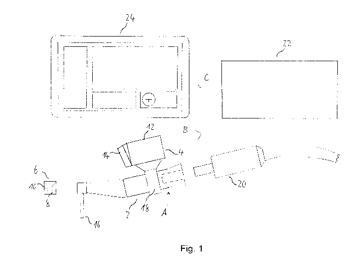
Fig. 1 shows a schematic side view of a navigation system
according to an embodiment of the present invention;
Fig. 2 shows a perspective view of the navigation system
shown in Figure 1;
Fig. 3 shows a schematic cross-sectional view of a marker
member according to an embodiment of the present invention in
a partly detached state from a patient's jaw; and
CA 02926261 2016-04-04
International Application PCT/EP2013/070553
Mininavident AG
Our Ref.: 166 428 v6/abr
24
Fig. 4 shows a schematic perspective view of a positioning
tool of an embodiment of the present disclosure.
Detailed description of currently preferred embodiments
Fig. 1 shows a schematic side view of a navigation system for
dental and cranio-maxillofacial surgery according to a
currently preferred embodiment of the present invention.
The navigation system comprises a surgical handpiece, namely
a dental drill 2, an imaging unit 4 which is movably attached
to the drill 2 and a marker member 6 which is attachable to a
cranial bone, a facial bone, a tooth or teeth of a patient.
The marker member 6 comprises a plurality of marker elements,
namely reference lines 8 and reference points 10, which are
detectable by the imaging unit 4.
The reference lines 8 and the reference points 10 are printed
onto a surface of a body of the marker member 6 and arranged
in a two-dimensional optical pattern. The body of the marker
member 6 is made of a rigid material, such as hard plastic,
metal, ceramic or the like. The
marker member 6 further
comprises an attachment element (not shown), such as a screw
element, a clamp element, an adhesive element or the like,
for attaching the marker member 6 to a cranial bone, a facial
bone, a tooth or teeth of the patient.
Further, the marker member 6 comprises a radiopaque material
in the form of a plurality of radiopaque beads (not shown)
made from titanium or tungsten which are disposed within the
body of the marker member 6.
The imaging unit 4 comprises a camera unit 12 capable of
imaging in the visible light spectrum and an optical 3D
surface scanner 14 for optically scanning the surface of the
patient's teeth or bone structure. The imaging unit 4 is
AMENDED SHEET
CA 02926261 2016-04-04
WO 2015/048994 PCT/EP2013/070553
configured to detect the marker elements 8, 10 of the marker
member 6.
The imaging unit 4 comprises an electronic device (not
shown), such as a chipset, for pre-processing data, such as
imaging data, provided by the camera unit 12. In particular,
the data may be pre-processed by performing a data reduction,
e.g., using a region of interest.
The imaging unit 4 is rotatably attached to the drill 2
through an attachment member 18, so as to be rotatable
relative to the drill 2 around a longitudinal axis of the
drill 2, as is indicated by arrow A in Fig. 1. The imaging
unit 4 is continuously rotatable relative to the drill 2 over
an angular range of 360 and lockable relative to the drill 2
in any angular position.
The imaging unit 4 may be arranged so as to be rotatable
relative to the drill 2 around one or more axes perpendicular
to the longitudinal axis of the drill 2 or rotatable both
around the longitudinal axis and one or more axes
perpendicular to the longitudinal axis.
The attachment member 18 comprises a sensor unit (not shown),
such as a piezoelectric sensor, for detecting the angular
position of the imaging unit 4 relative to the drill 2 and a
drive element (not shown), such as an electric motor, for
rotating the imaging unit 4 relative to the drill 2 around
the longitudinal axis of the drill 2.
The drill 2 comprises a drill head or drill tip 16. The
drill 2 is connectable to a surgical micro-motor 20 for
supplying power to the drill 2, driving the drill tip 16 so
as to rotate.
The navigation system according to the embodiment shown in
Fig. 1 further comprises a processing unit 22 and a display
CA 02926261 2016-04-04
WO 2015/048994 PCT/EP2013/070553
26
unit 24. The imaging unit 4 is configured to transmit, by
wired or wireless transmission, imaging data of the area of
surgery, including the marker elements 8, 10 of the marker
member 6, to the processing unit 22, as is indicated by arrow
B in Figure 1.
The processing unit 22 is configured to further process the
pre-processed imaging data transmitted thereto by the imaging
unit 4. In particular, the processing unit 22 is configured
to perform a 6D-processing of the imaging data.
Further, the processing unit 22 is configured to wirelessly
transmit the processed imaging data in real time to the
display unit 24, as is indicated by arrow C in Fig. 1.
The display unit 24 is configured to combine this processed
imaging data of the processing unit 22 with imaging data
obtained in X-ray imaging, such as cone beam CT, performed
prior to surgery, and with treatment planning data provided
by surgery planning software. In
particular, the display
unit 24 is configured to combine the processed imaging data
of the processing unit 22 and the imaging data of the X-ray
imaging using the marker member 6 as a reference for both the
imaging data obtained by the imaging unit and the X-ray
imaging data, thereby generating three-dimensional imaging
data of the area of surgery in real time.
Further, the display unit 24 is configured to display the
combined imaging data three-dimensionally and in real time.
The display unit 24 displays a target system, such as the
target system detailed above, based on the treatment planning
data, enabling the surgeon to reliably navigate the drill 2,
e.g., the tip 16 thereof, along the treatment planning in
terms of position and angulation.
In the following, an example of the operation of the
navigation system shown in Fig. 1, exemplifying an embodiment
CA 02926261 2016-04-04
WO 2015/048994 PCT/EP2013/070553
27
of the navigation method of the invention, will be described
with reference to Fig. 2.
First, as is shown in Fig. 2, the marker member 6 is attached
to the jaw bone 26 of the patient in the area in which
surgery is to be performed, by using the attachment element
thereof.
Subsequently, the patient is subjected to X-ray
imaging, such as cone beam CT, thereby obtaining X-ray
imaging data of the area of surgery, using the radiopaque
material of the marker member 6 as a reference. The X-ray
imaging data obtained in this way is stored in a memory (not
shown) of the display unit 24.
When starting the surgical procedure, the imaging unit 4 is
rotated relative to the drill 2 so as to detect the marker
elements 8, 10 of the marker member 6 attached to the jaw
bone 26, as is indicated by dashed lines in Fig. 2. The
imaging unit 4 images at least part of the area in which
surgery is performed and transmits the obtained imaging data
to the processing unit 22 in real time.
The processing unit 22 is configured to determine whether the
plurality of marker elements 8, 10 are detected by the
imaging unit 4. If it is determined by the processing unit
22 that the marker elements 8, 10 are not detected by the
imaging unit 4, the imaging unit 4 is rotated relative to the
drill 2 around the longitudinal axis thereof by the drive
element of the attachment member 18 to a position in which
the imaging unit 4 detects the marker elements 8, 10. The
imaging unit 4 is locked in this position relative to the
drill 2 by the attachment member 18.
In this way, it is ensured that the marker elements 8, 10 are
detected by the imaging unit 4 substantially throughout the
surgical procedure, so that the relative position between the
drill 2 and the marker elements 8, 10 of the marker member 6,
CA 02926261 2016-04-04
WO 2015/048994 PCT/EP2013/070553
28
and thus the area of surgery, can be reliably determined at
any time during the surgical procedure.
The imaging data transmitted by the imaging unit 4 to the
processing unit 22 in real time is further processed and
wirelessly transmitted to the display unit 24 by the
processing unit 22. In the display unit 24, the processed
imaging data is combined, i.e., superposed or overlaid, with
the treatment planning data and with the X-ray imaging data
stored in the memory of the display unit 24, using the
radiopaque material and the marker elements 8, 10 of the
marker member 6 as a reference, thereby generating real time
three-dimensional imaging data of the area in which surgery
is performed.
The three-dimensional real time imaging data thus generated
and the target system based on the treatment planning data
are displayed to the surgeon by the display unit 24. In
particular, the display unit 24 may be arranged next to,
e.g., beside or above, the patient's head during surgery,
allowing the surgeon to substantially simultaneously observe
the area of surgery and follow the real time three-
dimensional imaging data and the target system displayed on
the display unit 24.
Alternatively or additionally, the
target system may be displayed on one or more display members
(not shown), e.g., miniature displays, provided on the
imaging unit 4. In
particular, the one or more display
members may be arranged on a rear side of the imaging unit 4,
opposite to a front side of the imaging unit 4 where the
optical 3D surface scanner 14 is provided.
In this way, the surgeon is precisely and reliably guided by
the navigation system during the surgical procedure.
Fig. 3 shows a schematic cross-sectional view of the
patient's jaw bone 26, illustrating an attachment element of
CA 02926261 2016-04-04
International Application PCT/EP2013/070553
Mininavident AG
Our Ref.: 166 428 v6/abr
29
the marker member 6 and a method of attaching the marker
member 6 to the jaw bone 26.
The marker member 6 comprises a plurality of recesses or
sockets 28, namely three sockets 28 in the embodiment shown
in Fig. 3, which are attached to a bottom portion of the body
of the marker member 6 through a connection element 30. The
sockets 28 are configured for receiving heads 32 of screws
34. The
sockets 28 and the screws 34 together form the
attachment element of the marker member 6.
The screws 34 are screwed into the jaw bone 26 of the
patient, as is schematically shown in Fig. 3. Subsequently,
the marker member 6 is placed on top of the screws 34, so
that the screw heads 32 are received within the sockets 28,
thereby fixedly attaching the marker member 6 to the screws
34 and thus also the jaw bone 26.
In this way, the marker member 6 can be securely and reliably
attached directly to the jaw bone 26.
Hence, the marker
member 6 of the embodiment shown in Fig. 3 can be used
particularly advantageously for edentulous or toothless
patients.
Fig. 4 shows a schematic perspective view of a positioning
tool 40 according to an embodiment of the present disclosure.
The positioning tool 40 comprises a die element 42, such as
an impression tray or a partial impression tray, and a marker
member. The marker member comprises a deformable material 44
as a body thereof, as is indicated by the hatching in Fig. 4,
and a plurality of marker elements 46. The
deformable
material 44 is releasably received in the die element 42.
The plurality of marker elements 46 are arranged on a surface
of the deformable material 44 which faces the die element 42,
i.e., an inner surface of the die element 42.
AMENDED SHEET
CA 02926261 2016-04-04
International Application PCT/EP2013/070553
Mininavident AG
Our Ref.: 166 428 v6/abr
The deformable material 44 is a hardenable or curable paste,
such as an impression paste. The deformable material 44 is
deformable so as to conform, for example, to the teeth of the
patient. The marker elements 46 are made of metal, ceramic,
plastic or the like and have a color which is different from
that of the deformable material 44.
The marker elements 46 are partially embedded in the
deformable material 44 so as to be visible on the surface
thereof, thus forming an optically visible pattern which is
detectable by the imaging unit 4, in particular, the camera
unit 12. The deformable material 44 and the marker elements
46 in combination form the marker member according to the
embodiment of the present disclosure shown in Fig. 4.
In the following, an example of the operation of the
positioning tool 40 shown in Fig. 4, exemplifying an
embodiment of the method of the disclosure of positioning the
marker member on a patient's cranial bone, facial bone, tooth
or teeth, will be explained.
The die element 42 with the deformable material 44 releasably
received therein is placed over the teeth of the patient in
the area in which surgery is to be performed. The
die
element 42 is pressed onto the teeth, thus deforming the
deformable material 44 and conforming the deformable material
44 to the teeth, thereby attaching the deformable material 44
to the teeth at least by a form fit between the deformable
material 44 and the teeth. The
die element 42 with the
deformable material 44 received therein may be maintained in
this position on the patient's teeth for a predetermined
period of time, in order to allow the deformable material 44
to harden or cure.
Subsequently, the die element 42 is removed from the teeth of
the patient, while the deformable material 44 is held in its
position at least by the form fit with the teeth and, in some
AMENDED SHEET
CA 02926261 2016-04-04
WO 2015/048994 PCT/EP2013/070553
31
embodiments, also by an adhesive force between the,
preferably hardened or cured, deformable material 44 and the
teeth. In this way, the deformable material 44 is released
from the die element 42 upon removal of the die element 42
from the teeth.
The deformable material 44 is thus securely held on the teeth
of the patient. The surface of the deformable material 44
which faced the die element 42 forms an outer surface of the
attached marker member, which is comprised of the deformable
material 44 and the marker elements 46, and has the marker
elements 46 arranged thereon. These marker elements 46 are
detectable by the imaging unit 4 of the navigation system, as
is indicated by dashed lines in Fig. 4.
The positioning tool 40 allows for a particularly reliable
and precise positioning and attachment of the marker member
to the patient's cranial bone, facial bone, tooth or teeth
and can thus be particularly advantageously used with the
navigation system of the invention.
A set of positioning tools 40 is obtained by providing four
positioning tools 40 as shown in Fig. 4, wherein the die
elements 42 of the four positioning tools 40 have different
shapes and/or sizes from each other. In particular, the set
of positioning tools 40 may consist of four positioning tools
40, the die element 42 of each of which is configured, e.g.,
shaped and sized, for placement over the teeth of a different
quadrant of the human or animal jaw.
The foregoing embodiments and their variants have been
disclosed for illustrative purposes only, and further
variation is wholly possible within the capabilities of the
skilled reader. Accordingly, the appended claims are
intended to cover all modifications, substitutions,
alterations, omissions and additions which one skilled in the
art could achieve from the foregoing disclosure, taking into
CA 02926261 2016-04-04
WO 2015/048994 PCT/EP2013/070553
32
account his own general and speci,,713t knowledge and
expertise.