Note: Descriptions are shown in the official language in which they were submitted.
CA 02928235 2016-04-19
WO 2015/061685 PCT/US2014/062178
METHODS FOR TREATMENT OF MUSCULAR DYSTROPHIES
Claims of Priority
The present application claims priority under 35 U.S.C. 119(e) to U.S.
provisional
patent application, U.S.S.N. 61/895, 832, filed on October 25, 2013, which is
incorporated herein
by reference.
Background of Invention
The muscular dystrophies (MD) are a group of more than 30 genetic diseases
characterized by progressive weakness and degeneration of the skeletal muscles
that control
movement. MD weaken the musculoskeletal system and hamper locomotion. MD are
caused by
progressive degeneration of skeletal muscle fibres. The disease is
characterized by defects in
muscle proteins and the death of muscle cells and tissue.
Dystrophinopathies are a group of muscular dystrophies resulting from
mutations in the
dystrophin gene, located on the short arm of the X chromosome in the Xp21
region [Kunkel et
al. 1985; Monaco et al. 1985; Ray et al. 1985]. Of these, Duchenne muscular
dystrophy (DMD)
is the most common dystrophinopathy resulting from complete absence of the
dystrophin gene
product, the subsarcolemmal protein dystrophin [Hoffman et al. 1987a; Koenig
et al. 1987;
Hoffman et al. 1988]. Its allelic variant, Becker's muscular dystrophy (BMD)
is rarer with varied
severity and time of presentation.
Duchenne muscular dystrophy (DMD) is a relentlessly progressive skeletal
muscle
disorder which, left to its natural course, results in premature death by
respiratory failure by late
teens, early twenties. The incidence of DMD is approximately 1 in 3300
[Jeppesen et al. 2003;
CDC 2007] to 1:4700 [Dooley 2010] male births. Although a common mode of
inheritance is X-
linked recessive (i.e., the mother is a carrier), this disorder is associated
with a high spontaneous
mutation rate contributing to approximately 30% of cases [Brooks and Emery
1977; van Essen et
al. 1992]. This mutation rate is estimated to be 10 times higher than for any
other genetic
disorder [Hoffman et al. 1992] because of the extremely large Duchenne gene
size [Hoffman and
Kunkel 1989]. The 2.5 million base pairs constituting the gene (a full 1% of
the X chromosome)
provide a large target for random mutational events. Because of this high
mutation rate,
eradication of the disease through genetic counseling has proven difficult.
1
CA 02928235 2016-04-19
WO 2015/061685 PCT/US2014/062178
Current therapeutic approaches to MD, e.g., DMD include the use of anabolic
drugs, e.g.,
steroids, such as prednisolone, deflazacort, and dantrolene, which generally
result in modest
beneficial effects. However, treatment with anabolic drugs may also be
accompanied by severe
side-effects, including osteoporosis, hypertension, Cushing syndrome, weight
gain, cataracts,
short stature, gastrointestinal symptoms, behavioural changes, and liver
damage. There is a need
for new and improved treatments for MD, e.g., DMD.
Summary of the Invention
The present invention encompasses the recognition that unwanted side effects
of anabolic
drugs e.g., steroids, for treatment of MD, e.g., DMD, may be related to their
relevant effects on
androgen-sensitive tissues other than skeletal muscle, with the possibility
that beneficial effects
are masked by the action of the steroids on off-target sites. The present
invention provides,
among other things, compositions as described herein, e.g., a composition
comprising the
Compound (I), or a pharmaceutically acceptable salt, metabolite, or prodrug
thereof, that have
more specific actions on bone and skeletal muscle, e.g., as compared to
anabolic drugs, and can
be an alternative to treatment with anabolic drugs, e.g., steroids. The
present invention provides,
at least in part, methods for treating MD, e.g., DMD, and methods and kits for
evaluating,
identifying, and/or treating a subject, e.g., a subject suffering from or
susceptible to MD, e.g., a
subject suffering from or susceptible to DMD, with compositions comprising the
Compound (I),
or a pharmaceutically acceptable salt, metabolite, or prodrug thereof.
Provided compositions and
methods permit treatment of MD, e.g., DMD, with reduced associated negative
side effects.
In one aspect, the invention provides a method of treating muscular dystrophy
in a
subject, the method comprising administering to a subject suffering from
muscular dystrophy a
therapeutically effective amount of the Compound (I), or a pharmaceutically
acceptable salt
thereof,
0
N C F3A
41 $ N 41# =N
% 0
OH (I),
thereby treating the subject. In some embodiments, the muscular dystrophy is
selected from
Duchenne Muscular Dystrophy, Becker Muscular Dystrophy, Emery-Dreifuss
Muscular
Dystrophy, Limb-Girdle Muscular Dystrophy, Facioscapulohumeral Muscular
Dystrophy,
2
CA 02928235 2016-04-19
WO 2015/061685 PCT/US2014/062178
Myotonic Dystrophy, Oculopharyngeal Muscular Dystrophy, Distal Muscular
Dystrophy, or
congenital muscular dystrophy. In some embodiments, the muscular dystrophy is
Duchenne
Muscular Dystrophy.
In some embodiments, the method comprises partial or complete alleviation of
an
awkward manner of walking, stepping, or running; frequent falls; fatigue;
difficulty with motor
skills; muscle fiber deformities; pseudohypertrophy; skeletal deformities; low
endurance;
difficulties in standing unaided or inability to ascend staircases; loss of
movement; paralysis;
cardiomyopathy; development of congestive heart failure; and irregular
heartbeat.
In some embodiments, the method improves (e.g., increasing, prolonging)
lifespan. In some
embodiments, the method comprises improving at least one symptom e.g., a
symptom as
described herein. In some embodiments, the symptom is fatigue, learning
difficulties,
intellectual disability, muscle weakness, difficulty with motor skills,
difficulty walking,
breathing difficulty, heart disease, cardiomyopathy, congestive heart failure,
arrhythmia,
scoliosis, pseudohypertrophy, muscle wasting, muscle contractures, muscle
deformities, and
respiratory disorders (e.g., pneumonia).
In some embodiments, the Compound (I) or pharmaceutically acceptable salt
thereof is
administered in multiple doses, e.g., at a predetermined interval. In some
embodiments, the
Compound (I) or pharmaceutically acceptable salt thereof is administered
chronically (e.g., 1, 2,
3, 4, 5, 6, 7, 8, 9, or 10 times every 1, 2, 3, 4, 5, 6, days, 1, 2, 3, 4, 5,
6, 7, 8, 9 weeks, 1, 2, 3, 4,
5, 6, 7, 8, 9 months or longer) (e.g., for 1, 2, 3, 4, 5, 6, days, 1, 2, 3, 4,
5, 6, 7, 8, 9 weeks, 1, 2, 3,
4, 5, 6, 7, 8, 9 months or longer). In some embodiments, the Compound (I) or
pharmaceutically
acceptable salt thereof is administered once daily. In some embodiments, the
Compound (I) or
pharmaceutically acceptable salt thereof is administered in a single dose.
In some embodiments, the Compound (I) or a pharmaceutically acceptable salt
thereof is
administered at a dose of about 0.1 mg to about 1 mg (e.g., 0.1, 0.2, 0.3,
0.4, 0.5, 0.6, 0.7, 0.8,
0.9, or 1 mg) per subject. In some embodiments, the Compound (I) or a
pharmaceutically
acceptable salt thereof is administered at a dose of no more than 1 mg, 0.9
mg, 0.8 mg, 0.7 mg,
0.6 mg, 0.5 mg, 0.4 mg, 0.3 mg, 0.25 mg, 0.2 mg, or 0.1 mg per subject. In
some embodiments,
the dose is 0.1 mg per subject. In some embodiments, the dose is 0.25 mg per
subject.
3
CA 02928235 2016-04-19
WO 2015/061685 PCT/US2014/062178
In some embodiments, the dose is 0.5 mg per subject. In some embodiments, the
dose is 1 mg
per subject. In some embodiments, the dose is from e.g., about 0.2 mg to about
0.8 mg, about
0.3 mg to about 0.7 mg, or about 0.4 mg to about 0.6 mg.
In some embodiments, the Compound (I) or a pharmaceutically acceptable salt
thereof is
administered at a dose of about 2 lig to about 1000 lig per kilogram subject
weight. In some
embodiments, the Compound (I) or a pharmaceutically acceptable salt thereof is
administered at
a dose of no more than 1000 lig, 800 lig, 500 lig, 400 lig, 300 lig, 200 lig,
100 lig, 30 lig, 20 lig,
15 lig, 10 lig, 7 lig, or 2 lig per kilogram subject weight. In some
embodiments, the dose is 2 lig
per kilogram subject weight. In some embodiments, the dose is 7 lig per
kilogram subject
weight. In some embodiments, the dose is 15 lig per kilogram subject weight.
In some
embodiments, the dose is 30 lig per kilogram subject weight. In some
embodiments, the dose is
from about 2 lig to about 1000 lig, from about 5 lig to about 800 lig, from
about 10 lig to about
500 lig, from about 10 lig to about 300 lig, from about 10 lig to about 200
lig, or from about 10
lig to about 100 lig.
In some embodiments, the Compound (I) or a pharmaceutically acceptable salt
thereof is
administered after meal consumption. In some embodiments, the Compound (I) or
a
pharmaceutically acceptable salt thereof is administered at least 60 minutes
after meal
consumption. In some embodiments, the Compound (I) or a pharmaceutically
acceptable salt
thereof is administered about 10 minutes to about 120 minutes after meal
consumption.
In some embodiments, the Compound (I) or a pharmaceutically acceptable salt
thereof is
administered about 10 minutes, about 20 minutes, about 30 minutes, about 45
minutes, about 60
minutes, about 75 minutes, about 90 minutes, about 105 minutes, or about 120
minutes after
meal consumption. In some embodiments, the Compound (I) or a pharmaceutically
acceptable
salt thereof is administered before meal consumption. In some embodiments, the
Compound (I)
or a pharmaceutically acceptable salt thereof is administered about 10 minutes
to about 60
minutes before meal consumption. In some embodiments, the Compound (I) or a
pharmaceutically acceptable salt thereof is administered about 10 minutes,
about 20 minutes,
about 30 minutes, or about 45 minutes before meal consumption. In some
embodiments, the
Compound (I) or a pharmaceutically acceptable salt thereof is administered
from 60 minutes
before meal consumption to 2 hours after meal consumption.
4
CA 02928235 2016-04-19
WO 2015/061685 PCT/US2014/062178
In some embodiments, the compound converts in vivo to the Compound (II), or a
pharmaceutically acceptable salt or metabolite thereof,
0 CF3
Ni4
HO 44* N 411 ¨N
k 0
OH (II).
In some embodiments, the Compound (I) or pharmaceutically acceptable salt or
composition thereof, is administered via oral, subcutaneous, intravenous,
intramuscular,
intranasal, transdermal, transmucosal, buccal, sublingual, or lung
administration. In some
embodiments, the Compound (I) or pharmaceutically acceptable salt or
composition thereof, is
administered via oral administration.
In some embodiments, the subject is human. In some embodiments, the subject is
male.
In some embodiments, the subject is pediatric. In some embodiments, the
subject is
prepubescent. In some embodiments, the subject is from the age of about 1 year
to about 18
years. In some embodiments, the subject has diseased muscle (e.g., atrophy,
fibrotic).
In some embodiments, the Compound (I) is substantially free of any salts or
impurities.
In some embodiments, the compound is in at least 95% enantiomeric excess. In
some
embodiments, the compound is in at least 98% enantiomeric excess. In some
embodiments, the
compound is in at least 99% enantiomeric excess.
In some embodiments, the levels of testosterone in the treated subject are not
substantially changed as compared to levels of testosterone in the subject
before treatment.
In some embodiments, the method of treatment is substantially free of any side
effects
e.g., obesity, behavior problems, thinner and/or weaker bones (osteoporosis);
delayed puberty,
stomach problems (gastroesophageal reflux or GERD), cataracts, sensitivity to
infections;
hypogonadism, muscle wasting and osteoporosis; cardiovascular risk (e.g.,
cardiovascular
disease, coronary artery disease, hypertension, cardiac arrhythmias,
congestive heart failure,
heart attacks, sudden cardiac death); prostate cancer risks, hypogondism, and
conditions
pertaining to hormonal imbalances (e.g., induction of male puberty,
gynecomastia, testicular
atrophy, and decreased sperm production).
CA 02928235 2016-04-19
WO 2015/061685 PCT/US2014/062178
In some embodiments, the compound is characterized by one or both of: (a)
higher
activity on muscle and bones of the subject as compared to anabolic steroid
treatment; and (b)
lower activity on prostate of the subject as compared to anabolic steroid
treatment.
In one aspect, the invention provides a pharmaceutical composition comprising
the
Compound (I) or a pharmaceutically acceptable salt, metabolite or prodrug
thereof,
0 C F3
NJ(
411 N 41 -N
µ 0
OH (I),
wherein the pharmaceutical composition comprises about 0.1 mg to about 1 mg of
the
Compound (I) or a pharmaceutically acceptable salt thereof. In some
embodiments, the
pharmaceutical composition comprises 0.1, 0.2, 0.25, 0.3, 0.4, or 0.5 mg of
the Compound (I), or
a pharmaceutically acceptable salt thereof. In some embodiments, the
pharmaceutical
composition comprises a pharmaceutically acceptable excipient.
In some embodiments, the pharmaceutical composition is configured in a unit
dosage
form. In some embodiments, the pharmaceutical composition is configured in a
solid dosage
form (e.g., a capsule, a tablet). In some embodiments, the solid dosage form
is selected from the
group consisting of tablets, capsules, sachets, powders, granules and
lozenges. In some
embodiments, the pharmaceutical composition is configured in a liquid dosage
form.
In some embodiments, the pharmaceutical composition further comprises
administering
an additional therapeutic agent. In some embodiments, the additional
therapeutic agent is a
steroidal compound. In some embodiments, the steroidal compound is a
corticosteroid, e.g.,
prednilosone. In some embodiments, the therapeutic agent is a non-steroidal
compound.
In one aspect, the invention provides a pharmaceutical composition comprising
the
Compound (I) or a pharmaceutically acceptable salt thereof,
0 CF3
NJ.(
411 N 41 -N
µ 0
OH (I),
6
CA 02928235 2016-04-19
WO 2015/061685 PCT/US2014/062178
configured in a dosage form comprising no more than about 0.1 mg to about 1 mg
of the
Compound (I) or a pharmaceutically acceptable salt thereof per dosage form.
In one aspect, the invention provides a kit comprising the pharmaceutical
composition of
claim 34, and instructions for oral administration of the pharmaceutical
composition to a subject
in the dosage form of about 0.2 lig to about 1000 lig per kilogram subject
weight.
In one aspect, the invention provides a kit comprising one or more of:
Compound (I), a
composition comprising Compound (I), and instructions for use in treating a
subject having MD,
e.g., DMD.
In one aspect, the invention provides a method of treating muscular dystrophy
in a
subject, the method comprising:determining whether a subject suffers from or
is susceptible to
muscular dystrophy; selecting the subject for treatment based on the
determining; administering
a therapeutically effective amount of the Compound (I) or a pharmaceutically
acceptable salt
thereof, thereby treating muscular dystrophy in the subject. In some
embodiments, the
determining comprises comparing an observed value with a reference value. In
some
embodiments, said subject is evaluated for a parameter described herein, e.g.,
as described in
method of diagnosis described herein. In some embodiments, the determining
comprises
measuring muscle atrophy, e.g., walk test, stair climbing test.
All publications, patent applications, patents, and other references mentioned
herein are
incorporated by reference in their entirety.
Brief Description of the Drawings
The invention is herein described, by way of example only, with reference to
the
accompanying drawings.
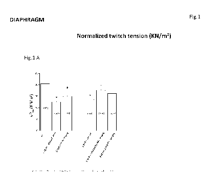
Figure 1 depicts exemplary effects of the drug treatments on contractile
properties
(twitch tension) of diaphragm.
Figure 2 depicts exemplary effects of the drug treatments on contractile
properties
(tetanic tension) of diaphragm.
7
CA 02928235 2016-04-19
WO 2015/061685 PCT/US2014/062178
.Figure 3 depicts exemplary effects of the drug treatments on contractile
properties (time
to peak) of diaphragm.
Figure 4 depicts exemplary effects of the drug treatments on contractile
properties
(relaxation time) of diaphragm.
Figure 5 depicts exemplary effects of the drug treatments on contractile
properties (ratio
of twitch tension to tetanic tension) of diaphragm.
Figure 6 depicts exemplary effects of the drug treatments on contractile
properties of
diaphragm.
Figure 7 depicts exemplary effects of the drug treatments on contractile
properties
(fatigue) of diaphragm.
Figure 8 depicts exemplary effects of the drug treatments on contractile
properties
(twitch tension) of EDL.
Figure 9 depicts exemplary effects of the drug treatments on contractile
properties
(tetanic tension) of EDL.
.Figure 10 depicts exemplary effects of the drug treatments on contractile
properties
(time to peak) of EDL.
Figure 11 depicts exemplary effects of the drug treatments on contractile
properties
(relaxation time) of EDL.
Figure 12 depicts exemplary effects of the drug treatments on contractile
properties (ratio
of twitch tension to tetanic tension) of EDL.
Figure 13 depicts exemplary effects of the drug treatments on contractile
properties of
EDL.
Figure 14 depicts exemplary effects of the drug treatments on contractile
properties
(fatigue) of EDL.
Figure 15 depicts exemplary effects of the drug treatments on mechanical
threshold.
Figure 16 depicts exemplary effects of the drug treatments on mechanical
threshold.
Figure 17 depicts exemplary effects of the drug treatments on mechanical
threshold.
Figure 18 depicts the effect of single drug treatment on total membrane ionic
conductance of EDL muscle fibers of mdx mice.
Figure 19 depicts exemplary effects of the drug treatments on levels of
creatine kinase.
8
CA 02928235 2016-04-19
WO 2015/061685 PCT/US2014/062178
Figure 20 depicts exemplary effects of the drug treatments on levels of
lactate
dehydrogenase.
Figure 21 depicts exemplary effects of the drug treatments on levels of
reactive oxygen
species.
Figure 22 depicts representative pictures of histology profile of diaphragm
and GC
muscles.
Figure 23 depicts representative morphometric analysis following drug
treatment.
Figure 24 depicts exemplary in vivo parameters for wild-type and mdx mice at
the
beginning and after 4 weeks treatment with and without Compound (I), NAND, and
PDN.
Figure 25 depicts exemplary in vivo parameters of wild-type and mdx mice
treated with
and without Compound (I) at 0.3, 3, and 30 mg/kg for up to 12 weeks.
Figure 26 depicts exemplary effects of treatment with Compound (I) on weight
of
androgen-sensitive and other potential target tissues.
Figure 27 depicts exemplary dose- and time-dependent effects of Compound (I)
on the
weight of androgen-sensitive tissues and other potential target tissues.
Figure 28 shows exemplary values of the maximal isometric twitch and tetanic
tension of
the diaphragm muscle from wt and mdx mice with various drug treatments.
Figure 29 depicts exemplary isometric and eccentric contraction of isolated
EDL
muscles from wild-type and mdx mice treated with and without Compound (I).
Figure 30 shows exemplary functional cellular parameters in EDL muscles in
wild-type
and mdx mice treated with and without Compound (I) and NAND, and PDN.
Figure 31 depicts exemplary functional cellular parameters in EDL muscles in
wild-type
and mdx mice treated with and without Compound (I).
Figure 32 depicts exemplary haematoxylin-eosin staining of the diaphragm and
gastrocnemius muscles from mdx mice treated with and without Compound (I).
Figure 33 shows exemplary effect on fibrosis markers of mdx mice treated with
and
without Compound (I), NAND, and PDN.
Figure 34 depicts exemplary plasma levels of Compound (I) over 8 hours after
subcutaneous delivery of the compound into mice.
Figure 35 shows exemplary serum testosterone levels for wild-type and
exercised or not-
exercised mdx mice treated with and without Compound (I).
9
CA 02928235 2016-04-19
WO 2015/061685 PCT/US2014/062178
Figure 36 depicts exemplary levels of target genes as compared to housekeeping
gene
GADPH after treatment with and without Compound (I).
Detailed Description
Definitions
As used herein, the articles "a" and "an" refer to one or to more than one
(e.g., to at least
one) of the grammatical object of the article.
"About" and "approximately" shall generally mean an acceptable degree of error
for the
quantity measured given the nature or precision of the measurements. Exemplary
degrees of
error are within 20 percent (%), typically, within 10%, and more typically,
within 5% of a given
value or range of values.
"Sample," "tissue sample," "subject or patient sample," "subject or patient
cell or tissue
sample" or "specimen" each refers to a biological sample obtained from a
tissue, e.g., a bodily
fluid, of a subject or patient. The source of the tissue sample can be solid
tissue as from a fresh,
frozen and/or preserved organ, tissue sample, biopsy, or aspirate; blood or
any blood constituents
(e.g., serum, plasma); bodily fluids such as cerebral spinal fluid, whole
blood, plasma and serum.
The sample can include a non-cellular fraction (e.g., plasma, serum, or other
non-cellular body
fluid). In one embodiment, the sample is a serum sample. In other embodiments,
the body fluid
from which the sample is obtained from an individual comprises blood (e.g.,
whole blood). In
certain embodiments, the blood can be further processed to obtain plasma or
serum. In some
embodiments, the sample contains a tissue, cells (e.g., peripheral blood
mononuclear cells
(PBMC)). In an embodiment the sample includes NK cells. For example, the
sample can be a
fine needle biopsy sample, an archival sample (e.g., an archived sample with a
known diagnosis
and/or treatment history), a histological section (e.g., a frozen or formalin-
fixed section, e.g.,
after long term storage), among others (e.g., a muscle tissue section, e.g.,
skeletal muscle, cardiac
muscle, smooth muscle). The term sample includes any material obtained and/or
derived from a
biological sample, including a polypeptide, and nucleic acid (e.g., genomic
DNA, cDNA, RNA)
purified or processed from the sample. Purification and/or processing of the
sample can involve
one or more of extraction, concentration, antibody isolation, sorting,
concentration, fixation,
addition of reagents and the like. The sample can contain compounds that are
not naturally
CA 02928235 2016-04-19
WO 2015/061685 PCT/US2014/062178
intermixed with the tissue in nature such as preservatives, anticoagulants,
buffers, fixatives,
nutrients, antibiotics or the like.
As used herein, "modulators" or "modulate" refers to the regulation of a
protein (e.g.,
enzyme, receptor (e.g., androgen receptor)) by the binding of a ligand (e.g.,
compound, drug).
Binding may be e.g., irreversible, reversible, complete or partial, at the
active site or at an
allosteric binding site. Modulators include antagonists, agonists, agonist-
antagonists, partial
antagonists, partial agonists. An "agonist" is a chemical, e.g., ligand,
compound, drug, that binds
to and/or upregulates some receptor (e.g., androgen receptor) of a cell and
triggers a cellular
response that often mimics the action of a naturally occurring substance. For
example, an
endogenous agonist for a particular receptor is a naturally occurring compound
produced by the
body that binds to and activates that receptor, e.g., endogenous agonists for
the androgen
receptor are androgens. An "antagonist" is a type of ligand or drug that does
not provoke a
biological response itself upon binding to a receptor, but blocks, dampens, or
downregulates
agonist-mediated responses. Antagonists generally have affinity but no
efficacy for their cognate
receptors, but disrupt the interaction and inhibit the function of an agonist
or inverse agonist at
receptors. Antagonists may be reversible or irreversible depending on the
longevity of the
antagonist-receptor complex. It will be appreciated by a person of skill in
the art that the activity
of a compound of the invention as an antagonist (complete or partial) or
agonist (complete or
partial) represents a continuous spectrum. Therefore, while some compounds
will be clearly
agonists or clearly antagonists, some compounds will exhibit both agonistic
and antagonistic
activity.
Methods of treatment
The present invention relates to, inter alia, methods for treating MD, e.g.,
DMD,
comprising administering a composition comprising a compound as described
herein, e.g.,
Compound (I), or a pharmaceutically acceptable salt, metabolite, or prodrug
thereof. Provided
compositions and methods of the present invention may, for example, increase
skeletal muscle
mass and/or strength, enhance protein synthesis, as well as enhance
regeneration and/or metabolic
efficiency.
11
CA 02928235 2016-04-19
WO 2015/061685 PCT/US2014/062178
Muscular Dystrophy
MD are a group of more than 30 genetic diseases characterized by progressive
weakness
and degeneration of the skeletal muscles that control movement. MD weaken the
musculoskeletal system and hamper locomotion. Some forms of MD are seen in
infancy or
childhood, while others may not appear until middle age or later. The
disorders differ in terms of
the distribution and extent of muscle weakness (some forms of MD also affect
cardiac muscle),
age of onset, rate of progression, and pattern of inheritance. MD are caused
by progressive
degeneration of skeletal muscle fibers. MD are characterized by defects in
muscle proteins and
the death of muscle cells and tissue. In the most severe forms, such as DMD,
regeneration is
exhausted and skeletal muscle is progressively replaced by fat and fibrous
tissue. DMD generally
causes progressive weakness in the patient and eventually death by respiratory
and/or cardiac
failure.
Dystrophinopathies are a group of muscular dystrophies resulting from
mutations in the
dystrophin gene, located on the short arm of the X chromosome in the Xp21
region [Kunkel et
al. 1985; Monaco et al. 1985; Ray et al. 1985]. Of these, Duchenne muscular
dystrophy (DMD)
is the most common dystrophinopathy resulting from complete absence of the
dystrophin gene
product, the subsarcolemmal protein dystrophin [Hoffman et al. 1987a; Koenig
et al. 1987;
Hoffman et al. 1988]. Its allelic variant, Becker's muscular dystrophy (BMD)
is rarer with varied
severity and time of presentation.
The dystrophin gene is the largest human gene isolated to date. About 90% of
boys have
an absence of dystrophin corresponding to an "out-of-frame" mutation that
disrupts normal
dystrophin transcription [Gillard et al. 1989]. These mutations can cause a
premature stop codon
and early termination of mRNA transcription. As a result, an unstable RNA can
be produced,
that undergoes rapid decay, and leads to the production of nearly undetectable
concentrations of
truncated protein. If the mutation maintains translational reading, an "in-
frame" deletion, the
BMD phenotype with variably decreased amounts of abnormal molecular weight
dystrophin, is
present [Hoffman et al. 1988]. This reading frame hypothesis holds for about
90% of cases and is
commonly used both as a diagnostic confirmation of dystrophinopathies and for
the differential
diagnosis of DMD and BMD. Exceptions to these two typical situations occur in
approximately
10-13% of patients. [Nevo et al. 2003], [Muntoni et al. 1994]. About 60% of
Duchenne and
Becker patients manifest structural rearrangements of the deletion type
[Kunkel 1986; den
12
CA 02928235 2016-04-19
WO 2015/061685 PCT/US2014/062178
Dunnen et al. 1987]. Two deletion hotspots includes exons 45-55 and exons 2-19
[Den Dunnen
et al. 1989; Oudet et al. 1992; Nobile et al. 1995]. The other 40% of patients
results from small
mutations (point mutations resulting in frame-shift or nonsense mutations) or
duplications.
Because the genetic defect is an X-linked recessive trait, dystrophinopathies
are expressed
primarily in boys and young men. However, girls may manifest symptoms of DMD
if they also
exhibit skewed X-inactivation [Lesca et al. 2003].
Dystrophin localizes to the subsarcolemmal region in skeletal and cardiac
muscle and
composes 0.002% of total muscle protein [Hoffman et al. 1987a]; [Hoffman et
al. 1987b].
Dystrophin binds to the cytoskeletal actin and to the cytoplasmic tail of the
transmembrane
Dystrophin glycoprotein complex (DGC) protein alpha-dystroglycan, and thus
forms a link from
the cytoskeleton to the extracellular matrix. Dystrophin is organized in
costamers and is present
in greater amounts at myotendinous and neuromuscular junctions than in other
muscle areas. In
the heart it is also associated with T tubules. In smooth muscle it is
discontinuous along
membranes alternating with vinculin.
Muscle cell death in the muscular dystrophies (by apoptosis and necrosis) may
be
conditional and reflects a propensity that varies between muscles and changes
with age [Rand
2001a]. The fact that adjacent muscle groups in DMD can be completely normal
while others are
undergoing active necrosis suggests progression is not inevitable and may be
treatable. If
endogenous biochemical mechanisms alter the susceptibility of a muscle cell to
live or die while
the genetic and biochemical defects remain constant, then pharmacological
modulation of these
pathways may result in successful therapies for DMD and other muscular
dystrophies [Rand
2001b]. Signs and symptoms of MD include progressive muscular wasting, poor
balance,
drooping eyelids, atrophy, scoliosis, inability to walk, frequent falls,
waddling gait, calf
deformation, limited range of movement, respiratory difficulty, joint
contractures,
cardiomyopathy, arrhythmias, and muscle spasms. Symptoms also include fatigue,
learning
difficulties, intellectual disability, muscle weakness, difficult with motor
skills, difficulty
walking, breathing difficulty, heart disease, cardiomyopathy, congestive heart
failure,
arrhythmia, scoliosis, pseudohypertrophy, muscle wasting, muscle contractures,
muscle
deformities, and respiratory disorders (e.g., pneumonia).
13
CA 02928235 2016-04-19
WO 2015/061685 PCT/US2014/062178
Diagnosis of MD can be based on the results of a muscle biopsy,
electromyography,
electrocardiography, DNA analysis, and/or determination of increased creatine
phosphokinase.
A physician's examination and patient's medical history will aid a doctor's
diagnosis in
determining the type of MD a patient presents.
Existing therapeutic approaches to MD can involve steroids, e.g.,
prednisolone,
deflazacort, and dantrolene, which result in modest beneficial effects and are
typically
accompanied by severe side-effects including osteoporosis, hypertension,
Cushing syndrome,
weight gain, cataracts, short stature, gastrointestinal symptoms, behavioural
changes in the case
of steroids, and liver damage.
MD includes, for example, Duchenne, Becker, Limb-girdle, Congenital,
Facioscapulohumeral, Myotonic, Oculopharyngeal, Distal, and Emery-Dreifuss
muscular
dystrophies. In particular embodiments, certain types of MD are characterized,
at least in part,
by a deficiency or dysfunction of the protein dystrophin. Such muscular
dystrophies include
DMD and Becker Muscular Dystrophy (BMD). The various MD are discussed in
further detail
below.
Duchenne muscular dystrophy (DMD). DMD is a relentlessly progressive skeletal
muscle disorder which, left to its natural course, can result in premature
death by respiratory
failure by late teens, early twenties. The incidence of DMD is approximately 1
in 3300 [Jeppesen
et al. 2003; CDC 2007] to 1:4700 [Dooley 2010] male births. Although a common
mode of
inheritance is X-linked recessive (i.e., the mother is a carrier), this
disorder is associated with a
high spontaneous mutation rate contributing to approximately 30% of cases
[Brooks and Emery
1977; van Essen et al. 1992]. This mutation rate is estimated to be 10 times
higher than for any
other genetic disorder [Hoffman et al. 1992] because of the extremely large
Duchenne gene size
[Hoffman and Kunkel 1989]. The 2.5 million base pairs constituting the gene (a
full 1% of the X
chromosome) provide a large target for random mutational events. Because of
this high mutation
rate, eradication of the disease through genetic counseling has proven
difficult.
While dystrophin deficiency can be a primary cause of DMD, multiple secondary
pathways are responsible for the progression of muscle necrosis, abnormal
fibrosis and failure of
regeneration that results in a progressively worsening clinical status. There
is evidence
supporting oxidative radical damage to myofibers [Rand 2002], inflammation
[Spencer and
Tidball 2001; Porter et al. 2002], abnormal calcium homeostasis [Allen 2010;
Millay 2009],
14
CA 02928235 2016-04-19
WO 2015/061685 PCT/US2014/062178
myonuclear apoptosis [Rand 2001b; Sandri et al. 2001; Tews 2002], abnormal
fibrosis and
failure of regeneration [Rand 2001b; Bernasconi 1995]; [Melone 2000; Morrison
2000; Luz
2002]. This body of literature has been validated by cross sectional genome-
wide approaches that
allow an overall analysis of multiple defective mechanisms in DMD [Chen et al.
2000; Porter
2003]. The main symptom of DMD is muscle weakness associated with muscle
wasting first
with the voluntary muscles, e.g., the hips, pelvic area, thighs, shoulders,
and calf muscles.
Muscle weakness also occurs e.g., in the arms, neck, and other areas, but
later than as in the
lower half of the body. Symptoms also include an awkward manner of walking,
stepping, or
running (e.g., patient may walk on their forefeet, because of increased calf
tonus, or toe walk to
compensate for knee extensor weakness). Also, frequent falls, fatigue,
difficulty with motor
skills (e.g., running, hopping, jumping), increased lumbar lordosis, (e.g.,
leading to shortening of
the hip-flexor muscles), muscle contracutures of Achilles tendon and
hamstrings impairing
functionality because muscle fibers shorten and fibrosis occurs in connective
tissue, progressive
difficulty in walking, muscle fiber deformities, pseudohypertrophy (enlarging)
of tongue and calf
muscles, higher risk of neurobehavioral disorders (e.g., attention deficit
hyperactivity disorder,
learning disorders (dyslexia), and non-progressive weaknesses in specific
cognitive skills),
eventual loss of ability to walk, and skeletal deformities may be associated
with patients with
DMD.
Symptoms usually appear in male children before the age of 6 and may be
visible early in
infancy. Even though symptoms do not appear until early in infancy, laboratory
testing can
identify children who carry the active mutation at birth. Exemplary genetic
testing for early
diagnosis of DMD, e.g., before onset of symptoms, are described herein and in
e.g., Prior et al.,
Arch Pathol Lab Med. 1991 Oct;115(10):984-90. Progressive proximal muscle
weakness of the
legs and pelvis associated with a loss of muscle mass is observed first, with
the weakness
eventually spreading to the arms, neck, and other areas. Early signs may
include enlargement of
calf and deltoid muscles (pseudohypertrophy), low endurance and difficulties
in standing
unaided or inability to ascend staircases. As the condition progresses, muscle
tissue experiences
wasting and is eventually replaced by fat and fibrotic tissue (fibrosis). By
age 10, braces may be
required to aid in walking, and by age 12, most patients are wheelchair
dependent.
CA 02928235 2016-04-19
WO 2015/061685 PCT/US2014/062178
Later symptoms may include abnormal bone development that lead to skeletal
deformities, including curvature of the spine. The progressive deterioration
of muscle leads to
loss of movement, eventually leading to paralysis. A patient with DMD may or
may not present
intellectual impairment. When a patient presents intellectual impairment, it
typically does not
progressively worsen with age. The average life expectancy for DMD patients is
around 25
years of age.
DMD may be observed clinically by observing a patient's disintegrating ability
to walk,
for example, between the time a boy is 9 to 12 years of age. Muscle wasting
begins in the legs
and pelvis, progressing to the muscles of the shoulders and neck, followed by
the loss of arm
muscles and respiratory muscles. Calf muscle enlargement (pseudohypertrophy)
can become
apparent. Cardiomyopathy (e.g., dilated cardiomyopathy, DCM) is common, and
the
development of congestive heart failure or irregular heartbeats (arrhythmias)
occurs
occasionally. Children with DMD will usually tire more easily, have less
overall strength than
their peers, may have extremely high levels of creatine kinase, a genetic
error in the Xp21 gene,
and/or have an electromyography showing weakness caused by destruction of
muscle tissue
rather than by damage to nerves. A muscle biopsy or genetic test can confirm
the absence of
dystrophin.
The progression of DMD in an untreated boy can follow a predictable course.
However,
the disease course can be modified with aggressive pharmacological (e.g.,
corticosteroids) and
rehabilitation treatments. The following sequence of events can eventually
occur in both, treated
and untreated DMD, but generally at a later age in the former. The disease is
present in infancy,
with muscle fiber necrosis and a high serum creatine kinase enzyme level;
however, the clinical
manifestations are typically not recognized until 3 years of age or later.
This "therapeutic
window" has been previously under-emphasized, however it lends itself to the
development of
early therapeutic interventions to possibly prevent or delay the onset of
symptoms secondary to
advance muscle degeneration. Walking might be delayed with increased falls.
Gait abnormality
is typically apparent at 3 to 4 years of age. Muscle weakness is usually
present initially in neck
flexor muscles with power being less than antigravity. As a result, the child
generally needs to
turn on his side when getting up from a supine position in the floor, showing
the initial sign of
the Gower's maneuver. Hypertrophy of calf muscles typically occurs, often
being very
16
CA 02928235 2016-04-19
WO 2015/061685 PCT/US2014/062178
prominent by age 3 or 4 years. Hip girdle muscles are generally affected
earlier than shoulder
girdle muscles. Due to weakness of the hip extensor muscles these patients
tend to sway from
side to side when walking, producing a waddling gait typical of the older DMD
boy. Anterior hip
rotation caused by muscle weakness results in increased lumbar lordosis
necessary to keep the
center of balance stable with shoulders lined up over hips, knees, and ankles.
The preschooler
can have difficulty rising from the floor, turning 45, then 90 and finally 180
degrees (depending
on the degree of neck flexor weakness), and placing the hands on the floor to
get up. Later, the
complete Gower's' sign may be exhibited. As muscle deterioration proceeds,
climbing stairs can
become difficult, requiring the use of both hands on a railing or crawling on
all fours. Distal
muscles of the arms and legs can show weakness as the disease progresses.
Ambulation can be
lost by age 10 in steroid naïve, and about 3 to 10 years later in steroid-
treated DMD.
Contractures of heel cords, iliotibial bands and hip flexors requiring
vigorous, daily stretching
may be a major problem starting as early as 4 to 5 years of age.
Accelerated deterioration in strength and balance often results from
intercurrent disease
or surgically induced immobilization. When ambulation is no longer possible, a
wheelchair can
be required. Contractures may become more pronounced in the lower extremities
and soon
involve the shoulders. Kyphoscoliosis may develop after ambulation is lost.
Adolescent patients
manifest increasing weakness and are unable to perform routine daily tasks
with their arms,
hands, and fingers. Pulmonary function can become compromised because of
weakness of
intercostal and diaphragmatic muscles and severe scoliosis, can occur later in
the disease stage in
non-ambulatory boys and can be a primary cause of mortality in DMD. Delaying
the time to
reach non-ambulatory status can have a significant impact on the development
of scoliosis and
respiratory function, thus in survival, which has been the case with
corticosteroid treatment
[Biggar et al, 2004]. The use of mechanical ventilation and good pulmonary and
cardiac care
have increased survival [Gomez-Merino and Bach 2002] to about 58% at age 25
(even in
untreated DMD) in some countries [Eagle et al. 2002].
Boys with DMD can be at risk for cardiomyopathy, especially if they have
deletion of
exons 48 to 53 [Nigro et al. 1994]. Early screening for cardiomyopathy at age
5 to 6 years and
then again at 10 to 12 years with an electrocardiogram (ECG) and
echocardiogram can allow
detection of cardiomyopathy with impaired cardiac output often before signs of
heart failure are
17
CA 02928235 2016-04-19
WO 2015/061685 PCT/US2014/062178
apparent. Mild degrees of cardiac compromise in DMD may occur in up to 95% of
boys
[Melacini et al. 1996]. Chronic heart failure may affect up to 50% [Melacini
et al. 1996]. Sudden
cardiac failure can occur, especially during adolescence. Subclinical or
clinical cardiac
insufficiency is generally present in about 90% of the DMD/BMD patients but is
the cause of
death in only 20% of the DMD and 50% of the BMD patients [Melacini et al.
1996; Finsterer
and Stollberger 2003].
Serum creatine kinase (CK) level can be a valuable and universally used
diagnostic
enzyme indicator of Duchenne dystrophinopathy. CK, the muscle isoenzyme, is
greatly elevated,
typically from 10,000 to 30,000 times normal, early in the course of the
disease. Genetic testing
for DMD and BMD is widely available, especially for the deletions in the two
"hot spots" of the
gene. The screening of only 19 exons by multiplex PCR identifies about 98% of
all deletions
[Beggs et al. 1990]. Southern Blot analysis of these samples can frequently
predict if the
deletion, when in the rod domain, will shift the reading frame, and thus can
be conclusive for
DMD or BMD. The technique is very effective for the molecular diagnosis of
common deletions
(60% of patients).More recent technology has enabled the screening the entire
dystrophin gene in
search for the specific molecular defects responsible for the other 40% of DMD
and BMD
[Mendell et al. 2001; Dent et al. 2005]. Muscle biopsy shows fiber size
variation, degenerating
and regenerating fibers, clusters of smaller fibers, endomesial fibrosis, and
a few scattered
lymphocytes. Absence of immunoreactivity for dystrophin with monoclonal
antibodies against
the C-terminal, rod domain and N-terminal provide accurate diagnoses of DMD.
Quantitative
dystrophin analysis by immunoblot is typically more accurate for diagnosis
than
immunostaining, with dystrophin being less than 5% in DMD patients.
A marked elevation of plasma creatine kinase is a typical diagnostic marker of
MD, e.g.,
DMD.
A DNA test to detect the muscle-specific isoform of the dystrophin gene
mutated, muscle
biopsy to reveal the absence of dystrophin protein, and prenatal tests for the
presence of the most
common mutations in an unborn child will indicate whether a child has the
condition.
There is no cure currently for DMD. Treatment generally aimed at controlling
symptoms
and maximizing quality of life include corticosteroids (e.g., prednisolone,
deflazacort), beta2-
18
CA 02928235 2016-04-19
WO 2015/061685 PCT/US2014/062178
agonists, mild, non-jarring physical activity, physical therapy, orthopedic
appliances (e.g.,
braces, wheelchairs), and appropriate respiratory support.
Becker muscular dystrophy (BMD). BMD is a recessive X-linked form of muscular
dystrophy caused by a gene mutation that results in the abnormal production of
the protein
dystrophin (e.g., not enough dystrophin or faulty dystrophin). BMD is a less
severe variant of
DMD in that the symptoms appear later and progress more slowly. BMD affects
only 1 in
30,000 males, with symptoms usually appearing between the ages of 2 and 16 and
occasionally
appearing as late as age 25. The condition can cause heart problems and the
severity will vary.
BMD patients usually survive into old age.
Congenital muscular dystrophy. Congenital muscular dystrophies present in
patients at
birth or in the first few months of life, progress slowly, and affect both
males and females.
Symptoms include general muscle weakness and possible joint deformities. The
two identified
forms, Fukuyama and congenital muscular dystrophy with myosin deficiency,
cause muscle
weakness along with severe and early contractures (e.g., shortening or
shrinking of muscles, joint
problems). Fukuyama congenital muscular dystrophy causes abnormalities in the
brain (e.g.,
seizures). Congenital MD typically progresses slowly and generally results in
shortened life
span. Resultant muscle degeneration may be mild or severe, may be restricted
to skeletal muscle
or paired with effects on the brain and other organ systems. Some forms of
congenital MD are
caused by defects in proteins that relate to the dystrophin-gycloprotein
complex and to the
connections between muscle cells and their surrounding cellular structure.
Distal muscular dystrophy. Distal muscular dystrophy is a rare form of
muscular
dystrophy that affects both adult men and women, typically from about 20 to 60
years of age,
causing weakness and wasting of distal muscles (e.g., forearms, hands, lower
legs, feet). Distal
muscular dystrophy is less severe, progresses more slowly, and affects fewer
muscles than other
forms of muscular dystrophy. Distal MD is typically not life-threatening.
Emery-Dreifuss Muscular Dystrophy. Emery-Dreifuss is a rare form of muscular
dystrophy appearing from childhood to early teenage years and affects only
males. Muscle
shortening (contractures) can occur early in the disease, progressing slowly
with muscle
weakness spreading to the limb-girdle mucles, e.g., chest and pelvic muscles
later. Emery-
19
CA 02928235 2016-04-19
WO 2015/061685 PCT/US2014/062178
Dreifuss causes muscle weakness and wasting in the shoulders, upper arms, and
lower legs, but
causes less severe muscle weakness than other forms of muscular dystrophy.
Cardiac conduction
defects and arrhythmias can also effect patients, which if left untreated
increase the risk of stroke
and sudden death.
Three subtypes of Emery-Dreifuss MD exist, usually distinguishable by their
pattern of
inheritance: X-linked, autosomal dominant, and autosomal recessive. The X-
linked form is the
most common. The disease can be caused by mutations in the LMNA gene, also
known as the
EMD gene. Both genes encode for proteins of the nuclear envelope.
Facioscapulohumeral muscular dystrophy (FSHD). FSHD is a form of muscular
dystrophy that effects the muscles that move the face, shoulder blade, chest,
upper arm bone,
arms, and legs. FSHD usually begins in the teenage years to early adulthood
and can affect both
males and females. The condition generally progresses slowly, with short
periods of rapid
muscle deterioration and weakness. The severity can range from very mild to
completely
disabling, often affecting walking, chewing, swallowing, and causing speaking
problems. Most
FSHD patients live a normal life span, with about half retaining the ability
to walk throughout
their life.
Limb-girdle muscular dystrophy (LGMD). LGMD cause progressive weakness that
begins in the hips and moves to the shoulders, arms, and legs. Walking can
become difficult or
impossible within 20 years, and patients with LGMD typically live to middle
age to late
adulthood. Many forms of LGMD have been identified, showing different patterns
of
inheritance, e.g., autosomal recessive, autosomal dominant. The recessive
forms have been
associated with defects of proteins of the dystrophin-glycoprotein complex.
Patients that suffer
from LGMD can lead a normal life with some assistance, but in extreme cases
can die from e.g.,
cardiopulmonary complications.
Myotonic muscular dystrophy. Mytonic muscular dystrophy is also known as MMD
or
Steinert's disease, and is the most common form of muscular dystrophy in
adults. Myotonic
muscular dystrophy affects both men and women and usually present any time
from early
childhood to adulthood. It will sometimes appear in newborns (e.g., congenital
MMD). A
symptom of myotonic muscular dystrophy is prolonged spasm or stiffening of
muscles (or
CA 02928235 2016-04-19
WO 2015/061685 PCT/US2014/062178
myotonia), which can be worse in cold temperatures. The condition also affects
the central
nervous system, heart, gastrointestinal tract, eyes, and hormone-producing
glands. MMD does
not usually restrict daily living, although patients with myotonic muscular
dystrophy have a
decreased life expectancy. Mytonic dystrophy varies in severity and its
manifestations and
affects many body systems in addition to skeletal muscles, e.g., the heart,
endocrine organs, eyes,
and the gastrointestinal tract. MMD is typified by prolonged muscle spasms,
cataracts, cardiac
abnormalities, and endocrine disturbances. Individuals with MMD typically have
long, thin
faces, drooping eyelids, and a swan-like neck.
Steinert disease is the most common form of MD and results from the expansion
of a
short repeat in the DNA sequence of the myotonic dystrophy protein kinase
gene. Myotonic MD
type 2 is much rarer and is a result of the expansion of the CCTG repeat in
the zinc finger protein
9 gene, which may interfere with the production of important muscle proteins.
Oculopharyngeal muscular dystrophy. Oculopharyngeal muscular dystrophy can
appear
both in men and women in their 40s, 50s, and 60s, and causes weakness in the
eye and face
muscles. Oculopharyngeal muscular dystrophy can lead to difficulty swallowing,
with weakness
in the pelvic and shoulder muscles generally occurring later. Choking and
recurrent pneumonia
can occur in patients with this condition.
Methods of the invention may include administering, for example, Compound (I),
or a
pharmaceutically acceptable salt, metabolite, or prodrug thereof, or a
composition comprising
Compound (I), or a pharmaceutically acceptable salt, metabolite, or prodrug
thereof, that may
show good absorption, good half-life, good solubility, good bioavailability,
low protein binding
affinity, reduced drug-drug interaction, good metabolic stability, and reduced
side effects, e.g.,
less off-target effects, for example, as compared to an alternative therapy,
e.g., anabolic drug
therapy. In an aspect, compounds of the present invention exhibit significant
improvements in
pharmacological properties, e.g., improved bioavailability, improved efficacy,
reduction of side
effects. Where a compound of the present invention exhibits any one or more of
these
improvements, it would be expected that such a compound will confer advantages
in the
potential uses of the compound. For example, where a provided compound
exhibits improved
bioavailability, it would be expected that the compound could be administered
at a lower dose,
thus reducing the occurrence of possible undesired side effects.
21
CA 02928235 2016-04-19
WO 2015/061685 PCT/US2014/062178
Provided methods may be used to effectively treat individuals suffering from
or
susceptible to MD, e.g., DMD. The term, "treat" or "treatment," as used
herein, refers to the
application or administration of a compound and/or composition, alone or in
combination with,
one or more additional compounds to a subject, e.g., a subject, or application
or administration of
the compound and/or composition to an isolated tissue or cell, e.g., cell
line, from a subject, e.g.,
a subject, who has a disorder (e.g., a disorder as described herein), a
symptom of a disorder, or a
predisposition toward a disorder, with the purpose to cure, heal, alleviate,
relieve, alter, remedy,
ameliorate, improve or affect the disorder, one or more symptoms of the
disorder or the
predisposition toward the disorder (e.g., to minimize at least one symptom of
the disorder or to
delay onset of at least one symptom of the disorder), and/or lessening of the
severity or
frequency of one or more symptoms of the disease. Exemplary symptoms of MD
include, but
are not limited to, muscle degeneration, muscle weakness, muscle wasting,
awkward manner of
walking, stepping, or running, frequent falls, fatigue, difficulty with motor
skills, muscle fiber
deformities, pseudohypertrophy, skeletal deformities, low endurance,
difficulty in standing
unaided or inability to ascend staircases, loss of movement, paralysis,
cardiomyopathy, and
development of congestive heart failure or irregular heartbeats.
It will be appreciated that symptoms of MD may be measured by any available
method.
For example, muscle atrophy may be measured by percent functional activity
remaining, as
determined by e.g., an ambulation test (e.g., duration walk test, distance
walk test), timed
function tests (e.g., time to stand from supine position, time to run/walk 10
meters, time to
ascend or descend stairs), myometer (e.g., upper, lower extremity myometry),
health-related
quality of life (e.g., physical, emotional, social function), energy
expenditure (e.g., active versus
resting heart rate divided by walking velocity), respiratory function, or
electrical impedance
myography (EIM). EIM is a non-invasive technique that can measure the health
of a muscle and
track its changes over time by measuring electrical impedance of individual
muscles as a
diagnostic tool.
In some embodiments, treatment refers to partial or complete alleviation,
amelioration,
relief, inhibition, delaying onset, reducing severity and/or incidence of
muscle degeneration,
muscle weakness, or muscle wasting. In some embodiments, muscle degeneration,
muscle
weakness, or muscle wasting is characterized by awkward manner of walking,
stepping, or
22
CA 02928235 2016-04-19
WO 2015/061685 PCT/US2014/062178
running, frequent falls, fatigue, difficulty with motor skills, muscle fiber
deformities,
pseudohypertrophy, skeletal deformities, low endurance, difficulties in
standing unaided or
inability to ascent staircases, loss of movement, paralysis, cardiomyopathy,
and development of
congestive heart failure or irregular heartbeats. In some embodiments,
treatment refers to partial
or complete alleviation, amelioration, relief, inhibition, delaying onset,
reducing severity and/or
incidence of awkward manner of walking, stepping, or running, frequent falls,
fatigue, difficulty
with motor skills, muscle fiber deformities, pseudohypertrophy, skeletal
deformities, low
endurance, difficulties in standing unaided or inability to ascent staircases,
loss of movement,
paralysis, cardiomyopathy, and development of congestive heart failure or
irregular heartbeats.
In some embodiments, treatment refers to improving (e.g., increasing,
prolonging) lifespan.
In some embodiments, provided methods improve one or more symptoms of MD,
e.g.,
DMD, in a subject. For example, a compound of the present invention may be
administered for a
time and in an amount sufficient to reduce fatigue, learning difficulties,
intellectual disability,
muscle weakness, difficulty with motor skills, difficulty walking, breathing
difficulty, heart
disease, cardiomyopathy, congestive heart failure, arrhythmia, scoliosis,
pseudohypertrophy,
muscle wasting, muscle contractures, muscle deformities, and respiratory
disorders (e.g.,
pneumonia) associated with MD, e.g., DMD, thereby improving the symptom(s) of
MD, e.g.,
DMD. Such improvements in symptoms may be determined in the subject by one or
more
methods described herein.
In certain embodiments, a symptom as described herein, is decreased by about
5%, 10%,
15%, 20%, 25%, 30%, 35%, 40%, 45%, 50%, 55%, 60%, 65%, 70%, 75%, 80%, 85%,
90%,
100% or more in a subject as compared to a control, e.g., reference or
historical sample,
untreated subject or subject treated with placebo.
In some embodiments, treatment refers to increased survival (e.g., survival
time). For
example, treatment can result in an increased life expectancy of a patient. In
some embodiments,
treatment according to the present invention results in an increase life
expectancy of a patient by
more than about 5%, about 10%, about 15%, about 20%, about 25%, about 30%,
about 35%,
about 40%, about 45%, about 50%, about 55%, about 60%, about 65%, about 70%,
about 75%,
about 80%, about 85%, about 90%, about 95%, about 100%, about 105%, about
110%, about
115%, about 120%, about 125%, about 130%, about 135%, about 140%, about 145%,
about
23
CA 02928235 2016-04-19
WO 2015/061685 PCT/US2014/062178
150%, about 155%, about 160%, about 165%, about 170%, about 175%, about 180%,
about
185%, about 190%, about 200% or more, as compared to the average life
expectancy of one or
more control individuals with similar disease without treatment. In some
embodiments,
treatment according to the present invention results in an increased life
expectancy of a patient
by more than about 6 months, about 7 months, about 8 months, about 9 months,
about 10
months, about 11 months, about 12 months, about 2 years, about 3 years, about
4 years, about 5
years, about 6 years, about 7 years, about 8 years, about 9 years, about 10
years or more, as
compared to the average life expectancy of one or more control individuals
with similar disease
without treatment. In some embodiments, treatment according to the present
invention results in
long term survival of a patient. As used herein, the term "long term survival"
refers to a survival
time or life expectancy longer than about 40 years, 45 years, 50 years, 55
years, 60 years, or
longer.
Target tissues
As used herein, the term "target tissues" refers to any tissue that is
affected by MD, e.g.,
DMD. Exemplary target tissues include bone, skeletal muscle (e.g., diseased
skeletal muscle),
voluntary muscles (e.g., hips, pelvic area, thighs), muscles of the upper body
(e.g., arms, neck,
shoulders), muscles of the lower body (e.g., hip-flexor muscles, calf muscles,
Achilles tendon,
hamstrings). In some embodiments, a target tissue is cardiac muscle. In some
embodiments, a
target tissue is diaphragm. In some embodiments, target tissues include those
tissues in which
there is an absence or abnormal presence of dystrophin protein (e.g.,
deficiency or dysfunction in
dystrophin protein). Target tissues may, for example, refer to skeletal
muscle, e.g., diseased
skeletal muscle. In some embodiments, the methods of the present invention
affect skeletal
muscle. Skeletal muscle is one of three major muscle types (skeletal, cardiac,
and smooth).
Skeletal muscle is a form of striated muscle tissue and is controlled by the
somatic nervous
system (it is voluntarily controlled). Skeletal muscles are attached to bones
by tendons, which
are bundles of collagen fibers.
"Off-target tissues" refer to any tissue that is not a target tissue, e.g.,
the heart, sex-
related organs, organs related to reproduction (e.g., prostate).
In some embodiments, the methods as described herein are delivered
preferentially to one
or more target tissues. In some embodiments, the compounds described herein
(e.g., Compound
24
CA 02928235 2016-04-19
WO 2015/061685 PCT/US2014/062178
(I)) bind to a target tissue with e.g., 2, 3, 4, 5, 6, 7, 8, 9, 10 or more
fold higher affinity than they
bind off-target tissues. In some embodiments, the compounds described herein
(e.g., Compound
(I)) bind to a target tissue with e.g., 100%, 150%, 200% 250%, 300% or more
higher affinity than
binding to off-target tissues.
Side effects
Adverse side effects that may result from treatment of subjects with MD, e.g.,
DMD, with
existing therapies, e.g., anabolic drugs, include obesity, behavior problems,
thinner and/or weaker
bones (osteoporosis), delayed puberty, stomach problems (gastroesophageal
reflux or GERD),
cataracts, and sensitivity to infections. Provided compositions and methods
can act, e.g., exert
biological effect, e.g., modulate the androgen receptor, in target tissues,
e.g., specifically,
decreasing or reducing adverse side effects. Androgens, e.g., testosterone and
dihydrotestosterone, control a broad spectrum of physiological processes
through intracellular
androgen receptors. Alteration of the circulating levels of androgens or
androgen receptor
modulation, e.g., mutation or change in the dynamic intracellular androgen
receptor complex, can
lead to disorders such as hypogonadism, muscle wasting and osteoporosis.
Therefore, treatment
with testosterone is associated with potential cardiovascular risk (e.g.,
cardiovascular disease,
coronary artery disease, hypertension, cardiac arrhythmias, congestive heart
failure, heart attacks,
sudden cardiac death) and prostate cancer risks.
Anabolic steroids e.g., nandrolone, oxandrolone, are steroidal drugs that have
similar
effects to testosterone in the body. Anabolic steroids can produce effects on
androgen-sensitive
tissues other than the skeletal muscle that mask the beneficial effects of the
steroids in target
tissues. Undesired side effects from anabolic steroids may be related to
action of the steroids on
off-target sites (sites other than the target tissues) and include
cardiovascular risk, prostate cancer
risks, and hypogondism. Side effects also include conditions pertaining to
hormonal imbalances
(e.g., induction of male puberty, gynecomastia, testicular atrophy, and
decreased sperm
production), harmful changes in cholesterol levels (e.g., increased low-
density lipoprotein and
decreased high-density lipoprotein), acne, high blood pressure, liver damage,
and dangerous
changes in the structure of the left ventricle of the heart. Side effects will
vary depending on the
length of use, can damage the immune system, elevate blood pressure (e.g.,
especially in those
with existing hypertension), produce premature baldness, cause liver damage,
reduce sexual
CA 02928235 2016-04-19
WO 2015/061685 PCT/US2014/062178
function, and result in temporary infertility. Particularly in adolescents,
side effects may include
premature stop of the lengthening of bones (premature epiphyseal fusion
through increased levels
of estrogen metabolites), stunted growth, accelerated bone maturation,
increased frequency and
duration of erections, and premature sexual development. Psychiatric side
effects include poorer
attitudes related to health, aggression, violence, mania, psychosis, mood
disorders, and suicide.
Provided methods may result in levels of testosterone in the treated subject
that are not
substantially changed as compared to levels of testosterone present in the
subject before
treatment. In some embodiments, provided methods result in testosterone levels
in the treated
subject that are within a normal reference range for a non-treated subject of
the same sex and age.
In some embodiments, the method of treatment is substantially free of any side
effects in the
subject.
Subjects
A subject to be treated by provided compositions and/or methods suffer from or
are
susceptible to an MD, such as Becker muscular dystrophy, congenital muscular
dystrophy, Duchenne muscular dystrophy, distal muscular dystrophy, Emery-
Dreifuss muscular
dystrophy, facioscapulohumeral muscular dystrophy, limb-girdle muscular
dystrophy,
myotonic muscular dystrophy, or oculopharyngeal muscular dystrophy. A subject
to be treated
can have diseased muscle (e.g., atrophy, fibrotic), for example, as determined
by a muscle biopsy
or other diagnostic method. As used herein, the term "subject" is intended to
include human and
non-human animals, e.g., vertebrates, large animals, and primates. In certain
embodiments, the
subject is a mammalian subject, and in particular embodiments, the subject is
a human subject.
Although applications with humans are clearly foreseen, veterinary
applications, e.g., with non-
human animals, are also envisaged herein. The term "non-human animals" of the
invention
includes all vertebrates, e.g., non-mammals (such as chickens, amphibians,
reptiles) and
mammals, such as non-human primates, domesticated and/or agriculturally useful
animals, e.g.,
sheep, dog, cat, cow, pig, among others.
In some embodiments, the subject is male. In some embodiments, the subject is
pediatric, e.g., from birth to about age 21 years. For example, the subject
may be 21 years of age
or younger, e.g., 18 years, 16 years, 14 years, 12 years, 10 years, 8 years, 6
years, 4 years, 2
years, 1 year of age or younger. In some embodiments, the subject is
prepubescent, e.g., in
26
CA 02928235 2016-04-19
WO 2015/061685 PCT/US2014/062178
males, puberty typically begins around age 11 or 12. Typically, puberty in
males is complete by
ages 16 to 17. For example, the subject may be a male between ages 10 and 18
years, between
ages 11 and 17 years, between ages 12 and 16 years, between ages 13 and 15
years.
Exemplary human subjects include a human subject having a disorder, e.g., a
disorder
described herein, or a normal subject.
As discussed above, MD, e.g., DMD refers to a group of muscle diseases having
defects
in muscle membrane or muscle proteins characterized, in part, by ongoing
muscle degeneration
and regeneration leading to progressive muscle weakness, increased
susceptibility to muscle
damage, and degeneration and death of muscle cells and tissues. The
determination as to whether
a subject has MD, as well as the determination of a particular type of MD, can
be made by any
measure accepted and utilized by those skilled in the art. For example,
diagnosis of subjects can
include a targeted medical history and examination, biochemical assessment,
muscle biopsy,
and/or genetic testing.
A subject's medical history may be used to diagnose MD, e.g., DMD. For
example,
subjects with DMD are typically symptomatic before the age of 5 years, and
experience
difficulty running, jumping, and climbing steps. Proximal weakness causes
individuals to use
their arms in rising from the floor (i.e. Gowers' sign). Independent
ambulation is often lost by 14
years of age, with subsequent deterioration in respiratory function and
development of
contractures and scoliosis. Subjects commonly suffer static cognitive
impairment.
Approximately one third of boys with DMD develop cardiomyopathy by 14 years of
age, and
most all do after 18 years. Congestive heart failure and arrhythmias are
common in end-stage
DMD. Most young men with DMD die in their late teens or early twenties from
respiratory
insufficiency or cardiac failure.
Biochemical assessments, such as, for example, measurement of enzymatic
activity and
expression levels, e.g., serum creatine kinase levels, lactate dehydrogenase
levels, may be used
to diagnose a subject having muscular dystrophy (e.g., DMD). Increased serum
creatine kinase
levels indicate increased muscle damage. The present invention provides
treatment of subjects
having muscular dystrophy with high or elevated serum creatine kinase levels.
In certain
embodiments, a human subject suitable for treatment using the present methods
is a subject
having MD, e.g., DMD with high or elevated serum creatine kinase levels,
particularly when the
27
CA 02928235 2016-04-19
WO 2015/061685 PCT/US2014/062178
subject has a condition as described herein. Increased serum lactate
dehydrogenase levels
indicate increased metabolic distress. The present invention provides
treatment of subjects
having MD, e.g., DMD with high or elevated lactate dehydrogenase levels. In
certain
embodiments, a human subject suitable for treatment using the present methods
is a subject
having MD, e.g., DMD with high or elevated serum lactate dehydrogenase levels,
particularly
when the subject has a condition as described herein. In some embodiments, the
serum creatine
kinase, as measured in units of enzymatic activity per liter (U/L), is greater
than 5000, 6000,
7000, 8000, 9000, 10000, or 11000. In some embodiments, the serum creatine
kinase, as
measured in units of enzymatic activity per liter (U/L), is between 5000 to
25000, 7500 to 20000,
or 10000 to 20000. In some embodiments, the serum creatine kinase levels are
2, 3, 4, 5, 6, 7, 8,
9, 10, 20, 30, 40, 50, 60, 70, 80, 90, 100, or more times higher than the
serum creatine kinase
levels at birth.
Muscle biopsy may also be used to diagnose a subject as having MD, e.g., DMD.
For
example, muscle biopsy from DMD patients shows degeneration, regeneration, and
variability of
fiber size with replacement of muscle by fat and connective tissue. The
present invention
provides methods for treatment of MD, e.g., DMD in a subject with reduced or
low muscle
dystrophin levels.
Genetic testing may also be employed to diagnose a subject as having muscular
dystrophy. Techniques used in genetic testing include the polymerase chain
reaction (PCR),
Southern blotting, mutation scanning, and/or sequence analysis. Deletions in
the dystrophin gene
are detected in 65% of DMD patients and 85% of BMD patients. Quantitative
assays of
dystrophin may be used to predict phenotype (e.g., DMD patients have less than
5% of the
normal quantity of dystrophin, BMD patients have at least 20% normal
dystrophin levels).
Analysis of genes involved in the control of muscle mass, (e.g., a marker of
muscle regeneration
or muscle growth, e.g., myogenin, IGF-1, follistatin); modulators of muscle
metabolism and of
mechanotransduction signaling, (e.g., peroxisome proliferator receptor 7-
coactivator (PGC)-
lcc) can be used to diagnose a subject as having MD, e.g., DMD. For example,
to perform
genetic testing, a single routine blood sample may be collected which can be
analyzed for a
mutation in the dystrophin DNA. The test can also determine the type of
mutation (e.g.,deletion,
duplication, insertion, missense, nonsense) and determine its location within
the dystrophin gene.
28
CA 02928235 2016-04-19
WO 2015/061685 PCT/US2014/062178
Deletions and duplications in the dystrophin DNA may be first tested for,
followed by a second
test involving gene sequencing and sequence analysis, which can determine
e.g., gene changes,
insertions, missense, nonsense mutations.
Biochemical assessments, such as, for example, metabolite profiling or
measurement of
metabolite levels, e.g., testosterone levels (e.g., free, total), may be used
to determine off-target
effects of a compound, composition, or method of treatment of a subject having
MD (e.g.,
DMD). Testosterone levels may be determined from e.g., a blood test, saliva
test, urine test, and
testosterone levels can be analyzed e.g., through an electrochemiluminescent
immunoassay
(ECLIA), liquid chromatography-mass spectrometry (LC/MS) method.
In some embodiments, the methods as described herein result in subjects with
increased
levels of testosterone in target tissues, (e.g., skeletal muscle, e.g.,
diseased skeletal muscle),
relative to off-target tissues, (e.g., prostate), as compared to untreated
subjects. The present
invention provides, among other things, treatment of subjects having muscular
dystrophy, which
treatment results in normal levels, e.g., physiological levels of testosterone
in off-target tissues.
In some embodiments, the levels of testosterone in the treated subject are not
substantially
changed as compared to levels of testosterone present in the subject before
treatment. In some
embodiments, provided compositions and methods are characterized by one or
both of: a) higher
activity on the muscle and bones of the subject as compared to anabolic drug,
e.g., steroid
treatment, b) lower activity on prostate of the subject as compared to
anabolic drug, e.g., steroid
treatment.
Patient selection and monitoring
Provided herein are compositions and methods for treating MD, e.g., DMD, in a
subject.
Further provided are methods of determining whether a subject suffers from MD,
e.g., DMD;
selecting the subject for treatment based on the determining (e.g., measuring
muscle wasting,
muscle fibrosis); administering an effective amount of the Compound (I) or a
pharmaceutically
acceptable salt thereof, thereby treating MD, e.g., DMD in the subject. Also
described herein are
methods of predicting a subject who is at risk of developing MD, e.g., DMD
(e.g., by
biochemical assessments, e.g., measurement of enzymatic activity and
expression levels, e.g.,
29
CA 02928235 2016-04-19
WO 2015/061685 PCT/US2014/062178
serum creatine kinase levels, lactate dehydrogenase levels; by genetic
testing, e.g., quantitative
assays of dystrophin, myogenin, ICF-1, follistatin, and or (PGC)-1a).
In some embodiments, a subject is selected for treatment based on a
determination that a
subject has MD, e.g., DMD as diagnosed by e.g., medical history, genetic
testing, muscle biopsy,
biochemical assessments of a subject.
In some embodiments, the subject has previously been treated for MD, e.g., DMD
with
one or more of steroids, albuterol, angiotensin-converting enzyme inhibitors,
beta-blockers,
diuretics, proton pump inhibitors, amino acids, carnitine, coenzyme Q10,
creatine, fish oil, green
tea extract, or vitamin E.
In one aspect, the present invention is a method of evaluating treatment of
MD, e.g.,
DMD in a subject, comprising: acquiring a MD, e.g., DMD status value in the
subject; responsive
to the acquired MD, e.g., DMD value, administering a pharmaceutical
composition comprising
Compound (I) to the subject; detecting a change in the MD, e.g., DMD status
value in the subject
at one or more predetermined time intervals; thereby evaluating the treatment
of MD, e.g., DMD
in the subject. In some embodiments, the method comprises performing one or
more of the
following: continuing administration of the pharmaceutical composition at the
same schedule,
time course, or dosing; administering an altered dose of the pharmaceutical
composition; altering
the schedule or time course of administration of the pharmaceutical
composition; or
administering alternative therapy, thereby treating MD, e.g., DMD, in the
subject.
Compounds
Compound (I) (also known as GLPG0492, G100192) is a compound that can affect
the
activity of, e.g., modulate, the androgen receptor (AR). The active agent is
the Compound (I):
0
N C F3A
41 $ N 41# =N
% 0
OH (I),
or a pharmaceutically acceptable salt, metabolite, or prodrug thereof, for
example, as disclosed in
WO 2010/029119. In some embodiments, the active agent is a prodrug of Compound
(I). In
some embodiments, the active agent is a metabolite of the Compound (I). In
some embodiments,
Compound (I) is metabolized, e.g., oxidized in vivo, into the Compound (II):
CA 02928235 2016-04-19
WO 2015/061685 PCT/US2014/062178
0 CF3
N A
HO 4100 N 41 =N
% 0
OH (II),
or a pharmaceutically acceptable salt thereof. In some embodiments, the active
agent is
Compound (II). In some embodiments, the active agent is a prodrug of Compound
(II).
As used herein, the term "metabolite" refers to a compound that has been
processed, e.g.,
in the body of a subject, into a drug. Metabolites are the intermediates and
products of
metabolism, e.g., formed as a part of the natural biochemical process of
degradation and
elimination of compounds. In an embodiment, the processing comprises the
breaking or
formation of a bond, e.g., a covalent bond. In some embodiments, the
processing comprises
oxidation of a compound. In some embodiments, the processing comprises
chemical
modification of a compound, e.g., glucuronidation, glycosylation.
Purity
The "enantiomeric excess" or "% enantiomeric excess" of a composition can be
calculated using the equation shown below. In the example shown below a
composition contains
90% of one enantiomer, e.g., the R enantiomer, and 10% of the other
enantiomer, i.e., the S
enantiomer.
ee = (90-10)/100 = 80%.
Thus, a composition containing 90% of one enantiomer and 10% of the other
enantiomer is said
to have an enantiomeric excess of 80%.
In some embodiments, a provided composition contains an enantiomeric excess of
at
least 50%, 75%, 90%, 95%, or 99% of e.g., the R-enantiomer of the Compound
(I). In other
words, the composition contains an enantiomeric excess of the R enantiomer
over the S
enantiomer.
Pharmaceutical compositions
31
CA 02928235 2016-04-19
WO 2015/061685 PCT/US2014/062178
As used herein, an amount of a composition or compound effective to treat a
disorder, or
a "therapeutically effective amount" refers to an amount of the composition or
compound which
is effective, upon single or multiple dose administration to a subject, in
treating a tissue, or in
curing, alleviating, relieving or improving a subject with a disorder beyond
that expected in the
absence of such treatment.
The term "pharmaceutically acceptable carrier or adjuvant," as used herein,
refers to a
carrier or adjuvant that may be administered to a subject, together with a
compound of this
invention, and which does not destroy the pharmacological activity thereof and
is nontoxic when
administered in doses sufficient to deliver a therapeutic amount of the
compound.
The term, "pharmaceutically acceptable salts," as used herein, refers to
derivatives of the
disclosed compounds wherein the parent compound is modified by converting an
existing acid or
base moiety to its salt form. Examples of pharmaceutically acceptable salts
include, but are not
limited to, mineral or organic acid salts of basic residues such as amines;
alkali or organic salts
of acidic residues such as carboxylic acids; and the like. Pharmaceutically
acceptable salts of the
disclosure include the conventional non-toxic salts of the parent compound,
e.g., Compound (I),
or a pharmaceutically acceptable salt, metabolite, or prodrug thereof, formed,
for example, from
non-toxic inorganic or organic acids. Pharmaceutically acceptable salts of the
disclosure can be
synthesized from the parent compound, e.g., Compound (I), or a
pharmaceutically acceptable
salt, metabolite, or prodrug thereof, which contains a basic or acidic moiety
by conventional
chemical methods. Generally, such salts can be prepared by reacting the free
acid or base forms
of these compounds with a stoichiometric amount of the appropriate base or
acid in water or in
an organic solvent, or in a mixture of the two; generally, nonaqueous media
like ether, ethyl
acetate, ethanol, isopropanol, or acetonitrile are preferred. Lists of
suitable salts are found in
ed., th
Remington's Pharmaceutical Sciences, 17 Mack Publishing Company, Easton,
Pa., 1985,
p. 1418 and Journal of Pharmaceutical Science, 66, 2 (1977), each of which is
incorporated
herein by reference in its entirety.
The phrase, "pharmaceutically acceptable derivative or prodrug," as used
herein refers to
any pharmaceutically acceptable salt, ester, salt of an ester, or other
derivative of a compound,
e.g., a hydrochloride salt, which, upon administration to a recipient, is
capable of providing
(directly or indirectly) a therapeutic agent. For example, a prodrug may refer
to a compound that
is processed, in the body of a subject, into a drug. In an embodiment, the
processing comprises
32
CA 02928235 2016-04-19
WO 2015/061685 PCT/US2014/062178
the breaking or formation of a bond, e.g., a covalent bond. In an embodiment,
the processing
comprises the oxidation of a compound, e.g., hydroxylation or addition of a "-
OH" group.
Exemplary derivatives and prodrugs include those that increase the
bioavailability of the
compounds of this invention when such compounds are administered to a mammal
(e.g., by
allowing an orally administered compound to be more readily absorbed into the
blood) or which
enhance delivery of the parent compound to a biological compartment (e.g., the
brain or
lymphatic system) relative to the parent species. Prodrugs include derivatives
where a group
which enhances aqueous solubility or active transport through the gut membrane
is appended to
the structure of formulae described herein.
Oral formulations
The term, "oral dosage form," as used herein, refers to a composition or
medium used to
administer an agent, e.g., Compound (I), or a pharmaceutically acceptable
salt, metabolite, or
prodrug thereof, to a subject. Typically, an oral dosage form is administered
via the mouth,
however, "oral dosage form" is intended to cover any substance which is
administered to a
subject and is absorbed across a membrane, e.g., a mucosal membrane, of the
gastrointestinal
tract, including, e.g., the mouth, esophagus, stomach, small intestine, large
intestine, and colon.
For example, "oral dosage form" covers a solution which is administered
through a feeding tube
into the stomach. "Oral dosage forms" may be administered buccally or
sublingually. Oral
dosage forms may comprise, in addition to Compound (I), or a pharmaceutically
acceptable salt,
metabolite, or prodrug thereof, a pharmaceutically acceptable carrier, one or
more
pharmaceutically acceptable excipients, e.g., binding agents, stabilizers,
diluents, surfactants,
flavors, and odorants.
The term, "dissolvable," as used here, refers to a compound or composition
whereby at
least 50% (wt/wt), e.g., 70%, e.g., 80%, e.g., 90%, e.g., 98% of the compound
or composition
goes into solution e.g., aqueous solution, within 120 minutes when the
compound or composition
is placed in a preponderance of solvent, e.g., the compound or composition is
placed in solvent at
a ratio of at least 10:1 solvent:compound or composition (wt/wt).
Pharmaceutically acceptable carriers can be sterile liquids, e.g., water and
oils, including
those of petroleum, animal, vegetable or synthetic origin, such as peanut oil,
soybean oil, mineral
oil, sesame oil and the like. Water is a preferred carrier when the oral
dosage form is a liquid.
33
CA 02928235 2016-04-19
WO 2015/061685 PCT/US2014/062178
Saline solutions and aqueous dextrose and glycerol solutions can also be
employed as liquid
carriers. Oral dosage forms may be manufactured by processes well known in the
art, e.g., by
means of conventional mixing, dissolving, granulating, surface deposition,
dragee-making,
levigating, emulsifying, encapsulating, entrapping or lyophilizing processes.
Further techniques
for formulation and administration of active ingredients may be found in
"Remington's
Pharmaceutical Sciences," Mack Publishing Co., Easton, Pa., latest edition,
which is
incorporated herein by reference as if fully set forth herein. Oral dosage
forms for use in
accordance with the present invention thus may be formulated in conventional
manner using one
or more pharmaceutically acceptable carriers comprising excipients and
auxiliaries, which
facilitate processing of the active ingredients into preparations which, can
be used
pharmaceutically.
For oral administration, the active ingredients, e.g., Compound (I), or a
pharmaceutically
acceptable salt, metabolite, or prodrug thereof, can be formulated readily by
combining the
active ingredient with pharmaceutically acceptable carriers well known in the
art. Such carriers
enable the active ingredients of the invention to be formulated as tablets,
pills, dragees, capsules,
liquids, gels, syrups, slurries, powders or granules, suspensions or solutions
in water or non-
aqueous media, and the like, for oral ingestion by a patient. Pharmacological
preparations for
oral use can be made using a solid excipient, optionally grinding the
resulting mixture, and
processing the mixture of granules, after adding suitable auxiliaries if
desired, to obtain tablets or
dragee cores. Suitable excipients such as thickeners, diluents, flavorings,
dispersing aids,
emulsifiers, binders or preservatives may be desirable.
Dragee cores are provided with suitable coatings. For this purpose,
concentrated sugar
solutions may be used which may optionally contain gum arabic, talc, polyvinyl
pyrrolidone,
carbopol gel, polyethylene glycol, titanium dioxide, lacquer solutions and
suitable organic
solvents or solvent mixtures. Dyestuffs or pigments may be added to the
tablets or dragee
coatings for identification or to characterize different combinations of
active ingredient doses.
Pharmaceutical compositions, which can be used orally, include push-fit
capsules made
of gelatin as well as soft, sealed capsules made of gelatin and a plasticizer,
such as glycerol or
sorbitol. The push-fit capsules may contain the active ingredients in
admixture with filler such
as lactose, binders such as starches, lubricants such as talc or magnesium
stearate and, optionally,
stabilizers. In soft capsules, the active ingredients may be dissolved or
suspended in suitable
34
CA 02928235 2016-04-19
WO 2015/061685 PCT/US2014/062178
liquids, such as fatty oils, liquid paraffin, or liquid polyethylene glycols.
In addition, stabilizers
may be added. All formulations for oral administration should be in dosages
suitable for the
chosen route of administration.
The dosage may vary depending upon the dosage form employed and the route of
administration utilized. The exact formulation, route of administration and
dosage can be chosen
by the individual physician in view of the patient's condition. (See e.g.,
Fingl, et al., 1975, in
"The Pharmacological Basis of Therapeutics", Ch. 1 p. 1). Lower or higher
doses than those
recited above may be required. Specific dosage and treatment regimens for any
particular subject
will depend upon a variety of factors, including the activity of the specific
compound employed,
the age, body weight, general health status, sex, diet, time of
administration, rate of excretion,
drug combination, the severity and course of the disease, condition or
symptoms, the subject's
disposition to the disease, condition or symptoms, and the judgment of the
treating physician.
Upon improvement of a subject's condition, a maintenance dose of a compound,
composition or combination of this invention may be administered, if
necessary. Subsequently,
the dosage or frequency of administration, or both, may be reduced, as a
function of the
symptoms, to a level at which the improved condition is retained when the
symptoms have been
alleviated to the desired level. Subjects may, however, require intermittent
treatment on a long-
term basis upon any recurrence of disease symptoms.
Oral dosage forms may, if desired, be presented in a pack or dispenser device,
such as an
FDA approved kit, which may contain one or more unit dosage forms containing
the active
ingredient. The pack may, for example, comprise metal or plastic foil, such as
a blister pack. The
pack or dispenser device may be accompanied by instructions for
administration. The pack or
dispenser may also be accompanied by a notice associated with the container in
a form
prescribed by a governmental agency regulating the manufacture, use or sale of
pharmaceuticals,
which notice is reflective of approval by the agency of the form of the
compositions or human or
veterinary administration. Such notice, for example, may be of labeling
approved by the U.S.
Food and Drug Administration for prescription drugs or of an approved product
insert.
The term, "parenteral dosage form," as used herein, refers to a composition or
medium
used to administer an agent, e.g., Compound (I), or a pharmaceutically
acceptable salt,
metabolite, or prodrug thereof, to a subject by way other than mouth or the
gastrointestinal tract.
Exemplary parenteral dosage forms or modes of administration include
intranasal, buccal,
CA 02928235 2016-04-19
WO 2015/061685 PCT/US2014/062178
intravenous, intramuscular, subcutaneous, intraparenteral, mucosal,
sublingual, intraoccular, and
topical (e.g., intravenous or subcutaneous).
When employed as pharmaceuticals, a composition of the invention is typically
administered in the form of a pharmaceutical composition. Such compositions
can be prepared
in a manner well known in the pharmaceutical art and comprise at least one
active compound.
Generally, a compound of the invention is administered in a therapeutically
effective amount.
The amount of the compound actually administered will typically be determined
by a physician,
in the light of the relevant circumstances, including the condition to be
treated, the chosen route
of administration, the actual compound administered, the age, weight, and
response of the
individual patient, the severity of the patient's symptoms.
Compositions for oral administration can take the form of bulk liquid
solutions or
suspensions, or bulk powders. More commonly, however, compositions are
presented in unit
dosage forms to facilitate accurate dosing. The term "unit dosage forms"
refers to physically
discrete units suitable as unitary dosages for human subjects and other
mammals, each unit
containing a predetermined quantity of active material calculated to produce
the desired
therapeutic effect, in association with a suitable pharmaceutical excipient,
vehicle or carrier.
Typical unit dosage forms include prefilled, premeasured ampules or syringes
of the liquid
compositions or pills, tablets, capsules or the like in the case of solid
compositions. In such
compositions, the active compound is usually a minor component (from about 0.1
to about 50%
by weight or preferably from about 1 to about 40% by weight) with the
remainder being various
vehicles or carriers and processing aids helpful for forming the desired
dosing form.
Liquid forms suitable for oral administration may include a suitable aqueous
or
nonaqueous vehicle with buffers, suspending and dispensing agents, colorants,
flavors and the
like. Solid forms may include, for example, any of the following ingredients,
or compounds of a
similar nature: a binder such as microcrystalline cellulose, gum tragacanth or
gelatin; an
excipient such as starch or lactose, a disintegrating agent such as alginic
acid, Primogel, or corn
starch; a lubricant such as magnesium stearate; a glidant such as colloidal
silicon dioxide; a
sweetening agent such as sucrose or saccharin; or a flavoring agent such as
peppermint, methyl
salicylate, or orange flavoring.
36
CA 02928235 2016-04-19
WO 2015/061685 PCT/US2014/062178
The above-described components for orally administrable compositions are
merely
representative. Other materials as well as processing techniques and the like
are set forth in Part
8 of Remington's Pharmaceutical Sciences, 17th edition, 1985, Mack Publishing
Company,
Easton, Pennsylvania, which is incorporated herein by reference.
Compounds of the invention can also be administered in sustained release forms
or
from sustained release drug delivery systems. A description of representative
sustained release
materials can be found in Remington's Pharmaceutical Sciences.
In certain embodiments, the active agent may be prepared with a carrier that
will protect
the compound against rapid release, such as a controlled release formulation,
including implants,
and microencapsulated delivery systems. Biodegradable, biocompatible polymers
can be used,
such as ethylene vinyl acetate, polyanhydrides, polyglycolic acid, collagen,
polyorthoesters, and
polylactic acid. Many methods for the preparation of such formulations are
patented or generally
known. See, e.g., Sustained and Controlled Release Drug Delivery Systems, J.
R. Robinson, ed.,
Marcel Dekker, Inc., New York, 1978.
Pharmaceutical compositions can be administered with medical devices. For
example,
pharmaceutical compositions can be administered with a needleless hypodermic
injection device,
such as the devices disclosed in U.S. Pat. No. 5,399,163, 5,383,851,
5,312,335, 5,064,413,
4,941,880, 4,790,824, or 4,596,556. Examples of well-known implants and
modules include: U.S.
Pat. No. 4,487,603, which discloses an implantable micro-infusion pump for
dispensing
medication at a controlled rate; U.S. Pat. No. 4,486,194, which discloses a
therapeutic device for
administering medicaments through the skin; U.S. Pat. No. 4,447,233, which
discloses a
medication infusion pump for delivering medication at a precise infusion rate;
U.S. Pat. No.
4,447,224, which discloses a variable flow implantable infusion apparatus for
continuous drug
delivery; U.S. Pat. No. 4,439,196, which discloses an osmotic drug delivery
system having multi-
chamber compartments; and U.S. Pat. No. 4,475,196, which discloses an osmotic
drug delivery
system. Of course, many other such implants, delivery systems, and modules are
also known.
Dosage unit form or "fixed dose" as used herein refers to physically discrete
units suited
as unitary dosages for the subjects to be treated; each unit contains a
predetermined quantity of
active compound calculated to produce the desired therapeutic effect in
association with the
required pharmaceutical carrier and optionally in association with the other
agent.
37
CA 02928235 2016-04-19
WO 2015/061685 PCT/US2014/062178
In some embodiments, the pharmaceutical composition comprises a
pharmaceutically
acceptable excipient. In some embodiments, the pharmaceutical composition is
configured in a
unit dosage form. In some embodiments, the pharmaceutical composition is
configured in a
solid dosage form (e.g., a capsule, a tablet). In some embodiments, the solid
dosage form is
selected from the group consisting of tablets, capsules, sachets, powders,
granules and lozenges.
In some embodiments, the pharmaceutical composition is configured in a liquid
dosage form. In
some embodiments, the pharmaceutical composition is administered orally.
Combinations
In some cases, provided compositions, e.g., a composition comprising the
Compound (I),
or a pharmaceutically acceptable salt, metabolite, or prodrug thereof, further
comprise an
additional agent, e.g., therapeutic agent, or are administered in combination
with a composition
comprising an additional agent, e.g., therapeutic agent.
In one implementation, Compound (I), or a pharmaceutically acceptable salt,
metabolite,
or prodrug thereof and additional agent are provided as a composition, and the
composition is
administered to the subject. It is further possible, e.g., at least 24 hours
before or after
administering the composition, to administer separately one dose of the
composition comprising
the Compound (I), or a pharmaceutically acceptable salt, metabolite, or
prodrug thereof, and then
one dose of a composition comprising an additional agent, e.g., therapeutic
agent. In another
implementation, the composition comprising Compound (I), or a pharmaceutically
acceptable
salt, metabolite, or prodrug thereof and the additional agent, e.g.,
therapeutic agent, are provided
as separate compositions, and the step of administering includes sequentially
administering the
composition comprising Compound (I), or a pharmaceutically acceptable salt,
metabolite, or
prodrug thereof, and the composition comprising the additional agent.
Sequential
administrations can be provided on the same day (e.g., within one hour of one
another or at least
3, 6, or 12 hours apart) or on different days.
Generally, the compositions of Compound (I), or a pharmaceutically acceptable
salt,
metabolite, or prodrug thereof, and the additional agent are each administered
as a plurality of
doses separated in time. Compositions are generally each administered
according to a regimen.
The regimen for one or both compositions may have a regular periodicity. The
regimen for the
38
CA 02928235 2016-04-19
WO 2015/061685 PCT/US2014/062178
composition comprising Compound (I), or a pharmaceutically acceptable salt,
metabolite, or
prodrug thereof, can have a different periodicity from the regimen for the
composition
comprising the additional agent, e.g., one can be administered more frequently
than the other.
For example, in one implementation, the composition of the Compound (I), or a
pharmaceutically acceptable salt, metabolite, or prodrug thereof, and the
composition of the
additional agent is administered once daily and the other once weekly.
In some embodiments, each of a composition of Compound (I), or a
pharmaceutically
acceptable salt, metabolite, or prodrug thereof, and an additional agent is
administered at the
same dose as each is prescribed for monotherapy. In other embodiments, a
composition of
Compound (I), or a pharmaceutically acceptable salt, metabolite, or prodrug
thereof, is
administered at a dosage that is equal to or less than an amount required for
efficacy if
administered alone. Likewise, the additional agent can be administered at a
dosage that is equal
to or less than an amount required for efficacy if administered alone.
Non-limiting examples of additional agents for treating MD, e.g., DMD, in
combination
with Compound (I), or a pharmaceutically acceptable salt, metabolite, or
prodrug thereof,
include:
Additional agents include modulators (e.g., agonists, antagonists) of the
androgen
receptor. Exemplary additional agents include anabolic agents (e.g., sa-
methylprednisolone,
nandrolone, oxandrolone), androgens (e.g., testosterone, dihydrotestosterone),
myostatin-
blocking agents, I32-adrenocepttor agonists, and/or selective androgen
receptor modulators
(SARMs). Exemplary additional agents include steroids, e.g.,
glucocorticosteroids, e.g.,
prednisone (also prednisolone), deflazacort. In some embodiments, additional
agents include
creatine monohydrate; glutamine; agents that bind the ribosome and cause read
through of
premature stop codons (nonsense mutations) such as aminoglycoside antibiotics,
e.g.,
gentamicin; agents that cause skipping of abnormal stop codons, e.g., PTC124;
or agents that
force the splicing machinery of the cell to skip the dystrophin gene exon that
contains the
mutation, e.g., antisense RNA or morpholino antisense oligonucleotides.
Additional agents also include supplements or other drugs include co-enzyme
Q10,
carnitine, amino acids (e.g., glutamine, arginine), anti-
inflammatories/antioxidants (e.g., fish oil,
vitamin E, green tea extract, pentoxifylline), herbal or botanical extracts.
39
CA 02928235 2016-04-19
WO 2015/061685 PCT/US2014/062178
In addition to a composition of an additional agent, it is also possible to
deliver other
agents to the subject. However, in some embodiments, no additional agent,
e.g., small molecule
therapeutic, other than Compound (I), or a pharmaceutically acceptable salt,
metabolite, or
prodrug thereof, are administered to the subject as a pharmaceutical
composition.
In some embodiments, compositions of the present invention are administered in
combination with non-pharmacological management. For example, with the
progression of
muscle weakness, loss of respiratory muscle strength, with ensuing ineffective
cough and
decreased ventilation, leads to pneumonia, atelectasis, and respiratory
insufficiency in sleep and
while awake [Gozal 2000]. These complications are generally preventable with
careful follow up
and assessments of respiratory function. Patients with DMD may have routine
immunizations,
including the pneumococcal vaccine and annual influenza vaccine. The older
ambulatory DMD
boys may have annual spirometry measures. Once the child is wheelchair bound
and if his force
vital capacity (FVC) falls below 80% predicted, and/or the child is 12 years
of age, he may be
seen twice a year by a physician specializing in pediatric respiratory care
[Finder et al. 2004].
More advanced patients who require mechanically assisted airway clearance
therapy or
mechanically assisted ventilation may see a pulmonologist every 3 to 6 months.
Routine
evaluations at these visits may include oxyhemoglobin saturation by pulse
oxymetry, spirometry,
and measures of inspiratory and expiratory pressures and peak cough flow [Bach
et al. 1997].
The use of assisted cough technologies may be recommended when peak cough flow
is less than
270 L/minute and/or whose maximal expiratory pressures are less than 60 cm H20
[Finder et al.
2004]. DMD patients have increase risk for sleep apnea, nocturnal hypopneas
and hypoxemia.
Treatment of these with noninvasive nocturnal ventilation can significantly
increase quality of
life [Baydur et al. 2000].
While high-resistance exercise, especially those involving eccentric
contractions (i.e.
weight lifting) may be damaging to the muscle cell membrane and should be
avoided [Ansved
2003], a sedentary life may be equally damaging [McDonald 2002]. Keeping an
active lifestyle,
e.g., non-resistive exercises such as swimming, may prevent excessive weight
gain, especially if
the child is on steroids. Swivel walkers may be used to provide low-energy
ambulation and
improve life quality.
CA 02928235 2016-04-19
WO 2015/061685 PCT/US2014/062178
Contractures of the Achilles tendons, and later of other joints, are common.
Active range
of motion exercises supplemented by passive stretching is important to prevent
contractures early
on and maintain better gait mechanics. A standing board may be used for non-
ambulant boys to
provide constant stretching of the Achilles tendons. If strenuous stretching
is not effective,
surgical release of tight heel cords may be beneficial [Bushby 2010b]. Long
leg bracing can be
offered to keep some ambulation after contractures are corrected. The
iliotibial bands may also
tighten because of broad-based gait used to maintain stability. The hip
flexors may become
contracted when ambulation is still present as a result of the anterior
rotation of the hips or later
because of sitting for prolonged periods in a wheelchair. Hip flexion
contractures may benefit
from surgical release followed by application of long leg braces. Resection of
the fascia lata
(Rideau procedure) may be beneficial for some patients [Do 2002].
Many patients with DMD develop scoliosis after losing independent ambulation.
The use
of solid seat and back inserts in properly fitted wheelchairs may be helpful
in preventing
scoliosis by keeping truncal posture erect. For some boys, long leg braces can
be fitted to allow
braced upright daily standing to prevent curvature. The use of steroids,
perhaps because it
prolongs ambulation beyond the growth spur of early teenage years, delays or
prevents scoliosis,
even if the child is eventually wheelchair bound [Alman et al. 2004; Yilmaz et
al. 2004]. Once
scoliosis reaches 30 degrees, it typically progresses with age and growth.
Failure to repair
scoliosis in DMD can result in increased hospitalization rates, worsening or
pulmonary function
and poor quality of life [bFinder et al. 2004]. Surgical intervention may
occur while lung and
cardiac function are satisfactory (with the best recovery generally when FVC
is > 40%), however
there are no absolute contraindications for scoliosis surgery based on
pulmonary function [Finder
et al. 2004]. Surgery is usually scheduled once the Cobb angle measured on
scoliosis films is
between 30 and 50 degrees [Brook et al. 1996].
A correlation of cardiac involvement with prognosis of DMD may be made by
measuring
left ventricular dysfunction by echocardiography [Corrado et al. 2002]. Recent
guidelines for the
study of cardiac involvement in DMD [Bushby 2003; Finsterer and Stollberger
2003; Bushby
2010b] recommend an EKG and echocardiography at the time of diagnosis and then
screened
every 2 years up to age 10 and subsequently every year. The early, preventive
use of ACE
41
CA 02928235 2016-04-19
WO 2015/061685 PCT/US2014/062178
inhibitors and later beta-blockers may be used if needed [Bushby 2003;
Finsterer and Stollberger
2003].
Administration and Dosage
Methods of administration
Inventive methods of the present invention contemplate single as well as
multiple
administrations of a therapeutically effective amount of a composition as
described herein.
Compositions, e.g., a composition as described herein, can be administered at
regular intervals,
depending on the nature, severity and extent of the subject's condition. In
some embodiments, a
composition described herein is administered in a single dose. In some
embodiments, a
composition described herein is administered in multiple doses. In some
embodiments, a
therapeutically effective amount of a composition, e.g., a composition
described herein, may be
administered orally and periodically at regular intervals (e.g., 1, 2, 3, 4,
5, 6, 7, 8, 9, 10 or more
times every 1, 2, 3, 4, 5, or 6 days, or every 1, 2, 3, 4, 5, 6, 7, 8, or 9
weeks, or every 1, 2, 3, 4, 5,
6, 7, 8, 9 months or longer).
In some embodiments, a compositions described herein is administered at a
predetermined interval (e.g., 1, 2, 3, 4, 5, 6, 7, 8, 9, 10 or more times
every 1, 2, 3, 4, 5, or 6
days, or every 1, 2, 3, 4, 5, 6, 7, 8, or 9 weeks, or every 1, 2, 3, 4, 5, 6,
7, 8, 9 months or longer).
In some embodiments, a composition is administered chronically.
In some embodiments, a composition is administered once daily.
Dosage levels of from about 0.01 to about 100 mg/kg body weight per day,
preferably
from about 0.01 to about 10 mg/kg body weight per day are useful for the
treatment of MD, e.g.,
DMD. In some embodiments, dosage levels are from about 0.01 to about 5 g/day,
for example
from about 0.025 to about 2 g/day, from about 0.05 to about 1 g/day, per
subject (based on the
average size of a subject calculated at about 20 kg). Typically, the
pharmaceutical compositions
of, and according to, this invention will be administered from about 1 to
about 5 times per day,
preferably from about 1 to about 3 times per day.
In some embodiments, Compound (I) or pharmaceutically acceptable salt,
metabolite, or
prodrug thereof is administered chronically. In some embodiments, Compound (I)
or
pharmaceutically acceptable salt, metabolite, or prodrug thereof is
administered 1, 2, 3, 4, 5, 6, 7,
42
CA 02928235 2016-04-19
WO 2015/061685 PCT/US2014/062178
8, 9, 10 times or more every 1, 2, 3, 4, 5, 6, days, 1, 2, 3, 4, 5, 6, 7, 8, 9
weeks, 1, 2, 3, 4, 5, 6, 7,
8, 9 months or more. In some embodiments, Compound (I) or pharmaceutically
acceptable salt,
metabolite, or prodrug thereof is administered once daily.
In some embodiments, the dose of Compound (I), or a pharmaceutically
acceptable salt,
metabolite, or prodrug thereof can be a dose, e.g., about 0.1 mg to about 10
mg a day, e.g., about
0.25 mg or about 1 mg a day. For example, a dose of about 0.5/day of Compound
(I), or a
pharmaceutically acceptable salt, metabolite, or prodrug thereof can be
administered to a patient,
e.g., as a 0.5 mg dose once a day. In some embodiments, the 0.5 mg dose is in
an about 5 mg, 10
mg, 20 mg, 25 mg, 30 mg, 50 mg, 75 mg, 100 mg, 150 mg, 200 mg or larger
tablet. As an
example, a dose of about 0.5 mg/day of Compound (I), or a pharmaceutically
acceptable salt,
metabolite, or prodrug thereof can be administered to a patient, e.g., about
0.25 mg administered
two times a day.
In some embodiments, Compound (I), or a pharmaceutically acceptable salt,
metabolite,
or prodrug thereof is administered in a dose of about 0.1 mg to 1 mg per
subject, about 0.2 mg to
about 0.8 mg per subject, about 0.3 mg to about 0.7 mg per subject, about 0.4
mg to about 0.6
mg per subject.
In some embodiments, Compound (I), or a pharmaceutically acceptable salt,
metabolite,
or prodrug thereof is administered at a dose of no more than 1 mg, 0.5 mg,
0.25 mg, or 0.1 mg
per subject. In some embodiments, the dose is 0.1 mg per subject. In some
embodiments, the
dose is 0.25 mg per subject. In some embodiments, the dose is 0.5 mg per
subject. In some
embodiments, the dose is 1 mg per subject.
In some embodiments, Compound (I), or a pharmaceutically acceptable salt,
metabolite,
or prodrug thereof is administered in a dose of about 0.1 ng to about 1 g per
kg subject weight,
about 100 ng to about 10 mg per kg subject weight, about 1 lig to about 100
lig per kg subject
weight, about 5 lig to about 25 lig, about 10 lig to about 20 lig, or about 3
lig to about 30 lig per
kg subject weight. In some embodiments, Compound (I), or a pharmaceutically
acceptable salt,
metabolite, or prodrug thereof is administered at a dose of no more than 250
lig, 150 lig, 100 lig,
50 lig, 30 lig, 15 lig, 7 lig, or 3 lig per kilogram subject weight.
43
CA 02928235 2016-04-19
WO 2015/061685 PCT/US2014/062178
In some embodiments, Compound (I), or a pharmaceutically acceptable salt,
metabolite,
or prodrug thereof is administered at a dose of about 3 lig per kilogram
subject weight. In some
embodiments, Compound (I), or a pharmaceutically acceptable salt, metabolite,
or prodrug
thereof is administered at a dose of about 7 lig per kilogram subject weight.
In some
embodiments, Compound (I), or a pharmaceutically acceptable salt, metabolite,
or prodrug
thereof is administered at a dose of about 15 lig per kilogram subject weight
(e.g.,). In some
embodiments, Compound (I), or a pharmaceutically acceptable salt, metabolite,
or prodrug
thereof is administered at a dose of about 30 lig per kilogram subject weight.
In some embodiments, Compound (I), or a pharmaceutically acceptable salt,
metabolite,
or prodrug thereof is administered in a single dose.
In some embodiments, the pharmaceutical composition described herein is
provided in an
oral dosage form, e.g., an oral dosage form as described herein. In some
embodiments, the oral
dosage form contains at least about 10%, at least about 20%, at least about
30%, at least about
40%, at least about 50%, at least about 60%, at least about 70%, at least
about 80%, at least
about 90% or greater of Compound (I), or a pharmaceutically acceptable salt,
metabolite, or
prodrug thereof.
In some embodiments, Compound (I), or a pharmaceutically acceptable salt,
metabolite,
or prodrug thereof is not 100% potent or pure (e.g., the potency or purity is
at least about 75%, at
least about 80%, at least about 90%, at least about 92%, at least about 95%,
at least about 98%,
at least about 99% potent), in which case the doses described above refer to
the amount of potent
or pure Compound (I), or a pharmaceutically acceptable salt, metabolite, or
prodrug thereof
administered to a patient rather than the total amount of Compound (I), or a
pharmaceutically
acceptable salt, metabolite, or prodrug thereof. These doses can be
administered to a patient as a
monotherapy and/or as part of a combination therapy, e.g., as described
herein.
Such administration can be used as a chronic therapy. The amount of active
ingredient
that may be combined with the carrier materials to produce a single dosage
form and may vary
depending upon the subject treated. A typical preparation will contain from
about 5% to about
95% active Compound (w/w). Preferably, such preparations contain from about
20% to about
80%, from about 25% to about 70%, from about 30% to about 60% active Compound
(w/w).
44
CA 02928235 2016-04-19
WO 2015/061685 PCT/US2014/062178
When the compositions of this disclosure involve a combination of the Compound
(I), or
a pharmaceutically acceptable salt, metabolite, or prodrug thereof and one or
more additional
therapeutic or prophylactic agents, both the compound and the additional agent
should be present
at dosage levels of between about 10 to 100%, and more preferably between
about 10 to 80% of
the dosage normally administered in a monotherapy regimen.
Upon improvement of a patient's condition, a maintenance dose of a composition
as
described herein may be administered, if necessary. Subsequently, the dosage
or frequency of
administration, or both, may be reduced, e.g., to about 1/2 or 1/4 or less of
the dosage or
frequency of administration, as a function of the symptoms, to a level at
which the improved
condition is retained when the symptoms have been alleviated to the desired
level, treatment
should cease. Patients may, however, require intermittent treatment on a long-
term basis upon
any recurrence of disease symptoms.
It should also be understood that a specific dosage and treatment regimen of
any
particular patient will depend upon a variety of factors, including the
activity of the specific
compound employed, the age, body weight, general health, diet, time of
administration, rate of
excretion, drug combination, and the judgment of the treating physician and
the severity of the
disease treated. The amount of active ingredients will also depend upon the
particular described
compound and the presence or absence and the nature of the additional agent in
the composition.
Food effect
Provided compositions and methods may be affected by meal consumption, by the
treated
subject. For example, meal consumption may lead to an increase or decrease in
the effectiveness
or therapeutic activity of the treatment. For example, meal consumption can
affect therapeutic
activity by e.g., increasing or decreasing the bioavailability of a compound,
e.g., compound as
described herein; affect the ability of a compound, e.g., compound as
described herein, to
modulate a protein, e.g., receptor (e.g., AR). It will be appreciated that the
term "meal
consumption" generally refers to intake of nutrients, for example, by intake
of calorie-containing
liquids or solids. In some embodiments, a meal can be a glass of milk or other
protein-
containing drink. In general, compositions to be administered with a meal are
not to be taken
during a fasting period, e.g., a period of fasting of 2, 3, 4, 5, 6, 7, 8, 9,
10, 11, 12 or more hours.
CA 02928235 2016-04-19
WO 2015/061685 PCT/US2014/062178
Compositions to be administered in the absence of a meal are to be taken
during a fasting period,
e.g., a period of fasting of 2, 3, 4, 5, 6, 7, 8, 9, 10, 11, 12 or more hours.
In some embodiments, the compositions described herein are administered after
meal
consumption. In some embodiments, the compositions described herein are
administered at least
minutes, at least 20 minutes, at least 30 minutes, at least 45 minutes, at
least 60 minutes, at
least 90 minutes, at least 120 minutes, at least 3 hours, at least 4 hours, at
least 6 hours, after
meal consumption.
In some embodiments, the compositions described herein are administered before
meal
consumption. In some embodiments, the compositions described herein are
administered at least
10 minutes, at least 20 minutes, at least 30 minutes, at least 45 minutes, at
least 60 minutes, at
least 90 minutes, at least 120 minutes, at least 3 hours, at least 4 hours, at
least 6 hours, before
meal consumption.
Kits
In another aspect, the invention features kits for evaluating a sample, e.g.,
a sample from
an MD, e.g., DMD patient, to detect or determine the level of one or more
genes as described
herein. The kit includes a means for detection of (e.g., a reagent that
specifically detects) one or
more genes as described herein. In certain embodiments, the kit includes an
MD, e.g., DMD
therapy.
The methods, devices, reaction mixtures, kits, and other inventions described
herein can
further include providing or generating, and/or transmitting information,
e.g., a report, containing
data of the evaluation or treatment determined by the methods, assays, and/or
kits as described
herein. The information can be transmitted to a report-receiving party or
entity (e.g., a patient, a
health care provider, a diagnostic provider, and/or a regulatory agency, e.g.,
the FDA), or
otherwise submitting information about the methods, assays and kits disclosed
herein to another
party. The method can relate to compliance with a regulatory requirement,
e.g., a pre- or post
approval requirement of a regulatory agency, e.g., the FDA. In one embodiment,
the report-
receiving party or entity can determine if a predetermined requirement or
reference value is met
by the data, and, optionally, a response from the report-receiving entity or
party is received, e.g.,
by a physician, patient, diagnostic provider.
46
CA 02928235 2016-04-19
WO 2015/061685 PCT/US2014/062178
A compound of the invention described herein may be provided in a kit. The kit
includes
a composition provided herein, e.g., composition comprising Compound (I) or a
pharmaceutically acceptable salt, prodrug, or metabolite thereof described
herein and, optionally,
a container, a pharmaceutically acceptable carrier and/or informational
material. The
informational material can be descriptive, instructional, marketing or other
material that relates to
the methods described herein and/or the use of the a4 antagonist for the
methods described
herein.
The informational material of the kits is not limited in its form. In one
embodiment, the
informational material can include information about production of the
composition provided
herein, e.g., composition comprising Compound (I), or a pharmaceutically
acceptable salt,
prodrug, or metabolite thereof, physical properties of the compound,
concentration, date of
expiration, batch or production site information, and so forth. In one
embodiment, the
informational material relates to methods for administering the composition
provided herein, e.g.,
composition comprising Compound (I), or a pharmaceutically acceptable salt,
prodrug, or
metabolite thereof, e.g., by a route of administration described herein and/or
at a dose and/or
dosing schedule described herein.
In one embodiment, the informational material can include instructions to
administer a
composition provided herein, e.g., composition comprising Compound (I), or a
pharmaceutically
acceptable salt, prodrug, or metabolite thereof described herein in a suitable
manner to perform
the methods described herein, e.g., in a suitable dose, dosage form, or mode
of administration
(e.g., a dose, dosage form, or mode of administration described herein). In
another embodiment,
the informational material can include instructions to administer a
composition provided herein,
e.g., composition comprising Compound (I), or a pharmaceutically acceptable
salt, prodrug, or
metabolite thereof to a suitable subject, e.g., a human, e.g., a human having
a muscular
dystrophy, e.g., a human having DMD.
The informational material of the kits is not limited in its form. In many
cases, the
informational material, e.g., instructions, is provided in printed matter,
e.g., a printed text,
drawing, and/or photograph, e.g., a label or printed sheet. However, the
informational material
can also be provided in other formats, such as Braille, computer readable
material, video
recording, or audio recording. In another embodiment, the informational
material of the kit is
contact information, e.g., a physical address, email address, website, or
telephone number, where
47
CA 02928235 2016-04-19
WO 2015/061685 PCT/US2014/062178
a user of the kit can obtain substantive information about a composition
provided herein, e.g.,
composition comprising Compound (I), or a pharmaceutically acceptable salt,
prodrug, or
metabolite thereof as described herein and/or its use in the methods described
herein. The
informational material can also be provided in any combination of formats.
In addition to a composition provided herein, e.g., composition comprising
Compound (I),
or a pharmaceutically acceptable salt, prodrug, or metabolite thereof as
described herein, the
composition of the kit can include other ingredients, such as a surfactant, a
lyoprotectant or
stabilizer, an antioxidant, an antibacterial agent, a bulking agent, a
chelating agent, an inert gas, a
tonicity agent and/or a viscosity agent, a solvent or buffer, a stabilizer, a
preservative, a
pharmaceutically acceptable carrier and/or a second agent for treating a
condition or disorder
described herein. Alternatively, the other ingredients can be included in the
kit, but in different
compositions or containers than a composition provided herein, e.g.,
composition comprising
Compound (I), or a pharmaceutically acceptable salt, prodrug, or metabolite
thereof as described
herein.
In some embodiments, a component of the kit is stored in a sealed vial, e.g.,
with a rubber
or silicone closure (e.g., a polybutadiene or polyisoprene closure). In some
embodiments, a
component of the kit is stored under inert conditions (e.g., under Nitrogen or
another inert gas
such as Argon). In some embodiments, a component of the kit is stored under
anhydrous
conditions (e.g., with a desiccant). In some embodiments, a component of the
kit is stored in a
light blocking container such as an amber vial.
A composition provided herein, e.g., composition comprising Compound (I), or a
pharmaceutically acceptable salt, prodrug, or metabolite thereof described
herein can be provided
in any form, e.g., liquid, frozen, dried or lyophilized form. It is preferred
that a composition
including the composition provided herein, e.g., composition comprising a
SARM, e.g.,
Compound (I), or a pharmaceutically acceptable salt, prodrug, or metabolite
thereof described
herein be substantially pure and/or sterile. When a composition provided
herein, e.g.,
composition comprising Compound (I), or a pharmaceutically acceptable salt,
prodrug, or
metabolite thereof described herein is provided in a liquid solution, the
liquid solution preferably
is an aqueous solution, with a sterile aqueous solution being preferred. In
one embodiment, the
composition provided herein, e.g., composition comprising a SARM, e.g.,
Compound (I), or a
48
CA 02928235 2016-04-19
WO 2015/061685 PCT/US2014/062178
pharmaceutically acceptable salt, prodrug, or metabolite thereof is supplied
with a diluents or
instructions for dilution. The diluent can include for example, a salt or
saline solution, e.g., a
sodium chloride solution having a pH between 6 and 9, lactated Ringer's
injection solution, D5W,
or PLASMA-LYTE A Injection pH 7.4 (Baxter, Deerfield, IL).
The kit can include one or more containers for the composition containing a
composition
provided herein, e.g., composition comprising Compound (I), or a
pharmaceutically acceptable
salt, prodrug, or metabolite thereof described herein. In some embodiments,
the kit contains
separate containers, dividers or compartments for the composition and
informational material.
For example, the composition can be contained in a bottle, vial, IV admixture
bag, IV infusion
set, piggyback set or syringe, and the informational material can be contained
in a plastic sleeve
or packet. In other embodiments, the separate elements of the kit are
contained within a single,
undivided container. For example, the composition is contained in a bottle,
vial or syringe that
has attached thereto the informational material in the form of a label. The
containers of the kits
can be air tight, waterproof (e.g., impermeable to changes in moisture or
evaporation), and/or
light-tight.
The invention is further illustrated by the following examples, which should
not be
construed as further limiting.
Examples
Example 1. Multidisciplinary assessment of the effect of in vivo treatment of
Compound (I) in
comparison with Nandrolone and cc-methy-prednisolone (PDN) on the model of
exercised mdx
mice
Introduction
The present study is aimed at testing, by means of multidisciplinary in vivo
and ex vivo
approaches, the effects of Compound (I), a selective androgen receptor
modulator (SARM) with
muscle specific action, on the model of chronically exercised mdx mice. In
agreement with the
clinical use of glucocorticoids in Duchenne patients, the effects of Compound
(I) (30 mk/kg, s.c.
6 day/week) were compared with those of a parallel treatment with cc-methyl-
prednisolone
(PDN) (1 mg/kg i.p. 6 days/week) as well as with those of the anabolic drug
nandrolone (5
mg/kg, s.c. 6 day/week).
49
CA 02928235 2016-04-19
WO 2015/061685 PCT/US2014/062178
The experiment describes the results obtained by ex vivo determination of
primary
functional and morphological end-points, revising them in relation to the
methodological
approach and in vivo data.
Methods
Compound (I) and nandrolone were dissolved in 10% Ethanol/90% Corn Oil (Sigma-
Aldrich) in order to have the final dosage needed in the volume of 0.1 m1/10 g
body weight. PDN
(from the commercial formulation URBASON) was diluted sterile water for
injection (0.1 m1/10
g body weight). 40 mdx mice 5-6 weeks old (Charles River Italy for Jackson
Lab) were first (at
the beginning of the exercise/treatment period (Time 0)) randomized in groups
homogeneous for
body weight, fore limb strength and normalized force (fore limb strength/ body
weight), as
follows:
= 7 SEDENTARY MDX + VEHICLE (ethanol and corn oil)
= 7 UNTREATED EXERCISED MDX + VEHICLE (ethanol and corn oil)
= 9 EXERCISED MDX + Compound (1)30 mg/kg
= 8 EXERCISED MDX + Nandrolone 5 mg/kg
= 3 EXERCISED MDX + VEHICLE (sterile water)
= 6 EXERCISED MDX + PDN 1 mg/kg
The duration of the treatment was 4-6 weeks. At the end of 4 weeks, 34 mice
remained
on the protocol, as follows:
= 7 SEDENTARY MDX + VEHICLE (ethanol and corn oil)
= 7 UNTREATED EXERCISED MDX + VEHICLE (ethanol and corn oil)
= 6 EXERCISED MDX + Compound (1)30 mg/kg
= 5 EXERCISED MDX + Nandrolone 5 mg/kg
= 3 EXERCISED MDX + VEHICLE (sterile water)
= 6 EXERCISED MDX + PDN 1 mg/kg
The chronic exercise consisted of 30 mm running on horizontal treadmill at 12
m/min
twice a week. Drug treatment started one day before the exercise protocol. At
least 4 weeks of
CA 02928235 2016-04-19
WO 2015/061685 PCT/US2014/062178
exercise were performed before starting the ex vivo experiments. In vivo
parameters were
monitored weekly throughout. Age of the mice at the time of ex-vivo
experiments: 9-12 weeks.
The effectiveness of the test compounds was then evaluated ex vivo for:
= Mechanical properties of EDL muscle and diaphragm strips by isometric
contraction
= Mechanical threshold of EDL muscle, i.e. the voltage threshold for fiber
contraction, as
an index of excitation-contraction coupling mechanism and calcium homeostasis
(two
microelectode "point" voltage clamp method)
= Cable parameters and macroscopic ionic conductance (two microelectrode
current clamp
recordings)
= Spectrophotometric determination of plasma level of creatine kinase (C
K), as an index of
sarcolemmal damage, lactate dehydrogenase (LDH) as an index of metabolic
sufferance,
reactive oxygen species, as a marker of oxidative stress
= Morphometric analysis of gastrocnemious (GC) muscle and diaphragm
Values are expressed as mean S.E.M. Statistical analysis was made by ANOVA
test of
variance for multiple comparison, followed by post-hoc Bonferroni's t test.
Student's t test was
also used for comparison between two groups.
Plasma samples were stored and later analyzed for plasma level of Compound
(I).
Whole-leg bones were dissected, cleaned of surrounding tissue and frozen at -
80 C.
Controlateral tibiae were also collected, cleaned and stored in ethanol 40% at
4 C. Bone samples
used for bone density and morphology analyses. Organs that are either possible
targets of
SARM action (heart, prostate, levator anii, soleus) or possible markers of
toxic drug action (liver,
kidney, spleen) were also collected and weighed. Extra muscle samples (GC, TA,
Diaphragm
strips) were collected, snap frozen in liquid nitrogen or frozen in cooled
isopentane and stored at
-80 C for further eventual biochemical analyses (pro-fibrotic and/or pro-
inflammatory cytokines
and/or growth and transcription factors by ELISA) or for immunohistochemistry
(DHE staining,
NF-kB staining, utrophin).
Results
51
CA 02928235 2016-04-19
WO 2015/061685 PCT/US2014/062178
Mechanical properties of EDL and diaphragm muscle by isometric contraction
Standard isometric contraction measurements on both EDL muscle and diaphragm
strips
were obtained by electric field stimulation via two axial platinum wires. For
each preparation, a
preliminary stabilization procedure (for proper temperature equilibration and
relaxation after
handling) was followed by the determination of the optimal resting length,
i.e. the resting tension
that allowed the maximal tension to be elicited by 40V depolarizing steps of
0.2ms duration. The
preparations were then allowed to rest for about 30 min before starting the
recording procedure.
Diaphragm
Twitch tension: 5 single twitches elicited by pulses of 40 V and 0.2 ms (every
30 sec)
determination of maximal twitch tension and contraction kinetic (time to peak
and half
relaxation time);
Force-frequency curve: 450 ms trains of 0.2ms 40V pulses from 10 to 140Hz
determination of maximal tetanic tension and frequency for half-maximal
activation (Hz50)
Fatigue: 5 tetani at 100Hz (450 ms) and 5sec intervals
determination of % drop of tension
The effects of the drug treatments on contractile properties of diaphragm are
shown in
Figures 1 to 7. For each Figure the panels B show the values from the two
vehicle-treated groups
of exercised mdx mice pooled together. Mean and individual values of absolute
and normalized
twitch and tetanic tension and contractile kinetics are also provided.
Both twitch and tetanic tension were significantly lower in diaphragm strips
of exercised
mdx mice with respect to weight (Figure 1 and 2). The twitch tension values of
Compound (I)
and nandrolone-treated diaphragm strips were greater than those of untreated
ones and no more
significantly different with respect to those of weight. On this parameter the
two anabolic
compounds exerted a greater protection than PDN. A clear trend toward an
increase in tetanic
force was also observed in the drug-treated groups, and especially in the mice
treated with the
two anabolic compounds. When the groups of vehicle-treated exercised mdx mice
were pooled
together, the values of tetanic force of Compound (I), nandrolone and PDN
treated diaphragms
were significantly greater than those of untreated, although still lower than
weight ones (Figure
2B). No significant differences were observed in calcium-dependent parameters
(twitch/tetanic
ratios and Hz50) nor in the contractile kinetics (time-to peak, relaxation
time, etc) between
experimental groups (Figures 3-6). Diaphragm muscles of mdx mice were more
fatigable than
52
CA 02928235 2016-04-19
WO 2015/061685 PCT/US2014/062178
weight ones and a further increase of fatigue was observed in the exercised
group (Figure 7).
Interestingly, protection was observed in drug-treated groups, specifically
with Compound (I)
and PDN treatment. In these two groups the drop of force after 5 repetitive
tetani was not
significantly different with respect to that of weight.
EDL muscle
Twitch tension: 5 single twitches elicited by pulses of 40 V and 0.2 ms (every
30 sec)
determination of maximal twitch tension and contraction kinetic (time to peak
and half
relaxation time);
Force-frequency curve: 350 ms trains of 0.2ms 40V pulses from 10 to 140Hz
determination of maximal tetanic tension and frequency for half-maximal
activation (Hz50)
Fatigue: 5 tetani at 100Hz (350 ms) and 5sec intervals determination of % drop
of tension
The results are shown in Figures 8-14. For each Figure the panels B show the
values
from the two vehicle-treated groups of exercised mdx mice pooled together.
Mean and individual
values of absolute and normalized twitch and tetanic tension and contractile
kinetics are also
provided.
Mdx EDL muscles, either sedentary or exercised, showed significantly lower
values of
normalized twitch and tetanic tension with respect to weight EDL muscles. A
slight decrease of
muscle force was observed in exercised vs. sedentary animals. No significant
amelioration was
observed on twitch or tetanic forces, as both absolute and normalized values,
in the groups of
drug-treated animals (Figures 8-9). A tendency toward an increase in twitch
tension was
observed in nandrolone-treated group (Figure 8). No significant differences
were observed
between experimental groups for contraction and relaxation times (Figures 10 -
11). The
parameters that are indices of calcium homeostasis, and in particular the
twitch/tetanus ratio and
the force-frequency curve were then determined. The twitch/tetanus ratio is
significantly
increased in untreated exercised mdx vs. weight EDL muscle; this is in line
with the described
increase in cytosolic calcium level. However, no effect was observed in either
Compound (I) nor
in nandrolone treated animals, with a slight but not significant decrease
being observed in PDN
treated group (Figure 12). Similarly, no significant effects were observed on
the frequency for
half-maximal activation (Figure 13).
53
CA 02928235 2016-04-19
WO 2015/061685 PCT/US2014/062178
The possible ability of the treatment to protect the muscle against the
fatigue was then
tested. 250ms tetani were applied at 5 sec intervals, focusing on the drop
occurring during the
first 5 tetani, since this represents the dynamic phase of fatigue. The
exercised mdx EDL muscles
fatigue more than weight and mdx sedentary ones, these latter showing an
unexpected resistance
to fatigue, which maybe related to the active regeneration occurring in limb
muscles around this
age (De Luca et al. 2003). Interestingly, a partial recovery of this parameter
was observed with
nandrolone and also with PDN. In fact, the muscles treated with PDN showed
values similar to
those of sedentary mdx (Figure 14B). No significant protection by any of the
treatment was
found on the eccentric contraction, as a similar 60-70 % drop of force was
observed after 10
stretched protocols (20% stretch over resting tension during maximal tetanic
contraction) in all
groups (data not shown).
Electrophysiological recordings on EDL muscle
Mechanical threshold (MT) is an electrophysiological index of excitation
contraction-
coupling and of calcium homeostasis (De Luca et al., JPET 2003; Fraysse et
al., Neurobiol. Dis.,
2004). The application of depolarizing voltage steps of increasing durations
shifts the contraction
for fiber contraction toward more negative potential until a constant rheobase
voltage is reached.
The rheobase voltage represents the voltage at which the calcium that is
released from the
sarcoplasmis reticulum and the calcium reuptaken are at the steady-state. A
shift of rheobase
voltage toward more negative potential, as occurring in dystrophic mdx EDL
myofibers, is
indicative of more calcium being available for contraction, either resulting
from greater release
or slower reuptake or higher basal cytosolic levels.
The effects of the drug treatments on MT are shown in Figures 15-17. As can be
seen,
the treatment with Compound (I) led to a significant shift of the potential
for fiber contraction
toward weight values at all durations of the depolarizing pulses. Both
nandrolone and PDN were
less effective than Compound (I) (Figure 15). In fact the rheobase voltage
calculated from the fit
of the data points showed that the rheobase voltage of Compound (I) treated
EDL muscles was
almost overlapping that of weight, while nandrolone and PDN showed values
intermediate
between untreated mdx and weight (Figure 16). An amelioration was also
observed in the
kinetic process for reaching the equilibrium. In fact the time constant to
reach the rheobase for
the Compound (I)-treated EDL myofibers was remarkably shorter than those of
untreated
54
CA 02928235 2016-04-19
WO 2015/061685 PCT/US2014/062178
exercised and not significantly different with respect to that of weight
(Figure 17). The effect of
Compound (I) on this parameter was greater than that of nandrolone and PDN.
The effect of
PDN on both strength-duration curve, rheobase voltage and time constant was
consistent with
that which was observed in previous trials (De Luca et al., JPET, 2003).
Cable parameters
Passive cable properties are calculated for the spatial and temporal changes
of membrane
potential in response to a hyperpolarizing square current pulses. These
changes are dependent of
fiber diameter, membrane capacitance and membrane resistance that can be
calculated from the
experimental values by using a standard cable analysis. Among the cable
parameters, a relatively
low membrane resistance (Rm) value is a typical feature of skeletal muscle
fibers, due to the
high total membrane ionic conductance (gm). The high gm is due to the high
permeability of
resting sarcolemma to chloride and potassium ions, through specific channels
open at resting
membrane potential. In particular the total gm of EDL myofibers is due for the
80% to the
chloride channel conductance (gC1) of C1C-1 chloride channels, while the
remaining 20% is due
to potassium conductance of different potassium channel subsets. An increase
in Rm and a
significant decrease of gm (mostly due to a decrease in gC1) are typical
cellular hallmark of mdx
diaphragm and exercised EDL muscle. The decrease in gm is related to complex
mechanisms
involving both expression and biochemical modulation of C1C-1 channels during
muscle
degeneration. A decrease in gm is considered a cellular marker of tissue
sufferance.
In the present study, higher values of Rm in EDL myofibers of exercised mdx
mice vs.
sedentary mdx and weight mice, in the absence of any change of resting
membrane potential was
observed (Figure 18). A slight difference was observed in the Rm and gm values
of the two
vehicle-treated groups of mdx mice. All the drug treatments lead to a
significant decrease of Rm
paralleled by an increase in gm. The effect was particularly evident with
Compound (I) which
produced effects comparable to that of PDN. PDN produced an effect consistent
with that
observed in previous studies (De Luca et al., JPET 2003).
Biochemical markers: effect of the treatment on creatine kinase, lactate
dehydrogenase and
reactive oxygen species
CA 02928235 2016-04-19
WO 2015/061685 PCT/US2014/062178
A marked elevation of plasma creatine kinase is a typical diagnostic marker of
muscular
dystrophy. In parallel an increase in lactate dehydrogenase is also observed
as a sign of
metabolic sufferance while an increase on reactive oxygen species can occur as
a result of
ongoing oxidative stress. Generally, these biochemical indices are further
aggravated by the
exercise protocol. However, in the present study all the three parameters CK,
LDH and ROS
were particularly altered in the sedentary mdx mice and therefore no
remarkable effect of
exercise was observed. The effects observed with the various drug treatment is
shown in Figures
19-21. As can be seen no significant amelioration was observed with any of the
drug used on any
of these biochemical markers. A slight, but not significant reduction of LDH
was observed in
Compound (I)-treated mdx mice. This confirms the lack of effect of PDN on CK
and LDH that
has been already found in previous studies.
Histology and morphometry
Representative pictures of histology profile of diaphragm and GC muscles in
the various
experimental conditions are shown in Figure 22. Both muscles showed the
typical dystrophic
features, such as the alteration of the muscle architecture, with the presence
of area of necrosis,
infiltrates and large non- muscle area, likely due to deposition of fibrotic
and adipose tissue. A
large variability in fiber size and the presence of centronucleated fibers
(CNF) were also clearly
detectable. The alterations were still present in the groups of treated
muscles, although some
qualitative signs of amelioration could be observed. A preliminary
morphometric analysis on a
restricted number of sections suggested no change in the percentage of
centronucleated fibers
and slight reduction in necrosis and/or in non-muscle area in diaphragm and GC
muscle of drug-
treated animals. An increase in fiber area of both normal and centronucleated
fibers in
nandrolone and PDN, but not in Compound (I), treated muscle has been also
observed (Figure
23).
Example 2. Comparison of treatment of mdx mice with Compound (I), nandrolone,
and a-
methylprednisolone
Compound (I), nandrolone, and a-methylprednisolone were given 6 days per week
to
wild-type (Wt) and mdx mice. Figure 24 shows in vivo parameters at the
beginning (TO) and
after 4 (T4) weeks of the protocol for wild-type (Wt) and mdx mice treated
either with corn oil
56
CA 02928235 2016-04-19
WO 2015/061685 PCT/US2014/062178
(Mdx + V1) or with 30 mg/kg composition comprising Compound (I) (Mdx +
Compound (I)),
mg/kg nandrolone (Mdx + NAND), water (Mdx + V2) or 1 mg/kg a-
methylprednisolone
(Mdx + PDN). In each graph, the bars are the means S.E.M. from 5 to 7
animals. Significant
differences between groups were evaluated using the ANOVA test for multiple
comparisons and
the Bonferroni t-test post hoc correction.
In (A), the bars show the body weight values (body weight) in g. No
significant
differences were observed between the values of mdx mice (either treated or
not) using an
ANOVA test. In (B), the bars show the maximal forelimb strength (Forelimb
force) in kg. The
ANOVA test did not indicate any significant differences at time 0 (TO). A
significant difference
was found at time 4 (T4) (F> 5.79; p <0.005). The post hoc Bonferroni t-test
results are
indicated as follows: *significantly different with respect to Wt mice with p
< 0.003;
significantly different with respect to respective vehicle-treated mdx
exercised mice with
0.007 <p <0.01. In (C), the bars show the normalised fore limb force values
(Normalised
forelimb force) calculated by normalising for each mouse the fore limb
strength to the respective
body weight. The ANOVA test did not show significant difference for time 0
(TO). A significant
difference was found for time 4 (T4) (F> 5.8; p < 0.006). The post hoc
Bonferroni t-test results
are as follows: *significantly different with respect to Wt mice with 0.0006
<p <0.03;
significantly different with respect to mdx exercised mice with 0.003 <p
<0.02. In (D), the
total distance (in m) is shown for running in a treadmill exhaustion test. All
values were
significantly different with respect to wt animals at both TO and T4. The post
hoc Bonferroni t-
test results are indicated as follows: *significantly different with respect
to wt mice with
5.5 x 10⁻⁷ < p < 0.01.
Example 3. Treatment of mdx mice with various amounts of Compound (I)
Compound (I) was given 6 days per week to wild-type (Wt) and mdx mice. Figure
25
indicates that at various time points, from the beginning (TO) up to 12 weeks
of protocol (T12),
the in vivo parameters of wild-type (Wt) and mdx mice treated with corn oil
(Mdx + V1) or with
Compound (I) (Mdx + Compound (I)) at 0.3, 3 and 30 mg/kg. In each graph, the
values, as the
means S.E.M., from 5 to 8 animals are indicated. The significant differences
between groups
were evaluated using an ANOVA test for multiple comparison and the Bonferroni
t-test post hoc
correction.
57
CA 02928235 2016-04-19
WO 2015/061685 PCT/US2014/062178
In (A), the bars indicate the body weight values (Body weight) in g. The ANOVA
test did
not indicate a significant difference for BW at time 0, time 4 and time 6. A
significant difference
was found for BW at time 8 (F> 3.9; p < 0.02) and time 12 (F> 3.8; p < 0.03).
The post hoc
Bonferroni t-test results are indicated as follows: *significantly different
with respect to Wt mice
with 0.006 <p < 0.01 and significantly different with respect to mdx
exercised mice with
p < 0.05. In (B), the bars indicate the maximal forelimb strengths (forelimb
force), in kg at either
the beginning (Fmax TO), the 4th (Fmax T4), 8th (Fmax T8) and the 12th (Fmax
T12) week of
the protocol. The ANOVA test indicated a significant difference at TO (F> 9; p
<0.0006), T4
(F> 11; p <0.0002), T8 (F > 3.76; p <0.02) and T12 (F > 5.4; p <0.006). The
post hoc
Bonferroni t-test results are indicated as follows: *significantly different
with respect to wt mice
with 9.3 x 10⁻⁸ <p < 0.02 and significantly different with respect to mdx
exercised mice with
3.6 x 10⁻⁶ <p <0.01. In (C), the normalised forelimb force values (normalised
forelimb force)
were calculated by normalising, for each mouse, the forelimb strength to the
respective body
weight. The ANOVA test indicated significant differences at all time points
from T4 onward
(F> 4; p < 0.02). The post hoc Bonferroni t-test results are indicated as
follows: *significantly
different with respect to wt mice with p < 0.0005 and significantly different
with respect to mdx
exercised mice with 0.002 < p < 0.02. In (D), the total distance (in m) run in
an exhaustion test
on the treadmill is shown. Significant differences between groups were
evaluated by ANOVA
test and Student's t test. All values are significantly different with respect
to wt animals at
corresponding time point. A significant difference was found at T4 (F> 5.4; p
<0.007), T8
(F> 4; p < 0.02), and T12 (F> 5; p < 0.009). The post hoc Bonferroni t-test
results are indicated
as follows: *significantly different with respect to wt mice with 0.0009 <p <
0.02 and
significantly different with respect to mdx exercised mice with 0.009 <p
<0.03.
Example 4. Effect of treatment with Compound (I), nandrolone, and a-
methylprednisolone on
androgen-sensitive and other potential target tissues
Treatment of mdx mice with Compound (I), nandrolone, and a-methylprednisolone
was
given for 6 days per week. Figure 26 shows the effect of a 4-week treatment
with Compound (I)
and comparators on the weight of androgen-sensitive tissues and other
potential target tissues.
Each bar represents the mean S.E.M. from 5 to 10 animals and shows the
tissue mass
normalised with respect to the individual body weight of mdx mice treated with
either vehicle
58
CA 02928235 2016-04-19
WO 2015/061685 PCT/US2014/062178
(corn oil and water; Mdx + VT0T) or with 30 mg/kg Compound (I) (Mdx + Compound
(I)),
mg/kg nandrolone (Mdx + NAND) or 1 mg/kg a-methylprednisolone (Mdx + PDN).
In (A), the figure shows the weight of androgen-sensitive tissues, i.e., the
heart, prostate,
levator ani, EDL and soleus muscles. The normalised values for the levator ani
have been scaled
by a factor of ten for graphical reasons. The ANOVA analysis and Bonferroni t
test indicated
significant differences only for the levator ani weight (F > 4; p < 0.015).
Significantly different
vs. mdx vehicle-treated (p < 0.05). In (B), the figure shows the weights of
the spleen, liver and
kidneys. The normalised values for the liver have been scaled by a factor of
ten for graphical
reasons. An ANOVA analysis and the Bonferroni t test indicated significant
differences only for
liver weight (F> 3; p <0.04); significantly different vs. mdx vehicle-treated
(p <0.02).
Example 5. Dose- and time-dependent effect treatment on androgen-sensitive
tissues and other
potential target tissues
The dose- and time-dependent effect of Compound (I) on the weight of androgen-
sensitive tissues and other potential target tissues is shown on Figure 27.
Each bar represents the
mean S.E.M. from 5 to 8 animals and show the tissue mass normalised with
respect to the
individual body weight of mdx mice treated with either corn oil (Mdx + Vi) or
with Compound
(I) at 0.3, 3 or 30 mg/kg (Mdx + Compound (I)). The drugs were given 6 days
per week.
In (A), the figure shows the weights of androgen-sensitive tissues, i.e.,
heart, prostate, levator
ani, EDL and soleus muscles. The normalised values for the levator ani have
been scaled by a
factor of ten for graphical reasons. An ANOVA analysis and the Bonferroni t
test indicated
significant differences only for prostate weight (F> 12; p <5.4 x 10⁻⁵);
significantly different
vs. mdx vehicle-treated (p < 1.4 x 10⁻⁵). In (B), the figure shows the weight
of the spleen, liver
and kidneys. The normalised values for the liver have been scaled by a factor
of ten for graphical
reasons. An ANOVA analysis and Bonferroni t test indicated significant
differences only for
kidney weight (F> 19; p < 1.9 x 10⁻⁶); significantly different vs. mdx
vehicle-treated
(p <0.03).
Example 6. Effect of various drug treatments on the maximal isometric twitch
and tetanic
tension of the diaphragm
59
CA 02928235 2016-04-19
WO 2015/061685 PCT/US2014/062178
Figure 28, (A) and (B) list the normalised values of the maximal isometric
twitch (sPt,
measured in kN/m2) and tetanic tension (sPo measured in kN/m2) of the
diaphragm strips from wt
and mdx mice, treated or not, from the first study. The figures list the
following groups: wild-
type mice (Wt) and mdx mice treated with vehicle (water or corn oil: Mdx +
VToT), 30 mg/kg
Compound (I) (Mdx + Compound (I)), 5 mg/kg nandrolone (Mdx + NAND) or 1 mg/kg
a-
methylprednisolone (Mdx + PDN). The drugs were given 6 days per week. Each bar
is the
mean S.E.M. for 4-7 animals per group. The significant differences between
groups were
evaluated by ANOVA test for multiple comparison (F values) as follows: F = 3;
p <0.05. The
Bonferroni t-test post hoc correction was used to estimate significant
differences between
individual mean values and are indicated as follows: *significant difference
vs wt
(0.001 <p < 0.05); significant difference vs. Mdx + VToT (1) < 0.01). In (C)
and (D), the
normalised values of the maximal isometric twitch (sPt, measured in kN/m2) and
tetanic tension
(sPo measured in kN/m2) of diaphragm strips from WT and mdx mice, treated or
not, belonging
to the second study are shown. The figures show the wild-type mice (Wt) and
mdx mice treated
with vehicle (only corn oil: mdx + V1) or with Compound (I) at 0.3, 3 or 30
mg/kg
(mdx + Compound (I)). The drugs were given 6 days per week. Each bar
represents the mean
S.E.M. for 4-7 animals per group. The significant difference between groups
was evaluated by
the ANOVA test for multiple comparisons (F values) as follows: F = 3; p <
0.05. A Bonferroni t-
test post hoc correction was used and the results are indicated as follows:
*significant difference
vs wt (0.001 <p <0.05) and significant difference vs Mdx + V1 (p <0.01).
Example 7. Effect of treatment on contractile parameters of isolated EDL
muscles
The contractile parameters of the isolated EDL muscles from wt and mdx mice
treated 6
days per week with either corn oil (Mdx + V1) or with Compound (I) at 0.3, 3
or 30 mg/kg
(Mdx + Compound (I)) is shown in Figure 29.
In (A), the normalised values for maximal isometric twitch (sPt, measured in
kN/m2) are
shown. ANOVA test indicated significant differences with F = 4 and p < 0.05.
The post hoc
Bonferroni t-test results are indicated as follows: *significant difference
vs. wt (p < 0.05) and vs
Mdx + V1 (0.005 <p <0.05). In (B), the normalised values of maximal isometric
tetanic tension
(sPo measured in kN/m2) are shown. ANOVA test indicated significant
differences with F = 4
and p <0.03. The post hoc Bonferroni t-test results are indicated as follows:
*significant
CA 02928235 2016-04-19
WO 2015/061685 PCT/US2014/062178
difference vs wt (0.01 <p < 0.05). In (C), the muscle fatigue, defined as the
percentage drop of
force at the 10th pulse with respect to the first contraction, is shown. No
significant difference
was observed as evaluated with ANOVA. A Bonferroni t-test indicated
significant differences,
and the results are indicated as follows: *significant difference vs wt (p <
0.005). In (D), the
percentage of tension reduction during eccentric contraction (calculated as
the drop at the 10th
pulse vs the tension at the first eccentric stimulus) is shown. An ANOVA test
indicated
significant differences with F = 4 and p <0.02. The post hoc Bonferroni t-test
results are
indicated as follows: *significant difference vs wt (p < 0.05). Each bar
represents the mean
S.E.M. for 4-7 animals per group.
Example 8. Comparison of the mechanical threshold in mdx treated with various
drugs
In Figure 30(A), the data, expressed as the means S.E.M. from 14 to 30
values from 2
to 5 preparations, show the voltages for the contraction of EDL myofibres
(mechanical
threshold) at increasing pulse duration in wild-type mice (WT, black circles)
and in mdx mice
treated with either vehicle (corn oil and water; Mdx + VT0T, white circles),
30 mg/kg Compound
(I) (white triangles), 5 mg/kg nandrolone (upside-down black triangles) or 1
mg/kg PDN (white
rhombus). The drugs were given 6 days per week. The voltage threshold values
of myofibres of
mdx mice treated with 30 mg/kg Compound (I), 5 mg/kg nandrolone or 1 mg/kg PDN
were
significantly more positive with respect to those of mdx mice treated with
vehicle (p <0.03 or
less by Student's t test) at each pulse duration. For some data points, the
standard error bar is not
visible because it is smaller than the symbol size. In (B) and (C), the
rheobase voltage, in mV
and the time constant, in ms, with relative standard errors, have been
calculated from the fit of
data points of the voltage-duration curves in A. In (D), the total resting
membrane ionic
conductances (gm) in [tS/cm2 of EDL muscle fibres of the same experimental
groups described in
A are shown. The bars represent the means SEM from the number of 3-5 prep/25-
37 fibres.
For each parameter, the significant differences between groups were evaluated
using ANOVA
for multiple comparisons (F values) and the Bonferroni t-test post hoc
correction. Significant
differences were found for rheobase voltage (F> 4; p < 0.003) and gm (F> 7; p
< 0.0002). The
post hoc Bonferroni t-test results are indicated as follows: *significantly
different with respect to
wt mice with p <0.05 and significantly different with respect to mdx
exercised mice with
p <0.02.
61
CA 02928235 2016-04-19
WO 2015/061685 PCT/US2014/062178
Example 9. Comparison of mechanical threshold for treated and untreated mdx
mice
In Figure 31(A), the data, expressed as the means S.E.M. from 27 to 41
values from 3
preparations, show the voltages for the contraction of EDL myofibres
(mechanical threshold) at
increasing pulse duration in wild type mice (WT, black circles) and in mdx
mice treated with
either corn oil (Mdx + V1, white circles) or Compound (I) at 0.3 (white
square), 3 (black square)
or 30 mg/kg (white triangles). The drugs were given 6 days per week. The
voltage threshold
values of the myofibres of mdx mice treated with Compound (I) at any dose were
significantly
more positive with respect to those of mdx mice treated with vehicle (p < 0.01
or less by
Student's t test). For some data points, the standard error bar is not visible
because it is smaller
than the symbol size. In (B) and (C), the rheobase voltages, in mV and time
constant, in ms, with
relative standard errors, respectively, have been calculated from the fit of
data points of the
voltage-duration curves in A. In (D), the total resting membrane ionic
conductances (gm) in
[tS/cm2 of EDL muscle fibres of the same experimental groups described in A
are shown. The
bars represent the means SEM from the values of 2-3 prep/21-41 fibres. For
each parameter,
the significant differences between groups were evaluated using an ANOVA test
for multiple
comparisons (F values) and the Bonferroni t-test post hoc correction.
Significant differences
were found for rheobase voltage (F> 4; p <0.003) and gm (F> 22; p < 1.3 x 10-
6). The post hoc
Bonferroni t-test results are indicated as follows: *significantly different
with respect to wt mice
with 1.1 x 10⁻¹³ <p <0.02 and significantly different with respect to mdx
exercised mice with
p < 1 x 10⁻⁶.
Example 10. Histology of diaphragm and gastrocnemius muscles after treatment
with
Compound (I)
Haematoxylin¨eosin staining showing the morphological profiles of diaphragm
(DIA)
and gastrocnemius (GC) muscles from mdx mice either untreated (Vehicle) or
treated with
GPL0492 at different dosages (0.3, 3, and 30 mg/kg) is shown on Figure 32. The
drugs were
given 6 days per week. For qualitative comparison, a typical profile of a wt
GC muscle is shown
at the top of the figure. The sections show the poorly homogenous structure of
dystrophic
muscle, with great variability in fibre dimension, large areas of necrosis
accompanied by
62
CA 02928235 2016-04-19
WO 2015/061685 PCT/US2014/062178
mononuclear infiltrates and/or small regenerating fibres. The areas of non-
muscle tissue are also
visible. The images are at 20x magnification.
Example 11. Effect of Compound (I), nandrolone, or a-methylprednisolone on
fibrosis markers
Mdx mice were treated either with corn oil (Mdx + V1) or with 30 mg/kg
Compound (I)
(Mdx + Compound (I)), 5 mg/kg nandrolone (Mdx + NAND), water (Mdx + V2) or 1
mg/kg a-
methylprednisolone (Mdx + PDN) for 6 days per week. Figure 33(A) depict the
percentage of
area of muscle damage (left) and the percentage of non-muscle area (right) of
diaphragm muscle,
as measured by haematoxylin¨eosin staining. Each bar is the mean of at least 3
muscles/approximately 10 fields per muscle. Significant differences between
groups were
evaluated using an ANOVA test, and the Bonferroni t-test post hoc correction.
The results are
indicated as follows: significantly different with respect to mdx mice
treated with corn oil
p < 0.03. In (B), the bars show the levels of total (left) and active TGF-I31
(right), in diaphragm
muscle, in mdx mice treated with either vehicle (corn oil, Mdx + V1) 30 mg/kg
Compound (I)
(Mdx + Compound (I)), or 5 mg/kg nandrolone (Mdx + NAND), as measured by
ELISA. Each
value is the mean S.E.M. from 4 to 5 preparations. An ANOVA test for
multiple comparisons
between the groups did not indicate any significant difference in TGF-I31
levels. The post hoc
Bonferroni t-test results are indicated as follows: significantly different
with respect to vehicle-
treated mdx mice, p < 0.03. In (C), the bars show the levels of total (left)
and active TGF-I31
(right) in diaphragm muscle for mdx mice treated with either corn oil (Mdx +
V1), or with
Compound (I) at 0.3, 3 or 30 mg/kg (Mdx + Compound (I)), as measured by ELISA.
The drugs
were given 6 days per week. Each value is the mean S.E.M. from 4 to 5
preparations.
Significant differences between groups were evaluated using Student's t test.
Significantly
different with respect to mdx exercised mice 0.05 <p <0.025.
Example 12. Plasma levels of Compound (I) after subcutaneous injection
Figure 34 shows Compound (I) plasma levels assessed over an 8-h period after
s.c.
delivery of 0.3 mg/kg (A), 3 mg/kg (B), or 30 mg/kg (C) of the compound into
wild-type mice
receiving a single acute dose (black circle with a slash) or into mdx mice
receiving chronic
dosing (black circle).
63
CA 02928235 2016-04-19
WO 2015/061685 PCT/US2014/062178
Example 13. Comparison of testosterone levels in wild-type, exercised and not-
exercised mdx
mice.
In Figure 35(A), the bars show the serum testosterone levels of 8-week-old
wild type and
mdx mice either exercised for 4 weeks (WT EXER; MDX EXER) or not (WT SED; MDX
SED).
Each bar is the mean S.E.M. from 5 to 6 animals. Significant differences
between groups were
evaluated by Student's t test. *Significantly different with respect to wt
mice with p < 0.05. In
(B), the bars show the effect of Compound (I) on plasma testosterone levels in
mdx mice. Each
bar is the mean S.E.M. from 5 to 7 animals.
Example 14. Effect of Compound (I) treatment on ICF-1 and follistatin gene
levels
Real-time PCR analysis was performed for insulin-like growth factor-1 (IGF-1)
and
follistatin, genes involved in the control of muscle mass; myogenin, a marker
of muscle
regeneration; and peroxisome proliferator receptor y-coactivator (PGC)-1 a, a
modulator of
muscle metabolism and of mechano-transduction signalling.
Figure 36 shows the normalised values of the target genes with respect to a
housekeeping
gene (GADPH) for vehicle (Mdx + V1); 0.3 mg/kg Compound (I) (Mdx + 0.3 mg/kg
Compound
(I)) and 3 mg/kg Compound (I) (Mdx + 3 mg/kg Compound (I)) in diaphragm (left
side; DIA)
and gastrocnemius (right side; GC). The drugs were given 6 days per week. Each
value is the
mean S.E.M. from 4 to 5 preparations.
REFERENCES
Alman, B. A., Raza, S. N. and Biggar, W. D. (2004). "Steroid treatment and the
development of
scoliosis in males with duchenne muscular dystrophy." J Bone Joint Surg Am 86-
A(3): 519.
Ansved, T. (2003). "Muscular dystrophies: influence of physical conditioning
on the disease
evolution." Curr Opin Clin Nutr Metab Care 6(4): 435.
Bach, J. R., Ishikawa, Y. and Kim, H. (1997). "Prevention of pulmonary
morbidity for patients
with Duchenne muscular dystrophy." Chest 112(4): 1024.
Barton-Davis, E. R., Cordier, L., Shoturma, D. I., et al. (1999).
"Aminoglycoside antibiotics
restore dystrophin function to skeletal muscles of mdx mice [see comments]." J
Clin Invest
104(4): 375.
64
CA 02928235 2016-04-19
WO 2015/061685 PCT/US2014/062178
Baydur, A., Layne, E., Aral, H., et al. (2000). "Long term non-invasive
ventilation in the
community for patients with musculoskeletal disorders: 46 year experience and
review." Thorax
55(1): 4.
Beggs, A. H., Koenig, M., Boyce, F. M., et al. (1990). "Detection of 98% of
DMD/BMD gene
deletions by polymerase chain reaction." Hum Genet 86(1): 45.
Bernasconi, P., Torchiana, E., Confalonieri, P., et al. (1995). "Expression of
transforming growth
factor-beta 1 in dystrophic patient muscles correlates with fibrosis.
Pathogenetic role of a
fibrogenic cytokine." J Clin Invest 96(2): 1137.
Biggar, W. D., Politano, L., Harris, V. A., et al. (2004). "Deflazacort in
Duchenne muscular
dystrophy: a comparison of two different protocols." Neuromuscul Disord 14(8-
9): 476.
Brook, P. D., Kennedy, J. D., Stern, L. M., et al. (1996). "Spinal fusion in
Duchenne's muscular
dystrophy." J Pediatr Orthop 16(3): 324.
Brooks, A. P. and Emery, A. E. (1977). "The incidence of Duchenne muscular
dystrophy in the
South East of Scotland." Clin Genet 11(4): 290.
Bushby, K., Muntoni, F. and Bourke, J. P. (2003). "107th ENMC international
workshop: the
management of cardiac involvement in muscular dystrophy and myotonic
dystrophy. 7th-9th
June 2002, Naarden, the Netherlands." Neuromuscul Disord 13(2): 166.
Campbell, C. and Jacob, P. (2003). "Deflazacort for the treatment of Duchenne
Dystrophy: a
systematic review." BMC Neurology 3(1): 7.
Chen, Y. W., Zhao, P., Borup, R., et al. (2000). "Expression profiling in the
muscular
dystrophies: identification of novel aspects of molecular pathophysiology." J
Cell Biol 151(6):
1321.
Corrado, G., Lissoni, A., Beretta, S., et al. (2002). "Prognostic value of
electrocardiograms,
ventricular late potentials, ventricular arrhythmias, and left ventricular
systolic dysfunction in
patients with Duchenne muscular dystrophy." Am J Cardiol 89(7): 838.
den Dunnen, J. T., Bakker, E., Breteler, E. G., et al. (1987). "Direct
detection of more than 50%
of the Duchenne muscular dystrophy mutations by field inversion gels." Nature
329(6140): 640.
CA 02928235 2016-04-19
WO 2015/061685 PCT/US2014/062178
Den Dunnen, J. T., Grootscholten, P. M., Bakker, E., et al. (1989).
"Topography of the Duchenne
muscular dystrophy (DMD) gene: FIGE and cDNA analysis of 194 cases reveals 115
deletions
and 13 duplications." Am J Hum Genet 45(6): 835.
Dent, K. M., Dunn, D. M., von Niederhausern, A. C., et al. (2005). "Improved
molecular
diagnosis of dystrophinopathies in an unselected clinical cohort." Am J Med
Genet A.
Do, T. (2002). "Orthopedic management of the muscular dystrophies." Curr Opin
Pediatr 14(1):
50.
Eagle, M., Baudouin, S. V., Chandler, C., et al. (2002). "Survival in Duchenne
muscular
dystrophy: improvements in life expectancy since 1967 and the impact of home
nocturnal
ventilation." Neuromuscul Disord 12(10): 926.
Escolar, D, ....... (2011). Randoized controlled blinded trial of daily
vs.weekly
prednisone treament in Ducehnne Muscular Dystrophy. Neurology, in press.
Fenichel, G. M., Florence, J. M., Pestronk, A., et al. (1991). "Long-term
benefit from prednisone
therapy in Duchenne muscular dystrophy." Neurology 41(12): 1874.
Finder, J. D., Birnkrant, D., Carl, J., et al. (2004). "Respiratory care of
the patient with Duchenne
muscular dystrophy: ATS consensus statement." Am J Respir Crit Care Med
170(4): 456.
Finsterer, J. and Stollberger, C. (2003). "The heart in human
dystrophinopathies." Cardiology
99(1): 1.
Gillard, E. F., Chamberlain, J. S., Murphy, E. G., et al. (1989). "Molecular
and phenotypic
analysis of patients with deletions within the deletion-rich region of the
Duchenne muscular
dystrophy (DMD) gene." Am J Hum Genet 45(4): 507.
Systemic administration of PRO051 in Duchenne's muscular dystrophy.
Goemans NM, Tulinius M, van den Akker JT, Burm BE, Ekhart PF, Heuvelmans N,
Holling T,
Janson AA, Platenburg GJ, Sipkens JA, Sitsen JM, Aartsma-Rus A, van Ommen GJ,
Buyse G,
Darin N, Verschuuren JJ, Campion GV, de Kimpe SJ, van Deutekom JC.
N Engl J Med. 2011 Apr 21;364(16):1513-22. Epub 2011 Mar 23.
66
CA 02928235 2016-04-19
WO 2015/061685 PCT/US2014/062178
Gomez-Merino, E. and Bach, J. R. (2002). "Duchenne muscular dystrophy:
prolongation of life
by noninvasive ventilation and mechanically assisted coughing." Am J Phys Med
Rehabil 81(6):
411.
Gozal, D. (2000). "Pulmonary manifestations of neuromuscular disease with
special reference to
Duchenne muscular dystrophy and spinal muscular atrophy." Pediatr Pulmonol
29(2): 141.
Griggs, R. C., Moxley, R. T. d., Mendell, J. R., et al. (1991). "Prednisone in
Duchenne
dystrophy. A randomized, controlled trial defining the time course and dose
response. Clinical
Investigation of Duchenne Dystrophy Group." Arch Neurol 48(4): 383.
Hoffman, E. P., Arahata, K., Minetti, C., et al. (1992). "Dystrophinopathy in
isolated cases of
myopathy in females." Neurology 42(5): 967.
Hoffman, E. P., Brown, R. H., Jr. and Kunkel, L. M. (1987a). "Dystrophin: the
protein product of
the Duchenne muscular dystrophy locus." Cell 51(6): 919.
Hoffman, E. P., Fischbeck, K. H., Brown, R. H., et al. (1988).
"Characterization of dystrophin in
muscle-biopsy specimens from patients with Duchenne's or Becker's muscular
dystrophy." N
Engl J Med 318(21): 1363.
Hoffman, E. P., Knudson, C. M., Campbell, K. P., et al. (1987b). "Subcellular
fractionation of
dystrophin to the triads of skeletal muscle." Nature 330(6150): 754.
Hoffman, E. P. and Kunkel, L. M. (1989). "Dystrophin abnormalities in
Duchenne/Becker
muscular dystrophy." Neuron 2(1): 1019.
Jeppesen, J., Green, A., Steffensen, B. F., et al. (2003). "The Duchenne
muscular dystrophy
population in Denmark, 1977-2001: prevalence, incidence and survival in
relation to the
introduction of ventilator use." Neuromuscul Disord 13(10): 804.
Koenig, M., Hoffman, E. P., Bertelson, C. J., et al. (1987). "Complete cloning
of the Duchenne
muscular dystrophy (DMD) cDNA and preliminary genomic organization of the DMD
gene in
normal and affected individuals." Cell 50(3): 509.
Kunkel, L. M. (1986). "Analysis of deletions in DNA from patients with Becker
and Duchenne
muscular dystrophy." Nature 322(6074): 73.
67
CA 02928235 2016-04-19
WO 2015/061685 PCT/US2014/062178
Kunkel, L. M., Monaco, A. P., Middlesworth, W., et al. (1985). "Specific
cloning of DNA
fragments absent from the DNA of a male patient with an X chromosome
deletion." Proc Natl
Acad Sci U S A 82(14): 4778.
Lesca, G., Demarquay, G., Llense, S., et al. (2003). "[Symptomatic carriers of
dystrophinopathy
with chromosome X inactivation bias]." Rev Neurol (Paris) 159(8-9): 775.
Lim, J. H., Kim, D. Y. and Bang, M. S. (2004). "Effects of exercise and
steroid on skeletal
muscle apoptosis in the mdx mouse." Muscle Nerve 30(4): 456.
Luz, M. A., Marques, M. J. and Santo Neto, H. (2002). "Impaired regeneration
of dystrophin-
deficient muscle fibers is caused by exhaustion of myogenic cells." Braz J Med
Biol Res 35(6):
691.
Manzur, A. Y., Kuntzer, T., Pike, M., et al. (2004). "Glucocorticoid
corticosteroids for Duchenne
muscular dystrophy." Cochrane Database Syst Rev(2): CD003725.
McDonald, C. M. (2002). "Physical activity, health impairments, and disability
in neuromuscular
disease." Am J Phys Med Rehabil 81(11 Suppl): S108.
Melacini, P., Fanin, M., Duggan, D. J., et al. (1999). "Heart involvement in
muscular dystrophies
due to sarcoglycan gene mutations." Muscle Nerve 22(4): 473.
Melacini, P., Vianello, A., Villanova, C., et al. (1996). "Cardiac and
respiratory involvement in
advanced stage Duchenne muscular dystrophy." Neuromuscul Disord 6(5): 367.
Melone, M. A., Peluso, G., Galderisi, U., et al. (2000). "Increased expression
of IGF-binding
protein-5 in Duchenne muscular dystrophy (DMD) fibroblasts correlates with the
fibroblast-
induced downregulation of DMD myoblast growth: an in vitro analysis." J Cell
Physiol 185(1):
143.
Mendell, J. R., Buzin, C. H., Feng, J., et al. (2001). "Diagnosis of Duchenne
dystrophy by
enhanced detection of small mutations." Neurology 57(4): 645.
Mendell, J. R., Moxley, R. T., Griggs, R. C., et al. (1989). "Randomized,
double-blind six-month
trial of prednisone in Duchenne's muscular dystrophy [see comments]." N Engl J
Med 320(24):
1592.
Gentamicin-induced readthrough of stop codons in Duchenne muscular dystrophy.
68
CA 02928235 2016-04-19
WO 2015/061685 PCT/US2014/062178
Malik V, Rodino-Klapac LR, Viollet L, Wall C, King W, Al-Dahhak R, Lewis S,
Shilling CJ,
Kota J, Serrano-Munuera C, Hayes J, Mahan JD, Campbell KJ, Banwell B, Dasouki
M, Watts V,
Sivakumar K, Bien-Willner R, Flanigan KM, Sahenk Z, Barohn RJ, Walker CM,
Mendell JR.
Ann Neurol. 2010 Jun;67(6):771-80.
Monaco, A. P., Bertelson, C. J., Middlesworth, W., et al. (1985). "Detection
of deletions
spanning the Duchenne muscular dystrophy locus using a tightly linked DNA
segment." Nature
316(6031): 842.
Morrison, J., Lu, Q. L., Pastoret, C., et al. (2000). "T-cell-dependent
fibrosis in the mdx
dystrophic mouse." Lab Invest 80(6): 881.
Moxley, R. T., 3rd, Ashwal, S., Pandya, S., et al. (2005). "Practice
parameter: corticosteroid
treatment of Duchenne dystrophy: report of the Quality Standards Subcommittee
of the
American Academy of Neurology and the Practice Committee of the Child
Neurology Society."
Neurology 64(1): 13.
Muntoni, F., Gobbi, P., Sewry, C., et al. (1994). "Deletions in the 5' region
of dystrophin and
resulting phenotypes." J Med Genet 31(11): 843.
'
Nevo, Y., Muntoni, F., Sewry, C., et al. (2003). "Large in-frame deletions of
the rod-shaped
domain of the dystrophin gene resulting in severe phenotype." Isr Med Assoc J
5(2): 94.
Nigro, G., Politano, L., Nigro, V., et al. (1994). "Mutation of dystrophin
gene and
cardiomyopathy." Neuromuscul Disord 4(4): 371.
Nobile, C., Galvagni, F., Marchi, J., et al. (1995). "Genomic organization of
the human
dystrophin gene across the major deletion hot spot and the 3' region."
Genomics 28(1): 97.
Oudet, C., Hanauer, A., Clemens, P., et al. (1992). "Two hot spots of
recombination in the DMD
gene correlate with the deletion prone regions." Hum Mol Genet 1(8): 599.
Porter, J. D., Khanna, S., Kaminski, H. J., et al. (2002). "A chronic
inflammatory response
dominates the skeletal muscle molecular signature in dystrophin-deficient mdx
mice." Hum Mol
Genet 11(3): 263.
69
CA 02928235 2016-04-19
WO 2015/061685 PCT/US2014/062178
Porter, J. D., Merriam, A. P., Leahy, P., et al. (2003b). "Dissection of
temporal gene expression
signatures of affected and spared muscle groups in dystrophin-deficient (mdx)
mice." Hum Mol
Genet 12(15): 1813.
Rando, T. A. (2001a). "The dystrophin-glycoprotein complex, cellular
signaling, and the
regulation of cell survival in the muscular dystrophies." Muscle Nerve 24(12):
1575.
Rando, T. A. (2001b). "Role of nitric oxide in the pathogenesis of muscular
dystrophies: a "two
hit" hypothesis of the cause of muscle necrosis." Microsc Res Tech 55(4): 223.
Rando, T. A. (2002). "Oxidative stress and the pathogenesis of muscular
dystrophies." Am J
Phys Med Rehabil 81(11 Suppl): S175.
Ray, P. N., Belfall, B., Duff, C., et al. (1985). "Cloning of the breakpoint
of an X;21
translocation associated with Duchenne muscular dystrophy." Nature 318(6047):
672.
Sandri, M., El Meslemani, A. H., Sandri, C., et al. (2001). "Caspase 3
expression correlates with
skeletal muscle apoptosis in Duchenne and facioscapulo human muscular
dystrophy. A potential
target for pharmacological treatment?" J Neuropathol Exp Neurol 60(3): 302.
Spencer, M. J. and Tidball, J. G. (2001). "Do immune cells promote the
pathology of dystrophin-
deficient myopathies?" Neuromuscul Disord 11(6-7): 556.
St-Pierre, S. J., Chakkalakal, J. V., Kolodziejczyk, S. M., et al. (2004).
"Glucocorticoid treatment
alleviates dystrophic myofiber pathology by activation of the calcineurin/NF-
AT pathway."
Faseb J 18(15): 1937.
Tarnopolsky, M. A., Mahoney, D. J., Vajsar, J., et al. (2004). "Creatine
monohydrate enhances
strength and body composition in Duchenne muscular dystrophy." Neurology
62(10): 1771.
Tews, D. S. (2002). "Apoptosis and muscle fibre loss in neuromuscular
disorders." Neuromuscul
Disord 12(7-8): 613.
van Essen, A. J., Busch, H. F., te Meerman, G. J., et al. (1992). "Birth and
population prevalence
of Duchenne muscular dystrophy in The Netherlands." Hum Genet 88(3): 258.
Wagner, K. R., Hamed, S., Hadley, D. W., et al. (2001). "Gentamicin treatment
of Duchenne and
Becker muscular dystrophy due to nonsense mutations." Ann Neurol 49(6): 706.
CA 02928235 2016-04-19
WO 2015/061685 PCT/US2014/062178
Walter, M. C., Lochmuller, H., Reilich, P., et al. (2000). "Creatine
monohydrate in muscular
dystrophies: A double-blind, placebo- controlled clinical study." Neurology
54(9): 1848.
Yilmaz, 0., Karaduman, A. and Topaloglu, H. (2004). "Prednisolone therapy in
Duchenne
muscular dystrophy prolongs ambulation and prevents scoliosis." Eur J Neurol
11(8): 541.
Zanardi, M. C., Tagliabue, A., Orcesi, S., et al. (2003). "Body composition
and energy
expenditure in Duchenne muscular dystrophy." Eur J Clin Nutr 57(2): 273.
Zatz, M., de Paula, F., Starling, A., et al. (2003). "The 10 autosomal
recessive limb-girdle
muscular dystrophies." Neuromuscul Disord 13(7-8): 532.
Zatz, M., Marie, S. K., Cerqueira, A., et al. (1998). "The facioscapulohumeral
muscular
dystrophy (FSHD1) gene affects males more severely and more frequently than
females." Am J
Med Genet 77(2): 155.
Zatz, M., Marie, S. K., Passos-Bueno, M. R., et al. (1995). "High proportion
of new mutations
and possible anticipation in Brazilian facioscapulohumeral muscular dystrophy
families." Am J
Hum Genet 56(1): 99.
Zatz, M., Passos-Bueno, M. R. and Rapaport, D. (1989). "Estimate of the
proportion of
Duchenne muscular dystrophy with autosomal recessive inheritance." Am J Med
Genet 32(3):
407.
Zhang, R. Z., Sabatelli, P., Pan, T. C., et al. (2002). "Effects on collagen
VI mRNA stability and
microfibrillar assembly of three COL6A2 mutations in two families with Ullrich
congenital
muscular dystrophy." J Biol Chem 277(46): 43557.
Zhao, Y., Haginoya, K., Sun, G., et al. (2003). "Platelet-derived growth
factor and its receptors
are related to the progression of human muscular dystrophy: an
immunohistochemical study." J
Pathol 201(1): 149.
Zubrzycka-Gaarn, E. E., Bulman, D. E., Karpati, G., et al. (1988). "The
Duchenne muscular
dystrophy gene product is localized in sarcolemma of human skeletal muscle."
Nature
333(6172): 466.
1: Allen DG, Gervasio OL, Yeung EW, Whitehead NP. Calcium and the damage
pathways
in muscular dystrophy. Can J Physiol Pharmacol. 2010 Feb;88(2):83-91.
71
CA 02928235 2016-04-19
WO 2015/061685
PCT/US2014/062178
2: Bushby K, Finkel R, Birnkrant DJ, Case LE, Clemens PR, Cripe L, Kaul A,
Kinnett K, McDonald C, Pandya S, Poysky J, Shapiro F, Tomezsko J, Constantin
C;
DMD Care Considerations Working Group. Diagnosis and management of Duchenne
muscular dystrophy, part 2: implementation of multidisciplinary care. Lancet
Neurol. 2010 Feb;9(2):177-89. Review. Erratum in: Lancet
Neurol. 2010 Mar;9(3):237.
3: Bushby K, Finkel R, Birnkrant DJ, Case LE, Clemens PR, Cripe L, Kaul A,
Kinnett K, McDonald C, Pandya S, Poysky J, Shapiro F, Tomezsko J, Constantin
C;
DMD Care Considerations Working Group. Diagnosis and management of Duchenne
muscular dystrophy, part 1: diagnosis, and pharmacological and psychosocial
management. Lancet Neurol. 2010 Jan;9(1):77-93. Review.
4: Centers for Disease Control and Prevention (CDC). Prevalence of
Duchenne/Becker muscular dystrophy among males aged 5-24 years - four states,
2007. MMWR Morb Mortal Wkly Rep. 2009 Oct 16;58(40):1119-22.
5: Dooley J, Gordon KE, Dodds L, MacSween J. Duchenne muscular dystrophy: a
30-year population-based incidence study. Clin Pediatr (Phila). 2010
Mar;49(2):177-9.
6: Escolar DM, Buyse G, Henricson E, Leshner R, Florence J, Mayhew J, Tesi-
Rocha
C, Gorni K, Pasquali L, Patel KM, McCarter R, Huang J, Mayhew T, Bertorini T,
Carlo J, Connolly AM, Clemens PR, Goemans N, Iannaccone ST, Igarashi M, Nevo
Y,
Pestronk A, Subramony SH, Vedanarayanan VV, Wessel H; CINRG Group. CINRG
randomized controlled trial of creatine and glutamine in Duchenne muscular
dystrophy. Ann Neurol. 2005 Jul;58(1):151-5.
7: Fisher I, Abraham D, Bouri K, Hoffman EP, Muntoni F, Morgan J.
72
CA 02928235 2016-04-19
WO 2015/061685
PCT/US2014/062178
Prednisolone-induced changes in dystrophic skeletal muscle. FASEB J. 2005
May;19(7):834-6. Epub 2005 Feb 25. Erratum in: FASEB J. 2005 May;19(7):1 p
following 836. Hoffmann, Eric P [corrected to Hoffman, Eric 131.
8: Kinali M, Arechavala-Gomeza V, Feng L, Cirak S, Hunt D, Adkin C, Guglieri
M,
Ashton E, Abbs S, Nihoyannopoulos P, Garralda ME, Rutherford M, McCulley C,
Popplewell L, Graham IR, Dickson G, Wood MJ, Wells DJ, Wilton SD, Kole R,
Straub
V, Bushby K, Sewry C, Morgan JE, Muntoni F. Local restoration of dystrophin
expression with the morpholino oligomer AVI-4658 in Duchenne muscular
dystrophy:
a single-blind, placebo-controlled, dose-escalation, proof-of-concept study.
Lancet Neurol. 2009 Oct;8(10):918-28. Epub 2009 Aug 25. Erratum in: Lancet
Neurol. 2009 Dec;8(12):1083.
9: Matthews DJ, James KA, Miller LA, Pandya S, Campbell KA, Ciafaloni E,
Mathews
KD, Miller TM, Cunniff C, Meaney FJ, Druschel CM, Romitti PA, Fox DJ. Use of
Corticosteroids in a Population-Based Cohort of Boys With Duchenne and Becker
Muscular Dystrophy. J Child Neurol. 2010 Mar 5.
10: Millay DP, Goonasekera SA, Sargent MA, Maillet M, Aronow BJ, Molkentin JD.
Calcium influx is sufficient to induce muscular dystrophy through a
TRPC-dependent mechanism. Proc Natl Acad Sci U S A. 2009 Nov 10;106(45):19023-
8.
Epub 2009 Oct 28.
11: van Deutekom JC, Janson AA, Ginjaar TB, Frankhuizen WS, Aartsma-Rus A,
Bremmer-Bout M, den Dunnen JT, Koop K, van der Kooi AJ, Goemans NM, de Kimpe
SJ,
Ekhart PF, Venneker EH, Platenburg GJ, Verschuuren JJ, van Ommen GJ. Local
dystrophin restoration with antisense oligonucleotide PRO051. N Engl J Med.
2007
Dec 27;357(26):2677-86.
73