Note: Descriptions are shown in the official language in which they were submitted.
- 1 -
TITLE: Methods for Extracellular Vesicle Isolation and Selective
Removal
FIELD OF INVENTION
[001] The present invention relates to methods for the isolation of
extracellular
vesicles, including exosomes, from liquid samples, including bodily fluids.
The present
invention further relates to methods for the production of extracellular
vesicle-depleted liquid
samples, including serum.
BACKGROUND
[002] There are a number of different types of vesicles released by cells,
which are
known as extracellular vesicles (EV). It is generally accepted that there are
3 main types of
EVs which include apoptotic bodies (50 ¨ 5,000 nm), microvesicles (100 ¨ 1000
nm), and
exosomes (40 ¨ 150 nm). Apoptotic bodies are shed from dying cells whereas
microvesicles
are shed from the plasma membrane of viable cells. Exosomes are of endocrytic
origin and
are formed intracellularly by inward budding of the membrane of endocytic
compartments,
which then leads to vesicle-containing endosomes called multivesicular bodies
(MVBs).
These MVBs will then fuse with the plasma membrane, releasing their internal
vesicles (the
exosomes) into the extracelluar medium (Cvjetkovic et al. The influence of
rotor type and
centrifugation time on the yield and purity of extracellular vesicles. Journal
of Extracellular
Vesicles, 3:23111.2014).
[003] Exosomes are secreted by most cell types including epithelial cells,
hematopoietic cells, dendfitic cells, B cells, T cells, mast cells, platelets,
microglia and some
tumor cells. Exosomes can be found in various body fluids including urine,
saliva, plasma,
serum, amniotic fluid, bronchoalveolar fluid and breast milk.
[004] Increasing evidence has suggested that exosomes play an important
role in
cell-to-cell signaling. In particular, exosomes have been shown to contain
cell-specific
Date Recue/Date Received 2021-06-17
CA 02929268 2016-05-09
- 2 -
proteins, lipids and RNAs, which are transported to other cells, where they
can alter function
and/or physiology. Depending on the cellular origin, exosomes may contain
various cellular
proteins including MHC molecules, tetraspanins, adhesion molecules and
metalloproteinases.
In addition to the exosomal proteins, mRNA and miRNA has been recently
reported to be
found in exosomes, which has brought the attention of many researchers to
explore the role
of exosomes. Moreover, it has been shown that these exosomal mRNAs can be
translated
into proteins by recipient cells and that the exosomal miRNAs are able to
modulate gene
expression in recipient cells.
[005] Exosomes have been shown to be involved in the pathogenesis of cancer
and
degenerative diseases. Therefore, analysis of exosomal contents can be
potentially used for
non-invasive diagnostics of cancer and other disorders.
[006] There are a number of different methods for isolating exosomes. The
original
and most commonly used method involves multiple centrifugation and
ultracentrifugation
steps (see Thery et al. Isolation and Characterization of Exosontes from Cell
Culture
Supernatants and Biological Fluids. Unit 3.22, Subcellular Fractionation and
Isolation of
Organelles, in Current Protocols in Cell Biology, John Wiley and Sons Inc.,
2006). There are
numerous drawbacks associated with the use of ultracentrifugation for exosome
isolation.
First, this method is not specific for exosomes and will co-purify larger
vesicles, protein
aggregates and even ribosomes. In addition, this method is time consuming and
labour
intensive, as it can involve as many as 5 centrifugation steps, with some of
the steps requiring
speeds of 100,000 x g for several hours. Furthermore, this method requires the
use of
expensive and specialized ultracentrifuges.
[007] Newer methods have been described, which are based upon immuno-
magnetic
capture of exosomes using magnetic beads coated with antibodies directed
against proteins
exposed on exosomal membranes. While these antibody based methods eliminate
the need
for ultracentrifugation, these methods are not suitable for the purification
of large amounts of
exosomes and are still quite costly.
CA 02929268 2016-05-09
- 3 -
[008] Methods based on the use of volume-excluding polymers, such as PEG,
have
been recently described by a number of different groups (U.S. Pat. App!.
20130273544, U.S
Pat. Appl. 20130337440). Two such products are ExoQuick (System Biosciences,
Mountain
View, USA) and Total Exosome Isolation Reagent (Life Technologies, Carlsbad,
USA).
These polymers work by tying up water molecules and forcing less-soluble
components such
as extracellular vesicles, as well as proteins out of solution, allowing them
to be collected by
a short, low-speed centrifugation. While the use of precipitation agents
eliminate the need
for ultracentrifugation and are less expensive that antibodies and beads,
there is still the
problem of contamination of the exosomes with protein aggregates and
macromolecular
complexes.
SUMMARY OF INVENTION
[009] In one aspect, disclosed is a method for the isolation of
extracellular vesicles
from a liquid sample, the method comprising the steps of: (a) adjusting the pH
of a liquid
sample comprising extracellular vesicles to a preselected, binding pH; (b)
contacting the
liquid sample with silicon carbide, wherein at the preselected, binding pH,
the extracellular
vesicles bind to the silicon carbide; and (c) eluting the bound extracellular
vesicles from the
silicon carbide.
[0010] In an embodiment, the isolated extracellular vesicles comprise
cxosomes.
[0011] In a further embodiment, the liquid sample comprises cell culture
media.
[0012] In a further embodiment, the liquid sample comprises a biological
fluid. The
biological fluid can be whole blood, blood serum, plasma, urine, saliva,
sputum, breast milk,
ascetic fluid, semen, vaginal fluid, amniotic fluid, cerebrospinal fluid,
sweat or tears.
[0013] In a further embodiment, the silicon carbide is in a slurry format.
Step (a) of
the method can occur before Step (b), such that pH of the liquid sample is
adjusted to the
preselected binding p11 before contacting the liquid sample with the silicon
carbide.
Alternatively, Step (b) of the method can occur before Step (a), such that the
liquid sample is
CA 02929268 2016-05-09
- 4 -
contacted with the silicon carbide before the pH of the liquid sample is
adjusted to the
preselected binding pH to affect binding of the extracellular vesicles to the
silicon carbide.
[0014] In a further embodiment, prior to eluting the bound extracellular
vesicles from
the silicon carbide, the method may further comprise the step of separating
the silicon
carbide with the bound extracellular vesicles from the liquid sample. The
silicon carbide
with the bound extracellular vesicles can be separated from the liquid sample
using
centrifugation or by gravity settling.
[0015] In a further embodiment, the silicon carbide is packed into a solid
support
column.
[0016] In a further embodiment, the preselected, binding pH is about 2 to
about 4. In
a still further embodiment, the preselected, binding pH is about 3.
[0017] In a further embodiment, the preselected, binding pH is about 7 to
about 11.
In a still further embodiment, the preselected, binding pH is about 8.5.
[0018] In a still further embodiment, the bound extracellular vesicles are
eluted from
the silicon carbide using a low salt buffer having a pH of about 4 to about 7.
The isolated
extracellular vesicles can have an average diameter of about 40 nin to about
150 nm.
[0019] In a further aspect, provided is a method for producing a liquid
sample
substantially depleted of extracellular vesicles, the method comprising the
steps of: (a)
adjusting the pH of a liquid sample comprising extracellular vesicles to a
preselected, binding
pH; (b) contacting the liquid sample with silicon carbide, wherein at the
preselected, binding
pH, the extracellular vesicles bind to the silicon carbide; (c) separating the
silicon carbide
with the bound extracellular vesicles from the liquid sample to yield a liquid
substantially
depleted of extracellular vesicles; and (d) collecting the liquid
substantially depleted of
extracellular vesicles.
[0020] In a further embodiment, the isolated extracellular vesicles
comprise
exosornes.
CA 02929268 2016-05-09
- 5 -
[0021] In a further embodiment, the liquid sample is depleted of at least
95% of the
extracellular vesicles initially present in the liquid sample.
[0022] In a further embodiment, the liquid sample comprises a serum for
supplementing growth in cell culture.
[0023] In a further embodiment, the silicon carbide is in a slurry format.
Step (a) of
the method can occur before Step (b), such that ph I of the liquid sample is
adjusted to the
preselected binding pH before contacting the liquid sample with the silicon
carbide.
Alternatively, Step (b) of the method can occur before Step (a), such that the
liquid sample is
contacted with the silicon carbide before the pH of the liquid sample is
adjusted to the
preselected binding pH.
[0024] In a further embodiment, the silicon carbide is packed into a solid
support
column.
[0025] In a further embodiment, the preselected, binding pH is about 2 to
about 4. In
a still further embodiment, the preselected, binding pH is about 3.
[0026] In a further embodiment, the preselected, binding pH is about 7 to
about II.
In a still further embodiment, the preselected, binding pH is about 8.5.
BRIEF DESCRIPTION OF THE DRAWINGS
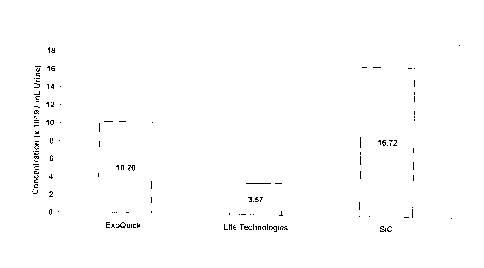
[0027] Figure 1 is a bar graph showing the concentration of exosomes
isolated from 5
rn1_, of urine using SiC, ExoQuick-TC for Tissue Culture Media and Urine
(System
Biosciences, Mountain View, USA) and Total Exosome Isolation Reagent (from
urine) (Life
Technologies, Carlsbad, USA).
[0028] Figure 2 is a line graph showing the concentration and particle size
of
exosomes isolated from 5 mL of urine using SiC, ExoQuick-TC for Tissue Culture
Media
and Urine (System Biosciences, Mountain View, USA) and Total Exosome Isolation
Reagent
(from urine) (Life Technologies, Carlsbad, USA).
CA 02929268 2016-05-09
- 6 -
[0029] Figure 3 is a bar graph showing the concentration of exosomes
isolated from 1
mL of plasma using SiC, ExoQuick Plasma Prep and Exosome Purification Kit
(System
Biosciences, Mountain View, USA) and Total Exosome Isolation Kit (from plasma)
(Life
Technologies, Carlsbad, USA).
[0030] Figure 4 is a line graph showing the concentration and particle size
of
exosomes isolated from 1 mL of plasma using SiC, ExoQuick Plasma Prep and
Exosome
Purification Kit (System Biosciences, Mountain View, USA) and Total Exosome
Isolation
Kit (from plasma) (Life Technologies, Carlsbad, USA).
[0031] Figure 5A is a line graph showing the concentration and particle
size of
exosomes purified from 10 mL of urine using SiC.
[0032] Figure 5B is a line graph showing the concentration and particle
size of
exosomes purified from 10 mL of urine using ultracentrifugation.
[0033] Figure 6 is a bar graph showing the relative amount of exosomal RNA
isolated from 5 mL of urine using SiC, ExoQuick-TC for Tissue Culture Media
and Urine
(System Biosciences, Mountain View, USA) and Total Exosome Isolation Reagent
(from
urine) (Life Technologies, Carlsbad, USA).
[0034] Figure 7 is a bar graph showing the relative amount of exosomal RNA
isolated from increasing amounts of urine using SiC and ultracentrifugation.
DESCRIPTION
Methods for Isolating Extracellular Vesicles
100351 It has been unexpectedly found that silicon carbide (SiC) can be
used to
selectively isolate extracellular vesicles, including exosomes, from liquid
samples. The
extracellular vesicles (e.g. exosomes) isolated using SiC have been found to
have the
characteristics of true extracellular vesicles, as examined by microRNA
markers, as well as
examination of the size and structure of the extracellular vesicles. Further,
the extracellular
vesicles isolated using SiC have been found to have substantially reduced
levels of
CA 02929268 2016-05-09
- 7 -
contaminating materials, such as macromolecular complexes, ribosomes and
proteins, as
compared to extracellular vesicles isolated using prior art methods requiring
ultracentrifugation or the use of precipitating agents.
[0036] Disclosed are methods for the selective isolation of extracellular
vesicles,
including exosomes, which are rapid, inexpensive, and do not require the use
of specialized
equipment (e.g. ultracentrifuges). In one embodiment, disclosed is a method
for the isolation
of extracellular vesicles, including exosomes, from a liquid sample. The
method can
comprise the steps of:
adjusting the pH of a liquid sample comprising extracellular vesicles to a
preselected,
binding pH;
contacting the liquid sample with SiC, wherein at the preselected, binding
p11, the
extracellular vesicles bind to the SiC; and
eluting the bound extracellular vesicles from the SiC.
[0037] The disclosed method may be used to isolate extracellular vesicles,
including
exosomes, from any liquid sample containing extracellular vesicles. The liquid
sample may
comprise biological fluids, such as but not limited to, whole blood, blood
serum, plasma,
urine, saliva, sputum, breast milk, ascetic fluid, semen, vaginal fluid,
amniotic fluid,
cerebrospinal fluid, sweat, tears and cell culture media. The biological fluid
may come from
any mammal, including humans.
[0038] The disclosed method may also be used to isolate extracellular
vesicles,
including exosomes, from biological tissues. The biological tissues are first
lysed and the
resulting lysate can be used as the liquid sample. The biological tissue may
include, but is
not limited to, surgical samples, biopsy samples, tissues, feces, plant
tissue, insect tissue and
cultured cells.
[0039] Depending on the composition of the liquid sample, it may be
desirable prior
to contacting the liquid sample with the SiC, to remove any cells and/or
cellular debris
CA 02929268 2016-05-09
- 8 -
contained in the liquid sample. This can be accomplished, for example, by
subjecting the
liquid sample to centrifugation or filtration to yield a supernatant that is
substantially free of
cells and cellular debris.
[0040] In a preferred embodiment, the SiC can be provided in a slurry
format. The
SiC slurry can be prepared with a typical industrial preparation of SiC, which
is composed of
about 97.8% silicon carbide and small amounts of silicon dioxide, silicon,
iron, aluminum
and carbon. SiC is available in a variety of grit sizes or grades, with each
grade having a
different average particle size. The SiC slurry can be prepared using any
grade of SiC and an
appropriate liquid carrier, such as Phosphate Buffered Saline (PBS) buffers
(e.g. IX PBS, pH
7) and Tris buffers (e.g. 10 mM Tris, pH 7). The SiC can have a grit size
between 500-2500
(diameter ca. 1-10 um), preferably a grit size between 2000-2500 and even more
preferably,
a grit size of 2000. The SiC slurry can be prepared in various ratios of SiC
to liquid carrier,
with a preferred ratio being between 30% and 70% (w:v), and even more
preferred ratio
being 50% (ww).
[0041] The pH of the liquid sample can be adjusted to the appropriate
binding pH
prior to contacting the liquid sample to the SiC slurry. Alternatively, the pH
of the liquid
sample can be adjusted to the appropriate binding pH, after the liquid sample
has already
come into contact with the SiC slurry.
[0042] To achieve selective binding of the extracellular vesicles to the
SiC, the pH of
the liquid sample can be adjusted to a binding pH of between pH 2 and pH 4,
more
preferably between pH 2.5 and p14 3.5, and even more preferably pH 3. The
binding pH can
also be between pH 7 and pH 11, more preferably between pH 8 and 10, and even
more
preferably between pH 8.5 and 9.
[0043] The pH of the liquid sample can be adjusted using buffers, acids or
bases
known in the art. The choice of an appropriate buffer, acid or base will
depend on the initial
pli of the liquid sample containing the extracellular vesicles. For example,
the starting pH of
some urine samples can be about pH 4 to pH 6, whereas the pH of plasma can be
about pH 7.
Having regard to the initial pH of the liquid sample, a person skilled in the
art can readily
CA 02929268 2016-05-09
- 9 -
determine the appropriate strength and type of buffer, acid or base for
adjusting the pH of the
liquid sample and also the appropriate amount to he added to the liquid sample
to obtain the
desired binding pH. Generally, optimal binding conditions can be achieved
through the use
of a binding solution comprising a low salt buffer with a strong buffering
capacity.
Examples of suitable low salt buffers include, but are not limited to, 'Iris
acetate buffers, Tris
borate buffers, Tris HC1 buffers, or Tris buffers.
[0044] Following the binding of the extracellular vesicles to the SiC, the
liquid
portion of the sample can be separated from the bound SiC. As SiC is known to
have a high
density (3.21 g/cm3), low speed centrifugation can be employed to pellet the
SiC and the
bound extracellular vesicles. After centrifugation for a few minutes, the
remaining liquid
portion can be decanted. The extracellular vesicles can then be eluted from
the SiC using an
elution solution having a pH between 5 and 7, and more preferably a pH of 6.
Carrying out
the elution step at a pH between 5 and 7 maximizes the selective elution of
the extracellular
vesicles, including exosomes, and minimizes co-elution of any contaminating
proteins that
are bound to the SiC. An elution solution comprising a low salt buffer and
having a pH
between 5 and 7 can be used. Suitable low salt buffers include, but are not
limited to,
Phosphate Buffered Saline (PBS), Tris Buffer or TE Buffer. The SiC can be
removed from
the extracellular vesicle containing eluent, for example, by centrifugation
and/or filtration to
yield a supernatant containing the purified extracellular vesicles, including
exosomes.
[0045] The purified extracellular vesicles, including exosomes, may be used
in
various downstream applications, including isolation of exosomal RNA or
isolation of
exosomal proteins.
[0046] In another preferred embodiment of the method, the SiC can be used
in a
column format. A SiC slurry as described above (see paragraph [0040]), can be
packed into
a column of any size, from small spin columns all the way to large
chromatography columns
operating through the use of gravity or pumps. The choice of column size will
depend on the
volume of the liquid sample to be processed. The pH of the liquid sample
comprising the
extracellular vesicles can be adjusted to the appropriate binding pH (see
paragraphs [0041]
and [0042]) using a suitable buffer, acid or base as described above (see
paragraph [00431).
CA 02929268 2016-05-09
- 10 -
The pH adjusted liquid sample can then be introduced into the SiC column. As
the liquid
sample travels through the SiC column, the extracellular vesicles, including
exosomes,
contained in the liquid sample will come into contact with the SiC and will
selectively bind
to the SiC. The bound extracellular vesicles, including exosomes, can be
eluted from the SiC
by passing an appropriate elution solution, as described above (sec paragraph
[0044]),
through the column and the eluted extracellular vesicles can be collected for
downstream
applications.
Methods for Producing Liquids Depleted of Extracellular Vesicles
[0047] Media for the growth of cultured cells is often supplemented with
serum,
which is known to contain extracellular vesicles, including exosomes. These
exogenous
exosomes present in the serum can negatively affect experimental results by
interfering with
the exosomes being studied in the cell culture, as they will be co-purified
with the exosomes
being produced by the cell line of interest. Therefore, researchers who are
working with cell
culture to carry out research involving exosomes often require exosome-free
media or serum
to ensure that their results are not affected or altered.
[0048] Further disclosed are methods for depleting extracellular vesicles,
including
exosomes, from liquid materials, including cell culture media and serum.
Through the use of
SiC, extracellular vesicles present in the liquid material can be selectively
removed from the
liquid material. In contrast to prior art methods for the preparation of cell
culture media and
serum free of extracellular vesicles (e.g. ultracentrifugation or filtration
methods) the
disclosed depletion method is rapid, inexpensive, and does not require the use
of specialized
equipment. Further, the disclosed method may be used to remove extracellular
vesicles,
including exosomes, from large volumes of cell culture media, serum or other
liquid
materials.
[0049] In one embodiment, disclosed is a method for producing a liquid
sample
substantially depleted of extracellular vesicles, including exosomes,
comprising the steps of:
CA 02929268 2016-05-09
- -
adjusting the pH of a liquid sample comprising extracellular vesicles to a
preselected,
binding pH;
contacting the liquid sample with SiC, wherein at the pre-selected, binding
pH, the
extracellular vesicles bind to the SiC;
separating the SiC with the bound extracellular vesicles from the liquid
sample to
yield a liquid substantially depleted of extracellular vesicles; and
collecting the liquid substantially depleted of extracellular vesicles.
[0050] As used herein, a liquid sample that has been depleted of at least
95% of the
extracellular vesicles initially present in the liquid sample prior to
contacting the liquid
sample with SiC is considered to be a "liquid sample substantially depleted of
extracellular
vesicles".
[0051] In a preferred embodiment, the liquid sample to be substantially
depleted of
extracellular vesicles, including exosomes, may be a serum sample. The serum
sample may
be selected from any type of serum used to supplement cell culture for cell
growth, including
but not limited to, fetal bovine serum, horse serum, and fetal calf serum.
While the
embodiments set out below describe the preparation of serum samples depleted
of
extracellular vesicles, it will be appreciated that this method can be used
with other types of
liquid samples, including liquid samples comprising other types of bodily
fluids and also
liquid samples such as cell culture media.
[0052] The SiC can be provided as a slurry as described above (see
paragraph
[0040]). The pH of the serum sample can be adjusted to an appropriate binding
pH prior to
contacting the liquid sample to the SiC slurry. Alternatively, the pH of the
serum sample can
be adjusted to an appropriate binding pH, after the liquid sample has already
come into
contact with the SiC slurry.
[0053] To achieve selective binding of the extracellular vesicles to the
SiC, the pH of
the serum sample is adjusted to a binding pH of between pH 2 and pH 4, more
preferably
CA 02929268 2016-05-09
- 12 -
between pH 2.5 and pH 3.5, and even more preferably pH 3. The binding pH can
also be
between pH 7 and pH 11, more preferably between pH 8 and 10, and even more
preferably
between pH 8.5 and 9.
[0054] The pH of the serum sample can be adjusted using buffers, acids or
bases
known in the art. The choice of an appropriate buffer, acid or base will
depend on the initial
pH of the serum sample. Having regard to the initial pH of the serum sample, a
person
skilled in the art can readily determine the appropriate strength and type of
buffer, acid or
base for adjusting the pH of the serum sample and also the appropriate amount
to be added to
the serum sample to obtain the desired binding pH. Generally, optimal binding
conditions
can be achieved through the use of a binding solution comprising a low salt
buffer with a
strong buffering capacity. Examples of suitable binding buffers include are
but not limited to
Tris acetate buffers, Tris borate buffers, Tris 1-ICI buffers, or Tris
buffers.
[0055] Following the binding of the extracellular vesicles to the SiC, the
bound SiC
can be removed to yield a serum sample substantially depleted of extracellular
vesicles. Low
speed centrifugation can be employed to pellet the SiC and the bound
extracellular vesicles.
After centrifugation for a few minutes, the serum sample substantially
depleted of
extracellular vesicles can be decanted for use in other applications.
[0056] In another preferred embodiment of the method, the SiC can be used
in a
column format. A SiC slurry as described above (see paragraph [0040]), can be
packed into
a column of any size, from small spin columns all the way to large
chromatography columns
operating through the use of gravity or pumps. The choice of column size will
depend on the
volume of the serum sample to be processed. The pH of the serum sample,
comprising the
extracellular vesicles to be depleted, can be adjusted to the appropriate
binding pH (see
paragraphs [0052] and [0053]) using a buffer, acid or base as described above
(see paragraph
[0054]). The pH adjusted serum sample can then be introduced into the column.
As the
serum sample travels through the column, the extracellular vesicles, including
exosomes,
contained in the serum sample will come into contact with the SiC and will
selectively bind
to the SiC. The remaining extracellular vesicle-depleted serum that flows
through the
column can be collected for use in other applications.
CA 02929268 2016-05-09
- 13 -
[0057] Through the use of SiC in either a slurry format or a column format,
scrum
samples can depleted of over 95% of extracellular vesicles initially present
in the serum
sample. The extracellular vesicle-depleted serum samples can be use in cell
culture, and
more specifically for growing cultured mammalian cells.
Kits for the Isolation or Depletion of Extracellular Vesicles
[0058] In a further embodiment, provided are kits for the isolation of
extracellular
vesicles, including exosomes, from liquid samples. The kits can be used to
carry out the
isolation method disclosed herein. Such kits may comprise vessels containing a
SiC slurry, a
binding solution to adjust the pll of the liquid sample to an appropriate
binding pH to bind
the extracellular vesicles to the SiC, and an elution solution to adjust the
pH in order to
specifically release the bound extracellular vesicles from the SiC. The kit
may further
contain filter columns, which may aid in removing the SiC from the liquid
samples following
elution of the extracellular vesicles. Alternatively, the kit may comprise SiC
columns,
vessels containing a binding solution to adjust the pH of the liquid sample to
an appropriate
binding pH to bind the extracellular vesicles to the SiC, and an elution
solution to adjust the
pl I in order to specifically release the bound extracellular vesicles from
the SiC.
[0059] In another embodiment, provided are kits for the depletion of
extracellular
vesicles from liquid samples. The kits can be used to carry out the depletion
method
disclosed herein. Such kits may comprise vessels containing a SiC slurry and a
binding
solution to adjust the pH of the liquid sample to an appropriate binding pH to
bind the
extracellular vesicles to the SiC. The kit may further contain filter columns
which may aid in
removing the SiC from the liquid samples to produce the substantially
extracellular vesicles-
depleted sample. Alternatively, the kit may comprise SiC columns and vessels
containing a
binding solution to adjust the pH of the liquid sample to an appropriate
binding pH to bind
the extracellular vesicles to the SiC.
[0060] Although the invention has been described with reference to
illustrative
embodiments, it is to be understood that the invention is not limited to these
precise
CA 02929268 2016-05-09
- 14 -
embodiments, and that various changes and modification are to be intended to
be
encompassed in the appended claims.
EXAMPLES
[0061] These examples are described for the purposes of illustration and
are not
intended to limit the scope of the invention.
[0062] Example 1 ¨ Comparing the Efficiency of SiC versus Commercially
Available Kits in Isolating Extracellular Vesicles, including Exosomes, from
Urine
[0063] SiC in a slurry format was tested for its ability to isolate
exosomes from 5 mL
of urine, and the performance of the SiC was compared to two different
commercially
available exosome precipitation reagents. The SiC slurry was prepared by
adding 6.25 grams
of SiC (G/S 2500) to 10 mL of 1X PBS (pH 7). Fifteen mL of urine was collected
into a
conical tube and centrifuged at 200 x g (-1.000 RPM) for 10 minutes to remove
urine
exfoliated cells and debris. The cell-free urine was decanted into a new tube
and centrifuged
at 1,800 x g (-3,000 RPM) for 10 minutes to remove any residual debris or
bacterial cells.
Five mL of the cell-free urine was transferred to a new tube and 400 uL of the
SiC slurry was
added to the urine samples. The pll was then adjusted to pH 8.5 using 1M Tris
Base (pH 11)
to allow for the exosomes to bind to the SiC resin. The sample was mixed by
vortexing for
seconds and left to stand at room temperature for 10 minutes. The suspension
was again
mixed well by vortexing for 10 seconds, and then centrifuged for 2 minutes at
2,000 RPM.
The supernatant was then discarded. Next. 400 piL of 1X PBS (p1-1 7) was added
to the slurry
pellet in order to adjust the pH to 6, and mixed well by vortexing for 10
seconds. The
resuspended slurry pellet was then incubated for 10 minutes at room
temperature. After
incubation, the slurry pellet was mixed well by vortexing for 10 seconds then
centrifuged for
2 minutes at 500 RPM. The supernatant containing the eluted exosomes was then
transferred
to a filter column (0.2 uM pore size) and centrifuged for 1 minute at 6,000
RPM.
[0064] Exosomes were also isolated from 5 mL of urine using ExoQuick-IC for
Tissue Culture Media and Urine (System Biosciences, Mountain View, USA) and
from 5 mL
CA 02929268 2016-05-09
- 15 -
of urine using Total Exosome Isolation Reagent (from urine) (Life
Technologies, Carlsbad,
USA) according to the manufacturer's recommendations.
[0065] In order to analyze the purified exosomes, the eluted exosomes were
visualized using a NanoSight LM10 instrument. The analysis showed that the SiC
method
isolated the highest amount of exosomes, with a recovery of 16.72 x 10'9
particles /ml,
urine, which is higher than both the other methods tested. The graph showing
the
concentration of exosomes isolated using SiC and the 2 other methods can be
seen in Figure
1. No impurities were found to be contaminating the exosomes purified using
SiC, as
indicated by the Nanosight analysis. The graph showing the particle size
distribution using
SiC and the other 2 methods can be seen in Figure 2. As shown in Figure 2, the
majority of
extracellular vesicles isolated by SiC are in the size range of 40-150 nm,
which is typical for
exosomes.
[0066] Example 2 - Comparing the Efficiency of SiC versus Commercially
Available Kits in Isolating Extracellular Vesicles, including Exosomes, from
Plasma
[0067] SiC in a slurry format was tested for its ability to isolate
extraeellular vesicles,
including exosomes, from 1 mL of plasma, and the performance of the SiC was
compared to
two different commercially available exosome precipitation reagents. The SiC
slurry was
prepared by adding 6.25 grams of SiC (G/S 2500) to 10 mL of IX PBS (pH 7).
Blood was
collected on EDTA or citrate using a standard blood collection tube. The
collection tube was
centrifuged at 2000 RPM for 15 minutes. The upper plasma fraction was
collected and
transferred to a fresh tube and centrifuged at 2000 RPM for 10 minutes. The
clear
supernatant containing the purified plasma was collected. One mL of the
prepared plasma
was transferred to a tube and 2004 of the SiC slurry was added to the plasma
samples. The
pH was then adjusted to pH 8.5 using 1M Tris Base (p11 11) to allow for the
exosomes to
bind to the SiC resin. The sample was mixed by vortexing for 10 seconds and
left to stand at
room temperature for 5 minutes. The suspension was again mixed well by
vortexing for 10
seconds, and then centrifuged for 2 minutes at 2,000 RPM. The supernatant was
then
discarded. Next, 200 tiL of lx PBS (pH 7) was added to the slurry pellet in
order to adjust
the pH to 6, and mixed well by vortexing for 10 seconds. The resuspended
slurry pellet was
CA 02929268 2016-05-09
- 16 -
then incubated for 5 minutes at room temperature. After incubation, the slurry
pellet was
mixed well by vortexing for 10 seconds then centrifuged for 2 minutes at 500
RPM. The
supernatant containing the eluted exosomes was then transferred to a filter
column (0.2 tiM
pore size) and centrifuged for I minute at 6,000 RPM.
[00681 Exosomes were also isolated from I mL of plasma using ExoQuick
Plasma
Prep and Exosome Purification Kit (System Bioseiences, Mountain View, USA) and
from 1
mL of plasma using Total Exosome Isolation Kit (from plasma) (Life
Technologies,
Carlsbad, USA) according to the manufacturer's recommendations.
[0069] In order to analyze the purified exosomes, the eluted exosomes were
visualized using a NanoSight LM10 instrument. The analysis showed that the SiC
method
isolated the highest amount of exosomes, with a recovery of 9.64 x 10'13
particles / mL of
plasma, which is higher than both the other methods tested. The graph showing
the
concentration of exosomes isolated from I mL plasma using SiC and the 2 other
methods can
be seen in Figure 3. No impurities were found to be contaminating the exosomes
purified
using SiC, as indicated by the Nanosight analysis. The graph showing the
particle size
distribution using SiC and the other 2 methods can be seen in Figure 4. As
shown in Figure
4, the majority of extracellular vesicles isolated by SiC are in the size
range of 40-150 nm,
which is typical for exosomes.
[0070] Example 3 - Comparing the Efficiency of SiC versus
Ultracentrifugation
in Isolating Extracellular Vesicles, including Exosomes, from Urine
[0071] SiC in a slurry format was tested for its ability to isolate
exosomes from 10
mL of urine, and the performance of the SiC was compared to the traditional
method of
ultracentrifugation. The SiC slurry was prepared by adding 6.25 grams of SiC
(G/S 2500) to
mL of IX PBS (pH 7). Ten mL of urine was collected into a conical tube and
centrifuged
at 200 x g (-1,000 RPM) for 10 minutes to remove urine exfoliated cells and
debris. The
cell-free urine was decanted into a new tube and centrifuged at 1,800 x g (-
3,000 RPM) for
10 minutes to remove any residual debris or bacterial cells. The remaining
cell-free urine
sample was transferred to a new tube and 400 !..tL of the SiC slurry was added
to the urine
CA 02929268 2016-05-09
- 17 -
samples. The pH was then adjusted to pH 8.5 using 1M Tris Base (pH II) to
allow for the
exosomes to bind to the SiC resin. The sample was mixed by vortexing for 10
seconds and
left to stand at room temperature for 10 minutes. The suspension was again
mixed well by
vortexing for 10 seconds, and then centrifuged for 2 minutes at 2,000 RPM. The
supernatant
was then discarded. Next, 400 aL of IX PBS (p11 7) was added to the slurry
pellet order to
adjust the pH to 6, and mixed well by vortexing for 10 seconds. The
resuspended slurry
pellet was then incubated for 10 minutes at room temperature. After
incubation, the slurry
pellet was mixed well by vortexing for 10 seconds then centrifuged for 2
minutes at 500
RPM. After incubation, the slurry pellet was mixed well by vortexing for 10
seconds then
centrifuged for 2 minutes at 500 RPM. The supernatant containing the eluted
exosomes was
then transferred to a filter column (0.2 aM pore size) and centrifuged for 1
minute at 6,000
RPM.
[0072] Exosomes were also isolated from urine using traditional
ultracentrifugation
as outlined in Thery et al. (Current Protocols in Cell Biology, 2006). For
this procedure, 240
mL of urine was processed and the final exosome pellet was resuspended in 240
of IX
PBS (pH 7). Therefore, for analysis of exosomes. 10 ttL of the elution was
analyzed which
would correspond to 10 mL of initial urine input.
[0073] The purified exosomes were analyzed and visualized using a NanoSight
LM10 instrument. The results are show in Figures 5A and 5B. The analysis
showed that
the SiC method (Figure 5A) isolated exosomes with a recovery of 7.63 x 10^8
particles /
mL, and the majority of extracellular vesicles isolated are in the size range
of 40-150 nm,
which is typical for exosomes. In contrast, ultracentriguation (Figure 5B)
purified larger
extracellular vesicles, with a size range from 125 nm to 235 nm, with a total
recovery of 1.56
x 101'8 particles / mL. The larger extracellular vesicles isolated using
ultracentrifugation do
not correspond to the typical size range of exosomes, indicating that this
gold-standard
method is not as specific as the use of SiC to purify exosomes.
CA 02929268 2016-05-09
- 18 -
[0074] Example 4 ¨ Comparing RNA Recovery from Exosomes Isolated Using
SiC versus Commercially Available Kits
[0075] SiC in a slurry format was tested for its ability to isolate
exosomes from 5 mL
of urine, and the performance of the SIC was compared to two different
commercially
available exosome precipitation reagents. The SiC slurry was prepared by
adding 6.25 grams
of SiC (G/S 2500) to 10 mL of 1X PBS (pH 7). The exosomes were isolated from
15 mL
urine samples using the SiC slurry as described in Example 1 above.
[0076] Exosomes were also isolated from 5 mL of urine using ExoQuick-TC for
Tissue Culture Media and Urine (System Biosciences, Mountain View, USA) and
from 5 mL
of urine using Total Exosome Isolation Reagent (from urine) (Life
Technologies, Carlsbad,
USA) according to the manufacturer's recommendations.
[0077] RNA was then purified from the isolated exosomes. For exosomes
purified
using SiC, the RNA was isolated by adding 300 tit of Lysis Buffer from Norgen
Biotek's
Plasma/Serum RNA Purification Mini Kit (Cat # 55000, Norgen Biotek, Thorold,
Canada)
and 37.5 1_, of Lysis Additive from Norgen Biotek's Fatty Tissue RNA
Purification Kit
(Cat# 36200, Norgen Biotek, Thorold, Canada) to the purified exosomes. This
was mixed
and then incubated at room temperature for 10 minutes. Next, 500 tit of
ethanol was added
and mixed well. This was then added to a column containing SiC resin, and
centrifuged for 1
minute at 3,300 x g (-6,000 RPM). The flowthrough was discarded, and the bound
RNA
was washed using Wash Solution from Norgen Biotek's Plasma/Serum RNA
Purification
Mini Kit (Cat# 55000, Norgen Biotek, Thorold, Canada) by centrifugation for 30
seconds at
3,300 x g (-6,000 RPM). The flowthrough was discarded, and the wash step was
repeated 2
more times. After a dry spin, 50 uL of Elution Solution from Norgen Biotek's
Plasma/Serum
RNA Purification Mini Kit (Cat# 55000, Norgen Biotek, Thorold, Canada) was
added to the
column and centrifuged for 1 minute at 2,000 RPM, followed by 2 minutes at
8,000 RPM.
[0078] Exosomal RNA was isolated from the exosomes isolated with ExoQuick-
TC
for Tissue Culture Media and Urine (System Biosciences, Mountain View, USA)
using
- 19 -
Norgen Biotek's Total RNA Purification Kit (Cat# 17200, Norgen Biotek,
Thorold, Canada)
according to the manufacturer's recommendations.
[0079] Exosomal RNA was isolated from exosomes isolated with Total
Exosome
Isolation Reagent (from urine) (Life Technologies, Carlsbad, USA) using Life
Technologies'
Total Exosome RNA and Protein Isolation Kit (Cat# 4478545, Life Technologies,
Carlsbad,
USA) according to the manufacturer's recommendations.
[0080] For analysis, the purified RNA was used as a template in qPCR
reactions to
amplify miR-30a, which is known to be a urinary exosomal miRNA. The initial
reverse
transcription was set up as follows:
3 uL exosomal RNA
0.5 uL, 50 uM SLRT-miR-30a reverse primer
0.5 uL, 10mM dNTPs
4 uL 5X RT Buffer (TruScript Reverse Transcriptase Kit, Catalog #54440, Norgen
Biotek, Thorold, Canada)
0.2 uL, reverse-transcriptase
11.8 uL water
[0081] The reverse transcription was then run according to the
following program
using a BioRad CFX Connect:
Cycle 1: (1X) Step 1:50.0 C for 30:00
Cycle 2: (1X) Step 1: 70.0 C for 10:00
Cycle 3: (1X) Step 1: 4.0 C for 99:99
[0082] Next, the qPCR was set up as follows:
3 uL cDNA
uL, 2X SybrTM Green Mix (Cat# 170-8880, BioRad, Hercules, USA)
0.12 uL 50 M miR-30a Forward Primer
Date Recue/Date Received 2021-06-17
CA 02929268 2016-05-09
-20-
0.12 uL 500/1 SLR Reverse Primer
6.76 uL water
[0083] The qPCR was run according to the following program using a BioRad
CFX
Connect:
Cycle 1: (1X) Step I: 95.0 C for 03:00
Cycle 2: (40X) Step 1: 95.0 C for 00:15
Step 2: 58.0 C for 00:30
Step 3: 72.0 C for 00:45
Data collection and real-time analysis enabled.
Cycle 3: ( 1X) Step 1: 57.0 C for 01:00
Cycle 4: (80X) Step 1: 57.0 C for 00:10
Increase setpoint temperature after cycle 2 by 0.5 C
Melt curve data collection and analysis enabled.
[0084] The resulting Ct values were then analyzed, and the relative amount
of
exosomal RNA isolated using each method was determined. As seen in Figure 6,
the SiC
method resulted in the highest recovery of the urinary exosomal miRNA when
compared to
the other 2 methods.
[0085] Example 5 ¨ Comparing RNA Recovery from Exosomes Isolated Using
SiC versus Ultracentrifugation
[0086] SiC in a slurry format was tested for its ability to isolate
exosomes from
increasing amounts of urine, and the performance of the SiC was compared to
the traditional
method of ultracentrifugation. The SiC slurry was prepared by adding 6.25
grams of SiC
(G/S 2500) to 10 mI_, of 1X PBS (pH 7). One litre of urine was co lected and
processed as
described in Example 3 above. Next, the SiC slurry was used to isolate the
exosomes from
CA 02929268 2016-05-09
- 2 I -
0.1 mL, 0.5 mL, 1 mL, 2.5 mL, 5 mL, 10 mL and 30 mL of the urine using the
procedure
described in Example 3.
[0087] Exosomes were also isolated from urine using traditional
ultracentriftigation
as outlined in Thery et al. (Current Protocols in Cell Biology, 2006).
[0088] RNA was then purified from the isolated exosomes. For exosomes
purified
using SiC, the RNA was isolated by adding 300 uL of Lysis Buffer from Norgen
Biotek's
Plasma/Serum RNA Purification Mini Kit (Cat# 55000, Norgen Biotek, Thorold,
Canada)
and 37.5 t.t1_, of Lysis Additive from Norgen Biotek's Fatty Tissue RNA
Purification Kit
(Cat# 36200, Norgen Biotek, Thorold, Canada), to the purified exosomes. This
was mixed,
then incubated at room temperature for 10 minutes. Next, 500 uL of ethanol was
added and
mixed well. This was then added to a column containing SiC resin, and
centrifuged for 1
minute at 3,300 x g (-6,000 RPM). The flowthrough was discarded, and the bound
RNA
was washed using Wash Solution from Norgen Biotek's Plasma/Serum RNA
Purification
Mini Kit (Cat # 55000, Norgen Biotek, Thorold, Canada) by centrifugation for
30 seconds at
3,300 x g (-6,000 RPM). The flowthrough was discarded, and the wash step was
repeated 2
more times. After a dry spin, 50 uL of Elution Solution from Norgen Biotek's
Plasma/Serum
RNA Purification Mini Kit (Cat# 55000, Norgen Biotek, Thorold, Canada) was
added to the
column and centrifuged for 1 minute at 2,000 RPM, followed by 2 minutes at
8,000 RPM.
[0089] Exosomal RNA was isolated from the exosomes isolated by
centrifugation
using Norgen's Total RNA Purification Kit (Catg 17200, Norgen Biotek, Thorold,
Canada)
according to the manufacturer's recommendations.
[0090] For analysis, the purified RNA was used as a template in qPCR
reactions to
amplify miR-30a, which is known to be a urinary exosomal miRNA. The initial
reverse
transcription was set up as follows:
3 p.L cxosomal RNA
0.5 FL 50 FM SLRT-miR-30a reverse primer
0.5 FL 10mM dNTPs
CA 02929268 2016-05-09
-22 -
4 uL 5X RI Buffer (TruScript Reverse Transcriptase Kit, Catalog #54440, Norgen
Biotek, Thor Id, Canada)
0.2 uL reverse-transcriptase
11.8 ut water
[0091] The reverse transcription was then run according to the following
program
using a BioRad CFX Connect:
Cycle 1: (1X) Step 1:50.0 C for 30:00
Cycle 2: (IX) Step 1: 70.0 C for 10:00
Cycle 3: (1X) Step : 4.0 C for 99:99
[0092] Next, the qPCR was set up as follows:
3 jiL cDNA
uL 2X Syber Green Mix (Cat# 170-8880, BioRad, Hercules, USA)
0.12 uL 50p,M miR-30a Forward Primer
0.12 )11_, 50 M SLR Reverse Primer
6.76 uL Water
[0093] The ciPC,R was run according to the following program using a BioRad
CFX
Connect:
Cycle 1: (1X) Step 1: 95.0 C for 03:00
Cycle 2: (40X) Step 1:95.0 C for 00:15
Step 2: 58.0 C for 00:30
Step 3: 72.0 C for 00:45
Data collection and real-time analysis enabled.
Cycle 3: (1X) Step 1: 57.0 C for 01:00
CA 02929268 2016-05-09
- 23 -
Cycle 4: (80X) Step 1: 57.0 C for 00:10
Increase setpoint temperature after cycle 2 by 0.5 C
Melt curve data collection and analysis enabled.
[0094] The resulting Ct values were then analyzed, and the relative amount
of
exosomal RNA isolated from each volume of urine using SiC and
ultracentrifugation was
determined. As can be seen in Figure 7, the SiC method resulted in the highest
recovery of
the urinary exosomal miRNA at each volume tested when compared to
ultracentrifugation.