Note: Descriptions are shown in the official language in which they were submitted.
CA 02930001 2016-05-06
WO 2015/070038 PCT/US2014/064589
TARGETED TREATMENT OF ANEROBIC CANCER
CROSS-REFERENCE TO RELATED APPLICATIONS
The present application claims the benefit of U.S. Provisional Patent
Application No.
61/902,456, filed on. November 11, 2013, which is incorporated herein by
reference.
FIELD OF THE INVENTION
The present invention relates to a pharmaceutical cocktail and methods of
cancer
treatment. In particular, one such cocktail comprises a combination of
effective amounts of
lactate transporter inhibitor, a carbonic anhydrase inhibitor, a sodium
potassium chloride
cofactor (NKCC) transporter inhibitor, a member of the hydroxycinnamate class
of drugs or a
derivative thereof, and/or an angiogenesis inhibitor, including a vascular
endothelial growth
factor (VEGF) inhibitor such as bevacizumab in combination with blood vessel
occlusion. As
most cancers in an untreated state uses both aerobic and anaerobic/glycolytic
pathways
treatments contemplated herein can affect both metabolic pathways.
BACKGROUND OF THE INVENTION
While a number of anti-angiogenesis agents have been reported, including
bevacizumab,
it is not clear whether they possess the appropriate pharmacological
effectiveness required to be
therapeutically useful in the treatment of cancer in many situations.
Therefore, there is a
continued need for additional therapeutics to target such cancer and augment
or revive the
effectiveness of anti-angiogenesis agents to provide effective treatment of
cancer.
Cancers and cancerous lesions are known for their ability to adapt to
treatment in various
1
CA 02930001 2016-05-06
WO 2015/070038 PCT/US2014/064589
ways including shifts in metabolism, i.e. aerobic to glycoysis, or mutations
to avoid
pharmaceutical treatments. What is needed in the art is a method of treatment,
which can hinder
the metabolic pathways or options for such adaptive cancers.
SUMMARY OF THE INVENTION
The present invention relates to a pharmaceutical cocktail and methods of
cancer
treatment. In particular, one such cocktail comprises a combination of
effective amounts of a
lactate transporter inhibitor, a carbonic anhydrase inhibitor, a sodium
potassium chloride
cofactor (NKCC) transporter inhibitor, a member of the hydroxycinnamate class
of drugs or a
derivative thereof, and/or an angiogenesis inhibitor, including a vascular
endothelial growth
factor (VEGF) inhibitor such as bevacizumab in combination with blood vessel
occlusion. As
most cancers in an untreated state uses both aerobic and anaerobic/glycolytic
pathways
treatments contemplated herein can affect both metabolic pathways.
Although it is not necessary to understand the mechanism of an invention, it
is believed
that treatments that target the anaerobic and aerobic metabolic pathways more
completely
deprives cancer of ATP energy production, thereby producing greater damage or
killing of
cancerous cells. Treatment of the aerobic pathway alone temporarily controls
cancer but it
induces mutation to a glycolytic form, which does not respond to anti-VEGF or
other anti-
vascular growth factor agents.
In other embodiments, the present invention relates to compositions and
methods of
treating cancer involving effective amounts of a member of the
hydroxycinnamate class of drugs.
Pharmaceutical compositions and methods of treating cancer (eliminating the
tumor, shrinking
the tumor, prolonging the life of the patient, increasing quality of life by
decreasing the grade of
adverse events seen with other cancer treatments, and/or preventing/reducing
the likelihood of
2
CA 02930001 2016-05-06
WO 2015/070038 PCT/US2014/064589
the tumor's metastases) are additional aspects of the present invention. In
addition, the present
invention may be used to favorably affect the therapeutic result of patients
who have not
responded to alternative, traditional anti-cancer therapy.
In one embodiment, the invention contemplates a method for treating cancer
comprising:
a) administering an effective amount of a lactate transporter inhibitor to a
patient comprising a
cancerous lesion, wherein the cancerous lesion comprises a plurality of blood
vessels, and b)
occluding at least one of said plurality of blood vessel. In one embodiment,
said lactate
transporter inhibitor is delivered via liposomes. In one embodiment, said
lactate transporter
inhibitor is delivered via a small particle delivery system. In one
embodiment, said small
particle delivery system is selected from the group comprising liposomes, poly
(lactide-co-
glycolide) (PLG), nanoparticles formed by poly(beta-amino ester)s (PBAEs), and
drug
containing microbubbles which rupture upon insonation by ultrasound. In one
embodiment, the
occluding reduces blood flow to said cancerous lesion. In one embodiment, the
method further
comprises administering to said patient an effective amount of an angiogenesis
inhibitor. In one
embodiment, said angiogenesis inhibitor is a humanized monoclonal antibody. In
one
embodiment, said treating comprises repeated administration of at least one of
the lactate
transporter inhibitor and angiogenesis inhibitor. In one embodiment, said
antibody is
bevacizumab. In one embodiment, said lactate transporter inhibitor is a
hydoxycinnamate
derivative. In one embodiment, said hydoxycinnamate derivative is selected
from the group
consisting of ferrulic acid, caffeic acid, chorogenic acid, resveratrol
ferulate, and phloretin
ferulate. In one embodiment, said cancer is a hypoxic cancer. In one
embodiment, said occluding
results in the shrinkage of said cancer. In one embodiment, said occluding
further comprises an
embolism. In one embodiment, said embolism is produced by the introduction of
an embolic
composition. In one embodiment, said embolic composition comprises a plurality
of polymers
3
CA 02930001 2016-05-06
WO 2015/070038 PCT/US2014/064589
embedded with lactate transporter inhibitors. In one embodiment, said embolic
composition
comprises liposomes that contain lactate transporter inhibitor(s). In one
embodiment, said
embolic composition comprises a small particle delivery system that contain
lactate transporter
inhibitor(s). In one embodiment, said small particle delivery system is
selected from the group
comprising liposomes, poly (lactide-co-glycolide) (PLG), nanoparticles formed
by poly(beta-
amino ester)s (PBAEs), and drug containing microbubbles which rupture upon
insonation by
ultrasound. In one embodiment, said embolic composition comprises a plurality
of glass beads
coated with at least one lactate transporter inhibitor. In one embodiment,
said occluding further
comprises thermal ablation.
In one embodiment, the invention contemplates a method of treating cancer
comprising
administering to a patient an effective amount of at least one lactate
transporter inhibitor, at least
one a carbonic anhydrase inhibitor, at least one NKCC inhibitor, and at least
one angiogenesis
inhibitor. In one embodiment, said angiogenesis inhibitor is a humanized
monoclonal antibody.
In one embodiment, said administering is repeated. In one embodiment, said
antibody is
bevacizumab. In one embodiment, said lactate transporter inhibitor, a carbonic
anhydrase
inhibitor, and an angiogenesis inhibitor are administered as a pharmaceutical
cocktail. In one
embodiment, said lactate transporter inhibitor is delivered via liposomes. In
one embodiment,
said lactate transporter inhibitor is delivered via a small particle delivery
system. In one
embodiment, said small particle delivery system is selected from the group
comprising
liposomes, poly (lactide-co-glycolide) (PLG), nanoparticles formed by
poly(beta-amino ester)s
(PBAEs), and drug containing microbubbles which rupture upon insonation by
ultrasound. In
one embodiment, said NKCC inhibitor is delivered via a small particle delivery
system. In one
embodiment, said small particle delivery system is selected from the group
comprising
liposomes, poly (lactide-co-glycolide) (PLG), nanoparticles formed by
poly(beta-amino ester)s
4
CA 02930001 2016-05-06
WO 2015/070038
PCT/US2014/064589
(PBAEs), and drug containing microbubbles which rupture upon insonation by
ultrasound. In
one embodiment, both said lactate transporter inhibitor and said NKCC
inhibitor are both
delivered via a small particle delivery system. In one embodiment, said small
particle delivery
system is selected from the group comprising liposomes, poly (lactide-co-
glycolide) (PLG),
nanoparticles formed by poly(beta-amino ester)s (PBAEs), and drug containing
microbubbles
which rupture upon insonation by ultrasound. In one embodiment, said at least
one lactate
transporter inhibitor, at least one a carbonic anhydrase inhibitor, and at
least one angiogenesis
inhibitor are administered in series. In one embodiment, said cancer is a
hypoxic cancer. In one
embodiment, said carbonic anhydrase inhibitor is a carbonic anhydrase 9 and
carbonic anhydrase
12 inhibitor. In one embodiment, said administering results in the shrinkage
of a cancerous
lesion. In one embodiment, said administering reduces metastases of said
cancerous lesion.
= In one embodiment, the invention contemplates a pharmaceutical
composition
comprising an effective amount of a lactate transporter inhibitor, a loop
diuretic NKCC inhibitor,
and an angiogenesis inhibitor. In one embodiment, said angiogenesis inhibitor
is bevacizumab. In
one embodiment, said loop diuretic is bumetanide. In one embodiment, said
lactate transporter
inhibitor is a hydoxycinnamate derivative. In one embodiment, said
angiogenesis inhibitor is
packaged within liposomes. In one embodiment, said angiogenesis inhibitor is
packaged within a
small particle delivery system. In one embodiment, said small particle
delivery system is
selected from the group comprising liposomes, poly (lactide-co-glycolide)
(PLG), nanoparticles
formed by poly(beta-amino ester)s (PBAEs), and drug containing microbubbles
which rupture
upon insonation by ultrasound. In one embodiment, said lactate transporter
inhibitor is packaged
within liposomes. In one embodiment, said lactate transporter inhibitor is
packaged within a
small particle delivery system. In one embodiment, said small particle
delivery system is
selected from the group comprising liposomes, poly (lactide-co-glycolide)
(PLG), nanoparticles
5
CA 02930001 2016-05-06
WO 2015/070038 PCT/US2014/064589
formed by poly(beta-amino ester)s (PBAEs), and drug containing microbubbles
which rupture
upon insonation by ultrasound. In one embodiment, said NKCC inhibitor is
delivered via
liposomes. In one embodiment, said NKCC inhibitor is delivered via a small
particle delivery
system. In one embodiment, said small particle delivery system is selected
from the group
comprising liposomes, poly (lactide-co-glycolide) (PLG), nanoparticles formed
by poly(beta-
amino ester)s (PBAEs), and drug containing microbubbles which rupture upon
insonation by
ultrasound. In one embodiment, said angiogenesis inhibitor, said lactate
transporter inhibitor,
and said NKCC inhibitor are all delivered via a small particle delivery
system. In one
embodiment, said small particle delivery system is selected from the group
comprising
liposomes, poly (lactide-co-glycolide) (PLG), nanoparticles formed by
poly(beta-amino ester)s
(PBAEs), and drug containing microbubbles which rupture upon insonation by
ultrasound. In
one embodiment, said hydoxycinnamate derivative includes, but is not limited
to, ferrulic acid,
caffeic acid, chorogenic acid, resveratrol ferulate, and phloretin ferulate.
In one embodiment,
said composition is formulated for oral administration. In one embodiment,
said composition is
formulated for parenteral administration. In one embodiment, said composition
is formulated for
intravenous administration.
In one embodiment, the invention contemplates a method of treating cancer
comprising
administering to a patient a composition comprising an effective amount of a
lactate transporter
inhibitor, a carbonic anhydrase inhibitor, and an angiogenesis inhibitor. In
one embodiment, said
composition is delivered via liposomes. In one embodiment, said composition is
delivered via a
small particle delivery system. In one embodiment, said small particle
delivery system is
selected from the group comprising liposomes, poly (lactide-co-glycolide)
(PLG), nanoparticles
formed by poly(beta-amino ester)s (PBAEs), and drug containing microbubbles
which rupture
upon insonation by ultrasound. In one embodiment, said angiogenesis inhibitor
is a humanized
6
CA 02930001 2016-05-06
WO 2015/070038 PCT/US2014/064589
monoclonal antibody. In one embodiment, said composition comprising at least
one lactate
transporter inhibitor, a carbonic anhydrase inhibitor, and an angiogenesis
inhibitor is
administered as a pharmaceutical cocktail. In one embodiment, said treating
comprises repeated
administration of at least one of the lactate transporter inhibitor, a
carbonic anhydrase inhibitor,
and an angiogenesis inhibitor. In one embodiment, said antibody is
bevacizumab. In one
embodiment, said lactate transporter inhibitor, a carbonic anhydrase
inhibitor, and an
angiogenesis inhibitor are administered to said patient at the same time. In
one embodiment, said
cancer is hypoxic cancer. In one embodiment, said carbonic anhydrase inhibitor
is a carbonic
anhydrase 9 and carbonic anhydrase 12 inhibitor. In one embodiment, said
carbonic anhydrase
inhibitor is acetazolamide. In one embodiment, said administering results in
the shrinkage of said
cancerous lesion. In one embodiment, said patient has metastases and said
administration
reduces metastases of said cancerous lesion.
In one embodiment, the invention contemplates a method of treating cancer
comprising
administering to a patient a composition comprising an effective amount of a
lactate transporter
inhibitor, a NKCC inhibitor, and an angiogenesis inhibitor. In one embodiment,
said composition
is delivered via liposomes. In one embodiment, said composition is delivered
via a small particle
delivery system. In one embodiment, said small particle delivery system is
selected from the
group comprising liposomes, poly (lactide-co-glycolide) (PLG), nanoparticles
formed by
poly(beta-amino ester)s (PBAEs), and drug containing microbubbles which
rupture upon
insonation by ultrasound. In one embodiment, said angiogenesis inhibitor is a
humanized
monoclonal antibody. In one embodiment, said composition comprising at least
one lactate
transporter inhibitor, NKCC inhibitor, and an angiogenesis inhibitor is
administered as a
pharmaceutical cocktail. In one embodiment, said treating comprises repeated
administration of
at least one of the lactate transporter inhibitor, NKCC inhibitor, and an
angiogenesis inhibitor. In
7
CA 02930001 2016-05-06
WO 2015/070038 PCT/US2014/064589
one embodiment, said antibody is bevacizumab. In one embodiment, said lactate
transporter
inhibitor, a NKCC inhibitor, and an angiogenesis inhibitor are administered to
said patient at the
same time. In one embodiment, said cancer is hypoxic cancer. In one
embodiment, said NKCC
inhibitor is bumetanide. In one embodiment, said administering results in the
shrinkage of said
cancerous lesion. In one embodiment, said patient has metastases and said
administration
reduces metastases of said cancerous lesion.
In one embodiment, the invention contemplates a pharmaceutical composition
comprising an effective amount of a lactate transporter inhibitor, a carbonic
anhydrase inhibitor,
and an angiogenesis inhibitor. In one embodiment, said angiogenesis inhibitor
is bevacizumab.
In one embodiment, said carbonic anhydrase inhibitor is acetazolamide. In one
embodiment, said
lactate transporter inhibitor, carbonic anhydrase inhibitor, and said
angiogenesis inhibitor are in a
mixture. In one embodiment, said composition is formulated for oral
administration. . In one
embodiment, said composition is formulated for parenteral administration. In
one embodiment,
said composition is formulated for intravenous administration. . In one
embodiment, said
composition is packaged within liposomes. In one embodiment, said composition
is packaged
within a small particle delivery system. In one embodiment, said small
particle delivery system
is selected from the group comprising liposomes, poly (lactide-co-glycolide)
(PLG),
nanoparticles formed by poly(beta-amino ester)s (PBAEs), and drug containing
microbubbles
which rupture upon insonation by ultrasound.
In one embodiment, the invention contemplates a pharmaceutical composition
comprising an effective amount of a lactate transporter inhibitor, a NKCC
inhibitor, and an
angiogenesis inhibitor. In one embodiment, said angiogenesis inhibitor is
bevacizumab. In one
embodiment, said lactate transporter inhibitor, a NKCC inhibitor, and said
angiogenesis inhibitor
are in a mixture. In one embodiment, said composition is formulated for oral
administration. . In
8
CA 02930001 2016-05-06
WO 2015/070038 PCT/US2014/064589
one embodiment, said composition is formulated for parenteral administration.
In one
embodiment, said composition is formulated for intravenous administration. .
In one
embodiment, said composition is packaged within liposomes. In one embodiment,
said
composition is packaged within a small particle delivery system. In one
embodiment, said small
particle delivery system is selected from the group comprising liposomes, poly
(lactide-co-
glycolide) (PLG), nanoparticles formed by poly(beta-amino ester)s (PBAEs), and
drug
containing microbubbles which rupture upon insonation by ultrasound.
In one embodiment, the invention contemplates a pharmaceutical composition
comprising an effective amount of a lactate transporter inhibitor, a carbonic
anhydrase inhibitor,
NKCC inhibitor, and an angiogenesis inhibitor. In one embodiment, said
angiogenesis inhibitor
is bevacizumab. In one embodiment, said lactate transporter inhibitor,
carbonic anhydrase
inhibitor, and said angiogenesis inhibitor are in a mixture. In one
embodiment, said composition
is formulated for oral administration. In one embodiment, said composition is
formulated for
parenteral administration. In one embodiment, said composition is formulated
for intravenous
administration. In one embodiment, said composition is packaged within
liposomes. In one
embodiment, said composition is packaged within a small particle delivery
system. In one
embodiment, said small particle delivery system is selected from the group
comprising
liposomes, poly (lactide-co-glycolide) (PLG), nanoparticles formed by
poly(beta-amino ester)s
(PBAEs), and drug containing microbubbles which rupture upon insonation by
ultrasound.
In one embodiment, the invention contemplates a method for treating a patient
with
cancer, wherein said cancer is unresponsive to traditional therapy, said
method comprising
administering to said patient a composition comprising at least one lactate
transporter inhibitor.
In one embodiment, the lactate transporter inhibitor is a hydroxycinnamate
derivative. In one
embodiment, the administering results in a clinical remission of said cancer.
In one embodiment,
9
CA 02930001 2016-05-06
WO 2015/070038 PCT/US2014/064589
the administering results in an increased quality of life. In one embodiment,
the administering
prolongs the survival of the patient. In one embodiment, said administering
results in the
shrinkage of tumor size and/or diameter. In one embodiment, said administering
induces cancer
dormancy. In one embodiment, said administering results in a complete
remission of said
cancer. In one embodiment, said lactate transporter inhibitor is a
hydoxycinnamate derivative. In
one embodiment, said hydoxycinnamate derivative includes, but is not limited
to, ferrulic acid,
caffeic acid, chorogenic acid, resveratrol femlate, and phloretin ferulate. In
one embodiment,
said composition is packaged within liposomes. In one embodiment, said
composition is
packaged within a small particle delivery system. In one embodiment, said
small particle
delivery system is selected from the group comprising liposomes, poly (lactide-
co-glycolide)
(PLG), nanoparticles formed by poly(beta-amino ester)s (PBAEs), and drug
containing
microbubbles which rupture upon insonation by ultrasound.
In one embodiment, the invention contemplates a method for treating a patient
with
cancer, wherein said cancer is unresponsive to traditional therapy, said
method comprising
administering to said patient a combination of a lactate transporter inhibitor
a carbonic anhydrase
inhibitor, and an angiogenesis inhibitor. In one embodiment, the lactate
transporter inhibitor is a
hydroxycinnamate derivative. In one embodiment, said combination is packaged
within
liposomes. In one embodiment, said combination is packaged within a small
particle delivery
system. In one embodiment, said small particle delivery system is selected
from the group
comprising liposomes, poly (lactide-co-glycolide) (PLG), nanoparticles formed
by poly(beta-
amino ester)s (PBAEs), and drug containing microbubbles which rupture upon
insonation by
ultrasound. In one embodiment, the administering results in a clinical
remission of said cancer.
In one embodiment, the administering results in an increased quality of life.
In one embodiment,
the administering prolongs the survival of the patient. In one embodiment,
said administering
CA 02930001 2016-05-06
WO 2015/070038 PCT/US2014/064589
results in the shrinkage of a tumor. In one embodiment, the administering
induces cancer
dormancy. In one embodiment, said administering results in a complete
remission of said
cancer. In one embodiment, said angiogenesis inhibitor is bevacizumab. In one
embodiment,
said carbonic anhydrase inhibitor is bumetanide. In one embodiment, said
hydoxycinnamate
derivative includes, but is not limited to, ferrulic acid, caffeic acid,
chorogenic acid, resveratrol
ferulate, and phloretin ferulate.
In one embodiment, the invention relates to the treatment of hypoxic cancer.
In one
embodiment, treatment of hypoxic cancer includes an intravenous injection of a
carbonic
anhydrase inhibitor. In one embodiment, said intravenous injection of carbonic
anhydrase
inhibitor comprises injection into the blood vessels directly adjacent to said
cancer. In one
embodiment, the carbonic anhydrase inhibitor is acetazolamide. In one
embodiment, treatment
comprises catheterization of the hepatic artery. In one embodiment, treatment
comprises
occluding arteries with the treatment of acetazolamide. In one embodiment,
treatment comprises
embolization. In one embodiment, treatment comprises inducing an embolism with
a plurality of
polymers embedded with carbonic anhydrase inhibitors. In one embodiment, said
embolization
comprises embolization with carbonic anhydrase inhibitors on glass beads or
other inert material.
In one embodiment, said carbonic anhydrase inhibitors include a carbonic
anhydrase 9 or 12
inhibitor, such as acetazolamide. In one embodiment, said polymers are
embedded with carbonic
anhydrase inhibitors that slowly release acetazolamide. In one embodiment,
said treatment
bumetanide is given intravenously in combination with a plurality of polymers
embedded with
carbonic anhydrase inhibitors.
In one embodiment, the invention contemplates the treatment of cancer. In one
embodiment, said cancer comprises well-defined tumors. In one embodiment, said
treatment
involves thermal ablation of arteries supplying blood to well defined tumors
in combination with
11
CA 02930001 2016-05-06
WO 2015/070038 PCT/US2014/064589
treatment with a hydoxycinnamate derivative. In one embodiment, said
hydoxycinnamate
derivative includes, but is not limited to, ferrulic acid, caffeic acid,
chorogenic acid, resveratrol
ferulate, and phloretin ferulate. In one embodiment, treatment comprises
additional treatment
with an angiogenesis inhibitor. In one embodiment, said angiogenesis inhibitor
includes but is
not limited to ZD6474, ZD 6126, AZD2171, SU6668 and SU5416, bevacizumab,
mv833, anti-
FLT-1 ribozyme, SU5416, PTK 787, ZD4190, ZD6474, CEP-7055, SU11248, and
mixtures
thereof.
In one embodiment, the invention contemplates a method for treating a patient
with
cancer, said method comprising administering to said patient a lactate
transporter inhibitor and
occlusion of blood vessels providing blood to said cancer effective to provide
a clinical benefit
remission, an increased quality of life or prolongation of survival of the
patient. In one
embodiment, said lactate transporter inhibitor is packaged within liposomes.
In one
embodiment, said lactate transporter inhibitor is packaged within a small
particle delivery
system. In one embodiment, said small particle delivery system is selected
from the group
comprising liposomes, poly (lactide-co-glycolide) (PLG), nanoparticles formed
by poly(beta-
amino ester)s (PBAEs), and drug containing microbubbles which rupture upon
insonation by
ultrasound. In one embodiment, said lactate transporter inhibitor is a
hydoxycinnamate
derivative. In one embodiment, said hydoxycinnamate derivative is selected
from the group
consisting of ferrulic acid, caffeic acid, chorogenic acid, resveratrol
ferulate, and phloretin
ferulate. In one embodiment, said cancer is hypoxic cancer. In one embodiment,
said treatment
results in the shrinkage of a tumor or prolonged stability of the cancer. In
one embodiment, said
method results in a complete remission of said cancer. In one embodiment, said
occlusion of
blood vessels providing blood to said cancer comprises embolization. In one
embodiment, said
embolization comprises embolization with polymers embedded with lactate
transporter
12
CA 02930001 2016-05-06
WO 2015/070038 PCT/US2014/064589
inhibitors. In one embodiment, said embolization comprises embolization with
lactate transporter
inhibitors on glass beads or other inert material. This embodiment provides
treatment of aerobic
cancer cells by occlusion of the arteries and treatment of the glycolytic
cancer cells by direct
action of the lactate transporter inhibitor and indirectly by inhibition of
lactate transportation. In
one embodiment, said occlusion of blood vessels providing blood to said cancer
comprises
thermal ablation. In one embodiment, said treatment of said cancer with
thermal ablation is
preceded with lactate transporter inhibitor treatment.
The described features, structures, or characteristics of the invention may be
combined in
any suitable manner in one or more embodiments. In the following description,
numerous
specific details are recited to provide a thorough understanding of
embodiments of the invention.
One skilled in the relevant art will recognize, however, that the invention
may be practiced
without one or more of the specific details, or with other methods,
components, materials, and so
forth. In other instances, well-known structures, materials, or operations are
not shown or
described in detail to avoid obscuring aspects of the invention.
DEFINITIONS
To facilitate the understanding of this invention, a number of terms are
defined below.
Terms defined herein have meanings as commonly understood by a person of
ordinary skill in
the areas relevant to the present invention. Terms such as "a", "an" and "the"
are not intended to
refer to only a singular entity, but include the general class of which a
specific example may be
used for illustration. The terminology herein is used to describe specific
embodiments of the
invention, but their usage does not delimit the invention, except as outlined
in the claims.
The term "Prevention" or "preventing" as used herein includes: (1) inhibiting
the onset of
a disease in a subject or patient which may be at risk and/or predisposed to
the disease but does
13
CA 02930001 2016-05-06
WO 2015/070038 PCT/US2014/064589
not yet experience or display any or all of the pathology or symptomatology of
the disease,
and/or (2) slowing the onset of the pathology or symptomatology of a disease
in a subject or
patient which may be at risk and/or predisposed to the disease but does not
yet experience or
display any or all of the pathology or symptomatology of the disease.
The terms "reduce," "inhibit," "diminish," "suppress," "decrease," "prevent"
and
grammatical equivalents (including "lower," "smaller," etc.) when in reference
to the expression
of any symptom in an untreated subject relative to a treated subject, mean
that the quantity
and/or magnitude of the symptoms in the treated subject is lower than in the
untreated subject by
any amount that is recognized as clinically relevant by any medically trained
personnel. In one
embodiment, the quantity and/or magnitude of the symptoms in the treated
subject is at least
10% lower than, at least 25% lower than, at least 50% lower than, at least 75%
lower than,
and/or at least 90% lower than the quantity and/or magnitude of the symptoms
in the untreated
subject.
The term "effective," as that term is used in the specification and/or claims,
means
adequate to accomplish a desired, or hoped for result.
As used herein, the terms "treat" and "treating" are not limited to the case
where the
subject (e.g. patient) is cured and the disease is eradicated. Rather, the
present invention also
contemplates treatment that merely reduces symptoms, improves (to some degree)
and/or delays
disease progression. It is not intended that the present invention be limited
to instances wherein
a disease or affliction is cured. It is sufficient that symptoms are reduced.
As used herein, the term "patient" or "subject" refers to a living animal,
generally a
mammalian organism, such as a human, monkey, cow, sheep, goat, dog, cat,
mouse, rat, guinea
pig, or transgenic species thereof. In certain embodiments, the patient or
subject is a primate.
14
CA 02930001 2016-05-06
WO 2015/070038 PCT/US2014/064589
Non-limiting examples of human subjects are adults, juveniles, infants and
fetuses. In certain
embodiments, "patient" or "subject" is used to describe an animal, generally a
mammal and
preferably a human, to whom treatment, including prophylactic treatment, with
the compositions
according to the present invention is provided. For treatment of those
infections, conditions or
disease states, which are specific for a specific animal such as a human
patient, the term patient
refers to that specific animal.
As used herein, "embolization" refers to a non-surgical, minimally invasive
procedure
performed by an interventional radiologist and interventional
neuroradiologists. It involves the
selective occlusion of blood vessels by purposely introducing emboli. The
purpose of
embolization is to prevent blood flow to an area of the body, which
effectively can shrink a
tumor or block an aneurysm and/or deliver therapeutic drugs or/and agents. The
procedure is
carried out as an endovascular procedure by a consultant radiologist in an
interventional suite. It
is common for most patients to have the treatment carried out with little or
no sedation, although
this depends largely on the organ to be embolized. Patients who undergo
cerebral embolization
or portal vein embolization are usually given a general anesthetic. Access to
the organ in
question is acquired by means of a guidewire and catheter(s). Depending on the
organ, this can
be very difficult and time consuming. The position of the correct artery or
vein supplying the
pathology in question is located by digital subtraction angiography (DSA).
These images are
then used as a map for the radiologist to gain access to the correct vessel by
selecting an
appropriate catheter and or wire, depending on the 'shape' of the surrounding
anatomy. Once in
place, the treatment can begin. The artificial embolus used is usually, but
not limited to, one of
the following: Guglielmi detachable coil or hydrocoil, beads, particles, foam,
and plug.
As used herein, "embolic compositions" refers to compositions that can be used
to
prevent or to treat certain conditions in the body. For example, in
therapeutic vascular occlusions
CA 02930001 2016-05-06
WO 2015/070038 PCT/US2014/064589
(sometimes called "embolizations"), particulate embolic compositions can be
used to block, or
occlude, vessels in the body. The embolic compositions can be used to block
microvascular
supplies of blood to tumors (thereby depriving the tumors of resources to
grow), or to block
hemorrhagic conditions in the body (thereby reducing or stopping bleeding).
The compositions
can be delivered to a target site using a catheter that has been introduced
into the vessel.
The term "neoplasia" or "cancer" is used throughout the specification to refer
to the
pathological process that results in the formation and growth of a cancerous
or malignant
neoplasm, i.e., abnormal tissue that grows by cellular proliferation, often
more rapidly than
normal and continues to grow after the stimuli that initiated the new growth
cease. Malignant
neoplasms show partial or complete lack of structural organization and
functional coordination
with the normal tissue and most invade surrounding tissues, metastasize to
several sites, and are
likely to recur after attempted removal and to cause the death of the patient
unless adequately
treated. As used herein, the term neoplasia is used to describe all cancerous
disease states and
embraces or encompasses the pathological process associated with malignant
hematogenous,
ascitic and solid tumors. Representative cancers include, for example,
stomach, colon, rectal,
liver, pancreatic, lung, breast, cervix uteri, corpus uteri, ovary, prostate,
testis, bladder, renal,
brain/CNS, head and neck, throat, Hodgkin's disease, non-Hodgkin's lymphoma,
multiple
myeloma, leukemia, melanoma, acute lymphocytic leukemia, acute myelogenous
leukemia,
Ewing's sarcoma, small cell lung cancer, choriocarcinoma, rhabdomyosarcoma,
Wilms' tumor,
neuroblastoma, hairy cell leukemia, mouth/pharynx, oesophagus, larynx, kidney
cancer and
lymphoma, among others, including soft tissue sarcomas, which may be treated
by the
combination of compounds according to the present invention.
The term "remission" or "clinical benefit remission" is used to describe a
remission in a
patient's cancer, which may be a complete remission, a partial remission or
evidence of stability
16
CA 02930001 2016-05-06
WO 2015/070038 PCT/US2014/064589
of the disease.
The term "coadministration" or "combination therapy" is used to describe a
therapy in
which at least two active compounds or compositions in effective amounts (in
the present
application, at least bumetanide is coadministered with the angiogenesis
inhibitor, preferably
bevacizumab also being coadministered or being administered before or after
the administration
of bumetanide) to treat cancer, and preferably both compounds are used to
treat a disease state or
condition as otherwise described herein at the same time. In some embodiments,
the invention
involves administration of an additional chemotherapy compound(s) or
composition(s).
Although the term coadministration preferably includes the administration of
at least two
active compounds to the patient at the same time, it is not necessary that the
compounds be
administered to the patient at the same time, although effective amounts of
the individual
compounds will be present in the patient at the same time.
The term "traditional cancer therapy" as used herein includes, but is not
limited to
radiation, surgical removal of cancerous tissue, and treatment with
chemotherapeutic drugs,
which generally have significant toxicity and undesirable side effects.
The term "carbonic anhydrase(s)" (CAs) as used herein refer to a large family
of zinc
metalloenzymes that catalyze the reversible hydration of carbon dioxide. They
participate in a
variety of biological processes, including, but not limited to, respiration,
calcification, acid-base
balance, bone resorption, and the formation of aqueous humor, cerebrospinal
fluid, saliva, and
gastric acid. Carbonic anhydrase 9 (CA9) is an enzyme that in humans is
encoded by the CA9
gene and carbonic anhydrase 12 (CA12) is an enzyme that in humans is encoded
by the CA12
gene. CA9 and CA12 are most commonly present in many cancer types, i.e. colon,
breast, brain,
kidney, lung etc. but uncommonly present in normal tissues, making them
suitable for
therapeutic targeting.
17
CA 02930001 2016-05-06
WO 2015/070038 PCT/US2014/064589
The term "angiogenesis inhibitor", "vascular endothelial growth factor
inhibitor" "VEGF
inhibitor" or "anti-VEGF therapy" all used within context, refers to a
compound, composition or
therapy which inhibits or otherwise prevents the angiogenesis effects of
vascular endothelial
growth factor (VEGF, a factor which is involved in the angiogenesis of tissue,
including growth
in and vascularization of tumors), regardless of mechanism.
As used herein, bumetanide (also known under trade names Bumex or Burinex) is
a loop
diuretic, a NKCC inhibitor, and an aquaporin inhibitor. Bumetanide is a
thiazide diuretic. The
IUPAC name is 3-butylamino-4-phenoxy-5-sulfamoyl-benzoic acid. Bumetanide has
the
0
HO NH2
chemical structure: o 0'
As used herein, a NKCC inhibitor refers to an inhibitor of a Na-K-Cl
cotransporter
(NKCC) protein that aids in the active transport of sodium, potassium, and
chloride into and out
of cells.
As used herein, acetazolamide (also known under trade name Diamox) is a
carbonic
O
N s
anhydrase inhibitor and a diuretic. Acetazolamide has the chemical structure:
H 6
As used herein, "hydroxycinnamate class of drugs" refers to a class of
polyphenols
having a C6-C3 skeleton. These compounds are hydroxy derivatives of cinnamic
acid. Particular
examples include ferulic acid, and caffeic acid.
18
CA 02930001 2016-05-06
WO 2015/070038 PCT/US2014/064589
As used herein, "cinnamic acid" refers to a compound with the following
structure:
40
OH
As used herein, "ferulic acid" refers to a compound with the following
structure:
H300 io
OH
HO
As used herein, "caffeic acid" refers to a compound with the following
structure:
Ho =OH
HO
As used herein, "phlorietin" refers to a compound with the following
structure:
OH
HO OH
OHO
As used herein, "substituted resveratrol" refers to a compound with the
following
0R,
Ri , R2, R3 = -H or
R30 is 0
H3C0
su bstituted
resveratrol
ORi HO
structure:
As used herein, thiazides are a class of drug that promotes water loss from
the body
((diuretics)). They inhibit Na+/C1- reabsorption from the distal convoluted
tubules in the kidneys.
Thiazides also cause loss of potassium and an increase in serum uric acid. The
chemical structure
of the original thiazide diuretics contained a thiazide ring system; the term
is also used for drugs
with a similar action that are not chemically thiazides, such as
chorthalidone.
As used herein, aquaporins refer to proteins embedded in the cell membrane
that regulate
19
CA 02930001 2016-05-06
WO 2015/070038 PCT/US2014/064589
the flow of water. Aquaporins selectively conduct water molecules in and out
of the cell, while
preventing the passage of ions and other solutes. Also known as water
channels, aquaporins are
integral membrane pore proteins. Some of them, known as aquaglyceroporins,
transport also
other small uncharged solutes, such as glycerol, carbon dioxide, ammonia and
urea across the
membrane, depending on the size of the pore.
As used herein, thermal ablation is a method of removing aberrant tissue from
within the
body preferably via minimally invasive procedures. There are several types of
thermal ablation
used to destroy targeted tissue: cryoablation uses extremely cold temperatures
to freeze diseased
tissue, radiofrequency ablation uses heat generated by radiofrequency energy,
microwave
ablation uses heat generated by microwave energy, Laser ablation uses heat
from a laser beam,
and ultrasound ablation uses heat from focused ultrasound energy.
The term "occluding" as used herein refers to cause to become closed, such as
blood
vessels; to obstruct or occlude an artery. Embolization is one method of
occluding blood vessels
or lymphatic vessels.
The term "salts", as used herein, refers to any salt that complexes with
identified
compounds contained herein while retaining a desired function, e.g.,
biological activity.
Examples of such salts include, but are not limited to, acid addition salts
formed with inorganic
acids (e.g. hydrochloric acid, hydrobromic acid, sulfuric acid, phosphoric
acid, nitric acid, and
the like), and salts formed with organic acids such as, but not limited to,
acetic acid, oxalic acid,
tartaric acid, succinic acid, malic acid, fumaric acid, maleic acid, ascorbic
acid, benzoic acid,
tannic acid, pamoic acid, alginic acid, polyglutamic, acid, naphthalene
sulfonic acid, naphthalene
disulfonic acid, and polygalacturonic acid. Pharmaceutically acceptable salts
also include base
addition salts, which may be formed when acidic protons present are capable of
reacting with
inorganic or organic bases. Suitable pharmaceutically-acceptable base addition
salts include
CA 02930001 2016-05-06
WO 2015/070038 PCT/US2014/064589
metallic salts, such as salts made from aluminum, calcium, lithium, magnesium,
potassium,
sodium and zinc, or salts made from organic bases including primary, secondary
and tertiary
amines, substituted amines including cyclic amines, such as caffeine,
arginine, diethylamine, N-
ethyl piperidine, histidine, glucamine, isopropylamine, lysine, morpholine, N-
ethyl morpholine,
piperazine, piperidine, triethylamine, and trimethylamine. All of these salts
may be prepared by
conventional means from the corresponding compound of the invention by
reacting, for example,
the appropriate acid or base with the compound of the invention. Unless
otherwise specifically
stated, the present invention contemplates pharmaceutically acceptable salts
of the considered
pro-drugs.
In addition, atoms making up the compounds of the present invention are
intended to
include all isotopic forms of such atoms. Isotopes, as used herein, include
those atoms having
the same atomic number but different mass numbers. By way of general example
and without
limitation, isotopes of hydrogen include tritium and deuterium, and isotopes
of carbon include
13C and 14C. Similarly, it is contemplated that one or more carbon atom(s) of
a compound of the
present invention may be replaced by a silicon atom(s). Furthermore, it is
contemplated that one
or more oxygen atom(s) of a compound of the present invention may be replaced
by a sulfur or
selenium atom(s).
In structures wherein stereochemistry is not explicitly indicated, it is
assumed that all
stereochemistry is considered and all isomers claimed.
Any undefined valency on an atom of a structure shown in this application
implicitly
represents a hydrogen atom bonded to the atom. Bonds to copper (Cu) metal may
be coordinate
bonds and are not necessarily considered covalent.
The term "hydrate" when used as a modifier to a compound means that the
compound
has less than one (e.g., hemihydrate), one (e.g., monohydrate), or more than
one (e.g., dihydrate)
21
CA 02930001 2016-05-06
WO 2015/070038 PCT/US2014/064589
water molecules associated with each compound molecule, such as in solid forms
of the
compound.
An "isomer" of a first compound is a separate compound in which each molecule
contains the same constituent atoms as the first compound, but where the
configuration of those
atoms in three dimensions differs.
The term "Pharmaceutically acceptable" means that which is useful in preparing
a
pharmaceutical composition that is generally safe, non-toxic and neither
biologically nor
otherwise undesirable and includes that which is acceptable for veterinary use
as well as human
pharmaceutical use.
"Pharmaceutically acceptable salts" means salts of compounds of the present
invention
which are pharmaceutically acceptable, as defined above, and which possess the
desired
pharmacological activity. Such salts include acid addition salts formed with
inorganic acids such
as hydrochloric acid, hydrobromic acid, sulfuric acid, nitric acid, phosphoric
acid, and the like;
or with organic acids such as 1,2-ethanedisulfonic acid, 2-
hydroxyethanesulfonic acid,
2-naphthalene sulfonic acid, 3-phenylpropionic acid, 4,4'-methylenebi s (3 -
hydroxy-2-ene-1
-carboxylic acid), 4-methylbicyclo[2.2.2]oct-2-ene-1-carboxylic acid, acetic
acid, aliphatic
mono- and dicarboxylicacids, aliphatic sulfuric acids, aromatic sulfuric
acids, benzenesulfonic
acid, benzoic acid, camphorsulfonic acid, carbonic acid, cinnamic acid, citric
acid,
cyclopentanepropionic acid, ethanesulfonic acid, fumaric acid, glucoheptonic
acid, gluconic acid,
glutamic acid, glycolic acid, heptanoic acid, hexanoic acid, hydroxynaphthoic
acid, lactic acid,
laurylsulfuric acid, maleic acid, malic acid, malonic acid, mandelic acid,
methanesulfonic acid,
muconic acid, o-(4-hydroxybenzoyl)benzoic acid, oxalic acid, p-
chlorobenzenesulfonic acid,
phenyl-substituted alkanoic acids, propionic acid, p-toluenesulfonic acid,
pyruvic acid, salicylic
acid, stearic acid, succinic acid, tartaric acid, tertiarybutylacetic acid,
trimethylacetic acid, and
22
CA 02930001 2016-05-06
WO 2015/070038 PCT/US2014/064589
the like. Pharmaceutically acceptable salts also include base addition salts,
which may be
formed when acidic protons present are capable of reacting with inorganic or
organic bases.
Acceptable inorganic bases include sodium hydroxide, sodium carbonate,
potassium hydroxide,
aluminum hydroxide and calcium hydroxide. Acceptable organic bases include
ethanolamine,
diethanolamine, triethanolamine, tromethamine, N-methylglucamine and the like.
It should be
recognized that the particular anion or cation forming a part of any salt of
this invention is not
critical, so long as the salt, as a whole, is pharmacologically acceptable.
Additional examples of
pharmaceutically acceptable salts and their methods of preparation and use are
presented in
Handbook of Pharmaceutical Salts: Properties, and Use (P. H. Stahl & C. G.
Wermuth eds.,
Verlag Helvetica Chimica Acta, 2002) [1] herein incorporated by reference.
Unless otherwise
specifically stated, the present invention contemplates pharmaceutically
acceptable salts of the
considered pro-drugs.
As used herein, "predominantly one enantiomer" means that a compound contains
at least
about 85% of one enantiomer, or more preferably at least about 90% of one
enantiomer, or even
more preferably at least about 95% of one enantiomer, or most preferably at
least about 99% of
one enantiomer. Similarly, the phrase "substantially free from other optical
isomers" means that
the composition contains at most about 15% of another enantiomer or
diastereomer, more
preferably at most about 10% of another enantiomer or diastereomer, even more
preferably at
most about 5% of another enantiomer or diastereomer, and most preferably at
most about 1% of
another enantiomer or diastereomer.
The term "saturated" when referring to an atom means that the atom is
connected to other
atoms only by means of single bonds.
A "stereoisomer" or "optical isomer" is an isomer of a given compound in which
the
same atoms are bonded to the same other atoms, but where the configuration of
those atoms in
23
CA 02930001 2016-05-06
WO 2015/070038 PCT/US2014/064589
three dimensions differs. "Enantiomers" are stereoisomers of a given compound
that are mirror
images of each other, like left and right hands. "Diastereomers" are
stereoisomers of a given
compound that are not enantiomers.
Enantiomers are compounds that individually have properties said to have
"optical
activity" and consist of molecules with at least one chiral center, almost
always a carbon atom.
If a particular compound is dextrorotary, its enantiomer will be levorotary,
and vice-versa. In
fact, the enantiomers will rotate polarized light the same number of degrees,
but in opposite
directions. "Dextrorotation" and "levorotation" (also spelled laevorotation)
refer, respectively,
to the properties of rotating plane polarized light clockwise (for
dextrorotation) or
counterclockwise (for levorotation). A compound with dextrorotation is called
"dextrorotary,"
while a compound with levorotation is called "levorotary."
A standard measure of the degree to which a compound is dextrorotary or
levorotary is
the quantity called the "specific rotation" "M". Dextrorotary compounds have a
positive
specific rotation, while levorotary compounds have negative. Two enantiomers
have equal and
opposite specific rotations. A dextrorotary compound is prefixed "(+)-" or "d-
". Likewise, a
levorotary compound is often prefixed "(+" or "1-". These "d-" and "1-"
prefixes should not be
confused with the "D-" and "L-," prefixes based on the actual configuration of
each enantiomer,
with the version synthesized from naturally occurring (+)-compound being
considered the D-
form. A mixture of enantiomers of the compounds is prefixed "( )-". An equal
mixture of
enantiomers of the compounds is considered "optically inactive."
As used herein, "liposomes" means an artificially-prepared vesicle composed of
a lipid
bilayer. The liposome can be used as a vehicle for administration of nutrients
and pharmaceutical
drugs. Liposomes can be prepared by disrupting biological membranes (such as
by sonication).
Liposomes are often composed of phosphatidylcholine-enriched phospholipids and
may also
24
CA 02930001 2016-05-06
WO 2015/070038 PCT/US2014/064589
contain mixed lipid chains with surfactant properties such as egg
phosphatidylethanolamine. A
liposome design may employ surface ligands for attaching to unhealthy tissue.
The major types
of liposomes are the multilamellar vesicle (MLV), the small unilamellar
vesicle (SUV), the large
unilamellar vesicle (LUV), and the cochleate vesicle [2]. A number of
liposomes (lipidic
nanoparticles) are on the market, and many more are in the pipeline [3]. The
liposomes may
additionally contain one or more types of charged vesicle forming lipids, e.g.
phosphatidylglycerol, phosphatidyletha nolamine, (di)stearylamine,
phosphatidylserine, dioleoyl
trimethylammonium propane, phosphatidic acids and cholesterol hemisuccinate.
As used herein, "poly (lactide-co-glycolide) (PLG)" refers to a biodegradable
synthetic
polymer for sustained release formulations, such as described in Madhu et al.
(2009) [4]. In
some embodiments, PLG may also include PLGA or poly(lactic-co-glycolic acid).
PLGA is
synthesized by means of ring-opening co-polymerization of two different
monomers, the cyclic
dimers (1,4-dioxane-2,5-diones) of glycolic acid and lactic acid. Polymers can
be synthesized as
either random or block copolymers thereby imparting additional polymer
properties.
As used herein, "poly(beta-amino ester)s or (PBAEs)" refers to nanoparticles
of
poly(beta-amino) esters. Poly(beta-amino) esters are degraded by hydrolysis of
the ester bonds
in the polymer backbone, enabling reduced cytotoxicity when compared to non-
degradable
controls. Poly(beta-amino) esters may also be end modified to synthetically
attach one or more
desired therapeutic agents.
As used herein, "drug containing microbubbles which rupture upon insonation by
ultrasound" refers to bubbles smaller than one millimeter in diameter, but
larger than one
micrometer [5]. Microbubbles may be used for drug delivery [6]. Two possible
strategies for
delivering drugs and genes with microbubbles are emerging. The first consists
on the ultrasound-
mediated microbubble destruction, which is based on the cavitation of
microbubbles induced by
CA 02930001 2016-05-06
WO 2015/070038 PCT/US2014/064589
ultrasound application, and the second is the direct delivery of substances
bound to microbubbles
in the absence of ultrasound. In some embodiments, liposomes and microbubbles
may be
combined.
The invention contemplates that for any stereocenter or axis of chirality for
which
stereochemistry has not been defined, that stereocenter or axis of chirality
can be present in its R
form, S form, or as a mixture of the R and S forms, including racemic and non-
racemic mixtures.
The present invention contemplates the above-described compositions in
"therapeutically
effective amounts" or "pharmaceutically effective amounts", which means that
amount which,
when administered to a subject or patient for treating a disease, is
sufficient to effect such
treatment for the disease or to ameliorate one or more symptoms of a disease
or condition (e.g.
ameliorate pain).
In a specific embodiment, the term "pharmaceutically acceptable" means
approved by a
regulatory agency of the federal or a state government or listed in the U.S.
Pharmacopeia or other
generally recognized pharmacopeia for use in animals, and more particularly in
humans. The
term "carrier" refers to a diluent, adjuvant, excipient or vehicle with which
the active compound
is administered. Such pharmaceutical vehicles can be liquids, such as water
and oils, including
those of petroleum, animal, vegetable or synthetic origin, such as peanut oil,
soybean oil, mineral
oil, sesame oil and the like. The pharmaceutical vehicles can be saline, gum
acacia, gelatin,
starch paste, talc, keratin, colloidal silica, urea, and the like. In
addition, auxiliary, stabilizing,
thickening, lubricating and coloring agents can be used. When administered to
a subject, the
pharmaceutically acceptable vehicles are preferably sterile. Water can be the
vehicle when the
active compound is administered intravenously. Saline solutions and aqueous
dextrose and
glycerol solutions can also be employed as liquid vehicles, particularly for
injectable solutions.
Suitable pharmaceutical vehicles also include excipients such as starch,
glucose, lactose, sucrose,
26
CA 02930001 2016-05-06
WO 2015/070038 PCT/US2014/064589
gelatin, malt, rice, flour, chalk, silica gel, sodium stearate, glycerol
monostearate, talc, sodium
chloride, dried skim milk, glycerol, propylene glycol, water, ethanol and the
like. The present
compositions, if desired, can also contain minor amounts of wetting or
emulsifying agents, or pH
buffering agents.
Pharmaceutically acceptable sugars include but are not limited to sucrose,
dextrose,
maltose, galactose, rhamnose, and lactose. Pharmaceutically acceptable sugar
alcohols include
but are not limited to mannitol, xylitol, and sorbitol.
As used herein, "extended release" refers to providing continuous therapeutic
level of an
active agent (e.g., neuregulin) over a period of time. The extended release
includes, without
limitation various forms of release, such as continuous release, controlled
release, delayed
release, depot, gradual release, long-term release, programmed release,
prolonged release,
proportionate release, protracted release, repository, retard, slow release,
spaced release,
sustained release, time coat, timed release, delayed action, extended action,
layered-time action,
long acting, prolonged action, repeated action, slow acting, sustained action,
sustained-action
medications, and controlled release. The ability to obtain extended release,
controlled release,
timed release, sustained release, delayed release, long acting, pulsatile
delivery or immediate
release is performed using well-known procedures and techniques available to
the ordinarily
skilled artisan.
The amount of time over which the active agent continues to be released
depends on the
characteristics of the active agent and the extended release technology or
technologies used, but
in all cases is longer than that of administration of the active agent without
the extended release
technology or technologies. Other forms of slow release compositions are
described in the
following: U.S. Patent No. 4,828,836 [7], 6,190,591 [8].
27
CA 02930001 2016-05-06
WO 2015/070038 PCT/US2014/064589
DESCRIPTION OF THE FIGURES
The accompanying figures, which are incorporated into and form a part of the
specification, illustrate several embodiments of the present invention and,
together with the
description, serve to explain the principles of the invention. The figures are
only for the purpose
of illustrating a preferred embodiment of the invention and are not to be
construed as limiting the
invention.
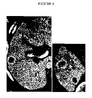
Figure 1A shows one well defined lesion with increased vascularization in the
periphery.
Figure 1B shows progressive growth of multiple larger masses which are
hypovascular and
hypoxic.
Figure 2 shows a perfusion study by MDCT at 7, 14, 21, and 28 days.
Figure 3 shows a graph plotting tumor size, X axis, and arterial flow, Y axis,
over the 28
day period. The arterial flow is measured in the enhancing rim only. The
cancer growth
continues unabated, even though arterialization decreases. R correlation =
0.373 with p<0.042.
With permission of publisher, Wu, Exner, Shi, Bear and Haaga, Dynamic
Evolutoinal Changes
in Blood Flow Academic Radiology 2009;16;1483-92 [9].
Figure 4A and Figure 4B show quantification of the nestin-positive cells
outside the
tumor core (G and H) shows a 68% increase in cell invasion after treatment
from Keunen,0 et al,
PNAS, March 1, 2011,vol 108, no 9, p3749-3754 [10].
Figure 5A shows two large masses, one in segment 3 and the second in medial
segment 7.
Figure 5B. Several months later, both lesions have reduced in size but
multiple enlarging masses
are noted in the lateral part of segment 7 and 8.
Figure 6 shows substrate and metabolic profiles found in premalignant
intraductal tumor
using reaction-diffusion modeling. Oxygen concentrations (solid line), glucose
concentrations
(dashed line), and H+ concentrations (dotted line) are shown. Graph shows that
as the distance
28
CA 02930001 2016-05-06
WO 2015/070038 PCT/US2014/064589
from artery supply to cells increase oxygen (solid line) levels drop because
of poor diffusion.
Glucose levels (dashed line) remain constant because glucose diffuses well and
is actively
transported. With permission of the publisher, Gillies and Gatenby, (2007) J.
Bioenerg.
Biomembr. 39:251-257 [11].
Figure 7A shows autogradiography of FDG in a tumor mass. The very highest
concentration of the FDG is centrally where the hypoxia is greatest and the
GLUT transporters
are the highest, Zhao, S, Kuge, Y, Mochizuki, T, et al, (2005), J.Nucl Med,
46(5);675-682 [12],
Figure 7B. Fluorescent oxygen imaging of tumor in transparent window model
shows
oxygenation in the periphery but severe hypoxia centrally, Dewhirst et al,
(1999), British Journal
of Cancer 79(11/12), 1717-1722 [13].
Figure 8 shows growth of a tumor from single 411 cells in a BALB/c mouse
window
chamber. Approximately 20 cells were injected in a BALB/c mouse window
chamber, and their
growth was followed serially after the initial implantation. Note that both
processes (i.e. growth
and angiogenesis) were visible at the approximately 20- to 50-cell stage of
tumor growth. before
the 105 cell number which is the threshold for hypoxia. Accordingly, the
vasculogenesis is likely
due to increased lactate from cancer cells. The cancer cells with fluorescent
green show motility
and move between days 1, 2, and 4. Such cell movement also depends on lactate,
which
activates the motogenic genes [14, 15].
Figure 9 shows a CT scan revealing breast cancer in medial portion of right
breast,
vertical arrow, and an enlarged lymph node seen in lateral portion of breast,
horizontal arrow.
Spread to lymph nodes is enhanced by increased fluid flow, but, also, the
ameboid movement of
cancer cells. Cell movement depends upon lactate's induction of the molecular
hyaluronan,
which activates the motogenic genes [15].
Figure 10 shows a correlation of radioresistance to high lactate levels.
29
CA 02930001 2016-05-06
WO 2015/070038 PCT/US2014/064589
Figure 11 shows a flow chart demonstrating the five mechanisms by which
lactate
initiates vasculogenesis: 1)microenvironment release of FGF and VEGF
2)induction from in situ
and chemoattracted cells 3)production of HIF la by the effects of lactate on
multiple
mechanisms. Both lactate and hypoxia increase HIF (which regulates VEGF, etc.)
by decreasing
the HIF's degradation enzyme PhD (prolyl hydroxylase 4) ROS reactive oxygen
species
produced by both lactate and hypoxia increases VEGF 5) stem cell chemo
attraction of the
unique CD34+133+VEGFR3+ cells. These mechanisms for lactate function at all
oxygenation
levels normoxia, hypoxia, or hyperbaric. There is even a feed forward
mechanism for lactate to
HIF to glycolysis to lactate, etc.
Figure 12 shows the rate of lymphatic endothelial cell proliferation is
greater than that of
vascular endothelial cells during the transition into the malignant form (SCC-
I-P, SCC-I-C, SCC-
II-P).VEC and LEC proliferation in premalignant and carcinoma tissue.
Quantitative analysis of
proliferating VECs and LECs in ¨LM, premalignant and carcinoma tissue.
Proliferating LECs
were identified in the periphery and center of well-differentiated grade 1
SCCs (SCC-I) but
limited to periphery of less-differentiated grade 2 SCCs. Absence of open
lumen lymphatic
vessels SCC-II centers precluded analysis of LECs in that locale. *, P < 0.05,
two-tailed unpaired
nonparametric Mann-Whitney U, (2007) Cancer Res, 67(11): 5211-20 [16].
Figure 13 shows FGF-2 stimulates corneal lymphangiogenesis. Lowering the dose
of the
FGF2 pellet to 12.5 ng (P) and moving it farther from the limbus results in
less angiogenesis,
although lymphatic vessels still reach the pellet, Chang et al, (2004) Proc
Natl Acad Sci U S A.
101(32): 11658-11663 [17].
Figure 14 shows three different mother veins at different stages of dilation .
The center
cell shows very early separation of pericyte from wall. The vein to the right
shows some
dissolution of the basement membrane and minimal separate of the pericyte. The
vein on the left
CA 02930001 2016-05-06
WO 2015/070038 PCT/US2014/064589
shows degradation of the basement membrane and complete separation of the
pericytes.
Reproduced with permission of publisher, Pettersson, et al. (2000) Lab Invest
80:99-115 [18].
Figure 15 shows microdissection from Patan et al., showing large ecstatic host
venule
with intussception and in the process of dividing into multiple veins, Patan,
S et al. (2001) Circ
Res. 89:732-739 [19].
Figure 16 shows a schematic diagram summarizing the progression of the
angiogenic
response that follows introduction of aden-vpf/vegf into adult tissues of
immunodeficient mice
and rats. The host venule changes into a mother vessel by degradation of the
basement
membrane and detachment of the pericytes. From this state the vein may sprout
or develop
endothelial bridging created multiple channels which form multiple small
daughter veins.
Muscular fibers may develop to become artery/vein over weeks. The glomerulid
structure is a
transient entity, Pettersson et al. (2000) Lab Invest, 80:99-115 [18].
Figure 17 shows 0.5 week c11, Control; c12, TAE; c13, Bumex (Bumetanide); c14,
ferulic acid; c15, caffeic acid.
Figure 18 shows 1 week: c17, Control; c18, TAE; c19, Bumex (Bumetanide); c20,
ferulic acid; c21, caffeic acid.
Figure 19 shows 1.5 Week: c23, Control; c24, TAE; c25, Bumex (Bumetanide);
c26,
ferulic acid; c27, caffeic acid.
Figure 20 shows 2 Week: c29, Control; c30, TAE; c31, Bumex (Bumetanide); c32,
ferulic acid; c33, caffeic acid.
Figure 21 shows 2.5 Week: c35, Control; c36, TAE; c37, Bumex (Bumetanide);
c38,
ferulic acid; c39, caffeic acid.
Figure 22 shows 3 Week: c41, Control; c42, TAE; c43, Bumex (Bumetanide); c44,
ferulic acid; c45, caffeic acid.
31
CA 02930001 2016-05-06
WO 2015/070038
PCT/US2014/064589
Figure 23 shows 3.5 Week: c47, Control; c48, TAE; c49, Bumex (Bumetanide);
c50,
ferulic acid; c51, caffeic acid.
Figure 24A-C show the changes of each rat under different conditions. Figure
24A
shows a control group. Figure 24B shows the TAE treatment group. Figure 24C
shows the
results of the TAE + Bumex (butetanide).
Figure 25 shows the relative tumor volume comparing the different treatments
over four
weeks. Bumex (Bumetanide), ferulic acid, and caffeic acid proved effective at
reducing tumor
volume in the mouse leg tumor model.
Figure 26 show a graph comparing tumor volume versus time for control, TAE,
and TAE
+ three antiglycolytic agents.
Figure 27 shows the results at 0.5 week for different treatments of tumors:
cll, TAE; c12,
Bumex (Bumetanide); c13, ferulic acid; c14, caffeic acid.
Figure 28 shows the results at 1 week for different treatments of tumors: c16,
TAE; c17,
Bumex (Bumetanide); c18, ferulic acid; c19, caffeic acid.
Figure 29 shows the results at 1.5 weeks for different treatments of tumors::
c21, TAE;
c22, Bumex (Bumetanide); c23, ferulic acid; c24, caffeic acid.
Figure 30 shows the results at 2.0 weeks for different treatments of tumors:
e26, TAE;
c27, Bumex (Bumetanide); c28, ferulic acid; c29, caffeic acid.
Figure 31 shows the results at 2.5 weeks for different treatments of tumors:
c31, TAE;
c32, Bumex (Bumetanide); c33, ferulic acid; c34, caffeic acid.
Figure 32 shows the results at 3.0 weeks for different treatments of tumors:
c36, TAE;
c37, Bumex (Bumetanide); c38, ferulic acid; e39, caffeic acid.
Figure 33 shows the results at 3.5 weeks for different treatments of tumors:
c41, TAE;
c42, Bumex (Bumetanide); c43, ferulic acid; c44, caffeic acid.
32
CA 02930001 2016-05-06
WO 2015/070038 PCT/US2014/064589
Figure 34 shows a graph demonstrating the change in tumor diameter over time
with the
different treatment regimens.
Figure 35 shows a graph demonstrating the change in tumor diameter over time
with the
different treatment regimens with confidence intervals.
Figure 36 shows a graph demonstrating the change in tumor diameter over time
with the
different treatment regimens with confidence intervals.
Figure 37 shows a graph demonstrating the change in tumor diameter over time
with the
different treatment regimens.
Figure 38 shows the reaction scheme for the synthesis of GODP macromolecular
contrast
agent for DCE-MR1 techniques.
Figure 39 shows a graphical representation of the AATH impulse response
function that
was used for DCE-1VIR1 parametric analysis.
Figure 40A-C shows contrast enhanced-time curves related to the study The
images in
(Figure 40A) and (Figure 40B) are contrast-enhanced time curves obtained from
a representative
mouse in the saline control and bumetanide-treated groups, respectively. The
contrast enhanced-
time curves in (Figure 40C) were obtained from all of the mice in this study
at the 3-week time
point and show that the tumor uptake of GODP contrast agent is significantly
compromised by
the bumetanide therapy, suggesting a regression in vascularity.
Figure 41A-C shows parametric mappings were constructed by applying the AATH
model on a pixel-by-pixel basis. The images displayed here show the spatial
changes in the Fp
(Figure 41A), PS (Figure 41B), and Vp (Figure 41C) parameters that developed
over time for
both the saline control and bumetanide treatment groups. By the end of the 3-
week treatment
period, vascularity and permeability were largely confined to the periphery of
bumetanide-
treated tumors, contrary to that of the control tumors.
33
CA 02930001 2016-05-06
WO 2015/070038 PCT/US2014/064589
Figure 42 shows average parametric values were obtained from the DCE-MRI
analysis
using the AATH tracer kinetic model. This figure shows the percent reductions
between the pre-
and post-treatment levels of each parameter. As seen here, the bumetanide
therapy was able to
induce significantly greater reductions in the PS (p=0.003) and Vp (p=0.002)
parameters over the
course of the 3-week treatment period as compared to the saline control
therapy. However, no
significant differences were observed in the flow rate.
Figure 43A shows IHC stains for CD31 expression reveal that vascularity is
significantly
compromised in both the periphery and core tissue of the bumetanide-treated
tumors, compared
to those treated with the saline control.
Figure 43B shows pimonidazole staining of samples of both saline and
bumetanide
therapies. Not surprisingly, the decrease in CD31 coincided with an increase
in tumor hypoxia,
as evidenced by the increase in pimonidazole staining intensity.
Figure 44A shows Western blot data.
Figure 44B reveals that VEGF expression was 42.9% lower in the bumetanide-
treated
tumors after 3 weeks of therapy, in comparison to the control tumors
(p=0.021).
Figure 44 shows IHC images in support this result and show that, much like
CD31, a
decrease in VEGF expression is associated with greater levels of hypoxia.
Figure 45A shows the bumetanide therapy did not exhibit any effects on tumor
growth
and proliferation. Tumor size remained unchanged during the course of the
treatment period in
comparison to the control tumors.
Figure 45B&C shows IHC staining of the proliferation marker Ki67 also did not
show a
significant difference between the two groups (Figure 45B and Figure 45C).
Figure 46 shows a Western blotting showed that the HIF-la expression was 59.1%
greater in the bumetanide-treated tumors than in the control tumors (p=0.003).
The increase in
34
CA 02930001 2016-05-06
WO 2015/070038 PCT/US2014/064589
this transcription factor coincides with the increase in tumor hypoxia, as
shown in Figure 43.
DETAILED DESCRIPTON OF THE INVENTION
The present invention relates to a pharmaceutical cocktail and methods of
cancer. In
particular, one such cocktail comprises a combination of effective amounts of
a carbonic
anhydrase inhibitor, a member of the hydroxycinnamate class of drugs or a
derivative thereof,
and/or an angiogenesis inhibitor, including a vascular endothelial growth
factor (VEGF) inhibitor
such as bevacizumab in combination with blood vessel occlusion. As most
cancers in an
untreated state uses both aerobic and anaerobic/glycolytic pathways treatments
contemplated
herein can affect both metabolic pathways.
Although it is not necessary to understand the mechanism of an invention, it
is believed
that treatments that target the anaerobic and aerobic metabolic pathways more
completely
deprives cancer of ATP energy production, thereby producing greater damage or
killing of
cancerous cells. Treatment of the aerobic pathway alone temporarily controls
cancer but it
induces mutation to a glycolytic form, which does not respond to anti-VEGF or
other anti-
vascular growth factor agents.
In other embodiments, it relates to compositions and methods of treating
cancer involving
effective amounts of a member of the hydroxycinnamate class of drugs.
Pharmaceutical
compositions and methods of treating cancer (eliminating the tumor, shrinking
the tumor,
prolonging the life of the patient, increasing quality of life by decreasing
the grade of adverse
events seen with other cancer treatments, and/or preventing/reducing the
likelihood of the
tumor's metastases) are additional aspects of the present invention. In
addition, the present
CA 02930001 2016-05-06
WO 2015/070038 PCT/US2014/064589
invention may be used to favorably affect the therapeutic result of patients
who have not
responded to alternative, traditional anti-cancer therapy.
I. INTRODUCTION
In recent years, it has become recognized that cancers use both aerobic and
glycolytic
metabolism [20-22] and that glycolysis may also play a role. The need for
vasculogenesis with
aerobic metabolism has been well established [23, 24], but the character of
vasculogenesis for
glycolysis has only recently been described [25]. The traditional
vasculogenesis concept explains
that hypoxia induces vascular growth factors which support arterial
development to supply
oxygen [20, 23, 24]. The nature of glycolytic vasculogenesis formulated from
the modem
literature and data has recently been reported [25].
One vasculogenesis hypothesis, ALPHA (Acidic Lactate sequentially induced
Lymphogenesis, PHlebogenesis, and Arteriogenesis) [25], suggests that
glycolytic cancers,
independent of oxygenation, may produce increased lactate, which stimulate
vascular growth
factors causing the sequential production of lymphatics, veins and arteries.
For cancer to thrive,
lactate levels are usually at low or moderate levels which is generally
believed to enhance
cancerous processes. Glycolytic vasculogenesis enhances the development of
drainage vessels to
better manage lactate levels.
Inconsistencies of the traditional theory are commonly observed in the
diagnostic
imaging follow-up of cancer patients and those treated with embolization. A
seldom recognized
inconsistency with the traditional vasculogenesis concept is the evolutionary
changes in tumor
contrast vascular enhancement over the clinical course of a patient, Figure lA
&B. In most
patients early small metastatic lesions show considerable enhancement which
decreases over
36
CA 02930001 2016-05-06
WO 2015/070038 PCT/US2014/064589
time as the tumors grow. Although the arterial enhancement decreases, the
tumors continue
growth unabated or faster in the hypovascular/hypoxic state. This continued
growth and
invasiveness of hypoxic tumors has been emphasized in the clinical literature
[26, 27].
Both the natural evolution and growth of glycolytic tumors and their increased
aggressiveness have been documented in the literature. The conversion to
glycolysis which
sustains rapid tumor growth was confirmed in a long term VX2 liver implant
model in rabbits by
our group [9]. After tumor implantation , the tumor size and its MDCT
perfusion were
quantified weekly over a 28 day period. The arterial flow was the greatest at
1 week and
decreased subsequently with each weekly examination, Figure 2A & B. The TDC
(time contrast
curves) over the growth period graphically display the evolution from a
vascular aerobic tumor
to a hypovascular tumor, Figure 2B. Tumor growth continued unaffected by the
progressive
decrease in arterial flow. An R correlation between the arterial flow of the
enhancing rim with
the area of the viable tumor confirmed no correlation = -0.373, p= 0.042, see
Figure 3.
When arterial flow is reduced by anti-VEGF drug treatments [10] or arterial
embolization
[28], cancerous lesions may temporarily recede but can recur as a more
aggressive glycolytic
form. Keunen et aL who studied a glioblastoma xenograft model treated with
anti-VEGF
antibodies confirmed that tumors become more aggressive and invasive after
hypoxia conversion
to a mostly glycolytic tumor, Figure 4. With hypoxia aerobic cells either die
or lose
mitochondrial function. Similarly, tumors embolized with particles, even
Yttrium may respond
initially but can recur with an even more aggressive form, Figure 5. This
continued growth of
hypoxic tumors is counter to the traditional concept but modern data explains
that glycolysis
drives cellular proliferation and tumor growth by abundant energy, building
substrates and other
advantages, as subsequently described.
37
CA 02930001 2016-05-06
WO 2015/070038 PCT/US2014/064589
Teleological Basis and Tenants of Glycolytic Vasculogenesis
Glycolytic vasculogenesis has numerous tenets, including, but not limited to:
1) Glycolysis
produces increased ATP and increased lactate using only glucose without oxygen
so arteries are
not needed; 2) Glycolysis with lactate production is the preferred metabolic
pathway at all
oxygenation levels, because it modulates many advantageous processes which
support cancer;
3) Excessive high lactate levels impairs or stops glycolysis, which cause
cells to lapse into
cellular arrest; 4) To restore cell growth, proliferation and pro-cancer
advantages, excessive
lactate levels must be reduced to favorable levels by lymphatics and veins; 5)
Lactate increases
the vascular growth factors and other mechanisms for initiating vasculogenesis
which
complements and supplements traditional vasculogenesis; and. 6) To give
lactate drainage
priority, the sequence of vessel development is lymphatics first, veins, and
then arteries.
Glycolysis produces increased ATP and increased lactate using only glucose
without
oxygen, so arteries are not needed. Considering the chemical reactions of
oxidative
phosphorylation and glycolysis, the types of vessels needed for each process
become quite clear.
Aerobic metabolism very efficiently uses its substrates, oxygen and glucose,
to make large
amounts of ATP energy: 1 glucose molecule and 1 oxygen molecule make 38 ATP's
with 2 CO2.
In contrast, glycolysis uses 19 glucose molecules without oxygen to produce
the equivalent 38
ATP's with 38 lactates. Arteries for oxygen or glycose are not needed but
drainage vessels are
(predominantly) lymphatics to clear lactate.
Abundant arterial supply is necessary for oxygen transport but not for glucose
supply.
Oxygen diffuses poorly over distance however glucose diffuses quite well and
has specialized
glucose transporters (GLUTS), Figure 6. Even if the center of a mass has poor
arterial flow and
is hypoxic there is always abundant glucose in the deepest part of the tumor,
Figure 7. This has
08
CA 02930001 2016-05-06
WO 2015/070038 PCT/US2014/064589
been confirmed by comparison studies between clinical radionuclide blood flow
and FDG PET
studies. These show there is no correlation between arterial flow/oxygenation
and FDG uptake
[29] which confirms abundant glucose if available for glycolysis without
significant arterial
flow.
Some suggest that cancer cells prefer oxidative phosphorylation because it is
so
chemically efficient (38 ATP per glucose), but glycolysis provides advantages
which supereede
this efficiency. While glycolysis is not as efficient in its use of glucose,
it more than compensates
to provide abundant energy by way of its very rapid reaction speed. Glycolysis
produces ATP
energy 100 times faster than aerobic metabolism, because it has fewer
reactions and two feed
forward catalytic steps [25].
Glycolysis with lactate production is the preferred metabolic pathway for
cancer at all
oxygenation levels because it provides many advantages for cancer. For
example, such cancer
advantages may include but is not limited to the production of building
substrates for cellular
growth and the initiation and support of many pro-cancer processes.
The role of glycolysis for providing building substrates for cell growth and
proliferation
has been reported [30, 31]. The essential proteins, lipids, nucleotides, etc.
are produced by
numerous side reactions related to glycolysis. Before cell proliferation can
occur, mother cells
must double their biomass before division to ensure that each daughter cell is
fully endowed with
the necessary cellular components, such as DNA, RNA, cell membranes,
organelles etc. Without
such growth, cell division cannot proceed and cells lapse into cellular arrest
or quiescence.
The lactate produced by glycolysis is generally considered a "waste product",
but in fact
it has variable roles and modulatory effects at different concentrations,
which are beneficial to
cancer cells. At low concentrations, lactate is a high energy substrate which
can be used by
09
CA 02930001 2016-05-06
WO 2015/070038 PCT/US2014/064589
adjacent aerobic normal or cancerous cells [32, 33]. At moderate
concentrations, lactate initiates
many processes which support cancerous activities: a) lactate creates a
hostile low pH/high
lactate selective environment, which kills normal or un-adapted cancer cells
but selective
adapted cancer cells have special waste management(Carbonic anhydrase IX and
lactate
transporter MCT4) survive [11, 14, 34]. These enzymes are believed to protect
the surviving
aggressive cancerous phenotypes. b) low pH and lactate initiates
transformation of the
microenvironment by activating metalloprotease enzymes, which alters tissue
structure and
releases and activates biomarkers [11, 15]. c) lactate initiates cancer cell
motility through an
intermediate, molecule, hyaluronan, which turns on the motogenic genes [15,
35], Such
movement facilitates local cell invasion or lymph node metastases, see Figure
8 and Figure 9, b)
increased lactate levels impair the local host immune cells, so that
lymphocytes, natural killer
cells, dendritic, and macrophages cannot proliferate, recognize antigens, nor
produce cytotoxic
cytokines [25, 36]. e) cancer cells, incubating in lactate and ketones from
glycolysis, transform
into a stem cell like state capable of unlimited mutations [37]. f) molecules
are induced by
lactate, which are anti-apoptotic [38-41]. g) lactate, independent of
oxygenation, increases
HIF1a, a very important modulator for cancer proteins and enzymes [25, 42-46].
h) finally
lactate directly and indirectly stimulates different cells and processes which
produce vascular
growth factors (VEGF, FGF, VEGFC, etc) for vasculogenesis [45-55]. This
production occurs at
all oxygenation states (normoxia, hypoxia, or hyperbaric oxygen), so lactate
complements and
supplements the traditional hypoxic vasculogenesis.
High lactate levels impairs cancer growth because it causes cellular
quiescence or arrest.
High lactate levels and the associated low pH can impair or stop glycolysis,
which precludes the
benefits described above. This cessation of glycolysis occurs because of basic
chemical
CA 02930001 2016-05-06
WO 2015/070038 PCT/US2014/064589
principles. The low pH caused by lactate stops glycolysis at the rate limiting
enzyme
phosphofructose kinase. Further, high lactate inhibits glycolysis by end
product inhibition or
"negative feedback" [56-60]. With the loss of the abundant building substrates
and increased ATP
needed for cell growth and division, cell proliferation is reduced or ceases.
Non-growing mother
cells lapse into cellular quiescence or arrest [30, 31]. Sattler and Quennet
[61, 62] reported that
increased lactate directly correlates with radioresistance. Quennet found the
correlation of
radioresistance to high lactate levels was R=0.9824, p+0.0028, Figure 10. The
causes of
radioresistances have not been elucidated but likely cellular arrest and anti-
apoptotic processes
[38-40, 55] contribute to radioresistance.
To prevent excessively increased lactate which can retard cell growth,
proliferation, and
pro-cancer advantages, lymphatics and veins are needed to clear lactate [61,
62] to maintain
optimal lactate levels. Lymphatic vessels may drain lactate laden
extracellular interstitial fluid
[63]. Rutz asserted that lactate from glycolysis is a factor causing increased
interstial fluid [64].
When cancer cells make excess lactate it is transported through the cell
membrane into the
interstial space. The exported lactate makes the interstitial fluid
hypertonic, which causes the
influx of free water increasing the interstitial fluid pressure and subsequent
flow. A recent de
novo skin cancer model reported by Eitchen [16] emphasized that lymphatic
development may
occur when cells make the transition from a premalignant state to carcinoma in
situ. They
observed that lymphatics are normal sized in the premalignant phase but when
carcinoma in situ
and locally invasive progresses, the normal lymphatics dilate with increased
fluid flow and rapid
proliferation of lymphatic endothelial cells.
Lactate increases vascular growth factors [45-55] and causes chemoattraction
of key cells
important for vasculogenesis [65-67]. Increase of vascular growth factors by
lactate comes from
multiple processes, Figure 11, including the release of donnantly stored
factors in the
41
CA 02930001 2016-05-06
WO 2015/070038 PCT/US2014/064589
extracellular matrix, non HIF induction of growth factors from many cells and
increased HIF la,
(hypoxia induction factor). There are two additional independent vasculogenic
pathways, ROS
(reactive oxygen species) and stem cell angiogenesis which are stimulated by
lactate.
The initial increase of factors are FGF (fibroblast growth factors) and VEGF
(vascular
endothelial growth factor) which are released from their dormant form in the
extracellular matrix
[17, 47, 48]. These are released when low pH and lactate activate
metalloproteases which
degrade and transform the microenvironment [15, 34]. The disruption of the
existing matrix and
collagen facilitates cellular movement and vasculogenesis. Additional
production of vascular
growth factors is stimulated by lactate from in situ cells and chemoattracted
cells (cancer cells,
fibroblasts, endothelial cells and macrophages). The chemoattractant SDF-1
(stromal derived
factor-1) for these cells is produced by lactate via HIF la (Hypoxia Induction
Factor) as an
intermediary [46].
More vascular growth factors are made by these cells by the action of HIF1a,
which is a
modulator of VEGF and/or glycolysis. HIF la is stimulated by hypoxia but it is
also produced in
greater amounts by lactate through multiple mechanisms [25, 42-46]. Both
hypoxia and
increased lactate increase HIF by reducing its degradation enzyme PhD (prolyl
hydroxylase),
Figure 12. When PhD decreases, the levels of HIF rise because less HIF is
destroyed. The lactate
mechanisms are more significant than hypoxia because lactate increases HIF in
normoxia,
hypoxia and hyperoxia [25, 42-46]. Further, greater amounts of HIF are made by
lactate because
it causes a "feed forward" reaction, which can be self perpetuating. When
lactate increases
HIFI a, glycolytic enzymes are increased which increases lactate [43] which
increases HIF, etc.
The two other lactate related mechanisms which may increase vasculogenesis are
ROS
(reactive oxygen species) [45] and the chemoattraction of marrow progenitor
stem cells [66, 67].
42
CA 02930001 2016-05-06
WO 2015/070038 PCT/US2014/064589
The attracted stem cells have two separate roles, assisting existing vessel
morphogenesis
and initiating an additional independent pathway. Unspecified angioblasts
assist with
morphogenesis of existing vessels by aiding the "budding" and channel
formation. The specific
character of the vessel formed (i.e., for example, lymphatic, veins, and/or
arteries) is determined
by the interaction of different biomarkers (i.e., for example, PI3K and Ephrin
B4 for veins;
Notch and Ephrin B2 for arteries).
The unique stem cell CD34+,133+ with VEGFR3+ ,which is a lymphatic endothelial
cell
precursor has a remarkably unique role. Although the mechanism has not yet
been defined, this
cell through VEGFC and Wnt/respondin pathways can initiate vasculogenesis in
and of itself
without other processes [66, 67].
To give lactate drainage priority, the essential growth factors VEGFA and FGF2
produce
vessel development with lymphatics first, veins, and then arteries. To
appreciate the numerous
reports on vessel development it is important to understand the important role
of VEGFA and
FGF2 for starting the initial angiogenic burst which results in sustained
vasculogenesis [68-70].
The recent report by Indraccolla et al [68] confirmed that a transient
angiogenic burst caused by
VEGFA and FGF2 within the transformed microenvironment starts the entire
vasculogenesis
process.
The VEGFA molecule was originally named VPF (vascular permeability factor),
because
it controls vascular permeability but the name was changed after synthesis of
the VEGFA
molecule and introduction of anti-VEGF antibodies [18, 711. The receptor for
VEGFA(VPF)
and permeability occur in veins, not arteries [71-73] as proven by
immunohistochemistry,
electron microscopy, radionuclide and macromolecule diffusion. Since the
primary receptor sites
aren't arterial it is logical that hypoxia caused by anti-VEGF drugs occurs by
means of venous
infarction. If veins are closed, arterial flow ceases.
43
CA 02930001 2016-05-06
WO 2015/070038 PCT/US2014/064589
The sequence of vessel morphogenesis (lymph, veins, arteries) has been
confirmed by
many different models (de novo spontaneous squamous cell cancer model,
xenograft implants.
FGF growth factor pellet implants; and VEGF gene transfection).
In a de novo skin cancer model [16], lymphatic cells proliferated and enlarged
existing
lymphatics before blood vessel changes. This occurred when the tumor changed
from
premalignant to malignant occurred. After the tumor became larger and more
invasive, blood
vessel endothelial cells proliferated Figure 12.
Chang et al [17] characterized the types of vessels morphed by implanted FGF2
pellets,
see Figure 13. At low concentrations, FGFb induced lymphatics via VEGFC and D
without
hemangiogenesis. Higher concentrations of FGFb equally stimulated both
lymphatics and blood
vessels via VEGFA. VEGFA came from attracted macrophages and released from the
extracellular matrix [17, 34, 47, 48]. VEGFC and D for lymphatics came from
attracted
macrophages [171.
The effects of the VEGFA on vessel formation were determined by adenovirus
transfection of a VEGFA gene into normal mouse ears [18, 74, 75]. Lymphatic
morphogenesis
occurred at 1-3 days. Veins dilated and divided into smaller daughter veins at
3-5 days [18, 19,
71, 75], Figure 14 and Figure 15. Arterial capillaries and arterial venous
malformations
development occurred later at 7-10 days [18, 74, 75], Figure 16.
Patan et al. [19], also, reported that the first tumor blood vessels to form
are venous,
which develop from the pre-existing host veins. Patan studied a xenograft
colon cancer implant
mouse model which grew for 21 days. From examination of the harvested masses
she
concluded, "Reconstruction of 3500 histological serial sections demonstrated
that a new vascular
network composed of venous-venous loops of varying sizes grows inside the
tumor from the wall
44
CA 02930001 2016-05-06
WO 2015/070038 PCT/US2014/064589
of the adjacent main vein". It should be noted that the author did not see any
arterial changes in
these dissections, Figure 15.
Cancers can use both aerobic and glycolytic metabolic pathways. Each pathway
requires
vasculogensis, but of a different nature. While aerobic metabolism is
supported by arteries to
supply oxygen, glycolytic metabolism depends upon lymphatic and venous
drainage (ALPHA
vasculogenesis) to manage lactate levels. The two processes are complementary,
but ALPHA has
a greater role because virtually all tumors eventually may lose their arterial
supply and convert to
predominantly glycolytic metabolism. Understanding the nature of ALPHA is
important to
radiologists because the modern imaging perfusion techniques are more
consistent with ALPHA
than the traditional oxygen based theory.
Example 1 describes a comparison study on hepatocellular rat model treatment
using
arterial embolization and embolization combined with anti-glycolytic agents.
The example
demonstrates that anti-glycolytic agents could enhance the effect of TAE on
liver tumor.
Although it is not necessary to understand the mechanism of the current
invention, it is
believed that when either bumetanide or the hydroxycinnamates are given,
glycolysis is
interrupted. Although it is not necessary to understand the mechanism of the
current invention, it
is believed that bumetanide and acetazolamide may block carbonic anhydrases IX
and XII which
cause the intracellular pH of cancer to become less or more acidic. The low pH
inhibits the rate
limiting step of glycolysis, phosphofructose kinase. Although it is not
necessary to understand
the mechanism of the current invention, it is believed that bumetanide acts as
a NKCC inhibitor.
Although it is not necessary to understand the mechanism of the current
invention, it is believed
that with hydroxycinnamates the lactate transporters on the cell membrane are
blocked so lactate
accumulates inside the cell making it more acid and blocking the glycolytic
reaction by "end
product inhibition". Although it is not necessary to understand the mechanism
of the current
CA 02930001 2016-05-06
WO 2015/070038 PCT/US2014/064589
invention, it is believed that when either of these occurs glycolysis ceases
so there is no ATP
produced nor building substrates (proteins, lipids, nucleotides)produced which
permit growth of
the ''mother cell" which is needed so division to occur to produce two
daughter cells fully
endowed with membranes, etc. If the cells cannot divide, they lapse into GO
cellular arrest. Non-
dividing cells are not sensitive to radiation, or radioresistant i.e.
radioprotectant. This is true of
normal cells (radioprotectant) or cancer cells (radioresistant). When the
activity of the drug
wears off, cancer metabolism can again use both glycolysis and aerobic
metabolism with existing
mitochondria. Normal cells are more likely to have mitochondria so they have
less reduction of
ATP.
EMBOLIZATION
Preoperative embolization of tumors is a well-established procedure that has
been
successfully applied in various clinical situations [76-80]. Preoperative
embolization can reduce
the vascularity of tumors resulting in a clearer operative field, easier
dissection, decreased blood
loss, and, in some cases, a decrease in tumor size.
A number of different materials have been used for embolization of tumors.
Gelfoam has
been used commonly in the past; however, because of the relatively large
particle size, proximal
occlusion of large vessels can lead to ineffective embolization. Sundaresan et
al. used absolute
ethanol for the preoperative embolization of spinal metastases from renal
cancer with good
results [81]. N-butyl cyanoacrylate and polymethlymethacrylate are commonly
used embolic
agents [82], and one author has described the use of a detachable balloon to
occlude a vertebral
artery before surgery for a cervical vertebral tumor Breslau et al. [83]. Non-
absorbable PVA
particles are one of the most frequently used embolic agents reported in the
literature. PVA
particles 150¨ 250-mm in diameter result in distal embolization with occlusion
at the capillary
46
CA 02930001 2016-05-06
WO 2015/070038 PCT/US2014/064589
level. Larger particles, 250¨ 500-mm in diameter, can then be used to embolize
larger vessels or
anastomoses. The use of non-absorbable PVA microspheres might prevent the
recurrence of
tumor hypervascularity due to recanalization of an embolized artery, which is
commonly seen
with absorbable gelatin sponges. Complications related to embolization are
believed to be
primarily due to devascularization of areas adjacent to the tumors being
embolized, and in
regions such as the spine, damage to adjacent areas can lead to permanent loss
of functionality
[84].
In some embodiments, the embolization material for embolic compositions
comprises a
polymer. Examples of polymers that can be used for creating particles for
embolization include
polyvinyl alcohols, polyacrylic acids, polymethacrylic acids, poly vinyl
sulfonates,
carboxymethyl celluloses, hydroxyethyl celluloses, substituted celluloses,
polyacrylamides,
polyethylene glycols, polyamides, polyureas, polyurethanes, polyesters,
polyethers, polystyrenes,
polysaccharides, polylactic acids, polyethylenes, polymethylmethacrylates,
polycaprolactones,
polyglycolic acids, poly(lactic-co-glycolic) acids (e.g., poly(d-lactic-co-
glycolic) acids) and
copolymers or mixtures thereof. A preferred polymer is polyvinyl alcohol
(PVA). The polyvinyl
alcohol, in particular, is typically hydrolyzed in the range of from about 80
percent to about 99
percent. The weight average molecular weight of the base polymer can be, for
example, in the
range of from about 9000 to about 186,000 (e.g., from about 85,000 to about
146,000, from
about 89,000 to about 98,000).
In general, individual embolization particles can have a diameter of from
about ten
microns to about 3,000 microns (e.g., from about 40 microns to about 2,000
microns; from about
100 microns to about 700 microns; from about 500 microns to about 700 microns;
from about
100 microns to about 500 microns; from about 100 microns to about 300 microns;
from about
300 microns to about 500 microns; from about 500 microns to about 1,200
microns; from about
47
CA 02930001 2016-05-06
WO 2015/070038 PCT/US2014/064589
500 microns to about 700 microns; from about 700 microns to about 900 microns;
from about
900 microns to about 1,200 microns). In some embodiments, particle 10 can have
a diameter of
about 3,000 microns or less (e.g., about 2,500 microns or less; about 2,000
microns or less; about
1,500 microns or less; about 1,200 microns or less; about 1,000 microns or
less; about 900
microns or less; about 700 microns or less; about 500 microns or less; about
400 microns or less;
about 300 microns or less; about 100 microns or less) and/or about ten microns
or more (e.g.,
about 100 microns or more; about 300 microns or more; about 400 microns or
more; about 500
microns or more; about 700 microns or more; about 900 microns or more; about
1,000 microns
or more; about 1,200 microns or more; about 1,500 microns or more; about 2,000
microns or
more; about 2,500 microns or more).
In some embodiments, multiple particles are combined with a carrier fluid
(e.g., a saline
solution, a contrast agent, or both) to form an embolic composition. Such
embolic compositions
can be delivered to various sites in the body, including, for example, sites
having cancerous
lesions, such as the breast, prostate, lung, thyroid, or ovaries. The embolic
compositions can be
used in, for example, neural, pulmonary, and/or AAA (abdominal aortic
aneurysm) applications.
The compositions can be used in the treatment of, for example, fibroids,
tumors, internal
bleeding, arteriovenous malformations (AVMs), and/or hypervascular tumors in
addition to
cancerous lesions. The compositions can be used as, for example, fillers for
aneurysm sacs, AAA
sac (Type II endoleaks), endoleak sealants, arterial sealants, and/or puncture
sealants, and/or can
be used to provide occlusion of other lumens such as fallopian tubes, Fibroids
can include uterine
fibroids which grow within the uterine wall (intramural type), on the outside
of the uterus
(subserosal type), inside the uterine cavity (submucosal type), between the
layers of broad
ligament supporting the uterus (interligamentous type), attached to another
organ (parasitic type),
or on a mushroom-like stalk (pedunculated type). Internal bleeding includes
gastrointestinal,
48
CA 02930001 2016-05-06
WO 2015/070038 PCT/US2014/064589
urinary, renal and varicose bleeding. AVMs are for example, abnormal
collections of blood
vessels, e.g. in the brain, which shunt blood from a high pressure artery to a
low pressure vein,
resulting in hypoxia and malnutrition of those regions from which the blood is
diverted. In some
embodiments, a composition containing the particles can be used to
prophylactically treat a
condition.
The magnitude of a dose of an embolic composition can vary based on the
nature,
location and severity of the condition to be treated, as well as the route of
administration. A
physician treating the condition, disease or disorder can determine an
effective amount of
embolic composition. An effective amount of embolic composition refers to the
amount
sufficient to result in amelioration of symptoms or a prolongation of survival
of the subject. The
embolic compositions can be administered as pharmaceutically acceptable
compositions to a
subject in any therapeutically acceptable dosage, including those administered
to a subject
intravenously, subcutaneously, percutaneously, intratrachealy,
intramuscularly, intramucosaly,
intracutaneously, intra-articularly, orally or parenterally.
An embolic composition can include a mixture of particles (e.g., particles
that include
different types of therapeutic agents), or can include particles that are all
of the same type. In
some embodiments, an embolic composition can be prepared with a calibrated
concentration of
particles for ease of delivery by a physician. A physician can select an
embolic composition of a
particular concentration based on, for example, the type of embolization
procedure to be
performed. In certain embodiments, a physician can use an embolic composition
with a relatively
high concentration of particles during one part of an embolization procedure,
and an embolic
composition with a relatively low concentration of particles during another
part of the
embolization procedure.
49
CA 02930001 2016-05-06
WO 2015/070038 PCT/US2014/064589
Suspensions of particles in saline solution can be prepared to remain stable
(e.g., to
remain suspended in solution and not settle and/or float) over a desired
period of time. A
suspension of particles can be stable, for example, for from about one minute
to about 20
minutes (e.g. from about one minute to about ten minutes, from about two
minutes to about
seven minutes, from about three minutes to about six minutes).
In some embodiments, particles can be suspended in a physiological solution by
matching the density of the solution to the density of the particles. In
certain embodiments, the
particles and/or the physiological solution can have a density of from about
one gram per cubic
centimeter to about 1.5 grams per cubic centimeter (e.g., from about 1.2 grams
per cubic
centimeter to about 1.4 grams per cubic centimeter, from about 1.2 grams per
cubic centimeter to
about 1.3 grams per cubic centimeter).
In some embodiments, the carrier fluid of an embolic composition can include a
surfactant. The surfactant can help the particles to mix evenly in the carrier
fluid and/or can
decrease the likelihood of the occlusion of a delivery device (e.g., a
catheter) by the particles. In
some embodiments, the surfactant can enhance delivery of the embolic
composition (e.g., by
enhancing the wetting properties of the particles and facilitating the passage
of the particles
through a delivery device). In certain embodiments, the surfactant can
decrease the occurrence of
air entrapment by the particles in a composition (e.g., by porous particles in
a composition).
Examples of liquid surfactants include Tween 80 (available from Sigma-
Aldrich) and
Cremophor EL (available from Sigma-Aldrich). An example of a powder
surfactant is
Pluronic F127 NF (available from BASF). In certain embodiments, an embolic
composition
can include from about 0.05 percent by weight to about one percent by weight
(e.g., about 0.1
percent by weight, about 0.5 percent by weight) of a surfactant. A surfactant
can be added to the
CA 02930001 2016-05-06
WO 2015/070038 PCT/US2014/064589
carrier fluid prior to mixing with the particles and/or can be added to the
particles prior to mixing
with the carrier fluid.
In some embodiments, a plurality of the particles (e.g., in an embolic
composition) can be
delivered through a catheter having a lumen with a cross-sectional area that
is smaller (e.g.,
about 50 percent or less) than the uncompressed cross-sectional area of the
particles. In such
embodiments, the particles are compressed to pass through the catheter for
delivery into the
body. Typically, the compression force is provided indirectly, by depressing
the syringe plunger
to increase the pressure applied to the carrier fluid. In general, the
particles are relatively easily
compressed to diameters sufficient for delivery through the catheter into the
body. The relatively
robust, rigid surface region of the particles can resist abrasion when the
particles contact hard
surfaces such as syringe surfaces, hard plastic or metal stopcock surfaces,
and/or the catheter
lumen wall (made of, e.g., Teflon) during delivery. Once in the body, the
particles can
substantially recover to original diameter and shape for efficient transport
in the carrier and body
fluid stream. At the point of occlusion, the particles can again compress as
they aggregate in the
occlusion region. The particles can form a relatively dense occluding mass.
The compression of
the particles in the body is generally determined by the force provided by
body fluid flow in the
lumen. In some embodiments, the compression may be limited by the compression
profile of the
particles, and the number of particles needed to occlude a given diameter may
be reduced.
In one embodiment, the present invention contemplates a method for cancer
treatment
comprising embolization in combination with metabolic pathway modulation
drugs. Non-
limiting examples of polymeric embolization materials and methods are
described in U.S. Patent
7,964,123 [85]. Although it is not necessary to understand the mechanism of
the current
invention, it is believed that when embolic material is administered with
either bumetanide or the
ferrulic acid or caffeic acid, the ATP production is more severely reduced
because neither
51
CA 02930001 2016-05-06
WO 2015/070038 PCT/US2014/064589
pathway is functional. Closure of the vessel by embolic blockage prevents
blood flow and
oxygenation. With markedly reduce ATP, cells die from programmed cell death or
apoptosis (if
ATP reduced by about 20%) or necrosis if reduced more than 20%).
Although it is not necessary to understand the mechanism of the current
invention, it is
believed that when Avastin is combined with Bumetanide, a similar effect
occurs. Small arteries
are occluded from anti-VEGF damage to the small veins. With vein occlusion, it
is believed that
arterial occlusion concomitantly occurs because artery inflow is dependent
upon venous outflow.
With Avastin administration, sites of neoangiogenesis are more affected so
tumors are affected
more than stable normal tissue. Resulting in, for example, reduced ATP energy,
apoptosis or
necrosis as described above.
Although it is not necessary to understand the mechanism of the current
invention, it is
believed that either Avastin or bumetanide/hydoxycinnamates may partially
affect tumors
temporarily until cancer switches from aerobic to glycolysis or back
(apparently there can be
"gained" attributes from mutations, i.e. mitochondria can regenerate in cancer
cells. However,
without ATP it is believed that the cells may not be able to live.
Both Cancer cells and normal cells make ATP by BOTH aerobic (oxygen/artery)
and
glycolysis. If the arteries are impaired by occlusion /emboli and cessation of
glycolysis occurs,
the cells die. It is said if ATP is reduced by 20%, programmed death occurs
(apoptosis). If a
greater reduction occurs, the cells necrose quickly. Without ATP production,
the cells cannot
live. The animal models show the effect of interrupting both aerobic and
glycolysis
simultaneously.
Two separate reports, one showing the effects of anti VEGF( i.e. Avastin) on a
glioblastoma tumor (Keunen 2011 [10]) and the effects of a hydroxycinnamate
compound on a
52
CA 02930001 2016-05-06
WO 2015/070038 PCT/US2014/064589
glioblastoma (Colen 2011 [86]) reflect possible impacts of individual compound
therapy.
Individually, both show a reduction of tumor size. Combining the two compounds
is thought to
likely destroy aerobic cells and glycolytic cells (blocks lactate transport
and thereby end product
inhibition of glycolysis). With the two components, cancer should be denied of
all ATP. Normal
cells which use aerobic may have little or no lactate, i.e. lactate
transporters. Normal tissues may
not have new angiogenesis (target of Avastin).
Although it is not necessary to understand the mechanism of the present
invention, it is
believed that hydroxycinnamates have an effect in which the compounds block
the transport of
lactate through the MCT transporters so that glycolysis temporarily blocks
glycolysis by virtual
of end product inhibition of glycolysis by increased lactate. Bumetanide
likely blocks glycolysis
by prevent CAIX from producing HCO3 which buffers acidity, thereby producing
low pH
intracellularly. The rate-limiting step of glycolysis phosphofructose kinase
is very sensitive to
low pH so inhibits glycolysis. The effects of either compound is to impair
glycolysis so less
ATP is available and Mother dividing cells lapse into GO because the
substrates derived from the
side reactions of glycolysis (makes proteins, nucleotides, etc).
Although it is not intended to limit the present invention, it is believed
that it is possible
that long term Bumetanide or long-term hydroeinnamates could induce cellular
arrest GO. These
are very safe drugs and putting a tumor into dormancy (e.g., not growing) is
perhaps as good as
or better than killing a tumor. Seeking ways to put tumors into dormancy for
management as
chronic diseases, in lieu of killing them, may be a preferable therapeutic
avenue. Cancer cell
killing therapies have disadvantages because most chemical treatments select
out "resistant
clones" which then become more aggressive. Although it is not intended to
limit the present
invention, it is believed that because hydroxycinnmates or Bumetanide inhibit
glycolysis
temporarily, sustained administration may produce long-term dormancy. They can
both be taken
53
CA 02930001 2016-05-06
WO 2015/070038 PCT/US2014/064589
orally.
Although the invention has been described with reference to these preferred
embodiments, other embodiments can achieve the same results. Variations and
modifications of
the present invention may be obvious to those skilled in the art and it is
intended to cover in the
appended claims all such modifications and equivalents. The entire disclosures
of all
applications, patents, and publications cited above, and of the corresponding
application are
hereby incorporated by reference.
Thus, specific compositions and methods of targeted treatment of anaerobic
cancer have
been disclosed. It should be apparent, however, to those skilled in the art
that many more
modifications besides those already described are possible without departing
from the inventive
concepts herein. The inventive subject matter, therefore, is not to be
restricted except in the spirit
of the disclosure. Moreover, in interpreting the disclosure, all terms should
be interpreted in the
broadest possible manner consistent with the context. In particular, the terms
"comprises" and
"comprising" should be interpreted as referring to elements, components, or
steps in a non-
exclusive manner, indicating that the referenced elements, components, or
steps may be present,
or utilized, or combined with other elements, components, or steps that are
not expressly
referenced.
All publications mentioned herein are incorporated herein by reference to
disclose and
describe the methods and/or materials in connection with which the
publications are cited. The
publications discussed herein are provided solely for their disclosure prior
to the filing date of
the present application. Nothing herein is to be construed as an admission
that the present
invention is not entitled to antedate such publication by virtue of prior
invention. Further, the
dates of publication provided may be different from the actual publication
dates, which may need
to be independently confirmed.
54
CA 02930001 2016-05-06
WO 2015/070038 PCT/US2014/064589
DETAILED DESCRIPTION OF DRUGS
VEGF inhibitors or anti-VEGF therapy may involve binding of an agent to VEGF
to
prevent its modulation of a receptor such as VEGFR-1 (fit-1), VEGFR-2 (flk-1
or KDR), or
through inhibition of tyrosine kinase in promoting angiogenesis or it may
inhibit the binding of
VEGF to one or more of its receptors by any one or more mechanisms. Regardless
of the
mechanism of action, anti-VEGF activity associated with the use of an
effective amount of a
VEGF inhibitor in the present invention results in a reduction in VEGF
activity
(angiogenesis/vascularization) in the tumor, and a response which is
inhibitory to cancer growth,
elaboration and metastases and which helps to promote cancer remission in
combination with the
other agents. Bevacizumab is a preferred VEGF inhibitor for use in the present
invention.
Compounds/compositions according to the present invention which represent anti-
VEGF therapy
(angiogenesis inhibitors) include for example, ZD6474, ZD 6126, AZD2171 (Astra
Zeneca),
SU6668 and SU5416 (Sugen), bevacizumab (Avastin), mv833, anti-FLT-1 ribozyme
(Angiozyme), and the tyrosine kinase inhibitors SU5416 (Semaxanib), PTK 787
(ZK 222584),
ZD4190, ZD6474, CEP-7055, SU11248 and mixtures thereof. In one embodiment anti-
angiogenic agents include tumor-vascular disrupting agents described by
Siemann (2011) [87],
incorporated herein by reference.
Vandetanib (rINN, trade name Caprelsa), also known as ZD6474, is an antagonist
of the
vascular endothelial growth factor receptor (VEGFR) and the epidermal growth
factor receptor
(EGFR). It is a tyrosine kinase inhibitor, being developed by AstraZeneca.
ZD6126 is a vascular-targeting agent and a prodrug of N-acetylcolchinol,
related to
colchicine.
CA 02930001 2016-05-06
WO 2015/070038 PCT/US2014/064589
Cediranib (tentative trade name Recentin), also known as AZD2171, is a potent
inhibitor
of vascular endothelial growth factor (VEGF) receptor tyrosine kinases
developed by Astra
Zeneca.
SU6668, a multitargeted angiogenesis inhibitor described in Klenke et al.
(2007) [88],
incorporated herein by reference.
Semaxanib (SU5416) is a tyrosine-kinase inhibitor drug designed by SUGEN as a
cancer
therapeutic. It is an experimental stage drug, not licensed for use on human
patients outside of
clinical trials. Semaxanib is a potent and selective synthetic inhibitor of
the Flk-1/KDR vascular
endothelial growth factor (VEGF) receptor tyrosine kinase. It targets the VEGF
pathway, and
both in vivo and in vitro studies have demonstrated antiangiogenic potential.
Mv833 is anti-human VEGF monoclonal antibody.
Anti-FLT-1 ribozyme or Angiozyme is a substance that is being studied in the
treatment
of kidney cancer. It may prevent the growth of blood vessels from surrounding
tissue to the
tumor. It belongs to the families of drugs called VEGF receptor and
angiogenesis inhibitors.
Angiozyme is also called RPI.4610.
The tyrosine kinase inhibitors include, but are not limited to: SU5416
(Semaxanib), PTK
787 (Vatalanib), ZD4190, ZD6474 (Vandetanib), CEP-7055, and SW1248
(Sunitinib).
Semaxanib (S1J5416) is a tyrosine-kinase inhibitor drug designed by SUGEN as a
cancer
therapeutic. Semaxanib is a potent and selective synthetic inhibitor of the
Flk-1/KDR vascular
endothelial growth factor (VEGF) receptor tyrosine kinase. It targets the VEGF
pathway, and
both in vivo and in vitro studies have demonstrated antiangiogenic potential.
Vatalanib (also known as PTK787 or PTK/ZK) is a small molecule protein kinase
inhibitor that inhibits angiogenesis. Vatalanib is being developed by Bayer
Schering and
56
CA 02930001 2016-05-06
WO 2015/070038 PCT/US2014/064589
Novartis. It inhibits all known VEGF receptors, as well as platelet-derived
growth factor
receptor-beta and c-kit, but is most selective for VEGFR-2.
Vandetanib (trade name Caprelsa), also known as ZD6474, is an antagonist of
the
vascular endothelial growth factor receptor (VEGFR) and the epidermal growth
factor receptor
(EGFR). It is a tyrosine kinase inhibitor, being developed by AstraZeneca.
Sunitinib (marketed as Sutent by Pfizer, and previously known as SU11248) is
an oral,
small-molecule, multi-targeted receptor tyrosine kinase (RTK) inhibitor.
Bevacizumab (Avastine) (rhuMAb-VEGF)( Anti-VEGF monoclonal antibody) is a
recombinant human/murine chimeric monoclonal antibody directed against
vascular endothelial
growth factor (VEGF).). It is prepared by engineering VEGF-binding residues of
a murine anti-
VEGF monoclonal antibody into framework regions of human immunoglobulin-1
(IgG1) (Prod
Info Avastin, 2004). Only 7% of the amino acid sequence is derived from the
murine antibody,
with 93% from IgGI, Figg, W. D. et al. (2002) [89] incorporated herein by
reference.
Human VEGF mediates neoangiogenesis in normal and malignant vasculature; it is
overexpressed in most malignancies and high levels have correlated with a
greater risk of
metastases and poor prognosis in many. When VEGF interacts with its receptor
in in vitro
models of angiogenesis, endothelial cell proliferation and new blood vessel
formation occur. In
animal models, VEGF has been demonstrated to induce vascular endothelial-cell
proliferation/migration, sustain survival of newly-formed blood vessels, and
enhance vascular
permeability. Bevacizumab binds and neutralizes all human VEGF forms via
recognition of
binding sites for the two human VEGF receptor types (fit-1 and ilk-1). In
animal models, the
antibody has been shown to stabilize established tumors or suppress tumor
growth by inhibiting
angiogenesis induced by VEGF, Gordon, M. S. et al. (2001) [90] incorporated
herein by
reference.
57
CA 02930001 2016-05-06
WO 2015/070038 PCT/US2014/064589
Toxicology of Bevacizumab: Minor bleeding or hemorrhage (e.g., epistaxis, and
hemoptysis), and thromboembolic events (e.g., deep vein thrombosis) have
accompanied
administration of bevacizumab in some cancer patients. Other serious but
uncommon events
included; gastrointestinal hemorrhage, subarachnoid hemorrhage, fatal
pulmonary hemorrhage,
and hemorrhagic stroke (Prod Info Avastin(Tm), 2004). Grade 3/4 hypertension
(12%), deep
venous thrombosis (9%), and intra-abdominal thrombosis (3%) occurred in
patients receiving
bolus irinotecan/5-fluorouracil/leucovorin plus bevacizumab in a trial of
patients with untreated
metastatic colorectal cancer. Myocardial infarction and hypotension have also
been reported.
Modest increases in diastolic and systolic blood pressures and clinical
hypertension have been
reported frequently during bevacizumab therapy (23% to 34% of patients) and
may need to be
controlled with antihypertensive medications. Mild asthenia and headache have
been common
during therapy (up to 70% and 50% of patients, respectively), but may be dose-
dependent.
Dizziness (22%), hypokalemia (14%) and bilirubinemia (4%) vomiting (50%),
anorexia (40%),
constipation (30%), stomatitis (30%), dyspepsia (20%), weight loss (15%),
taste disorder (16%)
and flatulence (16%), myalgia (10%), skin ulcer (6%) and confusion (3%) may
occur. Grade 1/4
diarrhea (30%) and abdominal pain (6%) were also reported. Nausea and vomiting
may be more
severe with higher doses. Gastrointestinal perforation occurred in 2% of
patients receiving bolus
irinotecan/5-fluorouracil/leueovorin plus bevacizumab versus 4% of patients
receiving 5-
fluorouracil/leucovorin plus bevacizumab in a trial of patients with untreated
metastatic
colorectal cancer; a typical presentation included abdominal pain,
constipation, and vomiting,
Hurwitz, H. (2003)[91], incorporated herein by reference.
Proteinuria of varying severity or nephrotic syndrome has been described
during therapy
with bevacizumab, Cobleigh, M. A. et al. (2003) [92] incorporated herein by
reference. Life
threatening or fatal pulmonary hemorrhage occurred in 3 to 1% of patients with
squamous cell
58
CA 02930001 2016-05-06
WO 2015/070038 PCT/US2014/064589
non-small cell lung cancer (4% nonsquamous cell histology) receiving
bevacizumab in
combination with chemotherapy compared to 0% in the chemotherapy alone group;
these events
presented suddenly as major hemoptysis and occurred in patients with
cavitation and/or necrosis
of the tumor, either preexisting or developing during therapy, Chen, et al.
(2001) [93]
incorporated herein by reference. Skin rash (type unspecified) has been
described in some
patients following infusion. Low-grade fever and infection have occurred with
variable
frequency during therapy. The incidence of immunogenicity with bevacizumab
exists, but has
not been determined (prod info avastin(Tm), 2004). No antibodies to
bevacizumab were reported
in a phase 1 study (n=25) where patients received four doses of 0.1 to 10
mg/kg over 42 days,
and assays were performed for up to 70 days, Gordon, M. S. et al. (2001) [90]
incorporated
herein by reference. There is insufficient clinical experience with
bevacizumab to confirm its
safety in pregnancy.
Black Box Warnings for Bevacizumab: Gastrointestinal Perforations/Wound
Healing
Complications: Avastin administration can result in the development of
gastrointestinal
perforation and wound dehiscence, in some instances resulting in fatality.
Gastrointestinal
perforation, sometimes associated with intra-abdominal abscess, occurred
throughout treatment
with Avastin (i.e., was not correlated to duration of exposure). The incidence
of gastrointestinal
perforation in patients receiving bolus-1FL with Avastin was 2%. The typical
presentation was
reported as abdominal pain associated with symptoms such as constipation and
vomiting.
Gastrointestinal perforation should be included in the differential diagnosis
of patients presenting
with abdominal pain on Avastin. Avastintherapy should be permanently
discontinued in patients
with gastrointestinal perforation or wound dehiscence requiring medical
intervention. The
appropriate interval between termination of Avastin and subsequent elective
surgery required to
avoid the risks of impaired wound healing/wound dehiscence has not been
determined.
59
CA 02930001 2016-05-06
WO 2015/070038 PCT/US2014/064589
Hemorrhage: Serious, and in some cases fatal, hemoptysis has occurred in
patients with
non-small cell lung cancer treated with chemotherapy and Avastin. In a small
study, the
incidence of serious or fatal hemoptysis was 31% in patients with squamous
histology and 4% in
patients with adenocarcinoma receiving Avastin as compared to no cases in
patients treated with
chemotherapy alone. Patients with recent hemoptysis should not receive
Avastin.
Pharmacology of Bevacizumab: The pharmacokinetics of bevacizumab are linear
after
doses of 0.3 mg/kg or greater. Following 90-minute intravenous infusions of
0.3, 1, 3, and 10
mg/kg in advanced cancer patients (n=25), peak serum concentrations of
bevacizumab ranged
from 5 to 9 mcg/mL, 21 to 39 mcg/mL, 52 to 92 mcg/mL, and 186 to 294 mcg/mL,
respectively;
slight accumulation was observed with repeat doses (weekly), but this was not
significant and
pharmacokinetics remained linear. Steady-state levels of bevacizumab were
obtained in 100 days
in 491 patients who received 1 to 20 mg/kg weekly, every 2 weeks, or every 3
week Following
90-minute intravenous infusions of 0.3, 1, 3, and 10 mg/kg in advanced cancer
patients (n=25),
AUC int-values ranged from 31 to 87, 240 to 382, 550 to 1720, and 2480 to 6010
mcg/mLxday,
respectively, Gordon, M. S. et al. (2001) [90] incorporated herein by
reference. Central volume
of distribution of bevacizumab was greater in males than in females (3.25 L
vs. 2.66 L) in 491
patients who received 1 to 20 mg/kg weekly, every 2 weeks, or every 3 week.
The clearance of
bevacizumab was higher (0.262 L/day vs. 0.207 L/day) in males than females;
patients with a
higher tumor burden (at or above median value of tumor surface area) also had
a higher
clearance (0.249 L/day vs. 0.199 L/day). The estimated elimination half-life
of bevacizumab was
20 days (range 11 to 50 days) in a pharmacokinetic population analysis of 491
patients receiving
1 to 20 mg/kg weekly, every 2 weeks, or every 3 weeks.
VEGF Serum Level Changes: In advanced cancer patients, free VEGF serum levels
were
reduced significantly following the first dose of bevacizumab 1 to 10 mg/kg,
and remained
CA 02930001 2016-05-06
WO 2015/070038 PCT/US2014/064589
below the limit of detection for the duration of the study (repeat doses at
28, 35, and 42 days).
Levels of total VEGF increased with all doses (0.1 to 10 mg/kg), presumably as
a result of
increased VEGF synthesis/distribution or reduced VEGF clearance secondary to
complex
formation (between VEGF and bevacizumab), Gordon, M. S. et al. (2001) [90]
herein
incorporated by reference.
Storage And Stability: Store bevacizumab vials protected from light, under
refrigeration
at 2 to 8 degrees Celsius/36 to 46 degrees Fahrenheit. Do not freeze or shake.
This product
contains no preservative (Prod Info Avastin(Tm), 2004).
Diluted solutions of bevacizumab in 100 mL 0.9% Sodium chloride Injection may
be
stored for up to 8 hours under refrigeration (2 to 8 degrees Celsius/36 to 46
degrees Fahrenheit)
(Prod Info Avastin(Tm), 2004). Early phase I trials were conducted with
bevacizumab diluted in
5% Dextrose for Injection. However, results indicate that dextrose inactivates
bevacizumab.
Dosage and Administration: The recommended dose of bevacizumab is 5
milligrams/kilogram infused intravenously over 30 minutes every 2 weeks until
disease
progression diminishes. Bevacizumab should follow chemotherapy. Efficacy of
single-agent
bevacizumab has not been established. The calculated dose of bevacizumab in
100 milliliters of
0.9% Sodium Chloride Injection should initially be infused over 90 minutes;
subsequent doses
can be administered in shorter periods of time (60 minutes for the second
infusion and 30
minutes for the third infusion, if well-tolerated). Do not administer as an
intravenous bolus or
push (Prod Info Avastin(Tm), 2004).
The term "effective" or "effective amount" means an amount of a compound which
is
used to effect an intended result. In the present application, the favorable
treatment of cancer is
the intended effect, manifest in a remission or shrinkage of the cancer/tumor
and/or the
prevention or a reduction in or the likelihood of the spread (metastases) of
the cancer and a
61
CA 02930001 2016-05-06
WO 2015/070038 PCT/US2014/064589
substantial increase in the time of survival. The present method may result in
an increase in
survival of a patient diagnosed with cancer to at least about 1.5 times, at
least about 2 times, at
least about 2.5 times, at least about 3 times, at least about 3.5 times, at
least about 4 times, at
least about 5 times, at least about 6 times, at least about 7 times, at least
about 8 times, at least
about 9 times and at least about 10 times or more the length of time of
survival of the untreated
patient determined from the time the cancer is diagnosed in the patient.
Optimally, the present
invention may result in the improvement of the well being of the patient, a
shrinkage of the
tumor, a prolongation of survival, the remission of cancer and the prevention
(as a manifestation
of a reduced likelihood or prevention) of metastases of the cancer to other
areas of the patient's
body. In general, effective amounts of each of the compounds used in the
combined therapy
according to the present invention include:
Bumetanide ¨between about 100 mg and 2.5 grams, preferably about 500 mg to
about
2000 mg, preferably about 800 mg, about 1000 mg or about 1500 mg/mm 2. A slow
release form
of bumetanide is preferably used such that release of the drug would be evenly
released over 8 to
12 hours. In another embodiment, the bumetanide is incorporated into polymers
for much longer
term release.
Bevacizumab (which may be coadministered with bumetanide, or within a week
before
or after chemotherapy), is administered intravenously, at about 1 mg/kg to
about 15 mg/kg,
preferably about 5 mg/kg.
The above combination is preferably administered once about every one-two
weeks
(preferably about every two weeks twice with each course¨one course equals 2
dosages¨
(preferably a total of 6 courses) preferably being administered over a 4-8
week period (preferably
over 4 weeks), although the regimen may be administered more frequently
depending upon the
disease state. Of course, further courses of the combination therapy may be
given, as the disease
62
CA 02930001 2016-05-06
WO 2015/070038 PCT/US2014/064589
state merits. The dosage of each of the components may be modified to reflect
the size and
weight of the patient, as well as the severity of the disease state to be
treated.
In some aspects of the present invention, the combined therapy described above
is
administered once every two weeks for a total of 12 dosages. The components
are preferably co-
administered, although it is sometimes desirable to administer the bevacizumab
(anti-VEGF
therapy) within one week of the chemotherapeutic compounds or compositions
and/or a carbonic
anhydrase inhibitor, such as acetazolamide.
In additional aspects of the present invention, the premedications
dexamethasone, at
about 5-10 (preferably 8 mg) mg every 12 hours for six doses (three days)
and/or zofran (5-10
mg, preferably 8 mg IV) are administered in effective amounts prior to
chemotherapy and then
intermittently during further therapy pursuant to physician discretion. The
dosage schedules
according the present invention are referred to herein as low dose, frequent
administration.
FORMULATIONS
A "pharmaceutically acceptable monosaccharide" is a pharmaceutically
acceptable aldose
sugar, a pharmaceutically acceptable ketose sugar, or other specified sugar.
Among the
pharmaceutically acceptable aldose sugars within the contemplation of the
present invention are
erythrose, threose, ribose, arabinose, xylose, lyxose, allose, altrose,
glucose, mannose, gulose,
idose, galactose and talose. Among the pharmaceutically acceptable ketose
sugars preferred for
use in the composition of the present invention are erythrulose, ribulose,
xylulose, psicose,
fructose, sorbose, tagatose, and sedoheptulose. Among the other specified
sugars preferred for
use in the composition of the present invention are fucose, fuculose,
rhamnose, or any other
deoxy sugar. Although either (D) or (L) isomers may be employed, the (D) form
is generally
63
CA 02930001 2016-05-06
WO 2015/070038 PCT/US2014/064589
preferable.
The pharmaceutical compositions of the present invention may be prepared by
formulating them in dosage forms that are suitable for peroral, rectal or
nonparenteral
administration, the last-mentioned including intravenous injection and
administration into the
cerebrospinal fluid. For this purpose, common carriers and routine formulation
techniques may
be employed.
"API" or "active pharmaceutical ingredient" means the substance in a
pharmaceutical
drug that is biologically active.
"Common carriers" means those that are employed in standard pharmaceutical
preparations and includes excipients, binders and disintegrators the choice of
which depends on
the specific dosage form used. Typical examples of the excipient are starch,
lactose, sucrose,
glucose, mannitol and cellulose; illustrative binders are
polyvinylpyrrolidone, starch, sucrose,
hydroxypropyl cellulose and gum arabic; illustrative disintegrators include
starch, agar, gelatin
powder, cellulose, and CMC. Any other common excipients, binders and
disintegrators may also
be employed.
In addition, of the carriers described above, the pharmaceutical composition
of the
present invention preferably contains antioxidants for the purpose of
stabilizing the effective
ingredient. Appropriate antioxidants may be selected from among those that are
commonly
incorporated in pharmaceuticals and include ascorbic acid, N-acetylcysteine,
acetylcysteine, L-
cystein, D, L-a-tocopherol, and natural tocopherol.
Formulations of the pharmaceutical composition of the present invention which
are
suitable for peroral administration may be provided in the form of tablets,
capsules, powders,
granules, or suspensions in non-aqueous solutions such as syrups, emulsions or
drafts, each
containing one or more of the active compounds in predetermined amounts.
64
CA 02930001 2016-05-06
WO 2015/070038 PCT/US2014/064589
The granule may be provided by first preparing an intimate mixture of one or
more of the
active ingredients with one or more of the auxiliary components shown above,
then granulating
the mixture, and classifying the granules by screening through a sieve.
The tablet may be prepared by compressing or otherwise forming one or more of
the
active ingredients, optionally with one or more auxiliary components.
The capsule may be prepared by first making a powder or granules as an
intimate mixture
of one or more of the active ingredients with one or more auxiliary
components, then charging
the mixture into an appropriate capsule on a packing machine, etc.
The pharmaceutical composition of the present invention may be formulated as a
suppository (for rectal administration) with the aid of a common carrier such
a cocoa butter. The
pharmaceutical composition of the present invention may also be formulated in
a dosage form
suitable for non-parenteral administration by packaging one or more active
ingredients as dry
solids in a sterile nitrogen-purged container. The resulting dry formulation
may be administered
to patients non-parenterally after being dispersed or dissolved in a given
amount of aseptic water.
The dosage forms are preferably prepared from a mixture of the active
ingredients,
routine auxiliary components and one or more of the antioxidants listed above.
If desired, the
formulations may further contain one or more auxiliary components selected
from among
excipients, buffers, flavoring agents, binders, surfactants, thickening
agents, and lubricants.
The dose of the various pro-drugs may of course vary with the route of
administration,
the severity of the disease to be treated, and the patient to be treated, but
the exact dose
ultimately chosen should be left to the good discretion of the doctor
responsible for the
treatment. If a desired dose is determined, the active ingredient may be
administered once a day
or, alternatively, it may be administered in up to as many portions as deemed
appropriate at
suitable intervals. The active ingredient may be straightforwardly
administered without being
CA 02930001 2016-05-06
WO 2015/070038 PCT/US2014/064589
mixed with any other components. However, for several reasons, typically for
the purpose of
providing ease in controlling the dose level, the active compound is
preferably administered in a
pharmaceutical dosage form.
EXPERIMENTAL
EXAMPLE 1
Comparison Study on hepatocellular rat model treatment using arterial
embolization and
embolization combined with anti-glycolytic agents
Purpose: To detettuine if anti-glycolytic agents (AG) combined with
transarterial embolization
(TAE) improves treatment of an N1-S1 tumor model implanted in Sprague-Dawley
rat livers.
Methods: The animal experiments were approved by the Institutional Animal Care
and Use
Committee. Subcapsular implantation of N1-S1 hepatoma in the liver after
laparotomy was
carried out in Sprague Dawley rats. 10-15 days later, a laparotomy and
retrograde placement of a
catheter into the gastroduodenal artery was performed, the following different
agents of
interventional treatment were injected into the hepatic artery on 5 different
treatment groups: 1)
Control (n=5, 1 ml normal saline); 2) TAE (n=4, 10mg 50-150ttm polyvinyl
alcohol (PVA)
particle in lml normal saline), 3) TAE+Bumetanide (n=5, 10mg PVA in 1ml AG-B
(Btunetanide)); 4) TAE+AG-F (ferulic acid) (n=5, 10mg PVA+30mg AG-F (ferulic
acid) in 1 ml
normal saline); 5) TAE+AG-C (caffeic acid) (n=5, 10mg PVA+30mg AG-C (caffeic
acid) in 1 ml
normal saline). The agents were slowly injected within 2 minutes. Tumor length
(L), width (W),
and height (R) was measured by 2D-ultrasound before treatment and twice a week
for 4 weeks
after treatment. Tumor volume (V) was calculated by the formula: V= 0.5*L*W*H.
Relative
tumor volume after treatment was calculated as the percentage of pre-treatment
tumor volume.
66
CA 02930001 2016-05-06
WO 2015/070038 PCT/US2014/064589
Kruskal-Wallis test with Minitab 16 was used to compare the difference of
relative tumor volume
between five groups on each observation time point.
RESULTS: The differences between all five groups were not significant (706.1
mm3 + 633.7,
580.4 mm3 192.4, 455.8 mm3 192.1, 732.2 mm3 549.2, and 467.6 mm3 181.6 in
groups 1,
2, 3, 4 and 5, respectively), Three animals were euthanized before the end of
observation (on
days 10, 11, and 21 respectively) due to rapid tumor growth and anorexia in
control group. In
TAE group, one kept growing after treatment. In other three animals, the tumor
volume increased
in the early observation time points (1 within 1 week, 2 within 2 weeks after
treatment) and then
shrunk. In other three TAE+AG groups, the tumor volumes decreased after
treatment. Significant
differences in relative tumor volume were noted between control group and 3
TAE+AG groups
on all observation time point except on week 3.5 and 4 in TAE+AG-F (ferulic
acid) group.
Significant differences were also noted between TAE group and TAE+AG-C
(caffeic acid) group
on week 2-4. At 4 weeks after treatment, the median relative tumor volumes
were 3,174.5% in
control group, 58.2% in TAE group, 9.6% in TAE+AG-B (Bumetanide) group, 23.8%
in
TAE+AG-F (ferulic acid) group, and 13.4% in TAE+AG-C (caffeic acid) group, see
Figure 17-
Figure 23.
Conclusions: Anti-glycolytic agents could enhance the effect of transarterial
embolization on
liver tumor. In the baseline state, both glycolysis and aerobic metabolism can
be used by either
normal cells or cancerous cells. The difference between the two is that cancer
is more dependent
upon glycolysis than normal cells (see last item on Burnetanide after
reading). The types of
normal cells that use aerobic and glycolysis are rapidly proliferating cells.
EXAMPLE 2
Additional Comparison Study on hepatocellular rat model treatment using
arterial
67
CA 02930001 2016-05-06
WO 2015/070038 PCT/US2014/064589
embolization and embolization combined with anti-glycolytic agents
Purpose: To determine if anti-glycolytic agents (AG) combined with
transarterial embolization
(TAE) improves treatment of an N1-S1 tumor model implanted in Sprague-Dawley
rat livers.
Methods: The animal experiments were approved by the Institutional Animal Care
and Use
Committee. Subcapsular implantation of N1-S1 hepatoma in the liver after
laparotomy was
carried out in Sprague Dawley rats. 10-15 days later, a laparotomy and
retrograde placement of a
catheter into the gastroduodenal artery was performed, the following different
agents of
interventional treatment were injected into the hepatic artery on 4 different
treatment groups: 1)
TAE (n=4, 10mg 50-150um polyvinyl alcohol (PVA) particle in lml normal
saline), 2)
TAE+Bumetanide (n=5, 10mg PVA in lml AG-B (Bumetanide)); 3) TAE+AG-F (ferulic
acid)
(n=5, 10mg PVA+30mg AG-F (ferulic acid) in 1 ml normal saline); 4) TAE+AG-C
(caffeic acid)
(n=5, 10mg PVA+30mg AG-C (caffeic acid) in 1 ml normal saline). The agents
were slowly
injected within 2 minutes. Tumor length (L), width (W), and height (H) was
measured by 2D-
ultrasound before treatment and twice a week for 4 weeks after treatment.
Tumor volume (V) was
calculated by the formula: V= 0.5*L*W*H. Relative tumor volume after treatment
was calculated
as the percentage of pre-treatment tumor volume. Kruskal-Wallis test with
Minitab 16 was used
to compare the difference of relative tumor volume between 4 groups on each
observation time
point.
RESULTS: See Figure 24-Figure 37.
EXAMPLE 3
DCE-MIZI with a biodegradable macromolecular contrast agent reveals anti-
angiogenic
effects of bumetanide in a colon cancer model
68
CA 02930001 2016-05-06
WO 2015/070038 PCT/US2014/064589
Objectives: The FDA approved drug, bumetanide, is an inhibitor of Na+-K+-2C1-
(NKCC1) cotransporters in the cell. Although it is not traditionally used in
cancer therapy, it is
possible that it may work to to reduce the proliferation and migration of
tumor and cells, while
also enhancing susceptibility to apoptotic mechanisms in the presence of
chemotherapeutics.
The efficacy of bumetanide in cancer treatment was evaluated in an
investigational study,
described in the following, that utilized DCE-MRI to assess its potential anti-
angiogenic effects.
Materials and Methods: Dynamic contrast-enhanced (DCE) MRI is a non-invasive
imaging technique that is able to quantify the anatomical and physiological
progression of tumor
angiogenesis in response to a variety of different cancer therapies. In order
to allow for the safe
use of macromolecular contrast medium, a novel polydisulfide-based agent,
polya(Gd-DOTA)-
DETN-co-DTBP) (GODP) was designed, which initially starts as a macromolecular
agent
during an DCE-MRI protocol, and is then gradually degraded by endogenous
thiols in the
reductive environment of the bloodstream to improve renal clearance. In this
study, two groups
of 3 mice, bearing flank HT29 colon cancer xenografts, were treated daily with
either
bumetanide or saline for a total of 3 weeks. DCE-MRI was performed before and
after the start
of treatment on a weekly basis, and the collected was analyzed using the
adiabiatic
approximation to the tissue homogeneity (AATH) model. IHC and western blot
analysis was
performed to study the changes in CD31, VEGF, and tissue hypoxia.
Results: Here, the new GODP contrast agent was shown able to aid in detecting
the
ability of bumetanide to reduce tumor vascularity during the treatment period.
DCE-MRI
revealed that the bumetanide therapy induced greater reductions in the PS and
Vp parameters
than the saline control therapy, in comparison to their respective pre-
treatment levels. The
accuracy of these changes was verified by the significant decline of CD31 and
VEGF expression.
Despite a significant regression in vascularity, the tumors remained highly
proliferative, as tumor
69
CA 02930001 2016-05-06
WO 2015/070038 PCT/US2014/064589
size and Ki67 levels did not subside at all. Overexpression of the
transcription factor HIF-la in
response to elevated hypoxia is thought to be the driving force behind the
uninterrupted tumor
expansion. Since VEGF is a downstream target of HIF-1 a, it is possible that
bumetanide
possesses a direct inhibitory effect on the production of this angiogenic
factor, thus contributing
to the decline in tumor vascularity.
Conclusions: This study demonstrated the effectiveness of GODP in detecting
vascular
changes in the tumor microenvironment following the administration of
bumetanide therapy.
The findings suggest that bumetanide has the potential ability to curtail
growth of the tumor
vasculature and can be employed in future therapeutic strategies, although
further studies are
required to uncover the biological mechanisms behind its anti-angiogenic
effects.
INTRODUCTION
Dynamic contrast enhanced (DCE) MRI is a robust imaging methodology that is
commonly used to non-invasively characterize changes in tumor vascularity over
time, as a way
to assess the therapeutic efficacy of anticancer strategies. With this
technique, an intravenously
administered bolus of contrast agent is monitored and profiled over a select
period of time as it
passes through a region of interest [94]. Angiogenic tumor vasculature is
known to be very
leaky, owing to the fact that they contain large gaps in the endothelial and
basement membrane
linings of the blood vessels. Therefore, once in circulation, the
extravasation of contrast agents
from the vasculature is significantly enhanced at these tumor sites.
Concentration-time curves
can be constructed and characterized by a variety of qualitative and
quantitative measures to
provide numerical biomarkers that provide physiological insight to changes in
the tumor
vasculature [95]. Qualitative analysis superficially describes the uptake
curves, and typically
CA 02930001 2016-05-06
WO 2015/070038 PCT/US2014/064589
include calculations of area-under-the-curve, peak enhancement, time to peak
enhancement, and
maximum upslope [96]. On the other hand, quantitative analysis involves
fitting
pharmacokinetic models to the data in order to extract parametric values that
reflect underlying
physiological and anatomical properties of the vasculature. Such measures are
extremely
valuable due to their insensitivity to variations in the imaging protocol and
scanner that may be
used in separate studies [97].
However, the ability to quantify the vascular parameters in DCE-MRI relies on
the use of
a proper gadolinium-based contrast agent for Ti-weighted imaging.
Traditionally, low
molecular weight clinical agents (LMCM) (<1000 Da), such as Gd-DTPA, have been
utilized in
DCE-MRI studies. Although they are readily excreted through the kidneys, LMCMs
are not
optimal agents for this imaging technique since they can non-selectively
extravasate from both
the normal and tumor vasculature. In addition, studies have shown that their
extremely high
diffilsional rates can mask physiological changes in permeability and plasma
volume fraction
that may occur during anti-angiogenic therapies, limiting the robustness of
these vascular
parameters as informative biomarkers. Therefore, macromolecular Gd-based
contrast agents
(MMCM) are preferable for DCE-MRI since their large size (>20 kDa) limits
extravasation
strictly to the hyper-permeable vessels within the tumor, while also slowing
diffusion into the
interstitium to make the pharmacokinetic modeling analysis more sensitive to
vascular
remodeling events. However, there are inherent safety concerns regarding the
use of MMCMs
since they are too large for renal filtration (>5nm). Some agents have been
shown to persist in
the body for several weeks until they are degraded and cleared, and during
this time, the Gd ions
may dissociate from their chelates to threaten patient safety [98-100].
As a result, a new class of biodegradable polydisulfide Gd(III) macromolecular
contrast
agents that is excreted more readily than tradition MMCM to alleviate
potential toxicity issues
71
CA 02930001 2016-05-06
WO 2015/070038 PCT/US2014/064589
was previously developed. Degradation of these polydisulfide contrast agents
gradually occurs
as endogenous thiols in the blood plasma effectively break the disulfide bonds
in their polymeric
backbones, facilitating degradation into low molecular weight monomeric
subunits for fast renal
excretion. In addition, DCE-MRI with such biodegradable macromolecular agents
was
demonstrated that it provides a more accurate assessment of tumor vascularity
and therapeutic
response to anticancer treatment than low molecular weight alternatives.
However, the reported
polydisulfide Gd(III) contrast agents were originally composed of linear Gd-
DTPA bisamide
subunits, which possess low chelating stability. Therefore, a polydisulfide
agent was recently
synthesized that incorporated Gd(III) macrocyclic, rather than linear,
chelates in order to
improve stability. In this study, the latest polymeric contrast agent was
further optimized by
incorporating smaller monomeric subunits into the backbone to achieve greater
Ti relaxivity.
The new macrocyclic agent, poly([(Gd-DOTA)-DETA]-co-DTBP) (GODP), is
synthesized by
covalently attaching DETA-(DOTA-Gd) and dithiobispropionic acid (DTBP)
monomers together
using NHS ester reaction chemistry.
The applicability of GODP for DCE-MRI evaluation of tumor response to cancer
therapy
was assessed here in an investigational study exploring the potential anti-
angiogenic effects of
bumetanide, an FDA approved drug. Bumetanide is an inhibitor of the Naf-K+-2C1-
(NKCC1)
cotransporter, which is responsible for modulating the volume changes in
cells. NKCC1 is able
to exert such an effect by actively pumping sodium, potassium, and chloride
ions into the cells,
driving the additional influx of water to maintain osmotic equilibrium.
Although bumetanide is a
traditional loop diuretic in the clinic, several groups have discovered that
bumetanide displays a
few features that could ultimately be exploited for cancer therapy.
By blocking the NKCC1 cotransporter, bumetanide has shown the ability to
suppress the
proper volume regulation required to support tumor growth and migration in an
orthotopic
72
CA 02930001 2016-05-06
WO 2015/070038 PCT/US2014/064589
glioma model [101]. Bumetanide is also able to heighten glioma
sensitivity to
chemotherapeutics by preventing tumor's ability to resist the significant
cellular volume loss that
occurs at the onset of apoptosis [102]. Lastly, by inhibiting the volume
increase that
accompanies cell growth and division, bumetanide can effectively slow down the
transition
between the G1 and S phases of the cell cycle to decrease the overall rate of
proliferation [103].
Together, these changes have the potential to greatly affect tumor progression
and viability,
although there is currently limited information detailing how bumetanide
affects the
development of various types of tumors.
In this study, the therapeutic efficacy of bumetanide in treating cancer was
analyzed by
utilizing DCE-MRI, with our in-house GODP contrast agent, to non-invasively
evaluate its
ability to significantly inhibit angiogenesis and reduce tumor vascularity. A
subcutaneous HT29
colon cancer mouse model, which is known to express the NKCC1 cotransporter,
was treated
with bumetanide on a daily basis over a period of 3 weeks. DCE-MRI was
performed weekly
throughout the treatment in order to visualize gradual changes in the tumor
vasculature. The
adiabatic approximation to the tissue homogeneity (AATH) model was used to
analyze the DCE-
MRI data and calculate blood flow (Fp), permeability-surface area product
(PS), and volume
fractions of the plasma space (Vp) of the tumor [104, 105]. Histological
analysis of CD31 and
VEGF expression was performed upon completion of the therapy in order to
validate the DCE-
MRI parametric analysis. HT29 tumor growth and proliferation were also
monitored during this
study to determine whether changes in tumor vascularity correlated to changes
in tumor burden
following bumetanide administration.
MATERIALS AND METHODS
73
CA 02930001 2016-05-06
WO 2015/070038 PCT/US2014/064589
Materials
Diethylenetriamine (DETA), di-tert-butyl dicarbonate (Boc), N,N-
diisopropylethylamine
(DIPEA), formic acid, sodium hydroxide (NaOH), 3,3 '-
dithiobis(sulfosuccinimidylpropionate)
(DTSSP), and gadolinium(III) acetate were all purchased from Sigma-Aldrich.
Ethyl acetate,
hexane, dimethylformamide (DMF), dichloromethane (DCM) and tetrahydrofuran
(THF) were
purchased from Fisher. Benzotriazol-1-yl-oxytripyrrolidinophosphonium
hexafluorophosphate
(PyBOP) was purchased from Chem-Impex International, Inc. Primary antibodies
for HIF-1ct
and CD31 were purchase from Novus Biologicals (Littleton, CO) and Abeam
(Cambridge, MA),
respectively, while the primary antibody for I3-actin was purchased from Cell
Signaling
(Danvers, MA). The secondary antibodies Dk-anti-Rb-HRP and Dk-anti-Rb-Alexa647
were
purchased from Jackson ImmunoResearch (West Grove, PA). The pimonidazole
hypoxia stain
was supplied by Hypoxyprobe Inc (Burlington MA).
Synthesis of Polv(f(Gd-DOTA)-DETAl-co-DTBP)(GODP)maeromolecular contrast akent
The synthetic reaction scheme for the biodegradable macromolecular contrast
agent
poly(DTSSP-DOTA) is presented in Figure 38. The following is a detailed
description of each
step in this process.
Synthesis of DiBoc-DETA: DETA (1) (1.03 g, 10 mmol) was dissolved in 10 mL
anhydrous THF and bubbled with nitrogen for 20 min. At 0 C, a 3x excess of Boc-
ON (2),
dissolved in 15 mL anhydrous THF, was added drop-wise into the DETA solution.
The mixture
was stirred at this temperature for another 2 h. The solvent was removed under
vacuum and the
product DiBoc-DETA (3) was purified using column chromatography with an ethyl
74
CA 02930001 2016-05-06
WO 2015/070038 PCT/US2014/064589
acetate:hexane mobile phase at a 1:15 ratio (yield=90%).
Synthesis of DOTA-DETA: DiBoc-DETA (3) (1.8 g, 5.95 mmol) was dissolved into
20
mL anhydrous DMF. TB-DOTA (4) (1.8 g, 3.14 mmol) was then added into this
solution, along
with PyBOP (2.45 g, 4.71 mmol) and DIPEA (0.65 g, 4.71 mmol) to catalyze the
coupling
reaction with DiBoc-DETA (3). After reacting overnight at room temperature the
solvent was
removed under vacuum and the DiBoc-DETA-DOTA (5) product was purified with
column
chromatography using an ethyl acetate:DCM mobile phase at a 1:15 ratio
(yield=85%). DiBoc-
DETA-DOTA (5) (2.0 g) was dissolved into 30 mL formic acid and stirred at 50 C
to remove the
Boc protecting groups. Upon completion, the formic acid was removed under
vacuum and the
DOTA-DETA (6) product was purified with column chromatography using an ethyl
acetate:DCM mobile phase at a 10:1 ratio (yield=95%).
Synthesis of poly([DOTA-DETAJ-co-DTBP): DETA-DOTA (6) (0.29g 0.5 mmol) was
dissolved in 0.8 mL DI water. Afterwards, the basicity of this solution was
adjusted to pH=11
with saturated NaOH solution. DTSSF' (7) (0.35 g, 0.56 mmol) was then added
into the DETA-
DOTA solution gradually over a 30 minute timespan. The NHS active esters on
each end of the
DTSSP were allowed to react to the amine groups on both ends of the DETA-DOTA
structure at
room temperature overnight to form a polymeric structure. FPLC analysis showed
that 20% of
the resulting product was the poly([DOTA-DETN-co-DTBP) polymer (8) with a
molecular
weight of approximately 21 kDa. The polymer was then purified with a PG-50
column
(polydispersity index = 1.10). Afterwards, the poly([DOTA-DETA]-co-DTBP)
polymer was
dissolved in DI water and mixed with a 1.5 molar excess of gadolinium(III)
acetate. The acidity
of this mixture was adjusted to pH=6, and the reaction proceeded for a total
of 12 hours. The pH
CA 02930001 2016-05-06
WO 2015/070038 PCT/US2014/064589
was monitored every two hours to ensure that the acidity remained constant.
Once the
complexation reaction was complete, the final product, poly([(Gd-DOTA)-DETA]-
co-DTBP)
(GODP) (9), was purified with dialysis and then lyophilized.
Relaxation measurements: Relaxation rates (R1 and R2) of the GODP contrast
agent at
five different concentrations were measured on a Bruker Medical Minispec
machine at 1.5T.
Longitudinal and transverse relaxivities (r 1 and r2) of the polymeric
contrast agent equaled the
slopes of the linear curves obtained by plotting the measured R1 and R2
relaxation rates versus
Gd concentration.
Mouse model and in-vivo tumor treatment with bumetanide
The anti-angiogenic effects of bumetanide were investigated in a mouse model
bearing
subcutaneous HT29 colon adenocarcinoma xenografts. HT29 cells were initially
cultured in
McCoy's 5A growth media (ATCC), supplemented with 10% FBS, 100m/mL of
streptomycin,
and 100 units/mL of penicillin (Invitrogen). A total of 1x10⁶ cells were then
inoculated into
athymic nude mice in a 250 L volume of Matrigel (BD Bioscienes). Before the
start of
treatment, tumors were allowed to grow for 10 days, until they were
approximately 0.5 cm in
diameter. This study comprised of 2 groups of 3 mice, one of which was treated
with
bumetanide, and the other with a saline control. The drug and saline control
were injected
intraperitoneally on a daily basis for 3 weeks, with a bumetanide dose of 10
mg/kg. All mice
were sacrificed on day 21 of the treatment regimen. Tumor growth was measured
at several time
points during the therapy with a caliper. Volumes were calculated using the
formula
(1/6)7012D22, where D1 and D2 were two diameters measured along perpendicular
axes of the
tumor lesion.
DCE-MRI
76
CA 02930001 2016-05-06
WO 2015/070038 PCT/US2014/064589
Changes in the tumor vascularity were monitored during the treatment by DCE-
MRI.
This imaging technique was performed on each mouse from both groups before the
start of the
bumetanide and saline therapies to acquire baseline data. The mice were then
imaged again on
days 7, 14, and 21 during the treatment period. Each mouse was catheterized in
the tail vein
using a 30 gauge needle that was inserted into a thin tubing approximately 1.5
meters long. The
mice were then placed inside the scanner, where they were kept under constant
anesthesia using
isoflurane. The DCE-MRI acquisition was carried out on a Bruker 7T system.
Once inside the
scanner, the mice were properly positioned with the aid of a simple tri-pilot
sequence.
Afterwards, a set of 17 2D axial images were acquired using a spin-echo
sequence to identify the
center position of the tumors for the ensuing DCE-MRI. Once these two pre-
scans were
completed, DCE-MRI acquisition commenced using a 3-dimensional FLASH gradient
echo
sequence with the following parameters: TR=10msec, TE=1.05msec, flip angle=15
, number of
averages =1, FOV=3.53cm x 3.53cm x 2.04 cm, and pixel matrix size=128 x 96 x
17. The
spatial resolution was 0.276mm x 0.368mm x 1.2mm. Images were acquired for 90
seconds
prior to the GODP bolus injection in order to obtain a baseline signal
intensity. The contrast
agent was then injected manually into the tail vein at a dose of 0.1mmol Gd/kg
in saline (100pL)
within 5 seconds. A total of 400 scans were acquired at a temporal resolution
of 4.32 seconds.
The entire scan time lasted for 28 min 48 sec.
DCE-MRI data analysis usink the AATH model
After the DCE-MRI was complete, two different slices were extracted from 3D
scan were
acquired. One slice was selected to analyze the tumor, and the other slice was
selected so that it
contained a major artery in the body near the tumor tissue. Signal enhancement
values (AST)
were calculated for each pixel in these slices to account for the change in
signal intensity over
77
CA 02930001 2016-05-06
WO 2015/070038 PCT/US2014/064589
time by the GODP contrast agent. The general equation ASI(t) = [S(t)-S0]/So
was used for this
calculation, where S(t) is the signal intensity in the image at time 't'
during the DCE scan, and So
is the average baseline signal intensity during the 90 second scanning period
prior to GODP
injection. Here, it was assumed that AST was proportional to the GODP
concentration, which is a
typical approximation when utilizing low concentrations that are within the
linear range.
Contrast-enhanced time curves were created for both the tumor and the artery
by
plotting ASI versus scan time. Parametric non-linear curve-fitting was
performed in MATLAB
to mathematically fit the contrast-enhanced time curves to the AATH model in
order to calculate
the changes in blood flow, permeability, and plasma volume fraction that
occurred during the
bumetanide and saline control treatments. The parametric analysis was
accomplished using
average contrast-enhanced data from ROIs that covered the entire tumors. It
was also done on a
pixel-by-pixel basis to gather spatial heterogeneity information for each
parameter.
The AATH model is a simplified form of its parent tissue homogeneity model,
with the
approximation that the concentration of contrast agent changes much slower in
the extravascular
space due to diffusion, than it does in the intravascular space due to blood
flow. This assumption
decouples the intra- and extra-vascular compartments, and as a result, the
impulse response
function RAATH (Figure 39) possesses two separate phases, whereby:
{ 1 0 t < E.
RAATH(t) =
E exp{¨EF, (t ¨ TO}(1)
t> 77,
Ve
Here, the capillary transit time T=V/F, and the extraction coefficient E=1-
exp(-PS/Fp).
The concentration in the tumor tissue Ct was found by convolving the impulse
response function
with the arterial input function Ca, so that Ct(t)=FpCa(t)*RAATH. The arterial
input function was
equal to the contrast-enhanced time curve from the center pixel of the artery.
78
CA 02930001 2016-05-06
WO 2015/070038 PCT/US2014/064589
Immunohistochernistry and western blotting
Once the final DCE-MRI scans were complete at the end of the therapy, the mice
were
intravenously injected with 60mg/kg of pimonidazole, an established marker for
the detection of
tissue hypoxia. After one hour following the pimonidazole staining, the tumors
from these mice
were resected for further biological analysis. Half of each tumor that was
collected was snap-
frozen in liquid nitrogen for western blot, while the other half was sliced
into thin sections and
formalin-fixed/paraffin-imbedded for immunohistochemistry (IHC) staining. For
western blot,
cell lysates from the frozen tissue were prepared by homogenizing entire tumor
tissues in RIPA
lysis buffer supplemented with a Halt protease inhibitor cocktail (Thermo
Fisher Scientific). A
total of 50 jig protein from whole tumor lysate was loaded into each well of a
12% TGX gel, and
after SDS-PAGE, the separated protein extracts were transferred onto a PVDF
membrane
(BioRad). The blots were blocked for 1 hr and then stained overnight at 4 C
with primary
antibodies for H1F-la and I3-actin, the latter of which serving as the
housekeeper control. Image
J software was utilized to quantify the relative protein expression of HIF-1 a
from the blots by
calculating the total pixel intensity from each protein band, and then
normalizing these signals
with its corresponding 13-actin stain. Sections of the formalin-fixed tissue
were stained with the
antibodies for CD31 and the Ki67 nuclear proliferation marker. They were also
stained with a
primary antibody for the detection of the pimonidazole hypoxia marker. All of
the primary
antibodies used in this study were produced in rabbits, and were thus paired
with donkey anti-
rabbit secondary antibodies, either conjugated to a horseradish peroxidase for
western blot, or to
an AlexaFluor-647 dye for IHC. The primary antibody for pimonidazole was
conjugated to a
FITC label, and therefore did not require a secondary antibody.
Statistical Analysis
79
CA 02930001 2016-05-06
WO 2015/070038 PCT/US2014/064589
Statistical analyses were performed using unpaired two-tailed Student's t-
tests with a
95% confidence interval, assuming equal variances. Probability values of
p<0.05 were
considered to be significant. Mean data values from this study are reported
with their standard
deviations (mean SD).
RESULTS
Synthesis of biodegradable GODP polymer
The biodegradable macromolecular contrast agent GODP was synthesized according
to
the reaction scheme in Figure 38. DETA was first conjugated to the macrocyclic
ligand DOTA to
give DOTA-DETA, which possesses two available primary amine groups for
polymerization.
DOTA-DETA was then copolymerized with the disulfide-containing active ester
monomer
DTSSP to obtain the polymer poly(DOTA-DETA-co-dithiobispropionic acid). The
final contrast
agent poly[(Gd-DOTA)-DETA-co-dithiobispropionic acid] (GODP) was then
synthesized by
complexing the polymer with Gd(III). The number and weight average molecular
weights of
GODP were XX and XX kDa, and its rl and r2 relaxivities were 11.45 and 13.09
mM-is-1 per
Gd(III) at 1.5T, respectively.
DCE- MRI assessment of tumor vascularity
DCE-1VIRI data were acquired on a weekly basis both before and after the start
of the
bumetanide and saline treatments for a total of 3 weeks. Contrast-enhanced
time curves were
constructed from the DCE-MRI data by plotting the average signal enhancements
for regions-of-
interest covering the entire tumor for each individual mouse. Figure 40A shows
a representative
set of contrast-enhanced time curves that were acquired at each weekly imaging
time point for a
CA 02930001 2016-05-06
WO 2015/070038 PCT/US2014/064589
single mouse in the saline control group, while Figure 40B displays a similar
set of time curves
for a mouse treated with Bumetanide instead.
In addition, Figure 40C displays the final
contrast-enhanced time curves that were obtained from all the mice in both
groups at the
completion of the 3-week treatment period. From the peak enhancements of these
images, it is
evident that the degree of GODP contrast agent uptake was significantly lower
in the
bumetanide-treated tumors than in the control tumors, suggesting that the
tumor vascularity is
significantly reduced by the bumetanide therapy.
The AATH tracer kinetic model (Eq. 1, Figure 39) was then parametrically
fitted to the
DCE-MRI data in order to quantitatively characterize how the bumetanide
therapy compromised
the vascular network, both anatomically and physiologically, in comparison to
the control. The
blood flow rate (Fp), permeability-surface area product (PS), and fractional
plasma volume (Vp)
parameters of the tumor vasculature were all calculated not only on a pixel-by-
pixel basis, but
also on a macroscopic level using average signal intensity data from regions-
of-interest spanning
entire tumor areas. Figure 41A&B shows a time-course of Fp, PS, and Vp pixel
mappings from
two representative mice in each treatment group. These images demonstrate a
significant anti-
angiogenic effect of the bumetanide therapy.
Prior to the start of treatment, each tumor lesion was uniformly well perfused
(Fp) and
well vascularized (Vp), while also exhibiting high permeability (PS). By the
end of the three
week period, the control mice maintained a similar spatial pattern in the PS
and Vp mappings,
suggesting significant vascular coverage and permeability throughout the
lesion (Figure 41A).
However, there appeared to be a significant reduction in Fp in the tumor
interior, as the values of
this parameter were much greater along the tumor periphery.
Meanwhile, the bumetanide-treated mice did not show this same result, as seen
from the
mappings in Figure 41B. Here, it appears that after 2 weeks of the bumetanide
treatment, the PS
81
CA 02930001 2016-05-06
WO 2015/070038 PCT/US2014/064589
and Vp parameters both significantly declined in each of the tumor cores, with
most of the
remaining angiogenic activity limited to the tumor periphery. The changes
observed in these
parametric spatial distributions demonstrate the gradual vascular regression
that occurs over
time, to the point of near collapse in the interior microenvironment of the
tumor tissue.
Nevertheless, by the end of the 3-week treatment period, it was also
consistently observed a
significant reduction in the Vp mappings along the tumor periphery, although
this was not
necessarily the case for PS parameter. Despite the observed differences in the
PS and Vp
mappings between the two treatment groups, the Fp mappings from the bumetanide
therapy
appeared to be similar to those of the saline control therapy, thus
demonstrating that the majority
of the blood flow was concentrated to the outer rim of the tumor lesions,
regardless of the
administered treatment.
Average parametric values for the entire tumor lesions at the 3-week time
point are
presented in Figure 42. These results show that although both the control and
bumetanide groups
induced a decline in the average PS and Vp parameter levels in relation to
their respective pre-
treatment levels, the bumetanide therapy was able to induce greater reductions
in these
parameters compared to the control therapy. It was determined that the average
reduction in PS
and Vp was 29.2 7.39% and 31.3 6.71%, respectively, for the control group,
while the
bumetanide therapy was able to induce reductions of 72.2 9.26% and 75.6 8.97%,
indicating
that the latter had a significantly greater inhibitory effect on tumor
vascularity (p=0.003 for PS
and p=0.002 for Vp). These results also support the parametric mappings from
Figure 41A and
Figure 41B, which showed that the magnitudes of both PS and Vp were
significantly lower in the
tumor cores following administration of the bumetanide therapy.
On the contrary though, a similar trend was not observed in the Fp parameter
from Figure
42. The average value for this parameter decreased between pre- and post-
treatment levels by
82
CA 02930001 2016-05-06
WO 2015/070038 PCT/US2014/064589
63.9 10.6% in the control group and 71.2 8.01% in the bumetanide group
(p=0.397). However,
this lack of a significant difference is in congruence with the Fp mappings in
Figure 41A&B,
which revealed that the spatial distributions and magnitudes of perfusion were
not noticeably
different between the two treatments.
Bumetanide reduces CD31 and VEGF expression in tumor tissue
After the 3-week therapies were complete, IFIC analysis of the tumor
vasculature
revealed a significant decrease in CD31 expression following bumetanide
administration,
suggesting a regression in the blood vessel network. The images in Figure 43A
demonstrate that
the number of vessels in both the tumor periphery and core regions is
significantly lower in the
bumetanide group than in the control group. The apparent vascular regression
observed here
supports the reduction in the average Vp parameter obtained from the DCE-MRI
analysis in
Figure 42. In addition, the lack of substantial CD31 expression in the core
tissue of the
bumetanide-treated tumors is also strongly related to very low levels observed
in the Vp
parametric mappings of Figure 41B.
Not surprisingly, the decrease in CD31 expression coincided with an elevation
in tumor
hypoxia levels. This is evident by the increased intensity in the
representative pimonidazole
stainings displayed in Figure 43B. An increase in hypoxia following bumetanide
therapy was
expected, given that severe regression of the vascular network limits its
ability to deliver oxygen
throughout the tumor tissue.
Similar to CD31, it was also discovered through western blot and IHC that
VEGF, a
common pro-angiogenic growth factor in tumors, was also significantly reduced
in response to
the bumetanide therapy compared to the saline control. The results in Figure
44 show that
VEGF protein levels were 42.9% lower, on average, in the bumetanide-treated
group upon
83
CA 02930001 2016-05-06
WO 2015/070038 PCT/US2014/064589
completion of the 3 week treatment period (p=0.021). The reduction in this
angiogenic promoter
may constitute a reason for the decline in CD31 expression and blood vessel
coverage in
response to the bumetanide therapy.
Tumor growth rate and Ki67 expression remain unchanged after bumetanide
therapy
Despite the fact that bumetanide inhibited angiogenesis and induced vascular
regression,
the therapy was not able to slow down the tumor growth rate over the course of
the study. The
growth curves in Figure 45A show that no significant difference in tumor size
was observed
between the bumetanide-treated and saline control groups at any point during
the 3-week
administration period. In support of this result, IHC analysis revealed that
expression of the
nuclear cell proliferation marker Ki67 also did not change in response to the
bumetanide therapy,
which can be seen in Figure 45B. Ki67 is a nuclear protein that is minimally
expressed in
quiescent cells, and undergoes a progressive increase until it reaches a
maximum level during
mitosis. Therefore, expression of this protein is widely used as a biomarker
to determine the
proliferative capacity of a tumor. A Ki67 index was calculated by determining
the percentage of
nuclei in a given area of the tissue expressed this factor. As a result, it
was found that the Ki67
was approximately 25.3 3.79% and 24.7 4.51% for the control and bumetanide-
treated tumors,
respectively, suggesting that the tumors in each group were equally
proliferative (Figure 45C).
Bumetanide increases HIF-la expression in tumor tissue
Intuitively, the sustained tumor growth and proliferation was observed in
response to the
bumetanide therapy appears to be contradictory to its inhibitory effects on
vascularity. When the
blood vessel network is compromised in tumors, the lesions are unable to grow
more than 1-
2mm3 in size due to an inadequate supply of nutrients and oxygen. However, a
variety of tumor
types are able to circumvent the inhibitory growth effects of an insufficient
angiogenic network
84
CA 02930001 2016-05-06
WO 2015/070038 PCT/US2014/064589
by initiating the hypoxia response pathway and stabilizing the HIF-la
transcription factor under
low oxygen conditions. HIF-la then promotes a number of adaptive measures that
function to
protect the tumor, and even instill greater invasive properties. After
discovering an increase in
tumor hypoxia, the tumor tissue was analyzed using western blot analysis and
found that the
bumetanide therapy increased the expression of HIF-1 a by an average of 59.2%
(p=0.003),
relative to the protein levels that were observed in the saline control mice
(Figure 46). Such an
increase may be may be the reason why bumetanide was unable to reduce the
tumor growth rate,
even though it was found to inhibit angiogenesis.
DISCUSSION
In this study, a novel biodegradable macromolecular MRI contrast agent was
developed
that optimizes DCE-MRI techniques for accurate evaluation of tumor
angiogenesis in response to
a cancer therapy. This method has developed into a popular alternative to a
more common and
traditional approach of characterizing the angiogenic network of tumors that
involves the use of a
biopsy to calculate a histological estimate of microvascular density (MVD).
Although the MVD
biomarker has exhibited a correlation with the frequency of tumor metastasis
and decreased
survival time in a variety of cancers, it possesses several significant
limitations for clinical
application. This measurement is inherently invasive by requiring an intact
tissue sample, fails
to capture the spatial heterogeneity of the vascular network, and does not
provide any functional
information, such as the flow and permeability through these vessels. The
latter limitation is
especially important considering that only a fraction of tumor blood vessels
are able to actively
transport blood throughout the tumor tissue [106, 107].
The development of medical imaging modalities offers the opportunity to
overcome the
CA 02930001 2016-05-06
WO 2015/070038 PCT/US2014/064589
problems associated with MVD by providing the ability to periodically, yet non-
invasively,
monitor the angiogenic progression of tumors in vivo, throughout the entire
lesion, as a way of
evaluating the response to drug therapy[108]. Such prognostic tools have been
shown to uncover
anti-angiogenic effects in the vascular network within the first week of
treatment, much before
any reductions in tumor growth can be observed [109]. As a result, the use of
imaging
modalities can provide an enormous benefit for the identification of non-
responders at the
beginning stages of treatment, allowing physicians to determine if therapeutic
strategies need to
be altered at earlier points.
Among the many imaging modalities that are currently used in the clinic, MRI
offers
great potential for imaging angiogenesis since it is widely available, has
good spatial resolution,
and does not involve radiation exposure. In particular, DCE-MR1 is a powerful
technique that
has the ability to capture changes in the angiogenic network by profiling the
passage of a contrast
agent bolus through tumor tissue. After data acquisition, pharmacokinetic
compartment models
can be applied to the concentration curves in order to extract physiological
and anatomical
information of the vascular network that cannot be captured from MVD
measurements alone
[110, 111]. When the adiabiatic approximation to the tissue homogeneity (AATH)
model is
utilized, as done in this study, such information includes estimates of blood
flow (Fp),
permeability-surface area product (PS), and volume fraction of the plasma
space (Vp). Changes
in these vascular parameters during the course of an anti-cancer therapy have
been shown to
correlate with MVD density measurements and tumor growth inhibition in order
to provide a
robust diagnostic technique for disease management [112]. This reflects the
fact that the extent
of vascularization exhibits a strong relationship to both tumor grade and
malignancy.
Traditional MR1 contrast agents, such as Gd-DTPA chelates, with molecular
weights less
than 1 kDa, have been widely used for DCE-MRI protocols due to their clinical
availability.
86
CA 02930001 2016-05-06
WO 2015/070038 PCT/US2014/064589
However, their small size facilitates non-selective extravasation from both
tumor and normal
vasculature, leading to large first-pass extraction. On the other hand,
macromolecular contrast
media (MMCM), with molecular weights over 20 kDa, are not able to permeate
through the
vessel walls of normal vasculature. Instead, their rather large hydrodynamic
diameters limit their
extravasation through only hyperpermeable blood vessels of the tumor, albeit
at a significantly
reduced rate compared to low molecular weight agents. As a result, preclinical
studies have
shown that DCE-MRI performed with MMCMs can detect subtle changes in vascular
parameter
estimations that are not revealed by lower molecular weight alternatives with
much greater wash-
in and wash-out rates. Unfortunately, MMCMs are not readily excreted by the
kidneys since
they are generally greater than the 5 nm cutoff size for glomerular
filtration. As a result,
elimination of these agents are rather slow, and their prolonged retention in
the body can persist
for several weeks, potentially leading to toxic side effects [106, 1131.
In recent years, the design of a novel class of biodegradable macromolecular
contrast
agents for DCE-MRI analysis has been explored. Such agents possess accelerated
clearance
profiles because they can readily degrade, following the completion of an
imaging protocol, into
low molecular weight subunits that are readily removed by the kidneys.
Previous studies with
Avastin have shown that our biodegradable contrast medium is also able to
improve the accuracy
of DCE-MRI parameter estimation, enhancing the characterization of progressive
tumor
vascularity changes. The new agent synthesized here, GODP, was a polymeric
construct, around
21 kDa in size, with monomeric subunits that contained reducible disulfide
bonds. Degradation
and renal clearance was mediated through the disruption of these disulfide
bonds in the presence
of endogenous thiols during circulation. Macrocyclic DOTA chelators were also
incorporated
into the monomeric subunits, in place of the linear DTPA chelators was
previously utilized, in
order to improve the stability of Gd complexation and make this new
macromolecular agent
87
CA 02930001 2016-05-06
WO 2015/070038 PCT/US2014/064589
more desirable for in-vivo applications.
The GODP contrast agent was utilized in a DCE-MRI study that investigated the
potential anti-angiogenic properties of the FDA approved drug bumetanide, an
inhibitor of the
Na+-1( -2C1- (NKCC1) cotransporter. NKCC1 is ubiquitously expressed in most
tissue types,
aiding in cell volume regulation by pumping sodium, potassium, and chloride
ions into the cell,
along with osmotically obligated water. Historically, this drug has only been
used as a loop
diuretic in the clinic to treat hypertension and edema. However, the results
from several earlier
publications demonstrate the potential benefits this drug may have for cancer
therapy.
Most notably, an orthotopic glioma study by Haas and Sontheimer revealed that
bumetanide was able to inhibit the growth, cell migration, and invasion of
glioma tumors. This
effect was attributed to the fact that tumor cells require NKCC1 to localize
to the leading edge of
their invading processes and regulate the volume changes that are necessary to
enable migration
through tight extracellular spaces of the tumor interstitium. Therefore,
inhibition of NKCC1
activity played a major role in preventing glioma cell dispersion away from
the primary tumor
mass [101]. Another glioma study by Algharabil et al. revealed that bumetanide
was able to
effectively enhance sensitivity to the chemotherapeutic drug temezolomide,
inducing significant
cell death. One of the major hallmarks of apoptotic events is a significant
reduction in cell
volume, resulting from a loss of K+ and CF ions. However, by possessing the
NKCC1
transporter, tumor cells are able to counteract such loss in ionic strength
and pump water back
into the cell, thus maintaining volume homeostasis and evading the induction
of apoptosis, by
chemotherapeutics. Therefore, blockage of the NKCC1 transporter with
bumetanide inhibits this
resistance pathway and facilitates the ensuing cell death mechanisms [102].
Lastly, studies have
also demonstrated that bumetanide-mediated inhibition of the NKCC I
cotransporter can reduce
the proliferation of human skin fibroblasts, bovine endothelial cells, rat
vascular smooth muscle
88
CA 02930001 2016-05-06
WO 2015/070038 PCT/US2014/064589
cells, and human lung cells in culture by inhibiting the regulatory volume
increase necessary for
the transition between the G1 and S phases of the normal cell cycle [103].
Although bumetanide has not been widely investigated for tumor therapy, the
above
studies show that this drug induces several downstream biological effects that
reduce cellular
growth and viability, which may be exploited for successful cancer treatment.
As a result, it was
hypothesized that blockage of the NKCC1 transporter with bumetanide would
suppress proper
volume regulation in the tumor cells, causing a reduction in cellular
migration and proliferation,
while also affecting tumor cell viability by increasing the sensitivity and
susceptibility to
apoptosis. In turn, this would ideally slow tumor growth, and subsequently
hinder expansion of
the vascular network. Nevertheless, blockage of NKCC1 could even induce a
direct anti-
angiogenic effect since bumetanide has also exhibited the potential to alter
the growth of
endothelial cells. It was anticipated that DCE-MRI imaging with the GODP
contrast agent
would be able to correlate changes in the tumor vasculature with changes in
the overall growth
rate. If successful, this agent could be used to provide an effective early,
yet non-invasive,
indication of tumor responsiveness to a multitude of other anti-cancer
therapies.
In this study, the anti-angiogenic efficacy of bumetanide was investigated
using an HT29
colon tumor model since literature has shown that this type of cancer
possesses the NKCC1
cotransporter. Daily administration of the Bumetanide drug and a saline
control lasted for 21
days after the start of the treatment. By performing weekly DCE-MRI scans of
each mouse, the
progressive changes in tumor vascularity was able to characterized throughout
the entire study.
As presented in Figure 42, significant decreases in the average parametric
values for Fp, PS, and
Vp were observed for both treatments groups in comparison to their respective
pre-treatment
levels. However, relative to the control group, the bumetanide therapy was
significantly more
effective at reducing Vp and PS. Such changes are clearly supported by the
representative
89
CA 02930001 2016-05-06
WO 2015/070038 PCT/US2014/064589
parametric mappings in Figure 41A&B. Here, the pre-treated tumors from both
the control and
bumetanide groups displayed a rather uniform vascular network, possessing high
Vp and PS
levels throughout the tumor lesion. However, during the 3-week treatment
period, the magnitude
of these values not only declined at a faster rate in the bumetanide-treated
tumors, but their
spatial distributions appeared to differ as well. From the Vp and PS parameter
mappings, it
appears that the tumor vasculature severely regressed in the core regions of
only the bumetanide
treatment group, and not the control. Together, the greater reductions in
these two vascular
parameters, relative to the saline control, suggest that bumetanide is able to
significantly induce
anti-angiogenic effects in HT29 tumor tissue.
Interestingly though, a significant difference was not observed in the average
Fp
parameter between the control and bumetanide therapies after the 3-week
treatment period.
Considering that bumetanide caused the Vp and PS parameters to regress in
comparison to the
control, it was expected that the average Fp would be lower as well. However,
this was not case,
as both treatments actually resulted in a substantial decrease in Fp
throughout the tumor cores
during the course of the study (Figure 41A&B). As a result, unlike the Vp and
PS parameters,
Fp did not prove to be a good prognostic indicator of anti-angiogenic activity
for bumetanide.
The percent reduction in Fp from pre-treatment levels, following the
bumetanide therapy, was
similar to that of the Vp and PS parameters, and therefore, was a reflection
of the decline in
tumor vascularity. Unfortunately the reduction in Fp was statistically the
same for the control
treatment, but potentially for a different reason. Since Vp and PS levels were
greater in the
controls, the presence of low flow in these tumors was likely not due to a
vascular regression, but
instead due to high interstitial pressures that collapse the blood vessels
within the rapidly
growing tumor mass
In order to validate the parametric analysis, and thus the effectiveness of
our new
CA 02930001 2016-05-06
WO 2015/070038 PCT/US2014/064589
biodegradable, macromolecular GODP contrast agent for DCE-MRI, tissue sections
were stained
for CD31 expression as a way to directly visualize changes in the number of
blood vessels. As
seen in Figure 43, the control tumors possessed a more extensive vascular
network than their
bumetanide-treated counterparts by the completion of the study, correlating
with the changes
observed in the Vp parameter from DCE-MRI analysis. This result demonstrates
that the
application of GODP for DCE-MRI is able to facilitate accurate assessment of
tumor vascularity,
and can ultimately aid in providing a strong indicator of tumor malignancy and
therapeutic
performance. Since angiogenesis is usually a strong indicator tumor
malignancy, the application
of this contrast agent for dynamic imaging can potentially eliminate the need
to acquire biopsies
for evaluating the performance of cancer therapies.
Although the bumetanide drug was effective at inhibiting angiogenesis, it did
not
necessarily correlate with the reduction in tumor growth curve and Ki67
proliferation index that
was originally anticipated. It is possible that the greater levels of tissue
hypoxia, and subsequent
elevation of HIF-la, were the major contributors to the unimpeded tumor
expansion observed,
despite the apparent reduction in vascularity. HIF-1 a is a transcription
factor that is
constitutively expressed in cancer cells, but constantly degraded by prolyl
hydroxylases in
non-noxic, well-oxygenated microenvironments. Under hypoxia, HIF-1 a is
stabilized and
induces the transcription of a variety of downstream genes, many of which play
an essential role
in preserving cell survival, metabolism, and proliferation under low oxygen
tension. In fact,
many studies have reported the failures of anti-angiogenic therapies,
including Avastin, due to
the compensatory up-regulation of HIF-la.
One of the primary roles of HIF- la in cancer cells is to counteract the onset
of tissue
hypoxia by inducing angiogenesis to restore tumor growth and exacerbate
invasion and
metastasis. HIF-la increases tumor vascularity through the production and
secretion of several
91
CA 02930001 2016-05-06
WO 2015/070038 PCT/US2014/064589
pro-angiogenic factors, the most prominent of which is VEGF. However, the
opposite effect was
observed in this study, whereby an increase in HIF-1 a expression was instead
accompanied by a
decrease in VEGF levels during the 3-week bumetanide therapy. This suggests
that bumetanide
was able to subdue angiogenic activity through an off-target inhibitory effect
on VEGF
production in HT29 tumor cells. It is widely known that VEGF production is
affected by
multiple signal transduction pathways in tumor cells, and it is possible that
bumetanide may
interact any of these mechanisms to regulate the expression of this pro-
angiogenic growth factor.
In the future, a more complete biological investigation needs to be conducted
in order to
verify if such a phenomenon exists, and whether or not it is ultimately
responsible for the
significant anti-angiogenic behavior of bumetanide. It is well known that VEGF
is not only
produced and secreted by tumor cells, but also by other stromal cells in the
highly active
microenvironment, including fibroblasts and tumor associated macrophages.
Nevertheless,
VEGF can also be secreted by endothelial cells as part of an autocrine
signaling loop that is
required for vascular homeostasis. Considering that VEGF can originate from a
number of
secondary sources within the microenvironment, a systematic analysis must be
completed to
determine if NKCC1 cotransporter inhibition from bumetanide affects the
integrity of stromal
and endothelial cells, and ultimately how their abilities to produce VEGF are
compromised. As
noted earlier, studies have revealed that bumetanide can impair the normal
volume increase
required for proper progression through the cell cycle. Any delays in the
stromal and endothelial
cell cycles may diminish the degree at which these two cell types can produce
VEGF and support
tumor angiogenesis.
Although it appears that that bumetanide was not effective at reducing tumor
growth, its
anti-angiogenic capabilities can be exploited in combination treatment
regimens as a supplement
to more traditional cytotoxic chemotherapeutic drugs. Combination therapies
are currently
92
CA 02930001 2016-05-06
WO 2015/070038 PCT/US2014/064589
gaining momentum as a new strategy to treat tumors in order to prevent the
onset of resistance
and to achieve long-term efficacy. Many studies have shown that the
incorporation of anti-
angiogenic agents into combination strategies is able to increase response
rates and prolong
survival in cancer patients. As a result, with further understanding and
optimization of its anti-
angiogenic capabilities, bumetanide may potentially be employed an
advantageous drug
candidate for future cancer therapies.
CONCLUSION
In conclusion, this investigative study demonstrated and validated the use of
GODP as an
efficient polymeric, biodegradable, macromolecular contrast agent for DCE-MRI
applications.
With the aid of the AATH pharmacokinetic model, this agent was shown to be
able to reveal the
anti-angiogenic activity of the NKCC1 inhibitor bumetanide. After 3 weeks of
daily injections,
bumetanide was able to induce a significantly greater decline in the vascular
permeability and
plasma volume fraction of HT29 xenografts compared to that achieved by the
saline control
therapy. The decline in tumor vascularity was verified by concurrent
reductions in the CD31 and
VEGF expression within the tumor tissue. This subsequently led to an increase
in tumor hypoxia
and H1F-1a expression, likely contributing to the observation that tumor
growth and proliferation
did not subside. Since HIF-la typically induces VEGF expression, it is
possible that bumetanide
reduces tumor vascularity by inhibiting the production of this angiogenic
growth factor. Future
work is necessary to uncover the complex biology events responsible for VEGF
inhibition and to
optimize the use of this drug for combination therapies.
93
CA 02930001 2016-05-06
WO 2015/070038
PCT/US2014/064589
REFERENCES:
1. Stahl, P. H. and Wermuth, C. G, (Eds.) (2002) Handbook of Pharmaceutical
Salts:
Properties Selection and Use, Verlag Helvetica Chimica Acta/Wiley-VCH, Zurich.
2. Cai, J. et al. "Identifying Molecules Modulating Protein-Protein
Interaction," WIPO PCT
Patent Publication Number WO/2009/032716, Application PCT/US2008/074543, filed
8/28/2008. (published 3/12/2009).
3. Allen, T. M. and Cullis, P. R. (2013) "Liposomal drug delivery systems:
From concept to
clinical applications," Adv. Drug Delivery Rev. 65(1), 36-48.
4. Madhu, M. et al. (2009) "Biodegradable Injectable Implant Systems for
Sustained
Delivery Using Poly (Lactide-Co-Glycolide) Copolymers," InL J. Pharm. Pharm.
Sci.
/(1), 103-107.
5. Tsutsui, J. M. et al. (2004) "The use of microbubbles to target drug
delivery," Cardiovasc.
Ultrasound 2, 23.
6. Sirsi, S. and Borden, M. (2009) "Microbubble Compositions, Properties
and Biomedical
Applications," Bubble Sci. Eng. TechnoL /(1-2), 3-17.
7. Elger, G A. et al. "Controlled release pharmaceutical composition,"
United States Patent
4,828,836, Application 07/052,580, filed 5/19/1987. (issued 5/9/1989).
8. Van Lengerich, B. H. "Embedding and encapsulation of controlled release
particles,"
United States Patent 6,190,591, Application 09/269,763, filed 5/17/1999.
(issued
2/20/2001).
9. Wu, H. et al. (2009) "Dynamic Evolutionary Changes in Blood Flow
Measured by
MDCT in a Hepatic VX2 Tumor Implant over an Extended 28-day Growth Period:
Time-
Density Curve Analysis," Acad. RadioL /6(12), 1483-1492.
94
CA 02930001 2016-05-06
WO 2015/070038
PCT/US2014/064589
10. Keunen, 0. et at. (2011) "Anti-VEGF treatment reduces blood supply and
increases
tumor cell invasion in glioblastoma," P.N.A.S. 108(9), 3749-3754.
11. Gillies, R. and Gatenby, R. (2007) "Adaptive landscapes and emergent
phenotypes: why
do cancers have high glycolysis?," J. Bioenerg. Biomembr. 39(3), 251-257.
12. Zhao, S. et at. (2005) "Biologic Correlates of Intratumoral
Heterogeneity in 18F-FDG
Distribution with Regional Expression of Glucose Transporters and Hexokinase-
II in
Experimental Tumor," J. Nucl. Med. 46(4), 675-682.
13. Dewhirst, M. W. et at. (1999) "Quantification of longitudinal tissue
p02 gradients in
window chamber tumours: impact on tumour hypoxia," Br J. Cancer 79(11-12),
1717-
1722.
14. Swietach, P. et at. (2007) "Regulation of tumor pH and the role of
carbonic anhydrase 9,"
Cancer Metastasis Rev. 26(2), 299-310.
15. Baumann, F. et at. (2009) "Lactate promotes glioma migration by TGF-
I32¨dependent
regulation of matrix metalloproteinase-2," Neuro-oncol. //(4), 368-380.
16. Eichten, A. et al. (2007) "Distinctive Features of Angiogenesis and
Lymphangiogenesis
Determine Their Functionality during De novo Tumor Development," Cancer Res.
67(11), 5211-5220.
17. Chang, L. K. et at. (2004) "Dose-dependent response of FGF-2 for
lymphangiogenesis,"
P.IV.A.S. 101(32), 11658-11663.
18. Pettersson, A. et at. (2000) "Heterogeneity of the Angiogenic Response
Induced in
Different Normal Adult Tissues by Vascular Permeability Factor/Vascular
Endothelial
Growth Factor," Lab. Invest. 80(1), 99-115.
19. Patan, S. et at. (2001) "Vascular Morphogenesis and Remodeling in a
Human Tumor
Xenograft: Blood Vessel Formation and Growth After Ovariectomy and Tumor
CA 02930001 2016-05-06
WO 2015/070038
PCT/US2014/064589
Implantation," Circ. Res. 89(8), 732-739.
20. Gimbrone, M. A. et at. (1972) "Tumor Dormancy in Vivo by Prevention of
Neovascularization," J. Exp. Med. 136(2), 261-276.
21. Warburg, 0. (1966) "The Prime Cause and Prevention of Cancer," in
Annual Meeting of
Nobel Laureates June 30, 1966, Landau, Germany.
22. Kim, J.-w. and Dang, C. V. (2006) "Cancer's Molecular Sweet Tooth and
the Warburg
Effect," Cancer Res. 66(18), 8927-8930.
23. Li, C.-Y. et at. (2000) "Initial Stages of Tumor Cell-Induced
Angiogenesis: Evaluation
Via Skin Window Chambers in Rodent Models," J. NatL Cancer Inst. 92(2), 143-
147.
24. Folkman, J. (2000) "Incipient Angiogenesis," I NatL Cancer Inst. 92(2),
94-95.
25. Haaga, J. R. and Haaga, R. (2013) "Acidic lactate sequentially induced
lymphogenesis,
phlebogenesis, and arteriogenesis (ALPHA) hypothesis: Lactate-triggered
glycolytic
vasculogenesis that occurs in normoxia or hypoxia and complements the
traditional
concept of hypoxia-based vasculogenesis," Surgery(0), (e-published 7/13/2013).
26. Coleman, C. N. et al. (2002) "Tumor Hypoxia: Chicken, Egg, or a Piece
of the Farm?," J.
Clin. Oncol. 20(3), 610-615.
27. Chaudary, N. and Hill, R. P. (2007) "Hypoxia and Metastasis," Clin.
Cancer Res. 13(7),
1947-1949.
28. Llovet, J. M. et at. (2002) "Arterial embolisation or chemoembolisation
versus
symptomatic treatment in patients with unresectable hepatocellular carcinoma:
a
randomised controlled trial," Lancet 359(9319), 1734-1739.
29. Mankoff, D. A. et at. (2002) "Blood Flow and Metabolism in Locally
Advanced Breast
Cancer: Relationship to Response to Therapy," J. NucL Med. 43(4), 500-509.
30. Vander Heiden, M. G. et at. (2009) "Understanding the Warburg Effect:
The Metabolic
96
CA 02930001 2016-05-06
WO 2015/070038
PCT/US2014/064589
Requirements of Cell Proliferation," Science 324(5930), 1029-1033.
31. Weinberg, R. A. (2007) "pRb and Control of the Cell Cycle Clock," in
Biology of Cancer,
Garland Science, New York.
32. Sonveaux, P. et al. (2008) "Targeting lactate-fueled respiration
selectively kills hypoxic
tumor cells in mice," J. Clin. Invest. 118(12), 3930-3942.
33. Semenza, G L. (2008) "Tumor metabolism: cancer cells give and take
lactate," J. Clin.
Invest. 118(12), 3835-3837.
34. Gatenby, R. et al. (2006) "Acid-mediated tumor invasion: a
multidisciplinary study,"
Cancer Res. 66(10), 5216-5223.
35. Maxwell, C. et al. (2008) "Cell-surface and mitotic-spindle RHAMM:
moonlighting or
dual oncogenic functions?," J. Cell Sci. 121(Pt 7), 925-932.
36. Fischer, K. et al. (2007) "Inhibitory effect of tumor cell-derived
lactic acid on human T
cells," Blood 109(9), 3812-3819.
37. Martinez-Outschoorn, U. E. et al. (2011) "Ketones and lactate increase
cancer cell
"sternness," driving recurrence, metastasis and poor clinical outcome in
breast cancer:
Achieving personalized medicine via Metabolo-Genomics," Cell Cycle 10(8), 1271-
1286.
38. Samuvel, D. J. et al. (2009) "Lactate Boosts TLR4 Signaling and NF-KB
Pathway-
Mediated Gene Transcription in Macrophages via Monocarboxylate Transporters
and
MD-2 Up-Regulation," ./ Immunol. 182(4), 2476-2484.
39. Brown, M. et al. (2008) "NF-KB in carcinoma therapy and prevention,"
Expert Opin.
Ther. Targets 12(9), 1109-1122.
40. Karin, M. (2009) "NF-KB as a Critical Link Between Inflammation and
Cancer," Cold
Spring Harb. Perspect. Biol. 1(5).
41. Kondoh, H. et al. (2005) "Glycolytic Enzymes Can Modulate Cellular Life
Span,"
97
CA 02930001 2016-05-06
WO 2015/070038 PCT/US2014/064589
Cancer Res. 65(1), 177-185.
42. Lu, H. et at. (2002) "Hypoxia-inducible Factor 1 Activation by Aerobic
Glycolysis
Implicates the Warburg Effect in Carcinogenesis," J. Biol. Chem. 277(26),
23111-23115.
43. Lu, H. et at. (2005) "Reversible Inactivation of HIF-1 Prolyl
Hydroxylases Allows Cell
Metabolism to Control Basal HIF-1," J. Biol. Chem, 280(51), 41928-41939.
44. McFate, T. et at. (2008) "Pyruvate Dehydrogenase Complex Activity
Controls Metabolic
and Malignant Phenotype in Cancer Cells," J. Biol. Chem. 283(33), 22700-22708.
45. Hunt, T. et at. (2007) "Aerobically derived lactate stimulates
revascularization and tissue
repair via redox mechanisms," Antioxid. Redox Signal. 9(8), 1115-1124.
46. Milovanova, T. N. et at. (2008) "Lactate Stimulates Vasculogenic Stem
Cells via the
Thioredoxin System and Engages an Auto crine Activation Loop Involving Hypoxia-
Inducible Factor 1," MoL Cell. Biol. 28(20), 6248-6261.
47. D'Arcangelo, D. et at. (2000) "Acidosis Inhibits Endothelial Cell
Apoptosis and Function
and Induces Basic Fibroblast Growth Factor and Vascular Endothelial Growth
Factor
Expression," Circ. Res. 86(3), 312-318.
48. Goerges, A. L. and Nugent, M. A. (2004) "pH Regulates Vascular
Endothelial Growth
Factor Binding to Fibronectin: A Mechanism for Control of Extracellular Matrix
Storage
and Release," J. Biol. Chem. 279(3), 2307-2315.
49. Fukumura, D. et at. (2001) "Hypoxia and Acidosis Independently Up-
Regulate Vascular
Endothelial Growth Factor Transcription in Brain Tumors in Vivo," Cancer Res.
6/(16),
6020-6024.
50. Xu, L. et at. (2002) "Acidic Extracellular pH Induces Vascular
Endothelial Growth Factor
(VEGF) in Human Glioblastoma Cells via ERK1/2 M_APK Signaling Pathway," J.
Biol.
Chem. 277(13), 11368-11374.
98
CA 02930001 2016-05-06
WO 2015/070038
PCT/US2014/064589
51. Shi, Q. et at. (2001) "Regulation of vascular endothelial growth factor
expression by
acidosis in human cancer cells," Oncogene 20(28), 3751-3756.
52. Kumar, V. B. S. et at. (2007) "Endothelial cell response to lactate:
Implication of PAR
modification of VEGF," J. Cell PhysioL 211(2), 477-485.
53. Zabel, D. D. et al. (1996) "Lactate stimulation of macrophage-derived
angiogenic activity
is associated with inhibition of Poly(ADP-ribose) synthesis," Lab. Invest.
74(3), 644-649.
54. Jensen, J. A. et at. (1986) "Effect of lactate, pyruvate, and pH on
secretion of
angiogenesis and mitogenesis factors by macrophages," Lab. Invest. 54(5), 574-
578.
55. Vegran, F. et al. (2011) "Lactate Influx through the Endothelial Cell
Monocarboxylate
Transporter MCT1 Supports an NF-K3/IL-8 Pathway that Drives Tumor
Angiogenesis,"
Cancer Res. 71(7), 2550-2560.
56. Leite, T. C. et at. (2011) "Lactate downregulates the glycolytic
enzymes hexokinase and
phosphofructokinase in diverse tissues from mice," FEBS Lett. 585(1), 92-98.
57. Lao, M.-S. and Toth, D. (1997) "Effects of Ammonium and Lactate on
Growth and
Metabolism of a Recombinant Chinese Hamster Ovary Cell Culture," BiotechnoL
Prog.
13(5), 688-691.
58. Marx, E. et at. (1988) "Lactate-induced inhibition of tumor cell
proliferation," Int. J.
Radiat. Oncol. BioL Phys. 14(5), 947-955.
59. Cruz, H. et at. (2000) "Effects of ammonia and lactate on growth,
metabolism, and
productivity of BHK cells," Enzyme illicrob. Technol. 27(1-2), 43-52.
60. Ozturk, S. S. et al. (1992) "Effects of ammonia and lactate on
hybridoma growth,
metabolism, and antibody production," BiotechnoL Bioeng. 39(4), 418-431.
61. Sattler, U. G. A. et at. (2010) "Glycolytic metabolism and tumour
response to fractionated
irradiation," Radiother Oncol. 94(1), 102-109.
99
CA 02930001 2016-05-06
WO 2015/070038
PCT/US2014/064589
62. Quennet, V. et al. (2006) "Tumor lactate content predicts for response
to fractionated
irradiation of human squamous cell carcinomas in nude mice," Radiother. Oncol.
81(2),
130-135.
63. Gullino, P.M. et al. (1964) "The Interstitial Fluid of Solid Tumors,"
Cancer Res. 24(5),
780-797.
64. Rutz, H. P. (1999) "A biophysical basis of enhanced interstitial fluid
pressure in tumors,"
Med. Hypotheses 53(6), 526-529.
65. Tseng, D. etal. (2011) "Targeting SDF-1/CXCR4 to inhibit tumour
vasculature for
treatment of glioblastomas," Br. J. Cancer 104(12), 1805-1809.
66. Salven, P. etal. (2003) "VEGFR-3 and CD133 identify a population of
CD34+
lymphatic/vascular endothelial precursor cells," Blood 101(1), 168-172.
67. Cao, Y. et al. (1998) "Vascular endothelial growth factor C induces
angiogenesis in vivo,"
P.N.A.S. 95(24), 14389-14394.
68. Indraccolo, S. et at. (2006) "Interruption of tumor dormancy by a
transient angiogenic
burst within the tumor microenvironment," Proc. Natl. Acad. Sci. U. S. A
103(11), 4216-
4221.
69. Yoshiji, H. etal. (1997) "Vascular Endothelial Growth Factor Is
Essential for Initial but
not Continued in Vivo Growth of Human Breast Carcinoma Cells," Cancer Res.
57(18),
3924-3928.
70. Giavazzi, R. et al. (2001) "Modulation of Tumor Angiogenesis by
Conditional
Expression of Fibroblast Growth Factor-2 Affects Early but not Established
Tumors,"
Cancer Res. 61(1), 309-317.
71. Dvorak, H. F. (2003) "How Tumors Make Bad Blood Vessels and Stroma,"
Am. J. Pathol.
162(6), 1747-1757.
100
CA 02930001 2016-05-06
WO 2015/070038
PCT/US2014/064589
72. Dvorak, H. et aL (1988) "Identification and characterization of the
blood vessels of solid
tumors that are leaky to circulating macromolecules," Am. J. PathoL 133, 95-
109.
73. Kohn, S. et aL (1992) "Pathways of macromolecular tracer transport
across venules and
small veins. Structural basis for the hyperpermeability of tumor blood
vessels," Lab.
Invest. 67(5), 596-607.
74. Nagy, J. A. et aL (2002) "Vascular Permeability Factor/Vascular
Endothelial Growth
Factor Induces Lymphangiogenesis as well as Angiogenesis," J. Exp. Med.
196(11),
1497-1506.
75. Nagy, J. A. et al. (2006) "Permeability properties of tumor surrogate
blood vessels
induced by VEGF-A," Lab. Invest. 86(8), 767-780.
76. Robert, J. et aL (2008) "Preoperative Embolization of Hypervascular
Castleman's
Disease of the Mediastinum," Cardiovasc. Intervent. Radiol. 3/(1), 186-188.
77. Puma, F. et al. (2008) "Preoperative embolization in surgical
management of giant
thoracic sarcomas," Eur. I Cardiothorac. Surg. 33(1), 127-129.
78. Shi, H. B. et aL (1999) "Preoperative Transarterial Embolization of
Spinal Tumor:
Embolization Techniques and Results," Am. J. Neuroradiol. 20(10), 2009-2015.
79. Kwon, J. H. et aL (2010) "Preoperative transcatheter arterial
embolization of
hypervascular metastatic tumors of long bones," Acta Radiol. 51(4), 396-401.
80. Carli, D. F. M. et al. (2010) "Complications of Particle Embolization
of Meningiomas:
Frequency, Risk Factors, and Outcome," Am. J. Neuroradiol. 3/(1), 152-154.
81. Sundaresan, N. et aL (1990) "Treatment of spinal metastases from kidney
cancer by
presurgical embolization and resection," J. Neurosurg. 73(4), 548-554.
82. Breslau, J. and Eskridge, J. M. (1995) "Preoperative Embolization of
Spinal Tumors," J.
Vase. Interv. Radiol. 6(6), 871-875.
101
CA 02930001 2016-05-06
WO 2015/070038
PCT/US2014/064589
83. Schirmer, C. M. et al. (2006) "Preoperative Embolization of
Hypervascular Spinal
Metastases Using Percutaneous Direct Injection with n-Butyl Cyanoacrylate:
Technical
Case Report," Neurosurgery 59(2), E431-E432.
84. Finstein, J. L. et al. (2006) "Postembolization paralysis in a man with
a thoracolumbar
giant cell tumor," Clin. Orthop. Relat. Res, 453, 335-340.
85. Lanphere, J. et al. "Embolization," United States Patent 7,964,123,
Application
12/652,552, filed 1/5/2010. (issued 6/21/2011).
86. Colen, C. B. et al. (2011) "Metabolic Targeting of Lactate Efflux by
Malignant Glioma
Inhibits Invasiveness and Induces Necrosis: An In Vivo Study," Neoplasia
13(7), 620-
632.
87. Siemann, D. W. (2011) "The unique characteristics of tumor vasculature
and preclinical
evidence for its selective disruption by Tumor-Vascular Disrupting Agents,"
Cancer
Treat. Rev. 37(1), 63-74.
88. Klenke, F. et al. (2007) "Tyrosine kinase inhibitor SU6668 represses
chondrosarcoma
growth via antiangiogenesis in vivo," BMC Cancer 7(1), 49.
89. Figg, W. D. et al. (2002) "Inhibition of Angiogenesis: Treatment
Options for Patients with
Metastatic Prostate Cancer," Invest. New Drugs 20(2), 183-194.
90. Gordon, M. S. et al. (2001) "Phase I Safety and Pharmacokinetic Study
of Recombinant
Human Anti-Vascular Endothelial Growth Factor in Patients With Advanced
Cancer,"
Clin. Oncol. 19(3), 843-850.
91. Hurwitz, H. (2003) "Bevacizumab (Avastin, a monoclonal antibody to
vascular
endothelial growth factor) prolongs survival in first-line colorectal cancer
(CRC): results
of a phase III trial of bevacizumab in combination with bolus IFL (irinotecan,
5-
fluorouracil, leucovorin)," in Presented at the 39th Annual American Society
of Clinical
102
CA 02930001 2016-05-06
WO 2015/070038
PCT/US2014/064589
Oncology Meeting, Chicago, IL.
92. Cobleigh, M. A. et al. (2003) "A phase I/II dose-escalation trial of
bevacizumab in
previously treated metastatic breast cancer," Semin. OncoL 30, 117-124.
93. Chen, H. X. et al. (2001) "Clinical trials referral resource: Current
clinical trials of the
anti-VEGF monoclonal antibody bevacizumab," Oncology (Williston Park) 15(8),
1017,
1020, 1023-1016.
94. O'Connor, J. P. et al. (2012) "Dynamic contrast-enhanced MRI in
clinical trials of
antivascular therapies," Nature Reviews Clinical Oncology 9(3), 167-177.
95. Padhani, A. R. and Husband, J. E. (2001) "Dynamic contrast-enhanced MRI
studies in
oncology with an emphasis on quantification, validation and human studies,"
Clin.
Radiol. 56(8), 607-620.
96. Koh, T. S. et al. (2011) "Fundamentals of tracer kinetics for dynamic
contrast-enhanced
MRI," J. Magn. Reson. Imaging 34(6), 1262-1276.
97. Sourbron, S. (2010) "Technical aspects of MR perfusion," Eur. J. RadioL
76(3), 304-313.
98. Barrett, T. et al. (2006) "Macromolecular MRI contrast agents for
imaging tumor
angiogenesis," Eur. I RadioL 60(3), 353-366.
99. Brasch, R. et al. (1997) "Assessing tumor angiogenesis using
macromolecular MR
imaging contrast media," J. Magn. Reson. Imaging 7(1), 68-74.
100. Feng, Y. et al. (2008) "Characterization of tumor angiogenesis with
dynamic contrast-
enhanced MRI and biodegradable macromolecular contrast agents in mice," Magn.
Reson. Med. 60(6), 1347-1352.
101. Haas, B. R. and Sontheimer, H. (2010) "Inhibition of the Sodium-Potassium-
Chloride
Cotransporter Isoform-1 reduces glioma invasion," Cancer research 70(13), 5597-
5606.
102. Algharabil, J. et al. (2012) "Inhibition of Na(+)-K(+)-2C1(-)
cotransporter isoform 1
1.03
CA 02930001 2016-05-06
WO 2015/070038
PCT/US2014/064589
accelerates temozolomide-mediated apoptosis in glioblastoma cancer cells,"
Cell.
Physiol. Biochem 30(1), 33-48.
103. Iwamoto, L. M. et aL (2004) "Na-K-2C1 cotransporter inhibition impairs
human lung
cellular proliferation," Am. J. PhysioL Lung. Cell MoL PhysioL 287(3), L510-
514.
104. St Lawrence, K. S. and Lee, T. Y. (1998) "An adiabatic approximation to
the tissue
homogeneity model for water exchange in the brain: I. Theoretical derivation,"
J. Cereb.
Blood Flow Metab. 18(12), 1365-1377.
105. Henderson, E. et al. (2000) "Simultaneous MRI measurement of blood flow,
blood
volume, and capillary permeability in mammary tumors using two different
contrast
agents," J. Magn. Reson. Imaging 12(6), 991-1003.
106. Barrett, T. et aL (2007) "M RI of tumor angiogenesis," I Magn. Reson.
Imaging 26(2),
235-249.
107. O'Connor, J. P. et al. (2007) "DCE-MRI biomarkers in the clinical
evaluation of
antiangiogenic and vascular disrupting agents," Br J. Cancer 96(2), 189-195.
108. Turkbey, B. et al. (2010) "The role of dynamic contrast-enhanced MRI in
cancer
diagnosis and treatment," Diagn. Interv. RadioL 16(3), 186-192.
109. Padhani, A. R. and Leach, M. 0. (2005) "Antivascular cancer treatments:
functional
assessments by dynamic contrast-enhanced magnetic resonance imaging," Abdom.
Imaging 30(3), 324-341.
110. Tofts, P. S. (1997) "Modeling tracer kinetics in dynamic Gd-DTPA MR
imaging," J.
Magn. Reson. Imaging 7(1), 91-101.
111. Tofts, P. S. et al. (1995) "Quantitative analysis of dynamic Gd-DTPA
enhancement in
breast tumors using a permeability model," Magn. Reson. Med. 33(4), 564-568.
112. Sourbron, S. P. and Buckley, D. L. (2012) "Tracer kinetic modelling in
MRI: estimating
104
CA 02930001 2016-05-06
WO 2015/070038
PCT/US2014/064589
perfusion and capillary permeability," Phys. Med. Biol. 57(2), R1-33.
113. Wu, X. et al. (2009) "Tumor characterization with dynamic contrast
enhanced magnetic
resonance imaging and biodegradable macromolecular contrast agents in mice,"
Pharm.
Res. 26(9), 2202-2208.
105