Note: Descriptions are shown in the official language in which they were submitted.
CA 02930307 2016-05-10
WO 2015/073721 PCT/US2014/065546
MONOVALENT ANTIGEN BINDING CONSTRUCTS TARGETING EGFR
AND/OR HER2 AND USES THEREOF
Cross Reference to Related Applications
[0001] This application claims the benefit of U.S. Provisional Application No.
61/903,825,
filed November 13, 2013, which is hereby incorporated in its entirety by
reference.
Sequence Listing
[0002] The instant application contains a Sequence Listing which has been
submitted via
EFS-Web and is hereby incorporated by reference in its entirety. Said ASCII
copy, created on
Month XX, 20XX, is named XXXXXPCT sequencelisting.txt, and is X,XXXXXX bytes
in
size.
Background
[0003] Human epidermal growth factor receptor (also known as HER-1 or Erb-B1)
is a 170
kDa transmembrane receptor encoded by the c-erbB protooncogene, and exhibits
intrinsic
tyrosine kinase activity (Modjtahedi et al., Br. J. Cancer 73:228-235 (1996);
Herbst and Shin,
Cancer 94:1593-1611(2002)). SwissProt database entry P00533 provides the
sequence of
EGFR. There are also isoforms and variants of EGFR (e.g., alternative RNA
transcripts,
truncated versions, polymorphisms, etc.) including but not limited to those
identified by
Swissprot database entry numbers P00533-1, P00533-2, P00533-3, and P00533-4.
EGFR is
known to bind ligands including epidermal growth factor (EGF), transforming
growth factor-
alpha (TGF-alpha), amphiregulin, heparin-binding EGF (hb-EGF), betacellulin,
and
epiregulin (Herbst and Shin, Cancer 94:1593-1611 (2002); Mendelsohn and
Baselga,
Oncogene 19:6550-6565 (2000)). EGFR regulates numerous cellular processes via
tyrosine-
kinase mediated signal transduction pathways, including, but not limited to,
activation of
signal transduction pathways that control cell proliferation, differentiation,
cell survival,
apoptosis, angiogenesis, mitogenesis, and metastasis (Atalay et al., Ann.
Oncology 14:1346-
1363 (2003); Tsao and Herbst, Signal 4:4-9 (2003); Herbst and Shin, Cancer
94:1593-1611
(2002); Modjtahedi et al., Br. J. Cancer 73:228-235 (1996)).
[0004] Overexpression of EGFR has been reported in numerous human malignant
conditions,
including cancers of the bladder, brain, head and neck, pancreas, lung,
breast, ovary, colon,
prostate, and kidney. (Atalay et al., Ann. Oncology 14:1346-1363 (2003);
Herbst and Shin,
Cancer 94:1593-1611(2002) Modjtahedi et al., Br. J. Cancer 73:228-235 (1996)).
In many of
these conditions, the overexpression of EGFR correlates or is associated with
poor prognosis
of the patients. (Herbst and Shin, Cancer 94:1593-1611 (2002) Modjtahedi et
al., Br. J.
1
CA 02930307 2016-05-10
WO 2015/073721 PCT/US2014/065546
Cancer 73:228-235 (1996)). EGFR is also expressed in the cells of normal
tissues,
particularly the epithelial tissues of the skin, liver, and gastrointestinal
tract, although at
generally lower levels than in malignant cells (Herbst and Shin, Cancer
94:1593-1611
(2002)).
[0005] Various strategies to target EGFR and block EGFR signaling pathways
have been
reported. Small-molecule tyrosine kinase inhibitors like gefitinib, erlotinib,
and CI-1033
block autophosphorylation of EGFR in the intracellular tyrosine kinase region,
thereby
inhibiting downstream signaling events (Tsao and Herbst, Signal 4: 4-9
(2003)). Monoclonal
antibodies, on the other hand, target the extracellular portion of EGFR, which
results in
blocking ligand binding and thereby inhibits downstream events such as cell
proliferation
(Tsao and Herbst, Signal 4: 4-9 (2003)).
[0006] Chimeric antibodies comprising portions of antibodies from two or more
different
species (e.g., mouse and human) have been developed as an alternative to
"conjugated"
antibodies. For example, U.S. Pat. No. 5,891,996 (Mateo de Acosta del Rio et
al.) discusses a
mouse/human chimeric antibody, R3, directed against EGFR, and U.S. Pat. No.
5,558,864
discusses generation of chimeric and humanized forms of the murine anti-EGFR
MAb 425.
Also, ErbituxTM is a chimeric mouse/human anti-EGFR monoclonal antibody (based
on
mouse M225 monoclonal antibody, which resulted in HAMA responses in human
clinical
trials) that has been reported to demonstrate antitumor efficacy in various
human xenograft
models. (Herbst and Shin, Cancer 94:1593-1611 (2002)). The efficacy of
ErbituxTM has been
attributed to several mechanisms, including inhibition of cell events
regulated by EGFR
signaling pathways, and possibly by increased antibody-dependent cellular
toxicity (ADCC)
activity (Herbst and Shin, Cancer 94:1593-1611(2002)). ErbituxTM was also used
in clinical
trials, including in combination with radiotherapy and chemotherapy (Herbst
and Shin,
Cancer 94:1593-1611(2002)). Abgenix, Inc. (Fremont, Calif.) has developed ABX-
EGF for
cancer therapy. ABX-EGF is a fully human anti-EGFR monoclonal antibody. (Yang
et al.,
Crit. Rev. Oncol./Hematol. 38: 17-23 (2001)). U.S. Pat. No. 8,097,436 provides
further
examples of EGFR targeting antibodies.
[0007] Therapy with anti-EGFR monoclonal antibodies and other EGFR inhibitors
is known
to be associated with a high prevalence of skin toxicity, which is throught to
occur due to the
expression of EGFR on normal tissues of the epidermis, sebaceous glands and
hair follicular
epeithelium. The most often reported side-effect is a papulo-pustular rash
primarily in the
seborrheic areas seen in up to 90% of patients, 30% of which are severe enough
to require
medical intervention. In some cases, the dermatological side effects are
severe enough that
2
CA 02930307 2016-05-10
WO 2015/073721 PCT/US2014/065546
therapy with anti-EGFR monoclonals is suspended, continued at reduced dosage
or
discontinued. (Boone et al., Oncology 72:152-159 (2007)).
[0008] This application is also related to co-owned patent applications
PCT/CA2011/001238,
filed November 4, 2011, PCT/CA2012/050780, filed November 2, 2012,
PCT/CA2013/00471, filed May 10, 2013, and PCT/CA2013/050358, filed May 8,
2013, the
entire disclosure of each is hereby incorporated by reference in its entirety
for all purposes.
SUMMARY
[0009] Provided herein is a method of treating a subject having an epidermal
growth factor
receptor (EGFR)-expressing tumor, comprising: contacting the tumor with an
effective
amount of an isolated monovalent EGFR-binding construct comprising at least
one antigen-
binding polypeptide comprising a heavy chain variable domain coupled, with or
without a
linker, to a heterodimeric Fc, wherein the antigen-binding polypeptide binds
or specifically
binds to EGFR, and wherein the construct binds to EGFR with a greater B. as
compared to
the corresponding isolated monospecific bivalent antigen-binding construct
that binds or
specifically binds EGFR.
[0010] In some asepcts, the Fc is a heterodimeric human IgG1 Fc having the
mutations
T350V L35 lY F405A Y407V in Chain A, according to EU numbering, and the
mutations
T350V T366L K392L T394W in Chain B, according to EU numbering, wherein the
antigen-binding polypeptide binds to an epitope located in the extracellular
domain of EGFR,
wherein the subject experiences less skin toxicity from the treatment compared
to a subject
treated with the isolated corresponding monospecific bivalent antigen-binding
construct that
binds or specifically binds EGFR, and wherein the tumor expresses a first
level of cell surface
EGFR that is equal to or less than a second level of cell surface EGFR of one
or more than
one of the following cell lines: A431, A549, BT474, CACO2, HCT116, JIMT1, MDA-
MB-
231, SKOV3, MCF7, or SKBR3.
[0011] In some aspects, the isolated monovalent EGFR-binding construct is OA-
CTX
(v4353) or 0A-EG2 (v1323).
[0012] In some aspects, the Fc is a heterodimeric IgG1 Fc, the Fc comprising
at least two
CH3 sequences, wherein the Fc is coupled, with or without a linker, to the
antigen-binding
polypeptide. In some aspects, the Fc is a human heterodimeric IgG1 Fc having
the mutations
T350V L35IY F-105A Y407V in Chain A, according to EU numbering, and the
mutations
1'350\'1.366[.. IC3921 '1.394W in Chain B, according to EU numbering. In some
aspects,
the isolated monovalent EGFR-binding construct comprises a CDR1, CDR2, and/or
CDR3,
and wherein the CDR1, CDR2, and/or CDR3 is the corresponding sequence shown in
Table
3
CA 02930307 2016-05-10
WO 2015/073721 PCT/US2014/065546
B. In some aspects, the isolated monovalent EGFR-binding construct binds to an
epitope
located in the extracellular domain of EGFR. In some aspects, the construct is
a construct
descibred herein, e.g., an isolated monovalent EGFR-binding construct.
[0013] In some aspects, the monovalent EGFR-binding construct is afucosylated.
In some
aspects, the monovalent EGFR-binding construct is conjugated to a drug,
optionally wherein
the drug is maytansinoid or DM1. In some aspects, the time period for
treatment of the
subject with the isolated monovalent EGFR-binding construct with increased
efficacy and
reduced adverse effects is greater than the time period for treatment with the
corresponding
isolated monospecific bivalent antigen-binding construct that binds or
specifically binds
EGFR. In some aspects, the tumor is an epidermal cell-derived cancer, a lung
cancer, a
breast cancer, a triple negative breast cancer, a ductal breast ductal cancer,
a gastric cancer,
an ovarian cancer, a HER2+ cancer, glioblastoma, a cervical cancer, a renal
cancer, an uterine
cancer, or a colorectal cancer.
[0014] In some aspects, the isolated monovalent EGFR-binding construct blocks
binding of
EGF to EGFR on the tumor. In some aspects, the isolated monovalent EGFR-
binding
construct blocks constitutive EGFR signaling in the tumor. In some aspects,
contacting the
tumor with the isolated monovalent EGFR-binding construct results in ADCC. In
some
aspects, contacting the tumor with the isolated monovalent EGFR-binding
construct results in
internalization of the isolated monovalent EGFR-binding construct.
[0015] In some aspects, the tumor expresses a first level of cell surface EGFR
that is equal to
or less than or less than a second level of cell surface EGFR of one or more
than one of the
following cell lines: A431, A549, BT474, CACO2, HCT116, JIMT1, MDA-MB-231,
SKOV3, MCF7, or SKBR3. In some aspects, a sample of the tumor expresses a
median level
of EGFR of less than or equal to 3+, less than or equal to 2+, or less than or
equal to 1+, as
assessed using immunohistochemistry (IHC) staining. In some aspects, the tumor
expresses a
median of 3.5 x10⁶ or less, 2.8 x10⁶ or less, 1.2x10⁶ or less, 2.4x10⁵ or
less, 2.6x10⁵ or less,
4
or 4.2x10 or less EGFRs per cell.
[0016] In some aspects, the treatment results in shrinking the tumor,
inhibiting the growth of
the tumor, increasing time to progression of the tumor, prolonging disease-
free survival of the
subject, decreasing metastases, increasing the progression-free survival of
the subject, or
increasing the overall survival of a population of subjects.
[0017] In some aspects, the subject is administered a fixed dose of the
construct and
experiences less skin toxicity from the treatment compared to a subject
treated with a fixed
4
CA 02930307 2016-05-10
WO 2015/073721 PCT/US2014/065546
dose of the corresponding isolated monospecific bivalent antigen-binding
construct that binds
or specifically binds EGFR, and optionally wherein the fixed dose is
determined on a molar
basis. In some aspects, the growth of the subject's keratinocytes is reduced
less following
treatment with a fixed dose of the construct compared to a subject treated
with a fixed dose of
the corresponding isolated monospecific bivalent antigen-binding construct
that binds or
specifically binds EGFR, and optionally wherein the fixed dose is determined
on a molar
basis.
[0018] In some aspects, the tumor is resistant or refractory to trastuzumab
and/or pertuzumab
and/or cetuximab.
[0019] In some aspects, the subject is a human subject.
[0020] In some aspects, the method further comprises providing an additional
agent. In some
aspects, the additional agent binds HER2. In some aspects, the additional
agent is
pertuzamab or trastuzamab. In some aspects, the monovalent EGFR binding
construct and
the additional agent are provided simultaneously. In some aspects, the
monovalent EGFR
binding construct and the additional agent are provided separately. In some
aspects, the
additional agent is a second isolated antigen binding construct. In some
aspects, the second
isolated antigen binding construct t binds or specifically binds to HER2 or an
extracellular
domain of HER2. In some aspects, the second isolated antigen binding construct
binds or
specifically binds to ECD2 and/or ECD4 of HER2.
[0021] In some aspects, the treatment results in shrinking the tumor,
inhibiting the growth of
the tumor, increasing time to progression of the tumor, prolonging disease-
free survival of the
subject, or increasing the survival of the subject. In some aspects, the
second isolated antigen
binding construct is identical to an isolated monovalent EGFR-binding
construct described
herein except that the antigen-bind polypeptide of the second isolated antigen
binding
construct binds or specifically binds HER2 or an extracellular domain of HER2.
[0022] Also described herie is an isolated monovalent antigen-binding
construct comprising:
at least one antigen-binding polypeptide comprising a heavy chain variable
domain, wherein
the antigen-binding polypeptide binds or specifically binds epidermal growth
factor receptor
(EGFR); and a heterodimeric Fc, the Fc comprising at least two CH3 sequences,
wherein the
Fc is coupled, with or without a linker, to the antigen-binding polypeptide;
wherein the
monovalent antigen-binding construct selectively and/or binds or specifically
binds EGFR
with a greater B. as compared to an isolated, corresponding monospecific
bivalent antigen-
binding construct that binds or specifically binds EGFR; and wherein the
dimerized CH3
sequences have a melting temperature (Tm) of about 68 C or higher.
CA 02930307 2016-05-10
WO 2015/073721 PCT/US2014/065546
[0023] In some aspects, the isolated monovalent EGFR-binding construct is OA-
CTX
(v4353) or 0A-EG2 (v1323).
[0024] In some aspects, a construct described herein at a construct to target
ratio of 1:1 the
increase in B. relative to the monospecific bivalent antigen-binding construct
is observed
at a concentration greater than the observed equilibrium constant (Kd) of the
constructs up to
saturating concentrations.
[0025] In some aspects, the isolated monovalent antigen-binding construct has
a lower
affinity for EGFR relative to isolated, corresponding monospecific bivalent
antigen-binding
construct that binds or specifically binds EGFR.
[0026] In some aspects, the isolated monovalent antigen-binding construct
binds to an
epitope located in extracellular domains 1, 2, 3, or 4 of EGFR or the
extracellular domain of
EGFR.
[0027] In some aspects, the antigen-binding polypeptide further comprises a
light chain
variable domain, a light chain CL1 domain, and/or a heavy chain CH1 domain. In
some
aspects, the amino acid sequence of the heavy chain variable domain is at
least 80, 85, 90, 91,
92, 93, 94, 95, 96, 97, 98, 99, or 100% identical to the amino acid sequence
of an EGFR-
specific antigen-binding polypeptide heavy chain variable domain set forth in
Table B, and
wherein the amino acid sequence of the light chain variable domain is at least
80, 85, 90, 91,
92, 93, 94, 95, 96, 97, 98, 99, or 100% identical to the amino acid sequence
of an EGFR-
specific antigen-binding polypeptide light chain variable domain set forth in
Table B. In
some aspects, the amino acid sequence of the light chain CL1 domain is at
least 80, 85, 90,
91, 92, 93, 94, 95, 96, 97, 98, 99, or 100% identical to the amino acid
sequence of an EGFR-
specific antigen-binding polypeptide light chain CL1 domain set forth in Table
B, and
wherein the amino acid sequence of the heavy chain CH1 domain is at least 80,
85, 90, 91,
92, 93, 94, 95, 96, 97, 98, 99, or 100% identical to the amino acid sequence
of an EGFR-
specific antigen-binding polypeptide heavy chain CH1 domain set forth in Table
B. In some
aspects, the antigen binding polypeptide is an Fab fragment, an scFv, an sdAb,
an antigen
binding peptide, or a protein domain capable of binding the antigen.
[0028] In some aspects, the antigen binding polypeptide comprises a heavy
chain polypeptide
and a light chain polypeptide. In some aspects, the heavy chain polypeptide
comprises an
amino acid sequence at least 80, 85, 90, 91, 92, 93, 94, 95, 96, 97, 98, 99,
or 100% identical
to the amino acid sequence of an EGFR-specific antigen-binding polypeptide
heavy chain set
forth in Table B and the light chain polypeptide comprises an amino acid
sequence at least
6
CA 02930307 2016-05-10
WO 2015/073721 PCT/US2014/065546
80, 85, 90, 91, 92, 93, 94, 95, 96, 97, 98, 99, or 100% identical to the amino
acid sequence of
an EGFR-specific antigen-binding polypeptide light chain set forth in Table B.
[0029] In some aspects, a construct described herein has a binding affinity
(KD) for EGFR of
less than or equal to 1.16E-8 M to 8.51E-10 M.
[0030] In some aspects, a construct described herein, when bound to EGFR,
inhibits A431
cell growth relative to a control and/or increases % ADCC-mediated target cell
lysis of BT-
474 cells relative to a control and/or causes internalization of EGFR, and/or
causes
downregulation of EGFR.
[0031] In some aspects, the construct is internalized into a cell upon binding
to EGFR on the
cell.
[0032] In some aspects, the Fc is fused to the antigen-binding polypeptide by
a linker. In
some aspects, the linker is a polypeptide linker. In some aspects, the linker
comprises an
IgG1 hinge region.
[0033] In some aspects, EGFR is EGFR isoform A or EGFRvIII.
[0034] In some aspects, the construct is conjugated to at least one drug. In
some aspects, the
drug is a maytansinoid. In some aspects, the maytansinoid is DM1. In some
aspects, the
maytansinoid is conjugated to the construct through an SMCC linker.
[0035] In some aspects, the construct or the antigen-binding polypeptide is
neutralizing. In
some aspects, the construct or the antigen-binding polypeptide is non-
neutralizing.
[0036] In some aspects, the Fc is a human Fc. In some aspects, the human Fc is
a human
IgG1 Fc.
[0037] In some aspects, the dimerized CH3 sequences have a melting temperature
(Tm) of
about 68, 69, 70, 71, 72, 73, 74, 75, 76, 77, 77.5, 78, 79, 80, 81, 82, 83,
84, or 85 C or higher.
In some aspects, the Fc is a heterodimer formed with a purity greater than
about 75, 76, 77,
78, 79, 80, 81, 82, 83, 84, 85, 86, 87, 88, 89, 90, 91, 92, 93, 94, 95, 96,
97, 98, or 99% when
expressed. In some aspects, the Fc is a heterodimer formed with a purity
greater than about
75, 76, 77, 78, 79, 80, 81, 82, 83, 84, 85, 86, 87, 88, 89, 90, 91, 92, 93,
94, 95, 96, 97, 98, or
99% when expressed via a single cell.
[0038] In some aspects, the heterodimeric Fc comprises one or more
modifications in at least
one of the CH3 sequences. In some aspects, the heterodimeric Fc domain
comprises one or
more modifications in at least one of the CH3 sequences that promote the
formation of a
heterodimer with stability comparable to a wild-type homodimeric Fc. In some
aspects, the
heterodimeric Fc domain comprises is a heterodimeric IgG1 Fc having the
mutations
7
CA 02930307 2016-05-10
WO 2015/073721 PCT/US2014/065546
T350V L35 lY F405A Y407V in Chain A, according to EU numbering, and the
mutations
T350V T366L K392L T394W in Chain B, according to EU numbering.
[0039] In some aspects, the heterodimeric Fc further comprises at least one
CH2 domain. In
some aspects, the CH2 domain(s) of the heterodimeric Fc comprises one or more
modifications.
[0040] In some aspects, the heterodimeric Fc comprises one or more
modifications to
promote selective binding of Fc-gamma receptors.
[0041] Also described herein is a second isolated monovalent antigen-binding
construct that
competes for binding to EGFR with an isolated monovalent antigen-binding
construct
described herein, optionally wherein, the second isolated monovalent antigen-
binding
construct displaces the isolated monovalent antigen-binding construct
according to any
preceding construct claim by greater than 50%, 60%, 70%, 80%, 90%, 95%, 99%,
or 100%.
[0042] Also described herein is an isolated monovalent antigen-binding
construct, wherein
the construct is characterized by one or more of:
a. higher cell surface binding (BM) as determined by FACS on one or
more of BT474 cells, HCT116 cells, MDA-MB-234 cells, or SKOV3
cells compared to the corresponding isolated monospecific bivalent
antigen-binding construct that binds or specifically binds EGFR,
b. mediation of increased antibody dependent cellular cytotoxicity
(ADCC) of BT-474 cells compared to that mediated by the
corresponding isolated monospecific bivalent antigen-binding
construct that binds or specifically binds EGFR, or
c. internalization by JIMT1 cells;
when the cells are contacted by the construct.
[0043] Also described herein is an isolated monovalent antigen-binding
construct,
wherein the construct is afucosylated, and wherein the construct mediates a
1.9 fold increase
in ADCC of A549 cells and/or a 1.4-fold increase in ADCC of HCT116 cells over
that
mediated by the corresponding isolated monospecific bivalent antigen-binding
construct that
binds or specifically binds EGFR.
[0044] In some aspects, an antigen binding construct comprises at least one
modification, and
wherein the modification is afucosylation.
[0045] Also described herein is an isolated polynucleotide or set of isolated
polynucleotides
comprising at least one sequence that encodes an isolated monovalent antigen-
binding
8
CA 02930307 2016-05-10
WO 2015/073721 PCT/US2014/065546
construct described herein. In some aspects, the polynucleotide or set of
polynucleotides is
cDNA.
[0046] Also described herein is a vector or set of vectors comprising one or
more of the
polynucleotides or sets of polynucleotides described herein. In some aspects,
the vector is
selected from the group consisting of a plasmid, a viral vector, a non-
episomal mammalian
vector, an expression vector, and a recombinant expression vector.
[0047] Also described herein is an isolated cell comprising a polynucleotide
or set of
polynucleotides described herein or a vector described herein. In some
aspects, the cell is a
hybridoma, a Chinese Hamster Ovary (CHO) cell, or a HEK293 cell.
[0048] Also described herein is a pharmaceutical composition comprising an
isolated
monovalent antigen-binding construct described herein and a pharmaceutically
acceptable
carrier. In some aspects, the composition further includes one or more
substances selected
from the group consisting of a buffer, an antioxidant, a low molecular weight
molecule, a
drug, a protein, an amino acid, a carbohydrate, a lipid, a chelating agent, a
stabilizer, and an
excipient.
[0049] In some aspects, the composition further includes a second isolated
antigen binding
construct. In some aspects, the second construct specifically binds to HER2 or
an
extracellular domain of HER2. In some aspects, the second construct
specifically binds to
extracellular domain (ECD)2 and/or ECD4 of HER2. In some aspects, the second
construct
is identical to the isolated monovalent EGFR-binding construct described
herein except that
the antigen-bind polypeptide specifically binds HER2 or an extracellular
domain of HER2.
[0050] Also described herein is a pharmaceutical composition comprising a
construct
described herein for use in a medicine. In some aspects, the composition is
for use in treating
a cancerous condition. In some aspects, the cancerous condition is an EGFR-
expressing
cancer, an epithelial cell-derived cancer, breast cancer, a HER2-expressing
cancer, a lung
cancer, a triple negative breast cancer, a ductal breast ductal cancer, a
gastric cancer, an
ovarian cancer, a head and neck cancer, glioblastoma, a cervical cancer, a
renal cancer, an
uterine cancer, a pancreatic cancer, or a colorectal cancer.
[0051] Also described herein is a method of obtaining an isolated monovalent
antigen-
binding construct described herein, the method comprising the steps of: (a)
obtaining a host
cell culture, wherein the host cell comprises one or more nucleic acid
sequences encoding the
antigen-binding construct; (b) culturing the host cell culture under
conditions sufficient to
express the isolated monovalent antigen-binding construct; and (c) recovering
the antigen-
binding construct from the host cell culture.
9
CA 02930307 2016-05-10
WO 2015/073721 PCT/US2014/065546
[0052] Also described herein is a method of treating cancer or a disorder
related to EGFR
and/or HER signaling in a subject comprising providing to a subject in need
thereof an
effective amount of a pharmaceutical composition or a construct described
herein.
[0053] In some aspects, the cancer is an EGFR-expressing cancer, an epithelial
cell-derived
cancer, breast cancer, a HER2-expressing cancer, a lung cancer, a triple
negative breast
cancer, a ductal breast ductal cancer, a gastric cancer, an ovarian cancer,
glioblastoma, a
cervical cancer, a renal cancer, an uterine cancer, or a colorectal cancer.
[0054] In some aspects, the method comprises providing the isolated monovalent
construct in
addition to an additional agent. In some aspects, the isolated monovalent
construct is
provided simultaneously with the additional agent. In some aspects, the
isolated monovalent
construct is provided separately from the additional agent. In some aspects,
the additional
agent is a second, distinct isolated antigen binding construct. In some
aspects, the second
construct specifically binds to HER2 or an extracellular domain of HER2. In
some aspects,
the second construct specifically binds to ECD2 and/or ECD4 of HER2. In some
aspects, the
second construct is identical to the isolated monovalent antigen-binding
construct of claim 1
except that the antigen-bind polypeptide specifically binds HER2 or an
extracellular domain
of HER2.
[0055] In some aspects, the isolated monovalent EGFR-binding construct blocks
binding of
EGF to EGFR on the tumor. In some aspects, the isolated monovalent EGFR-
binding
construct blocks constitutive EGFR signaling in the tumor. In some aspects,
contacting the
tumor with the isolated monovalent EGFR-binding construct results in ADCC. In
some
aspects, contacting the tumor with the isolated monovalent EGFR-binding
construct results in
internalization of the isolated monovalent EGFR-binding construct.
[0056] Also described herein is a method of inhibiting growth of a tumor,
shrinking a tumor,
or increasing the survival of a subject having a tumor, comprising contacting
the tumor with
an effective amount of a composition or construct described herein.
[0057] In some aspects, the tumor is an epithelial cell-derived tumor or a
HER2+ tumor. In
some aspects, the isolated monovalent EGFR-binding construct blocks binding of
EGF to
EGFR on the tumor. In some aspects, the isolated monovalent EGFR-binding
construct
blocks constitutive EGFR signaling in the tumor. In some aspects, contacting
the tumor with
the isolated monovalent EGFR-binding construct results in ADCC. In some
aspects,
contacting the tumor with the isolated monovalent EGFR-binding construct
results in
internalization of the isolated monovalent EGFR-binding construct.
CA 02930307 2016-05-10
WO 2015/073721 PCT/US2014/065546
[0058] Also described herein is a method of inhibiting, reducing or blocking
the EGFR
and/or HER signaling in a cell, comprising contacting the cell with an
effective amount of a
construct or composition described herein.
[0059] In some aspects, the cell is an EGFR-expressing cancer cell, a breast
cancer cell, an
epithelial cell-derived tumor cell, a HER2+ tumor cell, a lung cancer cell, a
triple negative
breast cancer cell, a ductal breast cancer cell, a gastric cancer cell, and
head and neck cancer
cell, a pancreatic cancer cell, an ovarian cancer cell, a glioblastoma cell, a
cervical cancer
cell, a renal cancer cell, an uterine cancer cell, or a colorectal cancer
cell.
[0060] Also described herein is a kit comprising an isolated antigen binding
construct
described herein and instructions for use, and optionally, further comprising
a second isolated
antigen binding construct.
[0061] Also described herein is an isolated antigen binding construct as
described herein for
use in the manufacture of a medicament for treating a disease, optionally
wherein the disease
is cancer, e.g., an EGFR-expressing cancer, an epithelial cell-derived cancer,
breast cancer, a
HER2-expressing cancer, a lung cancer, a triple negative breast cancer, a
ductal breast ductal
cancer, a gastric cancer, an ovarian cancer, glioblastoma, a cervical cancer,
a renal cancer, an
uterine cancer, or a colorectal cancer.
BRIEF DESCRIPTION OF THE DRAWINGS
[0062] These and other features, aspects, and advantages of the present
invention will
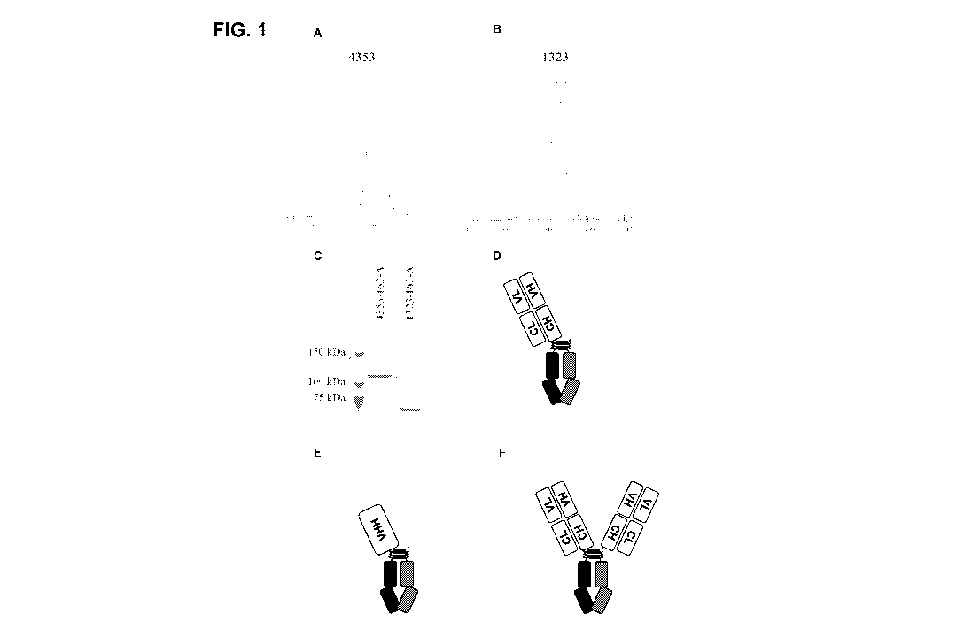
become better understood with regard to the following description, and
accompanying
drawings, where:
[0063] Figure 1 depicts the assessment of purity of exemplary one armed anti-
EGFR
antibodies (OA-EGFR), v4353 and 1323. Figure lA shows the SEC profile of v4353
with
the main peak at retention volume of 79.95 ml. Figure 1B shows the SEC profile
of v1323
with main peak at retention volume of 84.74 ml. Figure 1C shows the SDS-PAGE
analysis of
both v4353 and v1323 with species at approximately 110 kDa and 66 kDa,
respectively.
Figure 1D is a schematic drawing of an exemplary one armed anti-EGFR antibody
in the
format of v4353. Figure lE is a schematic drawing of an exemplary one armed
anti-EGFR
antibody in the format of v1323. Figure lE is a schematic drawing of a
bivalent (full size)
anti-EGFR antibody in the format of v7180.
[0064] Figure 2 depicts the ability of exemplary neutralizing and non-
neutralizing OA-EGFR
antibodies to bind to EGFR, as measured by Surface Plasmon Resonance (SPR).
Figures 2A
and 2B depict the sensorgrams for v4353 (neutralizing antibody) and its
ability to bind to
EGFR and lack of binding to HER2, respectively. Figures 2C and 2D depict the
sensorgrams
11
CA 02930307 2016-05-10
WO 2015/073721 PCT/US2014/065546
for v1323 (non-neutralizing) and its ability to bind to EGFR and lack of
binding to HER2,
respectively.
[0065] Figure 3 depicts the ability of exemplary OA-EGFR antibodies to bind in
a
concentration-dependent and saturable manner to HER2 3+ EGFR expressing breast
cancer
BT-474 cells. OA-EGFR antibodies show higher Bmax compared to ErbituxTM , a
full sized
bivalent anti-EGFR antibody.
[0066] Figure 4 depicts the ability of exemplary neutralizing and non-
neutralizing OA-EGFR
antibodies to inhibit the growth of A431 lung cancer cells expressing EGFR
over a 5 day
exposure, with inhibition seen with the neutralizing OA-EGFR v4353 but not the
non-
neutralizing OA-EGFR v1323.
[0067] Figure 5 depicts the ability of exemplary neutralizing and non-
neutralizing OA-EGFR
antibodies to mediate concentration dependent ADCC with an effector to target
E:T ratio of
25:1 in breast BT-474 cancer cells.
[0068] Figure 6 depicts measurement of the ability of an exemplary OA-EGFR
antibody to
be internalized and to downregulate EGFR expression. Figure 6A shows the
effect of 20 nM
of v4353 to internalize and downregulate EGFR expression in JIMT-1 cells.
Figure 6B
shows the effect of 100 nM and 200 nM of v4353 to internalize and downregulate
EGFR
expression in JIMT-1 cells, either alone, or in combination with other
antibodies. For each
experimental group in 6A and 6B, the left bar is surface at 4C degrees; the
middle bar is
surface at 37C degrees; and the right bar is internal at 37C degrees.
[0069] Figure 7 depicts the ability of combinations of an exemplary OA-EGFR
with an OA-
HER2 antibody to inhibit growth of CTX-resistant established ovarian tumor
SKOV3 in a
mouse xenograft model.
[0070] Figure 8 depicts a Kaplan-Meier plot showing survival data for mice
exposed to
combinations of an exemplary OA-EGFR with an 0A-HER2 antibody in the SKOV3
xenograph model of Figure 7.
[0071] Figure 9 shows the UPLC-SEC chromatogram of an exemplary afucosylated
OA-
EGFR v4353 (v7192) antibody following protein A and SEC purification.
[0072] Figure 10 shows the UPLC-SEC chromatogram (Figures 10A and 10B) and non-
reducing SDS-PAGE (Figure 10C) of the exemplary afucosylated OA-EGFR v4353
final
product (v7192).
[0073] Figure 11 shows the glycan analysis of the tryptic digest of the
exemplary
afucosylated antibody v7192 by LC-MS.
12
CA 02930307 2016-05-10
WO 2015/073721 PCT/US2014/065546
[0074] Figure 12 shows the overlay of the HIC-HPLC chromatogram of the
unconjugated
v7104 and DM1-conjugated v7192 exemplary OA-EGFR antibodies.
[0075] Figure 13 shows the overlay of the HPLC-SEC chromatogram of the
unconjugated v
7104 and conjugated v7192 exemplary OA-EGFR antibodies.
[0076] Figure 14 shows the whole cell saturation binding on various human
tumour cell lines.
The Bmax fold increase of OA-EGFR compared to the corresponding bivalent
antibody was
1.55, 1.68 and 1.38 in colorectal HCT116 (Figure 14A), triple negative breast
cancer (TNBC)
MDA-MB-231 (Figure 14 B) and ovarian SKOV3 (Figure 14 C) respectively.
[0077] Figures 15A and 15B show the concentration dependent ADCC dose response
curves
of the exemplary OAAs on the Caco2 cell line assessed at a PBMC effector to
target Caco2
E:T ratio of 50:1.
[0078] Figure 16 shows the ADCC dose response curves of the afucosylated
(v7192) and
non-afucosylated (v4353) exemplary OA-EGFR on TNBC MDA-MB-231 cells assessed
at a
NK92 (CD16a: 158V/V) effector to target TNBC MDA-MB-231 E:T ratio of 5:1.
[0079] Figure 17 shows the growth inhibition dose response curve of the
exemplary OAADC
on a human triple negative breast cancer cell line MDA-MB-231.
[0080] Figure 18A shows the growth inhibition dose response curves of
exemplary OA-
EGFR antibodies v4353 and v7192 compared to ErbituxTM and HerceptinTM on an
immortalized HACAT keratinocyte cell line in the presence of serum. Figure 18B
shows the
growth inhibition dose response curves of OA-EGFR antibody v1323 compared to
that of
ErbituxTM in the absence of serum.
[0081] Figure 19 shows the ADCC dose response curves of the afucosylated
(v7192)
exemplary OA-EGFR and ErbituxTM (v7180) on A431 cells A549 cells and HCT116
cells,
which express on their cell surfaces high, medium and low levels of EGFR
respectively.
DETAILED DESCRIPTION
[0082] Provided herein are monovalent antigen-binding constructs comprising an
antigen-
binding polypeptide construct which monovalently binds an antigen. In some
aspects, the
construct includes a dimeric Fc polypeptide construct comprising two monomeric
Fc
polypeptides each comprising a CH3 domain, wherein one said monomeric Fc
polypeptide is
fused to at least one polypeptide from the antigen-binding polypeptide
construct; wherein
said monovalent antigen-binding construct displays an increase in binding
density and B. to
a target cell displaying said antigen as compared to a corresponding
monospecific bivalent
antigen-binding construct with two antigen binding regions, and wherein said
monovalent
antigen-binding construct shows superior efficacy and/or bioactivity as
compared to the
13
CA 02930307 2016-05-10
WO 2015/073721 PCT/US2014/065546
corresponding bivalent antigen-binding construct, and wherein said superior
efficacy and/or
bioactivity is the result of the increase in binding density and resulting
increase in decoration
of a target cell. The increase in B. or binding density and resultant increase
in target
decoration by the monovalent antigen-binding construct provided here is the
effect of specific
target binding and not due to nonspecific binding. In certain embodiments the
maximum
binding occurs at a target to antibody ratio of 1:1.
[0083] In certain embodiments, the monovalent antigen-binding constructs
provided herein
possess at least one or more of the following attributes: increased B.
compared to
corresponding monospecific bivalent antigen-binding constructs (FSA); Kd
comparable to
corresponding FSA; same or slower off-rate compared to corresponding FSA;
decreased or
partial agonism; no cross-linking and dimerization of targets; specificity
and/or selectivity for
target cell of interest; full or partial or no inhibition of target cell
growth; complete Fc
capable of inducing effector activity; and ability to be internalized by
target cell.
[0084] In certain embodiments, the monovalent antigen-binding constructs
provided herein
possess the following minimal attributes: increased B. compared to
corresponding FSA; Kd
comparable to corresponding FSA; same or slower off-rate compared to
corresponding FSA;
decreased or partial agonism; no cross-linking and dimerization of targets;
specificity and/or
selectivity for target cell of interest; full or partial or no inhibition of
target cell growth;
complete Fc capable of inducing effector activity; and optionally ability to
be internalized by
target cell.
[0085] Provided herein is a monovalent antigen-binding construct wherein said
construct is at
least one of: a monovalent lytic antibody, a monovalent internalizing antibody
and
combinations thereof In some embodiments, the antigen-binding construct is a
monovalent
lytic antibody and/or a monovalent internalizing antibody depending on the
balance these
antibodies display between the following efficacy factors: a) the ability of
the monovalent
antigen-binding construct to be internalized, b) the increased B. and Kd/on-
off rate of the
monovalent antigen-binding construct, and c) the degree of agonism/partial
agonism of the
monovalent antigen-binding construct
[0086] Provided herein is a method of increasing antibody concentration in at
least one
target cell comprising providing to the target cell a monovalent antigen-
binding construct
comprising: an antigen-binding polypeptide construct which monovalently binds
an antigen;
a dimeric Fc domain; wherein said monovalent antigen-binding construct
displays an
increase in binding density and Bmax (maximum binding) to a target cell
displaying said
antigen as compared to a corresponding bivalent antigen-binding construct with
two antigen
14
CA 02930307 2016-05-10
WO 2015/073721 PCT/US2014/065546
binding regions, and wherein said monovalent antigen-binding construct shows
better
therapeutic efficacy compared to a corresponding bivalent antigen-binding
construct, and
wherein said efficacy is not caused by crosslinking of the antigen, antigen
dimerization,
prevention of antigen modulation, or prevention of antigen activation.
Conversely, the other
is true that efficacy can be caused by antigen modulation or antigen
activation so long as
these do not overcome the net killing effect.
[0087] In some embodiments is an isolated monovalent antigen-binding construct
described
herein, wherein said antigen-binding construct exhibits no avidity.
[0088] Provided herein is an isolated monovalent antigen-binding construct
comprising an
antigen-binding polypeptide construct which monovalently binds an antigen; and
a dimeric
Fc polypeptide construct comprising two monomeric Fc polypeptides each
comprising a CH3
domain, wherein one said monomeric Fc polypeptide is fused to at least one
polypeptide from
the antigen-binding polypeptide construct; wherein said monovalent antigen-
binding
construct displays an increase in binding density and Bmax (maximum binding)
to a target
cell displaying said antigen as compared to a corresponding FSA construct with
two antigen
binding regions, wherein said monovalent antigen-binding construct shows
superior efficacy
and/or bioactivity as compared to the corresponding bivalent antigen-binding
construct, and
wherein said superior efficacy and/or bioactivity is the result of the
increase in binding
density.
[0089] Provided in certain embodiments is an isolated monovalent antigen-
binding construct
described herein, wherein the increase in binding density and Bmax relative to
a
monospecific bivalent antibody is observed at a concentration greater than the
observed
equilibrium constant (Kd) and at saturating concentrations of the antibodies.
In some
embodiments the superior efficacy and/or bioactivity is the result of
increased FcyR or
complement (Cl q) binding and at least one of higher ADCC, higher ADCP and
higher CDC
as compared to said corresponding bivalent antigen-binding construct. In
specific
embodiments, the isolated monovalent antigen-binding construct is anti-
proliferative and is
internalized. In certain embodiments is an isolated monovalent antigen-binding
construct
described herein wherein said increase in binding density and Bmax relative to
the FSA is
independent of the density of the antigen on the target cell. In some
embodiments is provided
an isolated monovalent antigen-binding construct described herein, wherein the
target cell is
a cancer cell, or an EGFR and/or HER2 expressing diseased cell. In an
embodiment, the
isolated monovalent antigen-binding construct described herein exhibits no
avidity.
CA 02930307 2016-05-10
WO 2015/073721 PCT/US2014/065546
[0090] Definitions
[0091] It is to be understood that this invention is not limited to the
particular protocols; cell
lines, constructs, and reagents described herein and as such may vary. It is
also to be
understood that the terminology used herein is for the purpose of describing
particular
embodiments only, and is not intended to limit the scope of the present
invention, which will
be limited only by the appended claims.
[0092] Unless defined otherwise, all technical and scientific terms used
herein have the same
meaning as commonly understood to one of ordinary skill in the art to which
this invention
belongs. Although any methods, devices, and materials similar or equivalent to
those
described herein can be used in the practice or testing of the invention, the
preferred methods,
devices and materials are now described.
[0093] All publications and patents mentioned herein are incorporated herein
by reference for
the purpose of describing and disclosing, for example, the constructs and
methodologies that
are described in the publications, which might be used in connection with the
presently
described invention. The publications discussed herein are provided solely for
their disclosure
prior to the filing date of the present application. Nothing herein is to be
construed as an
admission that the inventors are not entitled to antedate such disclosure by
virtue of prior
invention or for any other reason.
[0094] A "dimer" or "heterodimer" is a molecule comprising at least a first
monomer
polypeptide and a second monomer polypeptide. In the case of a heterodimer,
one of said
monomers differs from the other monomer by at least one amino acid residue. In
certain
embodiments, the assembly of the dimer is driven by surface area burial. In
some
embodiments, the monomeric polypeptides interact with each other by means of
electrostatic
interactions and/or salt-bridge interactions that drive dimer formation by
favoring the desired
dimer formation and/or disfavoring formation of other non-desired specimen. In
some
embodiments, the monomer polypeptides inteact with each other by means of
hydrophobic
interactions that drive desired dimer formation by favoring desired dimer
formation and/or
disfavoring formation of other assembly types. In certain embodiments, the
monomer
polypeptides interact with each other by means of covalent bond formation. In
certain
embodiments, the covalent bonds are formed between naturally present or
introduced
cysteines that drive desired dimer formation. In certain embodiments described
herein, no
covalent bonds are formed between the monomers. In some embodiments, the
polypeptides
inteact with each other by means of packing/size-complementarity/knobs-into-
holes/protruberance-cavity type interactions that drive dimer formation by
favoring desired
16
CA 02930307 2016-05-10
WO 2015/073721 PCT/US2014/065546
dimer formation and/or disfavoring formation of other non-desired embodiments.
In some
embodiments, the polypeptides interact with each other by means of cation-pi
interactions
that drive dimer formation. In certain embodiments the individual monomer
polypeptides
cannot exist as isolated monomers in solution.
[0095] The term "Fc domain" or "Fc", as used herein, generally refers to a
dimer complex
comprising the C-terminal polypeptide sequences of an immunoglobulin heavy
chain,
wherein a C-terminal polypeptide sequence is that which is obtainable by
papain digestion of
an intact antibody. The Fc domain may comprise native or variant Fc sequences.
Although
the boundaries of the Fc sequence of an immunoglobulin heavy chain might vary,
the human
IgG heavy chain Fc sequence is usually defined to stretch from an amino acid
residue at
about position Cys226, or from about position Pro230, to the carboxyl terminus
of the Fc
sequence. The Fc sequence of an immunoglobulin generally comprises two
constant domains,
a CH2 domain and a CH3 domain, and optionally comprises a CH4 domain. By "Fc
polypeptide" herein is meant one of the polypeptides that make up an Fc
domain. An Fc
polypeptide may be obtained from any suitable immunoglobulin, such as IgGl,
IgG2, IgG3,
or IgG4 subtypes, IgA, IgE, IgD or IgM. In some embodiments, an Fc polypeptide
comprises
part or all of a wild type hinge sequence (generally at its N terminus). In
some embodiments,
an Fc polypeptide does not comprise a functional or wild type hinge sequence.
[0096] Antibody "effector functions" refer to those biological activities
attributable to the Fc
domain (a native sequence Fc domain or amino acid sequence variant Fc domain)
of an
antibody. Examples of antibody effector functions include Clq binding;
complement
dependent cytotoxicity; Fc receptor binding; antibody-dependent cell-mediated
cytotoxicity
(ADCC); phagocytosis; down regulation of cell surface receptors (e.g. B cell
receptor; BCR),
etc.
[0097] "Antibody-dependent cell-mediated cytotoxicity" and "ADCC" refer to a
cell-
mediated reaction in which nonspecific cytotoxic cells that express Fc
receptors (FcRs) (e.g.
Natural Killer (NK) cells, neutrophils, and macrophages) recognize bound
antibody on a
target cell and subsequently cause lysis of the target cell.
[0098] "Complement dependent cytotoxicity" and "CDC" refer to the lysing of a
target in the
presence of complement. The complement activation pathway is initiated by the
binding of
the first component of the complement system (Clq) to a molecule (e.g. an
antibody)
complexed with a cognate antigen.
[0099] "Antibody-dependent cellular phagocytosis and "ADCP" refer to the
destruction of
target cells via monocyte or macrophage-mediated phagocytosis.
17
CA 02930307 2016-05-10
WO 2015/073721 PCT/US2014/065546
[00100] The terms "Fe receptor" and "FcR" are used to describe a receptor that
binds to
the Fe domain of an antibody. For example, an FcR can be a native sequence
human FcR.
Generally, an FcR is one which binds an IgG antibody (a gamma receptor) and
includes
receptors of the FcyRI, FcyRII, and FcyRIII subclasses, including allelic
variants and
alternatively spliced forms of these receptors. FcyRII receptors include
FcyRIIA (an
"activating receptor") and FcyRIIB (an "inhibiting receptor"), which have
similar amino acid
sequences that differ primarily in the cytoplasmic domains thereof.
Immunoglobulins of other
isotypes can also be bound by certain FcRs (see, e.g., Janeway et al., Immuno
Biology: the
immune system in health and disease, (Elsevier Science Ltd., NY) (4th ed.,
1999)).
Activating receptor FcyRIIA contains an immunoreceptor tyrosine-based
activation motif
(ITAM) in its cytoplasmic domain. Inhibiting receptor FcyRIIB contains an
immunoreceptor
tyrosine-based inhibition motif (ITIM) in its cytoplasmic domain (reviewed in
Daeron, Annu.
Rev. Immunol. 15:203-234 (1997)). FcRs are reviewed in Ravetch and Kinet,
Annu. Rev.
Immunol 9:457-92 (1991); Capel et al., Immunomethods 4:25-34 (1994); and de
Haas et al.,
J. Lab. Clin. Med. 126:330-41 (1995). Other FcRs, including those to be
identified in the
future, are encompassed by the term "FcR" herein. The term also includes the
neonatal
receptor, FcRn, which is responsible for the transfer of maternal IgGs to the
fetus (Guyer et
al., J. Immunol. 117:587 (1976); and Kim et al., J. Immunol. 24:249 (1994)).
[00101] A "disorder" is any condition that would benefit from treatment with
an antibody
or method of the invention. This includes chronic and acute disorders or
diseases including
those pathological conditions which predispose the mammal to the disorder in
question. Non-
limiting examples of disorders to be treated herein include malignant and
benign tumors;
non-leukemias and lymphoid malignancies; neuronal, glial, astrocytal,
hypothalamic and
other glandular, macrophagal, epithelial, stromal and blastocoelic disorders;
and
inflammatory, immunologic and other angiogenesis-related disorders.
[00102] The terms "cancer" and "cancerous" refer to or describe the
physiological
condition in mammals that is typically characterized by unregulated cell
growth/proliferation.
Examples of cancer include but are not limited to, carcinoma, lymphoma,
blastoma, sarcoma,
and leukemia. More particular examples of such cancers include squamous cell
cancer, small-
cell lung cancer, non-small cell lung cancer, adenocarcinoma of the lung,
squamous
carcinoma of the lung, cancer of the peritoneum, myeloma (e.g., multiple
myeloma),
hepatocellular cancer, gastrointestinal cancer, pancreatic cancer,
glioblastoma/glioma (e.g.,
anaplastic astrocytoma, glioblastoma multiforme, anaplastic oligodendroglioma,
anaplastic
oligodendroastrocytoma), cervical cancer, ovarian cancer, liver cancer,
bladder cancer,
18
CA 02930307 2016-05-10
WO 2015/073721 PCT/US2014/065546
hepatoma, breast cancer, colon cancer, colorectal cancer, endometrial or
uterine carcinoma,
salivary gland carcinoma, kidney cancer, liver cancer, prostate cancer, vulval
cancer, thyroid
cancer, hepatic carcinoma and various types of head and neck cancer.
[00103] As used herein, "treatment" refers to clinical intervention in an
attempt to alter the
natural course of the individual or cell being treated, and can be performed
either for
prophylaxis or during the course of clinical pathology. Desirable effects of
treatment include
preventing occurrence or recurrence of disease, alleviation of symptoms,
diminishing of any
direct or indirect pathological consequences of the disease, preventing
metastasis, decreasing
the rate of disease progression, amelioration or palliation of the disease
state, and remission
or improved prognosis. In some embodiments, antibodies of the invention are
used to delay
development of a disease or disorder. In one embodiment, antibodies and
methods of the
invention effect tumor regression. In one embodiment, antibodies and methods
of the
invention effect inhibition of tumor/cancer growth.
[00104] A "recombinant host cell" or "host cell" refers to a cell that
includes an exogenous
polynucleotide, regardless of the method used for insertion, for example,
direct uptake,
transduction, f-mating, or other methods known in the art to create
recombinant host cells.
The exogenous polynucleotide may be maintained as a nonintegrated vector, for
example, a
plasmid, or alternatively, may be integrated into the host genome.
[00105] As used herein, the term "medium" or "media" includes any culture
medium,
solution, solid, semi-solid, or rigid support that may support or contain any
host cell,
including bacterial host cells, yeast host cells, insect host cells, plant
host cells, eukaryotic
host cells, mammalian host cells, CHO cells, prokaryotic host cells, E. coli,
or Pseudomonas
host cells, and cell contents. Thus, the term may encompass medium in which
the host cell
has been grown, e.g., medium into which the protein has been secreted,
including medium
either before or after a proliferation step. The term also may encompass
buffers or reagents
that contain host cell lysates, such as in the case where an antigen binding
construct described
herein is produced intracellularly and the host cells are lysed or disrupted
to release the
heteromultimer.
[00106] As used herein, the term "modulated serum half-life" means the
positive or
negative change in circulating half-life of an antigen binding polypeptide
that is comprised by
an antigen-binding construct described herein relative to its native form.
Serum half-life is
measured by taking blood samples at various time points after administration
of the construct,
and determining the concentration of that molecule in each sample. Correlation
of the serum
concentration with time allows calculation of the serum half-life. Increased
serum half-life
19
CA 02930307 2016-05-10
WO 2015/073721 PCT/US2014/065546
desirably has at least about two-fold, but a smaller increase may be useful,
for example where
it enables a satisfactory dosing regimen or avoids a toxic effect. In some
embodiments, the
increase is at least about three-fold, at least about five-fold, or at least
about ten-fold.
[00107] The term "modulated therapeutic half-life" as used herein means the
positive or
negative change in the half-life of the therapeutically effective amount of an
antigen binding
polypeptide comprised by a monovalent antigen-binding construct described
herein, relative
to its non-modified form. Therapeutic half-life is measured by measuring
pharmacokinetic
and/or pharmacodynamic properties of the molecule at various time points after
administration. Increased therapeutic half-life desirably enables a particular
beneficial dosing
regimen, a particular beneficial total dose, or avoids an undesired effect. In
some
embodiments, the increased therapeutic half-life results from increased
potency, increased or
decreased binding of the modified molecule to its target, increased or
decreased breakdown
of the molecule by enzymes such as proteases, or an increase or decrease in
another
parameter or mechanism of action of the non-modified molecule or an increase
or decrease in
receptor-mediated clearance of the molecule.
[00108] The term "isolated," when applied to a nucleic acid or protein,
denotes that the
nucleic acid or protein is free of at least some of the cellular components
with which it is
associated in the natural state, or that the nucleic acid or protein has been
concentrated to a
level greater than the concentration of its in vivo or in vitro production. It
can be in a
homogeneous state. Isolated substances can be in either a dry or semi-dry
state, or in solution,
including but not limited to, an aqueous solution. It can be a component of a
pharmaceutical
composition that comprises additional pharmaceutically acceptable carriers
and/or excipients.
Purity and homogeneity are typically determined using analytical chemistry
techniques such
as polyacrylamide gel electrophoresis or high performance liquid
chromatography. A protein
which is the predominant species present in a preparation is substantially
purified. In
particular, an isolated gene is separated from open reading frames which flank
the gene and
encode a protein other than the gene of interest. The term "purified" denotes
that a nucleic
acid or protein gives rise to substantially one band in an electrophoretic
gel. Particularly, it
may mean that the nucleic acid or protein is at least 85% pure, at least 90%
pure, at least 95%
pure, at least 99% or greater pure.
[00109] The term "nucleic acid" refers to deoxyribonucleotides,
deoxyribonucleosides,
ribonucleosides, or ribonucleotides and polymers thereof in either single- or
double-stranded
form. Unless specifically limited, the term encompasses nucleic acids
containing known
analogues of natural nucleotides which have similar binding properties as the
reference
CA 02930307 2016-05-10
WO 2015/073721 PCT/US2014/065546
nucleic acid and are metabolized in a manner similar to naturally occurring
nucleotides.
Unless specifically limited otherwise, the term also refers to oligonucleotide
analogs
including PNA (peptidonucleic acid), analogs of DNA used in antisense
technology
(phosphorothioates, phosphoroamidates, and the like). Unless otherwise
indicated, a
particular nucleic acid sequence also implicitly encompasses conservatively
modified
variants thereof (including but not limited to, degenerate codon
substitutions) and
complementary sequences as well as the sequence explicitly indicated.
Specifically,
degenerate codon substitutions may be achieved by generating sequences in
which the third
position of one or more selected (or all) codons is substituted with mixed-
base and/or
deoxyinosine residues (Batzer et al., Nucleic Acid Res. 19:5081 (1991);
Ohtsuka et al., J.
Biol. Chem. 260:2605-2608 (1985); Rossolini et al., Mol. Cell. Probes 8:91-98
(1994)).
[00110] The terms "polypeptide," "peptide" and "protein" are used
interchangeably herein
to refer to a polymer of amino acid residues. That is, a description directed
to a polypeptide
applies equally to a description of a peptide and a description of a protein,
and vice versa.
The terms apply to naturally occurring amino acid polymers as well as amino
acid polymers
in which one or more amino acid residues is a non-naturally encoded amino
acid. As used
herein, the terms encompass amino acid chains of any length, including full
length proteins,
wherein the amino acid residues are linked by covalent peptide bonds.
[00111] The term "amino acid" refers to naturally occurring and non-naturally
occurring
amino acids, as well as amino acid analogs and amino acid mimetics that
function in a
manner similar to the naturally occurring amino acids. Naturally encoded amino
acids are the
20 common amino acids (alanine, arginine, asparagine, aspartic acid, cysteine,
glutamine,
glutamic acid, glycine, histidine, isoleucine, leucine, lysine, methionine,
phenylalanine,
praline, serine, threonine, tryptophan, tyrosine, and valine) and pyrrolysine
and
selenocysteine. Amino acid analogs refers to compounds that have the same
basic chemical
structure as a naturally occurring amino acid, i.e., an a carbon that is bound
to a hydrogen, a
carboxyl group, an amino group, and an R group, such as, homoserine,
norleucine,
methionine sulfoxide, methionine methyl sulfonium. Such analogs have modified
R groups
(such as, norleucine) or modified peptide backbones, but retain the same basic
chemical
structure as a naturally occurring amino acid. Reference to an amino acid
includes, for
example, naturally occurring proteogenic L-amino acids; D-amino acids,
chemically
modified amino acids such as amino acid variants and derivatives; naturally
occurring non-
proteogenic amino acids such as 13-alanine, ornithine, etc.; and chemically
synthesized
21
CA 02930307 2016-05-10
WO 2015/073721 PCT/US2014/065546
compounds having properties known in the art to be characteristic of amino
acids. Examples
of non-naturally occurring amino acids include, but are not limited to, a-
methyl amino acids
(e.g. a-methyl alanine), D-amino acids, histidine-like amino acids (e.g., 2-
amino-histidine, 13-
hydroxy-histidine, homohistidine), amino acids having an extra methylene in
the side chain
("homo" amino acids), and amino acids in which a carboxylic acid functional
group in the
side chain is replaced with a sulfonic acid group (e.g., cysteic acid). The
incorporation of
non-natural amino acids, including synthetic non-native amino acids,
substituted amino acids,
or one or more D-amino acids into the proteins of the present invention may be
advantageous
in a number of different ways. D-amino acid-containing peptides, etc., exhibit
increased
stability in vitro or in vivo compared to L-amino acid-containing
counterparts. Thus, the
construction of peptides, etc., incorporating D-amino acids can be
particularly useful when
greater intracellular stability is desired or required. More specifically, D-
peptides, etc., are
resistant to endogenous peptidases and proteases, thereby providing improved
bioavailability
of the molecule, and prolonged lifetimes in vivo when such properties are
desirable.
Additionally, D-peptides, etc., cannot be processed efficiently for major
histocompatibility
complex class II-restricted presentation to T helper cells, and are therefore,
less likely to
induce humoral immune responses in the whole organism.
[00112] Amino acids may be referred to herein by either their commonly known
three
letter symbols or by the one-letter symbols recommended by the IUPAC-IUB
Biochemical
Nomenclature Commission. Nucleotides, likewise, may be referred to by their
commonly
accepted single-letter codes.
[00113] "Conservatively modified variants" applies to both amino acid and
nucleic acid
sequences. With respect to particular nucleic acid sequences, "conservatively
modified
variants" refers to those nucleic acids which encode identical or essentially
identical amino
acid sequences, or where the nucleic acid does not encode an amino acid
sequence, to
essentially identical sequences. Because of the degeneracy of the genetic
code, a large
number of functionally identical nucleic acids encode any given protein. For
instance, the
codons GCA, GCC, GCG and GCU all encode the amino acid alanine. Thus, at every
position where an alanine is specified by a codon, the codon can be altered to
any of the
corresponding codons described without altering the encoded polypeptide. Such
nucleic acid
variations are "silent variations," which are one species of conservatively
modified variations.
Every nucleic acid sequence herein which encodes a polypeptide also describes
every
possible silent variation of the nucleic acid. One of ordinary skill in the
art will recognize that
22
CA 02930307 2016-05-10
WO 2015/073721 PCT/US2014/065546
each codon in a nucleic acid (except AUG, which is ordinarily the only codon
for methionine,
and TGG, which is ordinarily the only codon for tryptophan) can be modified to
yield a
functionally identical molecule. Accordingly, each silent variation of a
nucleic acid which
encodes a polypeptide is implicit in each described sequence.
[00114] As to amino acid sequences, one of ordinary skill in the art will
recognize that
individual substitutions, deletions or additions to a nucleic acid, peptide,
polypeptide, or
protein sequence which alters, adds or deletes a single amino acid or a small
percentage of
amino acids in the encoded sequence is a "conservatively modified variant"
where the
alteration results in the deletion of an amino acid, addition of an amino
acid, or substitution of
an amino acid with a chemically similar amino acid. Conservative substitution
tables
providing functionally similar amino acids are known to those of ordinary
skill in the art.
Such conservatively modified variants are in addition to and do not exclude
polymorphic
variants, interspecies homologs, and alleles of the invention.
[00115] Conservative substitutions providing functionally similar amino acids
are known
to those of ordinary skill in the art. The following eight groups each contain
amino acids that
are conservative substitutions for one another: 1) Alanine (A), Glycine (G);
2) Aspartic acid
(D), Glutamic acid (E); 3) Asparagine (N), Glutamine (Q); 4) Arginine (R),
Lysine (K); 5)
Isoleucine (I), Leucine (L), Methionine (M), Valine (V); 6) Phenylalanine (F),
Tyrosine (Y),
Tryptophan (W); 7) Serine (S), Threonine (T); and [0139] 8) Cysteine (C),
Methionine (M)
(see, e.g., Creighton, Proteins: Structures and Molecular Properties (W H
Freeman & Co.;
2nd edition (December 1993)
[00116] The terms "identical" or percent "identity," in the context of two or
more nucleic
acids or polypeptide sequences, refer to two or more sequences or subsequences
that are the
same. Sequences are "substantially identical" if they have a percentage of
amino acid residues
or nucleotides that are the same (i.e., about 60% identity, about 65%, about
70%, about 75%,
about 80%, about 85%, about 90%, or about 95% identity over a specified
region), when
compared and aligned for maximum correspondence over a comparison window, or
designated region as measured using one of the following sequence comparison
algorithms
(or other algorithms available to persons of ordinary skill in the art) or by
manual alignment
and visual inspection. This definition also refers to the complement of a test
sequence. The
identity can exist over a region that is at least about 50 amino acids or
nucleotides in length,
or over a region that is 75-100 amino acids or nucleotides in length, or,
where not specified,
across the entire sequence of a polynucleotide or polypeptide. A
polynucleotide encoding a
polypeptide of the present invention, including homologs from species other
than human,
23
CA 02930307 2016-05-10
WO 2015/073721 PCT/US2014/065546
may be obtained by a process comprising the steps of screening a library under
stringent
hybridization conditions with a labeled probe having a polynucleotide sequence
of the
invention or a fragment thereof, and isolating full-length cDNA and genomic
clones
containing said polynucleotide sequence. Such hybridization techniques are
well known to
the skilled artisan.
[00117] For sequence comparison, typically one sequence acts as a reference
sequence, to
which test sequences are compared. When using a sequence comparison algorithm,
test and
reference sequences are entered into a computer, subsequence coordinates are
designated, if
necessary, and sequence algorithm program parameters are designated. Default
program
parameters can be used, or alternative parameters can be designated. The
sequence
comparison algorithm then calculates the percent sequence identities for the
test sequences
relative to the reference sequence, based on the program parameters.
[00118] A "comparison window", as used herein, includes reference to a segment
of any
one of the number of contiguous positions selected from the group consisting
of from 20 to
600, usually about 50 to about 200, more usually about 100 to about 150 in
which a sequence
may be compared to a reference sequence of the same number of contiguous
positions after
the two sequences are optimally aligned. Methods of alignment of sequences for
comparison
are known to those of ordinary skill in the art. Optimal alignment of
sequences for
comparison can be conducted, including but not limited to, by the local
homology algorithm
of Smith and Waterman (1970) Adv. Appl. Math. 2:482c, by the homology
alignment
algorithm of Needleman and Wunsch (1970) J. Mol. Biol. 48:443, by the search
for similarity
method of Pearson and Lipman (1988) Proc. Nat'l. Acad. Sci. USA 85:2444, by
computerized
implementations of these algorithms (GAP, BESTFIT, FASTA, and TFASTA in the
Wisconsin Genetics Software Package, Genetics Computer Group, 575 Science Dr.,
Madison, Wis.), or by manual alignment and visual inspection (see, e.g.,
Ausubel et al.,
Current Protocols in Molecular Biology (1995 supplement)).
[00119] One example of an algorithm that is suitable for determining percent
sequence
identity and sequence similarity are the BLAST and BLAST 2.0 algorithms, which
are
described in Altschul et al. (1997) Nuc. Acids Res. 25:3389-3402, and Altschul
et al. (1990)
J. Mol. Biol. 215:403-410, respectively. Software for performing BLAST
analyses is publicly
available through the National Center for Biotechnology Information available
at the World
Wide Web at ncbi.nlm.nih.gov. The BLAST algorithm parameters W, T, and X
determine the
sensitivity and speed of the alignment. The BLASTN program (for nucleotide
sequences)
uses as defaults a wordlength (W) of 11, an expectation (E) or 10, M=5, N=-4
and a
24
CA 02930307 2016-05-10
WO 2015/073721 PCT/US2014/065546
comparison of both strands. For amino acid sequences, the BLASTP program uses
as defaults
a wordlength of 3, and expectation (E) of 10, and the BLOSUM62 scoring matrix
(see
Henikoff and Henikoff (1992) Proc. Natl. Acad. Sci. USA 89:10915) alignments
(B) of 50,
expectation (E) of 10, M=5, N=-4, and a comparison of both strands. The BLAST
algorithm
is typically performed with the "low complexity" filter turned off.
[00120] The BLAST algorithm also performs a statistical analysis of the
similarity
between two sequences (see, e.g., Karlin and Altschul (1993) Proc. Natl. Acad.
Sci. USA
90:5873-5787). One measure of similarity provided by the BLAST algorithm is
the smallest
sum probability (P(N)), which provides an indication of the probability by
which a match
between two nucleotide or amino acid sequences would occur by chance. For
example, a
nucleic acid is considered similar to a reference sequence if the smallest sum
probability in a
comparison of the test nucleic acid to the reference nucleic acid is less than
about 0.2, or less
than about 0.01, or less than about 0.001.
[00121] The phrase "selectively (or specifically) hybridizes to" refers to
the binding,
duplexing, or hybridizing of a molecule only to a particular nucleotide
sequence under
stringent hybridization conditions when that sequence is present in a complex
mixture
(including but not limited to, total cellular or library DNA or RNA).
[00122] The phrase "stringent hybridization conditions" refers to
hybridization of
sequences of DNA, RNA, or other nucleic acids, or combinations thereof under
conditions of
low ionic strength and high temperature as is known in the art. Typically,
under stringent
conditions a probe will hybridize to its target subsequence in a complex
mixture of nucleic
acid (including but not limited to, total cellular or library DNA or RNA) but
does not
hybridize to other sequences in the complex mixture. Stringent conditions are
sequence-
dependent and will be different in different circumstances. Longer sequences
hybridize
specifically at higher temperatures. An extensive guide to the hybridization
of nucleic acids is
found in Tijssen, Laboratory Techniques in Biochemistry and Molecular Biology--
Hybridization with Nucleic Probes, "Overview of principles of hybridization
and the strategy
of nucleic acid assays" (1993).
[00123] As used herein, the term "eukaryote" refers to organisms belonging to
the
phylogenetic domain Eucarya such as animals (including but not limited to,
mammals,
insects, reptiles, birds, etc.), ciliates, plants (including but not limited
to, monocots, dicots,
algae, etc.), fungi, yeasts, flagellates, microsporidia, protists, etc.
[00124] As used herein, the term "prokaryote" refers to prokaryotic organisms.
For
example, a non-eukaryotic organism can belong to the Eubacteria (including but
not limited
CA 02930307 2016-05-10
WO 2015/073721 PCT/US2014/065546
to, Escherichia coli, Thermus thermophilus, Bacillus stearothermophilus,
Pseudomonas
fluorescens, Pseudomonas aeruginosa, Pseudomonas putida, etc.) phylogenetic
domain, or the
Archaea (including but not limited to, Methanococcus jannaschii,
Methanobacterium
thermoautotrophicum, Halobacterium such as Haloferax volcanii and
Halobacterium species
NRC-1, Archaeoglobus fulgidus, Pyrococcus furiosus, Pyrococcus horikoshii,
Aeuropyrum
pernix, etc.) phylogenetic domain.
[00125] The term "subject" as used herein, refers to an animal, in some
embodiments a
mammal, and in other embodiments a human, who is the object of treatment,
observation or
experiment. An animal may be a companion animal (e.g., dogs, cats, and the
like), farm
animal (e.g., cows, sheep, pigs, horses, and the like) or a laboratory animal
(e.g., rats, mice,
guinea pigs, and the like).
[00126] The term "effective amount" as used herein refers to that amount of
monovalent
antigen-binding construct being administered, which will relieve to some
extent one or more
of the symptoms of the disease, condition or disorder being treated.
Compositions containing
the construct described herein can be administered for prophylactic,
enhancing, and/or
therapeutic treatments.
[00127] The terms "enhance" or "enhancing" means to increase or prolong either
in
potency or duration a desired effect. Thus, in regard to enhancing the effect
of drug molecule
or therapeutic agents, the term "enhancing" refers to the ability to increase
or prolong, either
in potency or duration, the effect of therapeutic agents on a system. An
"enhancing-effective
amount," as used herein, refers to an amount adequate to enhance the effect of
another
therapeutic agent or drug in a desired system. When used in a patient, amounts
effective for
this use will depend on the severity and course of the disease, disorder or
condition, previous
therapy, the patient's health status and response to the drugs, and the
judgment of the treating
physician.
[00128] The term "modified," as used herein refers to any changes made to a
given
polypeptide, such as changes to the length of the polypeptide, the amino acid
sequence,
chemical structure, co-translational modification, or post-translational
modification of a
polypeptide. The form "(modified)" term means that the polypeptides being
discussed are
optionally modified, that is, the polypeptides under discussion can be
modified or
unmodified.
[00129] The term "post-translationally modified" refers to any modification of
a natural or
non-natural amino acid that occurs to such an amino acid after it has been
incorporated into a
polypeptide chain. The term encompasses, by way of example only, co-
translational in vivo
26
CA 02930307 2016-05-10
WO 2015/073721 PCT/US2014/065546
modifications, co-translational in vitro modifications (such as in a cell-free
translation
system), post-translational in vivo modifications, and post-translational in
vitro
modifications.
[00130] The term "monospecific bivalent antigen-binding construct" as used
herein refers
to an antigen-binding construct which has two antigen binding domains
(bivalent), both of
which bind to the same epitope/antigen (monospecific). The antigen binding
domains could
be, but are not limited to, protein constructs such as Fab (fragment antigen
binding), scFv
(single chain Fv) and sdab (single domain antibody). The monospecific bivalent
antigen-
binding construct is also referred to herein as a "full-size antibody" or
"FSA." In some
embodiments, a monospecific bivalent antigen-binding construct is a reference
against which
the properties of the monovalent antigen-binding constructs are measured. In
other
embodiments, a combination of two monospecific bivalent antigen-binding
constructs is a
reference against which the properties of a combination of two monovalent
antigen-binding
constructs are measured. In cases where a combination of two monospecific
bivalent
antigen-binding constructs is used, the monospecific bivalent antigen-binding
constructs bind
to non-overlapping epitopes on EGFR. In some embodiments, where a combination
of two
monospecific bivalent antigen-binding constructs is used, a single
monospecific bivalent
antigen-binding construct is used as a reference, where the single
monospecific bivalent
antigen-binding construct represents a standard of care (SOC) therapy, e.g.,
Erbitux.
[00131] The phrase that a construct "specifically binds" to a target refers to
a binding
reaction which is determinative of the presence of the target in the presence
of a
heterogeneous population of other biologics. Thus, under designated
immunoassay
conditions, a specified construct specifically binds to a particular target
and does not bind in a
significant amount to other biologics present in the sample. Specific binding
of an antigen
binding construct to a target under such conditions requires the antigen
binding construct to
be specific to the target. A variety of immunoassay formats can be used to
select antigen
binding constructs specifically immunoreactive with a particular protein. For
example, solid-
phase ELISA immunoassays are routinely used to select antigen binding
constructs (e.g.,
monoclonal antibodies) specifically immunoreactive with a protein. See, e.g.,
Harlow and
Lane (1988) Antibodies, A Laboratory Manual, Cold Spring Harbor Publications,
New York,
for a description of immunoassay formats and conditions that can be used to
determine
specific immunoreactivity.
[00132] The term "avidity" is used here to refer to the combined synergistic
strength of
binding affinities and a key structure and biological attribute of therapeutic
monospecific
27
CA 02930307 2016-05-10
WO 2015/073721 PCT/US2014/065546
bivalent antibodies. Lack of avidity and loss of synergistic strength of
binding can result in
reduced apparent target binding affinity. On the other hand, on a target cell
with fixed
number of antigens, avidity resulting from the multivalent (or bivalent)
binding causes
increased occupancy of the target antigen at a lower number of antibody
molecules relative to
antibody which displays monovalent binding. With a lower number of antibody
molecules
bound to the target cell, in the application of bivalent lytic antibodies,
antibody dependent
cytotoxic killing mechanisms may not occur efficiently resulting in reduced
efficacy. Not
enough antibodies are bound to mediate ADCC, CDC, ADCP as these types of
effector
functions are generally considered to be Fc concentration threshold dependent.
In the case of
agonistic antibodies, reduced avidity reduces their efficiency to crosslink
and dimerize
antigens and activate the pathway.
[00133] "Single domain antibodies" or "Sdab" ¨ Single domain antibodies such
as the
Camelid VhH domain are individual immunoglobulin domains. Sdabs are fairly
stable and
easy to express as fusion partner with the Fc chain of an antibody (Harmsen
MM, De Haard
HJ (2007). "Properties, production, and applications of camelid single-domain
antibody
fragments". Appl. Microbiol Biotechnol. 77(1): 13-22).
[00134] As used herein, the term "EGFR" refers to epidermal growth factor
receptor (also
known as HER-1 or Erb-B1), including the human form(s) (Ulrich, A. et al.,
Nature 309:418-
425 (1984); SwissProt Accession #P00533; secondary accession numbers: 000688,
000732,
P06268, Q14225, Q92795, Q9BZS2, Q9GZX1, Q9H2C9, Q9H3C9, Q9UMD7, Q9UMD8,
Q9UMG5), as well as naturally-occurring isoforms and variants thereof Such
isoforms and
variants include but are not limited to the EGFRvIII variant, alternative
splicing products
(e.g., as identified by SwissProt Accession numbers P00533-1, P00533-2, P00533-
3, P00533-
4), variants GLN-98, ARG-266, Lys-521, ILE-674, GLY-962, and PRO-988
(Livingston, R.
J. et al., NIEHS-SNPs, environmental genome project, NIEHS ES15478, Department
of
Genome Sciences, Seattle, Wash. (2004)), and others identified by the
following accession
numbers: NM005228.3, NM201282.1, NM201283.1, NM201284.1 (REFSEQ mRNAs);
AF125253.1, AF277897.1, AF288738.1, A1217671.1, AK127817.1, AL598260.1,
AU137334.1, AW163038.1, AW295229.1, BC057802.1, CB160831.1, K03193.1,
U48722.1,
U95089.1, X00588.1, X00663.1; H5448451, H5448453, H5448452 (MIPS assembly);
DT.453606, DT.86855651, DT.95165593, DT.97822681, DT.95165600, DT.100752430,
DT.91654361, DT.92034460, DT.92446349, DT.97784849, DT.101978019, DT.418647,
DT.86842167, DT.91803457, DT.92446350, DT.95153003, DT.95254161, DT.97816654,
DT.87014330, DT.87079224 (DOTS Assembly). All accession numbers referenced
herein
28
CA 02930307 2016-05-10
WO 2015/073721 PCT/US2014/065546
are taken from the NCBI database (or other relevant, referenced database) as
of November 8,
2013.
[00135] A "HER receptor" is a receptor protein tyrosine kinase which belongs
to the
human epidermal growth factor receptor (HER) family and includes EGFR, HER2,
HER3
and HER4 receptors. The HER receptor will generally comprise an extracellular
domain,
which may bind an HER ligand; a lipophilic transmembrane domain; a conserved
intracellular tyrosine kinase domain; and a carboxyl-terminal signaling domain
harboring
several tyrosine residues which can be phosphorylated.
[00136] The extracellular (ecto) domain of HER2 comprises four domains, Domain
I
(ECD1, amino acid residues from about 1-195), Domain II (ECD2, amino acid
residues from
about 196-319), Domain III (ECD3, amino acid residues from about 320-488), and
Domain
IV (ECD4, amino acid residues from about 489-630) (residue numbering without
signal
peptide). See Garrett et al. Mol. Cell. 11: 495-505 (2003), Cho et al. Nature
421: 756-760
(2003), Franklin et al. Cancer Cell 5:317-328 (2004), Tse et al. Cancer Treat
Rev. 2012
Apr;38(2):133-42 (2012), or Plowman et al. Proc. Natl. Acad. Sci. 90:1746-1750
(1993).
[00137] The expressions "ErbB2" and "HER2" are used interchangeably herein and
refer
to human HER2 protein described, for example, in Semba et al., PNAS (USA)
82:6497-6501
(1985) and Yamamoto et al. Nature 319:230-234 (1986) (Genebank accession
number
X03363). The term "erbB2" and "neu" refers to the gene encoding human ErbB2
protein.
p185 or p185neu refers to the protein product of the neu gene. Preferred HER2
is native
sequence human HER2.
[00138] By "HER ligand" is meant a polypeptide which binds to and/or activates
an HER
receptor. The HER ligand of particular interest herein is a native sequence
human HER
ligand such as epidermal growth factor (EGF) (Savage et al., J. Biol. Chem.
247:7612-7621
(1972)); transforming growth factor alpha (TGF-a) (Marquardt et al., Science
223:1079-1082
(1984)); amphiregulin also known as schwanoma or keratinocyte autocrine growth
factor
(Shoyab et al. Science 243:1074-1076 (1989); Kimura et al. Nature 348:257-260
(1990); and
Cook et al. Mol. Cell. Biol. 11:2547-2557 (1991)); betacellulin (Shing et al.,
Science
259:1604-1607 (1993); and Sasada et al. Biochem. Biophys. Res. Commun.
190:1173 (1993));
heparin-binding epidermal growth factor (HB-EGF) (Higashiyama et al., Science
251:936-
939 (1991)); epiregulin (Toyoda et al., J. Biol. Chem. 270:7495-7500 (1995);
and
Komurasaki et al. Oncogene 15:2841-2848 (1997)); a heregulin (see below);
neuregulin-2
(NRG-2) (Carraway et al., Nature 387:512-516 (1997)); neuregulin-3 (NRG-3)
(Zhang et al.,
Proc. Natl. Acad. Sci. 94:9562-9567 (1997)); neuregulin-4 (NRG-4) (Harari et
al. Oncogene
29
CA 02930307 2016-05-10
WO 2015/073721 PCT/US2014/065546
18:2681-89 (1999)) or cripto (CR-1) (Kannan et al. J. Biol. Chem. 272(6):3330-
3335 (1997)).
HER ligands which bind EGFR include EGF, TGF-a, amphiregulin, betacellulin, HB-
EGF
and epiregulin. HER ligands which bind HER3 include heregulins. HER ligands
capable of
binding HER4 include betacellulin, epiregulin, HB-EGF, NRG-2, NRG-3, NRG-4 and
heregulins.
[00139] "Heregulin" (HRG) when used herein refers to a polypeptide encoded by
the
heregulin gene product as disclosed in U.S. Pat. No. 5,641,869 or Marchionni
et al., Nature,
362:312-318 (1993). Examples of heregulins include heregulin-a, heregulin-131,
heregulin-132
and heregulin-133 (Holmes et al., Science, 256:1205-1210 (1992); and U.S. Pat.
No.
5,641,869); neu differentiation factor (NDF) (Peles et al. Cell 69: 205-216
(1992));
acetylcholine receptor-inducing activity (ARIA) (Falls et al. Cell 72:801-815
(1993)); glial
growth factors (GGFs) (Marchionni et al., Nature, 362:312-318 (1993)); sensory
and motor
neuron derived factor (SMDF) (Ho et al. J. Biol. Chem. 270:14523-14532
(1995)); y-
heregulin (Schaefer et al. Oncogene 15:1385-1394 (1997)). The term includes
biologically
active fragments and/or amino acid sequence variants of a native sequence HRG
polypeptide,
such as an EGF-like domain fragment thereof (e.g. HRG131177-244).
[00140] "HER activation" or "HER2 activation" refers to activation, or
phosphorylation,
of any one or more HER receptors, or HER2 receptors. Generally, HER activation
results in
signal transduction (e.g. that caused by an intracellular kinase domain of a
HER receptor
phosphorylating tyrosine residues in the HER receptor or a substrate
polypeptide). HER
activation may be mediated by HER ligand binding to a HER dimer comprising the
HER
receptor of interest. HER ligand binding to a HER dimer may activate a kinase
domain of one
or more of the HER receptors in the dimer and thereby results in
phosphorylation of tyrosine
residues in one or more of the HER receptors and/or phosphorylation of
tyrosine residues in
additional substrate polypeptides(s), such as Akt or MAPK intracellular
kinases.
[00141] The "Fab fragment" of an antibody (also referred to as fragment
antigen binding)
contains the constant domain (CL) of the light chain and the first constant
domain (CH1) of
the heavy chain along with the variable domains VL and VH on the light and
heavy chains
respectively. The variable domains comprise the complementarity determining
loops (CDR,
also referred to as hypervariable region) that are involved in antigen
binding. Fab' fragments
differ from Fab fragments by the addition of a few residues at the carboxy
terminus of the
heavy chain CH1 domain including one or more cysteines from the antibody hinge
region.
[00142] "Single-chain Fv" or "scFv" antibody fragments comprise the VH and VL
domains of an antibody, wherein these domains are present in a single
polypeptide chain. In
CA 02930307 2016-05-10
WO 2015/073721 PCT/US2014/065546
one embodiment, the Fv polypeptide further comprises a polypeptide linker
between the VH
and VL domains which enables the scFv to form the desired structure for
antigen binding. For
a review of scFv see Pluckthun in The Pharmacology of Monoclonal Antibodies,
vol. 113,
Rosenburg and Moore eds., Springer-Verlag, New York, pp. 269-315 (1994). HER2
antibody
scFv fragments are described in W093/16185; U.S. Pat. No. 5,571,894; and U.S.
Pat. No.
5,587,458.
[00143] "Humanized" forms of non-human (e.g., rodent) antibodies are chimeric
antibodies that contain minimal sequence derived from non-human
immunoglobulin. For the
most part, humanized antibodies are human immunoglobulins (recipient antibody)
in which
residues from a hypervariable region of the recipient are replaced by residues
from a
hypervariable region of a non-human species (donor antibody) such as mouse,
rat, rabbit or
nonhuman primate having the desired specificity, affinity, and capacity. In
some instances,
framework region (FR) residues of the human immunoglobulin are replaced by
corresponding non-human residues. Furthermore, humanized antibodies may
comprise
residues that are not found in the recipient antibody or in the donor
antibody. These
modifications are made to further refine antibody performance. In general, the
humanized
antibody will comprise substantially all of at least one, and typically two,
variable domains,
in which all or substantially all of the hypervariable loops correspond to
those of a non-
human immunoglobulin and all or substantially all of the FRs are those of a
human
immunoglobulin sequence. The humanized antibody optionally also will comprise
at least a
portion of an immunoglobulin constant region (Fc), typically that of a human
immunoglobulin. For further details, see Jones et al., Nature 321:522-525
(1986); Riechmann
et al., Nature 332:323-329 (1988); and Presta, Curr. Op. Struct. Biol. 2:593-
596 (1992).
[00144] Humanized HER2 antibodies include huMAb4D5-1, huMAb4D5-2, huMAb4D5-
3, huMAb4D5-4, huMAb4D5-5, huMAb4D5-6, huMAb4D5-7 and huMAb4D5-8 or
Trastuzumab (HERCEPTINO) as described in Table 3 of U.S. Pat. No. 5,821,337
expressly
incorporated herein by reference; humanized 520C9 (W093/21319) and 20'
humanized 2C4
antibodies as described in US Patent Publication No. 2006/0018899.
[00145] The "epitope 2C4" is the region in the extracellular domain of HER2 to
which the
antibody 2C4 binds. In order to screen for antibodies which bind to the 2C4
epitope, a routine
cross-blocking assay such as that described in Antibodies, A Laboratory
Manual, Cold Spring
Harbor Laboratory, Ed Harlow and David Lane (1988), can be performed.
Alternatively,
epitope mapping can be performed to assess whether the antibody binds to the
2C4 epitope of
HER2 using methods known in the art and/or one can study the antibody-HER2
structure
31
CA 02930307 2016-05-10
WO 2015/073721 PCT/US2014/065546
(Franklin et al. Cancer Cell 5:317-328 (2004)) to see what domain(s) of HER2
is/are bound
by the antibody. Epitope 2C4 comprises residues from domain II in the
extracellular domain
of HER2. 2C4 and Pertuzumab bind to the extracellular domain of HER2 at the
junction of
domains I, II and III. Franklin et al. Cancer Cell 5:317-328 (2004).
[00146] The "epitope 4D5" is the region in the extracellular domain of HER2 to
which the
antibody 4D5 (ATCC CRL 10463) and Trastuzumab bind. This epitope is close to
the
transmembrane domain of HER2, and within Domain IV of HER2. To screen for
antibodies
which bind to the 4D5 epitope, a routine cross-blocking assay such as that
described in
Antibodies, A Laboratory Manual, Cold Spring Harbor Laboratory, Ed Harlow and
David
Lane (1988), can be performed. Alternatively, epitope mapping can be performed
to assess
whether the antibody binds to the 4D5 epitope of HER2 (e.g. any one or more
residues in the
region from about residue 529 to about residue 625, inclusive, see FIG. 1 of
US Patent
Publication No. 2006/0018899).
[00147] The "epitope 7C2/F3" is the region at the N terminus, within Domain I,
of the
extracellular domain of HER2 to which the 7C2 and/or 7F3 antibodies (each
deposited with
the ATCC, see below) bind. To screen for antibodies which bind to the 7C2/7F3
epitope, a
routine cross-blocking assay such as that described in Antibodies, A
Laboratory Manual,
Cold Spring Harbor Laboratory, Ed Harlow and David Lane (1988), can be
performed.
Alternatively, epitope mapping can be performed to establish whether the
antibody binds to
the 7C2/7F3 epitope on HER2 (e.g. any one or more of residues in the region
from about
residue 22 to about residue 53 of HER2, see FIG. 1 of US Patent Publication
No.
2006/0018899).
[00148] The term "antigen modulation" as used herein refers to a change or
loss of surface
receptor density via internalization or down regulation) such as in the ADC.
32
CA 02930307 2016-05-10
WO 2015/073721 PCT/US2014/065546
ANTIGEN-BINDING CONSTRUCTS
[00149] Provided in certain embodiments is an isolated monovalent antigen-
binding
construct that binds EGFR and/or HER2 on a target cell with low EGFR and/or
HER2
expression, comprising: an antigen binding polypeptide construct which
monovalently binds
EGFR and/or HER2; and a dimeric Fc polypeptide construct comprising two
monomeric Fc
polypeptides each comprising a CH3 domain, wherein one said monomeric Fc
polypeptide is
fused to at least one polypeptide from the antigen-binding polypeptide
construct; wherein
said antigen-binding construct is anti-proliferative and is internalized by a
target cell, wherein
said construct displays an increase in binding density and Bmax (maximum
binding) to
EGFR and/or HER2 displayed on the target cell as compared to a corresponding
bivalent
antigen-binding construct which binds EGFR and/or HER2, and wherein said
construct
displays at least one of higher ADCC, higher ADCP and higher CDC as compared
to said
corresponding bivalent EGFR and/or HER2 binding antigen-binding constructs. In
certain
embodiments, the target cell with low EGFR and/or HER2 expression is a cancer
cell. In
some embodiments the target cell with low EGFR and/or HER2 expression is an
epithelial
cell-derived cancer cell, a breast cancer cell, a lung cancer cell, a triple
negative breast cancer
cell, a ductal breast ductal cancer cell, a gastric cancer cell, a head and
neck cancer cell, a
pancreatic cancer cell, an ovarian cancer cell, or a colorectal cancer cell.
In some
embodiments, the target cell with low EGFR and/or HER2 expression is a breast
cancer cell.
[00150] In certain embodiments, antigen-binding polypeptide constructs which
monovalently bind an antigen can be derived from known antibodies or antigen-
binding
domains, or can be derived from novel antibodies or antigen-binding domains.
The
identification of an antigen-binding polypeptide construct for the monovalent
antigen-binding
construct is based on the selection of the target cell and on the selection of
an antigen
expressed on the surface of the target cell. For example, once the target cell
has been
selected, an antigen is then selected that is a) expressed on the cell surface
of the target cell,
but not expressed on the surface of other cells, or b) expressed at higher
levels on the cell
surface of the target cell, but expressed at lower levels on the surface of
other cells. This
allows for selective targeting of the target cell.
[00151] EGFR binding constructs
[00152] In some embodiments the monovalent antigen-binding construct described
herein
is designed to target a cell expressing EGFR and the antigen-binding
polypeptide construct
binds EGFR. EGFR is proto-oncogene belonging to the human epidermal growth
factor
receptor (EGFR) family and is often over-expressed in a subset of cancers. In
some
33
CA 02930307 2016-05-10
WO 2015/073721
PCT/US2014/065546
embodiments, the antigen-binding polypeptide construct binds EGFR and the
target cell is a
low, medium or high EGFR expressing cell. In an embodiment, the antigen-
binding
polypeptide construct binds EGFR and the target cell is a low EGFR expressing
cell. In
another embodiment, the antigen-binding polypeptide construct binds EGFR and
the target
cell is a low EGFR expressing cell with decreased binding to bivalent EGFR
binding
antibodies. In a further embodiment, the antigen-binding polypeptide construct
binds EGFR
and the target cell is a low EGFR expressing cell. In an embodiment, the
antigen-binding
polypeptide construct binds EGFR and the target cell is a cancer cell. In a
certain
embodiment, the antigen-binding polypeptide construct binds EGFR and the
target cell is an
epithelial cell-derived cancer cell, a breast cancer cell, a lung cancer cell,
a triple negative
breast cancer cell, a ductal breast ductal cancer cell, a gastric cancer cell,
a head and neck
cancer cell, a pancreatic cancer cell, an ovarian cancer cell, or a colorectal
cancer cell.
[00153] In some embodiments of the monovalent antigen-binding construct
described
herein, the dimeric Fc polypeptide construct is heterodimeric. In some
embodiments of the
monovalent antigen-binding construct described, the antigen-binding
polypeptide construct
binds EGFR. In some embodiments, the antigen-binding polypeptide construct
binds at least
one EGFR extracellular domain. In certain embodiments, the antigen-binding
polypeptide
construct binds EGFR expressed by a target cell which is a low, medium or high
EGFR
expressing cell. In certain embodiments, the EGFR expressing cell displays
decreased
binding to bivalent EGFR binding antibodies. In an embodiment, the antigen-
binding
polypeptide construct binds EGFR and the target cell is at least one of an
estrogen receptor
negative cell, a progesterone receptor negative cell and anti-EGFR antibody
resistant tumor
cell with decreased binding to bivalent EGFR binding antibodies.
[00154] In some embodiments of the monovalent antigen-binding construct
described
herein, the dimeric Fc polypeptide construct is heterodimeric. In certain
embodiments of the
monovalent antigen-binding construct described herein, the monovalent antigen
binding
polypeptide construct is a Fab fragment, an scFv, and sdAb, an antigen binding
peptide or a
protein domain capable of binding the antigen. In some embodiments is provided
an isolated
monovalent antigen-binding construct as described herein wherein the
monovalent antigen
binding polypeptide construct is a Fab fragment comprising a heavy chain
polypeptide and a
light chain polypeptide.
[00155] Provided herein is an isolated monovalent antigen-binding construct
that binds
EGFR comprising: an antigen binding polypeptide construct which monovalently
binds
EGFR; and a dimeric Fc polypeptide construct comprising two monomeric Fc
polypeptides
34
CA 02930307 2016-05-10
WO 2015/073721 PCT/US2014/065546
each comprising a CH3 domain, wherein one said monomeric Fc polypeptide is
fused to at
least one polypeptide from the antigen-binding polypeptide construct; wherein
said antigen-
binding construct is anti-proliferative and is internalized by a target cell,
wherein said
construct displays an increase in binding density and Bmax (maximum binding)
to EGFR
displayed on the target cell as compared to a corresponding bivalent antigen-
binding
construct which binds EGFR, and wherein said construct displays at least one
of higher
ADCC, higher ADCP and higher CDC as compared to said corresponding bivalent
EGFR
binding antigen-binding constructs.
[00156] Provided in certain embodiments is an isolated monovalent antigen-
binding
construct that binds EGFR on a target cell with low EGFR expression,
comprising: an antigen
binding polypeptide construct which monovalently binds EGFR; and a dimeric Fc
polypeptide construct comprising two monomeric Fc polypeptides each comprising
a CH3
domain, wherein one said monomeric Fc polypeptide is fused to at least one
polypeptide from
the antigen-binding polypeptide construct; wherein said antigen-binding
construct is anti-
proliferative and is internalized by a target cell, wherein said construct
displays an increase in
binding density and Bmax (maximum binding) to EGFR displayed on the target
cell as
compared to a corresponding bivalent antigen-binding construct which binds
EGFR, and
wherein said construct displays at least one of higher ADCC, higher ADCP and
higher CDC
as compared to said corresponding bivalent EGFR binding antigen-binding
constructs. In
certain embodiments, the target cell with low EGFR expression is a cancer
cell. In some
embodiments, the target cell with low EGFR expression is a epithelial cell-
derived cancer
cell.
[00157] In an embodiment is the isolated monovalent antigen-binding construct
described
herein, wherein the antigen-binding construct inhibits target cell
proliferation. In some
embodiments is an isolated monovalent antigen-binding construct described
herein wherein
said monovalent EGFR binding polypeptide construct is at least one of Fab, an
scFv, an
sdAb, or a polypeptide. In some embodiments is the isolated monovalent antigen-
binding
construct described herein, wherein said construct possesses a higher degree
of at least one of
the ADCC, ADCP and CDC of a corresponding bivalent antigen-binding construct
with two
antigen binding polypeptide construct. In some embodiments is the isolated
monovalent
antigen-binding construct described herein, wherein said construct possesses
at least about
105% of at least one of the ADCC, ADCP and CDC of a corresponding bivalent
antigen-
binding construct with two antigen binding polypeptide construct. In some
embodiments is an
isolated monovalent antigen-binding construct described herein, wherein said
construct
CA 02930307 2016-05-10
WO 2015/073721 PCT/US2014/065546
possesses greater than about 110% of at least one of the ADCC, ADCP and CDC of
a
corresponding bivalent antigen-binding construct with two antigen binding
polypeptide
constructs.
[00158] HER2 binding constructs
[00159] In some embodiments the monovalent antigen-binding construct described
herein
is designed to target a cell expressing HER2 and the antigen-binding
polypeptide construct
binds HER2. HER2 is proto-oncogene belonging to the human epidermal growth
factor
receptor (EGFR) family and is often overexpressed in a subset of breast
cancers. The HER2
protein is also referred as the product of the neu gene, EGFR2, CD340, ErbB2
and p185. In
some embodiments, the antigen-binding polypeptide construct binds HER2 and the
target cell
is a low, medium or high HER2 expressing cell. In an embodiment, the antigen-
binding
polypeptide construct binds HER2 and the target cell is a low HER2 expressing
cell. In
another embodiment, the antigen-binding polypeptide construct binds HER2 and
the target
cell is a low HER2 expressing cell with decreased binding to bivalent HER2
binding
antibodies. In a further embodiment, the antigen-binding polypeptide construct
binds HER2
and the target cell is a low HER2 expressing cell with decreased binding to
trastuzumab. In
an embodiment, the antigen-binding polypeptide construct binds HER2 and the
target cell is a
cancer cell. In a certain embodiment, the antigen-binding polypeptide
construct binds HER2
and the target cell is a breast cancer cell.
[00160] In some embodiments of the monovalent antigen-binding construct
described
herein, the dimeric Fc polypeptide construct is heterodimeric. In some
embodiments of the
monovalent antigen-binding construct described, the antigen-binding
polypeptide construct
binds HER2. In some embodiments, the antigen-binding polypeptide construct
binds at least
one HER2 extracellular domain. In certain embodiments, the extracellular
domain is at least
one of ECD1, ECD2, ECD3 and ECD4. In certain embodiments, the antigen-binding
polypeptide construct binds HER2 expressed by a target cell which is a low,
medium or high
HER2 expressing cell. In certain embodiments, the HER2 expressing cell
displays decreased
binding to bivalent HER2 binding antibodies. In an embodiment, the antigen-
binding
polypeptide construct binds HER2 and the target cell is at least one of an
estrogen receptor
negative cell, a progesterone receptor negative cell and anti-HER2 antibody
resistant tumor
cell with decreased binding to bivalent HER2 binding antibodies.
[00161] In some embodiments of the monovalent antigen-binding construct
described
herein, the dimeric Fc polypeptide construct is heterodimeric. In certain
embodiments of the
monovalent antigen-binding construct described herein, the monovalent antigen
binding
36
CA 02930307 2016-05-10
WO 2015/073721 PCT/US2014/065546
polypeptide construct is a Fab fragment, an scFv, and sdAb, an antigen binding
peptide or a
protein domain capable of binding the antigen. In some embodiments is provided
an isolated
monovalent antigen-binding construct as described herein wherein the
monovalent antigen
binding polypeptide construct is a Fab fragment comprising a heavy chain
polypeptide and a
light chain polypeptide.
[00162] Provided herein is an isolated monovalent antigen-binding construct
that binds
HER2 comprising: an antigen binding polypeptide construct which monovalently
binds
HER2; and a dimeric Fc polypeptide construct comprising two monomeric Fc
polypeptides
each comprising a CH3 domain, wherein one said monomeric Fc polypeptide is
fused to at
least one polypeptide from the antigen-binding polypeptide construct; wherein
said antigen-
binding construct is anti-proliferative and is internalized by a target cell,
wherein said
construct displays an increase in binding density and Bmax (maximum binding)
to HER2
displayed on the target cell as compared to a corresponding bivalent antigen-
binding
construct which binds HER2, and wherein said construct displays at least one
of higher
ADCC, higher ADCP and higher CDC as compared to said corresponding bivalent
HER2
binding antigen-binding constructs.
[00163] Provided in certain embodiments is an isolated monovalent antigen-
binding
construct that binds HER2 on a target cell with low HER2 expression,
comprising: an antigen
binding polypeptide construct which monovalently binds HER2; and a dimeric Fc
polypeptide construct comprising two monomeric Fc polypeptides each comprising
a CH3
domain, wherein one said monomeric Fc polypeptide is fused to at least one
polypeptide from
the antigen-binding polypeptide construct; wherein said antigen-binding
construct is anti-
proliferative and is internalized by a target cell, wherein said construct
displays an increase in
binding density and Bmax (maximum binding) to HER2 displayed on the target
cell as
compared to a corresponding bivalent antigen-binding construct which binds
HER2, and
wherein said construct displays at least one of higher ADCC, higher ADCP and
higher CDC
as compared to said corresponding bivalent HER2 binding antigen-binding
constructs. In
certain embodiments, the target cell with low HER2 expression is a cancer
cell. In some
embodiments, the target cell with low HER2 expression is a breast cancer cell.
[00164] Provided herein is an isolated monovalent antigen-binding construct
that binds
HER2 comprising: an antigen binding polypeptide construct which monovalently
binds
HER2 at an extracellular domain (ECD) which is at least one of ECD 1, ECD 2
and ECD 3-4;
and a dimeric Fc polypeptide construct comprising two monomeric Fc
polypeptides each
comprising a CH3 domain, wherein one said monomeric Fc polypeptide is fused to
at least
37
CA 02930307 2016-05-10
WO 2015/073721 PCT/US2014/065546
one polypeptide from the antigen-binding polypeptide construct; wherein said
antigen-
binding construct is anti-proliferative and is internalized by a target cell,
wherein said
construct displays an increase in binding density and Bmax (maximum binding)
to at least
one of HER2 ECD 1, 2, and 3-4 displayed on the target cell as compared to a
corresponding
bivalent antigen-binding construct which binds HER2, and wherein said
construct displays at
least one of higher ADCC, higher ADCP and higher CDC as compared to said
corresponding
bivalent HER3 binding antigen-binding constructs.
[00165] Provided herein is an isolated monovalent antigen-binding construct
that binds
HER2 comprising: an antigen binding polypeptide construct which monovalently
binds
HER2 at an extracellular domain (ECD) which is at least one of ECD 1, ECD 2,
ECD 3 and
ECD4; and a dimeric Fc polypeptide construct comprising two monomeric Fc
polypeptides
each comprising a CH3 domain, wherein one said monomeric Fc polypeptide is
fused to at
least one polypeptide from the antigen-binding polypeptide construct; wherein
said antigen-
binding construct is anti-proliferative and is internalized by a target cell,
wherein said
construct displays an increase in binding density and Bmax (maximum binding)
to at least
one of HER2 ECD 1, 2, 3 and 4 displayed on the target cell as compared to a
corresponding
bivalent antigen-binding construct which binds HER2, and wherein said
construct displays at
least one of higher ADCC, higher ADCP and higher CDC as compared to said
corresponding
bivalent HER2 binding antigen-binding constructs.
[00166] In an embodiment is the isolated monovalent antigen-binding construct
described
herein, wherein the antigen-binding construct inhibits target cell
proliferation. In some
embodiments is an isolated monovalent antigen-binding construct described
herein wherein
said monovalent HER2 binding polypeptide construct is at least one of Fab, an
scFv, an
sdAb, or a polypeptide. In some embodiments is the isolated monovalent antigen-
binding
construct described herein, wherein said construct possesses a higher degree
of at least one of
the ADCC, ADCP and CDC of a corresponding bivalent antigen-binding construct
with two
antigen binding polypeptide construct. In some embodiments is the isolated
monovalent
antigen-binding construct described herein, wherein said construct possesses
at least about
105% of at least one of the ADCC, ADCP and CDC of a corresponding bivalent
antigen-
binding construct with two antigen binding polypeptide construct. In some
embodiments is an
isolated monovalent antigen-binding construct described herein, wherein said
construct
possesses greater than about 110% of at least one of the ADCC, ADCP and CDC of
a
corresponding bivalent antigen-binding construct with two antigen binding
polypeptide
constructs.
38
CA 02930307 2016-05-10
WO 2015/073721 PCT/US2014/065546
[00167] Selection of target cells
[00168] The target cell is selected based on the intended use of the
monovalent antigen-
binding construct. In one embodiment, the target cell is a cell which is
activated or amplified
in a cancer, an infectious disease, an autoimmune disease, or in an
inflammatory disease.
[00169] In one embodiment, where the monovalent antigen-binding construct is
intended
for use in the treatment of cancer, the target cell is derived from a tumor
that exhibits EGFR
and/or HER2 3+ overexpression. In one embodiment, the target cell is derived
from a tumor
that exhibits EGFR and/or HER2 low expression. In one embodiment, the target
cell is
derived from a tumor that exhibits EGFR and/or HER2 resistance. In one
embodiment, the
target cell is derived from a tumor that is a triple negative (ER/PR/HER2)
tumor.
[00170] In embodiments where the monovalent antigen-binding construct is
intended for
use in the treatment of cancer, the target cell is a cancer cell line that is
representative of
EGFR and/or HER2 3+ overexpression. In one embodiment, the target cell is a
cancer cell
line that is representative of EGFR and/or HER2 low expression. In one
embodiment, the
target cell is a cancer cell line that is representative of EGFR and/or HER2
resistance. In one
embodiment, the target cell is a cancer cell line that is representative of
breast cancer triple
negative e.g., MDA-MD-231 cells.
[00171] In one embodiment, the monovalent antigen-binding construct according
to the
invention is designed to target a breast cancer cell or epithelial cell-
derived cancer cell.
[00172] In one embodiment, the monovalent antigen-binding construct described
herein is
designed to target Gastric and Esophageal Adenocarcinomas. Exemplary
histologic types
include: HER2 positive proximal gastric carcinomas with intestinal phenotype
and HER2
positive distal diffuse gastric carcinomas. Exemplary classes of gastric
cancer cells include
but are not limited to (N-87, 0E-19, SNU-216 and MKN-7).
[00173] In another embodiment, a monovalent antigen-binding construct
described herein
is designed to target Metastatic HER2+ Breast Cancer Tumors in the Brain.
Exemplary
classes of gastric cancer cells include but are not limited to BT474.
[00174] In embodiments where the monovalent antigen-binding construct
according to the
invention is designed to target a cancer cell, the antigen-binding polypeptide
construct
monovalently binds an antigen that is expressed on the surface of the cancer
cell. Suitable
antigens include, but are not limited to EGFR and/or HER2. In one embodiment,
the epitope
that the antigen-binding polypeptide construct binds to an extracellular
domain of the target
antigen on the target cell.
39
CA 02930307 2016-05-10
WO 2015/073721 PCT/US2014/065546
[00175] In embodiments where the monovalent antigen-binding construct
comprises an
antigen-binding polypeptide construct that binds to EGFR, the antigen-binding
polypeptide
construct binds to EGFR or to a particular domain or epitope of EGFR. In one
embodiment,
the antigen-binding polypeptide construct binds to an extracellular domain of
EGFR.
[00176] Selection of Antibodies
[00177] In embodiments where the monovalent antigen-binding construct
comprises an
antigen-binding polypeptide construct that binds to EGFR, the antigen-binding
polypeptide
construct can be derived from known anti-EGFR antibodies or anti-EGFR binding
domains in
various formats including Fab fragments, scFvs, and sdab. In certain
embodiments the
antigen-binding polypeptide construct can be derived from humanized, or
chimeric versions
of these antibodies. In one embodiment, the antigen-binding polypeptide
construct is derived
from a Fab fragment of trastuzumab, pertuzumab, cetuximab, or humanized
versions thereof
In one embodiment, the antigen-binding polypeptide construct is derived from
an scFv. In
one embodiment, the antigen-binding polypeptide construct is derived from an
sdab.
CA 02930307 2016-05-10
WO 2015/073721 PCT/US2014/065546
FC
[00178] The antigen-binding constructs can comprise an Fc, e.g., a dimeric Fc.
[00179] The term "Fc domain" or "Fc region" herein is used to define a C-
terminal region
of an immunoglobulin heavy chain that contains at least a portion of the
constant region. The
term includes native sequence Fc regions and variant Fc regions. Unless
otherwise specified
herein, numbering of amino acid residues in the Fc region or constant region
is according to
the EU numbering system, also called the EU index, as described in Kabat et
al, Sequences of
Proteins of Immunological Interest, 5th Ed. Public Health Service, National
Institutes of
Health, Bethesda, MD, 1991. An "Fc polypeptide" of a dimeric Fc as used herein
refers to
one of the two polypeptides forming the dimeric Fc domain, i.e. a polypeptide
comprising C-
terminal constant regions of an immunoglobulin heavy chain, capable of stable
self-
association. For example, an Fc polypeptide of a dimeric IgG Fc comprises an
IgG CH2 and
an IgG CH3 constant domain sequence.
[00180] An Fc domain comprises either a CH3 domain or a CH3 and a CH2 domain.
The
CH3 domain comprises two CH3 sequences, one from each of the two Fc
polypeptides of the
dimeric Fc. The CH2 domain comprises two CH2 sequences, one from each of the
two Fc
polypeptides of the dimeric Fc.
[00181] In some aspects, the Fc comprises at least one or two CH3 sequences.
In some
aspects, the Fc is coupled, with or without one or more linkers, to a first
antigen-binding
construct and/or a second antigen-binding construct. In some aspects, the Fc
is a human Fc.
In some aspects, the Fc is a human IgG or IgG1 Fc. In some aspects, the Fc is
a
heterodimeric Fc. In some aspects, the Fc comprises at least one or two CH2
sequences.
[00182] In some aspects, the Fc comprises one or more modifications in at
least one of the
CH3 sequences. In some aspects, the Fc comprises one or more modifications in
at least one
of the CH2 sequences. In some aspects, an Fc is a single polypeptide, e.g., a
dimeric Fc
where the two polypeptides of the dimer are linked by a polypeptide linker. In
some aspects,
an Fc is multiple peptides, e.g., two polypeptides.
[00183] In some aspects, an Fc is an Fc described in patent applications
PCT/CA2011/001238, filed November 4, 2011 or PCT/CA2012/050780, filed November
2,
2012, the entire disclosure of each of which is hereby incorporated by
reference in its entirety
for all purposes.
[00184] Modified CH3 Domains
[00185] In some aspects, the antigen-binding construct described herein
comprises a
heterodimeric Fc comprising a modified CH3 domain that has been asymmetrically
modified.
41
CA 02930307 2016-05-10
WO 2015/073721
PCT/US2014/065546
The heterodimeric Fe can comprise two heavy chain constant domain
polypeptides: a first Fe
polypeptide and a second Fe polypeptide, which can be used interchangeably
provided that
Fe comprises one first Fe polypeptide and one second Fe polypeptide.
Generally, the first Fe
polypeptide comprises a first CH3 sequence and the second Fe polypeptide
comprises a
second CH3 sequence.
[00186] Two CH3 sequences that comprise one or more amino acid modifications
introduced in an asymmetric fashion generally results in a heterodimeric Fe,
rather than a
homodimer, when the two CH3 sequences dimerize. As used herein, "asymmetric
amino acid
modifications" refers to any modification where an amino acid at a specific
position on a first
CH3 sequence is different from the amino acid on a second CH3 sequence at the
same
position, and the first and second CH3 sequence preferentially pair to form a
heterodimer,
rather than a homodimer. This heterodimerization can be a result of
modification of only one
of the two amino acids at the same respective amino acid position on each
sequence; or
modification of both amino acids on each sequence at the same respective
position on each of
the first and second CH3 sequences. The first and second CH3 sequence of a
heterodimeric
Fe can comprise one or more than one asymmetric amino acid modification.
[00187] Table A provides the amino acid sequence of the human IgG1 Fe
sequence,
corresponding to amino acids 231 to 447 of the full-length human IgG1 heavy
chain. The
CH3 sequence comprises amino acid 341-447 of the full-length human IgG1 heavy
chain.
[00188] Typically an Fe can include two contiguous heavy chain sequences (A
and B) that
are capable of dimerizing. In some aspects, one or both sequences of an Fe
include one or
more mutations or modifications at the following locations: L351, F405, Y407,
T366, K392,
T394, T350, S400, and/or N390, using EU numbering. In some aspects, an Fe
includes a
mutant sequence shown in Table A. In some aspects, an Fe includes the
mutations of Variant
1 A-B. In some aspects, an Fe includes the mutations of Variant 2 A-B. In some
aspects, an
Fe includes the mutations of Variant 3 A-B. In some aspects, an Fe includes
the mutations of
Variant 4 A-B. In some aspects, an Fe includes the mutations of Variant 5 A-B.
[00189] Table A: IgG1 Fe sequences
Human IgG1 Fe sequence 231- APELLGGPSVFLFPPKPKDTLMISRTPEVTCVVVDVSHEDPEVKFNWYV
447 (EU-numbering) DGVEVHNAKTKPREEQYNSTYRVVSVLTVLHQDWLNGKEYKCKVSNKAL
PAPIEKTISKAKGQPREPQVYTLPPSRDELTKNQVSLTCLVKGFYPSDI
AVEWESNGQPENNYKTTPPVLDSDGSFFLYSKLTVDKSRWQQGNVFSCS
VMHEALHNHYTQKSLSLSPGK (SEQ ID NO: 370)
42
CA 02930307 2016-05-10
WO 2015/073721 PCT/US2014/065546
Variant IgG1 Fe sequence Chain Mutations
(231-447)
1 A L351Y_F405A_Y407V
1 B T366L_K392M_T394W
2 A L351Y_F405A_Y407V
2 B T366L_K392L_T394W
3 A T350V_L351Y_F405A_Y407V
3 B T350V_T366L_K392L_T394W
4 A T350V_L351Y_F405A_Y407V
4 B T350V_T366L_K392M_T394W
A T350V_L351Y_S400E_F405A_Y407V
5 B T350V_T366L_N390R_K392M_T394W
[00190] The first and second CH3 sequences can comprise amino acid mutations
as
described herein, with reference to amino acids 231 to 447 of the full-length
human IgG1
heavy chain. In one embodiment, the heterodimeric Fc comprises a modified CH3
domain
with a first CH3 sequence having amino acid modifications at positions F405
and Y407, and
a second CH3 sequence having amino acid modifications at position T394. In one
embodiment, the heterodimeric Fc comprises a modified CH3 domain with a first
CH3
sequence having one or more amino acid modifications selected from L351Y,
F405A, and
Y407V, and the second CH3 sequence having one or more amino acid modifications
selected
from T366L, T366I, K392L, K392M, and T394W.
[00191] In one embodiment, a heterodimeric Fc comprises a modified CH3 domain
with a
first CH3 sequence having amino acid modifications at positions L351, F405 and
Y407, and a
second CH3 sequence having amino acid modifications at positions T366, K392,
and T394,
and one of the first or second CH3 sequences further comprising amino acid
modifications at
position Q347, and the other CH3 sequence further comprising amino acid
modification at
position K360. In another embodiment, a heterodimeric Fc comprises a modified
CH3
domain with a first CH3 sequence having amino acid modifications at positions
L351, F405
and Y407, and a second CH3 sequence having amino acid modifications at
position T366,
K392, and T394, one of the first or second CH3 sequences further comprising
amino acid
modifications at position Q347, and the other CH3 sequence further comprising
amino acid
modification at position K360, and one or both of said CH3 sequences further
comprise the
amino acid modification T350V.
43
CA 02930307 2016-05-10
WO 2015/073721 PCT/US2014/065546
[00192] In one embodiment, a heterodimeric Fe comprises a modified CH3 domain
with a
first CH3 sequence having amino acid modifications at positions L351, F405 and
Y407, and a
second CH3 sequence having amino acid modifications at positions T366, K392,
and T394
and one of said first and second CH3 sequences further comprising amino acid
modification
of D399R or D399K and the other CH3 sequence comprising one or more of T411E,
T411D,
K409E, K409D, K392E and K392D. In another embodiment, a heterodimeric Fe
comprises a
modified CH3 domain with a first CH3 sequence having amino acid modifications
at
positions L351, F405 and Y407, and a second CH3 sequence having amino acid
modifications at positions T366, K392, and T394, one of said first and second
CH3
sequences further comprises amino acid modification of D399R or D399K and the
other CH3
sequence comprising one or more of T411E, T411D, K409E, K409D, K392E and
K392D,
and one or both of said CH3 sequences further comprise the amino acid
modification T350V.
[00193] In one embodiment, a heterodimeric Fe comprises a modified CH3 domain
with a
first CH3 sequence having amino acid modifications at positions L351, F405 and
Y407, and a
second CH3 sequence having amino acid modifications at positions T366, K392,
and T394,
wherein one or both of said CH3 sequences further comprise the amino acid
modification of
T350V.
[00194] In one embodiment, a heterodimeric Fe comprises a modified CH3 domain
comprising the following amino acid modifications, where "A" represents the
amino acid
modifications to the first CH3 sequence, and "B" represents the amino acid
modifications to
the second CH3 sequence: A:L351Y 1:405A Y407V, 13:1366L I092M 1.394W,
A:L351Y F405AY407V, B:T366LK392LT394W, A:T350 V L351Y F405A Y407V,
B:T350V T366L K3921. T394W, A:1'350V L35 lY F405A \TOW,
B:T350V T366L1(3921\4T394W, A:T350V L35 IYS400E F405AY-407V, and/or
B:1'350V T366I, N39OR K392N1 '1394 \V.
[00195] The one or more asymmetric amino acid modifications can promote the
formation
of a heterodimeric Fe in which the heterodimeric CH3 domain has a stability
that is
comparable to a wild-type homodimeric CH3 domain. In an embodiment, the one or
more
asymmetric amino acid modifications promote the formation of a heterodimeric
Fe domain in
which the heterodimeric Fe domain has a stability that is comparable to a wild-
type
homodimeric Fe domain. In an embodiment, the one or more asymmetric amino acid
modifications promote the formation of a heterodimeric Fe domain in which the
heterodimeric Fe domain has a stability observed via the melting temperature
(Tm) in a
differential scanning calorimetry study, and where the melting temperature is
within 4 C of
44
CA 02930307 2016-05-10
WO 2015/073721 PCT/US2014/065546
that observed for the corresponding symmetric wild-type homodimeric Fe domain.
In some
aspects, the Fe comprises one or more modifications in at least one of the CH3
sequences that
promote the formation of a heterodimeric Fe with stability comparable to a
wild-type
homodimeric Fe.
[00196] In one embodiment, the stability of the CH3 domain can be assessed by
measuring
the melting temperature of the CH3 domain, for example by differential
scanning calorimetry
(DSC). Thus, in a further embodiment, the CH3 domain has a melting temperature
of about
68 C or higher. In another embodiment, the CH3 domain has a melting
temperature of about
70 C or higher. In another embodiment, the CH3 domain has a melting
temperature of about
72 C or higher. In another embodiment, the CH3 domain has a melting
temperature of about
73 C or higher. In another embodiment, the CH3 domain has a melting
temperature of about
75 C or higher. In another embodiment, the CH3 domain has a melting
temperature of about
78 C or higher. In some aspects, the dimerized CH3 sequences have a melting
temperature
(Tm) of about 68, 69, 70, 71, 72, 73, 74, 75, 76, 77, 77.5, 78, 79, 80, 81,
82, 83, 84, or 85 C
or higher.
[00197] In some embodiments, a heterodimeric Fe comprising modified CH3
sequences
can be formed with a purity of at least about 75% as compared to homodimeric
Fe in the
expressed product. In another embodiment, the heterodimeric Fe is formed with
a purity
greater than about 80%. In another embodiment, the heterodimeric Fe is formed
with a purity
greater than about 85%. In another embodiment, the heterodimeric Fe is formed
with a purity
greater than about 90%. In another embodiment, the heterodimeric Fe is formed
with a purity
greater than about 95%. In another embodiment, the heterodimeric Fe is formed
with a purity
greater than about 97%. In some aspects, the Fe is a heterodimer formed with a
purity
greater than about 75, 76, 77, 78, 79, 80, 81, 82, 83, 84, 85, 86, 87, 88, 89,
90, 91, 92, 93, 94,
95, 96, 97, 98, or 99% when expressed. In some aspects, the Fe is a
heterodimer formed
with a purity greater than about 75, 76, 77, 78, 79, 80, 81, 82, 83, 84, 85,
86, 87, 88, 89, 90,
91, 92, 93, 94, 95, 96, 97, 98, or 99% when expressed via a single cell.
[00198] Additional methods for modifying monomeric Fe polypeptides to promote
heterodimeric Fe formation are described in International Patent Publication
No. WO
96/027011 (knobs into holes), in Gunasekaran et al. (Gunasekaran K. et al.
(2010) J Biol
Chem. 285, 19637-46, electrostatic design to achieve selective
heterodimerization), in Davis
et al. (Davis, JH. et al. (2010) Prot Eng Des Sel ;23(4): 195-202, strand
exchange engineered
domain (SEED) technology), and in Labrijn et al [Efficient generation of
stable bispecific
CA 02930307 2016-05-10
WO 2015/073721 PCT/US2014/065546
IgG1 by controlled Fab-arm exchange. Labrijn AF, Meesters JI, de Goeij BE, van
den
Bremer ET, Neijssen J, van Kampen MD, Strumane K, Verploegen S, Kundu A,
Gramer MJ,
van Berkel PH, van de Winkel JG, Schuurman J, Parren PW. Proc Natl Acad Sci U
S A. 2013
Mar 26;110(13):5145-50.
[00199] In some embodiments an isolated antigen-binding construct described
herein
comprises an antigen binding polypeptide construct which binds an antigen; and
a dimeric Fc
that has superior biophysical properties like stability and ease of
manufacture relative to an
antigen binding construct which does not include the same dimeric Fc. A number
of amino
acid modifications in the Fc region are known in the art for selectively
altering the affinity of
the Fc for different Fcgamma receptors. In some aspects, the Fc comprises one
or more
modifications to promote selective binding of Fc-gamma receptors. These types
of amino
acid modifications are typically located in the CH2 domain or in the hinge
region of antigen-
binding construct.
[00200] CH2 domains
[00201] The CH2 domain of an Fe is amino acid 231-340 of the sequence shown
in Table
A. Exemplary mutations are listed below:
= 5298A/E333A/K334A, 5298A/E333A/K334A/K326A (Lu Y, Vernes JM, Chiang
N, et al. J Immunol Methods. 2011 Feb 28;365(1-2):132-41);
= F243L/R292P/Y300LN305I/P396L, F243L/R292P/Y300L/L235V/P396L
(Stavenhagen JB, Gorlatov S, Tuaillon N, et al. Cancer Res. 2007 Sep
15;67(18):8882-90; Nordstrom JL, Gorlatov S, Zhang W, et al. Breast Cancer
Res.
2011 Nov 30;13(6):R123);
= F243L (Stewart R, Thom G, Levens M, et al. Protein Eng Des Sel. 2011
Sep;24(9):671-8.), 5298A/E333A/K334A (Shields RL, Namenuk AK, Hong K, et al.
J Biol Chem. 2001 Mar 2;276(9):6591-604);
= 5239D/I332E/A330L, 5239D/I332E (Lazar GA, Dang W, Karki S, et al. Proc
Natl
Acad Sci U S A. 2006 Mar 14;103(11):4005-10);
= 5239D/5267E, 5267E/L328F (Chu SY, Vostiar I, Karki S, et al. Mol Immunol.
2008 Sep;45(15):3926-33);
= 5239D/D265S/5298A/I332E, 5239E/5298A/K326A/A327H, G237F/5298A/A33
OL/I332E, 5239D/I332E/5298A, 5239D/K326E/A330L/I332E/5298A, G236A/5239
D/D270L/I332E, 5239E/5267E/H268D, L234F/5267E/N325L, G237FN266L/5267D
and other mutations listed in W02011/120134 and W02011/120135, herein
46
CA 02930307 2016-05-10
WO 2015/073721 PCT/US2014/065546
incorporated by reference. Therapeutic Antibody Engineering (by William R.
Strohl
and Lila M. Strohl, Woodhead Publishing series in Biomedicine No 11, ISBN 1
907568 37 9, Oct 2012) lists mutations on page 283.
[00202] In some embodiments a CH2 domain comprises one or more asymmetric
amino
acid modifications. In some embodiments a CH2 domain comprises one or more
asymmetric
amino acid modifications to promote selective binding of a FcyR. In some
embodiments the
CH2 domain allows for separation and purification of an isolated construct
described herein.
Additional modifications to improve effector function.
[00203] In some embodiments an antigen binding construct described herein can
be
modified to improve its effector function. Such modifications are known in the
art and
include afucosylation, or engineering of the affinity of the Fc towards an
activating receptor,
mainly FCGR3a for ADCC, and towards Clq for CDC. The following Table B1
summarizes
various designs reported in the literature for effector function engineering.
[00204] Thus, in one embodiment, a construct described herein can include a
dimeric Fc
that comprises one or more amino acid modifications as noted in Table B1 that
confer
improved effector function. In another embodiment, the construct can be
afucosylated to
improve effector function.
[00205] Table Bl: CH2 domains and effector function engineering.
Reference Mutations Effect
Lu, 2011, Ferrara 2011, Mizushima Increased
Afucosylated
2011 ADCC
Increased
Lu, 2011 S298A/E333A/K334A
ADCC
Increased
Lu, 2011 S298A/E333A/K334A/K326A
ADCC
Increased
Stavenhagen, 2007 F243L/R292P/Y300LN305I/P396L
ADCC
Increased
Nordstrom, 2011 F243L/R292P/Y300L/L235V/P396L
ADCC
Increased
Stewart, 2011 F243L
ADCC
Increased
Shields, 2001 5298A/E333A/K334A
ADCC
Increased
Lazar, 2006 5239D/I332E/A330L
ADCC
47
CA 02930307 2016-05-10
WO 2015/073721
PCT/US2014/065546
Increased
Lazar, 2006 S239D/I332E
ADCC
Increased
Bowles, 2006 AME-D, not specified mutations
ADCC
Increased
Heider, 2011 37.1, mutations not disclosed
ADCC
Increased
Moore, 2010 S267E/H268F/S324T
CDC
[00206] Fe modifications reducing FcyR and/or complement binding and/or
effector
function are known in the art. Recent publications describe strategies that
have been used to
engineer antibodies with reduced or silenced effector activity (see Strohl, WR
(2009), Curr
Opin Biotech 20:685-691, and Strohl, WR and Strohl LM, "Antibody Fe
engineering for
optimal antibody performance" In Therapeutic Antibody Engineering, Cambridge:
Woodhead Publishing (2012), pp 225-249). These strategies include reduction of
effector
function through modification of glycosylation, use of IgG2/IgG4 scaffolds, or
the
introduction of mutations in the hinge or CH2 regions of the Fe. For example,
US Patent
Publication No. 2011/0212087 (Strohl), International Patent Publication No. WO
2006/105338 (Xencor), US Patent Publication No. 2012/0225058 (Xencor), US
Patent
Publication No. 2012/0251531 (Genentech), and Strop et al ((2012) J. Mol.
Biol. 420: 204-
219) describe specific modifications to reduce FcyR or complement binding to
the Fe.
[00207] Specific, non-limiting examples of known amino acid modifications
include
those identified in the following table:
[00208] Table C: modifications to reduce FcyR or complement binding to the
Fe
Company Mutations
GSK N297A
Ortho Biotech L234A/L235A
Protein Design labs IGG2 V234A/G237A
Wellcome Labs IGG4 L235A/G237A/E318A
GSK IGG4 S228P/L236E
Alexion IGG2/IGG4combo
Merck IGG2 H268QN309L/A330S/A331S
48
CA 02930307 2016-05-10
WO 2015/073721 PCT/US2014/065546
Bristol-Myers C220S/C226S/C229S/P238S
Seattle Genetics C226S/C229S/E3233P/L235V/L235A
Amgen E.coli production, non glyco
Medimune L234F/L235E/P331S
Trubion Hinge mutant, possibly C2265/P2305
[00209] In one embodiment, the Fe comprises at least one amino acid
modification
identified in the above table. In another embodiment the Fe comprises amino
acid
modification of at least one of L234, L235, or D265. In another embodiment,
the Fe
comprises amino acid modification at L234, L235 and D265. In another
embodiment, the Fe
comprises the amino acid modification L234A, L235A and D265S.
[00210] FcRn binding and PK parameters
[00211] As is known in the art, binding to FcRn recycles endocytosed
antibody from
the endosome back to the bloodstream (Raghavan et al., 1996, Annu Rev Cell Dev
Biol
12:181-220; Ghetie et al., 2000, Annu Rev Immunol 18:739-766). This process,
coupled with
preclusion of kidney filtration due to the large size of the full-length
molecule, results in
favorable antibody serum half-lives ranging from one to three weeks. Binding
of Fe to FcRn
also plays a key role in antibody transport. Thus, in one embodiment, the
antigen-binding
constructs of the described herein are able to bind FcRn.
[00212] Linkers
[00213] The antigen-binding constructs described herein can include one or
more antigen
binding polypeptide constructs operatively coupled to an Fe described herein.
In some
aspects, an Fe is coupled to the one or more antigen binding polypeptide
constructs with one
or more linkers. In some aspects, Fe is directly coupled to the one or more
antigen binding
polypeptide constructs. In some aspects, Fe is coupled to the heavy chain of
each antigen
binding polypeptide by a linker.
[00214] In some aspects, the one or more linkers are one or more polypeptide
linkers. In
some aspects, the one or more linkers comprise one or more IgG1 hinge regions.
49
CA 02930307 2016-05-10
WO 2015/073721 PCT/US2014/065546
ADDITIONAL MODIFICATIONS OF ANTIGEN BINDING CONSTRUCTS TO
IMPROVE A FUNCTION
[00215] In some embodiments is an isolated monovalent antigen-binding
construct
described herein comprising an antigen-binding polypeptide construct which
monovalently
binds an antigen; and a dimeric Fc polypeptide construct comprising a CH3
domain and
further comprising a variant CH2 domain. In some embodiments the variant CH2
domain is
comprising asymmetric amino acid modifications to promote selective binding of
a FcyR. In
some embodiment the variant CH2 domain allows for seperation and purification
of the
isolated monovalent antibody described herein.
[00216] In some embodiment is an isolated monovalent antigen-binding construct
described herein comprising an antigen binding polypeptide that monovalently
binds an
antigen; and wherein the antigen binding polypeptide is fused via a
polypeptide to a
monomeric Fc polypeptide comprising CH2 and CH3 domains.
[00217] In some embodiment is an isolated monovalent antigen-binding construct
described herein comprising an antigen binding polypeptide that monovalently
binds an
antigen; and wherein the antigen binding polypeptide is a Fab, wherein the
heavy chain of the
Fab is fused via a polypeptide to a monomeric Fc polypeptide comprising CH2
and CH3
domains and the light chain of the Fab is fused via a polypeptide to a second
monomeric Fc
polypeptide comprising CH2 and CH3 domains.
[00218] In some embodiment is an isolated monovalent antigen-binding construct
described herein comprising an antigen binding polypeptide that monovalently
binds an
antigen; and where in the antigen binding polypeptide is fused to a monomeric
Fc
polypeptide comprising CH2 and CH3 domains and a second polypeptide incapable
of
binding to any antigen; wherein the second polypeptide is fused to the second
monomeric Fc
polypeptide comprising the CH2 and CH3 domains; wherein the two monomeric Fc
polypeptides pair to form a dimer.
[00219] In some embodiments the monovalent antigen-binding constructs
according to the
invention may be modified to improve their effector function. Such
modifications are known
in the art and include afucosylation, or engineering of the affinity of the Fc
portion of
antibodies towards the activating receptors, mainly FCGR3a for ADCC, and
towards Cl q, for
CDC. The following table A3 summarizes the different designs reported in the
literature for
effector function engineering.
CA 02930307 2016-05-10
WO 2015/073721 PCT/US2014/065546
Table A3
Reference Mutations Effect
Lu, 2011, Ferrara 2011, Mizushima Increased
Afucosylated
2011 ADCC
Increased
Lu, 2011 S298A/E333A/K334A
ADCC
Increased
Lu, 2011 S298A/E333A/K334A/K326A
ADCC
Increased
Stavenhagen, 2007 F243L/R292P/Y300LN305I/P396L
ADCC
Increased
Nordstrom, 2011 F243L/R292P/Y300L/L235V/P396L
ADCC
Increased
Stewart, 2011 F243L
ADCC
Increased
Shields, 2001 5298A/E333A/K334A
ADCC
Increased
Lazar, 2006 5239D/I332E/A330L
ADCC
Increased
Lazar, 2006 5239D/I332E
ADCC
Increased
Bowles, 2006 AME-D, not specified mutations
ADCC
Increased
Heider, 2011 37.1, mutations not disclosed
ADCC
Increased
Moore, 2010 5267E/H268F/5324T
CDC
[00220] Thus, in one embodiment, the monovalent antigen-binding constructs can
include
a dimeric Fc polypeptide construct that comprises one or more amino acid
modifications as
noted in the above table that confer improved effector function. In another
embodiment, the
monovalent antigen-binding construct are afucosylated to improve effector
function.
[00221] Methods of producing antigen-binding constructs with little or no
fucose on the
Fcy glycolsylation site (Asn 297 EU numbering) without altering the amino acid
sequence are
well known in the art. The GlymaX0 technology (ProBioGen AG) is based on the
introduction of a gene for an enzyme which deflects the cellular pathway of
fucose
biosynthesis into cells used for antigen-binding construct production. This
prevents the
51
CA 02930307 2016-05-10
WO 2015/073721 PCT/US2014/065546
addition of the sugar "fucose" to the N-linked antibody carbohydrate part by
antigen-binding
construct-producing cells. (von Horsten et al. (2010) Glycobiology. 2010 Dec;
20 (12):1607-
18. Another approach to obtaining antigen-binding constructs with lowered
levels of
fucosylation can be found in U.S. patent 8,409,572, which teaches selecting
cell lines for
antigen-binding construct production for their ability to yield lower levels
of fucosylation on
antigen-binding constructs Antigen-binding constructs can be fully
afucosylated (meaning
they contain no detectable fucose) or they can be partially afucosylated,
meaning that the
isolated antibody contains less than 95%, less than 85%, less than 75%, less
than 65%, less
than 55%, less than 45%, less than 35%, less than 25%, less than 15% or less
than 5% of the
amount of fucose normally detected for a similar antibody produced by a
mammalian
expression system.
[00222] In instances where it is desirable to increase the affinity of the
antigen-binding
polypeptide construct for its cognate antigen, methods known in the art can be
used to
increase the affinity of the antigen-binding polypeptide construct for its
antigen. Examples of
such methods are described in the following references, Birtalan et at. (2008)
JMB 377,
1518-1528; Gerstner et al. (2002) JMB 321, 851-862; Kelley et at. (1993)
Biochem 32(27),
6828-6835; Li et at. (2010) JBC 285(6), 3865-3871, and Vajdos et at. (2002)
JMB 320, 415-
428.
[00223] One example, of such a method is affinity maturation. One exemplary
method
for affinity maturation of HER2 antigen-binding domains is described as
follows. Structures
of the trastuzumab/HER2 (PDB code 1N8Z) complex and pertuzumab/HER2 complex
(PDB
code 1S78) are used for modeling. Molecular dynamics (MD) can be employed to
evaluate
the intrinsic dynamic nature of the WT complex in an aqueous environment. Mean
field and
dead-end elimination methods along with flexible backbones can be used to
optimize and
prepare model structures for the mutants to be screened. Following packing a
number of
features will be scored including contact density, clash score, hydrophobicity
and
electrostatics. Generalized Born method will allow accurate modeling of the
effect of solvent
environment and compute the free energy differences following mutation of
specific
positions in the protein to alternate residue types. Contact density and clash
score will
provide a measure of complementarity, a critical aspect of effective protein
packing. The
screening procedure employs knowledge-based potentials as well as coupling
analysis
schemes relying on pair-wise residue interaction energy and entropy
computations.
Literature mutations known to enhance HER2 binding, and combinations of
thereof are
summarized in the following tables:
52
CA 02930307 2016-05-10
WO 2015/073721 PCT/US2014/065546
[00224] Table A4. Trastuzumab mutations known to increase binding to HER2 for
the
Trastuzumab-HER2 system.
Mutation Reported Improvement
H_D102W (H_D98W) 3.2X
H_D102Y 3.1X
H_D102K 2.3X
H_D102T 2.2X
H_N55K 2.0X
H_N55T 1.9X
L_H91F 2.1X
L_D28R 1.9X
[00225] Table A5. Pertuzumab mutations known to increase binding to HER2 for
the
Pertuzumab-HER2 system.
Mutation Reported Improvement
L_I31A 1.9X
L_Y96A 2.1X
L_Y96F 2.5X
H_T30A 2.1X
H_G56A 8.3X
H_F63V 1.9X
[00226] The monovalent antigen-binding constructs described herein are
internalized once
they bind to the target cell. In one embodiment, the monovalent antigen-
binding constructs
are internalized to a similar degree compared to the corresponding
monospecific bivalent
antigen-binding constructs. In some embodiments, the monovalent antigen-
binding
constructs are internalized more efficiently compared to the corresponding
monospecific
bivalent antigen-binding constructs.
53
CA 02930307 2016-05-10
WO 2015/073721 PCT/US2014/065546
DISSOCIATION CONSTANT (KD) AND MAXIMAL BINDING (BMAX)
[00227] In some embodiments, an antigen binding construct is described by
functional
characteristics including but not limited to a dissociation constant and a
maximal binding.
[00228] The term "dissociation constant (1(13)" as used herein, is
intended to refer to
the equilibrium dissociation constant of a particular ligand-protein
interaction. As used
herein, ligand-protein interactions refer to, but are not limited to protein-
protein interactions
or antibody-antigen interactions. The KD measures the propensity of two
proteins (e.g. AB)
to dissociate reversibly into smaller components (A+B), and is define as the
ratio of the rate
of dissociation, also called the "off-rate (koff)", to the association rate,
or "on-rate (kon)".
Thus, KD equals koffikon and is expressed as a molar concentration (M). It
follows that the
smaller the KD, the stronger the affinity of binding. Therefore, a KD of 1 mM
indicates weak
binding affinity compared to a KD of 1 nM. KD values for antigen binding
constructs can be
determined using methods well established in the art. One method for
determining the KD of
an antigen binding construct is by using surface plasmon resonance (SPR),
typically using a
biosensor system such as a Biacore0 system. Isothermal titration calorimetry
(ITC) is another
method that can be used to determine.
[00229] The binding characteristics of an antigen binding construct can be
determined
by various techniques. One of which is the measurement of binding to target
cells expressing
the antigen by flow cytometry (FACS, Fluorescence-activated cell sorting).
Typically, in
such an experiment, the target cells expressing the antigen of interest are
incubated with
antigen binding constructs at different concentrations, washed, incubated with
a secondary
agent for detecting the antigen binding construct, washed, and analyzed in the
flow cytometer
to measure the median fluorescent intensity (MFI) representing the strength of
detection
signal on the cells, which in turn is related to the number of antigen binding
constructs bound
to the cells. The antigen binding construct concentration vs. MFI data is then
fitted into a
saturation binding equation to yield two key binding parameters, Bmax and
apparent KD.
[00230] Apparent KD, or apparent equilibrium dissociation constant,
represents the
antigen binding construct concentration at which half maximal cell binding is
observed.
Evidently, the smaller the KD value, the smaller antigen binding construct
concentration is
required to reach maximum cell binding and thus the higher is the affinity of
the antigen
binding construct. The apparent KD is dependent on the conditions of the cell
binding
experiment, such as different receptor levels expressed on the cells and
incubation conditions,
and thus the apparent KD is generally different from the KD values determined
from cell-free
54
CA 02930307 2016-05-10
WO 2015/073721 PCT/US2014/065546
molecular experiments such as SPR and ITC. However, there is generally good
agreement
between the different methods.
[00231] The term "Bmax", or maximal binding, refers to the maximum antigen
binding construct binding level on the cells at saturating concentrations of
antigen binding
construct. This parameter can be reported in the arbitrary unit MFI for
relative comparison, or
converted into an absolute value corresponding to the number of antigen
binding constructs
bound to the cell with the use of a standard curve. In some embodiments, the
antigen binding
constructs display a Bmax that is 1.1, 1.2, 1.3, 1.4, 1.5, 1.6, 1.7, 1.8, 1.9,
or 2.0 times the
Bmax of a reference antigen binding construct.
[00232] For the antigen binding constructs described herein, the clearest
separation in
Bmax versus FSA occurs at saturating concentrations and where Bmax can no
longer be
increased with a FSA. The significance is less at non-saturating
concentrations. In one
embodiment the increase in Bmax and KD of the antigen binding construct
compared to a
reference antigen binding construct is independent of the level of target
antigen expression on
the target cell.
[00233] In some embodiments is an isolated antigen binding construct described
herein,
wherein said antigen binding construct displays an increase in Bmax (maximum
binding) to a
target cell displaying said antigen as compared to a corresponding reference
antigen binding
construct. In some embodiments said increase in Bmax is at least about 125% of
the Bmax of
the corresponding reference antigen binding construct. In certain embodiments,
the increase
in Bmax is at least about 150% of the Bmax of the corresponding reference
antigen binding
construct. In some embodiments, the increase in Bmax is at least about 200% of
the Bmax of
the corresponding reference antigen binding construct. In some embodiments,
the increase in
Bmax is greater than about 110% of the Bmax of the corresponding reference
antigen binding
construct.
CA 02930307 2016-05-10
WO 2015/073721 PCT/US2014/065546
ANTIGEN BINDING CONSTRUCTS AND ANTIBODY DRUG CONJUGATES
(ADC)
[00234] In certain embodiments an antigen binding construct is conjugated
to a drug,
e.g., a toxin, a chemotherapeutic agent, an immune modulator, or a
radioisotope. Several
methods of preparing ADCs (antibody drug conjugates or antigen binding
construct drug
conjugates) are known in the art and are described in US patents 8,624,003
(pot method),
8,163,888 (one-step), and 5,208,020 (two-step method) for example.
[00235] In some embodiments, the drug is selected from a maytansine,
auristatin,
calicheamicin, or derivative thereof In other embodiments, the drug is a
maytansine selected
from DM1 and DM4. Further examples are described below.
[00236] In some embodiments the drug is conjugated to the isolated antigen
binding
construct with an SMCC linker (DM1), or an SPDB linker (DM4). Additional
examples are
described below. The drug-to-antigen binding protein ratio (DAR) can be, e.g.,
1.0 to 6.0 or
3.0 to 5.0 or 3.5-4.2.
[00237] In some embodiments the antigen binding construct is conjugated to
a
cytotoxic agent. The term "cytotoxic agent" as used herein refers to a
substance that inhibits
or prevents the function of cells and/or causes destruction of cells. The term
is intended to
include radioactive isotopes (e.g. At211, 1131, 1125, Y90, Re186, Re188,
5m153, Bi212, P32
and radioactive isotopes of Lu), chemotherapeutic agents, and toxins such as
small molecule
toxins or enzymatically active toxins of bacterial, fungal, plant or animal
origin, including
fragments and/or variants thereof Further examples are described below.
[00238] Drugs
[00239] Examples of drugs or payloads used in various embodiments of ADCs
include
DM1 (maytansine, N2'-deacetyl-N2'-(3-mercapto-1-oxopropy1)- or N2'-deacetyl-
N2'-(3-
mercapto-1-oxopropy1)-maytansine), mc-MMAD (6-maleimidocaproyl-
monomethylauristatin-D or N-methyl-L-valyl-N-[(1S,2R)-2-methoxy-4-[(2S)-2-
[(1R,2R)-1-
methoxy-2-methy1-3-oxo-3-[[(1S)-2-pheny1-1-(2-thiazolyl)ethyl]amino]propyl]-1-
pyr
rolidiny1]-1-[(1S)-1-methylpropy1]-4-oxobutyl]-N-methyl-(9C1)-L-valinamide),
mc-MMAF
(maleimidocaproyl-monomethylauristatin F or N-[6-(2,5-dihydro-2,5-dioxo-1H-
pyrrol-1-y1)-
1-oxohexyl]-N-methyl-L-valyl-L-valy1-(3R,4S,5S)-3-methoxy-5-methyl-4-
(methylamino)heptanoy1-(aR, 13R,2S)-13-methoxy-a-methy1-2-pyrrolidinepropanoyl-
L-
phenylalanine) and mc-Val-Cit-PABA-MMAE (6-maleimidocaproyl-ValcCit-(p-
aminobenzyloxycarbony1)-monomethylauristatin E or N-[[[4-[[N-[6-(2,5-dihydro-
2,5-dioxo-
1H-pyrrol-1-y1)-1-oxohexyl]-L-valyl-N5-(aminocarbony1)-L-
56
CA 02930307 2016-05-10
WO 2015/073721 PCT/US2014/065546
ornithyl]amino]phenyl]methoxy]carbony1]-N-meth yl-L-valyl-N-[(1S,2R)-4-[(2S)-2-
[(1R,2R)-3-[[(1R,2S)-2-hydroxy-l-methyl-2-phenylethyl]amino]-1-methoxy-2-
methyl-3-
oxopropyl]-1-pyrrolidinyl] -2-methoxy-1-[(1S)-1-methylpropy1]-4-oxobuty1]-N-
methyl-L-
valinamide). DM1 is a derivative of the tubulin inhibitor maytansine while
MMAD, MMAE,
and MMAF are auristatin derivatives.
[00240] Maytansinoid Drug Moieties
[00241] In some embodiments the drug is a maytansinoid. Maytansine
compounds
inhibit cell proliferation by inhibiting the formation of microtubules during
mitosis through
inhibition of polymerization of the microtubulin protein, tubulin (Remillard
et al (1975)
Science 189:1002-1005; U.S. Pat. No. 5,208,020). Maytansine and maytansinoids
are highly
cytotoxic but their clinical use in cancer therapy has been greatly limited by
their severe
systemic side-effects primarily attributed to their poor selectivity for
tumors. Clinical trials
with maytansine had been discontinued due to serious adverse effects on the
central nervous
system and gastrointestinal system (Issel et al (1978) Can. Treatment. Rev.
5:199-207.
[00242] Maytansinoid drug moieties are attractive drug moieties in
antibody-drug
conjugates because they are: (i) relatively accessible to prepare by
fermentation or chemical
modification, derivatization of fermentation products, (ii) amenable to
derivatization with
functional groups suitable for conjugation through the non-disulfide linkers
to antibodies, (iii)
stable in plasma, and (iv) effective against a variety of tumor cell lines.
[00243] Maytansine compounds suitable for use as maytansinoid drug
moieties are
well known in the art, and can be isolated from natural sources according to
known methods,
produced using genetic engineering techniques (see Yu et al (2002) PNAS
99:7968-7973), or
maytansinol and maytansinol analogues prepared synthetically according to
known methods.
[00244] Exemplary maytansinoid drug moieties include those having a
modified
aromatic ring, such as: C-19-dechloro (U.S. Pat. No. 4,256,746) (prepared by
lithium
aluminum hydride reduction of ansamitocin P2); C-20-hydroxy (or C-20-demethyl)
+/¨C-19-
dechloro (U.S. Pat. Nos. 4,361,650 and 4,307,016) (prepared by demethylation
using Streptomyces or Actinomyces or dechlorination using LAH); and C-20-
demethoxy, C-
20-acyloxy (-000R), +/¨dechloro (U.S. Pat. No. 4,294,757) (prepared by
acylation using
acyl chlorides), and those having modifications at other positions
[00245] Exemplary maytansinoid drug moieties also include those having
modifications such as: C-9-SH, prepared by the reaction of maytansinol with
H25 or P2S5
(U.S. Pat. No. 4,424,219); C-14-alkoxymethyl(demethoxy/CH2 OR)(U.S. Pat. No.
4,331,598); C-14-hydroxymethyl or acyloxymethyl (CH2OH or CH20Ac) prepared
57
CA 02930307 2016-05-10
WO 2015/073721 PCT/US2014/065546
from Nocardia (U.S. Pat. No. 4,450,254); C-15-hydroxy/acyloxy, prepared by the
conversion
of maytansinol by Streptomyces(U.S. Pat. No. 4,364,866); C-15-methoxy,
isolated from
Trewia nudlflora (U.S. Pat. No. 4,313,946 and U.S. Pat. No. 4,315,929); C-18-N-
demethyl,
prepared by the demethylation of maytansinol by Streptomyces (U.S. Pat. No.
4,362,663 and
U.S. Pat. No. 4,322,348); and 4,5-deoxy, prepared by the titanium
trichloride/LAH reduction
of maytansinol (U.S. Pat. No. 4,371,533).
[00246] Many positions on maytansine compounds are known to be useful as
the
linkage position, depending upon the type of link. For example, for forming an
ester linkage,
the C-3 position having a hydroxyl group, the C-14 position modified with
hydroxymethyl,
the C-15 position modified with a hydroxyl group and the C-20 position having
a hydroxyl
group are all suitable.
[00247] All stereoisomers of the maytansinoid drug moiety are contemplated
for the
compounds of the invention, i.e. any combination of R and S configurations at
the chiral
carbons of D. Embodiments of the ADC include DM1, DM3, DM4 (see
U520090202536).
[00248] Steric hindrance conferred by alkyl groups such as the methyl
groups on the
carbon adjacent to the sulfur atom of DM3 and DM4 may affect the rate of
intracellular
cleavage of the ADC (US 2004/0235840 Al). The variable alkyl unit (CR2)m may
therefore
affect potency, efficacy, and safety/toxicity in vitro and in vivo.
[00249] Auristatins
[00250] In some embodiments, the drug is an auristatin, such as auristatin
E (also
known in the art as a derivative of dolastatin-10) or a derivative thereof.
The auristatin can
be, for example, an ester formed between auristatin E and a keto acid. For
example, auristatin
E can be reacted with paraacetyl benzoic acid or benzoylvaleric acid to
produce AEB and
AEVB, respectively. Other typical auristatins include AFP, MMAF, and MMAE. The
synthesis and structure of exemplary auristatins are described in U.S. Pat.
Nos. 6,884,869,
7,098,308, 7,256,257, 7,423,116, 7,498,298 and 7,745,394, each of which is
incorporated by
reference herein in its entirety and for all purposes.
[00251] Auristatins have been shown to interfere with microtubule dynamics
and
nuclear and cellular division and have anticancer activity. Auristatins of the
present invention
bind tubulin and can exert a cytotoxic or cytostatic effect on a 5T4
expressing cell or cell line.
There are a number of different assays, known in the art, that can be used for
determining
whether an auristatin or resultant antibody-drug conjugate exerts a cytostatic
or cytotoxic
effect on a desired cell or cell line. Methods for determining whether a
compound binds
tubulin are known in the art. See, for example, Muller et al., Anal. Chem
2006, 78, 4390-
58
CA 02930307 2016-05-10
WO 2015/073721 PCT/US2014/065546
4397; Hamel et al., Molecular Pharmacology, 1995 47: 965-976; and Hamel et
al., The
Journal of Biological Chemistry, 1990 265:28, 17141-17149.
[00252] Chemotherapeutic agents
[00253] In some embodiments the antigen binding construct is conjugated to
a
chemotherapeutic agent. Examples include but are not limited to Cisplantin and
Lapatinib.
A "chemotherapeutic agent" is a chemical compound useful in the treatment of
cancer.
[00254] Examples of chemotherapeutic agents include alkylating agents such
as
thiotepa and cyclosphosphamide (CYTOXANTm); alkyl sulfonates such as busulfan,
improsulfan and piposulfan; aziridines such as benzodopa, carboquone,
meturedopa, and
uredopa; ethylenimines and methylamelamines including altretamine,
triethylenemelamine,
trietylenephosphoramide, triethylenethiophosphaoramide and
trimethylolomelamine; nitrogen
mustards such as chlorambucil, chlornaphazine, cholophosphamide, estramustine,
ifosfamide,
mechlorethamine, mechlorethamine oxide hydrochloride, melphalan, novembichin,
phenesterine, prednimustine, trofosfamide, uracil mustard; nitrosureas such as
carmustine,
chlorozotocin, fotemustine, lomustine, nimustine, ranimustine; antibiotics
such as
aclacinomysins, actinomycin, authramycin, azaserine, bleomycins, cactinomycin,
calicheamicin, carabicin, carminomycin, carzinophilin, chromomycins,
dactinomycin,
daunorubicin, detorubicin, 6-diazo-5-oxo-L-norleucine, doxorubicin,
epirubicin, esorubicin,
idarubicin, marcellomycin, mitomycins, mycophenolic acid, nogalamycin,
olivomycins,
peplomycin, potfiromycin, puromycin, quelamycin, rodorubicin, streptonigrin,
streptozocin,
tubercidin, ubenimex, zinostatin, zorubicin; anti-metabolites such as
methotrexate and 5-
fluorouracil (5-FU); folic acid analogues such as denopterin, methotrexate,
pteropterin,
trimetrexate; purine analogs such as fludarabine, 6-mercaptopurine,
thiamiprine, thioguanine;
pyrimidine analogs such as ancitabine, azacitidine, 6-azauridine, carmofur,
cytarabine,
dideoxyuridine, doxifluridine, enocitabine, floxuridine, 5-FU; androgens such
as calusterone,
dromostanolone propionate, epitiostanol, mepitiostane, testolactone; anti-
adrenals such as
aminoglutethimide, mitotane, trilostane; folic acid replenisher such as
frolinic acid;
aceglatone; aldophosphamide glycoside; aminolevulinic acid; amsacrine;
bestrabucil;
bisantrene; edatraxate; defofamine; demecolcine; diaziquone; elfornithine;
elliptinium
acetate; etoglucid; gallium nitrate; hydroxyurea; lentinan; lonidamine;
mitoguazone;
mitoxantrone; mopidamol; nitracrine; pentostatin; phenamet; pirarubicin;
podophyllinic acid;
2-ethylhydrazide; procarbazine; PSK7; razoxane; sizofiran; spirogermanium;
tenuazonic
acid; triaziquone; 2, 2',2'=-trichlorotriethylamine; urethan; vindesine;
dacarbazine;
mannomustine; mitobronitol; mitolactol; pipobroman; gacytosine; arabinoside
("Ara-C");
59
CA 02930307 2016-05-10
WO 2015/073721 PCT/US2014/065546
cyclophosphamide; thiotepa; taxanes, e.g. paclitaxel (TAXOLO, Bristol-Myers
Squibb
Oncology, Princeton, N.J.) and doxetaxel (TAXOTEREO, Rhone-Poulenc Rorer,
Antony,
France); chlorambucil; gemcitabine; 6-thioguanine; mercaptopurine;
methotrexate; platinum
analogs such as cisplatin and carboplatin; vinblastine; platinum; etoposide
(VP-16);
ifosfamide; mitomycin C; mitoxantrone; vincristine; vinorelbine; navelbine;
novantrone;
teniposide; daunomycin; aminopterin; xeloda; ibandronate; CPT-11;
topoisomerase inhibitor
RFS 2000; difluoromethylornithine (DMF0); retinoic acid; esperamicins;
capecitabine; and
pharmaceutically acceptable salts, acids or derivatives of any of the above.
Also included in
this definition are anti-hormonal agents that act to regulate or inhibit
hormone action on
tumors such as anti-estrogens including for example tamoxifen, raloxifene,
aromatase
inhibiting 4(5)-imidazoles, 4-hydroxytamoxifen, trioxifene, keoxifene,
LY117018,
onapristone, and toremifene (Fareston); and anti-androgens such as flutamide,
nilutamide,
bicalutamide, leuprolide, and goserelin; and pharmaceutically acceptable
salts, acids or
derivatives of any of the above.
[00255] Conjugate Linkers
[00256] In some embodimenents, the drug is linked to the antigen binding
construct,
e.g., antibody, by a linker. Attachment of a linker to an antibody can be
accomplished in a
variety of ways, such as through surface lysines, reductive-coupling to
oxidized
carbohydrates, and through cysteine residues liberated by reducing interchain
disulfide
linkages. A variety of ADC linkage systems are known in the art, including
hydrazone-,
disulfide- and peptide-based linkages.
[00257] Suitable linkers include, for example, cleavable and non-cleavable
linkers. A
cleavable linker is typically susceptible to cleavage under intracellular
conditions. Suitable
cleavable linkers include, for example, a peptide linker cleavable by an
intracellular protease,
such as lysosomal protease or an endosomal protease. In exemplary embodiments,
the linker
can be a dipeptide linker, such as a valine-citrulline (val-cit), a
phenylalanine-lysine (phe-lys)
linker, or maleimidocapronic-valine-citruline-p-aminobenzyloxycarbonyl (mc-Val-
Cit-
PABA) linker. Another linker is Sulfosuccinimidy1-44N-
maleimidomethyl]cyclohexane-1-
carboxylate (smcc). Sulfo-smcc conjugation occurs via a maleimide group which
reacts with
sulfhydryls (thiols, ¨SH), while its Sulfo-NHS ester is reactive toward
primary amines (as
found in Lysine and the protein or peptide N-terminus). Yet another linker is
maleimidocaproyl (mc). Other suitable linkers include linkers hydrolyzable at
a specific pH
or a pH range, such as a hydrazone linker. Additional suitable cleavable
linkers include
disulfide linkers. The linker may be covalently bound to the antibody to such
an extent that
CA 02930307 2016-05-10
WO 2015/073721 PCT/US2014/065546
the antibody must be degraded intracellularly in order for the drug to be
released e.g. the mc
linker and the like.
[00258] A linker can include a group for linkage to the antibody. For
example, linker
can include an amino, hydroxyl, carboxyl or sulfhydryl reactive groups (e.g.,
malemide,
haloacetamides (e.g., iodo, bromo or chloro), haloesters (e.g., iodo, bromo or
chloro),
halomethyl ketones (e.g., iodo, bromo or chloro), benzylic halides (e.g.,
iodide, bromide or
chloride), vinyl sulfone and pyridylthio). See generally Wong, Chemistry of
Protein
Conjugation and Cross-linking; CRC Press, Inc., Boca Raton, 1991.
[00259] In one embodiment, covalent attachment of the antibody and the
drug moiety
requires the linker to have two reactive functional groups, i.e. bivalency in
a reactive sense.
Bivalent linker reagents which are useful to attach two or more functional or
biologically
active moieties, such as peptides, nucleic acids, drugs, toxins, antibodies,
haptens, and
reporter groups are known, and methods have been described their resulting
conjugates
(Bioconjugate Techniques, Third Edition by Greg T. Hermanson, Academic Press
2013
ISBN-13: 978-0123822390).
[00260] In another embodiment, the linker may be substituted with groups
which
modulated solubility or reactivity. For example, a sulfonate substituent may
increase water
solubility of the reagent and facilitate the coupling reaction of the linker
reagent with the
antibody or the drug moiety, or facilitate the coupling reaction of Ab-L with
D, or D-L with
Ab, depending on the synthetic route employed to prepare the ADC.
[00261] In another embodiment, a linker has a reactive functional group
which has a
nucleophilic group that is reactive to an electrophilic group present on an
antibody. Useful
electrophilic groups on an antibody include, but are not limited to, aldehyde
and ketone
carbonyl groups. The heteroatom of a nucleophilic group of a linker can react
with an
electrophilic group on an antibody and form a covalent bond to an antibody
unit. Useful
nucleophilic groups on a liner include, but are not limited to, hydrazide,
oxime, amino,
hydrazine, thiosemicarbazone, hydrazine carboxylate, and arylhydrazide. The
electrophilic
group on an antibody provides a convenient site for attachment to a linker.
[00262] Linkers can be peptidic, comprising one or more amino acid units.
Peptide
linker reagents may be prepared by solid phase or liquid phase synthesis
methods (E.
Schroder and K. Liibke, The Peptides, volume 1, pp 76-136 (1965) Academic
Press) that are
well known in the field of peptide chemistry, including t-BOC chemistry
(Geiser et al
"Automation of solid-phase peptide synthesis" in Macromolecular Sequencing and
Synthesis,
Alan R. Liss, Inc., 1988, pp. 199-218) and Fmoc/HBTU chemistry (Fields, G. and
Noble, R.
61
CA 02930307 2016-05-10
WO 2015/073721 PCT/US2014/065546
(1990) "Solid phase peptide synthesis utilizing 9-fluoroenylmethoxycarbonyl
amino acids",
Int. J. Peptide Protein Res. 35:161-214), on an automated synthesizer such as
the Rainin
Symphony Peptide Synthesizer (Protein Technologies, Inc., Tucson, Ariz.), or
Model 433
(Applied Biosystems, Foster City, Calif.).
[00263] The compounds expressly contemplate, but are not limited to, ADC
prepared
with cross-linker reagents: BMPEO, BMPS, EMCS, GMBS, HBVS, LC-SMCC, MBS,
MPBH, SBAP, SPDB, SIA, SIAB, SMCC, SMPB, SMPH, sulfo-EMCS, sulfo-GMBS, sulfo-
KMUS, sulfo-MBS, sulfo-SIAB, sulfo-SMCC, and sulfo-SMPB, and SVSB
(succinimidyl-
(4-vinylsulfone)benzoate), and including bis-maleimide reagents: DTME, BMB,
BMDB,
BMH, BMOE, BM(PEO)2, and BM(PEO)3, which are commercially available from
Pierce
Biotechnology, Inc., Customer Service Department, P.O. Box 117, Rockford, Ill.
61105
U.S.A, U.S.A 1-800-874-3723, International +815-968-0747. See pages 467-498,
2003-2004
Applications Handbook and Catalog.
[00264] Bis-maleimide reagents allow the attachment of a free thiol group
of a cysteine
residue of an antibody to a thiol-containing drug moiety, label, or linker
intermediate, in a
sequential or concurrent fashion. Other functional groups besides maleimide,
which are
reactive with a thiol group of an antibody, maytansinoid drug moiety, or
linker intermediate
include iodoacetamide, bromoacetamide, vinyl pyridine, disulfide, pyridyl
disulfide,
isocyanate, and isothiocyanate.
[00265] Useful linker reagents can also be obtained via other commercial
sources, such
as Molecular Biosciences Inc. (Boulder, Colo.), or synthesized in accordance
with procedures
described in Toki et al (2002) J. Org. Chem. 67:1866-1872; U.S. Pat. No.
6,214,345 to
Firestone et al; WO 02/088172; US 2003130189; U52003096743; WO 03/026577; WO
03/043583; and WO 04/032828.
[00266] The linker may be a dendritic type linker for covalent attachment
of more than
one drug moiety through a branching, multifunctional linker moiety to an
antibody (Sun et al
(2002) Bioorganic & Medicinal Chemistry Letters 12:2213-2215; Sun et al (2003)
Bioorganic & Medicinal Chemistry 11:1761-1768; King et al (2002) Tetrahedron
Letters
43:1987-1990). Dendritic linkers can increase the molar ratio of drug to
antibody, i.e.
loading, which is related to the potency of the ADC. Thus, where an antibody
bears only one
reactive cysteine thiol group, a multitude of drug moieties may be attached
through a
dendritic linker.
[00267] Preparation of ADCs
62
CA 02930307 2016-05-10
WO 2015/073721 PCT/US2014/065546
[00268] The ADC may be prepared by several routes, employing organic
chemistry
reactions, conditions, and reagents known to those skilled in the art,
including: (1) reaction of
a nucleophilic group or an electrophilic group of an antibody with a bivalent
linker reagent, to
form antibody-linker intermediate Ab-L, via a covalent bond, followed by
reaction with an
activated drug moiety D; and (2) reaction of a nucleophilic group or an
electrophilic group of
a drug moiety with a linker reagent, to form drug-linker intermediate D-L, via
a covalent
bond, followed by reaction with the nucleophilic group or an electrophilic
group of an
antibody. Conjugation methods (1) and (2) may be employed with a variety of
antibodies,
drug moieties, and linkers to prepare the antibody-drug conjugates described
here.
[00269] Several specific examples of methods of preparing ADCs are known
in the art
and are described in US patents 8,624,003 (pot method), 8,163,888 (one-step),
and 5,208,020
(two-step method).
[00270] Nucleophilic groups on antibodies include, but are not limited to:
(i) N-
terminal amine groups, (ii) side chain amine groups, e.g. lysine, (iii) side
chain thiol groups,
e.g. cysteine, and (iv) sugar hydroxyl or amino groups where the antibody is
glycosylated.
Amine, thiol, and hydroxyl groups are nucleophilic and capable of reacting to
form covalent
bonds with electrophilic groups on linker moieties and linker reagents
including: (i) active
esters such as NHS esters, HOBt esters, haloformates, and acid halides; (ii)
alkyl and benzyl
halides such as haloacetamides; (iii) aldehydes, ketones, carboxyl, and
maleimide groups.
Certain antibodies have reducible interchain disulfides, i.e. cysteine
bridges. Antibodies may
be made reactive for conjugation with linker reagents by treatment with a
reducing agent such
as DTT (Cleland's reagent, dithiothreitol) or TCEP (tris(2-
carboxyethyl)phosphine
hydrochloride; Getz et al (1999) Anal. Biochem. Vol 273:73-80; Soltec
Ventures, Beverly,
Mass.). Each cysteine disulfide bridge will thus form, theoretically, two
reactive thiol
nucleophiles. Additional nucleophilic groups can be introduced into antibodies
through the
reaction of lysines with 2-iminothiolane (Traut's reagent) resulting in
conversion of an amine
into a thiol.
[00271] Antibody-drug conjugates may also be produced by modification of
the
antibody to introduce electrophilic moieties, which can react with
nucleophilic substituents
on the linker reagent or drug. The sugars of glycosylated antibodies may be
oxidized, e.g.
with periodate oxidizing reagents, to form aldehyde or ketone groups which may
react with
the amine group of linker reagents or drug moieties. The resulting imine
Schiff base groups
may form a stable linkage, or may be reduced, e.g. by borohydride reagents to
form stable
amine linkages. In one embodiment, reaction of the carbohydrate portion of a
glycosylated
63
CA 02930307 2016-05-10
WO 2015/073721
PCT/US2014/065546
antibody with either galactose oxidase or sodium meta-periodate may yield
carbonyl
(aldehyde and ketone) groups in the protein that can react with appropriate
groups on the
drug (Hermanson, G. T. (1996) Bioconjugate Techniques; Academic Press: New
York, p234-
242). In another embodiment, proteins containing N-terminal serine or
threonine residues can
react with sodium meta-periodate, resulting in production of an aldehyde in
place of the first
amino acid (Geoghegan & Stroh, (1992) Bioconjugate Chem. 3:138-146; U.S. Pat.
No.
5,362,852). Such aldehyde can be reacted with a drug moiety or linker
nucleophile.
[00272] Likewise, nucleophilic groups on a drug moiety include, but are
not limited to:
amine, thiol, hydroxyl, hydrazide, oxime, hydrazine, thiosemicarbazone,
hydrazine
carboxylate, and arylhydrazide groups capable of reacting to form covalent
bonds with
electrophilic groups on linker moieties and linker reagents including: (i)
active esters such as
NHS esters, HOBt esters, haloformates, and acid halides; (ii) alkyl and benzyl
halides such as
haloacetamides; (iii) aldehydes, ketones, carboxyl, and maleimide groups.
[00273] Maytansine may, for example, be converted to May-SSCH3, which can
be
reduced to the free thiol, May-SH, and reacted with a modified antibody (Chari
et al (1992)
Cancer Research 52:127-131) to generate a maytansinoid-antibody
immunoconjugate with a
disulfide linker. Antibody-maytansinoid conjugates with disulfide linkers have
been reported
(WO 04/016801; U.S. Pat. No. 6,884,874; US 2004/039176 Al; WO 03/068144; US
2004/001838 Al; U.S. Pat. No. 6,441,163; U.S. Pat. No. 5,208,020; U.S. Pat.
No. 5,416,064;
WO 01/024763). The disulfide linker SPP is constructed with linker reagent N-
succinimidyl
4-(2-pyridylthio) pentanoate. ADC of the invention include SMCC linkers and
the DM1
maytansinoid drug moiety.
[00274] In one embodiment of Ab-(SMCC-DM1)p the average p is 1, 2, 3, or
4. (WO
2005/037992). Another embodiment of an ADC is Ab-(SIAB-DM1)p.
[00275] The drug has, or is modified to include, a group reactive with a
conjugation
point on the antibody. For example, a drug can be attached by alkylation
(e.g., at the epsilon-
amino group lysines or the N-terminus of antibodies), reductive amination of
oxidized
carbohydrate, transesterification between hydroxyl and carboxyl groups,
amidation at amino
groups or carboxyl groups, and conjugation to thiols. In some embodiments, the
number of
drug moieties, p, conjugated per antibody molecule ranges from an average of 1
to 8; 1 to 7, 1
to 6, 1 to 5, 1 to 4, 1 to 3, or 1 to 2. In some embodiments, p ranges from an
average of 2 to 8,
2 to 7,2 to 6,2 to 5,2 to 4 or 2 to 3. In other embodiments, p is an average
of 1,2, 3,4, 5, 6,
7 or 8. In some embodiments, p ranges from an average of about 1 to about 8;
about 1 to
about 7, about 1 to about 6, about 1 to about 5, about 1 to about 4, about 1
to about 3, or
64
CA 02930307 2016-05-10
WO 2015/073721
PCT/US2014/065546
about 1 to about 2. In some embodiments, p ranges from about 2 to about 8,
about 2 to about
7, about 2 to about 6, about 2 to about 5, about 2 to about 4 or about 2 to
about 3. For
examples of chemistries that can be used for conjugation, see, e.g., Current
Protocols in
Protein Science (John Wiley & Sons, Inc.), Chapter 15 (Chemical Modifications
of Proteins)
(the disclosure of which is incorporated by reference herein in its entirety.)
[00276] For
example, when chemical activation of the protein results in formation of
free thiol groups, the protein may be conjugated with a sulfhydryl reactive
agent. In one
aspect, the agent is one which is substantially specific for free thiol
groups. Such agents
include, for example, malemide, haloacetamides (e.g., iodo, bromo or chloro),
haloesters
(e.g., iodo, bromo or chloro), halomethyl ketones (e.g., iodo, bromo or
chloro), benzylic
halides (e.g., iodide, bromide or chloride), vinyl sulfone and pyridylthio.
CA 02930307 2016-05-10
WO 2015/073721 PCT/US2014/065546
TESTING OF THE MONOVALENT ANTIGEN-BINDING CONSTRUCTS
[00277] The monovalent antigen-binding constructs according to the invention
can exhibit
enhanced effector function compared to the corresponding monospecific bivalent
antigen-
binding construct. The effector functions of the monovalent antigen-binding
constructs can
be tested as follows. In vitro and/or in vivo cytotoxicity assays can be
conducted to assess
ADCP, CDC and/or ADCC activities. For example, Fc receptor (FcR) binding
assays can be
conducted to measure FcyR binding. The primary cells for mediating ADCC, NK
cells,
express FcyRIII only, whereas monocytes express FcyRI, FcyRII and FcyRIII. FcR
expression on hematopoietic cells is summarized in Table 3 on page 464 of
Ravetch and
Kinet, Annu. Rev. Immunol 9:457-92 (1991). An example of an in vitro assay to
assess
ADCC activity of a molecule of interest is described in U.S. Pat. No.
5,500,362 or 5,821,337.
Useful effector cells for such assays include peripheral blood mononuclear
cells (PBMC) and
Natural Killer (NK) cells. Alternatively, or additionally, ADCC activity of
the molecule of
interest may be assessed in vivo, e.g., in a animal model such as that
disclosed in Clynes et al.
PNAS (USA) 95:652-656 (1998). Clq binding assays may also be carried out to
determine if
the monovalent antigen-binding constructs are capable of binding Clq and hence
activating
CDC. To assess complement activation, a CDC assay, e.g. as described in
Gazzano-Santoro
et al., J. Immunol. Methods 202:163 (1996), may be performed. FcRn binding
such as by
SPR and in vivo PK determinations of antibodies can also be performed using
methods well
known in the art.
[00278] The presence and quantity of antigen-binding constructs described
herein may be
determined using ELISA, a well known immunoassay known in the art. In one
ELISA
protocol that would be useful for detecting/quantifying heteromultimers
described herein,
comprises the steps of coating an ELISA plate with an anti-human serum albumin
antibody,
blocking the plate to prevent non-specific binding, washing the ELISA plate,
adding a
solution containing the protein described herein (at one or more different
concentrations),
adding a secondary anti- antigen-binding construct polypeptide specific
antibody coupled to a
detectable label (as described herein or otherwise known in the art), and
detecting the
presence of the secondary antibody.
[00279] As indicated herein, the monovalent antigen-binding constructs
described herein
display superior efficacy and/or bioactivity as compared to the corresponding
monospecific
bivalent antigen-binding construct. One non-limiting example of the efficacy
and/or
bioactivity of the monovalent antigen-binding constructs according to the
invention are
represented by the ability of the monovalent antigen-binding construct to
inhibit growth of
66
CA 02930307 2016-05-10
WO 2015/073721 PCT/US2014/065546
the target cell. In one embodiment, the superior efficacy and/or bioactivity
of the monovalent
antigen-binding constructs is mainly a result of increased effector function
of the monovalent
antigen-binding construct compared to the monospecific bivalent antigen-
binding construct.
Examples of this type of monovalent antigen-binding construct are represented
by the
monovalent lytic antibodies (MV-L).
[00280] Increased effector functions that can be tested include at least one
of ADCC,
ADCP, or CDC.
[00281] ADCC
[00282] In one embodiment, the monovalent antigen-binding construct exhibits a
higher
degree of cell killing by ADCC than does the corresponding monospecific
bivalent antigen-
binding construct. In accordance with this embodiment, the monovalent antigen-
binding
construct exhibits an increase in ADCC activity of between about 1.2- to 1.6-
fold over that of
the corresponding monospecific bivalent antigen-binding construct. In one
embodiment, the
monovalent antigen-binding construct exhibits about a 1.3-fold increase in
cell killing by
ADCC than does the corresponding monospecific bivalent antigen-binding
construct. In one
embodiment, the monovalent antigen-binding construct exhibits about a 1.4-fold
increase in
cell killing by ADCC than does the corresponding monospecific bivalent antigen-
binding
construct. In one embodiment, the monovalent antigen-binding construct
exhibits about a
1.5-fold increase in cell killing by ADCC than does the corresponding
monospecific bivalent
antigen-binding construct.
[00283] In one embodiment, the monovalent antigen-binding construct comprises
an
antigen-binding polypeptide construct that binds to EGFR and/or HER2 and
exhibits an
increase in ADCC activity of between about 1.2- to 1.6-fold over that of the
corresponding
monospecific bivalent antigen-binding construct. In one embodiment, the
monovalent
antigen-binding construct comprises an antigen-binding polypeptide construct
that binds to
EGFR and/or HER2 and exhibits about a 1.3-fold increase in cell killing by
ADCC than does
the corresponding monospecific bivalent antigen-binding construct. In one
embodiment, the
monovalent antigen-binding construct comprises an antigen-binding polypeptide
construct
that binds to EGFR and/or HER2 and exhibits about a 1.5-fold increase in cell
killing by
ADCC than does the corresponding monospecific bivalent antigen-binding
construct.
[00284] In one embodiment, the monovalent antigen-binding construct comprises
an
afucosylated antigen-binding polypeptide construct that binds to EGFR and/or
HER2 and
exhibits an increase in ADCC activity of relative to that of the corresponding
non-
67
CA 02930307 2016-05-10
WO 2015/073721 PCT/US2014/065546
afucosylated antigen binding construct. In some aspects, the increase in ADCC
is between
about 1 to 3-fold or greater, e.g., 1, 2, or 3-fold.
[00285] ADCP
[00286] In one embodiment, the monovalent antigen-binding construct exhibits a
higher
degree of cell killing by ADCP than does the corresponding monospecific
bivalent antigen-
binding construct.
[00287] CDC
[00288] In one embodiment, the monovalent antigen-binding construct exhibits a
higher
degree of cell killing by CDC than does the corresponding monospecific
bivalent antigen-
binding construct. In one embodiment, the monovalent antigen-binding construct
comprises
an antigen-binding polypeptide construct that binds to EGFR and/or HER2 and
exhibits about
a 1.5-fold increase in cell killing by CDC than does the corresponding
monospecific bivalent
antigen-binding construct.
[00289] In some embodiments is an isolated monovalent antigen-binding
construct
described herein, wherein said construct possesses at least about 125% of at
least one of the
ADCC, ADCP and CDC of a corresponding bivalent antigen-binding construct with
two
antigen binding polypeptide constructs. In some embodiments is an isolated
monovalent
antigen-binding construct described herein, wherein said construct possesses
at least about
150% of at least one of the ADCC, ADCP and CDC of a corresponding bivalent
antigen-
binding construct with two antigen binding polypeptide constructs. In some
embodiments is
an isolated monovalent antigen-binding construct described herein, wherein
said construct
possesses at least about 300% of at least one of the ADCC, ADCP and CDC of a
corresponding bivalent antigen-binding construct with two antigen binding
polypeptide
constructs.
[00290] Increased binding capacity to FcyRs
[00291] In some embodiments, the monovalent antigen-binding constructs exhibit
a higher
binding capacity (Rmax) to one or more FcyRs. In one embodiment where the
monovalent
antigen-binding construct comprises an antigen-binding polypeptide construct
that binds to
HER2, the monovalent antigen-binding construct exhibits an increase in Rmax to
one or more
FcyRs over the corresponding monospecific bivalent antigen-binding construct
of between
about 1.3- to 2-fold. In one embodiment where the monovalent antigen-binding
construct
comprises an antigen-binding polypeptide construct that binds to EGFR and/or
HER2, the
monovalent antigen-binding construct exhibits an increase in Rmax to a CD16
FcyR of
68
CA 02930307 2016-05-10
WO 2015/073721 PCT/US2014/065546
between about 1.3- to 1.8-fold over the corresponding monospecific bivalent
antigen-binding
construct. In one embodiment where the monovalent antigen-binding construct
comprises an
antigen-binding polypeptide construct that binds to EGFR and/or HER2, the
monovalent
antigen-binding construct exhibits an increase in Rmax to a CD32 FcyR of
between about
1.3- to 1.8-fold over the corresponding monospecific bivalent antigen-binding
construct. In
one embodiment where the monovalent antigen-binding construct comprises an
antigen-
binding polypeptide construct that binds to EGFR and/or HER2, the monovalent
antigen-
binding construct exhibits an increase in Rmax to a CD64 FcyR of between about
1.3- to 1.8-
fold over the corresponding monospecific bivalent antigen-binding construct.
[00292] Increased affinity for FcyRs
[00293] In some embodiments, the monovalent antigen-binding constructs
provided herein
have an unexpectedly increased affinity for FcyR as compared to corresponding
bivalent
antigen-binding constructs. The increased Fc concentration resulting from the
decoration is
consistent with increased ADCC, ADCP, CDC activity.
[00294] In some embodiments, the monovalent antigen-binding constructs exhibit
an
increased affinity for one or more FcyRs. In one embodiment, where the
monovalent
antigen-binding construct comprises an antigen-binding polypeptide construct
that binds to
HER2, the monovalent antigen-binding constructs exhibit an increased affinity
for at least
one FcyR. In accordance with this embodiment, the monovalent antigen-binding
construct
exhibits an increased affinity for CD32.
[00295] In another embodiment, is a monovalent antigen-binding construct
described
herein that exhibits increased internalization compared to a corresponding
monospecific
bivalent antigen-binding construct, thereby resulting in superior efficacy
and/or bioactivity.
[00296] Pharmacokinetic parameters
[00297] In certain embodiments, a monovalent antigen-binding construct
provided herein
exhibits pharmacokinetic (PK) properties comparable with commercially
available
therapeutic antibodies. In one embodiment, the monovalent antigen-binding
constructs
described herein exhibit PK properties similar to known therapeutic
antibodies, with respect
to serum concentration, t1/2, beta half-life, and/or CL. In one embodiment,
the monovalent
antigen-binding constructs display in vivo stability comparable ro or greater
than said
monospecific bivalent antigen-binding construct. Such in vivo stability
parameters include
serum concentration, t1/2, beta half-life, and/or CI,
69
CA 02930307 2016-05-10
WO 2015/073721 PCT/US2014/065546
[00298] In one embodiment, the monovalent antigen-binding constructs provided
herein
show a higher volume of distribution (Vss) compared to the corresponding
monospecific
bivalent antigen-binding constructs. Volume of distribution of an antibody
relates to volume
of plasma or blood (Vp), the volume of tissue (VT), and the tissue-to-plasma
partitioning
(kP). Under linear conditions, IgG antibodies are primarily distributed into
the plasma
compartment and the extravascular fluid following intravascular administration
in animals or
humans. In some embodiments, active transport processes such as uptake by
neonatal Fc
receptor (FcRn) also impact antibody biodistribution among other binding
proteins.
[00299] In another embodiment, the monovalent antigen-binding constructs
according to
the invention show a higher volume of distribution (Vss) and bind FcRn with
similar affinity
compared to the corresponding monospecific bivalent antigen-binding
constructs.
[00300] Competition Assays
[00301] Competition between antigen binding constructs can be determined by an
assay in
which an antigen binding construct under test inhibits specific binding of a
reference antigen
binding construct to a common antigen (see, e.g., Junghans et at., Cancer Res.
50:1495,
1990). A test antigen binding construct competes with a reference antigen
binding construct
if an excess of a test antigen binding construct (e.g., at least 2x, 5x, 10x,
20x, or 100x)
inhibits binding of the reference antigen binding construct by, e.g., at least
50%, 60%, 70%,
75%, 80%, 85%, 90%, 95%, or 99% as measured in a competitive binding assay.
Antigen
binding constructs identified by competition assay (competing antigen binding
construct)
include antigen binding constructs binding to the same epitope as the
reference antigen
binding construct and antigen binding constructs binding to an adjacent
epitope sufficiently
proximal to the epitope bound by the reference antigen binding construct for
steric hindrance
to occur. For example, a second, competing isolated monovalent antigen-binding
construct
can be identified that competes for binding to EGFR with a first isolated
monovalent antigen-
binding construct described herein. In certain instances, the second construct
can inhibit
binding of the first construct by, e.g., at least 50%, 60%, 70%, 75%, 80%,
85%, 90%, 95%,
or 99% as measured in a competitive binding assay. In certain instances, the
second
construct can displace the first construct by greater than 50%, 60%, 70%, 75%,
80%, 85%,
90%, 95%, or 99%.
[00302] Kits
[00303] Provided herein is a kit for detecting the presence of a biomarker of
interest in an
individual, said kit comprising (a)an isolated monovalent antigen-binding
construct described
herein; and (b) instructions for use. In certain embodiments are kits for the
detection of at
CA 02930307 2016-05-10
WO 2015/073721 PCT/US2014/065546
least one of EGFR and/or HER2 and a soluble ECD thereof, said kit comprising
(a) an
isolated monovalent EGFR and/or HER2 binding antigen-binding construct
described herein;
and (b) instructions for use. In some embodiments is a kit for determining
concentration of at
least one of EGFR and/or HER2 and a soluble ECD thereof, said kit comprising
(a) an
isolated monovalent EGFR and/or HER2 binding antigen-binding construct
described herein;
and (b) instructions for use.
71
CA 02930307 2016-05-10
WO 2015/073721 PCT/US2014/065546
PRODUCTION OF ANTIGEN-BINDING CONSTRUCTS
[00304] Antigen-binding constructs may be produced using recombinant methods
and
compositions, e.g., as described in U.S. Pat. No. 4,816,567. In one
embodiment, isolated
nucleic acid encoding an antigen-binding construct described herein is
provided. Such nucleic
acid may encode an amino acid sequence comprising the VL and/or an amino acid
sequence
comprising the VH of the antigen-binding construct (e.g., the light and/or
heavy chains of the
antigen-binding construct). In a further embodiment, one or more vectors
(e.g., expression
vectors) comprising such nucleic acid are provided. In one embodiment, the
nucleic acid is
provided in a multicistronic vector. In a further embodiment, a host cell
comprising such
nucleic acid is provided. In one such embodiment, a host cell comprises (e.g.,
has been
transformed with): (1) a vector comprising a nucleic acid that encodes an
amino acid
sequence comprising the VL of the antigen-binding construct and an amino acid
sequence
comprising the VH of the antigen-binding polypeptide construct, or (2) a first
vector
comprising a nucleic acid that encodes an amino acid sequence comprising the
VL of the
antigen-binding polypeptide construct and a second vector comprising a nucleic
acid that
encodes an amino acid sequence comprising the VH of the antigen-binding
polypeptide
construct. In one embodiment, the host cell is eukaryotic, e.g. a Chinese
Hamster Ovary
(CHO) cell, or human embryonic kidney (HEK) cell, or lymphoid cell (e.g., YO,
NSO, Sp20
cell). In one embodiment, a method of making an antigen-binding construct is
provided,
wherein the method comprises culturing a host cell comprising nucleic acid
encoding the
antigen-binding construct, as provided above, under conditions suitable for
expression of the
antigen-binding construct, and optionally recovering the antigen-binding
construct from the
host cell (or host cell culture medium).
[00305] For recombinant production of the antigen-binding construct, nucleic
acid
encoding an antigen-binding construct, e.g., as described above, is isolated
and inserted into
one or more vectors for further cloning and/or expression in a host cell. Such
nucleic acid
may be readily isolated and sequenced using conventional procedures (e.g., by
using
oligonucleotide probes that are capable of binding specifically to genes
encoding the heavy
and light chains of the antigen-binding construct).
[00306] Suitable host cells for cloning or expression of antigen-binding
construct-
encoding vectors include prokaryotic or eukaryotic cells described herein. For
example,
antigen-binding construct may be produced in bacteria, in particular when
glycosylation and
Fc effector function are not needed. For expression of antigen-binding
construct fragments
and polypeptides in bacteria, see, e.g., U.S. Pat. Nos. 5,648,237, 5,789,199,
and 5,840,523.
72
CA 02930307 2016-05-10
WO 2015/073721 PCT/US2014/065546
(See also Charlton, Methods in Molecular Biology, Vol. 248 (B.K.C. Lo, ed.,
Humana Press,
Totowa, N.J., 2003), pp. 245-254, describing expression of antibody fragments
in E. coli.)
After expression, the antigen-binding construct may be isolated from the
bacterial cell paste
in a soluble fraction and can be further purified.
[00307] In addition to prokaryotes, eukaryotic microbes such as filamentous
fungi or yeast
are suitable cloning or expression hosts for antigen-binding construct-
encoding vectors,
including fungi and yeast strains whose glycosylation pathways have been
"humanized,"
resulting in the production of an antigen-binding construct with a partially
or fully human
glycosylation pattern. See Gerngross, Nat. Biotech. 22:1409-1414 (2004), and
Li et al., Nat.
Biotech. 24:210-215 (2006).
[00308] Suitable host cells for the expression of glycosylated antigen-
binding constructs
are also derived from multicellular organisms (invertebrates and vertebrates).
Examples of
invertebrate cells include plant and insect cells. Numerous baculoviral
strains have been
identified which may be used in conjunction with insect cells, particularly
for transfection
of Spodoptera frugiperda cells.
[00309] Plant cell cultures can also be utilized as hosts. See, e.g., U.S.
Pat. Nos. 5,959,177,
6,040,498, 6,420,548, 7,125,978, and 6,417,429 (describing PLANTIBODIESTm
technology
for producing antigen-binding constructs in transgenic plants).
[00310] Vertebrate cells may also be used as hosts. For example, mammalian
cell lines
that are adapted to grow in suspension may be useful. Other examples of useful
mammalian
host cell lines are monkey kidney CV1 line transformed by 5V40 (COS-7); human
embryonic kidney line (293 or 293 cells as described, e.g., in Graham et al.,
J. Gen
Virol. 36:59 (1977)); baby hamster kidney cells (BHK); mouse sertoli cells
(TM4 cells as
described, e.g., in Mather, Biol. Reprod. 23:243-251 (1980)); monkey kidney
cells (CV1);
African green monkey kidney cells (VERO-76); human cervical carcinoma cells
(HELA);
canine kidney cells (MDCK; buffalo rat liver cells (BRL 3A); human lung cells
(W138);
human liver cells (Hep G2); mouse mammary tumor (MMT 060562); TRI cells, as
described,
e.g., in Mather et al., Annals N.Y. Acad. Sci. 383:44-68 (1982); MRC 5 cells;
and FS4 cells.
Other useful mammalian host cell lines include Chinese hamster ovary (CHO)
cells,
including DHFR- CHO cells (Urlaub et al., Proc. Natl. Acad. Sci. USA 77:4216
(1980)); and
myeloma cell lines such as YO, NSO and Sp2/0. For a review of certain
mammalian host cell
lines suitable for antigen-binding construct production, see, e.g., Yazaki and
Wu, Methods in
Molecular Biology, Vol. 248 (B.K.C. Lo, ed., Humana Press, Totowa, N.J.), pp.
255-268
(2003).
73
CA 02930307 2016-05-10
WO 2015/073721 PCT/US2014/065546
[00311] In one embodiment, the antigen-binding constructs described herein are
produced
in stable mammalian cells, by a method comprising: transfecting at least one
stable
mammalian cell with: nucleic acid encoding the antigen-binding construct, in a
predetermined ratio; and expressing the nucleic acid in the at least one
mammalian cell. In
some embodiments, the predetermined ratio of nucleic acid is determined in
transient
transfection experiments to determine the relative ratio of input nucleic
acids that results in
the highest percentage of the antigen-binding construct in the expressed
product.
[00312] In some embodiments is the method of producing a monovalent antigen-
binding
construct in stable mammalian cells as described herein wherein the expression
product of the
at least one stable mammalian cell comprises a larger percentage of the
desired glycosylated
monovalent antibody as compared to the monomeric heavy or light chain
polypeptides, or
other antibodies.
[00313] In some embodiments is the method of producing a glycosylated
monovalent
antigen-binding construct in stable mammalian cells described herein, said
method
comprising identifying and purifying the desired glycosylated monovalent
antibody. In some
embodiments, the said identification is by one or both of liquid
chromatography and mass
spectrometry.
[00314] If required, the antigen-binding constructs can be purified or
isolated after
expression. Proteins may be isolated or purified in a variety of ways known to
those skilled in
the art. Standard purification methods include chromatographic techniques,
including ion
exchange, hydrophobic interaction, affinity, sizing or gel filtration, and
reversed-phase,
carried out at atmospheric pressure or at high pressure using systems such as
FPLC and
HPLC. Purification methods also include electrophoretic, immunological,
precipitation,
dialysis, and chromatofocusing techniques. Ultrafiltration and diafiltration
techniques, in
conjunction with protein concentration, are also useful. As is well known in
the art, a variety
of natural proteins bind Fc and antibodies, and these proteins can find use in
the present
invention for purification of antigen-binding constructs. For example, the
bacterial proteins A
and G bind to the Fc region. Likewise, the bacterial protein L binds to the
Fab region of some
antibodies. Purification can often be enabled by a particular fusion partner.
For example,
antibodies may be purified using glutathione resin if a GST fusion is
employed, Ni 2 affinity
chromatography if a His-tag is employed, or immobilized anti-flag antibody if
a flag-tag is
used. For general guidance in suitable purification techniques, see, e.g.
incorporated entirely
by reference Protein Purification: Principles and Practice, 3rd Ed., Scopes,
Springer-Verlag,
NY, 1994, incorporated entirely by reference. The degree of purification
necessary will vary
74
CA 02930307 2016-05-10
WO 2015/073721 PCT/US2014/065546
depending on the use of the antigen-binding constructs. In some instances no
purification is
necessary.
[00315] In certain embodiments the antigen-binding constructs are purified
using Anion
Exchange Chromatography including, but not limited to, chromatography on Q-
sepharose,
DEAE sepharose, poros HQ, poros DEAF, Toyopearl Q, Toyopearl QAE, Toyopearl
DEAE,
Resource/Source Q and DEAE, Fractogel Q and DEAE columns.
[00316] In specific embodiments the proteins described herein are purified
using Cation
Exchange Chromatography including, but not limited to, SP-sepharose, CM
sepharose, poros
HS, poros CM, Toyopearl SP, Toyopearl CM, Resource/Source S and CM, Fractogel
S and
CM columns and their equivalents and comparables.
[00317] In addition, antigen-binding constructs described herein can be
chemically
synthesized using techniques known in the art (e.g., see Creighton, 1983,
Proteins: Structures
and Molecular Principles, W. H. Freeman & Co., N.Y and Hunkapiller et al.,
Nature,
310:105-111(1984)). For example, a polypeptide corresponding to a fragment of
a
polypeptide can be synthesized by use of a peptide synthesizer. Furthermore,
if desired,
nonclassical amino acids or chemical amino acid analogs can be introduced as a
substitution
or addition into the polypeptide sequence. Non-classical amino acids include,
but are not
limited to, to the D-isomers of the common amino acids, 2,4diaminobutyric
acid, alpha-
amino isobutyric acid, 4aminobutyric acid, Abu, 2-amino butyric acid, g-Abu, e-
Ahx, 6amino
hexanoic acid, Aib, 2-amino isobutyric acid, 3-amino propionic acid,
ornithine, norleucine,
norvaline, hydroxyproline, sarcosine, citrulline, homocitrulline, cysteic
acid, t-butylglycine, t-
butylalanine, phenylglycine, cyclohexylalanine,13-alanine, fluoro-amino acids,
designer
amino acids such as 13-methyl amino acids, Ca-methyl amino acids, N a-methyl
amino acids,
and amino acid analogs in general. Furthermore, the amino acid can be D
(dextrorotary) or L
(levorotary).
[00318] Also provided are antigen-binding constructs which are differentially
modified
during or after translation, e.g., by glycosylation, acetylation,
phosphorylation, amidation,
derivatization by known protecting/blocking groups, proteolytic cleavage,
linkage to an
antibody molecule or other cellular ligand, etc. Any of numerous chemical
modifications may
be carried out by known techniques, including but not limited, to specific
chemical cleavage
by cyanogen bromide, trypsin, chymotrypsin, papain, V8 protease, NaBH4;
acetylation,
formylation, oxidation, reduction; metabolic synthesis in the presence of
tunicamycin; etc.
CA 02930307 2016-05-10
WO 2015/073721 PCT/US2014/065546
[00319] Additional post-translational modifications encompassed herein
include, for
example, e.g., N-linked or 0-linked carbohydrate chains, processing of N-
terminal or C-
terminal ends), attachment of chemical moieties to the amino acid backbone,
chemical
modifications of N-linked or 0-linked carbohydrate chains, and addition or
deletion of an N-
terminal methionine residue as a result of procaryotic host cell expression.
The antigen-
binding constructs can be modified with a detectable label, such as an
enzymatic, fluorescent,
isotopic, or affinity label to allow for detection and isolation of the
protein.
[00320] Examples of suitable enzymes include horseradish peroxidase, alkaline
phosphatase, beta-galactosidase, or acetylcholinesterase; examples of suitable
prosthetic
group complexes include streptavidin biotin and avidin/biotin; examples of
suitable
fluorescent materials include umbelliferone, fluorescein, fluorescein
isothiocyanate,
rhodamine, dichlorotriazinylamine fluorescein, dansyl chloride or
phycoerythrin; an example
of a luminescent material includes luminol; examples of bioluminescent
materials include
luciferase, luciferin, and aequorin; and examples of suitable radioactive
material include
iodine, carbon, sulfur, tritium, indium, technetium, thallium, gallium,
palladium,
molybdenum, xenon, fluorine.
[00321] In specific embodiments, antigen-binding constructs or fragments or
variants
thereof are attached to macrocyclic chelators that associate with radiometal
ions.
[00322] As mentioned, the antigen-binding constructs described herein can be
modified by
either natural processes, such as post-translational processing, or by
chemical modification
techniques which are well known in the art. It will be appreciated that the
same type of
modification may be present in the same or varying degrees at several sites in
a given
polypeptide. Polypeptides of the invention may be branched, for example, as a
result of
ubiquitination, and they may be cyclic, with or without branching. Cyclic,
branched, and
branched cyclic polypeptides may result from posttranslation natural processes
or may be
made by synthetic methods. Modifications include acetylation, acylation, ADP-
ribosylation,
amidation, covalent attachment of flavin, covalent attachment of a heme
moiety, covalent
attachment of a nucleotide or nucleotide derivative, covalent attachment of a
lipid or lipid
derivative, covalent attachment of phosphotidylinositol, cross-linking,
cyclization, disulfide
bond formation, demethylation, formation of covalent cross-links, formation of
cysteine,
formation of pyroglutamate, formylation, gamma-carboxylation, glycosylation,
GPI anchor
formation, hydroxylation, iodination, methylation, myristylation, oxidation,
pegylation,
proteolytic processing, phosphorylation, prenylation, racemization,
selenoylation, sulfation,
transfer-RNA mediated addition of amino acids to proteins such as
arginylation, and
76
CA 02930307 2016-05-10
WO 2015/073721 PCT/US2014/065546
ubiquitination. (See, for instance, PROTEINS--STRUCTURE AND MOLECULAR
PROPERTIES, 2nd Ed., T. E. Creighton, W. H. Freeman and Company, New York
(1993);
POST-TRANSLATIONAL COVALENT MODIFICATION OF PROTEINS, B. C. Johnson,
Ed., Academic Press, New York, pgs. 1-12 (1983); Seifter et al., Meth.
Enzymol. 182:626-
646 (1990); Rattan et al., Ann. N.Y. Acad. Sci. 663:48-62 (1992)).
[00323] In certain embodiments, antigen-binding constructs may also be
attached to solid
supports, which are particularly useful for immunoassays or purification of
polypeptides that
are bound by, that bind to, or associate with albumin fusion proteins of the
invention. Such
solid supports include, but are not limited to, glass, cellulose,
polyacrylamide, nylon,
polystyrene, polyvinyl chloride or polypropylene.
[00324] Also provided herein are chemically modified derivatives of the
antigen-binding
constructs which may provide additional advantages such as increased
solubility, stability and
circulating time of the polypeptide, or decreased immunogenicity (see U.S.
Pat. No.
4,179,337). The chemical moieties for derivitization may be selected from
water soluble
polymers such as polyethylene glycol, ethylene glycol/propylene glycol
copolymers,
carboxymethylcellulose, dextran, polyvinyl alcohol and the like. The proteins
may be
modified at random positions within the molecule, or at predetermined
positions within the
molecule and may include one, two, three or more attached chemical moieties.
[00325] The polymer may be of any molecular weight, and may be branched or
unbranched. For polyethylene glycol, the preferred molecular weight is between
about 1 kDa
and about 100 kDa (the term "about" indicating that in preparations of
polyethylene glycol,
some molecules will weigh more, some less, than the stated molecular weight)
for ease in
handling and manufacturing. Other sizes may be used, depending on the desired
therapeutic
profile (e.g., the duration of sustained release desired, the effects, if any
on biological
activity, the ease in handling, the degree or lack of antigenicity and other
known effects of the
polyethylene glycol to a Therapeutic protein or analog). For example, the
polyethylene glycol
may have an average molecular weight of about 200, 500, 1000, 1500, 2000,
2500, 3000,
3500, 4000, 4500, 5000, 5500, 6000, 6500, 7000, 7500, 8000, 8500, 9000, 9500,
10,000,
10,500, 11,000, 11,500, 12,000, 12,500, 13,000, 13,500, 14,000, 14,500,
15,000, 105,500,
16,000, 16,500, 17,000, 17,500, 18,000, 18,500, 19,000, 19,500, 20,000,
25,000, 30,000,
35,000, 40,000, 45,000, 50,000, 55,000, 60,000, 65,000, 70,000, 75,000,
80,000, 85,000,
90,000, 95,000, or 100,000 kDa.Provided in certain embodiments is a method of
producing a
glycosylated monovalent antigen-binding construct in stable mammalian cells,
comprising:
transfecting at least one stable mammalian cell with: a first DNA sequence
encoding a first
77
CA 02930307 2016-05-10
WO 2015/073721 PCT/US2014/065546
heavy chain polypeptide comprising a heavy chain variable domain and a first
Fc domain
polypeptide; a second DNA sequence encoding a second heavy chain polypeptide
comprising
a second Fc domain polypeptide, wherein said second heavy chain polypeptide is
devoid of a
variable domain; and a third DNA sequence encoding a light chain polypeptide
comprising a
light chain variable domain, such that the said first DNA sequence, said
second DNA
sequence and said third DNA sequences are transfected in said mammalian cell
in a pre-
determined ratio; translating the said first DNA sequence, said second DNA
sequence, and
said third DNA sequence in the at least one mammalian cell such that said
heavy and light
chain polypeptides are expressed as the desired glycosylated monovalent
asymmetric
antibody in said at least one stable mammalian cell. In some embodiments is
the method of
producing a glycosylated monovalent antigen-binding construct in stable
mammalian cells
described herein, comprising transfecting at least two different cells with
different pre-
determined ratios of said first DNA sequence, said second DNA sequence and
said third
DNA sequence such that each of the two cells expresses the heavy chain
polypeptides and the
light chain polypeptide in a different ratio. In some embodiments is the
method of producing
a glycosylated monovalent antigen-binding construct in stable mammalian cells
described
herein, comprising transfecting the at least one mammalian cell with a multi-
cistrionic vector
comprising said first, second and third DNA sequence. In some embodiments, the
at least one
mammalian cell is selected from the group consisting of a VERO, HeLa, HEK,
HEK293,
NSO, Chinese Hamster Ovary (CHO), W138, BHK, COS-7, Caco-2 and MDCK cell, and
subclasses and variants thereof
[00326] In some embodiments is the method of producing a glycosylated
monovalent
antigen-binding construct in stable mammalian cells described herein wherein
the
predetermined ratio of the first DNA sequence: second DNA sequence: third DNA
sequence
is about 1:1:1. In some embodiments, the said predetermined ratio of the first
DNA sequence:
second DNA sequence: third DNA sequence is such that the amount of translated
first heavy
chain polypeptide is about equal to the amount of the second heavy chain
polypeptide, and
the amount of the light chain polypeptide.
[00327] In some embodiments is the method of producing a glycosylated
monovalent
antigen-binding construct in stable mammalian cells described herein wherein
the expression
product of the at least one stable mammalian cell comprises a larger
percentage of the desired
glycosylated monovalent antibody as compared to the monomeric heavy or light
chain
polypeptides, or other antibodies.
78
CA 02930307 2016-05-10
WO 2015/073721 PCT/US2014/065546
[00328] In some embodiments is the method of producing a glycosylated
monovalent
antigen-binding construct in stable mammalian cells described herein, said
method
comprising identifying and purifying the desired glycosylated monovalent
antibody. In some
embodiments, the said identification is by one or both of liquid
chromatography and mass
spectrometry.
[00329] Provided herein is a method of producing antigen-binding constructs
with
improved ADCC comprising: transfecting at least one stable mammalian cell
with: a first
DNA sequence encoding a first heavy chain polypeptide comprising a heavy chain
variable
domain and a first Fc domain polypeptide; a second DNA sequence encoding a
second heavy
chain polypeptide comprising a second Fc domain polypeptide, wherein said
second heavy
chain polypeptide is devoid of a variable domain; and a third DNA sequence
encoding a light
chain polypeptide comprising a light chain variable domain, such that the said
first DNA
sequence, said second DNA sequence and said third DNA sequences are
transfected in said
mammalian cell in a pre-determined ratio; translating the said first DNA
sequence, said
second DNA sequence, and said third DNA sequence in the at least one mammalian
cell such
that said heavy and light chain polypeptides are expressed as a glycosylated
monovalent
antibody in said at least one stable mammalian cell, wherein said glycosylated
monovalent
asymmetric antibody has a higher ADCC as compared to a corresponding wild-type
antibody.
[00330] Provided herein is a method of producing a glycosylated monovalent
antigen-
binding construct in stable mammalian cells, comprising: transfecting at least
one stable
mammalian cell with: a first DNA sequence encoding a first heavy chain
polypeptide
comprising a heavy chain variable domain and a first Fc domain polypeptide; a
second DNA
sequence encoding a second heavy chain polypeptide comprising a second Fc
domain
polypeptide, wherein said second heavy chain polypeptide is devoid of a
variable domain;
and a third DNA sequence encoding a light chain polypeptide comprising a light
chain
variable domain, such that the said first DNA sequence, said second DNA
sequence and said
third DNA sequences are transfected in said mammalian cell in a pre-determined
ratio;
translating the said first DNA sequence, said second DNA sequence, and said
third DNA
sequence in the at least one mammalian cell such that said heavy and light
chain polypeptides
are expressed as the desired glycosylated monovalent asymmetric antibody in
said at least
one stable mammalian cell.
[00331] Also provided are transgenic organisms modified to contain nucleic
acid
molecules described herein to encode and express monovalent antigen-binding
constructs
described herein.
79
CA 02930307 2016-05-10
WO 2015/073721 PCT/US2014/065546
PHARMACEUTICAL COMPOSITIONS
[00332] The antigen binding constructs described herein can be formulated and
administered by any method well known to one of skill in the art and depending
on the
application. In some embodiments the antigen-binding construct is formulated
in a
pharmaceutical composition of the antigen-binding construct and a
pharmaceutically
acceptable carrier.
[00333] The term "pharmaceutically acceptable" means approved by a regulatory
agency
of the Federal or a state government or listed in the U.S. Pharmacopeia or
other generally
recognized pharmacopeia for use in animals, and more particularly in humans.
The term
"carrier" refers to a diluent, adjuvant, excipient, or vehicle with which the
therapeutic is
administered. Such pharmaceutical carriers can be sterile liquids, such as
water and oils,
including those of petroleum, animal, vegetable or synthetic origin, such as
peanut oil,
soybean oil, mineral oil, sesame oil and the like. In some aspects, the
carrier is a man-made
carrier not found in nature. Water can be used as a carrier when the
pharmaceutical
composition is administered intravenously. Saline solutions and aqueous
dextrose and
glycerol solutions can also be employed as liquid carriers, particularly for
injectable
solutions. Suitable pharmaceutical excipients include starch, glucose,
lactose, sucrose,
gelatin, malt, rice, flour, chalk, silica gel, sodium stearate, glycerol
monostearate, talc,
sodium chloride, dried skim milk, glycerol, propylene, glycol, water, ethanol
and the like.
The composition, if desired, can also contain minor amounts of wetting or
emulsifying
agents, or pH buffering agents. These compositions can take the form of
solutions,
suspensions, emulsion, tablets, pills, capsules, powders, sustained-release
formulations and
the like. The composition can be formulated as a suppository, with traditional
binders and
carriers such as triglycerides. Oral formulation can include standard carriers
such as
pharmaceutical grades of mannitol, lactose, starch, magnesium stearate, sodium
saccharine,
cellulose, magnesium carbonate, etc. Examples of suitable pharmaceutical
carriers are
described in "Remington's Pharmaceutical Sciences" by E. W. Martin. Such
compositions
will contain a therapeutically effective amount of the compound, preferably in
purified form,
together with a suitable amount of carrier so as to provide the form for
proper administration
to the patient. The formulation should suit the mode of administration.
[00334] In certain embodiments, the composition comprising the antigen-binding
constructs is formulated in accordance with routine procedures as a
pharmaceutical
composition adapted for intravenous administration to human beings. Typically,
compositions for intravenous administration are solutions in sterile isotonic
aqueous buffer.
CA 02930307 2016-05-10
WO 2015/073721 PCT/US2014/065546
Where necessary, the composition may also include a solubilizing agent and a
local
anesthetic such as lignocaine to ease pain at the site of the injection.
Generally, the
ingredients are supplied either separately or mixed together in unit dosage
form, for example,
as a dry lyophilized powder or water free concentrate in a hermetically sealed
container such
as an ampoule or sachette indicating the quantity of active agent. Where the
composition is to
be administered by infusion, it can be dispensed with an infusion bottle
containing sterile
pharmaceutical grade water or saline. Where the composition is administered by
injection, an
ampoule of sterile water for injection or saline can be provided so that the
ingredients may be
mixed prior to administration.
[00335] In certain embodiments, the compositions described herein are
formulated as
neutral or salt forms. Pharmaceutically acceptable salts include those formed
with anions
such as those derived from hydrochloric, phosphoric, acetic, oxalic, tartaric
acids, etc., and
those formed with cations such as those derived from sodium, potassium,
ammonium,
calcium, ferric hydroxide isopropylamine, triethylamine, 2-ethylamino ethanol,
histidine,
procaine, etc.
[00336] The amount of the composition described herein which will be effective
in the
treatment, inhibition and prevention of a disease or disorder associated with
aberrant
expression and/or activity of a protein can be determined by standard clinical
techniques. In
addition, in vitro assays may optionally be employed to help identify optimal
dosage ranges.
The precise dose to be employed in the formulation will also depend on the
route of
administration, and the seriousness of the disease or disorder, and should be
decided
according to the judgment of the practitioner and each patient's
circumstances. Effective
doses are extrapolated from dose-response curves derived from in vitro or
animal model test
systems.
[00337] In certain embodiments is a pharmaceutical composition comprising the
monovalent antigen-binding construct described herein and an adjuvant. In
certain
embodiments is the pharmaceutical composition described herein, further
comprising a drug
molecule conjugated to the monovalent antigen-binding construct. In certain
embodiments,
the drug molecule is for the treatment of an autimmune disorder. In some
embodiments, the
drug molecule is for the treatment of a cancer. In some embodiments, the drug
molecule is a
chemotherapeutic agent.
81
CA 02930307 2016-05-10
WO 2015/073721 PCT/US2014/065546
BIOLOGICAL AND THERAPEUTIC USES
[00338] In certain embodiments, constructs described herein, are used in
assays to test for
one or more biological activities. If a construct exhibits an activity in a
particular assay, it is
likely that the antigen binding construct comprised by the antigen-binding
construct is
implicated in the diseases associated with the biological activity. Thus, the
construct is of use
in a treatment of the associated disease.
[00339] In certain embodiments is use of a monovalent antigen-binding
construct
described herein for the manufacture of a medicament for inhibiting
multimerization of an
antigen molecule. In certain embodiments is use of a monovalent antigen-
binding construct
for inhibiting binding of an antigen to its cognate binding partner.
[00340] In certain embodiments, provided is a method of treating a disease or
disorder
comprising administering to a patient in which such treatment, prevention or
amelioration is
desired, an antigen-binding construct described herein, in an amount effective
to treat,
prevent or ameliorate the disease or disorder.
[00341] In certain embodiments, antigen-binding constructs described herein
are used in
the diagnosis, prognosis, prevention and/or treatment of diseases and/or
disorders of the
endocrine system. In some embodiments, antigen-binding constructs described
herein are
used in the diagnosis, prognosis, prevention and/or treatment of diseases
and/or disorders of
the nervous system.
[00342] In certain embodiments, antigen-binding constructs described herein
are used in
the diagnosis, prognosis, prevention and/or treatment of diseases and/or
disorders of the
immune system. In certain embodiments, antigen-binding constructs described
herein are
used in the diagnosis, prognosis, prevention and/or treatment of diseases
and/or disorders of
the respiratory system.
[00343] In certain embodiments, antigen-binding constructs described herein
are used in
the diagnosis, prognosis, prevention and/or treatment of diseases and/or
disorders of the
cardiovascular system. In some embodiments, antigen-binding constructs
described herein
are used in the diagnosis, prognosis, prevention and/or treatment of diseases
and/or disorders
of the reproductive system.
[00344] In certain embodiments, antigen-binding constructs described herein
are used in
the diagnosis, prognosis, prevention and/or treatment of diseases and/or
disorders of the
digestive system. In certain embodiments, antigen-binding constructs described
herein are
used in the diagnosis, prognosis, prevention and/or treatment of diseases or
disorders relating
to the blood.
82
CA 02930307 2016-05-10
WO 2015/073721 PCT/US2014/065546
[00345] In some embodiments, antigen-binding constructs described herein
and/or
polynucleotides encoding the antigen-binding constructs described herein, are
used in the
diagnosis, detection and/or treatment of diseases and/or disorders associated
with activities
that include, but are not limited to, prohormone activation, neurotransmitter
activity, cellular
signaling, cellular proliferation, cellular differentiation, and cell
migration.
[00346] In an aspect, antigen-binding constructs described herein are directed
to antibody-
based therapies which involve administering antigen-binding constructs, to a
patient for
treating one or more of the disclosed diseases, disorders, or conditions.
Therapeutic
compounds described herein include, but are not limited to antigen-binding
constructs
described herein, nucleic acids encoding antigen-binding constructs described
herein.
[00347] In a specific embodiment, are antibody-based therapies which involve
administering antigen-binding constructs described herein comprising at least
a fragment or
variant of an antibody to a patient for treating one or more diseases,
disorders, or conditions,
including but not limited to: neural disorders, immune system disorders,
muscular disorders,
reproductive disorders, gastrointestinal disorders, pulmonary disorders,
cardiovascular
disorders, renal disorders, proliferative disorders, and/or cancerous diseases
and conditions,
and/or as described elsewhere herein.
[00348] The antigen-binding constructs described herein, comprising at least a
fragment or
variant of an antibody may be administered alone or in combination with other
types of
treatments (e.g., radiation therapy, chemotherapy, hormonal therapy,
immunotherapy and
anti-tumor agents). Generally, administration of products of a species origin
or species
reactivity (in the case of antibodies) that is the same species as that of the
patient is preferred.
Thus, in an embodiment, human antibodies, fragments derivatives, analogs, or
nucleic acids,
are administered to a human patient for therapy or prophylaxis.
[00349] Provided are methods of treatment, inhibition and prophylaxis by
administration
to a subject of an effective amount of an antigen-binding construct or
pharmaceutical
composition described herein. In an embodiment, the antigen-binding construct
is
substantially purified (e.g., substantially free from substances that limit
its effect or produce
undesired side-effects). In certain embodiments, the subject is an animal,
including but not
limited to animals such as cows, pigs, horses, chickens, cats, dogs, etc., and
in certain
embodiments, a mammal, and most preferably human.
[00350] Various delivery systems are known and can be used to administer an
antigen-
binding construct formulation described herein, e.g., encapsulation in
liposomes,
microparticles, microcapsules, recombinant cells capable of expressing the
compound,
83
CA 02930307 2016-05-10
WO 2015/073721 PCT/US2014/065546
receptor-mediated endocytosis (see, e.g., Wu and Wu, J. Biol. Chem. 262:4429-
4432 (1987)),
construction of a nucleic acid as part of a retroviral or other vector, etc.
Methods of
introduction include but are not limited to intradermal, intramuscular,
intraperitoneal,
intravenous, subcutaneous, intranasal, epidural, and oral routes. The
compounds or
compositions may be administered by any convenient route, for example by
infusion or bolus
injection, by absorption through epithelial or mucocutaneous linings (e.g.,
oral mucosa, rectal
and intestinal mucosa, etc.) and may be administered together with other
biologically active
agents. Administration can be systemic or local. In addition, in certain
embodiments, it is
desirable to introduce the antigen-binding construct compositions described
herein into the
central nervous system by any suitable route, including intraventricular and
intrathecal
injection; intraventricular injection may be facilitated by an
intraventricular catheter, for
example, attached to a reservoir, such as an Ommaya reservoir. Pulmonary
administration
can also be employed, e.g., by use of an inhaler or nebulizer, and formulation
with an
aerosolizing agent.
[00351] In a specific embodiment, it is desirable to administer the antigen-
binding
constructs, or compositions described herein locally to the area in need of
treatment; this may
be achieved by, for example, and not by way of limitation, local infusion
during surgery,
topical application, e.g., in conjunction with a wound dressing after surgery,
by injection, by
means of a catheter, by means of a suppository, or by means of an implant,
said implant being
of a porous, non-porous, or gelatinous material, including membranes, such as
sialastic
membranes, or fibers. Preferably, when administering a protein, including an
antibody, of the
invention, care must be taken to use materials to which the protein does not
absorb.
[00352] In another embodiment, the antigen-binding constructs or composition
can be
delivered in a vesicle, in particular a liposome (see Langer, Science 249:1527-
1533 (1990);
Treat et al., in Liposomes in the Therapy of Infectious Disease and Cancer,
Lopez-Berestein
and Fidler (eds.), Liss, New York, pp. 353-365 (1989); Lopez-Berestein, ibid.,
pp. 317-327;
see generally ibid.)
[00353] In yet another embodiment, the antigen-binding constructs or
composition can be
delivered in a controlled release system. In one embodiment, a pump may be
used (see
Langer, supra; Sefton, CRC Crit. Ref. Biomed. Eng. 14:201 (1987); Buchwald et
al., Surgery
88:507 (1980); Saudek et al., N. Engl. J. Med. 321:574 (1989)). In another
embodiment,
polymeric materials can be used (see Medical Applications of Controlled
Release, Langer
and Wise (eds.), CRC Pres., Boca Raton, Fla. (1974); Controlled Drug
Bioavailability, Drug
Product Design and Performance, Smolen and Ball (eds.), Wiley, New York
(1984); Ranger
84
CA 02930307 2016-05-10
WO 2015/073721 PCT/US2014/065546
and Peppas, J., Macromol. Sci. Rev. Macromol. Chem. 23:61 (1983); see also
Levy et al.,
Science 228:190 (1985); During et al., Ann. Neurol. 25:351 (1989); Howard et
al., J.
Neurosurg. 71:105 (1989)). In yet another embodiment, a controlled release
system can be
placed in proximity of the therapeutic target, e.g., the brain, thus requiring
only a fraction of
the systemic dose (see, e.g., Goodson, in Medical Applications of Controlled
Release, supra,
vol. 2, pp. 115-138 (1984)).
[00354] In certain embodiments a one arm monovalent antigen-binding construct
described herein is administered as a combination with other one arm
monovalent or
multivalent antibodies with non-overlapping binding target epitopes.
[00355] In some embodiments is a method of treating an immune system disorder
comprising providing to a patient in need thereof an effective amount of a
pharmaceutical
composition described herein. In certain embodiments is a method of inhibiting
growth of a
tumor, comprising contacting the tumor with a composition comprising an
effective amount
of a monovalent antigen-binding construct described herein. Provided is a
method of
shrinking a tumor, comprising contacting the tumor with a composition
comprising an
effective amount of a monovalent antigen-binding construct described herein.
In some
embodiments is a method of inhibiting multimerization of an antigen molecule,
comprising
contacting the antigen with a composition comprising an effective amount of a
monovalent
antigen-binding construct described herein. Provided herein is a method of
inhibiting binding
of an antigen to its cognate binding partner comprising contacting the antigen
with a
composition comprising an amount of a monovalent antigen-binding construct
sufficient to
bind to the antigen.
[00356] Provided herein is a method of increasing antibody concentration in at
least one
target cell comprising providing to the target cell a monovalent antigen-
binding construct
comprising: an antigen-binding polypeptide construct which monovalently binds
an antigen;
a dimeric Fc domain; wherein said monovalent antigen-binding construct
displays an
increase in binding density and Bmax (maximum binding) to a target cell
displaying said
antigen as compared to a corresponding bivalent antigen-binding construct with
two antigen
binding regions, and wherein said monovalent antigen-binding construct shows
better
therapeutic efficacy compared to a corresponding bivalent antigen-binding
construct, and
wherein said efficacy is not caused by crosslinking of the antigen, antigen
dimerization,
prevention of antigen modulation, or prevention of antigen activation.
[00357] Provided herein are isolated monovalent antigen-binding constructs
comprising an
antigen-binding polypeptide construct which monovalently binds an antigen; and
a dimeric
CA 02930307 2016-05-10
WO 2015/073721 PCT/US2014/065546
Fe polypeptide construct comprising a CH3 domain; wherein said monovalent
antigen-
binding construct displays an increase in binding density and Bmax (maximum
binding) to a
target cell displaying said antigen as compared to a corresponding bivalent
antigen-binding
construct with two antigen binding regions, and wherein said monovalent
antigen-binding
construct shows better therapeutic efficacy compared to a corresponding
bivalent antigen-
binding construct, and wherein said efficacy is not caused by crosslinking of
the antigen,
antigen dimerization, prevention of antigen modulation, or prevention of
antigen activation.
[00358] Provided herein are isolated monovalent antigen-binding construct that
binds
EGFR and/or HER2 comprising: an antigen binding polypeptide construct which
monovalently binds EGFR and/or HER2; and a dimeric Fe polypeptide construct
comprising
a CH3 domain; wherein said antigen-binding construct is internalized by a
target cell,
wherein said construct displays an increase in binding density and Bmax
(maximum binding)
to EGFR and/or HER2 displayed on the target cell as compared to a
corresponding bivalent
antigen-binding construct which bivalently binds EGFR and/or HER2, and wherein
said
construct displays at least one of higher ADCC, higher ADCP and higher CDC as
compared
to said corresponding bivalent EGFR and/or HER2 binding antigen-binding
constructs.
[00359] Also provided is a method of preventing antigen extra-cellular domain
proteolytic
cleavage by binding of the antigen to a monovalent antigen-binding construct
provided
herein.
[00360] Treatment of Cancers
[00361] Provided herein is the use of a monovalent antigen-binding construct
described
herein for the manufacture of a medicament for treating cancer. Also provided
is use of a
monovalent antigen-binding construct described herein for the manufacture of a
medicament
for an immune system disorder. In certain embodiments is use of a monovalent
antigen-
binding construct described herein for the manufacture of a medicament for
inhibiting growth
of a tumor. In certain embodiments is use of a monovalent antigen-binding
construct
described herein for the manufacture of a medicament for shrinking a tumor.
[00362] Provided herein is the use of a monovalent EGFR and/or HER2 binding
antigen-
binding construct described herein for the manufacture of a medicament for
treating cancer.
In certain embodiments, the cancer is a low EGFR and/or HER2 expressing
cancer. In certain
embodiments, the cancer is resistant to treatment with a bivalent EGFR and/or
HER2
antibody. Provided herein is the use of a monovalent EGFR and/or HER2 binding
antigen-
binding construct described herein for the manufacture of a medicament for
treating cancers
resistant to treatment with Trastazaumab.
86
CA 02930307 2016-05-10
WO 2015/073721 PCT/US2014/065546
[00363] In one embodiment, the monovalent antigen-binding constructs described
herein
are used in the treatment of cancer. In one embodiment, monovalent antigen-
binding
constructs comprising an EGFR and/or HER2 binding polypeptide construct
described herein
are useful in the treatment of a cancer or any proliferative disease
associated with EGFR
and/or HER dysfunction, including HER1 dysfunction, HER2 dysfunction, HER 3
dysfunction, and/or HER4 dysfunction. In certain embodiments the cancer is at
least one of
breast cancer, triple negative breast cancer, KRAS mutation positive cancer,
gastric cancer,
brain cancer, lung cancer, ovarian cancer, epidermoid-derived cancer, bladder
cancer, head
and neck cancer, pancreatic cancer or is at least one type of carcinoma.
[00364] In one embodiment, EGFR and/or HER2 binding monovalent antigen-binding
constructs described herein are used in the treatment of a breast cancer cell.
In certain
embodiments, the EGFR and/or HER2 binding monovalent antigen-binding
constructs are
used in the preparation of a pharmaceutical composition for administration to
an individual
suffering from cancer. In some embodiments is the treatment of cancer in an
individual by
providing to said individual an effective amount of at least one EGFR and/or
HER2 binding
monovalent antigen-binding construct described herein.
[00365] In one embodiment, an EGFR and/or HER2 binding monovalent antigen-
binding
construct described herein is used to treat patients that are partially
responsive to current
therapies. In one embodiment, EGFR and/or HER2 binding monovalent antigen-
binding
constructs described herein are used to treat patients that are resistant to
current therapies. In
another embodiment, EGFR and/or HER2 binding monovalent antigen-binding
constructs
described herein are used to treat patients that are developing resistance to
current therapies.
[00366] In one embodiment, EGFR and/or HER2 binding monovalent antigen-
binding
constructs described herein are useful to treat patients that are unresponsive
to current
therapies. In certain embodiments, these patients suffer from a triple
negative cancer. In
some embodiments, the triple-negative cancer is a breast cancer with low to
negligent
expression of the genes for estrogen receptor (ER), progesterone receptor (PR)
and Her2. In
certain other embodiments the EGFR and/or HER2 binding monovalent antigen-
binding
constructs described herein are provided to patients that are unresponsive to
current therapies,
optionally in combination with one or more current anti-HER2 therapies. In
some
embodiments the current anti-HER2 therapies include, but are not limited to,
anti-HER2 or
anti-HER3 monospecific bivalent antibodies, trastuzumab, pertuzumab, T-DM1, a
bi-specific
HER2/HER3 scFv, or combinations thereof In one embodiment, a monovalent
antigen-
87
CA 02930307 2016-05-10
WO 2015/073721 PCT/US2014/065546
binding construct described herein is used to treat patients that are not
responsive to
trastuzumab, pertuzumab, T-DM1, anti-HER2, or anti-HER3, alone or in
combination.
[00367] In one embodiment, an EGFR and/or HER2 binding monovalent antigen-
binding
construct that comprise an antigen-binding polypeptide construct that binds
EGFR and/or
HER2 can be used in the treatment of patients with metastatic breast cancer.
In one
embodiment, an EGFR and/or HER2 binding monovalent antibody is useful in the
treatment
of patients with locally advanced or advanced metastatic cancer. In one
embodiment, an
EGFR and/or HER2 binding monovalent antibody is useful in the treatment of
patients with
refractory cancer. In one embodiment, an EGFR and/or HER2 binding monovalent
antibody
is provided to a patient for the treatment of metastatic cancer when said
patient has
progressed on previous anti-HER2 therapy. In one embodiment, an EGFR and/or
HER2
binding monovalent antibody described herein can be used in the treatment of
patients with
triple negative breast cancers. In one embodiment, an EGFR and/or HER2 binding
monovalent antibody described herein is used in the treatment of patients with
advanced,
refractory HER2-amplified, heregulin positive cancers.
[00368] In some types of EGFR-expressing cancers, for example non-small cell
lung
cancer therapy using the bivalent anti-EGFR antibody cetuximab is more
efficacious against
the cancer if the level of EGFR expression is high (Pirker et al., Lancet 13:
33-42 (2102).
Methods are known in the art for determining the level of EGFR expression of a
cell and for
determining the level of EGFR expression in a tumor, and commercial kits are
available for
this purpose e.g. the DAKO pharmDX kit (Glostrup, Denmark). Tumors may be
scored for
membrane staining intensity of individual tumor cells in a sample (on a scale
of 1+ to 3+) and
also the fraction of tumor cells in the sample staining at each intensity.
Membrane staining is
graded as follows: 0=no staining; 1+=weak staining visible only at high
magnification; 2+=
between 1+ and 3+; 3+= dark linear membrane staining visible with low
magnification.
Pirker et al. reported a study in which the intensity of staining was
integrated with the
frequency of staining in the tumor sample. An immunochemistry (IHC) score on a
scale of 1-
300 for each tumor sample was calculated using the formula: 1 X (percentage of
cells
staining 1+) + 2X (percentage of cells staining 2+) + 3 X (percentage of cells
staining 3+),
giving a maximum score of 300 (for 100% of cells staining 3+). Tumor having
IHC scores of
200 or higher were considered to be high-EGFR expressing tumors. This method
of scoring
was shown to be highly reproducible in a subsequent study (Ruschoff el al.
Arch Pathol Lab
Med 137: 1255-1261 (2013).
88
CA 02930307 2016-05-10
WO 2015/073721 PCT/US2014/065546
[00369] Also disclosed herein is a method of treating a subject having an
epidermal
growth factor receptor (EGFR)-expressing tumor, comprising: contacting the
tumor with an
effective amount of an isolated monovalent EGFR-binding construct comprising
at least one
antigen-binding polypeptide comprising a heavy chain variable domain coupled,
with or
without a linker, to a heterodimeric Fc, wherein the antigen-binding
polypeptide specifically
binds to EGFR. In some aspects, the construct binds to EGFR with a greater B.
as
compared to the corresponding isolated monospecific bivalent antigen-binding
construct that
specifically binds EGFR.
[00370] In certain aspects, a tumor to be treated by a method disclosed herein
expresses a
low level of EGFR. In certain aspects, the tumor expresses a low level of EGFR
relative to a
control tumor. In some aspects, the tumor expresses a first level of cell
surface EGFR that is
equal to or less than a second level of cell surface EGFR of one or more than
one of the
following cell lines: A431, A549, BT474, CACO2, HACAT, HCT116, JIMT1, MDA-MB-
231, SKOV3, MCF7, or SKBR3. See Table AA below for further details. In certain
aspects,
6
the tumor expresses a median of about 3.5x10 or less EGFRs per cell. In
certain aspects, the
tumor expresses a median of about 2.8x10⁶ or less EGFRs per cell. In certain
aspects, the
tumor expresses a median of about 1.2x10⁶ or less EGFRs per cell. In certain
aspects, the
tumor expresses a median of about 2.4x10 or less EGFRs per cell. In certain
aspects, the
tumor expresses a median of about 2.6x10⁵ or less EGFRs per cell. In certain
aspects, the
tumor expresses a median of about 4.2x10⁴ or less EGFRs per cell. In certain
aspects, a
sample of the tumor expresses a median level of EGFR of less than 3+ when
assessed using
immunohistochemistry staining. In certain aspects, a sample of the tumor
expresses a median
level of EGFR of less than 2+ when assessed using immunohistochemistry
staining. . In
certain aspects, a sample of the tumor expresses a median level of EGFR of
less than 1+
when assessed using immunohistochemistry staining. In certain aspects the
tumor expresses
EGFRs at a level of 300 or less when assessed by the method described in
Pirker et al. In
certain aspects the tumor expresses EGFRs at a level of 200 or less when
assessed by the
method described in Pirker et al. . In certain aspects the tumor expresses
EGFRs at a level of
100 or less when assessed by the method described in Pirker et al.
[00371] In certain aspects, a subject treated by a method disclosed herein and
administered
a fixed dose of an isolated monovalent EGFR-binding construct disclosed herein
experiences
less skin toxicity from the treatment compared to a subject treated with a
fixed dose of the
89
CA 02930307 2016-05-10
WO 2015/073721 PCT/US2014/065546
corresponding isolated monospecific bivalent antigen-binding construct that
specifically
binds EGFR.
[00372] In certain aspects, a subject is treated by a method disclosed herein
and
administered a fixed dose of an isolated monovalent EGFR-binding construct
disclosed
herein, wherein following treatment, growth of the subject's keratinocytes is
reduced less
compared to a subject treated with a fixed dose of the corresponding isolated
monospecific
bivalent antigen-binding construct that specifically binds EGFR.
[00373] In certain aspects the EGFR-expressing tumor being treated with an
isolated
monovalent EGFR-binding construct is an epidermal cell-derived cancer, a lung
cancer, a
breast cancer, a triple negative breast cancer, a ductal breast ductal cancer,
a pancreatic
cancer, a head and neck cancer, a gastric cancer, an ovarian cancer, a HER2+
cancer, or a
colorectal cancer. In certain aspects, the cancer is KRAS mutation-positive.
In certain
aspects the isolated monovalent EGFR-binding construct is afucosylated. In
some aspects,
the result of treatment is shrinking the tumor, inhibiting the growth of the
tumor, increasing
time to progression of the tumor, prolonging disease-free survival of the
subject, or
increasing the survival of the subject.
[00374] Also provided herein are EGFR and/or HER2 binding monovalent antigen-
binding constructs to be administered in combination with other known
therapies for the
treatment of cancer. In accordance with this embodiment, the monovalent
antigen-binding
constructs can be administered in combination with other monovalent antigen-
binding
constructs or multivalent antibodies with non-overlapping binding target
epitopes to
significantly increase the B. and antibody dependent cytotoxic activity above
FSAs. For
example, a monovalent EGFR-binding construct according to the invention can be
administered 1) in combination with one or more monovalent HER2-binding
constructs such
as OA-Tras or OA-Pert or 2) in combination with one or more a bilvalent HER2-
binding
constructs such as pertuzumab or trastuzumab, or 3) in multiple combinations
of non-
competing antibodies directed at the same and different surface antigens on
the same target
cell. In certain embodiments, the monovalent antigen-binding constructs
described herein are
administered in combination with a therapy selected from HerceptinTM, TDM1,
afucosylated
antibodies, antibodies conjugated to toxins such as DM1, or PerjetaTM.
[00375] Provided herein is a method of treating cancer comprising providing to
a patient in
need thereof an effective amount of a pharmaceutical composition described
herein. In one
embodiment, the disease to be treated is cancer. In another embodiment, the
cancer to be
treated is a breast cancer, e.g., wherein the cells of the breast cancer
express HER2 protein in
CA 02930307 2016-05-10
WO 2015/073721 PCT/US2014/065546
high, medium, or low density. The following Table A6 describes the expression
level of
HER2 on several representative breast cancer cell lines (Subik et al. (2010)
Breast Cancer:
Basic Clinical Research:4; 35-41; Prang et a. (2005) British Journal of Cancer
Research:92;
342-349). As shown in the table, MCF-7 and MDA-MB-231 cells are considered to
be low
HER2 expressing cells; SKOV3 cells are considered to be medium HER2 expressing
cells,
and SKBR3 cells are considered to be high HER2 expressing cells.
[00376] Table A6:
Cell Line HER2 level HER2 Bmax (X103)
MCF-7 0-1+ 25
MDA-MB-231 0-1+ 14 (triple negative)
SKOV3 2+ 300
SKBr3 3+ 976
[00377] Gene Therapy:
[00378] In a specific embodiment, nucleic acids comprising sequences encoding
antigen-
binding constructs described herein are administered to treat, inhibit or
prevent a disease or
disorder associated with aberrant expression and/or activity of a protein, by
way of gene
therapy. Gene therapy refers to therapy performed by the administration to a
subject of an
expressed or expressible nucleic acid. In this embodiment of the invention,
the nucleic acids
produce their encoded protein that mediates a therapeutic effect. Any of the
methods for gene
therapy available in the art can be used.
[00379] In a specific embodiment comprising a nucleic acid encoding antigen-
binding
constructs decribed herein, the nucleic acid can be administered in vivo to
promote
expression of its encoded protein, by constructing it as part of an
appropriate nucleic acid
expression vector and administering it so that it becomes intracellular, e.g.,
by use of a
retroviral vector (see U.S. Pat. No. 4,980,286), or by direct injection, or by
use of
microparticle bombardment (e.g., a gene gun; Biolistic, Dupont), or coating
with lipids or
cell-surface receptors or transfecting agents, or by administering it in
linkage to a homeobox-
like peptide which is known to enter the nucleus (see e.g., Joliot et al.,
Proc. Natl. Acad. Sci.
USA 88:1864-1868 (1991)), etc. Alternatively, a nucleic acid can be introduced
intracellularly and incorporated within host cell DNA for expression, by
homologous
recombination.
91
CA 02930307 2016-05-10
WO 2015/073721 PCT/US2014/065546
[00380] Various publications are cited herein, the disclosures of which are
incorporated by
reference in their entireties.
[00381] References:
[00382] Bowles JA, Wang SY, Link BK, Allan B, Beuerlein G, Campbell MA,
Marquis D,
Ondek B, Wooldridge JE, Smith BJ, Breitmeyer JB, Weiner GJ. Anti-CD20
monoclonal
antibody with enhanced affinity for CD16 activates NK cells at lower
concentrations and
more effectively than rituximab. Blood. 2006 Oct 15;108(8):2648-54. Epub 2006
Jul 6.
[00383] Desjarlais JR, Lazar GA. Modulation of antibody effector function. Exp
Cell Res.
2011 May 15;317(9):1278-85.
[00384] Ferrara C, Grau S, Jager C, Sondermann P, Brunker P, Waldhauer I,
Hennig M,
Ruf A, Rufer AC, Stihle M, Umaila P, Benz J. Unique carbohydrate-carbohydrate
interactions are required for high affinity binding between FcgammaRIII and
antibodies
lacking core fucose. Proc Natl Acad Sci U S A. 2011 Aug 2;108(31):12669-74.
[00385] Heider KH, Kiefer K, Zenz T, Volden M, Stilgenbauer S, Ostermann E,
Baum A,
Lamche H, Kiipcii Z, Jacobi A, Muller S, Hirt U, Adolf GR, Borges E. A novel
Fc-
engineered monoclonal antibody to CD37 with enhanced ADCC and high
proapoptotic
activity for treatment of B-cell malignancies. Blood. 2011 Oct 13;118(15):4159-
68. Epub
2011 Jul 27. Blood. 2011 Oct 13;118(15):4159-68. Epub 2011 Jul 27.
[00386] Lazar GA, Dang W, Karki S, Vafa 0, Peng JS, Hyun L, Chan C, Chung HS,
Eivazi A, Yoder SC, Vielmetter J, Carmichael DF, Hayes RJ, Dahiyat BI.
Engineered
antibody Fc variants with enhanced effector function. Proc Natl Acad Sci U S
A. 2006 Mar
14;103(11):4005-10. Epub 2006 Mar 6.
[00387] Lu Y, Vernes JM, Chiang N, Ou Q, Ding J, Adams C, Hong K, Truong BT,
Ng D,
Shen A, Nakamura G, Gong Q, Presta LG, Beresini M, Kelley B, Lowman H, Wong
WL,
Meng YG. Identification of IgG(1) variants with increased affinity to FcyRIIIa
and unaltered
affinity to FcyRI and FcRn: comparison of soluble receptor-based and cell-
based binding
assays. J Immunol Methods. 2011 Feb 28;365(1-2):132-41. Epub 2010 Dec 23.
[00388] Mizushima T, Yagi H, Takemoto E, Shibata-Koyama M, Isoda Y, Iida S,
Masuda
K, Satoh M, Kato K. Structural basis for improved efficacy of therapeutic
antibodies on
defucosylation of their Fc glycans. Genes Cells. 2011 Nov;16(11):1071-1080.
[00389] Moore GL, Chen H, Karki S, Lazar GA. Engineered Fc variant antibodies
with
enhanced ability to recruit complement and mediate effector functions. MAbs.
2010 Mar-
Apr;2(2):181-9.
92
CA 02930307 2016-05-10
WO 2015/073721 PCT/US2014/065546
[00390] Nordstrom JL, Gorlatov S, Zhang W, Yang Y, Huang L, Burke S, Li H,
Ciccarone
V, Zhang T, Stavenhagen J, Koenig S, Stewart SJ, Moore PA, Johnson S, Bonvini
E. Anti-
tumor activity and toxicokinetics analysis of MGAH22, an anti-HER2 monoclonal
antibody
with enhanced Fc-gamma receptor binding properties. Breast Cancer Res. 2011
Nov
30;13(6):R123. [Epub ahead of print]
[00391] Richards JO, Karki S, Lazar GA, Chen H, Dang W, Desjarlais JR.
Optimization of
antibody binding to FcgammaRIIa enhances macrophage phagocytosis of tumor
cells. Mol
Cancer Ther. 2008 Aug;7(8):2517-27.
[00392] Schneider S, Zacharias M. Atomic resolution model of the antibody Fc
interaction
with the complement Clq component. Mol Immunol. 2012 May;51(1):66-72.
[00393] Shields RL, Namenuk AK, Hong K, Meng YG, Rae J, Briggs J, Xie D, Lai
J,
Stadlen A, Li B, Fox JA, Presta LG. High resolution mapping of the binding
site on human
IgG1 for Fc gamma RI, Fc gamma RII, Fc gamma Rill, and FcRn and design of IgG1
variants with improved binding to the Fc gamma R. J Biol Chem. 2001 Mar
2;276(9):6591-
604.
[00394] Stavenhagen JB, Gorlatov S, Tuaillon N, Rankin CT, Li H, Burke S,
Huang L,
Vijh S, Johnson S, Bonvini E, Koenig S. Fc optimization of therapeutic
antibodies enhances
their ability to kill tumor cells in vitro and controls tumor expansion in
vivo via low-affinity
activating Fcgamma receptors. Cancer Res. 2007 Sep 15;67(18):8882-90.
[00395] Stewart R, Thom G, Levens M, Giller-Gane G, Holgate R, Rudd PM,
Webster C,
Jermutus L, Lund J. A variant human IgGl-Fc mediates improved ADCC. Protein
Eng Des
Sel. 2011 Sep;24(9):671-8. Epub 2011 May 18.
93
CA 02930307 2016-05-10
WO 2015/073721 PCT/US2014/065546
EXAMPLES
[00396] Below are examples of specific embodiments for carrying out the
present
invention. The examples are offered for illustrative purposes only, and are
not intended to
limit the scope of the present invention in any way. Efforts have been made to
ensure
accuracy with respect to numbers used (e.g., amounts, temperatures, etc.), but
some
experimental error and deviation should, of course, be allowed for.
[00397] The practice of the present invention will employ, unless otherwise
indicated,
conventional methods of protein chemistry, biochemistry, recombinant DNA
techniques and
pharmacology, within the skill of the art. Such techniques are explained fully
in the
literature. See, e.g., T.E. Creighton, Proteins: Structures and Molecular
Properties (W.H.
Freeman and Company, 1993); A.L. Lehninger, Biochemistry (Worth Publishers,
Inc., current
addition); Sambrook, et al., Molecular Cloning: A Laboratory Manual (2nd
Edition, 1989);
Methods In Enzymology (S. Colowick and N. Kaplan eds., Academic Press, Inc.);
Remington '1s Pharmaceutical Sciences, 18th Edition (Easton, Pennsylvania:
Mack Publishing
Company, 1990); Carey and Sundberg Advanced Organic Chemistry 3rd Ed. (Plenum
Press)
Vols A and B(1992).
[00398] Table AA provides a list of the cell lines used in the examples of
specific
embodiments shown herein.
[00399] Table AA: Cell lines used in the Examples showing the origin and the
level of
EGFR and HER2 expression
Cell line Type EGFR HER2 Reference
A431 epidermoid carcinoma6
3.5x10 (3+) / low Kurai 2007 /
2.8x10⁶ Spangler 2012 /
Anido 2003
A549 lung alveolar 1.2x10⁶
low (0) Spangler 2012 /
adenocarcinoma Nakamura 2005 /
Bunn 2001
BT474 breast ductal (1+) (3+) Subik 2010
carcinoma
CACO2 epithelial colorectal low mid/low Xu 2005
adenocarcinoma
HACAT immortalized mid/low low Rao 2012
94
CA 02930307 2016-05-10
WO 2015/073721 PCT/US2014/065546
keratinocyte
(non-cancerous)
HCT116 colon carcinoma mid/low / 2.4x10⁵ mid/low Xu 2005 /
Spangler 2012
JIMT1 invasive breast ductal mid mid / (3+) Dragowska 2011
carcinoma / Tanner 2004
MDA- breast mid / (1+) (0-1+) Neve 2006 /
MB-231 adenocarcinoma, triple Subik 2010
negative breast cancer
(TNBC)
SKOV3 ovarian carcinoma 2.6x10⁵ 1.4x10⁶ McDonagh 2012
"Menial ilPI=11111
i1111=111,4ili.#66. .4.EMENEIMEENEMENIEMIENEENI
...............................................................................
...............................................................................
................................................................
...............................................................................
...............................................................................
.................................................................
1151041111.1104.140000#0.0*.ii4 2u10mmi(0) 118.100(Subik 2010)!
HimimaimmammiNain
...............................................................................
...............................................................................
................................................................
* Expression level is based on results from immunohistological stains reported
(when
available), following the criteria described for HercepTest. References for
receptor count
per cell were reported in McDonagh et at Mol Cancer Ther. 2012 Mar;11(3):582-
93, Subik
et at Breast Cancer (Auckl). 2010 May 20;4:35-41, Kurai et at Clin Cancer Res
2007;13(5):1552-61, Gaborit et at J Biol Chem. 2011 Apr 1;286(13):11337-45,
Spangler et
al, PNAS 2010; 107(30):13252-13257, Anido et al, Clin Cancer Res 2003;9:1274-
1283,
Nakamura et al, Cancer Letters 230 (2005) 33-46, Bunn et al, Clin Cancer Res
2001;7:3239-3250, Xu et al, Mol Cancer Ther 2005;4:435-442, Rao et al,
Oncogene (2012)
31, 2888-2898, Dragowska et al. BMC Cancer 2011, 11:420, Tanner et al, Mol
Cancer
Ther 2004;3:1585-1592 and Neve et al, Cancer Cell 2006 Dec;10(6):515-27. Some
minor
variations to the receptor count have been observed depending on the method
employed for
determination.
Example 1 - Preparation of exemplary OA-EGFR antibodies and controls
[00400] Table 1 provides an identification of the OA-EGFR antibodies and
controls that
were prepared.
[00401] Table 1: List of OA-EGFR antibodies and controls
CA 02930307 2016-05-10
WO 2015/073721
PCT/US2014/065546
Molecules variant # description
hIgG1 6908 polyclonal human IgG1
HerceptinTM 6336 commercial trastuzumab
FSA-Tras 506 full sized trastuzumab
OA-Tras 1040 one armed trastuzumab
Erbitux TM 7180 commercial cetuximab
OA-CTX 4353 one armed cetuximab
0A-EG2 1323 one armed domain antibody
OA-Pert 4182 one armed pertuzumab
OA-CTX- 7192 afucosylated one armed
afuco cetuximab (Example 9)
OA-CTX- 7104 afucosylated one armed
afuco-ADC cetuximab conjugated to
DM1 (Example 10)
FSA-Tras- 6246 FSA-Tras (v506)
ADC conjugated to DM1
hIgG-ADC 6249 Human IGgl conjugated to
DM1
[00402] hIgG1 (Cat. No. 009-000-003) was purchased from Jackson ImmunoResearch
(West Grove, PA).
[00403] HerceptinTM (trastuzumab) was purchased from Roche. Trastuzumab binds
to
extracellular domain 4 (ECD4) of HER2.
[00404] ErbituxTM (cetuximab) was purchased from Bristol-Meyers Squibb.
Cetuximab
binds to ECD3 of EGFR.
[00405] FSA-Tras (v506) is a wild-type trastuzumab produced in-house in CHO
cells as a
control. Both Fab HER2 binding domains are the same as trastuzumab and the Fc
is a wild
type homodimer; the epitope of the antigen binding domain is domain 4 of HER2.
96
CA 02930307 2016-05-10
WO 2015/073721 PCT/US2014/065546
[00406] OA-Tras (v1040) is a monovalent anti-HER2 antibody, where the HER2
binding
domain is an Fab on chain A, and the Fe region is a heterodimer having the
mutations
1'350\1_1_351 F 405A_Y407 V in Chain A (EU numbering), and
T350V T3661 K392I. T304W in Chain B (EU numbering); the epitope of the antigen
binding domain is domain 4 of HER2.
[00407] OA-Pert (v4182) is a monovalent anti-HER2 antibody, where the HER2
binding
domain is an Fab on chain A, and the Fe region is a heterodimer having the
mutations
T350V 1_351Y F-405A Yzi 07V in Chain A, and T350V T366L K3921_, T39-4VvT in
Chain
B. The epitope of the antigen binding domain is domain 2 of HER2.
[00408] OA-CTX (v4353) is a monovalent anti-EGFR antibody with a heterodimeric
IgG1
Fe. The Fab, whose protein sequence was derived from Genbank accession numbers
1YY8 B and 1YY8 A (http://www.ncbi.nlm.nih.gov/protein/66361248 and
http://www.ncbi.nlm.nih.gov/protein/66361247, respectively; accessed on
November 10,
2014), is mounted on heavy chain A, containing T350V L35 lY F405A Y407V and
heavy
chain B corresponds to the Fe fragment with T350V T366L K392L T394W. The
epitope
recognized by the antigen binding domain is domain 3 of EGFR. The molecule is
also
expected to be neutralizing, like cetuximab, in that it is capable of
preventing both the
ligand-dependent and ligand-independent activation of EGFR (Li et at Cancer
Cell. 2005
Apr;7(4):301-11).
[00409] 0A-EG2 (v1323) is a one armed antibody of a single domain antibody EG2
(Bell
et at Cancer Lett. 2010 Mar 1;289(1):81-90) on a heterodimeric IgG1 Fe. The
Fab is
mounted on heavy chain A, containing T350V L35 lY F405A Y407V and heavy chain
B
corresponds to the Fe fragment with T350V T366L K392L T394W. 0A-EG2 binds to
an
extracellular domain of EGFR and does not compete with cetuximab or EGF for
EGFR
binding.
[00410] OA-CTX-afuco is an afucosylated form of OA-CTX (see Example 9).
[00411] OA-CTX-afuco-ADC is an acfucosylated form of OA-CTX that has been
conjugated to DM1 (see example 10).
[00412] Sequences are provided in Table B.
TABLE B - SEQUENCES
SEQ DESCRIPTION SEQUENCE
ID NO
1 4353 VII
QVQLKQSGPGLVQPSQSLSITCTVSGESLTNYGVHWVRQSPGKGLEWLGVIWSGGNTDYNTPFTSRLSINKD
NSKSQVFFKMNSLQSNDTAIYYCARALTYYDYEFAYWGQGTLVTVSA
97
CA 02930307 2016-05-10
WO 2015/073721 PCT/US2014/065546
CAGGICCAGCTGAAGCAGAGCGGACCAGGACTGGIGCAGCCTICACAGAGCCTGICCATCACTTGCACCGTG
TCCGGAT TCTCTCTGACAAACTACGGAGTCCACTGGGTGCGACAGAGTCCAGGAAAAGGCCTGGAGTGGCTG
GGCGT GAT C T GGAGCGGAGGGAACAC T GAC TATAATAC T CC T T T TACCAGT CGGC T GT
CAAT TAACAAGGAT
AACTCTAAGAGT CAGGT GT TCTT TAAGAT GAACAGCCTGCAGT CCAAT GACACAGC TAT C TAC TAT
TGCGC T
AGAGCACTGACTTACTATGATTACGAGTTCGCATATTGGGGGCAGGGAACACTGGICACTGIGICTGCC
2 4353 VH CDR1 GFSLTNYGVH
GGATTCTCTCTGACAAACTACGGAGTCCAC
3 4353 VH CDR2 I WSGGNT
ATCTGGAGCGGAGGGAACACT
4 4353 VH CDR3 AL T TY-DYE FAY-
GCACTGACTTACTATGATTACGAGTTCGCATAT
4353 VL D ILL T QS PVIL SVS PGERVS F SCRASQS IGTNIHWYQQRTNGS PR KYASE S
I SGI P SR F SGSGSGT DF T
LS INSVE SE D IADYYCQQNNNWPT T FGAGTKLE LK
GACATCCTGCTGACTCAGAGCCCAGTGATCCT@TCAGTCAGCCCAGGAGAGCGGGT@TCCTTCICTTGCAGA
GCAAGTCAGTCAATCGGAACAAATAT T CAC T GGTACCAGCAGAGGAC TAACGGC T CCCC T CGCC T GC
T GAT T
AAGTAT GC TAGCGAAT CCAT CT CTGGCAT T CCAT CT CGGT TCAGTGGCTCAGGGAGCGGAACAGACT
T TACT
C T GT CCAT _;AAT T C T GT GGAGAGT GAAGACAT TGCCGAT TAC TAT T GC
CAGCAGAACAATAAC T GGCC CAC C
AA T F S3GACCAAGC T GGAGC T GAAA
6 4353 VL CDR1 RASQSIGTNIH
AGAGCAAGT CAGT CAAT C AA AAA AT T CAC
7 4353 VL CDR2 YASE S I S
TATGCTAGCGAATCCATCTCT
8 4353 VL CDR3 QQNNNWPTT
CAGCAGAACAATAACTGGCCCACCACA
9 4353 LC CL RTVAAP SVF I FPP S DEQLKSGTASVVCLLNNFIPREAKVQWKVDNALQSGNSQE
SVTEQDSKDST YSL SSTL
TLSKADYEKHKVYACEVTHQGLSSPVTKSFNRGEC
CGAACAGTGGCCGC TCCT' ;TGICTT -AT( T T TCCCCC
TAGTGACGAACAGCTGAAAAGCGGCACAGCCTCC
GT GGT C T GT C T GC T GAATAAC T T T
TACCCAAGAGAGGCAAAGGTGCAGTGGAAAGTCGATAATGCCCTGCAG
T CAGGGAACAGCCAGGAGT CCGT GAC TGAACAGGAC TC TAAGGATAGTACC TAT
TCACTGAGCTCCACTCTG
ACCC T GT CCAAAGC T GAT TACGAGAAGCACAAAGT GTAT GCAT GCGAAGT CACCCAT CAGGGGC T
GT C TAG T
CCCGTGACAAAGAGCTTTAACCGGGGAGAGTGT
4353 HC CH1 AS TKGP SVFPLAP S SKS T SGGTAALGCLVKDIFPE PVTVSWNSGAL T SGVHT
FPAVLQ SSGL YSL S SVVTVP
S S SLGTQ T Y I CNVNIIKPSNTKVDKKV
GC TAGTACCAAGGGACCAAGCGT GT T TCCACTGGCACCAAGCTCCAAATCAACCAGCGGAGGCACAGCAGCC
C T GGGAT GT C T GGT GAAGGAC TAc. T T CCCAGAGCCCGT CACAGT GT CAT
GGAACAGCGGCGCAC T GACAT C C
GGGGTCCATACTTTTCCTGCCGTGCTGCAGTCTAGTGGCCTGTACTCTCTGTCAAGCGTGGTCACCGTGCCA
T CC T C TAGT C T GGGGACACAGAC T TAT AT C T GCAACGT GAAT CACAAGCC T
TCCAATACAAAAGTCGACAAG
AAAGTG
11 4353 EPKSCDKTHTCPPCPAPELLGGP
Hinge-1
GAACCAAAGICTIGTGATAAAACCCATACATGCCCACCTIGTCCTGCACCAGAGCTGCTGGGAGGACCA
98
CA 02930307 2016-05-10
WO 2015/073721 PCT/US2014/065546
12 4353 EPKSSDKTHTCPPCPAPELLGGP
Hinge-2
GAACCTAAAAGCAGCGACAAGACCCACACATGCCCCCCTTGTCCAGCTCCAGAACTGCTGGGAGGACCA
13 4353 CH2-1
SVFLFPPKPKDTLMISRTPEVICVVVDVSHEDPEVKFNWYVDGVEVHNAKTKPREEQYNSTYRVVSVLTVLH
QDWLNGKEYKCKVSNKALPAPIEKTISKAK
CGTGTTCCTUTTTCCACCCAAGCCCAAAGACACCCTGATGATTTCCCGCACTCCAGAAGTCACCTGCGTG
GICGTGGACGTGTCTCACGAGGACCCCGAAGTCAAGTTCAACTGGTACGTGGATGGCGTCGAGGTGCATAAT
GcCAAGACAAAACCACGGGAGGAACAGTACAATAGTACTTATAGAGTCGTGTCAGTCCTGACCGTGCTGCAC
CAGGACTGGCTGAACGGCAAGGAGTATAAGTGCAAAGTGAGCAATAAGGCCCTGCCCGCTCC,ATCGAGAAA
ACCATTAGCAAGGLAAAA
14 4353 CH2-2
SVFLFPPKPKDTLMISRTPEVTCVVVDVSHEDPEVKFNWYVDGVEVHNAKTKPREEQYNSTYRVVSVLTVLH
QDWLNGKEYKCKVSNKALPAPIEKTISKAK
AGCGTGTTCCTUTTTC=ACCCAAGCCCAAAGATACACTGATGATCAGCCGAACTCCCGAGGTCACCTGCGTG
GTCGTGGACGTGTCCCAuGAGGAuCCCGAAGTCAAGTTCAACTGGTACGTGGACGGCGTCGAAGTGCATAAT
GCAAAGACTAAACCACGGGAGGAACAGTACAACTCTACATATAGAGTCGTGAGTGTCCTGACTGTGCTGCAT
CAGGATTGGCTGAACGGCAAAGAGTATAAGTGCAAAGTGTCTAATAAGGCCCTGCCTGCTCCAATCGAGAAA
ACTATTAGTAAGGCAAAA
15 4353 CH3-1
GQPREPQVYVYPPSRDELTKNQVSLTCLVKGFYPSDIAVEWESNGQPENNYKTTPPVLDSDGSFALVSKLTV
DKSRWQQGNVFSCSVMHEALHNHYTQKSLSLSPGK
GGGCAGCCTAGGGAACCACAGGTCTACGTGTATCCTCCATCACGCGACGAGCTGACCAAGAACCAGGTCAGC
CTGACATGTCTGGTGAAAGGGTTTTACCCCTCTGATATCGCTGTGGAGTGGGAAAGTAATGGACAGCCTGAA
AACAATTATAAGACCACACCCCCTGTGCTGGACTCCGATGGATCTTTCGCCCTGGTCAGCAAGCTGACTGTG
GATAAATCCAGGTGGCAGCAGGGCAACGTCTTTTCCTGTTCTGTGATGCATGAGGCTCTGCACAATCATTAC
P.---A-AAGAGICTGICACTGAGCC'T--:AAA
16 4353 CH3-2
GQPREPQVYVLPPSRDELTKNQVSLLLVKGFYPSDIAVEWESNGQPENNYLTWPPVLDSDGSFFLYSKLTV
DKSRWQQGNVFSCSVMHEALHNHYTQKSLSLSPGK
GGGCAGCCCAGGGAACCTCAGGTCTACGTGCTGCCTCCAAGTCGCGACGAGCTGACCAAGAACCAGGTCTCA
CTGCTGTGTCTGGTGAAAGGATTCTATCCTTCCGATATTGCCGTGGAGTGGGAATCTAATGGCCAGCCAGAG
AACAATTACCTGACCTGGCCCCCTGTGCTGGACAGCGATGGGTCCTTCTTTCTGTATTCAAAGCTGACAGTG
GACAAAAGCAGATGGCAGCAGGGAAACGTCTTTAGCTGTTCCGTGATGCACGAAGCCCTGCACAATCATTAC
P. CCACAAGTCTCTGAGTCTGTCACCTGGCAAA
17 4353 HC-1-Full
QVQLKOSGPGLVQPSQSLSITCTVSGFSLTNYGVHWVRQSPGKGLEWLGVIWSGGNIDYNTPFTSRLSINKD
NSKSQVFFKMNSLQSNDTAIYYCARALTYYDYEFAYWGQGTLVTVSAASTKGPSVFPLAPSSKSTSGGTAAL
GCLVKDYEPEPVTVSWNSGALTSGVETEPAVLQSSGLYSLSSVVTVPSSSLGTQTYICNVNHKPSNTKVDKK
VEPKSCDIPTHTCPPCPAPELLSGPSVELEPPKPKDTLMISRTPEVTCVVVDVSHEDPEVKFNWYVDGVEVHN
AKTKPREEQYNSTYRVVSVLTVLHQDWLNGKEYKCKVSNKALPAPIEKTISKAKGQPREPQVYVYPPSRDEL
TKNQVSLTCLVKGFYPSDIAVEWESNGQPENNYKTTPPVLDSDGSFALVSKLTVDKSRWQQGNVFSCSVMHE
ALHNHYTQKSLSLSPGK
18 4353 HC-2 -Full
EPKSSDKTHTCPPCPAPELLGGPSVFLFPPKPKDTLMISRTPEVTCVVVDVSHEDPEVKFNWYVDGVEVHNA
KTKPREEQYNSTYRVVSVLTVLHQDWLNGKEYKCKVSNKALPAPIEKTISKAKGQPREPQVYVLPPSRDELT
KNQVSLLCLVKGFYRSDIAVEWESNGQPENNYLTWPRVLDSDGSFELYSKLTVDKSRWQQGNVFSCSVMHEA
LHNHYTQKSLSLSPGK
GAACCTAAAAGGAGCGACAAGACCCACACATGCCCCCCTTGTCCAGCTCCAGAACTGCTGGGAGGACCAAGC
GLGTTCCTGTTTCCACCCAAGCCCAAAGATACACTGATGATCAGCCGAACTCCCGAGGTCACCTGCGTGGTC
GIGGACGTGTCCCACGAGGACCCCGAAGTCAAGTTCAACTGGTACGTGGACGGCGTCGAAGTGCATAATGCA
AAGACTAAACCACGCCAGGAACAGTACAACTCTACATATAGAGTCGTGAGTGICCTGACTGTGCTGCATCAG
99
CA 02930307 2016-05-10
WO 2015/073721 PCT/US2014/065546
GAT TGGCTGAACGGCAAAGAGTATAAGTGCAAAGTGICTAATAAGGCCCTGCCTGCTCCAATCGAGAAAACT
AT TAGTAAGGCAAAAGGGCAGCCCAGGGAACC T CAGGT C TACGT GC T GCC T CCAAGT CGCGACGAGC
T GAC C
AAGAACCAGGTCTCACTGCTGTGTCTGGTGAAAGGAT TCTATCCT TCCGATAT TGCCGTGGAGTGGGAATCT
AATGGCCAGCCAGAGAACAAT TACCTGACCTGGCCCCCTGTGCTGGACAGCGATGGGTCCT TCT T TCTGTAT
TCAAAGCTGACAGTGGACAAAAGCAGATGGCAGCAGGGAAACGTCT T TAGC T GT T CCGT GAT
GCACGAAGC C
CTGCACAAT CAT TACACCCAGAAGICTCT GAGICTGICACCIGGCAAA
19 4353 LC - Full D ILL T QS PVIL SVS PGERVSFSCRASQS I GTNIHWYQQRTNGS
PRLL IKYASES I SGI P SR F SGSGSGT DF T
L S I NSVE SE DIADYYCQQNNNWPT TFGAGTKLELKRTVAAPSVF I FPP S DEQLKSGTASVVCL
LNNFY PREA
KVQWKVDNALQSGNSQESVTEQDSKDSTYSLSSTLTL SKADYEKHKVYACEVTHQGLSSPVTKS FNRGEC
GACATCCTGCTGACTCAGAGCCCAGTGATCCTGTCAGTCAGCCCAGGAGAGCGGGTGTCCTTCTCTTGCAGA
GCAAGTCAGTCAATCGGAACAAATAT T CAC T GGTACCAGCAGAGGAC TAACGGC T CCCC T CGCC T GC
T GAT T
AAGTAT GC TAGCGAAT CCATCTCTGGCAT TCCATCTCGGT TCAGTGGCTCAGGGAGCGGAACAGACT T
TACT
C T GT COAT CAAT T C T GT GGAGAGT GAAGACAT TGCCGAT TAC TAT
TGCCAGCAGAACAATAACTGGCCCACC
ACAT TCGGCGCTGGGACCAAGCTGGAGCTGAAACGAACAGTGGCCGCTCCT TCTGTCT TCATCT T TCCCCCT
AGT GACGAACAGC T GAAAAGCGGCACAGCC T CCGT GGT C T GT C T GC T GAATAAC TT T
TACCCAAGAGAGGCA
AAGGT GCAGT GGAAAGT CGATAAT GC CC T GCAGT CAGGGAACAGCCAGGAGT CCGT GAC T
GAACAGGAC T C T
AAGGATAGTACC TAT T CAC T GAGC T C CAC T C T GACC C T GT CCAAAGC T GAT
TACGAGAAGCACAAAGTGTAT
GCATGCGAAGICACCCATCAGGGGCTGICTAGTCCCGTGACAAAGAGCTTTAACCGGGGAGAGTGT
20 1323 VHH QVKLEE SGGGLVQAGD SLRVSCAASGRDF S DYVMGWFRQAP GK ER VAAI
SRN GL T TRYAD SVKGRF T I SR
DNDKNMVYLQMNS LK PE DTAVYYCAVNSAGTYVSPRSREYDYWGQGTQVTVSS
21 1323 VHH CDR1 GRDFSDYVMG
22 1323 VHH CDR2 I SRNGL T T
23 1323 VHH CDR3 NSAGTYVSPRSREYDY
24 1323 Hinge-1 EPKSCDKTHTCPPCPAPELLGGP
25 1323 Hinge-2 EPKSSDKTHTCPPCPAPELLGGP
GAA CC TAAAAGCAGCGACAAGACCCACACAT
TTGICCAGCTCCAGAACTGCTGGGAGGACCA
26 1323 CH2-1 SVFLEPPKPKDILMI SR T PEVTCVVVDVS HE
DPEVKFNWYVDGVEVHNAKTKPREEQYNSTYRVVSVLTVLH
QDWLNGKEYKCKVSNKAL PAP IE K T I SKAK
27 1323 CH2-2
SVFLEPPKPKDILMISRTPEVICVVVDVSHEDPEVKFNWYVDGVEVHNAKTKPREEQYNSTYRVVSVLTVLH
QDWLNGKEYKCKVSNKAL PAP IE KT I SKAK
AGCGTGT TCCTGT T TcCACCCAAGCCCAAAGATACACTGATGATCAGCCGAAC'TCCCGAGGTCACCTGCGTG
GT CGTGGACGTGT cCCACGAGGACCCCGAAGTCAAGT TCAACTGGTACGTGGACGGCGTCGAAGTGCATAAT
GCAAAGAC TAAACCACGGGAGGAACAGTACAAC T C TACATATAGAGT CGT GAGT GT CC T GAC T GT
GC T GCAT
CAGGAT T GGC T GAACGGCAAAGAGTATAAGT GCAAAGT GT C TAATAAGGCCC T GCC T GC T
CCAAT CGAGAAA
AC TAT TAGTAAGGCAAAA
28 1323 CH3-1 GQPRE PQVYVYPP SR DEL TKNQVSL TCLVKGF YPS D IAVEWE
SNGQPENNYKT T P PVLDS DGS FALVSKL TV
DKSRWQQGNVESGSVMHEALHNHYTQKSLSLSPGK
29 1323 CH3-2 GQPRE PQVYVLPP SRDEL TKNQVSLLCLVKGFYPS D IAVEWE SNGQPENNYL
T WPPVLDS DGS FEL YSKL TV
DKSRWQQGNVF SC SVMHEALHNHY T QKSL SLS PGK
100
CA 02930307 2016-05-10
WO 2015/073721 PCT/US2014/065546
GGGCAGCCCAGGGAACCTCAGGTCTACGTGCTGCCTCCAAGTCGCGACGAGCTGACCAAGAACCAGGTCTCA
CTGCTGTGTCTGGTGAAAGGATTCTATCCTTCCGATATTGCCGTGGAGTGGGAATCTAATGGCCAGCCAGAG
AACAATTACCTGACCTGGCCCCCTGTGCTGGACAGCGATGGGTCCTTCTTTCTGTATTCAAAGCTGACAGTG
GACAAAAGCAGATGGCAGCAGGGAAACGTCTTTAGCTGTTCCGTGATGCACGAAGCCCTGCACAATCATTAC
ACCCAGAAGTCTCTGAGTCTGTCACCTGGCAAA
30 1323 HC-1 - Full
_
31 1323 HC-2 - Full EPKSSDKTHTCPPCPAPELLGGPSVFLFPPKPKDTLMI
SRTPEVTCVVVDVSHEDPEVKFNWYVDGVEVHNA
KTKPREEQYNSTYRVVSVLTVLHQDWLNGKEYKCKVSNKALPAPIEKT I SKAKGQPREPQVYVLPPSRDELT
KNQVSLLCLVKGFY PS D IAVEWESNGQPENNYLTWPPVLDS DGS FFLYSKLTVDKSRWQQGNVFSCSVMHEA
LHNHYTQKSLSLSPGK
GAACCTAAAAGCAGCGACAAGACCCACACATGCCCCCCTTGTCCAGCTCCAGAACTGCTGGGAGGACCAAGC
GTGTTCCTGTTTCCACCCAAGCCCAAAGATACACTGATGATCAGCCGAACTCCCGAGGTCACCTGCGTGGTC
GTGGACGTGTCCCACGAGGACCCCGAAGTCAAGTTCAACTGGTACGTGGACGGCGTCGAAGTGCATAATGCA
AAGACTAAACCACGGGAGGAACAGTACAACTCTACATATAGAGTCGTGAGTGTCCTGACTGTGCTGCATCAG
GAT TGGC TGAACGGCAAAGAGTATAAGTGCAAAGTGTC TAATAAGGCCC TGCC TGC TCCAATCGAGAAAACT
AT TAGTAAGGCAAAAGGGCAGCCCAGGGAACC TCAGGTC TACGTGC TGCC TCCAAGTCGCGACGAGC TGACC
AAGAACCAGGTCTCACTGCTGTGTCTGGTGAAAGGATTCTATCCTTCCGATATTGCCGTGGAGTGGGAATCT
AATGGCCAGCCAGAGAACAATTACCTGACCTGGCCCCCTGTGCTGGACAGCGATGGGTCCTTCTTTCTGTAT
TCAAAGCTGACAGTGGACAAAAGCAGATGGCAGCAGGGAAACGTCTTTAGCTGTTCCGTGATGCACGAAGCC
CTGCACAATCATTACACCCAGAAGTCTCTGAGTCTGTCACCTGGCAAA
_
1 32 OA-Tras HC-1 EVQLVESGGGLVQPGGSLRLSCAASGFNIKDTY I
HWVRQAPGKGLEWVARI YPTNGYTRYADSVKGRFT I SA
DTSKNTAYLQMNSLFtAEDTAVYYCSRWGGDGFYAMDYWGQGTLVTVSSASTKGPSVFPLAPSSKSTSGGTAA
LGCLVKDYFPEPVTVSWNSGALTSGVHTFPAVLQSSGLYSLSSVVTVPSSSLGTQTY I CNVNHKPSNTKVDK
KVEPKSCDKTHTCPPCPAPELLGGPSVFLFPPKPKDTLMI SRTPEVTCVVVDVSHEDPEVKFNWYVDGVEVH
NAKTKPREEQYNSTYRVVSVLTVLHQDWLNGKEYKCKVSNKALPAPIEKT I SKAKGQPREPQVYVYPPSRDE
LTKNQVSLTCLVKGFY PS D IAVEWESNGQPENNYKT T PPVLDS DGS
FALVSKLTVDKSRWQQGNVFSCSVMH
EALHNHYTQKSLSLSPGK
GAAGTCCAGCTGGTCGAAAGCGGAGGAGGACTGGTGCAGCCAGGAGGGTCTCTGCGACTGAGTTGCGCCGCT
TCAGGCTTCAACATCAAGGACACCTACATTCACTGGGTGCGCCAGGCTCCTGGAAAAGGCCTGGAGTGGGTG
GCACGAATCTATCCAACTAATGGATACACCCGGTATGCAGACAGCGTGAAGGGCCGGTTCACCATTAGCGCA
GATACATCCAAAAACAC TGCC TACC TGCAGATGAACAGCC TGCGAGCCGAAGATAC TGC TGTGTAC TAT
TGC
AGTCGGTGGGGAGGCGACGGC T TC TACGC TATGGAT TAT TGGGGGCAGGGAACCC TGGTCACAGTGAGC
TCC
GCATCTACAAAGGGGCCTAGTGTGTTTCCACTGGCCCCCTCTAGTAAATCCACCTCTGGGGGAACAGCAGCC
CTGGGATGTCTGGTGAAGGACTATTTCCCAGAGCCCGTCACTGTGAGTTGGAACTCAGGCGCCCTGACATCC
GGGGTCCATACTTTTCCTGCTGTGCTGCAGTCAAGCGGCCTGTACTCTCTGTCCTCTGTGGTCACCGTGCCA
AGTTCAAGCCTGGGGACTCAGACCTATATCTGCAACGTGAATCACAAGCCAAGCAATACAAAAGTCGACAAG
AAAGTGGAACCCAAGAGCTGTGATAAAACACATACTTGCCCCCCTTGTCCTGCACCAGAGCTGCTGGGAGGA
CCATCCGTGTTCCTGTTTCCACCCAAGCCTAAAGACACCCTGATGATTTCCAGGACTCCAGAAGTCACCTGC
GTGGTCGTGGACGTGTCTCACGAGGACCCCGAAGTCAAGTTCAACTGGTACGTGGATGGCGTCGAGGTGCAT
AATGCCAAGACAAAACCCAGGGAGGAACAGTACAACTCAACTTATCGCGTCGTGAGCGTCCTGACCGTGCTG
CACCAGGACTGGCTGAACGGCAAGGAGTATAAGTGCAAAGTGAGCAATAAGGCTCTGCCCGCACCTATCGAG
AAAACCATTAGCAAGGCCAAAGGGCAGCCTAGAGAACCACAGGTCTACGTGTATCCTCCAAGCAGGGACGAG
CTGACCAAGAACCAGGTCTCCCTGACATGTCTGGTGAAAGGGTTTTACCCCAGTGATATCGCTGTGGAGTGG
GAATCAAATGGACAGCCTGAAAACAATTATAAGACCACACCCCCTGTGCTGGACAGCGATGGCAGCTTCGCT
CTGGTCTCCAAGCTGACTGTGGATAAATCTCGGTGGCAGCAGGGCAACGTCTTTAGTTGTTCAGTGATGCAT
GAGGCACTGCACAATCATTACACCCAGAAGAGCCTGTCCCTGTCTCCCGGCAAA
33 OA-Tras HC-2 EPKSSDKTHTCPPCPAPELLGGPSVFLFPPKPKDTLMI
SRTPEVTCVVVDVSHEDPEVKFNWYVDGVEVHNA
_
101
CA 02930307 2016-05-10
WO 2015/073721 PCT/US2014/065546
KTKPREEQYNST YRVVSVLTVLHQDWLNGKEYKCKVSNKALPAPIEKT I SKAKGQPREPQVYVLPPSRDELT
KNQVSLLCLVKGFY PS D IAVEWESNGQPENNYLTWPPVLDS DGS FFLYSKLTVDKSRWQQGNVFSCSVMHEA
LHNHYTQKSLSLSPGK
GAACCTAAAAGCAGCGACAAGACCCACACATGCCCCCCTTGTCCAGCTCCAGAACTGCTGGGAGGACCAAGC
GTGTTCCTGTTTCCACCCAAGCCCAAAGATACACTGATGATCAGCCGAACTCCCGAGGTCACCTGCGTGGTC
GTGGACGTGTCCCACGAGGACCCCGAAGTCAAGTTCAACTGGTACGTGGACGGCGTCGAAGTGCATAATGCA
AAGACTAAACCACGGGAGGAACAGTACAACTCTACATATAGAGTCGTGAGTGTCCTGACTGTGCTGCATCAG
GAT TGGC TGAACGGCAAAGAGTATAAGTGCAAAGTGTC TAATAAGGCCC TGCC TGC TCCAATCGAGAAAACT
AT TAGTAAGGCAAAAGGGCAGCCCAGGGAACC TCAGGTC TACGTGC TGCC TCCAAGTCGCGACGAGC TGACC
AAGAACCAGGTCTCACTGCTGTGTCTGGTGAAAGGATTCTATCCTTCCGATATTGCCGTGGAGTGGGAATCT
AATGGCCAGCCAGAGAACAATTACCTGACCTGGCCCCCTGTGCTGGACAGCGATGGGTCCTTCTTTCTGTAT
TCAAAGCTGACAGTGGACAAAAGCAGATGGCAGCAGGGAAACGTCTTTAGCTGTTCCGTGATGCACGAAGCC
CTGCACAATCATTACACCCAGAAGTCTCTGAGTCTGTCACCTGGCAAA
34 0A-Tras LC D I QMTQS PSSLSASVGDRVT I TCRASQDVNTAVAWYQQKPGKAPKLL I
YSASFLYSGVPSRFSGSRSGTDFT
LT I SSLQPEDFATYYCQQHYTTPPTFGQGTKVE IKRTVAAPSVF I FPPSDEQLKSGTASVVCLLNNFYPFtEA
KVQWKVDNALQSGNSQESVTEQ DSK DST YS LSST LT LSKADYEKHKVYACEVT HQGLSS PVTKS
FNRGEC
GATATTCAGATGACCCAGTCCCCTAGCTCCCTGTCCGCTTCTGTGGGCGACAGGGTCACTATCACCTGCCGC
GCATCTCAGGATGTGAACACCGCAGTCGCCTGGTACCAGCAGAAGCCTGGGAAAGCTCCAAAGCTGCTGATC
TACAGTGCATCATTCCTGTATTCAGGAGTGCCCAGCCGGTTTAGCGGCAGCAGATCTGGCACCGACTTCACA
C TGAC TATC TC TAGTC TGCAGCC TGAGGAT T T TGCCACATAC TAT TGCCAGCAGCAC
TATACCACACCCCCT
AC T T TCGGCCAGGGGACCAAAGTGGAGATCAAGCGAAC TGTGGCCGC TCCAAGTGTC T TCAT T T T
TCCACCC
AGCGACGAACAGCTGAAATCCGGCACAGCTTCTGTGGTCTGTCTGCTGAACAACTTCTACCCCAGAGAGGCC
AAAGTGCAGTGGAAGGTCGATAACGCTCTGCAGAGTGGCAACAGCCAGGAGAGCGTGACAGAACAGGACTCC
AAAGATTCTACTTATAGTCTGTCAAGCACCCTGACACTGAGCAAGGCAGACTACGAAAAGCATAAAGTGTAT
GCCTGTGAGGTGACCCATCAGGGGCTGTCTTCTCCCGTGACCAAGTCTTTCAACCGAGGCGAATGT
35 OA-Pert HC-1 EVQLVE SGGGLVQ PGGS LRLSCAASG FT FT DY
TMDWVRQAPGKGLEWVADVNPNSGGS I YNQRFKGRFTLSV
DRSKNTLYLQMNSLRAE DTAVYYCARNLGPS FY FDYWGQGTLVTVSSASTKGPSVFPLAPSSKSTSGGTAAL
GCLVKDY FPE PVTVSWNSGALTSGVHT FPAVLQSSGLYSLSSVVTVPSSSLGTQT Y I CNVNHKPSNTKVDKK
VEPKSCDKTHTCPPCPAPELLGGPSVFLFPPKPKDTLMI SRTPEVTCVVVDVSHEDPEVKFNWYVDGVEVHN
AKTKPREEQYNSTYRVVSVLTVLHQDWLNGKEYKCKVSNKALPAPIEKT I SKAKGQPREPQVYVYPPSRDEL
TKNQVSLTCLVKGFY PS D IAVEWESNGQPENNYKT T PPVLDS DGS
FALVSKLTVDKSRWQQGNVFSCSVMHE
ALHNHY TQKSLSLS PG
GAAGTGCAGCTGGTCGAATCTGGAGGAGGACTGGTGCAGCCAGGAGGGTCCCTGCGCCTGTCTTGCGCCGCT
AGTGGCTTCACTTTTACCGACTACACCATGGATTGGGTGCGACAGGCACCTGGAAAGGGCCTGGAGTGGGTC
GCCGATGTGAACCCAAATAGCGGAGGCTCCATCTACAACCAGCGGTTCAAGGGCCGGTTCACCCTGTCAGTG
GACCGGAGCAAAAACACCC TGTATC TGCAGATGAATAGCC TGCGAGCCGAAGATAC TGC TGTGTAC TAT
TGC
GCCCGGAATCTGGGGCCCTCCTTCTACTTTGACTATTGGGGGCAGGGAACTCTGGTCACCGTGAGCTCCGCC
TCCACCAAGGGACCTTCTGTGTTCCCACTGGCTCCCTCTAGTAAATCCACATCTGGGGGAACTGCAGCCCTG
GGCTGTCTGGTGAAGGACTACTTCCCAGAGCCCGTCACAGTGTCTTGGAACAGTGGCGCTCTGACTTCTGGG
GTCCACACCTTTCCTGCAGTGCTGCAGTCAAGCGGGCTGTACAGCCTGTCCTCTGTGGTCACCGTGCCAAGT
TCAAGCCTGGGAACACAGACTTATATCTGCAACGTGAATCACAAGCCATCCAATACAAAAGTCGACAAGAAA
GTGGAACCCAAGTCTTGTGATAAAACCCATACATGCCCCCCTTGTCCTGCACCAGAGCTGCTGGGAGGACCA
AGCGTGTTCCTGTTTCCACCCAAGCCTAAAGATACACTGATGATTAGTAGGACCCCAGAAGTCACATGCGTG
GTCGTGGACGTGAGCCACGAGGACCCCGAAGTCAAGTTTAACTGGTACGTGGACGGCGTCGAGGTGCATAAT
GCCAAGACTAAACCCAGGGAGGAACAGTACAACAGTACCTATCGCGTCGTGTCAGTCCTGACAGTGCTGCAT
CAGGATTGGCTGAACGGGAAAGAGTATAAGTGCAAAGTGAGCAATAAGGCTCTGCCCGCACCTATCGAGAAA
ACAATTTCCAAGGCAAAAGGACAGCCTAGAGAACCACAGGTGTACGTGTATCCTCCATCAAGGGATGAGCTG
102
CA 02930307 2016-05-10
WO 2015/073721 PCT/US2014/065546
ACAAAGAACCAGGICAGCCTGACTIGICIGGIGAAAGGATICTATCCCICTGACATTGCTGIGGAGIGGGAA
AGTAATGGCCAGCCTGAGAACAATTACAAGACCACACCCCCTGTGCTGGACTCAGATGGCAGCTTCGCGCTG
GTGAGCAAGCTGACCGTCGACAAATCCCGGTGGCAGCAGGGGAATGTGTTTAGTTGTTCAGTCATGCACGAG
GCACTGCACAACCATTACACCCAGAAGTCACTGTCACTGTCACCAGGG
36 0A-PertHC-2
EPKSSDKTHTCPPCPAPELLGGPSVFLFPPKPKDTLMISRTPEVTCVVVDVSHEDPEVKFNWYVDGVEVHNA
KIKPREEQYNSTYRVVSVLIVLHQDWLNGKEYKCKVSNKALPAPIEKTISKAKGQPREPQVYVLPPSRDELI
KNQVSLLCLVKGFYPSDIAVEWESNGQPENNYLTWPPVLDSDGSFFLYSKLTVDKSRWQQGNVFSCSVMNEA
LHNHYTQKSLSLSPGK
GAACCTAAAAGCAGCGACAAGACCCACACATGCCCCCCTTGTCCAGCTCCAGAACTGCTGGGAGGACCAAGC
GTGTTCCTGTTTCCACCCAAGCCCAAAGATACACTGATGATCAGCCGAACTCCCGAGGTCACCTGCGTGGTC
GTGGACGTGTCCCACGAGGACCCCGAAGTCAAGTTCAACTGGTACGTGGACGGCGTCGAAGTGCATAATGCA
AAGACTAAACCACGGGAGGAACAGTACAACTCTACATATAGAGTCGTGAGTGTCCTGACTGTGCTGCATCAG
GATTGGCTGAACGGCAAAGAGTATAAGTGCAAAGTGTCTAATAAGGCCCTGCCTGCTCCAATCGAGAAAACT
AITAGTAAGGCAAAAGGGCAGCCCAGGGAACCTCAGGTCTACGTGCTGCCTCCAAGTCGCGACGAGCTGACC
AAGAACCAGGTCTCACTGCTGTGTCTGGTGAAAGGATTCTATCCTTCCGATATTGCCGTGGAGTGGGAATrI
AATGGCCAGCCAGAGAACAATTACCTGACCTGGCCCOCTGTGCTGGACAGCGATGGGTOCTTCTTTCTGTAI
ICAAAGCTGACAGTGGACAAAAGCAGATGGCAGCAGGGAAACGTCTTTAGCTGTTCCGTGATGCACGAAGCC
CTGCACAATCATTACACCCAGAAGICICTGAGICTGICACCIGGCAAA
37 0A-Pert LC
DIQMIQSPSSLSASVGDRVTITCKASQDVSIGVAWYQQKPGKAPKLLIYSASYRYTGVPSRFSGSGSGTDFT
LITSSLQPEDFATYYCQQYYTYPYTFGQGTKVETKRTVAAPSVFIFPPSDEQLKSGTASVVCLLNNFYPREA
KVQWKVDNALQSGNSQESVTEQDSKDSTYSLSSILILSKADYEKHKVYACEVTHQGLSSPVTKSFNRGEC
GATAITCAGATGACCCAGTCCCCAAGCTCCCTGAGTGCCTCAGTGGGCGACCGAGTCACCATCACATGCAAG
GCTTCCCAGGATGTGTCTATTGGAGTCGCATGGTACCAGCAGAAGCCAGGCAAAGCACCCAAGCTGCTGATC
TATAGCGCCTCCTACCGGTATACCGGCGTGCCCTCTAGATTCTCTGGCAGTGGGTCAGGAACAGACTTTACT
CTGACCATHTCTAGTCTGCAGCCTGAGGATTTCGCTACCTACTATTGCCAGCAGTACTATATHTACCCATAI
I CTTTGGCCAGGGGACAAAAGTGGAGATCAAGAGGACTGTGGCCGCTCCCTCCGTHTT(.1IIITTCCCCCI
TGAuGAACAGCTGAAAAGTGGCACAGCCAGCGTGGTCTGTCTGCTGAACAATTAuCCTCGCGAAGC_
AAAGTGCAGTGGAAGGTCGATAACGCTCTGCAGAGCGGCAACAGCCAGGAGTCTGTGACTGAACAGGACAGI
AAAGATTCAACCTATAGCCTGTCAAGCACACTGACTrTGAGCAAGGCAGACTACGAGAAGCACAAAGTGTAI
GCCTGCGAAGTCACACATCAGGGGCTGTCCTCTCCTGTGACTAAGAGCTTTAACAGAGGAGAGTGT
38 HER2ECDs tqvctgtdmk lrlpaspeth ldmlrhlyqg cqvvqgnlel tylptnasls
Domain 1: 1-165
flqdiqevqg yvliahnqvr qvplqrlriv rgtqlfedny alavldngdp
Domain 2: 166-322
lnnttpvtga spgglrelql rslteilkgg vliqrnpqlc yqdtilwkdi
Domain 3: 323-488
Domain 4: 489-607 fhknnqlalt lidtnrsrac hpcspmckgs rcwgessedc qsltrtvcag
gcarckgplp tdccheqcaa gctgpkhsdc laclhfnhsg icelhcpalv
tyntdtfesm pnpegrytfg ascvtacpyn ylstdvgsct lvcplhnqev
taedgtqrce kcskpcarvc yglgmehlre vravtsaniq efagckkifg
slaflpesfd gdpasntapl qpeqlqvfet leeitgylyi sawpdslpdl
svfqnlqvir grilhngays ltlqglgisw lglrslrelg sglalihhnt
hlcfvhtvpw dqlfrnphqa llhtanrped ecvgeglach qlcarghcwg
pgptqcvncs qflrgqecve ecrvlqglpr eyvnarhclp chpecqpqng
svtcfgpead qcvacahykd ppfcvarcps gvkpdlsymp iwkfpdeega
cqpcpin
39 EGFR ECD LEEKKVCQGTSNKLTQLGTFEDHFLSLQRMFNNCEVVLGNLEITYVQRNYDLSFLKTI
comprises 621 aa,
QEVAGYVLIALNTVERIPLENLQIIRGNMYYENSYALAVLSNYDANKTGLKELPMRNL
103
CA 02930307 2016-05-10
WO 2015/073721
PCT/US2014/065546
from aa 25-645 of the QE I LHGAVRFSNNPALCNVE S I QWRD IVS
SDFLSNMSMDFQNHLGSCQKCDPSCPNGS
full length
CWGAGEENCQKLTKI I CAQQC SGRCRGKS PS DCCHNQCAAGCTGPRE S DCLVCRKFRD
unprocessed sequence
EATCKDTCPPLMLYNPTTYQMDVNPEGKYSFGATCVKKCPRNYVVTDHGSCVRACGAD
ECD1 is 1-165 SYEMEEDGVRKCKKCEGPCRKVCNG I G I GEFKDS L S INATNIKHFKNCTS I
SGDLH I L
ECD2 is 166-310 PVAFRGDS FTHT PPLDPQELD I LKTVKE I TGFLL I
QAWPENRTDLHAFENLE I IRGRT
ECD3 is 311-480 KQHGQFS LAVVS LN I TSLGLRSLKE I SDGDVI I SGNKNLCYANT
INWKKLFGTSGQKT
ECD4 is 481-621
KI I SNRGENSCKATGQVCHALCS PEGCWGPEPRDCVSCRNVSRGRECVDKCNLLEGEP
REFVENSEC I QCHPECL PQAMN I TCTGRGPDNC I QCAHY I DGPHCVKTC PAGVMGENN
TLVWKYADAGHVCHLCHPNCTYGCTGPGLEGCPTNGPKI PS
Final expressed sequences excluding the signal peptide
Example 2: Expression and purification of OA-EGFR antibodies and controls
[00413] The
OA-EGFR antibodies and controls described in Example 1 were expressed
and purified in 50 mL cultures as follows.
[00414] The genes encoding the antibody heavy and light chains were
constructed via gene
synthesis using codons optimized for human/mammalian expression. The final
gene products
were sub-cloned into the mammalian expression vector pTT5 (NRC-BRI, Canada)
and
expressed in CHO cells (Durocher, Y., Perret, S. & Kamen, A. High-level and
high-
throughput recombinant protein production by transient transfection of
suspension-growing
CHO cells. Nucleic acids research 30, E9 (2002)). The DNA of genes encoding
the heavy
and light chains of the antibody constructs were all generated by gene
synthesis, except for
v1323 which was cloned using standard molecular biology DNA recombination
techniques.
[00415] The CHO cells were transfected in exponential growth phase (1.5 to 2
million
cells/mL) with aqueous lmg/mL 25kDa polyethylenimine (PEI, Polysciences) at a
PEI:DNA
ratio of 2.5:1.(Raymond C. et al. A simplified polyethylenimine-mediated
transfection
process for large-scale and high-throughput applications. Methods. 55(1):44-51
(2011)). In
order to determine the optimal concentration range for forming Fc
heterodimers, the DNA of
the heavy chain A (HC-A), light chain (LC), and heavy chain B (HC-B) were
initially
transfected at different ratios. The optimal HC:Fc:LC DNA ratio of v4353 was
36:24:40. The
optimal HC:Fc DNA ratio of v1323 was 60:40. Transfected cells were harvested
after 5-6
days with the culture medium collected after centrifugation at 4000 rpm and
clarified using a
0.45 [tm filter.
[00416] The clarified culture medium was loaded onto a MabSelect SuRe (GE
Healthcare)
protein-A column and washed with 10 column volumes of PBS buffer at pH 7.2.
The
antibody was eluted with 10 column volumes of citrate buffer at pH 3.6 with
the pooled
104
CA 02930307 2016-05-10
WO 2015/073721 PCT/US2014/065546
fractions containing the antibody neutralized with TRIS at pH 11. The protein
was then
desalted using an Econo-Pac 10DG column (Bio-Rad).
[00417] The protein was further purified by gel filtration, 3.5mg of the
antibody mixture
was concentrated to 1.5mL and loaded onto a Superdex 200 HiLoad 16/600 200pg
column
via an AKTA Express FPLC (GE Healthcare) at a flow-rate of lmL/min. PBS buffer
at pH
7.4 was used at a flow-rate of lmL/min. Fractions corresponding to the
purified antibody
were collected, concentrated to ¨1mg/mL and stored at -80 C.
[00418] Figure lA shows the SEC profile of v4353 after protein A purification
with the
main peak at retention of 79.95. Figure 1B shows the SEC profile of v1323
after protein A
purification with the main peak at retention at 84.74. Figure 1C shows the
purity of both
v4353 and v1323 after protein A and SEC purification as measured by non-
reducing SDS-
PAGE analysis with species at approximately 110 kDa and 66 kDa, respectively.
Table 2 below provides a summary of the yield of antibodies through the
purification process.
[00419] Table 2:
Variant Cone after Amount mg Cone after Amount
HC-A/HC-B/LC protein A after GFC mg/ml mg after
mg/ml protein A GFC
v4353 1.43 6.4 0.65 2.47
(36/24/40)
v1323 2.79 11.16 0.8 3
(60/40/0)
[00420] When expressed using optimized DNA ratios, the exemplary one armed
antibodies v4353 and v1323 did not show significant amounts of homodimer
contaminants or
high molecular weight aggregates, as seen in the SEC chromatogram following
the protein A
purifications step (Figures lA and B). SDS-PAGE of the pooled SEC fractions
indicated a
high level of purity of the heterodimeric Fc (Fig 1C). The purity was further
confirmed to be
over 95% by UPLC SEC and mass spectroscopy.
[00421] V4353 was scaled up to a 10 L production without problem, yielding
titres up to
97.5 mg/L after protein A and SEC purification.
105
CA 02930307 2016-05-10
WO 2015/073721 PCT/US2014/065546
Example 3: OA-EGFR antibodies bind specifically to EGFR
[00422] This experiment was performed to assess the ability of two exemplary
OA-EGFR
antibodies, v4353 and v1323 to specifically bind to EGFR. Specific binding to
EGFR was
measured using surface plasmon resonance (SPR) according to the method
described
below, using the BIAcore T200 instrument (GE Healthcare).
[00423] Anti-human IgG Fc antibody (Jackson ImmunoResearch, Cat. No. 109-005-
098)
was covalently coupled to the Series S Sensor chip CM5 (Cat.No. BR-1005-30, GE
Healthcare) via primary amine groups. The chip was first equilibrated with HBS-
EP (10
mM HEPES pH 7.4, 150 mM NaC1, 3.4 mM EDTA, 0.005% surfactant P20) for 5 min at
a flow rate of 10 [Ll/min. The chip was then activated by injecting a 1 to 1
mixture of NHS
(N-hydroxysuccinimide) and EDC (1-ethy1-3-(3-dimethylaminopropyl)
carbodiimide),
flowed at 10 [Ll/min for 7 min. The capture antibody was then injected at a
flow rate of 10
[tl/min for 7 min, and finally the excess reactive groups were deactivated by
injecting ethanolamine at a flow rate 10 [Ll/min for 7 min.
[00424] The antibody to antigen binding was tested as follows. The chip
surface was first
stabilized by 3 identical analysis cycles of HBS-EP running buffer. The sample
antibody
was injected for capture at 100 nM at 10 [Ll/min, for 20 s to 2 min to achieve
a capture level
of approximately 100 RU (response units). The chip was then equilibrated using
the
running buffer. The antigen, which was HER2 extracellular domain (ECD) or EGFR
ECD,
was then injected at increasing concentrations (e.g. from 1, 3, 9, 27 to 81
nM) for
measuring antibody binding, at a flow rate 30 [Ll/min over 3 to 4 minutes.
This was
followed by a dissociation step, in which the running buffer was flowed at 30
[Ll/min for
about 20 min or until the antigen is fully dissociated. The surface was
regenerated with the
running buffer twice, flowed at flow rate 10 [tl/min for 0.5 min each time,
followed by 6
regeneration buffer (glycine-HCL pH 1.7) injection cycles flowed at 10
[Li/min, for 0.25
min. This was repeated using the next higher antigen concentration until a set
of binding
sensograms was complete.
[00425] The results are shown in Figures 2A to 2D. In Figures 2B and 2D, the
absence of
HER2 binding was confirmed in which subsection 'a' correspond to the
sensorgram of the
antibody capture and subsection 'b' correspond to the sensorgram following
HER2
injection.
[00426] A summary of the EGFR binding characteristics for the OA-EGFR
antibodies is
shown in Table 3.
[00427] Table 3:
106
CA 02930307 2016-05-10
WO 2015/073721 PCT/US2014/065546
ka (1/Ms) kd (1/s) KD (M)
v1323* 3.11E+05 3.61E-03 1.16E-08
v4353 9.22E+05 7.85E-04 8.51E-10
[00428] The binding of OA-CTX to EGFR was quantified by SPR with a KD of
approximately 0.85 nM (Fig 2A). This is comparable to the KD reported for
cetuximab (1
nM) determined by similar SPR experiments using antibody capture protocols
which
characterizes the monovalent binding interactions. No binding was observed to
HER2. The
binding of 0A-EG2 to EGFR was also determined, with a KD of approximately 12
nM. The
binding of 0A-EG2 was also specific to EGFR. Together, these results
demonstrate that the
one armed antibodies can bind different epitopes of EGFR by design and can
retain the
same monovalent binding affinity compared to the corresponding bivalent
antibody from
which they were derived.
Example 4: Ability of exemplary OA-EGFR antibodies to bind to cells expressing
a low
level of EGFR (EGFR low)
[00429] This experiment was performed in order to assess the binding of OA-
EGFR
antibodies to the breast BT-474 cancer cell line, a cell line that expresses
low levels of
EGFR (McDonagh et at Mol Cancer Ther. 2012 Mar;11(3):582-93). The experiment
was
carried out as described below.
[00430] The BT-474 cells were incubated with various concentrations of
antibodies, 8
concentration points distributed geometrically from 0.3 pM to 300 nM 4 C for 1
hr. The
cells were then washed 3 times with PBS, after which anti-human IgG-FITC was
added in
excess. The cells were further incubated for 1 hr at 4 C in the dark, and
washed again 3
times with PBS. Finally, the cells were resuspended in PBS and the MFI were
measured
using FACSCalibur (BD Biosciences).
[00431] The results are shown in Figure 3. Table 4A provides a summary of the
cell
binding data on BT-474.
[00432] Table 4A:
Bmax (MFI) KD (nM)
Erbitux 8.2 0.0004
OA-CTX 65.5 13
0A-EG2 29.2 8.2
107
CA 02930307 2016-05-10
WO 2015/073721 PCT/US2014/065546
[00433] The FACS binding results showed that the one armed antibodies
displayed a
higher Bmax than a corresponding bivalent antibody, towards saturating
concentrations.
OA-CTX displayed a higher Bmax than 0A-EG2, which could be explained by
different
binding and dissociation kinetics to EGFR.
Example 5: Assessment of the ability of exemplary OA-EGFR antibodies to
inhibit
growth of cells EGFR-expressing A431 cells
[00434] This experiment was performed to determine the ability of the
exemplary OA-
EGFR antibodies to inhibit the growth of an epidermoid carcinoma cell line
(ATCCO CRL-1555) expressing high levels of EGFR. The growth inhibition assay
was
carried out lines as described below.
[00435] Five thousand A431 cells were seeded into each well of a 96-well
plate.
Antibodies OA-CTX v4353, 0A-EG2 v1323 or ErbituxTM were added in triplicate to
a final
concentration of 300 nM, 30 nM, 3 nM and 0.3 nM. The final assay volume of the
growth
medium was 200 [iL, and the 96-well plate was incubated 37 C for 5 days. Media
was
removed from the plate, and 501AL PBS was added to each well. Then, 501AL of
CellTiter-
Glo0 (Promega) reaction mixture was added to each well and the plate was
incubated for 10
min. Finally, the RLU (relative light unit) values were read by a Pherastar
plate reader. The
percentage of cell growth relative to the untreated control was calculated by:
% cell growth = 100% x (RLUsample)/(RLUuntreated)
[00436] The results are shown in Figures 4. OA-CTX inhibited the growth of
A431 as
much as ErbituxTM at saturating conditions, whereas 0A-EG2 did not inhibit the
growth of
A431. The inhibition of EGFR dependent growth in the high EGFR expressing A431
cells is
consistent with 0A-CTX's ability to neutralize EGF and 0A-EG2's inability to
neutralize
EGF. The differential activity also reflects different abilities of OAAs
(depending on the
binding epitope) to block EGFR constitutive receptor signaling driving cell
growth.
Example 6: Ability of exemplary OA-EGFR antibodies to mediate ADCC
[00437] The ability of OA-EGFR antibodies to mediate ADCC in BT-474 a mammary
ductal carcinoma-derived cell line was measured. The ADCC assay was carried
out as
described below.
[00438] The target BT-474 cells (10,000 cells, 50 pl) were added to each well
of a 96-well
plate, to which different concentrations of antibodies were added, distributed
geometrically
from 3 pM to 300 nM (final concentration). The plate was incubated for 30 min
before
PBMC effector cells were added to an effector cell to target cell (E :T) ratio
of 25:1. The cells
108
CA 02930307 2016-05-10
WO 2015/073721 PCT/US2014/065546
were gently mixed by cross shaking and the plate was further incubated at 37 C
/ 5% CO2 for
6 hr.
[00439] The percentage of cells lysed was determined by measuring the amount
of LDH
released into the supernatant using the LDH kit and Flexstation 3. The
absorbance values at
492 nm were all background-subtracted with those at 650 nm. The calculation of
the results
was as shown below and the dose response curve parameters were fitted in
Graphpad Prism:
% cell lysis = 100% x (0Dsample ¨ ODnonspecific) / (0Dmax ¨ 0Dmin)
[00440] where: ODsample corresponds to the background subtracted value of the
sample;
ODnonspecific corresponds to the readout in the LDH assay when the target
cells were
incubated with the effector cells, without other treatment; 0Dmax corresponds
to the
maximum amount of target cell lysed. This readout was generated by adding 1%
Triton X-
100 to the target cells, incubated with antibody but without effector cells;
0Dmin
corresponds to the minimum amount of target cell lysed. The target cells were
incubated in
the assay buffer without effector cells and antibody.
[00441] The results are shown in Figure 5. OA-CTX (v4353) and 0A-EG2 (v1323)
showed greater ADCC mediated cell lysis compared to ErbituxTM.
[00442] Table 5 provides a summary of the dose response parameters of the ADCC
lysis
of BT-474 cells.
[00443] Table 5:
Maximum cell lysis (%) EC50 (nM)
Erbitux 20* n/a
OA-CTX v4353 55 5.2
0A-EG2 v1323 52 22
Herceptin 59 0.008
* lysis at highest antibody concentration tested
[00444] Both exemplary OA-EGFR showed a higher percentage of target cell lysis
compared to ErbituxTM, with a difference of approximately 2-fold at the
highest antibody
concentration tested. This result was expected based on the increased level of
cell surface
decoration observed for the one armed antibodies over the corresponding full
sized
antibodies as shown in Example 4. Herceptin TM was also included as a positive
control,
and although BT-474 express 13-fold higher HER2 receptor level than EGFR, both
OA-
CTX v4353 and 0A-EG2 v1323 were able to attain a similar level of maximum
percentage
ADCC lysis as HerceptinTM. These results demonstrate that the one armed
antibodies are
109
CA 02930307 2016-05-10
WO 2015/073721 PCT/US2014/065546
able to achieve higher ADCC lysis not attainable by the corresponding full
sized antibodies.
This would likely be particularly importantwhen the receptor level is not
typically sufficient
to induce efficient effector mediated cell killing. Additionally, the level of
ADCC mediated
by one armed antibodies can match that mediated by a full sized antibody
targeting a
different receptor that is expressed at a significantly higher level on the
cell surface.
Example 7: Ability of an exemplary OA-EGFR to be internalized and to
downrmulate
surface EGFR expression
[00445] The ability of v4353 to be internalized and to downregulate surface
EGFR
expression was measured in trastuzumab resistant breast cancer JIMT1 cells
(expressing high
levels of EGFR) cells.
[00446] This experiment was performed to determine whether an exemplary one
armed
anti-EGFR antibodies could be internalized in EGFR-expressing cells, as well
as
characterizing the properties of its combination with an anti-HER2 antibody.
The direct
internalization method was followed according to the protocol detailed in
Schmidt, M. et al.,
Kinetics of anti-carcinoembryonic antigen antibody internalization: effects of
affinity,
bivalency, and stability. Cancer Immunol Immunother (2008) 57:1879-1890.
Specifically, the
antibodies were directly labeled using the AlexaFluor0 488 Protein Labeling
Kit (Invitrogen,
cat. no. A10235), according to the manufacturer's instructions.
[00447] For the internalization assay, 12 well plates were seeded with 1 x 10⁵
cells / well
and incubated overnight at 37 C / 5% CO2. The following day, the labeled
antibodies were
added at 200 nM in DMEM + 10% FBS and incubated 24 hours at 37 C /5% CO2.
Under
dark conditions, media was aspirated and wells were washed 2 x 5001AL PBS. To
harvest
cells, cell dissociation buffer was added (2501AL) at 37 C. Cells were
pelleted and
resuspended in 1001AL DMEM + 10% FBS without or with anti-Alexa Fluor 488,
rabbit IgG
fraction (Molecular Probes, A11094, lot 1214711) at 50 [ig/mL, and incubated
on ice for 30
min. Prior to analysis 3001AL DMEM + 10% FBS the samples filtered 4 pl
propidium iodide
was added. Samples were analyzed using the LSRII flow cytometer.
[00448] For the downregulation assay, the level of fluorescence of the
antibodies were also
measured for the cells incubated at 4 C.
[00449] The results of the internalization and downregulation experiment at 20
nM are
shown in Figure 6A. The results measured at 100 nM and 200 nM of the
combinations are
shown in Figure 6B. For the combinations, the molecule that carries the
fluorophore is
marked by *. The measurement for 'surface 4 deg' of 1040+4353* was not
available.
110
CA 02930307 2016-05-10
WO 2015/073721 PCT/US2014/065546
[00450] At 20 nM, OA-CTX (v4353) internalized to a similar extent compared to
Erbitux
and OA-CTX (v4353) downregulated surface EGFR but to a lesser extent than
Erbitux.
[00451] At 100 nM, ErbituxTM did not potentiate the internalization of FSA-
Tras (v506)
nor HER2 downregulation.
[00452] At 200 nM, OA-CTX v4353 + OA-Tras in combination, OA-CTX did not
impact
the internalization of OA-Tras nor HER2 receptor level induced by OA-Tras, and
OA-Tras
(v1040) appeared to slightly reduce the internalization of OA-CTX, but not the
EGFR
receptor level. OA-CTX v4353 downregulated surface EGFR to a lesser extent, as
seen at 20
nM.
[00453] The extent of receptor downregulation and internalization were tested
at different
antibody concentrations. At 20 nM, 53% of EGFR remained on the surface when
tested with
OA-CTX v4353, but the Erbitux TM mediated a slightly higher level of receptor
downregulation at 36%. However, both molecules were internalized to almost
identical
extents. Note also that a concentration dependence on receptor downregulation
and
internalization was observed when compared to OA-CTX tested at 200 nM.
[00454] At 100 nM, the impact of Erbitux TM on FSA-Tras internalization was
tested, and
the results showed that Erbitux TM did not impact the HER2 internalization and
downregulation properties of FSA-Tras.
[00455] At 200 nM, the behavior of the one armed antibody combinations was
investigated. Here, OA-CTX v4353 did not alter HER2 internalization and
upregulation
mediated by OA-Tras v1040, consistent with the observation for the full sized
counterparts.
However, OA-Tras appeared to slightly reduce the amount of internalization
mediated by
OA-CTX v4353, from 1300 MFI to 1050 MFI. It also appeared that the extent of
EGFR
downregulation was reduced in the combination.
[00456] The internalization profile of the exemplary one armed antibody
indicates that it
could be a suitable candidate for an antibody-drug conjugate (ADC).
Example 8: Anti-tumor activity of a combination of OA-EGFR and 0A-HER2
antibodies in an SKOV3 xenograft model
[00457] This experiment was carried out to determine if a combination of a OA-
EGFR and
0A-HER2 was able to decrease tumor volume or increase survival in a cell line-
derived
xenograft model.
[00458] A human ovarian cell line derived xenograft model, SKOV-3, was used to
assess
the antitumor efficacy of an anti-HER2 one armed antibody in combination with
an anti-
EGFR one-armed monoclonal antibodies as single agents or in combinations to
suppress
111
CA 02930307 2016-05-10
WO 2015/073721 PCT/US2014/065546
tumor growth. Female Fox in nude mice were inoculated with the tumor via the
insertion of a
1 mm3 tumor fragment in the subcutaneous tissue. Tumor measurements were taken
biweekly
until a volume of 200mm3was reached; animals were then randomized into 3
treatment
groups. The treatment groups were:
= Group ( a) Non-specific hIgG control, dosed twice weekly
= Group (b) One Armed trastuzumab (OA-Tras; v1040), dosed twice weekly for
21 days, and then treatment was converted to One-Armed trastuzumab + One
Armed pertuzumab (OA-Pert; v4182) on day 22
= Group (c) One-Armed trastuzumab + One-Armed cetuximab (OA-CTX; v4353),
dosed twice weekly
[00459] Treatment groups were dosed intravenously with a loading dose of
15mg/kg on TO
and maintenance doses of 10mg/kg according to their dosing schedule for up to
a total of 4
weeks time. Tumor volume was evaluated by measuring tumor diameters biweekly
during the
treatment period and once a week during the follow-up period.
[00460] The serum concentrations on day 11 were determined for the test
cohorts. For
group b (OA-Tras), the mean serum concentration was 70.9 jig/ml (range of 35-
95 [tg/m1)
and for group c (OA-Tras + OA-CTX), the mean serum concentration of total test
antibodies
was 165.6 jig/ml (range of 100-280 [ig/m1).
[00461] Treatment of the SKOV-3 tumor bearing mice with group (c) most
efficiently
inhibited the growth of tumors compared to group a.
[00462] The effect of the combination of OA antibodies on tumor volume is
shown in
Figure 7.
[00463] Table 6 provides a summary of the in vivo tumour growth inhibition
results at Day
22, in which drug conversion on selected cohorts occurred.
[00464] Table 6:
Day 22, n=15 Group (a) Group (b) Group (c)
hIgG v1040 v1040+v4353
Mean TV (mm3) 1908 1194 349*
(% change from Baseline) (+766%) (+446%) (+60%)
T/C (IgG) 1 0.62 0.18
Responders
0/15 7/15 15/15
(TV <50% of IgG)
112
CA 02930307 2016-05-10
WO 2015/073721 PCT/US2014/065546
Complete response
0/15 0/15 0/15
(>10% baseline regression)
RECIST assessment**
Progressive disease (x>20%) 15/15 15/15 11/15
Stable disease (-30%<x<20%) 0/15 0/15 4/15
Partial response (-100%<x<-30%) 0/15 0/15 0/15
Complete response (x=-100%) 0/15 0/15 0/15
*Similar mouse body weights across cohorts.
** x = % change in TV from baseline
[00465] The results showing the effect of the combination of OA antibodies on
survival
rates are shown in Figure 8.
[00466] In the SKOV3 mouse xenograft model, the OA-CTX + OA-Tras combination
demonstrated the highest efficacy at inhibiting growth of established tumours.
Statistical
significance was established within 2 weeks of treatment with respect to the
control and the
other treatment groups (Figure 7). Compared to OA-Tras, the addition of OA-CTX
confers
further tumour growth inhibition. An indirect comparison was made with the
combination of
two anti-HER2 one armed antibodies when the OA-Tras cohort was converted into
a
combination group by the addition of one armed pertuzumab, after which the
tumour growth
rate was not significantly reduced. The data suggests that tumour growth
inhibition was more
efficacious when treated by a combination of one-armed antibodies targeting
different
receptors. Based on the biological activity of the exemplary one armed
antibodies observed in
vitro in the same cancer cell line, it appears that the added efficacy arises
from antibody
mediated effector function from additional cell surface decoration.
[00467] The efficacy of the OA-CTX + OA-Tras was also apparent from the Kaplan-
Meier
plot (Figure 8) in which a tumour volume of 2000 mm3 was used as the terminal
endpoint
serving as surrogate readout for survival. As shown in Figure 8, no mice in
the hIgG control
group survived past day 33 after treatment. In comparison, the survival of
mice in the OA-
Tras/+0A-Pert group was reduced to less than 33% on day 60. In the OA-CTX + OA-
Tras
group, however, all of the animals remained alive, further demonstrating the
efficacy of the
one armed antibody combination.
[00468] Overall, the exemplary combination of OAA has demonstrated superior
tumour
growth inhibition properties in vivo compared to controls. All tumours in the
OAA
combination cohort responded to the treatment, according to our internal
criteria.
113
CA 02930307 2016-05-10
WO 2015/073721 PCT/US2014/065546
Additionally, the OAA combination reduces the number of progressive diseases
as well as at
least a 2.3-fold delay in the mean time to progression compared to controls,
according to the
RECIST assessment of solid tumour growth.
[00469] Table 7 provides a summary of the results shown in the Kaplan-Meier
plot (Figure
8).
[00470] Table 7:
Terminations due to Group (b) Group (c)
TV>2000 mm3 (n=15) Group (a) OA-Tras v1040 OA-CTX v4353
hIgG v6908 (+OA-Pert + OA-Tras
v4182) v1040
Study day 22 9 1 0
Study day 26 10 2 0
Study day 29 13 2 0
Study day 33 15 5 0
Study day 46 15 9 0
Median survival (days) 22 46 undefined
[00471] Table 8 provides a summary of results on tumour growth kinetics.
[00472] Table 8:
Tumour growth Kinetics hIgG v1040 v1040+v4353
114
CA 02930307 2016-05-10
WO 2015/073721 PCT/US2014/065546
OA-Tras OA-Tras + OA-
CTX
Mean Tumour Doubling 5.6 (n=15) 9.6 (n=15) 29* (n=11)
Time (days)
Mean time to progression <5 (n=15) <5.2 (n=15) >11.7 (n=15)**
by RECIST assessment
(days)
Mean TGD to 1000 mm3 12 22 undefined
(days)
** tumours identified as stable disease were assigned t=22 days as the time
limit for onset of
progression for calculation purpose
Example 9: Transient CHO expression, purification and yield of afucosylated
exemplary antibodies
[00473] Afucosylated antibodies are known to enhance ADCC and other antibody
effector
functions. An afucosylated exemplary antibody (OA-CTX-afuco, v7192) was
produced using
the same transient CHO expression system and protein A and size exclusion
chromatography
purification procedure as described Example 2, with the addition of an extra
clone encoding a
GDP-6-deoxy-D-lyxo-4-hexulose reductase (RMD) from Pseudomonas aeruginosa PA01
to
15% of the total DNA transfected (von Horsten et al Glycobiology (2010) 20
(12): 1607-
1618), i.e. the final DNA ratios of HC:Fc:LC:RMD was 30.6:20.4:34:15.
[00474] The afucosylated exemplary antibody was expressed in a 10 L culture.
After
protein A purification (Figure 9A), 1 g of protein was recovered. Upon further
purification by
size exclusion chromatography using the Superdex 200 column (Figure 9B), 975
mg of
protein was recovered.
[00475] The endotoxin level was less than 0.001 EU/mg. The purity, as assessed
by
UPLC-SEC, was 99.81%. Figures 10A and 10B show the UPLC-SEC chromatograms of
purified v7192. Figure 10C shows a non-reducing SDS-PAGE of the final v7192
product.
[00476] In brief, the glycan analysis of the antibody was performed by first
reducing the
sample using 10 mM dithiothreitol (DTT) at 56 C for 1 hr, alkylation with 55
mM
iodoacetamide at room temperature for 1 hr, and then digestion with trypsin in
50 mM
ammonium bicarbonate at 37 C overnight. The digested sample was analyzed on
the
nanoLC-MS/MS on the Q-Tof Ultimate MS. The NCBI database was searched with
Mascot
115
CA 02930307 2016-05-10
WO 2015/073721 PCT/US2014/065546
to identify proteins sequences. MaxEnt3 (MassLynx) was used to deconvolute the
glycopeptide ions and to quantify the relative abundance of different
glycoforms.
[00477] No significant fucosylation was detected in the exemplary antibody by
LC-MS
(>98% afucosylated). Figure 11 shows the glycan analysis of the tryptic digest
of the
exemplary afucosylated antibody v7192-afuco by LC-MS.
[00478] These results show that the exemplary OAA antibody can be produced in
large
scale as an afucosylated antibody, and purified to very high purity using
standard procedures.
[00479] Table 9 is summarizes the purity and yield of the afucosylated
exemplary
antibody.
[00480] Table 9:
Variant post protein post protein post SEC post SEC
A conc. A yield (mg) conc. yield (mg)
(mg/ml) (mg/ml)
v7192 15.03 1022 7.8 975
Example 10: Conjugation of the exemplary antibodies to a toxic drug payload to
generate an ADC
[00481] Exemplary OA-CTX-afuco v7192 was conjugated to mertansine (DM1) to
form
an antibody-drug conjugate (ADC, v7104) using a one-step procedure.
[00482] Conjugation was performed as follows. The starting protein sample was
first
exchanged into a buffer composed of 50 mM potassium phosphate pH 6.5, 50 mM
NaC1
and 2 mM EDTA using a PD-10 column, and adjusted to a protein concentration of
10
mg/ml. A 10 mM solution of SMCC-DM1 (prepared in house) dissolved in
dimethylacetamide (DMA) was then added to 7.5 molar equivalents of the protein
sample.
DMA was further added to a final concentration of 10% v/v and the sample was
mixed
briefly. The reaction mixture was incubated at 25 C overnight with mixing. The
progress of
the reaction, determined by the content of unconjugated protein sample, was
monitored by
hydrophobic interaction chromatography-high performance liquid chromatography
(HIC-
HPLC). This was performed using the Tosoh TSK gel Butyl-NPR column (4.6 mm x
3.5
mm x 2.5 mm). Elution was performed at 1 ml/min using a gradient of 10-90%
buffer B
over 25 min followed by 100% buffer B for 4 min. Buffer A comprises 20 mM
sodium
phosphate, 1.5 M ammonium sulphate, pH 7Ø Buffer B comprises 20 mM sodium
phosphate, 25% v/v isopropanol, pH 7Ø SMCC-DM1 was added in small increments
until
the amount of unconjugated protein was less than 5%. The product was then
exchanged into
116
CA 02930307 2016-05-10
WO 2015/073721 PCT/US2014/065546
a buffer composed of 20 mM sodium succinate pH 5.0 using a PD-10 column, and
the
protein concentration and drug-to-antibody ratio (DAR) were calculated based
on the
absorbance at 252 and 280 nm. The buffer was adjusted to a final composition
of 20 mM
sodium succinate, 6% w/v trehalose and 0.02% w/v polysorbate 20, pH 5Ø High
performance liquid chromatography-size exclusion chromatography (HPLC-SEC) was
performed to determine the purity of the ADC, using the Tosoh G3000-SWXL
column (7.8
mm x 30 cm), in 100 mM sodium phosphate, 300 mM sodium chloride, pH 7.0, at a
flow
rate of 1 ml/min.
Conjugation was performed on 234 mg of afucosylated exemplary antibody, to
yield an
ADC of 98% purity and a DAR of 3.09. There was 3.63% of unconjugated antibody
and
4.81% of high molecular weight (HMW) contaminant. The recovery was 73% and the
endotoxin level was <0.25 EU/mg. Figure 12 shows the overlay of the HIC-HPLC
chromatogram of the unconjugated OA-CTX-afuco v7192 and the conjugated 0A-CTX-
afuco v7104. The delayed elution profile of v7192 was expected based on the
increased
hydrophobicity of the ADC as a result of chemical conjugation to SMCC-DM1. The
breadth of the v7192 chromatogram is also consistent with the conjugation of
SMCC-DM1
to a random number (typically between 0-10) of accessible lysine residues on
the antibody.
Figure 13 shows the SEC-HPLC chromatogram of exemplary unconjugated
afucosylated
OA-CTX v7192 and the conjugated afucosylated OA-CTX v7104. The close
superposition
of the chromatogram shows that the overall structural integrity of the
antibody is retained,
and the exemplary OA-CTX-afuco is amenable to standard conjugation procedure
without
forming any undesired HMW contaminant.
[00483] These results show that the exemplary antibody is amenable to standard
large
scale conjugation procedures.
Example 11: Exemplary OA-EGFR binds cells with a higher Bmax than the
corresponding bivalent antibody
[00484] Whole cell binding assays were performed to compare the level of
binding
between the exemplary OA-EGFR and the corresponding bivalent antibody, on
human
tumour cells expressing different levels of target antigen EGFR (see Table 1
for cell lines).
[00485] Flow cytometry was performed as described in Example 4, with the
modification
that the incubation of primary antibody was for 2 hr. Also, AF488 anti-human
IgG (Fc
specific) antibody (Jackson Immunochemicals) was used as secondary antibody
for
detection instead.
117
CA 02930307 2016-05-10
WO 2015/073721 PCT/US2014/065546
[00486] In all human tumour cell lines tested, the exemplary OA-CTX v4353
showed
approximately a 1.38-1.68 fold higher Bmax than the corresponding bivalent
antibody
depending on the cell line tested. Additionally, the Bmax for the combination
of OA-CTX
+ OA-Tras v1040 was higher than each of the individual OAAs by themselves, and
is
approximately equal to the sum of their individual Bmax's.
[00487] Figures 14A, 14B and 14C show the results of whole cell binding
experiments on
colorectal HCT116, breast MDA-MB-231 and ovarian SKOV3 cell lines.
[00488] The FACS binding results show that the exemplary OA-EGFR can bind
human
tumor cells to a higher level than the corresponding bivalent antibody in that
more antibody
molecules are bound to the cells. The level of cell binding can be further
increased by using
a combination of antibodies that do not compete for the same binding site.
Example 12: Ability of exemplary OAAs to mediate ADCC in human colorectal
cancer
cells Caco2
[00489] The ability of exemplary anti-EGFR OAAs to mediate ADCC in a different
EGFR
expressing cell, the human colorectal cancer cell line Caco2, was compared
with that of a
full size bivalent anti-EGFR antibody.
[00490] The ADCC assay was performed as described in Example 6, except that
the
PBMC E:T ratio was 50:1.
[00491] Both exemplary anti-EGFR OAAs v4353 and v1323 displayed slightly
higher %
target cell lysis (i.e. higher efficacy) than the bivalent ErbituxTM at 300 nM
antibody
concentration. There is however a large difference in EC50; the considerably
lower potency
of the exemplary v1323 is consistent with its relatively high dissociation
rate from the cell
surface EGFR. Nevertheless, the combination of v1040 (one armed anti-HER2
antibody)
with v1323 results in slightly higher % target cell lysis at near-saturating
concentrations.
Figures 15A and 15B show the ADCC dose response curves of exemplary OAAs on
Caco2
cells.
[00492] These results demonstrated that anti-EGFR OAAs mediate higher ADCC
efficacy
compared to bivalent anti-EGFR antibodies. The combination of antibodies
binding
different epitopes also leads to higher ADCC efficacy. This is consistent with
the increased
level of antibody binding the target cells. However, the relative increase
between cell
binding and ADCC efficacy appears to vary depending on the relevant receptor
expression
level of the target cells.
118
CA 02930307 2016-05-10
WO 2015/073721 PCT/US2014/065546
Example 13: Exemplary OAAs show higher ADCC efficacy than the corresponding
bivalent antibody
[00493] Afucosylation of antibodies is known to enhance effector function. The
exemplary
afucosylated OA-CTX-afuco v7192 was tested against the non-afucosylated
counterpart
v4353 in an ADCC assay on MDA-MB-231 (EGFR mid/high) to assess changes in
efficacy
and potency as a result of afucosylation.
[00494] The ADCC assay was performed as described in Example 6, except that
NK92/CD16a(158VN) cells (generated by Genscript) were used as effector cells,
at an E:T
ratio of 5:1. The results are shown in Figure 16.
[00495] The afucosylated exemplary OAA v7192 showed higher efficacy than the
non-
afucosylated counterpart v4353 (62% vs. 45% target cell lysis). The
afucosylated antibody
was also much more potent, with an EC50 of 9 pM, which was approximately 16-
fold
lower than the EC50 of the corresponding non-afucosylated OAA.
[00496] The afucosylated OAA v7192 also showed a slightly higher % target cell
lysis
compared to ErbituxTM, (v7180) although they displayed similar potency.
[00497] These results demonstrated that afucosylation of the exemplary OAA can
result in
an enhanced efficacy and potency in effector mediated functions such as ADCC.
Example 14: Exemplary OA-ADC inhibited growth of human triple negative breast
cancer cell line MDA-MB-231
[00498] The potency and efficacy of the exemplary OA-CTX-afuco-ADC v7104 on
the
human triple negative breast cancer cell line MDA-MB-231 (EGFR mid/high, HER2
low,
KRAS G13D mutant) was determined using an in vitro growth inhibition assay.
[00499] In brief, 2,000 MDA-MB-231 cells were seeded into 96-well plates and
incubated
for 24 hr at 37 C, 5% CO2 in RPMI supplemented with 10% FBS. Then, antibodies
were
added in triplicate, starting from a final concentration of 30 nM which was
serially diluted
3-fold down to 0.00457 nM. The cells were further incubated for 5 days. Cell
growth was
measured using the Sulforhodamine B (Sigma) assay following the manufacturer's
recommended protocol. The untreated control cells grew approximately 6-fold
over the
course of the experiment.
[00500] The results in Figure 17 show that the exemplary OA-CTX-afuco-ADC
v7104
displayed significant growth inhibition (up to 50%) compared to the negative
control v6249
(IgG-ADC), which showed no significant growth inhibition at up to 10 nM. At 30
nM, non-
specific activity was observed for the control, but the exemplary OA-CTX-afuco-
ADC
exhibited higher growth inhibition levels of approximately 67%. Higher
antibody
119
CA 02930307 2016-05-10
WO 2015/073721 PCT/US2014/065546
concentrations were not tested. The EC50 of v7104 is estimated to be 3-10 nM
whereas that
of v6249 is approximately 30 nM.
[00501] T-DM1 (v6246, Trastuzumab emtansine produced in house) was also tested
as
another control, but it displayed the same dose response profile as the
negative control
v6249. This is not surprising considering the very low HER2 expression status
on MDA-
MB-231.
[00502] These results demonstrated that the exemplary OA-CTX-afuco-ADC is
potent and
efficacious against the EGFR expressing human triple negative breast cancer
cell line
MDA-MB-231. The results are consistent with the ability of the anti-EGFR OAA
to
become internalized by target cancer cells. Additionally, the OA-CTX-afuco-ADC
demonstrated efficacy in a cell line with a KRAS Gl3D mutation, which is known
to confer
resistance to conventional anti-EGFR treatments such as cetuximab.
Example 15: Exemplary OAA growth inhibited keratinocytes with reduced potency
[00503] Skin rash is a side effect frequently observed among patients treated
with EGFR
inhibitors including anti-EGFR antibodies such as cetuximab. The direct
toxicity of EGFR
inhibitors on keratinocytes is believed to be one of the mechanisms that
causes skin rash.
The potential skin toxicity of exemplary anti-EGFR OAAs was assessed using a
growth
inhibition assay on HACAT keratinocytes (EGFR medium-expressors).
[00504] The growth inhibition assay was performed as described in Example 5.
In brief,
5,000 or 10,000 HACAT cells were seeded into each well. After an overnight
incubation,
the cells were treated with antibodies and incubated at 37 C, 5% CO2 for 3 or
5 days. The
level of growth was determined using the CellTiter 96 AQueous Non-Radioactive
Cell
Proliferation Assay (MTS) kit (Promega). OA-CTX v4353 and OA-CTX-afuco v7192
were
tested in the presence of 10% FBS and 0A-EG2 v1323 was tested in the absence
of FBS.
The results are shown in Figures 18A and 18B.
[00505] The exemplary antibody OA-CTX v4353 showed similar growth inhibition
properties to ErbituxTM at the highest concentration (300 nM) of antibody
tested. However,
OA-CTX showed a much reduced potency compared to ErbituxTM as evidenced by
much
less cell growth inhibition at lower antibody concentrations. The OA-CTX-afuco
showed a
dose response similar to that of OA-CTX.
[00506] In comparison, 0A-EG2 showed no significant growth inhibition at the
concentrations tested. This is consistent with EG2 being an antibody that does
not
neutralize EGFR.
120
CA 02930307 2016-05-10
WO 2015/073721 PCT/US2014/065546
[00507] The results indicate that the exemplary OA-CTX antibody has lower
toxicity
against skin cells than the corresponding bivalent antibody.
Example 14: ADCC activity of exemplary OAA in human cancer cells expressing
different EGFR levels
[00508] Additional human cancer cell lines expressing different levels of EGFR
were
further tested to demonstrate the superior ADCC activity of the monovalent
anti-EGFR
OAA over the bivalent counterpart.
[00509] The relative level of EGFR expression and antibody binding (at 300 nM)
on the
cancer cell lines were performed as described in Example 11, using Alexa Fluor
488-
conjugated AffiniPure Fab Fragment Goat Anti-Human IgG(H+L) as secondary
antibody.
[00510] The ADCC assay was performed as described in Example 6. A431 and A549
cells
ADCC employed NK92/FcyR3a(158VN) cells while HCT116 cells employed PMBC pre-
stimulated overnight by IL2.
[00511] Table 14.1 ¨ Relative EGFR level on human cancer cell lines
represented as
median fluorescent intensity (MFI) values.
Cell line v4353 v7192 v7180 EGFR level
designation
A431 2300 2200 1700 high (3+)
A549 200 200 130 medium (2+)
HCT116 39 30 10 low (1+)
[00512] The relative EGFR expression of the 3 additional human cancer cell
lines was
determined. As shown in the table above, a high, medium and low EGFR
expressing cell
line was tested. These qualitative terms are assigned based on the literature
reported EGFR
level of A431, which is in the order of millions of receptor molecules per
cell, and is being
considered as a high level of receptor expression.
[00513] In the high EGFR expressing A431 epidermoid cancer cells, the ADCC
potency
(EC50 of 0.09 nM vs 0.07 nM) and efficacy (53% vs 51%) between the exemplary
v7192
and the control v7180 were very similar.
[00514] In the medium EGFR expressing A549 lung cancer cells, v7192 showed a
similar
potency to v7180 (EC50 of 0.13 nM vs 0.052 nM), but a 1.9-fold enhancement in
ADCC
efficacy (68% vs 35%).
121
CA 02930307 2016-05-10
WO 2015/073721 PCT/US2014/065546
[00515] Similarly, in the low EGFR expressing HCT116 colorectal cancer cells,
the
potency between the monovalent v7192 and bivalent v7180 was similar (EC50 8 pM
vs 4
pM) but the OAA confers a 1.4-fold enhancement in efficacy.
[00516] These results show the OAA mediates equal or better efficacy than the
corresponding bivalent antibody in different EGFR expressing cancer cells.
Specifically,
the monovalent antibody is expected to be similar to the bivalent counterpart
in high EGFR
expressing cells, but is expected to significantly enhance the efficacy in
lower EGFR
expressing cells without any significant loss of potency.
[00517] The reagents employed in the examples are generally commercially
available or
can be prepared using commercially available instrumentation, methods, or
reagents known
in the art. The foregoing examples illustrate various aspects of the invention
and practice of
the methods of the invention. The examples are not intended to provide an
exhaustive
description of the many different embodiments of the invention. Thus, although
the forgoing
invention has been described in some detail by way of illustration and example
for purposes
of clarity of understanding, those of ordinary skill in the art will realize
readily that many
changes and modifications can be made thereto without departing from the
spirit or scope of
the appended claims.
[00518] All publications, patents, and patent applications mentioned in this
specification
are herein incorporated by reference into the specification to the same extent
as if each
individual publication, patent or patent application was specifically and
individually indicated
to be incorporated herein by reference.
122