Note: Descriptions are shown in the official language in which they were submitted.
CA 02930315 2016-05-10
WO 2015/073990
PCT/US2014/065997
COMPOSITIONS AND METHODS OF USING TRANSPOSONS
CROSS-REFERENCE TO RELATED APPLICATION
The present application is entitled to priority under 35 U.S.C. 119(e)
to U.S. Provisional Patent Application No. 61/905,819, filed November 18,
2013, which
is hereby incorporated by reference in its entirety herein.
BACKGROUND OF THE INVENTION
Recent advances in sequencing technologies enable the identification of
specific
mutations in individual tumors, raising the possibility for developing
targeted therapeutics for
specific tumors. Functional genomics has proven to be a powerful approach for
uncovering
the underlying drivers of human biological and disease processes. CRISPR-Cas9
and shRNA
libraries provide effective screening tools to knockout or knockdown protein-
coding genes.
Targeting specific oncogenic alterations and pathways in tumor cells has been
found to be
highly effective for treatment of some cancers including HER2 amplified breast
cancer and
acute promyelocytic leukemia. However, for many common mutations including
activating
RAS and loss of TP53, the approach of directly targeting the oncogenic
alteration or pathway
has proven difficult. Moreover, many diseases and biological phenotypes are
caused by gene
overexpression or abnormal elevation of gene activity. Therefore, it is highly
desirable to
utilize forward genetic screens to interrogate the human genome for synthetic
lethal
interactions in tumor cells with oncogenic mutations. While loss-of-function
screens on
cancer cells using shRNA libraries have been successfully applied to identify
synthetic lethal
targets, genome-wide gain-of-function screens for negatively selected genes
are lacking.
Therefore, a need exists in the art for improved methods to identify
negatively
selected genes, especially in the case of common oncogenic alterations that
lead to cancer.
SUMMARY OF THE INVENTION
As described below, the present invention includes methods and compositions
for
identifying therapeutic targets and pathways specific to cancer cells by
negatively selecting
genes in an insertional mutagenesis screen.
One aspect of the invention includes a method of identifying negatively
selected
genes in an insertional mutagenesis screen comprising inducing transposition
of a piggyBac
transposon in cells of interest; exposing a portion of the transposed cells to
a selective
pressure to induce expression of the pig gyBac transposon; comparing insertion
sites in
1
CA 02930315 2016-05-10
WO 2015/073990
PCT/US2014/065997
genomic DNA of transposed cells exposed to the selective pressure and
transposed cells not
exposed to the selective pressure; and identifying genes having one or more
insertion sites,
wherein the genes with insertion sites differentially present in the
transposed cells exposed to
the selective pressure and the transposed cells not exposed to the selective
pressure.
Another aspect of the invention includes a composition for reducing
proliferation of a
tumor cell expressing an oncogenic RAS comprising an activator of a WNT
pathway.
Yet another aspect of the invention includes a pharmaceutical composition
comprising
the composition as described herein and a pharmaceutically acceptable carrier.
Still another aspect of the invention includes a method of reducing
proliferation of
tumor cells in a subject in need thereof comprising administering an effective
amount of a
composition comprising an activator of a WNT pathway to the tumor cells of the
subject,
thereby reducing proliferation of the tumor cells.
Another aspect of the invention includes a method of reducing or improving
cancer
expressing an oncogenic RAS and/or symptom associated therewith in a subject
comprising
administering an activator of a WNT pathway.
Yet another aspect of the invention includes a composition for use in the
treatment of
an oncogenic RAS tumor the composition comprising an activator of a WNT
pathway.
In various embodiments of the above aspects or any other aspect of the
invention
delineated herein, the piggyBac transposon comprises an inducible antibiotic
resistance gene.
In one embodiment, the cells of interest are tumor cells, such that the tumor
cells are at least
one of lung, liver, gastrointestinal, colon, pancreatic, and skin tumor cells.
In another
embodiment, the step of inducing transposition further comprises propagating
the transposed
cells of interest. In yet another embodiment, the step of comparing insertion
sites comprises
sequencing the insertion sites. In still another embodiment, the insertion
sites are located in
at least one of an intron, an exon, and a promoter region of the gene. In
still yet another
embodiment, the genes are depleted from the transposed cells exposed to the
selective
pressure and present in the transposed cells not exposed to the selective
pressure. In another
embodiment, the genes impair growth or survival of the cells of interest.
In one embodiment, the activator is a glycogen synthase kinase (GSK)
inhibitor. In
another embodiment, the activator is selected from the group consisting of 2-
Amino-4-(3,4-
(methylenedioxy)benzylamino)-6-(3-methoxyphenyl)pyrimidine, LiC1, Kenpaullone
and 6-
bromoindirubin-30-oxime (BIO). In another embodiment, the activator is a small
molecule
agonist of the WNT pathway.
In another embodiment, the oncogenic RAS is selected from the group consisting
of
2
CA 02930315 2016-05-10
WO 2015/073990
PCT/US2014/065997
an oncogenic HRAS, oncogenic NRAS and oncogenic KRAS.
In still another embodiment, the composition of the invention is further
formulated for
delivery to at least one of a lung, liver, gastrointestinal, colon,
pancreatic, and skin tumor.
Accordingly, in some embodiments, a composition for use in the treatment of a
cancer
characterized by the expression of oncogenic Ras in the cells of the cancer,
comprising a first
agent that is an agonist of one or more members of the WNT pathway. In some
embodiments, the composition comprises a second agent that is an antagonist of
oncogenic
Ras.
In some embodiments, a method of treating a cancer characterized by the
expression
of oncogenic Ras in the cells of the cancer comprises administering to a
subject having the
cancer a composition comprising an effective amount of a first agent that is
an agonist of one
or more members of the WNT pathway, thereby treating the cancer in the
subject. In some
embodiments, the method comprises administering an effective amount of second
agent that
is an antagonist of oncogenic Ras. In some embodiments, the effective amount
of the first
and/or second agent is an amount effective to inhibit proliferation of the
cancer cells
In some embodiments, the first agent is an agonist of the protein product of
one or
more WNT pathway genes selected from the group consisting of LRP6, a-catenin,
8-catenin,
TCF7L1, CSNK1G1, CCNY, PCDH15, GNG7, IN080, SMARCC1, PRKCA, and MED13.
In some embodiments, the first agent is a small molecule agonist of the WNT
pathway. In
some embodiments, the first agent is selected from the group consisting of a
glycogen
synthase kinase (GSK) inhibitor, 2-Amino-4-(3,4-(methylenedioxy)benzylamino)-6-
(3-
methoxyphenyl)pyrimidine, LiC1, Kenpaullone and 6-bromoindirubin-30-oxime
(BIO), and
pharmaceutically acceptable salts, analogs, and derivatives thereof.
In some embodiments, the cancer cells express an oncogenic RAS selected from
the
group consisting of an oncogenic HRAS, oncogenic NRAS and oncogenic KRAS.
BRIEF DESCRIPTION OF THE DRAWINGS
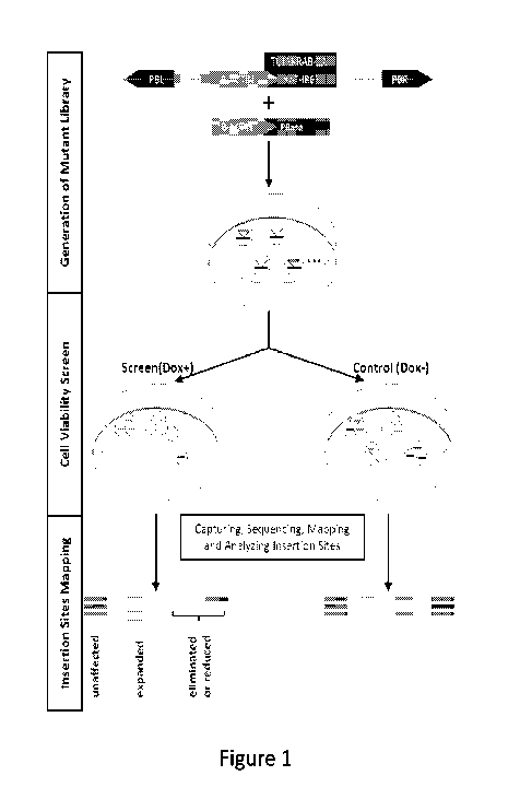
Figure 1 is a diagram showing the scheme of the PB transposon gain-of-function
screen to identify mutations that impair growth and/or survival;
Figure 2 shows images of Katushka-positive mutant cells collected by cell-
sorting
after brief induction with Dox. Mutant cells were then equally split into the
Dox+ pool for
screening and the Dox- pool as control. After mapping insertion sites and
counting reads, the
log2 reads ratio between Dox- and Dox+ pool was calculated for every insertion
site;
3
CA 02930315 2016-05-10
WO 2015/073990
PCT/US2014/065997
Figure 3 shows the analysis to identify candidate RAS antagonizing genes.
TCF7L1
was used to illustrate the biostatistics analysis for identifying negatively
selected genes. A
total of 150 genes were selected using the first binomial test and 95
candidate genes were
identified using the second binomial test;
Figure 4 is an illustration showing th candidate genes in the WNT pathway
identified
by the screen;
Figure 5A is a bar graph showing viable cell quantitation using an Alamarblue
assay
on AML-RAS stable cell lines conditionally overexpressing LRP6, TCF7L1, f3-
catenin, or o-
catenin. 3 days with (red) or without (blue) Dox induction
Figure 5B is a bar graph showing viable cell quantitation using an Alamarblue
assay
on TRI-102 stable cell lines conditionally overexpressing LRP6, TCF7L1, f3-
catenin, or o-
catenin. 3 days with (red) or without (blue) Dox induction;
Figure 6 shows DIC (20x) images of indicated cells after 24hr treatment with
vehicle
(top) or 20mM LiC1 (bottom);
Figure 7 is a bar graph showing percentage of viable cells 3 days after
treatment with
GSK3 inhibitors, 20mM LiC1, 5uM Kenpaullone or 2uM BIO, *** p<0.001;
Figure 8A is a representative image of soft agar assay on AML-RAS cells with
vehicle (top) or 20mM LiC1 treatment (bottom);
Figure 8B is a bar graph showing the quantitation of colony number;
Figure 9A is a line graph showing the percentage of tumor size change in
xenografts
over 28 days with vehicle (diamonds) or LiC1 (squares);
Figure 9B is a line graphs showing mean body weight over over 28 days with
vehicle
(diamonds) or LiC1 (squares);
Figure 9C is a representative image of tumors removed at 28 days;
Figure 10 is a bar graph showing activity of GSK3 inhibitors on a panel of RAS
tumor
cells. Percentage of viable cells compared to vehicle control after 5days in
indicated
melanoma, lung, colon, and pancreatic cancer cells treated with LiC1 (left
bar), Kenpaullone
(middle bar), or BIO (right bar). Non-transformed control: human mammary
epithelial cells;
Figure 11A is a bar graph showing viable cell quantitation on H1792 (top)
stable cell
lines conditionally overexpressing LRP6, TCF7L1, f3-catenin, or 6-catenin with
(right bar) or
without (left bar) Dox induction. *** p<0.001 , * p<0.05;
Figure 11B is a bar graph showing viable cell quantitation on A549 stable cell
lines
conditionally overexpressing LRP6, TCF7L1, f3-catenin, or 6-catenin with
(right bar) or
without (left bar) Dox induction. *** p<0.001 , * p<0.05;
4
CA 02930315 2016-05-10
WO 2015/073990
PCT/US2014/065997
Figure 12A shows a representative image of soft agar assay on A549 (left) or
H1792
(right) cells with vehicle (top) or 20mM LiC1 treatment;
Figure 12B is a bar graph showing quantitation of colony number;
Figure 13A is a line graph showing the growth curve of TRI-102 and AML-RAS
cells. Viable cells for TRI-102(more horizontal line) or AML-RAS(more vertical
line) were
measured daily by Celltiter-Glo for 4 days;
Figure 13B is a bar graph showing quantification of colony numbers for
anchorage
growth in soft agar assays on TRI-102 and AML-RAS cells (right).
Figure 14 is a diagram of capture based PCR method for PB insertion mapping.
Genomic DNA was digested with AluI. Genomic DNA fragment (Insertion site,
black line)
linked with PB arm (PBR, gray bar) was amplified and labeled with biotinylated
primer (dot)
through SPE reaction. Biotin-labeled insertion fragments were enriched by
streptavidin-
magnetic beads. After poly G tailing (Gs), insertion fragments were linked
with adapter
sequence (gray bar) and subjected for Illumina high-throughput sequencing; and
Figure 15 is a bar graph showing the genomic distribution of PB transpo son
insertion
sites. Total of 4,362,271 sequences that had the PB recognition site, TTAA,
were mapped to
UCSC hg18 database and 270,257 insertion sites were recovered. The
distribution of PB
insertions is illustrated.
Figure 16 is an illustration showing the PB transposon gain-of-function screen
to
identify mutations that impair growth and/or survival.
Figure 17 is a series of illustrations showing that after brief induction with
Dox,
Katushka-positive mutant cells were collected by cell-sorting. Mutant cells
were then equally
split into the Dox+ pool for gene-induction and the Dox- pool as control.
After mapping
insertion sites and counting reads, the log2 reads ratio between Dox- and Dox+
pool was
calculated for every insertion site.
Figure 18 is a panel of images showing the analysis to identify candidate RAS
antagonizing genes. TCF7L1 was used to illustrate the classification of
depleted insertions
(Red, 2fold Dox-Sites, M) and enriched insertions (Green, 2fold Dox+Sites, P).
Candidate
genes (shaded) in the canonical WNT pathway identified from the PB screen.
Figure 19 is an image showing a heatmap of Pearson Correlation Coefficient
Analysis
between 340 protein coding targets and 259 long noncoding targets across Human
BodyMap
2Ø
Figure 20 is an image showing four enriched RBPs, EIF4A3,SRSF1, FUS and U2AF2
are components of spliceosome. Representive noncoding target genes contain
binding sites
5
CA 02930315 2016-05-10
WO 2015/073990
PCT/US2014/065997
for the RBPs have been listed in the corresponding ovals, p-value is
calculated by
hypergeometric test.
Figure 21 is a graph showing the genomic distribution of PB transposon
insertion
sites. A total of 422,746 insertion sites were mapped to UCSC hg19 database.
The
distribution of PB insertions is illustrated.
Figure 22 is an illustration showing biostatistics analysis to identify
candidate genes.
Figure 23 is a list of candidate genes from the PB gain-of-function screen.
Figure 24 is a list of candidate genes from the kinome siRNA screen.
Figure 25 is a list of noncoding candidate genes for four enriched RNA binding
proteins (RBPs).
DETAILED DESCRIPTION OF THE INVENTION
Definitions
Unless defined otherwise, all technical and scientific terms used herein have
the same
meaning as commonly understood by one of ordinary skill in the art to which
the invention
pertains. Although any methods and materials similar or equivalent to those
described herein
may be used in the practice for testing of the present invention, the
preferred materials and
methods are described herein. In describing and claiming the present
invention, the
following terminology will be used.
It is also to be understood that the terminology used herein is for the
purpose of
describing particular embodiments only, and is not intended to be limiting.
As used herein, the articles "a" and "an" are used to refer to one or to more
than one
(i. e. , to at least one) of the grammatical object of the article. By way of
example, "an
element" means one element or more than one element.
As used herein when referring to a measurable value such as an amount, a
temporal
duration, and the like, the term "about" is meant to encompass variations of
20% or within
10%, 9%, 8%, 7%, 6%, 5%, 4%, 3%, 2%, 1%, 0.5%, 0.1%, 0.05%, or 0.01% of the
specified
value, as such variations are appropriate to perform the disclosed methods.
Unless otherwise
clear from context, all numerical values provided herein are modified by the
term about.
The phrase "differentially present" refers to differences in the quantity
and/or the
frequency an insertion site is present in a sample of transposed cells as
compared to a control
sample. Gene insertion sites can be differentially present in terms of
quantity, frequency or
both. Gene insertion sites are differentially present between two samples if
the insertion site
frequency is statistically significantly different from the frequency of the
insertion site
6
CA 02930315 2016-05-10
WO 2015/073990
PCT/US2014/065997
frequency in the other sample, such as a reference. Alternatively or
additionally, one or more
gene insertion sites are differentially present between two sets of samples if
the frequency of
detecting the insertion sites in transposed cells are statistically
significantly higher or lower
than in the control cells. A gene insertion site that is present in one
sample, but undetectable
in another sample is differentially present.
The term "transposon" refers to a DNA sequence that can change its position
within
the genome, sometimes creating or reversing mutations and altering the cell's
genome. The
term "piggBac transposon" or "PB" refers to a mobile genetic element that
transposes
between vectors and chromosomes via a "cut and paste" mechanism. During
transposition,
the PB transposase recognizes transposon-specific inverted terminal repeat
sequences (ITRs)
located on both ends of the transposon vector and efficiently moves the
contents from the
original sites and integrates into TTAA chromosomal sites. The resulting
transformed cells
or group of cells are stable transformants.
A "vector" is a composition of matter that comprises a nucleic acid of
interest. In
some embodiments, a vector comprises a piggyBac transposon and may be used to
deliver the
piggyBac transposon to the interior of a cell. In some embodiments, a vector
refers to any
plasmid containing piggyBac ends that is capable of moving foreign sequences
into the
genomes of a target organism or cell. "Expression vector" refers to a vector
engineered to
express a nucleic acid of interest. In some embodiments, an expression vector
comprises a
piggyBac transposon or piggyBac transposase and expression control sequences
operatively
linked to the piggyBac transposon or piggyBac transposase to be expressed. An
expression
vector comprises sufficient cis-acting elements for expression; other elements
for expression
may be supplied by the host cell or in an in vitro expression system.
Expression vectors
include all those known in the art, such as cosmids, plasmids (e.g., naked or
contained in
liposomes) and viruses (e.g., lentiviruses, retroviruses, adenoviruses, and
adeno-associated
viruses) that incorporate the recombinant polynucleotide. In some embodiments,
an
expression vector may be engineered to expression an agonist of a protein or
pathway (e.g., a
WNT pathway) disclosed herein. For example, an expression vector may be
engineered to
express one or more of the following genes: LRP6, a-catenin, 8-catenin,
TCF7L1,
CSNK1G1, CCNY, PCDH15, GNG7, IN080, SMARCC1, PRKCA, and MED13. By
"heterogenous DNA" is meant non native DNA to the location of insertion.
Exogenous DNA
includes, but is not limited to, genetically modified genes. For example, the
piggyBac
transposon excises host DNA and inserts exogenous DNA into the insertion
sites. Such
7
CA 02930315 2016-05-10
WO 2015/073990
PCT/US2014/065997
exogenous DNA includes engineered genes, like chimeric genes, for expression
in the host
cell.
The term "WNT pathway agonist" refers to an agent that activates the WNT
pathway.
In some embodiments, a WNT pathway agonist is a small molecule, peptide, or
fragment
thereof. The WNT pathway agonist may activate one or more genes in the WNT
pathway,
such as LRP6, a-catenin, 8-catenin, TCF7L1, CSNK1G1, CCNY, PCDH15, GNG7,
IN080,
SMARCC1, PRKCA, and MED13. In some embodiments, the WNT pathway agonist
includes, but is not limited to, glycogen synthase kinase (GSK) inhibitor, 2-
Amino-4-(3,4-
(methylenedioxy)benzylamino)-6-(3-methoxyphenyl)pyrimidine, LiC1, Kenpaullone
and 6-
bromoindirubin-30-oxime (BIO), or a pharmaceutically acceptable salt, analog,
and
derivative thereof.
By "selective pressure" is meant an effect of selection on the relative
frequency of one
or more genes within a population by exposing the population to a selective
agent. For
example, exposure of the mutated or transposed cells to a selective agent,
such as an
antibiotic, induces expression of the transposon. Mutated or transposed cells
with decreased
cell fitness to the selective pressure are depleted over time, while cells
with increased cell
fitness to the selective pressure are enriched over time.
By "selective agent" is meant an agent that produces a selection pressure on
the
transposed cells to enrich in cells that express a selective gene. Examples
are antibiotics,
such as puromycin, tetracycline, blasticidin, and neomycin.
By "selective gene" is meant a gene that provides resistance, insensitivity or
the
capacity to grow in the presence of the selective pressure. An example of
selective genes
includes, but is not limited to, resistance gene to an antibiotic, such as
puromycin,
tetracycline, blasticidin, and neomycin resistance genes.
By "oncogenic Ras" is meant one or more mutations that permanently activate
Ras.
Overactive Ras is the most common oncogene in cancer. In some embodiments, a
condition,
disorder, or disease characterized by the expression of oncogenic Ras refers
to a condition,
disorder, or disease characterized by the presence of cells (e.g., cancer
cells) that expres an
oncogenic Ras protein. In some embodiments, a condition, disorder or disease
characterized
by oncogenic Ras includes certain types of cancer (e.g., lung, liver,
gastrointestinal, colon,
pancreatic, and skin tumor). As used herein, the term Ras refers to any member
of the Ras
superfamily, including but not limited to the gene or protein product of any
of the human
HRAS, KRAS, or NRAS genes. In some embodiments, an oncogenic Ras gene or
protein is
8
CA 02930315 2016-05-10
WO 2015/073990
PCT/US2014/065997
characterized by an activating mutation such as one resulting in a Gl2V and/or
Q61K amino
acid change in the Ras protein.
In this disclosure, "comprises," "comprising," "containing" and "having" and
the like
can have the meaning ascribed to them in U.S. Patent law and can mean "
includes,"
"including," and the like; "consisting essentially or or "consists
essentially" likewise has the
meaning ascribed in U.S. Patent law and the term is open-ended, allowing for
the presence of
more than that which is recited so long as basic or novel characteristics of
that which is
recited is not changed by the presence of more than that which is recited, but
excludes prior
art embodiments.
By "effective amount" is meant the amount required to reduce or improve at
least one
symptom of a disease relative to an untreated patient. The effective amount of
an active
compound(s) used for therapeutic treatment of a disease varies depending upon
the manner of
administration, the age, body weight, and general health of the subject.
The term "expression" as used herein is defined as the transcription and/or
translation
of a particular nucleotide sequence driven by its promoter.
By "fragment" is meant a portion of a polynucleotide or nucleic acid molecule.
This
portion contains, preferably, at least 10%, 20%, 30%, 40%, 50%, 60%, 70%, 80%,
or 90% of
the entire length of the reference nucleic acids. A fragment may contain 10,
20, 30, 40, 50,
60, 70, 80, 90, or 100, 200, 300, 400, 500, 600, 700, 800, 900, 1000, 1500,
2000 or 2500 (and
any integer value in between) nucleotides. The fragment, as applied to a
nucleic acid
molecule, refers to a subsequence of a larger nucleic acid. A "fragment" of a
nucleic acid
molecule may be at least about 15 nucleotides in length; for example, at least
about 50
nucleotides to about 100 nucleotides; at least about 100 to about 500
nucleotides, at least
about 500 to about 1000 nucleotides, at least about 1000 nucleotides to about
1500
nucleotides; or about 1500 nucleotides to about 2500 nucleotides; or about
2500 nucleotides
(and any integer value in between).
The terms "insertion site" refer to the location of transposition in the DNA.
The
insertion sites of DNA transposons may be identified by short direct repeats
followed by a
series of inverted repeats important for the excision by the transposase. The
recognition
sequence for the piggyBac transposon is TTAA.
The terms "isolated," "purified," or "biologically pure" refer to material
that is free to
varying degrees from components which normally accompany it as found in its
native state.
"Isolate" denotes a degree of separation from original source or surroundings.
"Purify"
denotes a degree of separation that is higher than isolation. A "purified" or
"biologically
9
CA 02930315 2016-05-10
WO 2015/073990
PCT/US2014/065997
pure" protein is sufficiently free of other materials such that any impurities
do not materially
affect the biological properties of the protein or cause other adverse
consequences. That is, a
nucleic acid or peptide is purified if it is substantially free of cellular
material, viral material,
or culture medium when produced by recombinant DNA techniques, or chemical
precursors
or other chemicals when chemically synthesized. Purity and homogeneity are
typically
determined using analytical chemistry techniques, for example, polyacrylamide
gel
electrophoresis or high performance liquid chromatography. The term "purified"
can denote
that a nucleic acid or protein gives rise to essentially one band in an
electrophoretic gel. For
a protein that can be subjected to modifications, for example, phosphorylation
or
glycosylation, different modifications may give rise to different isolated
proteins, which can
be separately purified.
"Pharmaceutically acceptable" refers to those properties and/or substances
that are
acceptable to the patient from a pharmacological/toxicological point of view
and to the
manufacturing pharmaceutical chemist from a physical/chemical point of view
regarding
composition, formulation, stability, patient acceptance and bioavailability.
"Pharmaceutically
acceptable carrier" refers to a medium that does not interfere with the
effectiveness of the
biological activity of the active ingredient(s) and is not toxic to the host
to which it is
administered.
As used herein, the term "pharmaceutical composition" or "pharmaceuticaly
acceptable composition" refers to a mixture of at least one compound or
molecule useful
within the invention with a pharmaceutically acceptable carrier. The
pharmaceutical
composition facilitates administration of the compound or molecule to a
patient. Multiple
techniques of administering a compound or molecule exist in the art including,
but not
limited to, intravenous, oral, aerosol, parenteral, ophthalmic, pulmonary and
topical
administration.
As used herein, the term "pharmaceutically acceptable carrier" means a
pharmaceutically acceptable material, composition or carrier, such as a liquid
or solid filler,
stabilizer, dispersing agent, suspending agent, diluent, excipient, thickening
agent, solvent or
encapsulating material, involved in carrying or transporting a compound or
molecule useful
within the invention within or to the patient such that it may perform its
intended function.
Typically, such constructs are carried or transported from one organ, or
portion of the body,
to another organ, or portion of the body. Each carrier must be "acceptable" in
the sense of
being compatible with the other ingredients of the formulation, including the
compound
useful within the invention, and not injurious to the patient. Some examples
of materials that
CA 02930315 2016-05-10
WO 2015/073990
PCT/US2014/065997
may serve as pharmaceutically acceptable carriers include: sugars, such as
lactose, glucose
and sucrose; starches, such as corn starch and potato starch; cellulose, and
its derivatives,
such as sodium carboxymethyl cellulose, ethyl cellulose and cellulose acetate;
powdered
tragacanth; malt; gelatin; talc; excipients, such as cocoa butter and
suppository waxes; oils,
such as peanut oil, cottonseed oil, safflower oil, sesame oil, olive oil, corn
oil and soybean
oil; glycols, such as propylene glycol; polyols, such as glycerin, sorbitol,
mannitol and
polyethylene glycol; esters, such as ethyl oleate and ethyl laurate; agar;
buffering agents, such
as magnesium hydroxide and aluminum hydroxide; surface active agents; alginic
acid;
pyrogen-free water; isotonic saline; Ringer's solution; ethyl alcohol;
phosphate buffer
solutions; and other non-toxic compatible substances employed in
pharmaceutical
formulations. As used herein, "pharmaceutically acceptable carrier" also
includes any and all
coatings, antibacterial and antifungal agents, and absorption delaying agents,
and the like that
are compatible with the activity of the compound useful within the invention,
and are
physiologically acceptable to the patient. Supplementary active compounds may
also be
incorporated into the compositions. The "pharmaceutically acceptable carrier"
may further
include a pharmaceutically acceptable salt of the compound or molecule useful
within the
invention. Other additional ingredients that may be included in the
pharmaceutical
compositions used in the practice of the invention are known in the art and
described, for
example in Remington's Pharmaceutical Sciences (Genaro, Ed., Mack Publishing
Co., 1985,
Easton, PA), which is incorporated herein by reference.
By "reference" is meant a standard or control. A "reference " is a defined
standard or
control used as a basis for comparison.
As used herein, "sample" or "biological sample" refers to anything, which may
contain the cells of interest (e.g., cancer or tumor cells thereof) for which
the screening
method or treatment is desired. The sample may be a biological sample, such as
a biological
fluid or a biological tissue. In one embodiment, a biological sample is a
tissue sample
including pulmonary arterial endothelial cells. Such a sample may include
diverse cells,
proteins, and genetic material. Examples of biological tissues also include
organs, tumors,
lymph nodes, arteries and individual cell(s). Examples of biological fluids
include urine,
blood, plasma, serum, saliva, semen, stool, sputum, cerebral spinal fluid,
tears, mucus,
amniotic fluid or the like.
A "subject" or "patient," as used therein, may be a human or non-human mammal.
Non-human mammals include, for example, livestock and pets, such as ovine,
bovine,
porcine, canine, feline and murine mammals. Preferably, the subject is human.
11
CA 02930315 2016-05-10
WO 2015/073990
PCT/US2014/065997
As used herein, the terms "treat," treating," "treatment," and the like refer
to reducing
or improving a disorder and/or symptom associated therewith. It will be
appreciated that,
although not precluded, treating a disorder or condition does not require that
the disorder,
condition or symptoms associated therewith be completely ameliorated or
eliminated.
In some embodiments, "treatment", "treating" or "treat" relates to the
management
and care of a patient for the purpose of combating a disease, condition, or
disorder and
includes the administration of a compound or composition (e.g., a WNT pathway
agonist) of
the present invention, to alleviate the symptoms or complications of a
disease, condition or
disorder, or to eliminate the disease, condition or disorder.
As used herein, "prevention", "preventing" or "prevent" describes reducing or
eliminating the onset of the symptoms or complications of the disease,
condition or disorder
and includes the administration of a compound or composition (e.g., a WNT
pathway
agonist) of the present invention, to reduce the onset, development or
recurrence of
symptoms of the disease, condition or disorder.
As used herein, the term "alleviate" is meant to describe a process by which
the
severity of a sign or symptom of a disorder is decreased. Importantly, a sign
or symptom can
be alleviated without being eliminated. In some embodiments, administration of
a compound
or composition (e.g., a WNT pathway agonist) of the present invention leads to
the
elimination of a sign or symptom, however, elimination is not required.
Effective dosages
are expected to decrease the severity of a sign or symptom.
As used herein the term "symptom" is defined as an indication of disease,
illness,
injury, or that something is not right in the body. Symptoms are felt or
noticed by the
individual experiencing the symptom, but may not easily be noticed by others.
Others are
defined as non-health-care professionals.
As used herein the term "sign" is also defined as an indication that something
is not
right in the body. But signs are defined as things that can be seen by a
doctor, nurse, or other
health care professional.
Treating a disorder, disease or condition of the present invention can result
in a
reduction in size of a tumor. A reduction in size of a tumor may also be
referred to as "tumor
regression". In some embodiments, after treatment, tumor size is reduced by 5%
or greater
relative to its size prior to treatment; more preferably, tumor size is
reduced by 10% or
greater; more preferably, reduced by 20% or greater; more preferably, reduced
by 30% or
greater; more preferably, reduced by 40% or greater; even more preferably,
reduced by 50%
or greater; and most preferably, reduced by greater than 75% or greater. Size
of a tumor may
12
CA 02930315 2016-05-10
WO 2015/073990
PCT/US2014/065997
be measured by any reproducible means of measurement. The size of a tumor may
be
measured as a diameter of the tumor.
Treating a disorder, disease or condition of the present invention can result
in a
reduction in tumor volume. In some embodiments, after treatment, tumor volume
is reduced
by 5% or greater relative to its size prior to treatment; more preferably,
tumor volume is
reduced by 10% or greater; more preferably, reduced by 20% or greater; more
preferably,
reduced by 30% or greater; more preferably, reduced by 40% or greater; even
more
preferably, reduced by 50% or greater; and most preferably, reduced by greater
than 75% or
greater. Tumor volume may be measured by any reproducible means of
measurement.
Treating a disorder, disease or condition of the present invention can result
in a
decrease in number of tumors. In some embodiments, after treatment, tumor
number is
reduced by 5% or greater relative to number prior to treatment; more
preferably, tumor
number is reduced by 10% or greater; more preferably, reduced by 20% or
greater; more
preferably, reduced by 30% or greater; more preferably, reduced by 40% or
greater; even
more preferably, reduced by 50% or greater; and most preferably, reduced by
greater than
75%. Number of tumors may be measured by any reproducible means of
measurement. The
number of tumors may be measured by counting tumors visible to the naked eye
or at a
specified magnification. In some embodiments, the specified magnification is
2x, 3x, 4x, 5x,
10x, or 50x.
Treating a disorder, disease or condition of the present invention can result
in a
decrease in number of metastatic lesions in other tissues or organs distant
from the primary
tumor site. In some embodiments, after treatment, the number of metastatic
lesions is reduced
by 5% or greater relative to number prior to treatment; more preferably, the
number of
metastatic lesions is reduced by 10% or greater; more preferably, reduced by
20% or greater;
more preferably, reduced by 30% or greater; more preferably, reduced by 40% or
greater;
even more preferably, reduced by 50% or greater; and most preferably, reduced
by greater
than 75%. The number of metastatic lesions may be measured by any reproducible
means of
measurement. The number of metastatic lesions may be measured by counting
metastatic
lesions visible to the naked eye or at a specified magnification. In some
embodiments, the
specified magnification is 2x, 3x, 4x, 5x, 10x, or 50x.
Treating a disorder, disease or condition of the present invention can result
in an
increase in average survival time of a population of treated subjects in
comparison to a
population receiving carrier alone. In some embodiments, the average survival
time is
increased by more than 30 days; more preferably, by more than 60 days; more
preferably, by
13
CA 02930315 2016-05-10
WO 2015/073990
PCT/US2014/065997
more than 90 days; and most preferably, by more than 120 days. An increase in
average
survival time of a population may be measured by any reproducible means. An
increase in
average survival time of a population may be measured, for example, by
calculating for a
population the average length of survival following initiation of treatment
with an active
compound. An increase in average survival time of a population may also be
measured, for
example, by calculating for a population the average length of survival
following completion
of a first round of treatment with an active compound.
Treating a disorder, disease or condition of the present invention can result
in an
increase in average survival time of a population of treated subjects in
comparison to a
population of untreated subjects. In some embodiments, the average survival
time is
increased by more than 30 days; more preferably, by more than 60 days; more
preferably, by
more than 90 days; and most preferably, by more than 120 days. An increase in
average
survival time of a population may be measured by any reproducible means. An
increase in
average survival time of a population may be measured, for example, by
calculating for a
population the average length of survival following initiation of treatment
with an active
compound. An increase in average survival time of a population may also be
measured, for
example, by calculating for a population the average length of survival
following completion
of a first round of treatment with an active compound.
Treating a disorder, disease or condition of the present invention can result
in a
decrease in tumor growth rate. In some embodiments, after treatment, tumor
growth rate is
reduced by at least 5% relative to number prior to treatment; more preferably,
tumor growth
rate is reduced by at least 10%; more preferably, reduced by at least 20%;
more preferably,
reduced by at least 30%; more preferably, reduced by at least 40%; more
preferably, reduced
by at least 50%; even more preferably, reduced by at least 50%; and most
preferably, reduced
by at least 75%. Tumor growth rate may be measured by any reproducible means
of
measurement. Tumor growth rate can be measured according to a change in tumor
diameter
per unit time.
Treating a disorder, disease or condition of the present invention can result
in a
decrease in tumor regrowth. In some embodiments, after treatment, tumor
regrowth is less
than 5%; more preferably, tumor regrowth is less than 10%; more preferably,
less than 20%;
more preferably, less than 30%; more preferably, less than 40%; more
preferably, less than
50%; even more preferably, less than 50%; and most preferably, less than 75%.
Tumor
regrowth may be measured by any reproducible means of measurement. Tumor
regrowth is
measured, for example, by measuring an increase in the diameter of a tumor
after a prior
14
CA 02930315 2016-05-10
WO 2015/073990
PCT/US2014/065997
tumor shrinkage that followed treatment. A decrease in tumor regrowth is
indicated by
failure of tumors to reoccur after treatment has stopped.
Treating or preventing a cell proliferative disorder of the present invention
can result
in a reduction in the rate of cellular proliferation. In some embodiments,
after treatment, the
rate of cellular proliferation is reduced by at least 5%; more preferably, by
at least 10%; more
preferably, by at least 20%; more preferably, by at least 30%; more
preferably, by at least
40%; more preferably, by at least 50%; even more preferably, by at least 50%;
and most
preferably, by at least 75%. The rate of cellular proliferation may be
measured by any
reproducible means of measurement. The rate of cellular proliferation is
measured, for
example, by measuring the number of dividing cells in a tissue sample per unit
time.
Treating or preventing a cell proliferative disorder of the present invention
can result
in a reduction in the proportion of proliferating cells. In some embodiments,
after treatment,
the proportion of proliferating cells is reduced by at least 5%; more
preferably, by at least
10%; more preferably, by at least 20%; more preferably, by at least 30%; more
preferably, by
at least 40%; more preferably, by at least 50%; even more preferably, by at
least 50%; and
most preferably, by at least 75%. The proportion of proliferating cells may be
measured by
any reproducible means of measurement. In some embodiments, the proportion of
proliferating cells is measured, for example, by quantifying the number of
dividing cells
relative to the number of nondividing cells in a tissue sample. The proportion
of proliferating
cells can be equivalent to the mitotic index.
Treating a disorder, disease or condition of the present invention can result
in
cytotoxic effects (e.g., increase apoptosis, increased necrosis) in a diseased
cell population,
e.g., a cancer cell population. In some embodiments, a cytotoxic treatment
leads to a
reduction in a diseased cell population size of at least 10%, at least 20%, at
least 30%, at least
40%, at least 50%, at least 60%, at least 70%, at least 80%, at least 90%, at
least 95%, at least
99%, or more. Size of a cell population may be measured by any reproducible
means of
measurement. The size of a cell population may be measured as the number of
viable cells in
the population or sample thereof.
Treating or preventing a cell proliferative disorder of the present invention
can result in a
decrease in size of an area or zone of cellular proliferation. In some
embodiments, after
treatment, the size of an area or zone of cellular proliferation is reduced by
at least 5%
relative to its size prior to treatment; more preferably, reduced by at least
10%; more
preferably, reduced by at least 20%; more preferably, reduced by at least 30%;
more
preferably, reduced by at least 40%; more preferably, reduced by at least 50%;
even more
CA 02930315 2016-05-10
WO 2015/073990
PCT/US2014/065997
preferably, reduced by at least 50%; and most preferably, reduced by at least
75%. The size
of an area or zone of cellular proliferation may be measured by any
reproducible means of
measurement. The size of an area or zone of cellular proliferation may be
measured as a
diameter or width of an area or zone of cellular proliferation.
Treating or preventing a cell proliferative disorder of the present invention
can result
in a decrease in the number or proportion of cells having an abnormal
appearance or
morphology. In some embodiments, after treatment, the number of cells having
an abnormal
morphology is reduced by at least 5% relative to its size prior to treatment;
more preferably,
reduced by at least 10%; more preferably, reduced by at least 20%; more
preferably, reduced
by at least 30%; more preferably, reduced by at least 40%; more preferably,
reduced by at
least 50%; even more preferably, reduced by at least 50%; and most preferably,
reduced by at
least 75%. An abnormal cellular appearance or morphology may be measured by
any
reproducible means of measurement. An abnormal cellular morphology can be
measured by
microscopy, e.g., using an inverted tissue culture microscope. An abnormal
cellular
morphology can take the form of nuclear pleiomorphism.
As used herein, the term "selectively" means tending to occur at a higher
frequency in
one population than in another population. The compared populations can be
cell
populations, for example a diseased cell population (e.g., a tumor cell
population or
population of cells having a proliferative disorder) and a normal cell
population. As used
herein, a "normal cell" is a cell that cannot be classified as part of a "cell
proliferative
disorder". A normal cell lacks unregulated or abnormal growth, or both, that
can lead to the
development of an unwanted condition or disease. In some embodiments, a normal
cell
possesses normally functioning cell cycle checkpoint control mechanisms. In
some
embodiments, an event occurs selectively in population A relative to
population B if it occurs
greater than two times more frequently in population A as compared to
population B. An
event occurs selectively if it occurs greater than five times more frequently
in population A.
An event occurs selectively if it occurs greater than ten times more
frequently in population
A; in some embodiments, greater than fifty times; even more preferably,
greater than 100
times; and In some embodiments, greater than 1000 times more frequently in
population A as
compared to population B. For example, cell death may be said to occur
selectively in
diseased or hyper-proliferating cells if it occurred greater than twice as
frequently in diseased
or hyper-proliferating cells as compared to normal cells.
Ranges provided herein are understood to be shorthand for all of the values
within the
range. For example, a range of 1 to 50 is understood to include any number,
combination of
16
CA 02930315 2016-05-10
WO 2015/073990
PCT/US2014/065997
numbers, or sub-range from the group consisting 1, 2, 3, 4, 5, 6, 7, 8, 9, 10,
11, 12, 13, 14, 15,
16, 17, 18, 19, 20, 21, 22, 23, 24, 25, 26, 27, 28, 29, 30, 31, 32, 33, 34,
35, 36, 37, 38, 39, 40,
41, 42, 43, 44, 45, 46, 47, 48, 49, or 50.
The recitation of an embodiment for a variable or aspect herein includes that
embodiment as any single embodiment or in combination with any other
embodiments or
portions thereof.
Any compositions or methods provided herein can be combined with one or more
of
any of the other compositions and methods provided herein.
Insertion Site Identification
Genetic screening for genes that positively affect biological processes are
commonly
used. However, screening for genes that negatively affect processes, such as
growth or
tumorigenicity, are more difficult to identify due to the deletion and
selection against cells
that harbor expression of such genes.
The present invention describes the discovery of a screening method for
identifying
negatively selected genes. The screening method utilizes the pig gBac or PB
transposon, a
mobile genetic element that transposes DNA sequences between a transposon
vector and a
chromosome via a "cut and paste" mechanism. The piggyBac transposon machinery
(see
U.S. Pat. No. 6,218,815), recognizes transposon-specific inverted terminal
repeat sequences
(ITRs) or TTAA, the transposition recognition sequence, located on both ends
of the
transposon vector and within the genome of the host cell and specifically
excises and inserts a
heterologous DNA sequence found within the transposon vector into the genome
of the host
cell.
In some embodiments, the method of identifying negatively selected genes in an
insertional mutagenesis screen includes inducing transposition of a piggyBac
transposon in
cells of interest, exposing a portion of the transposed cells to a selective
pressure to induce
expression of the piggyBac transposon, comparing insertion sites in genomic
DNA of
transposed cells exposed to the selective pressure and transposed cells not
exposed to the
selective pressure, and identifying genes having one or more insertion sites,
wherein the
genes with insertion sites are differentially present in the transposed cells
exposed to the
selective pressure and the transposed cells not exposed to the selective
pressure.
In some embodiments, the piggyBac transposon includes an inducible gene
construct.
In one embodiment, induction of expression of the inducible gene construct in
the transposon
results in overexpression of an endogenous gene at the site of insertion of
the transposon. In
17
CA 02930315 2016-05-10
WO 2015/073990
PCT/US2014/065997
some embodiments, inducing expression of the transposon comprises inducing an
inducible
gene on the transposon and overexpressing an endogenous gene adjacent to the
site of
insertion of the transposon in the genome of the host cell (for example, by
transcriptional
read-through from the induced gene on the transposon). In another embodiment,
overexpression of the gene in the transposon results in negative selection of
the cells
harboring the pig gyBac transposon. The gene in the transposon may be
cytotoxic, resulting
in negative selection. However, in some embodiments, overexpression of the
gene in the
transposon does not itself result in negative selective. In such embodiment,
the gene may
include a detectable marker (e.g., a fluorescent or bioluminescent marker),
for example under
the control of an IRES.
In another embodiment, the pig gyBac transposon includes a selective gene,
such as an
inducible antibiotic resistance gene. In some embodiments, the transposon
comprises an
inducible gene. The inducible gene may include an inducible promoter, such as
one that is
inducible by exposure to an antibiotic (e.g., by tetracycline or a derivative
of tetracycline, for
example doxycycline). However, it should be appreciated that other inducible
promoters can
be used. The selective pressure can be a condition (e.g., exposure to an
agent, for example an
antibiotic) that results in induction of the inducible promoter. This results
in overexpression
of one or more endogenous gene, some of which may be selected against because
their
overexpression is cytostatic or cytotoxic in the transposon containing cell.
In some
embodiments, the pig gyBac transposon includes an inducible promoter that is
inducible by
the addition of an antibiotic but does not require antibiotic resistance. In
some embodiments,
the selective gene may be used to select for cells that harbor the transposon
or as a negative
selective agent that induces expression of the inducible gene in the
transposon thereby
increasing expression of an adjacent endogenous gene (e.g., by transcriptional
read-through
from the inducible promotor in the transposon).
For example, the pig gyBac transposon is expressed from a vector, such as
PB[Mut-
tet0-KAT-TETRKRAB] that includes a doxycycline inducible chimeric gene to
produce an
actin-Katushka red fluorescent fusion protein. The selective gene can include,
but is not
limited to, resistance to various antibiotics, such as puromycin,
tetracycline, blasticidin, and
neomycin. The transposon can further induce expression of heterogenous DNA,
such as a
chimeric gene. The heterogenous DNA chimeric gene can include genetically
modified
genes. For example, the pig gyBac transposon excises host DNA and inserts
exogenous DNA
into the insertion sites. Such exogenous DNA can include engineered genes,
like chimeric
genes, for expression in the host cell.
18
CA 02930315 2016-05-10
WO 2015/073990
PCT/US2014/065997
In another embodiment, cells of interest are co-transfected with the piggyBac
transposon and the transposase PBase to generate transposon mutagenized cells.
The cells of
interest include any cells that a particular phenotype can be analyzed
following the disruption
of one or more genes, such as cancer or tumor cells. The cells may also
possess a particular
phenotype and genetic screening for mutants that disrupt that phenotype would
be of interest.
For example, mutated or transposed cancer cells that are impaired or lack cell
growth
potential, non-metatastic, apoptotic, possess reduced cell survival, and/or
quiescent, possess
genes of interest affected by transposition.
Gene transfer of the transposon and transposase can be achieved using methods
known in the art. For example, non-viral means involving transfection in vitro
are of use.
Such methods include the use of calcium phosphate, DEAE dextran,
electroporation, and
protoplast fusion. Liposomes can also be potentially beneficial for delivery
of DNA into a
cell. Additionally, the non-viral based delivery can be nano-based or
aerosolized.
In one embodiment, the mutated or transposed cells are propagated to expand
the
population of mutated or transposed cells. The cells are grown in culture
without selection
for the transposon or induction of the heterogenous DNA inserted by the
transposon. In
embodiments where the heterogenous DNA is inducible with a selective agent,
any effect the
inserted heterogenous DNA may have on growth is minimized without its
induction.
Therefore, the mutated or transposed cells are able to expand in number.
In yet another embodiment, a portion of the mutated or transposed cells is
exposed to
a selective pressure. The selective pressure is used to enrich for a
population of cells of
interest. In embodiments where the piggyBac transposon inserts an inducible
selective gene
into host DNA, exposing the transposed cells to a selective agent enriches for
cells that
express the selective gene. In an exemplary embodiment, a portion of the
mutated or
transposed cells is enriched for expression of the selective gene by exposing
the mutated or
transposed cells to the selective pressure, while a portion of cells is not
exposed to the
selective pressure to maintain a mixed population of cells without enrichment.
By exposing a
portion of the mutated or transposed cells to the selective pressure and not
exposing a portion
of the mutated or transposed cells, any effects the inserted heterogenous DNA
may have on
growth is enriched for in cells under selective pressure, but not enriched in
cells propagated
without selective pressure.
For example, acute myeloid leukemia cells with KRASG12v (AML-RAS cells)
mutations are analyzed for impaired growth or survival after induction of the
chimeric gene
and compared to control cells cultured under the same conditions but without
induction of the
19
CA 02930315 2016-05-10
WO 2015/073990
PCT/US2014/065997
chimeric gene. Genomic DNA is isolated from both populations of cells and
compared for
transposon insertion sites in the respective genomes. If an insertional
mutation causes a
change in growth or survival of the AML-RAS cells then this change will be
reflected by
differences in the presence of that specific insertion site between the
mutated or transposed
cells and the control cells. Insertion sites that lead to an overall decrease
in cell fitness would
be depleted with successive cell passages. Likewise, insertion sites that lead
to cells with an
increase in cell fitness, faster cell growth rates etc., would be enriched
with successive cell
passages. Thus, depletion or enrichment of insertion sites or genes with
insertion sites
differentially present in the transposed cells are of interest as genes
involved in counteracting
the oncogenic phenotype. In some embodiments, the depletion of cells
identifies genes that
may be cytotoxic to oncogenic cells, e.g., cells with mutated RAS.
Identification of the genes harboring insertion sites can be accomplished
using
methods known in the art. Such methods can include, PCR capture and
sequencing. For
example, genomic DNA is prepared from the different populations of mutated or
transposed
cells. The genomic DNA is digested and amplified by PCR with primers specific
for the
insertion recognition sequence and transposon sequences. Sequence analysis of
the host
sequences flanking the insertion recognition sequence and transposon sequences
identifies the
location within the genome where the transposon inserted. Subsequent
identification of
genes with insertion sites differentially present between the two populations
of mutated or
transposed cells, transposed cells exposed to the selective pressure and the
transposed cells
not exposed to the selective pressure, results in identifying genes involved
in altering one or
more phenotypes of the mutated or transposed cells, such as overall growth
fitness,
oncogenicity or tumorigenicity.
Through the identification of genes involved in altering a cell's fitness for
oncogenicity or tumorigenicity, potential therapeutic targets and agents are
rapidly identified
for a broad spectrum of conditions, diseased or disorders, such as cancers,
especially those
conditions that lack effective treatments.
Compositions
The invention further provides, in one aspect, compositions for improving or
reducing
at least one symptom of a condition, disease or disorder by utilizing the
information gathered
from the insertion site identification. By screening cells of interest that
express an oncogenic
RAS, such as an oncogenic HRAS, oncogenic NRAS and oncogenic KRAS, genes that
affect
growth or survival of the cells of interest can be targeted for therapy. Thus,
compositions for
CA 02930315 2016-05-10
WO 2015/073990
PCT/US2014/065997
reducing proliferation of a tumor or cancer cell expressing an oncogenic RAS
that include an
activator of a WNT pathway are disclosed.
In particular embodiments, a composition is disclosed that includes an
activator of a
WNT pathway for reducing proliferation of a tumor cell expressing an oncogenic
RAS. In a
more particular embodiment, the the activator is a glycogen synthase kinase
(GSK) inhibitor,
2-Amino-4-(3,4-(methylenedioxy)benzylamino)-6-(3-methoxyphenyl)pyrimidine,
LiC1,
Kenpaullone and 6-bromoindirubin-30-oxime (BIO), and/or a small molecule
agonist of the
WNT pathway.
In some embodiments, compositions may include an agent that activates the WNT
pathway and reduces proliferation of a tumor cell expressing an ocogenic RAS,
where the
agent may include, but is not limited to, a small molecule activator or
agonist of the WNT
pathway, a gene capable of expressing a protein that activates the WNT pathway
such as a
coding gene, a gene that influences the activation of the WNT pathway such as
a non-coding
gene, a RNA molecule such as a RNAi molecule, and any combination thereof. By
delivering the composition to a subject in need thereof, the WNT pathway is
activated
through the small molecule activator or agonist of the WNT pathway, expression
of coding
gene, or the expression of a non-coding gene. Thus, the subject with the tumor
or cancer may
be treated. In embodiments that a gene is included in the compositions, the
gene is identified
through a negative screen with the Piggyback transposon.
Pharmaceutical Compositions
The invention also encompasses the use of a pharmaceutical composition of the
invention to practice the methods of the invention. Such a pharmaceutical
composition may
be provided in a form suitable for administration to a subject, and may be
comprise one or
more pharmaceutically acceptable carriers, one or more additional ingredients,
or some
combination of these. The at least one composition of the invention may
comprise a
physiologically acceptable salt, such as a compound contemplated within the
invention in
combination with a physiologically acceptable cation or anion, as is well
known in the art.
Pharmaceutical compositions that are useful in the methods of the invention
may be
suitably developed for inhalational, oral, rectal, vaginal, parenteral,
topical, transdermal,
pulmonary, intranasal, buccal, ophthalmic, intrathecal, intravenous or another
route of
administration. Other contemplated formulations include projected
nanoparticles, liposomal
preparations, resealed erythrocytes containing the active ingredient, and
immunologically-
based formulations. The route(s) of administration will be readily apparent to
the skilled
21
CA 02930315 2016-05-10
WO 2015/073990
PCT/US2014/065997
artisan and will depend upon any number of factors including the type and
severity of the
disease being treated, the type and age of the veterinary or human patient
being treated, and
the like.
The formulations of the pharmaceutical compositions described herein may be
prepared by any method known or hereafter developed in the art of
pharmacology. In
general, such preparatory methods include the step of bringing the active
ingredient into
association with a carrier or one or more other accessory ingredients, and
then, if necessary or
desirable, shaping or packaging the product into a desired single- or multi-
dose unit.
In one embodiment, the compositions of the invention are formulated using one
or
more pharmaceutically acceptable excipients or carriers. In one embodiment,
the
pharmaceutical compositions of the invention comprise a therapeutically
effective amount of
at least one compound of the invention and a pharmaceutically acceptable
carrier.
Pharmaceutically acceptable carriers, which are useful, include, but are not
limited to,
glycerol, water, saline, ethanol and other pharmaceutically acceptable salt
solutions such as
phosphates and salts of organic acids. Examples of these and other
pharmaceutically
acceptable carriers are described in Remington's Pharmaceutical Sciences
(1991, Mack
Publication Co., New Jersey).
The practice of the present invention employs, unless otherwise indicated,
conventional techniques of molecular biology (including recombinant
techniques),
microbiology, cell biology, biochemistry and immunology, which are well within
the purview
of the skilled artisan. Such techniques are explained fully in the literature,
such as,
"Molecular Cloning: A Laboratory Manual", fourth edition (Sambrook, 2012);
"Oligonucleotide Synthesis" (Gait, 1984); "Culture of Animal Cells" (Freshney,
2010);
"Methods in Enzymology" "Handbook of Experimental Immunology" (Weir, 1997);
"Gene
Transfer Vectors for Mammalian Cells" (Miller and Cabs, 1987); "Short
Protocols in
Molecular Biology" (Ausubel, 2002); "Polymerase Chain Reaction: Principles,
Applications
and Troubleshooting", (Babar, 2011); "Current Protocols in Immunology"
(Coligan, 2002).
These techniques are applicable to the production of the polynucleotides and
polypeptides of
the invention, and, as such, may be considered in making and practicing the
invention.
Particularly useful techniques for particular embodiments will be discussed in
the sections
that follow.
22
CA 02930315 2016-05-10
WO 2015/073990
PCT/US2014/065997
Method of Treatment
The present invention also includes methods for reducing proliferation of
tumor cells
or reducing or improving cancer expressing an oncogenic RAS and/or a symptom
associated
with cancer in a subject. As described herein, activation of the WNT pathway
impairs
proliferation and/or reduces survival of cancer cells that express oncogenic
RAS. Therefore,
administering an effective amount of a composition that includes an activator
of a WNT
pathway to a subject would provide a means for reducing proliferation of the
cancer or tumor
cells.
In one aspect, a method of reducing proliferation of tumor cells in a subject
in need
thereof includes administering an effective amount of a composition comprising
an activator
of a WNT pathway to the tumor cells of the subject, thereby reducing
proliferation of the
tumor cells. In an exemplary embodiment, the method is effective for tumor
cells that
express an oncogenic RAS, such as oncogenic HRAS, oncogenic NRAS and oncogenic
KRAS. The tumor or cancer cells include, but are not limited to, lung, liver,
gastrointestinal,
colon, pancreatic, and skin tumor. In another embodiment, the activator is a
glycogen
synthase kinase (GSK) inhibitor or a small molecule agonist of the WNT
pathway, such as 2-
Amino-4-(3 , 4- (methylenedioxy)benzylamino)-6-(3 -methoxyphenyl)pyrimidine,
LiC1,
Kenpaullone and 6-bromoindirubin-30-oxime (BIO). In another embodiment, the
agent may
include, but is not limited to, a small molecule activator or agonist of the
WNT pathway, a
gene capable of expressing a protein that activates the WNT pathway such as a
coding gene,
a gene that influences the activation of the WNT pathway such as a non-coding
gene, a RNA
molecule such as a RNAi molecule, and any combination thereof.
In another aspect, a method of reducing or improving cancer expressing an
oncogenic
RAS and/or symptom associated therewith in a subject includes administering an
activator of
a WNT pathway. Also included is a use of a composition for the manufacture of
a
medicament for the treatment of an oncogenic RAS tumor. The treatment includes
reducing
or improving cancer or the tumor expressing an oncogenic RAS and/or symptom in
the
subject.
The following examples are put forth so as to provide those of ordinary skill
in the art
with a complete disclosure and description of how to make and use the assay,
screening, and
therapeutic methods of the invention, and are not intended to limit the scope
of what the
inventors regard as their invention.
23
CA 02930315 2016-05-10
WO 2015/073990
PCT/US2014/065997
EXAMPLES
The invention is further described in detail by reference to the following
experimental
examples. These examples are provided for purposes of illustration only, and
are not
intended to be limiting unless otherwise specified. Thus, the invention should
in no way be
construed as being limited to the following examples, but rather, should be
construed to
encompass any and all variations which become evident as a result of the
teaching provided
herein.
Without further description, it is believed that one of ordinary skill in the
art can,
using the preceding description and the following illustrative examples, make
and utilize the
compounds of the present invention and practice the claimed methods. The
following
working examples therefore, specifically point out embodiments of the present
invention, and
are not to be construed as limiting in any way.
The Materials and Methods used in the experiments described in Example 1
disclosed
herein are now described.
Summary of Screen. PB[Mut-tet0-KAT-TETRKRAB] was generated by modification
of Luc-PB[Mut]. TRI-102 cells were obtained from the Rothberg Institute. AML-
RAS cell
was generated from TRI-102 by infection with retrovirus produced from pBabe-
Puro-
KRASG12v and selected with 2n/mL puromycin. To conduct gain-of-function
screen,
PB[Mut-tet0-KAT-TETRKRAB] was introduced into 2x10⁵ AML-RAS cells by co-
transfection with the transposase plasmid ACT-PBase. Four days after
transposon
transposition, mutated cells were transiently induced with Dox for 24hrs, and
then KAT
positive cells were collected by cell-sorting. The sorted cells were further
expanded for 3
days, and then separated equally to two pools. Each pool had 3x10⁶ cells, the
screen pool was
treated with 2ug/m1Dox for 5 days and control pool was treated with vehicle
control ddH20.
After 5 days screen, the genomic DNA from two pools were extracted. PB
insertion
sites were first enriched by capture based PCR method (Figure 14) and then
subjected for
Illumina high-throughput sequencing. The raw Illumina sequencing data was
first imported
into the Galaxy platform. Insertion site mapping, reads quantification, and
insertion site
distribution analysis were all processed by using the Galaxy software. Genes
were selected by
two rounds of binomial test. The candidate targets were chosen based on p-
value <0.01 for
both filters.
To determine the effects of various drug treatments on oncogenic RAS mutant
cells,
an AlamarBlue assay (Invitrogen) was performed to monitor cell viability.
Activation of
24
CA 02930315 2016-05-10
WO 2015/073990
PCT/US2014/065997
WNT pathway in vivo were also performed in a soft agar assay and a AML-RAS
cell
xenograft model.
Vectors and Cloning. PB[Mut-tet0-KAT-TETRKRAM was generated by modification
of Luc-PB[Mut] . The tet0 was obtained from pHUD10-3 and cloned upstream of
the CAG
promoter. The Katushka red fluorescent protein was amplified from pTurboFP636-
C
(Evrogen). The TetR-KRAB (Addgene Plasmid 11642) linked to KAT through a 2A
peptide
was created by overlapping PCR and cloned 3' of the ACT promoter. The
blasticidin cassette
was amplified from pCDNA6 (Invitrogen) and cloned into the NheI site upstream
of the tet0.
ACT-PBase has been previously described. Full-length cDNA clones for LRP6 and
fi-catenin
were obtained from Addgene (27242 and 19286), while TCF7L1 and 6-catenin were
from
DF/HCC DNA Resource Core (H5CD00339336 and H5CD00082615). Full-length cDNAs
were next subcloned into a PB vector, PBJ[BRT], which is a Tet-On vector
containing a
blasticidin selection cassette.
Cell Culture and Generation of Stable Cell lines. TRI-102 cells were obtained
from
the Rothberg Institute. To generate AML-RAS cell, TRI-102 were infected by
retrovirus
produced from pBabe-Puro- KRASG12v and selected with 2n/mL puromycin. To
generate
stable cell lines for conditional overexpression of LRP6, TCF7L1, f3-catenin
or 8-catenin,
TRI-102 or AML-RAS were co-transfected with corresponding gene in PBJ[BRT] and
ACT-
PBase and selected with 5n/mL blasticidin for two weeks. All the TRI-102 and
AML-RAS
cell lines were maintained in F12-DMEM with 10% FBS.
Oncogenic RAS melanoma cell lines YUDOSO, YUTICA and YUGASP were gifts
from Dr. David Stern. They were maintained in MEM with 10% FBS. Lung cancer
cell lines
H358, H441,H460,H1734,H1792 and A549 were from American Type Culture
Collection
(ATCC), and maintained in RPMI-1640 with 10% FBS. Colon cancer cell lines DLD-
1,
HCT116, SW1116 and pancreatic cancer AsPc 1, Capan2, MiaPaCa2, Panel were from
ATCC and maintained in DMEM with 10% FBS. To generate stable cell lines for
conditionally overexpression of LRP6, TCF7L1, f3-catenin or 8-catenin, lung
cancer cell lines
A549 and H1792 were co-transfected with corresponding gene in PBJ[BRT] and ACT-
PBase
and selected with 5n/mL blasticidin for two weeks.
PB gain-of-function screen. PB[Mut-tet0-KAT-TETRKRAB] was introduced into
2x10⁵ AML-RAS cells by co-transfection with the transposase plasmid ACT-PBase.
Four
days after transposon transposition, mutated cells were transiently induced
with Dox for
24hrs, and then KAT positive cells were collected by cell-sorting. The sorted
cells were
further expanded for 3 days, and then separated equally to two pools. Each
pool had 3x10⁶
CA 02930315 2016-05-10
WO 2015/073990
PCT/US2014/065997
cells, the screen pool was treated with 2ug/m1 Dox for 5 days and control pool
was treated
with vehicle control ddH20.
Genomic DNA preparation, capture based PCR and Minim sequencing (Figure 14.).
For genomic DNA extraction, Cells were resuspended in buffer containing 10mM
Tris-HC1
pH8.0, 2mM EDTA, 200mM NaC1, 0.2%SDS, 200ug/m1 RNAse A and 800ug/m1 proteinase
K and incubated in 55 C water bath for overnight (>12h), followed by addition
of 5M NaCl.
After 10000g centrifuge, genomic DNA was precipitated with isopropanol,
followed by
washing with 75% ethanol. The DNA pellet was resuspend in 10mM Tris-HCL pH 8.0
with
0.1% EDTA. Genomic DNA was then digested with AluI overnight (>12h). The
digested
genomic DNA was precipitated by adding 1/10 volume of 3M NaAc and 1/2 volume
of
isopropanol and centrifuged for 20 minutes at max-speed. The digested genomic
DNA pellet
was then resuspended in 10mM Tris-HCL pH 7.4 with 0.1% EDTA.
Capture based PCR was first carried out by single primer extension
reaction(SPE),
which contained 25ug digested genomic DNA, 300uM dNTPs, 300nM Biotin-PBR-F
primer
(CCCITTAGTGAGGGTTAATTAGCTCCAAGCGGCGACTGAGA, two italicized Ts were
replaced with Biotin-dT), lx Kapa HF buffer, 2u1 Kapa polymerase in 100u1
volume. The
PCR condition for SPE was 95 C for 5mins, 40 cycles of (98 C 20secs, 60 C
30secs, 72 C
lmin), and final step of extension at 72 C for 5mins. The SPE products were
purified by
Qiagen PCR purification kit. Purified SPE products were then mixed with
magnetic beads
(Promega, cat# Z5481). Biotinylated insertion site fragments were separated
from other
genomic DNA by biotin-streptavidin binding reaction according to
manufacturer's protocol.
Next, a 3 prime dG tailing reaction was setup on beads by using terminal
transferase (NEB,
cat#M0315S).
The insertion site fragments were then subjected to PCR reaction for addition
of
adaptor sequences for Illumina high-throughput sequencing. The PCR reaction
contained Sul
template, Sul 5X Kapa HF buffer, 0.75u1 of ILP11:
(AATGATACGGCGACCACCGAGATCTACACTCTTTCCCTACACGACGCTCTTCCG
ATCT), 0.75u1 of Idx6I:
(CAAGCAGAAGACGGCATACGAGATATTGGCGTGACTGGAGTTCAGACGTGTGCT
CTTCCGATCT)
Or 0.75u1 of Idx12:
(CAAGCAGAAGACGGCATACGAGATTACAAGGTGACTGGAGTTCAGACGTGTGC
TCTTCCGATCT), 0.75u1 of dNTP, 0.5u1 of Kapa and 12.25u1 of dH20, and setup as
95 C 5
26
CA 02930315 2016-05-10
WO 2015/073990
PCT/US2014/065997
mills, 15 cycles of (98 C 20secs, 60 C 30secs, 72 C lmin), 72 C 5mins. The
PCR products
were purified by Qiagen PCR purification kit and subjected to Illumina
sequencing.
Data analysis. The raw Illumina sequencing data was first imported into the
Galaxy
platform. Insertion site mapping, read quantification, and insertion site
distribution analysis
were all processed by using the Galaxy software. After generating a record for
each gene,
which contains total insertion sites (Ti), 2fold Dox- sites(Mi), 2fold Dox+
sites(Pi), and
unbiased sites, all the records were downloaded to excel. To identify these
genes, a two-
filtered statistical analysis was performed. It was first hypothesized that
genes that impair
growth or survival would harbor a statistically significant enhanced insertion
burden of 2fold
Dox- sites than would be expected by random chance alone. It was assumed that
for most
cells, all but one insertion would be bystander insertions and not contribute
to the fitness of
the cell. Real-time PCR revealed that there was on average 16 insertions per
clone.
Knowledge of transposon copy number was used to calculate a background
mutation
Er-J.Mi E X (1 ¨ ¨1 = 0.41 n Ti 161
1=3.
rate( ). Based on this background mutation rate, the
binomial
test p-value was calculated for every individual gene
Ti
p ¨ value =1CP110.41T1(1¨ 0.41)Ti-mt
p ¨ u ahte = Eirji cri, 0.4 lri(1 ¨ 0.4 1)Ti-mi). It was then hypothesized
that genes that
impair growth or survival will also have an increased frequency of 2fold Dox-
sites than
2fold Dox+ sites (Mi>Pi). The second binomial test p-value was calculated
Mi+Pi
p ¨ value = q,41t p10.5m1(1¨ 0.5)Pi
p ¨ value = +P' 0.5m' (1 ¨ 5)P'
. The candidate genes were chosen based on p-value < 0.01 for both filters.
Kinome siRNA Screen. Kinome siRNA library, which targets 779 human kinases,
was purchased from Dharmacon. AML-RAS cells were reverse-transfected in a 96-
well
format. After 3 days post transfection, cell viability was measured by
Celltiter-Glo (Promega)
Assay. The luciferase reading for each individual genes was normalized with
plate internal
controls. Genes, whose knock-down demonstrated a 20% decrease in luciferase
signal in two
out of three independent screens, were selected as candidates.
27
CA 02930315 2016-05-10
WO 2015/073990
PCT/US2014/065997
Cell viability assay. To determine the effects of various drug treatments on
oncogenic
RAS mutant cells, an AlamarBlue assay (Invitrogen) was performed. 2500 cells
were seeded
in triplicate in 96-well plates one day prior to drug treatments. For TRI-102
and AML-RAS
cells, fluorescence was measured at 72hrs after drug addition. For other
oncogenic RAS cells,
fluorescence was measured 120hrs after drug addition.
Softagar assay. Soft agar assays were performed in triplicate in 6-well
plates. For
each well, a bottom layer containing 1% agar in growth medium was added. Then
10,000
cells were plated in 0.5% agar in growth medium. Growth medium was added to
each well
and changed every 3 days. The colonies were counted after 3-4 weeks.
Xenograft. 106 AML-RAS cells were resuspended in PBS and inoculated
subcutaneously into both flanks of 6-week old female nude mice (Charles River
Laboratory).
Seven days after transplantation, animals were treated with intraperitoneal
injections of LiC1
(340 mg/kg body weight), or an equal volume of vehicle (PBS) every two days.
Tumor
volume (mm3) and body weight(g) were measured every 2 days. The tumor volume
was
estimated by using caliper measurements based on formula W2xL/2 (L is the
length of the
tumor, W is the width of the tumor). All experiments were approved by and
conducted in
compliance with the Yale Animal Resources Center and the Institutional Animal
Care and
Use Committee under protocol number 2008-10230.
The Materials and Methods used in Example 2 disclosed herein are now
described.
Insertional Muta genesis Screen. PB[Mut-tet0-KAT-TETRKRAB] was generated by
modification of Luc-PB[Mut] . TRI-102 cells were obtained from the Rothberg
Institute.
AML-RAS cell was generated from TRI-102 by infection with retrovirus produced
from
pBabe-Puro- KRASG12v and selected with 2n/mL puromycin. To conduct gain-of-
function
screen, PB[Mut-tet0-KAT-TETRKRAB] was introduced into 2x10⁵ AML-RAS cells by
co-
transfection with the transposase plasmid ACT-PBase. Four days after
transposon
transposition, mutated cells were transiently induced with Dox for 24hrs, and
then KAT
positive cells were collected by cell-sorting. The sorted cells were further
expanded for 3
days, and then separated equally to two pools. Each pool had 3x10⁶ cells, the
screen pool was
treated with 2ug/m1Dox for 5 days and control pool was treated with vehicle
control ddH20.
After 5 days , the genomic DNA from two pools were extracted. PB insertion
sites were first
enriched by capture based PCR method (Figure 14) and then subjected for
Illumina high-
throughput sequencing. The raw Illumina sequencing data was mapped to human
genome
28
CA 02930315 2016-05-10
WO 2015/073990
PCT/US2014/065997
hg19 and insertion sites were annotated with GENCODE v19. Genes were selected
by two
rounds of binomial test.
Cell Viability and Transformation assays. To determine the effects of various
drug
treatments on oncogenic RAS mutant cells, AlamarBlue assay (Invitrogen) was
performed to
monitor cell viability. Soft agar assays and an AML-RAS cell xenograft model
were used to
confirm the effect of WNT pathway activation on tumorigenesis.
Vectors and Cloning. PB[Mut-tet0-KAT-TETRKRAM was generated by modification
of Luc-PB[Mut]. The tet0 was obtained from pHUD10-3 and cloned upstream of the
CAG
promoter. The Katushka red fluorescent protein was amplified from pTurboFP636-
C
(Evrogen). The TetR-KRAB (Addgene Plasmid 11642) linked to KAT through a 2A
peptide
was created by overlapping PCR and cloned 3' of the ACT promoter. The
blasticidin cassette
was amplified from pCDNA6 (Invitrogen) and cloned into the NheI site upstream
of the tet0.
ACT-PBase has been previously described9. Full-length cDNA clones for LRP6 and
fi-catenin
were obtained from Addgene (27242 and 19286), while TCF7L1 and 6-catenin were
from
DF/HCC DNA Resource Core (H5CD00339336 and H5CD00082615). Full-length cDNAs
were next subcloned into a PB vector, PBJ[BRT], which is a Tet-On vector
containing a
blasticidin selection cassette.
Cell Culture and Generation of Stable Cell lines. TRI-102 cells were obtained
from
the Rothberg Institute. To generate AML-RAS cell, TRI-102 were infected by
retrovirus
produced from pBabe-Puro- KRASG12v and selected with 2n/mL puromycin. To
generate
stable cell lines for conditional overexpression of LRP6, TCF7L1, f3-catenin
or 8-catenin,
TRI-102 or AML-RAS were co-transfected with corresponding gene in PBJ[BRT] and
ACT-
PBase and selected with 5n/mL blasticidin for two weeks. All the TRI-102 and
AML-RAS
cell lines were maintained in F12-DMEM with 10% FBS.
Oncogenic RAS melanoma cell lines YUDOSO, YUTICA and YUGASP were gifts
from Dr. David Stern. They were maintained in MEM with 10% FBS. Lung cancer
cell lines
H358, H441, H460, H1734, H1792 and A549 were from American Type Culture
Collection
(ATCC), and maintained in RPMI-1640 with 10% FBS. Colon cancer cell lines DLD-
1,
HCT116, SW1116 and pancreatic cancer AsPc 1, Capan2, MiaPaCa2, Panel were from
ATCC and maintained in DMEM with 10% FBS. To generate stable cell lines for
conditional
overexpression of LRP6, TCF7L1, f3-catenin or 8-catenin, lung cancer cell
lines A549 and
H1792 were co-transfected with corresponding gene in PBJ[BRT] and ACT-PBase
and
selected with 5n/mL blasticidin for two weeks.
29
CA 02930315 2016-05-10
WO 2015/073990
PCT/US2014/065997
PB gain-of-function screen. PB[Mut-tet0-KAT-TETRKRAB] was introduced into
2x10⁵ AML-RAS cells by co-transfection with the transposase plasmid ACT-PBase.
Four
days after transposon transposition, mutated cells were transiently induced
with Dox for
24hrs, and then KAT positive cells were collected by cell-sorting. The sorted
cells were
further expanded for 3 days, and then separated equally to two pools. Each
pool had 3x10⁶
cells, the screen pool was treated with 2ug/m1 Dox for 5 days and control pool
was treated
with vehicle control ddH20.
Genomic DNA preparation, capture based PCR and Illumina sequencing (Fig. S2.).
For genomic DNA extraction, Cells were resuspended in buffer containing 10mM
Tris-HC1
pH8.0, 2mM EDTA, 200mM NaC1, 0.2%SDS, 200ug/m1 RNAse A and 800ug/m1 proteinase
K and incubated in 55 C water bath for overnight (>12h), followed by addition
of 5M NaCl.
After 10000g centrifuge, genomic DNA was precipitated with isopropanol,
followed by
washing with 75% ethanol. The DNA pellet was resuspend in 10mM Tris-HCL pH 8.0
with
0.1% EDTA. Genomic DNA was then digested with AluI overnight (>12h). The
digested
genomic DNA was precipitated by adding 1/10 volume of 3M NaAc and 1/2 volume
of
isopropanol and centrifuged for 20 minutes at max-speed. The digested genomic
DNA pellet
was then resuspended in 10mM Tris-HCL pH 7.4 with 0.1% EDTA.
Capture based PCR was first carried out by single primer extension
reaction(SPE),
which contained 25ug digested genomic DNA, 300uM dNTPs, 300nM Biotin-PBR-F
primer
(X-AGCTCCAAGCGGCGACTGAGA, X is 5'-biotin ), lx Kapa HF buffer, 2u1 Kapa
polymerase in 100u1 volume. The PCR condition for SPE was 95 C for 5mins, 40
cycles of
(98 C 20secs, 60 C 30secs, 72 C lmin), and final step of extension at 72 C
for 5mins. The
SPE products were purified by Qiagen PCR purification kit. Purified SPE
products were then
mixed with magnetic beads (Promega, cat# Z5481). Biotinylated insertion site
fragments
were separated from other genomic DNA by biotin-streptavidin binding reaction
according to
manufacturer's protocol. Next, a 3 prime dG tailing reaction was setup on
beads by using
terminal transferase (NEB, cat#M0315S).
The insertion site fragments were then subjected to PCR reaction for addition
of
adaptor sequences for Illumina high-throughput sequencing. The PCR reaction
contained Sul
template, Sul 5X Kapa HF buffer, 0.75u1 of
ILP11(AATGATACGGCGACCACCGAGATCTACACTCTTTCCCTACACGACGCTCTT
CCGATCT), 0.75u1 of Idx6I:
(CAAGCAGAAGACGGCATACGAGATATTGGCGTGACTGGAGTTCAGACGTGTGCT
CTTCCGATCT)
CA 02930315 2016-05-10
WO 2015/073990 PCT/US2014/065997
Or Idx12:
(CAAGCAGAAGACGGCATACGAGATTACAAGGTGACTGGAGTTCAGACGTGTGC
TCTTCCGATCT), 0.75u1 of dNTP, 0.5u1 of Kapa and 12.25u1 of dH20, and setup as
95 C 5
mins, 15 cycles of (98 C 20secs, 60 C 30secs, 72 C lmin), 72 C 5mins. The PCR
products
were purified by Qiagen PCR purification kit and subjected to Illumina
sequencing.
Data analysis. The raw Illumina sequencing data was mapped to human genome
hg19 and annotated with GENCODE v19. For each gene, the total insertion
sites(Ti) were
first classified as 2fold Dox- sites(Mi), 2fold Dox+ sites(Pi), or unbiased
sites. To identify
candidate genes, two-rounds of statistical analysis were performed. it was
hypothesized that
genes that impair growth or survival would harbor a statistically significant
enhanced
insertion burden of 2fold Dox- sites than would be expected by random chance
alone. It was
assumed that for most cells, all but one insertion would be bystander
insertions and not
contribute to the fitness of the cell. Real-time PCR revealed that there was
on average 16
insertions per clone. Knowledge of transposon copy number was utilized to
calculate a
E
1 L MRX (1 ¨ ¨)
1 T = 0.41
Et H 16
background mutation rate( ). Based on this background mutation
rate, the binomial test p-value was calculated for every individual gene
Egi i
gfi¨ Ei i l'ii,i'l ii= / Eq711iii .41E" 1 ¨ 0.41) Ell' Mil
OM
( p ¨ value = rim; Crimi 0 zIlTi (1 ¨
It was then hypothesized that genes that impair growth or survival will also
have an increased
frequency of 2fold Dox- sites than 2fold Dox+ sites (Mi>Pi). The second
binomial test p-
value was calculated
mil- Egld
RH-1 Ei co, l'i ii= Ei'1161i1,' E,1. I .5616m 1 _ 0.5) El Id
ITN
p ¨ value = Emml +Pi Cmmii+pi 0,514i (1 ¨ 0,5)P]. The candidate coding genes
were chosen based
on p-value < 0.0005 for both filters. As the noncoding genes have less
insertional mutations
per gene than coding genes, a relative relax threshold, p<0.01, was used to
choose the
noncoding candidate genes. RNA and RNA binding protein (RBP) interactions were
identified using starBase V2Ø
Kinome siRNA Screen. Kinome siRNA library, which targets 779 human kinases,
was purchased from Dharmacon. AML-RAS cells were reverse-transfected in a 96-
well
31
CA 02930315 2016-05-10
WO 2015/073990
PCT/US2014/065997
format. After 3 days post transfection, cell viability was measured by
Celltiter-Glo (Promega)
Assay. The luciferase reading for each individual genes was normalized with
plate internal
controls. Genes, whose knock-down demonstrated a 20% decrease in luciferase
signal in two
out of three independent screens, were selected as candidates.
Cell viability assay. To determine the effects of various drug treatments on
oncogenic
RAS mutant cells, AlamarBlue assay (Invitrogen) was performed. 2500 cells were
seeded in
triplicate in 96-well plates one day prior to drug treatments. For TRI-102 and
AML-RAS
cells, fluorescence was measured at 72hrs after drug addition. For other
oncogenic RAS cells,
fluorescence was measured 120hrs after drug addition.
Softagar assay. Soft agar assays were performed in triplicate in 6-well
plates. For
each well, a bottom layer containing 1% agar in growth medium was added. Then
10,000
cells were plated in 0.5% agar in growth medium. Growth medium was added to
each well
and changed every 3 days. The colonies were counted after 3-4 weeks.
Xenograft. 106 AML-RAS cells were resuspended in PBS and inoculated
subcutaneously into both flanks of 6-week old female nude mice (Charles River
Laboratory).
Seven days after transplantation, animals were treated with intraperitoneal
injections of LiC1
(340 mg/kg body weight), or an equal volume of vehicle (PBS) every two days.
Tumor
volume (mm3) and body weight(g) were measured every 2 days. The tumor volume
was
estimated using caliper measurements based on formula W2xL/2 (L is the length
of the tumor,
W is the width of the tumor). All experiments were approved by and conducted
in
compliance with the Yale Animal Resources Center and the Institutional Animal
Care and
Use Committee under protocol number 2008-10230.
The Results of Example 1 disclosed herein are now described.
Example 1: PB Gain-of-Function Screen
A PB transposon containing a doxycycline (Dox)-inducible system was generated
to
drive endogenous gene overexpression upon insertion (PB[Mut-tet0-KAT-
TETRKRAB],
Figure 1). The mutated cells are also labeled by co-expression of the Katushka
(KAT)
fluorescent marker. This was applied to a KRASG12vtransformed cell line, AML-
RAS, which
was generated by introducing oncogenic KRASG12v into a patient-derived TSC2-
deficient
angiomyolipoma cell line, TRI-102. TRI-102 is a slow growing benign tumor cell
line that
cannot form colonies in soft agar. Introducing KRASG12vinto these cells
increases
proliferation and allows anchorage-independent growth (Figures 13A and 13B),
transformed
32
CA 02930315 2016-05-10
WO 2015/073990
PCT/US2014/065997
features commonly displayed by patient-derived cancer cells with activated RAS
mutations.
Thus, AML-RAS and TRI-102 provide the ideal experimental and control cell
lines to screen
for RAS synthetic lethal mutations.
To screen for mutations that impair the growth or survival of AML-RAS cells, a
collection of transposon mutagenized cells was generated by co-transfection of
PB[Mut-tet0-
KAT-TETRKRAB] and the transposase PBase (Figure 1). To enrich for mutated
cells, four
days after transposon transposition, KAT-positive cells were collected by cell-
sorting after a
brief Dox-induction of KAT-fusion transcript expression (Figure 2). Next, the
mutated cells
were propagated without Dox induction and equally divided to two pools. In the
screen pool,
the cells were continuously cultured in the presence of Dox for five days
(Dox+ pool), which
allows sustained overexpression of mutated endogenous genes and depletion of
KRASG/2v
cells with mutations that impair growth or survival (Figure 1). In parallel,
the control pool of
cells was cultured under the same conditions without Dox in the medium (Dox-
pool).
Genomic DNA was extracted from the two pools and fragments from the transposon
insertion sites were enriched by a biotin-streptavidin capturing protocol
followed by Illumina
high-throughput sequencing (Figure 14). A total of 4,362,271 sequence reads
from the two
pools aligned to 270,257 sites across the genome (Figure 15). These sites
contain the PB
transposition recognition sequence, TTAA, indicating they are the transposon
insertion sites
in the genome.
An analysis of 175,944 insertion sites located within 13,872 of the 20,387
known
coding genes was performed. If an insertional mutation causes a change in
growth or
survival of a cell clone then this change will be reflected by the number of
sequencing reads
for that specific insertion site (Figure 1). The analysis was focused on
insertions that were
depleted or enriched upon the induction of transposon dependent gene
expression.
Specifically, insertions that were depleted at least 50% (log2 ratio>l, 2fold
Dox- site, Mi) or
increased at least 200% (log2 ratio<-1, 2fold Dox+ site, Pi) upon Dox
induction were
analyzed (Figure 3). Furthermore, each cell clone contains multiple transposon
insertions,
which was assumed that only one of them plays a causative role. Therefore, the
bystander
insertions will be co-depleted or enriched with the causative one and
introduce background
noise. It was hypothesized that genes, when overexpressed, decreased cell
fitness and would
contain more depleted insertion sites (2fold Dox- site) than would be expected
by random
chance. Therefore, the depletion or enrichment of all the insertion sites for
each gene was
analyzed. Based on this assumption, 150 genes that contained more depleted
insertion sites
than expected based on the background mutation rate have been identified
(Figure 3). It was
33
CA 02930315 2016-05-10
WO 2015/073990
PCT/US2014/065997
next hypothesized that these genes should also have an increased frequency of
depleted
versus enriched insertion sites (Mi > Pi). This second filter was applied to
the data and
finally identified 95 candidate genes (Figure 3 and Table 1).
Table I: List of Candidate Genes from PB Gain-of-Function Screen
Gene ID Gene Symbol Gene Description
uc002izo.1 MED13 mediator complex subunit 13
uc003hsz.2 GRID2 glutamate receptor, ionotropic, delta 2
uc003smx.1 SDK1 sidekick 1 isoform 1
uc003dgf.1 SFMBT1 Scm-like with four mbt domains 1
uc002vgo.1 AK090954 Homo sapiens cDNA FU14199 fis, clone
NT2RP3002713.
uc002veg.1 ERBB4 v-erb-a erythroblastic leukemia viral oncogene
uc002vgn.1 AK024261 Homo sapiens cDNA FU14199 fis, clone
NT2RP3002713.
uc002uiw.2 BC046497 Homo sapiens cDNA FU11228 fis, clone
PLACE1008329.
uc010izf.1 RGNEF Rho-guanine nucleotide exchange factor
uc003olo.1 MAPK14 mitogen-activated protein kinase 14 isoform 3
uc002kyg.1 MAPRE2 microtubule-associated protein, RP/EB family,
uc003pdx.1 PRIM2 DNA primase polypeptide 2
uc003yta.1 ASAP1 development and differentiation enhancing factor
uc004dby.2 IL1RAPL1 interleukin 1 receptor accessory protein-like 1
uc002jf1.1 CCDC46 coiled-coil domain containing 46 isoform a
uc002mIt.1 ZNF121 zinc finger protein 121
uc010hyf.1 MAP3K13 mitogen-activated protein kinase kinase kinase
uc003pqk.1 ASCC3 activating signal cointegrator 1 complex subunit
uc001iyu.2 CCNY cyclin Y isoform 2
uc010dav.1 KIAA1267 hypothetical protein L0C284058
uc001cym.2 C1orf168 hypothetical protein L0C199920
ucO1Ohjs.1 FKSG52 Homo sapiens FKSG52 (FKSG52) mRNA, complete cds.
uc004enz.1 COL4A5 type IV collagen alpha 5 isoform 2 precursor
uc003ewz.2 CP ceruloplasmin precursor
uc003xzm.2 STAU2 staufen homolog 2
uc001uqv.1 WASF3 WAS protein family, member 3
uc001xip.1 FUT8 fucosyltransferase 8 isoform a
uc003srz.1 PHF14 PHD finger protein 14 isoform 1
uc003cdu.2 SLC4A7 solute carrier family 4, sodium bicarbonate
uc003ndc.1 CDKAL1 CDK5 regulatory subunit associated protein
uc002soy.1 TCF7L1 HMG-box transcription factor TCF-3
uc003khn.1 DKFZp564C0362 HSPC116.
uc003weu.1 CNTNAP2 cell recognition molecule Caspr2 precursor
uc002rqx.1 HNRPLL heterogeneous nuclear ribonucleoprotein L-like
34
CA 02930315 2016-05-10
WO 2015/073990
PCT/US2014/065997
uc002qxf.1 KIAA1106 Homo sapiens mRNA for KIAA1106 protein, partial cds.
uc003vjd.1 CFTR cystic fibrosis transmembrane conductance
uc001uus.1 KL klotho
uc003mag.2 L0C100131897 hypothetical protein L0C100131897
uc001ymb.1 RCOR1 REST corepressor 1
uc001syw.1 SYT1 synaptotagmin I
uc001dpg.2 FAM69A hypothetical protein L0C388650
uc003zoe.2 MLLT3 myeloid/lymphoid or mixed-lineage leukemia
uc001rgd.2 BCAT1 branched chain aminotransferase 1, cytosolic
uc002rp1.1 VIT vitrin
uc002dby.2 5NX29 sorting nexin 29
uc002rgr.1 DTNB dystrobrevin, beta isoform 2
uc003etm.1 CLSTN2 calsyntenin 2
uc002rso.1 MTA3 metastasis associated 1 family, member 3
uc003vfq.1 IMMP2L IMP2 inner mitochondrial membrane protease-like
uc001csy.1 OSBPL9 oxysterol binding protein-like 9 isoform a
uc001xbu.1 FBX034 F-box only protein 34
uc001hxt.1 ERO1LB endoplasmic reticulum oxidoreductin 1-Lbeta
uc001tsp.1 ACAD10 acyl-Coenzyme A dehydrogenase family, member 10
uc001uur.1 klotho Homo sapiens klotho mRNA, complete cds.
uc003dpm.1 CNTN3 contactin 3
uc009vxf.1 KLF17 zinc finger protein 393
uc001zvw.1 SEMA6D semaphorin 6D isoform 1 precursor
uc001wwj.2 MDGA2 MAM domain containing 1 isoform 1
uc003jfa.1 CTNND2 catenin (cadherin-associated protein), delta 2
uc003hrg.1 ABCG2 ATP-binding cassette, sub-family G, member 2
uc003jpe.2 NDUFS4 NADH dehydrogenase (ubiquinone) Fe-S protein 4
uc002dmc.1 PRKCB protein kinase C, beta isoform 2
uc002cyq.1 BC108660 Homo sapiens cDNA clone IMAGE:5244947, ****
WARNING: chimeric clone ****.
uc001rah.2 LRP6 low density lipoprotein receptor-related protein
uc0011oj.2 SIRT3 sirtuin 3 isoform b
uc001clo.1 BC031250 Homo sapiens cDNA, FLJ98406.
uc001ynk.2 C14orf153 chromosome 14 open reading frame 153
uc002g1u.2 PIK3R5 Phosphoinositide 3-kinase regulatory subunit 5 (PI3-
kinase regulatory subunit 5) (P13-kinase p101 subunit)
(PtdIns-3-kinase p101) (p101- PI3K)
(Phosphatidylinosito1-4,5-bisphosphate 3-kinase
regulatory subunit) (PtdIns-3-kinase regulatory
subunit) (Protein FOAP-2).
uc003edq.2 GPR156 G protein-coupled receptor 156
uc001gwv.1 NAV1 neuron navigator 1
uc001tvz.1 C12orf49 hypothetical protein L0079794
uc002ckj.1 LMF1 lipase maturation factor 1
uc004deo.2 BCOR BCL-6 interacting corepressor isoform b
CA 02930315 2016-05-10
WO 2015/073990
PCT/US2014/065997
uc003dwd.1 CBLB Cas-Br-M (murine) ecotropic retroviral
uc002agx.1 RORA RAR-related orphan receptor A isoform a
uc002Imi.1 ZNF236 zinc finger protein 236
uc002tpr.1 UGCGL1 UDP-glucose ceramide glucosyltransferase-like 1
uc001rts.1 TUBA1C tubulin alpha 6
uc001xzm.1 SMEK1 SMEK homolog 1, suppressor of mek1
uc002qqw.1 ZNF606 zinc finger protein 606
uc003emz.2 TMCC1 transmembrane and coiled-coil domain family 1
uc001znI.1 CHP calcium binding protein P22
uc001asd.1 FRAP1 FK506 binding protein 12-rapamycin associated
uc003cdr.1 NEK10 NIMA (Never in mitosis gene a)-related kinase 10.
uc002qtb.2 SLC27A5 Homo sapiens very long-chain acyl-CoA synthetase
homolog 2 mRNA, complete cds.
uc001Io1.2 PSMD13 proteasome 26S non-ATPase subunit 13 isoform 1
uc001eeg.1 DCLRE1B DNA cross-link repair 1B (PS02 homolog, S.
uc002jhm.1 PRKAR1A cAMP-dependent protein kinase, regulatory
uc002qnz.1 ZIM3 zinc finger, imprinted 3
uc004bfu.1 SUSD1 sushi domain containing 1
uc001xuz.2 C14orf145 hypothetical protein L0C145508
uc002cne.1 C16orf73 hypothetical protein L0C254528
uc002qqy.1 AK000879 Homo sapiens cDNA FU10017 fis, clone
HEMBA1000508.
uc001zmq.1 FAM82A2 family with sequence similarity 82, member A2
uc001eeb.2 AP4B1 adaptor-related protein complex 4, beta 1
Interestingly, among the identified genes, five of them belong to known
components
of the WNT pathway including CCNY, LRP6, 6-catenin, MED13 and TCF7L1 (Figure
4).
While the relationship between RAS and WNT signaling pathways is not clearly
understood,
both antagonism and synergy have been reported. To verify that activation of
the WNT
pathway alone could impair the growth of oncogenic RAS cells, stable AML-RAS
and TRI-
102 cell lines that conditionally overexpress LRP6, TCF7L1, f3-catenin or 6-
catenin upon
Dox induction were established. Confirming the results of the genetic screen,
induced
overexpression of any of these genes specifically inhibits the growth of the
AML-RAS cells,
but not the TRI-102 cells (Figures 5A and 5B). These data suggest that
activation of the
WNT pathway by either transposon insertion-induced or transgene overexpression
antagonizes AML-RAS cell growth. In parallel, a kinome siRNA screen of 779
kinase genes
for impairment of AML-RAS cell growth was performed and identified nine genes
that have
been previously shown to inhibit WNT signaling (Table 2), providing
independent
verification of the PB transposon gain-of-function screen approach.
36
CA 02930315 2016-05-10
WO 2015/073990
PCT/US2014/065997
Table 2: List of Candidate Genes from Kinome siRNA Screen
Gene Description Negative
Symbol Regu later of WNT
Signaling
CHEK1 CHK1 checkpoint homolog (S. pombe)
CINP CDK2-interacting protein
CLK1 CDC-like kinase 1 X
ADRB2 adrenergic, beta-2-, receptor, surface
TEX14 testis expressed 14
WEE1 WEE1 homolog (S. pombe) X
BRAF v-raf murine sarcoma viral oncogene homolog B1 X
C60RF199 chromosome 6 open reading frame 199 X
CAMK1D calcium/calmodulin-dependent protein kinase ID
CDADC1 cytidine and dCMP deaminase domain containing 1
CDC42BPA CDC42 binding protein kinase alpha (DMPK-like)
COPB2 coatomer protein complex, subunit beta 2 (beta
prime)
CSNK2A2 casein kinase 2, alpha prime polypeptide X
DGKB diacylglycerol kinase, beta 90kDa
DGKI diacylglycerol kinase, iota
DGKQ diacylglycerol kinase, theta 110kDa
DGKZ diacylglycerol kinase, zeta 104kDa
EPHA6 EPH receptor A6
DLG3 discs, large homolog 3 (neuroendocrine-dlg,
Drosophila)
DMPK dystrophia myotonica-protein kinase
DUSP1 dual specificity phosphatase 1
DUSP8 dual specificity phosphatase 8
RAPGEF3 Rap guanine nucleotide exchange factor (GEF) 3
ETNK2 ethanolamine kinase 2
TPRXL tetra-peptide repeat homeobox-like
FYB FYN binding protein (FYB-120/130)
GALK2 galactokinase 2
GRK7 G protein-coupled receptor kinase 7 X
GSK3A glycogen synthase kinase 3 alpha X
ILK integrin-linked kinase
INSR insulin receptor
MGC26597
MINK misshapen-like kinase 1 (zebrafish)
MYLK myosin light chain kinase X
NAGK N-acetylglucosamine kinase
NEK11 NIMA (never in mitosis gene a)- related kinase 11
PCTK3 PCTAIRE protein kinase 3 X
PDGFRB platelet-derived growth factor receptor, beta
polypeptide
37
CA 02930315 2016-05-10
WO 2015/073990
PCT/US2014/065997
PLK1 polo-like kinase 1 (Drosophila)
PRKAG3 protein kinase, AMP-activated, gamma 3 non-
catalytic subunit
PRKCH protein kinase C, eta
RPS6KA5 ribosomal protein S6 kinase, 90kDa, polypeptide 5
The fact that both transposon gain-of-function and siRNA loss-of-function
screens
identified multiple WNT signaling genes strongly argues that the WNT pathway
is a major
antagonizing signal for oncogenic RAS and a potential therapeutic target.
The effect of pharmacological activators of the WNT pathway on AML-RAS cells
was analyzed. First, the GSK3 inhibitor, lithium chloride (LiC1), was used,
which is a drug
for bipolar disorder and an activator of WNT signaling. Treatment with 20mM
LiC1
specifically impaired the growth of the AML-RAS cells, but not the TRI-102
cells (Figures 6
and 7). Next, two other small molecule GSK3 inhibitors, kenpaullone and BIO,
were tested.
Again, both compounds specifically inhibited the growth of the AML-RAS cells,
but not the
TRI-102 cells (Figure 7). To further examine the effect of WNT pathway
activation on RAS-
induced oncogenic properties, the effect of LiC1 in soft agar assays was
tested and found that
anchorage-independent growth of the AML-RAS cells was prevented (Figures 8A
and 8B).
Finally, the in vivo effect of LiC1 in AML-RAS xenografts was examined and
found that
tumor growth is dramatically suppressed (Figures 9A-9C). Additionally, the
health of the
recipient mice was not affected at the tumor-suppressing dose (Figures 9A-9C).
These in
vitro and in vivo results with AML-RAS cells indicate that pharmacological
activation of
WNT signaling offers therapeutic potential for cancers with oncogenic RAS
mutations.
A panel of 17 patient-derived cancer cells with three pharmacological GSK
inhibitors
LiC1, Kenpaullone and BIO, was tested. These cancer cells represent different
tumor types
that commonly harbor endogenous oncogenic RAS mutations including lung, colon,
pancreatic, and melanocytic cancers. Similar to AML-RAS, pharmacological
activation of
WNT suppresses the growth of patient-derived cancer cells harboring endogenous
oncogenic
mutations at G12 or other residues in KRAS (Figure 10). Furthermore, the
growth of cancer
cells with oncogenic mutations in NRAS and HRAS were also suppressed (Figure
10). This
antagonism exists across all tumor types examined. However, one exception,
A549 lung
cancer cells, was found, whose growth was not significantly suppressed by WNT
activation.
Interestingly, A549 cells contain a frame shift deletion in SMARCA4, a
component of the
SWI/SNF chromatin remodeling complex, which is required for activation of the
WNT
pathway target genes. Consistent with this, overexpression of upstream
activators of the
38
CA 02930315 2016-05-10
WO 2015/073990
PCT/US2014/065997
WNT pathway also failed to suppress the growth of the A549 cells in contrast
to the H1792
lung cancer cells which do not harbor SMARCA4 mutations (Figures 11A and 11B).
Furthermore, LiC1 completely inhibited anchorage-independent growth of the
H1792 cells,
but not the A549 cells (Figures 12A and 12B). Together, these data demonstrate
that
activation of the WNT pathway has a broad antagonistic effect on tumor cells
harboring
oncogenic RAS mutations. Indeed, activation of the WNT pathway has been
associated with
favorable prognosis across different tumor types.
In summary, a new conditional gain-of-function insertional mutagenesis method
for
forward genetic screens in human cells was established. This technology
enables screening
for negatively selected mutations and allows the interrogation of the genome
for alterations
and pathways that selectively impair the growth and survival of RAS cancer
cells. As a proof
of principle, it was discovered that activation of the WNT pathway antagonizes
oncogenic
RAS, providing potential therapeutic targets and agents for a broad spectrum
of cancers that
lack effective treatment. This cost-efficient and powerful genetic approach is
scalable and
highly adaptable, empowering investigators to rapidly identify therapeutic
targets for tumors
with specific mutational composition, which is especially attractive for
individualized
medicine.
The Results of Example 2 disclosed herein are now described.
Example 2: PB transposon-based conditional mutagenesis screen.
A forward genetic approach is needed to functionally interrogate the large
number of
noncoding genes. Given the complexity of alternative splicing and the limited
characterization of noncoding genes in the human genome, a systematic gene
activation
approach is described herein to perform whole genome interrogation. A
screening method
utilizing piggyBac (PB) transposon mutagenesis-based conditional expression
system coupled
with high-throughput sequencing analysis was developed. Utilizing this method,
a negative
selection screen for genes that impair the growth and/or survival of cancer
cells expressing
oncogenic KRAS was conducted. In a single round of PB mutagenesis, 18,032
protein-coding
genes, 10,362 long noncoding RNAs (lncRNAs) and 8,683 pseudogenes were
successfully
interrogated. Intriguingly, both protein-coding and noncoding components of
the WNT
signaling pathway were uncovered to specifically antagonize oncogenic RAS.
Furthermore,
it was found that genetic and pharmacological activation of WNT signaling was
broadly
effective against patient-derived cancer cells with different oncogenic RAS
mutations across
tumor types. The PB mutagenesis screening approach provided herein allows a
whole
39
CA 02930315 2016-05-10
WO 2015/073990
PCT/US2014/065997
genome analysis platform that functionally interrogates both protein-coding
and noncoding
genes without the cost associated with generating and maintaining libraries.
These features
enable its broad application for studying disease and biology, and open up the
possibility for
screens to be routinely performed in individual patient-derived tumor cells to
identify
pathways and targets for personalized therapy.
Transposons have been widely used as functional genomic tools in lower
organisms.
The PB transposon can efficiently mobilize in human and mouse genomes.
Furthermore, PB
insertional mutagenesis has been used to identify cancer driver and drug
resistant genes. To
expand its application, the ability of PB to produce a high-coverage genome-
wide library of
insertional mutations with a single transfection and combine it with high-
throughput
sequencing to rapidly interrogate the genome of human cells was tested. In
comparison to
library-based technologies, random insertional mutagenesis with a transposon
vector not only
circumvents the cost and labor of library production and maintenance, but also
offers the
ability to interrogate the noncoding genome in addition to protein-coding
genes.
It is difficult to directly target many common cancer driving mutations. The
applicability of this PB mutagenesis approach to identify genes and pathways
that specifically
impair growth and survival of tumor cells harboring an oncogenic RAS mutation
was
determined. For this purpose an inducible transposon (PB[Mut-tet0-KAT-
TETRKRAB],
Figure 16) that can be used to identify negative selected genes was designed.
PB[Mut-tet0-
KAT-TETRKRAB] utilized the doxycycline (Dox)-inducible system to drive
endogenous gene
upregulation upon insertion and label cells containing transposon induced
genes by co-
expression of the Katushka (KAT) fluorescent marker. To identify genes that
specifically
impair cells harboring oncogenic RAS, an isogenic pair of cells, TRI-102, a
patient-derived
TSC2-deficient angiomyolipoma cell line, and AML-RAS, KRASG12v transformed TRI-
102
cells, was utilized. AML-RAS cells displayed increased proliferation and
anchorage-
independent growth (Figures 13A and 13B), transformed features commonly
displayed by
patient-derived cancer cells with activated RAS mutations.
To perform the screen, a diverse pool of cells harboring transposon insertions
across
the coding and non-coding genome was generated by co-transfection of PB[Mut-
tet0-KAT-
TETRKRAB] and the transposase PBase (Figure 16). Cells containing transposon
induced
genes were then enriched by cell sorting for KAT-positive cells (Figure 17).
Next, the
mutated cells were expanded and equally divided into two pools. To identify
negatively
selected genes, one pool was cultured without Dox (Dox- pool), while the other
was
continuously cultured in the presence of Dox (Dox+ pool) to induce gene
expression (Figure
CA 02930315 2016-05-10
WO 2015/073990
PCT/US2014/065997
16). After 5 days, genomic DNA was extracted from the two pools and DNA
fragments from
the transposon insertion sites were recovered by a biotin-streptavidin
capturing protocol
followed by Illumina high-throughput sequencing (Figure 14). Finally, the
sequencing reads
for each insertion site were compared between the two pools to identify sites
that were
depleted or enriched at least two fold upon induction of transposon dependent
gene
expression (log2 Dox-/Dox+) (Fig.1).
In a single round of mutagenesis, a total of 422,746 transposon insertion
sites were
recovered and mapped, in which 326,511 were located within 18,032 protein-
coding genes,
while 121,344 were mapped to 10,362 lncRNAs and 8,683 pseudogenes (Figure 21).
On
average, each protein-coding gene contained 18 different insertional
mutations, while each
long noncoding target harbored 6. The insertions were first classified into
the depleted sites
(log2 ratio>l, 2fold Dox- site, Mi) or the enriched sites (log2 ratio<-1,
2fold Dox+ site, Pi)
and then analyzed the depletion or enrichment of all the sites within each
gene (Figure 18). It
was hypothesized that if a gene decreases cell fitness; it should contain more
depleted
insertion sites (Mi) than enriched insertion sites (Pi) (Figure 18). Based on
Bernoulli
distribution, the p-value was calculated for each gene and 340 protein-coding
candidate genes
(p<0.0005) and 259 lncRNAs and pseudogenes (p<0.01) (Figures 22 and 23) were
identified.
Among the candidate genes, established negative regulators of RAS were
identified,
including MAPK14 and BRAP, which provides verification that the screen
successfully
identified genes that antagonize oncogenic RAS.
Next, a bioinformatic analysis was performed on the candidate genes to
identify key
pathways that impair RAS cell proliferation and survival. PANTHER pathway
analysis was
applied to the protein candidate genes and the WNT signaling pathway was
uncovered as the
most significantly enriched, with 12 components of the pathway identified
(LRP6, a-, 8-
catenin, TCF7L1, CSNK1G1 , CCNY, PCDH15, GNG7, IN080, SMARCC1, PRKCA, and
MED13; Figure 18). For the noncoding genes, expression profiles with the
identified
protein-coding genes were first compared using the Human BodyMap 2.0, which
contains
expression data from 16 different tissues. Pearman correlation coefficient
analysis showed
that the identified protein-coding and noncoding genes display concordant
expression
patterns, indicating co-regulation and involvement in similar biological
processes (Figure 19).
Next, the RNA binding protein (RBP) network among the noncoding genes was
analyzed and
enrichment of EIF4A3, FUS, SRSF1 and U2AF2 binding sites (p<0.0001; Figures 20
and 25)
was identified. Interestingly, these RBPs are components of spliceosome and
three of them
have been shown to regulate WNT signaling. Furthermore, two of the identified
noncoding
41
CA 02930315 2016-05-10
WO 2015/073990
PCT/US2014/065997
genes, MIAT and LncRNA-hLALR1, not only have binding sites for these RBPs, but
have
also been reported to promote WNT signaling. Together, this interrogation of
the coding and
non-coding genome suggested that WNT pathway activation counteracts oncogenic
RAS.
The interplay between the RAS and WNT signaling pathways is not clearly
understood, as both antagonism and synergy have been reported; however, an
antagonistic
relationship was independently verified in a kinome siRNA screen (Figure 24).
To further
validate this opposition, stable AML-RAS and TRI-102 cell lines were
established that
conditionally overexpress four components of the WNT pathway, LRP6, TCF7L1, f3-
catenin
or 6-catenin. Consistent with results of the screen, induced overexpression of
any of these
genes specifically inhibited the growth of the AML-RAS cells (Figures 5A and
5B).
The fact that both transposon gain-of-function and siRNA loss-of-function
screens
identified multiple WNT signaling genes strongly suggests that the WNT pathway
is a major
antagonizing signal for oncogenic RAS and a potential therapeutic target. The
effect of
pharmacological activators of the WNT pathway was thus examined. Treatment
with 20mM
Lithium Chloride (LiC1), a known GSK3 inhibitor and activator of WNT
signaling,
specifically impaired the growth of the AML-RAS cells, but not the TRI-102
cells (Figures 6
and 7). Two other small molecule GSK3 inhibitors, kenpaullone and BIO also
specifically
inhibited the growth of the AML-RAS cells (Figure 7). The effect of WNT
pathway
activation on RAS-tumor cells was further examined in established
transformation assays.
LiC1 dramatically suppressed RAS tumor cells in soft agar assays (Figures 8A
and 8B) and in
vivo xenograft experiments (Figures 9A-9C).
These in vitro and in vivo results indicated that pharmacological activation
of WNT
signaling offers therapeutic potential for cancers with oncogenic RAS
mutations. A panel of
17 patient-derived cancer cells, which represent different tumor types that
commonly harbor
oncogenic RAS mutations including lung, colon, pancreatic, and melanocytic
cancers, were
tested. Pharmacological activation of WNT suppressed the growth of all patient-
derived
cancer lines with one exception (Figure 10). The non-responsive cells, A549
lung cancer
cells, contain a frame shift deletion in SMARCA4, which is required for
activation of the
WNT pathway target genes. Furthermore, overexpression of WNT upstream
activators or
LiC1 treatment failed to suppress the anchorage independent growth of A549
cells, but
dramatically inhibited H1792 cells highlighting WNT pathway activation as the
critical signal
(Figures 11A-11B and 12A-12B). Importantly, the growth suppression by WNT
signaling
activation was not restricted to oncogenic mutations at G12 or other residues
in KRAS, but
also had effects on cells with oncogenic mutations in NRAS and HRAS (Figure
10).
42
CA 02930315 2016-05-10
WO 2015/073990
PCT/US2014/065997
Together, these data show that activation of the WNT pathway had a broad
antagonistic
effect on tumor cells harboring oncogenic RAS mutations.
In summary, a PB transposon-based conditional mutagenesis method was
established
for forward genetic screens in human cells. This technology provided the first
platform that
enabled whole genome interrogation of both coding and noncoding genes. As a
proof of
principle, activation of the WNT pathway antagonized oncogenic RAS, providing
a potential
therapeutic strategy for a broad spectrum of cancers that lack effective
treatment. This
efficient approach is scalable, highly adaptable, and considerably cheaper
than library-based
technologies, empowering investigators to conduct whole genome functional
interrogation of
disease and biological pathways. Importantly these qualities make it feasible
to utilize this
methodology routinely to identify therapeutic targets and pathways specific to
cancer cells
derived from individual patients.
Other Embodiments
The recitation of a listing of elements in any definition of a variable herein
includes
definitions of that variable as any single element or combination (or
subcombination) of
listed elements. The recitation of an embodiment herein includes that
embodiment as any
single embodiment or in combination with any other embodiments or portions
thereof.
The disclosures of each and every patent, patent application, and publication
cited
herein are hereby incorporated herein by reference in their entirety. While
this invention has
been disclosed with reference to specific embodiments, it is apparent that
other embodiments
and variations of this invention may be devised by others skilled in the art
without departing
from the true spirit and scope of the invention. The appended claims are
intended to be
construed to include all such embodiments and equivalent variations.
43