Note: Descriptions are shown in the official language in which they were submitted.
CA 02930400 2016-05-17
1
APPARATUS WITH PIXEL ALIGNMENT SYSTEM FOR ANALYZING
NUCLEIC ACID
2. BACKGROUND
2.1 TECHNICAL FIELD
The invention relates to methods for analyzing molecules and devices for
performing such analysis. The methods and devices allow reliable analysis of a
single
molecule of nucleic acids. Such single molecules may be derived from natural
samples
such as cells, tissues, soil, air, water, without separating or enriching
individual
components. In certain aspects of the invention, the methods and devices are
useful in
performing nucleic acid sequence analysis or nucleic acid quantification
including gene
expression.
2.2 SEQUENCE LISTING
The sequences of the polynucleotides described herein are listed in the
Sequence
Listing and are submitted on a compact disc containing the file labeled "CAL-
2C1P
PCT.txt"¨ 8.00 KB (8.192 bytes) which was created on an 1_,3M PC, Windows 2000
operating system on February 26, 2004 at 11 :26: 18 AM. A computer readable
format
("CRF") and three duplicate copies ("Copy 1," "Copy 2" and "Copy 3") of the
Sequence
Listing "CAL-2CIP PCT.txt" are submitted herein. Applicants hereby state that
the content
of the CRF and Copies 1, 2 and 3 of the Sequence Listing, submitted in
accordance with
37 CFR 1.821(c) and (e), respectively, are the same.
CA 02930400 2016-05-17
2
2.3. BACKGROUND
There are three established DNA sequencing technologies. The dominant
sequencing
method used today is based on Sanger's dideoxy chain termination process
(Sanger et al.,
Proc. Natl. Acad. Sci. USA 74:5463 (1977)
and relies on various gel-based separation instruments ranging from manual
systems to fully
automated capillary sequencers. The Sanger process is technically difficult
and is limited to
read lengths of about 1 kb or less, requiring multiple reads to achieve high
accuracy. A
second method, pyrosequencing, also uses polymerase to generate sequence
information by
monitoring production of pyrophosphate generated during consecutive cycles in
which
specific DNA bases are tested for incorporation into the growing chain
(Ronaghi, Genome
Res. 11:3 (2001). The
method provides an
elegant multi-well plate assay, but only for local sequencing of very short 10-
50 base
fragments. This read length restriction represents a serious limitation for
sequence-based
diagnostics.
Both of the above technologies represent direct sequencing methods in which
each
base position in a chain is determined sequentially by direct experimentation.
Sequencing
by hybridization (SBH) (U.S. Patent 5,202,231; Drmanac et al., Genomics 4:114
(1989)
uses the fundamental
life chemistry of base-specific hybridization of complementary nucleic acids
to indirectly
assemble the order of bases in a target DNA. In SBH, overlapping probes of
known
sequence are hybridized to sample DNA molecules and the resulting
hybridization pattern is
used to generate the target sequence using computer algorithms,
Drmanac et al., Science 260:1649-1652 (1993);
Dnnanac et al., Nat. Biotech. 16:54-58 (1998); Drmanac et al., "Sequencing and
Fingerprinting DNA by Hybridization with Oligonucleotide Probes," In:
Encyclopedia of
Analytical Chemistly, pp. 5232-5237 (2000); Drmanac et al., "Sequencing by
Hybridization
(SBH): Advantages, Achievements, and Opportunities," In: Advances in
Biochemical
Engineering/Biotechnology: Chip Technology, Hoheisel, J. (Ed.), Vol. 76, pp.
75-98 (2002).
Probes or DNA targets
may be arrayed in the form of high-density arrays (see, for example, Cutler et
al., Genome
Res. 11:1913-1925 (2001).
Advantages of
the SBH method include experimental simplicity, longer read length, higher
accuracy, and
multiplex sample analysis in a single assay.
CA 02930400 2016-05-17
3
Currently, there is a critical need for new biodefense technologies that can
quickly
and accurately detect, analyze, and identify all potential pathogens in
complex samples.
Current pathogen detection technologies generally lack the sensitivity and
selectivity to
accurately identify trace quantities of pathogens in such samples and are
often expensive and
difficult to operate. In addition, in their current implementations, all three
sequencing
technologies require large quantities of sample DNA. Samples are usually
prepared by one
of several amplification methods, primarily PCR. These methods, especially
SBH, can
provide good sequence-based diagnostics of individual genes or mixtures of 2-5
genes,
although with substantial cost associated with DNA amplification and array
preparation.
Thus, all current sequencing methods lack the speed and efficiency needed to
provide at
acceptable cost comprehensive sequence-based pathogen-diagnostics and
screening in
complex biological samples. This creates a wide gap between current technical
capacity and
new sequencing needs. Ideally, a suitable diagnostics process should permit a
simultaneous
survey of all critical pathogens potentially present in environmental or
clinical samples,
including mixtures in which engineered pathogens are hidden among organisms.
The requirements for such comprehensive pathogen diagnostics include the need
to
sequence 10-100 critical genes or entire genomes simultaneously for hundreds
of pathogens
and to process thousands of samples. Ultimately, this will require sequencing
10-100 Mb of
DNA per sample, or 100 Mb to 10 Gb of DNA per day for a lab performing
continuous
systematic surveys. Current sequencing methods have over 100 fold lower
sequencing
throughput and 100 fold higher cost than is required for such comprehensive
pathogen
diagnostics and pre-symptomatic surveys.
Current biosensor technologies use a variety of molecular recognition
strategies,
including antibodies, nucleic acid probes, aptamers, enzymes, bioreceptors,
and other small
molecule ligands (Iqbal et al., Biosensors and Bioeleetronics 15:549-578
(2000).
Molecular recognition elements must be coupled
to a reporter molecule or tag to allow positive detection events.
Both DNA hybridization and antibody-based technologies are already widely used
in
pathogen diagnostics. Nucleic acid-based technologies are generally more
specific and
sensitive than antibody-based detection, but can be time consuming and less
robust (Iqbal et
al., 2000, supra). DNA amplification (through PCR or cloning) or signal
amplification is
generally necessary to achieve reliable signal strength and accurate prior
sequence
knowledge is required to construct pathogen-specific probes. Although
development of
CA 02930400 2016-05-17
4
monoclonal antibodies has increased the specificity and reliability of
immunoassays, the
technology is relatively expensive and prone to false positive signals (Doing
et al., J Clin.
Microbial. 37:1582-1583 (1999); Marks, Clizz. Chem. 48:2008-2016 (2002).
Other molecular recognition
technologies such as phage display, aptamers and small molecule ligands are
still in their
early stages of development and not yet versatile enough to address all
pathogen detection
problems.
The main liability of all current diagnostic technologies is that they lack
the
sensitivity and versatility to detect and identify all potential pathogens in
a sample.
Weapons designers can easily engineer new biowarfare agents to foil most
pathogen-specific
probes or immunoassays. There is a clear urgent need for efficient sequence-
based
diagnostics.
To this end, Applicants have developed a high-efficiency genome sequencing
system, random DNA array-based sequencing by hybridization (rSBH). rSBH can be
useful
for genomic sequence analysis of all genomes present in complex microbial
communities as
well as individual limn an genome sequencing. rSBH eliminates the need for
DNA cloning
or DNA separation and reduces the cost of sequencing using methods known in
the art.
4. SUMMARY OF THE INVENTION
The present invention provides novel methods, compositions or mixtures and
apparatuses capable of analyzing single molecules of DNA to rapidly and
accurately
sequence any long DNA fragment, mixture of fragments, entire gene, mixture of
genes,
mixtures of mRNAs, long segments of chromosomes, entire chromosomes, mixtures
of
chromosomes, entire genome, or mixtures of genomes. Additionally, the present
invention
provides methods for identifying a nucleic acid sequence within a target
nucleic acid.
Through consecutive transient hybridizations, accurate and extensive sequence
information
is obtained from the compiled data. In an exemplary embodiment, a single
target molecule
is transiently hybridized to a probe or population of probes. After the
hybridization ceases to
exist with one or more probes, the target molecule again is transiently
hybridized to a next
probe or population of probes. The probe or population of probes may be
identical to those
of the previous transient hybridization or they may be different. Compiling a
series of
consecutive bindings of the same single target molecule with one or more
molecules of
probe of the same type provides reliable measurements. Thus, because it is
consecutively
CA 02930400 2016-05-17
contacted with probes, a single target molecule can provide a sufficient
amount of data to
identify a sequence within the target molecule. By compiling the data, the
nucleic acid
sequence of the entire target molecule can be determined.
Further provided by the present invention are methods, compositions and
apparatuses
5 for analyzing and detecting pathogens present in complex biological
samples at the single
organism level and identifying all virulence controlling genes.
The present invention provides a method of analyzing a target molecule
comprising
the steps of:
a) contacting the target molecule with one or more probe molecules in a series
of
consecutive binding reactions, wherein each association produces an effect on
the
target molecule or the probe molecule(s); and
b) compiling the effects of the series of consecutive binding reactions.
The present invention further provides a method of analyzing a target molecule
comprising the steps of:
a) contacting the target molecule with one or more probe molecules in a series
of
consecutive hybridization/dissociation reactions, wherein each association
produces an effect on the target molecule or the probe molecule(s); and
b) compiling the effects of the series of consecutive
hybridization/dissociation
reactions.
In certain embodiments, the series contains at least 5, at least 10, at least
25, at least
50, at least 100, or at least 1000 consecutive hybrirlintion/dissociation or
binding reactions.
In one embodiment, the series contains at least 5 and less then 50 consecutive
hybridization/dissociation or binding reactions.
The present invention includes embodiments wherein the probe molecule sequence
or structure is known or is determinable. One such advantage of such
embodiments is that
they are useful in identifying a sequence in the target from the compiled
effects of the one or
more probes of known/determinable sequence. Furthermore, when multiple
sequences that
overlap have been identified within a target molecule, such identified,
overlapping sequences
can be used to sequence the target molecule.
The present invention further provides a method of analyzing a target molecule
wherein the compilation of effects includes in the analysis a measurement
involving time
(i.e. length of time signal detected or the detection of signal over a preset
time period, etc.).
CA 02930400 2016-05-17
6
In certain embodiments, the effects are compiled by measuring the time that
the target
molecule(s) or probe molecule(s) produce a fluorescent signal.
Also provided by the present invention are methods wherein the effects are
compiled
by detecting a signal produced only upon hybriclintion or binding of the
target molecule to a
probe. Such methods include those wherein the effects are compiled by
determining an
amount of a time period that the signal is produced and those wherein the
effects are
compiled by determining the amount of signal produced. In certain embodiments,
the target
molecule(s) comprises a fluorescence resonance energy transfer (FRET) donor
and the probe
molecule(s) comprises a FRET acceptor. In other embodiments, the target
molecule(s)
comprises a FRET acceptor and the probe molecule(s) conaprises a FRET donor.
The invention also provides methods wherein the effect on one or more probes
is
modification of the probe(s). In certain embodiments, the probes are ligated
and the method
further comprises detecting the ligated probes. The probes may be labeled with
a nanotag.
In embodiments wherein the effect of hybridi7ation or binding on the probe(s)
is
modification, wherein modifications caused by full-match hybridizations occur
more
frequently than modifications caused by mismatch hybridizations and a full-
match is
determinable by the detection of the occurrence of a relatively higher number
of
modifications.
The methods of the present invention include those wherein:
a) the target molecule is produced by fragmentation of a nucleic acid
molecule;
b) the fragmentation is achieved through restriction enzyme digestion,
ultrasound
treatment, sodium hydroxide treatment, or low pressure shearing;
c) the target molecule is detectably labeled;
d) the target molecule and/or the probe molecule is detectably labeled with a
label
selected from the group consisting of a fluorescent label, a nanotag, a
chemiluminescent label, a quantum dot, a quantum bead, a fluorescent protein,
dendrimers with a fluorescent label, a micro-transponder, an electron donor
molecule or molecular structure, and a light reflecting particle;
e) the label is detected with a charge-coupled device (CCD);
f) probe molecules having the same information region are each associated with
the
same detectable label;
g) one or more probe molecules comprise multiple labels;
CA 02930400 2016-05-17
7
h) the probe molecules are divided into pools, wherein each pool comprises at
least
two probe molecules having different information regions, and all probe
molecules within each pool are associated with the same label which is unique
to
the pool as compared with every other pool;
i) a sequence of the target molecule is assembled by ordering overlapping
probe
sequences that hybridize to the target molecule;
j) a sequence of the target molecule is assembled by ordering
overlapping probe
sequences and determining the score/likelihood/probability of the assembled
sequence from the hybridintion efficiency of the incorporated probes.;
k) the probes are each independently between 4 and 20 nucleotides in length in
the
informative region;
1) the probes are each independently between 4 and 100 nucleotides
in length in the
informative region;
m) the target sequence of an attached molecule has a length that is between
about 20
and 20,000 bases;
n) one or more of the probes is comprised of at least one modified or
universal base;
o) one or more of the probes is comprised of at least one universal base at a
terminal
position;
p) the hybridization conditions are effective to permit hybridization between
the
target molecule and only those probes that are perfectly complementary to a
portion of the target molecule;
q) the contacting comprises at least about 10, at least about 100, at least
about 1000,
or at least about 10,000 probe molecules having informative regions that are
distinct from each other; and/or
r) fewer than 1000, 800, 600, 400, 200, 100, 75, 50, 25, or 10 target
molecules are
used.
In one embodiment, the method of the invention can be used for analyzing the
microbial genomes in microbial biofilms and percent composition thereof. The
biofilm
community comprises microbes including Leptospirillum ferriphilum phylotype,
Ferrospirillunz sp., Sidfobacillus thermosulfidooxidans phylotype, archaea
(including
Ferroplasma acidannanus, Aplaszna, Geneplasma phylotype), and eukaryotes
(including
protests and fungi).
CA 02930400 2016-05-17
8
The invention further provides a method for isothermal amplification using
strand
displacement en7ymes based on the formation of single stranded DNA for primer
annealing
by an invader oligonucleotide.
The invention further provides software that supports.rSBH whole-genome
(complex
DNA samples) and can process as much as 3 Gbp to 10 Gbp of sequence.
The invention further provides for reagents and kits to simultaneously analyze
a
plurality of genes or diagnostic regions, process, and prepare pathogen DNA
from blood
samples.
The invention further provides for compositions comprising mixtures of probes,
target nucleic acids, and ligating molecules to analyze a plurality of
pathogen genes or
diagnostic regions from blood, tissue, or environmental samples.
The present invention also provides a method for sequencing target nucleic
acids,
comprising:
(a) providing a random array of said target nucleic acids;
(b) hybridizing a first and second populations of oligonucleotide probes to
said
array;
(c) ligating oligonucleotide probes from said first and second populations
that are
hybridized to adjacent positions on said target nucleic acids to produce
ligated
probes;
(d) collecting a signal produced by said ligated probes;
(e) analyzing said signal to identify at least one nucleotide of said target
nucleic
acids;
(f) repeating steps (b) to (e) to provide multiple cycles of sequence
information
for said target nucleic acids, such that at least one nucleotide of said
target nucleic
acids is identified in more than one cycle; and
(g) assembling said multiple cycles of sequence information to provide
sequences
of all or a portion of said target nucleic acids.
The present invention also provides a method for determining all or part of
the
sequence of a plurality of target nucleic acid sequences, comprising:
(a) providing an array comprising a plurality of different target nucleic acid
sequences randomly distributed on a surface of said array, wherein each target
nucleic acid comprises a first target domain and an adjacent second target
domain,
CA 02930400 2016-05-17
8a
wherein said first and second target domains each comprise multiple
nucleotides;
(b) contacting said array with a first probe set comprising:
(i) a first probe pool comprising probes complementary to said first
target domains;
(ii) a second probe pool comprising probes complementary to said
second target domains;
wherein at least one of said first and second probe pools comprises a label;
(c) ligating probes from said first probe pool and probes from said second
probe pool when hybridized to said first and second target domains to form
1 0 first ligated probes;
(d) detecting said first ligated probes to determine at least one nucleotide
of
said first target domain;
(e) removing said first ligated probes; and
(f) repeating steps (b) to (d) to determine all nucleotides of said first
target
domain, wherein each nucleotide of said first target domain is determined
multiple times.
The present invention also provides a method for determining all or part of
the
sequence of a plurality of target nucleic acid sequences comprising:
(a) providing an array comprising a plurality of different target nucleic acid
sequences randomly distributed on a surface of said array, wherein each target
nucleic acid comprises a first target domain and an adjacent second target
domain, wherein said first and second target domains each comprise multiple
nucleotides;
(b) contacting said array with:
(i) a first probe pool comprising probes complementary to said first
target domains;
(ii) a second probe pool comprising probes complementary to said
second target domains;
wherein at least one of said first and second probe pools comprises a label;
(c) ligating probes from said first probe pool and probes from said second
probe pool when hybridized to said first and second target domains to form
first ligated probes;
CA 02930400 2016-05-17
8b
(d) detecting said first ligated probes to determine at least one nucleotide
of
said first target domain;
(e) removing said first ligated probes;
(f) contacting said array with a second probe set comprising:
(i) a third probe pool comprising probes complementary to said first
target domains;
(ii) a fourth probe pool comprising probes complementary to said
second target domains;
wherein at least one of said third and fourth probe pools comprises a label;
(g) ligating probes from said third probe pool and probes from said fourth
probe pool when hybridized to said first and second target domains to form
second ligated probes; and
(h) detecting said second ligated probes to determine at least one nucleotide
of said first target domain, wherein at least one nucleotide of said first
target
domain is determined by both said detecting of said first ligated probes and
said detecting of said second ligated probes.
The present invention also provides a method to analyze nucleic acids
comprising:
(a) providing a random array of target nucleic acids, wherein the target
nucleic acids are generated from multiple sources, and wherein each target
nucleic acid is tagged with an identifier of the particular source of the
nucleic
acid and wherein each target nucleic acid comprises a first target domain and
an adjacent second target domain;
(b) contacting said array with:
(i) a first probe pool comprising probes complementary to said first
target domains;
(ii) a second probe pool comprising probes complementary to said
second target domains;
(c) ligating probes from said first probe pool and probes from said second
probe pool when hybridized to said first and second target domains to form
first ligated probes;
CA 02930400 2016-05-17
8c
(d) detecting said first ligated probes to determine at least one nucleotide
of
said first target domain;
(e) removing said first ligated probes; and
(f) repeating steps (b) to (e) to determine all nucleotides of said first
target
domain, wherein each nucleotide of said first target domain is determined
multiple times, wherein nucleic acids from a particular source are recognized
by the assigned tag sequence.
The present invention also provides a method for determining all or part of
the
sequence of a plurality of target nucleic acid sequences, comprising:
(a) providing a random array of target nucleic acids, wherein said target
nucleic acids are generated from multiple sources, and wherein each target
nucleic acid is tagged with an identifier of the particular source of the
nucleic
acid and wherein each target nucleic acid comprises a first target domain and
an adjacent second target domain;
(b) contacting said array with:
(i) a first probe pool comprising probes complementary to said first
target domains;
(ii) a second probe pool comprising probes complementary to said
second target domains;
(c) ligating probes from said first probe pool and probes from said second
probe pool when hybridized to said first and second target domains to form
first ligated probes;
(d) detecting said first ligated probes to determine at least one nucleotide
of
said first target domain;
(e) removing said first ligated probes;
(f) contacting said array with:
(i) a third probe pool comprising probes complementary to said first
target domains;
(ii) a fourth probe pool comprising probes complementary to said
second target domains;
CA 02930400 2016-05-17
8d
wherein at least one of said third and fourth probe pools comprises a label;
(g) ligating probes from said third probe pool and probes from said fourth
probe pool when hybridized to said first and second target domains to form
second ligated probes; and
(h) detecting said second ligated probes to determine at least one nucleotide
of said first target domain, wherein at least one nucleotide of said first
target
domain is determined by both said detecting of said first ligated probes and
said detecting of said second ligated probes, and wherein nucleic acids from a
particular source are recognized by the assigned tag sequence.
The present invention also provides a system for determining sequence
information for a polynucleotide, comprising:
(a) an array of DNA molecules, wherein each DNA molecule comprises a
fragment of said polynucleotide;
(b) a set of informative oligonucleotide probes, wherein each probe comprises
1 5 a label and a nucleotide sequence comprising the formula NxByNz,
wherein:
(i) each N is independently a degenerate base;
(ii) each B is independently an informative base;
(iii) x and z are each at least one; and
(iv) y is 2 to 20;
(c) a detector configured to detect labeled probes from said set once the
probes have hybridized to DNA molecules in the array, and to send data
indicating the labeled probes that have been detected to a computer processor;
and
(d) a computer processor programmed to receive said data from the detector,
and to process the data to obtain said sequence information for the
polynucleotide.
The present invention also provides a system for determining sequence
information for a polynucleotide, comprising:
(a) an array of DNA molecules, wherein each DNA molecule comprises a
fragment of said polynucleotide;
(b) a first set of informative oligonucleotide probes, wherein each probe
comprises a label and a nucleotide sequence comprising the foimula NBy or
the formula ByNz, wherein:
CA 02930400 2016-05-17
Se
(i) each N is independently a degenerate base;
(ii) each B is independently an informative base;
(iii) x and z are each at least one; and
(iv) y is 2 to 20;
(c) a detector configured to detect labeled probes from said set once the
probes
have hybridized to DNA molecules in the array, and to send data indicating the
labeled probes that have been detected to a computer processor; and
(d) a computer processor programmed to receive said data from the detector,
and to process the data to obtain said sequence information for the
polynucleotide.
The present invention also provides a product for determining sequence
information for a polynucleotide, comprising:
(a) an array of DNA molecules, wherein each DNA molecule comprises a
fragment of said polynucleotide; and
(b) oligonucleotide probes hybridized to a plurality of the DNA molecules on
the array, said probes constituting a first set wherein each probe comprises a
label and a nucleotide sequence comprising the formula NBy or the formula
ByNz, wherein:
(i) each N is independently a degenerate base;
(ii) each B is independently an informative base;
(iii) x and z are each at least one; and
(iv) y is 2 to 20.
The present invention also provides a system for determining sequence
information for a polynucleotide, comprising:
(a) an array of DNA molecules, wherein each DNA molecule comprises a
fragment of said polynucleotide;
(b) a first set of informative oligonucleotide probes, wherein each probe
comprises a label and a nucleotide sequence comprising the folinula N,By or
the formula ByNz, wherein:
(i) each N is independently a degenerate base;
(ii) each B is independently an informative base; and
(iii) x, y, and z are each at least one;
(c) a second set of oligonucleotide probes, each configured to bind to a site
in
a DNA molecule of the array adjacent to a probe from the first set;
CA 02930400 2016-05-17
8f
(d) a detector configured to detect labeled probes from said set once the
probes have hybridized to DNA molecules in the array, and to send data
indicating the labeled probes that have been detected to a computer processor;
and
(e) a computer processor programmed to receive said data from the detector,
and to process the data to obtain said sequence information for
the
polynucleotide.
The present invention also provides a product for determining sequence
information for a polynucleotide, comprising:
(a) an array of DNA molecules, wherein each DNA molecule comprises a
fragment of said polynucleotide; and
(b) oligonucleotide probes hybridized to a plurality of the DNA molecules on
the array, said probes constituting a first set wherein each probe comprises a
label and a nucleotide sequence comprising the formula NBy or the formula
ByNz, wherein:
(i) each N is independently a degenerate base;
(ii) each B is independently an informative base;
(iii) x, y, and z are each at least one; and
(c) a second set of oligonucleotides each hybridized to a plurality of the DNA
molecules at a position that is adjacent to an oligonucleotide probe of the
first
set.
The present invention also provides the use of the above-mentioned system or
product for determining sequence information for a polynucleotide.
The present invention also provides the use of the above-mentioned system or
product for determining sequence information for a polynucleotide according to
the
above-mentioned method.
The present invention also provides a method for determining sequence
information for a polynucleotide, comprising:
(a) providing an array of DNA molecules, wherein each DNA
molecule comprises a fragment of said polynucleotide;
(b) contacting the array with a first set of informative oligonucleotide
probes, wherein each probe comprises a label and a nucleotide sequence
comprising the formula NxByN, wherein:
(i) each N is independently a degenerate base;
CA 02930400 2016-05-17
8g
(ii) each B is independently an informative base;
(iii) x and z are each at least one; and
(iv) y is 2 to 20;
(c) detecting labeled probes once the probes have hybridized to DNA
molecules in the array, thereby obtaining data indicating which of the labeled
probes are hybridized to each DNA molecule; and
(d) processing the data to obtain said sequence information for the
polynucleotide.
The present invention also provides a method for determining sequence
information for a polynucleotide, comprising:
(a) providing an array of DNA molecules, wherein each DNA
molecule comprises a fragment of said polynucleotide;
(b) contacting the array with a first set of informative oligonucleotide
probes, wherein each probe comprises a label and a nucleotide sequence
comprising the formula NBy or the formula ByNz, wherein:
(i) each N is independently a degenerate base;
(ii) each B is independently an informative base;
(iii) x and z are each at least one; and
(iv) y is 2 to 20;
(c) detecting labeled probes from said first set once the probes have
hybridized to DNA molecules in the array, thereby obtaining data indicating
which of the labeled probes are hybridized to each DNA molecule; and
. (d) processing the data to obtain said sequence information for the
polynucleotide.
The present invention also provides a method for determining sequence
information for a polynucleotide, comprising:
(a) providing an array of DNA molecules, wherein each DNA
molecule comprises a fragment of said polynucleotide;
(b) contacting the array with a first set of informative oligonucleotide
probes, wherein each probe comprises a label and a nucleotide sequence
comprising
the formula NB or the formula B N wherein:
xy y Z,
(i) each N is independently a degenerate base;
(ii) each B is independently an informative base; and
(iii) x, y, and z are each at least one;
CA 02930400 2016-05-17
8h
(c) contacting the array with a second set of oligonucleotide probes,
each configured to bind to a site in a DNA molecule of the array adjacent to a
probe from the first set;
(d) detecting labeled probes from said first set once oligonucleotide
probes from both the first and the second set have hybridized to DNA
molecules in the array, thereby obtaining data indicating which of the labeled
probes are hybridized to each DNA molecule; and
(e) processing the data to obtain said sequence information for the
polynucleotide.
The present invention also provides a system configured for analyzing a target
nucleic acid, comprising:
(a) a reaction platform;
(b) an array on a surface of the platform, wherein the array comprises
a solid substrate comprising a plurality of areas, each area configured for
immobilization of a polynucleotide comprising a fragment of the target nucleic
acid;
(c) a light source configured to excite fluorescent molecules at or near
the surface;
(d) a megapixel camera positioned above the reaction platform; and
(e) a lens configured to focus areas of the platform such that each area
of the array is focused on an individual pixel of the camera.
The present invention also provides a method for analyzing a target nucleic
acid, comprising:
(a) arraying polynucleotides comprising fragments of the target
nucleic acid on the reaction platform of a system as described herein, to form
an array having an average density of one polynucleotide per pixel;
(b) performing a sequencing reaction on the array;
(c) recording signals from each pixel; and
(d) repeating steps (b) to (c) to produce a sequence of the target
nucleic acid.
The present invention also provides an apparatus for determining sequence
information for a target nucleic acid by probe hybridization. comprising:
a sample integration module configured for mixing, introducing, and/or
removing reagents;
CA 02930400 2016-05-17
8i
a reaction cartridge configured for contacting an array of target nucleic acid
fragments with probe pools;
a subsystem configured for illuminating fluorophores on the array; and
a subsystem configured for detecting fluorophores on the array.
The present invention also provides an apparatus for determining sequence
information for a target nucleic acid by probe hybridization, comprising:
a sample integration module configured for mixing, introducing, and/or
removing reagents;
a disposable plug-in reaction cartridge configured for contacting an array of
target nucleic acid fragments with probe pools, wherein the reaction cartridge
comprises a slot for securing an array of single DNA molecules or amplicons,
and
quick connect ports for flow-through connection to the sample integration
module;
a subsystem configured for illuminating fluorophores on an array in the
reaction cartridge; and
a subsystem configured for detecting fluorophores on an array in the reaction
cartridge.
The present invention also provides a system for determining sequence
information for a target nucleic acid comprising an apparatus as described
herein, and
a plurality of probe pools.
The present invention also provides a method to analyze nucleic acids
comprising:
(a) hybridizing a population of oligonucleotide probes of the same informative
region sequence to an array of single molecule target nucleic acid under
conditions
that produce on average, more efficient hybridization to full-match than to
mismatch
sequences;
(b) collecting a signal produced by multiple consecutive hybridization of
oligonucleotides molecules to each target molecule; and
(c) analyzing said signal.
The present invention also provides a method to analyze nucleic acids
comprising:
(a) hybridizing one or more probes from a first set of detectably labeled
oligonucleotide probes to a random array of target nucleic acids;
(b) hybridizing one or more probes from a second set of detectably labeled or
unlabeled oligonucleotide probes to said target nucleic acids;
CA 02930400 2016-05-17
8j
(c) ligating at least one probe from each said set that are hybridized to said
target nucleic acid; and
(d) detecting and analyzing said ligated probe(s).
The present invention also provides a method to analyze nucleic acids
comprising:
(a) hybridizing a population of a first set of detectably labeled
oligonucleotide
probes to an array of target nucleic acids;
(b) hybridizing a population of a second set of detectably labeled
oligonucleotide probes to said arrayed target nucleic acids;
(c) ligating at least two detectably labeled probes that are hybridized to
said
target nucleic acid molecule; and
(d) detecting a fluorescence resonance energy transfer (FRET) signal between
said labeled probes.
The present invention also provides a method of amplifying a nucleic acid
comprising the steps of:
(a) binding of an invading oligonucleotide to one end of the target DNA;
(b) hybridizing of a first primer oligonucleotide to a first available single
stranded site of the target DNA;
(c) hybridizing of a second primer oligonucleotide to a second or opposite
single stranded site of the target DNA; and
(d) repeating steps (a)-(c).
The present invention also provides a kit comprising, a first set of probes
associated with a FRET acceptor molecule, a second set of probes optionally
associated with a FRET donor molecule, a ligating reagent, and substrates for
forming
random target DNA arrays.
The present invention also provides a kit comprising, a first set of probes
associated with a FRET acceptor molecule, a second set of probes associated
with a
FRET donor molecule, and a ligating reagent.
The present invention also provides a composition or mixture comprising a
first set of probes associated with a FRET acceptor molecule, a second set of
probes
associated with a FRET donor molecule, and a ligating molecule.
The present invention also provides an apparatus comprising:
(a) a plurality of reservoirs for probe solution; and
CA 02930400 2016-05-17
8k
(b) a reaction chamber for hybridizing and displaying a random array of target
DNA.
The present invention also provides an apparatus comprising:
(a) a reservoir or an inlet port for a sample;
(b) one or more reservoirs for chemical reagents or solutions for isolating
and/or fragmenting DNA from the sample;
(c) a plurality of reservoirs for probe solutions;
(d) optionally a mixing chamber or tube in which reagents are mixed with the
sample and/or the probes; and
(e) a reaction chamber for displaying a random array of target DNA from said
sample.
Numerous additional aspects and advantages of the invention will become
apparent to those skilled in the art upon consideration of the following
detailed
description of the invention which describes presently preferred embodiments
thereof.
5. DESCRIPTION OF THE DRAWINGS
The detailed description of the invention may be better understood in
conjunction with the accompanying figures as follows:
Figure 1 depicts adapter ligation and extension. Double stranded hairpin
adapters (solid lines) are maintained in the hairpin form by cross-linked
bases at the
hairpin end. B and F represent bound primer and fixed primer sequences,
respectively
and their complementary sequences are in lower case. Genomic sequence is shown
as
thin lines. A) Non-phosphorylated adapters are ligated to genomic DNA
resulting in
nicks in the strand with free 3' ends (arrowhead). B) Extension from the 3'
end
produces a displaced strand and the replication of adapter sequences.
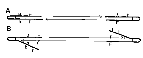
Figure 2 depicts adapter design and attachment to a DNA fragment, wherein
genomic DNA is represented by a solid black bar, 1 represents the free primer,
B
represents the bound primer, and f and b represent their complements,
respectively.
Figure 3 depicts ampliot production on the chip surface. A) After melting of
the adapter-captured genomic DNA, one strand is captured onto the surface of
the
slide by hybridization to bound primer B. Polymerase extension from primer B
produces a double stranded molecule. B) The template strand is removed by
heating
and washing of the slide and a free primer F is introduced and extended along
the
fixed strand. C) Continuous strand
CA 02930400 2016-05-17
9
displacement amplification by F results in the production of a strand that can
move to nearby
primer B hybridization sites. D) Displaced strands serve as template for
extension from new
primer B sites.
Figure 4 depicts ampliot production using an RNA intermediate. T7 represents
the
T7 phage RNA polymerase promoter. A) The single stranded adapter region is
hybridized to
the bound primer B and extended to form a second strand by DNA polymerase
resulting in
the formation of a double stranded T7 promoter. B) T7 RNA polymerase produces
an RNA
copy (dashed line). C) The RNA then binds to a nearby primer B and cDNA is
produced by
reverse transcriptase. Duplex RNA is then destroyed by RNase H.
Figure 5 depicts a schematic of the invader-mediated isothermal DNA
amplification
process.
Figure 6 depicts the random array sequencing by hybridization (rSBH) process.
From the top down: (a) A CCD camera is positioned above the reaction platform
and a lens
is used to magnify and focus on 1 1.Lm2 areas from the platform onto
individual pixels of the
CCD camera. (b) The array (-3 mm x 3 mm) consists of 1 million or more 1 um2
areas,
which act as virtual reaction wells (each corresponding to individual pixels
of the CCD
camera). Each pixel corresponds to the same location on the substrate. In a
series of
reactions in time, one CCD pixel can combine the data for several reactions,
thereby creating
the virtual reaction well. DNA samples are randomly digested and arrayed onto
the surface
of the reaction platform at an average concentration of one fragment per
pixel. (c) The array
is subjected to rSBH combinatorial ligation using one of several informational
probe pools.
The signals from each pixel are recorded. (d) Probes from the first pool are
removed and the
array is subjected to a second round of rSBH combinatorial ligation using a
different pool or
probes. (e) Insert showing molecular details of fluorescence resonance energy
transfer
(FRET) signal generation due to ligation of two adjacent and complementary
probes whose
compliment is represented by the target.
Figure 7 depicts the rSBH reaction. The total internal reflection microscopy
(TJRM)
detection system creates an evanescent field in which enhanced excitation
occurs only in the
region immediately above the glass substrate. FRET signals are generated when
probes are
hybridized to the arrayed target and subsequently ligated, thus positioning
the FRET pair
within the evanescent field. Unligated probes do not give rise to detectable
signals, whether
they are free in solution or transiently hybridized to the target. Hence, the
evanescent field
CA 02930400 2016-05-17
of the TERM system provided both intense signals within a desired plane while
reducing
background noise from nnreacted probes.
Figure 8 depicts sequence assembly. In general, in the SBH process, the target
sequence is assembled using overlapping positive probes. In this process each
base is read
5 several times (Le. 10 times with 10-mer probes, etc.) which assures very
high accuracy even
if some probes are not correctly scored.
Figure 9 depicts a schematic of a microfluidics device for the rSBH process.
The
device integrates DNA preparation, formation of random single molecule DNA
arrays,
combinatorial pool mixing, and cyclic loading and washing of the reaction
chamber. When
10 a sample tube is attached to the chip, a series of reactions is
performed with pre-loaded
reagents to isolate and fragment DNA, which is randomly attached to the array
surface at a
density of approximately one molecule per pixel. A microfluidics device is
then used to mix
two probe pools from 5' and 3' sets of informative probe pools (IIIPs) with
the reaction
solution. One set of probe pools is labeled with a FRET donor, the other with
a FRET
acceptor. Mixed pools containing DNA ligase are then traniferred to a reaction
chamber
above the single molecule DNA. Detectable ligation events occur when two
probes (one
from each pool) hybridize to adjacent complementary sequences of a target DNA
molecule
within a narrow zone of reflectance (-100 nm) above the array surface.
Ligation of 5' and 3'
probes within the zone of reflectance results in a FRET signal that is
detected and scored by
an ultra-sensitive CCD camera. After ligation events are scored, each pool mix
is removed
by a washing solution and a second pair of pools from the same sets of IPPs
preloaded on the
microfluidics chip is combined and introduced to the reaction chamber. By
combining all
possible pools within the two sets of1PPs, each target molecule in the array
is scored for the
presence/absence of every possible combination of probe sequences that exists
within the
two probe sets.
Figure 10 depicts the basic optics and. light path for the TIRM instrument.
(a)
Depiction of the traditional substrate positioned on top of the prisms and the
light path that
gives rise to an evanescent field. (b) and (c) show the use of galvanometers
to control the
light path from the laser to the prism assembly.
Figure 11 depicts a schematic representation of rSBH components and processes
showing the components of the rSBH instrument and stepwise description of the
experimental process. Sample is collected and prepared (Steps 1 and 2)
independent of the
instrument. Resultant crude sample preparation is further processed for rSBH
array
CA 02930400 2016-05-17
11
formation (Step 3) by the sample integration module (Component A). Targets are
subsequently arrayed on the substrate module within the reaction cartridge
(Component B).
Samples are subjected to SBH ligation assay (Step 4) using SBH probes
delivered by the
probe module (Component C). Resultant raw data is processed, resulting in
assembly of
sequence data (Step 5) and interpretive analysis (Step 6).
Figure 12 shows the full-match ligation signal for 4 spotted targets. Four
different
targets were spotted at 7 different concentrations ranging from 1 to 90 M.
Ligation probe
concentration (5' probe: 3' probe ratio is 1:1) were varied from 0.1 to 1
pmole/20 I.
Figure 13 shows a graphic representation of the spotted target serving as a
capture
probe for another target. The ligation signal was measured when the slide was
directly
hybridized/ligated with Tg2-5' probe and Tgt2-3' probe (circles) and when the
slide was pre-
hybridized with target Tgt2-Tgtl-rc and then ligated with Tgt2-5' probe and
Tgt2-3' probe
(squares).
6. DETAILED DESCRIPTION OF THE PREFERRED EMBODIMENTS
The present invention provides single molecule DNA analysis methods and
devices
to rapidly and accurately sequence any long DNA fragment, mixture of
fragments, entire
genes, mixture of genes, mixtures of naRNAs, long segments of chromosomes,
entire
chromosomes, mixtures of chromosomes, entire genome, or mixtures of genomes.
The
method of the invention allows detection of pathogens present in complex
biological
samples at the single organism level and identification of virulence
controlling genes. The
method of the invention combines hybridization and especially sequencing by
hybridization
(SBH) technology with total internal reflection microscopy (T1RM) or other
sensitive optical
methods using fluorescence, nanoparticles, or electrical methods. The present
invention also
provides a sample arraying technology which creates virtual reaction chambers
that are
associated with individual pixels of an ultra-sensitive charge-coupled device
(CCD) camera.
Using informative pools of complete/universal sets of fluorescent-labeled
oligonucleotide
probes and combinatorial ligation process, arrayed genomes are repeatedly
interrogated in
order to decipher their sequences. Bioinformatics algorithms (co-owned, co-
pending U.S.
.30 Patent Application Serial No. 09/874,772; Dnnanac et al., Science
260:1649-1652 (1993);
Drmanac et al., Nat. Biotech. 16:54-58 (1998); Drmanac et al., "Sequencing and
Fingerprinting DNA by Hybridization with Oligonucleotide Probes," In:
Encyclopedia of
Analytical Chenzistly, pp. 5232-5237 (2000); Drraanac et al., "Sequencing by
Hybridization
CA 02930400 2016-05-17
12
(SBH): Advantages, Achievements, and Opportunities," In: Advances in
Biochemical
Engineering/Biotechnology: Chip Technolozv, Hoheisel, J. (Ed.), Vol. 76, pp.
75-98 (2002),
are used to transform
informative fluorescent signals into assembled sequence data. The device can
sequence over
100 mega bases of DNA per hour (30,000 bases/sec) using a single compact
instrument
located in a diagnostic laboratory or small mobile laboratory. Trace
quantities of pathogen
DNA can be detected, identified and sequenced within complex biological
samples using the
method of the present invention due to the large capacity of random single
molecule arrays.
Thus, random array SBH (rSBH) provides the necessary technology to allow DNA
sequencing to play an important role in the defense against biowarfare agents,
in addition to
other sequencing applications.
The present invention provides a single DNA molecule analysis method to
rapidly
and accurately detect and identify any pathogen in complex biological mixtures
of pathogen,
host, and environmental DNA, and analyze any DNA in general, including
individual human
DNA. The method of the invention allows detection of pathogens present in the
sample at
the single organism level and identification of all virulence controlling
genes. The method
of the invention applies the process of combinatorial hybridization/ligation
of small sets of
universal informative probe pools (EPPs) to random single molecule arrays
directly or after
in situ amplification of individual arrayed molecules about 10- or 100-, or
1000- or 10,000-
fold.
In a typical test, millions of randomly arrayed single DNA naolecules obtained
from a
sanaple are hybridized with pairs of DI3Ps representing universal libraries of
all possible probe
sequences 8 to 10 bases in length. When two probes hybridize to adjacent
complementary
sequences in target DNAs, they are ligated to create a positive score for that
target molecule
and the accumulated set of such scores is compiled to assemble the target
sequence from
overlapping probe sequences.
In another embodiment of the present invention, the signature or sequence of
individual targets can be used to assemble longer sequences of entire genes or
genomes. In
addition, by counting how many times the same molecule or segments from the
same gene
occur in the array, quantification of gene expression or pathogen DNA may be
obtained and
such data may be combined with the obtained sequences.
SBH is a well developed technology that may be practiced by a number of
methods
known to those skilled in the art. Specifically, the techniques related to
sequencing by
CA 02930400 2016-05-17
13
hybridization discussed in the following documents,
Bains and Smith, J Theor. Biol. 135:303-307 (1988); Beaucage and
Caruthers, Tetrahedron Lett. 22:1859-1862 (1981); Broude et aL, Proc. Natl.
Acad. Sci. USA
91:3072-3076 (1994); Breslauer et al., Proc. Natl. Acad. Sci. USA 83:3746-3750
(1986);
Doty et al., Proc. Natl. Acad. Sci. USA 46:461-466 (1990); Chee et aL, Science
274:610-614
(1996); Cheng et al., Nat. Biotechnol. 16:541-546 (1998); Dian7ani et aL,
Genonzics 11:48-
53 (1991); PCT International Patent Application Serial No. WO 95/09248 to
Drmanac; PCT
International Patent Application Serial No. WO 96/17957 to Drmanac; PCT
International
Patent Application Serial No. WO 98/31836 to Drmanac; PCT International Patent
Application Serial No. WO 99/09217 to Drmanac et al.; PCT International Patent
Application Serial No. W000/40758 to Drmanac et al.; PCT International Patent
Application Serial No. WO 56937;
Drmanac and Crkvenjakov, Scientia Yugoslaviea 16:99-107
(1990); Dlluanac and Crkvenjakov, Intl. J. Genonze Res. 1:59-79 (1992);
Drmanac and
Drmanac, Meth. Enzymology 303:165-178 (1999); Drmanac et al.,U.S. Patent No.
5,202,231; Drmanac et aL, NucL Acids Res. 14:4691-4692 (1986); Drmanac et al.,
Genomics
4:114-128 (1989); Drmanac et al., J. BiomoL Struct. Dyn. 8:1085-1102 (1991);
Drmanac et
al., "Partial Sequencing by Hybridization: concept and Applications in Genome
Analysis,"
in: The First International Conference on Electrophoresis, Supercomputing and
the Human
Genome, pp. 60-74, World Scientific, Singapore, Malaysia (1991); Drmanac et
al.,
Proceedings of the First Intl. Conf Electrophoresis, Supercomputing and the
Hunzan
Genome, Cantor et al. eds, World Scientific Pub. Co., Singapore, 47-59 (1991);
Drmanac et
NucL Acids Res. 19:5839-5842 (1991); Drmanac et al., Electrophoresis 13:566-
573
(1992); Drmanac et aL, Science 260:1649-1652 (1993); Drmanac et aL, DNA and
Cell Biol.
9:527-534 (1994); Drmanac et aL, Genomics 37:29-40 (1996); Drmanac et aL,
Nature
Biotechnology 16:54-58 (1998); Gunderson et rd., Genome Res. 8:1142-1153
(1998); Hacia
et al., Nature Genetics 14:441-447 (1996); Hacia et al., Genome Res. 8:1245-
1258 (1998);
Hoheisel et aL, MoL Gen. 220:903-14:125-132 (1991); Hoheisel et al., Cell
73:109-120
(1993); Holey et aL, Science 147:1462-1465 (1965); Housby and Southern, NucL
Acids Res.
26:4259-4266 (1998); Hunkapillar et aL, Science 254:59-63 (1991); Khrapko,
FEBS Lett.
256:118-122 (1989); Kozal et aL, Nature Medicine 7:753-759 (1996); Labat and
Drmanac,
"Simulations of Ordering and Sequence Reconstruction of Random DNA Clones
Hybridized
with a Small Number of Oligomer Probes," in: The Second International
Conference on
CA 02930400 2016-05-17
14
Electrophoresis, Supercomputing and the Human Genome, pp. 555-565, World
Scientific,
Singapore, Malaysia (1992); Lehrach et al., Genome Analysis: Genetic and
Physical
Mapping 1:39-81 (1990), Cold Spring Harbor Laboratory Press; Lysov et al.,
DokL Akad.
Nauk. SSSR 303:1508-1511 (1988); Lockhart et al., Nat. Biotechnol.
14:16'7501680 (1996);
Maxam and Gilbert, Proc. Natl. Acad. Sci. USA 74:560-564 (1977); Meier et al.,
NucL Acids
Res. 26:2216-2223 (1998); Michiels et al., CABIOS 3:203-210 (1987);
Milosavljevic et al.,
Genome Res. 6:132-141 (1996); Milosavljevic et al., Genomics 37:77-86 (1996);
Nikiforov
et al., NucL Acids Res. 22:4167-4175 (1994); Pevzner and Lipschutz, "Towards
DNA
Sequencing Chips," in: Mathematical Foundations of Computer Science (1994);
Poustka and
Lehrach, Trends Genet. 2: 174-179 (1986); Privara et al., Eds., pp. 143-158,
The
Proceedings of the 19th International Symposium, MFCS '94, Kosice, Slovakia,
Springer-
Verlag, Berlin (1995); Saild et al., Proc. Natl. Acad. Sci. USA 86:6230-6234
(1989); Sanger
et al., Proc. Natl. Acad. Sci. USA 74:5463-5467 (1977); Scholler et al., NucL
Acids Res.
23:3842-3849 (1995); PCT International Application Serial No. WO 89/10977 to
Southern;
U.S. Patent No. 5,700,637 to Southern; Southern et al., Genomics 13:1008-1017
(1992);
Strezoska et al., Proc. Natl. Acad. Sci. USA 88:10089-10093 (1991); Sugimoto
et al., NucL
Acid Res. 24:4501-4505 (1996); Wallace et al., NucL Acids Res. 6:3543-3557
(1979); Wang
et al., Science 280:1077-1082 (1998); Wetmur, Grit. Rev. Biochem. MoL Biol.
26:227-259
(1991).
Advantages of rSBH:
rSBH minimizes or eliminates target-target blocking interactions between two
target
DNA molecules that are attached at an appropriate distance. The low complexity
of DNA
sequence (between 200-2000 bases) per spot reduces the likelihood of inverse
repeats that
can block each other. Palindromes and hairpin arms are separated in some
fragments with
one cut per every 20 bases of source DNA on average and attach to non-
complementary
primer DNA. False positives are minimized because overlapped fragments have
different
repeated and/or strong mismatch sequences. Probe-probe ligation products are
removable by
washing. The combination of hybridization/ligation specificity and
differential full-
match/mismatch stability for the 11-13-mer probes made by ligation has the
potential for
producing more accurate data. rSBH provides an efficient method of using three-
probe
ligation in solution, including analysis of short DNA. Pools of patterned
probes can be
efficiently used on both probe components to provide more informative data.
Another
advantage is that very low amounts of source DNA are required. The need for
standard
CA 02930400 2016-05-17
probe-spot array preparation is eliminated, thereby reducing cost. rSBH
provides for
multiplex sequencing of up to 1000 samples tagged with different primers or
adapters. In
addition, the invention provides for detection of a single variant in a pool
of up to one
million individual samples. Heterozygotes can be detected by counting two
variants. The
5 invention provides for 10- to 100,000-fold more information per surface
than the standard
arrays.
6.1 PREPARATION AND LABELING OF POLYNUCLEOTIDES
The practice of the instant invention employs a variety of polynucleotides.
Typically
10 some of the polynucleotides are detectably labeled. Species of
polynucleotides used in the
practice of the invention include target nucleic acids and probes.
The term "probe" refers to a relatively short polynucleotide, preferably DNA.
Probes
are preferably shorter than the target nucleic acid by at least one base, and
more preferably
they are 25 bases or fewer in length, still more preferably 20 bases or fewer
in length. Of
15 course, the optimal length of a probe will depend on the length of the
target nucleic acid
being analyzed. In de novo sequencing (no reference sequence used) for a
target nucleic acid
composed of about 100 or fewer bases, the probes are preferably at least 7-
mers; for a target
nucleic acid of about 100-200 bases, the probes are preferably at least 8-
mers; for a target
nucleic acid of about 200-400 bases, the probes are preferably at least 9-
mers; for a target
nucleic acid of about 400-800 bases, the probes are preferably at least 10-
mers; for a target
nucleic acid of about 800-1600 bases, the probes are at least 11-mers; for a
target nucleic
acid of about 1600-3200 bases, the probes are preferably at least 12-mers; for
a target nucleic
acid of about 3200-6400 bases, the probes are preferably at least 13-mers; and
for a target
nucleic acid of about 6400-12,800 bases, the probes are preferably at least 14-
mers. For
every additional two-fold increase in the length of the target nucleic acid,
the optimal probe
length is one additional base.
Those of skill in the art will recogni7e that for SBH applications utilizing
ligated
probes, the above-delineated probe lengths are post-ligation. Probes are
normally single
stranded, although double-stranded probes may be used in some applications.
While typically the probes will be composed of naturally-occurring bases and
native
phosphodiester backbones, they need not be. For example, the probes may be
composed of
one or more modified bases, such as 7-deazaguanosine or the universal "M"
base, or one or
more modified backbone interlinkages, such as a phosphorothioate. The only
requirement is
CA 02930400 2016-05-17
16
that the probes be able to hybridi7e to the target nucleic acid. A wide
variety of modified
bases and backbone interlinkages that can be used in conjunction with the
present invention
are known, and will be apparent to those of skill in the art.
The length of the probes described above refers to the length of the
informational
content of the probes, not necessarily the actual physical length of the
probes. The probes
used in SBH frequently contain degenerate ends that do not contribute to the
information
content of the probes. For example, SBH applications frequently use mixtures
of probes of
the formula NxByNz, wherein N represents any of the four bases and varies for
the
polynucleotides in a given mixture, B represents any of the four bases but is
the same for
each of the polynucleotides in a given mixture, and x, y, and z are integers.
Typically, x and
z are independent integers between 0 and 5 and y is an integer between 4 and
20. The
number of known bases By defines the "information content" of the
polynucleotide, since the
degenerate ends do not contribute to the information content of the probes.
Linear arrays
comprising such mixtures of immobilized polynucleotides are useful in, for
example,
sequencing by hybridization. Hybridization discrimination of mismatches in
these
degenerate probe mixtures refers only to the length.of the informational
content, not the full
physical length.
Probes for use in the instant invention may be prepared by techniques well
known in
the art, for example by automated synthesis using an Applied Biosystems
synthesizer.
Alternatively, probes may be prepared using Genosys Biotechnologies Inc.
methods using
stacks of porous Teflon wafers. For purposes of this invention, the source of
oligonucleotide
probes used is not critical, and one skilled in the art will recognin that
oligonucleotides
prepared using other methods currently known or later developed will also
suffice.
The term "target nucleic acid" refers to a polynucleotide, or some portion of
a
polynucleotide, for which sequence information is desired, typically the
polynucleotide that
is sequenced in the SBH assay. The target nucleic acid can be any number of
nucleotides in
length, depending on the length of the probes, but is typically on the order
of 100, 200, 400,
800, 1600, 3200, 6400, or even more nucleotides in length. A sample typically
may have
more than 100, more than 1000, more than 10,000, more than 100,000, more than
one
million, or more than 10 million targets. The target nucleic acid may be
composed of
ribonucleotides, deoxyribonucleotides, or mixtures thereof. Typically, the
target nucleic acid
is a DNA. While the target nucleic acid can be double-stranded, it is
preferably single
stranded. Moreover, the target nucleic acid can be obtained from virtually any
source.
CA 02930400 2016-05-17
17
Depending on its length, it is preferably sheared into fragments of the above-
delineated sizes
prior to using an SBH assay. Like the probes, the target nucleic acid can be
composed of
one or more modified bases or backbone interlinkages.
The target nucleic acid may be obtained from any appropriate source, such as
cDNAs, genomic DNA, chromosomal DNA, microdissected chromosomal bands, cosmid
or
yeast artificial chromosome (YAC) inserts, and RNA, including mRNA without any
amplification steps. For example, Sambrook et al. Molecular Cloning: A
Laboratory
Manual, Cold Spring Harbor Press, NY (1989)
describes three protocols for the isolation of high. molecular weight DNA from
mammalian cells (p. 9.14-9.23).
The polynucleotides would then typically be fragmented by any of the methods
known to those,of skill in the art including, for example, using restriction
enzymes as
described at 9.24-9.28 of Sambrook et al. (1989), shearing by ultrasound, and
NaOH
treatment. A particularly suitable method for fragmenting DNA utilizes the two
base
recognition endonuclease, CviJT, described by Fitzgerald et al., Nucl. Acids
Res. 20:3753.-
3762 (1992).
In a preferred embodiment, the target nucleic acids are prepared so that they
cannot
be ligated to each other, for example by treating the fragmented nucleic acids
obtained by
enzyme digestion or physical shearing with a phosphatase (i.e. calf intestinal
phosphatase).
Alternatively, nonligatable fragments of the sample nucleic acid imay be
obtained by using
random primers (Le. N5-N9, wherein N=A, G, T, or C), which have no phosphate
at their 5'-
ends, in a Sanger-dideoxy sequencing reaction with the sample nucleic acid.
In most cases it is important to denature the DNA to yield single stranded
pieces
available for hybridization. This may be achieved by incubating the DNA
solution for 2-5
minutes at 80-90 C. The solution is then cooled quickly to 2 C to prevent
renaturation of
the DNA fragments before they are contacted with the probes.
Probes and/or target nucleic acids may be detectably labeled. Virtually any
label that
produces a detectable signal and that is capable of being immobilized on a
substrate or
attached to a polynucleotide can be used in conjunction with the arrays of the
invention.
Preferably, the signal produced is amenable to quantification. Suitable labels
include, by
way of example and not limitation, radioisotopes, fluorophores, chromophores,
chemiluminescent moieties, etc.
CA 02930400 2016-05-17
18
Due to their ease of detection, polynucleotides labeled with fluorophores are
preferred. Fluorophores suitable for labeling polynucleotides are described,
for example, in
the Molecular Probes catalog (Molecular Probes, Inc., Eugene, OR), and the
references cited
therein. Methods for attaching fiuorophore labels to polynucleotides are well
known, and
can be found, for example, in Goodchild, Bioconjug. Chem. 1:165-187 (1990).
A preferred fluorophore label is Cy5 dye, which is
available from Am.ersham Biosciences.
Alternatively, the probes or targets may be labeled by any other technique
known in
the art. Preferred techniques include direct chemical labeling methods and
enzymatic
labeling methods, such as kinasing and nick-translation. Labeled probes could
readily be
purchased from a variety of commercial sources, including GENSET, rather than
synthesized.
In general, the label can be attached to any part of the probe or target
polynucleotide,
including the free terminus of one or more of the bases. In preferred
embodiments, the label
is attached to a terminus of the polynucleotide. The label, when attached to a
solid support
by means of a polynucleotide, must be located such that it can be released
from the solid
support by cleavage with a mismatch-specific endonuclease, as described in co-
owned, co-
pending U.S. Patent Publication No. 2002/0048760.
Preferably, the position of the label will not interfere with hybridization,
ligation, cleavage or other post-hybridization modifications of the labeled
polynucleotide.
Some embodiments of the invention employ multiplexing, i.e. the use of a
plurality
of distinguishable labels (such as different fluorophores). Multiplexing
allows the
simultaneous detection of a plurality of sequences in one hybridization
reaction. For
example, a multiplex of four colors reduces the number of hybridizations
required by an
additional factor of four.
Other embodiments employ the use of informative pools of probes to reduce the
redundancy normally found in SBH protocols, thereby reducing the number of
hybridization
reactions needed to unambiguously determine a target DNA sequence. Informative
pools of
probes and methods of using the same can be found in co-owned U.S. Patent
Serial No. 6,864,052.
CA 02930400 2016-05-17
19
6.2 ATTACHMENT OF POLYNUCLEOTIDES TO A SOLED SUBSTRATE
Some embodiments of the instant invention require polynucleotides, for example
target DNA fragments, to be attached to a solid substrate. In preferred
embodiments, the
appropriate DNA samples are detectably labeled and randomly attached to a
solid substrate
at a concentration of 1 fragment per pixel.
The nature and geometry of the solid substrate will depend upon a variety of
factors,
including, among others, the type of array and the mode of attachment (i.e.
covalent or non-
covalent). Generally, the substrate can be composed of any material which will
permit
immobilization of the polynucleotide and which will not melt or otherwise
substantially
degrade under the conditions used to hybridize and/or denature nucleic acids.
In addition,
where covalent immobilintion is contemplated, the substrate should be
activatable with
reactive groups capable of forming a covalent bond with the polynucleotide to
be
immobilized.
A number of materials suitable for use as substrates in the instant invention
have
been described in the art. In preferred embodiments, the substrate is made of
an optically
clear substance, such as glass slides. Other exemplary suitable materials
include, for
example, acrylic, styrene-methyl methacrylate copolymers, ethylene/acrylic
acid,
acrylonitrile-butadiene-styrene (ABS), ABS/polycarbonate, ABS/polysulfone,
ABS/polyvinyl chloride, ethylene propylene, ethylene vinyl acetate (EVA),
nitrocellulose,
nylons (including nylon 6, nylon 6/6, nylon 6/6-6, nylon 6/10, nylon 6/12,
nylon 11, and
nylon 12), polycarylonitrile (PAN), polyacrylate, polycarbonate, polybutylene
terephthalate
(PBT), polyethylene terephthalate (PET), polyethylene (including low density,
linear low
density, high density, cross-linked and ultra-high molecular weight grades),
polypropylene
homopolymer, polypropylene copolymers, polystyrene (including general purpose
and high
impact grades), polytetrafluoroethylene (PTFE), fluorinated ethylene-propylene
(FEP),
ethylene-tetrafluoroethylene (ETFE), perfluoroalkoxyethylene (PFA), polyvinyl
fluoride
(PVF), polyvinylidene fluoride (PVDF), polychlorotrifluoroethylene (PCTFE),
polyethylene-chlorotrifluoroethylene (ECTFE), polyvinyl alcohol (PVA), silicon
styrene-
acrylonitrile (SAN), styrene maleic anhydride (SMA), metal oxides and glass.
In general, polynucleotide fragments may be bound to a support through
appropriate
reactive groups. Such groups are well known in the art and include, for
example, amino (-
NH2), hydroxyl (-OH), or carboxyl (-COOH) groups. Support-bound polynucleotide
fragments may be prepared by any of the methods known to those of skill in the
art using
CA 02930400 2016-05-17
any suitable support such as glass. Immobilization can be achieved by many
methods,
including, for exarnple, using passive adsorption (Inouye and Hondo, J. Clin.
Microbiol.
28:1469-1472 (1990) using UV
light
(Dahlen et al., Mal. Cell Probes 1:159-168 (1987),
5 or by
covalent binding of base-modified DNA (Keller, et al., Anal. Biochem.
170:441-451 (1988), Keller et al., Anal. Biochem. 177:392-395 (1989),
or by formation of amide groups between
the probe and the support (Zhang et al., NucL Acids Res. 19:3929-3933 (1991).
10 It is
contemplated that a further suitable method for use with the present invention
is
that described in PCT Patent Application WO 90/03382 (to Southern et al.).
This method of preparing a polynucleotide fragment bound to a support
involves attaching a nucleoside 3'-reagent through the phosphate group by a
covalent
phosphodiester link to aliphatic hydroxyl groups carried by the support. The
oligonucleotide
15 is then synthesized on the supported nucleoside and protecting groups
removed from the
synthetic oligonucleotide chain under standard conditions that do not cleave
the
oligonucleotide from the support. Suitable reagents include nucleoside
phosphoramidite and
nucleoside hydrogen phosphorate.
Alternatively, addressable-laser-activated photodeprotection may be employed
in the
20 chemical synthesis of oligonucleotides directly on a glass surface, as
described by Fodor et
al., Science 251:767-773 (1991).
One particular way to prepare support-bound polynucleotide fragments is to
utilize
the light-generated synthesis described by Pease et al., Proc. Natl. Acad.
Sci. USA 91:5022-
5026 (1994). These
authors used current photolithographic
techniques to generate arrays of immobilized oligonucleotide probes, i.e. DNA
chips. These
methods, in which light is used to direct the synthesis of oligonucleotide
probes in high-
density, miniaturized arrays, utilize photolabile 5'-protected N-acyl-
deoxynucleoside
phosphoramidites, surface linker chemistry and versatile combinatorial
synthesis strategies.
A matrix of 256 spatially defined oligonucleotide probes may be generated in
this manner
and then used in SBH sequencing, as described herein.
In a preferred embodiment, the DNA fragments of the invention are connected to
the
solid substrate by means of a linker moiety. The linker may be comprised of
atoms capable
of forming at least two covalent bonds, such as carbon, silicon, oxygen,
sulfur, phosphorous,
CA 02930400 2016-05-17
21
and the like, or may be comprised of molecules capable of forming at least two
covalent
bonds, such as sugar-phosphate groups, amino acids, peptides, nucleosides,
nucleotides,
sugars, carbohydrates, aromatic rings, hydrocarbon rings, linear and branched
hydrocarbons,
and the like. In a particularly preferred embodiment of the invention, the
linker moiety is
composed of alkylene glycol moieties. In preferred embodiments, a detectable
label is
attached to the DNA fragment (i.e. target DNA).
6.3 FORMATION OF DETECTABLY LABELED DUPLEXES ON A SOLID
SUPPORT
In one preferred embodiment of the invention, a labeled probe is bound by
means of
complementary base-pairing interactions to a detectably labeled target nucleic
acid that is
itself attached to a solid substrate as part of a polynucleotide array,
thereby forming a
duplex. In another preferred embodiment, a labeled probe is covalently
attached, i.e. ligated,
to another probe that is bound by means of complementary base-pairing
interactions to a
target nucleic acid that is itself attached to a solid substrate as part of a
spatially-addressable
polynucleotide array, if the two probes hybridize to a target nucleic acid in
a contiguous
fashion.
As used herein, nucleotide bases "match" or are "complementary" if they form a
stable duplex or binding pair under specified conditions. The specificity of
one base for
another is dictated by the availability and orientation of hydrogen bond
donors and acceptors
on the bases. For example, under conditions commonly employed in hybridization
assays,
adenine ("A") matches thymine ("T"), but not guanine ("G") or cytosine ("C").
Similarly, G
matches C, but not A or T. Other bases which interact in less specific
fashion, such as
inosine or the Universal Base ("M" base, Nichols et al., Nature 369:492-493
(1994)
or other modified bases, for example methylated
bases, are complementary to those bases for which they form a stable duplex
under specified
conditions. Nucleotide bases which are not complementary to one another are
termed
"raismatches."
A pair of polynucleotides, e.g. a probe and a target nucleic acid, are termed
"complementary" or a "match" if, under specified conditions, the nucleic acids
hybridize to
one another in an interaction mediated by the pairing of complementary
nucleotide bases,
thereby forming a duplex. A duplex formed between two polynucleotides may
include one
or more base mismatches. Such a duplex is termed a "mismatched duplex" or
heteroduplex.
CA 02930400 2016-05-17
22
The less stringent the hybridization conditions are, the more likely it is
that mismatches will
be tolerated and relatively stable mismatched duplexes can be formed.
A subset of matched polynucleotides, termed "perfectly complementary" or
"perfectly matched" polynucleotides, is composed of pairs of polynucleotides
containing
continuous sequences of bases that are complementary to one another and
wherein there are
no mismatches (i.e. absent any surrounding sequence effects, the duplex formed
has the
maximal binding energy for the particular nucleic acid sequences). "Perfectly
complementary" and "perfect match" are also meant to encompass polynucleotides
and
duplexes which have analogs or modified nucleotides. A "perfect match" for an
analog or
modified nucleotide is judged according to a "perfect match rule" selected for
that analog or
modified nucleotide (e.g. the binding pair that has maximal binding energy for
a particular
analog or modified nucleotide).
In the case where a pool of probes with degenerate ends of the type NxByNz is
used,
as described above, a perfect match encompasses any duplex where the
information content
regions, i.e. the By regions, of the probes are perfectly matched.
Discrimination against
mismatches in the N regions will not affect the results of a hybridization
experiment, since
such mismatches do not interfere with the information derived from the
experiment.
In a particularly preferred embodiment of the invention, a polynucleotide
array is
provided wherein target DNA fragments are provided on a solid substrate under
conditions
which permit them to hybridize with at least one set of detectably labeled
oligonucleotide
probes provided in solution. Both within the sets and between the sets the
probes may be of
the same length or of different lengths. Guidelines for determining
appropriate hybridization
conditions can be found in papers such as Drmanac et al., (1990), Khrapko et
al. (1991),
Broude et al., (1994) (all cited supra) and WO 98/31836.
These articles teach the ranges of hybridization temperatures,
buffers, and washing steps that are appropriate for use in the initial steps
of SBH. The probe
sets may be applied to the target nucleic acid separately or simultaneously.
Probes that hybridize to contiguous sites on the target nucleic acid are
covalently
attached to one another, or ligated. Ligation may be implemented by a chemical
ligating
agent (e.g. water-soluble carbodiimide or cyanogen bromide), by a ligase
enzyme, such as
the commercially available T4 DNA ligase, by stacking interactions, or by any
other means
of causing chemical bond formation between the adjacent probes. Guidelines for
determining appropriate conditions for ligation can be found in papers such as
co-owned
CA 02930400 2016-05-17
23
U.S. Patent No. 6,864,052.
6.4 RANDOM ARRAY SBH (rSBH)
The method of the present invention uses random array SBH (rSBH) which extends
the combinatorial ligation process to single molecule arrays, greatly
increasing the
sensitivity and power of the method of the invention. rSBH relies on
successive
interrogations of randomly arrayed DNA fragments by informative pools of
labeled
oligonucleotides. In the method of the present invention, complex DNA mixtures
to be
sequenced are displayed on an optically clear surface within the focal plane
of a total internal
fluorescence reflection microscopy (TIRM) platform and continuously monitored
using an
ultra-sensitive mega pixel CCD camera. DNA fragments are arranged at a
concentration of
approximately 1 to 3 molecules per square micron, an area corresponding to a
single CCD
pixel. TIRM is used to visualize focal and close contacts between the object
being studied
and the surface to which it is attached. In TIRM, the evanescent field from an
internally
reflected excitation source selectively excites fluorescent molecules at or
near a surface,
resulting in very low background scattered light and good signal-to-background
contrast.
The background and its associated noise can be made low enough to detect
single
fluorescent molecules under ambient conditions. (see Abney et al. Biophys. J.
61:542-552
(1992); Ambrose et al., Cytometry 36:224-231 (1999); Axelrod, Traffic 2:764-
774 (2001);
Fang and Tan, "Single Molecule Imaging and Interaction Study Using Evanescent
Wave
Excitation," American Biotechnology Laboratory (ABL) Application Note, April
2000;
Kawano and Enders, "Total Internal Reflection Fluorescence Microscopy,"
American
Biotechnology Laboratory Application (ABL) Application Note, December 1999;
Reichert
and Truskey, J Cell Sci. 96 (Pt. 2):219-230 (1990).
Using microfluidic technology, pairs of probe pools labeled with donor and
acceptor
fluorophores are mixed with DNA ligase and presented to the random array. When
probes
hybridize to adjancent sites on a target fragment, they are ligated together
generating a
fluorescence resonance energy transfer (FRET) signal. FRET is a distance-
dependent
(between 10-100 A) interaction between the electronic excited states of two
fluorescent
molecules in which excitation is transferred from a donor molecule to an
acceptor molecule
without emission of a photon (Didenko, Biotechniques 31:1106-1121 (2001); Ha,
Methods
CA 02930400 2016-05-17
24
25:78-86 (2001); Klostermeier and Millar, Biopolymers 61:159-179 (2001-2002).
These signals are detected by
the CCD camera indicating a matching sequence string within that fragment.
Once the
signals from the first pool are detected, probes are removed and successive
cycles are used to
test different probe combinations. The entire sequence of each DNA fragment is
compiled
based on fluorescent signals generated by hundreds of independent
hybridization/ligation
events.
Although only one detectable color will suffice, multiple colors will increase
multiplexing of the combinatorics and improve the efficiency of the system.
The current
state of the art suggests that four colors can be used simultaneously. In
addition to
traditional direct fluorescence strategies, FRET-based systems, time-resolved
systems and
time-resolved FRET signaling systems will also be used (Didenko,
Biotecluziques 31:1106-
1121 (2001). New
custom chemistries, such
as quantum dot enhanced triple FRET systems may also be used. Overcoming a
weak signal
may be overcome using dendranaer technologies and related signal amplification
technologies.
U-nlike traditional hybridization processes, the method of the present
invention relies
on a synergistic interaction of hybridization and ligation, in which short
probes from two
pools are ligated together to generate longer probes with far more
informational power. For
example, two sets of 1024 five-mer oligonucleotides can be combined to detect
over a
million possible 10-mer sequence strings. The use of informative probe pools
(in which all
probes share a common label) greatly simplifies the process, allowing millions
of potential
probe pairings to occur with only a few hundred pool combinations. Multiple
overlapping
probes reading consecutive bases allow an accurate determination of DNA
sequence from
the obtained hybridization pattern. The combinatorial ligation and informative
pools
technologies described above are augmented by extending their use to single
molecule
sequencing.
6.5 STRUCTURED RANDOM DNA PREPARATION
A. DNA Isolation and Initial Fragmentation
Cells are lysed and DNA is isolated using basic well-established protocols
(Sambrook et al., supra, 1999; Current Protocols in Molecular Biology, Ausubel
et al., eds.
John Wiley and Sons, Inc., NY, 1999,
CA 02930400 2016-05-17
or commercial kits [e.g. those available from QIAGEN (Valencia, CA) or
Promega (Madison, WI)]. Critical requirements are: 1) the DNA is free of DNA
processing
enzymes and contaminating salts; 2) the entire genome is equally represented;
and 3) the
DNA fragments are between ¨5,000 and ¨100,000 bp in length. No digestion of
the DNA is
5 required because shear forces created during lysis and extraction will
give rise to fragments
in the desired range. In another embodiment, shorter fragments (1-5 kb) can be
generated by
enzymatic fragmentation. The input genome number of 10-100 copies will ensure
overlap of
the entire genome and tolerates poor capture of targets on the array. A
further embodiment
provides for carrier, circular synthetic double-stranded DNA to be used in the
case of small
10 amounts of DNA.
B. DNA Normalization
In some embodiments, normalization of environmental samples may be necessary
to
reduce the DNA contribution of prevalent species to maximize the total number
of distinct
species that are sequenced per array. Because rSBH requires as few as 10
genome
15 equivalents, a thorough DNA nounalization or subtraction process can be
implemented.
Normalization can be accomplished using commonly utilized methods used for
normalizing
cDNA libraries during their production. DNA collected from the sample is
divided in two,
with one being often-fold greater mass than the other. The sample of greater
quantity is
biotinylated by terminal transferase and ddCTP and attached as a single
stranded DNA to a
20 streptavidin column or streptavidin-coated beads. Alternatively,
biotinylated random
primers may be employed to generate sequence for attachment to streptavidin.
Whole
genome amplification methodologies (Molecular Staging, Inc., New Haven, CT)
can also be
applied. The sample to be normalized is then hybridized to the attached
molecules and those
molecules that are over-represented in the sample are preferentially removed
from the
25 solution due to the greater number of binding sites. Several
hybridization/removal cycles
can be applied on the same sample to achieve full normalization. Another
embodiment
provides for efficient hybridization oflong double-stranded DNA fragments
without DNA
denaturation by generating short terminal regions of single-stranded DNA with
a timed
lambda exonuclease digestion.
Further embodiments provide for sequencing low abundance members that are
difficult to analyze by combining DNA normalization and rSBH. Normalization of
one
sample against another allows monitoring of changes in community structure and
identifying
new members as conditions change.
CA 02930400 2016-05-17
26
C. Secondary DNA Fragmentation and Adapter Attachment
The present invention provides for long DNA fragments generated by shear
forces to
be suspended in solution within a chamber located on the glass slide. The
concentration of
the DNA is adjusted such that the volume occupied by each fragment is in the
order of 50 x
50 x 50 Arn. The reaction chamber comprises a mix of restriction enzymes, T4
DNA ligase,
a strand displacing polymerase, and specially designed adapters. Partial
digestion of the
DNA by the restriction enzymes yields fragments with an average length of 250
bp with
uniform overhang sequences. T4 DNA ligase joins non-phosphorylated double-
stranded
adapters to the ends of the genomic fragments via complementary sticky-ends
resulting in a
stable structure of genomic insert with one adapter at each end, but with a
nick in one of the
strands where the ligase was unable to catalyze the formation of the
phosphodiester bond
(Figure 1). T4 DNA ligase is active in most restriction enzyme buffers but
requires the
addition of ATP and a molar excess of adapters relative to genomic DNA to
promote the
ligation of adapters at each end of the genomic molecule. Using non-
phosphorylated
adapters is important to prevent adapter-adapter ligation. Additionally, the
adapters contain
two primer-binding sites and are held in a hairpin structure by cross-linked
bases at the
hairpin end that prevents dissociation of the adapters during melting at
elevated
temperatures. Extension from the 3'-ends with a strand-displacing polymerase
such as Vent
or Bst results in the production of a DNA strand with adapter sequences at
both ends.
However, at one end the adapter will be maintained in the hairpin structure
that is useful to
prevent association of complementary sequences on the other end of the DNA
fragment.
The invention provides for random DNA arrays to sequence multiples of highly
similar samples (i.e. individual DNA from patients) in one assay by tagging
DNA fragments
of each sample prior to random array formation. One or both adapters used for
incorporation
of primer sequences at the end of the DNA fragments can have a tag cassette. A
different
tag cassette can be used for each sample. Afler attaching adapters (preferably
by ligation),
DNA of all samples is mixed and single random array is formed. After
sequencing of
fragments is completed, fragments that belong to each sample are recognized by
the assigned
tag sequence. Use of the tag approach allows efficient sequencing of a smaller
number of
targeted DNA regions from about 10-1000 samples on high capacity random arrays
having
up to about 10 million DNA fragments.
CA 02930400 2016-05-17
27
D. DNA Attachment and in situ Amplification
The adapter-linked genomic DNA is then localized with other fragments from the
original 5-100 kb fragment onto the glass slide by hybridization to an
oligonucleotide that is
complementary to the adapter sequences (primer B). After adapter ligation and
DNA
extension, the solution is heated to denature the molecules which, when in
contact with a
high concentration of primer oligonucleotides attached to the surface of the
slide, hybridizes
to these complementary sequences during the re-annealing phase. In an
alternative
embodiment, in situ amplification does not occur and the adapter is attached
to the support
and the DNA fragments are ligated. Most of the DNA structures that arise from
the one
parent molecule are localized to one section of the slide in the order of 50 x
50 gm; therefore
if 1000 molecules are generated from the restriction digest of one parent
molecule, each
fragment will occupy, on average, a 1-4 A1112 region. Such 1-4 Am2 region can
be observed
by a single pixel of a CCD camera and represents a virtual reaction well
within an array of
one million wells.
Lateral diffusion of DNA fragments more than 50 Ara across the slide surface
is
unlikely to be significant in the short period of time in a 50-100 gin thick
capillary chamber
that prevents liquid turbulence. In addition, high viscosity buffers or gel
can be used to
minimize diffusion. In yet a further embodiment, limited turbulence is needed
to spread
hundreds of short DNA fragments derived from single 5-100 kb molecules over a
50 x 50
gm surface. Note that the spreading does not have to be perfect because SBH
can analyze
mixtures of a few DNA fragments at the same pixel location. A few fractions of
the original
sample with more uniform fraDnent length (i.e. 5-10 kb, 10-20 kb, 20-40 kb, 40-
1000 kb)
may be prepared to achieve equal spacing between short fragments. Furthermore,
an electric
field can be used to pool short DNA fragments to the surface for attachment.
Partially
structured arrays with local mixing of short fragments are almost as efficient
as fully
structured arrays because no short fragments from any single, long fragment is
mixed with
short fragments generated from about 10,000 other long initial fragments.
A further embodiment of the invention provides for a ligation process that
attaches
two primer sequences to DNA fragments. This approach is based on targeting
single
stranded DNA produced by denaturation of double-stranded DNA fragments.
Because
single-stranded DNA has unique 5' and 3' ends, specific primer sequences can
be attached to
each end. Two specific adapters, each comprising two oligonucleotides, are
designed (see
Figure 2) that have specifically modified ends, wherein F and B represent
unbound, solution-
CA 02930400 2016-05-17
28
free primer (F) and surface-bound (B) primer sequences and f and b represent
sequences
complementary to these primer sequences (i.e. primer f is complementary to
primer F). The
only 3'-OH group, that is necessary for ligation to the DNA fragment is on
primer F, the
other oligonucleotides can have a dideoxy 3' end (dd) to prevent adapter-
adapter ligation. In
addition to the 5'-phosphate group (P) present on primer b, primer B may also
have a 5'-P
group to be used for degradation of this primer after adapter ligation to
expose primer b
sequence for hybridization to the surface-attached primer/capture probe B. To
allow for
adapter ligation to any DNA fragment generated from the source DNA by random
fragmentation, the oligonucleotides f and B have several (approximately 3-9,
preferably 5-7)
degenerate bases (Ns).
Although rSBH detection is designed for single molecule detection, some
embodiments amplify each DNA target in situ. The method of the invention
provides for
isothermal, exponential amplification within a micron-sized spot of localized
amplicon,
herein denoted as "ampliot" (defined to be an amplicon spot) (Figure 3). The
amplification
is achieved by use of a primer bound to the surface (primer B) and one free
primer in
solution (primer F). Primer B first hybridizes to the original target sequence
and is extended,
copying the target sequence. The non-attached strand is melted and washed away
and new
reagent components are added, including a DNA polymerase with strand-
displacing
properties (such as Bst DNA polymerase), dNTSs, and primer F. A continuous
amplification reaction is then used to synthesize a new strand and displace
the previous
synthesized complement.
The continuous exponential amplification reaction produces a displaced strand,
which contains complementary sequences to the capture array oligonucleotide
and thus, in
tum, is captured and used as a template for further amplification. This
process of strand
displacement requires that the primer is able to continuously initiate
polymerization. There
are several described strategies in the art, such as ICANTm technology (Takara
BioEurope,
Gennevilliers, France) a.nd SPIA technology (NuGEN, San Carlos, CA; U.S.
Patent No.
6,251,639. The property of RNase H that
degrades RNA in an RNA/DNA duplex is utilized to remove the primer once
extension has
been initiated allowing another primer to hybridize and initiate
polymerization and strand
displacement. In a preferred embodiment, a primer F site is designed in the
adapter to be
A/T rich such that double-stranded DNA has the ability to frequently denature
and allow
binding of the F primer at the temperature optimal for the selected DNA
polymerase.
CA 02930400 2016-05-17
29
Approximately 100 to 1000 copies in the ampliot are generated through a
continuous
exponential amplification without the need for thermocycling.
Yet a further embodiment of the invention incorporates the T7 promoter into
the
adapter and synthesizes RNA as an intermediate (Figure 4). Double-stranded DNA
is first
generated on the slide surface using a nick translating or strand-displacing
polymerase. The
newly formed strand acts as the template for T7 polymerase and also forms the
necessary
double-stranded promoter by extension from primer B. Transcription from the
promoter
produces RNA strands that can hybridize to nearby surface-bound primer, which
in turn can
be reverse transcribed with reverse transcriptase. This linear amplification
process can
produce 100-1000 target copies. The cDNA produced can then be converted to
single
stranded DNA by degradation of the RNA strand in the RNA/DNA duplex with RNase
H or
by alkali and heat treatment. To minimize intramolecular hybridization of
primer B
sequence in the RNA molecule, half of the sequence of primer B can come from
the T7
promoter sequence, thus reducing the amount of complementary sequences
generated to
around ten bases.
Both amplification methods are isothermal assuring limited diffusion of the
synthesized strands to only within the ampliot region. The ampliot size is
about 2 Ara, but it
can be up to 10 ptra because amplified DNA signal can offset a 25-fold
increase in total
surface background per CCD pixel. Furthermore, primer B attachment sites are
spaced 4
about 10 nra apart (10,000/ ftm2) providing immediate capture of the displaced
DNA. Buffer
turbulence is almost eliminated by the enclosed capillary reaction chamber.
Yet a further embodiment of the invention provides a method for isothermal
amplification using strand displacement enzymes based on the formation of
single-stranded
DNA for primer annealing by an invader oligonucleotide (see Figure 5). Double
stranded
DNA can be ampli Hed at a constant temperature using two primers, one invader
oligonucleotide or other agent, and strand displacement polymerases, such as
Klenow
fragment polymerase. The invader oligonucleotide is in equal or higher
concentration
relative to the corresponding primer(s). The target DNA is initially about 100
to 100
million-fold or less concentrated than the primers.
The method of isothermal amplification using an invader oligonucleotide
comprises
the steps of:
1) Binding of the invader (that can be prepared in part from LNA or PNA or
other
modifications that provide stronger binding to DNA) to one of the 5'-end
sequences of the
CA 02930400 2016-05-17
target DNA by an invasion process. The invader can have a single-stranded or
double-
stranded overhang (Ds). Invasion can be helped by low duplex stability of
(TA)õ or similar
sequences that can be added to the corresponding end of the target DNA via an
adapter.
2) Hybridizing of primer 1 to the available single stranded DNA site and
initiation of
5 primer extension and displacement of one DNA strand by the polymerase.
The invader is
partially complementary to primer 1. To avoid complete blocking of the primer,
the size and
binding efficiency of the complementary portion are designed to provide a
bound/unbound
equilibrium of about 9:1 at the temperature and concentrations used.
Approximately 10% of
the free primer 1 is in excess over the target DNA.
10 3) Hybridizing of primer 2 to the opposite end of the single stranded
DNA and
creation of a new double stranded DNA by the polymerase.
4) Repeating steps 1-3 due to continuous initiation of steps 1-3 by the
initial and new
dsDNA molecules.
E. Probes and Pools Design
15 One or more detectable color can be used; however multiple colors would
reduce the
number of ligation cycles and improve the efficiency of the system. The
current state of the
art suggests that four colors can be used simultaneously. The preferred
embodiment of the
invention utili7es FRET-based systems, time resolved systems and time-resolved
FRET
signaling systems (Didenko, 2001, supra). Custom chemistries, such as quantum
dot
20 enhanced triple FRET systems, as well as dendramer technologies are also
contemplated.
Two sets of universal probes for FRET-based detection are used in the
preferred
embodiment. Using the probe design previously described in co-owned U.S.
Patent
No. 6,864,052 and Publication No. 2005/0019776,
all 4096 possible hexamers with 1024 or less individual synthesis are
produced.
25 Probes are subjected to the matriculation and QC (quality control)
processing protocols
(Callida Genomics, Inc., Sunnyvale, CA) prior to use in experiments. Probes
are designed to
have minimal efficiency difference and actual behavior of each probe with full-
match and
mismatch targets are determined by the QC assays and used by an advanced base-
calling
system (Callida Genomics, Inc.).
6.6 CORE TECHNOLOGIES
The method of the present invention relies on three core technologies: 1)
universal
probes, which allow complete sequencing by hybridization of DNA from any
organism and
CA 02930400 2016-05-17
jl
detection of any possible sequence alteration. These probes are designed using
statistical
principles without referring to a known gene sequence (see co-owned, co-
pending U.S.
Patent Publication No. 2005/0019776; 2)
combinatorial ligation, in which two small universal sets of short probes are
combined to
produce tens of thousands of long probe sequences with superior specificity
provided by
"enzymatic proofreading" by DNA ligase (see U.S. Patent Publication No.
2005/0019776;
3) informative probe pools (IPPs), mixtures of hundreds of identically tagged
probes of different sequences that simplify the hybridization process without
negative impact
on sequence determination (see U.S. Patent No. 6,864,052).
The method of the present invention uses millions of single molecule DNA
fragments, randomly arrayed on an optically clear surface, as templates for
hybridization/ligation of fluorescently tagged probe pairs from IPPs. A
sensitive mega pixel
CCD camera with advanced optics is used to simultaneously detect millions of
these
individual hybridization/ligation events on the entire array (Figure 6). DNA
fragments (25
to 1500 bp in length) are arrayed at a density of about 1 molecule per CCD
pixel (1 to 10
molecules per square micron of substrate). Each CCD pixel defines a virtual
reaction cell of
about 0.3 to 1 p.m containing one (or a few) DNA fragments and hundreds of
labeled probe
molecules. The ability of SBH to analyze mixtures of samples and assemble
sequences of
each included fragment is of great benefit for random arrays. DNA density can
be adjusted
to have 1-3 fragments that can be efficiently analyzed in more than 90% of all
pixels. The
volume of each reaction is about 1-10 femtoliters. A 3x3 mm array has the
capacity to hold
100 million fragments or approximately 100 billion DNA bases (the equivalent
of 30 human
genomes).
6.7 COMBINATORIAL SBH
As described above, standard SBH has significant advantages over competing gel-
based sequencing technologies, including improvements in sample read length.
Ultimately,
however, standard SBH processes are limited by the need to use exponentially
larger probe
sets to sequence longer and longer DNA targets.
Combinatorial SBH overcomes many of the limitations of standard SBH
techniques.
In combinatorial SBH (U.S. Patent No. 6,401,267 to Drmanac,
two complete, universal sets of short probes are exposed to target
CA 02930400 2016-05-17
32
DNA in the presence of DNA ligase. Typically, one probe set is attached to a
solid support
such as a glass slide, while the other set, labeled with a fluorophore, is
free in solution
(Figures 6 and 7). When attached and labeled probes hybridize the target at
precisely
adjacent positions, they are ligated generating a long, labeled probe that is
covalently lined
to the surface. After washing to remove the target and unattached probes,
fluorescent signals
at each array position are scored by a standard array reader. A positive
signal at a given
position indicates the presence of a sequence within the target that
complements the two
probes that were combined to generate the signal. Combinatorial SBH has
enormous read
length, cost and material advantages over standard SBH methods. For example,
in standard
SBH a full set of over a million 10-mer probes is required to accurately
sequence (for
purposes of mutation discovery) a DNA target of length 10-100 kb. In contrast,
with
combinatorial SBH, the same set of 10-mers is generated by combining two small
sets of
1024 5-mers. By greatly reducing experimental complexity, costs and material
requirements, combinatorial SBH allows dramatic improvements in DNA read
length and
sequencing efficiency.
6.8 INFORMATIVE PROBE POOLS
The efficiencies of combinatorial SBH are further amplified by the use of
informative
probe pools (lPPs). IPPs are statistically selected sets of probes that are
pooled during the
hybridization process to minimize the number of combinations that must be
tested. A set of
lEPPs, containing from 4 to 64 different pools, is designed to unambiguously
determine any
given target sequence. Each pool set comprises a universal set of probes.
Pools typically
range in size from 16 to 256 probes. When a positive signal results from one
or more of
these probes, all probes in the pool receive a positive score. The scores from
any
independent lPP pairings are used to generate a combined probability score for
each base
position. Accurate sequence data is virtually certain because scores for ten
or more
overlapping probes, each in different pools, are combined to generate the
score for each base
position. A false positive score for one probe is easily offset by the correct
scores of many
others from different pools. In addition, sequencing complementary DNA strands
independently minimizes the impact of pool-related false positive probes
because the real
positive probes for each complementary strand tend to fall, by chance, in
different pools.
IPPs of longer probes are actually more informative and provide more accurate
data than
CA 02930400 2016-05-17
33
individually scored shorter probes. For example, 16,000 pools of 64 10-mers
provide 100-
fold fewer false positives than 16,000 individual 7-mers for a 2 kb DNA
fragment.
Sets of IPPs will be used to acquire sequence information from arrayed DNA
targets.
IPPs are carefully selected pools of oligonucleotides of a given length, with
each pool
typically containing 16 to 128 individual probes. All possible oligos of that
length are
represented at least once in each set of Ens. One set of IPPs is labeled with
donor
fluorophores, the other set is labeled with acceptor fluorophores. These act
together to
generate FRET signals when ligation between probes from donor and acceptor
sets occurs.
Such ligation events occur only when the two probes hybricli7e simultaneously
to adjacent
complementary sites on a target, thus identifying an 8-10 base long
complementary sequence
within it. The length of DNA that can be analyzed per pixel is a function of
probe length,
pool size, and number of pairs of probe pools tested, and typically ranges
from 20 to 1500
bp. By increasing the number of pools and/or probes, several kilobases of
target DNA can
be sequenced. Partial sequencing and/or signature analysis of 1-10 kb of DNA
fragments
can be accomplished using small subsets of IPPs or even individual probe
pairs. PP pairs
may be tested in consecutive hybridization cycles or simultaneously, if
multiplex fluorescent
labels are used. The fixed position of the CCD camera relative to the array
ensures accurate
tracking of consecutive hybridi7ations to individual target molecules.
IPPs are designed to promote strong FRET signals and sequence-specific
ligation.
Typical probe design includes 5'-F. N1-4¨ B4-5¨ OH-3' for the first set of
IPPs and
5'-P ¨ B4_5 ¨ N1_4 ¨ Fy-3' for the second set, wherein Fx and Fy are donor and
acceptor
fluorophores, Br, are specific (informatic) bases, and N are degenerate
(randomly mixed)
bases. The presence of degenerate bases increases the effective probe length
without
increasing experimental complexity. Each probe set requires synthesizing 256
to 1024
probes and then mixing them to create pools of 16 or more probes per pool, for
a total of 8 to
64 IPPs per set. Individual probes may be present in one or more pools as
needed to
maximize experimental sensitivity, flexibility, and redundancy. Pools from the
donor set are
hybridized to the array sequentially with pools from the acceptor set in the
presence of DNA
ligase. Once each pool from the donor set has been paired with the acceptor
pool, all
possible combinations of 8-10 base informatic sequences have been scored, thus
identifying
the complementary sequences within the target molecules at each pixel. The
power of the
technique is that two small sets of synthetic oligonucleotide probes are used
combinatorially
to create and score potentially millions of longer sequences strings.
CA 02930400 2016-05-17
34
The precise biochemistry of the process relies on sequence-specific
hybridization and
enzymatic ligation of two short oligonucleotides using individual DNA target
molecules as
templates. Although only a single target molecule is interrogated per pixel at
any moment,
hundreds of probe molecules of the same sequence will be available to each
target for fast
consecutive interrogations to provide statistical significance of the
measurements. The
enzymatic efficiency of the ligation process combined with the optimized
reaction conditions
provides fast multiple interrogation of the same single target molecule. Under
relatively
high probe concentrations and high reaction temperatures, individual probes
hybridize
quickly (within 2 seconds) but dissociate even more rapidly (about 0.5
seconds) unless they
are ligated. Alternatively, ligated probes remain hybridized to the target for
approximately 4
seconds at optimized temperatures, continuously generating FRET signals that
are detected
by the CCD camera. By monitoring each pixel for 60 seconds at 1-10 image
frames per
second, on average 10 consecutive ligation events will occur at the matching
target
sequences, generating a light signal at that position for about 40 of the 60
seconds. In the
case of mismatched targets, ligation efficiency is about 30 fold lower, thus
rarely generating
ligation events and producing little or no signal during the 60 second
reaction time.
The main detection challenge is minimization of background signal, which may
result from the required excess of labeled probe molecules. Besides focusing
CCD pixels on
the smallest possible substrate area, our primary solution to this problem
relies on a
synergistic combination of surface proximity and the FRET technique (Figure
7). Long-
lasting excitation of the reporting label on one probe will occur only when a
pair of probes is
aligned on the same target molecule at close proximity to the illuminated
surface (for
example within a 100 nm wide evanescent field generated by total internal
reflection). Thus,
background signal will not be generated from excess non-hybridized probes in
solution,
since either the donor will be too far from the surface to be illuminated, or
the acceptor will
be too far from the donor to cause energy transfer. In addition, probe
molecules can be
tagged with multiple dye molecules (attached by branched dendrirners) to
increase probe
signal over general system background.
After all IPPs are tested, sequence assembly of individual molecules will be
performed using SBH algorithms and software
(Drmanac et al., Science 260:1649-1652 (1993);
Drmanac et al., Electrophoresis 13:566-573 (1992); Drmanac et al., I Biomol.
Struct. Dyn.
8:1085-1102 (1991); Drmanac et al., Genomics 4:114-128 (1989); U.S. Patent
Nos.
CA 02930400 2016-05-17
5,202,231 and 5,525,464 to almanac et al.
These advanced statistical procedures define the sequence that matches the
ligation data with the highest likelihood. The light intensities measured by
the CCD camera
are treated as probabilities that full-match sequences for the given probe
pairs exist at that
5 pixel/target site. Because several positive overlapping probes from
different pools
independently "read" each base in the correct sequence (Figure 8), the
combined probability
of these probes provides accurate base detennination even if a few probes
fail.
Alternatively, multiple independent probes corresponding to incorrect
sequences fail to
hybridize with the target, giving a low combined. probability for that
sequence. This occurs
10 even if a few probes corresponding to the incorrect sequence appear
positive because they
happen to be present in an 1PP having a true positive probe matching the real
sequence.
6.9 TH ________ l __ rSBH PROCESS
The core of the rSBH process of the invention involves the creation and
analysis of
15 high-density random arrays containing millions of genomic DNA fragments.
Such random
arrays eliminate the costly, time-consuming steps of arraying probes on the
substrate surface
and the need for individual preparation of thousands of sequencing templates.
Instead, they
provide a fast and cost-effective way to analyze complex DNA mixtures
containing 10 Mb
to 10 Gb in a single assay.
20 The rSBH process of the invention combines the advantages of: 1)
combinatorial
probe ligation of two liTs in solution to generate sequence-specific FRET
signals; 2) the
accuracy, long read length, and ability of the combinatorial method to analyze
DNA
mixtures in one assay; 3) TIRM, a highly sensitive low background fluorescence
detection
process; and 4) a commercial mega-pixel CCD camera with single photon
sensitivity. The
25 method of the invention provides the ability to detect ligation events
on single target
molecules because long lasting signals are generated only when two ligated
probes hybridize
to the attached target, bringing donor and acceptor fluorophores to within 6-8
nm of each
other and within the 500 nm wide evanescent field generated at the array
surface.
The method of the invention typically uses thousands to millions of single
molecule
30 DNA fragments, randomly arrayed on an optically clear surface, which
serve as templates
for hybridization/ligation of fluorescently tagged probe pairs from IPPs
(Figure 6). Pairs of
probe pools labeled with donor and acceptor fluorophores are mixed with DNA
ligase and
presented to the random array. When probes hybridize to adjacent sites on a
target fragment,
CA 02930400 2016-05-17
36
they are ligated together generating a FRET signal. A sensitive mega pixel CCD
camera
with advanced optics is used to simultaneously detect millions of these
individual
hybridization/ligation events on an entire array. Each matching sequence is
likely to
generate several independent hybridization/ligation events, since ligated
probe pairs
eventually diffuse away from the target and are replaced by newly hybridizing
donor and
acceptor probes. Non-ligated pairs that hybridize near one another may
momentarily
generate FRET signal, but do not remain bound to the target long enough to
generate
significant signal.
Once signals from the first pool are detected, the probes are removed and
successive
ligation cycles are used to test different probe combinations. The fixed
position of the CCD
camera relative to the array ensures accurate tracking of consecutive testing
of 256 pairs of
lPPs (16x16 LPPs) and takes 2-8 hours. The entire sequence of each DNA
fragment is
compiled based on fluorescent signals generated by hundreds of independent
hybridization/ligation events.
DNA fragments (50-1500 bp in length) are arrayed at a density of about 1
molecule
per square micron of substrate. Each CCD pixel defines a virtual reaction cell
of about 1x1
to 3 x3 microns containing one (or a few) DNA fragments and hundreds of
labeled probe
molecules. The method of the present invention effectively uses the ability of
SBH to
analyze mixtures of samples and assemble sequences for each fragment in the
mix. The
volume of each reaction is about 1-10 femtoliters. A 3x3 mm array has the
capacity to hold.
1-10 million fragments, or approximately 1-10 billion DNA bases, the upper
limit being the
equivalent of three human genomes.
The length of DNA fragments that can be analyzed per pixel is a function of
probe
length, pool size, and number of pairs of probe pools tested, and typically
ranges from 50 to
1500 bp. By increasing the number of pools and/or probes, several kilobase DNA
targets
can be sequenced. Partial sequencing and/or signature analysis of 1-10 kb DNA
fragments
can be accomplished using small subsets of IPPs, or even individual probe
pairs.
The rSBH method of the invention preserves all the advantages of combinatorial
SBH including the high specificity of the ligation process. At the same time,
it adds several
important benefits that result from the attachment of DNA fragments instead of
probes.
DNA attachment creates the possibility of using random DNA arrays with much
greater
capacity than regular probe arrays and allows FRET detection by ligation of
two labeled
CA 02930400 2016-05-17
37
probes in solution. In addition, having both probe modules in solution allows
expansion of
the IPP strategy to both probe sets, which is not possible in conventional
combinatorial SBH.
6.10 PROCESS STEPS
rSBH whole-sample analysis has the following processing steps that can be
integrated into a single microfluidics chip (Figure 9):
1) A simple sample treatment or DNA isolation (if necessary), including an
effective
way to collect pathogen DNA on a pathogen cocktail column;
2) Random DNA fragnentation to produce targets of proper length;
3) Direct end-attachment of DNA to the active substrate surface, for example
by
ligation to universal anchors;
4) Array washing to remove all unbound DNA and other molecules present in the
sample;
5) Introduction of the first IPP pair from two IPP sets at proper probe
concentration and
T4 figase or some other (i.e. thermostable) DNA ligase;
6) Incubation for less than 1 min with simultaneous illumination and signal
monitoring
at 1-10 frames per second;
7) Wash to remove the first IPP pair, followed by introduction of the second
IPP pair;
and
8) After all IPP pairs are tested, a computer program will generate signature
or sequence
for each fragment and then compare them with a comprehensive database of
siglatures or sequences and report the nature of the DNA present in the
sample.
6.11 DEVICE SIZE AND CHARACTERISTICS
The device used with the method of the invention is based on that described in
co-
owned, co-pending U.S. Patent Application Serial No. 10/738,108.
The apparatus of the present invention consists of three major
components: 1) the handling sub-system for handling (mixing, introducing,
removing) IPPs,
it is contemplated that this module can be expanded to incorporate "on the
chip" sample
preparation, 2) the reaction chamber ¨ a flow-through chamber with temperature
control that
harbors any substrate, and 3) the illumination/detection sub-system (Figure
10). These sub-
systems work together to provide single ftuorophore detection sensitivity.
CA 02930400 2016-05-17
38
The apparatus of the present invention operates a plug-in reaction chamber
with a
slot for array substrate and ports for connecting the probe module, and
potentially array
preparation module, if DNA attachment and/or in situ amplification is done
within the
chamber.
The cat ___________________________________________________ tiidge comprises
up to 64 individual reservoirs for up to 32 FRET donor
pools and up to 32 FRET acceptor pools (Figure 11). The cartridge comprises a
mixing
charnber connected to each of the pool reservoirs by means of a single
microfluidic channel
and an integral vacuum/pressure actuated micro-valve.
6.11.1 THE REACTION CHAMBER
The substrate, once attached to the reaction chamber, forms the bottom section
of a
hybridization chamber. This chamber controls the hybridization temperature,
provides ports
for the addition of probe pools to the chamber, removal of the probe pools
from the
evanescent field, redistribution of the probe pools throughout the chamber,
and substrate
washing. A labeled probe pool solution is introduced into the chamber and is
given time to
hybridize with the target DNA (a few seconds). Probes not involved in a
hybridization event
are pulled out of the evanescent field by creating a voltage potential in the
hybridization
solution. A high sensitivity CCD camera capable of single photon detection is
used to detect
FRET hybridization/ligation events (Ha, Methods 25:78-86 (2001)
by monitoring the substrate through a window at the top of the
reaction chamber. Images of the substrate are taken at regular intervals for
about 30
seconds. The chamber is then flushed to remove all probes and the next probe
pool is
introduced. This process is repeated 256-512 times until all probe pools have
been assayed.
6.11.2 THE ILLUMINATION SUB-SYSTEM
The illumination sub-system is based on the TIRIVI background reduction model.
TIRM creates a 100-500 nm thick evanescent field at the interface of two
optically different
materials (Tokunga et al., Biochem. Biophys. Res. Commun. 235:47-53 (1997).
The apparatus of the present invention uses an
illumination method that eliminates any effect that the Gaussian distribution
of the beam
would have on the assay. The laser and all other components in this sub-system
of the
device of the present invention are mounted to an optical table. A 1 cm scan
line is created
by moving the mirrors mounted on galvanometers 1 and 2 (Figure 10). The scan
line is then
CA 02930400 2016-05-17
39
directed into the substrate through prism 1 by galvanometer 3. Galvanometer 3
is adjusted
so that the scan line intersects the glass/water boundary at its critical
angle. The beam
undergoes total internal reflection creating an evanescent field on the
substrate. The
evanescent field is an extension of the beam energy that reaches beyond the
glass/water
interface by a few hundred nanometers (generally between 100-500 nm). The
evanescent
field of the invention can be used to excite fluorophores close to the
glass/water boundary
and virtually eliminates background from the excitation source.
6.113 THE DETECTOR SUB-SYSTEM
The device of the present invention uses a high sensitivity CCD camera (such
as
DV887 with 512 x512 pixels from Andor Technology (Hartford, CT)) capable of
photon
counting which is suspended above the hybridization chamber. The camera
monitors the
substrate through the window of the reaction chamber. The lens on the camera
provides
enough magnification so that each pixel receives the light from 3 square
microns of the
substrate. In another embodiment, the camera can be water-cooled for low-noise
applications.
The highly sensitive electron multiplying CCD (EMCCD) detector makes high-
speed
single fluorophore detection possible. Assuming a 1 Watt excitation laser at
532 nm (for
Cy3/Cy5 FRET), the number of photons emitted from the laser every second can
be
calculated and the number of photons which will reach the detector every
second can be
estimated. Using the equation e = hc/X wherein X represents wavelength, a
photon with a
532 nm wavelength has an energy of 3.73e-19 Joules. Given the laser outPut is
one Watt, or
one Joule/second, it is expected that 2.68e18 photons per second are emitted
from the laser.
Expanding this amount of energy across the 1 cm2 substrate area, it is
expected that each
square nm will receive about le-15 Joules of energy, or about 26,800 photons.
Assuming a
quanUirn yield of 0.5 for the fluorophore, an output of about 13,400 photos
per second is
expected. Using a high quality lens, about 25% of the total output should be
collected or a
total of 3350 photons, which are captured by the CCD. Andor's DV887 CCD has a
quantum
efficiency of about 0.45 at 670-700 nm where Cy5 emits. This yields
approximately 1500
photons per second that each pixel registers. At 10 frames per second, each
frame registers
150 counts. The dark current of the camera at -75 C is about 0.001
electrons/pixel/sec, on
average 1 false positive count every 1000 pixels once a second. Even if a 1
false positive
count per pixel per second is assumed, at 0.1 per pixel per frame, a 1500:1
signal to noise
CA 02930400 2016-05-17
ratio is obtained. In combination with the TIRM illumination technique, the
detector
background is virtually zero.
6.11.4 MINIATURIZATION OF THE DEVICE
5 In another embodiment, the method of the present invention can be
performed in a
miniature device. A simple physical device, requiring only a few off-the-shelf
components,
can perform the entire process. The illumination and detection components form
the core of
the system. This core system consists of only a CCD camera, a laser or other
light source,
none to three scanning galvanometers, quartz or equivalent supports for the
substrate, and a
10 reaction chamber. It is possible to place all of these components in a
one cubic foot device.
A miniature fluid-handling robot or micro-fluidics lab-on-a-chip device
(Figure 9) will
perform the assay by accessing pairs of IPPs from two libraries of 8 to 64
IPPs and can
occupy about 0.5 113. High-density multi-well plates or lab-on-a-chips with 64
reservoirs
will allow for ultra-compact storage of the library. A single board computer
or laptop can
15 run the device and perform the analysis. Such a system is easily
transportable and can fit
into almost any vehicle for field surveying of the environment or responding
to emergency
crew or biohazard workers. It is also possible for the device to fit in a
medical pack and run
on battery power to perform rapid, accurate screening in the field under
almost any
circumstance.
20 The components of the system include: 1) miniature personal computer (1
ft x 1 ft x
6 in), 2) robotic or lab-on-a-chip fluid handling system (1 ft x 1 ft x 2 in),
3) laser (6 in
cube), 4) scanning galvanometers with heat sink (3 in cube), 5)
slide/hybridi7ation chamber
assembly (3 in x 1 in x 2 in), 6) CCD camera (4 in x 4 in x 7 in), and 7)
fluid reservoirs
(approximately 10-1000 ml capacity).
25 Another embodiment of the device of the invention integrates a modular
micro-
fluidics based substrate upon which all assays are conducted for pathogen
detection (Figure
11). The consumable substrate is in the form of an integrated "reaction
cartridge." The
substrate component of the cartridge must accept three different kinds of
integrated
disposable modules including: probe pool module, sample integration module,
and reaction
30 substrate module. All machine functions act on this cartridge to produce
the assay result.
This substrate requires integrated fluidics such as quick connects which the
reaction
cartridge and related modules will provide.
CA 02930400 2016-05-17
41
Microfluidics is introduced to the substrate in order to handle informational
probe
pools on the detection surface of the substrate. A modular approach is used in
which the
initial probe-handling module is developed independent of the substrate and
the final design
can be added to the standard substrate cartridge using a "plug and play"
approach. The
cartridge contains up to 64 individual reservoirs for 32 FRET donor pools and
up to 32
FRET acceptor pools (see Figure 11). A larger number of IPPs can be stored on
one or a set
of cartridges, for example 2x64, or 2x128, or 2x256 or 2x512 or 2x1024 IPPs.
The cartridge
has a mixing chamber connected to the main charnel by its own microfluidic
channel and an
integral vacuum/pressure actuated micro-valve. When the valve is opened, a
vacuum is
applied to move a pool into the mixing chamber. The valve is then closed, and
the process is
repeated to add the second pool. The mixing chamber is in line with the wash
pump, which
is used to agitate the pools and push them into the reaction chamber.
6.12 SOFTWARE COMPONENTS AND ALGORITHMS
Row data represents about 3-30 intensity values at different time/temperature
points
for each pair of pools (i.e. 111Ps) in each pixel. Each value is obtained by
statistical
processing 10-100 CCD measurements (preferably 5-10 per second). Each fragment
has 512
sets of 3-30 intensity values. An array, with one million fragments comprises
about 10
billion intensity values. Signal normalization can be performed on groups of
hundreds of
pixels. All data points for a given pair of IPPs will be discarded if the set
does not meet
expected behavior. Each pixel (most of which will have proper DNA) with no
useful data
(i.e. not enough positive or negative data points) will be discarded. The
distribution of
intensity values in other pixels will be determined and used to adjust base
calling parameters.
All individual short fragments can be mapped using a score signature to a
corresponding reference sequence and analyzed using comparative sequencing
processes or
is sequence assembled using de novo SBH functions. Each approximately 250 base
fragment is assembled from about one million possible 10-mers starting from
the primer
sequences. The assembly process proceeds through evaluation of combined 10-mer
scores
calculated from overlapping 10-mers for millions of local candidate sequence
variants.
A group of fragments from one array location that has significant overlapping
sequences with groups of fragments from other array locations represents a
long continuous
genomic fragment. These groups can also be recognind by alignment of short
fragment
sequences to a reference sequence, or as an island of DNA containing pixels
surrounded by
CA 02930400 2016-05-17
42
empty pixels. Assigning short fragments to groups, especially in partially
structured arrays,
is an intriguing algorithmic problem.
Short fragments within a group have originated from a fragmented single DNA
molecule and do not overlap. But short sequences do overlap between
corresponding
groups, representing long, overlapping DNA fragments and allow assembly of
long
fragments by the process identical to sequence assembly of cosraid or BAC
clones in the
shotgun sequencing process. Because long genomic fragments in the rSBH process
vary
from 5-100 kb and represent 5-50 genome equivalents, the mapping information
is provided
at all relevant levels to guide accurate contig assembly. The process can
tolerate omissions
and errors in assignment of short fragments to long fragments and about 30-50%
randomly
missing fragments in individual groups.
The rSBH method of the invention provides detection of rare organisms or
quantification of numbers of cells or gene expression for each microbe. When
the dominant
species has lx genome coverage, then the species that occurs at the 0.1% level
are
represented by about 10 genomic fragments. DNA normalization can further
improve
detection sensitivity to 1 cell in more than 10,000 cells. DNA quantification
is achieved by
counting the number of occurrences of DNA fragments representing one gene or
one
organism. The absence of the cloning step implies that rSBH should provide a
more
quantitative estimate of the incidence of each DNA sequence type than
conventional
sequencing. For quantification studies, direct fragmentation of sample to 250
bp fragments
and formation of standard (non-structured) random arrays is sufficient.
Partial normalization
can be used to minimize but still keep occurrence difference and
standardization curves can
be used to calculate original frequencies. An array of one million fragments
is sufficient for
quantification of hundreds of genospecies and their gene expression.
6.12.1 rSBH SOFTWARE
The present invention provides software that supports rSBH whole-genome
(complex
DNA sample) sequencing. The software can scale up to analysis of the entire
human
genome (-3 Gbp) or mixtures of genomes up to 10 Gbp. Parallel computing on
several
CPUs is contemplated.
The rSBH instrument can generate a set of tiff images at the rate of up to
10/sec or
faster.= Each image represents a hybridization of the target to pairs of
pooled labeled probes.
Multiple images may be produced for each hybridization to provide signal
averaging. The
CA 02930400 2016-05-17
43
target is fragmented in multiple pieces approximately 100 to 500 bases long.
The fragments
are attached to the surface of a glass substrate in a random distribution.
After hybridization
and wash of the non-hybridized probes, the surface is imaged with a CCD
camera.
Ultimately, each pixel of the image may contain one fragment, although some
pixels may be
empty while others may have two or more fragments. The instrument can
potentially image
1-10 million, or even more fragments.
The total instrument run time is determined by the hybridization/wash/image
cycle
(-1 min.) multiplied by the number of pool sets used. With 1024 pool sets
(producing 1024
images), the run will last about 17 hours; two colors reduce this by one half.
The image
analysis software will process the images in near real time and send the data
to the base-
calling analysis software.
A. Parallel Processing
The rSBH analysis is ideally suited to parallel processing. Because each
"spot"
hybridizes to a different fragment, the base-calling analysis can be run in
parallel on each
spot with no need for communication between the analyses. The only
communication in the
entire analysis is between the control module (GUI) and the analysis programs.
Very minor
steps need to be taken to avoid race conditions. In practice the number of
CPU's limits the
number of parallel processes. For one million fragments a computer with 100
processors
will split the job into 100 parallel base-calling programs which each analyze
10,000 or more
fragments, in series.
A set of 200 fragments can be run on one processor, however it can also be run
on
several CPU's. An optimized base-calling program can finish in ¨100
milliseconds if there
are no mutations or mutation tests (update function). This time includes data
loading and
normalizations. Reference lookup time can add ¨100 milliseconds for the
longest reference
(see below). Reference lookup time scales with length and is negligible for
the short lengths.
Analyzing multiple mutations can extent the run time up to about one minute
per multiple
mutation site. If the average analysis time is one second per fragment, one
million fragments
can be analyzed in 10,000 seconds using 100 CPU's. Similarly, 200 fragments
can be
analyzed in 200 seconds using one CPU or 20 seconds using 10 CPU's. Optimizing
the
programs for speed requires a significant amount of RAM per CPU. As described
below, the
software is not limited by memory if each CPU has ¨2GB to 8GB, depending on
the number
of CPU's and number of fragments. Currently it is possible to purchase 32GB+
of RAM per
system.
CA 02930400 2016-05-17
44
B. Data Flow
The GUI and image analysis program run on one CPU, while the base calling
analysis programs run on several (N) CPU's. On startup, the image analysis
program is
supplied with the number N and monitors the directory that the CCD camera
writes tiff
images into. For each tiff file, it derives a score for each fragment and
group the scores into
N files, one for each analysis CPU. For example, if there are 200 fragments
and 10 CPU's,
the image analysis program writes the first 20 fragment scores into a file for
the first base-
calling analysis CPU, the second 20 fragment scores into a second file for the
second base-
calling analysis CPU, and so on. It is also contemplated that other
communication modes
can be used, for exaraple sockets or MPI. Therefore, the file I/0 can be
localized to one
module so that it can easily be swapped out later.
Over time there a multitude of image analysis files is created for the
continually
growing number of tiff files. The invention provides for a separate image
analysis directory
for each base-calling analysis CPU. The bases-calling analysis CPU's each
monitor their
respective image analysis directories and load the data as it becomes
available. The amount
of RAM/CPU necessary to store all the image data is [2 bytes x no. fragments x
no. images
N]. This is --2GB/CPU for 1 million fragments, 1024 images and 1 CPU, or
20010/CPU
for 10 CPU's.
The other significant (in terms of RAM) data input to the base-calling
analysis
program is the reference (length L). For speed optimi7ation, the reference is
converted to a
vector of 10-mer (and 11-mer, 12-mer) positions providing for a quick lookup
for the top
scoring probes for each fragment (see below). It is fastest to store the
reference position data
on every base-calling analysis CPU. The arnount of memory required to store
the reference
position data is 2 bytes x L, or 2 bytes x 412, which ever is greater. The
maximum RAM is 2
bytes x 10GB 20GB. The actual reference itself must also be stored, but this
can be stored
as 1 byte/base or even compressed to 0.25 bytes/base.
Analysis of each fragment generates a called sequence result. These are
concatenated into a file that is written to the image analysis directory
associated with each
CPU. When base calling is complete, the GUI processes the called sequence
files. It loads
all files, from the different CPU's, and reorders the fragments by position to
generate a final
complete called sequence. Note that reordering is trivial, as each fragment
was located
previously during the reference lookup step. The GUI can also provide a
visualization tool
of the called sequence. In addition, the GUI can display an intensity graph of
the final
CA 02930400 2016-05-17
sequence. In this case the base-calling program must also output the intensity
files
(concatenated as the called sequence data).
The current base calling program outputs a Short Report file based on the
reference
and spots scores (from the HyChipTm for example). This may not be useful for
rSBH since
5 the spots for each fragment are distributed among many hybridization
slides. instead, a new
"Short Report" can be generated for each hybridization that is more abstract
than the HyChip
Short Report. Specifically, the new report can list the number (N) of full
matches on each
slide and the median of the highest N scores. It can also give the median of
any control
spots such as markers or empties if any exist. The advantage of the new report
is that is can
10 be viewed in real time for each image on a constantly updated GUI table.
This will tell the
user early on (and throughout the run) if the rSBH system is generating useful
data, instead
of waiting a day to see the final results. An advanced use of the new report
allows user
feedback to the rSBH instrument. For example, pausing/stopping the run from
the GUI or
repeating a pool set if any one failed. The GUI can also display instrument
parameters in
15 real time during a run, such as hybridization and wash temperatures.
Ultimately, the product
can integrate the instrument into the command and control module of the user
GUI.
C. Base Calling
Since the pooled probes are the same for each fragment, the rSBH base-calling
program can read in the pooled probes only once for all fragments. The base-
calling
20 program requires a reference sequence input. For rSBH, the reference is
derived from an
analysis of the clustering of the top few hundred scores. A simple binning
algorithm of the
positions of the top scores is most efficient, since it requires a single pass
through the binned
positions to fmd the maximum bin counts. The window of maximum bin counts
locates the
position of the fragment in the reference. With 250 bp fragments and 1024
measurements, 1/4
25 of the fragment scores are positive (i.e. full match hybridization
score). Then, due to the
complexity of the pooled probes, 1/4 ofthe 10-mers represent positive scores.
Furthermore,
for a reference longer than 410, the probes are repeated, so that 1/4 of all
10-niers in the
reference are positive. The same applies for 11-mers and 12-mers; 1/4 of all
reference probes
are positive. For a processor able to bin one probe in 1 nsec, it would take
[L 4 109]
30 seconds to find the reference for a fragment. For the extreme
L=10,000,000,000, this is 2.5
seconds/fragment using one CPU. For 1 million fragments and 100 CPU's the
total time to
find the references is ¨25,000 seconds (6-8 hours).
CA 02930400 2016-05-17
46
An alternative to binning the top L scores is to perform a de novo type of
sequence
assembly on each fragment to reduce the number of probes to much less than 250
used in the
example above. This will speed the fragment lookup process if the de novo
algorithm is fast
(e.g. less than lmsec). A fast de novo algorithm can involve finding a few
sets of 10 or more
.. of the top 250 scores that have overlapping probes and can reduce required
time an order of
magnitude or more.
D. Base Calling Algorithm
1. Read probe pool files
2. Read reference (length RL) and store into Reference object.
2a. Generate reference positions data structure.
3. Read intensity files (in real time as they are generated from image
analyses).
3a. Store values into Scores data structure.
4. Accumulate about top L scores for each fragment (of median length L).
5. Analysis loop for each fragment:
5a. Create a list of positions in the reference for the top L scores.
5b. Create a vector whose length is [RL (mxL)), to bin the top score
positions into. This gives a bin length of mxL, where m should be ¨1.5 to
provide a margin on either side of the fragment.
5c. Bin the positions for the top L scores into the binning vector.
5d. Find the region of highest total bin count. This gives the fragment
reference to within (m-1)xL base positions.
5e. Perform base calling using fragment reference.
5f. Concatenate the called sequence onto a file: called Sequence (include the
position information)
6. End of analysis loop for each fragment.
6.13 ADDITIONAL EMBODIMENTS
The method of the present invention allows for multiple mechanisms by which
probes and IPPs are designed. In one embodiment, probes and IPPs are designed
by varying
.. the number of probes per pool, more specifically, in the range of 4 to 4096
probes per pool.
hi a second embodiment, probes and TPPs are designed by varying the number of
pools per
set, more specifically in the range of 4 to 1024 pools per set. Probes may
have 2 to 8
informative bases providing a total of 4-16 bases. In yet another embodiment,
probes are
CA 02930400 2016-05-17
47
prepared as pools with degenerate synthesis at some positions. A further
embodiment
comprises having two assemblies of two sets of IPPs wherein different probes
are mixed
within one pool.
A small set of 20 to a few hundred probes can provide a unique hybridization
signature of individual nucleic acid fragments. Hybridi7ation pattems are
matched with
sequences to identify pathogens or any other nucleic acid, for example for
counting mRNA
molecules. One embodiment of the method of the invention uses signatures to
recognize
identical molecules on different random arrays. This allows, after hybridizing
the same set
of probes on different arrays to produce signatures, hybridization of
different subsets of test
probes on different arrays prepared from the same sample followed by
combination of data
per individual molecules.
Another embodiment of the method of the invention performs single molecule DNA
analysis without combinatorial ligation, using only a single set of IPPs or
individual probes.
In this embodiment, FRET signals are detected by labeling the target with a
donor
fluorophore and the probes with an acceptor fluorophore, or labeling the
target with an
acceptor fluorophore and the probes with a donor fluorophore. Probes in the
form of 5'-Nx -
B4_16- Ny-3' may be synthesized individually or as pools containing degenerate
(mixed)
bases at particular positions. In another embodiment, probe/probe pool
hybridization are
combined with polymerase-based extension of the hybridized probe by
incorporation of one
or more labeled nucleotides, wherein the nucleotides are typically
differentially labeled.
Another embodiment of the method of the present invention utilins probe
removal to
achieve multiple tests of a target molecule with the same probe sequence,
probe molecules
can be repeatedly removed from and toward the support surface using electric
field,
magnetic field, or solution flow. The cycles occur from every 1-10 seconds up
to 20-30
seconds. Fluorescent signals are recorded for each phase of the cycle or
alternatively, only
after probe removal is initiated, or only after probe removal is completed.
The removal is
coupled with temperature cycling. In this embodiment, probe removal does not
require
FRET labeling and instead relies on direct fluorescence from one label.
Alternatively, the
FRET reaction occurs between a labeled probe and a dye molecule attached to a
target
molecule.
A further embodiment of the method of the invention involving repeated testing
of a
probe sequence utilizes repeated loading of the same probe species from the
outside
container into the reaction chamber. A quick removal of the previous probe
load is first
CA 02930400 2016-05-17
48
followed with a wash buffer that does not remove full-match hybrids (the
product of ligation
of two probes if ligation is used), but removes free probes. A second wash is
used that melts
all hybrids before a subsequent probe load is introduced.
In another embodiment, each probe species interaction with a target molecule
is
measured only once. This process relies on redundant representation of the
same DNA
segment at different places within the array and/or on the accuracy of a one-
time ligation
event.
In addition to preparing final fragments before loading a sample on the
support to
form an array, a two-level cutting procedure is used in another embodiment of
the method of
the invention. Sample DNA is first randomly cut to form longer fragments
(approximately
2-200 kb or more). A mixture of these fragments is loaded on the support that
may be
patterned by hydrophobic material in the form of a grid comprising cells of
approximately
10 x 10 um2 in size. Concentration of the sample is adjusted such that
predominantly one or
a few long fragments will be present in each cell. These fragments will be
further randomly
fragmented in situ to a final fragment length of approximately 20-2000 bases
and attached to
the support surface. The optimal cell size depends on the total length of the
DNA introduced
per cell, the preferred length of the final fragments, and the preferred
density of the final
fragments. This fragmentation method of the invention provides long-range
mapping
inforraation because all short fragments in one cell belong to one or a few
long fragments
from long overlapping fragments. This inference simplifies the assembly of
long DNA
sequences and may provide whole chromosome haplotype structure.
In another embodiment of the present invention, selected target DNA is
captured
from the complex sample using, for example, a column containing an equalized
number of
DNA molecules for certain genes or organisms. For example, selected viral or
bacterial
genomes or parts of genomes can be represented on these columns in the form of
attached
single-stranded DNA (ssDNA). Sample DNA is melted if double-stranded DNA
(dsDNA)
and complementary strands are captured by hybridization to immobilized DNA.
The excess
of complementary DNA or any other unrelated DNA is washed out. The captured
DNA is
then removed by high temperature or chemical denaturation. This process can be
used to
remove human and other complex DNA for diagnostics of infectious agents. It
also provides
a method to reduce the concentration of over-represented agents in order to
detect other
agents present in a low copy number present on a smaller array. The capture
process can be
performed in tubes, wells of multi-well plates or in microfluidics chips.
CA 02930400 2016-05-17
49
Selection of specific genes or other genomic fragments is achieved by cutting
DNA
with restriction enzymes with downstream cutting and ligation of matching
adaptors
(described in co-owned, co-pending U.S. Patent Publication No. 2005/0019776).
Fragments that are not captured by adapters will
be depredated or otherwise removed. Another embodiment uses oligonucleotides
of 6-60
bases, or more preferably, 10-40 bases, or even more preferably, 15-30 bases
designed to
match a given sequence with one or more mismatches allowing cutting of DNA
using
mismatch recognition along with cutting enzymes. Two oligonucleotides can be
designed
for cutting complementary strands with about a 1-20 base shift creating a
sticky end for
ligation of an adaptor or ligation to a vector arm. Two pairs of such
oligonucleotide cutting
templates from a genomic fragment can be obtained and captured or end modified
for
capture with a specific adaptor(s). Cutting templates are synthesized, or
alternatively, one or
more libraries of short oligonucleotides are designed to provide a universal
source of
necessary cutting templates for any DNA. Libraries of 256 oligonucleotides
represented by
the following consensus sequences nnnbbbnn, nnbbbbnn, or eggrumbbbbnn,
nnbbbnn,
imbbbinmeac , wherein n represents a mixture of four bases or a universal
base, b represents
a specific base, bbbb represents one of 256 possible 4-mer sequences, egg and
cae represent
examples of specific sequences shared by all members in the library, can be
used to create
cutting teniplates. To create cutting templates, an assembly template of
nnminni=, or gcc I I I I gtg, may be used to ligate two
or three members selected from corresponding oligonueleotide libraries.
In addition to various chemical attachment approaches, DNA fragments prepared
by
random cutting or by specific cutting may be attached to the surface using
adaptors attached
to fragments of anchors, adaptors, primers, other specific binders attached to
the surface or
both. One embodiment uses randomly attached anchors with sticky ends of
approximately
1-10 bases in length and ligates ssDNA fragments or dsDNA fragments with
matching sticky
ends. Sticky ends may be provided by adaptors attached to DNA fragments. This
approach
provides the possibility to have sections of substrate with anchors having
different sticky
ends to identify the end sequence of the attached fragment. Another embodiment
attaches
the primer to a support that is complimentary to an adaptor attached to a DNA
fragment.
After ssDNA hybridizes to primers, the polymerase is used to extend the
primer. The
produced dsDNA is melted to remove strand that is not attached to the support
of use for
DNA amplification as described below. Yet another embodiment coats the surface
with
CA 02930400 2016-05-17
specific binders (for example, cyclic peptides) that recognize 3' or 5' ends
of DNA
fragments and binds them with high affinity.
Analysis of short fragments attached to adaptors on one or both sides may help
in
reading through palindromes and hairpins because when there is a cut within a
5 palindrome/hairpin, a new adaptor sequence will be attached that is not
complementary to
the rest of the sequence. Adaptors allow every base of the target DNA to be
read with all
overlapping probes.
In yet another embodiment, detection accuracy and efficiency is increased by
using
random arrays of single molecules followed by in situ, localized amplification
(Drmanac and
10
Crkvenjakov, 1990, supra r to generate up to 10, up to 100,
up to 1000, up to 10,000 replica molecules attached within the same pixel
area. In this case,
there is no need for single molecule sensitivity because multiple scores of
probes are not
necessary, even though FRET and TIRM may still be used. The amplification
process
comprises the following steps: 1) using a support coated with one primer
(about 1000-50,000
15 primer molecules/pm2), 2) using sample DNA fragments modified with a
ligated adaptor and
second primer in solution. There is a need to minimize mixing and diffusion,
for example by
using a capillary chamber (a cover slip with only 10-100 gm space from the
support) or
embedding the target and second primer in a gel. The population of molecules
generated by
amplification for a single target molecule will form a spot, or "amplicon",
that should be less
20 than 10-100 gm in size. Amplification of hybridization or ligation
events may also be used
to increase the signal.
A preferred embodiment uses continuous isothermal amplification (i.e.
different
types of strand displacement) because there is no need to denature dsDNA using
high
temperature, which can cause large-scale diffusion or turbulence, the
displaced strand has no
25 other complementary DNA to bind to other than the attached primer, and a
high local
concentration of DNA can be produced. Another embodiment using isothermal
amplification is to design at least one adaptor (for one end of the target
DNA) with a core
sequence that has a low melting temperature (i.e. using TATATAT... sequence
having
between 3-13 TA repeats) and primers substantially matching to this core
sequence. At the
30 optimal temperature for the polymerase capable of strand displacement
used in this reaction,
the dsDNA at the TATATA... site will locally melt allowing hybridization of
the primer and
initiation of a new cycle of replication. The length (i.e. stability) of the
core can be adjusted
to accommodate temperatures between 30-80 C. In this Continuous Amplification
Reaction
CA 02930400 2016-05-17
51
(CAR), new strand synthesis can start as soon as the enzyme performing the
previous
synthesis moves from the priming site, which takes about a few seconds. The
process is
used to produce high concentrations of ssDNA starting with dsDNA if only one
primer is
used. For amplification where one primer is attached to the surface, the low
temperature
melting adaptor should be for the non-attaching end and the corresponding
primer will be
free in solution. CAR does not require any other enzymes in addition to the
polymerase.
Adaptors are introduced by ligation with DNA fragments or tail extensions of
target specific
primers for two or more initial amplification cycles on source dsDNA that may
require
melting by high temperature.
The nucleic acid analysis processes described above based on probe/probe pool
hybridization alone or in combination with base extension or two probe
ligation to random
arrays of sample DNA fragments is used for various applications including:
sequencing of
longer DNA (including bacterial artificial chromosomes (BACs) or entire
viruses, entire
bacterial or other complex genomes) or mixtures of DNA; diagnostic sequence
analysis of
selected genes; whole genome sequencing of newborn babies; agricultural
biotech research
for precise knowledge of the genetic makeup of new crops and animals;
individual cell
expression monitoring; cancer diagnostics; sequencing for DNA computing;
monitoring the
environment; food analysis; and discovery of new bacterial and viral
organisms.
The method of the present invention generates sufficient signal from a single
labeled
probe while reducing the background below the threshold of detection. Special
substrate
material or coating (such as metalli7ation) and advanced optics are used to
reduce high
system background that prevents parallel detection of millions of single
molecules from a 1
cm2 surface. Alternatively, background that is introduced with the sample or
during the
DNA attachment process is reduced by special treatment of the sample,
including affinity
columns, modified DNA attachment chemistry (e.g. ligation), or binding
molecules (e.g.
cyclic peptides) with exclusive DNA specificity. In some instances, reduction
of
background produced by non-ligated probe complexes in solution or assemblies
on the
substrate requires cyclic removal of non-hybridized/ligated probes by electric
field pulsing,
specially engineered ligase with optimized thermal stability and full match
specificity, or
triple FRET system with a third dye (e.g. quantum dot) attached to the target
molecule.
In another embodiment, the method of the invention requires concentration of
DNA
molecules 011 the support by an electric field in order to capture all
fragments from a
CA 02930400 2016-05-17
52
chromosome or genome on a random array surface. Chromosome fragmentation to
allow
correct assembly may require compartmentalized substrate and in situ
fragmentation of
initial individual 100 kb to 1 Mb DNA fragments to obtain linked groups of
shorter 1-10 kb
fragments.
Obtaining fast hybridization/ligation to allow multiple interrogations of the
target
with one pair of probe pools in less than 60 seconds/cycle may require the use
of optimi7ed
buffers and/or active probe manipulation, potentially using electromagnetic
fields.
Fluorescent dyes (or dendrimers) with excitation properties compatible with
DNA stability
and precise control of illumination (nanosecond laser pulsing) are used to
increase the
chemical and physical stability of the system (including arrayed target DNA
molecules) to
tolerate several hours of illumination.
Fast real time image processing and assembly of individual fragments from
overlapped probes and entire genome from overlapped DNA fragments may require
programmable logic arrays or multiprocessor systems for high speed
computation.
The method of the present invention relies on specific molecular recognition
of
complementary DNA sequences by labeled probes and DNA ligase to generate
visible
fluorescent signals. By relying on naturally evolved sequence recognition and
enzymatic
proofreading processes, rSBH eliminates the significant technical challenges
of physically
distinguishing individual DNA bases that are only 0.3 nm in size and differ by
only a few
atoms from one another. The method of the present invention also has very
simple sample
preparation and handling involving random fragmentation of chromosomal or
other DNA
and formation of small (1-10 mm2), random single-molecule arrays containing
approximately one DNA molecule per square micron. The method of the present
invention
simultaneously collects high speed data on millions of single molecule DNA
fragments.
Using ten fluorescent colors and a 10 mega pixel CCD camera, a single rSBH
device can
read 105 bases per second. The read length of the present invention is
adjustable, from about
20-20,000 bases per fragment, and totaling up to 100 billion bases per single
experiment on
one random array. By initial fragmentation of individual long fragments and
attachment of
corresponding groups of short fragments to isolated random subarrays, the
effective read
length of the rSBH process may be up to 1 Mb. Maximal sequencing accuracy
assured by
obtaining 100 independent measurements per base for each single DNA molecule
tested (i. e.
10 overlapping probe sequences, each tested on average by 10 consecutive
ligation events to
the same DNA molecule).
CA 02930400 2016-05-17
53
Combinatorial SBH using IPPs provides over 99.9% accurate sequence data on PCR
amplified saraples several thousand bases in length. This read length is many
times longer
than that obtained by currently used gel-based methods and provides whole gene
sequencing
in a single assay. The method of the present invention combines the advantages
of
-- parallelism, accuracy and simplicity of hybridi7ation-based DNA analysis
with the
efficiency of miniaturization and low material costs of single molecule DNA
analysis.
Application of universal probe sets, combinatorial ligation and informative
probe pools
allows efficient and accurate analysis of any and all DNA molecules and
detection of any
sequence changes within them using a single small set of oligonucleotide probe
pools. The
-- method of the present invention uses an integrated system to apply well-
known biochemistry
and informatics on ultra-high density, random single-molecule arrays to
achieve a dramatic
1,000 to 10,000 fold higher sequencing throughput than in current gel and SBH
sequencing
methods. The method of the present invention will allow sequencing of all
nucleic acid
molecules present in complex biological samples, including mixtures of
bacterial, viral,
-- human and environmental DNA without DNA amplification or manipulation of
millions of
clones. Minimized sample handling and low chemical consumption and a fully
integrated
process will decrease the cost per base, at least as much as 1,000 fold or
more. The method
of the present invention is capable of sequencing the entire human genome on a
single array
within one day.
Random arrays of short DNA fragments are easily prepared at densities 100 fold
higher than most standard DNA arrays currently in use. Probe hybridization to
such arrays
and advanced optics allows the use of mega-pixel CCD cameras for ultra-fast
parallel data
collection. Each pixel in the array monitors hybridization of a different DNA
molecule
providing tens of millions of data points at a rate of 1-10 frames per second.
Random arrays
-- can contain over 100 billion base pairs on a single 3x3 mm surface with
each DNA fragment
represented in 10-100 pixel cells. The inherent redundancy provided by the SBH
process (in
which several independent overlapping probes read each base) helps assure the
highest final
sequence accuracy.
To achieve the full capacity of the ligation method of the invention, which
allows
-- reading of up to 1000 bases per molecule, multiple IPP reagents must be
handled
simultaneously. The ligation method of the invention eliminates the need to
covalently
modify every target molecule analyzed. Because SBH probes are not covalently
attached to
targets, they can be easily removed or photo-bleached between cycles. In
addition, the
CA 02930400 2016-05-17
54
inclusion of a polymerase ensures that a base can be tested only once in any
given DNA
molecule. The hybridization/ligation process of the present invention allows
multiple
interrogations with each given probe and multiple interrogation of each base
by several
overlapping probes, providing a 100 fold increase in the number of
measurements per base.
In addition, ligase allows larger tag structures to be utilized (i.e.
dendrimers with multiple
fluorophores or Q-dots) than polymerase, which may further increase detection
accuracy.
The method of the present invention can generate universal signature analysis
of long
DNA molecules using smaller incomplete sets of long universal probes. Single
molecules
up to 10 kb may be analyzed per pixel. An array of 10 million fragments, each
10,000 bp in
length, contains one trillion (1012) DNA bases, the equivalent of 300 human
genoraes. Such
an array is analyzed with a single 10 mega pixel CCD camera. Informative
signatures are
obtained in 10-100 minutes depending on the level of multiplex labeling. An
analysis of a
10- or 100- or 1000-fold smaller array is very useful for signature or
sequencing or
quantification applications.
In one embodiment, a single pathogen cell or virus is represented with 10-
10,000
fragments in the array, thereby eliminating the need for DNA amplification.
The single
molecule signature approach of the instant application provides a
comprehensive survey of
every region of the pathogen genome, representing a dramatic improvement over
mUltiplex
amplification of thousands of DNA amplions analyzed on standard probe arrays.
DNA
amplification is a non-linear process and is unreliable at a single molecule
level. Instead of
amplifying a few segments per pathogen, the concentration of unwanted or
contaminating
DNA is reduced using pathogen affinity columns, and the entire genome of the
collected
pathogens can be analyzed. A single virus or bacterial cell can be collected
from among
thousands of human cells and is represented by 1 to 10 kb fragments on 10-1000
pixels,
providing accurate identification and precise DNA categorization.
In another embodiment, the method of the present invention is used to detect
and
defend against biowarfare agents. rSBH identifies structural markers allowing
immediate
detection of bioagents at a single organism level before pathogenicity and
symptoms
develop. rSBH provides a comprehensive analysis of any or all of the genes
involved in the
pathogen's mode of attack, virulence, and antibiotic sensitivities in order to
quickly
understand the genes involved and how to circumvent any or all of these genes.
rSBH can
analyze complex biological samples containing mixed pathogens, host, and
environmental
DNA. In addition, the method of the invention is used to monitor the
environment and/or
CA 02930400 2016-05-17
personnel using rapid, low cost comprehensive detections methods and can be
made
portable.
6.14 KITS
5 The present invention also provides for IPP kits to load on the
cartridge or cartridges
with preloaded probes as products, optionally including ligation mix with
buffer and
enzyme.
The present invention also provides for pathogen/gene-specific sample
preparation
kits and protocols for pathogens such as Bacillus anthracis and Yersina
pestis, from, for
10 example, blood samples. The present invention provides for integration
of sample
preparation DNA products into the substrate resulting in the formation of the
rSBH array of
the invention. A stepwise process is described that yields an array of an
individual target per
pixel and an optional in situ amplification yielding 10-1000 copies per pixel.
The result is a
random array of target DNA that is subjected to rSBH for sequence analysis.
The modular
15 approach of the invention to the evolving substrate allows early
versions of the substrate to
have a simple sample application site, whereas final development may have a
"plug and
play" array preparation module.
DNA samples meeting the minimal purity and quantity specifications will serve
as
starting material for real sample integration with the rSBH sample arraying
technology.
20 Sample integration begins with enzymatic digestion (restriction enzyme
or nuclease digest)
of the products from the crude sample creating specific (or random) sticky
ends providing
fragments roughly 250 bp in length. This enzyme cocktail represents one of
several
components that would be provided in a product kit.
Arraying of the digest involves ligation of the sticky ends to complements
arrayed
25 onto the surface. The array surface is modified from its original glass
surface as follows: 1)
formation of an aminopropylsilane monolayer; 2) activation with a symmetric di-
isotbiocynate; and 3) using a novel cocktail of aminolated oligonucleotides
(including
capture probes, primer probes and spacer probes) the activated array surface
is modified with
a heterogeneous monolayer of probes.
30 All of the attached probes share a conserved design (>90%), thus
preventing the
formation of homogeneous islands in which spacer and capture probes are
segregated. The
ratio of capture probe to all other probes gives rise to an average density
equal to 1
complementary ligation. site (for sample and capture probe) per each square
micron, and each
CA 02930400 2016-05-17
DO
square micron is observed by an individual pixel of an ultra-sensitive CCD.
Next, by adding
the digested DNA sample to the pre-formed array surface and ligating with T4
ligase to
capture probes, the novel rSBH reaction site is achieved consisting of one
target per pixel.
The excess sample is removed from the array surface and via heating and
additional
washing, the dsDNA gives rise to ssDNA. Here, a phosphorylation strategy is
employed
within the capture probe design to assure only one strand is actually
covalently ligated to the
rSBH array and the other is removed by the wash.
Localized in situ amplification of targets may be necessary to create
satisfactory
signals (amplitude and accuracy) for detection adapting well-known techniques
(Andreadis
and Chrisey, NucL Acids Res. 28:E5 (2000); Abath et al., Biotechniques 33:1210
(2002);
Adessi et al., Nucl. Acids Res. 28:E87 (2000).
Isothermal strand displacement techniques may be the best
suited for localized low copy number amplification. In order to space the
capture probe, it is
necessary to dope in spacer probes and primer probes. These probes share some
conserved
sequence and structure and each can function in the role described by their
name. Hence,
capture probes capture the target DNA, spacer probes help form the properly
spaced
monolayer of probes, and if necessary, primer probes are present for the in
situ
amplification. All targets work off the same arrayed primer sequence
simplifying the task.
Once the sample is ligated to the array, the free termini of the arrayed DNA
will get a
universal primer for amplification. The in situ amplification is conducted on
the molecules
within the array using standard protocols and materials (i.e. primers,
polymerase, buffer,
NTPs, etc.). Only approximately 50 copies are needed, although 10-1000 would
suffice.
Each target can be amplified with different efficiency without affecting
sequence analysis.
In summary, sample integration and rSBH array foimation requires DNA digestion
of the product from crude sample preparation, isolation, and integration into
the substrate to
form the rSBH array. The present invention provides for reagents and kits
related to each of
the digestion, isolation and ligation steps.
7. EXAMPLES
7.1 SEQUENCING A BACTERIAL GENOME
The entire bacterial genome of a common non-virulent lab strain is sequenced.
An E.
coli strain is chosen that has been well characterized and the sequence is
already known.
The entire genome is sequenced in a single one-day assay. This assay
demonstrates the full
CA 02930400 2016-05-17
57
operation of the diagnostic system as well as defines the critical
specifications related to
projecting input and output of the system and universal requirements for crude
sample
isolation and preparation.
A single colony from a streak plate or a few milliliters of liquid culture
provides
ample material. Cells are lysed and DNA is isolated using protocols well known
in the art
(see Sambrook et al., Molecular Cloning: A Laboratoiy Manual, Cold Spring
Harbor
Laboratory Press, NY (1989) or Ausubel et al., Current Protocols in Molecular
Biology,
John Wiley & Sons, New York, NY (1989);
The yield is not critical; rather the quality of DNA is the
important factor. Sample specifications defined in this example apply to all
other samples.
For final analysis, a genome copy number of 10-100 copies is used. The
additional
requirements for this assay are: 1) the DNA is free of DNA processing enzymes;
2) the
sample is free of contaminating salts; 3) the entire genome is equally
represented and
constitutes a majority of the total DNA; 4) the DNA fragments are between 500
and 50,000
bp in length; and 5) the sample is provided as a sterile solution of DNA at a
lmown
concentration (for example, 1 fil at 1.0 ug/rn1 is sufficient).
The input copy number of 1 0-1 00 copies assures overlap of the entire genome
and
tolerates poor capture of targets on the array. With 10-100 copies, enough
overlapping
fragments are obtained to assure excellent success of base calling and high
accuracy. The
mass of the rSBH sample is approximately 1-10 pg, the majority of which is
used to
characterize and quantify the sample. Samples for analysis are obtained by
serial dilution of
the characterized product.
The DNA must be free of proteins, particularly nucleases, proteases, and other
enzymes. Phenol-based extractions, such as PC1, are used to remove and
inactivate most
proteins (Sambrook et al., 1989, supra; Ausubel et al., 1989, supra).
Hypotonic lysis or
detergent-based lysis (with nuclease inhibitor cocktails such as EDTA and
EGTA) followed
by PC1 extraction is a rapid and efficient sample digest and DNA isolation in
a single step.
A phase lock extraction (available through 3'5') simplifies this task and
yields clean DNA.
No digestion of the DNA is required at this time since sheer forces during
lysis and
extraction give rise to fragments in the desired range. Remove of phenol is
achieved through
rigorous cleaning of the DNA (i.e. subsequent chloroform extraction, ethanol
precipitation,
and size exclusion). Phenol leaves an ultraviolet (UV) spectral signature
which is used to
test for purity and DNA quantification.
CA 02930400 2016-05-17
58
The DNA must be free of contaminating salts and organics and suspended in an
SBH
compatible Tris buffer. This is achieved by size exclusion chromatography or
micro-
dialysis.
The crude DNA sample ranges from 500 bp to 50,000 bp. Fragments below 500 bp
are difficult to recover in isolation and purification and also affect the
arraying process.
Fragments larger than 50,000 bp are difficult to dissolve and can irreversibly
aggregate.
The sample is provided as a sterile solution of at least 1 p.1 at lug/ml. The
total
required amount of crude DNA is only ¨1 ng to 1 pg, which is less than 1% of
the amount
carried over to sequencing.
For the final sample preparation, the DNA is digested to yield fragments of an
expected average length of approximately 250 bp harboring sticky ends which
are used to
array the molecules on the combinatoric array surface. The molecules are
spaced such that
one molecule is found per square micron, which is observed by a single pixel
of a CCD
camera and represents a virtual reaction well within an array of millions of
wells. This
requires elimination of self-assembly monolayer (SAM) effects. An enzyme-
driven protocol
is used which ligates samples to specific sites that are spaced within a
combinatorial array
monolayer that is chemically attached to the surface of the detection
substrate. The capture
array is driven via SAM chemistry, but the small variance 'in the terminal
complementary
overhangs should not give rise to islands of like sequence. Thus, the
substrate is prepared
with the capture array and samples are attached to the substrate surface by
enzymatic ligation
of appropriate overhangs.
Alternatively, it may be necessary to amplify each target in situ resulting in
an
"amplicon." The amplification is achieved using a universal primer adaptor
that is ligated to
the target sequence by the termini that did not get attached in the initial
capture ligation.
DNA polymerase and NTPs are used to synthesize a new strand and displace the
original
complement, providing a displaced strand which has complementary elements in
the capture
array and thus in turn is captured and ligated. It is expected to generate ¨10
copies through
linear amplification. Alternatively, exponential amplification strategies can
be used to yield
100-1000 copies per micron.
The arrayed sample, either single molecule or localized amplicon, is subjected
to
rSBH cycle sequencing using dedicated probes and integrated microfluidics.
Bioinformatics
is fully integrated for data collection, storage, analysis and sequence
alignment. The result is
CA 02930400 2016-05-17
59
reported as the genomic sequence of the candidate organism with statistical
analysis of base
calling and accuracy.
7.2 SAMPLE PREPARATION FROM B. ANTM?ACIS AND Y. PESTIS CELL
CULTURES OR BLOOD SAMPLES
= 7.2.1 WHOLE GENOME ANALYSIS
Isolation of a specific pathogen from a crude sample requires isolation or
enrichment
of the cells from the crude sample followed by lysis to yield the specific
genome. Standard
biochemistry and cell biology lab techniques, such as fractional
centrifugation, filtration,
culture, or affinity chromatography, are used to isolate the cells and then
extract the genome.
Typically, most pathogens are at least two orders of magnitude smaller than
human cells and
orders of magritude larger than most bio-molecular structures, thus allowing
reasonably
facile isolation by traditional physical techniques. It is preferable to
employ commercially
available antibodies or other affinity tools already available for certain
targets, such as viral
coat proteins, to streamline isolation and minimi7e risk. Upon enrichment of
the organism,
they are lysed using standard procedures and the DNA is isolated.
Alternatively, genomic amplification can be done using specific primers (for
heterogeneous crude samples) harboring reversible affinity tags or a universal
set of primers
(for isolated cell types). Samples are subjected to lysis and if necessary,
crude DNA
isolation. Primers are added to the crude sample along with the amplification
cocktail and
the product is isolated through reversible tagging and affinity capture.
7.2.2 GENOINTEC FOOTPRINT ANALYSIS
This method involves amplification of a specific set of footprinting genes
specific to
the organism of interest. By simultaneously examining multiple genetic
regions, different
strains of the same pathogen can be distinguished or large numbers of distinct
pathogens can
be screened. Assays that can be used to detect a variety of biothreat
pathogens are described
in Radnedge et al., App. Env. Micro. 67:3759-3762 (2001); Wilson et al.,
Molecular and
Cellular Probes 16:119-127 (2002); Radnedge et al., Microbiology 148:1687-1698
(2002);
Radnedge et al. (2003), Appl. Environ. Alicrobiol., 69(5):2755-64.
Regions of DNA are identified that are specific to the pathogen
of interest, but not present in close relatives of the pathogen. Primers are
then designed to
check for amplification of a DNA product in environmental samples. B.
anthracis and Y
CA 02930400 2016-05-17
pestis are used as model organisms. Defined quantities of pathogen cells are
mixed with
human blood to determine the sensitivity of detection. An early-stage
symptomatic patient
will have >104 cells/ml blood for either of these pathogens. The goal is to
detect the
pathogen before it gets to the symptomatic stage. Blood samples are examined
that possess
5 from 101 to 105 cells/ml to determine the accuracy of detection. Genomic
DNA is extracted
using the QiaAmp Tissue Kit 250 (Qiagen, Inc., Valencia, CA) or the NucleoSpin
Multi-8
blood kit (Macherey-Nagel Inc., Diiren, Germany). Pathogen concentration is
determined by
plating mid-log cells arid by microscopic counting with a haemocytometer: 10
1 of diluted
cells are added to 190 ul of human blood to approximate pre-symptomatic
concentrations.
10 Genomic DNA is then extracted and prepared for amplification of
diagnostic targets and
genes.
7.3 ASSAY FOR PREPARATION OF 100 DIAGNOSTIC TARGETS FROM
BIOHAZARD-FREE B. ANTHRACIS AND Y. PESTIS DNA SAMPLES
15 Targets are selected to identify regions of potential antibiotic
resistance, mutations in
virulence genes and vector sequences suggestive of genetic engineering. Such
targets,
especially virulence and antibiotic resistance genes, are generally not unique
to a specific
pathogen but provide additional qualitative information. Targeted DNA will be
amplified
with 50 primer pairs to interrogate relevant unique and qualitative regions of
each pathogen.
20 The products are pooled into one sample for SBH analysis. Multiplex
primer pairs can be
used to simplify the amplification of target sequences.
Primers are used that have a cleavable tag for isolation of the amplicon from
the
original complex DNA mixture. Preferably, the tag is biotin/streptavidin-based
with a DTT
cleavable disulfide bridge or specifically engineered restriction site within
the primer.
25 Amplicons are isolated by the affinity tag and released as a purified
DNA sample. Products
are further purified by size exclusion to remove any unwanted salts and
organics and then
quantified for downstream integration.
7.4 SEQITENCING SAMPLES FROM MICROBIAL BIOFILMS
30 rSBH in combination with field studies and FISH is used to examine the
biofilm
community genome. Using rSBH a biofilm community is sequenced at more than one
time
point and from distinct habitats to determine the number of genospecies. The
analysis is
facilitated by DNA normalization between samples to highlight differences in
the
CA 02930400 2016-05-17
61
genospecies level of community stricture and to provide significant coverage
of the
genomes of low abundance genospecies. 16S rDNA clone libraries are constructed
for each
sample according to well-established protocols. FISH probes to distinguish
phylotypes and
targeted to SNPs to distinguish subtle variants within phylotypes are used to
map out
patterns of distribution and allow correlation between SBH-defined genospecies
and 16S
rDNA phylotype distribution. Samples are collected from physically and
chemically distinct
habitats and key environmental parameters are measured at the time of sample
collection,
including pH, temperature, ionic strength, redox state (i.e. the Fe2+/Fe3+
ratio), and
concentration of dissolved organic carbon, copper, zinc, cadmium, arsenic and
other ions.
7.5 BASE CALLING SIMULATION TEST
Simulated data was generated for E. coli with 250 bp (average length)
fragments
overlapped by 90%. The first 10,000 fragments were analyzed using standard
single base
change calling. This amount was more than sufficient to check for accuracy and
timing.
The reference lookup successfully found the 10,000 fragment positions in the
full 4 Mbp
reference genome. Additionally, base calling was correctly performed on each
of the
fragments. Each fragment was binned against the full 4 Mbp reference, which
validated the
lookup timing and accuracy, independent of the number of fragments tested. The
time
required for reference lookup and base calling was 0.8 seconds/fragment. The
base calling
included testing for single base changes and normalizations used to optimize
the accuracy. A
margin on either side of the fragment was allowed in the reference lookup,
which increased
the resolving time.
7.6 ARRAYING AND IMAGING INDIVLDUAL Q-DOTS
Two microliters of 0, 8, 160 and 400 pM streptavidin-conjugated Qdots (Qdot
Inc,
Hayward, CA) were deposited on the surface (in the center of the coverslip) of
biotin-
modified coverslips (Xenopore Inc., Hawthorne, NJ) for 2 min. The droplet was
removed
via vacuum. 10 pi of DI water was applied and removed in the same manner. This
wash
was repeated 4 times. The coverslip was placed treated side down on a clean
glass slide. 1 1
of water was used to stick the slide to the surface. A small amount of
objective immersion
oil was placed at the edge of the coverslip to stop evaporation by creating a
seal around the
coverslip.
CA 02930400 2016-05-17
62
The substrate was imaged using a Zeiss axiovert 200 with epi-illumination
through a
Plan Fluar 100x oif immersion (1.45 na) objective. A standard chroma Cy3
filter set was
used to image the 655 nm emission from the Qdots. The transmission spectra for
the chroma
Cy3 emission filter overlaps with the emission filter for the 655 nm Qdots.
Images were
recorded using a Roper Scientific CoolSNAPHQTm camera (Roper Scientific, Inc.,
Tuscon,
AZ) using a 50 ms exposure time. From the images, it was apparent that higher
Qdot
concentrations produced more visible spots. Control coverslips spotted with
water had only
a few visible spots due to various contaminations. In addition to seeing
groups of Qdots
with steady fluorescence of expected color, individual blinking spots of
varying intensity and
color were also seen. These features indicated that these small spots were
single Qdots. The
intensity differences may be explained by far-off wavelength, out of the focus
plane, or by
variation in activity between individual particles. The significance of these
results is that
individual molecules, if labeled with Qdots, can be detected with advanced
microscopy.
Further reduction of background using the T1RF system and more efficient
excitation by
laser is expected to allow routine accurate detection of single fluorescent
molecules.
7.7 LIGATION SIGNALS AND SPOTTED TARGETS AND
OLIGONUCLETOIDES
These experiments were designed to demonstrate: 1) spotted target can be used
as a
template for ligation of two probes with good full-match specificity, 2)
spotted
oligonucleotides can be used as primers (or to capture probes) for attaching
target DNA to
the surface.
A. Slide setup
Four 5'-NH2-modified oligonucleotides (see Table 1 for sequences) that can
serve as
targets or primers or capture probe were spotted at 7 different concentrations
(1, 5, 10, 25,
50, 75, 90 pmole/u1) on a 1,4-phenylene diisothiocyanate derivatized slide,
and each
concentration was repeated 6 times. The long Tgt2-Tgtl-rc oligonucleotide
contains the
entire Tgt2 sequence and a portion of a sequence complementary to Tgtl
(underlined
portions are complementary in the anti-parallel orientation). Tgt2-Tgtl-rc was
used as a test
target that can be captured by Tgtl and the efficacy of capture can be tested
by comparing 2-
probe ligation ta Tgt2 sequence directly spotted and captured by Tgtl.
CA 02930400 2016-05-17
63
Table 1: Oligonucleotides used as targets or primers or capture probes
Primer name Sequence SEQ ID NO:
Tgt1 NH-C6-C18-C18-CCGATCTTAGCAACGCATACAAACGTCAGT-3' 1
(30mer)
Tgt2 NH-C6-C18-C18-TTCGACACGTCCAGGAACGTGCTTCAATGA-3' 2
(30mer)
Tgt3 NH-C6-C18-C18-GTCAACTGTACCTATTCAGTCACTACTCAT-3' 3
(30mer)
Tgt4 NH-C6-C18-C18-CAGCAGTACGATTCATACTTGCATAT-3' 4
_ (26mer)
Tgt2-Tgtl-rc TTCGACACGTCCAGGAACGTGCTTCAATGAACTGACGTTTGTA 5
TGCGTTG-3'
B. Experiment 1
Hybridi7ation/Ligation was carried out in a closed chamber at room temperature
for 1
hour. The reaction solution contained 50 mM Tris, 0.025 units/ 1 T4 ligase
(Epicentre,
Madison, WI), and 0.1 mg/ml BSA, 10 mM MgC12, 1 mM ATP, pH 7.8 and varying
amount
of ligation probe pools (see, Table 2) from 0.005 to 0.5 pmole/ittl. After
reaction, the slide
was washed by 3x SSPE for 30 minutes at 45 C, then rinsed with ddH20 3 times
and spun
dry. These slides were then scanned at Axon GenePix4000A with PMT setting at
600 mV.
Table 2: Ligation probe pools
Pool 1 FM-pool SMNI1-pool SMM2-pool
Tgt1-5'-probe 5'-NNNTGTATG 5'-NNNTGTAAG 5'-NNNTGTATG
(SEQ ID NO: 6) (SEQ D NO: 7) (SEQ ID NO: 6)
Tgtl -3 '-probe 5'-CGTTGNN-* 5'-CGTIGNN-* 5'-CGATGNN-*
(SEQ ID NO: 8) (SEQ ID NO: 8) (SEQ ID NO: 9)
Tgt2-5'-probe 5'-NNNCACGTT 5'-NNNCACGAT 5'-NNNCACGTT
(SEQ ID NO: 10) (SEQ ID NO: 11) (SEQ ID NO: 10)
Tgt2-3 '-probe 5'-CCTGGNN-* 5'-CCTGGNN-* 5'-CCAGGNN-*
(SEQ ID NO: 12) (SEQ ID NO: 12) (SEQ ID NO: 13)
Tgt3-5'-probe 5'-NNNGACTGA 5'-NNNGACTCA 5'-NNNGACTGA
(SEQ ID NO: 14) (SEQ ID NO: 15) (SEQ ID NO: 14)
Tgt3-3'-probe 5'-ATAGGNN-* 5'-ATAGGNN-* 5'-ATCGGNN-*
(SEQ ID NO: 16) (SEQ ID NO: 16) (SEQ ID NO: 17)
Tgt4-5'-probe 5'-NNNGTATGA 5'-NNNGTATCA 5'-NNNGTATGA
(SEQ ID NO: 18) (SEQ ID NO: 19) (SEQ ID NO: 18)
Tgt4-3'-probe 5'-ATCGTNN-* 5'-ATCGTNN-* 5'-ATGGTNN-*
(SEQ ID NO: 20) (SEQ ID NO: 20) (SEQ ID NO: 21)
Note: * indicates Tamra labeled, the underlined base indicates the position of
single
mismatch.
CA 02930400 2016-05-17
64
C. Experiment 2
A slide spotted with four NH2-modified 26-30-mers was hybridized with 1 pmole
of
long target Tgt2-Tgtl-rc (Table 1) in 20 ttl of 50 mM Tris, and 0.1 mg/ml BSA,
10 mM
MgC12, pH 7.8 at room temperature for 2 hour. The slide was washed with 6x
SSPE at 45 C
for 30 minutes, and then incubated with ligation probes (Tgt2-5'-probe and
Tgt2-3'-probe,
Table 2) at room temperature for 1 hour in the presence of 0.5 Unit/20 pi of
T4 ligase. After
the reaction, slide was washed and scanned as described above.
D. Results
1. LiRation signal depends on the concentration of spotted targets and the
concentrations of the 5'probe and 3'probe in the reaction solution.
Figure 12 shows the ligation signal dependence on spotted targets and ligation
probes
in the solution. The highest signal was achieved when spotted target
concentration was
approximately 75 pmole/t1, and ligation probes (probe-5' and probe-3') were
approximately 1
pmole in 20 ttl of reaction solution. These dependencies indicate that the
observed signals
were actually ligation-depend signals and spotted target can be used as a
template for ligation.
Discrimination between full match ligation probe and single mismatch probe was
about 4-20
fold (Table 3).
Table 3: Full match and single mismatch discrimination of ligation signal
Target FM/SMM of 5'-probe FM/SMM of 3'-probe
Tgtl 14 20
Tgt2 7 12
Tgt3 9 16
Tgt4 4 4
2. Spotted oligonucleotides can be used as a primer (or capture probe) to
efficiently
attach target DNA.
Oligonucleotide 1 (Tgtl) spotted on the slide served as a capture probe for
target Tgt2-
Tga-rc, which comprises a section of reverse complement sequence of Tgtl at
its 3'-side, and
a Tgt2 sequence at its 5'-side. After hybridization/capture of Tgt2-Tgt1-rc,
the ligation probes
(Tgt2-5'-probe and Tgt2-3'-probe) were hybridized/ligated on the dots of the
Tgt2 target as
well on the dots with the Tgtl target. The observed ligation signals are shown
in Figure 13.
Clearly, at this condition, spotted target can be used as a primer (or capture
probe) to attach
target DNA in the form available for hybridization/ligation of short probes
used for sequence
determination.