Note: Descriptions are shown in the official language in which they were submitted.
¨ 1 ¨
PASSIVE IMMUNIZATION FOR STAPHYLOCOCCUS INFECTIONS
[0001]
FIELD OF USE
[0002] Disclosed herein are methods and compositions for the passive
immunization
against Staphylococcus infection, particularly for the prevention or treatment
of osteomyelitis
and for infections arising from implantation of a medical device, or an
orthopedic implant or
graft. Antibodies that bind specifically to a Staphylococcus spp. autolysin N-
acetylmuramoyl-L-
alanine amidase catalytic domain and/or cell wall binding domain, and
pharmaceutical
compositions containing the same can be used for these purposes.
BACKGROUND
[0003] There is a great need for novel interventions of chronic
osteomyelitis (OM) as
approximately 112,000 orthopedic device-related infections occur per year in
the US, at an
approximate hospital cost of $15,000-70,000 per incident (Darouiche,
"Treatment of Infections
Associated With Surgical Implants," N. Engl. J. Med. 350(14):1422-9 (2004)).
Although
improvements in surgical technique and aggressive antibiotic prophylaxis have
decreased the
infection rate following orthopedic implant surgery to 1-5%, osteomyelitis
(OM) remains a
serious problem and appears to be on the rise from minimally invasive surgery
(Mahomed et al.,
"Rates and Outcomes of Primary and Revision Total Hip Replacement in the
United States
Medicare Population," I Bone Joint Surg. Am. 85(A-1):27-32 (2003); WHO Global
Strategy for
Containment of Antimicrobial Resistance, 2001). The significance of this
resurgence, 80% of
which is due to Staphylococcus aureus, is amplified by the fact that ¨50% of
clinical isolates are
methicillin resistant S. aureus (MRSA). While the infection rates for joint
prostheses and
fracture-fixation devices have been only 0.3-11% and 5-15% of cases,
respectively, over the last
decade (Lew and Waldvogel, "Osteomyelitis," Lancet 364(9431):369-79 (2004);
Toms et al.,
"The Management of Pen-Prosthetic Infection in Total Joint Arthroplasty," J.
Bone Joint Surg.
Br. 88(2):149-55 (2006)), this result may lead to amputation or death.
Additionally, the
popularization of "minimally invasive surgery" for elective total joint
replacements (TJR) in
which the very small incision often leads to complications from the prosthesis
contacting skin
during implantation, has markedly increased the incidence of OM (Mahomed et
al., "Rates and
Date Recue/Date Received 2021-03-17
CA 02931001 2016-05-17
WO 2015/089502 ¨ 2 ¨ PCT/US2014/070337
Outcomes of Primary and Revision Total Hip Replacement in the United States
Medicare
Population," I Bone Joint Surg. Am. 85(A-1):27-32 (2003); WHO Global Strategy
for
Containment of Antimicrobial Resistance, 2001). These infections require a
very expensive two-
stage revision surgery, and recent reports suggest that success rates could be
as low as 50%
(Azzam et al., "Outcome of a Second Two-stage Reimplantation for
Periprosthetic Knee
Infection," Clin. Orthop. Re/at. Res. 467(7):1706-14 (2009)). However, the
greatest concern is
the emergence of drug-resistant staphylococcal strains, most notably MRSA,
which has
surpassed HIV as the most deadly pathogen in North America, and continues to
make the
management of chronic OM more difficult and expensive, resulting in a great
demand for novel
therapeutic interventions to treat patients with these infections. There is a
great need for
alternative interventional strategies, particularly for immune-compromised
elderly who are the
primary recipients of TJR.
[0004] Presently, there are no prophylactic treatments that can
protect high-risk patients
from MRSA, most notably the aging "baby boomers" who account for most of the
1.5 million
TJR performed annually in the United States. A vaccine that would decrease the
MRSA
incidence by 50-80% would not only reduce the number one complication of joint
replacement
and open fracture repair procedures, but also cut the healthcare burden by a
similar amount.
[0005] Studies have documented that 80% of chronic OM is caused by S.
aureus. These
bacteria contain several factors that make them bone pathogens including
several cell-surface
adhesion molecules that facilitate their binding to bone matrix (Flock et al.,
"Cloning and
Expression of the Gene for a Fibronectin-Binding Protein from Staphylococcus
aureus," EMBO
J. 6(8):2351-7 (1987)), toxins capable of stimulating bone resorption (Nair et
al., "Surface-
Associated Proteins from Staphylococcus aureus Demonstrate Potent Bone
Resorbing Activity,"
./. Bone Miner. Res. 10(5):726-34 (1995)), and degradation of bone by
stimulating increased
osteoclast activity (Marriott et al., "Osteoblasts Express the Inflammatory
Cytokine Interleukin-6
in a Murine Model of Staphylococcus aureus Osteomyelitis and Infected Human
Bone Tissue,"
Am. J. Pathol. 164(4):1399-406 (2004)). The rate-limiting step in the
evolution and persistence
of infection is the formation of biofilm around implanted devices (Costerton
et al., "Bacterial
Biofilms: A Common Cause of Persistent Infections," Science 284(5418):1318-22
(1999)).
Shortly after implantation, a conditioning layer composed of host-derived
extracellular matrix
components (including fibrinogen, fibronectin, and collagen) forms on the
surface of the implant
and invites the adherence of either free-floating bacteria derived from
hematogenous seeding, or
bacteria from a contiguous nidus of infection such as from the skin adjacent
to a wound, surgical
inoculation of bacteria into bone, or trauma coincident with significant
disruption of the
CA 02931001 2016-05-17
WO 2015/089502 ¨ 3 ¨ PCT/US2014/070337
associated soft tissue bone envelope (Darouiche, "Treatment of Infections
Associated With
Surgical Implants," IV. Engl. J. Med. 350(14):1422-9 (2004)). Over the next
few days, increased
colonial adhesion, bacterial cell division, recruitment of additional
planktonic organisms, and
secretion of bacterial extracellular polymeric substances (such as those that
form the glycocalyx)
produces a bacterial biofilm. This biofilm serves as a dominant barrier to
protect the bacteria
from the action of antibiotics, phagocytic cells and antibodies and impairs
host lymphocyte
functions (Gray et al., "Effect of Extracellular Slime Substance from
Staphylococcus epidermidis
on the Human Cellular Immune Response," Lancet 1(8373):365-7 (1984); Johnson
et al.,
"Interference with Granulocyte Function by Staphylococcus epidermidis Slime,"
Infect. Immun.
54(1):13-20 (1986); Naylor et al., "Antibiotic Resistance of Biomaterial-
Adherent Coagulase-
Negative and Coagulase-Positive Staphylococci," Clin. Orthop. Re/at. Res.
261:126-33 (1990)).
100061 Another recent discovery is that S. aureus not only colonizes
bone matrix, but is
also internalized by osteoblasts in vitro (Ellington et al., "Involvement of
Mitogen-Activated
Protein Kinase Pathways in Staphylococcus aureus Invasion of Normal
Osteoblasts," Infect.
Immun. 69(9):5235-42 (2001)) and in vivo (Reilly et al., "In Vivo
Internalization of
Staphylococcus aureus by Embryonic Chick Osteoblasts," Bone 26(1):63-70
(2000)). This
provides yet another layer of antibody and antibiotic resistance. This phase
of infection occurs
under conditions of markedly reduced metabolic activity and sometimes appears
as so-called
small-colony variants that likely accounts for its persistence (Proctor et
al., "Persistent and
Relapsing Infections Associated with Small-Colony Variants of Staphylococcus
aureus," Clin.
Infect. Dis. 20(1):95-102 (1995)). At this point the bacteria may also express
phenotypic
resistance to antimicrobial treatment, also explaining the high failure rate
of short courses of
therapy (Chuard et al., "Resistance of Staphylococcus aureus Recovered From
Infected Foreign
Body in Vivo to Killing by Antimicrobials," J. Infect. Dis. 163(6):1369-73
(1991)). Due to these
extensive pathogenic mechanism, OM is notorious for its tendency to recur even
after years of
quiescence, and it is accepted that a complete cure is an unlikely outcome
(Mader and Calhoun,
"Long-Bone Osteomyelitis Diagnosis and Management," Hasp. Pract. (Off Ed)
29(10):71-6, 9,
83 passim (1994)).
100071 One of the key questions in the field of chronic OM is why
current knowledge of
factors that regulate chronic OM is so limited. Supposedly, the experimental
tools necessary to
elucidate bacterial virulence genes have been available for over a century.
There are three
explanations for this anomaly. First, although the total number of
osteomyelitis cases is high, its
incidence of 1-5% is too low for rigorous prospective clinical studies, with
the possible
exception of revision arthroplasty. Second, it is well known that in vitro
cultures rapidly select
CA 02931001 2016-05-17
WO 2015/089502 ¨ 4 ¨ PCT/US2014/070337
for growth of organisms that do not elaborate an extracellular capsule, such
that biofilm biology
can only be studied with in vivo models (Costerton et al., "Bacterial
Biofilms: A Common Cause
of Persistent Infections," Science 284(5418):1318-22 (1999)). This leads to
the "greatest
obstacle" in this field, which is the absence of a quantitative animal model
that can assess the
initial planktonic growth phase of the bacteria prior to biofilm formation. To
date, much of the
knowledge of its pathogenesis comes from animal models (Norden, "Lessons
Learned from
Animal Models of Osteomyelitis," Rev. Infect. Dis. 10(1):103-10 (1988)), which
have been
developed for the chicken (Daum et al., "A Model of Staphylococcus aureus
Bacteremia, Septic
Arthritis, and Osteomyelitis in Chickens," J. Orthop. Res. 8(6):804-13
(1990)), rat (Rissing et al.,
"Model of Experimental Chronic Osteomyelitis in Rats," Infect. Immun.
47(3):581-6 (1985)),
guinea pig (Passl et al., "A Model of Experimental Post-Traumatic
Osteomyelitis in Guinea
Pigs," J. Trauma 24(4):323-6 (1984)), rabbit (Worlock et al., "An Experimental
Model of Post-
Traumatic Osteomyelitis in Rabbits," Br. I Exp. Pathol. 69(2):235-44 (1988)),
dog (Varshney et
al., "Experimental Model of Staphylococcal Osteomyelitis in Dogs," Indian J.
Exp. Biol.
27(9):816-9 (1989)), sheep (Kaarsemaker et al., "New Model for Chronic
Osteomyelitis With
Staphylococcus aureus in Sheep," Clin. Orthop. Relat. Res. 339:246-52 (1997))
and most
recently mouse (Marriott et al., "Osteoblasts Express the Inflammatory
Cytokine Inter1eukin-6 in
a Murine Model of Staphylococcus aureus Osteomyelitis and Infected Human Bone
Tissue," Am.
J. Pathol. 164(4):1399-406 (2004)). While these models have been used to
confirm the
importance of bacterial adhesins identified from in vitro assays (Chuard et
al., "Susceptibility of
Staphylococcus aureus Growing on Fibronectin-Coated Surfaces to Bactericidal
Antibiotics,"
Antimicrob. Agents Chemother. 37(4):625-32 (1993); Buxton et al., "Binding of
a
Staphylococcus aureus Bone Pathogen to Type I Collagen," Microb. Pathog.
8(6):441-8 (1990);
Switalski et al., "A Collagen Receptor on Staphylococcus aureus Strains
Isolated From Patients
With Septic Arthritis Mediates Adhesion to Cartilage," Mol. Microbiol. 7(1):99-
107 (1993)),
they do not have an outcome measure of in vivo growth, bacterial load, or
osteolysis. Thus, they
cannot be efficiently used to assess drug effects, bacterial mutants, and the
role of host factors
with transgenic mice.
100081 Based on over 150 years of research, a clear paradigm to
explain staphylococcal
pathogenesis has emerged. This model also applies to OM. The initial step of
infection occurs
when a unicellular bacterium invades the body. At this point the microbe must
respond to
environmental changes and express virulence genes that will help it defeat
innate immunity and
provide it with adhesin receptors to attach to the host. The bacterium is also
dependent on the
stochastic availability of host adhesion targets from necrotic tissue or a
foreign body such as an
CA 02931001 2016-05-17
WO 2015/089502 ¨ ¨ PCT/US2014/070337
implant for adherence and surface colonization to occur. Successful completion
of these steps
leads to an exponential biofilm growth phase, which ceases at the point of
nutrient exhaustion
and/or the development of adaptive immunity. Following the exponential growth
phase the
bacteria persist under dormant growth conditions within a multilayered biofilm
until quorum
5 sensing-driven changes in gene expression allow for portions of the
biofilm to detach as
planktonic cells or mobile segments of biofilm patches (Yarwood, et al.,
"Quorum Sensing in
Staphylococcus aureus Biofilms," J. Bact. 186(6): 1838-1850 (2004)). However,
at this point
the infection is now chronic and cannot be eradicated by drugs or host
immunity. Thus, the focus
in this field has been on cell surface extracellular matrix components that
specifically interact
with a class of bacterial adhesins known as MSCRAMMs (microbial surface
components
recognizing adhesive matrix molecules) (Patti et al., "MSCRAMM-Mediated
Adherence of
Microorganisms to Host Tissues," Annu. Rev. Microbiol. 48:585-617 (1994)). In
fact, essentially
all anti-S. aureus vaccines developed to date have been directed against
MSCRAMMs that are
important for host tissue colonization and invasion. The goal of these
vaccines is to generate
antibodies that bind to these bacterial surface antigens, thereby inhibiting
their attachment to host
tissue and suppressing the biofilm formation which serves as a long term
reservoir of infection.
By opsonizing the bacterial surface, these antibodies can also mediate S.
aureus clearance by
phagocytic cells. Unfortunately, S. aureus has many adhesins, such that
inhibition of one or
more may not be sufficient to prevent bacterial attachment. Furthermore,
bacterial clearance by
phagocytic cells may be limited in avascular tissue such as bone such that an
antibody alone may
need additional anti-microbial mechanisms of action to significantly reduce
the in vivo
planktonic growth of S. aureus and prevent the establishment of chronic OM or
reinfection
during revision total joint replacement surgery.
[0009] While PCT Publication Nos. W02011/140114 and W02013/066876 to
Schwarz
et al. describe several monoclonal antibodies (hereinafter "rnAbs") that bind
specifically to
Staphylococcus glucosaminidase and inhibit in vivo growth of a Staphylococcus
strain, there
remains a need to identify additional mAbs that bind specifically to a
different Staphylococcus
target and inhibit its function.
[0010] The disclosed invention is directed to overcoming these and
other deficiencies in
the art.
CA 02931001 2016-05-17
WO 2015/089502 ¨ 6 ¨ PCT/US2014/070337
SUMMARY OF THE DISCLOSURE
100111 A first aspect relates to a monoclonal antibody or binding
portion thereof that
binds specifically to a Staphylococcus spp. autolysin N-acetylmuramoyl-L-
alanine amidase
catalytic domain or cell wall binding domain.
[0012] A second aspect relates to a cell line that expresses a monoclonal
antibody or
binding portion thereof as disclosed herein.
[0013] A third aspect relates to a pharmaceutical composition that
includes a carrier and
one or more monoclonal antibodies or monoclonal antibody binding portions as
disclosed herein.
[0014] A fourth aspect relates to a method of introducing an
orthopedic implant or
medical device into a patient that involves administering to a patient in need
of an orthopedic
implant an effective amount of a monoclonal antibody or monoclonal antibody
binding portion
according to the first aspect as disclosed herein, a pharmaceutical
composition according to the
third aspect as disclosed herein, or a combination thereof; and introducing
the orthopedic
implant, tissue graft, or medical device into the patient.
[0015] A fifth aspect relates to a method of treating or preventing a
Staphylococcus
infection that includes administering to a patient susceptible to or having a
Staphylococcus
infection an effective amount of a monoclonal antibody or monoclonal antibody
binding portion
according to the first aspect as disclosed herein, a pharmaceutical
composition according to the
third aspect as disclosed herein, or a combination thereof.
[0016] A sixth aspect relates to a method of treating osteomyelitis that
involves
administering to a patient having a Staphylococcus bone or joint infection an
effective amount of
a monoclonal antibody or monoclonal antibody binding portion according to the
first aspect as
disclosed herein, a pharmaceutical composition according to the third aspect
as disclosed herein,
or a combination thereof.
[0017] A seventh aspect relates to a method of determining the presence of
Staphylococcus in a sample that involves exposing a sample to a monoclonal
antibody or binding
portion according to the first aspect as disclosed herein, and detecting
whether an immune
complex forms between the monoclonal antibody or binding portion and
Staphylococcus or a
Staphylococcus amidase present in the sample, whereby the presence of the
immune complex
after said exposing indicates that presence of Staphylococcus in the sample.
[0018] Staphylococcus N-acetylmuramoyl-L-alanine amidase (hereinafter
"Amd" or
"amidase") has several properties that make it an attractive target for
passive immunization. The
amidase is involved in multiple crucial cell functions including bacterial
cell adhesion, cell
division, secretion and bio film glycocalyx formation through its mediation of
auto lysis which
CA 02931001 2016-05-17
WO 2015/089502 ¨ 7 ¨ PCT/US2014/070337
produces glycocalyx extracellular DNA; it is highly conserved among S. aureus
clinical isolates; it
is the target of vancomycin and it expressed throughout the cell cycle.
Further, because Amd is
displayed on the cell wall, it is accessible to antibodies present in the
extracellular milieu.
[0019] The monoclonal antibodies and binding portions thereof, as well
as
pharmaceutical compositions containing the same, are therapeutic agents
suitable for
immunotherapy in patients with or at risk for infection by Staphylococcus
strains. The power of
these monoclonal antibodies is derived from their multiple activities that
will hinder growth,
adhesion, and immune evasion by Staphylococcus strains. First, as antibodies,
they will promote
phagocytosis by neutrophils at the site of incipient Staphylococcus
infections. Second, as
inhibitors of the Staphylococcus amidase, an enzyme with multiple roles in
Staphylococcus
survival and surface colonization, these antibodies potentially hinder one or
both of cell division
and biofilm formation. Finally, as demonstrated herein, the disclosed
antibodies reduce
Staphylococcus spread, as evidenced by the formation of fewer abscesses, and
afford
macrophage invasion of abscesses, which promotes the formation of sterile
abscesses and
accelerates bone healing.
BRIEF DESCRIPTION OF THE DRAWINGS
[0020] Figure 1 is a schematic illustration of the domain structure of
S. aureus bifunctional
autolysin (At1A), which is representative of all Staphylococcus spp.
bifunctional autolysin
proteins. Bifunctional autolysin is synthesized as a 1276 amino acid pre-pro-
enzyme. The 31-
amino acid signal peptide (aa 1-31) is removed during secretion and the 167-
amino acid pro-
peptide (aa 32-197) is removed when the autolysin is inserted into the cell
wall. After cell
division, the mature autolysin is cleaved at amino acid 775 to yield
independent AmdR1R2 (N-
acetylmuramoyl-L-alanine amidase, or amidase (Amd); aa 198-775) and R3Gmd
(endo-I3-N-
acetylglucosaminidase (Gmd); aa 776-1276).
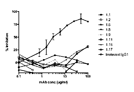
100211 Figure 2 is a graph showing inhibition of Amd enzymatic
activity eight anti-Amd
monoclonal antibodies and an isoytpe-matched antibody of irrelevant
specificity. Recombinant
Amd (rAmd) was prepared in E. coli (His-AmdR1R2-B in Table 1). rAmd (1.5
iitg/mL) was
mixed in PBS with a turbid suspension of peptidoglycan prepared from S. aureus
cell walls and
its lytic activity was measured by the reduction in turbidity (measured as
A490) following
incubation for 60 minutes at 37 C (A60). For the inhibition test, the
concentration of rAmd was
sufficient to reduce the A490 by 70%. Purified anti-Amd mAbs were added to the
rAmd at the
indicated concentrations and then lysis of peptidoglycan by the Mab:rArnd
mixture was
¨ 8 ¨
measured. Percent inhibition was calculated as: 100 x (1-(A60A490
inhibitor/A60A490no
inhibitor control)).
[0022] Figure 3 is an image of S. aureus precipitation by
representative anti-Amd
antibodies. When S. aureus cells are cultured in the presence of most
Staphylococcus-specific
mAbs they form into large clusters that fall out of suspension yielding a
relatively clear
supernatant. USA300LAC S. aureus were cultured in TSB at 37 C for eight hours
in the
presence of the indicated anti-Amd mAbs, each at 25 [tg/mL. The sample
containing no
antibody (No Ab) and an irrelevant isotype-matched antibody (Isotype control)
had turbid
supernatants without evident cell pellets; mAbs Amd1.1, Amd1.6, Amd1.8,
Amd1.11, and
Amd1.16 had clear supernatants and cell pellets. Other mAbs producing clear
supernatants and
cell pellets arc listed in Table 2, infra, as are mAbs that failed to
precipitate S. aureus from
suspension.
[0023] Figure 4 illustrates the biomolecular interaction analysis of
immobilized mAb
Amd1.6 with soluble Amd. The affinity of the interaction between mAb Amd1.6
and soluble
TM
Amd was measured on a Biacore T-200. Rabbit anti-mouse Fc IgG was immobilized
on the
surface of a CM-5 biosensor chip and used to capture mAb Amd1.6 which then
captured Amd
from a flowing field. The mass of Amd bound by mAb Amd1.6 is measured in
Resonance Units
(y-axis) against time on the x-axis. The capture (association, t = 0 to 120
sec) and release
(dissociation, t = 120 to 420 sec) phases are presented. The experiment was
repeated with
concentrations of Amd varying in two-fold increments from 1.56 to 25 nM.
Measurements were
made according the manufacturer's instructions and kinetic data were analyzed
using
TM
biomolecular interaction analysis (BIA) evaluation software (version 3.1) from
Biacore AB.
[0024] Figure 5 is a graph illustrating the inhibitory effect of anti-
Amd antibodies on in
vitro biofilm formation as compared to the Amd, Gmd and autolysin deletion
mutant strains. A
biofilm assay utilizing Calgary plates was performed by coating the plate and
lid pegs with
human plasma for 16 hours at 4 C. S. aureus was then seeded at OD 600nm of
0.05 in the
presence or absence of 25 1..tg/mL anti-Amd (Amd1.6), anti-Gmd (1C11) and
combination of
anti-Amd + anti-Gmd (Amd1.6 + 11) mAbs. Biofilm formation was allowed for 24
hours at
37 C. After washing, biofilms were stained with crystal violet and biofilm
content was
measured by spectrophotometry at 595 nm. As a positive control for biofilm
inhibition, UAMS-
1 deficient strain for amidase (Aaincl), glucosaminidase (Agmd) or autolysin
(Aatl) were seeded
at same OD. Results are reported as the amount of biofilm formation (i.e.,
crystal violet staining)
as a percentage of the wild type (WT), untreated UAMS-1 culture (A); * p <0.05
compared to
WT.
Date Recue/Date Received 2021-03-17
-9-
100251 Figures 6A-E illustrate the effect of passive immunization with
anti-Amd
monoclonal antibodies and a combination of anti-Amd and anti-Gmd monoclonal
antibodies on
biofilm formation on implants in vivo as compared to autolysin deficiency. Six-
to-ten week old,
female Balb/c mice (n? 3) were passively immunized intraperitoneally with anti-
Amd
(Amd1.6), a combination of anti-Amd and anti-Gmd (Amd1.6 + 1C11) or an IgG
isotype-
matched control mAb at a dose of 40 mg/kg. One day later each mouse was
infected with a trans-
tibial stainless steel pin contaminated with USA300 LAC CA-MRSA strain or its
isogenic Aatl
mutant. The pins were left in place to allow the biofilm-based infection to
mature. On Day 14
post-infection the pins were removed and examined by scanning electron
microscopy (SEM).
Representative micrographs showing the extent of bio film formation on the
infected implants
(pins) are shown: IgG control (Figure 6A); anti-Amd (Amd1.6, Figure 6B); anti-
Amd + anti-
Gmd (Amd1.6 + 1C1 I, Figure 6C); and infected with Aatl mutant (Figure 6D).
The percentage
of the region of interest (the 0.5 x 2.0 mm face of the flat pin) covered with
biofilm was
quantified with NIH software (Image J) and shown in Figure 6E; * p < 0.05.
[0026] Figures 7A-C illustrate the effect of passive immunization with anti-
Amd, anti-
Gmd, and a combination of anti-Amd and anti-Gmd monoclonal antibodies on the
reduction in
the amount of bone damage. Female Balb/c mice (n= 5) were passively immunized
with PBS or
anti-Gmd (1C11), anti-Amd (1.6) or a combination (1C11+1.6) at a 40mg/kg dose
i.p. as
previously described (Van-one et al., "Passive Immunization With Anti-
Glucosaminidase
Monoclonal Antibodies Protects Mice From Implant-Associated Osteomyelitis by
Mediating
Opsonophagocytosis of Staphylococcus aureus Megaclusters," J Orthop Res
32(10):1389-96
(2014)). Twenty-four hours later
all
mice received a trans-tibial pin contaminated with USA300 LAC::/ux, and
bioluminescent
imaging was performed on Day 3 to confirm the infection (Figure 7A). The mice
were
euthanized 14 days after infection, and the tibiae were harvested for micro-CT
analysis.
Representative 3D renderings of the infected tibiae are shown from the medial
and lateral side
(Figure 7B) to illustrate the relative level of osteolysis in each group (B)
of the tibias. The
osteolytic volume in each tibia was quantified using the formula: Osteolytic
volume (mmi) =
[medial osteolytic area + lateral osteolytic area (mm2)] X cortical thickness
(mm) (*p < 0.05 vs.
PBS). The results are illustrated graphically in Figure 7C.
[0027] Figures 8A-C illustrate the effects of passive immunization
with Amd1.6, which
show significantly reduced bacterial spread as evidenced by the formation of
fewer abscesses in
the medullary canal. 6-10 week old, female Balb/c mice (n = 5) were immunized
intraperitoneally with PBS (negative control), anti-Gmd mAb 1C 11, anti-Amd
mAb Amd1.6 or a
Date Recue/Date Received 2021-03-17
CA 02931001 2016-05-17
WO 2015/089502 - 10 ¨ PCT/US2014/070337
combination (1C11+Amd1.6) at a total dose of 40 mg/kg. Twenty-four hours later
each mouse
had inserted through its right tibia a pin contaminated with USA300 LAC::/ux,
a bioluminescent
CA-MRSA strain. The resulting infection was allowed to progress for fourteen
days when the
animals were sacrificed and the infected tibiae were harvested, fixed,
decalcified and sectioned
for histological analysis. Representative infected tibiae stained with Orange
G/alcian blue
(ABG/OH) are depicted for (Figure 8A) untreated controls and (Figure 8B) mice
treated with the
combination of anti-Gmd 1C11 and anti-Amd Amd1.6. The number of abscesses
observed in
each group of mice is presented in (Figure 8C). *, p < 0.05; **, p < 0.01.
[0028] Figures 9A-H illustrate the effect of passive immunization with
anti-Amd, anti-
Gmd, and a combination of anti-Amd and anti-Gmd monoclonal antibodies in
preventing
formation of staphylococcal abscess communities (SACs), which leads to sterile
abscesses. Mice
(n = 5) were immunized i.p. with PBS (negative control) or mAbs 1C11, Amd1.6
or a
combination (1C11+Amd1.6) at a 40 mg/kg dose. Twenty-four hours later all mice
received a
trans-tibial pin contaminated with USA300 LAC: :lux bioluminescent CA-MRSA
strain.
Representative infected tibias from Day 14 post-infection are shown for
histology sections that
were Gram-stained to reveal the bacteria. PBS-treated tibias show typical SAC
pathology,
containing a central nidus of bacteria surrounded by an eosinophilic
pseudocapsule within the
abscess area (Figures 9A-B) that are absent in mice treated with the following
mAbs: 1C11
(Figures 9C-D), Amd1.6 (Figures 9E-F), and combination 1C11+Amd1.6 (Figures 9G-
H).
[0029] Figures 10A-H illustrate the effect of passive immunization with
anti-Amd, anti-
Gmd, and a combination of anti-Amd and anti-Gmd monoclonal antibodies on
recruitment of
macrophage-like cells within the abscess. Six-to-ten week old female Balb/c
mice (n = 5) were
immunized i.p. with PBS or mAb 1C11, Amd1.6 or a combination (1C11 + Amd1.6)
at a 40
mg/kg dose. Twenty-four hours later all mice received an trans-tibial pin
contaminated with
USA300 LAC::/ux bioluminescent CA-MRSA strain. Representative infected tibias
from Day
14 post-infection are shown for histology sections that were stained with
Orange G/alcian blue
(ABG/OH). Passive immunization with anti-Amd, anti-Gmd, and a combination of
anti-Amd
and anti-Gmd mAbs recruits macrophage-like cells to the center of abscess
(Figures 10C-H,
arrowheads) while the PBS immunized mice do not show macrophage-like cell
recruitment
within the abscess (Figures 10A-B) and display cells that morphologically
resemble neutrophils.
Multiple abscesses are present in PBS treated tibias (Figure 10A) in the
medullary canal and soft
tissue around the bone, compared to a single abscess in Amd1.6 and combination
1C11+Amd1.6
treated mice (Figures 10E and 10G, respectively), or two abscess structures in
1C11 treated mice
(Figure 10C).
-11 ¨
100301 Figures 11A-E illustrate the effect of passive immunization
with a combination of
anti-Amd and anti-Gmd monoclonal antibodies, which accelerates bone healing by
recruiting M2
macrophages within the sterile abscess. Six-to-ten week old female Balb/c mice
(n = 5) were
immunized i.p. with PBS or mAb 1C11, Amd1.6 or a combination (1C11 + Amd1.6)
at a 40
mg/kg dose. Twenty-four hours later all mice received an trans-tibial pin
contaminated with
USA300 LAC::/ux bioluminescent CA-MRSA strain. Representative tibias from Day
14 post-
surgery are shown for histology sections that were stained with Orange
G/alcian blue (ABG/OH)
(Figures 11A-C). Remarkable healing is evident in mice immunized with the mAbs
comparable
to those receiving a sterile pin control (Figures 11A-C). To determine
correlation of healing with
macrophage phenotype associated with remodeling and wound healing process,
immunohistochemistry was performed with anti-Arginase-1 antibody to stain M2
macrophages.
M2 macrophages are recruited to the center of the abscess (brown staining) on
mice that were
passively immunized (Figure 11E), but excluded from abscess center on negative
control PBS
group (Figure 11D).
DETAILED DESCRIPTION
[0031] Disclosed herein are one or more monoclonal antibodies or
binding portions
thereof that binds specifically to a Staphylococcus spp. autolysin N-
acetylmuramoyl-L-alanine
amidase (Amd) catalytic domain or cell wall binding domain.
[0032] As used herein, the term "antibody" is meant to include
immunoglobulins derived
from natural sources or from recombinant sources, as well as immunoreactive
portions (i.e.
antigen binding portions) of immunoglobulins. The monoclonal antibodies
disclosed herein may
exist in or can be isolated in a variety of forms including, for example,
substantially pure
monoclonal antibodies, antibody fragments or binding portions, chimeric
antibodies, and
humanized antibodies (Ed Harlow and David Lane, USING ANTIBODIES: A LABORATORY
MANUAL (Cold Spring Harbor Laboratory Press, 1999)).
[0033] The monoclonal antibodies disclosed herein are characterized by
specificity for
binding to Staphylococcus N-acetylmuramoyl-L-alanine-amidase or fragments
thereof. The
antibody specifically binds to an epitope, typically though not exclusively an
immuno-dominant
epitope, in the amidase sub-unit of Staphylococcus autolysin (All). In certain
embodiments,
these monoclonal antibodies inhibit in vivo growth of a Staphylococcus strain.
In other
embodiments, these monoclonal antibodies inhibit biofilm establishment on
metal, plastic and/or
organic surfaces. In still further embodiments, one or more monoclonal
antibodies can be used
Date Recue/Date Received 2021-03-17
¨ 12 ¨
together to inhibit both in vivo growth of a Staphylococcus strain and biofilm
establishment on
metal, plastic and/or organic surfaces.
[0034] In accordance with this and all other aspects disclosed herein,
the Staphylococcus
strain is a strain that is, or can be, pathogenic to humans or animals. The
Staphylococcus can be
either coagulase-positive or coagulase-negative. Exemplary Staphylococcus
strains include,
without limitation, S. aureus, S. epidermidis, S. lugdunensis, S.
saprophyticus, S. haemolyticus,
S. caprae, and S. simiae. In one embodiment, the monoclonal antibodies
disclosed herein are
effective against antibiotic-resistant strains of Staphylococcus, including
methicillin-resistant or
vancomycin-resistant strains.
[0035] In certain embodiments, the epitope of the amidase subunit (that is
bound by the
mAb or binding fragment thereof) is an immuno-dominant epitope. Immuno-
dominant antigen
is a part of the antigenic determinant that is most easily recognized by the
immune system and
thus exerts the most influence on the specificity of the induced antibody. An
"immuno-dominant
epitope" refers to the epitope on an antigen that selectively provokes an
immune response in a
host organism to the substantial exclusion of other epitopes on that antigen.
[0036] Usually, the antigen likely to carry an immuno-dominant epitope
can be identified
by selecting antigens on the outer surface of the pathogenic organism. For
example, most simple
organisms, such as fungi, bacteria and viruses have one or two proteins that
are exposed on the
outer surface of the pathogenic organism. These outer surface proteins are
most likely to carry
the appropriate antigen. The proteins most likely to carry an immuno-dominant
epitope can be
identified in a Western assay in which total protein is run on a gel against
serum from an
organism infected with the pathogenic organism. Bound antibodies from the
scrum arc identified
by labeled anti-antibodies, such as in one of the well-known ELISA techniques.
The immuno-
dominant epitope can be identified by examining serum from a host organism
infected with the
pathogenic organism. The serum is evaluated for its content of antibodies that
bind to the
identified antigens that are likely to cause an immune response in a host
organism. If an
immuno-dominant epitope is present in these antigens, substantially all
antibodies in the serum
will bind to the immuno-dominant epitope, with little binding to other
epitopes present in the
antigen.
[0037] AtlA is one of the catalytically distinct peptidoglycan hydrolases
in
Staphylococcus aureus that is required to digest the cell wall during mitosis
(Baba and
Schneewind, "Targeting of Muralytic Enzymes to the Cell Division Site of Gram-
Positive
Bacteria: Repeat Domains Direct Autolysin to the Equatorial Surface Ring of
Staphylococcus
aureus," EA1B0. 17(16):4639-46 (1998)).
Date Recue/Date Received 2021-03-17
¨ 13 ¨
In addition to being an essential gene for growth, scanning electron
microscopy studies
have demonstrated that anti-At/A antibodies bound to S. aureus during binary
fission localize to
regions of the bacteria that are not covered by the cell wall (Yamada et al.,
"An Autolysin Ring
Associated With Cell Separation of Staphylococcus aureus," I Bacteriol.
178(6):1565-71
(1996)).
[0038] The AtlA enzyme is comprised of an amidase (62kD) and
glucosaminidase
(53kD), which are produced from the same AtlA precursor protein via a cleavage
process (Baba
and Schneewind, "Targeting of Muralytic Enzymes to the Cell Division Site of
Gram-Positive
Bacteria: Repeat Domains Direct Autolysin to the Equatorial Surface Ring of
Staphylococcus
aureus," Enzbo. J. 17(16):4639-46 (1998); Komatsuzawa et al., "Subcellular
Localization of the
Major Autolysin, ATL and Its Processed Proteins in Staphylococcus aureus,"
Microbiol
bninunol. 41:469-79 (1997); Oshida et al., "A Staphylococcus aureus Autolysin
That Has an N-
acetylmuramoyl-L-alanine Amidase Domain and an Endo-beta-N-
acetylglucosaminidase
Domain: Cloning, Sequence Analysis, and Characterization," Proc. Nat'l. Acad.
Sci. U.S.A.
92(1):285-9 (1995)). The autolysin
is held to the cell wall by three ¨150 amino acid cell wall binding domains,
which are designated
as R1, R2, and R3. In the final maturation step, proteolytic cleavage
separates the amidase
domain and its associated R1 and R2 domains (collectively, "Amd") from the
glucosaminidase
and its associated N-terminal R3 domain (collectively, "Gmd"). See Figure 1.
[0039] Exemplary encoded consensus protein and encoding open reading frame
sequences for His-Amd are identified as SEQ ID NOS: 1 and 2 below.
SEQ ID NO: 1
MHHHHHHSASAQPRSVAAT PKTS L PKYKPQVNS S INDY IRKNNLKAPKIEE DYT S YFPKYAYRN
GVGRPEGIVVHDTANDRST INGE I S YMKNNYQNAFVHAFVDGDR I I E TAPT DYL SWGVGAVGN P
RFINVEIVHTHDYASFARSMNNYADYAATQLQYYGLKPDSAEYDGNGTVWTHYAVSKYLGGTDH
ADPHGYLRSHNYSYDQLYDL INEKYLIKMGKVAPWGTQS T TT P T TPSKPTI P SKP S IGKLIVAA
NNGVAQ I KPTN S GLY T TVY DKT GKATNEVQKT FAVSKTAT LGNQKFYLVQDYNS GNKFGWVKE G
DVVYNTAKSPVNVNQSYSIKPGTKLYTVPWGT SKQVAGSVSGS GNQT FKASKQQQ I DKS I YLYG
SVNGKSGWVSKAYLVDTAKPTPT P PKP S PT TNNKLTVS SLNGVAQ INAKNNGLFITVYDKIG
KPTKEVQKTFAVTKEASLGGNKFYLVKDYNS P TL I GWVKQGDVI YNNAKS PVNVMQTYTVKPGT
KLY SVPWGTYKQEAGAVS GTGNQTFKATKQQQ I DKS I YLFGTVNGKS GWVSKAYLAVPAAPKKA
VAQPKTAVK
SEQ ID NO: 2
ATGCACCATCACCACCACCACAGCGCAAGCGCACAGCCTCGT TCCGTCGCCGCCACCCCGAAAA
CCAGCT TGCCGAAGTACAAACCGCAAGTTAATAGCAGCATCAACGACTACATCCGCAAAAACAA
CC T GAAGGCCCCGAAAAT T GAAGAGGAC TATACCAGCTAT TTCCCGAAATATGCT TACCGTAAT
GGIGTCGGTCGTCCGGAGSGTAT TGTGGTCCACGACACCGCGAATGACCGTAGCACCATCAACG
GTGAGAT TAGCTACATGAAAAACAATTACCAAAACGCGT TCGTGCACGCCT TCGTCGATGGCGA
Date Recue/Date Received 2021-03-17
¨ 14 ¨
TCGCATCATCGAAACCGCGCCAACCGACTATCIGTCCTGGGGIGIGGGIGCCGTTGGCAACCCG
CGTTTCATCAATGTGGAGATTGTTCATACCCACGACTACGCGAGCTTTGCACGTAGCATGAACA
ACTACGCCGATTATGCTGCAACGCAGCTGCAGTACTACGGCCTGAAACCGGATAGCGCGGAGTA
TGACGGTAACGGTACGGIGTGGACGCATTATGCGGTGAGCAAATACCTGGGTGGTACCGATCAT
GCTGATCCGCATGGCTACCTGCGCTCTCACAACTATAGCTACGACCAGTTGTACGACCTGATCA
ATGAGAAATATCTGATTAAGATGGGTAAGGTTGCACCGTGGGGTACGCAGAGCACCACGACGCC
GACCACGCCGAGCAAACCGACGACCCCGICCAAACCGTCTACCGGCAAACTGACGGICGCGGCT
AATAACGGTGTCGCGCAGATTAAACCGACCAACAGCGGTCTGTACACCACCGTCTATGATAAAA
CGGGCAAAGCCACCAATGAGGTTCAAAAGACGTTCGCAGTTAGCAAAACGGCGACCCTGGGTAA
CCAAAAGTTCTACCTGGTTCAGGATTACAATAGCGGCAACAAATTTGGTTGGGTGAAAGAAGGC
GACGTTGTGTACAATACC3CGAAGTCCCCGGTGAACGTTAATCAGAGCTATAGCATCAAGCCGG
GTACCAAATTGTATACGGIGCCGTGGGGTACCAGCAAGCAAGTTGCGGGTAGCGTCAGCGGCTC
TGGIAACCAGACCITCAAGGCGTCTAAGCAACAACAAATTGACAAAAGCATTIACCIGTAIGGT
AGCGTTAATGGTAAAAGC3GCTGGGTGTCTAAAGCGTATCTGGTCGACACCGCAAAGCCGACGC
CAACGCCGACCCCGAAGCC,GAGCACCCCAACCACCAACAACAAGCTGACGGTCAGCTCCCTGAA
TGGTGTTGCGCAAATCAATGCGAAGAATAATGGCCTGTTTACCACCGTTTACGATAAGACGGGC
AAGCCAACGAAAGAAGTCCAGAAAACCTTTGCTGTCACCAAAGAAGCCAGCCTGGGCGGTAACA
AGTTCTATCTGGTTAAGGACTACAACTCCCCGACGCTGATCGGTTGGGTCAAACAAGGCGATGT
CATTTACAATAACGCGAAAAGCCCGGTTAATGTGATGCAAACCTATACCGTCAAACCGGGTACG
AAGCTGTATTCCGTTCCGTGGGGCACGTACAAACAAGAAGCAGGCGCGGTGAGCGGTACCGGCA
ATCAGACCTTTAAGGCCACCAAGCACCACCAGATCGATAAATCTATTTACTTGTTTGGCACCGT
GAATGGCAAGAGCGGTTGSGTTTCTAAGGCATACCTGGCGGTGCCGGCAGCACCGAAGAAGGCG
GTGGCGCAGCCAAAGACCGCAGTGAAG
[0040] The Staphylococcus Amd can be synthesized by solid phase or solution
phase
peptide synthesis, recombinant expression, or can be obtained from natural
sources. Automatic
peptide synthesizers are commercially available from numerous suppliers, such
as Applied
Biosystems, Foster City, California. Standard techniques of chemical peptide
synthesis are well
known in the art (see e.g., SYNTHETIC PEPTIDES: A USERS GUIDE 93-210 (Gregory
A. Grant ed.,
1992) ). Protein or peptide production
via recombinant expression can be carried out using bacteria, such as E. coli,
yeast, insect or
mammalian cells and expression systems. Procedures for recombinant
protein/peptide
expression are well known in the art and are described by Sambrook et al,
Molecular Cloning: A
Laboratory Manual (C.S.H.P. Press, NY 2d ed., 1989).
[0041] Recombinantly expressed peptides can be purified using any one of
several
methods readily known in the art, including ion exchange chromatography,
hydrophobic
interaction chromatography, affinity chromatography, gel filtration, and
reverse phase
chromatography. The peptide is preferably produced in purified form
(preferably at least about
80% or 85% pure, more preferably at least about 90% or 95% pure) by
conventional techniques.
Depending on whether the recombinant host cell is made to secrete the peptide
into growth
medium (see U.S. Patent No. 6,596,509 to Bauer et al.),
the peptide can be isolated and purified by centrifugation (to separate
Date Recue/Date Received 2021-03-17
CA 02931001 2016-05-17
WO 2015/089502 ¨ 15 ¨ PCT/US2014/070337
cellular components from supernatant containing the secreted peptide) followed
by sequential
ammonium sulfate precipitation of the supernatant. The fraction containing the
peptide is
subjected to gel filtration in an appropriately sized dextran or
polyacrylamide column to separate
the peptides from other proteins. If necessary, the peptide fraction may be
further purified by
HPLC and/or dialysis.
[0042] In certain embodiments, the monoclonal antibodies or binding
portions may bind
specifically to an epitope of the Amd catalytic domain. As used herein, the
Amd catalytic
domain is at least 70% identical to amino acids 9-252 of SEQ ID NO: 1, or at
least 75% or 80%
identical to amino acids 9-252 of SEQ ID NO: 1, or even at least 85% or 90%
identical to amino
acids 9-252 of SEQ ID NO: 1. In certain embodiments, the amidase catalytic
domain is at least
95% identical to amino acids 9-252 of SEQ ID NO: 1.
[0043] In certain embodiments, the monoclonal antibody or binding
portion is produced
by a hybridoma cell line designated as Amd1.6, Amd1.10, Amd1.13, Amd1.16,
Amd1.17,
Amd2.1, or Arrid2.2.
[0044] In another embodiment, the monoclonal antibody or binding portion
binds to an
epitope wholly or partly within the Amd R1 or R2 cell wall binding domain. As
used herein, the
R1 or R2 cell wall binding domains are at least 70% identical to amino acids
253-399 or 421-568
of SEQ ID NO: 1, respectively; or at least 75% or 80% identical to amino acids
253-399 or 421-
568 of SEQ ID NO: 1, respectively; or even at least 85% or 90% identical to
amino acids 253-
399 or 421-568 of SEQ ID NO: 1, respectively. In certain embodiments, the cell
wall binding
domains are at least 95% identical to amino acids 253-399 or 421-568 of SEQ ID
NO: 1,
respectively.
[0045] In certain embodiments, the monoclonal antibody or binding
portion is produced
by a hybridoma cell line designated Amd1.1, Amd1.2, Amd1.5, Amd1.7, Amd1.8,
Amd1.9,
Amd1.11, Amd1.12, Amd1.14, Amd1.15, Amd2.4, or Amd2.5.
[0046] In certain embodiments the monoclonal antibody disclosed herein
binds to the
Amd catalytic domain or cell wall binding domain with an affinity greater than
10-8 M or 10-9 M,
but preferably greater than 10-19M.
[0047] As noted above, in certain embodiments the monoclonal
antibodies or binding
portions also inhibit in vivo growth of Staphylococcus. Inhibition of in vivo
growth of
Staphylococcus can be measured according to a number of suitable standards. In
one such
embodiment, the in vivo growth of Staphylococcus can be assessed according to
a
bioluminescence assay. By way of example, bioluminescent S. aureus (Xen 29;
ATCC 12600)
(Francis et al., "Monitoring Bioluminescent Staphylococcus aureus Infections
in Living Mice
¨ 16 ¨
Using a Novel luxABCDE Construct," Infect. Inunun. 68(6):3594-600 (2000); see
also Contag et
al., "Photonic Detection of Bacterial Pathogens in Living Hosts," Mol.
Microhiol. 18(4):593-603
(1995)) is used to dose a
transtibial implant with 500,000 CFU prior to surgical implant. Five week old
female BALB/cJ
mice can receive an intraperitoneal injection of saline or 1 mg of purified
antibody/antibody
fragment in 0.25 ml saline 3 days prior to surgery. The mice can be imaged to
assess
bioluminescence on various days (e.g., 0, 3, 5, 7, 11, and 14) and a
comparison of BLI images
can be compared to assess whether the antibody inhibits in vivo growth of S.
aureus relative to
the saline control or a control mouse injected with a placebo antibody.
[0048] In another embodiment, the in vivo growth of Staphylococcus can be
assessed
according to biofilm formation. By way of example, female Balb/c mice can be
passively
immunized intraperitoneally with antibody/antibody fragment or control at a
dose of 40 mg/kg,
and one day later each mouse can be infected with a trans-tibial stainless
steel pin contaminated
with a MRSA strain. On day 14 post-infection the pins can be removed and
examined by
scanning electron microscopy (SEM), and the percentage of a region of interest
(e.g., 0.5 x 2.0
mm face of the flat pin) covered with biofilm can be quantified with NIH
software (Image J).
[0049] In yet another embodiment, the Osteolytic Volume of infected
bone can be
measured using MicroCT imaging. By way of example, female Balb/c mice can be
passively
immunized intraperitoneally with antibody/antibody fragment or control at a
dose of 40 mg/kg,
and one day later each mouse can be infected with a trans-tibial stainless
steel pin contaminated
with a MRSA strain. After 14 days, the mice can be euthanized and the tibia
harvested. Using
the resulting images, the lesion area can be measured in two different views
(e.g., medial and
lateral), which are added together and multiplied by the cortical thickness
(see Varrone et al.,
"Passive Immunization With Anti-Glucosaminidase Monoclonal Antibodies Protects
Mice From
Implant-Associated Osteomyelitis by Mediating Opsonophagocytosis of
Staphylococcus aureus
Megaclusters," J Orthop Res 32(10):1389-96 (2014)).
[0050] In yet another embodiment, in vivo growth of Staphylococcus can
be assessed by
the presence (including frequency) or absence of Staphylococcus abscess
communities (SACs) in
the medullary canal or soft tissue surrounding the bone. By way of example,
female Balb/c mice
can be passively immunized intraperitoneally with antibody/antibody fragment
or control at a
dose of 40 mg/kg, and one day later each mouse can be infected with a trans-
tibial stainless steel
pin contaminated with a MRSA strain. After 14 days, the mice can be euthanized
and the tibia
and associated soft tissue harvested. Histological samples can be prepared and
stained with
Date Recue/Date Received 2021-03-17
CA 02931001 2016-05-17
WO 2015/089502 ¨ 17 ¨ PCT/US2014/070337
Orange Glalcian blue (ABG/OH), and then the presence or absence of abscesses
can be
determined upon analysis of the histologic samples.
[0051] According to one embodiment, the monoclonal antibody or binding
portion
comprises a VH domain comprising one of the following amino acid sequences
(CDR domains
underlined):
SEQ ID NO: 5 (Amd1.2):
PELVKPGASVKMSCKASGYT FT S Y IMHWVKQKPGQGLEW I GY INPYNDGTKYNEKFKGKATLT S
DKS S TTAYMELS SLT SE DXAVYYCARLDGYYDCFDYWGQGT T L TVS S
where X can be any amino acid. This amino acid sequence is encoded by the
following
nucleotide sequence (SEQ ID NO: 6):
OCT GAGC T GGTAAAGCC T GGGGCT T CAGT GAAGAT GT C CT GCAAGGC T TC T GGATACACAT
T CA
CTAGCTATATTATGCACTGGGTGAAGCAGAAGCCTGGGCAGGGCCT TGAGTGGAT TGGATATAT
TAATCCT TACAATGATGGTACTAAGTACAATGAGAAGT TCAAAGGCAAGGCCACAC TGAC T T CA
GACAAAT CC TCCACCACAGCC TACAT GGAGCT CAGCAGCCT GACCT C TGAGGACTNTGCGGTCT
ATTACTGTGCAAGACTTGATGGTTACTACGACTGCTTTGACTACTGGGGCCAAGGCACCACTCT
CACAGTCTCNTCAGCCAAAACGACACCCCCATCTGTCTATCCACTGGCCCCTGGATCTGCTGCC
CAAACTAACTCCATGGTGACCCTGGGATGCCNGGTCAAGGG
where each N can be A, T, C, or G, as long as the nucleic acid molecule
encodes the amino acid
sequence of SEQ ID NO: 5.
SEQ ID NO: 7 (Amd1.1):
QQSGAELVKPGASVKL SCTASGFNIKDTY I HWVKQRPEQGLEW I GRI DPANGI TNYDPKFQGRA
II TADT S SNIAYLQLT SLT SEGTAVYYCARGGYL S PYAMDYWGQGT SVTVS S
This amino acid sequence is encoded by the following nucleotide sequence (SEQ
ID NO: 8):
NTGCAGCAGTCTGGGGCAGAGCTTGTGAAGCCAGGGGCCTCAGTCAAGTTGTCCTGCACAGCTT
CTGGCTTCAACATTAAAGACACCTATATACATTGGGTGAAGCAGAGGCCTGAACAGGGCCTGGA
GIGGATTGGAAGGATTGATCCTGCGAATGGTATTACTAATTATGACCCGAAGTTCCAGGGCAGG
GCCACTATAACAGCAGACACATCCTCCAATATAGCCTACCTGCAGCTCACCAGCCTGACATCTG
AGGGCACTGCCGTCTACTACTGTGCTAGAGGGGGTTACCTATCCCCTTATGCTATGGACTACTG
GGGTCAAGGAACCTCAGTCACCGTCTCCTCAGCCAAAACGACACCCCCATCTGTCTATCCACTG
GCCCCTGGATCTGCTGCCCAAACTAACTCCATGGTGACCCTGGGATGCCTGGTCAAGGGCTATT
NCCCTGAGCCAG
where each N can be A, T, C, or G, as long as the nucleic acid molecule
encodes the amino acid
sequence of SEQ ID NO: 7.
CA 02931001 2016-05-17
WO 2015/089502 ¨ 18 ¨ PCT/US2014/070337
SEQ ID NO: 9 (Amd1.5):
QQS GAELVRPG.A.LVKL S CKA.S GFNI QDYY LEWMKQRPE QGLEW I GW I
DPENDNTVYDPKFRDR.A.
TADT F SNTAYLQL S T SE UMW YCARRDG I TTATRAMDYWGQGT WITS S
This amino acid sequence is encoded by the following nucleotide sequence (SEQ
ID NO: 10):
TGCAGCAGTCTGGGGCTGAGCTTGTGAGGCCAGGGGCCTTAGTCAAATTGTCCTGCAAAGCTTC
TGGC T TCAACAT TCAAGACTACTAT C TACACTGGATGAAACAGAGGCCTGAGCAGGGCC TGGAG
TGGAT TGGATGGAT TGATCCTGAGAATGATAATACTGTATATGACCCGAAGT TCCGGGACAGGG
CCAGTTTAACAGCAGACACAT T T T CCAACACAGCCTACC TACAGCT CAGCGGCC T GACATCT GA
AGACACT GCCGT C TAT TACT GTGC TAGAAGAGACGGCAT TAC TACGGCTACGCGGGCTATGGAC
TACTGGGGTCAAGGAACCTCAGTCACCGTCTCCTCAGCCAAAACGACACCCCCATCTGTCTATC
CACTGGCCCCTGGATCTGCTGCCCAAACTAACTCCATGGTGACCCTGGGATGCCIGGTCAAGGG
CNNNNNCC TGAGCCAG
where each N can be A, T, C, or G, as long as the nucleic acid molecule
encodes the amino acid
sequence of SEQ ID NO: 9.
SEQ ID NO: 11 (Amd1.6):
QSGTVLARPGT SVKMS CKAS GYSFTNYWMHWVRQRPGQGLEWIGS I Y PGNS DT T YNQKFKDKAK
LTAVTSAS TAYMELS S LINE DSAVYYCIGEDYSRFSYWGQGTLVIVSA
This amino acid sequence is encoded by the following nucleotide sequence (SEQ
ID NO: 12):
CAGTCTGGGACTGTACTGGCAAGGCCTGGGACT TCCGTGAAGATGTCCTGCAAGGCT TCTGGCT
ACAGCTTTACCAACTACTGGATGCACTGGGTAAGACAGAGGCCTGGACAGGGTCTAGAATGGAT
TGGTTCTATTTATCCTGGAAATAGTGATACTACCTACAACCAGAAGTTCAAGGACAAGGCCAAA
CTGACTGCAGTCACATCCGCCAGCACTGCCTACATGGAGCTCAGCAGCCTGACAAATGAGGACT
CT GCGGT C TAT TACT GTACGGGGGAT GAT TACTC TCGGT T T TCT TAC T GGGGCCAAGGGACTCT
GGTCACTGTCTCTGCAGCCAAAACGACACCCCCATCTGTCTATCCACTGGCCCCTGGATCTGCT
GCCCAAACTAACTCCATGGTGACCCTGGGATGCCTNGTCAAGGGCTNT T TCCCNGAGCCA
where each N can be A, T, C, or G, as long as the nucleic acid molecule
encodes the amino acid
sequence of SEQ ID NO: 11.
SEQ ID NO 13: (Amd1.7):
QQSGPELVKPGASVKI SCKASGYIFIDYNMHWVKQSHGKSLEWIGY I FPYNGDI DYNQKFKNKA
TLTVDNS S S TAYMDLRSLT SE DSAVYYCSRWGS YFDYWGQGT TLTVS S
This amino acid sequence is encoded by the following nucleotide sequence (SEQ
ID NO: 14):
CA 02931001 2016-05-17
WO 2015/089502 ¨ 19 - PCT/US2014/070337
TGCAGCAGTCAGGACCTGAGCTGGTGAAACCTGGGGCCTCAGTGAAGATATCCTGCAAGGCTTC
TGGATACACAT T CACT GACTACAACATGCACTGGGTGAAGCAGAGCCATGGAAAGAGCC T TGAG
TGGATTGGATATATTTTTCCTTACAATGGTGATACTGACTACAACCAGAAATTCAAGAACAAGG
CCACAT T GACT GTAGACAAT T CCT CCAGCACAGCCTACATGGACCT CCGCAGCC T GACATCT GA
GGACTCTGCAGTCTAT TACTGT TCAAGATGGGGGTCT TACT T TGACTACTGGGGCCAAGGCACC
ACTCTCACAGTCTCCTCAGCCAAAACGACACCCCCATCTGTCTATCCACTGGCCCCTGGATCTG
CTGCCCAAACTAACTCCATGGTGACCCTGGGATGCCTGNGTCAAGGGCT
where each N can be A, T, C, or G, as long as the nucleic acid molecule
encodes the amino acid
sequence of SEQ ID NO: 13.
SEQ ID NO: 15 (Amd1.9):
VES GGGLVKPGGSLKL SCAAS GET FS SYAMSWVRQT PKKSLEWVAS I T SGGSAYY PDSVKGRFT
I SRDNARNILNLQMS SLRSEDTAMYYCARDDGYFDYWGQGT TL TVS S
This amino acid sequence is encoded by the following nucleotide sequence (SEQ
ID NO: 16):
GTGGAGTCTGGGGGAGGCT TAGTGAAGCCTGGAGGGTCCCTGAAACTCTCCTGTGCAGCCTCTG
GAT TCACT T TCAGTAGCTATGCCATGTCT TGGGT TCGCCAGACTCCAAAAAAGAGTCTGGAGTG
GGTCGCATCCAT TACTAGTGGTGGTAGCGCCTACTATCCAGACAGTGTGAAGGGCCGAT TCACC
ATCTCCAGAGATAATGCCAGGAACATCCTGAACCTGCAGATGAGCAGTCTGAGGTCTGAGGACA
CGGCCATGTATTACTGTGCAAGAGACGACGGGTACTTTGACTACTGGGGCCAAGGCACCACTCT
CACAGTCTCCTCAGCCAAAACGACACCCCCATCTGTCTATCCACTGGCCCCTGGATCTGCTGCC
CAAACTAACTCCATGGTGACCCTGGGATGCCTGGTCAA
SEQ ID NO: 17 (Amd1.11):
QIQLVQSGPELKKPGETVKI SCKASGYTFTNYGMNWVKQAPGKGLEWMGWINTYTGEPTYADDF
KGRFAFSLET SAS TAYLL INNLKNEDTATYFCARRDGYFDAMDYWGQGT SVTVS S
This amino acid sequence is encoded by the following nucleotide sequence (SEQ
ID NO: 18):
NNCCTGATGGCAGCTGCCCAAAGTGCCCAAGCACAGATCCAGTTGGTGCAGTCTGGACCTGAGC
TGAAGAAGCCTGGAGAGACAGTCAAGATCTCCTGCAAGGCT TCTGGGTATACCT TCACAAACTA
TGGAATGAACT GGGT GAAGCAGGC T CCAGGAAAGGGT T TAGAGTGGATGGGCTGGATAAACACC
TACACTGGAGAGCCAACTTATGCTGATGACTTCAAGGGACGCTTTGCCTTCTCTTTGGAAACCT
CTGCCAGCACTGCCTATTTGCTGATCAACAACCTCAAAAATGAGGACACGGCTACATATTTCTG
TGCAAGAAGGGATGGT TACT TCGATGCTATGGAC TACTGGGGTCAAGGAACCTCAGTCACCGTC
TCCTCAGCCAAAACGACACCCCCATCTGTCTATCCACTGGCCCCTGGATCTGCTGCCCAAACTA
ACT CCAT GGTGACCCT GGGAT GCC T GGT CAAGGG
where each N can be A, T, C, or G, as long as the nucleic acid molecule
encodes the amino acid
sequence of SEQ ID NO: 17.
CA 02931001 2016-05-17
WO 2015/089502 ¨ 20 ¨ PCT/US2014/070337
SEQ ID NO: 19 (Amd1.12):
QQSGAELVRPGT SVKVSCKT SGYAFTNYL IEWVNQRPGQGLEW I GVINPGS GGTNYNEKFKAKA
TLTADKS S STAYMQLS SLTS DDSAVYFCARSERGYYGNYGAMDYWGQGTSVTVS S
This amino acid sequence is encoded by the following nucleotide sequence (SEQ
ID NO: 20):
NNGCAGCAGTCTGGAGCTGAGCTGGTAAGGCCTGGGACTTCAGTGAAGGTGTCCTGCAAGACT T
CT GGATAC GCC T T CAC TAAT TACT T GATAGAGT GGGTAAAT CAGAGGCCT GGACAGGGCC T T
GA
GTGGAT T GGGGT GAT TAATCC TGGAAGT GGTGGTACTAACTACAAT GAGAAGT T CAAGGCCAAG
GCAACAC T GAC I GCAGACAAAT CC T CCAGCACT GCC TACAT GCAGC T CAGCAGCC T GACATCT
G
ATGACTCTGCGGTCTAT TTCTGTGCAAGATCAGAGCGAGGCTACTATGGTAACTACGGAGCTAT
GGACTACTGGGGTCAAGGAACCTCAGTCACCGTCTCCTCAGCCAAAACGACACCCCCATCTGTC
TAT CCAC T GGCCCCT GGATC T GCT GCCCAAACTAACT CCAT GGTGACCCT GGGAT GCC T GGT CA
AGGGCTATNTCCCTGAGCCAG
where each N can be A, T, C, or G, as long as the nucleic acid molecule
encodes the amino acid
sequence of SEQ ID NO: 19.
SEQ ID NO: 21 (Amd1.13):
QQPGPELVKPGASLKI SCKASGYS FS S SWMNWVKQRPGQGLEW I GRI YPVDGDTNYNGKFKGKA
TLT TDKS S STAYMQLS SLTSVDSAVYFCARTGPYAMDYWGRGTSVTVS S
This amino acid sequence is encoded by the following nucleotide sequence (SEQ
ID NO: 22):
NNGCAGCAGCCIGGACCTGAGCTGGTGAAGCCTGGGGCCICACTGAAGAT T TCCTGCAAAGCT T
CT GGC TAC T CAT TCAGT T CC T CT T GGAT GAACT GGGT GAAGCAGAGGCCT GGACAGGGT C T
T GA
GTGGATTGGACGGATT TATCCTGTAGATGGAGATACTAACTACAATGGGAAGTTCAAGGGCAAG
GCCACACTGACTACAGACAAATCCTCCAGCACAGCCTACATGCAGCTCAGCAGCCTGACCTCTG
TGGACTCTGCGGTCTAT T TC T GTGCAAGAACTGGGCCC TAT GC TAT GGAC TACT GGGGT CGAGG
AACCTCAGTCACCGTCTCCTCAGCCAAAACGACACCCCCATCTGTCTATCCACTGGCCCCTGGA
TCT GCTGCCCAAACTAACTCCATGGTGACCCTGGGAT GCCT GGTCAAGGG
where each N can be A, T, C, or G, as long as the nucleic acid molecule
encodes the amino acid
sequence of SEQ ID NO: 21.
SEQ ID NO: 23 (Amd1.16):
GAELVRPGS SVK I SCKASGY T FS T YWMNWVKQRPGQGLEWI GQ I YPGDGDTNYNGKFKGKAT L T
ADKS S STAYMQL S SLT SDDSAVYFCARSMVTNYYFAMDYWGQGTSVTVS S
This amino acid sequence is encoded by the following nucleotide sequence (SEQ
ID NO: 24):
CA 02931001 2016-05-17
WO 2015/089502 ¨ 21 - PCT/US2014/070337
GGGGCTGAGCTGGTGAGGCCTGGGTCCTCAGTGAAGAT TTCCTGCAAGGCT TCTGGCTATACAT
TCAGTACC TAC T GGAT GAAC T GGGT GAAGCAGAGACC T GGACAGGGT CT T GAGT GGAT TGGACA
GAT T TAT CCTGGAGAT GGTGATAC TAACTACAAT GGAAAAT TCAAGGGTAAAGCCACACTGACT
GCAGACAAATCCTCCAGCACAGCCTACATGCAGCTCAGCAGCCTAACATCTGACGACTCTGCGG
TCTAT T TCTGTGCAAGATCGATGGTAACGAACTAT TACT T TGCTATGGACTACTGGGGTCAAGG
AACCTCAGTCACCGTCTCCTCAGCCAAAACGACACCCCCATCTGTCTATCCACTGGCCCCTGGA
TCTGCTGCCCAAACTAACTCCATGGTGACCCTGGGATGCCNGGTCAAGGG
where each N can be A, T, C, or G, as long as the nucleic acid molecule
encodes the amino acid
sequence of SEQ ID NO: 23.
SEQ ID NO: 25 (Amd1.17):
GGLVKPGGSLKL SCAASGFTFS DYYMYWVRQT PEKKLEWVAT I SDGGSYTYYPDSVKGRFT I SR
DNAKNNLYLQMS SLKSEDTAMYYCVRGLLGFDYWGQGT TLTVSS
This amino acid sequence is encoded by the following nucleotide sequence (SEQ
ID NO: 26):
GGGGAGGCTTAGTGAAGCCTGGAGGGTCCCTGAAACTCTCCTGTGCAGCCTCTGGATTCACTT T
CAGTGACTATTACATGTATTGGGT TCGCCAGACTCCGGAAAAGAAACTGGAGTGGGTCGCAACC
AT TAGTGATGGTGGTAGT TACACCTACTATCCAGACAGTGTGAAGGGGCGAT TCACCATCTCCA
GAGACAAT GCCAAGAACAACC TGTACCT GCAAAT GAGCAGT C TGAAGTCT GAGGACACAGCCAT
GTAT TACTGTGTAAGGGGGCTACTGGGTT T TGACTACTGGGGCCAAGGCACCACTCTCACAGTC
TCCTCAGCCAAAACGACACCCCCATCTGTCTATCCACTGGCCCCTGGATCTGCTGCCCAAACTA
ACTCCATGGTGACCCTGGGATGCCTGGTCAAGG
SEQ ID NO: 27 (Amd2.1):
GE"S7K.PGGS LKLSCAA.SGE'TFS S YAMS WRQ T PEMRLEWAS I S S GGSXTYYPDSVMGRE'
Ti SRDNARN I LNLQMS SLRSEDTAMYYCARVGLYYDYYYSMDYWGQGT SVTVS S
where X can be any amino acid. This amino acid sequence is encoded by the
following
nucleotide sequence (SEQ ID NO: 28):
GGCTTCGTGAAGCCTGGAGGGTCCCTGAAACTCTCCTGTGCAGCCTCTGGATTCACTTTCAGT.A.
GCTATGCCATGTCTTGGGTTCGCCAGACTCCAGAGATGAGGCTGGAGTGGGTCGCATCCATTAG
TAGT GGT GGTAGNNNCACCTAC TAT CCAGACAG T GTGATGGGCCGAT TCACCATCTCCAGAGAT
AATGCCAGGAACATCCTGAACCTCCAAATGAGCAGTCTGAGGTCTGAGGACACGGCCATGTATT
ACTGTGCAAGAGTGGGTCTCTACTATGAT .TATTACTATTCTATGGACTACTGGGGTCLAGGAAC
CTCAGTCACCGTCTCC T CA.G
where each N can be A, T, C, or G, as long as the nucleic acid molecule
encodes the amino acid
sequence of SEQ ID NO: 27.
SEQ ID NO: 29 (Amd2.2):
ESGPELVKPGASVKI SCKAS GYT F T DYNMHWVRQ S HGKS LEW I GY I Y PYNGGTGYNQKFKS
KAT I. T VDN S S S TAYMELRSLT SE I) SAVYYCARE DGYYGYFDYINGQGTTLTGSS
CA 02931001 2016-05-17
WO 2015/089502 ¨ ¨ PCT/US2014/070337
22
This amino acid sequence is encoded by the following nucleotide sequence (SEQ
ID NO: 30):
GAGT C AGG ACC T G AGC TGGTGAAACCTGGGGCC TCAGTGALAGA.TATCCTGCAAGGCTTCTGGA.T
ACACATTCACTGACTAT.AACATGCACTGGGTGAGGCAGAGCCATGGAAAGAGCCT TG.A.GTGGAT
TGGAT.A.TATTTATCCT T.A.CAATGGT GGT AC TGGC TAC.AACC.A.GAAGT
TCAAGAGTAAGGCC.A.CA
TTGACTGTAGACAA.TTCCTCCAGCACAGCCTACATGGAGCTCCGCAGCCTGACATCTGAGGACT
TGC AGT C TAT TACT GT GC AAGAGAGGAT GGT T AC T AC GGC TAC T T TGAC
TACTGGGGCCIt.AGG
CAC CAC T C T CACAGG C I CCT CAG
SEQ ID NO: 31 (Amd 2.4):
Q I QLVQS G PELKKPGE TVKI S CKAS GYT F TNYGMNWVKQAPGKGLKWMGW I NIT Y GE T
YADDF
KGRFAFS LE T SASAAYLQINNLKNEDTAT YFCARDYDGMYAMDYWGQGT SVTVS S
This amino acid sequence is encoded by the following nucleotide sequence (SEQ
ID NO: 32):
CAG AT CC AG T T GGT G CA.G TCTGGACC T GA.G C T GAAGAAG CC T G GA.G AGA.0 AG T
CAAGATCTcCT
GCAA.GGCT TCTGGGTA.TACCT TCA.CAAA.CTATGGAA.TGAA.C. TGGGTG.AAGC.AGGCTCCAGGAAA.
GGGT TTAAAGTGGATGGGCTGGATAAACACCTACACTGG.A.GAGCCAACATATGC T GAT G.ACT T C
AAGGGACGGTT TGCCT TCTCT TTGGAAACC TCTGCCAGCGCTGCCTATTTGCAGATCAACAACC
T C iz\ AAAA T GAG GACAC G C T ACA T A rrr T C TG/TGC AAG C7. G AC T A T GA T
GG T T AC T A T T AC T AT G C
TAT GGAC TACT GG GGT CAAGGAACC TCAG T CAC C GTC T COT CAG
100521 According to one embodiment, the monoclonal antibody or binding
portion comprises
a VL domain comprising one of the following amino acid sequences (CDR domains
underlined):
SEQ ID NO: 33 (Amd1.1):
ENVLTQS PAIMSASLGEKVTMTCRASSSVNYMFWFQQKSDASPKLWIYYT SNLAPGVPARFS GS
GSGNSYSLT I SSMEGEDAATYYCQEFTSFPYTFG
This amino acid sequence is encoded by the following nucleotide sequence (SEQ
ID NO: 34):
NTCAGTGTCTCAGTTGTAATGTCCAGAGGAGAAAATGTGCTCACCCAGTCTCCAGCAATCATGT
CTGCATCTCTAGGGGAGAAGGTCACCATGACCTGCAGGGCCAGCTCAAGTGTAAATTACATGTT
CTGGT TCCAGCAGAAGTCAGATGCCTCCCCCAAAT TGTGGAT T TAT TATACATCCAACCTGGCT
CCTGGAGTGCCAGCTCGCT TCAGTGGCAGTGGGTCTGGGAACTCT TAT TCTCTCACAATCAGCA
GCATGGAGGGTGAAGATGCTGCCACT TAT TACTGCCAGGAGT T TACTAGT T TCCCGTACACGT T
CGGA
where each N can be A, T, C, or G, as long as the nucleic acid molecule
encodes the amino acid
sequence of SEQ ID NO: 33.
SEQ ID NO: 35 (Amd1.2):
DIVLTQS PATLSVTPGDSVSLSCRASQS I SNNLHWYQQKSHESPRLL IKYASQS I SGI PSRFSG
SGSGTDFTLS INSVETEDFGMYFCQQSNSWPQYTF
CA 02931001 2016-05-17
WO 2015/089502 ¨ ¨ PCT/US2014/070337
23
This amino acid sequence is encoded by the following nucleotide sequence (SEQ
ID NO: 36):
TTATGCTTTTTTGGATTTCAGCCTCCAGAGGTGATATTGTGCTAACTCAGTCTCCAGCCACCCT
GTCTGTGACTCCAGGAGATAGCGTCAGTCTTTCCTGCAGGGCCAGCCAAAGTATTAGCAACAAC
CTACACT GGTAT CAACAAAAATCACATGAGTCT CCAAGGCT T CTCAT CAAGTAT GC T T CCCAGT
CCATCTCTGGGATCCCCTCCAGGT TCAGTGGCAGTGGATCAGGGACAGAT T TCAC TCTCAGTAT
CAACAGTGTGGAGACTGAAGATTTTGGAATGTATTTCTGTCAACAGAGTAACAGCTGGCCTCAG
TACACGTTCGG
SEQ ID NO: 37 (Amd1.6):
S IVMTQT PKFLLVSAGDRLT I TCKASQSVSNDVAWYQQKPGQSPKLL I YYT SNRYTGVPDRFTG
SGYGIDET FT I S TVQAEDLAVYFCQQDYNSPWITGGGIK
This amino acid sequence is encoded by the following nucleotide sequence (SEQ
ID NO: 38):
CCAGGTCTTCGTATTTCTACTGCTCTGTGTGTCTGGTGCTCATGGGAGTATTGTGATGACCCAG
AC T C CCAAAT T C C TGC T TGTATCAGCAGGAGACAGGC T TACCATAAC CTGCAAGGCCAGTCAGA
GTGTGAGTAATGATGTAGCT TGGTACCAACAGAAGCCAGGGCAGTCTCCTAAACTGCTGATATA
CTATACATCCAATCGCTACACTGGAGTCCCTGATCGCT TCACTGGCAGTGGATATGGGACGGAT
T TCACT T TCACCATCAGCACTGTGCAGGCTGAAGACCTGGCAGT T TAT T TCTGTCAGCAGGAT T
ATAACTCTCCGTGGACGTTCGGTGGAGGCACCAAG
SEQ ID NO: 39 (Amd1.7):
S IVMTQT PKFLLVSAGDRLT I TCKASQSVSNDVAWYQQKPGQSPKLL I YYT SNRYTGVPDRFTG
SGYGTDFT FT I S TVQAEDLAVYFCQQDYNSPWTFGGGTK
This amino acid sequence is encoded by the following nucleotide sequence (SEQ
ID NO: 40):
TGGTGCTCATGGGAGTAT TGTGATGACCCAGACTCCCAAAT TCCTGCT TGTATCAGCAGGAGAC
AGGC T TACCATAACCT GCAAGGCCAGTCAGAGT GTGAGTAAT GAT GTAGC T TGGTACCAACAGA
AGCCAGGGCAGTCTCCTAAACTGCTGATATACTATACATCCAATCGCTACACTGGAGTCCCTGA
TCGCTTCACTGGCAGTGGATATGGGACGGATTTCACTTTCACCATCAGCACTGTGCAGGCTGAA
GACCTGGCAGT T TAT T TCTGTCAGCAGGAT TATAACTCTCCGTGGACGT TCGGTGGAGGCACCA
AGC
SEQ ID NO: 41 (Amd1.8):
DIVMTQS PATLSVTPGDRVSLSCRASQS I SDYLHWYQQRSHESPRLL IKYVSQS I SGI PSRFSG
SGSGSDFTLS INSVEPEDVGVYYCQNGHS FPYT FG
This amino acid sequence is encoded by the following nucleotide sequence (SEQ
ID NO: 42):
CA 02931001 2016-05-17
WO 2015/089502 PCT/US2014/070337
CT TGGACT T T TGCT T T TCTGGACT TCAGCCTCCAGATGTGACAT TGTGATGACTCAGTCTCCAG
CCACCCTGTCTGTGAC TCCAGGAGATAGAGTCTCTCT T TCCTGCAGGGCCAGCCAGAGTAT TAG
CGAC TAC rrACACTGGTATCAACAAAGAT CACATGAGTCTCCAAGGCT"IC TCAT CAAATATGT T
TCCCAATCCATCTCTGGGATCCCCTCCAGGTTCAGTGGCAGTGGA.TCAGGGTCAGATTTCACTC
TCAG TAT CAAC AGTGTGGAACCT GAAGATG T TG'GAGTG T AT TAT TG TCAAAATGGT CAC AGCT
T
TCCGTACA.CGTTCGGA.
SEQ ID NO: 43 (Amd1.9):
DIQMTQSPASLSVSVGETVT I TCRT SENI FSNFAWYQQQPGKSPQLLVYGATNLADGVPSRFSG
SGSGTQYSLKI T SLQSEDFGS YYCQHFWGS PWT F
This amino acid sequence is encoded by the following nucleotide sequence (SEQ
ID NO: 44):
TTACAGATGCCAGATGTGACATCCAGATGACTCAGTCTCCAGCCTCCCTATCTGTATCTGTGGG
AGLAACTGTCACCATCACATGTCGAACAAGTGALAATATTTTCAGTAATT TCGCATGGTATCAG
CAGCAACCGGGAAAATCTCCTCA.GCTCCTGGTCTATGGTGCAA.CAAA.CTTA.GCAGATGGTGTGC
CAT CAAGG TCAGTGG CAGT GGAT CAGG CACACAGTAT TCCC TCAAGATCA.CCAGCCT GCAGT C
TGAAG.A.TTTTGGG.A.GT T.A.TTACTGTCAAC.A.TTT TTGGGGTAGTCCGTGGA.CGTTCGG
SEQ ID NO: 45 (Amd1.10):
QIVLTQS PALMSAS PGEKVTMTC SAS S SVSYMYWYQQKPRS S PKPWIYLT SNLAS GVPARFS GS
GSGT SYSL T I S SMEAEDAATYYCQQWS SNP PYT FG
This amino acid sequence is encoded by the following nucleotide sequence (SEQ
ID NO: 46):
TCAGTGCCTCAGTCATAATGTCCAGGGGACAAAT TGT TCTCACCCAGTCTCCAGCACTCATGTC
TGCATCTCCAGGGGAGAAGGTCACCATGACCTGCAGTGCCAGCTCAAGTGTAAGTTACATGTAC
TGGTACCAGCAGAAGCCAAGATCCTCCCCCAAACCCTGGAT T TATCTCACATCCAACCTGGCT T
CTGGAGTCCCTGCTCGCT TCAGTGGCAGTGGGTCTGGGACCTCT TACTCTCTCACAATCAGCAG
CATGGAGGCTGAAGATGCTGCCACT TAT TACTGCCAGCAGTGGAGTAGTAACCCACCCTACACG
TTCGGA
SEQ ID NO: 47 (Amd1.11):
DILLTQS PAILSVSPGERVSFSCRASQS 'GIS I HWYQQRTNGS PRLL IKYASES I SGI PSRFSG
SGSGTDFTLS INSVESEDIADYYCQQSNSWPAL T FG
This amino acid sequence is encoded by the following nucleotide sequence (SEQ
ID NO: 48):
GGACTTTTGCTTTTCTGGATTCCA.GCCTCCAGAGGTGAC.A.TCTTGCTGAC TCAGTCTCC.AGCC.A.
TCCTGTCTGTGAGTCCAGGAGAAAGAGTCAGTT TCTCCTGCAGGGCCAGTCAGAGCATTGGCAC
AAGCAT ACAC T GG TAT CAAC AASIGAACAAAT GG rEC T C CAAGGC T TC T CATAAAG TAT GC
TTC T
GAGTCTATCTCTGGGATCCCTTCCAGGTTTAGTGGCAGTGGATCAGGGACAGArITTACTCTIA
AGCGCTCACGTTCGGT
CA 02931001 2016-05-17
WO 2015/089502 ¨ ¨ PCT/US2014/070337
25
SEQ ID NO: 49 (Amd1.12):
DIQMTQS PASLSASVGDTVT I TCRASENI Y SYLAWYQQKQGKS PQLLVYNAKTFAEGVRSRFS G
SGSGTQFSLQI T SLQPEDFGS YYCQHHYGS PYT F
This amino acid sequence is encoded by the following nucleotide sequence (SEQ
ID NO: 50):
TCTGCTGC TGTGGCTTACAGGTGCCAGATGTGACATCCACATGACTCAGTCTCCAGCCTCCCTA
TCT GOAT C TGT GGGAGATAC T G TCACCAT CACAT G TCGAGCAAGT GAGAATAT T TACAGT"rAT
TAGCATGG TAT CA.GCAGAAACA GCGAAAAT CTCC TCA.GCTCC GGT C TAT AATG C AAAAACC T T
CGCACAAGGTGT GCGAT CAAGG CAGT GG CAG GGAT CAGG CACAO ACT T TTC TCTGCAGATC
ACCAGCCTGCAGCCTGAAGA.T TTTGGGAGT TAT T ACT GTCAACAT CAT TAT GGT T C TC CGTACA
CGT TCGG
SEQ ID NO: 51 (Amd1.13):
DIVMTQS PSSLTVTAGEKVTMSCKS SQSLLNSGNQKNYLTWYQQKPGQPPKLL I SWAS TRES GV
PDRFTGSGSGTDFTLT I SSVQAEDLAVYYCQNDYSYPFTFG
This amino acid sequence is encoded by the following nucleotide sequence (SEQ
ID NO: 52):
GGTACCTGTGGGG.A.CAT TGT G.ATGACGCAGTC T CCAT CC TCCC TGAC TGT GACAGCAGGAG.A.GA
AGGT CAC TATGAGCT GCAAGT CCAGTCAGAGTC T GT TAAACAGTGGAAAT CAAAAAAAC TAC T T
GACCTGGTACCAGCAGAAACCAGGGCAGCCTCC TAAACTGT T GAT C I CCT G GGCATCCAC TAG G
GAAT CTGG'GGT CCCT GATCGC T TCACAGGCAGT GGAT C TGGAACAGArr T CACT C T CAC CAT
CA
GC AG T G GCAGGC GAA.GAC C TGCCAGTT T AT T AC T GT C AGAAT GAC TAT ACT T AT CC
AT T CAC
GTTCGGC
SEQ ID NO: 53 (Amd1.15):
DIAMTQSHKEMS TSVGDRVS I TCKASQDVS TAVAWYQQKPGQS PKLL I YSASYRYTGVRDRFXG
SRCGIDET FP I S SVQGEDLAVYYCQQHYS IHSRS
where X can be any amino acid. This amino acid sequence is encoded by the
following
nucleotide sequence (SEQ ID NO: 54):
NCTGCTAT TCTGCTATGGGTATCTGGTGT TGACGGAGACAT TGCGATGACCCAGTCTCACAAAT
TCATGTCCACATCAGTAGGAGACAGGGTCAGCATCACCTGCAAGGCCAGTCAGGATGTGAGTAC
TGCTGTAGCCTGGTATCAACAGAAACCAGGACAATCTCCTAAACTACTGAT T TACTCGGCATCC
TACCGGTACACTGGAGTCCGTGATCGCTTCANTGGCAGTCGATGTGGGACGGATTTCACTTTCC
CCAT CAGCAGT GTGCAGGGT GAAGACCT GGCAGT T TAT TAC T GTCAGCAACAT TATAGTATCCA
TTCACGTTCGG
where each N can be A, T, C, or G, as long as the nucleic acid molecule
encodes the amino acid
sequence of SEQ ID NO: 53.
CA 02931001 2016-05-17
WO 2015/089502 ¨ ¨ PCT/US2014/070337
26
SEQ ID NO: 55 (Amd1.17):
DVLMTQT PLSLPVSLGDQAS I SCRS SQS IVHSNGNTYLEWYLQKPGQS PKLL YRVSNRFSGVP
DRFSGSGSGTDFTLKI SRVEAEDLGVYYCFQGSHVPWTFGGGT
This amino acid sequence is encoded by the following nucleotide sequence (SEQ
ID NO: 56):
TGGATCCCTGCTTCCAGCAGTGATGTTTTGATGACCCAAACTCCACTCTCCCTGCCTGTCAGTC
T TGGAGAT CAAGCCT CCATC T CT T GCAGAT CTAGTCAGAGCAT TGTACATAGTAATGGAAACAC
CTATTTAGAATGGTACCTGCAGAAACCAGGCCAGTCTCCAAAGCTCCTGATCTACAGAGTTTCC
AACCGAT T T TCTGGGGTCCCAGACAGGT TCAGTGGCAGTGGATCAGGGACAGAT T TCACACTCA
AGATCAGCAGAGTGGAGGCTGAGGATCTGGGAGT T TAT TACTGCT T TCAAGGT TCACATGT TCC
GTGGACGTTCGGTGGAGGCACCAA
SEQ ID NO: 57 (Amd 2.1):
DI VMTQS P S ST, TVTA.GEKVTMS CKS SQS.LLYSGNQKNYLTWYQQKPGQPPPcMLI YINAS
TRESGV
PDRFTGSG SGTH.FTLT I S SVOAEDLAI YCQNDYS Y PVT FGAGTKLELK
This amino acid sequence is encoded by the following nucleotide sequence (SEQ
ID NO: 58):
GACAT TGT GAT GACACAGTC CCAT CCT CCCTGACTGT GACAGCAGGAGAGAAGGTCAC TAT GA
GC T GCAAGTCCAGTCAGAGT C TGT TATACAGTGGAAAT CAAAAGAAC TAC T TGACC TGGTAC CA
GCAGAAACCAGGGCAGCCTCC TAAAATGT T GAT C TAC GGGC7ATCCACTAGGGAATCT GGGGT C
CC T GATCGC T"ICACAGGCAGT GGAT C TGGAACACATT T CAC T C TCAC CAT CAGCAG TGT
GCAGG
C T GAAGA.0 CTGGC kAT T T A.a"f AC T G T GAG AA TGATTATAGTT A. TCOGGTC A.0 G
CGG T GC T GG
GACCAA.GC TGGAGCT GAAAC
SEQ ID NO: 59 (Amd 2.2):
E I VI, TO PAT TAASLGQKVT I TCSAS S SVNYMHWYQQKSGT S PKPrii I YE I
SKLASGVPARFSGS
GSGTSYSLTIS SMEAE DAAI YY CQQWNYPI, I TFGAGTKLELK
This amino acid sequence is encoded by the following nucleotide sequence (SEQ
ID NO: 60):
C A
CCT GCAGT GCCAGCT CAAGT GTAAAT TACATGCACTGGTACCAGCA.GAAG CAGGCACC TCCCC
CAAACCAT GGAT T TAT GAAATATCCAAAC T GGC T TC T GGAGT CCCAGC TCGC T CAGT GGCAGT
GGGT CTGGGACC TOT TACTC T CTCACAAT CAGCAGCAT GGAGGC T GAAGAT GCT GCCAT T TAT T
ACT GCCAGCAGT GGAAT TAT CCTC T TAT CACGT TCGGTGCTGGGACCAAGCTGGAGCTGAAAC
SEQ ID NO: 61 (Amd 2.4):
ENALTQS PAIMS AS PGEKV TMTCS AS S YMEIVIYQQKL-3 SMS
YDT SKLAS GNP GRE'S G S
GSGNS YSLTIS SMEAEEVAT YCIPQG SGEF?VHVRRGDQVGNKT
This amino acid sequence is encoded by the following nucleotide sequence (SEQ
ID NO: 62):
CA 02931001 2016-05-17
WO 2015/089502 PCT/US2014/070337
GAAlt.ATGCTCTCACCCAGTCTCCAGCAATCATGTCTGCATCTCCAGGGGAAAAGGTCACCATGA
CCTGCAGTGCCAGCTCA..kGTGTAAGTTACATGCACTGGTACCAGCAGAAGTCAAGCATGTCCCC
CAAA.0 TC T GGAT T TAT GACACATC CAAAC T GGC T TC T G GAGT CCCAGGTC GC T T CAGT
GGCAG T
GGGTCTGGAAAC TCT TACTC TCTCACGATCAGCAGCATGGAGGCTGAAGAGGT TGCCACT TAT T
ACTGTTTTCAGGGG t.. AGTGGGT TCCCAGTACACG T TCGGAGGGGGGACCAAGT TGGAAATAAAA
SEQ ID NO: 63 (Amd 2.5):
D QMTOS PAST.,SASVGE TIT I TCRASGNI HNYL AIR YQOKQGF. S PHLLVFHARSLADGVP SRI'S
G
SGSGTOSLNINSLUEDFGIYYCQHFWYT PYTFGGGTKLEIK
This amino acid sequence is encoded by the following nucleotide sequence (SEQ
ID NO: 64):
GACATCCAGATGACTCAGTCTCCAGCCTCCCTATCTGCATCTGTGGGAGAAACTATCA.CCATCA
CA.TGTCGAGCAAGTGGGAAT AT TCACAAT TAT T TAGC A.TGGTATCA.GCAGAAACAGGGAAAA.TC
TCCTCACCTCCTGGTCTTTCATGCAAGATCCTTAGCAGATGGTGTGCCATCAAGGTTCAGTGGC
AG T GGAT CAG GAACACAAT AT TC T C T CAATAT CAACAG CC T GCAGCC TGAAGAT T GGAT T
T
ATI' AC T G T CAACArr T T T GG TAT AC T C C GTAC AC C:37.1T C G GAG G GGG G AC
C AAGC TGGAAATAAA
AC
[0053] Also encompassed by this disclosure are Amd antibodies, and Amd
binding
portions thereof, that bind to the same epitope of Amd as one or more of the
disclosed anti-Amd
antibodies. Additional antibodies and Amd binding antibody portions can
therefore be identified
based on their ability to cross-compete (e.g., to competitively inhibit the
binding of, in a
statistically significant manner) with the disclosed antibodies in Amd binding
assays. The ability
of a test antibody to inhibit the binding of an anti-Amd reference antibody
disclosed herein to an
Amd protein (e.g., an Amd protein or polypeptide having at least part of the
sequence of SEQ ID
NO:1, such as the catalytic domain or amino acids 9-252 of SEQ ID NO: 1 or the
cell wall
binding domain) demonstrates that the test antibody can compete with the
reference antibody for
binding to Amd. Such an antibody may, according to non-limiting theory, bind
to the same or a
related (e.g., a structurally similar or spatially proximal) epitope on the
Amd protein as the
reference antibody with which it competes. In certain embodiments, the
antibody that binds to
the same epitope on Amd as a reference antibody disclosed herein is a
humanized antibody. In
certain embodiments, the antibody that binds to the same epitope on Arad as a
reference antibody
disclosed herein is a human antibody. The Amd-binding antibodies and Anid
binding antibody
portions can also be other mouse or chimeric Amd-binding antibodies and Amd
binding antibody
portions which bind to the same epitope as the reference antibody.
[0054] The capacity to block or compete with the reference antibody
binding indicates
that an Amd-binding test antibody or Amd-binding antibody portion binds to the
same or similar
epitope as that defined by the reference antibody, or to an epitope which is
sufficiently proximal
¨ 28 ¨
to the epitope bound by the reference Amd-binding antibody. Such antibodies
are especially
likely to share the advantageous properties identified for the reference
antibody.
[0055] The capacity to block or compete with the reference antibody
may be d.etei mined
using techniques known in the art such as a competition binding assay. With a
competition
binding assay, the antibody or Arad-binding antibody portion under test is
examined for ability
to inhibit specific binding of the reference antibody to an An-id protein or a
portion of an Amd
protein (e.g., the catalytic domain or amino acids 9-252 of SEQ ID NO: 1, or
the cell wall
binding domain). A test antibody competes with the reference antibody for
specific binding to
the Amd protein or portion thereof, as antigen, if an excess of the test
antibody substantially
inhibits binding of the reference antibody. Substantial inhibition means that
the test antibody
reduces specific binding of the reference antibody usually by at least 10%,
25%, 50%, 75%, or
90%.
[0056] Known competition binding assays can be generally applied or
routinely adapted
to assess competition of an Amd-binding antibody or .Amd-binding antibody
portion with the
reference Amd-binding antibody for binding to an Amd protein or portion
thereof. Such
competition binding assays include, but are not limited to solid phase direct
or indirect
radioiminunoassay (RIA.), solid phase direct or indirect enzyme immunoassay
(MA), sandwich
competition assay (see Stahli et al., "Distinction of Epitopes by Monoclonal
Antibodies,"
Methods in Enzymology 92:242-253, (1983).);
solid phase direct biotin-avidin ETA (see Kirkland et al., "Analysis of the
Fine
Specificity and Cross-reactivity of Monoclonal Anti-lipid A Antibodies,").
Immunol. 137:3614-
3619 (1986));
solid phase direct labeled
assay, solid phase direct labeled sandwich assay (Ed Harlow and David Lane,
USING
ANTIBODIES: A LABORATORY MANUAL (Cold Spring Harbor Laboratory Press, 1999),);
solid phase direct label RIA using 1-125 label
(see Morel et al., "Monoclonal Antibodies to Bovine Serum Albumin: Affinity
and Specificity
Determinations," Molec. Immunol. 25:7-15 (1988):);
solid phase direct biotin-avidin EIA (Cheung et al., "Epitope-Specific
Antibody
Response to the Surface. Antigen of Duck Hepatitis B Virus in Infected Ducks,"
Virology
176:546-552 (1994); and direct labeled
R1A (Moldenhauer et al., "Identity of HML-1 Antigen on Intestinal
Intraepithelial T Cells and of
B-1y7 Antigen on Hairy Cell Leukaemia," &and. J. Immunol. 32:77-82 (1990) ).
Typically, such an assay involves the use of purified
antigen bound to a solid surface or cells bearing either of these, an
unlabeled test Amd-binding
Date Recue/Date Received 2021-03-17
CA 02931001 2016-05-17
WO 2015/089502 ¨ ¨ PCT/US2014/070337
29
antibody and a labeled reference antibody. Competitive inhibition is measured
by determining
the amount of label bound to the solid surface or cells in the presence of the
test antibody.
Usually the test antibody is present in excess. Antibodies and antigen binding
antibody portions
identified by competition assay (competing antibodies) include antibodies and
antigen binding
.. antibody portions that bind to the same epitope as the reference antibody
and antibodies binding
to an adjacent epitope sufficiently proximal to the epitope bound by the
reference antibody for
steric hindrance to occur.
[0057] In some embodiments, the antibody, or Amd binding portion
thereof, binds
specifically to Amd and cross competes with an anti-Arad antibody described
herein. In further
embodiments, the antibody, or Amd binding portion thereof, binds specifically
to Amd and cross
competes with an anti-Amd antibody selected from Amd1.1, Amd1.2, Amd1.5,
Amd1.6,
Amd1.7, Amd1.8, Amd1.9, Amd1.10, Amd1.11, Amd1.12, Amd1.13, Amd1.14, Amd1.15,
Amd1.16, Amd1.17, Amd2.1, Amd2.2, Amd.2.4, and Amd.2.5. In particular
embodiments, the
antibody, or Arad binding portion thereof, bind.s specifically to Amd and
cross competes with
antibody Amd1.6. In other embodiments, the antibody, or Amd binding portion
thereof, binds
specifically to Amd and cross competes with antibody Amd2.1. In further
embodiments, the
antibody, or .Amd binding portion thereof, binds specifically to .Amd, cross
competes with one or
more of the above anti-Amd antibodies, and inhibits Amd catalytic activity.
[0058] In additional embodiments, the antibody, or Arnd binding
portion thereof, binds to
the same epitope as an antibody described herein. In further embodiments, the
antibody, or Amd
binding portion thereof, binds to the same epitope as an antibody selected
from Amd1.1,
Amd1.2, Amd1.5, Amd1.6, Amd1.7, Amd1.8, Amd1.9, Amd1.10, Amd1.11, Amd1.12,
Amd1.13, Amd1.14, Amd1.15, Amd1.16, Amd1.17, Amd2.1, Amd2.2, Amd2.4, and
Amd2.5.
In particular embodiments, the antibody, or Amd binding portion thereof, binds
to the sam.e
epitope as antibody Amd1.6. In other embodiments, the antibody, or Amd binding
portion
thereof, binds to the same epi.tope as antibody Amd2.1. In further
embodiments, the antibody, or
Amd binding portion thereof, binds to the same epitope as one or more of the
above anti-Arnd
antibodies and inhibits Amd catalytic activity.
[0059] In some embodiments, the antibody, or Amd binding portion
thereof, binds
specifically to .Amd and cross competes with an anti-Amd antibody that binds a
Staphylococcus
spp. Amd catalytic domain. In further embodiments, the antibody, or Andd
binding portion
thereof, binds specifically to Arncl and cross competes with an anti-Amd
antibody selected from
Amd1.6, Amd1.10, Amd1.13, Amd1.16, Amd1.17, Amd2.1, and Amd2.2. In further
embodiments, the antibody, or Amd binding portion thereof, binds specifically
to Amd, cross
CA 02931001 2016-05-17
WO 2015/089502 ¨ 30 ¨ PCT/US2014/070337
competes with one or more of the above-identified anti-Amd antibodies, and
inhibits A.md.
catalytic activity.
[0060] In additional embodiments, the antibody, or Amd binding portion
thereof, binds to
the same epitope of an Amd catalytic domain as an antibody described herein.
In further
embodiments, the antibody, or Arnd binding portion thereof, binds to the same
epitope of an
Amd catalytic domain as an antibody selected from Amd1.6, Amd1.10, Amd1.13,
Amd1.16,
Amd1.17, Amd2.1, and Amd2.2. In further embodiments, the antibody, or Amd
binding portion
thereof, binds to the same epi.tope as one or more of the above-identified
anti-Amd antibodies
and inhibits Amd catalytic activity.
[0061] In some embodiments, the antibody, or Amd binding portion thereof,
binds
specifically to Amd and cross competes with an anti-Amd antibody that binds a
Staphylococcus
spp. Amd cell wall binding domain. In additional embodiments, the antibody, or
.Amd binding
portion thereof, binds specifically to Amd and cross competes with an anti-Amd
antibody
described herein that binds a cell wall binding domain. In further
embodiments, the antibody, or
Amd binding portion thereof, binds specifically to Amd and cross competes with
an antibody
selected from Amd1.1, Amd1.2, Amd1.5, Amd1.7, Amd1.8, Amd1.9, Amd1.11,
Amd1.12,
Amd1.14, Amd1.15, Amd2.4, and Amd2.5.
[0062] In some embodiments, the antibody, or Amd binding portion
thereof, binds to the
same epitope of an. Amd cell wall binding domain as an anti-.Amd antibody
described herein. In
further embodiments, the antibody, or Amd binding portion thereof, binds to
the same epitope of
an Amd cell wall binding domain as an antibody selected from Amd1.1, Amd1.2,
Amd1.5,
Amd1.7, Amd1.8, Amd1.9, Amd1.11, Amd1.12, Amd1.14, Amd1.15, Amd2.4, and
Amd2.5.
[0063] Antibodies disclosed herein may also be synthetic antibodies. A
synthetic
antibody is an antibody which is generated using recombinant DNA technology,
such as, for
example, an antibody expressed by a bacteriophage. Alternatively, the
synthetic antibody is
generated by the synthesis of a DNA molecule encoding the antibody, followed
by the
expression of the antibody (i.e., synthesis of the amino acid specifying the
antibody) where the
DNA or amino acid sequence has been obtained using synthetic DNA or amino acid
sequence
technology which is available and well known in the art.
[0064] In certain embodiments, the synthetic antibody is generated using
one or more of
the CDRs of a heavy chain variable domain as identified above, combinations of
CDRs from
different heavy chain variable domains as identified above, one or more of the
CDRs of a light
chain variable domain as identified, or combinations of CDRs from different
light chain variable
¨ 31 ¨
domains as identified above. By way of example, Amd1.6 and Amd2.1 include the
following
CDRs:
Source & CDR Sequence SEQ ID NO:
Amd1.6 V., CDR1 GYSFTNYW 65
Amd1.6 V., CDR2 IYPGNSDT 66
Amd1.6 VH , CD R3 DDYSRFSY 67
Amd1.6 VL, CDR1 QSVSND 68
Amd1.6 V, , CDR2 YTS 69
Amd2.1 VH CDR1 GPTFSSYA 70
Amd2.1 VH, CDR2 ISSGGSXT 71
Amd2.1 V11, CDR3 VGLYYDYYYSMDY 72
Amd2.1 V, , CDR1 QSLLYSGNQKNY 73
Amd2.1 VL , CDR2 WAS 74
In Amd2.1 VH CDR2 (SEQ ID NO: 71), X can be any amino acid.
[0065] In one embodiment, the monoclonal antibody or binding portion
is partially
humanized or fully human.
[0066] Humanized antibodies are antibodies that contain minimal
sequences from non-
human (e.g. murine) antibodies within the variable regions. Such antibodies
are used
therapeutically to reduce antigcnicity and human anti-mouse antibody responses
when
administered to a human subject. In practice, humanized antibodies are
typically human
antibodies with minimum to no non-human sequences. A human antibody is an
antibody
produced by a human or an antibody having an amino acid sequence corresponding
to an
antibody produced by a human.
[0067] An antibody can be humanized by substituting the complementarity
determining
region (CDR) of a human antibody with that of a non-human antibody (e.g.
mouse, rat, rabbit,
hamster, etc.) having the desired specificity, affinity, and capability (Jones
et al., "Replacing the
Complementarity-Determining Regions in a Human Antibody With Those From a
Mouse,"
Nature 321:522-525 (1986); Riechmann et al., "Reshaping Human Antibodies for
Therapy,"
Nature 332:323-327 (1988); Verhoeyen et al., "Reshaping Human Antibodies:
Grafting an
Antilysozyme Activity," Science 239:1534-1536 (1988) ).
The humanized antibody can be further modified by the substitution
of additional residues either in the Fv framework region and/or within the
replaced non-human
residues to refine and optimize antibody specificity, affinity, and/or
capability.
[0068] Humanized antibodies can be produced using various techniques known
in the art.
Immortalized human B lymphocytes immunized in vitro or isolated from an
immunized
individual that produce an antibody directed against a target antigen can be
generated (see e.g.
Date Recue/Date Received 2021-03-17
- 32 -
Reisfeld et al., MONOCLONAL ANTIBODIES AND CANCER THERAPY 77 (Alan R. Liss
ed., 1985)
and U.S. Patent No. 5,750,373 to Garrard).
Also, the humanized antibody can be selected from a phage library, where that
phage
library expresses human antibodies (Vaughan et al., "Human Antibodies with Sub-
Nanomolar
Affinities Isolated from a Large Non-immunized Phage Display Library," Nature
Biotechnology,
14:309-314 (1996); Sheets et al., "Efficient Construction of a Large Nonimmune
Phage
Antibody Library: The Production of High-Affinity Human Single-Chain
Antibodies to Protein
Antigens," Proc. Nat'l. Acad. Sci. U.S.A. 95:6157-6162 (1998); Hoogenboom et
at., "By-
passing Immunisation. Human Antibodies from Synthetic Repertoires of Germline
VH Gene
Segments Rearranged in vitro," J. Mol. Biol. 227:381-8 (1992); Marks etal.,
"By-passing
Immunization. Human Antibodies from V-gene Libraries Displayed on Phage," J.
Mol. Biol.
222:581-97 (1991) ). Humanized
antibodies can also be made in transgenic mice containing human immunoglobulin
loci that are
capable upon immunization of producing the full repertoire of human antibodies
in the absence
of endogenous immunoglobulin production. This approach is described in U.S.
Patent Nos.
5,545,807 to Surani et al.; 5,545,806 to Lonberg et al.; 5,569,825 to Lonberg
et al.; 5,625,126 to
Lonberg et al.; 5,633,425 to Lonberg et al.; and 5,661,016 to Lonberg et al.).
[0069] In certain embodiments, the humanized monoclonal antibody is
IgGl, IgG2, IgG3
class or IgG4 class. The IgG3 class is particularly preferred because of its
diminished Protein A
binding (see Natsume et al., "Engineered Antibodies of IgG1/IgG3 Mixed Isotype
with
Enhanced Cytotoxie Activities," Cancer Res 68(10):3863-72 (2008)).
[0070] Circulating half-life of these antibody classes can be enhanced
with modifications
to the Fc domains, such as the N434A and T307A/E380A/N434A substitutions
described by
Petkova et al. ("Enhanced Half-life of Genetically Engineered Human IgG1
Antibodies in a
Humanized FcRn Mouse Model: Potential Application in Humorally Mediated
Autoimmune
Disease," International Immunology 18(12):1759-1769 (2006),
or the N297Q substitution described by Balsitis et al. ("Lethal Antibody
Enhancement of Dengue Disease in Mice Is Prevented by Fe Modification," PloS
Pathogens
6(2): e1000790 (2010)).
[0071] The heavy and light chain sequences identified above as SEQ ID
NOS: 5, 7, 9, 11,
13, 15, 17, 19, 21, 23, 25, 27, 29, 31, 33, 35, 37, 39, 41, 43, 45, 47, 49,
51, 53, 55, 57, 59, 61, and
63, respectively, can be used to identify codon-optimized DNA sequences, which
can be
Date Recue/Date Received 2021-03-17
¨ 33 ¨
introduced into suitable expression systems for the production of recombinant,
chimeric
antibodies in accordance with the present invention. Alternatively, the DNA
sequences
identified above can be used for the preparation of suitable expression
systems for the production
of recombinant, chimeric antibodies in accordance with the present invention.
[0072] In addition to whole antibodies, the present invention encompasses
Amd binding
portions of such antibodies. Such Amd binding portions include, without
limitation, the
monovalent Fab fragments, Fv fragments (e.g., single-chain antibody, scFv),
single variable VH
and VL domains, and the bivalent F(ab')2 fragments, Bis-scFv, diabodies,
triabodies, and
minibodies. These antibody fragments can be made by conventional procedures,
such as
.. proteolytic fragmentation procedures, as described in James Goding,
MONOCLONAL ANTIBODIES:
PRINCIPLES AND PRACTICE 98-118 (Academic Press, 1983); Ed Harlow and David
Lane,
ANTIBODIES: A LABORATORY MANUAL (Cold Spring Harbor Laboratory, 1988); Houston
et al.,
"Protein Engineering of Antibody Binding Sites: Recovery of Specific Activity
in an Anti-
Digoxin Single-Chain Fv Analogue Produced in Escherichia coli," Proc. Natl.
Acad. Sci. USA
85:5879-5883 (1988); Bird et al, "Single-Chain Antigen-Binding Proteins,"
Science 242:423-426
(1988), or
other methods known in
the art.
100731 In some embodiments, the antibody, or Amd binding portion
thereof, comprises a
framework in which amino acids have been substituted into the antibody
framework from the
respective human VR or VL germline sequences. Example 6, infra, identifies
germline sequences
for a number of antibodies described herein.
[0074] It may further be desirable, especially in the case of antibody
fragments, to
modify the antibody to increase its serum half-life. This can be achieved, for
example, by
incorporation of a salvage receptor binding epitope into the antibody fragment
by mutation of the
appropriate region in the antibody fragment or by incorporating the epitope
binding site into a
peptide tag that is then fused to the antibody fragment at either end or in
the middle (e.g., by
DNA or peptide synthesis).
[0075] Antibody mimics are also suitable for use in accordance with
the present
invention. A number of antibody mimics are known in the art including, without
limitation,
those known as adnectins or monobodies, which are derived from the tenth human
fibronectin
type III domain (10Fn3) (Koide et al., "The Fibronectin Type III Domain as a
Scaffold for Novel
Binding Proteins," J. Mol. Biol. 284:1141-1151 (1998); Koide etal., "Probing
Protein
Conformational Changes in Living Cells by Using Designer Binding Proteins:
Application to the
Estrogen Receptor," Proc. Natl. Acad. Sci. USA 99:1253-1258 (2002));
Date Recue/Date Received 2021-03-17
¨ 34 ¨
and those known as affibodies, which are derived from
the stable alpha-helical bacterial receptor domain Z of staphylococcal protein
A (Nord et al.,
"Binding Proteins Selected from Combinatorial Libraries of an alpha-helical
Bacterial Receptor
Domain," Nature Biotechnol. 15(8):772-777 (1997)).
[0076] In preparing these antibody mimics the CDRs of the VH and/or VL
chains can be
spliced or grafted into the variable loop regions of these antibody mimics.
The grafting can
involve a deletion of at least two amino acid residues up to substantially all
but one amino acid
residue appearing in a particular loop region along with the substitution of
the CDR sequence.
Insertions can be, for example, an insertion of one CDR at one loop region,
optionally a second
CDR at a second loop region, and optionally a third CDR at a third loop
region. Any deletions,
insertions, and replacements on the polypeptides can be achieved using
recombinant techniques
beginning with a known nucleotide sequence (see infra).
[0077] Methods for monoclonal antibody production may be achieved
using the
techniques described herein or others well-known in the art (MONOCLONAL
ANTIBODIES ¨
PRODUCTION, ENGINEERING AND CLINICAL APPLICATIONS (Mary A. Ritter and Heather
M.
Ladyman eds., 1995).). Generally, the
process involves obtaining immune cells (lymphocytes) from the spleen of a
mammal which has
been previously immunized with the antigen of interest (i.e., Staphylococcus N-
acetylmuramoyl-
L-alanine amidase or peptide fragments thereof).
[0078] The antibody-secreting lymphocytes are then fused with myeloma
cells or
transformed cells, which arc capable of replicating indefinitely in cell
culture, thereby producing
an immortal, immunoglobulin-secreting cell line. Fusion with mammalian myeloma
cells or
other fusion partners capable of replicating indefinitely in cell culture is
achieved by standard
and well-known techniques, for example, by using polyethylene glycol (PEG) or
other fusing
agents (Milstein and Kohler, "Derivation of Specific Antibody-Producing Tissue
Culture and
Tumor Lines by Cell Fusion," Eur. J. Inununol. 6:511 (1976), which is hereby
incorporated by
reference in its entirety). The immortal cell line, which is preferably
murine, but may also be
derived from cells of other mammalian species, is selected to be deficient in
enzymes necessary
for the utilization of certain nutrients, to be capable of rapid growth, and
have good fusion
capability. The resulting fused cells, or hybridomas, are cultured, and the
resulting colonies
screened for the production of the desired monoclonal antibodies. Colonies
producing such
antibodies arc cloned, and grown either in vivo or in vitro to produce large
quantities of antibody.
Date Recue/Date Received 2021-03-17
-35-
100791 Thus, a second aspect of present invention relates to a cell
line that expresses a
monoclonal antibody or binding portion disclosed herein. In one embodiment the
monoclonal
antibody disclosed herein is produced by a hybridoma cell line designated
Amd1.1, Amd1.2,
Amd1.3, Amd1.5, Amd1.6, Amd1.7, Amd1.8, Amd1.9, Amd1.10, Amd1.11, Amd1.12,
Amd1.13, Amd1.14, Amd1.15, Amd1.16, Amd1.17, Amd2.1, Amd2.2, Amd2.4, and
Amd2.5.
[0080] As noted above, monoclonal antibodies can be made using
recombinant DNA
methods as described in U.S. Patent 4,816,567 to Cabilly et al.
The polynucleotides encoding a monoclonal antibody are isolated from
mature B-cells or hybridoma cells, for example, by RT-PCR using
oligonucleotide primers that
specifically amplify the genes encoding the heavy and light chains of the
antibody. The isolated
polynucleotides encoding the heavy and light chains are then cloned into
suitable expression
vectors, which when transfected into host cells such as E. coli cells, simian
COS cells, Chinese
hamster ovary (CHO) cells, or myeloma cells that do not otherwise produce
immunoglobulin
protein, generate host cells that express and secrete monoclonal antibodies.
Also, recombinant
monoclonal antibodies or fragments thereof of the desired species can be
isolated from phage
display libraries (McCafferty et al., "Phage Antibodies: Filamentous Phage
Displaying Antibody
Variable Domains," Nature 348:552-554 (1990); Clackson et al., "Making
Antibody Fragments
using Phage Display Libraries," Nature 352:624-628 (1991); and Marks et al.,
"By-Passing
Immunization. Human Antibodies from V-Gene Libraries Displayed on Phage," J.
WI. Biol.
222:581-597 (1991) ).
[0081] Still a further aspect relates to a DNA construct comprising a
DNA molecule that
encodes an antibody or binding portion disclosed herein, a promoter-effective
DNA molecule
operably coupled 5' of the DNA molecule, and a transcription termination DNA
molecule
operably coupled 3' of the DNA molecule. The present invention also
encompasses an
expression vector into which the DNA construct is inserted. A synthetic gene
for the
polypeptides can be designed such that it includes convenient restriction
sites for ease of
mutagenesis and uses specific codons for high-level protein expression
(Gribskov et al., "The
Codon Preference Plot: Graphic Analysis of Protein Coding Sequences and
Prediction of Gene
Expression," Nucl. Acids. Res. 12:539-549 (1984)).
[0082] The gene may be assembled as follows: first the gene sequence
can be divided
into parts with boundaries at designed restriction sites; for each part, a
pair of oligonucleotides
that code opposite strands and have complementary overlaps of about 15 bases
can be
synthesized; the two oligonucleotides can be annealed and single strand
regions can be filled in
Date Recue/Date Received 2021-03-17
¨ 36 ¨
using the Klenow fragment of DNA polymerase; the double-stranded
oligonucleotide can be
cloned into a vector, such as, the pET3a vector (Novagen) using restriction
enzyme sites at the
termini of the fragment and its sequence can be confirmed by a DNA sequencer;
and these steps
can be repeated for each of the parts to obtain the whole gene. This approach
takes more time to
assemble a gene than the one-step polymerase chain reaction (PCR) method
(Sandhu et al.,
"Dual Asymetric PCR: One-Step Construction of Synthetic Genes," Rio Tech.
12:14-16 (1992) ).
Mutations could likely be introduced
by the low fidelity replication by Taq polymerase and would require time-
consuming gene-
editing. Recombinant DNA manipulations can be performed according to SAMBROOK
&
RUSSELL, MOLECULAR CLONING: A LABORATORY MANUAL (2d ed. 1989), which is hereby
incorporated by reference in its entirety, unless otherwise stated. To avoid
the introduction of
mutations during one-step PCR, high fidelity/low error polymerases can be
employed as is
known in the art.
[0083] Desired mutations can be introduced to the polypeptide
sequence(s) using either
cassette mutagenesis, oligonucleotide site-directed mutagenesis techniques
(Deng & Nickoloff,
"Site-Directed Mutagenesis of Virtually any Plasmid by Eliminating a Unique
Site," Anal.
Biochem. 200:81-88 (1992),
or Kunkel
mutagenesis (Kunkel et al., "Rapid and Efficient Site-Specific Mutagenesis
Without Phenotypic
Selection," Proc. NatL Acad. Sci. USA 82:488-492 (1985); Kunkel et al., "Rapid
and Efficient
Site-Specific Mutagenesis Without Phenotypic Selection," Methods Enzymol.
154:367-382
(1987).).
[0084] Both cassette mutagenesis and site-directed mutagenesis can be
used to prepare
specifically desired nucleotide coding sequences. Cassette mutagenesis can be
performed using
the same protocol for gene construction described above and the double-
stranded DNA fragment
coding a new sequence can be cloned into a suitable expression vector. Many
mutations can be
made by combining a newly synthesized strand (coding mutations) and an
oligonucleotide used
for the gene synthesis. Regardless of the approach utilized to introduce
mutations into the
nucleotide sequence encoding a polypeptide according to the present invention,
sequencing can
be performed to confirm that the designed mutations (and no other mutations)
were introduced
by mutagenesis reactions.
[0085] In contrast, Kunkel mutagenesis can be utilized to randomly
produce a plurality of
mutated polypeptide coding sequences which can be used to prepare a
combinatorial library of
polypeptides for screening. Basically, targeted loop regions (or C-terminal or
N-terminal tail
regions) can be randomized using the NNK codon (N denoting a mixture of A, T,
G, C, and K
Date Recue/Date Received 2021-03-17
¨ 37 ¨
denoting a mixture of G and T) (Kunkel et at., "Rapid and Efficient Site-
Specific Mutagenesis
Without Phenotypic Selection," Methods Enzyinol. 154:367-382 (1987) ).
[0086] Regardless of the approach used to prepare the nucleic acid
molecules encoding
the antibody or Amd binding portion, the nucleic acid can be incorporated into
host cells using
conventional recombinant DNA technology. Generally, this involves inserting
the DNA
molecule into an expression system to which the DNA molecule is heterologous
(i.e., not
normally present). The heterologous DNA molecule is inserted into the
expression system or
vector in sense orientation and correct reading frame. The vector contains the
necessary
elements (promoters, suppressers, operators, transcription termination
sequences, etc.) for the
transcription and translation of the inserted protein-coding sequences. A
recombinant gene or
DNA construct can be prepared prior to its insertion into an expression
vector. For example,
using conventional recombinant DNA techniques, a promoter-effective DNA
molecule can be
operably coupled 5' of a DNA molecule encoding the polypeptide and a
transcription
termination (i.e., polyadenylation sequence) can be operably coupled 3'
thereof.
[0087] In accordance with this aspect, the polynucleotides are
inserted into an expression
system or vector to which the molecule is heterologous. The heterologous
nucleic acid molecule
is inserted into the expression system or vector in proper sense (5'¨>3')
orientation relative to the
promoter and any other 5' regulatory molecules, and correct reading frame. The
preparation of
the nucleic acid constructs can be carried out using standard cloning methods
well known in the
art as described by SAMBROOK & RUSSELL, MOLECULAR CLONING: A LABORATORY MANUAL
(Cold Springs Laboratory Press, 2001), which is hereby incorporated by
reference in its entirety.
U.S. Patent No. 4,237,224 to Cohen and Boyer,
also describes the production of expression systems in the form of recombinant
plasmids using restriction enzyme cleavage and ligation with DNA ligase.
[0088] Suitable expression vectors include those which contain
replicon and control
sequences that are derived from species compatible with the host cell. For
example, if E. coli is
used as a host cell, plasmids such as pUC19, pUC18 or pBR322 may be used. When
using
insect host cells, appropriate transfer vectors compatible with insect host
cells include, pVL1392,
pVL1393, pAcGP67 and pAcSecG2T, which incorporate a secretory signal fused to
the desired
protein, and pAcCHLT and pAcHLT, which contain GST and 6xHis tags (BD
Biosciences,
Franklin Lakes, NJ). Viral vectors suitable for use in carrying out this
aspect include, adenoviral
vectors, adeno-associated viral vectors, vaccinia viral vectors, nodaviral
vectors, and retroviral
vectors. Other suitable expression vectors are described in SAMBROOK AND
RUSSELL,
Date Recue/Date Received 2021-03-17
¨ 38 ¨
MOLECULAR CLONING: A LABORATORY MANUAL (Cold Springs Laboratory Press, 2001)..
Many known techniques and protocols for
manipulation of nucleic acids, for example in preparation of nucleic acid
constructs,
mutagenesis, sequencing, introduction of DNA into cells and gene expression,
and analysis of
proteins, are described in detail in CURRENT PROTOCOLS IN MOLECULAR BIOLOGY
(Fred M.
Ausubel et al. eds., 2003) .
100891 Different genetic signals and processing events control many
levels of gene
expression (e.g., DNA transcription and messenger RNA ("mRNA") translation)
and
subsequently the amount of antibodies or antibody fragments that are produced
and expressed by
the host cell. Transcription of DNA is dependent upon the presence of a
promoter, which is a
DNA sequence that directs the binding of RNA polymerase, and thereby promotes
mRNA
synthesis. Promoters vary in their "strength" (i.e., their ability to promote
transcription). For the
purposes of expressing a cloned gene, it is desirable to use strong promoters
to obtain a high
level of transcription and, hence, expression. Depending upon the host system
utilized, any one
of a number of suitable promoters may be used. For instance, when using E.
coli, its
bacteriophages, or plasmids, promoters such as the 17 phage promoter, lac
promoter, trp
promoter, recA promoter, ribosomal RNA promoter, the PR and PL promoters of
coliphage
lambda and others, including but not limited, to lacUV 5 , ompF , bla,lpp, and
the like, may be
used to direct high levels of transcription of adjacent DNA segments.
Additionally, a hybrid trp-
lacUV5 (tac) promoter or other E. coli promoters produced by recombinant DNA
or other
synthetic DNA techniques may be used to provide for transcription of the
inserted gene. When
using insect cells, suitable baculovirus promoters include late promoters,
such as 39K protein
promoter or basic protein promoter, and very late promoters, such as the p10
and polyhedron
promoters. In some cases it may be desirable to use transfer vectors
containing multiple
baculoviral promoters. Common promoters suitable for directing expression in
mammalian cells
include, without limitation, SV40, MMTV, metallothionein-1, adenovirus Ela,
CMV, immediate
early, immunoglobulin heavy chain promoter and enhancer, and RSV-LTR. The
promoters can
be constitutive or, alternatively, tissue-specific or inducible. In addition,
in some circumstances
inducible (Tet0n) promoters can be used.
[0090] Translation of mRNA in prokaryotes depends upon the presence of the
proper
prokaryotic signals, which differ from those of eukaryotes. Efficient
translation of mRNA in
prokaryotes requires a ribosome binding site called the Shine-Dalgarno ("SD")
sequence on the
mRNA. This sequence is a short nucleotide sequence of mRNA that is located
before the start
codon, usually AUG, which encodes the amino-terminal methionine of the
protein. The SD
Date Recue/Date Received 2021-03-17
¨ 39 ¨
sequences are complementary to the 3'-end of the 16S rRNA (ribosomal RNA) and
promote
binding of mRNA to ribosomes by duplexing with the rRNA to allow correct
positioning of the
ribosome. For a review on maximizing gene expression, see Roberts and Lauer,
"Maximizing
Gene Expression on a Plasmid Using Recombination in vitro," Methods in
Enzymology, 68:473-
82 (1979).
[0091] The present invention also includes a host cell transformed
with the DNA
construct disclosed herein. The host cell can be a prokaryote or a eukaryote.
Host cells suitable
for expressing the polypeptides disclosed herein include any one of the more
commonly
available gram negative bacteria. Suitable microorganisms include Pseudomonas
aeruginosa,
Escherichia coli, Salmonella gastroenteritis (typhimirium), S. typhi, S.
enteriditis, Shigella
flexneri, S. sonnie, S. dysenteriae, Neisseria gonorrhoeae, N. meningitides,
Haemophilus
influenzae, H. pleuropneumoniae, Pasteurella haemolytica, P. multilocida,
Legionella
pneumophila, Treponema pallidum, T. denficola, T. orales, Borrelia
burgdorftri, Borrelia spp.,
Leptospira interrogans, Klebsiella pneumoniae, Proteus vulgaris, P. morgan ii,
P. mirabilis,
Rickettsia pro wazeki, R.typhi, R. richettsii, Porphyromona.s (Bacterioales)
gin givalis, Chlarnyclia
psittaci, C. pneumoniae, C. trachomatis, Campylobacter fefuni, C. intermedis,
C. fetus,
Helicobacter pylori, Francisella tularenisis, Vibrio cholerae, Vibrio
parahaemolyticus,
Bordetella pertussis, Burkholderie pseudomallei, Bruce/la abortus, B. susi, B.
melitensis, B.
canis, Spin//urn minus, Pseudomonas mallei, Aeromonas hydrophila, A.
salmonicida, and
Yersinia pestis.
[0092] In addition to bacteria cells, animal cells, in particular
mammalian and insect
cells, yeast cells, fungal cells, plant cells, or algal cells arc also
suitable host cells for
transfection/transformation of the recombinant expression vector carrying an
isolated
polynucleotide molecule of the type disclosed herein. Mammalian cell lines
commonly used in
the art include Chinese hamster ovary cells, HeLa cells, baby hamster kidney
cells, COS cells,
and many others. Suitable insect cell lines include those susceptible to
baculoviral infection,
including Sf9 and Sf21 cells.
[0093] Methods for transforming/transfecting host cells with
expression vectors are well-
known in the art and depend on the host system selected, as described in
SAMBROOK & RUSSELL,
MOLECULAR CLONING: A LABORATORY MANUAL (Cold Springs Laboratory Press, 2001)..
For bacterial cells, suitable techniques include
calcium chloride transformation, electroporation, and transfection using
bacteriophage. For
eukaryotic cells, suitable techniques include calcium phosphate transfection,
DEAE-Dextran,
electroporation, liposome-mediated transfection, and transduction using
retrovirus or any other
Date Recue/Date Received 2021-03-17
¨ 40 ¨
viral vector. For insect cells, the transfer vector containing the
polynucleotide construct is co-
transfected with baculovirus DNA, such as AcNPV, to facilitate the production
of a recombinant
virus. Subsequent recombinant viral infection of Sfcells results in a high
rate of recombinant
protein production. Regardless of the expression system and host cell used to
facilitate protein
production, the expressed antibodies, antibody fragments, or antibody mimics
can be readily
purified using standard purification methods known in the art and described in
PHILIP L.R.
BONNER, PROTEIN PURIFICATION (Routledge 2007)..
[0094] The polynucleotide(s) encoding a monoclonal antibody can
further be modified
using recombinant DNA technology to generate alternative antibodies. For
example, the
constant domains of the light and heavy chains of a mouse monoclonal antibody
can be
substituted for those regions of a human antibody to generate a humanized (or
chimeric)
antibody, as discussed above. Alternatively, the constant domains of the light
and heavy chains
of a mouse monoclonal antibody can be substituted for a non-immunoglobulin
polypeptide to
generate a fusion antibody. In other embodiments, the constant regions are
truncated or removed
to generate the desired antibody fragment of a monoclonal antibody.
Furthermore, site-directed
or high-density combinatorial mutagenesis of the variable region can be used
to optimize
specificity and affinity of a monoclonal antibody.
100951 A further aspect relates to a pharmaceutical composition
comprising a carrier and
one or more monoclonal antibodies or one or more Amd binding portions thereof
in accordance
with the present invention. This pharmaceutical composition may contain two or
more
antibodies or binding fragments where all antibodies or binding fragments
recognize the same
epitope. Alternatively, the pharmaceutical composition may contain an antibody
or binding
fragment mixture where one or more antibodies or binding fragments recognize
one epitope of
Staphylococcus Amd and one or more antibodies or binding fragments recognize a
different
epitope of Staphylococcus Amd. For example, the mixture may contain one or
more antibodies
that bind specifically to an R1 or R2 domain of Staphylococcus Amd in
combination with any
other antibody that binds to Amd, such as an antibody that binds to the
catalytic domain of Amd.
The pharmaceutical composition may further contain a pharmaceutically
acceptable carrier or
other pharmaceutically acceptable components as described infra. In a
preferred embodiment,
the carrier is an aqueous solution.
[0096] A pharmaceutical composition containing the antibodies
disclosed herein can be
administered to a subject having or at risk of having Staphylococcus
infection. Various delivery
systems are known and can be used to administer the antibodies disclosed
herein. Methods of
Date Recue/Date Received 2021-03-17
¨ 41 ¨
introduction include but are not limited to intradermal, intramuscular,
intraperitoneal, intravenous,
subcutaneous, intranasal, epidural, and oral routes. The therapeutic agent can
be administered, for
example by infusion or bolus injection, by absorption through epithelial or
mucocutaneous linings
(e.g., oral mucosa, rectal and intestinal mucosa, and the like) and can be
administered together with
other biologically active agents, such as chemotherapeutic agents, antibiotic
agents, or other
immunotherapeutic agents. Administration can be systemic or local, i.e., at a
site of Staph
infection or directly to a surgical or implant site.
[0097] The pharmaceutical composition may also include a second
therapeutic agent to
the patient, wherein the second therapeutic agent is an antibiotic agent or
immunotherapeutic
agent. Exemplary antibiotic agents include, without limitation, vancomycin,
tobramycin,
cefazolin, erythromycin, clindamycin, rifampin, gentamycin, fusidic acid,
minocycline, co-
trimoxazole, clindamycin, linezolid, quinupristin-dalfopristin, daptomycin,
tigecycline,
dalbavancin, telavancin, oritavancin, ceftobiprole, ceftaroline, iclaprim, the
carbapenem CS-
023/RO-4908463, and combinations thereof. Exemplary immunotherapeutic agents
include,
without limitation, tefibazumab, BSYX-A110, AurexisTM, and combinations
thereof The above
lists of antibiotic agents and immunotherapeutic agents are intended to be non-
limiting examples;
thus, other antibiotic agents or immunotherapeutic agents are also
contemplated. Combinations
or mixtures of the second therapeutic agent can also be used for these
purposes. These agents
can be administered contemporaneously or as a single formulation.
[0098] In one embodiment, the immunotherapeutic agent includes a second
monoclonal
antibody or binding portion thereof that binds specifically to a
Staphylococcus glucosaminidase
(Gmd) and inhibits in vivo growth of a Staphylococcus strain. Preferably, the
second monoclonal
antibody is produced by a hybridoma cell line designated 1C1 1, 1E12, 2D11,
3A8, 3H6, or
4Al2, a humanized variant thereof, or a binding portion thereof (PCT
Publication Nos.
W02011/140114 and W02013/066876 to Schwarz et al.).
Also in accordance with this aspect, the humanized variant of the
second monoclonal antibody is preferably IgGl, IgG2, IgG3, or IgG4 class.
[0099] In another embodiment, the binding portion of the second
monoclonal antibody
comprises a Fab fragment, Fv fragment, single-chain antibody, a VH domain, or
a VL domain.
[0100] The pharmaceutical composition typically includes one or more
pharmaceutical
carriers (e.g., sterile liquids, such as water and oils, including those of
petroleum, animal, vegetable
or synthetic origin, such as peanut oil, soybean oil, mineral oil, sesame oil
and the like). Water is a
more typical carrier when the pharmaceutical composition is administered
intravenously. Saline
solutions and aqueous dextrose and glycerol solutions can also be employed as
liquid carriers,
Date Recue/Date Received 2021-03-17
CA 02931001 2016-05-17
WO 2015/089502 ¨ 42 ¨ PCT/US2014/070337
particularly for injectable solutions. Suitable pharmaceutical excipients
include, for example,
starch, glucose, lactose, sucrose, gelatin, malt, rice, flour, chalk, silica
gel, sodium stearate, glycerol
monostearate, talc, sodium chloride, dried skim milk, glycerol, propylene
glycol, water, ethanol, and
the like. The composition, if desired, can also contain minor amounts of
wetting or emulsifying
agents, or pH buffering agents. These compositions can take the form of
solutions, suspensions,
emulsion, tablets, pills, capsules, powders, sustained-release formulations
and the like. The
composition can be formulated as a suppository, with traditional binders and
carriers such as
triglycerides. Oral formulations can include standard carriers such as
pharmaceutical grades of
mannitol, lactose, starch, magnesium stearate, sodium saccharine, cellulose,
magnesium carbonate,
etc. Examples of suitable pharmaceutical carriers are described in
"Remington's Pharmaceutical
Sciences" by E. W. Martin. Such compositions will contain a therapeutically
effective amount of
the nucleic acid or protein, typically in purified form, together with a
suitable amount of carrier so as
to provide the form for proper administration to the patient. The formulations
correspond to the
mode of administration.
[0101] Effective doses of the compositions for the treatment of the above-
described
bacterial infections may vary depending upon many different factors, including
mode of
administration, target site, physiological state of the patient, other
medications administered, and
whether treatment is prophylactic or therapeutic. In prophylactic
applications, a relatively low
dosage is administered at relatively infrequent intervals over a long period
of time. Some
patients continue to receive treatment for the rest of their lives. In
therapeutic applications, a
relatively high dosage at relatively short intervals is sometimes required
until progression of the
disease is reduced or terminated, and preferably until the patient shows
partial or complete
amelioration of symptoms of disease. Thereafter, the patient can be
administered a prophylactic
regime. For prophylactic treatment against Staphylococcus bacterial infection,
it is intended that
the pharmaceutical composition(s) disclosed herein can be administered prior
to exposure of an
individual to the bacteria and that the resulting immune response can inhibit
or reduce the
severity of the bacterial infection such that the bacteria can be eliminated
from the individual.
For example, the monoclonal antibody or the pharmaceutical composition can be
administered
prior to, during, and/or immediately following a surgical procedure, such as
joint replacement or
any surgery involving a prosthetic implant.
[0102] For passive immunization with an antibody or binding fragment
disclosed herein,
the dosage ranges from about 0.0001 to about 100 mg/kg, and more usually about
0.01 to about
10 mg/kg, of the host body weight. For example, dosages can be about 1 mg/kg
body weight or
about 10 mg/kg body weight, or within the range of about 1 to about 10 mg/kg.
An exemplary
CA 02931001 2016-05-17
WO 2015/089502 ¨ 43 ¨ PCT/US2014/070337
treatment regime entails administration once per every two weeks or once a
month or once every
3 to 6 months. In some methods, two or more monoclonal antibodies with
different binding
specificities are administered simultaneously, in which case the dosage of
each antibody
administered falls within the ranges indicated. Antibody is usually
administered on multiple
occasions. Intervals between single dosages can be daily, weekly, monthly, or
yearly. In some
methods, dosage is adjusted to achieve a plasma antibody concentration of 1-
1000 g/m1 and in
some methods 25-300 [ig/ml. Alternatively, antibody can be administered as a
sustained release
formulation, in which case less frequent administration is required. Dosage
and frequency vary
depending on the half-life of the antibody in the patient. In general, human
antibodies show the
longest half-life, followed by humanized antibodies, chimeric antibodies, and
nonhuman
antibodies.
[0103] A further aspect relates to a method of introducing an
orthopedic implant, tissue
graft or medical device into a patient that includes administering to a
patient in need of such an
implant an effective amount of a monoclonal antibody, binding portion, or
pharmaceutical
composition disclosed herein, and introducing the orthopedic implant or
medical device into the
patient.
[0104] As used herein, "introducing" a medical device is defined as
introducing or
installing the device or graft for the first time, as well as resurfacing or
otherwise modifying a
previously installed device or graft, replacing _________ in whole or in part
a previously installed
device or graft, or otherwise surgically modifying a previously installed
device or graft.
[0105] In one embodiment, the method of introducing an orthopedic
implant, medical
device or graft includes administering to the patient in need of the
orthopedic implant, medical
device or graft an effective amount of a monoclonal antibody or binding
fragment or a
pharmaceutical composition containing the same, systemically or directly to
the site of
implantation. Alternatively, or in addition, the orthopedic implant, medical
device or graft can
be coated or treated with the monoclonal antibody or binding fragment or a
pharmaceutical
composition containing the same before, during, or immediately after
implantation thereof at the
implant site.
[0106] The orthopedic implant can be any type of implant that is
susceptible to
Staphylococcus infection, such as a joint prosthesis, graft or synthetic
implant. Exemplary joint
prostheses includes, without limitation, a knee prosthesis, hip prosthesis,
finger prosthesis, elbow
prosthesis, shoulder prosthesis, temperomandibular prosthesis, and ankle
prosthesis. Other
prosthetics can also be used. Exemplary grafts or synthetic implants include,
without limitation,
a vascular graft, a heart valve implant, an artificial intervertebral disk,
meniscal implant, or a
CA 02931001 2016-05-17
WO 2015/089502 ¨ 44 ¨ PCT/US2014/070337
synthetic or allograft anterior cruciate ligament, medial collateral ligament,
lateral collateral
ligament, posterior cruciate ligament, Achilles tendon, and rotator cuff.
Other grafts or implants
can also be used.
[0107] The medical device can be any medical device that is
susceptible to
Staphylococcus infection. Exemplary medical devices include, without
limitation, a cardiac
pacemaker, cerebrospinal fluid shunt, dialysis catheter, or prosthetic heart
valve.
101081 In accordance with this aspect, a second therapeutic agent may
also be
administered to the patient. The second therapeutic agent may be an antibiotic
agent or
immunotherapeutic agent. Exemplary antibiotic agents and immunotherapeutic
agents are
described above.
[0109] In one embodiment, the method of introducing an orthopedic
implant or medical
device is intended to encompass the process of installing a revision total
joint replacement.
Where infection, particularly Staphylococcus sp. infection of an original
joint replacement
occurs, the only viable treatment is a revision total joint replacement. In
this embodiment, the
infected joint prosthesis is first removed and then the patient is treated for
the underlying
infection. Treatment of the infection occurs over an extended period of time
(i.e. 6 months),
during which time the patient is immobile (or has only limited mobility) and
receives high doses
of antibiotics to treat the underlying infection and optionally one or more
monoclonal antibodies
or binding portions, or pharmaceutical compositions disclosed herein. Upon
treatment of the
underlying infection, the new joint prosthesis is installed. Immediately prior
(i.e., within the two
weeks preceding new joint prosthesis installation) and optionally subsequent
to installation of the
new joint prosthesis, the patient is administered one or more monoclonal
antibodies or binding
portions, or pharmaceutical compositions disclosed herein. This treatment can
be repeated one
or more times during the post-installation period. Antibiotic treatment may be
administered in
combination with or concurrently with the one or more monoclonal antibodies or
binding
portions, or pharmaceutical compositions disclosed herein. These treatments
are effective to
prevent infection or reinfection during the revision total joint replacement.
[0110] Another aspect relates to a method of treating or preventing a
Staphylococcus
infection that involves administering to a patient susceptible to or having a
Staphylococcus
infection an effective amount of a monoclonal antibody, a monoclonal antibody
binding portion,
or pharmaceutical composition disclosed herein, or a combination thereof.
[0111] In one embodiment of treating Staphylococcus infection, the
administration of the
monoclonal antibody, monoclonal antibody binding portion, pharmaceutical
composition, or
combination thereof, is repeated. The initial and repeated administrations can
be concurrent with
CA 02931001 2016-05-17
WO 2015/089502 ¨ 45 ¨ PCT/US2014/070337
or in sequence relative to other therapies and carried out systemically or
carried out directly to a
site of the Staphylococcus infection, or both.
[0112] The method of treating Staphylococcus infection can be used to
treat
Staphylococcus infection at sites which include, without limitation, infection
of the skin, muscle,
cardiac, respiratory tract, gastrointestinal tract, eye, kidney and urinary
tract, and bone or joint
infections.
101131 In one embodiment, this method is carried out to treat
osteomyelitis by
administering an effective amount of the monoclonal antibody or binding
fragment thereof or the
pharmaceutical composition to a patient having a Staphylococcus bone or joint
infection.
Administration of these agents or compositions can be carried out using any of
the routes
described supra; in certain embodiments, administration directly to the site
of the bone or joint
infection can be performed.
[0114] In each of the preceding embodiments, a second therapeutic
agent may also be
administered to the patient. The second therapeutic agent may be an antibiotic
agent or
immunotherapeutic agent. Exemplary antibiotic agents and immunotherapeutic
agents are
described above.
[0115] The methods of treatment as disclosed herein can be used to
treat any patient in
need, including humans and non-human mammals, however, the methods are
particularly useful
for immuno-compromised patients of any age, as well as patients that are older
than 50 years of
age.
[0116] In the preceding embodiments, the preventative or therapeutic
methods of
treatment can reduce the rate of infection, the severity of infection, the
duration of infection, or
any combination thereof. In certain embodiments, the preventative or
therapeutic methods of
treatment can reduce or altogether eliminate the total number of SRCs or
abscesses, andlor
increase the number of sterile SRCs or abscesses (assuming SRCs or abscesses
are present). In
certain embodiments, partial or complete healing of an osteolytic lesion is
contemplated, as
indicated by a reduction in lesion size or volume.
[0117] Another aspect relates to a method of determining presence of
Staphylococcus in
a sample that involves exposing a sample to a monoclonal antibody or binding
portion disclosed
herein and detecting whether an immune complex forms between the monoclonal
antibody or
binding portion and Staphylococcus or a Staphylococcus amidase present in the
sample, whereby
presence of the immune complex after said exposing indicates the presence of
Staphylococcus in
the sample.
CA 02931001 2016-05-17
WO 2015/089502 ¨ 46 ¨ PCT/US2014/070337
[0118] The sample can be a blood sample, a serum sample, a plasma
sample, a mucosa-
associated lymphoid tissue (MALT) sample, a cerebrospinal fluid sample, an
articular liquid
sample, a pleural liquid sample, a saliva sample, a urine sample, or a tissue
biopsy sample.
[0119] Detecting formation of an immune complex can be performed by
well known
methods in the art. In one embodiment, the detecting is carried out using an
immunoassay. The
immunoassay method used may be a known immunoassay method, and for example,
common
immunoassay methods such as latex agglutination methods, turbidimetric
methods,
radioimmunoassay methods (for example, RIA and RIMA), enzyme immunoassay
methods (for
example, ELISA and EIA), gel diffusion precipitation reaction, flow cytometry,
immunoelectrophoresis (for example Western blotting), dot blot methods,
immunodiffusion
assay, protein A immunoassay, fluorescent immunoassay (for example, F1A and
1FMA),
immunochromatography methods and antibody array methods may be mentioned, with
no
limitation to these. These immunoassay methods are themselves known in the
field, and can be
easily carried out by a person skilled in the art.
[0120] The monoclonal antibody or binding portion can be directly labeled
by various
methods known in the art. The label serves as reagent means for determining
the extent to which
the monoclonal antibody or binding portion is bound by analyte in the
immunoassay. The label
can be, without limitation, a radioisotope, enzyme, chromophore, fluorophore,
light-absorbing or
refracting particle. Preferably, the label is a radiolabel, fluorophore, or
chemiluminescent label.
It is preferable to label the antibody or binding portion as extensively as
possible without
destroying its immunoreactivity.
EXAMPLES
[0121] The examples below are intended to exemplify the practicing the
claimed subject
matter, but are by no means intended to limit the scope thereof.
Example 1 ¨ Preparation of Antigen
[0122] A recombinant form of the entire amidase domain of S. aureus
autolysin that
includes a hexa-histidine sequence near its N-terminus (His-Amd) was prepared.
The open reading
frame for His-Amd was designed by collecting known sequences of S. aureus auto
lysin,
determining the consensus protein sequence using GeneiousTM software, and then
optimizing
codon usage for expression in E. co/i. The encoded consensus protein and
encoding open reading
frame sequences for His-Amd are identified as SEQ ID NOS: 1 and 2 below.
CA 02931001 2016-05-17
WO 2015/089502 ¨ 47 ¨ PCT/US2014/070337
SEQ ID NO: 1 (Hex-histidine leader sequence plus Autolysin aa 198-775)
MHHHHHHSASAQPRSVAATPKTSLPKYKPQVNS S INDY IRKNNLKAPKIEEDYT SYFPKYAYRN
GVGRPEG I VVHDTANDRS T INGE I SYMKNNYQNAFVHAFVDGDRI I E TAP T DYL SWGVGAVGNP
RFINVE IVHTHDYASFARSMNNYADYAATQLQYYGLKPDSAEYDGNGTVWTHYAVSKYLGGTDH
ADPHGYLRSHNYSYDQLYDL INEKYLIKMGKVAPWGTQSTTTPTTPSKPT T PSKPSTGKLTVAA
NNGVAQ I KPTNS GLYT TVYDKIGKATNEVQKTFAVSKTATLGNQKFYLVQDYNSGNKFGWVKEG
DVVYNTAKS PVNVNQSYS IKPGTKLYTVPWGT SKQVAGSVS GSGNQT FKASKQQQ I DKS I YLYG
SVNGKSGWVSKAYLVDTAKPT PT P T PKPS T PT TNNKL TVS S LNGVAQ INAKNNGL FT TVYDKT G
KPTKEVQKTFAVTKEASLGGNKFYLVKDYNS PT L I GWVKQGDVI YNNAKS PVNVMQTYTVKPGT
KLY SVPWGTYKQEAGAVS GT GNQT FKATKQQQ I DKS I YLFGTVNGKSGWVSKAYLAVPAAPKKA
VAQPKTAVK
.. SEQ ID NO: 2
ATGCACCATCACCACCACCACAGCGCAAGCGCACAGCCTCGT TCCGTCGCCGCCACCCCGAAAA
CCAGCTTGCCGAAGTACAAACCGCAAGTTAATAGCAGCATCAACGACTACATCCGCAAAAACAA
CCT GAAGGCCCCGAAAAT TGAAGAGGACTATACCAGC TAT T TCCCGAAATATGCT TACCGTAAT
GGT GTCGGTCGT CCGGAGGGTAT T GTGGT CCACGACACCGCGAAT GACCGTAGCACCAT CAACG
GTGAGAT TAGCTACATGAAAAACAATTACCAAAACGCGTTCGTGCACGCCT TCGTCGATGGCGA
TCGCATCATCGAAACCGCGCCAACCGACTATCTGTCCTGGGGTGTGGGTGCCGT TGGCAACCCG
CGT T TCATCAATGTGGAGAT T GT T CATACCCACGACTACGCGAGCT T TGCACGTAGCATGAACA
ACTACGCCGAT TATGCTGCAACGCAGCTGCAGTACTACGGCCTGAAACCGGATAGCGCGGAGTA
TGACGGTAACGGTACGGTGTGGACGCATTATGCGGTGAGCAAATACCTGGGTGGTACCGATCAT
GCT GATCCGCAT GGCTACCT GCGC T CTCACAAC TATAGC TACGACCAGT T GTACGACC T GAT CA
ATGAGAAATATCTGAT TAAGATGGGTAAGGTTGCACCGTGGGGTACGCAGAGCACCACGACGCC
GACCACGCCGAGCAAACCGACGACCCCGTCCAAACCGTCTACCGGCAAACTGACGGTCGCGGCT
AATAACGGTGTCGCGCAGAT TAAACCGACCAACAGCGGTCTGTACACCACCGTCTATGATAAAA
CGGGCAAAGCCACCAATGAGGTTCAAAAGACGT TCGCAGTTAGCAAAACGGCGACCCTGGGTAA
CCAAAAGT TCTACCTGGTTCAGGAT TACAATAGCGGCAACAAATTTGGTTGGGTGAAAGAAGGC
GACGTTGTGTACAATACCGCGAAGTCCCCGGTGAACGT TAATCAGAGCTATAGCATCAAGCCGG
GTACCAAATTGTATACGGTGCCGTGGGGTACCAGCAAGCAAGTTGCGGGTAGCGTCAGCGGCTC
TGGTAACCAGAC C T T CAAGGC GTC TAAGCAACAACAAAT TGACAAAAGCAT TTACCTGTATGGT
AGCGTTAATGGTAAAAGCGGCTGGGTGTCTAAAGCGTATCTGGTCGACACCGCAAAGCCGACGC
CAACGCCGACCCCGAAGCCGAGCACCCCAACCACCAACAACAAGCT GACGGTCAGC TCCCTGAA
TGGT GT T GCGCAAAT CAATGCGAAGAATAATGGCC TGT TTACCACCGTTTACGATAAGACGGGC
AAGCCAACGAAAGAAGTCCAGAAAACCTT TGCTGTCACCAAAGAAGCCAGCCTGGGCGGTAACA
AGT T CTAT CTGGT TAAGGAC TACAACTCCCCGACGCT GATCGGT T GGGTCAAACAAGGCGAT GT
.. CAT T TACAATAACGCGAAAAGCCCGGTTAATGTGATGCAAACCTATACCGTCAAACCGGGTACG
AAGCTGTATTCCGTTCCGTGGGGCACGTACAAACAAGAAGCAGGCGCGGTGAGCGGTACCGGCA
ATCAGACC T T TAAGGCCACCAAGCAGCAGCAGAT CGATAAAT C TAT T TACT TGT T TGGCACCGT
GAATGGCAAGAGCGGT TGGGT T TC TAAGGCATACCTGGCGGT GCCGGCAGCACCGAAGAAGGCG
GTGGCGCAGCCAAAGACCGCAGTGAAG
[0123] The DNA molecule encoding His-Amd was synthesized de novo by
DNA2.0
(Menlo Park, CA), and then inserted into the pJexpress E. coli expression
vector.
[0124] His-Amd protein expressed in E. coli was primarily in the form
of insoluble
inclusion bodies which were harvested and solubilized in PBS with 8M urea.
After further
.. purification by metal chelation chromatography on TALON resin, the His-Amd
was renatured by
¨ 48 ¨
an extensive process of dialysis against phosphate buffered saline (PBS)
containing 1 mM Zn2'
and stepwise reductions in the level of urea.
[0125] The Amd catalytic domain (His-Amd-cat) was prepared in an
identical manner
except that the portion of the open reading frame encoding the R1 and R2
domains was omitted
.. (see Figure 1). The encoded consensus protein and encoding open reading
frame sequences for
His-Amd-cat are identified as SEQ ID NOS: 3 and 4 below.
SEQ ID NO: 3 (Hex-histidinc leader sequence plus Autolysin aa 198-441)
MHHHHHHSASAQPRSVAAT PKTSLPKYKPQVNS S INDY IRKNNLKAPK IEE DYT S YFPKYAYRN
GVGRPEGIVVHDTANDRST INGE I S YMKNNYQNAFVHAFVDGDR I I E TAPT DYL SWGVGAVGN P
RFINVE IVHT HDYAS FARSMNNYADYAATQLQYYGLKPDSAEYDGNGTVYNT HYAVSKYLGGT DH
ADPHGYLRSHNYSYDQLYDLINEKYLIKMGKVAPWGTQSTTTPTTPSKPTTPSKPSTGK
SEQ ID NO: 4
AT GCACCATCACCACCACCACAGCGCAAGCGCACAGCCT CGT T CCGT CGCCGCCACCCCGAAAA
CCAGCT T GCCGAAGTACAAACCGCAAGT TAATAGCAGCAT CAAC GAC TACAT CCGCAAAAACAA
CC T GAAGGCCCCGAAAAT T GAAGAGGAC TATACCAGCTAT TTCCCGAAATATGCT TACCGTAAT
GGTGTCGGTCGTCCGGAGSGTAT TGTGGTCCACGACACCGCGAATGACCGTAGCACCATCAACG
GT GAGAT TAGCTACATGAAAAACAATTACCAAAACGCGT TCGTGCACGCCT TCGTCGATGGCGA
TCGCAT CATCGAAACCGC3CCAACCGAC TAT C TGICCTOGGGT GTGGGTGCCGT T GGCAACCCG
CGT TTCATCAATGTGGAGATTGT TCATACCCACGACTACGCGAGCTT TGCACGTAGCATGAACA
AC TACGCCGAT TAT GC TGCAACGCAGC TGCAGTAC TACGGCCT GAAACCGGATAGCGCGGAGTA
TGACCGTAACGGTACGGIGTGGACGCAT TAT GCGGT GAGCAAATACCIGGGIGGTACCGATCAT
GC T GAT CCGCATGGC TACC TGCGC TCT CACAACTATAGC TACGACCAGT TGTACGACCT GATCA
AT GAGAAATAT C TGAT TAAGAT GGGTAAGGT TGCACCGTGGGGTACGCAGAGCACCACGACGCC
GACCACGCCGAGCAAACC3ACGACCCCGTCCAAACCGTCTACCGGCAAA
Example 2 ¨ Inoculation of Mice and Preparation of Hybridomas
[0126] For the initial hybridoma fusion (Fusion #1), six female Balb/c
mice were
immunized two times with 75 pg of His-AmdR1R2, in the Sigma Adjuvant System
(Sigma, Cat.
No. S6322) by intraperitoneal injection at seven-week intervals. Two of the
mice with the
highest titers in ELISA on immobilized His-AmdR1R2 were selected for hybridoma
fusion.
Each mouse received a final immunization of 350 pg of His-AmdR1R2, i.p., four
days prior to
sacrifice and hybridoma fusion.
[0127] For the second hybridoma fusion (Fusion #2), Balb/c mice were
immunized two
TM
times: first dose with 120 pg of His-AmdR1R2-B from GenScript (Lot Number
222933S05/P20011303) in Sigma Adjuvant System (Sigma, Cat. No. S6322), and a
second
immunization with 100 [tg of His-AmdR1R2-B conjugated with Keyhole limpet
hemocyanin
TM
(KLH) (Imject EDC mcKLH Spin Kit; Thermo Scientific; Cat # 77671) at twelve-
week
Date Recue/Date Received 2021-03-17
¨ 49 ¨
intervals. Two of the mice with the highest titers in ELISA on immobilized His-
AmdR1R2 were
selected for hybridoma fusion. Each mouse received a final immunization of 100
lig of His-
AmdR1R2, i.p., four days prior to sacrifice and hybridoma fusion.
[0128] Hybridomas were prepared from splenocytes by conventional
methods.
Example 3 ¨ Characterization of Monoclonal Antibodies
[0129] New monoclonal antibodies were screened on multiple related
proteins to
determine that they recognized native Amd (and not just the recombinant form)
and whether
their epitope was present on the catalytic (C) or cell wall binding domain
(R1, R2 or R3). The
proteins used for screening the monoclonal antibodies are identified in Table
1 below.
Table 1: Proteins Used for Screening the Monoclonal Antibodies
Protein/Antigen Name Region of Autolysin/Sequence Description
His-AmdR1R2 MGHHHHHH ¨ Autolysin aa 198 to 775
His-Amdcat MGHHHHHH ¨ Autolysin aa 198 to 441
Native Amd Mixture of S. aureus UAMS-1 Aspa proteins
including full
length autolysin (aa 198-1276), Amd (aa 198 to 775), and Gmd
(aa 776-1276)
His-AmdR1R2-B MGHHHHHH ¨ Autolysin aa 198 to 775 ¨ BirA
biotinylation
site
His-R3Gmd-B MGHHHHHH ¨ Autolysin aa 776 to 1276 ¨ BirA
biotinylation
site
[0130] Screening assays were carried out by ELISA using the proteins
identified in Table
1 as capture antigen. ELISA tests were performed using widely practiced
conventions.
TM
Specifically, antigens were adsorbed onto the wells of NUNC Maxisorp
microtiter plates. Each
antigen was prepared as a solution in phosphate-buffered saline (PBS) at 2
[tg/mL and 100
111 was added to assigned microtiter wells and antigens were allowed to adsorb
for either 1 hour
at RT or overnight at 4 C. Wells were blocked by the addition of 200 [it of
3% bovine serum
albumin (BSA), without removal of the coating antigen, and incubated for
either 1 hour at RT or
overnight at 4 C. Coated and blocked plates were then washed 3X with PBS
supplemented with
TM
0.05% Tween 20 (PBS-T) and either used immediately or stored at 4 C.
[0131] Cell-free hybridoma culture supernatants were added to assigned
wells and
incubated for 1 hour at RT and then washed six times with PBS-T. The secondary
antibody,
horseradish peroxidase-conjugated goat anti-mouse IgG (Southern Biotechnology)
was then
added, 100 4 per well at 0.1-0.5 ittg/rriL in PBS-T, and incubated 1 hour at
RT. Microtiter
plates were again washed six times with PBS-T and then developed by the
addition of 100 4 of
Date Recue/Date Received 2021-03-17
CA 02931001 2016-05-17
WO 2015/089502 - 50 - PCT/US2014/070337
either 3,3',5,5'-Tetramethylbenzidine (TMB) or 2,2'-azino-bis(3-
ethylbenzothiazoline-6-
sulphonic acid) (ABTS). The results of these ELISA are shown in Table 2 below.
Table 2. Summary of Successfully Cloned and Characterized anti-Amd mAbs
Anti-Amd Heavy Amidase IP with Precipitation KD
Amidase
mAb Chain Domain native of S. aureus
Enzyme
Class C or R1R2 Amidase
Inhibition
1.1 IgG1 R1R2 Yes Yes 2.5 nM No
1.2 IgG1 R1R2 Yes Yes ND No
1.4 IgG1 R1R2 Yes Yes ND ND
1.5 IgG1 R1R2 Yes Yes ND No
1.6 IgG1 C Yes Yes 2.1 nM Yes
1.7 IgG1 R1R2 Yes Yes ND No
1.8 IgG1 R1R2 Yes Yes 2.6 nM No
1.9 IgG1 R1R2 Yes Yes 2.6 nM No
1.10 IgG1 C No ND ND ND
1.11 IgG1 R1R2 Yes Yes 3.4 nM No
1.12 IgG1 R1R2 No No ND ND
1.13 IgG1 C Yes No ND ND
1.14 IgG1 R1R2 No ND ND ND
1.15 IgG1 R1R2 Yes Yes ND ND
1.16 IgG1 C Yes Yes 2.6 nM No
1.17 IgG1 C Yes No ND No
2.1 IgG1 C Yes Yes 4.9 nM Yes
2.2 IgG1 C Yes Yes 1.4 nM No
2.4 IgG1 R1R2 Yes Yes 1.9 nM No
2.5 IgG1 R1R2 Yes Yes 6.3 nM No
ND = Not Determined.
Example 4 - Inhibition of Amd Catalytic Activity in vitro
[0132] An attribute that may contribute to the potency of a
therapeutic monoclonal
antibody is its direct inhibition of the activity of an enzyme essential for
bacterial growth and
survival such as amidase. Some of the anti-Amd mAbs were tested for inhibition
of amidase
activity by measuring the extent to which they inhibited the ability of
amidase to clarify a turbid
suspension of S. aureus peptidoglycan. Results for eight antibodies from
Fusion #1 are
presented in Figure 2. MAb Amd1.6 was a potent inhibitor of amidase activity
while the others
were not, with the possible exception of Amdl .16, which appeared to be a low
affinity inhibitor.
Results for all of the antibodies are summarized in Table 2.
Example 5 - The Majority of Anti-Amd mAbs Precipitate S. aureus
[0133] Another attribute likely to be important for the potency of
therapeutic monoclonal
antibodies is the recognition of antigenic structures (epitopes) accessible
from the outside of the
CA 02931001 2016-05-17
WO 2015/089502 ¨ 51 ¨ PCT/US2014/070337
intact bacterial cell. A visible manifestation of this recognition is the
antibody-mediated
clustering of individual bacteria into large aggregates that precipitate from
suspension yielding a
cell-rich pellet and a less turbid supernatant. Many of the candidate mAbs
foimed conspicuous
precipitates as depicted in Figure 3. A summary of the precipitation activity
of the candidate
mAbs from fusions 1 and 2 is in Table 2.
Example 6 ¨ Uniqueness of Each mAb and Identification of Germ Line Assignments
Based on Sequencing
101341 Gene assignments were identified by matching nucleotide sequences
for anti-Amd
heavy and light chains with the files of known murine V-region sequences in
IgBLAST at the
National Center for Biotechnology Information. The results of this analysis
are presented in
Table 3 below. Each of the antibodies from Fusion #1 was unique except
possibly for Amd1.1
and 1.4 which were derived from the same germ-line VII gene segments. Two of
the antibodies
from Fusion#2 share heavy chain VH and JH gene segments with mAbs isolated in
Fusion #1
(mAb Amd2.4 with mAb Amd1.11; mAb Amd 2.2 with mAb Amd1.7). In each case the
light
chains are distinct.
Table 3: Most Probable Germ Line VH, JH, V. and JL Gene Segments
Hybridoma Germ Line VH Germ Line JH Germ Line VL Germ Line JL
Amd 1.1 IGHV14-3 (7) IGHJ4 (0) IGKV4-50 (6) IGKJ2 (0)
Amd 1.2 IGHV1-14 (4) IGHJ2 (0) IGKV5-43 (0) IGKJ2 (0)
Amd 1.4 IGHV14-3 (6) IGHJ4 (0) NA NA
Amd 1.5 IGHV14-1 (11) IGHJ4 (0) NA NA
Amd 1.6 IGHV1-5 (8) IGHJ3 (0) IGKV6-32 (3) IGKJ1 (0)
Amd 1.7 1GHV1S29 (7) 1GHJ2 (0) 1GKV6-32 (3) IGKJ1 (0)
Amd 1.8 NA NA 1GKV5-39 (2) IGKJ2 (0)
Amd 1.9 IGHV5S12 (5) IGHJ2 (0) IGKV12-46 (8) IGKJ1 (0)
Amd 1.10 NA NA IGKV4-68 (0) IGKJ2 (0)
Amd 1.11 IGHV9-3-1 (2) IGHJ4 (0) IGKV5-48 (0) IGKJ5 (0)
Amd 1.12 IGHV1-54 (3) IGHJ4 (0) IGKV12-44 (6) IGKJ2 (0)
Amd 1.13 IGHV1-82 (7) IGHJ4 (0) IGKV8-19 (1) IGKJ4 (0)
Amd 1.15 NA NA IGKV6-17 (7) IGKJ4 (0)
Amd 1.16 IGHV1-80 (6) IGHJ4 (0) NA NA
Amd 1.17 IGHV5-4 (2) IGHJ2 (0) IGKV1-117 (1) IGKJ1 (0)
Amd 2.1 IGHV5S12 (5) IGHJ4 (0) IGKV8- 19 (4) 1GKJ5 (0)
Amd 2.2 IGHV1S29 (5) IGHJ2 (0) IGKV4-86 (1) IiGKJ5 (0)
Amd 2.4 1GHV9-3-1 (I) IGHJ4 (0) IGKV4-63 (7) IGKJ2 (0)
Amd 2.5 NA NA IGKVI2-41 (9) IGI(12 (0)
Numbers in parentheses are the number of non-synonymous base changes observed
between the
anti-Amd sequence and the putative germ line precursor. NA = sequencing was
unsuccessful.
¨ 52 ¨
Example 7 - Measurement of the Affinity of anti-Amd mAbs for S. aureus Amidase
[0135] An essential attribute of an antibacterial antibody is high
affinity for the bacterial
antigen. The higher the affinity, the lower the dose required for prophylaxis
or therapy. While
some therapeutic antibodies have affinities, expressed as KD, in the range of
10 nM (KA = 108 M-
1) it is generally desirable to have antibodies with Kr) ¨ 1 nM (KA ¨ 109 M-
1). The affinity of
immobilized anti-Amd mAbs for soluble His-AmdR1R2-B was measured using surface
plasmon
resonance technology on a Biacorc T-200. Representative data for mAb Amd1.6 is
presented in
Figure 4. While its average affinity for Amd is about 2.1 nM, Figure 4
illustrates a measured
affinity of about 1 nM. Measured affinities for the other candidate mAbs are
listed in Table 2.
Example 8 ¨ Anti-Amd mAb Amd1.6 Inhibits in vitro Biellm Formation by S.
aureus Strain UAMS-1
[0136] Amd has been reported to be involved in the formation of biofilms
(Bose et al.,
"Contribution of the Staphylococcus aureus Atl AM and GL Murcin Hydrolasc
Activities in Cell
Division, Autolysis, and Biofilm Formation," PLoS One 7:c42244 (2012); Chen et
al., "Secreted
Proteases Control Autolysin-mediated Biofilm Growth of Staphylococcus aureus,"
J Biol Chem.
288: 2 Q440-2945 2 (2013); Houston et al., "Essential Role for the Major
Autolysin in the
Fibronectin-binding Protein-mediated Staphylococcus aureus Biofilm Phenotype,"
Infect
Immun.79:1153-1165 (2011)).
Biofilm formation is a process believed to be central to the persistence of S.
aureus infections in
vivo, especially those associated with orthopedic implants (Ehrlich and
Arciola, "From Koch's
Postulates to Biofilm Theory: The Lesson of Bill Costerton,"Internat'l J
Artificial Organs
35:695-699 (2012)). To measure the
ability of anti-Amd mAb1.6 to inhibit biofilm formation, S. aureus strain UAMS-
1 was grown in
Calgary plates (Ceri et al., "The Calgary Biofilm Device: New Technology for
Rapid
Determination of Antibiotic Susceptibilities of Bacterial Biofilms," J Clin
Microbiol. 37:1771-
1776 (1999),
which are specifically
designed for measuring biofilm formation. Deletion mutants in the autolysin
gene (AatI) and in
its Amd (Aama) and Gmd (Agind) subdomains each formed substantially less
biofilm than the
WT UAMS-1 (20-35% of WT). Amd1.6 alone or in combination with the anti-Gmd mAb
1C11
(see PCT Publication Nos. W02011/140114 to Schwarz et al,)
reduced biofilm formation by more than 50% while an isotype-matched
mAb of irrelevant specificity had no effect (Figure 5). Inhibition of the
extracellular Amd by
exogenous anti-Amd mAb is nearly as effective as deletion of the autolysin
gene.
Date Recue/Date Received 2021-03-17
¨ 53 ¨
Example 9¨ Anti-Amd mAb Amd1.6 Reduces Biellm Formation in an in vivo Model of
Implant-associated Osteomyelitis
[0137] Because implant-associated biofilms are thought to be a major
source of
persistence infection in orthopaedic indications, the ability to reduce the
extent of biofilm
formation on model implants can be interpreted as a measure of the potential
clinical benefit of
anti-Amd prophylaxis. Using a murine model of implant-associated osteomyelitis
in which
model implant with a defined region of interest, a 0.5 x 2.0 mm flat face on
the implant, the area
that was covered with biofilm during a 14-day infection with S. aureus was
measured. The
maximum extent of infection is around 40-50% as observed in Figure 6A where
the mice had
been treated with an isotype-matched antibody of irrelevant specificity. MAb
Amd1.6, alone or
in combination with the anti-Gmd mAb 1C11, reduced the formation of biofilm by
about 50%
relative to control (Figures 6B, 6C, 6E). This degree of reduction in biofilm
formation is
comparable to that resulting from a genetic deficiency in the autolysin gene
(Aatl) (Figures 6B,
6D, 6E), indicating that in terms of biofilm formation the internal genetic
deletion and
interference by the exogenous anti-Amd antibody are functionally equivalent.
Example 10 ¨ Passive Immunization with Anti-Amd mAb Amd1.6 Reduces the Volume
of
Bone Lysis Resulting from the S. aureus Infection
[0138] One of the characteristic features of S. aureus infections in
bone is the lysis of
bone resulting from the inflammatory response elicited by the infecting
bacteria. Consequently,
reduction in the volume of bone that is lysed (the Osteolytic Volume) is taken
as a measure of
limitation of the infection. To learn if anti-Amd mAb Amd1.6 would limit bone
damage, groups
of five 6-10 week old, female Balb/c mice were immunized intraperitoneally
with PBS
(untreated control), anti-Gmd mAb 1C11, anti-Amd mAb Amd1.6, or a combination
(1C11+Amd1.6) at a total dose of 40 mg/kg. Twenty-four hours later each mouse
had inserted
through its right tibia a pin contaminated with USA300 LAC: lux, a
bioluminescent CA-MRSA
strain.
[0139] Bioluminescent imaging of all mice was performed on Days 0, 3, 5, 7,
10, and 14
using the Xenogen IVIS Spectrum imaging system (Caliper Life Sciences,
Hopkinton, MA), and
the peak BL1 on Day 3 was quantified as previously described (Li et al.,
"Quantitative Mouse
Model of Implant-associated Osteomyelitis and the Kinetics of Microbial
Growth, Osteolysis,
and Humoral Immunity," J Orthop Res 26:96-105 (2008) ).
A representative BLI from each treatment group is illustrated in Figure
7A, indicated that bacterial load was present in each treatment group.
Date Recue/Date Received 2021-03-17
-54-
101401 The resulting infection was allowed to progress for fourteen
days when the
animals were sacrificed and the infected tibiae were harvested for analysis by
microCT as
previously described (Li et al., "Effects of Antiresorptive Agents on
Osteomyelitis: Novel
Insights into the Pathogenesis of Osteonecrosis of the Jaw," Ann N Y Acad Sci
1192:84-94
(2010)). In the untreated control, bone
lysis on both the medial and lateral sides was extensive (Figures 7B);
Osteolytic Volume
averaged over 0.4 mm3. Reductions in Osteolytic Volume were measured in all
three groups of
antibody-treated mice (Figure 7C). In one individual receiving the combination
therapy, the
Osteolytic Volume was calculated to be 0, indicating a complete healing of the
infected implant
site. The effect of the combined antibody therapy in this individual is
equivalent to both a sterile
pin and an infected pin that was cured with effective antibiotic therapy
(i.e., gentamicin
treatment in Li et al., "Quantitative Mouse Model of Implant-Associated
Osteomyelitis and the
Kinetics of Microbial Growth, Osteolysis, and 1-himoral Immunity," Orthop Res
26:96-105
(2008)). It is believed that this
individual represents the first ever successful healing of an infected implant
site in the absence of
antibiotic therapy.
Example 11 ¨ Passive Immunization with Anti-Amd mAb Amd1.6 Significantly
Reduces
Bacterial Spread
[0141] The formation of abscesses is another indication of the
severity of infection. The
number of abscesses formed was measured in the same mice examined in Example
10.
Histological sections were stained with Orange G/alcian blue (ABG/OH) which
reveals
abscesses as circular fields of inflammatory host cells delimited by an
unstained zone and,
sometimes, a densely red staining nidus at its center. Typically, the nidus is
the Staphylococcal
abscess community (SAC); the inflammatory cells are neutrophils, mostly dead
near the center
and mostly alive near the perimeter and the unstained zone is a capsule formed
from fibrin. In
the untreated mice multiple abscesses formed (Figure 8A) with an average of
nearly 4.5 per tibia
(Figure 8C). In contrast mAb Amd1.6-treated mice averaged only two abscesses
as did those
treated with the anti-Gmd mAb 1C11 or with the combination (Figures 8B, 8C).
Example 12 ¨ Passive Immunization with Anti-Amd mAb Amd1.6 Alone or in
Combination
with Anti-Gmd 1C11 Promotes the Formation of Sterile Abscesses and
Accelerates Bone Healing
[0142] Detailed examination of the same histological sections
presented in Figures 8B
revealed unexpected findings. Consistently, intramedullary gram-stained
abscesses were only
Date Recue/Date Received 2021-03-17
¨ 55 ¨
found in tibiae of the PBS-treated mice (Figures 9A-B), while the lesions in
the tibiae of the anti-
Atl treated mice were characteristic of sterile abscesses that did not contain
gram-positive
bacteria (Figures 9C-H). Moreover, while the lesions in the tibiae of the
placebo treated mice
had clear histologic features of Staphylococci abscess communities (SACs)
(Cheng et al.,
"Genetic Requirements for Staphylococcus aureus Abscess Formation and
Persistence in Host
Tissues," FASEB J 23(10):3393-3404 (2009); Cheng et al., "Contribution of
Coagulases
Towards Staphylococcus aureus Disease and Protective Immunity," PLoS Pathog
6(8):e1001036 (2010), no
SACs were observed in the tibiae of anti-Atl treated mice (compare Figures 10A-
B with Figures
10C-H). Finally, and most surprisingly, it was discovered that combined anti-
Amd and anti-
Gmd passive immunization not only clears the MRSA infection (confirmed to be
metabolically
active on day 3; Figure 7A) by day 14, but also allows for bone healing that
has never been
documented to occur in this murine model of implant-associated osteomyelitis
(compare Figures
11A-C). Specifically, osseus integration of the S. aureus contaminated implant
is documented in
Figure 11B, which displays a similar level of new bone formation around the
pin and cortex as
that observed in a sterile pin control (Figure 11C). Using arginase-1-positive
staining, the
presence or absence of tissue healing M2 macrophages was also analyzed. M2
macrophages,
which are unable to enter the SAC in the tibia of PBS treated mice (Figure
11D), extensively
invade the sterile abscesses in the tibia of combined anti-Amd and anti-Gmd
treated mice to
facilitate classical tissue healing (Figure 11E) (Murray and Wynn, "Protective
and Pathogenic
Functions of Macrophage Subsets," Nat Rev Inununol 11(11):723-737 (2011) ).
Example 13¨ Generation of Humanized Anti-Amd mAb Amd1.6
[0143]
The variable regions of the light and heavy chains of the Amdl .6 antibody
will be
PCR amplified using primers to permit cloning into the human antibody
expression vectors
described by Tiller et al. ("Efficient Generation of Monoclonal Antibodies
from Single Human B
Cells by Single Cell RT-PCR and Expression Vector Cloning," J. hnnzunol.
Methods 329(1-
2):112-24 (2008) ). . Plasmids containing
the Amd1.6 light and heavy chain variable regions and human kappa and IgG1
constant regions
will be prepared and co-transfected into HEK293 cells. After 3 days, the
medium will be
removed from the cells and assayed for the presence of human IgG and for
binding to
immobilized Amd protein by ELISA. Bound antibody will be detected using a goat
anti-Human
IgG antibody coupled to horseradish peroxidase and 3,3',5,5'
tetramethylbenzidene substrate.
Date Recue/Date Received 2021-03-17
-56-
101441 To establish that the human:mouse chimeric Amd1.6 reacted with
Amd as well as
the parental mouse Amd1.6, each will be tested for its ability to inhibit the
enzymatic activity of
His-Amd.
[0145] The humanized Amd1.6 antibody can be utilized in a phase I
clinical trial in
elderly patients (>65 yrs) undergoing primary total joint replacement. The
humanized Amd1.6
antibody will be used alone and in combination with a humanized IC11 anti-Gmd
antibody as
described in U.S. Patent Application Publ. No. 20130110249.
Example 14 ¨ Generation of Humanized Anti-Amd mAb Amd2.1
[0146] The variable regions of the light and heavy chains of the
Amd2.1 antibody will be
PCR amplified using primers to permit cloning into the human antibody
expression vectors
described by Tiller et al. ("Efficient Generation of Monoclonal Antibodies
from Single Human B
Cells by Single Cell RT-PCR and Expression Vector Cloning," J. Immunol.
Methods 329(1-
2):112-24 (2008) ).
Plasmids containing
the Amd2.1 light and heavy chain variable regions and human kappa and IgG1
constant regions
will be prepared and co-transfected into HEK293 cells. After 3 days, the
medium will be
removed from the cells and assayed for the presence of human IgG and for
binding to
immobilized Amd protein by ELISA. Bound antibody will be detected using a goat
anti-Human
IgG antibody coupled to horseradish peroxidase and 3,3',5,5'
tetramethylbenzidene substrate.
[0147] To establish that the human:mouse chimeric Amd2.1 reacted with
Amd as well as
the parental mouse Amd2.1, each will be tested for its ability to inhibit the
enzymatic activity of
His-Amd.
[0148] The humanized Amd2.1 antibody can be utilized in a phase I clinical
trial in
elderly patients (>65 yrs) undergoing primary total joint replacement. The
humanized Amd2.1
antibody will be used alone and in combination with a humanized 1C11 anti-Gmd
antibody as
described in U.S. Patent Application Publ. No. 20130110249..
101491 Although preferred embodiments have been depicted and described
in detail
herein, it will be apparent to those skilled in the relevant art that various
modifications, additions,
substitutions, and the like can be made without departing from the spirit of
the invention and
these are therefore considered to be within the scope of the invention as
defined in the claims
which follow.
Date Recue/Date Received 2021-03-17