Note: Descriptions are shown in the official language in which they were submitted.
CA 02931287 2016-05-19
WO 2015/080938
PCT/US2014/066601
DETERMING LENS ALIGNMENT ON AN EYE USING OPTICAL WAVEFRONT MEASUREMENTS
FIELD OF USE
The present invention relates generally to field of contact lenses, and more
specifically to
a system and method for determining rotation error and Decentration Error that
occur when a
patient wears a given contact lens. This information can be used to select or
design a more
optimal lens for that patient
BACKGROUND OF THE INVENTION
It is well known that various eye imaging and analysis technology, such as
wavefront
imaging, can be used to design and/or select a lens design for a given
patient, whether for
contacts or glasses. For contact lenses that are worn directly on the eye, it
is also known that the
physiology of the patient's eye itself, of the patient's eyelid, and the
interaction between the two
can affects the actual positioning of the lens upon the eye. Often, these
factors result in the
selected lens orienting itself upon the eye in a less than optimal manner,
such as laterally offset
from the intended position or at an angular orientation relative to what was
intended. This
results in less than optimal vision through that lens since the lens is not
positioned as designed.
In current practice, an eye care practitioner may attempt to correct these
errors by
viewing the selected contact lens on the patient's eye, often with the
assistance of fiducial, or
orientation marks scribed, printed, or otherwise produced upon the lens, and
using experience
and judgment in viewing the error in position to select another lens that when
placed on the eye
would better account for the position errors. Typically, another standard or
stock lens is then
selected for the patient and the process repeated until the eye care
practitioner is satisfied with
CA 02931287 2016-05-19
WO 2015/080938
PCT/US2014/066601
the performance of chosen lens. As this is a manual process dependent on the
eye care
practitioner's visualization and judgment, the next selected lens may not be
optimal for the
patient. Further, lenses are often produced without such fiducial marks,
rendering it much more
difficult and subject to error in the selection process.
The present invention provides a system and method to more precisely measure
positional errors of a contact lens on a patient's eye, providing the ability
to select or design a
subsequent lens for that patient that will better account for such errors.
SUMMARY OF THE INVENTION
The present invention provides a method for selecting a lens that accounts for
Decentration Error and/or Rotation Error, including the steps of obtaining
results of a first
wavefront exam performed on the patient's bare eye, the results including a
first wavefront map
and a first set of Zernike polynomials, selecting a first contact lens that
improves said patient's
vision using the results of the first wavefront exam, obtaining results of a
second wavefront exam
performed on said patient while wearing the selected first contact lens, the
second results
including a second wavefront map and a second set of Zernike polynomials,
calculating
Decentration Error or Rotation Error of the selected first lens by calculating
a difference between
the first and second sets of Zernike polynomials, and selecting a second lens
that better accounts
for the calculated Decentration Error or Rotation Error of the selected first
lens using said
calculated difference.
According to one embodiment, the determining step may further include first
calculating
one of Decentration Error or Rotation Error based upon said calculated
difference, generating a
third wavefront map and third set of Zernike polynomials that corrects said
calculated
-2-
CA 02931287 2016-05-19
WO 2015/080938
PCT/US2014/066601
Decentration Error or Rotation Error, and calculating the other of said
Decentration Error or
Rotation Error by calculating a difference between the third and second sets
of Zernike
polynomials, wherein said second selecting step further comprises selecting
said second lens that
accounts for both said calculated Decentration Error and Rotation Error.
In yet another embodiment, the method may further include, prior to said first
calculating
step, canceling out any coma terms that were present in said first set of
Zernike polynomials.
The method may include wavefront exams that are performed using a wavefront
aberrometer.
According to various embodiments, the second selected lens may include a
repositioned
optic zone as compared to the first selected lens, a corrected cylinder power
axis compared to
said first selected lens, an alternate base curve compared to said first
selected lens, an alternate
diameter as compared to said first selected lens, an alternate sag as compared
to said first
selected lens, an alternate stabilization zone as compared to said first
selected lens, or an
alternate shape as compared to said first selected lens.
The present invention further an apparatus for identifying a contact lens that
improves a
patient's vision, including a computer processor, a digital m.edia storage
device in
communication with the computer processor and storing executable software code
which is
executable upon demand and operative with the computer processor to receive as
input data
representing results of a first wavefront exam performed on a patient's bare
eye, and results of a
second wavefront exam performed on said patient's eye while wearing a first
selected contact
lens that improves said patient's vision. The input data includes at least a
first and second set of
Zernike polynomials corresponding to said first and second wavefront exams.
The software code
can further calculate one of Decentration Error or Rotation Error of the
selected lens on said
-3-
CA 02931287 2016-05-19
WO 2015/080938
PCT/US2014/066601
patient's eye by calculating a difference between said first and second set of
Zernike
polynomials, and identify a second lens suitable for the patient that will
substantially correct the
calculated Decentration Error or Rotation Error.
The executable software code of the apparatus may further be operative to
first calculate
Decenfration Error of the selected lens on the patient's eye by calculating a
difference between
the first and second sets of Zernike polynomials, generate a third set of
Zernike polynomials that
represent the second set of Zernike polynomials as adjusted to offset the
calculated Decenfration
Error, calculate Rotation Error of the selected lens on the patient's eye by
calculating a
difference between the second and third set of Zemike polynomials, and
identify the second
selected lens that will substantially correct the calculated Decentration
Error and Rotation Error.
In one embodiment, the executable software code may further be operative to,
prior to
calculating DecentTation Error, cancel out any coma terms present in the first
set of Zernike
polynomials.
In yet another embodiment, the computer processor is in digital communication
with a
wavefront exam apparatus, and the input data is digitally received from the
wavefront exam
apparatus. The wavefront exam apparatus may be a wavefront aberrometer.
According to various embodiments, the identified second lens may include a
repositioned
optic zone as compared to the first selected lens, a corrected cylinder power
axis compared to the
first selected lens, an alternate base curve compared to the first selected
lens, an alternate
diameter as compared to the first selected lens, an alternate sag as compared
to the first selected
lens, an alternate stabilization zone as compared to the first selected lens,
or an alternate shape as
compared to the first selected lens.
-4-
CA 02931287 2016-05-19
WO 2015/080938
PCT/US2014/066601
These and other objects, features and advantages of the present invention will
be apparent
from the following detailed description of illustrative embodiments thereof,
which is to be read
in connection with the accompanying drawings.
BRIEF DESCRIPTION OF THE DRAWINGS
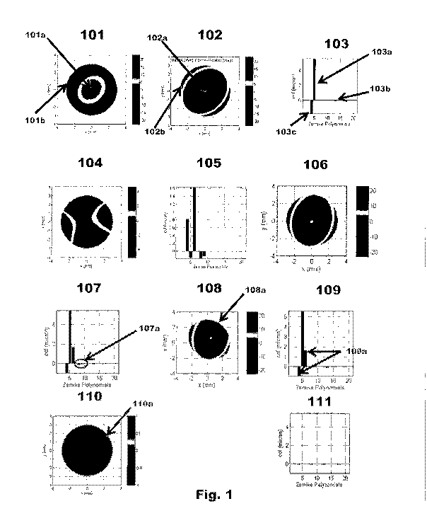
Fig. 1 illustrates an exemplary process of making correctional calculations
for positional
error of a lens resting upon a patient's eye using wavefront maps and Zemike
polynomials.
Fig. 2 illustrates a series of exemplary wavefront measurements demonstrating
the
aberrations resultant from a lens oriented on a patient's eye with
Decentration Error and no
Rotation Error.
Fig. 3 illustrates a series of exemplary wavefront measurements demonstrating
the
aberrations resultant from. a lens oriented with R.otation Error and no
Decentration Error. .
Fig. 4 illustrates in flow chart form an exemplary method of using wavefront
data to
calculate Positional Offset, and using such information to select or design a
more optimal lens
for a patient..
DETAILED DESCRIPTION OF THE INVENTION
The present invention provides a system and m.ethod for determining Rotational
Error
and/or Decentration Error of a contact lens wornwhen by a given patient. This
information may
be used to select or design a subsequent custom. lens for that patient. In the
following sections,
detailed descriptions of embodiments and methods will be given. The
description of both
preferred and alternative embodiments though are exemplary embodiments only,
and it is
understood that to those skilled in the art that variations, modifications and
alterations may be
-5-
CA 02931287 2016-05-19
WO 2015/080938
PCT/US2014/066601
apparent. It is therefore to be understood that the exemplary embodiments do
not limit the
broadness of the aspects of the underlying invention as defined by the claims.
GLOSSARY
In the description and claims directed to the present invention, various terms
may be used
for which the following definitions will apply:
"Decenfration Error" as used herein, refers to an orientation offset, often
descried in
terms of (x, y) coordinates, relative to a determined point upon a patient's
eye, such as the pupil
or iris center, or a limbal edge. For example, a Lens with Decentration Error
may orient itself
where only a fraction of the Optic Zone sits over pupil region and skewing the
corrective power
of the Lens.
"Fitting Lens" as used herein refers to a standard, preferably stabilized
contact lens that
is designed to aid a manufacturer in determining lens position on the eye, or
for selecting or
designing a contact lens. The fitting lens may have stability and measuring
points incorporated
in the lens to assist with measuring the rotational position of the lens and
the decentration of the
lens in relation to the patient's eye.
"Eye Physiology" or "Human Eye Physiology" as referred to herein includes the
patient's
unique shape of the front portion of the eye (the "anterior chamber") for whom
an ophthalmic
lens may be generatedlcustomized for best fit. This includes, but is not
limited to properties of a
patient's eyeball, eye lids, or tear function.
"Lens" as used herein refers to any ophthalmic device that resides in or on
the eye. These
devices can provide optical correction or may be cosmetic. For example, the
term lens can refer
to a contact lens, intraocular lens, overlay lens, ocular insert, optical
insert or other similar device
-6-
CA 02931287 2016-05-19
WO 2015/080938
PCT/US2014/066601
through which vision is corrected or modified, or through which eye physiology
is cosmetically
enhanced (e.g. iris color) without impeding vision. In some embodiments, the
preferred lenses of
the invention are soft contact lenses are made from silicone elastomers or
hydrogels, which
include but are not limited to silicone hydrogels, and fluorohydrogels.
"Lens Design" as used herein, refers to form, function or both of a desired
Lens, which if
fabricated, may provide optical power correction, acceptable Lens fit (e.g.,
corneal coverage and
movement), acceptable Lens rotation stability, etc. Lens Designs may be
represented in either a
hydrated or un-hydrated state, in Flat or Curved Space, in 2-dimensional or 3-
dimensional space,
and by a method including but not limited to, geometric drawings, power
profile, shape, features,
thicknesses etc. Lens Designs may contain data associated with a regularly or
irregularly spaced
grid.
"Lens Position Error" as used herein refers to a Lens which orients itself so
that a patient
suffers diminished fit, comfort, visual acuity, or any other desired aspect of
a Lens. This
includes, for example, a Lens oriented with Decentration Error or Rotation
Error, or both. This
may also include a Lens which loses stability as a result of eye movement or a
patient's blinking
dynamics. Any movement, either static or dynamic, that reduces the
effectiveness of any aspect
of the Lens may be considered a Lens Position Error.
"Optical Aberration", or "aberration", as used herein, refers to a distortion
in an image
formed by an optical system. Optical Aberrations may include either one or
both of low order
aberrations (e.g., sphere power, cylinder power, cylinder axis, etc.) and high
order aberrations
(e.g., spherical aberration, trefoil, coma, pentafoil, etc.).
-7-
CA 02931287 2016-05-19
WO 2015/080938
PCT/US2014/066601
"Optimal Lens Position" as used herein, refers to a lens positioned with no
Rotation Error
or Decentration Error relative to the needed corrective orientation of a Lens
on an eye.
Additionally, this term may refer to aspects of stability and variation, which
may or may not, be
the result of eye or eyelid movement.
"Rotation Error" as used herein, refers to a misalignment relative to an
angular
orientation that meets the needs of a patient's eye. For example, a Lens may
orient itself upon a
patient's eye at a 30 degree clockwise error, therefor skewing one or multiple
of the corrective
power axes.
Typically, a patient is given an eye exam as part of the process that an eye
care
practitioner uses to select a suitable contact lens for that patient. As
indicated previously,
however, a selected lens may not always behave as anticipated when actually
placed on the eye
due to the interaction between the lens and the patient's unique Eye
Physiology, which may
affect lens comfort, fit and/or vision when the lens is on the eye. Measuring
and evaluating the
positional and rotational parameters of a lens as it sits on a patient's eye,
and potentially using
that data to determine the appropriate lens that will provide a more Optimal
Lens Position is the
purpose of the present invention.
A wavefront exam is one test that may be administered on the patient during an
eye
exam. Generally speaking, a wavefront aberrometer measures how light bends as
it is introduced
to and returned from the patient's eye. These devices can diagnose both low
order vision errors
(e.g., nearsightedness, farsightedness and astigmatism) and higher order
vision errors (e.g.,
coma, trefoil and spherical aberration). An exemplary wavefront abetTometer is
the OPD-Scan
III, which is commercially available from Nidek Co., Ltd. of Japan.
-8-
CA 02931287 2016-05-19
WO 2015/080938
PCT/US2014/066601
Wavefront aberrometers generate a wavefront map or optical aberration map.
Where an
aberrometer detects zero optical aberrations, the generated map would be
perfectly flat,
representing an ideal situation where the bundle of rays remain parallel and
undistorted as they
pass through the cornea and lens (see e.g., Fig. 110). In reality,
imperfections in the eye due to
the unique Eye Physiology of any given patient cause distortions of the waves
so that the
resulting wavefront map represents a non-flat three-dimensional image, with
each point on the
displayed map representing the difference between zero optical aberrations and
the measured
optical aberrations. This three-dimensional map is typically displayed with
varying colors that
correspond to the relative divergence from zero aberrations at any given
point. Fig. 101
illustrates a generated wavefront map in grey scale rather than color,
although it is readily
understood that commercial aberrometers typically provide a colored display.
Different aberrations in the waves passing through an eye have been identified
and
classified as different vision errors in what is sometimes known as a Zernike
pyramid. These
identified aberrations can each be represented by a mathematical equation
known as a Zernike
polynomial. The sum of all the Zernike polynomials describes the total of the
optical aberrations
or the collective vision error in a given eye. Zernike polynomials are also
well known to those
skilled in the art of optics and vision science. 'Wavefront imaging devices
may also include, as
output, a display identifying Zernike polynomials for the captured image, such
as that shown in
Fig. 103.
The present invention leverages these technologies in a new and unforeseen
manner to
provide a system and method that more precisely and consistently determines
the Rotation Error
and/or Decentration Error of a lens on a patient's eye, which further enables
the design selection
of a more optimal lens for that patient. Referring now to Fig. 1, a wavefront
aberrometer or the
-9-
CA 02931287 2016-05-19
WO 2015/080938
PCT/US2014/066601
like is used to generate a wavefront map of the patient's bare eye, as
represented by Fig. 101. As
indicated previously, the relative grey scale represents deviations from a
perfect eye with, e.g.,
reference numeral 101a depicting what might be referred to as a "peak" or high
point, and
reference numeral 101b representing a "valley" or low spot, such that the
overall shape if in three
dimensions might represent an upside down bowl, elongated in one direction.
Once a wavefront map of a patient's bare eye is generated, those skilled in
the art will
readily understand how to read such a map and use it to select a contact lens
that will better
correct the patient's vision. As indicated, however, this selection does not
account for any
Positional Error that may occur when the lens is actually wo.rn by the
patient. Fig. 102
represents a wavefront map for a lens designed or selected to correct for the
wavefront error of
patient's eye that is revealed by Fig. 101, or alternatively, the wavefront
map of a lens, such as a
Fitting Lens, that will be placed on the patient's eye for the purpose of
evaluating whether the
Lens orients itself with a Positional Offset. Fig. 102 represents the
wavefront map of the lens
itself, independent of a patient's eye. 102a depicts a "valley" and 102b
represents a "peak" in a
manner somewhat opposite to the error seen in the wavefront map of Fig. 101,
with the idea
being that the selected lens will "cancel out" or neutralize errors identified
in Fig. 101.
Fig. 103 is a graph representing the Zernike polynomial coefficients of the
wavefront
map of Fig. 102. As alluded to previously, any wavefront can be represented as
a weighted
linear summation of Zernike polynomials based on these coefficients. The
graphical output
shown is common in wavefront aberration devices. In this example, the Zernike
polynomial
coefficients shown in Fig. 103 are representative of the corrective properties
of a designed lens,
such as that used to generate the wavefront map of Fig. 102. in particular,
the Zernike
polynomial coefficients at 103a, 103b, and 103c represent the amounts of
defocus, spherical
-10-
CA 02931287 2016-05-19
WO 2015/080938
PCT/US2014/066601
aberration, and astigmatism =Tied in the wavefront map of Fig.! 02. The
coefficients of all other
aberrations terms are zeros in this example.
Next, the selected lens used to generate the wavefront map of Fig. 102 is
inserted into the
patient's eye. A wavefront exam is then administered with the lens in place,
resulting in the
wavefront map shown in Fig. 104. If the selected lens optimally corrects the
patient's vision, the
resulting wavefront map would be perfectly flat, with no peaks and valleys,
such as the map
shown in Fig. 110. Due to Positional Errors, however, the wavefront map of
Fig. 104 shows
residual errors. Fig. 105 illustrates the Zernike polynomials for the
wavefront map of Fig. 104,
which show residual aberrations that exist as a result of Decentation Error
and Rotation Error.
In this example of Fig. 105, the Zernike polynomials also show error in terms
of astigmatism,
defocus, and coma, etc.
Next, a wavefront map (Fig. 106) is generated that represents the deviation
from or
difference between the wavefront map of Fig. 104 (that of the selected lens on
the patient's eye)
from the wavefront map of Fig. 102 (that of the lens itself). This difference
represents the net
wavefront error introduced by the decentered and/or rotated lens. Fig. 107
shows the Zernike
polynomial coefficients of the wavefront map of Fig. 106, which are different
from those shown
in 103 due to lens rotation and decentration. In this example, the coma
aberration terms 107a
are solely due to lens Position Error. A calculation (described further below)
may be made to
predict the amount of lens decentration from the coefficients of coma
aberration terms shown at
107a. If, however, the Zernike polynomials shown in Fig. 103 included coma
aberration terms,
these coma aberration terms would first need to be neutralized, or subtracted
out, so that the
remaining Zernike Polynomial coefficients were solely that due to lens
decentration.
-11-
CA 02931287 2016-05-19
WO 2015/080938
PCT/US2014/066601
Once the lens Decentration Errors are obtained, the wavefront error map shown
in
Fig.106 can be repositioned. In other words, the map 106 is centered by
adjusting the map by an
amount and in a corrective direction so that it is positioned as if the lens
had not undergone any
decentration at all. Another wavefront map represented by Fig. 108 is
generated based on the
repositioned map, which shows residual wavefront aberrations that remain after
the Decentration
Errors have been corrected. The portion of the wavefront map at 108a, which is
not displayed by
a wavefront representation, is due to the fact that then Decentration Errors
of the lens have been
corrected for by calculation, and an absence of values for a portion of the
now centered lens is
unavailable because the lens was out of position when the second wavefront
exam was
administered. Fig. 109 represents the Zernike polynomial coefficients of the
wavefront map of
Fig. 108. The Zemike polynomial coefficients shown in 109 are different from
those shown in
103. As wavefront map 108 has been adjusted for decentration, the difference
between the
Zemike polynomials of Fig. 109 and 103 is solely due to lens rotation. Such
differences are
shown in both astigmatism terms, at 109a. From these terms, a calculation
(described further
below) may be made to predict the amount of Rotation Error.
Once Decentration Errors and Rotation Error, for the lens as worn by the
patient, are
obtained, the optic zone of the lens can be adjusted to compensate for any
such error. For
example, the Decentration Error and Rotation Error data may be converted into
(x, y)
coordinates. From those coordinates, a new Lens Design may be produced where
the optic zone
of the new lens is re-positioned by the (x, y) coordinates relative to a
peripheral zone, or skirt of
the lens. When the newly selected or redesigned lens is centered on the eye,
the corrective
wavefront of this second lens, as worn by the patient, will now corresponds
more closely, if not
optimally, to Fig. 102, which is the desired correction for the patient.
Summation of the centered,
-12-
CA 02931287 2016-05-19
WO 2015/080938
PCT/US2014/066601
re-designed lens' wavefront and the wavefront errors of the bare eye (Fig.
101) leads to zero
aberration as shown in Fig. 110, which is represented by a flat wavefront map.
The graph of Fig.
111 illustrates the Zernike coefficients of the zero wavefront aberrations
illustrated in Fig. 110.
Optically, this means that the residual aberration of the new lens-on-eye
system is zero, as the
lens fully corrects for the aberration errors of the patient's eye (Fig. 101).
Referring again to Figs. 101-111, one manner in which the method and
calculations
described generally above can be implemented will now be described in more
detail. From the
wavefront map of Fig. 102, the Zernike polynomials shown in Fig. 103 may be
denoted as
which represents the Zernike polynomial coefficients of the wavefront of the
centered
1.0 designed lens. Next, the actual error of the lens on the eye is
calculated by taking the wavefront
error of Fig. 104 and finding the difference between that error and the
original wavefront error of
the eye (Fig. 101), That difference represents the Zernike polynomial
coefficients of the net
wavefront errors introduced by the actual decentered and rotated lens, which
may be represented
. =
by
x= =
Since the actual lens on the eye is decc.mtered and rotated, cl-'4*.at4'..."
is different from
which corresponds to the Zernike coefficients of the wavefront of lens if it
was
perfectly centered on the patient's eye. Such difference can be calculated as
2.0 ¨ 'CC.Leicntai_EP¨ 7..D.estgrt_EP The 8th and 9111 Zernikepolynomial terms
(denoted as 'AC8' and
'AC9' respectively) in AC represent coma terms. As is well known in the art,
these terms directly
relate with lens vertical and horizontal decentration (denoted as 'Ay' and
'Ax' respectively) and
inversely relate with spherical aberration of the centered lens design, which
is the 13th term
-13-
CA 02931287 2016-05-19
WO 2015/080938
PCT/US2014/066601
=,
W 4 = rkp
(denoted as Csign_Ep) Therefore, decentration can be readily
calculated by a e .cj"s4"'-' .
relation as follows:
C8
= k __
, k -13
(where k is a constant that changes with pupil size)
Crjesign_EP ('Design_EP
Once lens Decentration Error is obtained, the wavefront error map can be
repositioned, as
described above and as shown in Fig. 108. The Zernike coefficients of the
wavefront error of
Fig. 108 as denoted by eCL_adjustedl is represented by the graph of Fig. 109.
The difference
between (1-adjusted and 447-4"-.6P is solely due to lens rotation and can be
calculated as follows:
Rotation angle =
e
tan k,'-,60,San_EP1/0esigrt_E.P.) VUC.L_adjusted / (It
;Wasted)
wherein: CrIesi,
gn_EP and Ctesign_EP represents the 4th and 6m aberration coefficients in
Zemike vector
C OkW4F.:nsZ.P.
Cl_adjusted and CL adjusted represents the 4th and 6th aberration coefficients
in &mike vector,
e+CL_adjusted= Once the decentration and rotation of the lens are obtained,
the peripheral zone of
the lens can be adjusted to compensate for such decentration and rotation as
described above.
When the adjusted lens is centered on the eye, the residual aberration of lens-
on-eye system is
zero, as the lens optimally corrects aberration error of the eye.
By way of further example, Figs. 201-208 and 301-308 and the corresponding
description
illustrate what could be encountered in a patient that has only Decentration
Error (Figs. 201-208)
or only Rotation Error (Figs. 301-308), but not both, when wearing a selected
lens. First, Figs.
201-208 illustrate a situation where a selected lens, when placed on a
patient's eye exhibits only
-14-
CA 02931287 2016-05-19
WO 2015/080938
PCT/US2014/066601
Rotation Error. Similar to that described above with reference to Figures 101
and 102, Fig. 201
is a wavefront map of the patient's bare eye; Fig. 202 is a wavefront map of
the initially selected
lens; and Fig. 203 represents the Zernike polynomials for the wavefront map of
Fig. 202. Fig.
204 is a wavefront map of the selected lens as worn by the patient, which is
exemplary of a
situation where the selected lens orients itself with Decentration Error but
no Rotation Error.
Fig. 205 represents the Zernike polynomials of the residual wavefront
aberrations which
are shown by the wavefront map of Fig. 204. Next, as also described
previously, the Zernike
polynomials represented in Figs. 202 and 205 are used to calculate the
Decentration Error of the
selected lens. The Zernike polynomials resulting from this calculation are
shown in Fig. 206,
which illustrates the residual aberrations which represent the Decentration
Error of the Lens
oriented on the eye that must be accounted for when selecting or designing the
next lens for the
patient.
Assuming that an alternate lens with the desired parameters exists or is
custom designed,
and when worn by the patient the Lens orients itself similarly to the previous
Lens, then the
wavefront map of Fig. 207 represents the residual wavefront aberrations of the
re-designed lens,
resulting in zero residual aberrations as shown in the wavefront map of Fig.
207 and
corresponding Zernike polynomials represented in Fig. 208.
Figs. 301-308 illustrate an example where an initially selected lens, when
placed in a
patient's eye, exhibits Rotation Error but zero Decentration Errors. Fig. 301
is a wavefront map
of the patient's bare eye; Fig. 302 is a wavefront map of an initial lens
selected based on the
wavefront map of Fig. 301 and designed to correct for the wavefront errors in
that wavefront
map; and Fig. 303 shows the Zernike polynomials for the needed correction as
represented by the
wavefront map of Fig. 302. Fig. 304 is a wavefront map taken of the patient's
eye while wearing
-15-
CA 02931287 2016-05-19
WO 2015/080938
PCT/US2014/066601
the selected lens. Assuming the selected lens orients itself with Rotation
Error and zero
Decentration Errors, a wavefront map of the patient wearing that selected lens
would reveal
aberrations on the wavefront map such as those shown in Fig. 304.
Fig. 305 is a wavefront map representing the calculated residual wavefront
aberrations of
the rotated lens as derived from the wavefront aberrations of Figs. 302 and
305. As described
above with reference to Fig. 101-111, Fig. 306 represents the Zernike
coefficients of Fig. 305,
which illustrate residual aberrations that represent the Rotation Error of the
Lens oriented on the
eye that must be accounted for when selecting or designing the next Lens for
the patient to
account for the Rotation Error demonstrated by the initial Lens.
Assuming that an alternate Lens with the desired parameters exists or is
custom designed,
and when wo.rn by the patient the Lens orients itself similarly to the
previous Lens, then the
wavefront map of Fig. 307 represents the residual wavefront aberrations of the
newly selected or
re-designed lens, which is zero. Fig. 308 illustrates the Zernike polynomials
for the wavefront
map of Fig. 307.
Referring now to Fig. 4, a method of extracting wavefront data and calculating
Lens
Position Error using the wavefront data is shown in flowchart form.
Additionally, the flowchart
demonstrates the methods of correcting for the Lens Position Error by
providing a Lens that
accounts for that Lens Position Error.
At 401, a wavefront exam is administered on a patient's bare eye. in an
exemplary
embodiment, a wavefront exam may be administered with a wavefront aberrometer
device, such
as the OPD-Scan 111 noted previously. The wavefront exam provides wavefront
refraction data
typically in the form of a wavefront map as has also been discussed above. At
402, the
wavefront refraction data may then be used to choose an initial lens suitable
for the patient. in
-16-
CA 02931287 2016-05-19
WO 2015/080938
PCT/US2014/066601
an exemplary embodiment, the wavefront data, which may or may not be converted
into Zemike
coefficient space, may be used to select the appropriate standard lens, or
alternatively, to select
an appropriate Fitting Lens for the patient, and the selected lens is placed
onto the patient's eye
(403).
At 404, a subsequent wavefront exam is then administered while the patient is
wearing
the selected Lens, which provides wavefront data such as in the form of a
wavefront map, which
also may or may not be represented by Zemike coefficients. If the selected
lens orients itself
with Lens Position Error, this second wavefront exam will provide over-
refraction wavefront
data. The over-refraction wavefront data may be either in the form of a
wavefront map
illustrating wavefront aberrations or in the form of Zemike polynomials.
Following the
extraction of the second set of wavefront refraction data at step 405, the
first set of data, from
which the original lens was selected, may then be compared to the second set
of wavefront
refraction data.
Next, at 406, calculations are made using the wavefront data, and in some
embodiments,
the Zemike polynomial representations of the wavefront data, of the patient's
bare eye and of the
first selected lens on the patient's eye. The calculations at 406 can
determine the Decentration
Errors and/or Rotation Error. At 407, based on the errors calculated at 406, a
subsequent lens
may be selected that provides more optimal vision correction for the patient.
This subsequent
selected lens may be either a standard lens or a custom lens designed
specifically to account for
the errors calculated at 406. Additional wavefront exams may be administered,
at 408, on the
patient wearing the subsequent Lens, and any further lenses necessary, and the
same wavefront
data calculation method repeated until a Lens results in the Optimal Lens
Position available to
that patient based on that patient's Eye Physiology.
-17-
CA 02931287 2016-05-19
WO 2015/080938
PCT/US2014/066601
Once positioning data in terms of Decentration Error and Rotation Error is
obtained, a
second lens may be either selected or designed. The above examples mainly
demonstrate the
typical eye care practitioner practice using standard or stock lenses, where
there is a finite
amount of choices the eye care practitioner has when selected the second or
further subsequent
lenses. With a custom lens, such as a lens produced through a ContourForm
manufacturing
process (as is described in detail in U.S. Patent No. 8,317,505, which is
incorporated herein by
reference in its entirety), the positioning data provides more options for
designing a second or
further subsequent lens for the patient.
As mentioned above, one exemplary embodiment is to correct for the positioning
error of
the entire lens by repositioning only the optic zone relative to the remainder
of the lens. This
approach allows the lens to retain the same on-eye position, while moving the
optic zone to a
location on the lens that will provide the patient with the designed vision
correction.
In addition to moving the optic zone, additional embodiments exist that
involve designing
a lens so that it positions differently on the eye than the first lens. One
exemplary embodiment is
to produce a lens with a different base curve. Standard lens manufacturing
practice is to offer a
small number of back curve variations in a particular lens product line. A
ContourForm
manufacturing process may offer a wider selection of base curves or a custom
base curve.
Therefore, once positioning data has been obtained for a particular lens, an
analysis of that data
may allow for a design of a lens that incorporates an alternate base curve.
The alternate base
curve will interact differently with the patient's eye and eye lid, resulting
in a different lens
position than the first lens. Following the above methods, a series of lenses
with alternate base
curves may be selected until a minimum lens position error is obtained.
-18-
CA 02931287 2016-05-19
WO 2015/080938
PCT/US2014/066601
A further exemplary embodiment is designing a second lens with an alternate
diameter.
A lens edge has the additional condition of also interacting with the
patient's eye lids. Therefore,
an analysis of the positioning data may allow a lens design with an alternate
diameter. This
second lens with an alternate diameter may interact differently with the
patient's eye and eye lids
and therefore result in a different lens position. Following the above
methods, a series of lenses
designed with alternate diameters may be produced until a minimum lens
position error is
obtained.
Additionally, a derivative of the interaction between a lens edge in terms of
diameter is
the interaction between lens and patient's eye in terms of lens shape. Just as
a different diameter
may interact with a patient's eye and eye lid differently, the same is true of
a different lens shape.
In an exemplary embodiment, the initial lens shape may be the round shape
typical of standard or
stock. lenses. Positioning data may be analyzed to design a lens with an
alternate shape, such as a
lens with a wider lower portion and a narrower top portion. The change in lens
shape may
change the interaction with the patient's eye and eye lid therefore changing
the resulting lens
positioning. A series of lenses with alternate shapes may be produced until a
minimum lens
position error is obtained.
A further embodiment may include modifying internal features of the lens. In
an
exemplary embodiment, a lens may be produced with stabilization zones.
Stabilization zones, by
design, affect the stability andlor positioning of a lens on an eye. In
typical eye care practitioner
practice, the standard or stock lenses from which each subsequent lens may be
chosen have a
finite number of stabilization zone options, if any at all. In a manufacturing
process such as
ContourForm, it may be possible to produce the stabilization zones to provide
a custom fit for
the patient. Once the positioning data is obtained and analyzed, a lens design
may be produced
-19-
CA 02931287 2016-05-19
WO 2015/080938
PCT/US2014/066601
that modifies one or all of the stabilization zones to result in reduced lens
movement on the eye.
A series of lens may be produced with alternate stabilization zones until a
minimum lens position
error is obtained.
Further exemplary embodiments include combined modifications of the above
mentioned
lens design parameters. For example, lens sag is a function of the dimensions
of the lens
diameter, base curve and shape. Altering the lens sag may have a similar
effect of alternating the
base curve, diameter, shape or all three. However, lens sag specifically
refers to a distance from
the apex to a parallel line with the lens edge, in curved space. A lens may
interact with a
patient's eye and eye lids differently as a function of sag as opposed to a
function of solely base
curve, diameter or shape. Consequently, a lens design with alternate sag may
also include an
alternate diameter and/or shape, but the change in lens position may not be
identical to any
change in lens position based solely on one of the other parameters of
diameter or shape.
Another exemplary embodiment of a combination of the above parameters may
include a
lens designed with modified stabilization zones and a repositioned optic zone.
For example, the
second lens, or first few subsequent lenses may be designed with modified
stabilization zones.
However, the change in lens position due to the modified stabilization zones
may not correct for
the entire lens positioning error. Once an improved lens position has been
accomplished via
stabilization zone modification, the optic zone may then be repositioned to
correct for the
remaining amount of lens position error.
Although illustrative embodiments of the present invention have been described
herein
with reference to the accompanying drawings, it is to be understood that the
invention is not
limited to those precise embodiments and that various other changes and
modifications may be
-20-
CA 02931287 2016-05-19
WO 2015/080938
PCT/US2014/066601
effected herein by one skilled in the art without departing from the scope or
spirit of the
invention.
-21-