Note: Descriptions are shown in the official language in which they were submitted.
CA 02933440 2016-06-09
WO 2015/088915 PCT/US2014/068897
METHODS AND COMPOSITIONS FOR TREATING AGING-
ASSOCIATED CONDITIONS
CROSS-REFERENCE TO RELATED APPLICATIONS
Pursuant to 35 U.S.C. 119 (e), this application claims priority to the
filing date of
the United States Provisional Patent Application Serial No. 62/069,044, filed
October 27,
2014, and United States Provisional Patent Application Serial No. 61/913,812,
filed
December 9, 2013; the disclosure of which is herein incorporated by reference.
INTRODUCTION
Aging in an organism is accompanied by an accumulation of changes over time.
In
the nervous system, aging is accompanied by structural and neurophysiological
changes
that drive cognitive decline and susceptibility to degenerative disorders in
healthy
individuals. (Heeden, T. & Gabrieli, J.D., Insights into the ageing mind: a
view from
cognitive neuroscience. Nat. Rev. Neurosci. 5(2), 87-96 (2004); Raz, N. et al.
Neuroanatomical correlates of cognitive aging: evidence from structural
magnetic
resonance imaging. Neuropsychology 12(1), 95-114 (1998); Mattson, M.P. &
Magnus, T.,
Ageing and neuronal vulnerability. Nat. Rev. Neurosci. 7(4), 278-294 (2006);
Rapp, P.R. &
Heindel, W.C., Memory systems in normal and pathological aging. Curr. Opin.
Neurol. 7(4),
294-298 (1994)). Included in these changes are synapse loss and the loss of
neuronal
function that results. Thus, although significant neuronal death is typically
not observed
during the natural aging process, neurons in the aging brain are vulnerable to
sub-lethal
age-related alterations in structure, synaptic integrity, and molecular
processing at the
synapse, all of which impair cognitive function.
In addition to the normal synapse loss during natural aging, synapse loss is
an early
pathological event common to many neurodegenerative conditions, and is the
best correlate
to the neuronal and cognitive impairment associated with these conditions.
Indeed, aging
remains the single most dominant risk factor for dementia-related
neurodegenerative
diseases such as Alzheimer's disease (AD) (Bishop, N.A., Lu, T., & Yankner,
B.A., Neural
mechanisms of ageing and cognitive decline. Nature 464(7288), 529-535 (2010);
Heeden,
T. & Gabrieli, J.D., Insights into the ageing mind: a view from cognitive
neuroscience. Nat.
Rev. Neurosci. 5(2), 87-96 (2004); Mattson, M.P. & Magnus, T., Ageing and
neuronal
vulnerability. Nat. Rev. Neurosci. 7(4), 278-294 (2006)).
As human lifespan increases, a greater fraction of the population suffers from
aging-
associated cognitive impairments, making it crucial to elucidate means by
which to maintain
cognitive integrity by protecting against, or even counteracting, the effects
of aging (Hebert,
L.E. et al. Alzheimer disease in the US population: prevalence estimates using
the 2000
1
CA 02933440 2016-06-09
WO 2015/088915 PCT/US2014/068897
census. Arch. Neurol. 60(8), 1119-1122 (2003); Bishop, N.A., et al., Neural
mechanisms of
ageing and cognitive decline. Nature 464(7288), 529-535 (2010)).
SUMMARY
Methods and compositions are provided for treating a subject for aging-
associated
conditions, e.g., cognitive impairment conditions or age-related dementia.
Aspects of the
methods include administering a young plasma-comprising blood product to an
individual in
need thereof, e.g., an individual suffering from or at risk of developing the
aging-associated
condition, e.g., aging-associated cognitive impairment or age-related
dementia. Also
provided are compositions and kits thereof that find use in practicing methods
of the
invention.
BRIEF DESCRIPTION OF THE FIGURES
Figure 1. Genome-wide microarray analysis of heterochronic parabionts
identifies a
plasticity related expression profile in the old hippocampus. Microarray
analysis was
performed on hippocampi of old (18-month-old) isochronic and heterochronic
parabionts 5
weeks post-surgery. N=4 mice per group. For all analyses down-regulated genes
are shown
in shades of blue and up-regulated genes are shown in shades of yellow. a,
Schematic
depicting parabiotic pairings. lsochronic pairs shown in gray and
heterochronic pairs shown
in red. b. Heat map was generated by unsupervised hierarchical clustering with
data set of
genes differentially expressed between hippocampi of old isochronic and
heterochronic
parabionts using a cut-off at p<0.01 and d-score>2 based on Significance
Analysis of
Microarray (SAM). c, Hierarchical clustering of synaptic plasticity related
genes identified by
AmiG0 (Gene Ontology) using a cut-off at p<0.01 and d-score>1.5 based on SAM.
Color
bars in b and c reflect Z-scores. d. Biological pathways involved in synaptic
plasticity were
identified as part of the top signaling network (p<0.05) using Ingenuity
Pathway Analysis
(IPA) software based on differentially expressed genes in isochronic and
heterochronic
parabionts. Inferred molecular interactions identified by IPA are shown in
gray.
Figure 2. Heterochronic parabiosis enhances synapse formation and synaptic
plasticity in the old brain. a-g, Histological and electrophysiological
analysis was done on
old (18-month-old) isochronic and heterochronic parabionts analyzed 5 weeks
post surgery.
N=5-6 mice per group a, lmmunohistochemical detection of Egr1, cFos, and
phosphorylated cyclic AMP response element binding (CREB) protein in the DG of
the
hippocampus of old isochronic and heterochronic parabionts. Arrowheads depict
individual
cells. (scale bar: 100 pm). b-d, Quantification of immunostaining for Egr1
(b), c-Fos (c) and
phosphorylated CREB (d). 5 sections per mouse were analyzed. e,f
Representative Golgi
stain image (e) and quantification of dendritic spine density on tertiary
branches (f). 5
2
CA 02933440 2016-06-09
WO 2015/088915 PCT/US2014/068897
neurons per mouse were analyzed. g, Population spike amplitude (PSA) was
recorded
from DG of old parabionts. Representative long-term potentiation (LTP) levels
are shown for
isochronic and heterochronic parabionts. Data represented as Mean SEM;
*P<0.05;
**P<0.01; t-test (b-d).
Figure 3. Young blood administration improves hippocampal dependent learning
and memory in old mice. a-c. Old (18-month-old) mice were cognitively tested
after
treatment with young (3-month-old) or old (18-month-old) plasma 8 times over
24 days
(100p1/intravenous injection). N=8 mice per group. a, Schematic illustrating
the
chronological order used for plasma treatment and cognitive testing. b,c,
Hippocampal
learning and memory was assessed by contextual fear conditioning (b) and
radial arm
water maze (RAWM) (c) following plasma treatment. b, Percent freezing time 24
hours
after training. c, Number of entry arm errors prior to finding platform. Data
represented as
Mean SEM; *P<0.05; **P<0.01; t-test (b), ANOVA, Bonferroni post-hoc test
(c).
Figure 4. lsochronic parabiosis does not alter expression of synaptic
plasticity
markers. Histological analysis of synaptic plasticity markers was done on the
DG of the
hippocampus of old (18-month-old) isochronic parabionts and unpaired age-
matched
controls. N=5-6 mice per group. a-c, Quantification of immunostaining for Egr1
(b), c-Fos (c)
and phosphorylated CREB (d). 5 sections per mouse were analyzed. Bars are mean
+
SEM; n.s., not significant; t-test.
Figure 5. Heterochronic parabiosis does not alter dendritic complexity or
basal
synaptic transmission. a-c, Golgi analysis was done using Neurolucida Software
(v10, MBF
Bioscience) on 5 neurons per mouse (18-month-old) for a total of 25 neurons.
N=5 per
group. a, Sholl analysis was graphed as the average intersections per shell
per neuron
against the distance from the soma. b,c, Neuron tracings were used to quantify
the average
number of primary, secondary and tertiary dendritic branches (b) and total
dendrite length
(c). d, input-output curves indicate no statistical difference in synaptic
strength, a key
parameter of basal synaptic transmission, between old isochronic and
heterochronic
parabionts. Bars are mean + SEM; n.s., not significant; t-test.
Figure 6. Hippocampal dependent learning and memory is impaired in old mice. a-
e, Learning and memory was examined during normal aging in young (3-month-old)
versus
old (18-month-old) adult animals using contextual fear conditioning (a-c) and
RAWM (d-e)
paradigms. a, Young and old animals exhibited similar baseline freezing time
during fear
conditioning training. b, During contextual fear conditioning old mice
demonstrate
decreased freezing time during contextual memory testing. c, No differences in
cued
memory were detected 24 hours after training. e, Old mice demonstrate impaired
learning
and memory for platform location during the testing phase of the RAWM task.
Cognitive
deficits were quantified as the number of entry arm errors made prior to
finding the target
3
CA 02933440 2016-06-09
WO 2015/088915 PCT/US2014/068897
platform. No differences in swim speeds of were detected between young and old
animals.
Data are from 8 animals per group. Bars are mean + SEM; n.s., not significant;
t-test.
Figure 7. Exposure to young blood does not affect cued memory or swim speed. a-
c, Old adult male mice (18-month-old) were injected intravenously with plasma
(100p1/injection) derived from young (3-month-old) or old (age 18-month-old)
animals 8
times over 24 days. a, Animals intravenously injected with young or old plasma
exhibited
similar baseline freezing time during training. b, No differences in cued
memory were
detected between groups when re-exposed to the conditioned stimulus (tone and
light) in a
novel context 24 hours after training. c, Swim speeds of old and young plasma
treated mice
during the training phase of the RAWM. Data are from 8 animals per group. Bars
are mean
+ SEM; n.s., not significant; t-test.
Figure 8. Hippocampal dependent learning and memory is not altered by exposure
to old blood. a-e, Learning and memory was examined in untreated old adult
mice (18-
month-old) using fear conditioning and RAWM paradigms and compared to old
animals
injected intravenously with plasma (100p1/injection) derived from old (18-
month-old) animals
8 times over 24 days. N=8 per group. No differences in baseline freezing were
detected
during fear conditioning training (a), and no differences in freezing were
detected during
contextual (b) or cued (c) fear conditioning testing. d, No differences in
spatial learning and
memory were detected in the RAWM paradigm. e, No differences in swim speeds
were
observed between animals receiving old plasma and untreated controls. Bars are
mean +
SEM; n.s., not significant; t-test.
Figure 9. Denaturing young plasma abolishes positive cognitive effects of
plasma
treatment in old mice. a. Percentage freezing observed in old mice treated
with PBS, young
plasma, or young denatured plasma during the first minute of exposure to the
same context
as the training environment (n=10-12/group). b. Percentage freezing for old
mice treated
with PBS, young plasma, or denatured young plasma during the cued task in
which mice
are exposed to a new context but given the tone and light cues from training
(n=10-
12/group). Bars represent mean +/- SEM. Groups were compared by 1-way ANOVA
followed by Tukey's post hoc test for multiple comparisons (*P<0.05).
Figure 10. Three weekly administrations of young blood improve hippocampal
dependent learning and memory and neurogenesis in old mice. a, Schematic
illustrating the
chronological order used for plasma treatment, cognitive testing and
histological analysis.
Three 150p1 injections of young plasma (2-3 mo old) or PBS were given i.v.,
one per week
(day 0, 7, 14). After the third injection, a 3-day Radial Arm Water Maze
(RAWM) task was
performed, one group (mixed treatment) starting at day 21, another group
starting at day 24.
A fear-conditioning test (FC) was performed on day 30 (training) and day 31
(testing). All
mice were injected daily with BrdU (50mg/kg) i.p. 3 days prior to sacrifice,
after which
4
CA 02933440 2016-06-09
WO 2015/088915 PCT/US2014/068897
neurogenesis was assessed. b, Number of entry arm errors prior to finding
platform on the
training day (day 1) and testing days (day 2 and 3). The plasma treated group
performed
consistently better on days 2 and 3 than the PBS treated group. One block
represents 3
trials. c, Quantification of learning in the RAWM showing the number of errors
made on day
1, block 1 vs. day 3, block 15. The young plasma-treated group made
significantly fewer
errors in block 15 vs. block 1. d, Normalized freezing behavior in the
contextual fear-
conditioning test shows significantly more freezing in the young plasma-
treated group
compared to the PBS-treated group, consistent with improved memory for the
task. e, Mice
treated with young plasma show a significantly larger number of BrdU-positive
cells in the
dentate gyrus (DG) of the hippocampus compared to the PBS-treated group. Data
represented as Mean SEM; *P<0.05; **P<0.01; ANOVA, Bonferroni post-hoc test
(b), t-test
(c-e).
Figure 11: (a) Schematic depicting the three different types of parabiosis
between
mice: wildtype isochronic (WT iso), APP isochronic (APP iso) and APP
heterochronic (APP
het). lsochronic pairs are age-matched and the same age as the APP mouse from
the
heterochronic pair, which is connected to a young (2-3 month old) wildtype
mouse. One
cohort consisted of old (16-20 month) male mice and another of middle-aged (10-
12 month)
female mice. All pairs were surgically connected for 5 weeks. (b)
Quantification of
immunohistochemical detection of amyloid plaques (3D6 staining) in the
hippocampus of old
APP iso (n = 6) and APP het (n = 4) mice. (c) ELISA measurements of insoluble
total A13 and
A1342 levels in the hippocampus of old male APP iso (n = 6) and APP het (n =
4) mice. (d-e)
Quantification of synaptophysin-immunoreactivity (d) and calbindin-
immunoreactivity (e) in
the molecular layer of the dentate gyrus of old male parabionts; WT iso (n =
6), APP iso (n =
6), APP het (n = 4). (f) Quantification of calbindin-immunoreactivity in the
molecular layer of
the dentate gyrus of middle-aged female parabionts; WT iso (n = 9), APP iso (n
= 11), APP
het (n = 9). All data are shown as the mean + s.e.m. * P < 0.05, ** P < 0.01,
***P < 0.001,
Student's t test (b), two-way ANOVA, Sidak's post hoc test (c), one-way ANOVA,
Tukey's
post hoc test (d-f).
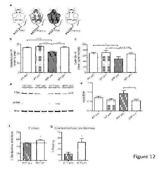
Figure 12: Administration of young blood plasma restores synaptic activity-
and
calcium-related proteins and improves cognition in hAPPus mice. (a) Schematic
depicting
the 4 treatment groups, wildtype (WT) or hAPPus mice treated with either PBS
or young
plasma (150 1 per intravenous tail vein injection, 8 times over 30 days). (b)
Quantification of
synaptophysin-immunoreactivity in the molecular layer of the dentate gyrus of
WT pbs (n =
14), WT plm (n = 13), APP pbs (n = 11) and APP plm (n = 13) mice. (c)
Quantification of
calbindin-immunoreactivity in the molecular layer of the dentate gyrus of WT
pbs (n = 15),
WT plm (n = 13), APP pbs (n = 10) and APP plm (n = 12) mice. (d-e) Western
blot analysis
was performed on hippocampus lysates from all 4 treatment groups, n = 8 per
group. (d)
Representative Western blot for ERK (44/42kDa), phosphorylated ERK (44/42kDa;
pERK)
5
CA 02933440 2016-06-09
WO 2015/088915 PCT/US2014/068897
and neuron specific enolase (NSE) as loading control. (e) Quantification of
the ratio of
pERK/ERK determined by densitometry of bands using ImageJ software. (f-g)
Cognitive
testing of APP mice injected with 8 intravenous injections of PBS (n = 11) or
young plasma
(n = 13). (f) Working memory assessed by spontaneous alternations in a Y-maze
test for 5
minutes. Dotted line represents chance level (50%). (g) Hippocampal-dependent
learning
and memory assessed by contextual fear conditioning indicated by percentage
freezing in
the same context 48 h after training. One mouse was excluded from the APP pbs
group due
to abnormal freezing behavior, determined by the ROUT method of identifying
outliers. All
data are shown as the mean + s.e.m. # P < 0.1, * P < 0.05, ** P < 0.01, ****P
<0.0001, one-
way ANOVA, Tukey's post hoc test (b-c, e), Student's t test (f-g)
Figure 13: 18 month-old mice (N=4-5/group) were injected intravenously 7 times
over 2 weeks with fractionated plasma isolated from 2-3 month-old C5761/6J
mice. One pool
of young plasma was fractionated using molecular weight cut-off dialysis
membranes, which
excluded molecules below molecular weights indicated (i.e., 3.5 kDa, 25 kDa,
50 kDa, and
3.5 kDa + depletion of IgG). Hippocampi from treated mice were isolated and
analyzed on
whole genome Affymetrix arrays for gene expression. The heat map shows near-
complete
segregation by treatment in terms of increased (red) or decreased (blue)
overall gene
expression.
Figure 14: 12-month-old mice were injected i.v. with 150p1 PBS or 150p1 plasma
(PLM) from 2-month-old mice twice a week for 4 weeks. Plasma factors were
analyzed with
a protein microarray (a) or Luminex cytokine assay (b-c). a) Heat map showing
six plasma
factors that were significantly increased or decreased in 12 month old mice
upon
administration of young blood plasma. Unsupervised complete linkage clustering
separates
PLM samples from the PBS samples. b-c) Interleukin-22 (IL-22) and Leukemia
Inhibitory
Factor (LIF) were increased in 12-month-old mice 4 weeks after administration
of young
blood plasma compared to PBS.
Figure 15. NSG mice display age-dependent changes in (a) doublecortin (DCX)+
cells in the dentate gyrus, (b) CD68 staining as a percentage of total
hippocampal area, and
(c) total number of cfos-positive cells in the dentate gyrus. (Mean +/- SEM;
Student's t test;
*P<0.05, **P<0.01, ****P<0.0001.)
Figure 16. (a) Levels of freezing in aged NSG mice are significantly lower
than in
young NSG mice in the last 90 seconds when exposed to a chamber to which they
have
been previously trained to associate with fear. (b) Quantification of freezing
levels in the final
intervals of contextual fear conditioning in young and old NSG mice from (a).
(c) Aged NSG
mice display deficits over days and within trials of the same day in finding
the escape hole
during the Barnes maze. (d) Aged NSG mice also display deficits compared to
young NSG
mice in terms of daily overall performance. (e) The rate of learning, the
difference in
individual probe trials from initial training trial, is significantly higher
in young NSG mice.
(Mean +/- SEM; Student's t test for 2-group comparisons and, where
appropriate, 2-way
6
CA 02933440 2016-06-09
WO 2015/088915 PCT/US2014/068897
repeated-measures ANOVA, followed by Bonferroni's post-hoc test for correction
of multiple
comparisons; *P<0.05, "P<0.01, ***P<0.001, ****P<0.0001.)
Figure 17. Heat map demonstrating a high degree of clustering in terms of
protein
expression by age group among plasma samples taken from human umbilical cord
donors
(N=15), young donors (N=19), or elderly donors (N=16). Blocks represent
individual
secreted signaling proteins that are enriched (yellow) or decreased (blue)
relative to the
levels of expression for that protein among all age groups. Proteins shown are
those that
were significant after time correlation SAM (q<5%).
Figure 18. Injections of human plasma (hPLM) from young or old donors in aged
NSG mice revealed changes in the percentage area occupied by CD68 staining in
the
hippocampus (left) or cortex (right) compared to vehicle-treated NSG mice.
(Mean +1- SEM;
Student's t test; *P<0.05, "P<0.01.)
Figure 19. After normalizing the levels of freezing in the contextual fear
conditioning
task (day 2) to the freezing observed during training (day 1), young human
plasma (hPLM)
increases contextual memory at 4.5 minutes compared to aged NSG mice treated
with old
hPLM. (Mean +1- SEM; Student's t test at the indicated interval; *P<0.05.)
Figure 20. Levels of gene expression by qPCR in aged NSG mice treated with
human cord or young plasma compared to vehicle-treated mice. Changes in
immediate early
gene expression (Egr1, Junb, fos) were assessed in brains isolated from aged
NSG mice
treated intravenously with human plasma or vehicle over 3 weeks. (Mean +1-
SEM; Student's
t test; *P<0.05.)
Figure 21. Additional plasticity-relevant genes BDNF and Camk2a were measured
by qPCR in aged NSG mice treated with human cord plasma or vehicle. (Mean +1-
SEM;
Student's t test; *P<0.05.)
Figure 22. (a) Quantification of levels of freezing in aged NSG mice treated
with cord
plasma or vehicle in the last 90 seconds when exposed to a chamber to which
they have
been previously trained to associate with fear. (b) Aged NSG mice treated with
cord plasma
display enhanced learning and memory by day 4 and within trials of the same
day in finding
the escape hole during the Barnes maze. (c) Cord plasma-treated mice also
display
improved learning and memory compared to vehicle-treated NSG mice in terms of
daily
overall performance. (d) The rate of learning, the difference in probe trials
from the initial
training trial, is significantly higher in cord plasma treated mice compared
to vehicle-treated
mice for the third probe trial. (Mean +1- SEM; Student's t test for 2-group
comparisons and,
where appropriate, 2-way repeated-measures ANOVA, followed by Bonferroni's
post-hoc
test for correction of multiple comparisons; *P<0.05, "P<0.01, ***P<0.001).
7
CA 02933440 2016-06-09
WO 2015/088915 PCT/US2014/068897
Figure 23. (a) Slices taken from brains of cord plasma-treated mice display
enhanced long-term potentiation (LTP) as assessed by measuring the population
spike
amplitudes in dentate gyrus after stimulation in the perforant path of the
hippocampus. (b)
Quantification of the maintenance phase of the PSA shown in (a). (Mean +/-
SEM; Student's
t test; *P<0.05.)
Figure 24. (a) Quantification of the number of TRAPed cells driving effector
protein
expression from cfos in the dentate gyrus (DG) for TRAP-FOS mice treated with
vehicle, old
human plasma (hPLM), or cord hPLM. (b) Quantification of the number of TRAPed
NeuN-
positive (neuron) cells driving effector protein expression from cfos in the
dentate gyrus (DG)
for TRAP-FOS mice treated with vehicle, old hPLM, or cord hPLM. (c)
Quantification of the
number of TRAPed cells driving effector protein expression from cfos in the
CA1 region for
TRAP-FOS mice treated with vehicle, old hPLM, or cord hPLM. (d) Quantification
of the
number of TRAPed NeuN-positive cells driving effector protein expression from
cfos in the
CA1 region for TRAP-FOS mice treated with vehicle, old hPLM, or cord hPLM.
(Mean +/-
SEM; 1-way ANOVA, followed by Tukey's post-hoc test for correction of multiple
comparisons; *P<0.05.)
DETAILED DESCRIPTION
Methods and compositions are provided for treating a subject for aging-
associated
conditions, e.g., cognitive impairment conditions, age-related dementia or age
related
decline of physiological function of peripheral organ(s). Aspects of the
methods include
administering a young plasma-comprising blood product to an individual in need
thereof,
e.g., an individual suffering from or at risk of developing the aging-
associated condition,
e.g., aging-associated cognitive impairment or pathological types of dementia.
Also
provided are compositions and kits thereof that find use in practicing methods
of the
invention.
Before the present methods and compositions are described, it is to be
understood
that this invention is not limited to a particular method or composition
described, as such
may, of course, vary. It is also to be understood that the terminology used
herein is for the
purpose of describing particular embodiments only, and is not intended to be
limiting, since
the scope of the present invention will be limited only by the appended
claims.
Where a range of values is provided, it is understood that each intervening
value, to
the tenth of the unit of the lower limit unless the context clearly dictates
otherwise, between
the upper and lower limits of that range is also specifically disclosed. Each
smaller range
between any stated value or intervening value in a stated range and any other
stated or
intervening value in that stated range is encompassed within the invention.
The upper and
lower limits of these smaller ranges may independently be included or excluded
in the
8
CA 02933440 2016-06-09
WO 2015/088915 PCT/US2014/068897
range, and each range where either, neither or both limits are included in the
smaller ranges
is also encompassed within the invention, subject to any specifically excluded
limit in the
stated range. Where the stated range includes one or both of the limits,
ranges excluding
either or both of those included limits are also included in the invention.
Unless defined otherwise, all technical and scientific terms used herein have
the
same meaning as commonly understood by one of ordinary skill in the art to
which this
invention belongs. Although any methods and materials similar or equivalent to
those
described herein can be used in the practice or testing of the present
invention, some
potential and preferred methods and materials are now described. All
publications
mentioned herein are incorporated herein by reference to disclose and describe
the
methods and/or materials in connection with which the publications are cited.
It is
understood that the present disclosure supersedes any disclosure of an
incorporated
publication to the extent there is a contradiction.
As will be apparent to those of skill in the art upon reading this disclosure,
each of
the individual embodiments described and illustrated herein has discrete
components and
features which may be readily separated from or combined with the features of
any of the
other several embodiments without departing from the scope or spirit of the
present
invention. Any recited method can be carried out in the order of events
recited or in any
other order which is logically possible.
It must be noted that as used herein and in the appended claims, the singular
forms
"a", "an", and "the" include plural referents unless the context clearly
dictates otherwise.
Thus, for example, reference to "a cell" includes a plurality of such cells
and reference to
"the peptide" includes reference to one or more peptides and equivalents
thereof, e.g.,
polypeptides, known to those skilled in the art, and so forth.
The publications discussed herein are provided solely for their disclosure
prior to the
filing date of the present application. Nothing herein is to be construed as
an admission
that the present invention is not entitled to antedate such publication by
virtue of prior
invention. Further, the dates of publication provided may be different from
the actual
publication dates which may need to be independently confirmed.
METHODS
As summarized above, aspects of the invention include methods for treating a
subject for aging-associated conditions. By aging-associated condition is
meant a condition,
e.g., a disease condition or other undesirable condition, which accompanies
aging of an
organism. The aging associated condition may manifest in a number of different
ways, e.g.,
as aging associated damage to central or peripheral organs of the body, such
as but not
limited to: cell injury, tissue damage, organ dysfunction, aging-associated
lifespan
9
CA 02933440 2016-06-09
WO 2015/088915 PCT/US2014/068897
shortening and carcinogenesis, where specific organs and tissues of interest
include, but
are not limited to skin, neuron, muscle, pancreas, brain, kidney, lung,
stomach, intestine,
spleen, heart, adipose tissue, testes, ovary, uterus, liver and bone. In some
instances,
treatment of a subject in accordance with the methods results in a change in a
central
organ, e.g., a central nervous system organ, such as the brain, spinal cord,
etc., where the
change may manifest in a number of different ways, e.g., as described in
greater detail
below, including but not limited to molecular, structural and/or functional.
In some instances,
treatment of a subject in accordance with the methods results in a change in a
peripheral
organ, such as liver, muscle, heart, blood, etc., where the change may
manifest in a number
of different ways, e.g., as described in greater detail below.
In some embodiments, the aging-associated condition that is treated is an
aging-
associated impairment in cognitive ability in an individual. By cognitive
ability, or
"cognition", it is meant the mental processes that include attention and
concentration,
learning complex tasks and concepts, memory (acquiring, retaining, and
retrieving new
information in the short and/or long term), information processing (dealing
with information
gathered by the five senses), visuospatial function (visual perception, depth
perception,
using mental imagery, copying drawings, constructing objects or shapes),
producing and
understanding language, verbal fluency (word-finding), solving problems,
making decisions,
and executive functions (planning and prioritizing). By "cognitive decline",
it is meant a
progressive decrease in one or more of these abilities, e.g., a decline in
memory, language,
thinking, judgment, etc. By "an impairment in cognitive ability" and
"cognitive impairment", it
is meant a reduction in cognitive ability relative to a healthy individual,
e.g., an age-matched
healthy individual, or relative to the ability of the individual at an earlier
point in time, e.g., 2
weeks, 1 month, 2 months, 3 months, 6 months, 1 year, 2 years, 5 years, or 10
years or
more previously. By "aging-associated cognitive impairment," it is meant an
impairment in
cognitive ability that is typically associated with aging, including, for
example, cognitive
impairment associated with the natural aging process, e.g., mild cognitive
impairment
(M.C.I.); and cognitive impairment associated with an aging-associated
disorder, that is, a
disorder that is seen with increasing frequency with increasing senescence,
e.g., a
neurodegenerative condition such as Alzheimer's disease, Parkinson's disease,
frontotemporal dementia, Huntington disease, amyotrophic lateral sclerosis,
multiple
sclerosis, glaucoma, myotonic dystrophy, vascular dementia, and the like.
By "treatment", "treating" and the like it is generally meant obtaining a
desired
pharmacologic and/or physiologic effect. The effect may be prophylactic in
terms of
completely or partially preventing a disease or symptom thereof and/or may be
therapeutic
in terms of a partial or complete cure for a disease and/or adverse effect
attributable to the
disease. "Treatment" as used herein covers any treatment of a disease in a
mammal, and
CA 02933440 2016-06-09
WO 2015/088915 PCT/US2014/068897
includes: (a) preventing the disease from occurring in a subject which may be
predisposed
to the disease but has not yet been diagnosed as having it; (b) inhibiting the
disease, i.e.,
arresting its development; or (c) relieving the disease, i.e., causing
regression of the
disease. Treatment may results in a variety of different physical
manifestations, e.g.,
modulation in gene expression, rejuvenation of tissue or organs, etc. The
therapeutic agent
may be administered before, during or after the onset of disease or injury.
The treatment of
ongoing disease, where the treatment stabilizes or reduces the undesirable
clinical
symptoms of the patient, is of particular interest. Such treatment may be
performed prior to
complete loss of function in the affected tissues. The subject therapy may be
administered
-- during the symptomatic stage of the disease, and in some cases after the
symptomatic
stage of the disease.
In some instances where the aging-associated condition is aging-associated
cognitive decline, treatment by methods of the present disclosure slows, or
reduces, the
progression of aging-associated cognitive decline. In other words, cognitive
abilities in the
-- individual decline more slowly following treatment by the disclosed methods
than prior to or
in the absence of treatment by the disclosed methods. In some instances,
treatment by
methods of the present disclosure stabilizes the cognitive abilities of an
individual. For
example, the progression of cognitive decline in an individual suffering from
aging-
associated cognitive decline is halted following treatment by the disclosed
methods. As
-- another example, cognitive decline in an individual e.g., an individual 40
years old or older,
that is projected to suffer from aging-associated cognitive decline, is
prevented following
treatment by the disclosed methods. In other words, no (further) cognitive
impairment is
observed. In some instances, treatment by methods of the present disclosure
reduces, or
reverses, cognitive impairment, e.g., as observed by improving cognitive
abilities in an
-- individual suffering from aging-associated cognitive decline. In other
words, the cognitive
abilities of the individual suffering from aging-associated cognitive decline
following
treatment by the disclosed methods are better than prior to treatment by the
disclosed
methods, i.e., they improve upon treatment. In some instances, treatment by
methods of
the present disclosure abrogates cognitive impairment. In other words, the
cognitive
-- abilities of the individual suffering from aging-associated cognitive
decline are restored, e.g.,
to their level when the individual was about 40 years old or less, following
treatment by the
disclosed methods, e.g., as evidenced by improved cognitive abilities in an
individual
suffering from aging-associated cognitive decline.
In practicing the subject methods, a young plasma-comprising blood product is
-- administered to an individual in need thereof, e.g., an individual
suffering or at risk of
suffering from an aging associated condition, e.g., aging-associated cognitive
impairment or
age-related dementia. As such, methods according to embodiments of the
invention
11
CA 02933440 2016-06-09
WO 2015/088915 PCT/US2014/068897
including administering a plasma-comprising product from a young individual
(the "donor
individual", or "donor") to an individual at least at risk of suffering from
an aging-associated
cognitive impairment, i.e., an individual suffering or at risk of suffering
from an aging-
associated cognitive impairment (the "recipient individual" or "recipient").
By a "plasma-
comprising blood product," it is meant any product derived from blood that
comprises
plasma. The term "plasma' is used in its conventional sense to refer to the
straw-
colored/pale-yellow liquid component of blood composed of about 92% water, 7%
proteins
such as albumin, gamma globulin, anti- hemophilic factor, and other clotting
factors, and 1%
mineral salts, sugars, fats, hormones and vitamins. Non-limiting examples of
plasma-
comprising blood products suitable for use in the subject methods include
whole blood
treated with anti-coagulant (e.g., EDTA, citrate, oxalate, heparin, etc.),
blood products
produced by filtering whole blood to remove white blood cells
("leukoreduction"), and blood
product consisting essentially of purified plasma. In some instances, young
plasma product
that is employed is a non-whole blood plasma product, by which is meant that
the product is
not whole blood, such that it lacks one or more components found in whole
blood, such as
erythrocytes, leukocytes, etc., at least to the extent that these components
are present in
whole blood. In some instances, the young plasma product is substantially, if
not
completely, acellular, where in such instances the cellular content may be 5%
or less, such
as 1% or less, including 0.5% or less.
The terms "individual," "subject," "host," and "patient," are used
interchangeably
herein and refer to any mammalian subject for whom diagnosis, treatment, or
therapy is
desired, particularly humans. Typically, the donor and recipient will be of
the same species.
Mammalian species that may be treated with the present methods include canines
and
felines; equines; bovines; ovines; etc. and primates, particularly humans. The
subject
methods, compositions, and reagents may also be applied to animal models,
particularly
small mammals, e.g., murine, lagomorpha, etc., for example, in experimental
investigations.
The discussion below will focus on the application of the subject methods,
compositions,
reagents, devices and kits to humans, but it will be understood by the
ordinarily skilled
artisan that such descriptions can be readily modified to other mammals of
interest based
on the knowledge in the art.
By a "young individual" it is meant an individual that is 40 years old or
younger, e.g.,
years old or younger, including 30 years old or younger, e.g., 25 years old or
younger. In
some instances, the individual that serves as the source of the young plasma-
comprising
blood product is one that is 10 years old or younger, e.g., 5 years old or
younger, including
35 1 year old or younger. In some instances, the subject is a newborn and
the source of the
plasma product is the umbilical cord, where the plasma product is harvested
from the
umbilical cord of the new born. As such, "young individual" may refer to a
subject that is
12
CA 02933440 2016-06-09
WO 2015/088915 PCT/US2014/068897
between the ages of 0 and 40, e.g., 0, 1, 5, 10, 15, 20, 25, 30, 35, or 40
years old. Usually,
the individual is healthy, e.g., the individual has no hematological
malignancy or
autoimmune disease at the time of harvest.
By "an individual suffering from or at risk of suffering from an aging-
associated
cognitive impairment" it is meant to include an individual that is about 50
years old or older,
e.g., 60 years old or older, 70 years old or older, 80 years old or older, and
usually no older
than 100 years old, such as 90 years old, i.e., between the ages of about 50
and 100, e.g.,
50, 55, 60, 65, 70, 75, 80, 85 or about 90 years old, and suffers from an
aging associated
condition, e.g., cognitive impairment, associated with the natural aging
process, e.g., M.C.I.;
an individual that is about 50 years old or older, e.g., 60 years old or
older, 70 years old or
older, 80 years old or older, 90 years old or older, and usually no older than
100 years old,
i.e., between the ages of about 50 and 100, e.g., 50, 55, 60, 65, 70, 75, 80,
85, 90, 95 or
about 100 years old, that has not yet begun to show symptoms of an aging
associated
condition, e.g., cognitive impairment; an individual of any age that is
suffering from a
cognitive impairment due to an aging-associated disease, e.g., Alzheimer's
disease,
Parkinson's disease, frontotemporal dementia, Huntington disease, amyotrophic
lateral
sclerosis, multiple sclerosis, glaucoma, myotonic dystrophy, dementia, and the
like, and an
individual of any age that has been diagnosed with an aging-associated disease
that is
typically accompanied by cognitive impairment, e.g., Alzheimer's disease,
Parkinson's
disease, frontotemporal dementia, progressive supranuclear palsy, Huntington
disease,
amyotrophic lateral sclerosis, spinal muscular atrophy, multiple sclerosis,
multi-system
atrophy, glaucoma, ataxias, myotonic dystrophy, dementia, and the like, where
the
individual has not yet begun to show symptoms of cognitive impairment.
In some instances, the donor of the blood product (i.e., the young individual)
is
different from the recipient (i.e., the individual suffering from or at risk
of suffering from an
aging associated-cognitive impairment). In other words, the blood product is
allogeneic to
the recipient. In some such instances, the blood product to be administered is
selected
based upon the blood type of the donor and the blood type of the recipient. By
blood type, it
is meant the presence or absence of A and B antigens and Rh antigen on the
donor and
recipient's red blood cells. For example, as is well understood in the art, an
individual may
have neither A or B antigens on his red blood cells (and hence will have
antibodies specific
for both A and B antigens in his plasma), in which case the individual is
"type 0". The
individual may have A antigen and not B antigen on his red blood cells (and
hence will have
antibodies specific for B antigen but not A antigen in his plasma), in which
case the
individual is "type A." The individual may have B antigen and not A antigen on
his red
blood cells (and hence antibodies specific for A antigen but not B antigen in
his plasma), in
which case the individual is "type B." The individual may have both A and B
antigens on his
13
CA 02933440 2016-06-09
WO 2015/088915 PCT/US2014/068897
red blood cells (and hence no antibodies for either A or B antigen in his
plasma), in which
case the individual is "type AB." As well known in the art, safe transfusion
of donor blood to
a recipient can occur if the donor is type 0 and the recipient is any type; if
the donor is type
A and the recipient is type A or type AB; if the donor is type B and the
recipient is type B or
type AB; or if the donor is type AB and the recipient is type AB.
Additionally, as is known in
the art, the Rh antigen may or may not be present, i.e., the individual is Rh-
positive or Rh-
negative, respectively. As is well known in the art, safe transfusion of donor
blood to a
recipient can occur if the donor is type Rh + or Rh + and the recipient is
type Rh; or if the
donor is type Rh- and the recipient is type Rh-. In other such instances,
e.g., when the
blood product is a fractionated product that comprises no cells displaying the
NB or Rh
antigens, for example, a blood product that consists essentially of plasma,
the blood product
from a donor of any blood type may be administered to the recipient.
In other instances, the donor and the recipient are the same individual, i.e.,
the
blood is drawn from an individual, and the blood product that is prepared from
that blood
draw is transferred back (restored) into the same individual, e.g., 10 years
or more later,
e.g., 10, 20, 30, 40, 50, 60, 70, 80, or 90 years later. In other words, the
blood product is
autologous to the recipient. For example, the blood may have been harvested
from the
individual when the individual was about 40 years old or younger, e.g.,
between the ages of
10 and 40, e.g., 10, 15, 20, 25, 30, 35, or 40 years old; and is transfused
back into the
individual when the individual is about 50 years old or older, e.g., between
the ages of 50
and 90, e.g., 50, 55, 60, 65, 70, 75, 80, 85, 90, 95 or about 100 years old.
Thus, in
particular embodiments of the invention, blood is harvested from an
individual, preserved,
and transferred back into that individual at an older age.
As indicated above, the blood product suitable for use in the subject methods
is a
plasma-comprising blood product prepared from blood drawn from a young
individual. The
blood may be drawn manually, with automated equipment, or with some
combination
thereof. Any convenient volume may be drawn that does not endanger the life of
the donor.
In some instances, a volume of 200-600 milliliters of plasma-comprising blood
product is
drawn, for example 300-550 ml, or 450-500 ml. The drawn blood may be treated
with an
agent that prevents coagulation, i.e., an anti-coagulant, e.g., EDTA, citrate,
oxalate,
heparin, etc. For example, anti-coagulant may be added to the blood as it is
drawn. As
another example, the receptacle into which the blood is collected may comprise
anti-
coagulant. Other agents, e.g., buffers, preservatives, e.g., phosphate,
dextrose, adenine,
glycerine, glucose, raffinose, etc., agents that kill viruses, e.g., solvent
detergent, etc., may
also be added to the blood.
In some instances, the blood may be fractionated, e.g., to remove leukocytes,
erythrocytes, platelets, antibodies, etc., and the plasma-comprising fraction,
i.e., the
14
CA 02933440 2016-06-09
WO 2015/088915 PCT/US2014/068897
"plasma-comprising blood product," retained for use. For example, the whole
blood may be
fractionated by filtration, centrifugation, etc., after collection is
complete. As another
example, the whole blood may be fractionated as it is drawn from the donor,
and non-
plasma components returned to the blood stream of the donor. For example,
fractionated
blood comprising plasma may be harvested by apheresis. By "apheresis" it is
meant an
automated blood collection in which harvested blood is passed through a
machine that
separates out certain components, e.g., leukocytes, red blood cells, plasma,
etc., and
returns the remaining blood components to the blood stream of the donor. In
some
instances, the apheresis is plasmapheresis, i.e., apheresis in which plasma is
separated out
-- and the remaining blood components returned to the donor's blood stream. In
some such
instances, the plasma-comprising blood product consists essentially of plasma.
In some embodiments, the plasma-comprising blood product, i.e., whole blood or
plasma-comprising fraction thereof, is further processed to remove one or more
polypeptide
fractions, such as a polypeptide fraction having an average molecular below a
-- predetermined threshold. While the predetermined threshold may vary,
thresholds of
interest include, but are not limited to: 3.5 kDa, 10kDa, 25 kDa, 50 kDa. In
some instances,
a specific proteinaceous component may also be removed, e.g., IgG, etc. By the
"average
molecular weight" it is meant the mass of a polypeptide as calculated by
multiplying the total
number of amino acids in the polypeptide by the average molecular weight of
110 kD for
-- each. A number of methods are known in the art for the removal of
polypeptides that are a
given molecular weight or less from liquid samples. For example, the blood
product may be
subject to size-exclusion chromatography (SEC), e.g., gel filtration
chromatography, in
which the plasma-comprising blood product is passed over a matrix of beads
comprising
pores that retard proteins of a given molecular weight or less, thereby
depleting the flow-
-- through of these small polypeptides. As another example, the blood product
may be
subjected to hydrodynamic chromatography (H DC), in which the parabolic or
Poiseuille-like
flow of a sample that develops under laminar flow through a tube or packed
column causes
larger particles to travel in the faster-moving flow at the center of the tube
and smaller
particles to be retarded along the slower-moving flow closer to the walls of
the tube. Any
-- convenient method, e.g., SEC, HDC, and the like, may be employed to remove
proteins that
have a given threshold average molecular weight or less from the blood
product. Specific
fractions of interest that may be employed in given embodiments of the
invention include,
but are not limited to: fractions in which polypeptides having an average
molecular weight of
3.5 kDa or less have been removed; fractions in which polypeptides having an
average
-- molecular weight of 10 kDa or less have been removed; fractions in which
polypeptides
having an average molecular weight of 25 kDa or less have been removed;
fractions in
which polypeptides having an average molecular weight of 50 kDa or less have
been
CA 02933440 2016-06-09
WO 2015/088915 PCT/US2014/068897
removed; and fractions in which polypeptides having an average molecular
weight of any of
the above thresholds (e.g., 3.5 kDa, 10kDa, 25 kDa, 50kDa) or less and IgG has
been
removed. In other words, the blood product may be viewed a given molecular
weight (e.g.,
3.5kD-; 10kD-; 25kD-; 50kD-) depleted plasma-comprising blood product. In some
instances, the fraction that is administered is not a denatured fraction.
Plasma-comprising blood product, e.g., whole blood or a plasma-comprising
fraction
thereof, so prepared may then be administered to the individual suffering from
or at risk of
developing an aging-associated condition, e.g., cognitive impairment. In some
embodiments, the plasma-comprising blood product is administered immediately,
e.g.,
within about 12-48 hours of collection, to the individual suffering from or at
risk of
developing an aging-associated cognitive impairment. In such instances, the
blood product
may be stored under refrigeration, e.g., 0-10 C. In other embodiments, the
plasma-
comprising blood product is preserved, e.g., by cryopreservation, etc., as
known in the art
until such time as when it is to be administered to a recipient.
For example, a preparation may be frozen e.g., within about 24 or 48 hours of
donation, i.e., immediately after collection to about 48 hours after
collection and stored at
about -20 C or less, e.g., -80 C or less, in some instances -90 C or less, or -
135 C or less,
e.g., -196 C. In some instances, the blood preparation is fresh-frozen, e.g.,
it is Fresh
Frozen Plasma (FFP). In other instances, a chemical preservative, e.g., a
cryopreservative,
e.g., dimethyl sulfoxide (DMSO), may be added to aid in preservation. See, for
example,
Kreher et al. (2003) Journal of Immunological Methods 278:79-93; Reimann, et
al. (2000)
Clin. Diagn. Lab. lmmunol. 7:352-359; and Romeu et al. (1992) J. lmmunol.
Methods
154:7-10. Cryopreservatives find particular use in maintaining the viability
of cells in the
blood product, for example, if the plasma-comprising blood product also
comprises
leukocytes, erythrocytes, etc. For example, 20% or more of the cells will
survive upon thaw,
for example, 40% or more, 60% or more, 80% or more cells, in some instances,
90% or
more, such as 95% or more, 97% or more, or 99% or more of the cells will be
viable after
removal of the preservative. The blood product may be preserved prior to or
after removal
of proteins that are below a given threshold, such as described above, e.g.,
having an
average molecular weight of 3.5 kD, 10kD, 25kD, 50kD or less. In some
instances, the
blood product will be preserved prior to the depletion. In other instances,
the blood product
will be preserved after depletion. Following such techniques or techniques in
the art, blood
product may be stored for a year or more, e.g., 2, 3, 4, or 5 years or more,
in some
instances, 10, 20, 30 or 40 years or more, for example, 50, 60, 70 or 80
years. Upon
thawing the blood product, the preservative, if used, may be replaced with any
convenient
solution, e.g., any suitable isotonic solution, in preparation for
administration to the
individual.
16
CA 02933440 2016-06-09
WO 2015/088915 PCT/US2014/068897
The plasma-comprising blood product may be administered using any convenient
protocol for administering blood product to an individual. In some instances,
the blood
product is administered intravenously. The blood product may be mixed with
intravenous
solutions as known in the art, e.g., 5% dextrose in water, an isotonic
electrolyte solution
such as isotonic saline (0.9%), etc. The blood product may be administered
using any
convenient access device, e.g., needle for intravenous injection, compressor
gun,
peripheral cannula, central IV line, etc., e.g., implantable port, tunneled
line, central venous
lines, peripherally inserted central catheters and the like. Administration
may be through any
vein typically used for transfusion, e.g., subclavian, internal jugular,
femoral, superior vena
cava, inferior vena cava, right atrium, etc., in a volume and at a rate
typically used for
transfusion as known in the art, e.g., 10-20 ml per Kg weight of the
individual per dose, at a
rate of about 5 ml per minute.
In practicing the subject methods, the individual suffering from or at risk of
suffering
from an aging-associated condition, e.g., cognitive impairment or age-related
dementia, is
administered an effective amount of the young plasma product to treat the
aging-associated
condition, e.g., aging-associate cognitive impairment. In a clinical sense, an
effective
amount, or dose, of blood product is an amount of young plasma product that,
when
administered for a suitable period of time, usually at least about one week,
and maybe
about two weeks, or more, up to a period of about 3 weeks, 4 weeks, 8 weeks,
or longer will
evidence a reduction in the cognition decline and/or cognitive improvement in
an individual
suffering from impaired cognition or other type of degenerative condition due
to natural
aging or an aging associated disorder. For example, an effective dose is the
dose that,
when administered for a suitable period of time, such as at least about one
week, and
maybe about two weeks, or more, up to a period of about 3 weeks, 4 weeks, 8
weeks, or
longer, will slow e.g., by about 20% or more, e.g., by 30% or more, by 40% or
more, or by
50% or more, in some instances by 60% or more, by 70% or more, by 80% or more,
or by
90% or more, e.g., will halt, cognitive decline in a patient suffering from
natural aging or an
aging-associated disorder. In some instances, an effective amount or dose of
blood product
will not only slow or halt the progression of the disease condition but will
also induce the
reversal of the condition, i.e., will cause an improvement in cognitive
ability. For example, in
some instances, an effective amount is the amount that when administered for a
suitable
period of time, usually at least about one week, and maybe about two weeks, or
more, up to
a period of about 3 weeks, 4 weeks, 8 weeks, or longer will improve the
cognitive abilities of
an individual suffering from an aging-associated cognitive impairment by, for
example 1.5-
fold, 2-fold, 3-fold, 4-fold, 5-fold, in some instances 6-fold, 7-fold, 8-
fold, 9-fold, or 10-fold or
more relative to cognition prior to administration of the blood product.
Cognition tests and IQ test for measuring cognitive ability, e.g., attention
and
17
CA 02933440 2016-06-09
WO 2015/088915 PCT/US2014/068897
concentration, the ability to learn complex tasks and concepts, memory,
information
processing, visuospatial function, the ability to produce and understanding
language, the
ability to solve problems and make decisions, and the ability to perform
executive functions,
are well known in the art, any of which may be used to measure the cognitive
ability of the
individual before and/or during and after treatment with the subject blood
product, e.g., to
confirm that an effective amount has been administered. These include, for
example, the
General Practitioner Assessment of Cognition (GPCOG) test, the Memory
Impairment
Screen, the Mini Mental State Examination (MMSE), the California Verbal
Learning Test,
Second Edition, Short Form, for memory, the Delis-Kaplan Executive Functioning
System
test, the Alzheimer's Disease Assessment Scale (ADAS-Cog), the Psychogeriatric
Assessment Scale (PAS) and the like. Progression of functional brain
improvements may
be detected by brain imaging techniques, such as Magnetic Resonance Imaging
(MRI) or
Positron Emission Tomography (PET) and the like. A wide range of additional
functional
assessments may be applied to monitor activities of daily living, executive
functions,
mobility, etc. In some embodiments, the method comprises the step of measuring
cognitive
ability, and detecting a decreased rate of cognitive decline, a stabilization
of cognitive
ability, and/or an increase in cognitive ability after administration of the
blood product as
compared to the cognitive ability of the individual before the blood product
was
administered. Such measurements may be made a week or more after
administration of
the blood product, e.g., 1 week, 2 weeks, 3 weeks, or more, for instance, 4
weeks, 6 weeks,
or 8 weeks or more, e.g., 3 months, 4 months, 5 months, or 6 months or more.
Biochemically speaking, by an "effective amount" or "effective dose" of blood
product
to prevent or treat an aging-associated cognitive impairment it is meant an
amount of blood
product that will inhibit, antagonize, decrease, reduce, or suppress by about
20% or more,
e.g., by 30% or more, by 40% or more, or by 50% or more, in some instances by
60% or
more, by 70% or more, by 80% or more, or by 90% or more, in some cases by
about 100%,
i.e., to negligible amounts, and in some instances reverse, the reduction in
synaptic
plasticity and loss of synapses that occurs during the natural aging process
or during the
progression of an aging-associated disorder. In other words, cells contacted
with an
effective amount of blood product will become more responsive to cues, e.g.,
activity cues,
which promote the formation and maintenance of synapses.
Improvements in synaptic plasticity may be observed both in vitro and in vivo
as an
induction of long term potentiation. For example, the induction of LTP in
neural circuits may
be observed in awake individuals, e.g., by performing non-invasive stimulation
techniques
on awake individuals to induce LTP-like long-lasting changes in localized
neural activity
(Cooke SF, Bliss TV (2006) Plasticity in the human central nervous system.
Brain. 129(Pt
7):1659-73); mapping plasticity and increased neural circuit activity in
individuals, e.g., by
18
CA 02933440 2016-06-09
WO 2015/088915 PCT/US2014/068897
using positron emission tomography, functional magnetic resonance imaging,
and/or
transcranial magnetic stimulation (Cramer and Bastings (2000) Mapping
clinically relevant
plasticity after stroke. Neuropharmacology. 39(5):842-51); and by detecting
neural plasticity
following learning, i.e., improvements in memory, e.g., by assaying retrieval-
related brain
activity (Buchmann A, et al. (2008) Prion protein M129V polymorphism affects
retrieval-
related brain activity. Neuropsychologia. 46(9):2389-402) or, e.g., by imaging
brain tissue by
functional magnetic resonance imaging (fMRI) following repetition priming with
familiar and
unfamiliar objects (So!den A, et al. (2008) Global familiarity of visual
stimuli affects
repetition-related neural plasticity but not repetition priming. Neuroimage.
39(1):515-26;
So!den A, et al. (2008) Aging does not affect brain patterns of repetition
effects associated
with perceptual priming of novel objects. J Cogn Neurosci. 20(10):1762-76). In
some
embodiments, the method includes the step of measuring synaptic plasticity,
and detecting
a decreased rate of loss of synaptic plasticity, a stabilization of synaptic
plasticity, and/or an
increase in synaptic plasticity after administration of the blood product as
compared to the
synaptic plasticity of the individual before the blood product was
administered. Such
measurements may be made a week or more after administration of the blood
product, e.g.,
1 week, 2 weeks, 3 weeks, or more, for instance, 4 weeks, 6 weeks, or 8 weeks
or more,
e.g., 3 months, 4 months, 5 months, or 6 months or more.
The calculation of the effective amount of blood product to be administered
may
vary. The final amount to be administered will be dependent upon the blood
product
administered, the route of administration, and the nature of the disorder or
condition that is
to be treated. In some instances, the blood product will be administered once.
In other
instances, the blood product will be administered more than once, e.g.,
regularly, e.g.,
weekly, monthly, biannually, or annually. For example, the blood product may
be
administered weekly for 2 weeks or more, e.g., 3 weeks, 4 weeks, 5 weeks, 6
weeks, 7
weeks, 8 weeks, more than 8 weeks, etc. As another example, the blood product
may be
administered monthly, e.g., for 2 months or more, e.g.õ 3 months, 4 months, 5
months, 6
months, 7 months, 8 months, 9 months, 10 months, 11 months, 12 months, or more
than 12
months. As another example, the blood product may be administered biannually,
or
annually. It will be understood by those of skill in the art that an initial
dose may be
administered for such periods of time, followed by maintenance doses, which,
in some
cases, will be at a reduced dosage.
In some embodiments, the subject blood product may be provided in conjunction
with an active agent having activity suitable to treat aging-associated
cognitive impairment.
For example, a number of active agents have been shown to have some efficacy
in treating
the cognitive symptoms of Alzheimer's disease (e.g., memory loss, confusion,
and problems
with thinking and reasoning), e.g., cholinesterase inhibitors (e.g.,
Donepezil, Rivastigmine,
19
CA 02933440 2016-06-09
WO 2015/088915 PCT/US2014/068897
Galantamine, Tacrine), Memantine, and Vitamin E. As another example, a number
of
agents have been shown to have some efficacy in treating behavioral or
psychiatric
symptoms of Alzheimer's Disease, e.g., citalopram (Celexa), fluoxetine
(Prozac), paroxeine
(Paxil), sertraline (Zoloft), trazodone (Desyrel), lorazepam (Ativan),
oxazepam (Serax),
aripiprazole (Abilify), clozapine (Clozaril), haloperidol (HaIdol), olanzapine
(Zyprexa),
quetiapine (Seroquel), risperidone (Risperdal), and ziprasidone (Geodon). In
some
embodiments, the subject blood product is provided before the second agent. In
some
embodiments, the subject blood product is provided after the second agent. In
some
embodiments, the subject blood product is provided concurrently with the
second agent. In
certain such embodiments, the subject blood product comprises one or more of
these
additional agents.
In some aspects of the subject methods, the method further comprises the step
of
measuring cognition and/or synaptic plasticity after treatment, e.g., using
the methods
described herein or known in the art, and determining that the rate of
cognitive decline or
loss of synaptic plasticity have been reduced and/or that cognitive ability or
synaptic
plasticity have improved in the individual. In some such instances, the
determination is
made by comparing the results of the cognition or synaptic plasticity test to
the results of the
test performed on the same individual at an earlier time, e.g., 2 weeks
earlier, 1 month
earlier, 2 months earlier, 3 months earlier, 6 months earlier, 1 year earlier,
2 years earlier, 5
years earlier, or 10 years earlier, or more.
In some embodiments, the subject methods further include diagnosing an
individual
as having a cognitive impairment, e.g., using the methods described herein or
known in the
art for measuring cognition and synaptic plasticity, prior to administering
the subject plasma-
comprising blood product. In some instances, the diagnosing will comprise
measuring
cognition and/or synaptic plasticity and comparing the results of the
cognition or synaptic
plasticity test to one or more references, e.g., a positive control and/or a
negative control.
For example, the reference may be the results of the test performed by one or
more age-
matched individuals that experience aging-associated cognitive impairments
(i.e., positive
controls) or that do not experience aging-associated cognitive impairments
(i.e., negative
controls). As another example, the reference may be the results of the test
performed by
the same individual at an earlier time, e.g., 2 weeks earlier, 1 month
earlier, 2 months
earlier, 3 months earlier, 6 months earlier, 1 year earlier, 2 years earlier,
5 years earlier, or
10 years earlier, or more.
In some embodiments, the subject methods further comprise diagnosing an
individual as having an aging-associated disorder, e.g., Alzheimer's disease,
Parkinson's
disease, frontotemporal dementia, progressive supranuclear palsy, Huntington's
disease,
amyotrophic lateral sclerosis, spinal muscular atrophy, multiple sclerosis,
multi-system
CA 02933440 2016-06-09
WO 2015/088915 PCT/US2014/068897
atrophy, glaucoma, ataxias, myotonic dystrophy, dementia, and the like.
Methods for
diagnosing such aging-associated disorders are well-known in the art, any of
which may be
used by the ordinarily skilled artisan in diagnosing the individual. In some
embodiments,
the subject methods further comprise both diagnosing an individual as having
an aging-
associated disorder and as having a cognitive impairment.
As summarized above, aspects of the invention further include treating a
subject for
aging-associated conditions that are not aging-associated cognitive impairment
conditions.
For example, aspects of the invention include administration of young plasma
products for
the treatment of aging associated decline in peripheral organ function. As
demonstrated in
the experimental section below, rejuvenating and regenerative effects of young
blood
products were observed in muscle, liver, brain, heart, and pancreas. In some
embodiments,
the peripheral organ that benefits from administration with young plasma will
include, but
not be limited to, muscle, liver, brain, heart, pancreas, as well as other
peripheral organs. In
some embodiments the organ that will benefit from systemic administration of
plasma will
be the recipient's blood. Specifically, intercellular communication factors,
which change with
age, will be restored to more youthful levels; e.g., inflammatory factors,
which increase with
age will be reduced, while trophic factors, which decrease with age, will be
increased.
In some instances, the methods result in a change in expression levels of one
or more
genes in one or more tissues of the host, e.g., as compared to a suitable
control (such as
described in the Experimental section, below). The change in expression level
a given gene
may be 0.5 fold or greater, such as 1.0 fold or greater, including 1.5 fold or
greater. The
tissue may vary, and in some instances is nervous system tissue, e.g., central
nervous
system tissue, including brain tissue, e.g., hippocampal tissue. In some
instances, the one
or more genes whose expression is modulated, e.g., enhanced, is a gene
encoding a
product that is a member of a plasticity related signaling pathway (i.e., a
synaptic plasticity
regulation gene), e.g., TIr4, Gria1, Kcnj10, Kdr, Ncam, Sdfr1, Egr1, Fos
proteins, e.g., c-
Fos, Drd1a, Stxbp1, Mef2c, Cntn2, Junb, Bdnf and CamK2a, etc. In some
instances, the
modulation of hippocampal gene expression is manifested as enhanced
hippocampal
plasticity, e.g., as compared to a suitable control. In some instances, the
one or more genes
whose expression is modulated, e.g., enhanced, is a gene encoding a product
that is a
member of network related to synaptic plasticity and learning and memory, such
as but not
limited to: RELN, NTRK3, EPHA4, etc.
In some instances, treatment results in an enhancement in the levels of one or
more
proteins in one or more tissues of the host, e.g., as compared to a suitable
control (such as
described in the Experimental section, below). The change in protein level of
a given protein
may be 0.5 fold or greater, such as 1.0 fold or greater, including 1.5 fold or
greater, where in
some instances the level may approach that of a healthy wild-type level, e.g.,
within 50% or
21
CA 02933440 2016-06-09
WO 2015/088915 PCT/US2014/068897
less, such as 25% or less, including 10% or less, e.g., 5% or less of the
healthy wild-type
level. The tissue may vary, and in some instances is nervous system tissue,
e.g., central
nervous system tissue, including brain tissue, e.g., hippocampal tissue.
Target proteins of
interest include, but are not limited to, synaptic proteins, e.g.,
synaptophysin, calcium
bindings proteins, e.g., calbindin,
In some instances, the methods result in one or more structural changes in one
or
more tissues. The tissue may vary, and in some instances is nervous system
tissue, e.g.,
central nervous system tissue, including brain tissue, e.g., hippocampal
tissue. Structure
changes of interest include an increase in dendritic spine density of mature
neurons in the
dentate gyrus (DG) of the hippocampus, e.g., as compared to a suitable
control. In some
instances, the modulation of hippocampal structure is manifested as enhanced
synapse
formation, e.g., as compared to a suitable control. In some instances, the
methods may
result in an enhancement of long term potentiation, e.g., as compared to a
suitable control.
In some instances, the methods result in enhancement in learning and memory,
e.g., as compared to a suitable control. Enhancement in learning and memory
may be
evaluated in a number of different ways, e.g., the contextual fear
conditioning and/or radial
arm water maze (RAWM) paradigms described in the experimental section, below.
When
measured by contextual fear conditioning, treatment results in some instances
in increased
freezing in contextual, but not cued, memory testing. When measured by RAWM,
treatment
results in some instances in enhanced learning and memory for platform
location during the
testing phase of the task. In some instances, treatment is manifested as
enhanced cognitive
improvement in hippocampal-dependent learning and memory, e.g., as compared to
a
suitable control.
In some instances, treatment in accordance with methods described herein
results
in organism wide changes in intercellular communication proteins in blood,
where the
resultant protein changes may have pleiotropic beneficial effects on multiple
tissues.
Proteins of interest whose levels may be beneficially enhanced following
treatment include,
but are not limited to: growth factors, including IL-22, LIF, etc.
Aspects of the invention further include methods of screening candidate
compositions for activity with respect to treatment of aging associated
conditions, e.g., for
use in methods of the invention. Embodiments of methods include administering
a
candidate composition to a suitable animal model, and evaluating the animal
model
following administration to assess whether the candidate composition has a
desired activity.
Animal models of interest include non-human mammalian models, e.g., murine
models, that
are able to tolerate human blood products, e.g., plasma, with experiencing
harmful effects
of immune rejection. Such animals include murine models that lack a functional
immune
system, such as NOD/SCID (NSG) mice (Shultz et al. Human lymphoid and myeloid
cell
22
CA 02933440 2016-06-09
WO 2015/088915 PCT/US2014/068897
development in NOD/LtSz-scid I L2R gamma null mice engrafted with mobilized
human
hemopoietic stem cells. J Immunol 174, 6477-6489 (2005)). The candidate
composition
may be any composition, such as but not limited to the blood products
described above.
The animals may be assessed in a number of different ways, including at the
gene
expression level, protein level, structural level and behavioral level, e.g.,
using any of the
assays and protocols described herein.
UTILITY
The subject methods and young plasma-comprising blood products find use in
treating, including preventing, aging-associated conditions, such as
impairments in the
cognitive ability of individuals. Individuals suffering from or at risk of
developing an aging-
associated cognitive impairment that will benefit from treatment with the
subject plasma-
comprising blood product, e.g., by the methods disclosed herein, include
individuals that are
about 50 years old or older, e.g., 60 years old or older, 70 years old or
older, 80 years old or
older, 90 years old or older, and usually no older than 100 years old, i.e.,
between the ages
of about 50 and 100, e.g., 50, 55, 60, 65, 70, 75, 80, 85, 90, 95 or about 100
years old, and
are suffering from cognitive impairment associated with natural aging process,
e.g., mild
cognitive impairment (M.C.I.); and individuals that are about 50 years old or
older, e.g., 60
years old or older, 70 years old or older, 80 years old or older, 90 years old
or older, and
usually no older than 100 years old, i.e., between the ages of about 50 and
90, e.g., 50, 55,
60, 65, 70, 75, 80, 85, 90, 95 or about 100 years old, that have not yet begun
to show
symptoms of cognitive impairment. Examples of cognitive impairments that are
due to
natural aging include the following:
Mild cognitive impairment (M.C.I.) is a modest disruption of cognition that
manifests
as problems with memory or other mental functions such as planning, following
instructions,
or making decisions that have worsened over time while overall mental function
and daily
activities are not impaired. Thus, although significant neuronal death does
not typically
occur, neurons in the aging brain are vulnerable to sub-lethal age-related
alterations in
structure, synaptic integrity, and molecular processing at the synapse, all of
which impair
cognitive function.
Individuals suffering from or at risk of developing an aging-associated
cognitive
impairment that will benefit from treatment with the subject plasma-comprising
blood
product, e.g., by the methods disclosed herein, also include individuals of
any age that are
suffering from a cognitive impairment due to an aging-associated disorder; and
individuals
of any age that have been diagnosed with an aging-associated disorder that is
typically
accompanied by cognitive impairment, where the individual has not yet begun to
present
with symptoms of cognitive impairment. Examples of such aging-associated
disorders
23
CA 02933440 2016-06-09
WO 2015/088915
PCT/US2014/068897
include the following:
Alzheimer's disease (AD). Alzheimer's disease is a progressive, inexorable
loss of
cognitive function associated with an excessive number of senile plaques in
the cerebral
cortex and subcortical gray matter, which also contains b-amyloid and
neurofibrillary tangles
consisting of tau protein. The common form affects persons > 60 yr old, and
its incidence
increases as age advances. It accounts for more than 65% of the dementias in
the elderly.
The cause of Alzheimer's disease is not known. The disease runs in families in
about 15 to 20% of cases. The remaining, so-called sporadic cases have some
genetic
determinants. The disease has an autosomal dominant genetic pattern in most
early-onset
and some late-onset cases but a variable late-life penetrance. Environmental
factors are
the focus of active investigation.
In the course of the disease, synapses, and ultimately neurons are lost within
the
cerebral cortex, hippocampus, and subcortical structures (including selective
cell loss in the
nucleus basalis of Meynert), locus caeruleus, and nucleus raphae dorsalis.
Cerebral
glucose use and perfusion is reduced in some areas of the brain (parietal lobe
and temporal
cortices in early-stage disease, prefrontal cortex in late-stage disease).
Neuritic or senile
plaques (composed of neurites, astrocytes, and glial cells around an amyloid
core) and
neurofibrillary tangles (composed of paired helical filaments) play a role in
the pathogenesis
of Alzheimer's disease. Senile plaques and neurofibrillary tangles occur with
normal aging,
but they are much more prevalent in persons with Alzheimer's disease.
Parkinson's Disease. Parkinson's Disease (PD) is an idiopathic, slowly
progressive,
degenerative CNS disorder characterized by slow and decreased movement,
muscular
rigidity, resting tremor, and postural instability. Originally considered
primarily a motor
disorder, PD is now recognized to also affect cognition, behavior, sleep,
autonomic function,
and sensory function. The most common cognitive impairments include an
impairment in
attention and concentration, working memory, executive function, producing
language, and
visuospatial function.
In primary Parkinson's disease, the pigmented neurons of the substantia nigra,
locus
caeruleus, and other brain stem dopaminergic cell groups are lost. The cause
is not known.
The loss of substantia nigra neurons, which project to the caudate nucleus and
putamen,
results in depletion of the neurotransmitter dopamine in these areas. Onset is
generally
after age 40, with increasing incidence in older age groups.
Secondary parkinsonism results from loss of or interference with the action of
dopamine in the basal ganglia due to other idiopathic degenerative diseases,
drugs, or
exogenous toxins. The most common cause of secondary parkinsonism is ingestion
of
antipsychotic drugs or reserpine, which produce parkinsonism by blocking
dopamine
receptors. Less common causes include carbon monoxide or manganese poisoning,
24
CA 02933440 2016-06-09
WO 2015/088915 PCT/US2014/068897
hydrocephalus, structural lesions (tumors, infarcts affecting the midbrain or
basal ganglia),
subdural hematoma, and degenerative disorders, including striatonigral
degeneration.
Frontotemporal dementia. Frontotemporal dementia (FTD) is a condition
resulting
from the progressive deterioration of the frontal lobe of the brain. Over
time, the
degeneration may advance to the temporal lobe. Second only to Alzheimer's
disease (AD)
in prevalence, FTD accounts for 20% of pre-senile dementia cases. Symptoms are
classified into three groups based on the functions of the frontal and
temporal lobes
affected: Behavioural variant FTD (bvFTD), with symptoms include lethargy and
aspontaneity on the one hand, and disinhibition on the other; progressive
nonfluent aphasia
(PNFA), in which a breakdown in speech fluency due to articulation difficulty,
phonological
and/or syntactic errors is observed but word comprehension is preserved; and
semantic
dementia (SD), in which patients remain fluent with normal phonology and
syntax but have
increasing difficulty with naming and word comprehension. Other cognitive
symptoms
common to all FTD patients include an impairment in executive function and
ability to focus.
Other cognitive abilities, including perception, spatial skills, memory and
praxis typically
remain intact. FTD can be diagnosed by observation of reveal frontal lobe
and/or anterior
temporal lobe atrophy in structural MRI scans.
A number of forms of FTD exist, any of which may be treated or prevented using
the
subject methods and compositions. For example, one form of frontotemporal
dementia is
Semantic Dementia (SD). SD is characterized by a loss of semantic memory in
both the
verbal and non-verbal domains. SD patients often present with the complaint of
word-finding
difficulties. Clinical signs include fluent aphasia, anomia, impaired
comprehension of word
meaning, and associative visual agnosia (the inability to match semantically
related pictures
or objects). As the disease progresses, behavioral and personality changes are
often seen
similar to those seen in frontotemporal dementia although cases have been
described of
'pure' semantic dementia with few late behavioral symptoms. Structural MRI
imaging shows
a characteristic pattern of atrophy in the temporal lobes (predominantly on
the left), with
inferior greater than superior involvement and anterior temporal lobe atrophy
greater than
posterior.
As another example, another form of frontotemporal dementia is Pick's disease
(PiD, also PcD). A defining characteristic of the disease is build-up of tau
proteins in
neurons, accumulating into silver-staining, spherical aggregations known as
"Pick bodies".
Symptoms include loss of speech (aphasia) and dementia. Patients with
orbitofrontal
dysfunction can become aggressive and socially inappropriate. They may steal
or
demonstrate obsessive or repetitive stereotyped behaviors. Patients with
dorsomedial or
dorsolateral frontal dysfunction may demonstrate a lack of concern, apathy, or
decreased
spontaneity. Patients can demonstrate an absence of self-monitoring, abnormal
self-
CA 02933440 2016-06-09
WO 2015/088915
PCT/US2014/068897
awareness, and an inability to appreciate meaning. Patients with gray matter
loss in the
bilateral posterolateral orbitofrontal cortex and right anterior insula may
demonstrate
changes in eating behaviors, such as a pathologic sweet tooth. Patients with
more focal
gray matter loss in the anterolateral orbitofrontal cortex may develop
hyperphagia. While
some of the symptoms can initially be alleviated, the disease progresses and
patients often
die within two to ten years.
Huntington's disease. Huntington's disease (HD) is a hereditary progressive
neurodegenerative disorder characterized by the development of emotional,
behavioral, and
psychiatric abnormalities; loss of intellectual or cognitive functioning; and
movement
abnormalities (motor disturbances). The classic signs of HD include the
development of
chorea ¨ involuntary, rapid, irregular, jerky movements that may affect the
face, arms, legs,
or trunk ¨ as well as cognitive decline including the gradual loss of thought
processing and
acquired intellectual abilities. There may be impairment of memory, abstract
thinking, and
judgment; improper perceptions of time, place, or identity (disorientation);
increased
agitation; and personality changes (personality disintegration). Although
symptoms typically
become evident during the fourth or fifth decades of life, the age at onset is
variable and
ranges from early childhood to late adulthood (e.g., 70s or 80s).
HD is transmitted within families as an autosomal dominant trait. The disorder
occurs as the result of abnormally long sequences or "repeats" of coded
instructions within
a gene on chromosome 4 (4p16.3). The progressive loss of nervous system
function
associated with HD results from loss of neurons in certain areas of the brain,
including the
basal ganglia and cerebral cortex.
Amyotrophic lateral sclerosis. Amyotrophic lateral sclerosis (ALS) is a
rapidly
progressive, invariably fatal neurological disease that attacks motor neurons.
Muscular
weakness and atrophy and signs of anterior horn cell dysfunction are initially
noted most
often in the hands and less often in the feet. The site of onset is random,
and progression
is asymmetric. Cramps are common and may precede weakness. Rarely, a patient
survives 30 years; 50% die within 3 years of onset, 20% live 5 years, and 10%
live 10
years. Diagnostic features include onset during middle or late adult life and
progressive,
generalized motor involvement without sensory abnormalities. Nerve conduction
velocities
are normal until late in the disease. Recent studies have documented the
presentation of
cognitive impairments as well, particularly a reduction in immediate verbal
memory, visual
memory, language, and executive function.
A decrease in cell body area, number of synapses and total synaptic length has
been reported in even normal-appearing neurons of the ALS patients. It has
been
suggested that when the plasticity of the active zone reaches its limit, a
continuing loss of
26
CA 02933440 2016-06-09
WO 2015/088915 PCT/US2014/068897
synapses can lead to functional impairment. Promoting the formation or new
synapses or
preventing synapse loss may maintain neuron function in these patients.
Multiple Sclerosis. Multiple Sclerosis (MS) is characterized by various
symptoms
and signs of CNS dysfunction, with remissions and recurring exacerbations. The
most
common presenting symptoms are paresthesias in one or more extremities, in the
trunk, or
on one side of the face; weakness or clumsiness of a leg or hand; or visual
disturbances,
e.g., partial blindness and pain in one eye (retrobulbar optic neuritis),
dimness of vision, or
scotomas. Common cognitive impairments include impairments in memory
(acquiring,
retaining, and retrieving new information), attention and concentration
(particularly divided
attention), information processing, executive functions, visuospatial
functions, and verbal
fluency. Common early symptoms are ocular palsy resulting in double vision
(diplopia),
transient weakness of one or more extremities, slight stiffness or unusual
fatigability of a
limb, minor gait disturbances, difficulty with bladder control, vertigo, and
mild emotional
disturbances; all indicate scattered CNS involvement and often occur months or
years
before the disease is recognized. Excess heat may accentuate symptoms and
signs.
The course is highly varied, unpredictable, and, in most patients, remittent.
At first,
months or years of remission may separate episodes, especially when the
disease begins
with retrobulbar optic neuritis. However, some patients have frequent attacks
and are
rapidly incapacitated; for a few the course can be rapidly progressive.
Glaucoma. Glaucoma is a common neurodegenerative disease that affects retinal
ganglion cells (RGCs). Evidence supports the existence of compartmentalized
degeneration
programs in synapses and dendrites, including in RGCs. Recent evidence also
indicates a
correlation between cognitive impairment in older adults and glaucoma (Yochim
BP, et al.
Prevalence of cognitive impairment, depression, and anxiety symptoms among
older adults
with glaucoma. J Glaucoma. 2012;21(4):250-254).
Myotonic dystrophy. Myotonic dystrophy (DM) is an autosomal dominant
multisystem disorder characterized by dystrophic muscle weakness and myotonia.
The
molecular defect is an expanded trinucleotide (CTG) repeat in the 3'
untranslated region of
the myotonin-protein kinase gene on chromosome 19q. Symptoms can occur at any
age,
and the range of clinical severity is broad. Myotonia is prominent in the hand
muscles, and
ptosis is common even in mild cases. In severe cases, marked peripheral
muscular
weakness occurs, often with cataracts, premature balding, hatchet facies,
cardiac
arrhythmias, testicular atrophy, and endocrine abnormalities (e.g., diabetes
mellitus).
Mental retardation is common in severe congenital forms, while an aging-
related decline of
frontal and temporal cognitive functions, particularly language and executive
functions, is
observed in milder adult forms of the disorder. Severely affected persons die
by their early
50s.
27
CA 02933440 2016-06-09
WO 2015/088915 PCT/US2014/068897
Dementia. Dementia describes class of disorders having symptoms affecting
thinking and social abilities severely enough to interfere with daily
functioning.
Other instances of dementia in addition to the dementia observed in later
stages of
the aging-associated disorders discussed above include vascular dementia, and
dementia with Lewy bodies, described below.
In vascular dementia, or "multi-infarct dementia", cognitive impairment is
caused by
problems in supply of blood to the brain, typically by a series of minor
strokes, or
sometimes, one large stroke preceded or followed by other smaller strokes.
Vascular
lesions can be the result of diffuse cerebrovascular disease, such as small
vessel disease,
or focal lesions, or both. Patients suffering from vascular dementia present
with cognitive
impairment, acutely or subacutely, after an acute cerebrovascular event, after
which
progressive cognitive decline is observed. Cognitive impairments are similar
to those
observed in Alzheimer's disease, including impairments in language, memory,
complex
visual processing, or executive function, although the related changes in the
brain are not
due to AD pathology but to chronic reduced blood flow in the brain, eventually
resulting in
dementia. Single photon emission computed tomography (SPECT) and positron
emission
tomography (PET) neuroimaging may be used to confirm a diagnosis of multi-
infarct
dementia in conjunction with evaluations involving mental status examination.
Dementia with Lewy bodies (DLB, also known under a variety of other names
including Lewy body dementia, diffuse Lewy body disease, cortical Lewy body
disease, and
senile dementia of Lewy type) is a type of dementia characterized anatomically
by the
presence of Lewy bodies (clumps of alpha-synuclein and ubiquitin protein) in
neurons,
detectable in post mortem brain histology. Its primary feature is cognitive
decline,
particularly of executive functioning. Alertness and short term memory will
rise and fall.
Persistent or recurring visual hallucinations with vivid and detailed pictures
are often an
early diagnostic symptom. DLB it is often confused in its early stages with
Alzheimer's
disease and/or vascular dementia, although, where Alzheimer's disease usually
begins
quite gradually, DLB often has a rapid or acute onset. DLB symptoms also
include motor
symptoms similar to those of Parkinson's. DLB is distinguished from the
dementia that
sometimes occurs in Parkinson's disease by the time frame in which dementia
symptoms
appear relative to Parkinson symptoms. Parkinson's disease with dementia (PDD)
would be
the diagnosis when dementia onset is more than a year after the onset of
Parkinson's. DLB
is diagnosed when cognitive symptoms begin at the same time or within a year
of Parkinson
symptoms.
Progressive supranuclear palsy. Progressive supranuclear palsy (PSP) is a
brain disorder that causes serious and progressive problems with control of
gait and
balance, along with complex eye movement and thinking problems. One of the
28
CA 02933440 2016-06-09
WO 2015/088915
PCT/US2014/068897
classic signs of the disease is an inability to aim the eyes properly, which
occurs
because of lesions in the area of the brain that coordinates eye movements.
Some
individuals describe this effect as a blurring. Affected individuals often
show
alterations of mood and behavior, including depression and apathy as well as
progressive mild dementia. The disorder's long name indicates that the disease
begins slowly and continues to get worse (progressive), and causes weakness
(palsy) by damaging certain parts of the brain above pea-sized structures
called
nuclei that control eye movements (supranuclear). PSP was first described as a
distinct disorder in 1964, when three scientists published a paper that
distinguished
the condition from Parkinson's disease. It is sometimes referred to as Steele-
Richardson-Olszewski syndrome, reflecting the combined names of the scientists
who defined the disorder. Although PSP gets progressively worse, no one dies
from
PSP itself.
Ataxia. People with ataxia have problems with coordination because parts of
the
nervous system that control movement and balance are affected. Ataxia may
affect the
fingers, hands, arms, legs, body, speech, and eye movements. The word ataxia
is often
used to describe a symptom of incoordination which can be associated with
infections,
injuries, other diseases, or degenerative changes in the central nervous
system. Ataxia is
also used to denote a group of specific degenerative diseases of the nervous
system called
the hereditary and sporadic ataxias which are the National Ataxia Foundation's
primary
emphases.
Multiple-system atrophy. Multiple-system atrophy (MSA) is a degenerative
neurological disorder. MSA is associated with the degeneration of nerve cells
in specific
areas of the brain. This cell degeneration causes problems with movement,
balance, and
other autonomic functions of the body such as bladder control or blood-
pressure regulation.
The cause of MSA is unknown and no specific risk factors have been identified.
Around
55% of cases occur in men, with typical age of onset in the late 50s to early
60s. MSA often
presents with some of the same symptoms as Parkinson's disease. However, MSA
patients
generally show minimal if any response to the dopamine medications used for
Parkinson's.
In some embodiments, the subject methods and compositions find use in slowing
the progression of aging-associated cognitive impairment. In other words,
cognitive abilities
in the individual will decline more slowly following treatment by the
disclosed methods than
prior to or in the absence of treatment by the disclosed methods. In some such
instances,
the subject methods of treatment include measuring the progression of
cognitive decline
after treatment, and determining that the progression of cognitive decline is
reduced. In
some such instances, the determination is made by comparing to a reference,
e.g., the rate
29
CA 02933440 2016-06-09
WO 2015/088915 PCT/US2014/068897
of cognitive decline in the individual prior to treatment, e.g., as determined
by measuring
cognition prior at two or more time points prior to administration of the
subject blood
product.
The subject methods and compositions also find use in stabilizing the
cognitive
abilities of an individual, e.g., an individual suffering from aging-
associated cognitive decline
or an individual at risk of suffering from aging-associated cognitive decline.
For example,
the individual may demonstrate some aging-associated cognitive impairment, and
progression of cognitive impairment observed prior to treatment with the
disclosed methods
will be halted following treatment by the disclosed methods. As another
example, the
individual may be at risk for developing an aging-associated cognitive decline
(e.g., the
individual may be aged 50 years old or older, or may have been diagnosed with
an aging-
associated disorder), and the cognitive abilities of the individual are
substantially
unchanged, i.e., no cognitive decline can be detected, following treatment by
the disclosed
methods as compared to prior to treatment with the disclosed methods.
The subject methods and compositions also find use in reducing cognitive
impairment in an individual suffering from an aging-associated cognitive
impairment. In
other words, cognitive ability is improved in the individual following
treatment by the subject
methods. For example, the cognitive ability in the individual is increased,
e.g., by 2-fold or
more, 5-fold or more, 10-fold or more, 15-fold or more, 20-fold or more, 30-
fold or more, or
40-fold or more, including 50-fold or more, 60-fold or more, 70-fold or more,
80-fold or more,
90-fold or more, or 100-fold or more, following treatment by the subject
methods relative to
the cognitive ability that is observed in the individual prior to treatment by
the subject
methods. In some instances, treatment by the subject methods and compositions
restores
the cognitive ability in the individual suffering from aging-associated
cognitive decline, e.g.,
to their level when the individual was about 40 years old or less. In other
words, cognitive
impairment is abrogated.
REAGENTS, DEVICES AND KITS
Also provided are reagents, devices and kits thereof for practicing one or
more of
the above-described methods. The subject reagents, devices and kits thereof
may vary
greatly. Reagents and devices of interest include those mentioned above with
respect to the
methods of preparing plasma-comprising blood product for transfusion into a
subject in
need thereof, for example, anti-coagulants, cryopreservatives, buffers,
isotonic solutions,
etc.
Also of interest are devices such as sterile cartridges, or columns,
comprising a
matrix that will retain or retard the flow of proteins having an average
molecular weight of
3.5kD or less. Such a cartridge will comprise (i) an inlet, (ii) a size
exclusion matrix, (iii) an
CA 02933440 2016-06-09
WO 2015/088915 PCT/US2014/068897
outlet; (iv) a housing that contains the matrix therein; and (v) a fluid path
through the
housing that connects the inlet to the outlet. The cartridge may comprise a
support
comprising at least one fluid-permeable membrane, one or more porous fiber(s),
or a
plurality of particles. The support may be formed separately from the housing
or as an
integral part thereof, and may be manufactured from any convenient material
include, for
example, alumina, cellulose, dextran, polyacrylamide, polyacrylate, polyamide,
or silica.
The cartridge may be configured for separation by membrane filtration or
column
chromatography, and may be able to bind from about a suitable amount of
protein, e.g.,
grams of protein. Aseptic packaging may surround the cartridge to maintain it,
the inlet, and
the outlet in sterile and pyrogen-free conditions. The housing may be sized to
comprise a
volume of about 200-500m1. Larger cartridges are also envisioned, e.g., for
the filtration of
larger volumes of blood product. Cartridge parts (e.g., inlet, outlet, and
housing) may be
manufactured from glass, polypropylene, polystyrene, or stainless steel. An
external pump
may provide line pressure through flexible tubing to the cartridge and,
thereby, control the
flow rate of a fluid phase through the matrix and column.
Kits comprising these reagents and/or cartridges are also envisioned. Kits may
also
comprise blood collection bags, tubing, needles, centrifugation tubes, and the
like.
In yet other embodiments, kits as described herein include two or more
containers of
young plasma product, such as three or more, four or more, five or more,
including six or
more containers of young plasma product. In some instances, the number of
distinct
containers of young plasma product in the kit may be 9 or more, 12 or more, 15
or more, 18
or more, 21 or more, 24 or more 30 or more, including 36 or more, e.g., 48 or
more. Each
container my have associated therewith identifying information which includes
various data
about the young plasma product contained therein, which identifying
information may
include one or more of the age of the donor of the young plasma product,
processing details
regarding the young plasma product, e.g., whether the plasma product was
processed to
remove proteins above an average molecule weight (such as described above),
blood type
details, etc. In some instances, each container in the kit includes
identifying information
about the young plasma contained therein, and the identifying information
includes
information about the donor age of the young plasma product, e.g., the
identifying
information provides confirming age-related data of the young plasma product
donor (where
such identifying information may be the age of the donor at the time of
harvest). In some
instances, each container of the kit contains a young plasma product from a
donor of
substantially the same age, i.e., all of the containers include product from
donors that are
substantially the same, if not the same, age. By substantially the same age is
meant that
the various donors from which the young plasma products of the kits are
obtained differ in
each, in some instances, by 5 years or less, such as 4 years or less, e.g., 3
years or less,
31
CA 02933440 2016-06-09
WO 2015/088915 PCT/US2014/068897
including 2 years or less, such as 1 year or less, e.g., 9 months or less, 6
months or less, 3
months or less, including 1 month or less. The identifying information can be
present on any
convenient component of the container, such as a label, an RFID chip, etc. The
identifying
information may be human readable, computer readable, etc., as desired. The
containers
may have any convenient configuration. While the volume of the containers may
vary, in
some instances the volumes range from 10 ml to 5000 ml, such as 25 ml to 2500
ml, e.g.,
50 ml to 1000 ml, including 100 ml to 500 ml. The containers may be rigid or
flexible, and
may be fabricated from any convenient material, e.g., polymeric materials,
including medical
grade plastic materials. In some instances, the containers have a bag or pouch
configuration. In addition to the containers, such kits may further include
administration
devices, e.g., as described above. The components of such kits may be provided
in any
suitable packaging, e.g., a box or analogous structure, configured to hold the
containers
and other kit components.
In addition to the above components, the subject kits will further include
instructions
for practicing the subject methods. These instructions may be present in the
subject kits in a
variety of forms, one or more of which may be present in the kit. One form in
which these
instructions may be present is as printed information on a suitable medium or
substrate,
e.g., a piece or pieces of paper on which the information is printed, in the
packaging of the
kit, in a package insert, etc. Yet another means would be a computer readable
medium,
e.g., diskette, CD, portable flash drive, etc., on which the information has
been recorded.
Yet another means that may be present is a website address which may be used
via the
internet to access the information at a removed site. Any convenient means may
be present
in the kits.
EXPERIMENTAL
The following examples are put forth so as to provide those of ordinary skill
in the art
with a complete disclosure and description of how to make and use the present
invention,
and are not intended to limit the scope of what the inventors regard as their
invention nor
are they intended to represent that the experiments below are all or the only
experiments
performed. Efforts have been made to ensure accuracy with respect to numbers
used (e.g.,
amounts, temperature, etc.) but some experimental errors and deviations should
be
accounted for. Unless indicated otherwise, parts are parts by weight,
molecular weight is
weight average molecular weight, temperature is in degrees Centigrade, and
pressure is at
or near atmospheric.
General methods in molecular and cellular biochemistry can be found in such
standard textbooks as Molecular Cloning: A Laboratory Manual, 3rd Ed.
(Sambrook et al.,
HaRBor Laboratory Press 2001); Short Protocols in Molecular Biology, 4th Ed.
(Ausubel et
32
CA 02933440 2016-06-09
WO 2015/088915 PCT/US2014/068897
al. eds., John Wiley & Sons 1999); Protein Methods (BoIlag et al., John Wiley
& Sons 1996);
Nonviral Vectors for Gene Therapy (Wagner et al. eds., Academic Press 1999);
Viral
Vectors (Kaplift & Loewy eds., Academic Press 1995); Immunology Methods Manual
(I.
Lefkovits ed., Academic Press 1997); and Cell and Tissue Culture: Laboratory
Procedures in
Biotechnology (Doyle & Griffiths, John Wiley & Sons 1998), the disclosures of
which are
incorporated herein by reference. Reagents, cloning vectors, and kits for
genetic
manipulation referred to in this disclosure are available from commercial
vendors such as
BioRad, Stratagene, lnvitrogen, Sigma-Aldrich, and Clontech.
Example 1
It is shown here that exposure of an old animal to young blood can counteract
the
pre-existing effects of brain aging at a structural, functional and cognitive
level. Using
genome-wide microarray analysis of heterochronic parabionts - in which the
circulatory
systems of young and old animals are connected - an expression profile was
identified
indicative of enhanced plasticity in the hippocampus of old mice. Moreover,
structural
enhancements in the dendritic spine density of mature neurons and functional
improvements in synaptic plasticity were observed in the hippocampi of old
heterochronic
parabionts. Lastly, intravenous administration of young blood plasma into old
mice improved
age-related cognitive impairments in both contextual fear conditioning and
spatial learning
and memory. Together, these data indicate that exposure to young blood late in
life is
capable of reversing age-related changes present in the old brain.
MATERIALS AND METHODS
Animals. C57BL/6 young mice (Jackson Laboratory) and C57BL/6 aged mice
(National Institutes on Aging) were housed under specific pathogen-free
conditions under a
12 h light-dark cycle and all animal handling and use was in accordance with
institutional
guidelines approved by the VA Palo Alto Committee on Animal Research.
Parabiosis surgery followed previously described procedures (Villeda, S.A. et
al. The
ageing systemic milieu negatively regulates neurogenesis and cognitive
function. Nature
477(7362), 90-94 (2011); Conboy, I.M. et al., Rejuvenation of aged progenitor
cells by
exposure to a young systemic environment. Nature 433 (7027), 760-764 (2005)).
Mirror-
image incisions at the left and right flanks were made through the skin and
shorter incisions
made through the abdominal wall. The peritoneal openings of the adjacent
parabionts were
sutured together. Elbow and knee joints from each parabiont were sutured
together and the
skin of each mouse was stapled (9mm Autoclip, Clay Adams) to the skin of the
adjacent
parabiont. Each mouse was injected subcutaneously with Baytril antibiotic and
Buprenex as
directed for pain and monitored during recovery.
33
CA 02933440 2016-06-09
WO 2015/088915
PCT/US2014/068897
Gene Microarray Analysis. Hippocampi from isochronic and heterochronic
parabionts were dissected and total RNA was extracted using Trizol reagent
(Invitrogen).
cDNA and cRNA were sequentially synthesized and amplified using RNA
Amplification Kit
(Ambion) according to the manufacture's protocol. cRNA was then hybridized to
Illumine
beadchip array MouseWG-6 v2.0 (Illumine) according to the manufacturer's
instructions.
Data were analyzed by Illumine beadstudio data analysis software (Illumine)
following
manufacturer's guidelines. Cluster 3.0 software was used for unsupervised
hierarchical
clustering of Z-scored data sets. Java TreeView software was used for
generating heat
maps. A cut-off at P<0.01 and absolute d-score>2 (Figure 2b) or d-score>1.5
(Figure 2b),
respectively, based on Significance Analysis of Microarray software (SAM 3.00
algorithm;
found on the world wide web by placing a "www." before
"stat.stanford.eduhtibs/SAM/index.htm" was applied for data set analysis.
Significantly
changed probe sets were analyzed for statistically enriched pathways using
Ingenuity
Pathway Analysis (IPA; Ingenuity Systems, www.ingenuity.com) and categorized
for
biological function using AmiG0 (The Gene Ontology Consortium, found on the
world wide
web by placing a "www." before "godatabase.org/cgi-bin/amigo/go/cgi".
Immunohistochemistry. Tissue processing and immunohistochemistry was
performed on free-floating sections following standard published techniques
(Ruckh et al.,
Rejuvenation of Regeneration in the Aging Central Nervous System, Stem Cell
10, 96-103
(2012)) . Mice were anesthetized with chloral hydrate (Sigma-Aldrich),
transcardially
perfused with 0.9% saline and brains removed and fixed in phosphate-buffered
4%
paraformaldehyde for 48h before cryprotection with 30% sucrose. Free floating
corona!
sections (40 pm) were incubated overnight with either, rabbit anti-Egr-1
(1:500; Santa
Cruz), rabbit anti-cFos (1:500; Millipore) or rabbit anti-phospho CREB
(1:5000; Oncogene
Research Products) primary antibodies and staining was revealed using
biotinylated
secondary antibodies and the ABC kit (Vector) with Diaminobenzidine (DAB,
Sigma-
Aldrich). Individual cell number was quantified Egr-1 and cFos, and
phosphorylated-CREB
was quantified as mean signal intensity using NIH ImageJ software.
Golgi Staining. After brain removal hemispheres were immersed in 10 ml
'modified
Golgi-Cox staining solution' (Jing, Deqiang and Lee, Francis, Cornell
University) for 7-10
days at room temperature in the dark. Brains were transferred to 30% sucrose
in dH20 at
4 C for 72 hrs. Sections (150 pm) were mounted onto slides coated with 0.3%
gelatin in
dH20. After briefly drying, slides were dipped in 40% sucrose 3 times and air-
dried for 72
hrs in the dark. Slides were immersed into dH20, 3 x 10 min with gentle
shaking, then
transferred to a developing solution for 6 min. Slides were then rinsed 3x10
min in dH20,
dehydrated through graded ethanol, immersed in Histoclear, and then
coverslipped using
DPX mounting medium. Neurons were traced at 100X and all subsequent analysis
was
34
CA 02933440 2016-06-09
WO 2015/088915 PCT/US2014/068897
done using Neurolucida Software (v10, MBF Bioscience). Sholl analysis was
performed for
each neuron by placing concentric spheres at 10 pm intervals from the soma.
The number
of times the dendrite intersected each sphere and the total dendritic length
within each
sphere was quantified. Dendritic length was summed across distance in the x,
y, and z
planes and across multiple dendritic branches of the neurons that are
contained within each
radius.
Extracellular Electrophysiology. Extracellular electrophysiology was performed
as
previously described (Rosenzweig, E.S. & Barnes, C.A., (2003) Impact of aging
on
hippocampal function: plasticity, network dynamics, and cognition. Progress
Neurobiol.
69(3), 143-179). Acute hippocampal slices (400 pm thick) were prepared from
old
parabionts. Slices were maintained in artificial cerebrospinal fluid (ACSF; in
mM: NaCI
124.0; KCI 2.5; KH2PO4 1.2; CaCl2 2.4; Mg504 1.3; NaHCO3 26.0; glucose 10.0)
continuously oxygenated with 5% 002/95% 02. Recordings were performed with an
Axopatch- 2B amplifier and pClamp 10.2 software (Axon Instruments). Submerged
slices
were continuously perfused with oxygenated ACSF at a flow rate of 2 ml/min
from a
reservoir by gravity feeding. Field potential (population spikes) was recorded
using glass
microelectrodes filled with ACSF (resistance: 4-8 MO). Biphasic current pulses
(0.2 ms
duration for one phase, 0.4ms in total) were delivered in 10 s intervals
through a concentric
bipolar stimulating electrode (FHC, Inc.). No obvious synaptic depression or
facilitation was
observed with this frequency stimulation. To record field population spikes in
the dentate
gyrus, the recording electrode was placed in the lateral or medial side of the
dorsal part of
the dentate gyrus. The stimulating electrode was placed right above the
hippocampal
fissure to stimulate the perforant pathway fibers. Signals were filtered at 1
KHz and digitized
at 10 KHz. Tetanic stimulation consisted of 2 trains of 100 pulses (0.4 ms
pulse duration,
100 Hz) delivered with an inter-train interval of 5 seconds. The amplitude of
population
spike was measured from the initial phase of the negative wave. Up to five
consecutive
traces were averaged for each measurement. Synaptic transmission was assessed
by
generating input-output curves, with stimulus strength adjusted to be ¨30% of
the
maximum. LTP was calculated as mean percentage change in the amplitude of the
population spike following high frequency stimulation relative to its basal
amplitude.
Contextual Fear Conditioning. Paradigm follows previously published techniques
(Alberini, C.M., Transcription factors in long-term memory and synaptic
plasticity. Physiol.
Rev. 89(1), 121-145 (2009)). Mice learned to associate the environmental
context (fear
conditioning chamber) with an aversive stimulus (mild foot shock;
unconditioned stimulus,
US) enabling testing for hippocampal-dependent contextual fear conditioning.
The mild foot
shock was paired with a light and tone cue (conditioned stimulus, CS) in order
to also
assess amygdala-dependent cued fear conditioning. Conditioned fear was
displayed as
CA 02933440 2016-06-09
WO 2015/088915 PCT/US2014/068897
freezing behavior. Specific training parameters are as follows: tone duration
is 30 seconds;
level is 70 dB, 2 kHz; shock duration is 2 seconds; intensity is 0.6 mA. On
day 1 each
mouse was placed in a fear-conditioning chamber and allowed to explore for 2
minutes
before delivery of a 30-second tone (70 dB) ending with a 2-second foot shock
(0.6mA).
Two minutes later, a second CS-US pair was delivered. On day 2 each mouse was
first
place in the fear-conditioning chamber containing the same exact context, but
with no
administration of a CS or foot shock. Freezing was analyzed for 1-3 minutes.
One hour
later, the mice were placed in a new context containing a different odor,
cleaning solution,
floor texture, chamber walls and shape. Animals were allowed to explore for 2
minutes
before being re-exposed to the CS. Freezing was analyzed for 1-3 minutes.
Freezing was
measured using a FreezeScan video tracking system and software (Cleversys,
Inc).
Radial Arm Water Maze. Paradigm followed previously described protocol (Jones,
M.W. et al. A requirement for the immediate early gene Zif268 in the
expression of late LTP
and long-term memories. Nat. Neurosci. 4(3), 289-296 (2001)). The goal arm
location
containing a platform remains constant throughout the training and testing
phase, while the
start arm is changed during each trial. On day one during the training phase,
mice are
trained for 15 trials, with trials alternating between a visible and hidden
platform. On day two
during the testing phase, mice are tested for 15 trials with a hidden
platform. Entry into an
incorrect arm is scored as an error, and errors are averaged over training
blocks (three
consecutive trials).
Plasma collection. Pooled mouse plasma was collected from 200-300 young (3-
month-old) or old (18-month-old) mice by intracardial bleed at time of
sacrifice. Plasma was
prepared from blood collected with EDTA followed by centrifugation. Aliquots
were stored at
-80 C until use. Prior to administration plasma was dialyzed in PBS through a
3.5 kDa
molecular weight exclusion membrane to remove EDTA and proteins having an
average
molecular weight of 3.5kD or less. Young adult mice were systemically treated
with plasma
(100p1/injection) isolated from young or aged mice via intravenous injections
into the tail
vein 8 times over 24 days.
Data and statistical analysis. Data are expressed as mean SEM. Statistical
analysis was performed with Prism 5.0 software (GraphPad Software). Means
between two
groups were compared with two-tailed, unpaired Student's t test. Comparisons
of means
from multiple groups with each other or against one control group were
analyzed with 1-way
ANOVA and Bonferroni post hoc tests. All histology, electrophysiology and
behavior
experiments conducted were done in a randomized and blinded fashion.
RESULTS
In humans and mice, the hippocampus is a particularly vulnerable brain region
to the
36
CA 02933440 2016-06-09
WO 2015/088915 PCT/US2014/068897
effects of aging, exhibiting morphological alterations and reduced plasticity
that result in
impairments in spatial and episodic cognitive functions (Andrews-Hanna, J.R.
et al.
Disruption of large-scale brain systems in advanced aging. Neuron 56(5), 924-
935 (2007);
Scheff, S.W. et al. Synaptic alterations in CA1 in mild Alzheimer's disease
and mild
cognitive impairment. Neurology 68, 1501-1508 (2007); Nicholson, D.A. et al.
Reduction in
size of perforated postsynaptic densities in hippocampal axospinous synapses
and age-
related spatial learning. J. Neurosci. 24, 7648-7653 (2004); Smith, T.D. et
al. Circuit-specific
alterations in hippocampal synaptophysin immunoreactivity predict spatial
learning
impairments in aged rats. J. Neurosci. 20, 6587-6593 (2000); Geinisman, Y. et
al. Loss of
perforated synapses in the dentate gyrus: morphological substrate of memory
deficits in age
rats. Proc. Natl. Acad. Sci. USA 83, 3027-3031 (1986); Heeden, T. & Gabrieli,
J.D., Insights
into the ageing mind: a view from cognitive neuroscience. Nat. Rev. Neurosci.
5(2), 87-96
(2004); Morrison, J.H. & Baxter, M.G., The ageing cortical synapse: hallmarks
and
implications for cognitive decline. Nature Rev Neurosci 13(4), 240-250 (2012);
Villeda, S.A.
et al. The ageing systemic milieu negatively regulates neurogenesis and
cognitive function.
Nature 477(7362), 90-94 (2011); Raz, N. et al. Neuroanatomical correlates of
cognitive
aging: evidence from structural magnetic resonance imaging. Neuropsychology
12(1), 95-
114 (1998); Mattson, M.P. & Magnus, T., Ageing and neuronal vulnerability.
Nat. Rev.
Neurosci. 7(4), 278-294 (2006); Rapp, P.R. & Heindel, W.C., Memory systems in
normal
and pathological aging. Curr. Opin. Neurol. 7(4), 294-298 (1994); Rosenzweig,
E.S. &
Barnes, C.A., Impact of aging on hippocampal function: plasticity, network
dynamics, and
cognition. Progress Neurobiol. 69(3), 143-179 (2003)). Therefore, we
investigated potential
benefits that exposure to young blood has within the aging hippocam pus of
mice at a
molecular, structural, functional and cognitive level using a combination of
heterochronic
parabiosis (Figure la) and blood plasma administration (Figure 3a).
To gain broad insight into how systemic exposure to young blood affects the
aging
hippocampus, we performed a genome-wide microarray analysis of hippocampi from
old
isochronic (old-old) and heterochronic (old-young) parabionts (Figure la).
Unsupervised
hierarchical clustering revealed a distinct gene expression profile between
isochronic and
heterochronic groups (Figure 1b). Using gene ontology (GO) classification of
biological
processes we identified a subset of genes differentially expressed in
heterochronic
parabionts categorized under synaptic plasticity regulation (Figure 1c).
Furthermore,
Ingenuity Pathway Analysis (IPA) also detected a prominent involvement of
plasticity related
signaling pathways that include Egr1 (Alberini, C.M., Transcription factors in
long-term
memory and synaptic plasticity. Physiol. Rev. 89(1), 121-145 (2009); Jones,
M.W. et al. A
requirement for the immediate early gene Zif268 in the expression of late LTP
and long-
term memories. Nat. Neurosci. 4(3), 289-296 (2001)) and cyclic AMP response
element
37
CA 02933440 2016-06-09
WO 2015/088915 PCT/US2014/068897
binding (CREB) protein (Alberini, C.M., Transcription factors in long-term
memory and
synaptic plasticity. Physiol. Rev. 89(1), 121-145 (2009); Guzowski, J.F. et
al. Experience-
dependent gene expression in the rat hippocampus after spatial learning: a
comparison of
the immediate-early genes Arc, c-fos, and zif268. J. Neurosci. 21(14), 5089-
5098 (2001);
Sanciu, M., et al. Phosphorylated cAMP response element binding protein in the
mouse
brain after fear conditioning: relationship to Fos production. Brain Res. Mol.
Brain Res.
94(1-2), 15-24 (2001)) as part of the top signaling network in heterochronic
parabionts
(Figure 1d). Together, our microarray analysis points to the existence of a
transcriptional
profile indicative of enhanced plasticity in the hippocampus of heterochronic
parabionts.
To further investigate molecular changes involved in synaptic plasticity, and
corroborate our microarray analysis, we examined the activation of a subset of
the identified
factors from our microarray analysis. Specifically, we examined the protein
expression of
the activity dependent immediate early genes Egr1 and c-Fos, as well as CREB
phosphorylation, by immunohistochemistry in the dentate gyrus (DG) of the
hippocampus of
old parabionts (Figure 2a-d). We observed an increase in the number of cells
expressing
Egr1 (Figure 2a,b) and c-Fos (Figure 2a,c), and a corresponding increase in
the levels of
phosphorylated CREB (Figure 2a,d) in heterochronic as compared to isochronic
parabionts.
As previously reported, peripherally derived cells were rarely detected in the
brain of
parabionts (Villeda, S.A. et al. The ageing systemic milieu negatively
regulates
neurogenesis and cognitive function. Nature 477(7362), 90-94 (2011); Ajami, B.
et al., Local
self-renewal can sustain CNS microglia maintenance and function throughout
adult life. Nat
Neuro 10(12), 1538-1543 (2007) and molecular changes were not elicited by the
parabiosis
surgical procedure itself (Figure 4). These data indicate that synaptic
plasticity in the
hippocampus of old animals may be enhanced by systemic exposure to young
blood.
Having identified molecular changes involved in synaptic plasticity, we next
characterized structural changes underlying synapse formation in old
parabionts. Using
Golgi analysis, we examined pre-existing mature neurons in the DG of the
hippocampus for
changes in spine formation and dendritic arborization. Interestingly, we
observed that the
density of dendritic spines along individual dendrites of hippocampal granule
cell neurons
was increased in heterochronic compared to isochronic parabionts (Figure
2e,f). However,
no differences in dendritic complexity (Figure 5a), dendrite branch number
(Figure 5b) or
dendrite branch length (Figure Sc) were observed between heterochronic and
isochronic
groups. Together, these structural data indicate that exposure of an old
animal to young
blood selectively enhances synapse formation within the aging hippocampus. To
investigate
whether functional enhancements in the old brain could also be elicited by
exposure to
young blood, we performed extracellular electrophysiological recordings on
hippocam pal
slices prepared from old parabionts (Figure 2g). While long-term potentiation
(LTP) in the
38
CA 02933440 2016-06-09
WO 2015/088915 PCT/US2014/068897
DG of isochronic parabionts quickly reached baseline level, LTP in
heterochronic parabionts
was maintained above baseline throughout the recording period (Figure 2g). No
differences
in synaptic strength were observed between groups (Figure 5d). These
functional data
indicate that synaptic plasticity in the hippocampus of old animals is
enhanced by exposure
to young blood.
Considering that learning and memory is mediated at a cellular level by
synapse
formation (Rosenzweig, E.S. & Barnes, C.A., Impact of aging on hippocampal
function:
plasticity, network dynamics, and cognition. Progress Neurobiol. 69(3), 143-
179 (2003);
Bliss, T.V. & Collingridge, G.L., A synaptic model of memory: long-term
potentiation in the
hippocampus. Nature 361(6407), 31-39 (1993); Martin, S.J., Grimwood, P.D. &
Morris, R.G.
Synaptic plasticity and memory: an evaluation of the hypothesis. Annu. Rev.
Neurosci. 23,
649-711(2000)) with LTP serving as a putative functional correlate (Bliss,
T.V. &
Collingridge, G.L., A synaptic model of memory: long-term potentiation in the
hippocampus.
Nature 361(6407), 31-39 (1993); Martin, S.J., Grimwood, P.D. & Morris, R.G.
Synaptic
plasticity and memory: an evaluation of the hypothesis. Annu. Rev. Neurosci.
23, 649-711
(2000)), it is exciting to postulate that the structural and functional
enhancements observed
after the exposure to young blood may accompany improvements in higher order
cognitive
processes. In particular, the hippocampus is of fundamental importance in the
acquisition
and retention of new memories. However these processes are greatly susceptible
to
impairment by the influence of aging (Villeda, S.A. et al. The ageing systemic
milieu
negatively regulates neurogenesis and cognitive function. Nature 477(7362), 90-
94 (2011);
Rapp, P.R. & Heindel, W.C., Memory systems in normal and pathological aging.
Curr. Opin.
Neurol. 7(4), 294-298 (1994)). Therefore, to determine whether exposure to
young blood
could in fact improve impairments in hippocampal-dependent learning and memory
in old
mice, we used contextual fear conditioning and radial arm water maze (RAWM)
paradigms
(Figure 3). As a control, we first tested a cohort of young and old untreated
animals and
observed age-related cognitive impairments with both behavioral paradigms
(Figure 6).
Subsequently, an independent cohort of old adult mice was intravenously
injected with
young or old plasma a total of eight times over three weeks prior to cognitive
testing (Figure
3a). During fear conditioning training, all mice exhibited similar baseline
freezing regardless
of treatment (Figure 7a). However, old mice receiving young plasma
demonstrated
increased freezing in contextual (Figure 3b), but not cued (Figure 7b) memory
testing.
Additionally, in the training phase of the RAWM paradigm all mice showed
similar spatial
learning capacity (Figure 3c) and swim speeds (Figure 7c). Excitingly, old
animals
administered with young plasma demonstrated enhanced learning and memory for
platform
location during the testing phase of the task (Figure 3c), consistent with our
contextual fear
conditioning data (Figure 3b). Importantly, no cognitive differences in either
fear
39
CA 02933440 2016-06-09
WO 2015/088915 PCT/US2014/068897
conditioning or the RAWM were detected between untreated and old plasma
treated
animals (Figure 8), substantiating the importance of young derived blood.
Together, these
behavioral data indicate that administration of young blood - even late in
life - is capable of
eliciting cognitive improvements in hippocampal-dependent learning and memory
in old
animals.
Cumulatively, our data demonstrate that exposure to young blood cannot only
increase the regenerative capacity of the aging nervous system, but
furthermore, can even
counteract the pre-existing effects of aging itself at a structural,
functional and cognitive
level. Interestingly, current data are not consistent with respect to a causal
link between
decreased neurogenesis and age- related cognitive decline (Morrison, J.H. &
Baxter, M.G.,
The ageing cortical synapse: hallmarks and implications for cognitive decline.
Nature Rev
Neurosci 13(4), 240-250 (2012); Merrill, D.A. et al. Hippocampal cell genesis
does not
correlate with spatial learning ability in age rats. J. Comp. Neurol. 459, 201-
207 (2003);
Bizon, J.L. & Gallagher, M. Production of new cells in the rat dentate gyrus
over the
lifespan: relation to cognitive decline. Eur. J. Neuorsci. 18, 215-219 (2003);
Drapeau, E. et
al. Spatial memory performances of aged rats in the water maze predict levels
of
hippocampal neurogenesis. Proc. Natl. Acad. Sci. USA 100, 14385-14390 (2003);
Leuner,
B. et al. Diminished adult neurogenesis in the marmoset brain precedes old
age. Proc. Natl.
Acad. Sci. USA 104, 17169-17173 (2007); Luo, J. et al., Glia-dependent TGF-
beta
signaling, acting independently of the TH17 pathway, is critical for
initiation of murine
autoimmune encephalomyelitis. J Clin Invest 117 (11), 3306-3315 (2007)). As a
result, this
suggests that the cognitive improvements observed in our study after exposure
to young
blood are not predominantly due to changes in regenerative capacity, but
rather the result
of enhancements in plasticity.
Ultimately, our findings show the feasibility of utilizing young blood towards
therapeutic interventions aimed at reversing cognitive impairments in the
elderly by
harnessing the latent plasticity remaining within the old brain. Importantly,
these studies
indicate the beneficial effects of administering young blood may extend beyond
normal
aging towards reversing cellular and cognitive decline in those suffering from
age-related
neurodegenerative disorders such as AD.
Example 2
To assess the necessity of soluble factors present in young plasma in
mediating
rejuvenating effects on cognition, we treated old mice (18 mos.) by tail vein
injection with
young plasma or heat-denatured plasma. PBS was injected as a control to assess
the
possibility that dilution of negative factors present in the circulation could
underlie cognitive
improvement. Plasma was collected from young (8 weeks) C5761/6J mice and
pooled prior
CA 02933440 2016-06-09
WO 2015/088915 PCT/US2014/068897
to dialysis with a 3.5 kDa molecular weight exclusion membrane. A portion of
plasma was
heat-denatured by 2-3 minutes of denaturation at 95 C. Mice were injected 8
times over
several weeks and exposed to a fear conditioning paradigm to assess
hippocampal-
dependent memory. Following the first day of training in which mice were given
a mild foot
shock paired with a light and tone, mice were exposed to the same context and
freezing
behavior was assessed. The cage environment was then changed and freezing
behavior
was measured to assess memory that was not hippocampal-dependent (cued). As
demonstrated in Figure 9, denaturation of young plasma abolished the positive
effects of
young plasma on the cognitive abilities of old mice (Figure 9)
Example 3
To optimize the delivery of young plasma, an experiment was conducted in which
plasma from 2-month-old mice was administered to 18-month-old mice only once a
week
(150 p1/injection) for three weeks before cognitive testing and histological
analysis (Figure 8).
These studies were conducted completely blinded. As illustrated in Figure 10,
we observed
increased freezing in the contextual fear conditioning paradigm, better RAWM
performance,
and twice as many BrdU+ neurons in mice treated with young plasma compared
with age-
matched saline treated controls.
Example 4
It is shown here that exposure of a mouse that models Alzheimer's disease to a
young healthy circulatory environment reduces the neuropathology known to be
associated
with these models and known to similarly occur in human patients with the
disease.
Transgenic mouse models overproducing human amyloid precursor protein (APP)
containing mutations found in families with autosomal dominant Alzheimer's
disease
reproduce important aspects of the disease, including amyloid plaques,
neurodegeneration,
and behavioral deficits. Transgenic mice which overexpress human APP751V7171,
K670M/N671L
(aka London and Swedish mutations) in neurons under control of a Thy1.2
promoter,
specifically Line 41 generated by the Masliah lab (Rockenstein, E. et al.
Early Formation of
Mature Amyloid-B Protein Deposits in a Mutant APP Transgenic Model Depends on
Levels
of A13 1-42. Journal of Neuroscience Research 66:573-582 (2001)), develop
amyloid
pathology, neurodegeneration, and cognitive deficits (Rockenstein et al.,
supra). These
mice (hAPPus) have been studied by multiple independent academic laboratories
(Pickford
et al. The autophagy-related protein beclin 1 shows reduced expression in
early Alzheimer
disease and regulates amyloid 13 accumulation in mice. J. Olin. Invest (2008);
Faizi et al.
Thy1-hAPPLond/Swe+ mouse model of Alzheimer's disease displays broad
behavioral
deficits in sensorimotor, cognitive and social function. 2012. Wiley
Periodicals, Inc. Brain
41
CA 02933440 2016-06-09
WO 2015/088915 PCT/US2014/068897
and Behavior; Knowles et al. The p75 Neurotrophin Receptor Promotes Amyloid-
13(1-42)-
Induced Neuritic Dystrophy in Vitro and in Vivo The Journal of Neuroscience,
August 26, 29
(34):10627-10637 (2009)) and have been used as a model for drug development.
To determine the impact of young circulatory factors on AD-like disease in
mice, we
used heterochronic parabiosis, in which we joined young animals together with
APP751us
mice (Figure 11a). In the hippocampus of old male hAPPus mice, exposure to
young blood
did not affect insoluble A13 levels, as measured by immunohistochemical and
biochemical
analysis (Figure 11b,c).
Synaptic and calcium binding proteins have consistently been shown to be
depleted
early in AD and in mouse models of the disease. Quantification of
synaptophysin
immunoreactivity in presynaptic terminals in the molecular layer of the
dentate gyrus (DG)
of the hippocampus showed a significant decrease in APP isochronic parabionts
compared
with wildtype isochronic parabionts, which was partially restored in APP
heterochronic
parabionts (Figure 11d). The same region was analyzed for calbindin
immunoreactivity.
Although calbindin was not completely depleted, a significant decrease was
observed in the
DG of APP isochronic parabionts compared to wildtype isochronic parabionts,
which was
increased after exposure to a young systemic environment as demonstrated in
APP
heterochronic mice (Figure 11e). Female hAPPus mice have an accelerated A13
deposition
compared to males and show decreased hippocampal calbindin and synaptophysin
levels
at middle-age. Similar to male mice, calbindin immunoreactivity was restored
in middle-
aged female APP heterochronic parabionts (Figure 1f), indicating that the
benefit of a young
circulation applies to both sexes.
We have previously shown that the beneficial effects of heterochronic
parabiosis on
the aging brain can be achieved in part by systemic injection of plasma from
young mice
(Villeda et al. Young blood reverses age-related impairments in cognitive
function and
synaptic plasticity in mice. Nat Med 20, 659-663 (2014)), hence we examined
whether
young plasma might have similar benefits in hAPPus mice. We intravenously
injected either
young plasma or PBS in a group of hAPPus mice and a group of wildtype
littermates (Figure
12a). Each group received 8 tail vein injections over a period of 4 weeks.
Quantification of synaptophysin immunoreactivity in the DG of all 4 treatment
groups
demonstrated that young plasma administration in hAPPus mice restored
expression levels
to that of wildtype controls (Figure 12b). Synaptophysin levels in hAPPus mice
were also
restored to WT levels in the neocortex, another area in which synaptic
terminals were
decreased in hAPPus mice, as shown by others. Young plasma administration did
not affect
calbindin immunoreactivity in the DG of wildtype mice. However, the
significant decrease in
calbindin immunoreactivity in hAPPus mice compared to wildtype mice was absent
when
hAPPus mice were injected with young plasma. This result indicates that plasma
42
CA 02933440 2016-06-09
WO 2015/088915 PCT/US2014/068897
administration can restore pathways related to synaptic activity and calcium
binding, similar
to heterochronic parabiosis. We then assessed the effect of young plasma
administration on
one of the major signaling molecules involved in the calcium network, the MAP
kinase ERK,
which is known to be increased in APP mice. Western blot analysis of
phosphorylated ERK
(pERK) and ERK (Figure 12d) demonstrated a decreased ratio of pERK versus ERK
in
hAPPus mice as a result of plasma administration (Figure 12e), indicating a
decreased ERK
activation.
Since intracellular calcium regulation is crucial for synaptic plasticity and
memory
function and reduced synaptophysin and calbindin in AD are correlated with
cognitive
decline, we hypothesized that restoration of these molecules and their
functions by young
plasma administration could enhance the memory of hAPPus mice. To assess
spatial
working memory in these mice, we used a Y-maze spontaneous alternation test
(Figure
12f). Although PBS-treated hAPPus mice performed under the 50% chance level
(P< 0.41),
indicative of an impaired working memory, plasma-treated hAPPus mice performed
above
chance level (P < 0.0082) and made significantly more spontaneous
alternations. No
difference was observed between the total number of arm entries between the
two
treatment groups, indicating that the improvements in working memory after
plasma
treatment was not due to a change in activity. To assess associative learning
and memory
we performed a cued and contextual fear conditioning test. During the training
phase, both
groups exhibited similar baseline freezing and no difference was observed in
amygdala-
dependent cued memory. However, hAPPus mice receiving young plasma
demonstrated
increased freezing in the hippocampus-dependent contextual memory test (Figure
12g),
demonstrating that young plasma can restore these learning and memory deficits
in a
mouse model of AD.
Example 5
It is shown here that defined fractions of young plasma that encompass a
subset of
proteins and molecules of those present in intact plasma are sufficient to
activate synaptic
plasticity gene networks. Moreover, proteins or molecules vastly different in
molecular size
are able to communicate with the brain and induce changes in gene expression
in the
hippocampus. Pooled mouse plasma harvested from 2-3-month-old mice was
segregated
by size using dialysis membranes with defined molecular weight cut-offs (3.5
kDa, 25 kDa,
50 kDa) into the following fractions roughly based on molecular weight
inclusion of its
components: > 3.5 kDa, > 25 kDa, and > 50 kDa. An additional fraction was
generated by
using the > 3.5 kDa fraction and depleting IgG immunoglobulins by Protein G
affinity
precipitation. Each fraction was injected intravenously (125 [LI per
injection) 7 times over two
weeks into 18-month-old mice (n = 4-5 mice per fraction); phosphate buffered
saline (PBS)
43
CA 02933440 2016-06-09
WO 2015/088915 PCT/US2014/068897
was injected as a control. At the end of the treatment, brains were dissected
and
hippocampal RNA was extracted and analyzed using Affymetrix gene arrays.
Several
hundred genes were significantly changed in the brains of mice treated with
any of the
plasma fractions when compared with PBS treated mice (Figure 13).
Bioinformatics analysis
with the software tool Ingenuity Pathway Analysis (IPA) revealed several
networks related
to synaptic plasticity and learning and memory (e.g. long-term potentiation,
branching of
neuritis, behavior) which were significantly enriched in the >25 kDa fraction,
but were not
enriched, or less so, in other fractions. Prominent genes in these networks
included known
players in learning and memory including Reelin, Neurotrophic tyrosine kinase
3 receptor
and ephrin receptor 4A (Table 1).
TABLE 1
Disease or Predicted
Activation Highlighted Gene
Functional P-Value Functional
Z Score (Increased)
Pathway Activation
Neuritogenesis 6.7 x 10⁻¹³ Increased 2.87 RELN (Reelin)
Behavior 1.22 x 10⁻⁹ Increased 3.31 NTRK3
(Neurotrophic
tyrosine kinase receptor
3)
Branching of 4.09 x 10⁻⁹ Increased 2.00 RELN (Reelin)
neurites
Long-term 7.41 x 10⁻⁸ Increased 3.13 EPHA4 (Ephrin
receptor
potentiation A4)
Table 1: Using significantly changed genes detected in brains treated with
plasma
above 25 kDa or vehicle, pathway analysis (IPA) was performed, revealing
significantly
enriched networks of genes within the category "Nervous System Development and
Function" as the top network. This network was comprised of 386 molecules.
Specific
pathways within this network are shown with associated P values and a
functional prediction
based on gene changes in that network.
Together, these findings demonstrate that molecules of different molecular
weight
are capable of activating genes related to learning and memory and that some
fractions of
44
CA 02933440 2016-06-09
WO 2015/088915 PCT/US2014/068897
plasma are sufficient, while others are superior in activating these genes in
the brain.
Example 6
It is shown here that administration of young plasma to an old mouse results
in
systemic changes in blood, indicating organism-wide effects of the treatment.
Specifically,
12-month-old, aged mice were injected with plasma from 2-month-old mice twice
a week for
a total of 4 weeks (n = 13-14 mice per group). Each injection comprised 150 1
phosphate
buffered saline (PBS) or plasma, thus accounting for approximately 5% of the
body weight
of the mouse per injection. Four days after the last injection blood was
collected from all
mice and levels of > 200 growth factors and other intercellular communication
proteins were
measured using antibody-based microarrays or Luminex based quantitative
assays.
Unsupervised complete linkage clustering of the top six proteins measured with
microarrays
separates blood from PBS or plasma treated mice almost perfectly (Figure 14a)
demonstrating that the blood and systemic environment of aged mice treated
with young
plasma is changed considerably. Many of the factors that increased have known
functions
in tissue regeneration. Examples of factors which are increased in blood from
plasma
treated aged mice include interleukin 22 (IL-22) and leukemia inhibitory
factor (LIF) (Figure
14b,c). These factors, which were measured independently and confirmed the
results from
the microarrays, have been shown to have beneficial effects on multiple
tissues. For
example, LIF improves heart function and regeneration after myocardial
infarction (Zouein
et al., Eur Cytokine Netw. 24:11-9, (2013)), supports skeletal muscle
regeneration (Hunt et
al. Histochem Cell Biol. 139:13-34, (2013)) and facilitates optic nerve
regeneration and axon
regeneration (Fischer D, Leibinger M., Prog Retin Eye Res. 31:688-701, 2012).
Likewise,
IL-22 has been demonstrated to have beneficial effects in multiple tissues
including skin,
pancreas, liver, and gut (Sabat et al. Nat Rev Drug Discov. 13:21-38, (2014)).
Together,
these findings illustrate that young plasma treatment leads to organism wide
changes in
intercellular communication proteins in blood and that these proteins have
pleiotropic
beneficial effects on multiple tissues.
Example 7
It is shown here that mice treated with young human plasma show increases in
neural activity in the brain, that human plasma from umbilical cord is most
potent in
activating neurons, and that longer term treatment with young human plasma
improves
cognition in aged mice. We have previously demonstrated that young mouse
plasma is
sufficient to enhance cognitive function in aged mice, in part, by enhancing
synaptic
plasticity in the hippocampus, a brain region involved in learning and memory
(Villeda et al.
Young blood reverses age-related impairments in cognitive function and
synaptic plasticity
CA 02933440 2016-06-09
WO 2015/088915 PCT/US2014/068897
in mice. Nat Med 20, 659-663 (2014)). In order to demonstrate translational
utility of young
plasma as a restorative agent to rejuvenate the aged brain, we sought to
develop a mouse
model in which human blood plasma could be injected and tolerated without the
harmful
effects of immune rejection. We hypothesized that NOD/SCID (NSG) mice (Shultz
et al.
Human lymphoid and myeloid cell development in NOD/LtSz-scid IL2R gamma null
mice
engrafted with mobilized human hemopoietic stem cells. J Immunol 174, 6477-
6489
(2005)), which lack a functional immune system, would be a tractable model for
this
purpose given that antibody production and complement activation is severely
impaired. To
demonstrate its utility as a model to study soluble plasma factors in the
context of aging, we
first assessed the ability of the NSG model to recapitulate keys aspects of
brain aging. As
shown in Figure 15, we find that NSG mice display age-related deficits in the
number of
newborn neurons in the dentate gyrus of the hippocampus, as well as increased
microgliosis, as reflected by CD68 staining in the hippocampus. We also found
an age-
dependent decline in the number of cfos-positive cells in the dentate gyrus,
which is a
neuronal surrogate for the immediate early gene family that plays a role in
synaptic
plasticity.
As shown in Figure 16, we observed striking cognitive deficits in aged NSG
mice,
whether in the contextual fear conditioning task or when we asked mice to
remember the
location of an escape hole in the Barnes maze task. Importantly, all age-
dependent deficits
were observed using NSG mice that were approximately middle-aged, an age in
normal
mice at which deficits are usually only subtle. Taken together, the model
demonstrates a
facile tool with which the relevance of plasma treatment can be tested using
human material
in a rapid fashion.
We next sought to assess whether soluble factors present human plasma differ
substantially across developmental stages of aging. We isolated blood plasma
from
umbilical cord plasma donors, as well as young and elderly individuals and
analyzed the
relative levels of approximately 600 secreted signaling proteins using our in-
house protein
microarray platform. As shown in Figure 17, there is clear segregation by the
age of human
plasma, especially when comparing cord plasma samples to those of young or
elderly
donors. There is a striking enrichment of many factors present in cord plasma
compared to
young or elderly samples, revealing a subset of proteins that display an age-
dependent
decline in expression. We next sought to assess whether injection of human
plasma in aged
NSG mice would alter neuroinflammation, a reproducible brain aging phenotype.
As shown
in Figure 18, we report a subtle, but significant decrease in microgliosis in
the hippocampus
and cortex of aged NSG mice treated with young human plasma. Old human plasma
was
not sufficient to alter the level of microgliosis. Mice treated with young
human plasma
exhibited higher levels of contextual fear conditioning compared to mice
treated with old
46
CA 02933440 2016-06-09
WO 2015/088915 PCT/US2014/068897
human plasma, revealing that young plasma possesses factors sufficient to
enhance
cognition (Figure 19).
Immediate early gene expression, especially expression of cfos, Junb, and
Egr1, is
a well-characterized correlate underlying synaptic plasticity (Bailey et al
Toward a molecular
definition of long-term memory storage. Proc Natl Acad Sci U S A 93, 13445-
13452 (1996)).
We examined levels of immediate early gene expression by qPCR in human plasma-
treated
NSG mice. We find that both cord and young human plasma are sufficient to
enhance
expression of the immediate early genes Egrl, Junb, and fos, the gene that
encodes cfos
protein (Figure 20) as well as Bdnf and CamK2a (Figure 21). Overall we find
more
significant enhancements of plasticity-related genes in cord plasma-treated
NSG mice than
in mice treated with young or old human plasma. Taken together, our data
suggest that
factors present in cord plasma may be capable of rejuvenating brain aging
phenotypes
related to learning and memory. To test whether human cord plasma is capable
of reversing
age-dependent behavioral deficits, we treated aged NSG mice with vehicle or
cord plasma
and performed contextual fear conditioning and Barnes maze testing. As shown
in Figure
22, cord plasma significantly enhanced the level of freezing in the contextual
fear
conditioning task compared to vehicle-treated mice. Exposing the same mice to
Barnes
maze revealed that cord plasma-treated mice eventually learn to remember where
the
escape hole is located to a significantly greater extent than mice treated
with vehicle. This
effect was especially prominent on the final day of testing in the last 3
trials, during which
untreated aged NSG mice usually have difficulty performing the task. The rate
of learning,
as indicated by the difference between subsequent probe trials and the initial
training trial
on Day 4, was also significantly greater in aged NSG mice treated with cord
plasma than in
vehicle-treated mice.
We examined whether enhancements in long-term potentiation, a cellular and
electrophysiological correlate for increased synaptic strength and plasticity
(Bliss and
Collingridge. A synaptic model of memory: long-term potentiation in the
hippocampus.
Nature 361, 31-39, (1993)), may underlie the increased learning and memory
observed in
aged NSG mice treated with cord plasma. As shown in Figure 23, we observed
significantly
higher levels of LTP in hippocampal slices from cord plasma-treated mice than
in slices
from vehicle-treated mice. Taken together, our data indicate a mechanism by
which factors
present in cord plasma enhance learning and memory in the aged brain, likely
by increasing
expression of genes involved in learning and memory that ultimately leading to
cellular
changes that underlie increased LTP.
Our data reveal that human plasma, particularly cord plasma, can rejuvenate
aspects of brain aging in NSG mice, including changes in cfos expression in
the
hippocampus. To assess whether these changes are also observed in the setting
of a
47
CA 02933440 2016-06-09
WO 2015/088915 PCT/US2014/068897
functional immune system, we utilized the TRAP-FOS mouse model developed by
Liqun
Luo's laboratory at Stanford University (Guenthner et al. Permanent genetic
access to
transiently active neurons via TRAP: targeted recombination in active
populations. Neuron
78, 773-784 (2013)). Acute manipulations often result in transient changes in
immediate
early gene expression, including cfos expression, which can be difficult to
detect at the time
of analysis. In the Targeted Recombination in Active Recombination (TRAP)
model,
tamoxifen-dependent CreER-T2 can be expressed in a manipulation-dependent
manner
from the FOS promoter, which results in fluorescent effector protein
expression that is
permanent once expressed. In this way, we are able to provide an acute
treatment with
human cord plasma in order to examine rapid changes in cfos expression, while
still
preventing a significant immune response. After only a single injection, we
find that
treatment with cord plasma in TRAP-FOS mice resulted in significantly more
TRAPed
neurons driving fluorescent protein from the cfos promoter in both the dentate
gyrus and
CA1 of hippocampus compared to TRAP-FOS mice treated with vehicle (Figure 24).
Old
human plasma was insufficient to increase such expression. Our results
demonstrate clear
functional importance for factors present in human plasma in reversing brain
aging
processes in vivo. Given our results in mice using human plasma factors,
fractionated
human plasma will target similar biological processes in the human brain,
providing clear
benefits for patients experiencing age-related cognitive decline.
Example 8
A human patient with mild to moderate Alzheimer's disease is infused
intravenously
with 200 mls of human plasma from a young blood donor (younger than 30 years
of age).
This procedure is repeated, e.g. once per week, for 4 weeks, during which time
the caregiver
records general functions and activities of daily living of the patient. After
the treatment is
completed the brain of the patient is scanned for resting state brain activity
using functional
MRI and cognitive function of the patient is assessed with a battery of
neuropsychological
tests. At all times, the patient, the caregiver, and the physicians
administrating the
treatments or test are unaware whether the patient has received young blood
plasma or
saline solution as a control. The measurements obtained after the treatment
are then
compared to similar measurements obtained in the patient before the treatment
was
initiated. It is observed that the patients receiving young plasma demonstrate
improved
general functions and activities of daily living.
Notwithstanding the appended clauses, the disclosure is also defined by the
following clauses:
1. A method of treating an aging-associated condition in a subject, the
method
comprising:
48
CA 02933440 2016-06-09
WO 2015/088915 PCT/US2014/068897
administering to a subject an effective amount of a young plasma product to
treat
the subject for the aging-associated condition.
2. The method according to Clause 1, wherein the young plasma product
is not whole
blood.
3. The method according to Clauses 1 and 2, wherein the young plasma
product lacks
erythrocyte and/or leukocytes.
4. The method according to Clauses 1, 2 or 3, wherein the young plasma
product is
acellular.
5. The method according to any of the preceding clauses, wherein the young
plasma
product lacks proteins having an average molecular weight that is below a
predetermined
threshold.
6. The method according to any of the preceding clauses, wherein the young
plasma
product is obtained from a donor that is 40 years old or less.
7. The method according to Clause 6, wherein the young plasma product is
obtained
from an umbilical cord of a newborn.
8. The method according to any of the preceding clauses, wherein the aging-
associated condition is aging-associated cognitive impairment.
9. The method according to clause 8, wherein the aging-associated cognitive
impairment comprises an impairment in a cognitive ability selected from the
group
consisting of attention and concentration; learning complex tasks and
concepts; memory;
information processing; visuospatial function; producing and understanding
language;
verbal fluency; solving problems; making decisions; and executive functions.
10. The method according to Clause 9, wherein the aging-associated
cognitive
impairment is cognitive impairment associated with natural aging.
11. The method according to Clause 9, wherein the aging-associated
cognitive
impairment is cognitive impairment associated with an aging-associated disease
or
disorder.
12. The method according to Clause 11, wherein the aging-associated disease
or
disorder is Alzheimer's disease, Parkinson's disease, Huntington disease,
frontotemporal
dementia, amyotrophic lateral sclerosis, multiple sclerosis, glaucoma,
myotonic dystrophy,
progressive supranuclear palsy, spinal muscular atrophy, multi-system atrophy,
ataxias,
vascular dementia, or other dementias.
13. The method according to any of the preceding clauses, wherein the
method further
comprises:
measuring the cognitive ability in the subject, and
detecting a decrease in the rate of decline of the cognitive ability in the
subject after
administration of the young plasma product.
49
CA 02933440 2016-06-09
WO 2015/088915 PCT/US2014/068897
14. The method according to any of Clauses 1 to 12, wherein the method
further
comprises:
measuring the cognitive ability in the subject, and
detecting a stabilization or improvement in the cognitive ability in the
subject after
administration of the blood product.
15. The method according to any of Clauses 1 to 7, wherein the aging
associated
condition is improvement in function of an organ.
16. The method according to Clause 15, wherein the organ is a central
nervous system
organ.
17. The method according to Clause 15, wherein the organ is a peripheral
organ.
18. The method according to any of the preceding clauses, wherein the
subject is a
human.
19. The method according to Clause 18, wherein the subject is 50 years old
or older.
20. A kit comprising:
two or more containers, wherein each container comprises:
a young plasma product; and
identifying information comprising age related data of the young plasma
product donor.
21. The kit according to Clause 20, wherein the age related data comprises
the age of
the young plasma product donor at the time of harvest.
22. The kit according to Clauses 20 to 21, wherein the young plasma product
lacks
erythrocytes.
23. The kit according to any of Clauses 20 to 22, wherein the young plasma
product
lacks leukocytes.
24. The kit according to any of Clauses 20 to 23, wherein the young plasma
product is
acellular.
25. The kit according to any of Clauses 20 to 24, wherein the young plasma
product
lacks proteins having an average molecular weight that is below a
predetermined threshold.
26. The kit according to any of Clauses 20 to 25, wherein the young plasma
product is
obtained from a donor that is 40 years old or less.
27. The kit according to Clause 26, wherein the young plasma product is
obtained from
an umbilical cord of a newborn.
28. A method of producing a blood product for use in treating an aging-
associated
cognitive impairment in a subject, the method comprising:
removing proteins having an average molecular weight that is below a
predetermined threshold from a plasma-comprising blood product from a young
donor to
produce a blood product for use in treating aging-associated cognitive
impairment.
CA 02933440 2016-06-09
WO 2015/088915 PCT/US2014/068897
29. The method according to Clause 28, wherein the removing comprises size
exclusion
chromatography.
30. The method according to Clauses 28 or 29, wherein the plasma-comprising
blood
product is harvested from a young donor by plasmapheresis.
31. The method according to any of Clauses 28 to 30, wherein the plasma-
comprising
blood product is prepared by a method comprising:
adding anticoagulant to whole blood harvested from a young donor,
centrifuging the anticoagulant-treated blood at a rate effective to pellet
cells in the
blood, and
collecting the supernatant, wherein the supernatant is the plasma-comprising
blood
product.
32. The method according to Clause 31, wherein the method further
comprises
cryopreserving the blood product prior to or after removing proteins having an
average
molecular weight that is below a predetermined threshold.
33. The method according to any of Clauses 28 to 32, wherein the donor is a
human.
34. The method according to Clause 33, wherein the donor is 40 years old or
younger.
35. A blood product for use in treating a subject for an aging-associated
condition, the
blood product prepared according to any of Clauses 28 to 34.
The preceding merely illustrates the principles of the invention. It will be
appreciated
that those skilled in the art will be able to devise various arrangements
which, although not
explicitly described or shown herein, embody the principles of the invention
and are
included within its spirit and scope. Furthermore, all examples and
conditional language
recited herein are principally intended to aid the reader in understanding the
principles of
the invention and the concepts contributed by the inventors to furthering the
art, and are to
be construed as being without limitation to such specifically recited examples
and
conditions. Moreover, all statements herein reciting principles, aspects, and
embodiments
of the invention as well as specific examples thereof, are intended to
encompass both
structural and functional equivalents thereof. Additionally, it is intended
that such
equivalents include both currently known equivalents and equivalents developed
in the
future, i.e., any elements developed that perform the same function,
regardless of structure.
The scope of the present invention, therefore, is not intended to be limited
to the exemplary
embodiments shown and described herein. Rather, the scope and spirit of the
present
invention is embodied by the appended claims.
51