Note: Descriptions are shown in the official language in which they were submitted.
CA 02934944 2016-06-22
WO 2015/097150
PCT/EP2014/078986
SYSTEM AND DEVICE FOR MULTI SPOT PHOTOCOAGULATION
CROSS-REFERENCE TO RELATED APPLICATIONS
[0001] This application claims the benefit of U.S. Provisional Patent
Application No. 61/920,202, filed December 23, 2013 and titled "SYSTEM AND
DEVICE FOR MULTI SPOT PHOTOCOAGULATION" which application is
incorporated herein by reference in its entirety.
FIELD AND BACKGROUND OF THE INVENTION
[0002] This invention relates to laser ophthalmic surgery and more
particularly to a method and system particularly suited to for
photocoagulation
procedures performed on a human patient.
[0003] Photocoagulation has been used for various ophthalmic
procedures
with such procedures being performed using either a slit-lamp (SL) laser
delivery
system or, when surgical intervention is required, endocular laser probes. In
the slit-
lamp system, laser energy is delivered from the laser source to the imaging
optics
via a single optical fiber and the procedure can be relatively fast and with
good
quality results. As is known, the imaging optics is used in conjunction with a
variety
of contact lenses, and must be capable of focusing the output end (distal) of
the fiber
onto the retina. The focal length of the imaging optics, is typically
variable, i.e.
zoom, to magnify the size of the fiber's image on the retina from 1 to 20
times,
corresponding to 50-1000 microns on the retina. Current SL systems offer a
single
fiber for single point exposure on the surgical area. The surgeon positions
the fiber
image to the desired location by observing a low energy aiming beam on the
1
CA 02934944 2016-06-22
WO 2015/097150
PCT/EP2014/078986
treatment area. By turning the laser on/off and moving the aiming beam, the
surgeon
can lay down a pattern of spots on the treatment area. The number of spots is
determined by the size of the treatment area and the laser spot size desired.
For
photocoagulation of micro aneurysms on the retina, the laser spot size should
be
small (<100 microns) to avoid damage to surrounding tissue.
[0004] The time to position the spot and deliver the laser energy
depends on
the features of the SL and the skill of the surgeon and is typically 1 second
per spot.
This means that the treatment time is in excess of 30 minutes which is
fatiguing to
the patient and surgeon. Also, laying down a uniform pattern is difficult and
the
pattern is typically more random than geometric in distribution. When the
treatment
requires surgical intervention, the SL is not used and instead a standard
endocular
laser probes are utilized. The treatment objectives are the same, however, to
lay
down a pattern of photocoagulative burns in the affected area using the endo-
laser
(or endocular) probe, the surgeon holds the distal tip close to the retina and
lays
down 1500-2000 spots, 500 microns in diameter. This procedure can take more
than
half an hour and using the probe close to the retina may increase the risk of
accidental tears with the length of the procedure tending to prolong the
anesthesia
time in high risk patient groups.
[0005] Therefore there is a need for a system that provides the
quality and
speed of slit lamps systems in an endocular probe oriented procedure.
SUMMARY OF THE INVENTION
[0006] The various embodiments of the photocoagulation system
described
herein allow performing multi-spot laser treatment procedures inside the eye
and
2
CA 02934944 2016-06-22
WO 2015/097150
PCT/EP2014/078986
close to the retina. In one example embodiment, a modified endocular probe
operates with a laser system to move the probe or a probe needle so as to
project a
multi-spot pattern on a patient's retina by controlling the rotation movement
of the
needle (and needle tip). In addition, the system facilitates maneuverability
and
angular deviation of the needle tip and synchronizes these different movements
with
the laser photocoagulator system so as to project the aiming beam and
thereafter the
laser treatment beam in the desired pattern location with the desired exposure
time
and power. In this and various example embodiments, the photocoagulator uses
wavelengths from about 514nm to about 815nm, and preferably in the range of,
but
not limited to, about 532nin to about 577nm.
[0007] Unlike prior art methods of using the endocular probe to
perform a
spot by spot pattern and treatment, the systems described herein are capable
of
generating numerous multi-spot patterns by transforming one spot into several
spots
using an optic member with at least one optical fiber that is divided into
several
spots at the fiber or probe's output or with several/multiple fibers mixed
together to
deliver the pattern (alignment or treatment). One of the advantages of the
teachings
herein is the ability to generate patterns with mechanical (translation or
rotation or
angular) movement of an endocular probe or its needle (and needle tip).
[0008] In another example embodiment, a photocoagulator system
includes a
standard ophthalmologic photocoagulator laser configured to facilitate
synchronization with a probe holder handle or device. The system further
includes a
specially configured probe holder device adapted to hold an endocular probe
and
permit the control of the movement of this probe or/and its needle and
eventually of
3
CA 02934944 2016-06-22
WO 2015/097150
PCT/EP2014/078986
a needle tip. The system also includes a specially configured endocular probe
having a fixed angle shaft or needle tip or, alternatively, a needle probe
with an
angle adjustable tip that operates within the holder device housing. This
system will
facilitate multi-spot treatment within the retina using only a fixed angle tip
endocular probe that can now form simple patterns, such as 4 spot square or
multi-
spot circle by tracing only a single circular movement (discussed and
illustrate
further below). In a related embodiment, using an endocular probe with an
angle
adjustable tip, by tracing or forming several circles or circular movement
with
circles of different diameters (and in concentric circle arrangements, in one
example), the user can generate complex patterns such as a large square, one
or two
circles or using the sub-patterns to fill in a larger sector or area (as
illustrated later in
the application).
100091 In a related embodiment, the photocoagulator laser is
configured with
an output plug adapted to drive the probe holder via a cable having
electrical,
electronic and communications capabilities. Synchronize the movement of the
probe
laser tip (rotation and angle deviation) and the delivery of the laser aiming
and
treatment beam. Ensure the safety of the position detection of the probe laser
tip in
case of problem. We will add also particular software to permit to the user to
choose
the desired pattern and to control all the process
10010] In one example embodiment, an endocular probe with an angularly
movable tip is provided that a user can hold and fix the probe in a desired
position.
In addition, the probe and/or the needle tip can be driven and controlled in a
rotational movement by a motor. The desired angular positions for the probe or
the
4
CA 02934944 2016-06-22
WO 2015/097150
PCT/EP2014/078986
needle are received from the laser system, which controls the precise
positioning
rotation with the use of a sensor to regulate/monitor the angular rotation
position and
which can stop the rotation at a desired location or position, spot by desired
spot
location. The laser system can also synchronize these probe/needle positions
so as
to deliver the alignment or treatment beam only at the desired location. To
ensure
position control safety and to stop the treatment laser if the location or
positions of
the probe or needle tip is not the desired or correct one. With certain
endocular
probes with angle adjustable tips, the laser system is configured to hold and
fix in a
certain position the probe or to drive and control rotation movement of the
probe or
the needle via a separate motor. In a related embodiment, movement, rotation,
longitudinal translation, etc. of the probe and/or needle (or tip) are
controlled
through a motor using an actuator or push button.
100111 In related embodiments, a probe holder housing is configurable
to
facilitate similar movements described herein. The probe holder is
configurable to
fix the probe in a desired position or to transmit instructions for the
rotation
movement of the probe or of the needle itself via a motor in the handle piece
holder.
Using an actuator as part of the probe holder assembly, commands are
transmitted to
facilitate angle deviation or movement of the needle tip by the motor in the
handle
piece holder. In a related embodiment, mechanical and/or electrical features
added
to endocular probe permit checking the positioning of rotation of the probe or
needle
or needle tip. The motor and sensor also ensure that the needle remains fixed
and
avoid any movement (rotational or otherwise).
CA 02934944 2016-06-22
WO 2015/097150
PCT/EP2014/078986
[0012] In one example embodiment, a photocoagulator laser system
includes
a system controller and a laser source for generating an aiming beam and a
treatment
beam, the system comprising: a probe having a distal end and a proximal end,
the
proximal end being coupled to a fiber optic cable that is coupled to the laser
source,
the distal end of the probe configured for ophthalmologic procedures and
configured
to have a longitudinal portion and an angled tip at the end of the
longitudinal
portion. In this example embodiment, the distal end of the probe is configured
to
angularly rotate thereby forming at least one circle with spots located
thereon that
form alignment pattern and/or a treatment pattern of spots. The system further
includes a probe holder adapted to hold the probe and configured to
operatively
communicate with the system controller. In one example embodiment, the system
includes a probe holder, which includes a motor for longitudinal displacement
of the
probe; a probe displacement sensor; and a control circuit member operatively
coupled to the displacement motor and the displacement sensor and adapted to
communicate with the system controller. In this embodiment, the displacement
motor is adapted to engage an actuator operatively coupled to the probe and
configured to control longitudinal displacement of the probe, and wherein the
displacement sensor is adapted to sense a displacement member located on the
probe
and configured to communicate displacement movement of the probe.
[0013] In a related example embodiment, the probe holder further
includes a
motor for angular rotation of the probe; a probe angular rotation sensor; and
a
control circuit member operatively coupled to the angular rotation motor and
the
angular rotation sensor and adapted to communicate with the system controller.
In
this example embodiment, the angular rotation motor is adapted to engage an
6
CA 02934944 2016-06-22
WO 2015/097150
PCT/EP2014/078986
actuator operatively coupled to the probe and configured to control angular
rotation
or displacement of the probe, and wherein the angular rotation sensor is
adapted to
sense an angular rotation member located on the probe and configured to
communicate angular rotation movement of the probe.
[0014] In another example embodiment, there is provided a laser or
endocular probe assembly including a housing, a motor for longitudinal
displacement of an endocular probe, a probe displacement sensor, and a control
circuit member operatively coupled to the displacement motor and the
displacement
sensor and adapted to control the motor and the sensor. In this example
embodiment, the displacement motor is adapted to engage an actuator
operatively
coupled to the probe and configured to control angle displacement of the probe
through a longitudinal movement, and wherein the displacement sensor is
adapted to
sense a displacement member located on the probe and configured to communicate
angle displacement movement of the probe or probe tip to the control circuit
member.
[0015] The invention now will be described more fully hereinafter with
reference to the accompanying drawings, which are intended to be read in
conjunction with both this summary, the detailed description and any preferred
and/or particular embodiments discussed or otherwise disclosed. This invention
may, however, be embodied in many different forms and should not be construed
as
limited to the embodiments set forth herein; rather, these embodiments are
provided
by way of illustration only and so that this disclosure will be thorough,
complete and
will fully convey the full scope of the invention to those skilled in the art.
7
CA 02934944 2016-06-22
WO 2015/097150
PCT/EP2014/078986
BRIEF DESCRIPTION OF THE DRAWINGS
[0016] Other important objects and advantages of the present invention
will
be apparent from the following detailed description of the invention taken in
connection with the accompanying drawings in which;
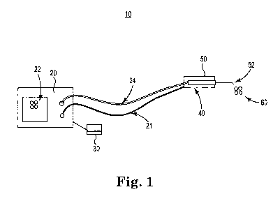
[0017] Fig. 1 is a multispot surgical laser system in accordance with
the
invention;
[0018] Figs. 2A-2G are various alignment and treatment patterns
configurable by the various systems disclosed herein;
[0019] Fig. 3 illustrate probe handle holder in accordance with the
invention;
[0020] Fig. 4 illustrate probe handle holder with an endocular probe
inserted
in accordance with the invention;
[0021] Figs. 5A-5B illustrate endocular probes which can fit inside
the probe
holder in accordance with the invention; and
[0022] Fig. 6 is another multispot surgical laser system in accordance
with
the invention.
DETAILED DESCRIPTION OF THE INVENTION
[0023] Following below are more detailed descriptions of various
related
concepts related to, and embodiments of, methods and apparatus according to
the
present disclosure for an improved diagnostic and treatment system that speeds
up
eye treatment time while improving accuracy and reliability of the selected
8
CA 02934944 2016-06-22
WO 2015/097150
PCT/EP2014/078986
treatment by the physician. It should be appreciated that various aspects of
the
subject matter introduced above and discussed in greater detail below may be
implemented in any of numerous ways, as the subject matter is not limited to
any
particular manner of implementation. Examples of particular implementations
and
applications are provided primarily for illustrative purposes.
[00241 Referring now to the Figures, Fig. 1 is a multispot surgical
laser
system 10 in accordance with the invention. System 10 includes laser
photocoagulator 20 coupled to a footswitch 30, which controls at firing mode
and a
slit lamp (among other items associated with the photocoagulator), and having
an
electrical cord or cable 21 to power the system and in some embodiments to use
for
control or communication as well. Photocoagulator 20 includes a screen or
display
22 to permit the user or physician to choose a desired alignment and/or
treatment
pattern and to control all the other treatment parameters such as, but not
limited to,
the power of the treatment beam and exposure time. An optical fiber 24 is
operatively coupled to photocoagulator 20 on one end and to a handheld member
or
holder 40 on the other end. In this example embodiment, fiber 24 is
operatively
coupled to an endocular probe 50, which may be housed within probe holder 40,
which has a needle 52 and needle tip 52A coupled thereto. In this example
embodiment, needle tip 52 projects therefrom an alignment pattern 60 (and when
actuated a treatment pattern that overlays the alignment pattern) on a
patient's retina.
The patterns that are configurable and generated by this system are discussed
herein.
[0025] Referring now to Figs. 2A-2G are various alignment and
treatment
patterns configurable by system 10 and various other laser systems disclosed
herein.
9
CA 02934944 2016-06-22
WO 2015/097150
PCT/EP2014/078986
Examples of possible patterns that can be generated from the systems taught
herein
include (of different sizes) a square, a circle, rectangle, a line, a define
sector or area
filled in with several spots by doing one or several turns of a particular
modified
endocular fiber 24 or needle 52 inside handle piece holder 40. This is
accomplished
by synchronization between laser 20 and endocular fiber needle 52 resulting in
turning or rotating needle tip 52A as well as creating an angle deviation at
the needle
tip (thereby projecting movement at the needle tip). The spot diameter on the
retina
will vary with the distance between endocular fiber tip and target tissue.
Fig. 2A
illustrates a single spot generated with a standard endocular fiber. Fig. 2B
illustrates, on the other hand, a four spot pattern (small square) generated
with
system 10 and probe 50, with one turn (or rotation) of a coupled or connected
fixed
angle endocular probe or, alternatively, a particular endocular probe with tip
angle
that is adjustable. Fig. 2C illustrates an example of a circular pattern with
10 spots
generated with about one turn of a coupled fixed angle endocular probe or a
coupled
endocular probe with an adjustable tip angle.
[0026] In Fig. 2D there is illustrated an example of a 12 spot pattern
generated with about two turns or rotations of a coupled endocular probe with
an
adjustable tip angle. Fig. 2E illustrates an example of a 16 spot pattern
(resulting in a
4 X 4 square pattern) generated with about three turns of a coupled endocular
probe
with an adjustable tip angle. Fig. 2F illustrates yet another example of a 10
spot
sector pattern, similar to an arc pattern, generated with about three turns of
a coupled
endocular probe with an adjustable tip angle. Fig. 2G illustrates yet another
example
of an 8 spot rectangular pattern generated with about two turns of a coupled
endocular probe with an adjustable tip angle. Hence, it is apparent to one
skilled in
CA 02934944 2016-06-22
WO 2015/097150
PCT/EP2014/078986
the art that numerous patterns and sector filling schemes are possible with
the laser
system taught herein.
[0027] Referring now to Figs. 3 and 4, there is illustrated an example
embodiment of probe handle holder 40 that includes a probe housing 40A and a
probe cylindrical opening 40B that spans along a length of housing 40A,
opening
40B configured to accept an endocular probe 50, with an optical fiber 51 being
coupled to probe 50. Probe housing 40A includes therein a motor 41 adapted to
drive an angle of probe or the tip of needle 52 and includes a sensor 45
adapted to
check the angle of probe 50 or the angle of needle tip 52A indirectly. In this
example embodiment, probe housing 40A further includes a motor 42 to rotate
needle 52 or probe 50 (depending on the desired embodiment) and a sensor 43 to
check the angle of rotation of the probe or needle. Housing 40A also includes
a
circuit board 44 which includes circuitry and processors that control the
various
motors in the probe housing to generate the desired probe position (angular
rotation
and/or displacement) or needle tip angle position 52A. In a related
embodiment,
board 44 is configured to communicate with a photocoagulator system
controller.
An electrical cord or cable 48 is coupled to housing 40 on one end and is
operatively
coupled to laser 20 at the other end and establishes synchronization for
movement of
the probe tip position angularly and rotationally. In this example embodiment,
the
longitudinal movement controls how much angle there will be in needle tip 52A.
Changing the needle tip 52A angle corresponds directly to the size of the
circle to be
made within the retina to assist in forming the various aiming beam patterns.
In a
related embodiment, the probe holder and motors control the longitudinal
movement
or displacement (or in/out of the holder) of the probe and needle.
11
CA 02934944 2016-06-22
WO 2015/097150
PCT/EP2014/078986
[0028] Referring now to Figs. 5A-5B are two example embodiments of
endocular probes 50 in accordance with the invention. In Fig. 5A, probe 50 is
coupled to fiber 51, while fiber 51 is optically and/or electrically coupled
to laser 20.
A button actuator 54 is included that is operatively coupled to probe 50 so as
to
facilitate one or more of the following: mechanical push/pull or
movement/translation longitudinally along probe housing 40A length (in and out
of
housing 40A) and within cylinder 40B; with rotational movement capability to
permit movement of the probe body or of needle tip 52 in various angles 52A of
the
endocular probe. Button or actuator 54 can also be configured to allow device
sensor 43 to verify or determine endocular probe tip angle 52A. Displacement
member 56 located on probe 50 is configured to permit the device sensor 43 to
determine each of the endocular probe and needle position in terms of angular
rotation. Angular rotation member 58 located on probe 50 is configured to
(mechanically or otherwise) fix or hold the probe holder so as to permit
angular
turning or rotating of the probe 50 or needle 52. The inset figures illustrate
movement of (in various angles) needle tip 52 in response to actuator 54.
[0029] Referring now Fig. 6, there provided another multispot surgical
laser
100 system with some similar elements as in system 10 except those elements
are
adapted to provide slit lamp type ophthalmology treatments as taught herein.
In this
example embodiment, electrical cord 121 and optic fiber 124 are coupled to
laser
source 20 with the other end of fiber tip 124A coupled to a device so as to
permit
translational/deviation movement as well as rotation of the tip using motors
(not
shown) and position sensors. Such device is then coupled to a fixed spot size
changer 126 which is also coupled to a focus lens 128. A patient's eye 180 is
12
CA 02934944 2016-06-22
WO 2015/097150
PCT/EP2014/078986
diagnosed and treated by a user 182 using a slit lamp 170 which is able to
control a
mirror with a micromanipulator 172 that reflects light from tip 124A through a
contact lens 174 to the patient's eye 180. Modifications known to one skilled
in the
art can be made to system 100 so as to project and treat several various
patterns on
the patient's eye, including the retina and cornea. The patterns illustrated
in Fig. 2A-
2G, which are generated by systems 10 and 100, are displayable to the user in
display 22 to ensure the correct treatment pattern is being used otherwise
adjustments can be made.
[0030] The aforementioned teachings are also applicable to slip lamp
systems where alignment and treatment patterns can be formed by rotational and
translational movement of the fiber without the use of a scanner which
deviates or
moves the laser beam as opposed to the fiber or probe as described herein. In
addition, where zoom is not needed for adjusting spot size we can use only one
fixed
spot size or several fixed spot sizes and form standard patterns using this
invention
to fill in a sector or area to be treated.
[0031] The following patents and publications that relate to
ophthalmology
diagnostic and treatment systems are herein incorporated by reference in their
entirety and constitute part of the disclosure herein: U.S. Patent and
Publication Nos.
6,096,028; 8,496,331; U.S. 2011/0144627; and WO 2008/024848 A2.
[0032] The foregoing particular embodiments of the invention as set
forth
herein are for illustrative purposes only. Various deviations and
modifications may
be made within the spirit and scope of the invention without departing from
the main
theme thereof.
13