Note: Descriptions are shown in the official language in which they were submitted.
CT SYSTEMS AND METHODS
TECHNICAL FIELD
Embodiments of the present disclosure relate to radiography and security
inspection technology, and more particularly, to multi-source static Computed
Tomography (CT) system and method for security inspection of luggage and
articles.
BACKGROUND
CT technology has been playing an important role in occasions such as
security inspection, thanks to its capability of eliminating influence from
object
overlapping. Conventional CT apparatuses use a slip ring device, and acquire
projection data at different angles by rotating X-ray source and detectors.
Then,
the CT apparatuses reconstruct a tomogram image to obtain information of the
inside of the inspected luggage or articles. In combination with dual-energy
or
multi-energy imaging technology, the existing inspection apparatuses can
reconstruct atomic number and electron density of the inspected object, and
identify materials contained in article, achieving good effects in detecting
explosives
or drugs, for example.
The existing CT technology for security inspection has disadvantages. First,
there is a problem with the scanning speed. A high speed is helpful to
mitigation
of pressure caused by a large number of passengers and cargoes. However, a
high-speed scanning typically requires a slip ring that can rotate at a high
speed.
Due to difficulties in fabrication precision and reliability, for example, the
high-speed
slip ring is very expensive in manufacture and maintenance, and thus is
difficult in
1
CA 2935086 2017-10-18
CA 02935086 2016-06-27
popularization. Second,
there exist problems such as false alarm and missing
alarm. It is difficult for the automatic identification and alarm functions in
the CT
technology to achieve an accuracy of 100`)/0, and thus detection of contraband
still
requires manual auxiliary examination, and sometimes it even requires opening
a
luggage case for examination. Such examination often takes several or tens of
minutes, which increases labor and time cost and limits improvement in
examination efficiency. In order to address these problems, an apparatus using
secondary scanning technology has been introduced into market, which can
reduce a frequency of opening the luggage case by performing a high-accuracy
to secondary
scanning on any suspicious luggage case to improve CT image quality.
This secondary scanning, however, also results in increased scanning time and
interruption in the security inspection process.
In recent years, the technology of carbon-nanotube X-ray tube has been
introduced to practical applications. Unlike
normal X-ray sources, the
carbon-nanotube X-ray tube does not require high temperature for generating
rays.
Instead, it generates cathode rays based on principle of discharging of
carbon-nanotube tip, and uses the cathode rays to strike a target to generate
X
rays. Such X-ray source has advantages of rapid switch-on/off, and a smaller
volume. A "static CT" apparatus without rotation can be formed by arranging
the
X-ray source properly and irradiating X-rays onto the object from different
directions.
This significantly accelerates the radiography. process while omitting the
slip-ring
structure and saving cost, thereby contributing a lot to the field of security
inspection.
SUMMARY
In view of one or more problems with the conventional technology,
embodiments of the present disclosure provide a CT system and method.
According to an aspect of the disclosure, a CT system is provided comprising:
a conveyor mechanism configured to convey and move an object under inspection
linearly; a first scanning stage comprising a first ray source, a first
detector, and a
first data acquisition device, and configured to scan the object and generate
a first
2
CA 02935086 2016-06-27
=
digital signal; a second scanning stage configured to be spaced from the first
scanning stage at a preset distance in a direction of the object's movement,
and
comprising a second ray source, a second detector, and a second data
acquisition
device; a processing device configured to reconstruct a CT image of the object
at a
first image quality based on the first digital signal, and analyze the CT
image; and a
control device configured to adjust a scanning parameter of the second
scanning
stage based on an analysis result of the processing device to cause the second
scanning stage to output a second digital signal, wherein the processing
device
reconstruct a CT image of the object at a second image quality higher than the
first
image quality at least based on the second digital signal.
In an embodiment, when the second scanning stage scans a part of the object,
the control device adjusts the scanning parameter of the second scanning stage
based on an analysis result of the processing device corresponding to the
part, to
cause the second scanning stage to output the second digital signal.
In an embodiment, the CT system further comprises a third scanning stage
that comprises a third ray source, a third detector, and a third data
acquisition
device, the control device is configured to adjust a scanning parameter of the
third
scanning stage based on the CT image of at least the first image quality to
cause
the third scanning stage to output a third digital signal, and the processing
device is
configured to reconstruct a CT image of the object at a third image quality
higher
than the first image quality at least based on the third digital signal.
In an embodiment, when the third scanning stage scans a part of the object,
the control device adjusts the scanning parameter of the third scanning stage
based on an analysis result of the processing device corresponding to the
part, to
cause the third scanning stage to output the third digital signal.
In an embodiment, each of the first, second and third scanning stages uses a
sparse-view scanning mode.
In an embodiment, each of the first, second and third scanning stages uses a
limited-angle scanning mode.
3
CA 02935086 2016-06-27
In an embodiment, each of the first, second and third ray source comprises a
plurality of source points provided in a plurality of scanning planes
perpendicular or
nearly perpendicular to the direction of the object's movement, and in each of
the
scanning planes the source points are distributed along one or more continuous
or
discontinuous segments of line or arc.
In an embodiment, the source points of the second scanning stage are preset
to use an increased voltage to increase ray energy when the analysis result of
the
processing device indicates that an increased penetrability is required to
discern a
metal object and its neighborhood.
In an embodiment, the source points of the second scanning stage are preset
to use an increased number of ray sources to increase a spatial resolution
when
the analysis result of the processing device indicates that it is required to
discern
tiny objects.
In an embodiment, the source points of the second scanning stage are
adjusted to have a preset number of activated ray sources when the analysis
result
of the processing device indicates that it is required to complete scanning
within a
prescribed time period.
In an embodiment, a beam spectrum for the source points of the second
scanning stage is adjusted when the analysis result of the processing device
indicates that a more accurate material identification is required.
In an embodiment, a beam intensity of the source points of each of the first,
second and third scanning stages can be adjusted according to a number of ray
sources preset in the plane where the source points are provided.
In an embodiment, when the number of the source points is large, the beam
intensity is increased to reduce beam-emitting time of each source point and
thus
ensure completion of scanning within a prescribed time period; when the number
of
the source points is small, a high beam intensity is used to increase a
signal-to-noise ratio of scan data.
4
CA 02935086 2016-06-27
According to another aspect of the disclosure, a method for a CT system is
provided, the CT system comprising a first scanning stage, and a second
scanning
stage spaced from the first scanning stage at a preset distance in a moving
direction of an object under inspection, the method comprises: scanning the
object
by the first scanning stage during the movement of the object, and generating
a
first digital signal; reconstructing a CT image of the object at a first image
quality
based on the first digital signal, and analyzing the CT image; and adjusting a
scanning parameter of the second scanning stage based on an analysis result to
cause the second scanning stage to output a second digital signal; and
io reconstructing
a CT image of the object at a second image quality higher than the
first image quality at least based on the second digital signal.
According to the embodiments of the present disclosure, scanning based on
multi-plane and adaptive scanning parameter is performed in a single scanning
process. This achieves high-accuracy scanning and speeds up the scanning
is processing while obtaining better image quality and increased
identification
accuracy.
BRIEF DESCRIPTION OF THE DRAWINGS
For a better understanding of the present disclosure, embodiments of the
present disclosure will be described below with reference to figures in which:
20 Fig. 1
illustrates a schematic block diagram of a CT system according to an
embodiment of the disclosure;
Fig. 2 illustrates a flowchart of a method for a CT system according to an
embodiment of the disclosure;
Figs. 3A, 3B, and 3C are schematic diagrams illustrating a sparse-view
25 scanning mode
used in respective scanning stages of a CT system
according to an embodiment of the disclosure; and
5
CA 02935086 2016-06-27
Figs. 4A, 4B, and 40 are schematic diagrams illustrating a limited-angle
scanning mode used in respective scanning stages of a CT system
according to an embodiment of the disclosure.
DETAILED DESCRIPTION OF THE EMBODIMENTS
The particular embodiments of the disclosure are described below in details.
It shall be noted that the embodiments herein are used for illustration only,
but not
limiting the disclosure. In the description below, a number of particular
details are
explained to provide a better understanding to the disclosure. However, it is
apparent to those skilled in the art the disclosure can be implemented without
these particular details. In other examples, well-known circuits, materials
or
methods are not described so as not to obscure the disclosure.
Throughout the specification, reference to "one embodiment," "an
embodiment," "one example" or "an example" means that the specific features,
structures or properties described in conjunction with the embodiment or
example
are included in at least one embodiment of the present disclosure. Therefore,
the
phrases "in one embodiment," "in an embodiment," "in one example" or "in an
example" occurred at various positions throughout the specification may not
refer
to one and the same embodiment or example. Furthermore, specific features,
structures or properties may be combined into one or several embodiments or
examples in any appropriate ways. Moreover, it should be understood by those
skilled in the art that figures here are for the purpose of illustration, and
not
necessarily drawn to scale. It should
be appreciated that "connecting" or
"coupling" a component to another component may mean that the component is
directly connected or coupled to the other component, or there may be a
component intervening between them. On the contrary, "directly connecting" or
"directly coupling" a component to another component mans that there is no
intervening component. Like reference signs refer to similar elements
throughout
the figures. The term "and/or" used herein means any and all combinations of
one
or more listed items.
6
CA 02935086 2016-06-27
In view of the problem with the conventional technology, embodiments of the
present disclosure propose a static CT system having multiple X-ray sources.
When a conveyor mechanism conveys and moves linearly an object under
inspection, a first scanning stage scans the object and generates a first
digital
signal. Then, a CT image of a first image quality is reconstructed for the
object
based on the first digital signal, and the CT image is analyzed. Next, a
scanning
parameter of a second scanning stage is adjusted based on the analysis result,
so
that the second scanning stage outputs a second digital signal. The second
scanning stage is provided apart from the first scanning stage at a preset
distance
in the direction of the object's movement. A CT image of a second image
quality is
then reconstructed for the object at least based on the second digital signal.
The
second image quality is higher than the first image quality. By using
distributed
X-ray sources, it is possible to avoid use of a high-speed slip ring, and
increase the
inspection speed while reducing cost for device manufacture and maintenance.
The scanning method based on multi-plane and adaptive scanning parameter can
integrate the two high-accuracy scanning processes in the slip-ring solution
into a
single scanning flow, and achieve higher image quality and increased
identification
accuracy while saving time and labor. The present disclosure can contribute to
acceleration of the CT scanning process in security inspection and improvement
of
accuracy in identifying contraband, and can be widely used in public places
like
station, airport and customs.
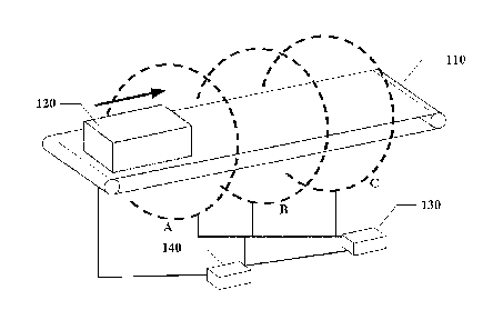
Fig. 1 illustrates a schematic block diagram of a CT system according to an
embodiment of the disclosure. As shown in Fig. 1, the multi-source static CT
system for security inspection of luggage and articles may include multiple
scanning stages (i.e., the first scanning stage A, the second scanning stage
B, the
third scanning stage C,...), a conveyor mechanism 110, a control device 140,
and a
processing device 130. The respective scanning stages are provided apart from
each other at a preset distance in the direction of the object's movement.
Each
scanning stages includes a ray source, a detector and an acquisition device.
Here,
the ray sources may include a plurality of distributed X-ray source points.
In the shown embodiments, the conveyor mechanism 110 conveys and moves
the object 120 under inspection linearly. The first scanning stage A includes
a first
7
CA 02935086 2016-06-27
ray source, a first detector, and a first data acquisition device, and is
configured to
scan the object and generate a first digital signal. The second scanning stage
B is
configured to be spaced from the first scanning stage at a preset distance in
a
direction of the object's movement, and includes a second ray source, a second
detector, and a second data acquisition device. The processing device 130 is
configured to reconstruct a CT image of the object at a first image quality
based on
the first digital signal, and analyze the CT image. The control device 140 is
couple
with the respective scanning stages and the processing device 130. The control
device 140 adjusts a scanning parameter of the second scanning stage based on
an analysis result of the processing device 130 to cause the second scanning
stage to output a second digital signal. The processing device 130
reconstructs a
CT image of the object at a second image quality higher than the first image
quality
at least based on the second digital signal (that is, based on the second
digital
signal, or based on the first and second digital signals).
According to some embodiments, when the second scanning stage scans a
part of the object, the control device adjusts the scanning parameter of the
second
scanning stage based on an analysis result of the processing device
corresponding
to the part, to cause the second scanning stage to output the second digital
signal.
In an example where 100 slices (tomograms) need to be reconstructed for a
target
luggage case, after the first scanning stage scans the 8th slice, the
processing
device (e.g., computer) reconstructs and analyzes this slice, and estimates a
scanning parameter for the second and subsequent scanning stages to scan this
slice. When the part of the object corresponding to the 8th slice passes
through
the second scanning stage, the second scanning stage adjusts the parameter
based on the analysis result, and scans the object. This is, the scanning
parameter is adjusted differently when different parts of the object pass
through the
second scanning stage.
The third scanning stage C includes a third ray source, a third detector, and
a
third data acquisition device. The control device 140 adjust a scanning
parameter
of the third scanning stage C based on the CT image of the first image quality
(e.g.,
the first resolution) to cause the third scanning stage to output a third
digital signal,
and the processing device reconstructs a CT image of the object at a third
image
8
CA 02935086 2016-06-27
quality higher than the first image quality at least based on the third
digital signal
(that is, based on the third digital signal, or based on the third digital
signal and at
least one of the first and second digital signals). When the third scanning
stage
scans a part of the object, the control device adjusts the scanning parameter
of the
third scanning stage based on an analysis result of the processing device
corresponding to the part, to cause the third scanning stage to output the
third
digital signal.
Each of the multi-point distributed X-ray source module may have, for example,
one or more source points. The energy for each source point may be set, and
the
order in which the source points are activated may be set. In the system, the
source points are distributed on multiple scanning planes (which are
perpendicular
or nearly perpendicular to the channel travel direction). In each plane, the
source
points are distributed in one or more continuous or discontinuous segments of
line
or arc. Since the energy for each source point may be set, several scanning
modes may be implemented during the beam-emitting process in which different
source points may have different energy spectra, or energy for source points
in
different planes may be different. The source points may be divided into
different
groups. For example, the source points in each module or in each plane may be
classified into a group. The order in which the source points in the same
group
cause electrons to strike a target is adjustable, and it is possible to
implement
sequential or alternate beam emission. Source points in different groups may
be
activated simultaneously to scan, and thus the scanning speed is increased.
Each of the scanning stages includes a complete set of area array X-ray
detector, sense circuit, acquisition-trigger signal circuit, and data transfer
circuit.
The ray sources are distributed in multiple planes, and thus a corresponding
detector array is provided for each plane. The detector array is arranged in a
circle or an arc. The central column of detectors may be in the same plane
where
the ray sources are located (when the source points are provided at a part of
the
circle, the detectors may be placed at the remaining part of the circle), or
in a plane
parallel to the plane where the ray sources are located (when the source
points are
distributed along the circle, there is no remaining room for placing the
detectors).
The distance between the two planes where the ray sources and the detectors
are
9
CA 02935086 2016-06-27
located respectively should be kept as small as possible to alleviate the
Oblique
Effect due to the fact that the ray sources and the source points are not in
the same
plane. The detector array may have one or more rows, and the detectors may be
of single-energy, dual-energy or spectral type.
The conveyor mechanism 110 includes a carrier table or a conveyance tape.
The control device 140 controls the X-ray machine and a rack for the
detectors.
By controlling the beam-emitting mode of the distributed ray sources or the
translation movement of the object or combination thereof, it is possible to
implement scanning in a spiral, circular or other special trajectory.
The control device 140 is responsible for control over operations of the CT
system including mechanic rotation, electrical control, and safety interlock
control.
The control device 140 particularly controls beam energy and sequence of the
ray
sources, and readout of detector data and data reconstruction.
Fig. 2 illustrates a flowchart of a method for a CT system according to an
embodiment of the disclosure. As shown in Fig. 2, the object is scanned by the
first
scanning stage during the movement of the object, and a first digital signal
is
generated at step S210. When the object 120 (e.g., luggage case) is imported
to
the system by the conveyor mechanism 110, the ray sources and detectors in the
first plane A of the system scan the luggage case, and transfer the scan data
to the
processing device 130 for CT reconstruction. At the same time, the system
records the time and location at which the scanned part of the luggage case
passes through the first plane. It is possible to calculate when the scanned
part will
pass through the subsequent plane based on the tape speed and coding.
At step S220, a CT image of the object at a first image quality is
reconstructed based on the first digital signal, and the CT image is analyzed.
In an example, the processing device 130 determines the overall feature of the
scanned part of the luggage case according to the CT reconstruction result.
The
determination may be made as to whether there are many high-density materials
CA 02935086 2016-06-27
(e.g., metals), whether there are many minutious substances (tiny articles),
and
whether there is any suspicious object in the luggage case.
At step S230, a scanning parameter of the second scanning stage is adjusted
based on an analysis result of the processing device to cause the second
scanning
stage to output a second digital signal.
The control device 140 sets in advance the scanning parameter (e.g., ray
source voltage, current, the number of ray sources to be activated) for the
subsequent scanning stage based on the analysis result. For example, the
source points of the subsequent scanning stage may be preset to use an
increased
to voltage to increase ray energy when an increased penetrability is
required to
discern a metal object and its neighborhood. The source points of the
subsequent
scanning stage may be preset to use an increased number of ray sources to
increase, for example, a spatial resolution when it is required to discern
tiny objects.
A beam intensity of each source point may also be adjusted according to the
number of activated ray sources preset in the plane where the source point is
provided, so that the scanning process can be completed with a prescribed time
period. For example, when the number of the source points is large, the beam
intensity is increased to reduce beam-emitting time of each source point; when
the
number of the source points is small, a high beam intensity is used to
increase a
signal-to-noise ratio of scan data and improve noise level in the
reconstructed
image.
At step S240, a CT image of the object is reconstructed at a second image
quality higher than the first image quality at least based on the second
digital
signal.
For example, when a part of the object 120 passes through the subsequent
scanning plane, the control device 140 controls the scanning plane to scan the
object according to the scanning parameter preset for this part, and to obtain
scan
data.
11
CA 02935086 2016-06-27
In some embodiments, when the second- scanning stage scans a part of the
object, the control device adjusts the scanning parameter of the second
scanning
stage based on an analysis result of the processing device corresponding to
the
part, to cause the second scanning stage to output the second digital signal
As such, when the part of the object 120 has passed through all the scanning
planes, the system integrates all the scan data, and reconstructs the object
using a
single-energy or spectral CT reconstruction algorithm to obtain a final 3D CT
reconstruction result as well as identifies contraband and generates alarms.
In some embodiments, the distributed ray sources and the detectors are
distributed on three circular rings spaced from each other. The planes A, B
and C
are the first, second and third scanning planes respectively, as shown in Fig.
1.
The sources in each plane may be sparsely arranged as shown in Figs. 3A, 3B
and
30, or densely arranged within a limited angle range as shown in Figs. 4A, 4B
and
4C.
In the above embodiments, a multi-source X-ray emission device is used to
irradiate luggage from different angles, and thus a rotation device in the
normal CT
system can be omitted, thereby reducing system cost and increasing detection
accuracy. The multi-plane scanning mode further improves scanning speed.
Use of distributed ray sources provides flexibility. By adjusting the energy
for ray
sources based on pre-processing results and incorporating multi-spectrum ray
detection technology, it is possible to improve identification of dangerous
and
suspicious objects, such as flammable material, explosives and drugs, and to
accommodate requirements of security inspection in various scenarios.
The embodiments take full advantage of distributed ray sources, and develop
a new control method. Distributing the ray sources over multiple scanning
planes
makes it possible to adjust spectrum, beam intensity and number of the ray
sources according to the object's characteristics. When the object passes
through
the first plane, a pre-reconstruction result can be obtained using a sparse-
view or
limited-angle reconstruct method, and then the spectrum, beam intensity and
number for the subsequent two planes can be changed based on the analysis
12
CA 02935086 2016-06-27
result and requirements. In this way, an optimal reconstruction result can be
obtained on one hand, and reconstruction results at different energy levels
can also
be obtain on the other hand to realize material identification.
In some embodiments, the problem of data synchronization between different
planes needs to addressed when the ray sources and detectors are distributed
over multiple locations. By triggering the X-ray sources to emit beams in a
tape-coding method, it is possible to ensure that detectors in different
planes
acquire data of the same plane of the object. Alternatively, time differences
between time points when the object passes through several planes of detectors
to may be determined according to the fixed locations of the several
planes, and thus
corresponding data may be extracted.
In some embodiments where data at different energy levels and different
angles are to be combined for reconstruction, data at all the angles may be
first
reconstructed using the normal single-energy CT reconstruction algorithm. The
reconstruction result maintains an accurate geometric structure. Then, by
using
the geometric structure as a priori knowledge, scan data may be grouped and
reconstructed on a group basis according to the beam energy of the source
points,
to obtain a reconstruction result of different X-ray energy levels.
The foregoing detailed description has set forth various embodiments of CT
system and method by use of block diagrams, flowcharts, and/or examples.
Insofar
as such block diagrams, flowcharts, and/or examples contain one or more
functions
and/or operations, it will be understood by those skilled in the art that each
function
and/or operation within such examples may be implemented, individually and/or
collectively, by a wide range of hardware, software, firmware, or virtually
any
combination thereof. In one embodiment, several portions of the subject matter
described herein may be implemented via Application Specific Integrated
Circuits
(ASICs), Field Programmable Gate Arrays (FPGAs), digital signal processors
(DSPs), or other integrated formats. However, those skilled in the art will
recognize
that some aspects of the embodiments disclosed herein, in whole or in part,
may
be equivalently implemented in integrated circuits, as one or more computer
programs running on one or more computers (e.g., as one or more programs
13
CA 02935086 2016-06-27
running on one or more computer systems), as one or more programs running on
one or more processors (e.g., as one or more programs running on one or more
microprocessors), as firmware, or as virtually any combination thereof, and
that
designing the circuitry and/or writing the code for the software and or
firmware
would be well within the skill of those skilled in the art in light of this
disclosure. In
addition, those skilled in the art will appreciate that the mechanisms of the
subject
matter described herein are capable of being distributed as a program product
in a
variety of forms, and that an illustrative embodiment of the subject matter
described
herein applies regardless of the particular type of signal bearing medium used
to
actually carry out the distribution. Examples of a signal bearing medium
include,
but are not limited to, the following: a recordable type medium such as a
floppy disk,
a hard disk drive, a Compact Disc (CD), a Digital Versatile Disk (DVD), a
digital
tape, a computer memory, etc.; and a transmission type medium such as a
digital
and/or an analog communication medium (e.g., a fiber optic cable, a waveguide,
a
wired communications link, a wireless communication link, etc.).
While the present disclosure has been described with reference to several
typical embodiments, it is apparent to those skilled in the art that the terms
are
used for illustration and explanation purpose and not for limitation. The
present
disclosure may be practiced in various forms without departing from the esprit
or
essence of the disclosure. It should be understood that the embodiments are
not
limited to any of the foregoing details, and shall be interpreted broadly
within the
esprit and scope as defined by the following claims. Therefore, modifications
and
alternatives falling within the scope of the claims and equivalents thereof
are to be
encompassed by the scope of the present disclosure which is defined by the
claims
as attached.
14