Note: Descriptions are shown in the official language in which they were submitted.
CA 02935480 2016-06-29
WO 2015/101886 PCT/IB2014/067059
1
"Apparatus and method for treatment of diagnostic information relating to
samples of microbiological material"
The present invention relates to an apparatus and a method for treatment of
diagnostic information relating to samples of microbiological material. The
invention in particular is applicable as a support for carrying out diagnoses
in the
health, clinical and environmental fields, in all cases in which samples of
microbiological materials are to be analysed, and in particular
bacteriological
material.
As is known, in bacteriological laboratories testing is carried out which, in
combination with other evidence obtained from further tests which can be
carried
out in other laboratories too, combine to convince a doctor of the presence of
any
infective agent afflicting a patient or which is in a different examination
site such as
a surface of an environment to be analysed; the final aim of the diagnosis is
to
lead to prescription, where required, of an adequate therapy, especially of an
antibiotic nature, with the aim of neutralizing the pathogen agent identified
by
means of the analyses carried out. The microbiologist and the laboratory team,
in
practice, starting from one or more samples collected from a same examination
site, carry out a series of tests, in general following the laboratory
guidelines,
which often originate from guidelines set down by the Health Ministry or the
corporations defining the norms and standards of Good Laboratory Practice.
It is also known that a microbiologist's work method requires an
"investigative"
approach, characterised by a non-linear process during the course of which
outcomes of a series of examinations are obtained in times that can be
variable
from test to test, the outcomes providing various evidence; and all the
preceding is
done in a diachronic timescale: for one or more samples collected from a same
investigative site various methods made the results available over a timescale
that
can go from a few minutes to a few days. In particular bacterial cultures can
be
long in incubation before enabling conclusions to be drawn in relation to the
presence or not of bacterial strains in the collected sample. The
microbiologist
therefore has an iterative approach, as it is often necessary to "re-read" the
elements already evaluated previously in the light of new elements to be
considered, and vice versa. At present the main method used in bacteriology is
the
bacterial culture, carried out on media that enable growth, contained in Petri
CA 02935480 2016-06-29
WO 2015/101886 PCT/IB2014/067059
2
dishes or capsules, following a period of incubation, of bacteria which might
afflict
a patient and which are present in a biological sample originating from the
patient
him or herself, such as for example urine, swabs (carried out in one or more
anatomical sites), faeces, CSF liquid, respiratory material, etc. The sample
can
come from a patient, an animal, or a surface, in cases of applications
respectively
in the veterinary sector or the environmental sector.
As well as bacterial culture, other methods are known and used in the
bacteriological sector, one of the most important of which is Gram-staining,
generally carried out on suitable slides, which together with the microscope
viewing also provides further evidence on the nature of the pathogen. Other
tests
used are biochemical in nature and provide further information on the type of
pathogen and its belonging to determined classes or families of micro-
organisms.
In recent years sometimes molecular biology methods are also used, based on
DNA and RNA analyses, with the aim of identifying with certainty the presence
of a
given pathogen, though the cost and specificity of these methods mean that
they
are used in support of the ones previously described, which are still the most
widely-used, as they are less specific and less expensive and complex to
perform.
The above-described processes for microbiological analyses are therefore long,
complex and difficult to manage, due to the heterogeneity of the tests to be
conducted, the iterativity required in the analysis of the single results, and
the
absence of automated systems for carrying out the laboratory testing and the
analysis of the results, which complicate the process of obtaining the correct
diagnosis, starting from the interpretation of the single examinations carried
out.
Further drawbacks of the known methods are the inefficiency and the lack of
unity
of the overall process, and the risk of errors in passing information, also
due to the
lack of a correct consideration of all the information which must contribute
to the
correct determination of the diagnosis, which are not all contemporaneously
accessible to a same subject. Further, the existing systems do not enable
sufficient traceability of the whole process carried out and the historical
aspect
relating to the analyses performed, the devices used and the subjects
involved,
thus missing a source of information that might be very important in some
specific
contexts.
CA 02935480 2016-06-29
WO 2015/101886 PCT/IB2014/067059
3
The culture dishes themselves are a perishable entity since after some days of
incubation the flora normally present in each sample tends to overgrow and
"cover" the colonies of the pathogen agent for which identification is sought.
The majority of the work processes performed in the bacteriological laboratory
are
usually done manually, and only some are done semi-automatically. Recently
automatic culture plate-seeding systems have appeared on the market, which
have introduced the concept of automation in bacteriology with a consequent
standardization of the processes and a greater traceability with respect to
manual
operations. Digital recording systems of the images of the culture dishes
before
and after specific incubation periods have also been introduced, together with
automated systems of movement of the dishes themselves by conveyor belt. Also
known are LIS (Laboratory Information Systems), in which the essential
information relating to each patient are collected, such as personal details,
which
are generally associated to the further data deriving from outcomes of
analyses
carried out or medical evidence. These systems too, however, enable only
marginally obviating those drawbacks mentioned herein above, since these
systems are able to transmit only strings of data and not actual information
in real-
life form, such as for example images.
A main aim of the present invention is to obviate one or more of the problems
encountered in the prior art.
An aim of the present invention is to disclose an apparatus and a method for
treatment of diagnostic information relating to samples of microbiological
material
which enable eliminating or at least significantly reducing risk of human
error in
identifying samples to be analysed, in carrying out analytical procedures
and/or in
determining results thereof.
A further aim of the present invention is to provide an apparatus and a method
for
treatment of diagnostic information relating to samples of microbiological
material
which enable significantly increasing the reliability and the quality of the
diagnostic
process, and therefore also the safety of the patients.
A further aim of the present invention is to provide an apparatus and a method
for
treatment of diagnostic information relating to samples of microbiological
material
which enable a more rapid, accurate and traced decision on the part of the
laboratory doctor concerning the diagnosis.
CA 02935480 2016-10-14
55508-14
A further aim of the present invention is to provide an apparatus and a method
for
treatment of diagnostic information relating to samples of microbiological
material which
exhibit a high degree of reliability.
A further aim of the present invention is to provide an apparatus and a method
for
treatment of diagnostic information relating to samples of microbiological
material which
are very flexible and adaptable to various operative requirements.
A further aim of the present invention is to provide an apparatus and a method
for
treatment of diagnostic information relating to samples of microbiological
material which
enable simplification and acceleration of treatment and retrieving processes
of the data
relating the sample analysis.
A further aim of the present invention is to provide an apparatus and a method
for
treatment of diagnostic information relating to samples of microbiological
material which
provide a high traceability of the historical data relating to the analyses
carried out and
the devices and subjects involved, so as to enable further studies and
statistics to be
made on the results themselves.
A further aim of the present invention is to provide an apparatus and a method
for
treatment of diagnostic information relating to samples of microbiological
material which
are simple to realise and which involve sufficiently low costs.
In some aspects of the invention, there is provided an apparatus and a method
for
treatment of diagnostic information relating to samples of microbiological
material.
Each of the aspects described in the following can further be taken alone or
in any
combination with the other described aspects.
In a further aspect, the invention further relates to an apparatus for
treatment of
diagnostic information relating to samples of microbiological material in
which a first
software program is configured so as to retrieve, analyse and visualize the
data and
relative images of the first and second supports relatively to a plurality of
patients.
In a further aspect, the invention further relates to an apparatus for
treatment of
diagnostic information relating to samples of microbiological material in
which a
- 4 -
CA 02935480 2016-06-29
WO 2015/101886 PCT/IB2014/067059
first software program is configured so as to enable entering comments,
symbols
and/or graphic signs together with the images, and/or memorising the position
of
the comments, symbols and/or signs with respect to the images, in order to
enable
signalling and tracing indications on which the bio-active agents (for example
5 isolated bacterial colonies grown on a culture dish) on which to carry
out further
tests.
In a further aspect, the invention further relates to a method for treatment
of
diagnostic information relating to samples of microbiological material in
which a
first image and/or a second image are retrieved from at least a first memory
and/or
directly from at least an image-acquiring device.
In a further aspect, the invention further relates to a method for treatment
of
diagnostic information relating to samples of microbiological material,
wherein a
first user interface comprises a plurality of displays or visualization of
interface
images or screen pages alternatingly representable on the viewing device and
reciprocally connected and available directly or indirectly, and the method
comprises steps of visualizing the first image, or the first plurality of
images, and
the second image, or the second plurality of images, in a same interface
screen
page, or in interface screen pages visualized alternatingly and in succession
on
the viewing device, and reciprocally mutually connected and available directly
or
indirectly.
In a further aspect, the invention further relates to a method for treatment
of
diagnostic information relating to samples of microbiological material in
which the
first biological sample and the second biological sample correspond to one
another and originate from a same original sample taken from the first
examination
site.
In a further aspect, the invention further relates to a method for treatment
of
diagnostic information relating to samples of biological material comprising a
step
of further viewing in the user interface further metadata associated to the
first
image, or to the first plurality of images of the first support and/or
associated to the
second image or second plurality of images of the second support.
In a further aspect, the invention further relates to a method for treating
diagnostic
information relating to samples of micro-biological material further
comprising
steps of entering and storing further diagnostic data deriving from a combined
use
and evaluation of the first image, or of the first plurality of images, of the
first
81798077
6
support, and/or of the second image, or the second plurality of images, of the
second
support.
In a further aspect, the invention further relates to a method for treating
diagnostic
information relating to samples of micro-biological material further
comprising steps of
carrying out the analysis of the first image with the aim of evaluating at
least the entity and
type of the bio-activity of the sample by evaluation of the detected
bacteriological growth,
by means of analysis by an operator or by an automatic image interpreting
system.
In a further aspect, the invention further relates to a method for treating
diagnostic
information relating to samples of micro-biological material further
comprising steps of
retrieving data, from a data system or laboratory information system, and
processing and
visualizing the data and the relative images of the first and second supports
relative to a
plurality of patients.
In a further aspect, the invention further relates to a method for treating
diagnostic
information relating to samples of micro-biological material further
comprising a step of
exchanging data, comprising at least data relating to the first examination
site and/or the
images relating to samples of micro-biological material, and/or diagnostic
data deriving
from the combined analysis of the images, between an apparatus for treatment
of
diagnostic information relating to samples of microbiological material and a
laboratory
information system.
In a further aspect, the invention further relates to a process for treatment
of diagnostic
information relating to samples of microbiological material, comprising at
least steps of:
acquiring, by at least a first device for acquiring images operatively
connected to a
processor, at least: a first image of a first support of a first type, for
microbiological culture,
which is a Petri dish with terrain for bacterial cultures, related to a first
microbiological
sample coming from a first examination site; and a second image of a second
support of
a second type different from the first type, for microbiological samples,
which is a slide for
one or both of Gram staining of microorganisms and morphological analysis of
microorganisms, related to a second microbiological sample coming from the
first
examination site; defining at least a first user interface on a first display;
retrieving and
displaying, in the first user interface on the first display, at least a first
datum, or a first
Date Recue/Date Received 2021-02-19
81798077
6a
plurality of data, related to the first examination site; retrieving at least
the first image of the
Petri dish with terrain for bacterial cultures; displaying at least the first
image in the first
user interface on the first display; further retrieving at least the second
image of the slide
for one or both of Gram staining of microorganisms and morphological analysis
of
microorganisms; and displaying at least the second image in the first user
interface on the
first display, in order to enable the user to make a combined use and
evaluation, by means
of simultaneous or subsequent viewing, of the data relating to the first
examination site
and of the images relating to the first and the second support related to the
first examination
site.
Brief Description of the Drawings
A detailed description is now provided by way of non-limiting example of one
or more
preferred embodiments of the invention, in which:
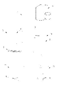
figure 1 is a schematic view of an apparatus for treatment of diagnostic
information relating
to samples of microbiological material according to a first embodiment;
figure 2 is a first example screen page display of a first user interface
produced on a
viewing device by an embodiment of the software program of the apparatus of
figure 1;
figure 3 is a second example screen page display of the first user interface
of figure 2;
figure 4 is a third example screen page display of the first user interface of
figure 2.
There now follows a description of an apparatus 1 for treatment of diagnostic
information
relating to samples of microbiological material, according to some embodiments
of the
invention.
Date Recue/Date Received 2021-02-19
CA 02935480 2016-06-29
WO 2015/101886 PCT/IB2014/067059
7
The apparatus 1 comprises at least a first processor 2 and a first viewing
device 3,
for example a display, operatively connected to the first processor 2.
The apparatus can further comprise a second processor and other processors
besides, operatively directly or indirectly connected to the first processor 2
such as
to carry out additional functions or part of the functions performed by the
first
processor 2.
The apparatus 1 further comprises at least a first software program operating
on
the first processor 2 and configured for defining at least a first user
interface 4 on
the first viewing device 3. The first software program can be memorized on a
local
memory 5, associated to the processor, or on an external memory or in a remote
location. The first processor 2, the first viewing device 3, as well as the
first user
interface 4 can also be integrated in a single device, such as for example a
tablet
or smartphone. In the present text the term "memory" comprises any type of
technological system for storing and making accessible data in a digital
format,
such as for example hard disks, flash memories, rams, roms and so on, locally
or
remotely available (for example using cloud technology). In the present
description
the term "software" is taken to comprise both an autonomous program stored and
operating locally on the processor, and a web-based program comprising the
remote software program accessible via a telematic web or the internet by
means
of a browser operating on the processor.
The apparatus 1 preferably comprises a data entry device 6 usable by a user,
such as a keyboard, touch-screen, mouse, bar code reader, or another device.
The first user interface 4 provides data and image-viewing functions, and
further
enables entry of data by an operator or user.
The software program can further comprise a data entry function relative to an
examination site, an operator or a user by receiving an identifying datum
coming
from a reading device operatively connected to the apparatus 1 directly or
indirectly, such as for example a bar code reader or another reading device
suitable for the purpose. In the present text, the term "image" is taken to
mean an
image in digital format obtained by an appropriate high-defining photographic
or
television camera, possibly associated to an optical or electronic microscope,
as
well as by means of other known systems of image-acquisition, for example by
scanning. The image acquisition can also be done by progressive reading and
subsequent assembly of partial portions of the support, for example by
progressive
CA 02935480 2016-06-29
WO 2015/101886 PCT/IB2014/067059
reading of lines (for example using a linear tv camera) or areas of the
support. The
first user interface 4 can comprise a plurality of interface screen pages 4a,
4b, 4c
representable on the viewing device 3 alternatively and/or contextually. The
screen pages are reciprocally connected to one another and can be available
one
following another, directly, thus by a direct link between two screen pages,
or
indirectly, i.e. via intermediate screen pages.
The first software program is further configured at least to retrieve and
visualize, in
the first user interface 4, at least a first datum 7, or a first plurality of
data 7,
relative to a first examination site. In the present description, the term
"examination
site" is taken to mean a collecting site in a patient, for example a patient's
orifice or
another surface at which a biological sample is taken, or a different
collecting site
with respect to which an analysis is to be performed, and therefore for
example
also a surface to be analysed in a room or environment.
In the present description, the expression "information relative to an
examination
site" are taken to mean data of various nature such as for example a patient's
personal data, identifying data of a surface to be analysed, identifying data
of an
examination site, data relating to a bar code, results in the form of text or
in
numerical form relating to diagnostic examinations carried out, textual data
supplied by doctors or laboratory experts, protocols used and the like. In the
present description the term "information" does not comprise images.
The first software is further configured such as to retrieve at least a first
image 8a
of a first support 8, of a first type, for a microbiological culture, in
particular a Petri
dish provided with Agar medium for bacterial cultures, relative to a first
microbiological sample originating from the first examination site and for
enabling
selective viewing at least of the first image 8a in the first user interface
4.
The first software is further configured such as to retrieve at least a second
image
9a of a second support 9 of a second and different type, for microbiological
samples, in particular a slide for Gram-staining of micro-organisms and/or a
slide
for morphological analysis of micro-organisms, relative to a second
microbiological
sample originating from the first examination site and for enabling selective
viewing at least of the second image 9a in the first user interface 4.
The possibility of retrieving and viewing the images 8a of the first support 8
and
the images 9a of the second support 9 in a single context enables the user or
the
specialized operator to carry out a combined analysis of the data relating to
the
CA 02935480 2016-06-29
WO 2015/101886 PCT/IB2014/067059
9
first examination site and of the images relating to the first and/or the
second
support 9. The combined analysis can be carried out in particular by a
simultaneous viewing of the data and images, or by a viewing thereof later on
but
quite close in terms of time, sufficiently to enable the operator or user to
consider
all the information that can be drawn from the data and images with the aim of
formulating her or his diagnosis.
In this way the operator can at any time reprocess the information already
obtained previously in the light of the more recent information received, not
being
limited to the reading only of a report written on the basis of the previously-
images, but with the possibility of viewing the original images (in a
digitized form) at the same time or in rapid succession, thus enabling a
complete
traceability of all the work previously carried out, a significant level of
verification,
and a reliability and quality in the definition of the diagnosis that is
impossible with
the traditional systems and approaches.
The first software program is further configured such as to retrieve the first
image
8a and/or the second image 9a from at least a first memory 5 operatively
connected to the processor. Alternatively the first software program can be
configured so as to retrieve the first image 8a and/or the second image 9a
directly
from at least a first and/or a second device 10, 11 for acquiring images
connected
to the processor, directly or indirectly.
The first biological sample and the second biological sample can be different
though coming from a same examination site, for example a site belonging to a
patients, but preferably correspond to one another and originate from a same
original sample taken. The first software program can further be configured
for
retrieving and viewing, selectively or contextually, in the first user
interface 4, a
first plurality of images 8a, 8b, 8c, 8d etc., of the first support 8
corresponding at
least to a corresponding plurality of various time instants correlated to
different
incubation periods of the first microbiological sample on the first support 9
or Petri
dish.
The first software program can further be configured to retrieve and
visualise,
selectively or contextually, in the first user interface 4, a second plurality
of images
9a, 9b, 9c, 9d etc., of the second support 9 corresponding to a corresponding
plurality of different time instants or different levels of enlargement, thus
at
CA 02935480 2016-06-29
WO 2015/101886 PCT/1B2014/067059
different levels of imaging zoom and images taken by immersion of the lens in
oil,
of the second sample arranged on the second support 9.
The first software program can further be configured for enabling visualizing
at
least selectively and alternatively, in the first user interface 4, the first
image 8a or
5 the first plurality of images 8a, 8b, 8c, and the second image 9a, or the
second
plurality of images 9a, 9b, 9c. The first software program can be further
configured
for further enabling simultaneous viewing in the first user interface 4 of the
first
image 8a, or the first plurality of images 8a, 8b, 8c, of the first support 8
and the
second image 9a, or the second plurality of images 9a, 9b, 9c, of the second
10 support 9.
The first software program can further be configured for enabling viewing, in
the
first user interface 4, of further metadata 12 associated to the first image
8a, or to
the first plurality of images 8a, 8b, Sc, of the first support 8 and/or
associated to
the second image 9a, or to the second plurality of images 9a, 9b, 9c, of the
second
support 9. The metadata 12 can comprise, for example, data and/or time of
taking
the photographic image, the incubation time interval of the support at the
moment
of taking the image, the place the image was taken at, mode of capture of the
image, the device that took the image, etc.
The first software program is further configured for enabling the user to
enter and
store further data, in particular diagnostic data, deriving from a combined
use and
evaluation of the first image 8a, or of the first plurality of images 8a, 8b,
8c, of the
first support 8, and/or of the second image 9a, or the second plurality of
images
9a, 9b, 9c, of the second support 9.
For example, the further data 13 can comprise data relating to the examined
micro-organisms, such as the morphology thereof (whether they are coccus,
diplococcus, bacillus, streptococcus or yeasts), the Gram-test (i.e. whether
they
are Gram-positive, Gram-negative or Gram-variable), the quantity or
numerousness of the micro-organisms, and so on.
The first software program is further configured so as to retrieve, process
and
visualise the data and relative images of the first and second supports
relative to a
plurality of patients, for example with the aim of enabling the operator to
carry out
a mass screening of the samples in order to identify the macroscopically
positive
cases, those in which growths or presence of pathogens are evidenced, to which
to give priority in the successive laboratory processes. The first software
program
CA 02935480 2016-06-29
WO 2015/101886 PCT/1B2014/067059
11
is configured so as to enable visualizing the first image 8a, or the first
plurality of
images 8a, 8b, 8c and the second image 9a, or the second plurality of images
9a,
9b, 9c in a same interface screen page, or in interface screen pages 4a, 4b,
4c
visualized alternatively and successively on the viewing device 3 and
reciprocally
connected and available directly or indirectly.
The apparatus 1 further comprises a connecting port 14 configured for, and
destined to, enabling operating connection of the apparatus 1 with a
laboratory
information system 15 or a laboratory technology information system and an
exchange of data, monodirectional or preferably bidirectional, between the
apparatus 1 and the laboratory information system 15. The information
exchanged
between the systems comprise at least the data relative to the first
examination
site and/or the images relative to samples of micro-biological material and/or
diagnostic data determined by the user and entered in the first user interface
4. In
other terms, the first software program can be provided with numerous
functionalities able to increase the practicality of use and the effectiveness
of the
apparatus 1 as a support in the diagnostic process, such as for example:
- user access with customised use configurations, protected by password and
other security systems of various types;
- possibility of connecting or integrated the apparatus 1 with external
systems of
both informative type, such as for example a laboratory information system 15
(or
LIS), and an instrument system such as for example television cameras,
videocameras and/or microscopes able to manually or automatically obtain the
images of the supports, including by means of bar code reading systems or
other
identifying systems of the supports;
- possibility of viewing, simultaneously or side-by-side, images of a same
support
made at different time instants;
- possibility of viewing, simultaneously or side-by-side, images of
different types of
supports able to provide different information relating to a same
microbiological or
bacteriological sample;
- function of larger-scale viewing of details of the images;
- possibility of obtaining textual data, for example personal data, or data
originating
from instrumental examinations of other types, of patients from external
systems,
such as a laboratory information system 15 and the like;
CA 02935480 2016-06-29
WO 2015/101886 PCT/1B2014/067059
12
- possibility of viewing metadata 12 associated to the single images and/or
entering further metadata 12 associable to the images;
- possibility of entering diagnostic information via the user interface,
correlated to
the results determined by the users on the basis of the functions offered by
the
apparatus 1 via the user interface;
- possibility of storing the diagnostic information on memories in the
apparatus 1 or
external thereof;
- possibility of sending to external systems, i.e. laboratory information
systems and
the like, images obtained and/or diagnostic information entered via the user
interface on the basis of the analysis of the images;
- possibility of retrieving data and images from various information
systems and
grouping them together according to optimal modalities desired by the users;
- possibility for the operator to add comments, symbols and graphic signs
on the
images, including the ability of the system to memorise the exact position of
the
signs (for example in the form of Cartesian coordinates or polar coordinates
or
other types), for example with the aim of signalling (and keep trace of) the
indication on which the bioactive agents (for example isolated bacterial
colonies
grown on a culture dish) on which to perform further tests are located;
- presence of connecting elements 16, for example video buttons, for
recalling the
images relating to various types of support and/or different temporal instants
and/or different modalities for acquiring the images;
- presence of menus 17 of various types which enable provision of further
functionalities for navigating among the different screen pages and/or for
activating
various functionalities of the software programs, for example for returning to
a
"home" page, for visualising the images, for receiving or sending data or
images to
the [IS or to another external device, for generating reports, for modifying
imports,
and so on.
The invention further relates to a process for treatment of diagnostic
information
relating to samples of microbiological material, comprising at least steps of:
- defining at least a first user interface 4 on a first viewing device 3;
- retrieving and visualising, in the first user interface 4, at least a
first datum 7, or a
first plurality of data 7, relative to a first examination site;
CA 02935480 2016-06-29
WO 2015/101886 PCT/1B2014/067059
13
- retrieving at least a first image 8a of a first support 8, of a first
type, for
microbiological culture, in particular a Petri dish with terrain for bacterial
cultures,
relative to a first microbiological sample coming from the first examination
site;
- visualising at least the first image 8a in the first user interface 4;
- further retrieving at least a second image 9a of a second support 9 of a
second
and different type, for microbiological samples, in particular a slide for
Gram
staining of microorganisms and/or a slide for morphological analysis of
microorganisms, relative to a second microbiological sample coming from the
first
examination site; and
- visualising at least the second image 9a in the first user interface 4, in
order to
enable the user to make a combined use and evaluation of the data relating to
the
first examination site and of the images relating to the first and the second
support
9.
The method can further comprise steps of retrieving and visualising,
selectively or
contextually, in the first user interface 4, a first plurality of images 8a,
8b, 8c of the
first support 8 corresponding at least to a corresponding plurality of
different time
instants correlated to different incubation periods of the first
microbiological
sample on the first type of support or Petri dish. The method can further
comprise
steps of retrieving and visualizing, selectively or contextually, in the first
user
interface 4, a second plurality of images 9a, 9b, 9c of the second support 9
corresponding to a corresponding plurality of different time instants or
different
levels of enlargement or to a different mode of image-detecting of the second
sample arranged on the second support 9.
The method can further comprise steps of visualizing at least selectively and
alternatingly, in the user interface, the first image 8a or the first
plurality of images
8a, 8b, 8c, and the second image 9a or the second plurality of images 9a, 9b,
9c.
The process can further comprise steps of simultaneously visualizing, in the
user
interface, the first image 8a, or the first plurality of images 8a, 8b, 8c, of
the first
support 8 and the second image 9a, or the second plurality of images 9a, 9b,
9c,
of the second support 9.
The first image 8a and/or the second image 9a can be retrieved for example
from
at least a first memory and/or directly from at least a device 10, 11 for
acquiring
images.
CA 02935480 2016-06-29
WO 2015/101886 PCT/1B2014/067059
14
The method can further comprise steps of displaying the first image 8a, or the
first
plurality of images 8a, 8b, 8c and the second image 9a, or the second
plurality of
images 9a, 9b, 9c in a same interface screen page, or in interface screen
pages
4a, 4b, 4c viewed alternatively or successively on the viewing device 3 and
reciprocally connected to one another and available directly or indirectly.
The method can comprise steps of further visualizing, in the user interface,
further
metadata 12 associated to the first image 8a, or to the first plurality of
images 8a,
8b, 8c of the first support 8 and/or associated to the second image 9a, or to
the
second plurality of images 9a, 9b, 9c of the second support 9.
The method can further comprise steps of inserting and memorising further
diagnostic data 13 deriving from a combined analysis of the first image 8a, or
the
first plurality of images 8a, 8b, 8c of the first support 8 and/or the second
image
9a, or the second plurality of images 9a, 9b, 9c of the second support 9.
The method can further comprise steps of retrieving, from a laboratory
information
system 15, processing and visualizing the data and/or the relative images of
the
first and second supports relatively to a plurality of patients.
The method can further comprise the steps of exchanging information,
comprising
at least data relative to the first examination site and/or the images,
relative to
samples of microbiological material, and/or diagnostic data deriving from the
combined fruition and evaluation of the images, between an apparatus 1 for
treatment of diagnostic information relative sample of microbiological
material and
a laboratory information system 15.
The present invention enables obtaining one or more of the following
advantages.
Primarily, the invention enables obviating the problems encountered in the
prior
art.
The invention further enables a rapid, accurate and traced decision on the
part of
the bacteriologist and the laboratory doctor regarding the diagnosis, based
not
only on other evidence (relative to bacterial growths on culture dish) by
indeed
having access to the digitized original images of the dishes and supports.
The invention further enables a simplification and acceleration of the
treatment
methods and a retrieval of the information relative to the analyses of the
samples.
The invention further enables eliminating or at least significantly reducing
the risk
of human error in identifying the samples to be analysed, in carrying out the
analytic procedures and/or in determining the results thereof.
CA 02935480 2016-06-29
WO 2015/101886 PCT/1B2014/067059
The invention further offers a high degree of reliability and repeatability of
the
results, and makes available an apparatus and a method which are extremely
flexible and adaptable to various operational needs and various types of
analysis.
The invention further enables simplification of the treatment processes of the
5 information relating to the analyses of the analysed samples.
The invention further enables simplifying the treatment processes of the
information relative to the analyses of the analysed samples.
The invention further enables complete traceability of the historical
information
relative to the analyses carried out and the devices and subjects involved,
and
10 further enables summings-up, studies and statistics on the results
themselves.
The invention further enables significantly increasing the reliability and
quality of
the diagnostic process, the safety of the analytical processes of the samples,
and
therefore also the safety of the patients.
Lastly, the invention is simple and convenient to actuate.