Note: Descriptions are shown in the official language in which they were submitted.
CA 02935858 2016-07-04
WO 2015/104664 PCT/1B2015/050122
1
MAGNETIC NANOPARTICLES FUNCTIONALIZED WITH CATHECOL,
PRODUCTION AND USE THEREOF
Field of the invention
The present invention relates to the field of functionalized nanoparticles,
their
production and their use.
Prior art
As known, magnetite is a mineral with ferromagnetic properties whose chemical
formula is Fe304 (sometimes also written as FeaFe203).
It is well known that magnetite in nanoparticle form, i.e. with dimensions
ranging
from a few nanometers to a few tens, if immersed in a variable magnetic field
in
the range of radio waves, interacts with the electromagnetic field and then
releases thermal energy to what is around it, thus giving rise to what is
called
hyperthermic effect or magnetic hyperthermia.
In oncology, hyperthermia is exploited to improve the efficacy of chemotherapy
or
radiotherapy; in fact, raising the temperature of a solid tumor between 41 and
45
C induces the apoptosis of the tumor cells; generally, this is applied by
means of
washings with liquids brought to the appropriate temperatures and circulated
in the
vicinity of the sites affected by tumor masses.
Recently, antennas are adopted which, inserted directly into the tumor mass,
generate microwaves and thus interact with the dipole molecules of water,
generating hyperthermia.
These treatments are generally extremely invasive and of poor efficacy (in the
first
case) and not devoid of possible negative side effects such as risk of
metastasis,
tissue necrosis, etc. in the second case.
Using magnetic nanoparticles that arrive in the immediate vicinity of the
tumor
tissues or preferably that penetrate into cancer cells, it is possible to
overcome the
above problems and achieve a high efficiency of the hyperthermic effect,
localizing
it at the cellular level.
Specifically sending nanostructures in the tumor cells of solid tumors, or on
pathological tissues or sites such as Alzheimer's amyloid plaques, or on the
damaged tissues of multiple sclerosis, it is thus possible to convey, in an
efficient
manner and free from side effects, multiple conjugated treatments, such as the
CA 02935858 2016-07-04
WO 2015/104664 PCT/1B2015/050122
2
hyperthermic and pharmacological ones.
In literature there are many examples of hybrid inorganic-polymer or protein
nanoparticles comprising a biocompatible core of nanoparticle magnetite and a
coating, either polymer or protein, possibly loaded with drugs and
functionalized
on the surface, with suitable targeting agents.
These nanoparticles are potential theranostic agents wherein the ability to
generate heat under the effect of an electromagnetic EM field (hyperthermic
effect), the possibility of drug delivery (DD) and the ability to be
identified during its
action with imaging techniques (MRI) are synergistically combined.
The International Patent Application WO 2004/071386 describes compounds
consisting of mono- or bi-lamellar liposomal microcapsules containing a
magnetic
nanoparticle and a biologically active molecule having the primary aim to
reach
and treat liver tumors.
In European patent EP 1 979 365, the Applicant has described constructs
consisting of nanometric magnetic particle functionalized with bifunctional
molecules wherein an end of said molecules is bound to the surface of the
magnetic particle while the other is free and can therefore be reacted with
complex
units such as biopolymers, cyclodextrins, antibodies and drugs for use in the
pharmaceutical and diagnostic field, allowing nanoparticle/binder complexes to
be
obtained wherein there occurs a total and compact coating of the nanoparticle
without significant alterations of the properties depending on it (e.g.
optical or
magnetic properties).
The subsequent patent EP 2 117 600 describes constructs in which the
functionalized particles similar to those described in the above patent EP 1
979
365 are coated with polymers in which a molecule having pharmacological
properties has possibly been dispersed.
Also European Patent Application 2 512 992 (to the name of the same Applicant)
describes polyol synthesis processes which allow easily obtaining magnetite
nanoparticles with even and controlled size (which therefore have a high
hyperthermic efficiency).
As can be seen, therefore, many solutions have been suggested in the
literature
for the solution of the problem of selectively directing within the body
particles
CA 02935858 2016-07-04
WO 2015/104664 PCT/1B2015/050122
3
capable of performing a therapeutic action both by application of hyperthermia
alone or in combination with traditional drugs; however, known products do not
fully meet the application needs to achieve an effective treatment of tumors
and
other diseases with nanostructures due to various problems not yet overcome.
The first problem is the specificity of the nanostructure, in fact it is known
from the
literature that hybrid inorganic-polymer/protein particles are quickly
eliminated from
the reticuloendothelial system when administered systemically (reticular
cells,
macrophages, Kupffer cells).
The clearance of the nanostructures is therefore responsible for the
inefficiency of
a nanotheranostic treatment at a systemic level; numerous attempts have been
made to overcome this difficulty, including the functionalization of the
nanoparticle
polymer/protein surface with delivery units such as monoclonal antibodies,
peptides and active molecules (such as sugars, etc.) but also in this case,
most of
the particles are eliminated by the reticuloendothelial system and only a
small
amount reaches the sites concerned, the tumor tissue and cancerous cells.
A second problem, a consequence of the first one, is that the amount of
magnetic
particles that reach the tumor or the pathological tissue may prove
insufficient to
carry out an efficient hyperthermic effect.
Finally, the current nanotheranostic systems have poor stability in biological
fluids
and thus tend to form large aggregates (up to over 500-1000 nm) that are
unlikely
to penetrate into the tumor mass or go beyond an intact blood-brain barrier,
which
worsens the specific targeting of these systems in the target cells, further
limiting
the efficiency of the treatment.
From the literature it is known that T lymphocytes within the immune system
are
the main protagonists of the anti-tumor responses.
They are able to selectively recognize the tumor cells due to their specific
receptor, called TCR. The T lymphocyte activation by the respective tumor
antigenic peptide may occur only if the antigen is presented by the cells
represented by monocytes, macrophages, dendritic cells, Langerhans cells,
microglia or also B lymphocytes.
For an effective T lymphocyte activation, membrane signals and soluble signals
are also required in addition to the antigen. Among the soluble signals, the
most
CA 02935858 2016-07-04
WO 2015/104664 PCT/1B2015/050122
4
powerful activation factor is interleukin 2 (IL-2), while among the membrane
signals, the most powerful is molecule B7.
Once the tumor is identified, it is destroyed by the lymphocytes through
various
mechanisms, among which the main ones are: the cytotoxic machinery linked to
perforin and that linked to Fas ligand.
Melanoma was one of the first tumors to be associated with a strong local
immune
response mediated by T lymphocytes, and over the years it has been possible to
prove that a strong T-Lymphocytic response is related to a better prognosis.
Through the use of nanoparticles, it is possible to develop a new custom
anticancer strategy based on the use of T lymphocytes specialized in killing a
tumor, armed by nanoparticles, ready to hit the tumor, after activation by
laser/electromagnetic fields.
Moreover, the literature widely describes the role played by the immune
system,
and in particular by lymphocytes and inflammatory cells, in diseases of the
nervous system such as multiple sclerosis, Alzheimer's disease.
Multiple sclerosis is indeed the prototype of autoimmune diseases in the
pathogenesis of which a crucial role is played by T lymphocytes (Elliot M.
Frohman, M.D., Michael K. Racke, M.D., and Cedric S. Raine, N Engl J Med 2006;
354:942-955). In particular, T-helper cells capable of producing important
inflammatory cytokines, such as interferon-gamma and lymphotoxin, called T-
helper cells 1 (Th1), but also T lymphocytes CD8, B lymphocytes and immune
cells of the monocyte line, are very important. Also in Alzheimer's disease
(Henry
W. Querfurth, and Frank M. LaFeria, N Engl J Med 2010; 362:329-344), an
important pathogenetic role is carried out by inflammatory mechanisms related
to
the production of interleukin 1, interleukin 6, tumor necrosis factor a, by
microglia
and astrocytes, due to amyloid proteins; nanoparticles according to the
invention
can therefore play an important role also in the treatment of these diseases.
Brief description of the figures
Figure 1 shows, taken with a Field emission gun scanning microscope in STEM
mode, the typical cluster formation taken by the nanoparticles according to
the
invention within a polymer matrix.
Figure 2 shows a mixture construct of magnetic particles and gold nanorods.
CA 02935858 2016-07-04
WO 2015/104664
PCT/1B2015/050122
Figure 3 schematically shows a construct model consisting of nanoparticles of
magnetite or magnetite and gold nanorods and coated with block polymers PLGA-
b-PEG-COOH.
Figure 4 shows the layout of the step by step preparation process of the
5 nanostructured construct according to the invention.
Figure 5 shows the layout of the process for the purification and selection of
lymphocytes.
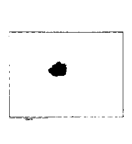
Figure 6 shows a picture taken with an optical microscope of
monocytes/macrophages charged with nanoparticles according to the invention.
Figure 7 and 8 show the 1H-NMR of the polymer PLGA-NHS conjugated with
NH2-PEG-COOH.
Figure 9 shows the UV-Vis spectrum of a product according to the invention.
Figure 10 shows a BCA Test on a product according to the invention.
Summary of the invention
There are described magnetic nanoparticles the surface of which is
functionalized
with catechol and constructs comprising a plurality of said nanoparticles
encapsulated in a biocompatible polymer matrix, wherein a molecule with
therapeutic action is optionally dispersed, said polymer matrix optionally
being in
turn further functionalized. It was surprisingly found that said polymeric
constructs
can be incorporated into immune system cells giving rise to the engineering
thereof.
Detailed description of the invention
It has now been surprisingly found that constructs comprising a plurality of
magnetic nanoparticles functionalized with catechol encapsulated in a
biocompatible polymer matrix can overcome the above problems, ensuring the
necessary stability in physiological media and in human blood.
Moreover, the structural features of these constructs helps ensure an
implemented
hyperthermic effect compared to that shown by monodisperse inorganic cores
described in the above patents; this advantage is due to a so-called "cluster
structure" (see Figure 1) of the magnetic particles which tend to combine in
structural centers of multiple particles within the polymer matrix carrying
out a
synergistic effect on the hyperthermic properties.
CA 02935858 2016-07-04
WO 2015/104664
PCT/1B2015/050122
6
The functionalization of the magnetic particles with catechol, according to
the
invention, is essential for the above cluster structure to occur and therefore
allows
obtaining much superior constructs than those known in the prior art as
regards
hyperthermic properties and stability over time.
Among the magnetic particles, magnetite is especially preferred. If preferred,
the
constructs according to the invention may have, in addition to the magnetic
nanoparticles as described above, a plurality of gold nanorods (see Figure 2).
The presence of nanorods allows a considerable hyperthermic effect by applying
an infrared laser radiation such as that generated by CO2 lasers, which goes
to
further increase the hyperthermic effect imparted by cluster structures of
magnetite.
This enables a combined laser and radio waves system which uses laser for
surface districts or those that can be reached via probe and the radio waves
for
deep districts.
Magnetic nanoparticles can be prepared through the known polyol process as
described for example in the above European patent application 2 512 992 which
describes a preparation process in which:
i) a polyol solution of FeIII is prepared starting from Fe ;
ii) magnetite nanoparticles are prepared in the polyol synthesis conditions.
The above step (i) is the well-known and described reaction of acid attack
(also
weak acids such as acetic acid) on iron according to the equation:
Fe + 2 H+ --> Fe2+ + 1/2 H2 1µ
Thereafter, it is possible to completely oxidize the solution of Fell in
polyols to Fern
(for example acetate) through air flushing and addition of H202 in the
reaction
environment at a temperature of less than 100 C.
Gold nanorods are prepared in known manner with a microwave-assisted
synthesis starting from gold in ionic form in the presence of various
additives:
alkyltrimethylammonium bromide, CnTAB n = 10-16, cetylpyridinium chloride, C16
PC and PVP [in this regard, see M. Tsuji, K. Matsumoto, T. Tsuji, H.
Kawazumid,
Mater. Lett. 59 (2005) 3856] or by reduction of HAuCI4 with ascorbic acid in
the
presence of CTAB and AgNO3 (in this regards, see Ratto F. et al. J
NANOPARTICLE RESEARCH 2010 and fRatto F. et al. J NANOPARTICLE
CA 02935858 2016-07-04
WO 2015/104664 PCT/1B2015/050122
7
RESEARCH 2012)
The surfaces of the magnetic and/or magneto-optical particles obtained as
described above are functionalized with catechol (bifunctional group) by
exploiting
the affinity of the polar groups OH to the surface of the particles and
allowing the
end part not bond to the particle surface to maintain a hydrophobic reactivity
suitable for the subsequent incorporation in a polymer/protein matrix.
The polymer matrix according to the present invention is understood to consist
of
biodegradable copolymers and is thus capable of allowing the release of the
drug,
which must proceed gradually as the matrix degrades in a physiological
environment.
Examples of suitable copolymers for the purpose are: biodegradable
nanomicelles, polyesters, polyesters, polyurethanes, polycarbonates and
poly(glutamic) acid, polyetheramine and polybenzylglutamate.
Particularly preferred are biodegradable nanomicelles, consisting of block
copolymers of poly(lactic-co-glycolic) acid and polyethylene glycol
carboxylate
(PLGA-b-PEG-COOH, MnPLGA range = 44-10 kDa, MnPEG = 2 - 3 kDa) having
formula (I)
0 0
OOH
_ _
0 _n
P
(I)
wherein m = [117-330]; n = [117-330]; p = [60-100].
This product is known and has already been employed in various other works of
Drug Delivery also at the level of Clinical Phase I for testing of anticancer
agents
(see X. Shuai et al, 2004 and X. Shuai, H. Ai, N. Nasonkla , S. Kim, J. Geo,
J.
Controlled Release, 2004, 98, 415).
The polymer in fact has features that allow assembling nanospherical systems
with a hydrophobic inner area, guaranteed by residues of PLGA, and a
hydrophilic
outer area which is imparted by the terminals of PEG-COOH (see Figure 3).
This dual feature allows the nanospheres to trap the organic active
ingredients in
CA 02935858 2016-07-04
WO 2015/104664
PCT/1B2015/050122
8
the hydrophobic part and to be dispersed in aqueous solution thanks to the
hydrophilic part.
If desired, the polymer can be admixed with molecules having a therapeutic
action
which is dispersed in the polymeric matrix according to known processes and as
illustrated in the examples given below.
Examples of molecules with therapeutic action according to the invention are
for
example anti-cancer drugs (taxanes, gemcitabine, vincristine, etc..),
peroxynitrite
scavengers, superoxide dismutase inhibitors, retinoids (bexarotene), cytokines
such as interleukin 10, TLR-ligands such as the HP-NAP molecule capable of
activating TLR2, aspirin.
In addition, the carboxylic acid functionality of the PEG-COOH fragment of the
micelles allows a chemical stable bond with monoclonal antibodies, proteins,
peptides or active molecules of interest (for example, and/or fluorescent
dyes) for
the specific recognition by the cellular over-expressions.
Among the antibodies useful for the functionalization according to the
invention we
may mention hERG, hEGFR, IgG, moAb, etc.
The examples (see example 10) describe the above functionalization, in
particular
using a specific monoclonal antibody hERG1 described and claimed in Italian
patent IT 1,367,861.
In particular, it is a specific monoclonal antibody against the extra-cellular
portion
S5-pore of protein HERG1 produced by a hybridoma comprising the product of a
fusion between an immortalized cell, belonging to the murine neoplastic cell
line
NSO, and a lymphocyte obtained by immunization of a mouse with a peptide of
sequence EQPHMDSRIGWLHN.
The construct according to the invention (hereinafter also referred to as
"nanobioreactor" or "NBR") containing magnetic nanoparticles functionalized
with
catechol is prepared by carrying out a nanoprecipitation, wherein two fluids:
- an organic solution of polymer dissolved in a solvent, mixed with the
suspension of nanoparticles coated with organic binder, both in the same
solvent,
and
- an aqueous solution of Na2HPO4 1mM)
CA 02935858 2016-07-04
WO 2015/104664
PCT/1B2015/050122
9
are mixed in a constant flow in a mixing cell with batch or continuous
synthesis.
For the batch synthesis, the organic suspension containing polymer and
particles
is injected with a syringe in the aqueous solution, without magnetic stirring,
in a
single step.
For the continuous synthesis, a double peristaltic pump system is prepared to
carry out the addition of the organic solution to the aqueous stream (organic
volume/water ratio 1/10). The respective tubes draw the solution directly from
the
lungs containing the organic suspension (with functionalized particles and
polymer) and the solution of Na2HP031mM (pH 7.4).
Once the dispersion of hybrid particles (consisting of magnetic nanoparticles
functionalized with catechol included in the polymer) has been obtained, part
of
the organic solvent is removed via a rotary evaporator so as to minimize the
amount of organic phase in the subsequent production steps.
The suspension is then dialyzed against aqueous solution Na2HP03 for the
removal of the organic phase and concentrated to the minimum volume possible
to
obtain a concentration of from 0.1 to 1% w/w.
Through a second concentration it is possible to obtain a much more
concentrated
product through membrane dialysis with a theoretical concentration factor
ranging
from 5x to 20x depending on usage. The product is then filtered with a filter
to 0.22
pm to remove the bacterial load. The product has an excellent hyperthermic
efficiency if irradiated for 30 minutes with alternating magnetic field of 21-
24 kA/m
and a frequency of 160-190 kHz, its temperature increases by at least 5 C.
The method described herein allows the preparation of constructs with a
dimensional distribution centered in a range from 30 to 60 nm.
The potential 4 of the product thus obtained (Malvern Zetasizer nano-S),
measured to have information about the electrostatic stability of the
suspension
and its ionic strength, is lower than -30 mV, which means that the particles
are
affected by the negative surface electrostatic repulsion produced by the
carboxylic
groups which at (physiological) pH 7.4 are partially deprotonated.
The experimental conditions described above allow making a suspension with
good stability after dilution into culture media typically used for cell
cultures
(DMEM, RPMI), exhibiting little tendency to aggregation and sedimentation also
CA 02935858 2016-07-04
WO 2015/104664
PCT/1B2015/050122
after a clear change in ionic strength conditions due to dilution.
Obtaining the product functionalized on the surface with monoclonal antibodies
and/or fluorescent dyes (e.g. Cyanine0, Dylight0, etc.) for a targeted
delivery and
for use in imaging techniques requires the use of NBR as a precursor before it
is
5 subjected to the second concentration process (see above).
Typically, at this stage, the product has a concentration of about 0.05-0.1%
wt. of
inorganic material.
The preliminary process step provides the activation of the end carboxyl
groups of
the polymer, exposed towards the outer part of the nanoparticle, in contact
with
10 the polar phase, with activators such as EDAC [1-ethy1-3-(-3-
dimethylaminopropyl)
carbodiimide hydrochloride] (molar ratio EDAC/COOH = 10/1) and sulfo-NHS
(NHS/COOH = 1/1), so as to promote the subsequent attack by esterification of
the end amine groups of the monoclonal antibody and/or of the fluorescent dye.
In the case of fluorescent dyes with emission at X600 ¨ 800 nm (suitable for
NIR
imaging applications in vivo), since only NHS ester-terminal molecules are
available on the market, it is necessary to provide for an intermediate step
where a
diamino-terminal linker is added for the bridge link on the one hand with the
fluorescent dye, and on the other with the carboxylic groups of the activated
polymer.
Once the surface of nanoparticles has been activated, the antibody and/or
amino-
terminal dye solution is added and let stand.
The suspension is then concentrated and dialyzed against aqueous Na2HP03 and
concentrated up to 0.2 ¨ 1.0% wt. of inorganic phase, depending on usage.
The product is then filtered with a filter to 0.22 pm to remove the bacterial
load.
The method described herein allows the preparation of constructs with a
dimensional distribution centered in a range from 40 to 70 nm.
The potential ocl) the product thus obtained (Malvern Zetasizer nano-
S),
measured to have information about the electrostatic stability of the
suspension
and its ionic strength, is less than -30 mV, but greater than that measured on
the
NBR product, which means that the negative charge exerted by the carboxylic
groups of the raw product is partly neutralized by the bound antibody/dye.
The contents of antibody bound to the particles is analyzed using the BCA
test:
CA 02935858 2016-07-04
WO 2015/104664 PCT/1B2015/050122
11
following the addition of suitable reagents to the solution containing the
protein
analyte, a complex of Cu2+ develops whose coloring at 562 nm is observed with
spectral analysis and from which the concentration of antibody is derived
using a
linear calibration.
With the procedure described herein it is possible for example to produce
nanoparticles functionalized with moAb, with moAb attack percentage between 5
and 30% wt compared to the inorganic phase content.
The production of the nanobioreactor/lipophilic drug (hereafter NBR_PTX) and
nanobioreactor/antibody/lipophilic drug (NBR_hERG_PTX) system (where the
lipophilic drug for example is Paclitaxel) is exactly the same as the
synthesis
process of the nanobioreactor as described above and therefore provides for
the
encapsulation of the inorganic nanoparticles, previously functionalized with
catechol, within a polymeric matrix based on PLGA-b-PEG-COOH. The only
variation to this process provides for the dissolution of the specific amount
of drug
within the polymer and the suspension of the functionalized nanoparticles.
Then, the nanobioreactor loaded with paclitaxel (NBR_PTX) is obtained using
the
nanoprecipitation method, where the aforementioned organic solution is
vigorously
added to the aqueous solution of Na2HP041 mm inside a mixing cell. There are
no
changes in the morphological properties of the suspension from a batch
synthesis
to a continuous one. The purification, filtration and concentration processes
applied are the same as described above.
For the product characterization, in addition to the determination of the
average
particle diameter, their potential 4 and the concentration of inorganic phase,
the
amount of active ingredient encapsulated is also determined by high
performance
liquid chromatography.
The product thus obtained and characterized can then be further functionalized
on
the surface with targeting units such as (hEGR, hEGFR, IgG, ...).
The process set up to this end accurately follows the targeting procedure of
the
nanobioreactor NBR_moAb as described above.
In fact, it provides for a preliminary step of activation of the carboxyl
groups
present on the polymer, with activators such as EDAC and sulfo-NHS and a step
of reaction with the monoclonal antibody, all according to the same
proportions as
CA 02935858 2016-07-04
WO 2015/104664 PCT/1B2015/050122
12
set out in the process previously described for the NBR_moAb.
The usual purification and characterization processes are then performed. The
nanoparticle suspensions thus obtained are characterized by a mean
hydrodynamic diameter of between 45 and 55 nm, while the potential 4 is well
below -30 mV.
According to a further embodiment of the invention, the constructs as defined
above, as an alternative to the decoration with proteins or with antibodies,
may be
incorporated into cells of the immune system.
Surprisingly, the constructs comprising clusters of magnetite particles
functionalized with catechol and coated with block copolymers of poly(lactic-
co-
glycolic) acid and polyethylene glycol carboxylate (PLGA-b-PEG-COOH, MnPLGA
range = 44-10 kDa, MnPEG = 2-3 kDa) as described above are easily
incorporated by cells of the immune system without compromising their
functionality and vitality.
Once the immune system cells are engineered with the introduction of the
constructs according to the invention, these can be used for the diagnosis of
tumor
diseases, degenerative diseases (e.g. Alzheimer's disease), of the central
nervous
system, cerebral cardiovascular and infectious diseases, transplants,
autoimmune
diseases and also for the therapy of tumors, cerebral cardiovascular diseases,
degenerative diseases (e.g. Alzheimer's disease), infectious diseases,
transplants,
liver cirrhosis and other conditions involving fibrogenesis, diseases
characterized
by multiple abortions, intrauterine fetal death, neonatal diseases, congenital
and
acquired coagulation disorders, genetic diseases, autoimmune diseases, and
finally for pain relief.
The induction of the release can take place with different methods, such as
the
specific antigen (e.g. MAGE-3 in the case of treatment of melanoma, MOG or
myelin antigens in the treatment of multiple sclerosis, etc.) or with
appropriate
immunomodulatory substances such as IL-2, CD40 ligand, TLR-agonists,
liposomes, immunostimulating complexes (ISCOMS).
It should be noted, in fact, that an important property of the cells of the
immune
system is represented by their ability to reach almost all the districts of
the body,
therefore, their use as a carrier to reach specific districts, carrying
through the
CA 02935858 2016-07-04
WO 2015/104664 PCT/1B2015/050122
13
construct according to the invention the particular product required to the
destination, exceeds the great current limitation of the nanotheranostics
represented by the low specificity of the treatment.
The cells of the immune system useful for the above purpose are for example
selected from:
T-lymphocytes, monocytes, macrophages, dendritic cells, natural killer cells,
B-
lymphocytes, neutrophil granulocytes, eosinophil granulocytes, basophil
granulocytes, gamma delta cells.
The cells are taken from the single patient, loaded with the desired
nanoparticles
and then re-introduced in the same patient topically or systemically.
The cells of the immune system will then be purified, as described below, and
in
order to facilitate the selective/preferential targeting of the body districts
affected
by the disease in question, the cells can be treated ex vivo with relevant
antigens
(or allergens), immunomodulatory drugs or engineered with immuno potentiating
or immuno suppressive molecules.
One of the ways to select T cells for diagnostic or therapeutic purposes is to
enrich
the number of T lymphocytes specific for a particular antigen which can be a
tumor
antigen as described above.
Lymphocytes, properly engineered with the constructs of the invention, can,
once
in place, release the particles by means of suitable chemical stimuli, the
particles
can then under irradiation of electromagnetic fields in the range of radio
waves
exert hyperthermia or release active ingredients such as antitumor drugs,
scavengers of molecules active in the oxidative stress of brain tissues, anti-
inflammatory molecules, etc. Nanoparticles can still perform their functions
even if
they remain confined within the lymphocytes themselves.
The magnetic nanoparticles can also perform the MRI imaging function, being
excellent T2 T2* contrast media (see the above patents), nanoparticles
containing
gold nanorods may be used in laser-mediated antitumor therapy and identified
by
methods of the photoacoustic spectrometry type.
Purification and selection of lymphocytes
T lymphocytes for use against tumors are purified from the peripheral blood or
from the tumor site or from the patient's lymph nodes after prior
administration of
CA 02935858 2016-07-04
WO 2015/104664 PCT/1B2015/050122
14
the relevant tumor antigens, or from other districts of the body as deemed
relevant, using standardized methods and/or with the aid of selective MACS
methods (Current Protocols in Immunology 2013; D'Elios et al J Immunol 1997;
158:962-967).
In order to select T lymphocytes specific for the tumor, T lymphocytes are
placed
in culture with the relevant tumor antigen (e.g. MAGE-3 for melanoma, at a
concentration of 10 pg/ml) in complete RPMI medium for five days. Then,
recombinant human IL-2 is added every three days, and then the cells will be
loaded with nanoparticles, washed and then administered to the patient
topically
and/or systemically.
T cells for use as diagnostic product, for example for multiple sclerosis with
magnetic resonance technology, are selected for their specificity for myelin
antigens or MOG (10 pg/ml) or other antigens as preferentially capable of
achieving the structures of the CNS.
To this end, they are cultured with one or more antigens for five days, then
expanded with IL-2, and then loaded with NP.
The same procedure can be used for other neurological diseases, such as
Alzheimer's disease, Parkinson's disease, stroke and other cerebro-
cardiovascular
diseases using appropriate relevant antigens.
Dendritic cells (highly efficient for their ability to present the antigen to
T
lymphocytes, and thus greatly able to activate T-lymphocytes) are obtained
using
traditional standardized methods and/or with the aid of selective methods MACS
(Current Protocols in Immunology 2013; Codolo et al. Arthr Rheum 2008; 58:3609-
17). They will be incubated for 36-44 hours with the desired antigen, then
loaded
with NP, washed and reinfused to the patient for therapeutic or diagnostic
purposes (see the process layout in Figure 5).
Natural killer cells and/or gamma delta lymphocytes, with strong cytotoxic
activity,
are obtained using traditional standardized methods and/or with the aid of
selective MACS methods (Current Protocols in Immunology, 2013), they are then
loaded with the desired NP as well as possibly with other immunomodulatory
compounds, washed and reintroduced into the patient for therapeutic (e.g.
antitumor) or also diagnostic purposes.
CA 02935858 2016-07-04
WO 2015/104664
PCT/1B2015/050122
The neutrophil granulocytes are obtained using traditional standardized
methods
and/or with the aid of selective MACS methods (Current Protocols in
Immunology, 2013), they are then loaded with the desired NP as well as
possibly
with other immunomodulatory compounds, washed and reintroduced into the
5 patient for diagnostic (e.g. to identify the presence of any foci of
infection in the
body which cannot be identified by other techniques) or also therapeutic
purposes.
Also other cell types may be selected for diagnostic and/or therapeutic use
(such
as effector cells to be used for the therapy of tumors, autoimmune diseases,
infections, degenerative diseases), such as B lymphocytes, eosinophils,
basophils,
10 which are obtained using traditional standardized methods and/or with
the aid of
selective MACS methods (Current Protocols in Immunology 2013).
They are then loaded with the desired NP or possibly with other
immunomodulatory compounds, washed and re-introduced into the patient.
Immune cells loaded with nanoparticles can be used to display with appropriate
15 imaging techniques body districts that are a location of the disease.
T cells and Jurkat cells are optimally filled with NP after 4 hours.
Monocytes/macrophages, dendritic cells, J774A.1 cells are capable of
incorporating the nanoparticles with a method according to the invention in
which
monocytes/macrophages, dendritic cells, J774A.1 cells are loaded with the
nanoparticles (NP) at a concentration of 0.05% in a suitable specific culture
medium (mmedium). To form the mmedium containing NP, the NP are first
dispensed and then the specific culture medium.
The mmedium consists of:
COMPLETE DMEM 10% FBS
COMPLETE DMEM:
= DMEM HIGH Glucose (DME/HIGH). (EUROCLONE) (code: ECB7501L)
= L-GLUTAMINE, solution 200 mM (100X). (EUROCLONE) (code: ECB
3000D)
= PENICILLIN-STREPTOMYCIN Solution (100X). (ATCC) (code: 30-2300)
> 10% fetal bovine serum FBS: Fetal Bovine Serum, Qualified. (Sigma-
Aldrich) (code: F6178-100mL)
Where necessary, autologous serum of the patient or media in the absence of
CA 02935858 2016-07-04
WO 2015/104664 PCT/1B2015/050122
16
serum will be used instead of fetal bovine serum.
Monocytes/macrophages, dendritic cells, J774A.1 cells are optimally filled
with NP
after 2 hours but the incorporation phenomenon is active after 15' up to 24h.
Figure 6 shows a picture taken with an optical microscope of the
monocytes/macrophages charged with nanoparticles.
The invention will be more and better understood in the light of the examples
given
below, also noting Figures 4 and 5 which schematically summarize the various
steps for the preparation of the construct and the engineering of the cells of
the
immune system.
Example 1
Preparation of iron acetate in diethylene glycol DEG
Reagents:
40g Fe (Fe< 99% <212mm) equal to 0.716 mol; 800 g water; 800 g CH3COOH
(80%) equal to 10.67 mol; 46.64 g oxygenated water (30%) equal to 0.41 mol;
0.12g concentrated HCI; DEG (diethylene glycol) 3850 g.
Synthesis:
Iron, the acetic acid and water solution and the hydrochloric acid were loaded
to a
5000mL 4-necked flask under nitrogen flow and the temperature was brought to
90 C and maintained for 6 hours. The system was left to cool under N2 and the
solution was filtered to remove the undissolved Fe. The oxygenated water is
added dropwise to the clear solution placed in a flask using a dripper,
keeping the
temperature at 35 C for lh, obtaining a clear solution equal to 1628.3 g
having an
iron titer of 2.40%w/w. Excess acids are then stripped by a first vacuum
distillation
at the T of 40 , a recirculation of the dry part with water and removal by
distillation
two times (two consecutive washes) and a final stripping at the T of about 50
.
3850 g DEG are added to the dry so as to bring the theoretical iron titer to
the
value of 1.01% w/w Fe.
Example 2
Preparation of Fe304 nanoparticles in diethylene glycol
Reagents:
1.50 g Fe (Fe < 99%, <212mm) Fe = 0.179 mol; 150 g DEG; 1,2g solution in
DEG 1/10 HCI conc. 37%; 300.00 g FeAc3 in DEG (1.01%w/w Fe").
CA 02935858 2016-07-04
WO 2015/104664 PCT/1B2015/050122
17
The metal iron and DEG were placed in a 500 mL 4-necked flask under N2 and the
temperature was brought to 150 C. The solution in DEG of hydrochloric acid
was
added to the system and left under stirring for 5 minutes. Iron acetate is
then
added in 10 equivalent aliquots, using a syringe, so as to ensure the correct
growth of the particles, bringing the temperature to 170 C, the reaction ends
within 24-36 hours.
The product was left to cool to 60 C and decanted in a beaker, magnetically
retaining the unreacted metal iron and then filtered on a 0.45 pm glass fiber.
450 g of a nanosuspension of magnetite in diethylene glycol having a titer in
ionic
Fe equal to 0.91% 0.05, which expressed in Fe304 corresponds to 1.253%
0.05. Hyperthermia was measured on this sample and the values were as shown
in the table
Field Frequency Starting
Sample SARm
KA/m KHz T (C)
Filtered Fe304 24 168 29.4 350.0
SARm: Specific absorption rate expressed on the mass of metal (Fe)
Example 3
Preparation of organic binder N-(3,4dihydroxyphenethyl)dodecanamide (DDA)
HO
0
HO
MW =335.48 g/mol
Reagents:
25 g Dopamine hydrochloride equal to 0.1318 mol; 1 L THF; 45 mL Triethylamine
0.32 mol; 31.20 mL Lauroyl chloride 0.135 mol;
Purification and Crystallization
400 mL THF (Aldrich 401757-2L- Lot S1BC4923V)
935 mL ethyl acetate (Aldrich 34972-2,5L- Lot 57BC011AV)
CA 02935858 2016-07-04
WO 2015/104664 PCT/1B2015/050122
18
315 mL petroleum ether (Aldrich 77379-2,5L- Lot BCBG7367V)
Synthesis:
To a 5L 4-necked flask, dopamine hydrochloride and then THF (1L) are placed
under nitrogen atmosphere and then the triethylamine is added, and the system
is
kept under stirring for about 20' to obtain a white suspension.
To a 3L flask with a flat bottom, THF (1L) and the acylating agent are added.
The
solution is stirred and added to the reagents contained in the 5L flask using
a
peristaltic pump at a rate of approximately 2 mL/min over 9h, obtaining a
solution
of yellow-orange color with some white solid on the bottom.
Purification:
The organic phase that contains the synthesis product is then purified and the
latter is recovered from the by-product formed during the reaction. The
purification
is carried out through the removal of the solvent via rotavapor with two
recirculation's (2x200 mL). On the other hand on the solid residue, and on the
residual traces in the synthesis flasks, aqueous extraction and treatment with
ethyl
acetate are carried out in a separating funnel. The organic phases are all
combined, dried with Na2SO4 and finally brought to dryness in a rotavapor. 57g
of orange oily product are obtained.
Crystallization:
450mL of a mixture of petroleum ether:ethyl acetate = 7: 3 are added to the
product. The suspension was put under cold water and a white solid began to
crystallize. The system was left 1 day to rest.
The solid was filtered on a Buckner, washed with mother liquor and dried using
an
oil pump. About 39 g were obtained (44g theoretical - yield 88.6%).
The mother liquor resulting from crystallization (2.45g- orange brown solid)
and
from the washes were brought to dryness (PRIME 27.13g- dark brown solid).
Example 4
Surface functionalization of Fe304 nanoparticles (in THF):
Reagents:
40.0 g Fe304 dispersion equal to 2.164 =10-3 mol; 1089 mg DAA equal to 3.247
=10-3 mol; 120 mL Et0H; 80.0 g THF.
1089 mg DDA in 120 mL Et0H are solubilized in a 250 mL flask; the solution
thus
CA 02935858 2016-07-04
WO 2015/104664
PCT/1B2015/050122
19
prepared is added to magnetite with a 60 ml syringe. It is then sonicated for
1h in
an ultrasound bath. The sample is left to stand for a few minutes and then 60
mL
H20 are added and the NP are settled on neodymium magnet; the supernatant is
separated and nanoparticles are dispersed again in 80.0 g THF. 4 drops of
triethylamine are added to the dispersion (the particles disperse after about
ten
minutes).
Characterization
DLS
SAMPLE PD! Z-ave Dvl % VI
Fe304-DDA 0.142 34.9 ( 0.4) 27.1 ( 0.5) 100
Example 5
Surface functionalization of Fe304 nanoparticles (in acetone):
Reagents:
4.0 g Fe304 dispersion equal to 0.2 .10-3 mol; 108.0 mg DAA equal to 0.3
mol; 12.0 mL Et0H; 13.6 mL acetone.
The suspension of magnetite is sonicated in an ultrasonic bath for 1h, then it
is
added to a solution of 108 mg DDA in 12.0 mL Et0H with a 25 mL syringe. Then,
it
is placed to sonicate for 30 min. The specimen is left to rest for a few
minutes. 6
mL H20 are added and NP are settled on neodymium magnet, then the
supernatant is separated and the NP dispersed again in 13.6 mL acetone. 2
drops
of triethylamine are added to the dispersion (the particles disperse
immediately).
Characterization
DLS
SAMPLE PD! Z-ave Dvl % Vi
Fe304-DDA 0.218 34.5 ( 0.2) 22.4 ( 0.5) 100
Example 6
Synthesis of polymer PLGA-b-PEG-COOH 43-3 kDa
For the synthesis of the block copolymer PLGA-b-PEG-COOH, the precursor
PLGA-COOH (MW 44-10 kDa) was activated with N-hydroxysuccinimide (NHS)
using the coupling chemistry of dicyclohexylcarbodiimide (DCC), and then
CA 02935858 2016-07-04
WO 2015/104664
PCT/1B2015/050122
combining the adduct with the amino-functional part PEG-NH2 (MW 2-3 kDa) in
dichloromethane (DCM) as described hereinafter:
Step 1: Activation of the carboxylic functionality with NHS
0
0 - 0 H DCC
1
- OH
____________________________________________________________________________
0 4-0
DOM, t. a. , 20 h
_ 0 0
PUGA-NHS
5
Reagents
98 g PLGA-COOH (50:50 Poly (DL-Lactide-co-glycolide), Carboxylate
End
Group
1.037g NHS (N-hydroxysuccinimide 98%)
10 720 mL DCM (Dichloromethane >99.9%)
1.98 g DCC (N,N Dicyclohexylcarbodiimide 99%)
990mL DCM (Dichloromethane >99.9%)
1600mL Diethyl ether (99.8)
Process
15 PLGA-COOH and 600 mL dichloromethane were added to a 5L four-necked
flask
under nitrogen. After solubilization of the polymer, N-hydroxysuccinimide
(NHS)
and then N,N Dicyclohexylcarbodiimide (DCC- about 0.25g at a time) were added;
the system was left under stirring for about 40h in an inert atmosphere. 120
mL
dichloromethane were used to wash the funnel from the solids in order not to
lose
20 the raw materials.
The yellow suspension was filtered into a 2L tailed flask in order to remove
dicyclohexylurea. The 5L flask was washed with 250mL (X3) and 190m1CH2C12.
The product was concentrated to approximately 400mL volume by a rotavapor in a
1L flask (50 ml dry DCM wash): a dense yellowish suspension was obtained.
The PLGA-NHS was precipitated using 400mL (X4) of cold diethyl ether. For each
wash, the white solid was decanted and the supernatant was removed.
Subsequently, the solid is dried for about 2h30' using the oil pump.
Step 2: Conjugation of PLGA-NHS with NH2-PEG-COOH
CA 02935858 2016-07-04
WO 2015/104664
PCT/1B2015/050122
21
PLGA-NHS
= = =
/
e õ o
CHCI3
CH2Cl2 - PM = 119.38
DIPEA N-Ethyldiisopropylamine
[(CH3)2CH]2NC2H5 ¨ PM=129.24
COOH-PEG-NH2 HCI x NH2-PEG-0-C3H6-COOH- PM PEG= 3000da
Reagents
PLGA-NHS (reaction intermediate)
1 L (synthesis) CHCI3 (Chloroform
100mL +250mL (washes) CHCI3 (Chloroform ?.99% stab. with 0.75%
ethanol)
1.2mL DIPEA (N,N-Diisopropylethylamine 99.5%)
7g COOH-PEG-NH2 (Polymer-hydrochloride form
ratio)
1250mL Diethyl ether (?_99.8%)
1250mL Deionized water
Process
In a 2L 3-necked flask equipped with mechanical stirrer, under nitrogen flow,
the
resulting intermediate was dissolved in 1L chloroform. 1.2 mL DIPEA were added
to the system using a syringe and subsequently 7g COOH-PEG-NH2 (small
additions). The system was left under stirring under inert flow for about 90h.
From the 3-necked flask, the yellow solution is transferred into a 2L 1-necked
flask
and washed with 100mL chloroform.
The product was concentrated to about 550m1 (distillate volume CHCI3 = 650m1)
by means of a rotavapor. The product is transferred from the 2L flask to the
1L 1-
necked flask (washing with 250mL CHCI3).
The copolymer was precipitated and washed with 250mL (X5) of cold diethyl
ether:
CA 02935858 2016-07-04
WO 2015/104664
PCT/1B2015/050122
22
at the beginning, the suspension must be added slowly and shaken with a glass
rod to prevent over-saturation. At each wash, the system was rested in an ice
bath
and then the supernatant was removed (opalescent suspension containing
quaternary salts and unreacted organic impurities).
The white solid of rubbery appearance was washed with 250mL (X5) deionized
water to remove traces of unreacted COOH-PEG-NH2-
The system was put under vacuum (liquid ring pump first and oil pump
thereafter),
alternating vacuum drying (oil pump-trap at -30 C), disintegration of the
polymer
to facilitate drying and storage in a freezer overnight. The procedure is
repeated
until no more weight loss is observed.
The product is stored in a freezer.
86.80g of polymer were recovered (yield of about 83%).
Example 7
Synthesis (PLGA-b- PEG-COOH 12-3 kDa)
Step 1: Activation of the carboxylic functionality with NHS
0
0 0 DCC
OH -OH _______________ HO
--}L
DCM,t.a.,20 h 0
0 -
0
0 0
PLGA-Nlis
Reagent Technical Specifications:
Reagents
7 g PLGA-COOH 7000-17000 (50:50 Poly (DL-Lactide-co-glycolide),Carboxylate
End Group ) 4 0.582 mmol
150 mL CH2Cl2 (Dichloromethane- >99.9%) for solubilization of PLGA-COOH
0.27 g NHS (N-hydroxysuccinimide- 98%) washed with 30mL CH2Cl2
4 2.34 mmol
0.51 g DCC (N,N Dicyclohexylcarbodiimide- 99%) washed with 50mL
CH2Cl2 4 2.493 mmol
CA 02935858 2016-07-04
WO 2015/104664 PCT/1B2015/050122
23
70 mL CH2Cl2 (Dichloromethane >99.9% to wash the 500mL flask before
filtration on Buckner
550mL Diethyl ether (Aldrich ?.99.8%)
PLGA-COOH and 150 mL dichloromethane were added to a 500m flask under
nitrogen. After solubilization of the polymer, the NHS was added (30mL CH2Cl2
for
funnel washing) and then DCC was added - consecutive additions - 50mL CH2Cl2
for funnel washing).
The system was left under stirring for about 24 hours in an inert atmosphere.
The colorless solution (with white solid in suspension) was filtered on
Buckner into
a 1L tailed flask in order to remove dicyclohexylurea. The 500mL flask was
washed with 70m1CH2C12.
The product was transferred to a pear-shaped 1-necked flask and concentrated
by
a Buchi rotavapor, after about lh, a thick white suspension was obtained.
Step 2: Conjugation of PLGA-NHS with NH2-PEG-COOH
0
0
,114 JPILL 11..õ
ri) "OH
no, W.
0
Reagents
PLGA-NHS (reaction intermediate) in thick suspension
260 mL CHCI3 (Chloroform- Aldrich .99.5%-Cat) for intermediate
20 mL CHCI3 for amino PEG COOH washing
0.35mL DIPEA (N,N-Diisopropylethylamine 99.5%)
1.82g COOH-PEG-NH2 (Polymer-hydrochloride form ratio) 4 0.6066 mmol
520 mL Diethyl ether ( .99.8%)
300mL Deionized water
Process
In a 500mL flask under nitrogen, intermediate 100 (X2) and 60mL CHCI3 were
solubilized. 0.35 mL DIPEA were added to the system using a syringe and
subsequently 1.82 g COOH-PEG-NH2 with 20mL funnel washing CHCI3). The
system was left under stirring under inert flow for 96h.
CA 02935858 2016-07-04
WO 2015/104664
PCT/1B2015/050122
24
From the 4-necked flask the suspension, filtered on Buckner for the presence
of
brown and white residues, was transferred to a 500mL 1-necked flask washing
with a few mL chloroform.
The product is concentrated (Volume CHCI3 distillate =170mL) by means of a
rotavapor. 120mL cold diethyl ether (white suspension-rubbing of glass rod-ice
bath), 60 mL (white suspension-ice bath), 80 mL (beginning of precipitation-
ice
bath), 60mL (freezer for about 1 hour) are added to the yellow solution. The
product was washed with 100mL (X2) cold diethyl ether. At each wash, the
system
was rested in freezer and then the supernatant was removed (opalescent
suspension containing quaternary salts and unreacted organic impurities). The
three fractions in ether were dried (1.20g).
The white solid of rubbery appearance was washed with 100mL (X3) deionized
water (ice bath) to remove traces of unreacted COOH-PEG-NH2.
The copolymer (18.10g) was placed under vacuum in Buchi rotavapor and was
then connected to the oil pump (ethanol-dry ice trap) for about 4h (7.78g-
drying
alternating with disintegration). The product was subjected to disintegration
and
then placed under vacuum, alternating stages of drying, disintegration and
freezer.
The procedure is repeated until no more weight loss is observed.
The product is stored in a freezer.
7,52g of polymer were recovered (yield of 88%-86%).
(2g) P.M.:11300 g/mol ¨ 0.1769 mmol
(5g) P.M.:15400 g/mol - 0.3246 mmol
mmol PLGA COOH: 0.5015
g PLGA PEG COOH (mol 2g* P.M.2g) (MO1 5g* P.M.59) = 2.529 + 5.972
=
8.5g
Calculations with PM=12000. Expected 8.74g PLGA PEG COOH
Example 8
Setup of the nanobioreactor (NBR)
Reagents:
40.0 g Fe304-DDA equal to 9.5e-04 mol Fe304; 220.0 mg PLGA-b-PEG-COOH
equal to 5e-06 mol polymer; 400 ml phosphate buffer 1 mM
CA 02935858 2016-07-04
WO 2015/104664
PCT/1B2015/050122
After having solubilized 220.0 mg polymer in 5 mL THF, 40.0 g Fe304-DDA are
injected in the organic solution. Using a 60mL syringe, the product is
concentrated
in a rotavapor to remove the THF present. The process is stopped when there is
no longer formation and condensation of organic vapors.
5 The product is then concentrated and dialyzed with Cogent M system with a
Pellicon 2mini 100kDa membrane, according to the following procedure:
Once the system has been drained, the suspension of NBR, concentrated by a
theoretical factor of 1.5 and then dialyzed with 2000mL UP water buffered 10-
3M is
introduced.
10 It is further concentrated to a volume of 100 mL in Pellicon XL system
with a 500
kDa membrane after keeping the system in Na0C11:10 for 30 minutes.
The process is stopped once the theoretical concentration factor of 20X has
been
reached (theoretical conc. of inorganic: 1.0%).
It is then filtered with Millipore Sterivex 0.22 pm filters in PES. It is
stored in
15 refrigerator.
Characterization
DLS
Sample PDI Z-ave V-mean (volume notes
peak)
NBR 0.125 43.10 ( 0.61) 34.55 ( 1.30) 100 After
filtration
Zpotential
Sample Zpot (Z Notes
peak)
NBR -43.2 100 After filtration
ICP
=
Sample Fe % %Fe304 mMolarity
NBR 0.735 1.015 43.84
CA 02935858 2016-07-04
WO 2015/104664
PCT/1B2015/050122
26
Stability in culture medium:
the sample is diluted to 1:20 (0.05% wt inorganic phase) in
DMEM+10%Fl3S+glutamine+antibiotic: it is clear without aggregates.
DLS
%
Sample PDI Z-ave V-mean
(volume peak)
NBR (20X) culture medium 0.161 139.60 ( 1.06) 143.20 ( 3.06) 100
Example 9
Continuous nanobioreactor production (NBR)
Reagents:
200 mg PLGA-b-PEG-COOH equal to 4.4e-06 mol polymer; 52.6 g THF; 36.4 g
Fe304-DDA equal to 8.6e-04 mol Fe304; 1000 mL H20 MilliQ with phosphate
buffer at pH 7.4 = 10-3M.
To a 100 mL Erlenmeyer flask, 0.2 g polymer PLGA-b-PEG-COOH and 52.6 g
THF are added, stirring until complete dissolution (several minutes). Finally,
36.4
g Fe304-DDA are added.
Synthesis:
A double peristaltic pump system was set up after calibration to perform the
addition of the organic solution in aqueous stream (THF/water volume ratio =
1/10). The respective tubes draw the solution directly from the lungs
containing the
THF solution (with PLGA-b-PEG-COOH and particles) and the phosphate buffer
solution prepared in 2.5 L bottle.
The product is first stripped in a rotavapor to remove the THF and then
brought to
dryness; once the volatile component has evaporated, the product is recovered.
The product is then concentrated and dialyzed with Cogent M system with
Pellicon
2mini 100kDa membrane.
Emptying the system, 254.5 g product are recovered.
Theoretical concentration factor: 4.7X
Theoretical concentration: 0.078%
Time needed for concentration and dialysis: 20'.
The product is further concentrated in Pellicon XL with 500 kDa membrane. Time
CA 02935858 2016-07-04
WO 2015/104664
PCT/1B2015/050122
27
needed to final concentration: 1h.
Recovered: 11.51 g net of the dead volume inside the membrane about 1-2 mL.
Theor. Conc. (considering the Vdead 1.5 mL): 14%
Characterization
DLS
%
Sample PD1 Z-ave V-mean (volume notes
peak)
After concentration in
NBR 0,125 41.8 (+0.4) 34.2 (+0.9) 100 Pellicon XL;
diluted
0.05% in buffer
for stability tests in serum at 0.05% (theoretical)
ICP
Sample Fe % %Fe304 mMolarity
NBR 1.078 1.490 64.35
Example 10
Set up of targeted nanobioreactor with moAb (NBR_hERG)
Reagents:
40.38 mL NBR equal to 0.533 pmol ¨COOH; 2.32 mL Sulfo-NHS 0.23 mM solution
equal to 0.533 pmol; 190 pL EDAC*HCI 28mM solution equal to 0.053mmol; 2.64
mL 1.52 mg/mL hERG solution equal to 4.0 mg hERG (2.7e-08 mol); 1500 mL
aqueous solution of Na2HP03= 10-3M
To a sterile 250 mL vessel, 40.38mL NBR are added and then 0.19 mL EDAC
(0.028 M) and 2.32 mL Sulfo-NHS are added. After 40' (at rest), 2.64 mL of the
hERG1 solution (1.52 mg/mL = 4.0 mg) are diluted with 15.52 mL phosphate
buffer 1 mM and this is added to 42.89 mL activated NBR. (Vfinai= 61.05 mL,
Fe304
= 0.056%). It is left to rest overnight.
The system is set up for the dialysis of the product using the Pellicon XL 500
kDa
membrane in PES. The system is washed with 300 mL H20 MilliQ, 400 mL of
0.5% sodium hypochlorite are flown and the system is left to sterilize for
about
30min. It is then washed with 400 mL buffer 1 mM.
The product is then concentrated to a volume of 22 mL, setting the pump speed
to
CA 02935858 2016-07-04
WO 2015/104664
PCT/1B2015/050122
28
about 15 mL/min (P = 0.27 bar). Also the first dialysis permeate is analyzed
via
BCA test.
It is dialyzed with 100 mL (4 volumes) of buffer 1 mM at a speed of 15 mL/min
(P =
0.27 bar) and concentrated to a volume of 4.7 mL.
Finally, the product is filtered with 0.22 pM filters in PES. It is stored in
refrigerator.
The product thus obtained exhibits a good stability after dilution in culture
medium
at 0.05% wt; there are no aggregates or solid forms in flocculation visible to
the
naked eye.
Characterization
DLS
%
Sample PDI Z-ave V-mean (volume notes
peak)
NBR_hERG 0.157 66.14 ( 0.30) 50.79 ( 0.32) 100
End product
Zpotential
Sample Zpot % (Z peak) Notes
NBR_hERG -40.0 100 End product
ICP
Sample Fe /0 %Fe304 mMolarity
NBR_hERG 0.226 0.312 13.465
Stability:
DLS
% (volume
Sample PDI Z-ave V-mean notes
peak)
136.7 137.6 End
NBR hERG 0.179 100
( 0.7) ( 8.4) product
BCA test
CA 02935858 2016-07-04
WO 2015/104664 PCT/1B2015/050122
29
For the execution of the BCA test, the concentration of NBR_hERG is normalized
with respect to that of the corresponding antibody-free (NBR), which therefore
serves as a white. Once the staining has developed by the addition of the
relevant
reagent, the samples are analyzed by UV-vis spectrophotometer, then the
absorbance values are interpolated on the calibration curve previously
prepared
(using a BSA standard) and the equivalent moAb concentrations is extrapolated.
The net amount of antibody present on the NBR_hERG is calculated by
subtracting the values of moAb found in the eluate and in the white (NBR) from
that corresponding to the sample of NBR_hERG. See equation below:
CmoAb (NBR_molokb) net = CmoAb (NBR_moAb) CmoAb (NBR) - CmoAb (eluate)
The following are the experimental values measured:
mAb Eluate = 45 pg/mL
mAb NBR = 435 pg/mL
mAb NBR_hERG = 863 pg/mL
Actual mAb (mAb NBR_hERG - mAb NBR) = 428 pg/mL
Ratio mAb/Fe304 = 0,14
Example 11
Set up of targeted nanobioreactor with Fluo-Dyes (NBR_Fluo)
Reagent Technical Specifications:
NBR [Fe304] = 0.081% [PLGA-b-PEG-COOH]
=
0.061% *
1,4-diaminobutane MW = 88.15 g/mol d = 0.877g/mL
Alexa Fluor 750 (750nm) MW = 1300 g/mol
Phosphate buffer in H20 UP C = 1M; pH 7.4
Reagents:
30.00 mL NBR (0.40 pmol PPGC43-3.1)
1 mg Alexa Fluor 750 (in 770 pL -4 [1 mM])
1500mL H20 MilliQ with phosphate buffer at 7.4 = 10-3M
Sulfo-NHS MW = 217.13 g/mol
EDAC.HCI MW = 191.7 g/mol
Preparation of Fluo-NH2 solution
The fluorophore is solubilized with 770 pL DMSO obtaining a solution 1
nmol/pL.
CA 02935858 2016-07-04
WO 2015/104664 PCT/1B2015/050122
In a 12-mL vial, 4950 pL 1 mM phosphate buffer are added and 50 pL of the
fluorophore Alexa Fluor 750 solution (50 nmol) are added. It is then placed
under
magnetic stirring and then 100 pL of solution 132 pg/mL 1,4-diaminobutane are
added (corresponding to 150 nmol = 13.2 g). The solution is left to react for
24h, in
5 the dark and under nitrogen flow. The NH2-terminal fluorophore thus
obtained will
be used for the subsequent synthesis step without being purified.
Preparation of the Sulfo-NHS solution (0.23 mM)
Weigh exactly 5.0 mg of Sulfo-NHS and solubilize in a 100 mL flask using 1 mM
phosphate buffer.
10 Preparation of the EDAC solution (0.028 mM)
To a 4 mL vial, 2.7 mg EDAC and 0.5 mL 1mM buffer are added. Cap and shake
to facilitate mixing. This solution must be prepared immediately before the
reaction.
Synthesis:
15 To a 100 mL vessel, 30.00 mL NBR DF are added and 0.14 mL EDAC (0.028 M)
and 1.72 mL Sulfo-NHS (0.23 mM) are added.
Total volume: 30.00 mL+0.14 mL+1.72mL = 31.86 mL
After 40' (at rest), 2.64 mL of the Alexa Fluor 750 solution (10 nmol/mL) are
added.
20 It is left to rest for 4h.
A control DLS is performed (NBR_Fluo TQ).
Purification:
The system is set up for the dialysis of the product using a Pellicon XL 500
kDa
membrane in PES and a peristaltic pump Masterflex L/S with easy-load II head.
25 The system is then sterilized fluxing sodium hypochlorite at 0.5% and
leaving to
react for about 30min. After washing with sterile MilliQ water and setting up
the
system with 1 mM phosphate buffer (also sterile), the product is concentrated
to a
volume of 10 mL (collect 23 ml of permeate) setting the pump speed to about 12
mL/min (P = 0.25 mbar). A rate of the first permeate is retained for the UV-
VIS
30 analysis. It is then dialyzed with 40 mL (4 volumes) of buffer 1 mM
working at a
speed of 12 mL/min (P = 0.25 mbar). At this point, it is concentrated to a
volume of
6 mL.
CA 02935858 2016-07-04
WO 2015/104664
PCT/1B2015/050122
31
Recovered: 5.00 g
Theoretical synthesis concentration factor: 5.8X
Theoretical concentration factor compared to NBR: 5X
Filtration:
The product is filtered with 0.22 pM Millex filters in PES. For the
purification of the
entire product, one filter is needed. It is stored in refrigerator.
NBR_27_Fluo_01 DF recovered = 4.49 g
Characterization
DLS
R0366/2013; R0368/2013
Sample PDI Z-ave V-mean % notes
NBR_Fluo 0.147 55.14 ( 0.59) 42.03 ( 1.59) 100 DF, dil. 1:10 in buffer 1 mM
Z potential
R0368/2013
Sample Zpot Zwidth Cond %Z QF Notes
NBR_Fluo -42.0 18.3 0.233 100 2.28 DF, dil. 1:10 in buffer 1 mM
ICP
R0368/2013
Sample Fe % %Fe304 mMolarity
NBR_ Fluo 0.243 0.336 14.532
UV-Vis
R0368/2013
Sample Abs Conc. (nmol/L) % linked
Eluate NBR_Fluo 0.125575 242000 558 27.1
Example 12
Set up of targeted nanobioreactor with Fluo-Dyes (NBR_Fluo)
Reagent Technical Specifications:
NBR [Fe304] = 0.081% [PPGC43-3.1] = 0.061% *
CA 02935858 2016-07-04
WO 2015/104664 PCT/1B2015/050122
32
1,4-diaminobutane MW = 88.15 g/mol d = 0.877g/mL
Alexa Fluor 750 (750nm) MW = 1300 g/mol
Phosphate buffer in H20 UP C = 1M; pH 7.4
Reagents:
30.00 mL NBR (0.40 pmol PPGC43-3.1)
50,00 nmol Cyanine 5-1,4-diaminobutane (in 5.0 mL ¨= [10 pM])
1500mL H20 MilliQ with phosphate buffer at 7.4 = 10-3M
Sulfo-NHS MW = 217.13 g/mol
EDAC=FICI MW = 191.7 g/mol
Preparation of Fluo-NH2 solution
To a 12mL vial, 5mL 1 mM phosphate buffer, 50nmol (1 bottle) Cyanine 5, NHS-
ester and then 150nmol (13.2 g) 1,4-diaminobutane are added. The solution is
left
to react for 24 h, in the dark, under magnetic stirring and nitrogen flow. The
NH2-
terminal fluorophore thus obtained will be used for the subsequent synthesis
step
without being purified.
nitrogen flow. The NH2-terminal fluorophore thus obtained will be used for the
subsequent synthesis step without being purified.
Preparation of the Sulfo-NHS solution (0.23mM)
Weigh exactly 5.0 mg of Sulfo-NHS and solubilize in a 100 mL flask using 1 mM
phosphate buffer.
Preparation of the EDAC solution (0.028mM)
To a 4mL vial, 2.7 mg EDAC and 0.5 mL buffer 1mM are added. Cap and shake to
facilitate mixing. This solution must be prepared immediately before the
reaction.
Synthesis:
To a 50 mL sterile vessel, 30.00mL NBR are added and 0.14 mL EDAC (0.028 M)
and 1.72 mL Sulfo-NHS (0.23 mM) are added.
Total volume: 30.00 mL+0.14 mL+1.72mL= 31.86 mL
After 40' (at rest), 2.64 mL of the Cyanin-5-NH2 solution (10 nmol/mL) are
added.
It is left to rest for 4h.
Purification:
The system is set up for the dialysis of the product using the Pellicon XL 500
kDa
membrane in PES.
CA 02935858 2016-07-04
WO 2015/104664 PCT/1B2015/050122
33
At this point, the product is concentrated to a volume of 10 mL (collect 23 mL
permeate). Setting the pump speed to about 12 mL/min (P = 0.25 mbar); t = 4'
Retain a rate of the first permeate for the UV-VIS analysis.
Dialyze with 40 mL (4 volumes) of buffer 1 mM. v 12 mL/min (P = 0.25 mbar); t
=
11'
At this point, it is concentrated to a volume of 6 mL; t = 5'.
Recovered: 4.29 g
Theoretical synthesis concentration factor: 5.5X
Theoretical concentration factor compared to NBR: 4.8X
Filtration:
The product is filtered with 0.22 pM Millex filters in PES. For the
purification of the
entire product, one filter is needed. It is stored in refrigerator.
NBR_Fluo recovered = 4.11 g
Example 13
Production of targeted nanobioreactor with moAb and Fluo-Dyes (NBR_hERG-
Fluo)
Reagent Technical Specifications:
NBR [Fe304] = 0.821% [PPGC43-3.1] = 0.616%
1,4-diaminobutane MW = 88.15 g/mol d = 0.877g/mL
Cyanine 5-NHS ester (650nm)
Phosphate buffer in H20 UP C = 1M; pH 7.4
Sulfo-NHS MW = 217.13 g/mol
EDAC=FICI MW = 191.7 g/mol
hERG1 MW = 15000 g/mol
Reagents:
4.79 mL NBR (0.64 pmol PPGC43-3.1)
25 nmol Cyanine 5-NHS ester (in 2.5 mL [10 pM])
2.7 mg EDAC
5.0 mg Sulfo-NHS
4.8 mg hERG (3.2 mL -- 1500 pg/ml)
1500mL H20 MilliQ with phosphate buffer at pH 7.4 = 10-3M
Preparation of Fluo-NH2 solution
CA 02935858 2016-07-04
WO 2015/104664 PCT/1B2015/050122
34
To a 12 mL vial, 5 mL 1 mM phosphate buffer, 50 nmol (1 bottle) Cyanine 5 NHS-
ester are added, place under magnetic stirring and then add 100 pL of 13.2
mg/100 mL solution of 1,4-diaminobutane (corresponding to 150 nmol = 13.2 pg).
The solution is left to react for 24h, in the dark and under nitrogen flow.
The NH2-
terminal fluorophore thus obtained will be used for the subsequent synthesis
step
without being purified.
Preparation of the Sulfo-NHS solution (0.23mM)
Weigh exactly 5.0 mg of Sulfo-NHS and solubilize in a 100mL flask using 1 mM
phosphate buffer.
Preparation of the EDAC solution (0.028 mM)
To a 4mL vial, 2.7 mg EDAC and 0.5 mL buffer 1mM are added. Cap and shake to
facilitate mixing. This solution must be prepared immediately before the
reaction.
Synthesis:
To a sterile 100 mL vessel, 7.95 mL buffer 1 mM and then 4.79 mL NBR are
added. The mixture is stirred gently to mix and then 2.29 mL EDAC (0.028 M)
and
2.79 mL Sulfo-NHS (0.23 mM) are added.
Total volume: 7.95 mL+4.79 mL+2.29 mL+2.79mL = 17.83 mL
After 40' (at rest), 2.5 mL of the Cyanin 5-NH5 ester solution (10 nmol/mL)
are
added. 3.2 mL of the hERG1 solution (1.5 mg/mL = 4.8 mg) are then diluted in
52.32 mL phosphate buffer 1 mM and added to the suspension containing
activated NBR and fluorophore. It is left to rest overnight.
Purification:
The system is set up for the dialysis of the product using the Pellicon XL 500
kDa
membrane in PES already used for NBR 19. Wash with 300 mL H20 MilliQ, then
flux with 400 mL of 0.5% sodium hypochlorite and leave in hypochlorite for
about
30min. Wash the system with 400 mL buffer 1 mM.
At this point, the product is concentrated to a volume of 20 mL (collect 50mL
permeate). Setting the pump speed to about 14 mL/min (P = 0.2 mbar); t = 15'
Retain a rate of the first permeate for the BCA test.
Dialyze with 80 mL (4 volumes) of buffer 1 mM. v 14 mL/min (P = 0.2 mbar); t =
16'
At this point, it is concentrated to a volume of 10 mL; t = 4'.
CA 02935858 2016-07-04
WO 2015/104664 PCT/1B2015/050122
Recovered: 6.46 g
Theoretical dilution factor compared to NBR: 1.3X
Filtration:
The product is filtered with 0.22 pM Millex filters in PES. For the
purification of the
5 entire product, one filter is needed. It is stored in refrigerator.
NBR hERG-Fluo recovered = 7.79 g
Characterization
DLS
V-
Sample Dates PDI Z-ave % notes
mean
NBR hERG1- 47.39 38.25 DF, dil 1:10 in
buff.
15-feb 0.118 100
Fluo ( 0.28) ( 0.89) 1mM
NBR_hERG1- 155.4 171.5 DF, dil 1:10 in
15-feb 0.176 100
Fluo DMEM ( 11.8) ( 19.4) DMEM All In
10 Z potential
Zwidt
Sample Dates Zpot Cond %Z QF Notes
NBR 19 hERG DF, dil. 1:10 in
15-feb -42.4 7.4 0.256 100 2.26
1 Fluo 01 DF buffer 1 mM
ICP
Sample Fe % %Fe304 mMolarity
NBR hERG1-Fluo 0.320 0.442 19.081
Example 14
15 Production of nanobioreactor loaded with active ingredient (NBR_PTX and
NBR hERG PTX)
Reagent Technical Specifications:
PPGC43-3.1 (batch 5-A) 50:50 Mw = 43000; PEG Mw 3000
Fe304-DDA [Fe304] = 0.55%
20 PTX Discovery Fine Chemicals
CA 02935858 2016-07-04
WO 2015/104664 PCT/1B2015/050122
36
Phosph. buffer 1 mM pH = 7.4
THF d = 0.89
Reagents:
35.6 g Fe304-DDA (40.0 mL) (195.8 mg Fe304)
490 mg/L (in
water)
212.6 mg PPGC43-3.1
490 mg/L (in
water)
21.2 mg PTX
400 ml phosph. buffer 1 mM (actual: 440 mL)
60 mL syringe 25G needle
Preparation of the THF solution [with PLGA-PEG (5.5 mg/g), PTX (0.55 mg/g) and
Fe304(5.5mg/g)]
212.6 mg PPGC43-3.1 are solubilized in 4 mL (4 mL vial) THF and 21.2 mg PTX
in 2.12 mL THF (4 mL vial) and this is added to 35.6 g Fe304-DDA in a 100 mL
flask
Synthesis:
The THF solution is stacked in 400 ml of phosphate buffer 1 mM using a 60 ml
syringe with 25G needle.
NBR PTX obtained: 455.8 g
Stripping
The product is treated in a rotavapor to remove the THF present. To this end,
it is
moved to a 1000 mL flask and the following conditions are set:
Bath T40
Pressure: 154 mbar
Revolutions: 80 rpm
After lh, once the evaporation of the volatile component has finished, the
product
is recovered and weighed.
NBR_PTX Rotavap recovered = 418.22 g (37.58 g THF removed)
Dialysis and concentration:
The product is concentrated and dialyzed with AMICON system with a 50 kDa
membrane, according to the following procedure:
1) wash with 50 mL osmotized H20 to remove the impurities in the membrane
CA 02935858 2016-07-04
WO 2015/104664 PCT/1B2015/050122
37
2) system wash with solution with 50 mL phosphate buffer in H20 UP, 10-3M.
Once the system has been drained, NBR_PTX is added and concentrated to
about 100 mL
Thereafter, 4 washings are carried out with 150 mL buff. 1mM. Finally, it is
concentrated to 75 mL discarding 45 mL eluate.
Emptying the system, 59.40 g of product (NBR_PTX) are recovered.
Theoretical concentration factor = 7.7X
Filtration:
The product is filtered with a Millipore Sterivex 0.22 pM filter in PES
(cylindrical
filters).
Characterization
DLS
Sample PDI Z-ave V-mean % notes
0.17 53.55 41.66 DC, dil. 1:10 in
buffer 1
NBR PTX 100
4 ( 0.58) ( 0.55) mM
ICP
Sample Fe % l%Fe304 mMolarity
NBR PTX 0.347 0.480 20.716
Stability:
The concentrated sample diluted 1:8 in DMEM+10 % FBS+glutamine+antibiotic: is
limpid without aggregates
CA 02935858 2016-07-04
WO 2015/104664 PCT/1B2015/050122
38
DLS
R0211/2013
Sample Dates PDI Z-ave V-mean1 % notes
NBR PTX DMEM 08- 126.1 121.8 DC, dil. 1:8
in
0.155 100
Al may ( 2.2) ( 4.6) DMEM All In
PTX analysis
Sample PTX
PTX:PLGA-PEG ratio FWR% LC% LE%
mg/mL
NBR_PTX_10 1:10 5.5 2.1 40.0 142.57
Example 15
Production of nanobioreactor loaded with active ingredient (NBR_PT-X and
NBR hERG PTX)
Reagent Technical Specifications:
NBR_PTX [Fe304] = 0.48 % [PPGC43-3.1] = 0.36 % *
Phosphate buffer in H20 UP C = 1M; pH 7.4
Sulfo-NHS MW = 217.13 g/mol
EDAC MW = 155.24 g/mold = 0.877 g/mL
hERG MW = 150000 g/mol
Reagents:
4.26 mL NBR_PTX_p10 DF (3.33*10-4 pmol PPGC43-3.1)
5.0 mg Sulfo-NHS (in 100mL ¨ [0.23 mM])
pL EDAC (in 4mL [28 mM])
20 2.5 mg hERG (0.833 mL*3 mg/mL)
1500mL phosphate buffer at pH 7.4 [] = 10-3M
Preparation of the Sulfo-NHS solution (0,23 mM)
Weigh 5.0 mg of Sulfo-NHS and solubilize in a 100 mL flask using 1 mM
phosphate buffer.
Preparation of the EDAC solution (0.028 mM)
To a 4mL vial, 2.7 mg EDAC and 0.5 mL buffer 1 mM are added. Cap and shake
CA 02935858 2016-07-04
WO 2015/104664 PCT/1B2015/050122
39
to facilitate mixing. This solution must be prepared immediately before the
reaction.
Synthesis:
To a sterile 40 mL vessel, 2.36 mL buffer 1 mM and then 4.26 mL NBR_PTX are
added. The mixture is stirred gently to mix and then 1.19 mL EDAC (0.028 M)
and
1.45 mL Sulfo-NHS are added.
Total volume: 2,36 mL+4,26 mL+1,19 mL+1.45 mL = 9.26 mL
After 40' (at rest), the HERG1 solution obtained by diluting 0.833 mL of the
hERG1
solution (3 mg/mL) with 27.25 mL buffer 1 mM is added. (Vfinal= 37.34 mL;
Fe304 =
0.056%).
It is leftto rest overnight.
Purification:
The system is set up for the dialysis of the product using the Pellicon XL 500
kDa
membrane in PES. Wash with 300 mL H20 MilliQ, then flux with 400 mL of 0.5%
sodium hypochlorite and leave in hypochlorite for about 30 min. Wash the
system
with 400 mL buffer 1 mM.
At this point, the product is concentrated to a volume of 13 mL (collect 25mL
permeate). Setting the pump speed to about 13 mL/min (P = 0.2 mbar); t = 5'
Retain a rate of the first permeate for the BCA test.
Dialyze with 60 mL (4 volumes) of buffer 1 mM. v 13 mL/min (P = 0.2 mbar); t =
14'
At this point, it is concentrated to a volume of 10 mL; t = 10'.
Recovered: 7.40 g
Theoretical synthesis concentration factor: 5.5X
Theoretical dilution factor Compared to NBR_PTX: 2.8X
Filtration:
The product is filtered with 0.22 pm Sterivex filters in PES. For the
purification of
the entire product, one filter is needed. It is stored in refrigerator.
NBR_PTX recovered = 6.88 g
Characterization
DLS
Sample Dates PDI Z-ave V-mean % notes
CA 02935858 2016-07-04
WO 2015/104664 PCT/1B2015/050122
26-
DF, diluted 1:10
NBR PTX 0.138 63.62 ( 0.58) 50.58 ( 0.60) 100
oct in buff. 1mM
Zpotential
Sample Dates Zpot Zwidth Cond %Z OF Notes
DF, diluted 1:10
NBR PTX 26-oct -37.3 13.8 0.231 100 2.17
in buff. 1mM
ICP
Sample Fe % %Fe304 mMolarity
NBR PTX 0.191 0.264 11.400
5 Actual yield of the process = 94.5%
Example 16
Incorporation of NBR in lymphocytes
T cells and Jurkat cells are capable of incorporating the nano particles with
a
10 method developed by the applicants. T cells/Jurkat cells are loaded with
NP at a
concentration of 0.05% in a suitable specific culture medium (medium). In
order to
form the medium containing the NP, the NP are dispensed first and then the
specific culture medium. The medium is made up as follows:
= RPMI 1640 MEDIUM, w 2.0 g/L NaHCO3 ¨ w/o L-Glutarnine. (BIOCHROM)
15 (code: F1215)- 500 mL added with:
= L-GLUTAMINE, solution 200 mM (100X). (EUROCLONE) (code: ECB
3000D)- 5.5 mL without dilution
= SODIUM PYRUVATE, 100mM (100X). (Gibco) (code: 11360-039)- 5.5 mL
without dilution
20 = MEM NEAA Minimum essential medium Non-Essential Aminoacids (100X).
(Gibco) (code: 11140-035)-5.5 mL without dilution
= GENTOMIL (gentamicin) 80mg/2m1(AIC N 029314059)- 1 2 mL vial
= 2-MERCAPTOETHANOL. (Merck) (code: 444203) - use 5.5 mL 2-
Mercaptoethanol as follows: 37 pl 2-MERCAPTOETHANOL in 99.963 mL
25 sterile H20 (final vol 100mL)
CA 02935858 2016-07-04
WO 2015/104664 PCT/1B2015/050122
41
= 10% fetal bovine serum FBS: Fetal Bovine Serum, Qualified. (Sigma-
Aldrich) (code: F6178)
Where necessary, autologous serum of the patient or media in the absence of
serum will be used instead of fetal bovine serum.