Note: Descriptions are shown in the official language in which they were submitted.
CA 02936100 2016-07-06
WO 2015/085096
PCT/US2014/068630
DESCRIPTION
ANALYSIS OF GENOMIC DNA, RNA, AND PROTEINS IN EXOSOMES FOR
DIAGNOSIS AND THERANOSIS
[0001]
This application claims the benefit of United States Provisional Patent
Application No. 61/911,863, filed December 4, 2013, which is incorporated
herein by
reference in its entirety.
BACKGROUND OF THE INVENTION
1. Field of the Invention
[0002] The present invention relates generally to the fields of genetics,
protein
biochemistry, and oncology. More particularly, it concerns the use of exosomal
genomic
DNA and proteins in genetic analysis and treatment.
2. Description of Related Art
[0003] Pancreaticoduodenectomy (Whipple procedure) can be curative for PDAC
patients if tumors are detected early with clear surgical margins. Due to the
late diagnosis of
pancreatic cancer, only around 15% of patients present with surgically
resectable tumors
(Conlon et al., 1996). Studies comparing stage of disease with outcome
following surgery
suggest that death rates for PDAC would be reduced if the disease were
diagnosed at an
earlier stage (Bilimoria et al., 2007).
[0004] In addition to direct cell-to-cell contact via soluble factors, such as
cytokines
and chemokines, there is emerging evidence that exosomes play a pivotal role
in intercellular
communication (Kahlert and Kalluri, 2013). Exosomes are small, membrane-bound
vesicles
with a size of 40-150 nm (Pan et al., 1985; Trams et al., 1981). They are
secreted by many
different cell types, such as cancer cells, mesenchymal cells, thrombocytes
(Kahlert and
Kalluri, 2013; Heijnen et al., 1999; Raposo et al., 1996), immune cells (Thery
et al., 2009),
platelets (Janowska-Wieczorek et al., 2005), and endothelial cells
(Hergenreider et al., 2012).
The first step in exosomes biogenesis involves the inward budding from the
limiting
membrane of late endosomes (Trajkovic et al., 2008). During this process,
exosomes are
packed with RNA molecules and proteins from the parental cell (Trams et al.,
1981;
Trajkovic et al., 2008). After the release into the extracellular space, tumor-
derived exosomes
can transfer proteins and RNAs with oncogenic activity to recipient cells
(Kacharzewska et
1
CA 02936100 2016-07-06
WO 2015/085096
PCT/US2014/068630
al., 2012; Grange et al., 2011; Peinado et al., 2012). Because exosomes are
very stable under
different conditions, they can protect their biological cargo against
degradation and
denaturation in the extracellular environment (Taylor and Gercel-Taylor,
2008). Genomic
DNA in circulation is mainly contained in exosomes (Kahlert et al., 2014).
Exosomes from
astrocytes and glioblastoma cells carry mitochondrial DNA (Guescini et al.,
2010).
Furthermore, it has been shown that exosomes from glioblastoma cell lines
contain small
amounts of single-stranded DNA as well as high levels of transposable elements
(Balaj et al.,
2011).
[0005] Exosomes are found in all body fluids of cancer patients, such as
serum,
saliva, cerebrospinal fluid, bone marrow aspirates, eye exudate/tears, and
ascites (Peinado et
al., 2012; Lau et al., 2013; Choi et al., 2011). As such, exosomes are
promising diagnostic
and predictive biomarkers in cancer. However, genetic profiling studies on
circulating DNA
from cancer patients are confounded by the fact that the isolated DNA
represents all cells of
the body, thus making mutation and genetic defects challenging (Murtaza et
al., 2013; Yong,
2014; Kirk, 2013; Corwley et al., 2013).
[0006] Several exosomes markers have been proposed and include members of the
tetraspanin family (CD9, CD63, CD81), members of the endosomal sorting
complexes
required for transport (ESCRT; TSG101, Alix), and heat shock proteins (Hsp60,
Hsp70,
Hsp90) (Taylor and Gercel-Taylor, 2011). Epithelial tumor cells secrete
exosomes carrying
the epithelial cell adhesion molecule (EpCAM) (Taylor and Gercel-Taylor, 2008;
Silva et al.,
2012; Runz et al., 2007). Melanoma-derived exosomes contain the tumor-
associated antigen
Mart-1 and tyrosinase-related protein-2 (TYRP2) (Peinado et al., 2012; Mears
et al., 2004;
Andre et al., 2002). Exosomes from gastric cancer, breast cancer, and
pancreatic cancer carry
members of the human epidermal growth factor receptor (HER) family (Adamczyk
et al.,
2011; Baran et al., 2010; Ciravolo et al., 2012). However, none of these
markers are specific
to cancer-derived exosomes and specific isolation of exosomes from the serum
of cancer
patients remains a challenge due to the lack of specific markers that can be
used to identify
and distinguish cancer exosomes from exosomes produced by other cells. A
marker for
cancer-derived exosomes will significantly increase the sensitivity of
detection for low
frequency mutations in circulation. Thus, a procedure to specifically detect
and isolate cancer
cell-derived exosomes in circulation is needed.
2
CA 02936100 2016-07-06
WO 2015/085096
PCT/US2014/068630
SUMMARY OF THE INVENTION
[0007] Therefore, the present invention provides that exosomes from human
serum
samples contain double-stranded genomic DNA that spans all chromosomes and may
be used
to determine the mutation status of, for example, KRAS and p53. In addition,
the present
invention provides methods to identify and isolate cancer cell-derived
exosomes, such as, for
example, based on the exosomes surface marker Glypican-1 (GPC1). Furthermore,
the
present invention provides that exosomes may be used to produce and deliver
therapeutic
proteins or nucleic acids (e.g., interfering RNA) to diseased cells.
[0008] In one embodiment, the present invention provides a method of isolating
genomic double-stranded DNA from a subject comprising (a) obtaining a sample
from a
patient; (b) isolating an exosomes fraction of the sample; and (c) isolating
genomic double-
stranded DNA from the exosomes fraction. In some aspects, step (b) may
comprise isolating
exosomes comprising glypican 1 (GPC1).
[0009] In some aspects, the method may comprise performing sequence analysis
of
the DNA, for example determining a mutation status of a gene (e.g., KRAS or
p53). In some
aspects, the mutation status may be a cancer biomarker. In some aspects, the
presence of the
cancer biomarker may be used to diagnose the patient as having cancer. In some
aspects, the
method may comprise reporting the mutation status of the gene and/or the
diagnosis of the
patient. In some aspects, reporting may comprise preparing a written or
electronic report. In
some aspects, reporting may comprise providing the report to the patient, a
doctor, a hospital
or an insurance company.
[0010] In some aspects, the sample may be lymph, saliva, urine, serum, or
cerebrospinal fluid. In some aspects, the sample may be essentially free of
cells.
[0011] In some aspects, the subject may have cancer, such as breast cancer,
lung
cancer, head & neck cancer, prostate cancer, esophageal cancer, tracheal
cancer, brain cancer,
liver cancer, bladder cancer, stomach cancer, pancreatic cancer, ovarian
cancer, uterine
cancer, cervical cancer, testicular cancer, colon cancer, rectal cancer or
skin cancer. In some
aspects, the cancer may be pancreatic ductal adenocarcinoma. In some aspects,
the subject
may have previously been treated for a cancer. In some aspects, the subject
may have
previously had a tumor surgically removed.
3
CA 02936100 2016-07-06
WO 2015/085096
PCT/US2014/068630
[0012] In one embodiment, the present invention provides a method of
identifying a
cancer biomarker in a subject comprising (a) isolating genomic DNA in
accordance with the
embodiments of the invention; (b) performing sequence analysis of the genomic
DNA; (c)
determining the mutation status of at least one gene, thereby identifying a
cancer biomarker.
In some aspects, step (c) may comprise determining the mutation status of at
least two genes.
[0013] In some aspects, the presence of the cancer biomarker may diagnose the
patient has having cancer. The cancer may be any type of cancer, such as a
breast cancer,
lung cancer, head & neck cancer, prostate cancer, esophageal cancer, tracheal
cancer, brain
cancer, liver cancer, bladder cancer, stomach cancer, pancreatic cancer,
ovarian cancer,
uterine cancer, cervical cancer, testicular cancer, colon cancer, rectal
cancer or skin cancer.
In one aspect, the cancer may be pancreatic ductal adenocarcinoma. In some
aspects, the
subject may have previously been treated for a cancer. In some aspects, the
subject may have
previously had a tumor surgically removed.
[0014] In some aspects, the method may comprise reporting the mutation status
of the
gene and/or the diagnosis of the patient. In some aspects, reporting may
comprise preparing
a written or electronic report. In some aspects, reporting may comprise
providing the report
to the patient, a doctor, a hospital or an insurance company.
[0015] In one embodiment, the present invention provides a method of treating
a
cancer in a subject comprising, identifying a subject as having a cancer
biomarker in
accordance with the embodiments of the invention and administering an anti-
cancer therapy
to the subject. In some aspects, the anti-cancer therapy may be a
chemotherapy, a radiation
therapy, a hormonal therapy, a targeted therapy, an immunotherapy or a
surgical therapy. In
one aspect, the subject may be a human.
[0016] In one embodiment, the present invention provides a method of treating
a
disease in a patient in need thereof comprising (a) obtaining exosomes from a
sample; (b)
transfecting the exosomes with a nucleic acid encoding a therapeutic protein;
and (c)
providing the transfected exosomes to a patient, thereby treating the disease
in the patient. In
some aspects, the exosomes may be autologous to the patient. In some aspects,
the disease
may be cancer.
[0017] In one embodiment, the present invention provides a method of
administering
a therapeutic protein to a patient in need thereof comprising (a) obtaining
exosomes from a
4
CA 02936100 2016-07-06
WO 2015/085096
PCT/US2014/068630
sample; (b) transfecting the exosomes with a nucleic acid encoding a
therapeutic protein; (c)
incubating the exosomes under conditions to allow for expression of the
therapeutic protein
within the exosomes; and (d) providing the incubated exosomes to a patient,
thereby
administering a therapeutic protein to the patient. In some aspects, the
exosomes may be
autologous to the patient.
[0018] In one embodiment, the present invention provides a method of treating
a
disease in a subject comprising, identifying a subject as having a cancer
biomarker in
accordance with the present embodiments and administering a therapeutic
protein to the
subject in accordance with the present embodiments. In one aspect, the cancer
biomarker
may be a p53 mutation and the therapeutic protein may be wild-type p53. In
another aspect,
the cancer biomarker may be a KRAS mutation and the therapeutic protein may be
wild-type
KRAS.
[0019] In one embodiment, the present invention provides a method of producing
a
recombinant protein comprising (a) obtaining exosomes from a sample; (b)
transfecting the
exosomes with a nucleic acid encoding a recombinant protein; and (c)
incubating the
exosomes under conditions to allow for expression of the recombinant protein,
thereby
producing the recombinant protein.
[0020] In some aspects, the method may comprise purifying the recombinant
protein.
In certain aspects, the method may comprise administering the purified,
recombinant protein
to a patient in need thereof In some aspects, the method may comprise
administering the
incubated exosomes to a patient in need thereof In some aspects, the exosomes
may be
autologous to the patient. In one aspect, the patient may have been diagnosed
with cancer.
[0021] In some aspects of the embodiment, a sample may be a tissue culture
media
sample. In other aspects of the embodiments, a sample may be a body fluid
sample (e.g.,
lymph, saliva, urine, cerebrospinal fluid, bone marrow aspirates, eye
exudate/tears, or serum).
In certain aspects, the body fluid sample, and thus the exosomes obtained
therefrom, may be
obtained from the patient undergoing the method of treatment.
[0022] In some aspects of the embodiments, the nucleic acid may be an mRNA. In
some aspects of the embodiments, the nucleic acid may be a plasmid.
5
CA 02936100 2016-07-06
WO 2015/085096
PCT/US2014/068630
[0023] In one embodiment, the present invention provides a purified
recombinant
protein produced according to a method of the embodiments.
[0024] In one embodiment, the present invention provides a method of isolating
cancer cell-derived exosomes comprising (a) obtaining a body fluid sample from
a cancer
patient; (b) isolating an exosomes fraction of the body fluid sample; and (c)
isolating
exosomes comprising glypican 1 from the exosomes fraction, thereby isolating
cancer cell-
derived exosomes. In some aspects, the method may comprise isolating genomic
double-
stranded DNA, RNA, or proteins from the cancer cell-derived exosomes. In some
aspects,
the method may comprise detecting the presence of a particular DNA sequence,
RNA
sequence, or protein in the cancer cell-derived exosomes. In some aspects,
detecting a
particular DNA sequence may comprise detecting a particular mutation or defect
in a DNA
sequence. In some aspects, detecting a particular DNA sequence may comprise
detecting a
particular epigenetic state of the DNA sequence. In some aspects, detecting a
particular RNA
sequence may comprise detecting a particular mutation or defect in a RNA
sequence. In
some aspects, detecting a protein may comprise detecting a defective protein,
such as, for
example, a mutated protein, an addition mutation protein, a deletion mutation
protein, a
modified protein (e.g., a protein with an altered state of post-translational
modification), or a
truncated protein. In some aspects, detecting a protein may comprise detecting
an epigenetic
change.
[0025] In certain aspects, the isolating of step (b) or (c) may comprise
immunomagnetic capture, adhesion-based sorting, magnetic-activated sorting, or
fluorescence-activated sorting (FACS). In some aspects, the method may
comprise
quantifying the number of cancer cell-derived exosomes in the patient. In some
aspects, the
method may comprise genotyping the cancer cell-derived exosomes.
[0026] In certain aspects, the body fluid sample may be lymph, saliva, urine,
or
serum. In certain aspects, the cancer may be a breast cancer, lung cancer,
head & neck
cancer, prostate cancer, esophageal cancer, tracheal cancer, brain cancer,
liver cancer, bladder
cancer, stomach cancer, pancreatic cancer, ovarian cancer, uterine cancer,
cervical cancer,
testicular cancer, colon cancer, rectal cancer or skin cancer.
[0027] In one embodiment, the present invention provides a method of
diagnosing
cancer in a patient comprising (a) obtaining a body fluid sample from a
patient; (b) isolating
6
CA 02936100 2016-07-06
WO 2015/085096
PCT/US2014/068630
an exosomes fraction of the body fluid sample; and (c) assaying for the
presence of glypican
1 in the exosomes fraction, wherein if glypican 1 is present, then the patient
is diagnosed as
having cancer. In some aspects, the method may comprise quantifying the number
of
glypican 1-containing exosomes in the patient. Quantifying the number of
glypican 1-
containing exosomes in the patient may comprise, for example, immunoaffinity
capture,
cytometric analysis, or ELISA.
[0028] In some aspects, the method may be defined as a method of monitoring
response to therapy in a cancer patient, wherein if the number of glypican 1-
containing
exosomes decreases over time, then the patient is said to have had a positive
response to
therapy. In some aspects, the patient may not have been previously diagnosed
with cancer
and the method may be a method of early cancer detection. In some aspects, the
patient may
be in remission and the method may be a method of detecting relapse. In one
aspect, the
method may comprise administering an anti-cancer therapy to the patient.
[0029] In certain aspects, the body fluid sample may be lymph, saliva, urine,
cerebrospinal fluid, bone marrow aspirates, eye exudate/tears, or serum. In
certain aspects,
the cancer may be a breast cancer, lung cancer, head & neck cancer, prostate
cancer,
esophageal cancer, tracheal cancer, brain cancer, liver cancer, bladder
cancer, stomach
cancer, pancreatic cancer, ovarian cancer, uterine cancer, cervical cancer,
testicular cancer,
colon cancer, rectal cancer or skin cancer.
[0030] In some aspects, the method may comprise reporting the diagnosis of the
patient. In some aspects, reporting may comprise preparing a written or
electronic report. In
some aspects, reporting may comprise providing the report to the patient, a
doctor, a hospital
or an insurance company.
[0031] In some embodiment, the present invention may provide a kit for use in
isolating exosomes from a sample, isolating genomic DNA from exosomes,
isolating cancer
cell-derived exosomes, quantifying the number of cancer cell-derived exosomes
in a sample
and/or patient, expressing a recombinant protein in exosomes, treating a
patient with a
recombinant protein expressed in exosomes, and/or treating a patient with
exosomes
expressing a recombinant protein.
[0032] In one embodiment, a composition is provided comprising exosomes
transfected with a nucleic acid encoding a therapeutic protein for use in the
treatment of a
7
CA 02936100 2016-07-06
WO 2015/085096
PCT/US2014/068630
disease in a patient. In some aspects, the disease may be a cancer. In some
aspects, the
exosomes may be autologous to the patient. In some aspects, the exosomes may
have been
incubated under conditions to allow for expression of the therapeutic protein
within the
exosomes. In some aspects, the patient may have been identified as having a
cancer
biomarker according to the present embodiments. In some aspects, the cancer
biomarker may
be a p53 mutation and the therapeutic protein may be wild-type p53.
[0033] In one embodiment, the use of exosomes transfected with a nucleic acid
encoding a therapeutic protein in the manufacture of a medicament for the
treatment of a
disease is provided. In some aspects, the disease may be a cancer.
[0034] Embodiments discussed in the context of methods and/or compositions of
the
invention may be employed with respect to any other method or composition
described
herein. Thus, an embodiment pertaining to one method or composition may be
applied to
other methods and compositions of the invention as well.
[0035] As used herein the specification, "a" or "an" may mean one or more. As
used
herein in the claim(s), when used in conjunction with the word "comprising,"
the words "a"
or "an" may mean one or more than one.
[0036] The use of the term "or" in the claims is used to mean "and/or" unless
explicitly indicated to refer to alternatives only or the alternatives are
mutually exclusive,
although the disclosure supports a definition that refers to only alternatives
and "and/or." As
used herein "another" may mean at least a second or more.
[0037] Throughout this application, the term "about" is used to indicate that
a value
includes the inherent variation of error for the device, the method being
employed to
determine the value, or the variation that exists among the study subjects.
[0038] Other objects, features and advantages of the present invention will
become
apparent from the following detailed description. It should be understood,
however, that the
detailed description and the specific examples, while indicating preferred
embodiments of the
invention, are given by way of illustration only, since various changes and
modifications
within the spirit and scope of the invention will become apparent to those
skilled in the art
from this detailed description.
8
CA 02936100 2016-07-06
WO 2015/085096
PCT/US2014/068630
BRIEF DESCRIPTION OF THE DRAWINGS
[0039] The following drawings form part of the present specification and are
included
to further demonstrate certain aspects of the present invention. The invention
may be better
understood by reference to one or more of these drawings in combination with
the detailed
description of specific embodiments presented herein.
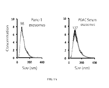
[0040] FIGS. 1A-F. Exosomes contain long fragments of double-stranded genomic
DNA. FIGS. 1A-B. The presence and concentration of exosomes from human
pancreatic
cancer cell lines and human serum samples from patients with pancreatic cancer
were
determined using a NanoSight0 LM10 (FIG. 1A) and electron microscopy (FIG.
1B). FIG.
1C. Exosomes were characterized by the exosomes-specific expression of TSG 101
by
western blotting. FIG. 1D. To exclude RNA contamination after exosomes lysis
and DNA
extraction, the DNA eluate from two cell lines (Panc-1 and T3M4) and the DNA
eluate from
corresponding exosomes was treated with DNAse I and RNAse A. Subsequently, the
eluate
was run on a 2% agarose gel. FIG. 1E. The presence of double-stranded DNA from
Panc-1
exosomes and human serum exosomes from patients with and without pancreatic
cancer was
confirmed by a double-stranded DNA detection kit (representative figure for
exosomal DNA
from Panc-1, one healthy donor, and one patient with pancreatic cancer). FIG.
1F. Exosomes
were characterized by the exosomes-specific expression of TSG 101 and CD63 by
western
blotting.
[0041] FIGS. 2A-E. Exosomes contain mutated KRAS and p53 DNA. FIG. 2A. A
466 bp fragment of KRAS spanning exon 2 and intron 2 and a 1564 bp fragment of
p53
spanning 4 exons and 3 introns were amplified by PCR. FIG. 2B. Sanger
sequencing of
genomic DNA from Panc-1 cells and corresponding exosomes revealed the same
heterozygous mutation of KRAS on codon 12 (GGT to GAT) and the similar
homozygous
mutation of p53 on codon 273 (CGT to CAT). T3M4 cells and corresponding
exosomes
displayed the same homozygous mutation of p53 on codon 220 (TAT to TGT). FIG.
2C.
PCR amplification provided evidence for long fragments of DNA in circulating
exosomes
from two patients with pancreatic cancer. A 466 bp fragment of KRAS DNA and
609 bp
fragment of p53 DNA spanning exons 7 and 8 and intron 7 were retrieved. When
serum
samples depleted of exosomes were subjected to PCR, no KRAS or p53 amplicon
was
detected. FIG. 2D. Sanger sequencing of serum exosome-derived DNA detected DNA
with a
KRAS mutation in codon 22. In a second patient, Sanger sequencing revealed a
KRAS
9
CA 02936100 2016-07-06
WO 2015/085096
PCT/US2014/068630
mutation in codon 12 and a p53 mutation in codon 273. FIG. 2E. PCR
amplification
provided evidence for long fragments of DNA in circulating exosomes from two
healthy
donors and two patients with pancreatic cancer.
[0042] FIGS. 3A-B. Serum-derived exosomes contain genomic DNA spanning all
chromosomes. Whole genome sequencing was conducted on serum-derived, exosomal
DNA
and corresponding primary tumor from two patients. BIC-seq control-free log2
copy-number
profile across all human chromosomes, bin size 1000 bp; RAW profile ¨ black,
segmented ¨
center, gray line. Profiles demonstrate somatic chromosomal gains (up) and
losses (down), as
well as normal polymorphism. In the second case (FIG. 3B), a lack of
structural
chromosomal rearrangement expected for PDAC is explained due to possible low
number of
cancer cells in the sample. Sequencing revealed that circulating exosomes
contain genomic
DNA spanning all chromosomes.
[0043] FIGS. 4A-E. FIG. 4A. Particle tracking analysis using NanoSight
technology. Left image shows a snapshot of a movie of exosomes. Right graph
shows the
integrated analysis of the size distribution of exosomes and their
concentration. FIG. 4B.
Electron microscopy showing images of exosomes collected from culture media.
FIG. 4C.
Immunogold staining of exosomes collected from culture media using anti-CD9
antibody, an
exosomes marker. FIG. 4D. Flow cytometry analysis of exosomes collected from
culture
media using anti-CD9 antibody. FIG. 4E. Immunoblot analysis using CD9 and CD63
exosomes markers to show the presence of exosomes in the media collected from
several
different cell lines.
[0044] FIGS. 5A-E. FIG. 5A. Northern blot of tRNAs of exosomes derived from
different cell lines. FIG. 5B. HeatMap of mass spectrometry analysis showing
the presence
of amino acids in exosomes extracted from different cell lines. FIG. 5C.
Quantitative RT-
PCR of 18S and 28S rRNAs in exosomes derived from different cell lines. Each
sample in
the legend, from top to bottom, represents each bar on the graph in order from
left to right.
FIG. 5D. Immunoblot analysis of eukaryotic translation initiation factor 3A,
4A1 and lA in
protein extracts of different exosomes. CD9 was used as an exosomes marker to
show the
presence of exosomes. FIG. 5E. Immunoprecipitation of eukaryotic translation
initiation
factor 4A1 followed by immunoblot of eukaryotic translation initiation factor
4A1 and 3A
showing interaction between these two proteins in exosomes.
CA 02936100 2016-07-06
WO 2015/085096
PCT/US2014/068630
[0045] FIGS. 6A-C. FIG. 6A. In vitro translation assay using protein extracts
from
exosomes and cells as a positive control and a GFP vector. Protein lysate from
the in vitro
translation assay kit was used as a second positive control. GADPH was used as
a loading
control. FIG. 6B. Immunogold staining of exosomes electroporated with GFP
vector using a
GFP antibody. Upper panels show negative controls and lower panels show GFP
staining in
exosomes. FIG. 6C. Autoradiography of exosomes cultured with [35S] methionine.
Cycloheximide and cells were used as negative and positive controls,
respectively.
[0046] FIG. 7. A plasmid encoding a wild-type p53 protein was electroporated
in
MDA-MB-231-derived exosomes. After 48 h of cell-free culture, electroportaed
exosomes
were used to treat MDA-MB-231 cells pre-treated with cycloheximide, and p21
expression
was evaluated as a downstream target of wild-type p53 function.
[0047] FIGS. 8A-C. FIG. 8A. Graphical representation of mass spectrometry
results
on glypican 1 in exosomes from culture media of E10, NIH-3T3, HDF, MCF10A and
MDA-
MB231 cells. FIG. 8B. Glypican 1 western of exosomes extracted from non-
tumorigenic
breast epithelial cells (MCF10A) and breast cancer cells (MCF7 and MDA-MB231).
FIG.
8C. Flow cytometry analysis of exosomes derived from non-tumorigenic breast
epithelial
cells (MCF10A) and breast cancer cells (MDA-MB231).
[0048] FIGS. 9A-E. GPC1 is present specifically on cancer exosomes. FIG. 9A.
Immunogold transmission electron micrographs of GPC1 in non-tumorigenic cell
line-
derived exosomes (HMLE) (left panel) and in pancreatic cancer-derived exosomes
(T3M4)
(right panel). Gold particles are depicted as black dots. Upper right images
show a digital
zoomed inset. FIG. 9B. Schematic representation of the FACs analysis of GPC1
on the
surface of exosomes. FIG 9C. Transmission electron micrographs (TEM) of
exosomes
coupled to aldehyde/sulphate beads (left panel). Immunogold labeling of GPC1
in T3M4 and
HMLE exosomes coupled to aldehyde/sulphate beads (two bottom panels). Gold
particles are
depicted as black dots. Negative control was performed using secondary
antibody only (top
right). FIG. 9D. Graph representing the percent of GPC1 + exosomes from cancer
cells (gray)
and from non-tumorigenic cells (black). FIG. 9E. Representative histograms of
FACS
analysis of GPC1 + exosomes coupled to aldehyde/sulphate beads from HMLE,
HMEL,
MDA-MB-231, T3M4, PANC-1, and MIA PaCa2 isolated by ultracentrifugation.
11
CA 02936100 2016-07-06
WO 2015/085096
PCT/US2014/068630
[0049] FIGS. 10A-F. GPC1 + circulating exosomes (crExos) derived from cancer
cells in tumor-bearing mice. FIG. 10A. Schematic diagram of the longitudinal
blood
collection from nude mice with orthotopically injected MDA-MB-231 cells. Blood
samples
were obtained prior to tumor cell injection and when the tumor volume reached
300, 550,
1000, and 1350 mm3. FIG. 10B. Representative scatter plots for FACS analysis
of GPC1+
crExos from nude mice with MDA-MB-231 tumors of the indicated volumes. FIG.
10C.
Correlation between tumor volume and percentage (%) of GPC1 + crExos in nude
mice with
orthotopically injected MDA-MB-231 cells (Pearson correlation test,
Correlation coefficient r
= 0. 98, P = 0.004). FIG. 10D. NanoSight coupled with a 488 laser of exosomes
derived
from MDA-MB-231 CD63-GFP cells. Black line represents the tracking analysis
without a
488 laser and the gray line represents the analysis with a 488 laser. FIG.
10E. NanoSight of
crExos from MDA-MB-231-CD63-GFP-injected mice. Black line represents the
tracking
analysis without a 488 laser and the gray line represents the tracking
analysis with a 488
laser. FIG. 10F. Co-localization study for the overlapping expression of CD63-
GFP and
GPC1 in crExos. FACS analysis assessed exosomes derived from MDA-MB-231 cells
as a
negative control (left upper panel), exosomes derived from MDA-MB-231 CD63-GFP
cells
as a positive control (middle upper panel), and exosomes derived from mice
injected with
MDA-MB-231 CD63-GFP cells and analyzed using an Alexa 594 conjugated secondary
antibody only as a negative control (right upper panel). FACS analysis shows
that only the
fraction of CD63-GFP + exosomes, derived from mice orthotopically injected
with MDA-MB-
231 CD63-GFP, were positive for GPC1 (three bottom graphs).
[0050] FIGS. 11A-I. GPC1 + crExos are a non-invasive biomarker for pancreatic
cancer. FIG. 11A. TEM of crExos from a patient with pancreatic cancer. Upper
right image
shows a digitally zoomed inset. FIG. 11B. TEM image of crExos immunogold
labeled for
CD9. Gold particles are depicted as black dots. Upper right image shows a
digitally zoomed
inset. FIG. 11C. Scatter plots representative of FACS analysis of GPC1 +
crExos in healthy
donors (n = 100), breast cancer patients (n = 32), and patients with
pancreatic ductal
adenocarcinoma (PDAC; n = 190) (analysis of variance (ANOVA), ****P < 0.0001).
FIG.
11D. Bar graph representative of the KRAS status of 47 patients with
pancreatic cancer. FIG.
11E. TEM of crExos from three patients with pancreatic cancer. Prior to
immunogold
labeling of GPC1, exosomes were separated using FACS into GPC1 + (left column)
and
GPC1- (right column) populations. Gold particles are depicted as black dots.
FIG. 11F.
Scatter plots representative of KRAS G12D, KRAS wild-type mRNA, and 18S rRNA
12
CA 02936100 2016-07-06
WO 2015/085096
PCT/US2014/068630
expression (left panel) or KRAS G12V, KRAS wild-type mRNA, and 18s rRNA
expression
(right panel) in exosomes that have been separated by FACS into GPC1+ (+;
gray) and GPC1-
(-; black) populations. FIG. 11G. Scatter plots representative for FACS
analysis of GPC1+
crExos in healthy donors (n = 100), patients with a benign pancreatic disease
(BPD; n = 26),
patients with a pancreatic cancer precursor lesion (PCPL; n = 7) and patients
with PDAC (n =
190; analysis of variance (ANOVA), **P <0.01, ****P <0.0001). FIG. 11H.
Scatter plots
representative of ELISA assay of serum CA 19-9 in the same cohort of patients
with
pancreatic cancer as in FIG. 11E (ANOVA, *P < 0.05, ****P < 0.0001). FIG. HI.
Receiver
Operating Characteristic (ROC) curve analysis for GPC1+ crExos (gray line), CA
19-9
(dashed gray line), exosomes concentration (black line), and exosomes size
(dashed black
line) in patients with pancreatic cancer (n = 190) vs. control (healthy donors
(n = 100) and
patients with a benign pancreatic disease (n = 26), total n = 126).
Abbreviations: Area under
the curve (AUC), confidence interval (CI), nanometer (nm).
[0051] FIGS. 12A-F. GPC1+ crExos specifically carry KRAS G12D mRNA. FIG.
12A. Schematic diagram to illustrate the blood collection of patients in the
longitudinal
cohort. Blood samples were obtained prior to surgery (pre-op) and
postoperative at day 7
after surgery. FIG. 12B. Scatter plots representative for FACS analysis of
GPC1+ crExos
after resection in patients of the longitudinal cohort with BPD (n = 4), PCPL
(n = 4), or
PDAC (n = 29) (paired two-tailed Student's t-test, **P < 0.01, ****P <
0.0001). FIG. 12C.
Kaplan¨Meier curves (log-rank test) displaying overall survival of patients
with a drop of
GPC1+ crExos > the median decrease (top line) and a drop of GPC1+ crExos < the
median
decrease (bottom line) after resection (P = 0.016). FIG. 12D. Kaplan¨Meier
curves (log-rank
test) displaying disease-specific survival of patients with a drop of GPC1+
crExos > the
median decrease (top line) and a drop of GPC1+ crExos < the median decrease
(bottom line)
after resection (P = 0.007). FIG. 12E Kaplan¨Meier curves (log-rank test)
displaying overall
survival of patients with a drop of CA 19-9 > the median decrease (top line)
and a drop of CA
19-9 < the median decrease (bottom line) between day 0 and day 7 (P = 0.120).
FIG. 12F.
Kaplan¨Meier curves (log-rank test) displaying disease-specific survival of
patients with a
drop of CA 19-9 > the median decrease (top line) and a drop of CA 19-9 < the
median
decrease (bottom line) between day 0 and day 7 (P = 0.180).
[0052] FIGS. 13A-G. GPC1+ crExos predict pancreas cancer in GEMM. FIG. 13A.
Schematic diagram to illustrate the blood collection from Ptfl die' ;I, Si,-
13
CA 02936100 2016-07-06
WO 2015/085096
PCT/US2014/068630
KrasGl2D/H;Tgfbr2fl0x'll0x (PKT) mice and control mice in the longitudinal
cohort. Blood
samples were obtained at the age of 4, 5, 6, 7, and 8 weeks prior to
euthanasia. FIG. 13B.
Scatter plots representative for FACS analysis of GPC1 + crExos in PKT mice
(E) and control
mice (C) measured at 4, 5, 6, 7, and 8 weeks of age (analysis of variance
(ANOVA), ****P <
0.0001). FIG. 13C. Correlation between tumor volume and GPC1+ crExos in PKT
mice
(Pearson correlation test, Correlation coefficient r = 0.67, P = 0.0005). FIG.
13D. Receiver
Operating Characteristic (ROC) curve analysis for GPC1 crExos (gray line),
exosomes
concentration (black line), and exosomes size (dashed line) in PKT mice at 4
weeks of age (n
= 7) vs. control (control littermate (n = 6) and mice with induced acute
pancreatitis (11 = 4:
total n = 10). FIG 13E. Schematic diagram to illustrate the blood collection
from PKT mice
and control mice in the cross sectional study. Blood samples were obtained at
the age of 16
days or at 20 days prior to euthanasia. F1G. 13F. Scatter plots representative
for FACS
analysis of GPC1+ exosomes in PKT mice and control mice of the cross-sectional
study.
Mice were sacrificed between the age of 16 ¨ 20 days (paired two-tailed
Student's t-test, P <
0.0001). FIG. 13G. Scatter plots representative for quantity of PanIN lesions
diagnosed in
PKT mice and control between the age of 16 ¨ 20 days (left panel).
[0053] FIGS. 14A-H. Exosomes isolation. FIG. 14A. NanoSight0 analysis shows
the exosomes size distribution and concentration of NIH/3T3, MCF 10A, HDF, MDA-
MB-
231 and El0 cells with a modal size of 105 nanometers (nm). FIG. 14B.
Transmission
electron micrograph (TEM) of MDA-MB-231-derived exosomes. Upper right image
shows a
digitally zoomed inset. FIG. 14C. Immunogold and TEM of MDA-MB-231-derived
exosomes of CD9. Gold particles are depicted as black dots. Upper right image
shows a
digitally zoomed inset. FIG. 14D. Immunoblot of flotillinl and CD81 in
exosomal proteins
extracted from E10, NIH/3T3, MDA-MB-231, MCF 10A and HDF cells. FIG. 14E. RT-
qPCR measurement of GPC1 mRNA in HMEL, HDF, HMLE, MCF7, MDA-MB-231,
T3M4, PANC-1, MIA PaCa2. Results are shown as mean + standard deviation (two-
tailed
Student's t-test, P < 0.05). FIG. 14F. Immunoblot of GPC1 in HMEL, HDF, HMLE,
MCF7,
MDA-MB-231, T3M4, PANC-1 and MIA PaCa2 cell lines (upper panel). 13-actin was
used as
a loading control (lower panel). FIG. 14G. Immunoblot of GPC1 to show protein
expression
in exosomes derived from three non-tumorigenic cell lines (HDF, HMEL, HMLE)
and five
tumorigenic cell lines (MCF7, MDA-MB-231, T3M4, PANC-1, MIA PaCa2) (upper
panel).
Immunoblot of flotillinl as loading control (lower panel). FIG. 14H.
Immunoblot of
14
CA 02936100 2016-07-06
WO 2015/085096
PCT/US2014/068630
flotillinl in different layers of a sucrose gradient to which MDA-MB-231 and
T3M4-derived
exosomes were subjected.
[0054] FIGS. 15A-C. NanoSight analysis in human serum samples. FIG. 15A.
Immunoblot of flotillinl of proteins extracted from different layers of a
sucrose gradient to
which patient serum-derived exosomes were subjected. FIG. 15B. NanoSight
analysis
shows the concentration of circulating exosomes (number of exosomes / 1 mL
serum) derived
from healthy donors (n = 100), from breast cancer patients (n = 32), and from
patients with
PDAC (n = 190) (analysis of variance (ANOVA), *P < 0.05, ****P < 0.0001). FIG.
15C.
NanoSight analysis shows the size of circulating exosomes derived from
healthy donors (n
= 100), from breast cancer patients (n = 32), and from patients with PDAC (n =
190)
(analysis of variance (ANOVA), ***P < 0.001).
[0055] FIGS. 16A-E. Tumor stage-specific analysis. FIG. 16A. Receiver
Operating
Characteristic (ROC) curve analysis for GPC1+ crExos (gray line), CA 19-9
(gray dashed
line), exosomes concentration (black line), and exosomes size (black dashed
line) in patients
with carcinoma in situ (CIS) or stage I pancreatic cancer (n = 5) vs. control
(healthy donors (n
= 100) and patients with a benign pancreatic disease (n = 26, total n = 126)).
FIG. 16B. ROC
curve analysis for GPC1+ crExos (gray line), CA 19-9 (gray dashed line),
exosomes
concentration (black line), and exosomes size (black dashed line) in patients
with stage ha
pancreatic cancer (n = 18) vs. control (healthy donors (n = 100) and patients
with a benign
pancreatic disease (n = 26), total n = 126). FIG. 16C. ROC curve analysis for
GPC1+ crExos
(gray line), CA 19-9 (gray dashed line), exosomes concentration (black line),
and exosomes
size (black dashed line) in patients with stage IIb pancreatic cancer (n =
117) vs. control
(healthy donors (n = 100) and patients with a benign pancreatic disease (n =
26, total n =
126)). FIG. 16D. ROC curve analysis for GPC1+ crExos (gray line), CA 19-9
(gray dashed
line), exosomes concentration (black line), and exosomes size (black dashed
line) in patients
with stage III pancreatic cancer (n = 11) vs. control (healthy donors (n =
100) and patients
with a benign pancreas disease (n = 26, total n = 126)). FIG. 16E. ROC curve
analysis for
GPC1+ crExos (gray line), CA 19-9 (gray dashed line), exosomes concentration
(black line),
and exosomes size (black dashed line) in patients with stage IV pancreas
cancer (n = 41) vs.
control (healthy donors (n = 100) and patients with a benign pancreatic
disease (n = 26, total
n = 126)). (Abbreviations: Area under the curve (AUC), confidence interval
(CI), nanometer
(nm)).
CA 02936100 2016-07-06
WO 2015/085096
PCT/US2014/068630
[0056] FIGS. 17A-B. Longitudinal human study. FIG. 17A. Scatter plots
representative for FACS analysis of GPC1+ crExos in patients with pancreatic
cancer
(ANOVA, *P < 0.05). FIG. 17B. Scatter plots representative for ELISA assay of
serum CA
19-9 (U/mL) at the preoperative day and postoperative day 7 in patients of the
longitudinal
cohort with benign pancreas disease (BPD) (n = 4), pancreatic cancer precursor
lesion
(PCPL) (n = 4), and pancreatic ductal adenocarcinoma (PDAC) (n = 29) (paired
two-tailed
Student's t-test, **P < 0.01).
[0057] FIGS. 18A-D. PDAC GEMM longitudinal study. FIG. 18A. Scatter plots
representative for NanoSight analysis of exosomes size in PKT mice (E) and
control mice
(C) measured at 4, 5, 6, 7, and 8 weeks of age (analysis of variance (ANOVA),
*P < 0.05).
FIG, 188. Scatter plots representative for NanoSight analysis of exosomes
concentration in
PKT mice (E) and control mice (C) measured at 4, 5, 6, 7, and 8 weeks of age
(ANOVA, *P
< 0.05). (Abbreviations: Control (C), Experimental (8)) FIG. 18C. Graph
showing tumor
volume measured by MRI and %GPCI' crExos in individual PKT mice iver time
(circles
with dashed lines: tumor volume; squares with solid lines: %GPC1-' crExos).
FIG. 1811
Scatter plots representative for FACS analysis of GPC1+ crExos in control mice
(n = 3) and
mice with Cerulin-induced acute pancreatitis (n = 4) (two-tailed Student's t-
test, us = P >
0.05).
[0058] FIG. 19. PDAC GEMM cross-sectional study. Scatter plots representative
for
KRAS G12D, KRAS wild-type and 18s mRNA expression in exosomes that were
separated
by FACS sorting into GPC1+ (+; gray) and GPC1- (-; black) populations.
DESCRIPTION OF ILLUSTRATIVE EMBODIMENTS
[0059] Exosomes are small vesicles (40-150 nm) of endocytic origin that are
released
by many different cell types. Exosomes in the tumor microenvironment may play
a key role
in facilitating cell-cell communication. Exosomes are reported to
predominantly contain
RNA and proteins. As taught herein, exosomes from pancreatic cancer cells and
serum of
patients with pancreatic ductal adenocarcinoma contain genomic DNA.
[0060] Herein, exosomes were found to contain long fragments of double-
stranded
genomic DNA, which contradicts the current opinion that circulating DNA is
highly
fragmented with an estimated length of only 60-100 bp (Mouliere and Thierry,
2012).
Mutations in KRAS and p53 may be detected using genomic DNA from exosomes
derived
16
CA 02936100 2016-07-06
WO 2015/085096
PCT/US2014/068630
from pancreatic cancer cell lines and serum of patients with pancreatic
cancer. In addition,
serum exosomes from patients with pancreatic cancer contain genomic DNA
spanning all
chromosomes and exosomes-derived DNA carry mutations identical to their
parental cancer
cells or tumors. These results indicate that serum-derived exosomes may be
used to determine
genomic DNA mutations to predict prognosis of cancer patients and improve
treatment via a
personalized medicine approach whereby the detection of specific mutations may
be used to
tailor treatment. As an example, KRAS mutations and EGFR amplifications are
predictive of
resistance to cetuximab, a drug proven to be efficient in some cases of
metastatic colorectal
cancer (Lievre et al., 2006). In addition, cancer patients with a KRAS
mutation in their tumor
do worse on EGFR-targeted therapy using erlotinib.
[0061] Also, exosomes were found to have the ability to perform mRNA
transcription
and protein translation. When exosomes were transfected with a plasmid
encoding p53, the
exosomes were able to express p53 protein and deliver the protein to p53-
deficient target
cells, thereby increasing p21 expression. These results indicate that exosomes
may be used to
express and/or deliver therapeutic proteins to diseased cells.
[0062] Using ultra performance liquid chromatography followed by mass
spectrometry (UPLC-MS) on exosomes derived from normal and cancer cells, a
cell surface
proteoglycan, glypican-1, was found to be specifically enriched on the surface
of cancer cell-
derived exosomes. Circulating GPC1+ exosomes (GPC1+ crExos) were monitored and
isolated using flow cytometry (FACS) from the serum of cancer patients and
mice with
cancer. GPC1+ crExos were detected in the serum of patients with pancreatic
cancer with
absolute specificity and sensitivity, distinguishing healthy subjects and
patients with a benign
pancreatic disease from patients with early and late stage pancreatic cancer.
Levels of GPC1+
crExos paralleled tumor burden in comparative analyses of serum from patients
pre- and
post-surgical tumor resection. GPC1+ crExos from patients and a genetically
engineered
mouse model (GEMM) with spontaneous pancreas tumors driven by pancreas
specific
KRASG12D specifically contained RNA with KRASG12D mutations. GPC1+ crExos
served as a
reliable biomarker for the detection of early PanIN lesions despite a negative
signal by MRI.
GPC1+ crExos can be used to specifically detect cancer exosomes in circulation
and are a
non-invasive diagnostic and screening tool to detect early stages of
pancreatic cancer that
could aid in the prospect of curative surgical therapy. Furthermore, isolation
of glypican 1-
17
CA 02936100 2016-07-06
WO 2015/085096
PCT/US2014/068630
positive exosomes provides a means to isolate cancer cell-derived genomic DNA,
RNA,
and/or proteins.
I. Exosomes
[0063] The terms "microvesicle" and "exosomes," as used herein, refer to a
membranous particle having a diameter (or largest dimension where the
particles is not
spheroid) of between about 10 nm to about 5000 nm, more typically between 30
nm and 1000
nm, and most typically between about 50 nm and 750 nm, wherein at least part
of the
membrane of the exosomes is directly obtained from a cell. Most commonly,
exosomes will
have a size (average diameter) that is up to 5% of the size of the donor cell.
Therefore,
especially contemplated exosomes include those that are shed from a cell.
[0064] Exosomes may be detected in or isolated from any suitable sample type,
such
as, for example, body fluids. As used herein, the term "sample" refers to any
sample suitable
for the methods provided by the present invention. The sample may be any
sample that
includes exosomes suitable for detection or isolation. Sources of samples
include blood, bone
marrow, pleural fluid, peritoneal fluid, cerebrospinal fluid, urine, saliva,
amniotic fluid,
malignant ascites, broncho-alveolar lavage fluid, synovial fluid, breast milk,
sweat, tears,
joint fluid, and bronchial washes. In one aspect, the sample is a blood
sample, including, for
example, whole blood or any fraction or component thereof A blood sample
suitable for use
with the present invention may be extracted from any source known that
includes blood cells
or components thereof, such as venous, arterial, peripheral, tissue, cord, and
the like. For
example, a sample may be obtained and processed using well-known and routine
clinical
methods (e.g., procedures for drawing and processing whole blood). In one
aspect, an
exemplary sample may be peripheral blood drawn from a subject with cancer.
[0065] Exosomes may also be isolated from tissue samples, such as surgical
samples,
biopsy samples, tissues, feces, and cultured cells. When isolating exosomes
from tissue
sources it may be necessary to homogenize the tissue in order to obtain a
single cell
suspension followed by lysis of the cells to release the exosomes. When
isolating exosomes
from tissue samples it is important to select homogenization and lysis
procedures that do not
result in disruption of the exosomes. Exosomes contemplated herein are
preferably isolated
from body fluid in a physiologically acceptable solution, for example,
buffered saline, growth
medium, various aqueous medium, etc.
18
CA 02936100 2016-07-06
WO 2015/085096
PCT/US2014/068630
[0066] Exosomes may be isolated from freshly collected samples or from samples
that have been stored frozen or refrigerated. Although not necessary, higher
purity exosomes
may be obtained if fluid samples are clarified before precipitation with a
volume-excluding
polymer, to remove any debris from the sample. Methods of clarification
include
centrifugation, ultracentrifugation, filtration, or ultrafiltration. Most
typically, exosomes can
be isolated by numerous methods well-known in the art. One preferred method is
differential
centrifugation from body fluids or cell culture supernatants. Exemplary
methods for isolation
of exosomes are described in (Losche et al., 2004; Mesri and Altieri, 1998;
Morel et al.,
2004). Alternatively, exosomes may also be isolated via flow cytometry as
described in
(Combes et al., 1997).
[0067] One accepted protocol for isolation of exosomes includes
ultracentrifugation,
often in combination with sucrose density gradients or sucrose cushions to
float the relatively
low-density exosomes. Isolation of exosomes by sequential differential
centrifugations is
complicated by the possibility of overlapping size distributions with other
microvesicles or
macromolecular complexes. Furthermore, centrifugation may provide insufficient
means to
separate vesicles based on their sizes. However, sequential centrifugations,
when combined
with sucrose gradient ultracentrifugation, can provide high enrichment of
exosomes.
[0068] Isolation of exosomes based on size, using alternatives to the
ultracentrifugation routes, is another option. Successful purification of
exosomes using
ultrafiltration procedures that are less time consuming than
ultracentrifugation, and do not
require use of special equipment have been reported. Similarly, a commercial
kit is available
(EXOMIRTm, Bioo Scientific) which allows removal of cells, platelets, and
cellular debris on
one microfilter and capturing of vesicles bigger than 30 nm on a second
microfilter using
positive pressure to drive the fluid. For this process, the exosomes are not
recovered, their
RNA content is directly extracted from the material caught on the second
microfilter, which
can then be used for PCR analysis. HPLC-based protocols could potentially
allow one to
obtain highly pure exosomes, though these processes require dedicated
equipment and are
difficult to scale up. A significant problem is that both blood and cell
culture media contain
large numbers of nanoparticles (some non-vesicular) in the same size range as
exosomes. For
example, some miRNAs may be contained within extracellular protein complexes
rather than
exosomes; however, treatment with protease (e.g., proteinase K) can be
performed to
eliminate any possible contamination with "extraexosomal" protein.
19
CA 02936100 2016-07-06
WO 2015/085096
PCT/US2014/068630
[0069] In another embodiment, cancer cell-derived exosomes may be captured by
techniques commonly used to enrich a sample for exosomes, such as those
involving
immunospecific interactions (e.g., immunomagnetic capture). Immunomagnetic
capture, also
known as immunomagnetic cell separation, typically involves attaching
antibodies directed to
proteins found on a particular cell type to small paramagnetic beads. When the
antibody-
coated beads are mixed with a sample, such as blood, they attach to and
surround the
particular cell. The sample is then placed in a strong magnetic field, causing
the beads to
pellet to one side. After removing the blood, captured cells are retained with
the beads. Many
variations of this general method are well-known in the art and suitable for
use to isolate
exosomes. In one example, the exosomes may be attached to magnetic beads
(e.g.,
aldehyde/sulphate beads) and then an antibody is added to the mixture to
recognize an
epitope on the surface of the exosomes that are attached to the beads.
[0070] As used herein, analysis includes any method that allows direct or
indirect
visualization of exosomes and may be in vivo or ex vivo. For example, analysis
may include,
but not limited to, ex vivo microscopic or cytometric detection and
visualization of exosomes
bound to a solid substrate, flow cytometry, fluorescent imaging, and the like.
In an exemplary
aspect, cancer cell-derived exosomes are detected using antibodies directed to
glypican 1 and
subsequently bound to a solid substrate and visualized using microscopic or
cytometric
detection.
II. Diagnosis, Prognosis, and Treatment of Diseases
[0071] Detection, isolation, and characterization of cancer cell-derived
exosomes,
using the methods of the invention, is useful in assessing cancer prognosis
and in monitoring
therapeutic efficacy for early detection of treatment failure that may lead to
disease relapse.
In addition, cancer cell-derived exosomes analysis according to the invention
enables the
detection of early relapse in presymptomatic patients who have completed a
course of
therapy. This is possible because the presence of cancer cell-derived may be
associated
and/or correlated with tumor progression and spread, poor response to therapy,
relapse of
disease, and/or decreased survival over a period of time. Thus, enumeration
and
characterization of cancer cell-derived exosomes provides methods to stratify
patients for
baseline characteristics that predict initial risk and subsequent risk based
upon response to
therapy.
CA 02936100 2016-07-06
WO 2015/085096
PCT/US2014/068630
[0072] Accordingly, in another embodiment, the invention provides a method for
diagnosing or prognosing cancer in a subject. Cancer cell-derived exosomes
isolated
according to the methods disclosed herein may be analyzed to diagnose or
prognose cancer in
the subject. As such, the methods of the present invention may be used, for
example, to
evaluate cancer patients and those at risk for cancer. In any of the methods
of diagnosis or
prognosis described herein, either the presence or the absence of one or more
indicators of
cancer, such as a genomic mutation or cancer-specific exosomes surface marker,
or of any
other disorder, may be used to generate a diagnosis or prognosis.
[0073] In one aspect, a blood sample is drawn from the patient and cancer cell-
derived exosomes are detected and/or isolated as described herein. For
example, the
exosomes may be labeled with one or more antibodies that bind to glypican 1,
and the
antibodies may have a covalently bound fluorescent label. Analysis may then be
performed to
determine the number and characterization of cancer cell-derived exosomes in
the sample,
and from this measurement, the number of cancer cell-derived exosomes present
in the initial
blood sample may be determined. The number of cancer cell-derived exosomes may
be
determined by cytometric or microscopic techniques to visually quantify and
characterize the
exosomes. Cancer cell-derived exosomes may be detected and quantifies by other
methods
known in the art (e.g., ELISA).
[0074] In various aspects, analysis of a subject's cancer cell-derived
exosomes
number and characterization may be made over a particular time course in
various intervals to
assess a subject's progression and pathology. For example, analysis may be
performed at
regular intervals such as one day, two days, three days, one week, two weeks,
one month, two
months, three months, six months, or one year, in order to track the level and
characterization
of cancer cell-derived exosomes as a function of time. In the case of existing
cancer patients,
this provides a useful indication of the progression of the disease and
assists medical
practitioners in making appropriate therapeutic choices based on the increase,
decrease, or
lack of change in cancer cell-derived exosomes. Any increase, be it 2-fold, 5-
fold, 10-fold or
higher, in cancer cell-derived exosomes over time decreases the patient's
prognosis and is an
early indicator that the patient should change therapy. Similarly, any
increase, be it 2-fold, 5-
fold, 10-fold or higher, indicates that a patient should undergo further
testing such as imaging
to further assess prognosis and response to therapy. Any decrease, be it 2-
fold, 5-fold, 10-fold
or higher, in cancer cell-derived exosomes over time shows disease
stabilization and a
21
CA 02936100 2016-07-06
WO 2015/085096
PCT/US2014/068630
patient's response to therapy, and is an indicator to not change therapy. For
those at risk of
cancer, a sudden increase in the number of cancer cell-derived exosomes
detected may
provide an early warning that the patient has developed a tumor thus providing
an early
diagnosis. In one embodiment, the detection of cancer cell-derived exosomes
increases with
the staging of the cancer.
[0075] In any of the methods provided herein, additional analysis may also be
performed to characterize cancer cell-derived exosomes to provide additional
clinical
assessment. For example, in addition to image analysis and bulk number
measurements, PCR
techniques may be employed, such as multiplexing with primers specific for
particular cancer
markers to obtain information such as the type of tumor from which the cancer
cell-derived
exosomes originated, metastatic state, and degree of malignancy. Additionally,
DNA or RNA
analysis, proteome analysis, or metabolome analysis may be performed as a
means of
assessing additional information regarding characterization of the patient's
cancer.
[0076] For example, the additional analysis may provide data sufficient to
make
determinations of responsiveness of a subject to a particular therapeutic
regime, or for
determining the effectiveness of a candidate agent in the treatment of cancer.
Accordingly,
the present invention provides a method of determining responsiveness of a
subject to a
particular therapeutic regime or determining the effectiveness of a candidate
agent in the
treatment of cancer by detecting/isolating cancer cell-derived exosomes of the
subject as
described herein and analyzing said cancer cell-derived exosomes. For example,
once a drug
treatment is administered to a patient, it is possible to determine the
efficacy of the drug
treatment using the methods of the invention. For example, a sample taken from
the patient
before the drug treatment, as well as one or more samples taken from the
patient concurrently
with or subsequent to the drug treatment, may be processed using the methods
of the
invention. By comparing the results of the analysis of each processed sample,
one may
determine the efficacy of the drug treatment or the responsiveness of the
patient to the agent.
In this manner, early identification may be made of failed compounds or early
validation may
be made of promising compounds.
[0077] Certain aspects of the present invention can be used to prevent or
treat a
disease or disorder based on the presence of genetic mutations found in
genomic DNA
isolated from exosomes. Other aspects of the present invention provide for
treating a patient
with exosomes that express a recombinant protein or with a recombinant protein
isolated
22
CA 02936100 2016-07-06
WO 2015/085096
PCT/US2014/068630
from exosomes. Other aspects of the present invention provide for diagnosing a
disease
based on the presence of cancer cell-derived exosomes in a patient sample.
[0078] The term "subject" as used herein refers to any individual or patient
to which
the subject methods are performed. Generally the subject is human, although as
will be
appreciated by those in the art, the subject may be an animal. Thus other
animals, including
mammals, such as rodents (including mice, rats, hamsters, and guinea pigs),
cats, dogs,
rabbits, farm animals (including cows, horses, goats, sheep, pigs, etc.), and
primates
(including monkeys, chimpanzees, orangutans, and gorillas) are included within
the
definition of subject.
[0079] "Treatment" and "treating" refer to administration or application of a
therapeutic agent to a subject or performance of a procedure or modality on a
subject for the
purpose of obtaining a therapeutic benefit of a disease or health-related
condition. For
example, a treatment may include administration of chemotherapy,
immunotherapy, or
radiotherapy, performance of surgery, or any combination thereof
[0080] The term "therapeutic benefit" or "therapeutically effective" as used
throughout this application refers to anything that promotes or enhances the
well-being of the
subject with respect to the medical treatment of this condition. This
includes, but is not
limited to, a reduction in the frequency or severity of the signs or symptoms
of a disease. For
example, treatment of cancer may involve, for example, a reduction in the
invasiveness of a
tumor, reduction in the growth rate of the cancer, or prevention of
metastasis. Treatment of
cancer may also refer to prolonging survival of a subject with cancer.
[0081] The term "cancer," as used herein, may be used to describe a solid
tumor,
metastatic cancer, or non-metastatic cancer. In certain embodiments, the
cancer may
originate in the bladder, blood, bone, bone marrow, brain, breast, colon,
esophagus,
duodenum, small intestine, large intestine, colon, rectum, anus, gum, head,
kidney, liver,
lung, nasopharynx, neck, ovary, pancreas, prostate, skin, stomach, testis,
tongue, or uterus.
[0082] The cancer may specifically be of the following histological type,
though it is
not limited to these: neoplasm, malignant; carcinoma; carcinoma,
undifferentiated; giant and
spindle cell carcinoma; small cell carcinoma; papillary carcinoma; squamous
cell carcinoma;
lymphoepithelial carcinoma; basal cell carcinoma; pilomatrix carcinoma;
transitional cell
carcinoma; papillary transitional cell carcinoma; adenocarcinoma; gastrinoma,
malignant;
23
CA 02936100 2016-07-06
WO 2015/085096
PCT/US2014/068630
cholangiocarcinoma; hepatocellular carcinoma; combined hepatocellular
carcinoma and
cholangiocarcinoma; trabecular adenocarcinoma; adenoid cystic carcinoma;
adenocarcinoma
in adenomatous polyp; adenocarcinoma, familial polyposis coli; solid
carcinoma; carcinoid
tumor, malignant; branchiolo-alveolar adenocarcinoma; papillary
adenocarcinoma;
chromophobe carcinoma; acidophil carcinoma; oxyphilic adenocarcinoma; basophil
carcinoma; clear cell adenocarcinoma; granular cell carcinoma; follicular
adenocarcinoma;
papillary and follicular adenocarcinoma; nonencapsulating sclerosing
carcinoma; adrenal
cortical carcinoma; endometroid carcinoma; skin appendage carcinoma; apocrine
adenocarcinoma; sebaceous adenocarcinoma; ceruminous adenocarcinoma;
mucoepidermoid
carcinoma; cystadenocarcinoma; papillary cystadenocarcinoma; papillary serous
cystadenocarcinoma; mucinous cystadenocarcinoma; mucinous adenocarcinoma;
signet ring
cell carcinoma; infiltrating duct carcinoma; medullary carcinoma; lobular
carcinoma;
inflammatory carcinoma; paget's disease, mammary; acinar cell carcinoma;
adenosquamous
carcinoma; adenocarcinoma w/squamous metaplasia; thymoma, malignant; ovarian
stromal
tumor, malignant; thecoma, malignant; granulosa cell tumor, malignant;
androblastoma,
malignant; sertoli cell carcinoma; leydig cell tumor, malignant; lipid cell
tumor, malignant;
paraganglioma, malignant; extra-mammary paraganglioma, malignant;
pheochromocytoma;
glomangiosarcoma; malignant melanoma; amelanotic melanoma; superficial
spreading
melanoma; malignant melanoma in giant pigmented nevus; epithelioid cell
melanoma; blue
nevus, malignant; sarcoma; fibrosarcoma; fibrous histiocytoma, malignant;
myxosarcoma;
liposarcoma; leiomyosarcoma; rhabdomyosarcoma; embryonal rhabdomyosarcoma;
alveolar
rhabdomyosarcoma; stromal sarcoma; mixed tumor, malignant; mullerian mixed
tumor;
nephroblastoma; hepatoblastoma; carcinosarcoma; mesenchymoma, malignant;
brenner
tumor, malignant; phyllodes tumor, malignant; synovial sarcoma; mesothelioma,
malignant;
dysgerminoma; embryonal carcinoma; teratoma, malignant; struma ovarii,
malignant;
choriocarcinoma; mesonephroma, malignant; hemangiosarcoma;
hemangioendothelioma,
malignant; kaposi's sarcoma; hemangiopericytoma, malignant; lymphangiosarcoma;
osteosarcoma; juxtacortical osteosarcoma; chondrosarcoma; chondroblastoma,
malignant;
mesenchymal chondrosarcoma; giant cell tumor of bone; ewing's sarcoma;
odontogenic
tumor, malignant; ameloblastic odontosarcoma; ameloblastoma, malignant;
ameloblastic
fibrosarcoma; pinealoma, malignant; chordoma; glioma, malignant; ependymoma;
astrocytoma; protoplasmic astrocytoma; fibrillary astrocytoma; astroblastoma;
glioblastoma;
oligodendroglioma; oligodendroblastoma; primitive neuroectodermal; cerebellar
sarcoma;
ganglioneuroblastoma; neuroblastoma; retinoblastoma; olfactory neurogenic
tumor;
24
CA 02936100 2016-07-06
WO 2015/085096
PCT/US2014/068630
meningioma, malignant; neurofibrosarcoma; neurilemmoma, malignant; granular
cell tumor,
malignant; malignant lymphoma; hodgkin's disease; hodgkin's; paragranuloma;
malignant
lymphoma, small lymphocytic; malignant lymphoma, large cell, diffuse;
malignant
lymphoma, follicular; mycosis fungoides; other specified non-hodgkin's
lymphomas;
malignant histiocytosis; multiple myeloma; mast cell sarcoma;
immunoproliferative small
intestinal disease; leukemia; lymphoid leukemia; plasma cell leukemia;
erythroleukemia;
lymphosarcoma cell leukemia; myeloid leukemia; basophilic leukemia;
eosinophilic
leukemia; monocytic leukemia; mast cell leukemia; megakaryoblastic leukemia;
myeloid
sarcoma; and hairy cell leukemia.
[0083] The terms "contacted" and "exposed," when applied to a cell, are used
herein
to describe the process by which a therapeutic agent are delivered to a target
cell or are
placed in direct juxtaposition with the target cell. To achieve cell killing,
for example, one or
more agents are delivered to a cell in an amount effective to kill the cell or
prevent it from
dividing.
[0084] An effective
response of a patient or a patient's "responsiveness" to
treatment refers to the clinical or therapeutic benefit imparted to a patient
at risk for, or
suffering from, a disease or disorder. Such benefit may include cellular or
biological
responses, a complete response, a partial response, a stable disease (without
progression or
relapse), or a response with a later relapse. For example, an effective
response can be reduced
tumor size or progression-free survival in a patient diagnosed with cancer.
[0085]
Treatment outcomes can be predicted and monitored and/or patients
benefiting from such treatments can be identified or selected via the methods
described
herein.
[0086]
Regarding neoplastic condition treatment, depending on the stage of
the neoplastic condition, neoplastic condition treatment involves one or a
combination of the
following therapies: surgery to remove the neoplastic tissue, radiation
therapy, and
chemotherapy. Other therapeutic regimens may be combined with the
administration of the
anticancer agents, e.g., therapeutic compositions and chemotherapeutic agents.
For example,
the patient to be treated with such anti-cancer agents may also receive
radiation therapy
and/or may undergo surgery.
CA 02936100 2016-07-06
WO 2015/085096
PCT/US2014/068630
[0087] For
the treatment of disease, the appropriate dosage of a therapeutic
composition will depend on the type of disease to be treated, as defined
above, the severity
and course of the disease, the patient's clinical history and response to the
agent, and the
discretion of the attending physician. The agent is suitably administered to
the patient at one
time or over a series of treatments.
[0088] Therapeutic and prophylactic methods and compositions can be provided
in a
combined amount effective to achieve the desired effect. A tissue, tumor, or
cell can be
contacted with one or more compositions or pharmacological formulation(s)
comprising one
or more of the agents, or by contacting the tissue, tumor, and/or cell with
two or more distinct
compositions or formulations. Also, it is contemplated that such a combination
therapy can
be used in conjunction with chemotherapy, radiotherapy, surgical therapy, or
immunotherapy.
[0089]
Administration in combination can include simultaneous
administration of two or more agents in the same dosage form, simultaneous
administration
in separate dosage forms, and separate administration. That is, the subject
therapeutic
composition and another therapeutic agent can be formulated together in the
same dosage
form and administered simultaneously. Alternatively, subject therapeutic
composition and
another therapeutic agent can be simultaneously administered, wherein both the
agents are
present in separate formulations. In another alternative, the therapeutic
agent can be
administered just followed by the other therapeutic agent or vice versa. In
the separate
administration protocol, the subject therapeutic composition and another
therapeutic agent
may be administered a few minutes apart, or a few hours apart, or a few days
apart.
[0090] A first anti-cancer treatment (e.g., exosomes that express a
recombinant
protein or with a recombinant protein isolated from exosomes) may be
administered before,
during, after, or in various combinations relative to a second anti-cancer
treatment. The
administrations may be in intervals ranging from concurrently to minutes to
days to weeks.
In embodiments where the first treatment is provided to a patient separately
from the second
treatment, one would generally ensure that a significant period of time did
not expire between
the time of each delivery, such that the two compounds would still be able to
exert an
advantageously combined effect on the patient. In such instances, it is
contemplated that one
may provide a patient with the first therapy and the second therapy within
about 12 to 24 or
72 h of each other and, more particularly, within about 6-12 h of each other.
In some
situations it may be desirable to extend the time period for treatment
significantly where
26
CA 02936100 2016-07-06
WO 2015/085096 PCT/US2014/068630
several days (2, 3, 4, 5, 6, or 7) to several weeks (1, 2, 3, 4, 5, 6, 7, or
8) lapse between
respective administrations.
[0091] In certain embodiments, a course of treatment will last 1-90 days or
more (this
such range includes intervening days). It is contemplated that one agent may
be given on any
day of day 1 to day 90 (this such range includes intervening days) or any
combination
thereof, and another agent is given on any day of day 1 to day 90 (this such
range includes
intervening days) or any combination thereof Within a single day (24-hour
period), the
patient may be given one or multiple administrations of the agent(s).
Moreover, after a
course of treatment, it is contemplated that there is a period of time at
which no anti-cancer
treatment is administered. This time period may last 1-7 days, and/or 1-5
weeks, and/or 1-12
months or more (this such range includes intervening days), depending on the
condition of
the patient, such as their prognosis, strength, health, etc. It is expected
that the treatment
cycles would be repeated as necessary.
[0092] Various combinations may be employed. For the example below a first
anti-
cancer therapy is "A" and a second anti-cancer therapy is "B":
A/B/A B/A/B B/B/A A/A/B A/B/B B/A/A A/B/B/B B/A/B/B
B/B/B/A B/B/A/B A/A/B/B A/B/A/B A/B/B/A B/B/A/A
B/A/B/A B/A/A/B A/A/A/B B/A/A/A A/B/A/A A/A/B/A
[0093] Administration of any compound or therapy of the present invention to a
patient will follow general protocols for the administration of such
compounds, taking into
account the toxicity, if any, of the agents. Therefore, in some embodiments
there is a step of
monitoring toxicity that is attributable to combination therapy.
1. Chemotherapy
[0094] A wide variety of chemotherapeutic agents may be used in accordance
with
the present invention. The term "chemotherapy" refers to the use of drugs to
treat cancer. A
"chemotherapeutic agent" is used to connote a compound or composition that is
administered
in the treatment of cancer. These agents or drugs are categorized by their
mode of activity
within a cell, for example, whether and at what stage they affect the cell
cycle. Alternatively,
an agent may be characterized based on its ability to directly cross-link DNA,
to intercalate
27
CA 02936100 2016-07-06
WO 2015/085096
PCT/US2014/068630
into DNA, or to induce chromosomal and mitotic aberrations by affecting
nucleic acid
synthesis.
[0095] Examples of chemotherapeutic agents include alkylating agents, such as
thiotepa and cyclosphosphamide; alkyl sulfonates, such as busulfan,
improsulfan, and
piposulfan; aziridines, such as benzodopa, carboquone, meturedopa, and
uredopa;
ethylenimines and methylamelamines, including altretamine,
triethylenemelamine,
trietylenephosphoramide, triethiylenethiophosphoramide, and
trimethylolomelamine;
acetogenins (especially bullatacin and bullatacinone); a camptothecin
(including the synthetic
analogue topotecan); bryostatin; callystatin; CC-1065 (including its
adozelesin, carzelesin
and bizelesin synthetic analogues); cryptophycins (particularly cryptophycin 1
and
cryptophycin 8); dolastatin; duocarmycin (including the synthetic analogues,
KW-2189 and
CB1-TM1); eleutherobin; pancratistatin; a sarcodictyin; spongistatin; nitrogen
mustards, such
as chlorambucil, chlomaphazine, cholophosphamide, estramustine, ifosfamide,
mechlorethamine, mechlorethamine oxide hydrochloride, melphalan, novembichin,
phenesterine, prednimustine, trofosfamide, and uracil mustard; nitrosureas,
such as
carmustine, chlorozotocin, fotemustine, lomustine, nimustine, and
ranimnustine; antibiotics,
such as the enediyne antibiotics (e.g., calicheamicin, especially
calicheamicin gammalI and
calicheamicin omegaI 1); dynemicin, including dynemicin A; bisphosphonates,
such as
clodronate; an esperamicin; as well as neocarzinostatin chromophore and
related
chromoprotein enediyne antiobiotic chromophores, ac lac inomys ins, actinomyc
in,
authrarnycin, azaserine, bleomycins, cactinomycin, carabicin, carminomycin,
carzinophilin,
chromomycinis, dactinomyc in, daunorubic in, detorub ic in, 6-diazo-5-oxo-L-
norleucine,
doxorubicin (including morpholino-doxorubicin, cyanomorpholino-doxorubicin, 2-
pyrrolino-
doxorubicin and deoxydoxorubicin), epirubicin, esorubicin, idarubicin,
marcellomycin,
mitomycins, such as mitomycin C, mycophenolic acid, nogalamycin, olivomycins,
peplomycin, potfiromycin, puromycin, quelamycin, rodorubicin, streptonigrin,
streptozocin,
tubercidin, ubenimex, zinostatin, and zorubicin; anti-metabolites, such as
methotrexate and 5-
fluorouracil (5-FU); folic acid analogues, such as denopterin, pteropterin,
and trimetrexate;
purine analogs, such as fludarabine, 6-mercaptopurine, thiamiprine, and
thioguanine;
pyrimidine analogs, such as ancitabine, azacitidine, 6-azauridine, carmofur,
cytarabine,
dideoxyuridine, doxifluridine, enocitabine, and floxuridine; androgens, such
as calusterone,
dromostanolone propionate, epitiostanol, mepitiostane, and testolactone; anti-
adrenals, such
as mitotane and trilostane; folic acid replenisher, such as frolinic acid;
aceglatone;
28
CA 02936100 2016-07-06
WO 2015/085096 PCT/US2014/068630
aldophosphamide glycoside; aminolevulinic acid; eniluracil; amsacrine;
bestrabucil;
bisantrene; edatraxate; defofamine; demecolcine; diaziquone; elformithine;
elliptinium
acetate; an epothilone; etoglucid; gallium nitrate; hydroxyurea; lentinan;
lonidainine;
maytansinoids, such as maytansine and ansamitocins; mitoguazone; mitoxantrone;
mopidanmol; nitraerine; pentostatin; phenamet; pirarubicin; losoxantrone;
podophyllinic
acid; 2-ethylhydrazide; procarbazine; PSKpolysaccharide complex; razoxane;
rhizoxin;
sizofiran; spirogermanium; tenuazonic acid; triaziquone; 2,2',2"-
trichlorotriethylamine;
trichothecenes (especially T-2 toxin, ven-acurin A, roridin A and anguidine);
urethan;
vindesine; dacarbazine; mannomustine; mitobronitol; mitolactol; pipobroman;
gacytosine;
arabinoside ("Ara-C"); cyclophosphamide; taxoids, e.g., paclitaxel and
docetaxel
gemcitabine; 6-thioguanine; mercaptopurine; platinum coordination complexes,
such as
cisplatin, oxaliplatin, and carboplatin; vinblastine; platinum; etoposide (VP-
16); ifosfamide;
mitoxantrone; vincristine; vinorelbine; novantrone; teniposide; edatrexate;
daunomycin;
aminopterin; xeloda; ibandronate; irinotecan (e.g., CPT-11); topoisomerase
inhibitor RFS
2000; difluorometlhylornithine (DMF0); retinoids, such as retinoic acid;
capecitabine;
carboplatin, procarbazine,plicomycin, gemcitabien, navelbine, farnesyl-protein
tansferase
inhibitors, transplatinum, and pharmaceutically acceptable salts, acids, or
derivatives of any
of the above.
2. Radiotherapy
[0096] Other factors that cause DNA damage and have been used extensively
include
what are commonly known as 7-rays, X-rays, and/or the directed delivery of
radioisotopes to
tumor cells. Other forms of DNA damaging factors are also contemplated, such
as
microwaves, proton beam irradiation (U.S. Patents 5,760,395 and 4,870,287),
and UV-
irradiation. It is most likely that all of these factors affect a broad range
of damage on DNA,
on the precursors of DNA, on the replication and repair of DNA, and on the
assembly and
maintenance of chromosomes. Dosage ranges for X-rays range from daily doses of
50 to 200
roentgens for prolonged periods of time (3 to 4 wk), to single doses of 2000
to 6000
roentgens. Dosage ranges for radioisotopes vary widely, and depend on the half-
life of the
isotope, the strength and type of radiation emitted, and the uptake by the
neoplastic cells.
3. Immunotherapy
[0097] The skilled artisan will understand that additional immunotherapies may
be
used in combination or in conjunction with methods of the invention. In the
context of
29
CA 02936100 2016-07-06
WO 2015/085096 PCT/US2014/068630
cancer treatment, immunotherapeutics, generally, rely on the use of immune
effector cells
and molecules to target and destroy cancer cells. Rituximab (Rituxan0) is such
an example.
The immune effector may be, for example, an antibody specific for some marker
on the
surface of a tumor cell. The antibody alone may serve as an effector of
therapy or it may
recruit other cells to actually affect cell killing. The antibody also may be
conjugated to a
drug or toxin (chemotherapeutic, radionuclide, ricin A chain, cholera toxin,
pertussis toxin,
etc.) and serve merely as a targeting agent. Alternatively, the effector may
be a lymphocyte
carrying a surface molecule that interacts, either directly or indirectly,
with a tumor cell
target. Various effector cells include cytotoxic T cells and NK cells.
[0098] In one aspect of immunotherapy, the tumor cell must bear some marker
that is
amenable to targeting, i.e., is not present on the majority of other cells.
Many tumor markers
exist and any of these may be suitable for targeting in the context of the
present invention.
Common tumor markers include CD20, carcinoembryonic antigen, tyrosinase (p97),
gp68,
TAG-72, HMFG, Sialyl Lewis Antigen, MucA, MucB, PLAP, laminin receptor, erb B,
and
p155. An alternative aspect of immunotherapy is to combine anticancer effects
with immune
stimulatory effects. Immune stimulating molecules also exist including:
cytokines, such as
IL-2, IL-4, IL-12, GM-CSF, gamma-IFN, chemokines, such as MIP-1, MCP-1, IL-8,
and
growth factors, such as FLT3 ligand.
[0099] Examples of immunotherapies currently under investigation or in use are
immune adjuvants, e.g., Mycobacterium bovis, Plasmodium falciparum,
dinitrochlorobenzene, and aromatic compounds (U.S. Patents 5,801,005 and
5,739,169; Hui
and Hashimoto, 1998; Christodoulides et al., 1998); cytokine therapy, e.g.,
interferons a, 13,
and y, IL-1, GM-CSF, and TNF (Bukowski et al., 1998; Davidson et al., 1998;
Hellstrand et
al., 1998); gene therapy, e.g., TNF, IL-1, IL-2, and p53 (Qin et al., 1998;
Austin-Ward and
Villaseca, 1998; U.S. Patents 5,830,880 and 5,846,945); and monoclonal
antibodies, e.g.,
anti-CD20, anti-ganglioside GM2, and anti-p185 (Hollander, 2012; Hanibuchi et
al., 1998;
U.S. Patent 5,824,311). It is contemplated that one or more anti-cancer
therapies may be
employed with the antibody therapies described herein.
4. Surgery
[00100] Approximately
60% of persons with cancer will undergo surgery of
some type, which includes preventative, diagnostic or staging, curative, and
palliative
CA 02936100 2016-07-06
WO 2015/085096 PCT/US2014/068630
surgery. Curative surgery includes resection in which all or part of cancerous
tissue is
physically removed, excised, and/or destroyed and may be used in conjunction
with other
therapies, such as the treatment of the present invention, chemotherapy,
radiotherapy,
hormonal therapy, gene therapy, immunotherapy, and/or alternative
therapies.Tumor
resection refers to physical removal of at least part of a tumor. In addition
to tumor resection,
treatment by surgery includes laser surgery, cryosurgery, electrosurgery, and
microscopically-controlled surgery (Mohs' surgery).
[00101]
Upon excision of part or all of cancerous cells, tissue, or tumor, a
cavity may be formed in the body. Treatment may be accomplished by perfusion,
direct
injection, or local application of the area with an additional anti-cancer
therapy. Such
treatment may be repeated, for example, every 1, 2, 3, 4, 5, 6, or 7 days, or
every 1, 2, 3, 4,
and 5 weeks or every 1, 2, 3, 4, 5, 6, 7, 8, 9, 10, 11, or 12 months. These
treatments may be
of varying dosages as well.
5. Other Agents
[00102] It is
contemplated that other agents may be used in combination with
certain aspects of the present invention to improve the therapeutic efficacy
of treatment.
These additional agents include agents that affect the upregulation of cell
surface receptors
and GAP junctions, cytostatic and differentiation agents, inhibitors of cell
adhesion, agents
that increase the sensitivity of the hyperproliferative cells to apoptotic
inducers, or other
biological agents. Increases in intercellular signaling by elevating the
number of GAP
junctions would increase the anti-hyperproliferative effects on the
neighboring
hyperproliferative cell population. In other embodiments, cytostatic or
differentiation agents
can be used in combination with certain aspects of the present invention to
improve the anti-
hyperproliferative efficacy of the treatments. Inhibitors of cell adhesion are
contemplated to
improve the efficacy of the present invention. Examples of cell adhesion
inhibitors are focal
adhesion kinase (FAKs) inhibitors and Lovastatin. It is further contemplated
that other
agents that increase the sensitivity of a hyperproliferative cell to
apoptosis, such as the
antibody c225, could be used in combination with certain aspects of the
present invention to
improve the treatment efficacy.
31
CA 02936100 2016-07-06
WO 2015/085096
PCT/US2014/068630
III. Pharmaceutical Compositions
[00103] It
is contemplated that exosomes that express a recombinant protein or
a recombinant protein isolated from exosomes can be administered systemically
or locally to
inhibit tumor cell growth and, most preferably, to kill cancer cells in cancer
patients with
locally advanced or metastatic cancers. They can be administered
intravenously,
intrathecally, and/or intraperitoneally. They can be administered alone or in
combination
with anti-proliferative drugs. In one embodiment, they are administered to
reduce the cancer
load in the patient prior to surgery or other procedures. Alternatively, they
can be
administered after surgery to ensure that any remaining cancer (e.g., cancer
that the surgery
failed to eliminate) does not survive.
[00104] It
is not intended that the present invention be limited by the particular
nature of the therapeutic preparation. For example, such compositions can be
provided in
formulations together with physiologically tolerable liquid, gel, solid
carriers, diluents, or
excipients. These therapeutic preparations can be administered to mammals for
veterinary
use, such as with domestic animals, and clinical use in humans in a manner
similar to other
therapeutic agents. In general, the dosage required for therapeutic efficacy
will vary
according to the type of use and mode of administration, as well as the
particular
requirements of individual subjects.
[00105]
Where clinical applications are contemplated, it may be necessary to
prepare pharmaceutical compositions comprising recombinant proteins and/or
exosomes in a
form appropriate for the intended application. Generally, pharmaceutical
compositions may
comprise an effective amount of one or more recombinant proteins and/or
exosomes or
additional agents dissolved or dispersed in a pharmaceutically acceptable
carrier. The
phrases "pharmaceutical or pharmacologically acceptable" refers to molecular
entities and
compositions that do not produce an adverse, allergic, or other untoward
reaction when
administered to an animal, such as, for example, a human, as appropriate. The
preparation of
a pharmaceutical composition comprising a recombinant protein and/or exosomes
as
disclosed herein, or additional active ingredient will be known to those of
skill in the art in
light of the present disclosure, as exemplified by Remington's Pharmaceutical
Sciences, 18th
Ed., 1990, incorporated herein by reference. Moreover, for animal (e.g.,
human)
administration, it will be understood that preparations should meet sterility,
pyrogenicity,
general safety, and purity standards as required by the FDA Office of
Biological Standards.
32
CA 02936100 2016-07-06
WO 2015/085096
PCT/US2014/068630
[00106]
Further in accordance with certain aspects of the present invention, the
composition suitable for administration may be provided in a pharmaceutically
acceptable
carrier with or without an inert diluent. As used herein, "pharmaceutically
acceptable
carrier" includes any and all aqueous solvents (e.g., water, alcoholic/aqueous
solutions,
ethanol, saline solutions, parenteral vehicles, such as sodium chloride,
Ringer's dextrose,
etc.), non-aqueous solvents (e.g., fats, oils, polyol (for example, glycerol,
propylene glycol,
and liquid polyethylene glycol, and the like), vegetable oil, and injectable
organic esters, such
as ethyloleate), lipids, liposomes, dispersion media, coatings (e.g.,
lecithin), surfactants,
antioxidants, preservatives (e.g., antibacterial or antifungal agents, anti-
oxidants, chelating
agents, inert gases, parabens (e.g., methylparabens, propylparabens),
chlorobutanol, phenol,
sorbic acid, thimerosal or combinations thereof), isotonic agents (e.g.,
sugars and sodium
chloride), absorption delaying agents (e.g., aluminum monostearate and
gelatin), salts, drugs,
drug stabilizers, gels, resins, fillers, binders, excipients, disintegration
agents, lubricants,
sweetening agents, flavoring agents, dyes, fluid and nutrient replenishers,
such like materials
and combinations thereof, as would be known to one of ordinary skill in the
art. The carrier
should be assimilable and includes liquid, semi-solid, i.e., pastes, or solid
carriers. In
addition, if desired, the compositions may contain minor amounts of auxiliary
substances,
such as wetting or emulsifying agents, stabilizing agents, or pH buffering
agents. The pH and
exact concentration of the various components in a pharmaceutical composition
are adjusted
according to well-known parameters. The proper fluidity can be maintained, for
example, by
the use of a coating, such as lecithin, by the maintenance of the required
particle size in the
case of dispersion, and by the use of surfactants.
[00107] A
pharmaceutically acceptable carrier is particularly formulated for
administration to a human, although in certain embodiments it may be desirable
to use a
pharmaceutically acceptable carrier that is formulated for administration to a
non-human
animal but that would not be acceptable (e.g., due to governmental
regulations) for
administration to a human. Except insofar as any conventional carrier is
incompatible with
the active ingredient (e.g., detrimental to the recipient or to the
therapeutic effectiveness of a
composition contained therein), its use in the therapeutic or pharmaceutical
compositions is
contemplated. In accordance with certain aspects of the present invention, the
composition is
combined with the carrier in any convenient and practical manner, i.e., by
solution,
suspension, emulsification, admixture, encapsulation, absorption, and the
like. Such
procedures are routine for those skilled in the art.
33
CA 02936100 2016-07-06
WO 2015/085096
PCT/US2014/068630
[00108]
Certain embodiments of the present invention may comprise different
types of carriers depending on whether it is to be administered in solid,
liquid, or aerosol
form, and whether it needs to be sterile for the route of administration, such
as injection. The
compositions can be administered intravenously, intradermally, transdermally,
intrathecally,
intraarterially, intraperitoneally, intranasally, intravaginally,
intrarectally, intramuscularly,
subcutaneously, mucosally, orally, topically, locally, by inhalation (e.g.,
aerosol inhalation),
by injection, by infusion, by continuous infusion, by localized perfusion
bathing target cells
directly, via a catheter, via a lavage, in lipid compositions (e.g.,
liposomes), or by other
methods or any combination of the forgoing as would be known to one of
ordinary skill in the
art (see, for example, Remington's Pharmaceutical Sciences, 18th Ed., 1990,
incorporated
herein by reference).
[00109] The
active compounds can be formulated for parenteral administration,
e.g., formulated for injection via the intravenous, intramuscular, sub-
cutaneous, or even
intraperitoneal routes. Typically, such compositions can be prepared as either
liquid
solutions or suspensions; solid forms suitable for use to prepare solutions or
suspensions
upon the addition of a liquid prior to injection can also be prepared; and the
preparations can
also be emulsified.
[00110] The
pharmaceutical forms suitable for injectable use include sterile
aqueous solutions or dispersions; formulations including sesame oil, peanut
oil, or aqueous
propylene glycol; and sterile powders for the extemporaneous preparation of
sterile injectable
solutions or dispersions. In all cases the form must be sterile and must be
fluid to the extent
that it may be easily injected. It also should be stable under the conditions
of manufacture
and storage and must be preserved against the contaminating action of
microorganisms, such
as bacteria and fungi.
[00111] The
therapeutics may be formulated into a composition in a free base,
neutral, or salt form. Pharmaceutically acceptable salts include the acid
addition salts, e.g.,
those formed with the free amino groups of a proteinaceous composition, or
which are
formed with inorganic acids, such as, for example, hydrochloric or phosphoric
acids, or such
organic acids as acetic, oxalic, tartaric, or mandelic acid and the like.
Salts formed with the
free carboxyl groups can also be derived from inorganic bases, such as, for
example, sodium,
potassium, ammonium, calcium, or ferric hydroxides; or such organic bases as
isopropylamine, trimethylamine, histidine, or procaine and the like. Upon
formulation,
34
CA 02936100 2016-07-06
WO 2015/085096
PCT/US2014/068630
solutions will be administered in a manner compatible with the dosage
formulation and in
such amount as is therapeutically effective. The formulations are easily
administered in a
variety of dosage forms, such as formulated for parenteral administrations,
such as injectable
solutions, or aerosols for delivery to the lungs, or formulated for alimentary
administrations,
such as drug release capsules and the like.
[00112] In
a specific embodiment of the present invention, the composition is
combined or mixed thoroughly with a semi-solid or solid carrier. The mixing
can be carried
out in any convenient manner, such as grinding. Stabilizing agents can be also
added in the
mixing process in order to protect the composition from loss of therapeutic
activity, i.e.,
denaturation in the stomach. Examples of stabilizers for use in a composition
include buffers,
amino acids, such as glycine and lysine, carbohydrates, such as dextrose,
mannose, galactose,
fructose, lactose, sucrose, maltose, sorbitol, mannitol, etc.
[00113] In
further embodiments, the present invention may concern the use of a
pharmaceutical lipid vehicle composition comprising one or more lipids and an
aqueous
solvent. As used herein, the term "lipid" will be defined to include any of a
broad range of
substances that is characteristically insoluble in water and extractable with
an organic
solvent. This broad class of compounds is well known to those of skill in the
art, and as the
term "lipid" is used herein, it is not limited to any particular structure.
Examples include
compounds that contain long-chain aliphatic hydrocarbons and their
derivatives. A lipid may
be naturally occurring or synthetic (i.e., designed or produced by man).
However, a lipid is
usually a biological substance. Biological lipids are well known in the art,
and include for
example, neutral fats, phospholipids, phosphoglycerides, steroids, terpenes,
lysolipids,
glycosphingolipids, glycolipids, sulphatides, lipids with ether- and ester-
linked fatty acids,
polymerizable lipids, and combinations thereof Of course, compounds other than
those
specifically described herein that are understood by one of skill in the art
as lipids are also
encompassed by the compositions and methods.
[00114] One
of ordinary skill in the art would be familiar with the range of
techniques that can be employed for dispersing a composition in a lipid
vehicle. For
example, the therapeutic agent may be dispersed in a solution containing a
lipid, dissolved
with a lipid, emulsified with a lipid, mixed with a lipid, combined with a
lipid, covalently
bonded to a lipid, contained as a suspension in a lipid, contained or
complexed with a micelle
or liposome, or otherwise associated with a lipid or lipid structure by any
means known to
CA 02936100 2016-07-06
WO 2015/085096
PCT/US2014/068630
those of ordinary skill in the art. The dispersion may or may not result in
the formation of
liposomes.
[00115] The
term "unit dose" or "dosage" refers to physically discrete units
suitable for use in a subject, each unit containing a predetermined quantity
of the therapeutic
composition calculated to produce the desired responses discussed above in
association with
its administration, i.e., the appropriate route and treatment regimen. The
quantity to be
administered, both according to number of treatments and unit dose, depends on
the effect
desired. The actual dosage amount of a composition of the present invention
administered to
a patient or subject can be determined by physical and physiological factors,
such as body
weight, the age, health, and sex of the subject, the type of disease being
treated, the extent of
disease penetration, previous or concurrent therapeutic interventions,
idiopathy of the patient,
the route of administration, and the potency, stability, and toxicity of the
particular
therapeutic substance. For example, a dose may also comprise from about 1
[ig/kg/body
weight to about 1000 mg/kg/body weight (this such range includes intervening
doses) or
more per administration, and any range derivable therein. In non-limiting
examples of a
derivable range from the numbers listed herein, a range of about 5 [ig/kg/body
weight to
about 100 mg/kg/body weight, about 5 [ig/kg/body weight to about 500
mg/kg/body weight,
etc., can be administered. The practitioner responsible for administration
will, in any event,
determine the concentration of active ingredient(s) in a composition and
appropriate dose(s)
for the individual subject.
[00116] The
actual dosage amount of a composition administered to an animal
patient can be determined by physical and physiological factors, such as body
weight,
severity of condition, the type of disease being treated, previous or
concurrent therapeutic
interventions, idiopathy of the patient, and on the route of administration.
Depending upon
the dosage and the route of administration, the number of administrations of a
preferred
dosage and/or an effective amount may vary according to the response of the
subject. The
practitioner responsible for administration will, in any event, determine the
concentration of
active ingredient(s) in a composition and appropriate dose(s) for the
individual subject.
[00117] In
certain embodiments, pharmaceutical compositions may comprise,
for example, at least about 0.1% of an active compound. In other embodiments,
an active
compound may comprise between about 2% to about 75% of the weight of the unit,
or
between about 25% to about 60%, for example, and any range derivable therein.
Naturally,
36
CA 02936100 2016-07-06
WO 2015/085096
PCT/US2014/068630
the amount of active compound(s) in each therapeutically useful composition
may be
prepared in such a way that a suitable dosage will be obtained in any given
unit dose of the
compound.
Factors, such as solubility, bioavailability, biological half-life, route of
administration, product shelf life, as well as other pharmacological
considerations, will be
contemplated by one skilled in the art of preparing such pharmaceutical
formulations, and as
such, a variety of dosages and treatment regimens may be desirable.
[00118] In
other non-limiting examples, a dose may also comprise from about 1
microgram/kg/body weight, about 5 microgram/kg/body weight, about 10
microgram/kg/body weight, about 50 microgram/kg/body weight, about 100
microgram/kg/body weight, about 200 microgram/kg/body weight, about 350
microgram/kg/body weight, about 500 microgram/kg/body weight, about 1
milligram/kg/body weight, about 5 milligram/kg/body weight, about 10
milligram/kg/body
weight, about 50 milligram/kg/body weight, about 100 milligram/kg/body weight,
about 200
milligram/kg/body weight, about 350 milligram/kg/body weight, about 500
milligram/kg/body weight, to about 1000 milligram/kg/body weight or more per
administration, and any range derivable therein. In non-limiting examples of a
derivable
range from the numbers listed herein, a range of about 5 milligram/kg/body
weight to about
100 milligram/kg/body weight, about 5 microgram/kg/body weight to about 500
milligram/kg/body weight, etc., can be administered, based on the numbers
described above.
IV. Nucleic Acids and Vectors
[00119] In
certain aspects of the invention, nucleic acid sequences encoding a
therapeutic protein or a fusion protein containing a therapeutic protein may
be disclosed.
Depending on which expression system is used, nucleic acid sequences can be
selected based
on conventional methods. For example, the respective genes or variants thereof
may be
codon optimized for expression in a certain system. Various vectors may be
also used to
express the protein of interest. Exemplary vectors include, but are not
limited, plasmid
vectors, viral vectors, transposon, or liposome-based vectors.
V. Recombinant Proteins
[00120]
Some embodiments concern recombinant proteins and polypeptides.
Particular embodiments concern a recombinant protein or polypeptide that
exhibits at least
one therapeutic activity. In further aspects, the protein or polypeptide may
be modified to
37
CA 02936100 2016-07-06
WO 2015/085096
PCT/US2014/068630
increase serum stability. Thus, when the present application refers to the
function or activity
of "modified protein" or a "modified polypeptide," one of ordinary skill in
the art would
understand that this includes, for example, a protein or polypeptide that
possesses an
additional advantage over the unmodified protein or polypeptide. It
is specifically
contemplated that embodiments concerning a "modified protein" may be
implemented with
respect to a "modified polypeptide," and vice versa.
[00121]
Recombinant proteins may possess deletions and/or substitutions of
amino acids; thus, a protein with a deletion, a protein with a substitution,
and a protein with a
deletion and a substitution are modified proteins. In some embodiments, these
proteins may
further include insertions or added amino acids, such as with fusion proteins
or proteins with
linkers, for example. A "modified deleted protein" lacks one or more residues
of the native
protein, but may possess the specificity and/or activity of the native
protein. A "modified
deleted protein" may also have reduced immunogenicity or antigenicity. An
example of a
modified deleted protein is one that has an amino acid residue deleted from at
least one
antigenic region that is, a region of the protein determined to be antigenic
in a particular
organism, such as the type of organism that may be administered the modified
protein.
[00122]
Substitution or replacement variants typically contain the exchange of
one amino acid for another at one or more sites within the protein and may be
designed to
modulate one or more properties of the polypeptide, particularly its effector
functions and/or
bioavailability. Substitutions may or may not be conservative, that is, one
amino acid is
replaced with one of similar shape and charge. Conservative substitutions are
well known in
the art and include, for example, the changes of: alanine to serine; arginine
to lysine;
asparagine to glutamine or histidine; aspartate to glutamate; cysteine to
serine; glutamine to
asparagine; glutamate to aspartate; glycine to proline; histidine to
asparagine or glutamine;
isoleucine to leucine or valine; leucine to valine or isoleucine; lysine to
arginine; methionine
to leucine or isoleucine; phenylalanine to tyrosine, leucine, or methionine;
serine to
threonine; threonine to serine; tryptophan to tyrosine; tyrosine to tryptophan
or
phenylalanine; and valine to isoleucine or leucine.
[00123] In
addition to a deletion or substitution, a modified protein may
possess an insertion of residues, which typically involves the addition of at
least one residue
in the polypeptide. This may include the insertion of a targeting peptide or
polypeptide or
simply a single residue. Terminal additions, called fusion proteins, are
discussed below.
38
CA 02936100 2016-07-06
WO 2015/085096
PCT/US2014/068630
[00124] The
term "biologically functional equivalent" is well understood in the
art and is further defined in detail herein. Accordingly, sequences that have
between about
70% and about 80%, or between about 81% and about 90%, or even between about
91% and
about 99% of amino acids that are identical or functionally equivalent to the
amino acids of a
control polypeptide are included, provided the biological activity of the
protein is maintained.
A recombinant protein may be biologically functionally equivalent to its
native counterpart in
certain aspects.
[00125] It
also will be understood that amino acid and nucleic acid sequences
may include additional residues, such as additional N- or C-terminal amino
acids or 5' or 3'
sequences, and yet still be essentially as set forth in one of the sequences
disclosed herein, so
long as the sequence meets the criteria set forth above, including the
maintenance of
biological protein activity where protein expression is concerned. The
addition of terminal
sequences particularly applies to nucleic acid sequences that may, for
example, include
various non-coding sequences flanking either of the 5' or 3' portions of the
coding region or
may include various internal sequences, i. e. , introns, which are known to
occur within genes.
[00126] As
used herein, a protein or peptide generally refers, but is not limited
to, a protein of greater than about 200 amino acids, up to a full length
sequence translated
from a gene; a polypeptide of greater than about 100 amino acids; and/or a
peptide of from
about 3 to about 100 amino acids. For convenience, the terms "protein,"
"polypeptide," and
"peptide are used interchangeably herein.
[00127] As
used herein, an "amino acid residue" refers to any naturally
occurring amino acid, any amino acid derivative, or any amino acid mimic known
in the art.
In certain embodiments, the residues of the protein or peptide are sequential,
without any
non-amino acids interrupting the sequence of amino acid residues. In other
embodiments, the
sequence may comprise one or more non-amino acid moieties. In particular
embodiments,
the sequence of residues of the protein or peptide may be interrupted by one
or more non-
amino acid moieties.
[00128]
Accordingly, the term "protein or peptide" encompasses amino acid
sequences comprising at least one of the 20 common amino acids found in
naturally
occurring proteins, or at least one modified or unusual amino acid.
39
CA 02936100 2016-07-06
WO 2015/085096
PCT/US2014/068630
[00129]
Certain embodiments of the present invention concern fusion proteins.
These molecules may have a therapeutic protein linked at the N- or C-terminus
to a
heterologous domain. For example, fusions may also employ leader sequences
from other
species to permit the recombinant expression of a protein in a heterologous
host. Another
useful fusion includes the addition of a protein affinity tag, such as a serum
albumin affinity
tag or six histidine residues, or an immunologically active domain, such as an
antibody
epitope, preferably cleavable, to facilitate purification of the fusion
protein. Non-limiting
affinity tags include polyhistidine, chitin binding protein (CBP), maltose
binding protein
(MBP), and glutathione-S-transferase (GST).
[00130] In a
particular embodiment, the therapeutic protein may be linked to a
peptide that increases the in vivo half-life, such as an XTEN polypeptide
(Schellenberger et
al., 2009), IgG Fc domain, albumin, or albumin binding peptide.
[00131]
Methods of generating fusion proteins are well known to those of skill
in the art. Such proteins can be produced, for example, by de novo synthesis
of the complete
fusion protein, or by attachment of the DNA sequence encoding the heterologous
domain,
followed by expression of the intact fusion protein.
[00132]
Production of fusion proteins that recover the functional activities of
the parent proteins may be facilitated by connecting genes with a bridging DNA
segment
encoding a peptide linker that is spliced between the polypeptides connected
in tandem. The
linker would be of sufficient length to allow proper folding of the resulting
fusion protein.
VI. Protein Purification
[00133]
Protein purification techniques are well known to those of skill in the
art. These techniques involve, at one level, the homogenization and crude
fractionation of the
cells, tissue, or organ to polypeptide and non-polypeptide fractions. The
protein or
polypeptide of interest may be further purified using chromatographic and
electrophoretic
techniques to achieve partial or complete purification (or purification to
homogeneity) unless
otherwise specified. Analytical methods particularly suited to the preparation
of a pure
peptide are ion-exchange chromatography, gel exclusion chromatography,
polyacrylamide
gel electrophoresis, affinity chromatography, immunoaffinity chromatography,
and
isoelectric focusing. A particularly efficient method of purifying peptides is
fast-performance
liquid chromatography (FPLC) or even high-performance liquid chromatography
(HPLC).
CA 02936100 2016-07-06
WO 2015/085096
PCT/US2014/068630
[00134] A
purified protein or peptide is intended to refer to a composition,
isolatable from other components, wherein the protein or peptide is purified
to any degree
relative to its naturally-obtainable state. An isolated or purified protein or
peptide, therefore,
also refers to a protein or peptide free from the environment in which it may
naturally occur.
Generally, "purified" will refer to a protein or peptide composition that has
been subjected to
fractionation to remove various other components, and which composition
substantially
retains its expressed biological activity. Where the term "substantially
purified" is used, this
designation will refer to a composition in which the protein or peptide forms
the major
component of the composition, such as constituting about 50%, about 60%, about
70%, about
80%, about 90%, about 95%, or more of the proteins in the composition.
[00135]
Various techniques suitable for use in protein purification are well
known to those of skill in the art. These include, for example, precipitation
with ammonium
sulphate, PEG, antibodies and the like, or by heat denaturation, followed by
centrifugation;
chromatography steps, such as ion exchange, gel filtration, reverse phase,
hydroxyapatite, and
affinity chromatography; isoelectric focusing; gel electrophoresis; and
combinations of these
and other techniques. As is generally known in the art, it is believed that
the order of
conducting the various purification steps may be changed, or that certain
steps may be
omitted, and still result in a suitable method for the preparation of a
substantially purified
protein or peptide.
[00136] Various
methods for quantifying the degree of purification of the
protein or peptide are known to those of skill in the art in light of the
present disclosure.
These include, for example, determining the specific activity of an active
fraction, or
assessing the amount of polypeptides within a fraction by SDS/PAGE analysis. A
preferred
method for assessing the purity of a fraction is to calculate the specific
activity of the
fraction, to compare it to the specific activity of the initial extract, and
to thus calculate the
degree of purity therein, assessed by a "-fold purification number." The
actual units used to
represent the amount of activity will, of course, be dependent upon the
particular assay
technique chosen to follow the purification, and whether or not the expressed
protein or
peptide exhibits a detectable activity.
[00137] There is no
general requirement that the protein or peptide will always
be provided in its most purified state. Indeed, it is contemplated that less
substantially
purified products may have utility in certain embodiments. Partial
purification may be
41
CA 02936100 2016-07-06
WO 2015/085096
PCT/US2014/068630
accomplished by using fewer purification steps in combination, or by utilizing
different forms
of the same general purification scheme. For example, it is appreciated that a
cation-
exchange column chromatography performed utilizing an HPLC apparatus will
generally
result in a greater "-fold" purification than the same technique utilizing a
low pressure
chromatography system. Methods exhibiting a lower degree of relative
purification may
have advantages in total recovery of protein product, or in maintaining the
activity of an
expressed protein.
[00138] In
certain embodiments a protein or peptide may be isolated or
purified. For example, a His tag or an affinity epitope may be comprised in a
recombinant
protein to facilitate purification. Affinity chromatography is a
chromatographic procedure
that relies on the specific affinity between a substance to be isolated and a
molecule to which
it can specifically bind. This is a receptor-ligand type of interaction. The
column material is
synthesized by covalently coupling one of the binding partners to an insoluble
matrix. The
column material is then able to specifically adsorb the substance from the
solution. Elution
occurs by changing the conditions to those in which binding will not occur
(e.g., altered pH,
ionic strength, temperature, etc.). The matrix should be a substance that does
not adsorb
molecules to any significant extent and that has a broad range of chemical,
physical, and
thermal stability. The ligand should be coupled in such a way as to not affect
its binding
properties. The ligand should also provide relatively tight binding. It should
be possible to
elute the substance without destroying the sample or the ligand.
[00139]
Size exclusion chromatography (SEC) is a chromatographic method in
which molecules in solution are separated based on their size, or in more
technical terms,
their hydrodynamic volume. It is usually applied to large molecules or
macromolecular
complexes, such as proteins and industrial polymers. Typically, when an
aqueous solution is
used to transport the sample through the column, the technique is known as gel
filtration
chromatography, versus the name gel permeation chromatography, which is used
when an
organic solvent is used as a mobile phase.
[00140] The
underlying principle of SEC is that particles of different sizes will
elute (filter) through a stationary phase at different rates. This results in
the separation of a
solution of particles based on size. Provided that all the particles are
loaded simultaneously
or near simultaneously, particles of the same size should elute together. Each
size exclusion
column has a range of molecular weights that can be separated. The exclusion
limit defines
42
CA 02936100 2016-07-06
WO 2015/085096
PCT/US2014/068630
the molecular weight at the upper end of this range and is where molecules are
too large to be
trapped in the stationary phase. The permeation limit defines the molecular
weight at the
lower end of the range of separation and is where molecules of a small enough
size can
penetrate into the pores of the stationary phase completely and all molecules
below this
molecular mass are so small that they elute as a single band.
[00141]
High-performance liquid chromatography (or high-pressure liquid
chromatography, HPLC) is a form of column chromatography used frequently in
biochemistry and analytical chemistry to separate, identify, and quantify
compounds. HPLC
utilizes a column that holds chromatographic packing material (stationary
phase), a pump that
moves the mobile phase(s) through the column, and a detector that shows the
retention times
of the molecules. Retention time varies depending on the interactions between
the stationary
phase, the molecules being analyzed, and the solvent(s) used.
VII. Kits and Diagnostics
[00142] In
various aspects of the invention, a kit is envisioned containing the
necessary components to purify exosomes from a body fluid and isolate genomic
DNA
therefrom. The kit may further contain oligonucleotides for use in amplifying
a target DNA
sequence and/or sequence a target segment of DNA. In other aspects, a kit is
envisioned
containing the necessary components to isolate exosomes and transfect them
with a nucleic
acid encoding a therapeutic protein. In yet other aspects, a kit is envisioned
containing the
necessary components to isolate exosomes and determine the presence of a
cancer cell-
derived exosome-specific marker within the isolated exosomes.
[00143] The
kit may comprise one or more sealed vials containing any of such
components. In some embodiments, the kit may also comprise a suitable
container means,
which is a container that will not react with components of the kit, such as
an eppendorf tube,
an assay plate, a syringe, a bottle, or a tube. The container may be made from
sterilizable
materials such as plastic or glass.
[00144] The
kit may further include an instruction sheet that outlines the
procedural steps of the methods set forth herein, and will follow
substantially the same
procedures as described herein or are known to those of ordinary skill. The
instruction
information may be in a computer readable media containing machine-readable
instructions
that, when executed using a computer, cause the display of a real or virtual
procedure of
43
CA 02936100 2016-07-06
WO 2015/085096
PCT/US2014/068630
purifying exosomes from a sample and isolating genomic DNA therefrom,
expressing a
recombinant protein therein, or identifying a cancer cell-derived marker
thereon.
VIII. Examples
[00145] The following examples are included to demonstrate preferred
embodiments of the invention. It should be appreciated by those of skill in
the art that the
techniques disclosed in the examples which follow represent techniques
discovered by the
inventor to function well in the practice of the invention, and thus can be
considered to
constitute preferred modes for its practice. However, those of skill in the
art should, in light
of the present disclosure, appreciate that many changes can be made in the
specific
embodiments which are disclosed and still obtain a like or similar result
without departing
from the spirit and scope of the invention.
Materials and Methods
[00146]
Patient samples and tissue collection. The Ethics Committee of the
University of Heidelberg approved serum collection from patients. The study
was conducted
according to the Reporting Recommendations for Tumor Marker Prognostic Studies
(REMARK) criteria. Serum samples and tissue samples from patients with
pancreatic cancer,
serum samples only from patients with a benign pancreatic disease and from
healthy donors,
who had no evidence of acute or chronic disease and had no surgery within the
past 12
months, were received from the department of General, Visceral and
Transplantation Surgery
from the University of Heidelberg. The pancreatic cohort included 190 patients
with an
adenocarcinoma of the pancreas (PDAC), 18 patients with pancreatitis, 8
patients with a
benign serous cystadenoma, five patients with an intraductal papillary
mucinous neoplasm
(IPMN), and two patients with a pancreatic intraepithelial neoplasia (PaNIN)
Ib. Patients
were subjected to surgery between 2006 and 2012 at the Department of General,
Visceral,
and Transplantation Surgery, University of Heidelberg. Clinical information
included age,
gender, AJCC tumor stage, tumor size (pT), presence and number of lymph node
mestastases
(pN), tumor grade (G), and treatment with (neo-)/adjuvant chemotherapy.
[00147]
Serum samples from 32 patients with breast cancer were collected at
the MD Anderson Cancer Center, Houston, Texas. Clinical information included
age, gender,
AJCC tumor stage, tumor size (pT), presence and number of lymph node
mestastases (pN),
tumor grade, and treatment with (neo-)/adjuvant chemotherapy.
44
CA 02936100 2016-07-06
WO 2015/085096
PCT/US2014/068630
[00148] A
written informed consent for the serum sampling and tumor
sampling was obtained preoperatively from all patients and from each healthy
donor prior to
collection with disclosure of planned analyses regarding potential prognostic
markers. No
neoadjuvant radiotherapy or chemotherapy was provided prior to surgical
resection of tumors
in the patients. On the day of surgery, 10 mL serum separator tubes were used
to collect
blood samples through a central venous catheter immediately before surgical
incision. To
prevent dilution with blocking saline, the first 5-7 mL of the drawn blood
were discarded.
The blood samples were then centrifuged at 2.500 x g for 10 min to extract the
serum, and the
serum was stored at -80 C until analysis. Likewise, blood samples were
collected on day 7
after surgery in 29 patients with an adenocarcinoma of the pancreas (PDAC), 4
patients with
chronic pancreatitis, and 4 patients with an intraductal papillary mucinous
neoplasm (IPMN).
[00149]
Animal Studies. Nude mice (nu/nu) (purchased from Jackson
Laboratory) underwent breast pad injections with 0.5 million MDA-MB-231 cells
or MDA-
MB-231-CD63GFP cells in 20 L of PBS injected per breast pad. Buprenorphine
was
administered subcutaneously (0.1 mg/kg in 0.1 mL saline) once prior to the
surgery and every
8-12 h post-operatively for 24 h. Blood was collected retro-orbitally and
exosomes were
isolated prior to injection and at tumor volumes of 250, 500, 1000, and 1500
mm3. Mice were
euthanized when the tumor size reached 1500 mm3 or when severe disease
symptoms were
present.
[00150] The disease progression and genotyping for the
Ptflacre/+;LSLKrasG12D/+; Tgfbr2flox/flox (PKT) mice was previously described
(Ijichi et
al., 2006; Ozdemir et al., 2014). In the longitudinal cohort, retro-orbital
blood collection was
performed at 4, 5, 6, 7, and 8 weeks of age. Mice were euthanized at an age of
8 weeks or
when severe disease symptoms were present. In 4 control littermate without
pancreatic
cancer, acute pancreatitis was induced by i.p. injections of Cerulean (50
ng/kg body weight
once an hour for 5 h (overall: 5 injections). Mice were sacrificed 24 h after
injection. All
mice were housed under standard housing conditions at the MD Anderson Cancer
Center
(MDACC) animal facilities, and all animal procedures were reviewed and
approved by the
MDACC institutional animal care and use committees.
[00151] Cell lines.
The following human cells lines were used: HMLE
(American Type Culture Collection (ATCC), Manassas, VA), BJ (ATCC), HDF
(ATCC),
HMEL (ATCC), MCF-7 (ATCC), MDA-MB231 (ATCC), PANC-1 (ATCC), 5W480
CA 02936100 2016-07-06
WO 2015/085096
PCT/US2014/068630
(ATCC), HCT 116 (ATCC), MIA PaCa-2 (ATCC), and T3M4 cells (Cell Bank, RIKEN
BioResource Centre, Japan). The following murine cells lines were used:
NIH/3T3 (ATCC),
El0 (ATCC), NMuMG (ATCC), 4T1 (ATCC), and B16-F10 cells (ATCC). HDF and BJ
cells were cultured in DMEM supplemented with 20% (v/v) fetal bovine serum
(FBS), 100
U/mL penicillin and 100 ug/mL streptomycin. HMLE and MCF 10A cells were grown
in
DMEM/F12 supplemented with 5% (v/v) horse serum, 100 U/mL penicillin, 100
ug/mL
streptomycin, 20 ng/mL EGF, 0.5 mg/mL hydrocortisone, 100 ng/mL cholera toxin,
and 10
ug/mL insulin. HMEL, MCF7, MDA-MB-231, HCT 116, SW480, 4T1, NIH/3T3, E10, U-
87, and B16 F10 cells were maintained in DMEM supplemented with 10% (v/v) FBS,
100
U/mL penicillin and 100 ug/mL streptomycin. PANC-1, MIA PaCa-2, and T3M4 cells
were
cultured in RPMI-1640 (Sigma, St. Louis, MO) supplemented with 10% (v/v) FBS,
100
U/mL penicillin, amphotericin B, and 100 ug/mL streptomycin. NMUMG cells were
grown
in DMEM supplemented with 10% (v/v) FBS, 100 U/mL penicillin, 100 ug/mL
streptomycin, and 10 ug/mL insulin. All cell lines were kept in a humidified
atmosphere at
5% CO2 and 37 C. Transfections were performed using Lipofectamine0 2000
reagent
(Invitrogen) for siRNA. GPC1 siRNA (Cat. Nos. SI00032445, SI00032459,
SI00032466,
SI03071033) and scramble siRN A were purchased from Qiagen (Hilden, Germany).
[00152]
Exosomes isolation from cells. Exosomes were obtained from
supernatant of cells as previously described with some modifications (Kahlert
et al., 2014).
Briefly, cells were grown in T225 cm2 flasks until they reached a confluency
of 80%-90%.
Next, the media was collected and centrifuged at 800 x g for 5 minutes,
followed by a
centrifugation step of 2000 x g for 10 minutes to discard cellular detritus.
Then, the media
was filtered using a 0.2 um pore filter (Syringe filter, Cat. No. 6786-1302,
GE Healthcare,
GB). Afterwards, the collected media was ultracentrifuged at 100,000 x g for 2
h at 4 C. The
exosomes pellet was washed with 35 mL lx PBS, followed by a second step of
ultracentrifugation at 100,000 x g for 2 h at 4 C. Afterwards, the supernatant
was discarded.
Exosomes used for RNA extraction were resuspended in 500 uL of TRIzol0;
exosomes used
for protein extraction were resuspended in 250 uL of lysis buffer (8 M
urea/2.5% SDS, 5
ug/mL leupeptin, 1 ug/mL pepstatin, and 1 mM phenylmethylsulphonyl fluoride
(PMSF)).
Exosomes used for flow cytometry analysis (FACS), transmission electron
microscopy
(TEM), and immunogold staining were resuspended in 100 uL lx PBS. Ten
microliters of
this sample were diluted at 1:100 in lx PBS and analyzed using a NanoSight0
LM10
(NanoSight Ltd., Minton Park, Amesbury, GB).
46
CA 02936100 2016-07-06
WO 2015/085096
PCT/US2014/068630
[00153]
Exosomes isolation from human serum samples. As previously
described, 250 uL of cell-free serum samples were thawed on ice (Kahlert et
al., 2014).
Serum was diluted in 11 mL 1 x PBS and filtered through a 0.2 um pore filter.
Afterward, the
samples were ultracentrifuged at 150,000 x g overnight at 4 C. Next, the
exosomes pellet was
washed in 11 mL lx PBS followed by a second step of ultracentrifugation at
150,000 x g at
4 C for 2 h. Afterwards, the supernatant was discarded. Exosomes used for RNA
extraction
were resuspended in 500 uL of TRIzol0; exosomes used for protein extraction
were
resuspended in 250 L of lysis buffer (8 M Urea/2.5% SDS, 5 ug/mL leupeptin, 1
ug/mL
pepstatin, and 1 mM PMSF). Exosomes used for FACS, TEM, and immunogold
staining
were resuspended in 100 L lx PBS. Ten microliters of this sample were diluted
at 1:100 in
lx PBS and analyzed using a NanoSight LM10 (NanoSight Ltd., Minton Park,
Amesbury,
GB).
[00154]
Flow cytometry analysis of exosomes. Exosomes were attached to 4
um aldehyde/sulfate latex beads (Invitrogen, Carlsbad, CA, USA) by mixing ¨30
ug
exosomes in a 100 uL volume of beads for 1 h at room temperature. This
suspension was
diluted to 1 mL with lx PBS, and the reaction was stopped using 100 mM glycine
and 2%
BSA in lx PBS. Exosomes-bound beads were washed in lx PBS/2% BSA, blocked with
2%
BSA, and stained for FACS with anti-glypican-1 (GPC1; PIPA528055, Thermo-
Scientific).
Secondary antibodies Alexa-488 or Alexa-594 (Life Technologies, NY, USA) were
used.
[00155] Cancer
antigen CA19-9 human ELISA. Serum cancer antigen CA 19-9
in patients with pancreatic cancer, pancreatic cancer precursor lesion, a
benign pancreatic
disease, and healthy donors were assessed using the Cancer Antigen CA19-9
Human ELISA
Kit (Abcam, ab108642) according to the manufacturer's protocol.
[00156] DNA
extraction from cells. Cells were grown in T225 cm2 flasks for 2-
3 days until they reached a confluence of 60%-70%. Next, cells were cultured
in serum-free
media for 48 h. The media was collected and centrifuged at 1000 rpm for 5 min,
followed by
a centrifugation step of 3000 rpm for 10 min to discard cellular detritus.
Afterwards, the
media was filtered using a 0.22 um pore filter (Thermo Fisher Scientific,
Waltham, MA,
USA). A total of 225 mL of conditioned media was collected and
ultracentrifuged at 4 C for
2 h. The supernatant was discarded and an additional 225 mL of conditioned,
filtered media
was ultracentrifuged at 4 C for 2 h. The exosomes pellets of each
ultracentrifugation step
were pooled and incubated with 10 uL DNase 1(1 U/ L, Cat. No. M6101, Promega,
USA) at
47
CA 02936100 2016-07-06
WO 2015/085096
PCT/US2014/068630
37 C for 30 min. Subsequently, 50 uL of DNase Stop Solution (Cat. No. M199A,
Promega,
USA) were added and the samples were heated at 65 C in a water bath for 5 min.
Next, the
pooled exosomes pellet was washed in PBS and a second step of
ultracentrifugation was
performed at 150,000 x g at 4 C for 2 h. After aspiration of the supernatant,
the pellet was
suspended in 200 uL PBS. Five microliters of this sample were obtained and
diluted at 1:100
and stored at -20 C for further analysis using a NanoSight LM10. The DNA of
the
remaining exosomes pellet was extracted using a commercial DNA extraction kit
(DNeasy0
Blood & Tissue Kit, Cat. No. 69506, Qiagen, Germany) according to the
manufacturer's
instructions. Finally, the DNA was eluted in 50 uL AE buffer and stored at ¨20
C until
processing. Double-stranded DNA was analyzed using an Agilent DNA 7500 Reagent
Kit
(Cat. No. 5067-1507, Agilent Technologies, USA).
[00157] DNA
extraction from human serum samples. After serum samples
were thawed, 500 uL of serum (5 mL of serum in case of Bioanalyzer analysis)
were diluted
in 11 mL lx PBS, filtered through a 0.22 um pore syringe filter (Cat. No. 6786-
1302, GE
Healthcare, GB) and ultracentrifuged at 150,000 x g at 4 C overnight.
Afterwards, the
exosome-depleted serum was collected and stored at ¨80 C until further
processing, whereas
the exosomes pellet was incubated with 1 uL DNase 1(1 U/ L, Cat. No. M6101,
Promega,
USA) at 37 C for 30 min. Subsequently, 5 uL of DNase Stop Solution (Cat. No.
M199A,
Promega, USA) were added and the samples were heated at 65 C in a water bath
for 5 min.
Next, the exosomes pellet was washed in 11 mL lx PBS and a second step of
ultracentrifugation was performed at 150,000 x g at 4 C for 2 h. After
aspiration of the
supernatant, the pellet was suspended in 200 uL PBS. Five microliters of this
sample were
diluted 1:100 and stored at ¨20 C for further analysis using a NanoSight0
LM10. The DNA
of the remaining exosomes pellet was extracted using a commercial DNA
extraction kit
(DNeasy0 Blood & Tissue Kit, Cat. No. 69506, Qiagen, Germany) according to the
manufacturer's instructions. Finally, the DNA was eluted in 50 uL AE buffer
and stored at ¨
20 C until processing.
[00158] DNA
extraction from human primary pancreatic cancer. Immediately
after resection, pancreatic tumor samples were snap-frozen in liquid nitrogen
and stored at ¨
80 C until further processing. A 10 um reference section of each sample was
cut and stained
with hematoxylin and eosin by standard methods to evaluate the proportion of
tumor tissue
and adjacent tumor stroma. Samples with a tumor stroma proportion >30% were
included
48
CA 02936100 2016-07-06
WO 2015/085096
PCT/US2014/068630
into this study. DNA isolation was performed using a commercial DNA extraction
kit
(DNeasy0 Blood & Tissue Kit, Cat. No. 69506, Qiagen, Germany) according to the
manufacturer's protocol. The amount of DNA from tumor samples was quantified
using a
Nanodrop0 1000 spectrophotometer (Thermo Fisher Scientific, Wilmington, DE,
USA).
[00159] RNA
extraction from cells and exosomes. RNA of cells and exosomes
was isolated using a TRIzol0 Plus RNA purification kit (Life Technologies,
Cat. No.
12183555) according to manufacture's protocol. RNA was quantified using a
Nanodrop0
ND-1000 (Thermo Fischer Scientific).
[00160] Western blot analysis and antibodies. To
monitor exosomal
expression of TSG 101 and other proteins, exosomes were harvested in 8 M
Urea/2.5% SDS
buffer containing 5 [tg/mL leupeptin, 1 [tg/mL pepstatin, and 1 mM PMSF, and
cells were
lysed in RIPA buffer containing 5 [tg/mL leupeptin, 1 [tg/mL pepstatin, and 1
mM PMSF.
Samples were loaded according to Bradford quantification and analyzed using
acrylamide
gels. Wet electrophoretic transfer was used to transfer the proteins in the
gel onto PVDF
membranes (Immobilon-P). The protein blot was blocked for 1 h at room
temperature with
5% non-fat dry milk in lx PBS and 0.05% Tween0 20 and incubated overnight at 4
C with
the following primary antibodies: 1:300 anti-TSG101 (anti-ab83; Abcam), 1:300
anti-GPC1
(PIPA528055; Thermo-Scientific); 1:300 anti-3-Actin (A3854; Sigma-Aldrich);
1:300 anti-
CD81 (sc-166029; Santa-Cruz); 1:300 anti-Flottilinl (sc-25506; Santa-Cruz).
Secondary
antibodies were incubated for 1 h at room temperature. Washes after antibody
incubations
were performed on an orbital shaker, four times at 10 mM intervals, with lx
PBS and 0.05%
Tween0 20. Blots were developed with chemiluminescent reagents from Pierce.
[00161]
Polymerase chain reaction (PCR). The amount of DNA from cells and
cell media-derived exosomes was quantified using a Nanodrop0 1000
spectrophotometer
(Thermo Fisher Scientific, Wilmington, DE, USA). The amount of DNA from human
serum-
derived exosomes was quantified using PicoGreen0 (Quant-iTTm PicoGreen0 dsDNA
Assay
Kit, Cat. No. P11496, Life Technologies, USA). PCR was performed in a 25 [tL
reaction tube
containing 10 [tL template DNA, 1 [tM of each primer, 2.5 mM of each dNTP, 2.5
10x PCR
buffer, 25 mM Mg solution, 0.5 [tL H20, and 2.5 [tL Taq polymerase.
Amplification was
carried out in a T100 ThermoCycler (Bio-Rad) under the following conditions:
94 C for 1
min, 2 cycles of 94 C for 10 s, 67 C for 30 s, 70 C for 30 s; 2 cycles of 94
C for 10 s, 64 C
for 30 s, 70 C for 30 s; 2 cycles of 94 C for 10 s, 61 C for 30 s, 70 C for 30
s; 35 cycles of
49
CA 02936100 2016-07-06
WO 2015/085096
PCT/US2014/068630
94 C for 10 s, 59 C for 30 s, 70 C for 30 s; and a final hold at 4 C. KRAS
analysis was
performed using the following primers: forward 5'-AAGGCCTGCTGAAAATGACTG-3'
(SEQ ID NO: 1), reverse 5'-TCACAATACCAAGAAACCCAT-3' (SEQ ID NO: 2). P53
analysis was performed using the following primers: p53 Exon 7-8p (609 bp):
forward 5'-
TCCTAGGTTGGCTCTGAC-3' (SEQ ID NO: 3), reverse 5'-CCTGCTTGCTTACCTCGCT-
3' (SEQ ID NO: 4); p53 Exon 5-8 (1564 bp): forward 5'-TTCCTCTTCCTACAGTACTCC-3'
(SEQ ID NO: 5), reverse 5'-CCTGCTTGCTTACCTCGCT-3' (SEQ ID NO: 6). PCR
products were purified using the QIAquick0 PCR purification kit (Qiagen,
Hilden,
Germany). Subsequently, sequencing reactions were performed using BigDye
terminator
kit (v3.1, Life Technologies, USA) according to the manufacturer's
instructions. For
sequencing, the following primers were used: KRAS forward 5'-
AAGGCCTGCTGAAAATGACTG-3' (SEQ ID NO: 7) and reverse 5'-
AGAATGGTCCTGCACCAGTAA-3' (SEQ ID NO: 8); p53 Exon 5-8 forward 5'-
TCTTCCTACAGTACTCCCCT-3' (SEQ ID NO: 9) and reverse 5'-
GCTTGCTTACCTCGCTTAGT-3' (SEQ ID NO: 10); p53 Exon 7-8 forward 5'-
TAGGTTGGCTCTGACTGT-3' (SEQ ID NO: 11) and reverse 5'-
GCTTGCTTACCTCGCTTAGT-3' (SEQ ID NO: 12). Sequencing products were separated
on an ABI 3730 automated sequencer (Life Technologies, USA). KRAS mutation
status was
evaluated using Finch TV (Geospiza, Inc., Seattle, WA, USA).
[001621 Quantitative
real-time PCR (qRT-PCR). qRT-PCR was performed
with DNase-treated RNA using the SuperScript III Platinum One-Step
Quantitative RT-
PCR System (Cat. No. 11732-088, Invitrogen, Life Technologies, Grand island,
NY, USA)
according to the manufacturer's recommendation on an 7300 Sequence Detector
System
(Applied Biosystems). Primers for KRAS G 12D mRNA and KRAS G12V mRNA (both
Sigma-Aldrich Corp., St. Louis, MO, USA) were designed as reported previously
(Rachagani
et al., 2011). Briefly, the altered base of KRAS G12D and KRASG12V mutation
was kept at
the 3' end of the forward primer. An additional base mutation was included two
positions
before the KRAS mutation in order to increase the specificity of the
amplification of the
mutant KRAS allele. Forward primer sequences for KRAS GI2D mRNA: F-5'-
ACTTGTGGTAGTTGGAGCAGA-3' (SEQ ID NO: 13). Forward primer sequences for
KRAS G1.21/ mRNA: F-5'-ACTTGTGGTAGTTGGAGCAGT-3' (SEQ ID NO: 14). Forward
primer sequences for KRAS wild-type mRNA: F-5'-ACTTGTGGTAGTTGGAGCTGG-3'
(SEQ ID NO: 15). Reverse primer for all KRAS: R-5'-TTGGATCATATTCGTCCACAA-3'
CA 02936100 2016-07-06
WO 2015/085096
PCT/US2014/068630
(SEQ ID NO: 16). GPC1 mRNA primer pairs (Cat. No. PPH06045A) and 18s mRNA
primer
pairs (Cat. No. QF00530467) were purchased from Qiagen (Hilden, Germany). The
threshold
cycle (Rothstein et al., 2001) (Ct) (i.e., the fractional cycle number at
which the amount of
amplified target reached a fixed threshold) was determined and expression was
measured
.. using the 2-Act formula, as previously reported (Livak and Schmittgen,
2001).
[00163]
Electron microscopy. Samples were placed on 400 mesh formvar-
coated copper grids treated with poly-L-lysine for 1 h. Excess samples were
blotted with
filter paper, then negatively stained with Millipore-filtered aqueous 1%
uranyl acetate for 1
min. Stain was blotted dry from the grids with filter paper and samples were
allowed to dry.
Samples were then examined in a JEM 1010 transmission electron microscope
(JEOL, USA,
Inc., Peabody, MA) at an accelerating voltage of 80 Ky. Digital images were
obtained using
the AMT Imaging System (Advanced Microscopy Techniques Corp., Danvers, MA).
[00164]
Immunogold labeling. Fixed specimens at an optimal concentration
were placed onto a 400 mesh carbon/formvar-coated grid and allowed to absorb
to the
formvar for a minimum of 1 min. For immunogold staining, the grids were placed
into a
blocking buffer for a block/permeablization step for 1 h. Without rinsing, the
grids were
immediately placed into the primary antibody at the appropriate dilution
overnight at 4 C
(1:300 anti-CD9 (ab92726, Abcam) and anti-GPC1 (PIPA528055, Thermo-
Scientific)). As
controls, some grids were not exposed to the primary antibody. The next day
all of the grids
were rinsed with PBS and then floated on drops of the appropriate secondary
antibody
attached with 10 nm gold particles (AURION, Hatfield, PA) for 2 h at room
temperature.
Grids were rinsed with PBS and were placed in 2.5% glutaraldehyde in 0.1 M
phosphate
buffer for 15 min. After rinsing in PBS and distilled water, the grids were
allowed to dry and
stained for contrast using uranyl acetate. The samples were viewed with a
TecnaiTm BioTwin
transmission electron microscope (FEI, Hillsboro, OR) and images were taken
with an AMT
CCD Camera (Advanced Microscopy Techniques Corp.).
[00165]
Sucrose gradients. To further characterize exosomes, sucrose density
gradients were performed. Briefly, exosomes were resuspended in 2 mL of
HEPES/sucrose
stock solution (2.5 M sucrose, 20 mM HEPES/NaOH solution, pH 7.4). The
exosomes
suspension was overlaid with a linear sucrose gradient (2.0-0.25 M sucrose, 20
mM
HEPES/Na0H, pH 7.4) in a 5W41 tube (Beckman). The gradients were
ultracentrifuged for
16 h at 210,000 x g at 4 C. Then, gradient fractions of 1 mL were collected
from top to
51
CA 02936100 2016-07-06
WO 2015/085096
PCT/US2014/068630
bottom. Densities were evaluated using a refractometer. Next, the exosomes
pellets were
washed in lx PBS followed by a second step of ultracentrifugation at 150,000 x
g at 4 C for
2 h. Exosomes pellets were resuspended in Laemmli buffer and/or PBS for
further
immunoblotting and FACS analysis.
[00166] Whole genome
shotgun sequencing. Whole genome sequencing was
performed using the ThruPLEXO-FD library prep technology (Cat. No. R40048,
Rubicon
Genomics, Ann Arbor, MI) in combination with the Illumina HiSeq2000
sequencing
platform, paired-end 2x51 bp, to a coverage depth of 4x in exosomes and
matched tumor
samples. To assess copy number profile and gain additional insights into
structural
rearrangements, an algorithm called BIC-seq was utilized (Xi et al., 2011).
[00167] MRI
imaging. MRI studies were conducted using a 7 T small animal
MR system. The BioSpec0 USR 70/30 (Bruker Biospin MRI, Billerica, MA) is based
on an
actively-shielded 7 T magnet with a 30-cm bore and cryo-refrigeration. The
system is
equipped with 6 cm inner-diameter gradients that deliver a maximum gradient
field of 950
mT/m. A 3.5 cm inner-diameter linear birdcage coil transmits and receives the
MR signal.
For image acquisition, T2 weighted, respiratory gated, multi-slice imaging was
performed
with respiration held to under 25 breaths/min to minimize motion artifacts in
the abdomen.
For mice where fat signal masked the T2 weighted image, the fat-suppression
pulse module
was utilized. Acquisition parameters were minimally modified from Schmid et
al. (2013).
The RARE-T2 weighted pulse sequence was modified to include an effective Te of
56 ms
with a total TR of 2265 ms. Between 18 and 20 coronal slices were acquired per
mouse with
a slice thickness of 0.75 mm and slice spacing of 1 mm. In plane, pixel sizes
of 0.156 mm x
0.156 mm with a matrix size of 256 x 192 (40 mm x 30 mm FOV) was chosen to
minimize in
plane partial volume effects, maintain a FOV sufficient to cover the abdomen,
while also
providing sufficient throughput for the experiment. To measure tumor burden,
the region of
suspected lesions were drawn blinded on each slice after image intensities
were normalized.
The volume was calculated by addition of delineated regions of interest in mm2
x 1 mm slice
distance.
[00168]
Statistical analysis. The GraphPad Prism version 6.0 (GraphPad
Software, La Jolla, CA, USA) and MedCalc statistical software version 13.0
(MedCalc
Software bvba, Acacialaan 22, Ostend, Belgium) were used for all calculations.
Student's t-
tests were applied to calculate expression differences of the qPCR results.
Analysis of
52
CA 02936100 2016-07-06
WO 2015/085096
PCT/US2014/068630
variance (ANOVA) tests were performed to calculate differences of multiple
serum factors in
murine and human serum samples. Tuckey-Kramer tests were applied for pairwise
comparisons of subgroups when the ANOVA test was positive. A paired two-tailed
Student's
t-test was applied to calculate differences of GPC1` population and CA 19-9 in
the
lon,9,itudina1 cohort between preoperative blood samples and postoperative
specimens
Receiver operating characteristic (ROC) curves were used to determine and
compare the
sensitivity, specificity, positive and negative predictive value, and area
under the curves
(AUC) of serum factors using the Delong method (DeLong et al., 1988). The cut-
off value
was determined using the Youden-Index. Univariate analysis by the log-rank
test was
conducted to visualize (Kaplan-Meier curves) and to assess disease-specific
survival (time
from diagnosis to cancer-related death or last follow-up) in the longitudinal
cohort of patients
with pancreatic cancer. A multivariate analysis using the Cox proportional
hazards regression
model was performed to evaluate the effect of a decrease of GPC1+ population
in addition to
age (continuous variable), AJCC tumor stage, and tumor grade (G) and CA 19-9
(U/mL).
Correlation analysis between murine tumor burden and GPC1+ exosomes was
performed
using the Spearman correlation test. Figures were prepared by using GraphPad
Prism
(GraphPad Software, La Jolla, CA, USA) and MedCalc statistical software
version 13.0
(MedCalc Software bvba, Acacialaan 22, Ostend, Belgium). All presented P
values are two-
sided and a P value <0.05 was considered to be statistically significant.
Example 1 ¨ Exosomes contain >10 kb fragments of double-stranded genomic
DNA
[00169]
Cellular exosomes were isolated from two human pancreatic cancer
cell lines (Panc-1 and T3M4) and serum of patients with pancreatic cancer
(Luga et al., 2012;
Thery et al., 2006). To reduce external DNA contamination, exosomes were
treated
extensively with DNase I prior to DNA extraction, as described previously
(Balaj et al.,
2011). The presence of exosomes and their concentration from both cancer cell
lines and
serum samples was confirmed using a NanoSight LM10 (FIG. 1A). Moreover,
exosomes
were identified as a homogenous population by electron microscopy (FIG. 1B)
and by the
expression of the exosomes markers, TSG 101 and CD63 (FIGS. 1C and F).
Additionally,
after extraction of exosomal DNA from cancer cell lines, the eluate was
subjected to RNase
A to exclude RNA. Subsequently, the pre-treated eluate was analyzed on a 2%
agarose gel
(FIG. 1D). This revealed the presence of long fragments of DNA in exosomes
without RNA.
53
CA 02936100 2016-07-06
WO 2015/085096
PCT/US2014/068630
By using a double-stranded DNA detection kit, it was shown that exosomes from
pancreatic
cancer cells and from serum samples contain genomic double-stranded DNA (FIG.
1E).
Example 2 ¨ Exosomes contain mutated KRAS and p53 DNA
[00170]
KRAS and p53 are the most frequently mutated genes in pancreatic
ductal adenocarcinoma (Biankin et al., 2012). A 466 bp fragment of KRAS
encoding exon 2
and a portion of intron 2 and a 1564 bp fragment of p53 spanning from exon 5
to exon 8,
including introns 5, 6, and 7 were amplified from both cell lines and DNA
isolated from
exosomes derived from the cell lines (FIG. 2A). KRAS and p53 mutations in Panc-
1 and
T3M4 have been described previously (Moore et al., 2001). Panc-1 displays a
heterozygous
KRAS mutation in codon 12 (glycine to aspartate) and a homozygous p53 mutation
in codon
273 (arginine to histidine) (Moore et al., 2001). T3M4 cells contain wild-type
KRAS but
display a homozygous p53 mutation in codon 220 (tyrosine to cysteine) (Moore
et al., 2001).
By Sanger sequencing of the PCR amplified DNA, the identical KRAS and p53
mutations
were detected in the DNA isolated from exosomes derived from Panc-1 cells and
the identical
p53 mutation were detected in the DNA isolated from exosomes derived from T3M4
cells
(FIG. 2B). Mutation in the KRAS DNA was not detected in T3M4 cells or the
exosomes
isolated therefrom.
[00171]
Based on the observations using cell lines, it was hypothesized that
circulating serum exosomes from patients with pancreatic cancer might also
contain KRAS
and p53 DNA. A 466 bp fragment of KRAS encoding exon 2 and a portion of intron
2 was
amplified. Subsequently, a 609 bp DNA fragment of p53 overlapping exons 7 and
8 and
intron 7 was isolated in all human samples (FIGS. 2C and E). PCR for KRAS and
p53 was
also performed using serum samples depleted of exosomes to evaluate the
presence of DNA
therein. However, no KRAS or p53 PCR products were amplified in the exosomes-
depleted
serum (FIGS. 2C and E). The PCR amplicons from the DNA isolated from exosomes
were
subjected to Sanger sequencing. Sanger sequencing detected DNA with a KRAS
mutation in
serum samples of patients with pancreatic cancer (FIG. 2D). One KRAS mutation
was
located in codon 12 and was characterized by a base change of GGT to TGT. The
second
KRAS mutation was found in codon 22 with a base change from CAG to CTG.
Additionally,
in one patient with pancreatic cancer, a p53 mutation was detected in codon
273 with a base
change from CGT to CAT (FIG. 2D).
54
CA 02936100 2016-07-06
WO 2015/085096
PCT/US2014/068630
Example 3 ¨ Circulating exosomes from the peripheral blood of PDAC patients
contain
double-stranded genomic DNA spanning all chromosomes
[00172] Two
pancreatic cancer samples were investigated using paired serum
exosomal DNA and matched tumor sample. A 4x whole genome sequence coverage was
achieved with an inferred library insert size of ¨160 bp. The percent of reads
mapped to the
human genome was around 96%. The properly paired percentage read ¨92% between
tumor
genomic DNA and exosomal genomic DNA. Sequence complexity as a number of
unique
reads was over 9 x 10⁸ in all samples. A bulk of serum-derived exosomes
contain DNA
spanning uniformly all chromosomes resembling nuclear genomic DNA (FIGS. 3A
and 3B).
Example 4 ¨ Mammalian exosomes produce proteins
[00173]
After the initial discovery of exosomes as byproducts of reticulocyte
differentiation (Raposo and Stoorvogel, 2013; Harding et al., 1984), exosomes
were widely
considered as mostly inert forms of cellular elimination of obsolete proteins.
However, it
soon became clear that exosomes are secreted by almost all mammalian cells and
could
indeed be found in most body fluids (El-Andaloussi et al., 2013). Exosomes are
now known
to have multiple functions in cell-cell communication, being involved in
processes as diverse
as antigen presentation (Raposo et al., 1996; Zeelenberg et al., 2008), spread
of pathogens,
such as HIV and malaria (Wiley and Gummuluru, 2006; Regev-Rudzki et al.,
2013), the
onset of fibrosis (Borges et al., 2013), and perhaps most notably, cancer
progression and
metastasis (Kahlert and Kalluri, 2013; Skog et al., 2008; Luga et al., 2013;
Peinado et al.,
2012). Due to their involvement in such a wide array of pathologies, a deeper
understanding
of exosomes biology and content became imperative. As a result, and
particularly in the
context of cancer, several studies have demonstrated that exosomes nucleic
acid or protein
profiles can correlate with disease progression (Skog et al., 2008; Silva et
al., 2012; Taylor
and Gercel-Taylor 2008; Ji et al., 2013). One such recent profile involving
proteomic
clustering of exosomes from colorectal cancer cells identified several
constituents of protein
biogenesis (Choi et al., 2012). This confirms a previous mass spectroscopy
study that
identified constituents of the protein translation machinery in exosomes, such
as eukaryotic
initiation factors, ADP ribosylation factors, and ribosomal proteins (Valadi
et al., 2007;
Pisitkun et al., 2004). Allied to the observation that mRNAs and their
corresponding proteins
can be found packaged inside the same exosomes, this raised the tantalizing
possibility that
CA 02936100 2016-07-06
WO 2015/085096
PCT/US2014/068630
exosomes could have the capability to translate nucleic acids into proteins,
independently
from their donor cells.
[00174]
Exosomes were isolated from different murine and human cell lines
(normal and immortalized fibroblasts, HDF and NIH 3T3; lung epithelial cells,
E10; non-
tumorigenic human epithelial breast, MCF10A; triple negative human metastatic
breast
carcinoma, MDA-MB-231; and mouse metastatic mammary carcinoma, 4T1) using
established ultracentrifugation techniques (Borges et al., 2013; Thery et al.,
2006).
NanoSight (Soo et al., 2012) nanoparticle tracking analysis revealed
particles with a size
distribution peaking at 104 1.5 nm in diameter (FIG. 4A). The exosomes
extracts were
further analyzed by transmission electron microscopy (TEM), which revealed
structures with
a lipid bilayer and size between 50-150 nm (FIG. 4B). In addition immunogold
labeling
using CD9 antibody revealed expression of the tetraspanin at the exosomes
surface (FIG.
4C). To confirm exosomes identity, flow cytometry analysis showing expression
of
exosomes tetraspanin surface marker CD9 was also performed (FIG. 4D).
Expression of the
CD9, CD63, and TSG101 markers was also confirmed by immunoblot analysis of
exosomes
protein extracts (FIG. 4E).
[00175]
Having confirmed the identity and purity of harvested exosomes from
the selected cell lines, the presence of components of the protein translation
machinery within
exosomes was determined. Exosomes are enriched not only in mRNAs but also in
small non-
coding RNAs, including miRNAs and tRNA fragments (Nolte-'t Hoen et al., 2012).
Through
Northern Blot analysis, the presence of tRNAs for methionine, serine, glycine,
valine, and
leucine were identified in RNA extracts from exosomes harvested from a series
of cell lines
(FIG. 5A). Additionally, high-performance liquid chromatography (HPLC)
analysis of
protein extracts from exosomes showed they contain free amino acids (FIG. 5B).
Previously,
it had been shown that exosomes contain ribosomal RNA using high-throughput
sequencing
(RNA-Seq) techniques. The presence of ribosomal RNAs was confirmed using
quantitative
PCR analysis of exosomal RNA extracts, which showed the presence of rRNA
fragments 18s
and 28s in all exosomes (FIG. 5C). Together with previously published
proteomic data that
identified the presence of ribosomal proteins in exosomes, this suggested the
existence of
functional ribosomal subunits within exosomes. In order for translation to
take place,
eukaryotic initiation factors (eIF) needs to form a complex with the 40s
ribosomal subunit
and methionine-coupled tRNA in order to recognize the mRNA and initiate
translation. An
56
CA 02936100 2016-07-06
WO 2015/085096
PCT/US2014/068630
eIF4 complex containing eIF4A, eIF4E, and eIF4G is of particular importance as
it
recognizes the 5' cap structure existing in eukaryotic mRNAs. Previous mass
spectrometry
studies have identified the presence of different eIFs in exosomes (Valadi et
al., 2007;
Pisitkun et al., 2004). The expression of eIF4A1, eIF3A, and eIF1A in exosomes
was
confirmed using immunoblot analysis (FIG. 5D). Furthermore, initiation factors
eIF4A and
eIF3A were co-immunoprecipitated, suggesting the presence of an initiation
complex within
the exosomes (FIG. 5E). Taken together, these data provide the intriguing
possibility that
active protein translation could be taking place within exosomes.
[00176] To
determine the translational capability of exosomes, protein extracts
of MCF10A and MDA-MB-231-derived exosomes were incubated with mRNA encoding
green fluorescent protein (GFP), which is not expressed in mammalian cells, in
an in vitro
translation assay. Western blot analysis of the protein extracts showed
expression of GFP
after incubation with its mRNA, confirming protein formation (FIG. 6A). The
same
translational competency was investigated in intact exosomes by
electroporating them with a
plasmid coding for GFP, using previously published techniques (El-Andaloussi
et al., 2012).
Electroporated exosomes were incubated at 37 C for 48 h to allow for protein
synthesis to
occur. NanoSight particle tracking analysis of electroporated exosomes
revealed the same
previously described peak of 100 nm, demonstrating that exosomes integrity was
not
compromised with the electroporation process. However, only electroporated
exosomes were
detected using particle tracking analysis with a 488 nm laser, suggesting GFP
protein
expression. Electron microscopy analysis of exosomes with a gold-labeled
antibody further
showed GFP expression in electroporated exosomes (FIG. 6B). GFP expression in
electroporated exosomes was confirmed by western blot analysis, and was not
observed with
the use of the protein translation inhibitor cycloheximide. Protein extracts
from the donor
cells were again used as positive controls, with a GFP band of equal size to
that seen in
exosomes observed in cells electroporated with the GFP plasmid. To probe the
existence of
de novo protein synthesis, MCF10A and MDA-MB-231-derived exosomes were
incubated
with [35S] methionine. Autoradiography of protein extracts from exosomes
cultured in the
presence of [35S] methionine, confirmed the incorporation of the amino acid
into newly
formed proteins (FIG. 6C). The incorporation of [35S] methionine could be
inhibited with the
addition of cycloheximide, a known inhibitor of protein translation. The
corresponding donor
cells were also incubated with the [35S] methionine and shown to incorporate
it, as a positive
control (FIG. 6C). Additionally, exosomes were electroporated with a
bicistronic plasmid
57
CA 02936100 2016-07-06
WO 2015/085096
PCT/US2014/068630
that expresses firefly luciferase in a 5' cap-dependent manner, and renilla
luciferase in a cap-
independent manner. Luminescence analysis demonstrated firefly luciferase
activity in
electroporated exosomes, demonstrating that they have the capability for
classic eukaryotic
cap-dependent translation.
[00177] Having
demonstrated the capacity of exosomes to synthesize proteins
de novo, independently from their original cells, it was next determined if
this could result in
the delivery of newly formed functional proteins to recipient cells. Exosomes
previously
electroporated with a GFP plasmid were incubated with normal human fibroblasts
treated
with cycloheximide, and as such with their translation ability impaired.
Observing exosomes-
treated fibroblasts with confocal microscopy, green signal was detected when
the cells were
treated with exosomes electroporated with GFP but not with control exosomes.
This
confirmed that newly synthesized proteins in exosomes can be delivered to
recipient cells. To
further confirm that proteins translated in exosomes are functionally active
when delivered to
recipient cells, studies were performed with MDA-MB-231 cells. This cell line
is known to
express a mutant inactive form of the tumor suppressor gene p53 (Gartel et
al., 2003). p53
can act in response to DNA damage to induce expression of p21, leading to cell
cycle arrest
(Zilfou and Lowe, 2009). Exosomes from MDA-MB-231 cells were electroporated
with a
plasmid encoding a wild-type form of p53 and incubated for 48 h to allow for
translation to
occur. Incubating electroporated exosomes back with the donor MDA-MB-231 cells
led to an
increase in p21 gene expression (FIG. 7). This suggests that a functional form
of p53, newly
translated in the exosomes, was delivered to the cells. Therefore, exosomes
from mammalian
cells have the capacity to translate functional proteins and deliver them to
recipient cells.
[00178]
Platelets have the ability to produce proteins from mRNAs left after
megakaryocyte differentiation in response to stimuli (Weyrich et al., 2004).
In neurobiology,
small foci of translation, including polyribosomes and mRNA binding proteins,
have been
observed on the dendritic spines of large neurons, along the synaptic region
(Steward and
Levy, 1982; Wells, 2006). Some biological structures have, therefore, acquired
biosynthetic
capacity remotely from the genetic center of the cell in order to support
their biological
function. However, this is the first and only report of extracellular protein
translation. It
comes in the wake of other recent observations that suggest an unexpected
level of biological
activity within exosomes. Exosomes from bovine milk infected with bovine
leukemia virus,
for example, have recently been shown to have reverse transcriptase activity
(Yamada et al.,
58
CA 02936100 2016-07-06
WO 2015/085096
PCT/US2014/068630
2013). Recent data further show that exosomes derived from cancer cells can
generate
miRNAs from their precursors. The biological significance of the existence of
protein
translation within exosomes remains to be elucidated. It is known, however,
that cells can
selectively incorporate mRNAs into exosomes (Raposo and Stoorvogel, 2013).
This raises
the intriguing possibility that mRNAs selectively packaged into exosomes could
be translated
into proteins whose expression is repressed in their cell of origin. This
could have potential
implications in terms of biomarker evaluation as well as therapeutic
harnessing of exosomes.
Example 5 ¨ GPC1 is a specific surface protein on exosomes from cancer cells
[00179]
Cancer cell-derived exosomes were specifically identified using the
protein marker glypican 1, which is a surface marker present on exosomes
derived from
cancer cells but not normal cells. Mass spectrometry was performed on exosomes
derived
from various cell lines, both cancerous and non-tumorigenic. The presence of
glypican 1
protein was noted exclusively on cancer cell-derived exosomes and not on
others (FIG. 8A).
Immunoblot analysis was performed and showed glypican 1 protein expression in
cancer-
derived exosomes and not in non-tumorigenic cell-derived exosomes (FIG. 8B).
Flow
cytometry analysis was performed and showed glypican 1 expression at the
surface of cancer-
derived exosomes (FIG. 8C).
[00180]
Exosomes from cancer cells (MDA-MB-231, triple negative human
metastatic breast carcinoma), fibroblasts (HDF, human dermal fibroblasts;
NIH/3T3, mouse
embryonic fibroblasts), and non-tumorigenic epithelial cells (MCF 10A, human
mammary
epithelial cells; E10, mouse lung epithelial cells) were isolated using
established
ultracentrifugation methods (Luga et al., 2012; Thery et al., 2006). The
harvested exosomes
were analyzed by NanoSight0 nanoparticle tracking analysis and transmission
electron
microscopy (TEM), which revealed a range of 105 5 nm and 112 4 nm in
diameter,
respectively (FIGS. 14A-B) (Thery et al., 2002). The exosomes purity was
assessed using
detection of CD9 by immunogold labeling and TEM (FIG. 14C) and western blot
analysis
for flotillinl and CD81 (FIG. 14D) (Thery et al., 2002). The exosomes proteome
was
evaluated using ultra performance liquid chromatography ¨ mass spectrometry
(UPLC-MS)
(Wilson et al., 2005). A total of 1120 proteins were found in all exosomes
from all cell types
(HDF, NIH/3T3, E10, MCF 10A, and MDA-MB-231), including the exosomes markers
TSG101, CD9, and CD63 (total number of proteins in each exosomes type were:
HDF = 261,
NIH/3T3 = 171, El0 = 232, MCF 10A = 214, and MDA-MB-231 = 242). Bioinformatics
59
CA 02936100 2016-07-06
WO 2015/085096
PCT/US2014/068630
analysis revealed 48 proteins (25 cytoplasmic, 7 nuclear, 5 transmembrane, 1
membrane-
anchored, and 7 secreted) exclusively present in the cancer cell-derived
exosomes (MDA-
MB-231; Table 1). Among these, Glypican-1 (GPC1) emerged as the only membrane
anchored protein that was also reported as overexpressed in a variety of
cancers, including
breast and pancreas cancer (Table 1) (Matsuda et al., 2001; Kleeff et al.,
1998; Su et al.,
2006). GPC1 expression was elevated in several breast and pancreas cancer cell
lines
compared to non-tumorigenic cells (FIGS. 14E-F). In contrast to exosomes
derived from
non-tumorigenic cell lines, GPC1 protein was only detected in cancer cell-
derived exosomes
by immunoblotting analysis (FIG. 14G). Additionally, GPC1 + exosomes were
detected by
immunogold TEM in cancer exosomes (T3M4 pancreas cancer line) but not in non-
cancer
exosomes (HMLE; FIG. 9A). FACS analysis of exosomes coupled to
aldehyde/sulphate
beads was used to detect GPC1 protein at the surface of exosomes (FIG. 9B).
Immunogold
and TEM showed cancer exosomes at the surface of beads with GPC1 expression
while non-
tumorigenic exosomes did not show GPC1 expression (FIG. 9C). Additionally,
exosomes
derived using sucrose gradients from cell lines identified GPC1 expression in
cancer
exosomes but not on exosomes derived from non-tumorigenic cell lines (FIGS. 9B-
D and
FIG. 14H). Different exosomes purification methods confirmed the specific
presence of
GPC1 on cancer exosomes isolated from diverse cancer cell lines (FIG. 9E).
Table 1. Proteins exclusively present in MDA-MB-231 cancer cell-derived
exosomes
Protein Name Gene ID Cellular Location
ATP-binding cassette sub-family A member 6 ABCA6 Transmembrane
Tetraspanin-4 TSPAN4 Transmembrane
8LIT and NTRK-like protein 4 SLITRK4 Transmembrane
Putative protocadherin beta-18 PCDHB18 Transmembrane
Myeloid cell surface antigen CD33 CD33 Transmembrane
Glypican-1 GPC1 Membrane anchored
Histone H2A type 2-A HIST1H2AA Nucleus
Histone H2A type 1-A HIST1H1AA Nucleus
Histone H3.3 H3F3A Nucleus
Histone H3.1 HIST1H3A Nucleus
Zinc finger protein 37 homolog ZFP37 Nucleus
Laminin subunit beta-1 LAMB1 Secreted
Tubulointerstitial nephritis antigen-like TINAGL1 Secreted
Peroxiredeoxin04 PRDX4 Secreted
Collagen alpha-2(IV) chain COL4A2 Secreted
Putative protein C3P1 C3P 1 Secreted
Hemicentin-1 HMCN1 Secreted
Putative rhophilin-2-like protein RHPN2P1 Not specified
Ankyrin repeat domain-containing protein 62 ANKRD62 Not specified
CA 02936100 2016-07-06
WO 2015/085096
PCT/US2014/068630
Protein Name Gene ID Cellular Location
Tripartite motif-containing protein 42 TRIM42 Not specified
Junction plakoglobin JUP Cytoplasm
Tubulin beta-2B chain TUBB2B Cytoplasm
Endoribonuclease Dicer DICER1 Cytoplasm
E3 ubiquitin-protein ligase TRIM71 TRIM71 Cytoplasm
Katanin p60 ATPase-containing subunit A-like 2 KATNAL2 Cytoplasm
Protein S100-A6 S100A6 Cytoplasm
5'-nucleotidase domain-containing protein 3 NT5DC3 Cytoplasm
Valine-tRNA ligase VARS Cytoplasm
Kazrin KAZN Cytoplasm
ELAV-like protein 4 ELAVL4 Cytoplasm
RING finger protein 166 RNF166 Cytoplasm
FERM and PDZ domain-containing protein 1 FRMPD1 Cytoplasm
78 kDa glucose-regulated protein HSPA5 Cytoplasm
Trafficking protein particle complex subunit 6A TRAPPC6A Cytoplasm
Squalene monooxygenase SQLE Cytoplasm
Tumor susceptibility gene 101 protein TSG101 Cytoplasm
Vacuolar protein sorting 28 homolog VP S28 Cytoplasm
Prostaglandin F2 receptor negative regulator PTGFRN Cytoplasm
Isobutyryl-CoA dehydrogenase, mitochondrial ACAD8 Cytoplasm
26S protease regulatory subunit 6B PSMC4 Cytoplasm
Elongation factor 1-gamma EEF 1G Cytoplasm
Titin TTN Cytoplasm
Tyrosine-protein phosphatase type 13 PTPN13 Cytoplasm
Triosephosphate isomerase TPI1 Cytoplasm
Ccarboxypeptidase E CPE Cytoplasm
[00181] To
determine whether GPC1+ exosomes could be isolated from
systemic circulation of tumor-bearing mice, MDA-MB-231 human breast cancer
cells were
implanted in the mammary fat pads of nude mice. The mice were bled prior to
cancer cell
inoculation, and repeatedly again when tumors reached an average volume of
300, 550, 1000,
and 1350 mm3, and circulating exosomes (crExos) were assessed for the presence
of GPC1
(FIG. 10A). The relative percentage of GPC1+ crExos increased proportionally
with tumor
growth and correlated with tumor burden (FIGS. 10B-C; r = 0.98, P = 0.004). To
further
confirm the cancer cell origin of GPC1+ crExos, MDA-MB-231 cells were
engineered to
stably express GFP under the promoter of CD63, an established exosomal marker
(Thery et
al., 2006). Cancer exosomes secreted by these cells (MDA-MB-231-CD63GFP) in
culture
were positive for GFP (FIG. 10D). Following orthotopic implantation of MDA-MB-
231-
CD63GFP cells in nude mice, crExos were collected from mice with tumors with a
size of
¨1500 mm3. A select population of crExos was found to be GFP+ (FIG. 10E), and
only
61
CA 02936100 2016-07-06
WO 2015/085096
PCT/US2014/068630
cancer-cell specific GFP+ crExos were positive for GPC1 as it was not detected
in GFP-
crExos (FIG. 10F).
Example 6 - GPC1+ exosomes are a biomarker for the presence of cancer
[00182]
Exosomes derived from cancer cell lines and circulating exosomes
from tumor-bearing mice were 100% positive for GPC1 (FIG. 9D and FIG. 10F).
Next,
crExos were isolated from patients with breast cancer (n = 32), pancreatic
ductal
adenocarcinoma (PDAC, n = 190), and healthy donors (n = 100) (patient data are
shown in
Table 2). TEM analyses of crExos purified from serum by ultracentrifugation
revealed a lipid
bilayer as well as CD9 positivity (FIGS. 11A-B). crExos purified by sucrose
gradient
isolation also showed expression of exosomes marker flotillinl (FIG. 15A)
(Thery et al.,
2002; Thery et al., 2006). Interestingly, the relative concentration of crExos
was significantly
higher in the sera of cancer patients compared to healthy individuals (FIG.
15B), and the
average size of PDAC crExos was significantly smaller compared to all other
crExos (breast
cancer patients and healthy donors, FIG. 15C). Analyses of sera from healthy
individuals
revealed baseline positivity for GPC1 in crExos, ranging from 0.3% to 4.7%
(average of
2.3%). Twenty-four out of 32 (75%) breast cancer patients demonstrated a level
of crExos
GPC1+ surpassing baseline levels noted in healthy individuals (P < 0.0001;
FIG. 15C). In
contrast, all 190 PDAC crExos showed levels of GPC1+ crExos surpassing levels
in healthy
individuals (P < 0.0001; FIG. 15C). These results indicate a strong
correlation between
GPC1+ crExos and cancer, particularly for PDAC.
62
Table 2. Demographics of patients and healthy participants
0
t..)
No. of % of
No. of % of o
1¨
vi
participants participants
participants participants 'a
c.e
(n = 323)
(n = 32) vi
o
o
Pancreatic Cancer
Breast cancer o
Total 190 58.82%
32 100%
Sex
Men 104 54.74% 0 0%
Women 86 45.26% 32 100%
Median Age (range) 66 (37 - 86) 57 (30 -
85)
AJCC stage
0 n.a. 2 6%
I 2 1.05% 12 38%
P
2
II n.a 17 53%
µ,2
0
Ha 19 10.00% n.a.
,
0
0
Hb 117 61.58% n.a.
"
0
,
III 11 5.79% 1 3%
0
,
0
,
IV 41 21.58% n.a.
'
0
0
Tumor grade
1 1 0.53% 8 25%
2 91 47.89% 13 41%
3 49 25.79% 10 31%
4 1 0.53% n.a.
Unknown 48 25.26% 1 3%
Tumor resected
1-d
n
Yes 152 80.00% 32 100%
No 38 20.00% 0 0%
cp
t..)
Neoadjuvant Radio-/Chemotherapy
=
Received 10 10 5.26% 0 0%
.6.
'a
Not received 180 94.74% 32 100%
o
oe
o
o
{00190891} 63
No. of % of
No. of % of
0
participants participants
participants participants t..)
(n = 323)
(n = 32) o
1..,
vi
Benign Pancreatic disease (BPD)
-a-,
oe
Total 26 8.05%
vi
o
o
Sex
o
Men 18 69.23%
Women 8 30.77%
Median Age (range) 58.5 (31 - 77)
Diagnosis
Chronic pancreatits 15 57.69%
Autoimmune pancreatitis 3 11.54%
Serous cystadenoma 8 30.77%
P
Pancreatic cancer precursor lesion (PCPL)
2
Total 7 2.17%
,
Sex
g
Men 3 42.86%
0
,
Women 4 57.14%
,
Median Age (range) 65 (46 - 74)
,
0
Neoplasms
IPMN 5 71.43%
P anIN 2 28.57%
Healthy donors
Total 100 30.96%
Abbreviations: American Joint Committee on Cancer (AJCC), Intraductal
papillary mucinous neoplasm (IPMN), Pancreatic Intraepithelial
Neoplasia (PanIN), not applicable (n.a.).
1-d
n
,¨i
cp
t..,
=
.6.
-a-,
c.,
oe
c.,
=
{00190891} 64
CA 02936100 2016-07-06
WO 2015/085096
PCT/US2014/068630
Example 7 - GPC1+ crExos specifically contain mRNA encoding oncogenic
ICRASG12D
[00183]
Exosomes can contain DNA and RNA (Kahlert and Kalluri, 2013).
KRAS is a frequently mutated gene in pancreatic cancer and mutated transcripts
have been
found in circulation (Skog et al., 2008; Morris et al., 2010; Chen et aL,
2013). Primary tumor
samples from 47 patients with PDAC were sequenced to assess oncogenic KRAS
status.
Sixteen PDAC tumors contained only the wild-type KRAS allele, 14 had a G12D
mutated
allele, 11 had a G12V mutated allele, five had a G 12R mutated allele, and one
contained a
G12V/C mutation (FIG. 11D). Sufficient amounts of corresponding serum were
available
from 10 patients with KRASG12D mutations and five with KRASG12v mutations.
GPC1+
crExos and GPC1- crExos from these patients were subjected to immunogold TEM
to
confirm specific GPC1 expression (FIG. 11E). All 15 GPC1+ crExos with tumor
validated
oncogenic KRAS mutation revealed identical mutation by qPCR analysis of
exosomal
mRNA using specific primers (FIG. 11F). Wild-type KRAS mRNA was found both in
GPC1+ and GPC1- crExos (FIG. 11F).
Example 8 - GPC1+ circulating exosomes detect early stage pancreas cancer
[00184]
Further analysis of sera from seven patients with histologically-
validated pancreatic cancer precursor lesions (PCPL) and sera from 26 patients
with
histologically-validated benign pancreatic disease (BPD) indicated that levels
of GPC1+
crExos could distinguish patients with PCPL from healthy individuals and
patients with BPD
(FIG. 11G). Specifically, GPC1+ crExos in the PCPL group (PaNIN, n = 2; IPMN,
n = 5)
was always greater than the healthy donor group (P = 0.0061) and also
significantly higher
than GPC1+ crExos in the BPD group (which includes 18 patients with chronic
pancreatitis
and eight with cystic adenomas; FIG. 11G). The BPD group exhibited similar
GPC1+ crExos
levels (average 2.1% GPC1+ crExos) compared to healthy donors (FIG. 11G).
[00185] The
specificity and sensitivity of GPC1+ crExos was compared with
CA 19-9, a circulating protein currently used as a tumor marker for patients
with pancreatic
adenocarcinoma (Del Villano et al., 1983). CA 19-9 levels were elevated in the
serum of
patients with PDAC when compared to healthy donors, but CA19-9 levels were
also
significantly elevated in the serum of patients with benign pancreatic
diseases (P < 0.0001;
FIG. 11H). Importantly, CA 19-9 serum levels failed to distinguish patients
with PCPL from
healthy donors (FIG. 11H). Receiver operating characteristic (ROC) curves
indicated that
GPC1+ crExos revealed a near perfect classifier with an AUC of 1.0 (95% CI:
0.988 ¨ 1.0), a
CA 02936100 2016-07-06
WO 2015/085096
PCT/US2014/068630
sensitivity of 100% (95% CI: 98.1-100%), a specificity of 100% (95% CI: 97.1-
100%), a
positive predictive value of 100% (95% CI: 98.1-100%), and a negative
predictive value of
100% (95%: 86.8-100%; FIG. HI), when comparing patients with pancreatic cancer
stage I
to IV with healthy donors and patients with benign pancreatic disease (FIGS.
111 and 16A-
E; Tables 3-8). In contrast, CA 19-9 was inferior in distinguishing between
patients with
pancreatic cancer and healthy controls (AUC of 0.739, 95% CI: 70.2-82.6%, P <
0.001;
FIGS. 111 and 16A-E; Tables 3-8). Of note, neither the concentration of
exosomes nor the
size of exosomes was a valid parameter to stratify patients with pancreatic
cancer versus
controls (FIGS. 11G, 111, and 16A-E; Tables 3-8). GPC1+ crExos showed a
sensitivity and
specificity of 100% in each stage of pancreatic cancer (carcinoma-in-situ,
stage I as well as
stages II-IV), supporting its utility at all stages of pancreatic cancer
progression and
emphasizing its potential role in early detection of pancreatic cancer.
Table 3. Receiver operating characteristic (ROC) curve analysis (corresponds
to FIG. 111)
Parameter AUC CI Cut-
off Sensitiv- 95% CI Specifi- 95%
value ity city CI
GPC 1+ exosomes 1 0.998- >7.6 100 98.1- 100 97.1-
(%) 1.00 100.0 100.0
CA 19-9 (U/mL) 0.739 0.687- >26.3063 76.84 70.2- 64.29 55.3-
0.787 82.6 72.6
Exosomes 0.57 0.513- >32.8 25.79 19.7- 92.06
85.9-
Concentration 0.625 32.6 96.1
(^ 1 0E09)
Exosomes Size 0.676 0.621- <122 63.16
55.9- 70.63 61.9-
(nm) 0.727 70.0 78.4
Table 4. Receiver operating characteristic (ROC) curve analysis (corresponds
to FIG. 16A)
Parameter AUC CI Cut-off
Sensitiv- 95% Specifi- 95%
value ity CI city CI
GPC 1+ exosomes 1 0.972- >7.6 100 47.8- 100 97.1-
(%) 1.00 100.0 100.0
CA 19-9 (U/mL) 0.735 0.651- >30.8435 80 28.4- 66.67 57.7-
0.808 99.5 74.8
Exosomes 0.581 0.492- <23.75E08 60 14.7-
43.65 34.8-
Concentration 0.667 94.7 52.8
(^ 1 0E09)
Exosomes Size 0.663 0.576- <107 60
14.7- 88.1 81.1-
(nm) 0.744 94.7 93.2
66
CA 02936100 2016-07-06
WO 2015/085096
PCT/US2014/068630
Table 5. Receiver operating characteristic (ROC) curve analysis (corresponds
to FIG. 16B)
Parameter AUC CI Cut-off
Sensitiv- 95% Specifi- 95%
value ity CI city CI
GPC 1+ exosomes 1 0.975- >7.6 100 81.5- 100 97.1-
(%) 1.00 100.0 100.0
CA 19-9 (U/mL) 0.668 0.585- >26.3063 66.67 41.0- 64.29
55.3-
0.744 86.7 72.6
Exosomes 0.648 0.564- >28.1E08 55.56 30.8- 78.57 70.4-
Concentration 0.726 78.5 85.4
(^ 1 0E09)
Exosomes Size 0.7 0.619- <124 77.78 52.4- 66.67 57.7-
(nm) 0.774 93.6 74.8
Table 6. Receiver operating characteristic (ROC) curve analysis (corresponds
to FIG. 16C)
Parameter AUC CI Cut-off
Sensitiv- 95% Specifi- 95%
value ity CI city CI
GPC 1+ exosomes 1 0.985- >7.6 100 96.9- 100 97.1-
(%) 1.00 100.0 100.0
CA 19-9 (U/mL) 0.74 0.680- >25.3562 79.49 71.0- 63.49 54.4-
0.794 86.4 71.9
Exosomes 0.559 0.494- >31.7E08 27.35 19.5- 89.68 83.0-
Concentration 0.622 36.4 94.4
(^ 1 0E09)
Exosomes Size 0.692 0.630- <122 66.67 57.4- 70.63 61.9-
(nm) 0.749 75.1 78.4
Table 7. Receiver operating characteristic (ROC) curve analysis (corresponds
to FIG. 16D)
Parameter AUC CI Cut-off
Sensitiv- 95% Specifi- 95%
value ity CI city CI
GPC 1+ exosomes 1 0.973- >7.6 100 71.5- 100 97.1-
(%) 1.00 100.0 100.0
CA 19-9 (U/mL) 0.729 0.646- >36.1015 72.73 39.0- 71.43
62.7-
0.801 94.0 79.1
Exosomes 0.566 0.478- >32.8E08 36.36 10.9- 92.06 85.9-
Concentration 0.650 69.2 96.1
(^ 1 0E09)
Exosomes Size 0.776 0.697- <132 100 71.5- 49.21 40.2-
(nm) 0.842 100.0 58.3
67
CA 02936100 2016-07-06
WO 2015/085096
PCT/US2014/068630
Table 8. Receiver operating characteristic (ROC) curve analysis (corresponds
to FIG. 16E)
Parameter AUC CI Cut-off
Sensitiv- 95% Specifi- 95%
value ity CI city CI
GPC 1+ exosomes 1 0.978- >7.6 100 91.4- 100 97.1-
(%) 1.00 100.0 100.0
CA 19-9 (U/mL) 0.788 0.718- >61.2284 75.61 59.7- 78.57
70.4-
0.848 87.6 85.4
Exosomes 0.569 0.490- >26.5E08 51.22 35.1-
70.63 61.9-
Concentration 0.645 67.1 78.4
(^ 1 0E09)
Exosomes Size 0.604 0.525- <122 56.1 39.7- 70.63 61.9-
(nm) 0.678 71.5 78.4
Example 9 - GPC1+ circulating exosomes inform pancreatic cancer burden
[00186] GPC1+ crExos levels correlated with tumor burden in mice
(FIGS.
10B-C). Therefore, whether GPC1+ crExos levels could inform on metastatic
disease burden
of patients with PDAC was evaluated. GPC1+ crExos of PDAC patients with
distant
metastatic disease showed significantly higher percentages of GPC1+ crExos
(average 58.5%)
when compared to patients with metastatic disease restricted to lymph nodes
(average 50.5%)
or no known metastases (average 39.9%; FIG. 17A). Furthermore, GPC1+ crExos
were
evaluated in serum of PDAC patients at pre-surgery and post-surgery stages
(post operative
day 7; PDAC n = 29, PCPL n = 4, and BPD n = 4; FIG. 12A). Twenty-eight out of
29 PDAC
patients and all PCPL patients with longitudinal blood collections showed a
significant
decrease in GPC1+ crExos levels following surgical resection (PDAC: P <
0.0001; PCPL: P
< 0.001; FIG. 12B). In contrast, CA 19-9 levels decreased in only 19 out of 29
PDAC
patients and in none of the PCPL patients (PDAC: P = 0.003; PCPL: P = 0.81;
FIG. 17B). In
BPD patients, neither GPC1+ crExos nor CA 19-9 showed a difference in pre- vs.
post-
resection (FIGS. 12B and 17B).
[00187] To determine the prognostic relevance of GPC1+ crExos in
the
longitudinal study cohort, patients were dichotomized into two groups. Group 1
was defined
by a decrease of GPC1+ crExos greater than or equal to (>) the median decrease
in GPC1+
crExos, and group 2 was defined by a decrease of GPC1+ crExos less than (<)
the median
decrease of GPC1+ crExos. Group 1 presented with improved overall (26.2
months) and
disease-specific (27.7 months) survival compared to group 2 (15.5 months for
both overall
and disease specific), indicating that a greater decrease in GPC1+ crExos
after surgery is
68
CA 02936100 2016-07-06
WO 2015/085096
PCT/US2014/068630
associated with increased survival (FIGS. 12C-D). While a decrease in CA 19-9
levels is
noted when comparing between pre- and post-resection blood draws, this
decrease did not
significantly associate with overall and disease-specific survival (FIGS. 12E-
F and 17B).
Using a Cox regression model for a multivariate test to include the drop of
GPC1 + crExos,
median age, AJCC stage, tumor grade, and CA 19-9 levels, only GPC1 + crExos
was revealed
to be an independent prognostic and predictive marker for disease-specific
survival (hazard
ratio: 8.23, CI: 2.37-28.54, P = 0.001; Tables 9 and 10).
Table 9. Multivariate analysis (Cox proportional hazards regression model) of
prognostic
parameters for overall survival in patients of the longitudinal cohort (n =
29) with pancreatic
cancer
Parameter Hazard Ratio 95% CI P-value
GPC1 drop between day 0 and day 7 5.511 1.697-17.892 0.005
Age 0.96 0.898-1.026 0.227
AJCC stage 1.203 0.429-3.374 0.726
Tumor grade 1.024 1.004-1.044 0.018
CA 19-9 drop between day 0 and day 7 2.453 0.885-6.796 0.084
Abbreviations: benign pancreatic disease (BPD), pancreatic cancer precursor
lesion (PCPL),
pancreatic ductal adenocarcinoma (PDAC), confidence interval (CI).
Table 10. Multivariate analysis (Cox proportional hazards regression model) of
disease-
specific survival in patients of the longitudinal cohort (n = 29) with
pancreatic cancer
Parameter Hazard Ratio 95% CI P-value
GPC1 drop between day 0 and day 7 5.353 1.651-17.358 0.005
Age 0.962 0.899-1.028 0.254
AJCC stage 1.177 0.428-3.237 0.752
Tumor grade 1.016 0.992-1.041 0.197
CA 19-9 drop between day 0 and day 7 2.138 0.762-5.993 0.149
Abbreviations: benign pancreatic disease (BPD), pancreatic cancer precursor
lesion (PCPL),
pancreatic ductal adenocarcinoma (PDAC), confidence interval (CI).
Example 10 - GPC1+ crExos can be used to detect early PanIN lesions
[00188] In
light of the highly specific and sensitive detection of GPC1 + crExos
in pancreatic cancer, the time course of GPC1 + crExos appearance was
evaluated in the serum
relative to pancreatic tumor burden. To this end, a genetically engineered
mouse model
(GEMM) for PDAC was used. Ptfla; LSL-KrasG12D/+; Tgfbr2fil'' mice (PKT mice)
(Ozdemir et al., 2014) develop PDAC with full penetrance that reliably
recapitulates the
clinical and histopathological features of the human disease (Odemir et al.,
2014; Ijichi et al.,
2006). The mice consistently progress from pancreatic intraepithelial
neoplasia (PanIN) at 4.5
weeks of age and die at 8 weeks of age due to PDAC (Odemir et al., 2014;
Ijichi et al., 2006).
69
CA 02936100 2016-07-06
WO 2015/085096 PCT/US2014/068630
In a longitudinal study, PKT and littermate control mice were bled repeatedly
at 4, 5, 6, 7,
and 8 weeks of age (n = 7 PKT mice and n = 6 control mice; FIG. 13A). Three
out of seven
PKT mice were euthanized by week seven along with four out of six controls,
while three
PKT mice and two controls were euthanized at week eight. At 4 weeks of age PKT
mice
showed an average of 8.4% GPC1+ crExos, and this increased proportionally with
time (and
tumor burden), whereas control mice showed an average of 1.2% GPC1+ crExos and
this
level remained constant with time (FIGS. 13B and 18A-B). Magnetic resonance
imaging
(MRI), an established imaging modality used for the evaluation of PDAC (Lee
and Lee,
2014), was performed at the same time points when mice were bled to measure
GPC1+
crExos (e.g., at 4, 5, 6, and 7 weeks). When evaluated as a group, GPC1+
crExos levels
appeared prior to MRI detectable pancreatic masses (FIGS. 13C and 18C). GPC1+
crExos
size and concentration minimally correlated with pancreatic cancer (FIGS. 18A-
B), whereas
GPC1+ crExos levels correlated with tumor volume determined by MRI, and
appeared to lead
the growth of the tumor (Pearson correlation test, r = 0.67, P = 0.0005, 95%
CI: 0.3504-
0.8462; FIGS. 13C and 18C). Importantly, no elevation of GPC1+ crExos was
noted in mice
with Cerulein-induced acute pancreatitis, supporting GPC1+ crExos elevation as
being
pancreatic cancer-specific (FIG. 18D). ROC curve analysis for GPC1+ crExos
showed an
AUC of 1.0 (95% CI: 0.75-1.0) in PKT mice compared to healthy littermate
control mice at
all ages evaluated (FIG. 13D and Tables 11-12).
Table 11. Receiver operating characteristic (ROC) curve analysis for crCiPC1-1-
- exosomes,
exosomes concentration, and exosomes size in PKT mice (n = 7) at 4 weeks of
age vs. control
(control littermate (n = 6) and mice with induced acute pancreatitis (n = 4),
total n = 10)
Parameter AUC CI Cut-off Sensitiv- 95% Sp ecifi- 95%
value ity CI city CI
GPC 1+ exosomes 1 0.805- >2.5 100 59.0- 100
69.2-
(%) 1.00 100.0 100.0
Exosomes 0.814 0.555- >5.76 100 59.0- 70
34.8-
Concentration 0.958 100.0 93.3
(^ 1 0E08)
Exosomes Size 0.657 0.393- >104 57.14 18.4- 80
44.4-
(nm) 0.865 90.1 97.5
CA 02936100 2016-07-06
WO 2015/085096
PCT/US2014/068630
Table 12. Receiver operating characteristic (ROC) curve analysis for GPC1-
positive
exosomes, exosomes concentration, and exosomes size in PKT mice (n = 7) at 5
(upper
table), 6 (middle table) and 7 (lower table) weeks of age vs. control (control
littermate n = 6
and mice with induced acute pancreatitis n = 4, total n = 10)
Parameter AUC CI Cut-
off Sensitiv- 95% Specifi- 95%
value ity CI city CI
weeks
GPC 1+ exosomes 1 0.794- >3.6 100 59.0- 100 66.4-
(%) 1.000 100.0 100.0
Exosomes 0.714 0.440- >8.02 85.71 42.1- 60 26.2-
Concentration 0.906 99.6 87.8
(^ 1 0E08)
Exosomes Size 0.746 0.472- >82 100 59.0- 40 12.2-
(nm) 0.925 100.0 73.8
6 weeks
GPC 1+ exosomes 1 0.794- >2.6 100 54.1- 100 69.2-
(%) 1.000 100.0 100.0
Exosomes 0.783 0.512- <743000 83.33 35.9- 80 44.4-
Concentration 0.945 000 99.6 97.5
(^ 1 0E08)
Exosomes Size 0.592 0.324- >104 50 11.8- 80 44.4-
(nm) 0.824 88.2 97.5
7 weeks
GPC 1+ exosomes 1 0.794- >2.5 100 54.1- 100 69.2-
(%) 1.000 100.0 100.0
Exosomes 0.725 0.451- >11.64 50 11.8- 100 69.2-
Concentration 0.913 88.2 100.0
(^ 1 0E08)
Exosomes Size 0.933 0.692- >104 100 54.1- 80 44.4-
(nm) 0.998 100.0 97.5
5
[00189] A
cross-sectional study was also initiated to assay tumor burden and
GPC1+ crExos in PKT mice, as early as 16 and 20 days of age (FIG. 13E). Mice
were
imaged by MRI, bled, and euthanized at these early time points, when mice
present with pre-
PanINs to early PanIN lesions (FIG. 13E). GPC1+ crExos were detected in all
PKT mice
(PKT: 8.3% average, control: 1.8% average; FIG. 13F). Histological analysis of
PKT mice
confirmed pre-PanIN lesions in three out of seven PKT mice, and despite no
observed
histological lesions in four out of seven PKT mice, GPC1+ crExos predicted
future pancreatic
cancer emergence (FIG. 13G). Moreover, pancreas-associated masses were not
observed by
MRI in 16 and 20 days old PKT mice. Of note, in four out of seven PKT mice
with no
observed histological lesions, downstream signals for Kras activation, such as
phosphorylated
ERK (pERK), were detected in the pancreas tissue (FIG. 13G). Exclusive
detection of
71
CA 02936100 2016-07-06
WO 2015/085096
PCT/US2014/068630
mutant KRASG12D mRNA in GPC1+ crExos compared to GPC1- crExos was also
observed
(FIG. 19).
* * *
[00190] All of the methods disclosed and claimed herein can be made and
executed
without undue experimentation in light of the present disclosure. While the
compositions and
methods of this invention have been described in terms of preferred
embodiments, it will be
apparent to those of skill in the art that variations may be applied to the
methods and in the
steps or in the sequence of steps of the method described herein without
departing from the
concept, spirit and scope of the invention. More specifically, it will be
apparent that certain
agents which are both chemically and physiologically related may be
substituted for the
agents described herein while the same or similar results would be achieved.
All such similar
substitutes and modifications apparent to those skilled in the art are deemed
to be within the
spirit, scope and concept of the invention as defined by the appended claims.
72
CA 02936100 2016-07-06
WO 2015/085096
PCT/US2014/068630
REFERENCES
The following references, to the extent that they provide exemplary procedural
or
other details supplementary to those set forth herein, are specifically
incorporated herein by
reference.
U.S. Patent 4,870,287
U.S. Patent 5,739,169
U.S. Patent 5,760,395
U.S. Patent 5,801,005
U.S. Patent 5,824,311
U.S. Patent 5,830,880
U.S. Patent 5,846,945
Adamczyk et al., Characterization of soluble and exosomal forms of the EGFR
released from
pancreatic cancer cells. Life Sciences, 89:304-312, 2011.
Al-Nedawi et al., Intercellular transfer of the oncogenic receptor EGFRvIII by
microvesicles
derived from tumour cells. Nature Cell Biology, 10:619-624, 2008.
Andre et al., Malignant effusions and immunogenic tumour-derived exosomes.
Lancet,
360:295-305, 2002.
Austin-Ward and Villaseca, Gene therapy and its applications. Rev. Med. Chil.,
126:838-845,
1998.
Balaj et al., Tumour microvesicles contain retrotransposon elements and
amplified oncogene
sequences. Nature Communications, 2:180, 2011.
Ballehaninna and Chamberlain, Biomarkers for pancreatic cancer: promising new
markers
and options beyond CA 19-9. Tumour Biology: The Journal of the International
Society for Oncodevelopmental Biology and Medicine, 34:3279-3292, 2013.
Baran et al., Circulating tumour-derived microvesicles in plasma of gastric
cancer patients.
Cancer Immunology, Immunotherapy: CII, 59:841-850, 2010.
Bardeesy and DePinho, Pancreatic cancer biology and genetics. Nature Reviews,
Cancer,
2:897-909, 2002.
Biankin et al., Pancreatic cancer genomes reveal aberrations in axon guidance
pathway
genes. Nature, 491:399-405, 2012.
73
CA 02936100 2016-07-06
WO 2015/085096
PCT/US2014/068630
Bilimoria et al., National failure to operate on early stage pancreatic
cancer. Annals of
Surgery, 246:173-180, 2007.
Borges et al., TGF-betal-containing exosomes from injured epithelial cells
activate
fibroblasts to initiate tissue regenerative responses and fibrosis. Journal of
the
American Society of Nephrology, 24:385-392, 2013.
Bukowski et al., Signal transduction abnormalities in T lymphocytes from
patients with
advanced renal carcinoma: clinical relevance and effects of cytokine therapy.
Clin.
Cancer Res., 4:2337-2347, 1998.
Chen et al., BEAMing and Droplet Digital PCR Analysis of Mutant IDH1 mRNA in
Glioma
Patient Serum and Cerebrospinal Fluid Extracellular Vesicles. Molecular
Therapy.
Nucleic Acids, 2:e109, 2013.
Choi et al., Proteomic analysis of microvesicles derived from human colorectal
cancer
ascites. Proteomics , 11:2745-2751, 2011.
Choi et al., The protein interaction network of extracellular vesicles derived
from human
colorectal cancer cells. Journal of Proteome Research, 11:1144-1151, 2012.
Christodoulides et al., Immunization with recombinant class 1 outer-membrane
protein from
Neisseria meningitidis: influence of liposomes and adjuvants on antibody
avidity,
recognition of native protein and the induction of a bactericidal immune
response
against meningococci. Microbiology, 144:3027-3037, 1998.
Ciravolo et al., Potential role of HER2-overexpressing exosomes in countering
trastuzumab-
based therapy. Journal of Cellular Physiology, 227:658-667, 2012.
Combes et al., A new flow cytometry method of platelet-derived microvesicle
quantitation in
plasma, Thromb. Haemost., 77:220, 1997.
Conlon et al., Long-term survival after curative resection for pancreatic
ductal
adenocarcinoma. Clinicopathologic analysis of 5-year survivors. Annals of
Surgery,
223:273-279, 1996.
Crowley et al., Liquid biopsy: monitoring cancer-genetics in the blood. Nature
Reviews,
Clinical Oncology, 10:472-484, 2013.
David et al., Molecular cloning of a phosphatidylinositol-anchored membrane
heparan sulfate
proteoglycan from human lung fibroblasts. The Journal of Cell Biology,
111:3165-
3176, 1990.
Davidson et al., Intralesional cytokine therapy in cancer: a pilot study of GM-
CSF infusion in
mesothelioma. J. Immunother., 21:389-398, 1998.
74
CA 02936100 2016-07-06
WO 2015/085096
PCT/US2014/068630
DeLong et al., Comparing the areas under two or more correlated receiver
operating
characteristic curves: a nonparametric approach. Biometrics, 44:837-845, 1988.
Del Villano et al., Radioimmunometric assay for a monoclonal antibody-defined
tumor
marker, CA 19-9. Clinical Chemistry, 29:549-552, 1983.
Demory Beckler et al., Proteomic analysis of exosomes from mutant KRAS colon
cancer
cells identifies intercellular transfer of mutant KRAS. Molecular & Cellular
Proteomics: MCP, 12:343-355, 2013.
El-Andaloussi et al., Exosome-mediated delivery of siRNA in vitro and in vivo.
Nature
Protocols, 7:2112-2126, 2012.
El-Andaloussi et al., Extracellular vesicles: biology and emerging therapeutic
opportunities.
Nature reviews. Drug Discovery, 12:347-357, 2013.
Gartel, A new method for determining the status of p53 in tumor cell lines of
different origin.
Oncology Research, 13:405-408, 2003.
Grange et al., Microvesicles released from human renal cancer stem cells
stimulate
angiogenesis and formation of lung premetastatic niche. Cancer Research,
71:5346-
5356, 2011.
Guescini et al., C2C12 myoblasts release micro-vesicles containing mtDNA and
proteins
involved in signal transduction. Experimental Cell Research, 316:1977-1984,
2010.
Hanibuchi et al., Therapeutic efficacy of mouse-human chimeric anti-
ganglioside GM2
monoclonal antibody against multiple organ micrometastases of human lung
cancer in
NK cell-depleted SCID mice. Int. J Cancer, 78:480-485, 1998.
Harding et al., Endocytosis and intracellular processing of transferrin and
colloidal gold-
transferrin in rat reticulocytes: demonstration of a pathway for receptor
shedding.
European Journal of Cell Biology, 35:256-263, 1984.
Heijnen et al., Activated platelets release two types of membrane vesicles:
microvesicles by
surface shedding and exosomes derived from exocytosis of multivesicular bodies
and
alpha-granules. Blood, 94:3791-3799, 1999.
Hellstrand et al., Histamine and cytokine therapy. Acta Oncol., 37:347-353,
1998.
Hergenreider et al., Atheroprotective communication between endothelial cells
and smooth
muscle cells through miRNAs. Nature Cell Biology, 14:249-256, 2012.
Hidalgo, Pancreatic cancer. The New England Journal of Medicine, 362:1605-
1617, 2010.
Hollander, Immunotherapy for B-cell lymphoma: current status and prospective
advances.
Front Immunol., 3:3, 2013.
CA 02936100 2016-07-06
WO 2015/085096
PCT/US2014/068630
Hui and Hashimoto, Pathways for Potentiation of Immunogenicity during Adjuvant-
Assisted
Immunizations with Plasmodium falciparum Major Merozoite Surface Protein 1.
Infec. Immun., 66:5329-5336, 1998.
Ijichi et al., Aggressive pancreatic ductal adenocarcinoma in mice caused by
pancreas-
specific blockade of transforming growth factor-beta signaling in cooperation
with
active Kras expression. Genes & Development, 20:3147-3160, 2006.
Janowska-Wieczorek et al., Microvesicles derived from activated platelets
induce metastasis
and angiogenesis in lung cancer. International Journal of Cancer, 113:752-760,
2005.
Jazieh et al., The clinical utility of biomarkers in the management of
pancreatic
adenocarcinoma. Seminars in Radiation Oncology, 24:67-76, 2014.
Ji et al., Proteome profiling of exosomes derived from human primary and
metastatic
colorectal cancer cells reveal differential expression of key metastatic
factors and
signal transduction components. Proteomics , 13:1672-1686, 2013.
Kahlert and Kalluri, Exosomes in tumor microenvironment influence cancer
progression and
metastasis. J. Mol. Med. (Berl.), 91:431-437, 2013.
Kahlert et al., Identification of double-stranded genomic DNA spanning all
chromosomes
with mutated KRAS and p53 DNA in the serum exosomes of patients with
pancreatic
cancer. The Journal of Biological Chemistry, 289:3869-3875, 2014.
Kirk, Breast cancer: Circulating tumour DNA the better of the blood
biomarkers. Nature
Reviews, Clinical Oncology, 10:247, 2013.
Kleeff et al., The cell-surface heparan sulfate proteoglycan glypican-1
regulates growth
factor action in pancreatic carcinoma cells and is overexpressed in human
pancreatic
cancer. The Journal of Clinical Investigation, 102:1662-1673, 1998.
Kosaka et al., Trash or Treasure: extracellular microRNAs and cell-to-cell
communication.
Frontiers in Genetics, 4:173, 2013.
Kucharzewska et al., Exosomes reflect the hypoxic status of glioma cells and
mediate
hypoxia-dependent activation of vascular cells during tumor development.
Proceedings of the National Academy of Sciences USA, 110:7312-7317, 2013.
Lau et al., Role of Pancreatic Cancer-derived Exosomes in Salivary Biomarker
Development.
The Journal of Biological Chemistry, 288:26888-26897, 2013.
Lee and Lee,Imaging diagnosis of pancreatic cancer: A state-of-the-art review.
World
Journal of Gastroenterology: WJG, 20:7864-7877, 2014.
Lievre et al., KRAS mutation status is predictive of response to cetuximab
therapy in
colorectal cancer. Cancer Res., 66:3992-3995, 2006.
76
CA 02936100 2016-07-06
WO 2015/085096
PCT/US2014/068630
Livak and Schmittgen, Analysis of relative gene expression data using real-
time quantitative
PCR and the 2(-Delta Delta C(T)) method. Methods, 25:402-408, 2001.
Locker et al., ASCO 2006 update of recommendations for the use of tumor
markers in
gastrointestinal cancer. Journal of Clinical Oncology: Official Journal of the
American Society of Clinical Oncology, 24:5313-5327, 2006.
Losche et al., Platelet-derived microvesicles transfer tissue factor to
monocytes but not to
neutrophils, Platelets, 15:109-115, 2004.
Luga et al., Exosomes mediate stromal mobilization of autocrine Wnt-PCP
signaling in breast
cancer cell migration. Cell, 151:1542-1556, 2012.
Matsuda et al., Glypican-1 is overexpressed in human breast cancer and
modulates the
mitogenic effects of multiple heparin-binding growth factors in breast cancer
cells.
Cancer Research, 61:5562-5569, 2001.
Mears et al., Proteomic analysis of melanoma-derived exosomes by two-
dimensional
polyacrylamide gel electrophoresis and mass spectrometry. Proteomics, 4:4019-
4031,
2004.
Mesri and Altieri, Endothelial cell activation by leukocyte microparticles, J.
Immunol.,
161:4382-4387, 1998.
Moore et al., Genetic profile of 22 pancreatic carcinoma cell lines. Analysis
of K-ras, p53,
p16 and DPC4/Smad4. Virchows Arch., 439:798-802, 2001.
Morel et al., Cellular microparticles: a disseminated storage pool of
bioactive vascular
effectors, Curr. Opin. Hematol., 11:156-164, 2004.
Morris et al., KRAS, Hedgehog, Wnt and the twisted developmental biology of
pancreatic
ductal adenocarcinoma. Nature Reviews, Cancer, 10:683-695, 2010.
Mouliere and Thierry, The importance of examining the proportion of
circulating DNA
originating from tumor, microenvironment and normal cells in colorectal cancer
patients. Expert Opinion on Biological Therapy, 12(Suppl. 1):S209-215, 2012.
Murphy et al., Genetic alterations associated with progression from pancreatic
intraepithelial
neoplasia to invasive pancreatic tumor. Gastroenterology, 145:1098-1109 e1091,
2013.
Murtaza et al., Non-invasive analysis of acquired resistance to cancer therapy
by sequencing
of plasma DNA. Nature, 497;108-112, 2013.
Nolte-'t Hoen et al., Deep sequencing of RNA from immune cell-derived vesicles
uncovers
the selective incorporation of small non-coding RNA biotypes with potential
regulatory functions. Nucleic Acids Research, 40:9272-9285, 2012.
77
CA 02936100 2016-07-06
WO 2015/085096
PCT/US2014/068630
Ostrowski et al., Rab27a and Rab27b control different steps of the exosome
secretion
pathway. Nature Cell Biology, 12:19-30; S11-S13, 2010.
Ozdemir et al., Depletion of carcinoma-associated fibroblasts and fibrosis
induces
immunosuppression and accelerates pancreas cancer with reduced survival.
Cancer
Cell, 25:719-734, 2014.
Pan et al., Electron microscopic evidence for externalization of the
transferrin receptor in
vesicular form in sheep reticulocytes. The Journal of Cell Biology, 101:942-
948,
1985.
Peinado et al., Melanoma exosomes educate bone marrow progenitor cells toward
a pro-
metastatic phenotype through MET. Nature Medicine, 18:883-891, 2012.
Pinzani et al., Circulating nucleic acids in cancer and pregnancy. Methods,
50:302-307, 2010.
Pisitkun et al., Identification and proteomic profiling of exosomes in human
urine.
Proceedings of the National Academy of Sciences USA, 101:13368-13373, 2004.
Qin et al., Interferon-beta gene therapy inhibits tumor formation and causes
regression of
established tumors in immune-deficient mice. Proc. Natl. Acad. Sci. U.S.A.,
95:14411-14416, 1998.
Rachagani et al., Activated KrasG(1)(2)D is associated with invasion and
metastasis of
pancreatic cancer cells through inhibition of E-cadherin. British Journal of
Cancer,
104:1038-1048, 2011.
Raposo et al., B lymphocytes secrete antigen-presenting vesicles. The Journal
of
Experimental Medicine, 183:1161-1172, 1996.
Raposo and Stoorvogel, Extracellular vesicles: exosomes, microvesicles, and
friends. The
Journal of Cell Biology, 200:373-383, 2013.
Regev-Rudzki et al., Cell-cell communication between malaria-infected red
blood cells via
exosome-like vesicles. Cell, 153:1120-1133, 2013.
Remington's Pharmaceutical Sciences, 18th Ed., A. R. Gennaro et al. (eds.),
Mack Publishing
Co., Easton, PA, 1990.
Rickes et al., Differentiation of pancreatic tumours by conventional
ultrasound, unenhanced
and echo-enhanced power Doppler sonography. Scandinavian Journal of
Gastroenterology, 37 :1313-1320, 2002.
Rothstein et al., Targeting signal 1 through CD45RB synergizes with CD40
ligand blockade
and promotes long term engratment and tolerance in stringent transplant
models. J.
Immunol., 166:322-329, 2001.
78
CA 02936100 2016-07-06
WO 2015/085096
PCT/US2014/068630
Runz et al., Malignant ascites-derived exosomes of ovarian carcinoma patients
contain CD24
and EpCAM. Gynecologic Oncology, 107:563-571, 2007.
Schellenberger et al., Nature Biotech., 27:1186-1190, 2009.
Schmid et al., Non-invasive monitoring of pancreatic tumor progression in the
RIP1-Tag2
mouse by magnetic resonance imaging. Molecular Imaging and Biology: MIB: The
Official Publication of the Academy of Molecular Imaging, 15:186-193, 2013.
Silva et al., Analysis of exosome release and its prognostic value in human
colorectal cancer.
Genes, Chromosomes & Cancer, 51:409-418, 2012.
Skog et al., Glioblastoma microvesicles transport RNA and proteins that
promote tumour
growth and provide diagnostic biomarkers. Nature Cell Biology, 10:1470-1476,
2008.
Soo et al., Nanoparticle tracking analysis monitors microvesicle and exosome
secretion from
immune cells. Immunology, 136:192-197, 2012.
Steward and Levy, Preferential localization of polyribosomes under the base of
dendritic
spines in granule cells of the dentate gyms. The Journal of Neuroscience,
2:284-291,
1982.
Su et al., Glypican-1 is frequently overexpressed in human gliomas and
enhances FGF-2
signaling in glioma cells. The American Journal of Pathology, 168:2014-2026,
2006.
Taylor and Gercel-Taylor, MicroRNA signatures of tumor-derived exosomes as
diagnostic
biomarkers of ovarian cancer. Gynecologic Oncology, 110:13-21, 2008.
Taylor and Gercel-Taylor, Exosomes/microvesicles: mediators of cancer-
associated
immunosuppressive microenvironments. Seminars in Immunopathology, 33:441-454,
2011.
Thery et al., Exosomes: composition, biogenesis and function. Nat. Rev.
Immunol., 2:569-
579, 2002.
Thery et al., Isolation and characterization of exosomes from cell culture
supernatants and
biological fluids. Current Protocols in Cell Biology, Ed., Juan S. Bonifacino
et al.,
Chapter 3, Unit 3.22, 2006.
Thery et al., Membrane vesicles as conveyors of immune responses. Nature
Reviews,
Immunology, 9:581-593, 2009.
Trajkovic et al., Ceramide triggers budding of exosome vesicles into
multivesicular
endosomes. Science, 319:1244-1247, 2008.
Trams et al., Exfoliation of membrane ecto-enzymes in the form of micro-
vesicles.
Biochimica et Biophysica Acta, 645:63-70, 1981.
79
CA 02936100 2016-07-06
WO 2015/085096
PCT/US2014/068630
Valadi et al., Exosome-mediated transfer of mRNAs and microRNAs is a novel
mechanism
of genetic exchange between cells. Nature Cell Biology, 9:654-659, 2007.
Wells, RNA-binding proteins: a lesson in repression. The Journal of
Neuroscience, 26:7135-
7138, 2006.
Weyrich et al., Change in protein phenotype without a nucleus: translational
control in
platelets. Seminars in Thrombosis and Hemostasis, 30:491-498, 2004.
Whipple et al., Discovery of a novel molecule that regulates tumor growth and
metastasis.
The Scientific World Journal, 8:1250-1253, 2008.
Whipple et al., KrasG12D-driven genetic mouse model of pancreatic cancer
requires
glypican-1 for efficient proliferation and angiogenesis. Oncogene, 31:2535-
2544,
2012.
Wiley and Gummuluru, Immature dendritic cell-derived exosomes can mediate HIV-
1 trans
infection. Proceedings of the National Academy of Sciences USA, 103:738-743,
2006.
Wilson et al., High resolution "ultra performance" liquid chromatography
coupled to oa-TOF
mass spectrometry as a tool for differential metabolic pathway profiling in
functional
genomic studies. Journal of Proteome Research, 4:591-598, 2005.
Xi et al., Copy number variation detection in whole-genome sequencing data
using the
Bayesian information criterion. Proceedings of the National Academy of
Sciences
USA, 108:E1128-1136, 2011.
Yachida et al., Distant metastasis occurs late during the genetic evolution of
pancreatic
cancer. Nature, 467:1114-1117, 2010.
Yamada et al., Cell Infectivity in Relation to Bovine Leukemia Virus gp51 and
p24 in Bovine
Milk Exosomes. PLoS One, 8:e77359, 2013.
Yong, Cancer biomarkers: Written in blood. Nature, 511:524-526, 2014.
Zeelenberg et al., Targeting tumor antigens to secreted membrane vesicles in
vivo induces
efficient antitumor immune responses. Cancer Research, 68:1228-1235, 2008.
Zilfou and Lowe, Tumor suppressive functions of p53. Cold Spring Harbor
Perspectives in
Biology, 1:a001883, 2009.