Note: Descriptions are shown in the official language in which they were submitted.
CA 02936349 2016-07-08
WO 2015/109260
PCT/US2015/011865
PRESSURE REFERENCE ASSEMBLIES FOR BODY FLUID
DRAINAGE SYSTEMS AND ASSOCIATED METHODS
CROSS-REFERENCE TO RELATED APPLICATION(S)
[0001] This
application claims the benefit of pending U.S. Provisional Patent
Application No. 61/928,286, filed January 16, 2014, which is incorporated
herein by
reference in its entirety.
TECHNICAL FIELD
[0002] The
present technology relates generally to draining excess body fluids. In
particular, several embodiments are directed toward pressure reference
assemblies for body
fluid drainage systems and associated methods.
BACKGROUND
[0003] A
variety of medical conditions cause a collection of excess body fluids within
the human body. Hydrocephalus, for example, is an accumulation of excess
cerebrospinal
fluid ("CSF") in the ventricles of the brain that increases intracranial
pressure ("ICP"). This
condition can be caused by the inability to reabsorb CSF, impaired CSF flow,
or excessive
production of CSF. Acute accumulations of excess CSF can also occur from brain
trauma,
brain hemorrhaging, strokes, brain tumors, spinal fluid leaks, meningitis, and
brain abscesses.
When left untreated, hydrocephalus and other excess accumulations of CSF can
progressively
enlarge the ventricles of the brain, which increases ICP. When left untreated,
high ICP
results in convulsions, mental disabilities, and eventually death.
[0004]
Treatment for hydrocephalus generally requires the installation of a CSF shunt
that drains CSF from the brain to an alternate location that can collect the
excess CSF or
reabsorb it into the body. A ventriculoperitoneal shunt ("VPS"), for example,
includes a
subcutaneously installed catheter inserted in the lateral ventricle (i.e., a
site of excess CSF)
and in fluid communication with the peritoneal cavity to facilitate
reabsorbtion of the excess
CSF into the body. A mechanical valve, generally implanted flush with the
skull, can
regulate CSF flow through the catheter.
[0005] Similar
to hydrocephalus, acute accumulations of CSF are treated by shunting
excess CSF to an alternate location. For example, temporary CSF diversion
generally
CA 02936349 2016-07-08
WO 2015/109260
PCT/US2015/011865
includes the installation of an external ventricular drain ("EVD") that
funnels CSF from the
lateral ventricle to an external drainage chamber, thereby reducing the
intracranial CSF
volume and lowering ICP. Alternatively, temporary CSF diversion can include
placing a
lumbar drain ("LD") at the base of the spine, and draining CSF from the lumbar
region to an
external drainage chamber. Despite having different insertion points, EVDs and
LDs use the
similar components to control drainage.
[0006] In
general, temporary and more permanent CSF diversion devices (e.g., VPSs)
include similar features, and are therefore subject to many of the same
technical challenges
and complications. For example, it is important to accurately measure a
patient's ICP to
ensure that the flow rate through the shunt provides the necessary pressure
relief to the brain.
In addition, accurate ICP measurements are helpful in determining whether the
CSF diversion
device is functioning properly. The inlet of the catheter, for example, can
incur in-growth of
intraventricular tissue. Valves can fail due to debris build-up (e.g., blood,
protein) within the
valve, and the outlet of the catheter can fail by fracturing, becoming
obstructed, or tethering
within scar tissue. Moreover, infection can be a significant risk factor both
during and after
implantation of a CSF shunt. When an infection occurs, the entire CSF shunt
must be
removed, and the patient must generally undergo 10-14 days of IV antibiotics
and re-
internalization of a new CSF shunt. These mechanical failures, infections, and
other
complications cause a majority of implanted CSF shunts to fail within two
years and nearly
all shunts fail within ten years. Due to this unreliability and the necessity
to locally monitor
and adjust ICPs, conventional CSF shunts require frequent monitoring and
intervention by
medical professionals.
BRIEF DESCRIPTION OF THE DRAWINGS
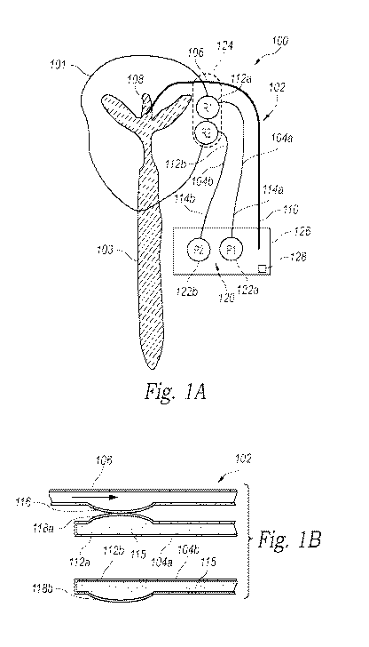
[0007] Figure
lA is a partially schematic illustration of a patient with a body fluid
drainage system configured in accordance with an embodiment of the present
technology.
[0008] Figure
1B is an enlarged cross-sectional view of a proximal portion of a
drainage catheter and pressure reference lines of the body fluid drainage
system of Figure 1A.
[0009] Figure
2A is a partially schematic illustration of a patient with a body fluid
drainage system configured in accordance with another embodiment of the
present
technology.
- 2 -
CA 02936349 2016-07-08
WO 2015/109260
PCT/US2015/011865
[0010] Figure
2B is an enlarged view of a proximal end portion of a reference line of
the body fluid drainage system of Figure 2A configured in accordance with an
embodiment
of the present technology.
[0011] Figures
2C and 2D are top and side views of proximal portions of the body fluid
drainage system of Figure 2A positioned with respect to a patient's head.
[0012] Figure
3A includes a front view and a side view of a drainage catheter and a
pressure sensor of a body fluid drainage system configured in accordance with
an
embodiment of the present technology.
[0013] Figure
3B is a side view of the drainage catheter and the pressure sensor of
Figure 3A placed in contact with each other in accordance with an embodiment
of the present
technology.
[0014] Figure
3C is a side view of a drainage catheter and a force sensor configured in
accordance with another embodiment of the present technology.
[0015] Figure 4
is a cross-sectional view of a drainage catheter and a sensor assembly
of a body fluid drainage system and a sensor assembly configured in accordance
with another
embodiment of the present technology.
[0016] Figure 5
is a side view of a drainage catheter and a pressure sensor of a body
fluid drainage system configured in accordance with yet another embodiment of
the present
technology.
[0017] Figure 6
is a side view of a drainage catheter and a pressure sensor of a body
fluid drainage system configured in accordance with a further embodiment of
the present
technology.
[0018] Figure 7
is a side view of a drainage catheter and a pressure sensor of a body
fluid drainage system configured in accordance with a still further embodiment
of the present
technology.
[0019] Figure 8
is a side view of a flexible reservoir for measuring flow rate of a body
fluid drainage system during various stages of filling in accordance with an
embodiment of
the present technology.
[0020] Figure 9
is a side view of a rigid reservoir for measuring flow rate of a body
fluid drainage system during various stages of filling in accordance with an
embodiment of
the present technology.
- 3 -
CA 02936349 2016-07-08
WO 2015/109260
PCT/US2015/011865
[0021] Figure
10 is a partially schematic illustration of an internal body fluid drainage
system installed within a patient in accordance with an embodiment of the
present
technology.
[0022] Figure
11 is a partially schematic illustration of an external body fluid drainage
system installed in a patient in accordance with an embodiment of the present
technology.
DETAILED DESCRIPTION
[0023] The
present technology is directed to devices, systems, and methods for draining
excess body fluids and pressure reference assemblies configured to determine
pressure at the
site of excess body fluid. In one embodiment, for example, a body fluid
drainage system can
be installed between a site of excess body fluid in a patient, such as within
a patient's head,
and a second location (e.g., an external receptacle, an internal cavity) that
can collect and/or
reabsorb the excess body fluid. The body fluid drainage system also includes a
pressure
reference assembly that determines the pressure at the site of excess body
fluid without
measuring the pressure directly at the site of excess fluid. Certain specific
details are set
forth in the following description and in Figures 1A-11 to provide a thorough
understanding
of various embodiments of the technology. For example, several embodiments of
body fluid
drainage systems that shunt cerebrospinal fluid ("CSF") are described in
detail below. The
present technology, however, may be used to drain a variety of excess body
fluids, such as
peritoneal fluid, blood, water, and/or other body fluids. Additionally, the
term "catheter" is
used broadly throughout the application to refer to any suitable tubing or
structure that
includes a lumen through which body fluids can flow. Other details describing
well-known
structures and systems often associated with CSF and other body fluid drainage
systems,
shunts, biomedical diagnostics, etc. have not been set forth in the following
disclosure to
avoid unnecessarily obscuring the description of the various embodiments of
the technology.
A person of ordinary skill in the art, therefore, will accordingly understand
that the
technology may have other embodiments with additional elements, or the
technology may
have other embodiments without several of the features shown and described
below with
reference to Figures 1A-11.
Selected Embodiments of Body Fluid Drainage Systems with Pressure Reference
Assemblies
[0024] Figure
lA is a partially schematic illustration of a patient 101 with a body fluid
drainage system 100 ("drainage system 100") configured in accordance with an
embodiment
of the present technology, and Figure 1B is an enlarged cross-sectional view
of a proximal
- 4 -
CA 02936349 2016-07-08
WO 2015/109260
PCT/US2015/011865
portion of the drainage system 100 of Figure 1A. The drainage system 100
includes a
drainage catheter 102, a first reference line 104a, and a second reference
line 104b (referred
to collectively as "reference lines 104"). The drainage catheter 102 has a
proximal portion
106 with an inlet 108 in fluid communication with a site of excess body fluid
within the
patient 101 and a distal portion 110 spaced apart from the proximal portion
and configured to
dispense the excess fluid in an external receptacle or internal body cavity
where the fluid can
be collected and/or reabsorbed. In the embodiment illustrated in Figure 1A,
for example, the
drainage system 100 is configured to shunt CSF away from the patient's brain,
and therefore
the inlet 108 of the drainage catheter 102 is positioned in the patient's
lateral ventricle in fluid
communication with the CSF system 103. In other embodiments, the drainage
system 100
can be used to drain excess fluid from other portions of the body.
[0025] The
reference lines 104 each comprise a tube or catheter that is at least
substantially filled with a reference fluid 115. The first reference line 104a
has a first
portion 112a and a second portion 114a opposite the first portion 112a, and
the second
reference line 104b has a first portion 112b opposite a second portion 114b.
As shown in
Figure 1A, the first portions 112a, 112b (collectively referred to as "first
portions 112") of the
first and second reference lines 104a and 104b are positioned proximate to
each other (e.g.,
substantially co-located) at a first location or reference point near the site
of excess body fluid
and the inlet 108 of the drainage catheter 102, and the second portions 114a,
114b
(collectively referred to as "second portions 114") of the first and second
reference lines 104a
and 104b are at a second location spaced away from the site of excess body
fluid. In the
illustrated embodiment, for example, the proximal ends of the reference lines
104 are
positioned near the patient's head (e.g., at the ear) close to the lateral
ventricle. The second
portions 114 of the reference lines 104 can be coupled to a pressure sensor
assembly 120 that
is configured to measure pressure at the second portions 114 of the first and
second reference
lines 104a and 104b. In the embodiment illustrated in Figure 1A, the pressure
sensor
assembly 120 includes a first pressure sensor 122a at the second location in
pressure
communication with the second portion 114a of the first reference line 104a,
and a second
pressure sensor 122b at the second location in pressure communication with the
second
portion 114b of the second reference line 104b. In other embodiments, the
pressure sensor
assembly 120 can include a single pressure sensor configured to measure the
differential
pressure between the first and second reference lines 104a and 104b at the
second location
and/or other types of sensors that can derive pressure within the reference
lines 104. In other
- 5 -
CA 02936349 2016-07-08
WO 2015/109260
PCT/US2015/011865
embodiments, the reference lines 104 can be operably coupled to a sensor
assembly 120 that
is configured to take different or addition types of measurements from the
reference
lines 104. For example, the sensor assembly 120 can be configured to take
force
measurements that can be used to determine pressure at the proximal portion
106 of the
drainage catheter 102.
[0026] As shown
in Figure 1B, the proximal portion 106 of the drainage catheter 102
has a flexible interface member 116, the first portion 112a of the first
reference line 104a has
a first flexible region 118a in pressure communication (e.g., physical
contact) with the
flexible interface member 116, and the first portion 112b of the second
reference line 104b
has a second flexible region 118b in pressure communication with the
surrounding
atmosphere of the flexible interface member 116 and the first and second
flexible
regions 118a and 118b (referred to collectively as "the flexible regions
118"). For example,
the second flexible region 118b can be exposed to the surrounding air (e.g.,
atmospheric
pressure). In operation, the flexible interface member 116 of the drainage
catheter 102
expands or inflates as fluid pressure within the drainage catheter 102
increases (e.g.,
representing an increase in ICP), and retracts or deflates as fluid pressure
within the drainage
catheter 102 decreases (e.g., representing a decrease in ICP). The
fluctuations of the flexible
interface member 116 (e.g., representative of fluctuations in ICP) are
communicated to the
first flexible region 118a. That is, when the pressure within the flexible
interface
member 116 increases, the flexible interface member 116 applies more pressure
against the
first flexible region 118a, and vice versa. Accordingly, the pressure measured
by the first
pressure sensor 122a at the second portion 108a of the first reference line
104a represents the
pressure within the drainage catheter 102 at the first location (e.g., near
the site of excess
fluid) plus the pressure head of the reference fluid 115 within the first
reference line 104a.
Because the second flexible region 118b is exposed to the atmosphere at the
first location, the
pressure measured by the second pressure sensor 122b at the second portion
108b of the
second reference line 104b corresponds to the pressure head within the second
reference
line 104b. This pressure measurement is at least substantially equal to the
pressure head in
the first reference line 104a because the two reference lines 104 contain the
same reference
fluid 115, have the same length, and the flexible regions 118 and the pressure
sensors 122 of
each reference line 104 are near the same location. Therefore, the pressure of
the drainage
catheter 102 at the first location is equivalent to the pressure measured by
the first pressure
sensor 122a less the pressure measured by the second pressure sensor 122b.
When the first
- 6 -
CA 02936349 2016-07-08
WO 2015/109260
PCT/US2015/011865
location is located near the lateral ventricles (e.g., at the side of the
patient's head), this
pressure is approximately equal to the ICP. If different reference fluids or
reference line
lengths are used, the drainage system 100 may include algorithms that correct
for such
differences to determine the pressure head in the first reference line 104a.
[0027] As
further shown in Figure 1B, the proximal ends of the reference lines 104 are
sealed from the environment and the distal ends can be similarly sealed to
enclose the
reference fluid 115 within the reference lines 104. The reference lines 104
can be made of
polyurethane tubing and/or other suitable materials for sealing the reference
fluid 115 therein.
In the embodiment illustrated in Figure 1B, the flexible region 118 of each
reference line 104
is spaced along the length of the corresponding reference line 104 and
positioned on a side of
the reference line 104. In other embodiments, one or both of the flexible
regions 118 can
extend from the proximal-most end of each reference line 104. The flexible
regions 118 of
the reference lines 104 can be flexible membranes or diaphragms made from
substantially
flexible materials that are sensitive to changes in pressure and the
application of small forces
thereon, such as the forces applied by the opposing flexible interface member
116 when
pressure changes within the drainage catheter 102. For example, the flexible
regions 118 can
be made from ether- or ester-based materials. In other embodiments, the
flexible regions 118
can be made from other suitable flexible materials. The flexible regions 118
can be attached
to the reference lines 104 via molding, adhesives, and/or other suitable
connection
techniques, or the flexible regions 118 can be integrally formed with the
reference lines 104.
For illustrative purposes, the flexible members 118 are shown protruding
outwardly from the
sides of the reference lines 104. However, under normal conditions when no
external
pressures are applied to the flexible members 118, the flexible members 118
can be in a
relaxed or flaccid state such that the material of the flexible members 118 is
not stretched or
placed under tension. Accordingly, the flexible members 118 may appear
substantially in
line with the sidewall of the reference lines 104. Then, when a force acts on
one of the
flexible members 118, it can move inwardly or outwardly depending on the force
applied. In
other embodiments, the flexible members 118 may be configured such that the
normal,
relaxed state of the material causes the flexible members 118 to protrude
outwardly or
inwardly. The drainage catheter 102 and the flexible interface member 116 can
be made
from similar materials as the reference lines 104 and the flexible regions
118, respectively.
[0028] The
reference fluid 115 can be configured to completely fill the reference lines
104 such that the flexible regions 118 are in a relaxed state such that they
can move (e.g.,
- 7 -
CA 02936349 2016-07-08
WO 2015/109260
PCT/US2015/011865
stretch) in either direction in response to the movement of an opposing
flexible interface
member 116. The reference fluid 115 can include silicone oil, mineral oil,
propylene glycol,
and/or other fluids with high vapor pressures that limit the amount of
evaporation of the fluid
during storage and use of the reference lines 104. In other embodiments, the
reference
lines 104 can be filled with other types of fluids, such a saline or water. In
certain
embodiments, the same reference fluid 115 is used in both reference lines 104
such that the
pressure measurements taken by the two pressure sensors 122 or a differential
pressure sensor
can be directly subtracted from each other to determine the pressure of the
drainage
catheter 102 at the reference point. In other embodiments, different reference
fluids 115 may
be used in the reference lines 104 and the pressure sensor assembly 120 can be
configured to
correct for the differences in fluid density.
[0029] In
various embodiments, the flexible interface member 116 of the drainage
catheter 102 and the flexible regions 118 of the reference lines can be housed
at least partially
within a cartridge 124 (Figure 1A; shown in broken lines). The cartridge 124
may be a
durable case or container that provides protection for the interface member
116, the flexible
regions 118, and/or any other system components (e.g., electronics) stored
therein, and
further include attachment features that position the interface member 116 and
the flexible
regions 118 appropriately with respect to each other. For example, the
cartridge 124 can
include protrusions or grooves that receive the reference lines 104 and the
drainage
catheter 102, position the two flexible regions 118 such that the two
reference lines 104
experience the same pressure head, and position the flexible interface member
116 to be in
pressure communication with one of the flexible regions 118. The cartridge 124
is further
configured to be positioned at a reference location on the patient 101 close
to the drainage
site. For example, the cartridge 124 can be positioned above the patient's ear
when the
drainage system 100 is configured for draining CSF from the brain. In other
embodiments,
the cartridge 124 can be positioned proximate to other drainage sites, such as
in the patient's
lumbar region when the drainage system 100 is used as a lumbar drain. In
certain
embodiments, the first portions 112 of the two reference lines 104 can be pre-
packaged
within the cartridge 124 such that the flexible regions 118 are affixed in a
desired position
(e.g., next to each other, at the same elevation, substantially co-located,
etc.). The proximal
portion 106 of the drainage catheter 102 can then be positioned within the
prepackaged
cartridge 124 such that the flexible interface member 116 is in pressure
communication (e.g.,
physically in contact) with one of the flexible regions 118. For example, the
cartridge 124
- 8 -
CA 02936349 2016-07-08
WO 2015/109260
PCT/US2015/011865
may include attachment features that appropriately position the flexible
interface member 116
with respect to one of the flexible regions 118. This embodiment facilitates
use of the
reference lines 104 and the associated assembly (e.g., the cartridge 124 and
the pressure
sensor assembly 120) with previously-implanted drainage catheters. In
addition, the
prepackaged configuration provides a multi-use reference line assembly (e.g.,
the reference
lines 104 and the pressure sensor assembly 120) that can be used on multiple
occasions
and/or with different patients. In other embodiments, the cartridge 124 can be
preassembled
with the drainage catheter 102 and the reference lines 104 such that the
flexible interface
member 116 and the flexible regions 118 are affixed in the desired positions
with the
interface member 116 contacting or attached to the first flexible region 118a.
In further
embodiments, the proximal elements of the drainage system 100 can be assembled
within the
cartridge 124 during or after the drain implantation procedure. In still
further embodiments,
the cartridge 124 can be omitted, and the proximal elements of the drainage
system 100 can
be positioned appropriately with respect to each other and with respect to the
patient 101
using other suitable means.
[0030] In
various embodiments, the reference line assembly (e.g., the two reference
lines 104 and related components) can be configured to measure negative
pressures within
the drainage catheter 102. When the flexible interface member 116 is subject
to negative
pressures, it may retract and, as a result, may come out of contact with the
opposing first
flexible region 118a of the first reference line 104. This loss of contact
prevents the first
flexible member 118a from translating the movement of the flexible interface
member 116 to
pressure measurements. Accordingly, the reference line assembly can include
features that
maintain contact between the first flexible region 118a and the flexible
interface member 116,
regardless of the direction of movement of the flexible interface member 116.
For example,
when the drainage catheter 102 and the first reference line 104a are
preassembled (e.g.,
within the cartridge 124), the flexible interface member 116 and the first
flexible region 118a
can be permanently bonded together. Various additional features for
maintaining at least
semi-permanent contact between the flexible interface member 116 and an
opposing
membrane (e.g., the first flexible region 118a) under negative pressures are
described below
with reference to Figures 3A-7, and can be used with the drainage system 100
to at least
temporarily attach the flexible interface member 116 to the first flexible
region 118a. In
further embodiments, the drainage catheter 102 and the first reference line
104a can be
attached (e.g., bonded) together at least in the area around the flexible
interface member 116
- 9 -
CA 02936349 2016-07-08
WO 2015/109260
PCT/US2015/011865
and a single membrane can be used to detect pressure changes in the drainage
catheter 102.
In this embodiment, the flexible interface member 116 or the first flexible
region 118a is
omitted, leaving an opening in one of the drainage catheter 102 or the first
reference
line 104a that is configured to receive the remaining of the first flexible
region 118a or the
flexible interface member 116. When the catheter 102 and the first reference
line 104a are
attached together, the remaining membrane (i.e., the flexible interface member
116 or the
first flexible region 118a) can be positioned between the reference fluid 115
and the fluid in
the drainage catheter 102 and act directly on the reference fluid 115 to
reflect changes in the
pressure of the drainage catheter 102.
[0031] As
further shown in Figure 1A, the drainage system 100 can also include a
housing 126 that carries the pressure sensor assembly 120 and the second
portions 114 of the
reference lines 104. Similar to the cartridge 124, the housing 126 can be
configured to secure
the reference lines 104, the pressure sensor assembly 120, and optionally the
drainage
catheter 102, and position these elements appropriately with respect to each
other. For
example, the housing 126 can include grooves or protrusions that position the
first and
second pressure sensors 122a and 122b at about the same elevation such that
they measure
the same amount of pressure head in the corresponding reference lines 104. In
the illustrated
embodiment, the drainage catheter 102 terminates at the same point as the
pressure
sensors 122, but in other embodiments the drainage catheter 102 can extend to
a different
location and/or beyond the housing 126 to an internal or external receptacle
(not shown) that
can collect the drained body fluid. The housing 126 can also carry a processor
or processing
device 128 (shown schematically; e.g., a central processing unit (CPU)) and/or
additional
elements of the drainage system 100, such as a receptacle (not shown) into
which the excess
body fluid from the catheter 102 can drain.
[0032] The
processing device 128 can be operably coupled to the pressure sensor
assembly 120 and/or other features of the drainage system 100 (e.g., valves).
The processing
device 128 can include or be part of a device that includes a hardware
controller that
interprets the signals received from input devices (e.g., the pressure sensors
122, other
sensors, user input devices, etc.) and communicates the information to the
processing
device 128 using a communication protocol. The processing device 128 may be a
single
processing unit or multiple processing units in a device or distributed across
multiple devices.
The processing device 128 may communicate with the hardware controller for
devices, such
as for a display that displays graphics and/or text (e.g., LCD display
screens). The processing
- 10 -
CA 02936349 2016-07-08
WO 2015/109260
PCT/US2015/011865
device 128 can also be in communication with a memory (e.g., within the
housing 126) that
includes one or more hardware devices for volatile and non-volatile storage,
and may include
both read-only and writable memory. For example, a memory may comprise random
access
memory (RAM), read-only memory (ROM), writable non-volatile memory, such as
flash
memory, hard drives, floppy disks, CDs, DVDs, magnetic storage devices, tape
drives, device
buffers, and so forth. A memory is not a propagating electrical signal
divorced from
underlying hardware, and is thus non-transitory. In certain embodiments, the
processing
device 128 can also be coupled to a communication device capable of
communicating
wirelessly or wire-based with a network node. The communication device may
communicate
with another device or a server through a network using, for example, TCP/IP
protocols.
[0033] The
processing device 128 can execute automated control algorithms to initiate,
terminate, and/or adjust operation of one or more features of the pressure
sensor
assembly 120 and/or receive control instructions from a user. The processing
device 128 can
further be configured to provide feedback to a user based on the data detected
by the pressure
sensor assembly 120 via an evaluation/feedback algorithm. For example, the
processing
device 128 can be configured to provide clinicians, patients, and/or other
users with a
patient's pressure level at a site of excess body fluid (e.g., ICP),
indicators of when a
threshold pressure level is exceeded, and/or other pressure-related
information based on the
information received from the pressure sensors 122. This information can be
provided to the
users via a display (e.g., a monitor on a computer, tablet computer, or smart
phone; not
shown) communicatively coupled to the processing device 128.
[0034] In
operation, the pressure in the drainage catheter 102 near a site of excess
body
fluid (e.g., the brain) can be determined using measurements taken from the
separate
reference lines 104, and do so using pressure measurements obtained at a
location spaced
apart from the site of excess body fluid. For example, when the drainage
system 100 is
configured to drain CSF from the patient's brain, ICP can be determined by
taking pressure
measurements with the pressure sensor assembly 120 at a location spaced
distant from and,
optionally, movable with respect to the patient's head. Thus,
the pressure sensor
assembly 120 can be spaced distant from the patient's head. This allows the
pressure
readings provided by the pressure sensor assembly 120 and/or the ICP
determined via the
pressure sensor assembly 120 or processing device 128 to be displayed to a
user at a
convenient location. For example, rather than a clinician having to look at a
pressure sensor
reading on a patient's head to determine ICP, the drainage system 100 allows
the pressure
-11-
CA 02936349 2016-07-08
WO 2015/109260
PCT/US2015/011865
sensor assembly 120 and associated display to be positioned at a location that
is convenient
and/or easily accessible for the clinician (e.g., at chest level when the
clinician is in a
standing location, at table level, spaced apart from the patient 101). The
clinician can use the
two pressure measurements to determine the desired pressure at the excess
fluid site, or the
processing device 128 can automatically calculate this information for the
clinician. The
mobility of the pressure sensor assembly 120 and associated devices (e.g., the
processing
device 128, displays, etc.) is also more comfortable for a patient 101 because
the pressure
sensor need not be attached to his or her head or body.
Accordingly, the drainage
system 100 allows ICP and other pressure measurements to be determined without
having a
pressure sensor directly at the patient's head or other site of excess body
fluid. In addition,
because the drainage system 100 does not take pressure measurements directly
from the
drainage catheter 102 itself, the pressure measurements taken by the pressure
sensor
assembly 120 are not subject to losses that may occur due to fluid flow
through the drainage
catheter 102. Accordingly, the drainage system 100 is expected to increase the
accuracy of
pressure measurements taken at a location spaced apart from the site of excess
body fluid.
[0035] Figure
2A is a partially schematic illustration of a patient 201 with a body fluid
drainage system 200 ("drainage system 200") configured in accordance with
another
embodiment of the present technology, and Figure 2B is an enlarged view of a
proximal end
portion of a reference line 204 of the drainage system 200. The drainage
system 200 can
include several features generally similar in structure and function to the
features of the
drainage system 100 described above with reference to Figures lA and 1B. As
shown in
Figure 2A, for example, the drainage system 200 includes a drainage catheter
202, a first
reference line 204a, and a second reference line 204b (collectively referred
to as "reference
lines 204"). The drainage catheter 202 has a proximal portion 206 with an
inlet 208 in fluid
communication with a site of excess body fluid within the patient 201 and a
distal portion 210
opposite the proximal portion 206. The first and second reference lines 204a
and 204b are
filled with a reference fluid 215 (Figure 2B), and each have a proximal or
first end
portion 212a, 212b (referred to collectively as "first end portions 212") and
a distal or second
end portion 214a, 214b (referred to collectively as "second end portions 214")
opposite the
first end portion 212a, 212b. The proximal end portions 212 include a flexible
region 218
(Figure 2B) that is in pressure communication with the surrounding air (i.e.,
exposed to
atmospheric pressure). As shown in Figure 2B, in certain embodiments the
flexible
region 218 is a pliable membrane that extends from the proximal end of the
reference
- 12 -
CA 02936349 2016-07-08
WO 2015/109260
PCT/US2015/011865
line 204 to form a balloon-like structure filled with the reference fluid 215
and exposed to the
atmosphere. An end cap 232 or other type of housing can be positioned around
the flexible
region 218 to prevent external objects from applying pressure to the flexible
region 218 such
that the flexible region 218 is only subject to changes in the surrounding
atmospheric
pressure. In other embodiments, the flexible regions 218 of one or both of the
reference
lines 204 can protrude outwardly from a sidewall of the first end portion 212
of the reference
line 204 (e.g., similar to the flexible regions 118 described with reference
to Figure 1B).
[0036] As shown
in Figure 2A, the drainage system 200 can further include a pressure
sensor assembly 220 positioned at the distal end portions 214 of the reference
lines 204 and
operably coupled to the distal portion 210 of the drainage catheter 202 and
the second end
portions 214 of the first and second reference lines 204a and 204b. The
pressure sensor
assembly 220 can be configured to measure pressure within the drainage
catheter 202 at the
distal portion 210, and the pressure within the reference lines 204 at the
second end
portions 214. In the illustrated embodiment, for example, the pressure sensor
assembly 220
includes a first pressure sensor 222a at the second end portion 216a of the
first reference
line 204a, a second pressure sensor 222b at the second end portion 216b of the
second
reference line 204b, and a third pressure sensor 222c at the distal portion
210 of the drainage
catheter 202. In other embodiments, the pressure sensor assembly 220 can
include less than
three pressure sensors, more than three pressure sensors, and/or other types
of sensors that
can be used to determine the pressure within the distal portions of the
drainage catheter 202
and the reference lines 204. As shown in the illustrated embodiment, the three
pressure
sensors 222a-222c can be positioned at the same location, at a position spaced
apart from the
site of excess body fluid. As described in further detail below, the
measurements taken from
the pressure sensor assembly 220 can be used to determine the pressure at the
site of excess
body fluid (i.e., the drainage site).
[0037] As
further shown in Figure 2A, the drainage system 200 can include a
housing 226 that carries the pressure sensor assembly 220. The housing 226 can
also carry
other features associated with the drainage system 200, such as a processing
device 228
and/or a display (not shown). Similar to the processing device 128 described
above, the
processing device 228 can include or be associated with a controller, and can
be configured to
run algorithms that control operation of the drainage system 200 and/or
provide feedback to
users regarding pressure measurements and/or the operation of the drainage
system 200. This
feedback can be provided to users on a display connected to the housing 226
and/or displays
- 13 -
CA 02936349 2016-07-08
WO 2015/109260
PCT/US2015/011865
remote from the drainage system 200 and communicatively coupled thereto (e.g.,
via a wired
or wireless connection).
[0038] During a
system set-up procedure, a clinician can position the flexible
regions 218 of the first end portions 212 of the first and second reference
lines 204 at two
points along an imaginary reference axis 230 (i.e., a straight line) that
extends through the site
of excess body fluid (i.e., the site at which the pressure measurement is
desired). This site
generally corresponds to the implantation location of the inlet 208 of the
drainage
catheter 202 and is also referred to herein as the "drainage site". The
flexible region 218 of
the first reference line 204a can be positioned at a first location along the
reference axis 230
to one side of the drainage site, and the flexible region 218 of the second
reference line 204b
can be positioned at a second location along the reference axis 230 on the
other side of the
drainage site. Accordingly, the proximal end portions 212 of the first and
second reference
lines 204a and 204b are positioned on either side of the drainage site along
the reference
axis 230.
[0039] For
example, when the drainage system 200 is intended to drain CSF from a
patient's brain, the reference axis 230 is a straight line that extends
through the lateral
ventricles or the Foramen of Monroe (i.e., the center of the head). As shown
in the
embodiment illustrated in Figure 2A, the first end portions 212 of the
reference lines 204 can
be placed along the reference axis 230 on either side of the patient's head,
approximately
equidistant from the drainage site. Figures 2C and 2D are top and perspective
side views
further illustrating the positioning of the first end portions 212 with
respect to a patient's head
and the lateral ventricles 205 (Figure 2D). In this embodiment, the ICP (i.e.,
the desired
pressure measurement) is equivalent to the pressure in the drainage catheter
202 measured by
the third pressure sensor 222c less the pressure head between the inlet 108 of
the drainage
catheter 202 and the third pressure sensor 222c (i.e., the distance between
the Foramen of
Monroe and the location at which the pressure measurement is taken). The
reference
lines 204, with the flexible regions 218 (Figure 2B) exposed to the
atmosphere, can be used
to determine the pressure head from the drainage site. More specifically,
because the first
end portions 212 of the two reference lines 204 are spaced approximately
evenly apart from
the Foramen of Monroe (i.e., the drainage site), the average of the two
pressures measured by
the first and second pressure sensors 222a and 222b is approximately
equivalent to the
pressure head (vertical distance) between the Foramen of Monroe and the
location of the
pressure sensors 222a-c. Therefore, the first and second reference lines 204a
and 204b can be
- 14 -
CA 02936349 2016-07-08
WO 2015/109260
PCT/US2015/011865
used to determine the pressure head from the actual drainage site (i.e., the
lateral ventricles of
the brain or other site at which the pressure measurement is desired) rather
than at a position
spaced laterally apart from the drainage site (e.g., over the patient's ear).
Accordingly, the
three pressure measurements taken from the three pressure sensors 222a-c can
be used to
determine the pressure at the Foramen of Monroe, which corresponds to the
patient's ICP.
More specifically, the ICP is equivalent to the pressure of the drainage
catheter (P3) minus
the average of measured pressures (P1 and P2) of the first and second
reference lines 204a
and 204b (i.e., ICP = P3 - (P 1+P2)/2). The calculation of drainage site
pressure can be
performed automatically via programs stored on the processing device 228
and/or manually
using the pressure read outs provided by the pressure sensor assembly 220. The
same type of
calculations can be performed to determine the pressure at other sites of
excess fluid around
which the drainage system 200 is positioned. For example, when the drainage
site is at the
patient's abdomen, the reference axis 230 can extend through the drainage
site, and the first
end portions 212 of the two reference lines 204 can be positioned along the
reference
axis 230.
[0040] In the
illustrated embodiment, the first end portions 212 of the reference
lines 204 are about equidistant from the Foramen of Monroe (i.e., the drainage
site).
However, in other embodiments the first end portions 212 of the two reference
lines 204 may
be spaced different distances apart from the drainage site along the reference
axis 230. In this
embodiment, the pressure measurements of the first and second reference lines
204a
and 204b can be weighted based on their position with respect to the drainage
site. For
example, the pressure measurement taken from the reference line 204 located
closer to the
drainage site would be weighted more heavily than the pressure measurement
taken from the
reference line that is spaced further from the drainage site, and the degree
to which the
pressure measurements are weighted can correspond to the relative closeness of
the two
reference lines from the drainage site. The weighted pressure measurements can
then be used
in conjunction with the measured pressure of the drainage catheter 202 to
determine the
pressure at the drainage site (e.g., ICP).
[0041] In use,
the reference lines 204 are used with the pressure measured in the
drainage catheter 202 to correct for the pressure head in the drainage
catheter 202 when
pressure is measured at a location spaced apart from the drainage site. The
use of the two
reference lines 204 placed along the reference axis 230 that passes through
the drainage site
allows for determination of the pressure head at a specific location (i.e.,
the drainage site) on
- 15 -
CA 02936349 2016-07-08
WO 2015/109260
PCT/US2015/011865
the reference axis 230 between the proximal end portions 212 of the reference
lines 204,
instead of simply the pressure head within the reference lines 204. This
allows the drainage
system 200 to account for differences between the pressure head at the
drainage site and the
pressure head at a location spaced laterally apart from the drainage site,
which may be caused
by the orientation of the patient 201 (e.g., when the patient 201 is laying
down rather than
standing). Accordingly, the drainage system 200 can be used to determine the
pressure at a
drainage site (e.g., ICP) using sensors spaced apart from the drainage site,
and does so with
increased accuracy by determining the pressure head at the actual drainage
site. For example,
in certain embodiments the drainage system 200 can be used to determine the
pressure at the
drainage site (e.g., ICP) within 10-20 cm of water of the true pressure at the
drainage site. In
other embodiments, the drainage system 200 can be used to determine drainage
site pressures
with higher accuracy.
[0042] In other
embodiments, the drainage system 200 can include more than two
reference lines 204, each with a proximal end portion positioned along
reference axis that
pass through the drainage site and distal end portions attached to pressure
sensors. The
pressure head at the drainage site can be determined using the pressure
measurements taken
from each of the reference lines 204. For example, in certain embodiments the
drainage
system 200 includes three reference lines 204 placed on the patient's head.
The three
reference lines 204 can be used to determine the orientation of the patient's
head and
triangulate the pressure at any location within the brain.
[0043] In
further embodiments, the drainage system 200 can be combined with the
drainage system 100 of Figures lA and 1B. For example, an additional reference
line can be
added that is in pressure contact with a flexible interface member on the
proximal portion 206
of the drainage catheter 202 (e.g., as shown in Figure 1B). In this
embodiment, pressure
measurements are taken from the additional reference line (e.g., a third
reference line) rather
than the drainage catheter 202 itself These pressure measurements represent
the pressure in
the drainage catheter (e.g., ICP) plus the pressure head, and therefore can be
used similarly to
the pressure measurements taken directly from the drainage catheter. As
discussed above, the
pressure measurements taken from this additional reference line are not
subject to losses
associated with fluid flow through the drainage catheter 202 because the
reference fluid
within this additional reference line is substantially stagnant. Accordingly,
this embodiment
can increase the accuracy with which the pressure at the site of excess body
fluid can be
determined.
- 16 -
CA 02936349 2016-07-08
WO 2015/109260
PCT/US2015/011865
Selected Embodiments of Body Fluid Drainage Systems for Measuring Negative
Pressures
[0044] Figures
3A-8 illustrate various embodiments of body fluid drainage systems in
which a flexible membrane or diaphragm of a drainage catheter is outwardly
biased. As
discussed above with respect to Figures lA and 1B, the pressure in a drainage
catheter can be
measured across two membranes that are in contact. For example, the drainage
catheter can
include a flexible membrane that contacts (1) an opposing membrane of a
reference line (e.g.,
the reference lines 104 described above) from which a pressure measurement can
be taken via
a pressure sensor along the reference line, (2) an opposing fluid-filled
membrane of a
pressure sensor, or (3) an opposing member of a force sensor (e.g., a load
cell). This
configuration isolates the measurements taken by the sensor from the fluid
being measured
(i.e., the fluid flowing through the drainage catheter). However, when the
fluid pressure
within the flexible membrane of the drainage catheter is less than the
surrounding atmosphere
(i.e., atmospheric pressure), the flexible membrane experiences a smaller
pressure on the
inside of the membrane than on the outside, and the membrane collapses or
becomes
retracted. This can result in the drainage catheter membrane from breaking
contact with the
sensing membrane or surface, and therefore prevents the sensing member (e.g.,
a pressure
sensor with a fluid-filled diaphragm, a load cell, or a reference line with a
pressure sensor
attached thereto) from measuring the negative pressure. The embodiments
described below
with reference to Figures 3A-8 allow for the measurement of both positive and
negative
pressures via flexible membranes on drainage catheters. The embodiments
described below
may be used in conjunction with the drainage systems 100 and 200 described
above with
reference to Figures 1A-2D, as well as with other drainage catheter systems.
[0045] Figure
3A includes a front view and a side view of a drainage catheter 302 and a
pressure sensor 332 for a body fluid drainage system configured in accordance
with an
embodiment of the present technology, and Figure 3B is a side view of the
drainage catheter
and the pressure sensor 332 of Figure 3A placed in contact with each other. As
shown in
Figure 3A, the drainage catheter 302 can include a flexible interface member
316, such as a
diaphragm or flexible membrane, that protrudes outwardly from a wall of the
drainage
catheter 302 when filled with a fluid (e.g., excess body fluid being drained
form the body).
The drainage catheter 302 and the flexible interface member 316 can have a
structure and
function at least generally similar to the structure and function of the
drainage catheters 102
and 202 and the flexible interface members 116 and 216 described above. The
flexible
interface member 316 can be positioned anywhere along the length of the
drainage
- 17 -
CA 02936349 2016-07-08
WO 2015/109260
PCT/US2015/011865
catheter 302, and can be used to determine the pressure at the drainage site.
For example, as
described above, the pressure detected at the flexible interface member 316 is
equal to the
pressure at the drainage site plus the pressure head between the drainage site
and the flexible
interface member 316. In embodiments including reference lines that measure
pressure at a
location spaced apart from the flexible interface member, the pressure head is
between the
drainage site and the location at which the pressure measurement is actually
taken. Various
features can be used to determine the pressure head (e.g., the reference lines
104 and 204
described above), and this information along with the pressure of the drainage
line measured
via the flexible interface member 316 can be used to derive the pressure at
the drainage site
(e.g., ICP).
[0046] As shown
in Figures 3A and 3B, the pressure sensor 332 can include a fluid-
filled diaphragm or membrane 334 that protrudes outwardly from a base portion
336 of the
pressure sensor 332. The base portion 336 can house electronics and/or other
features that
are used to detect pressure via the membrane 334. The pressure sensor 332 can
also include a
contact member 338 on or along the membrane 334 against which the flexible
interface
member 316 of the drainage catheter 302 can be pressed (e.g., as shown in
Figure 3B). In
various embodiments, the drainage catheter 302 can also include a contact
member 340 that
is configured to press against the opposing portion of the pressure sensor 332
and/or other
sensor. The contact members 338 and 340 can be separate structures attached to
the
membrane 334 and flexible interface member 316, respectively, and may have
different
material properties than the underlying membranes 334, 316. For example, the
contact
members 338 and 340 may be more rigid than the membranes 334, 316. In other
embodiments, the contact members 338 and 340 may be defined by a portion of
the sensor
membrane 334 and the flexible interface member 316, respectively. In various
embodiments,
only one of the membranes 334, 316 include a contact member.
[0047] As shown
in Figure 3A, the drainage catheter 302 may further include a spring
342 that acts on the flexible interface member 316 to create a chronic outward
force on the
flexible interface member 316, and thereby allows the flexible interface
member 316 to
remain extended even if the fluid pressure therein is negative. The spring 342
can have a first
end 344a attached to an interior wall of the drainage catheter 302 or embedded
therein and a
second end 344b that connects to an inner surface of the contact member 340
and/or another
portion of the flexible interface member 316. In certain embodiments, the
spring 342 may be
a relatively long spring that is compressed significantly (e.g., 40%, 50%, or
60% of the free
- 18 -
CA 02936349 2016-07-08
WO 2015/109260
PCT/US2015/011865
length of the spring) when the drainage catheter 302 is assembled with the
pressure sensor
332.
[0048] Figure
3B illustrates the drainage catheter 302 and the pressure sensor 332 as
they would be configured when assembled together in a cartridge (e.g., the
cartridge 124 of
Figure 1A) or other type of housing (not shown). In certain embodiments, the
pressure
sensor 332 can be preassembled in a housing, and the drainage catheter 302 can
be
subsequently attached to the housing 332. In this embodiment, the pressure
sensor 332 and
the housing may be reusable so that the expensive electronics of the pressure
sensor 332 can
be used multiple times with different drainage catheters. In other
embodiments, the drainage
catheter 302 can be preassembled with the pressure sensor 332 such that the
two opposing
membranes are correctly positioned with respect to each other before use. In
further
embodiments, the drainage catheter 302, the pressure sensor 332, and the
housing that
positions the catheter 302 and the pressure sensor 332 with respect to each
other can be
separate components that are assembled together before use of the device.
[0049] As shown
in Figure 3B, when assembled, the flexible interface member 316 and
the flexible sensor membrane 334 are forced into contact, which in certain
embodiments can
lead to further compression of the spring 342. During use, the pressure sensor
332 measures
the sum of two pressures: (1) the pressure created by the spring 342, and (2)
the pressure
created by the fluid within the catheter 302 acting on the flexible interface
member 316. The
pressure applied by the spring 342 on the flexible interface member 316 is a
known value,
and therefore the pressure of the fluid acting on the flexible interface
member 316 can be
determined be subtracting the spring pressure from the overall pressure
measured by the
pressure sensor 332. This calculation can be performed automatically via a
processor and/or
manually by the user based on the pressure readings of the pressure sensor
332.
[0050] In
certain embodiments, the spring pressure is known based on previous testing
performed during assembly or product specifications. In other embodiments, the
spring
pressure and spring properties are unknown before use. In this embodiment, the
pressure or
force applied by the spring on the flexible interface member 316 can be
determined by
measuring the pressure via the pressure sensor 332 when the fluid pressure
within the
drainage catheter 302 is zero. For example, the sensor reading must be taken
before
implantation of the drainage catheter 302 and/or after implantation by
disconnecting the
portion of the drainage catheter 302 with the flexible interface member 316
from the fluid
source and connecting it to the surrounding air pressure (i.e., a zero point
calibration). In
- 19 -
CA 02936349 2016-07-08
WO 2015/109260
PCT/US2015/011865
further embodiments, such as when the drainage catheter 302 cannot be
disconnected from
the fluid source, the spring pressure can be determined if certain properties
of the spring are
known. For example, the degree of compression of the spring 342 may be known
(e.g., based
on the mechanical arrangement of the spring 342 against a hard stop), and the
force
contributed by the spring can be known at any condition using the spring
properties (e.g., the
spring constant) and Hooke's Law. In this embodiment, using a significantly
compressed
spring (e.g., 50% of its free length) can reduce measurement errors since
small errors in the
measured mechanical position result in only small changes in spring force.
[0051] The
outward force provided by the spring 342 on the interface member 316
allows the pressure sensor 332 to measure negative pressures within the
drainage catheter 302
down to the level at which the negative pressure overcomes the spring force.
The drainage
catheter 302 can be designed such that the spring force is sufficient to
measure a desired
range of negative pressures. For example, when used for ICP measurements, it
may be
desirable to measure pressures of about -30 cm of water, and the spring 342
and the flexible
interface member 316 size can be selected such that the spring has sufficient
force to
maintain contact between the flexible interface member 316 and the opposing
sensor
membrane 334 under this condition. In other embodiments, the drainage catheter
302 can be
configured to have higher or lower threshold pressures depending on the
application. This
ability to measure negative pressures provided by the outwardly biased
interface member 316
increases both the range of pressure values that can be measured using the
drainage system
and the mobility of drainage systems as a whole because the sensors are less
limited by their
position relative to the patient. For example, when the patient is lying down,
the sensor 332
can be positioned vertically above the patient at chest or eye level with a
clinician to facilitate
monitoring the pressure measurements.
[0052] In
various embodiments, the pressure sensor 332 can be replaced by a force
sensor that measures the force acting on the flexible interface member 316.
Figure 3C, for
example, is a is a side view of the drainage catheter 302 assembled with a
force sensor 350
configured in accordance with an embodiment of the present technology. The
force
sensor 350 measures the sum of two forces: 1) the force created by the spring
342, and 2) the
force created by the fluid pressure acting on the flexible interface member
316. If the force
sensor 350 also has a spring-like behavior (e.g., a load cell), this force
also contributes to the
signal response measured by the force sensor 350 because the position of the
flexible
interface member 316 changes with changes in pressure, and is taken into
account when
- 20 -
CA 02936349 2016-07-08
WO 2015/109260
PCT/US2015/011865
determining the force or pressure the fluid applies to drainage catheter 302
with the flexible
interface member 316 from the fluid source and connecting it to the
surrounding air pressure
(i.e., a zero point calibration).
[0053] In
certain embodiments, the spring 342 can be removed and tension or elastic
force from the flexible interface member 316 itself may act in place of the
spring force. For
example, pressing the force sensor 350 (Figure 3C) or the pressure sensor 332
(Figure 3B)
against the flexible interface member 316 can create a tension force or
elastic force on the
flexible interface member 316. When a negative pressure is experienced, this
tension force or
elastic force acts to resist collapse of the flexible interface member 316,
just as the spring 342
does as described above with reference to Figures 3A-3C. In this embodiment,
the properties
of the flexible interface member 316 may change over time. For example, the
tensile or
elastic forces in the flexible interface member 316 may lessen over time so
the forces
provided by the flexible interface member 316 can be measured periodically to
ensure
sufficient outward force. Similar to the spring 342, when the membrane
properties of the
flexible interface member 316 are unknown (either initially or over a period
of time), the
system can be "zeroed" by disconnecting the portion of the drainage catheter
302 with the
flexible interface member 316 from the fluid source and connecting it to the
surrounding air
pressure (i.e., a zero point calibration) to determine the membrane
properties.
[0054] Figure 4
is a cross-sectional view of the drainage catheter 302 and a sensor
assembly 352 of a body fluid drainage system 400 ("drainage system 400")
configured in
accordance with another embodiment of the present technology. The drainage
system 400
includes the drainage catheter 302 with the flexible interface member 316, a
fluid-filled
tube 354 or other enclosure with a flexible reference membrane 356 protruding
therefrom,
and a housing 358 carrying the catheter 302 and the tube 354 such that the
flexible interface
member 316 and the reference membrane 356 are positioned adjacent to each
other. In
certain embodiments, the fluid-filled tube 354 can be defined by the reference
lines 104
and 204 described above with reference to Figures 1A-2D.
[0055] In the
embodiment illustrated in Figure 4, the flexible interface member 316 of
the drainage catheter 302 is not outwardly biased by an internal spring, but
instead maintains
connection with a lever 360 that is fixedly attached (e.g., bonded) to the
flexible interface
member 316 and the flexible reference membrane 356. The lever 360 can be
attached to the
housing 358 at a pivot point 362 via a shaft 364 such that the lever 360
pivots as the pressure
within the flexible interface member 316 changes. The drainage system 400
further includes
- 21 -
CA 02936349 2016-07-08
WO 2015/109260
PCT/US2015/011865
a first force sensor 350a and a second force sensor 350b (collectively
referred to as force
sensors 350) positioned on the opposite side of the lever 360 as the two
membranes and
attached to the housing 358 and configured to measure the changes in force
applied by the
flexible interface member 316 and the reference membrane 356 on the lever 360.
As shown
in Figure 3B, when the housing 358 is assembled the first force sensor 350a
presses against
the lever 360 opposite the flexible interface member 316, and the second force
sensor 350b
presses against the lever 360 opposite the reference membrane 356. The first
and second
force sensors 350a and 350b can be load cells and/or other suitable types of
force or pressure
sensors that measure the force or pressure applied against the lever 360 by
the flexible
interface member 316 and the reference membrane 356.
[0056] As the
pressure changes within the drainage catheter 302, the lever 360 pivots
about the pivot point 362 and remains connected to the flexible interface
member 316 and the
adjacent reference membrane 356. These changes in position of the lever 360
caused by the
force of the flexible interface member 316 and the reference membrane 356 are
detected by
the force sensors 350, and the detected force measurements can be used to
determine the
pressure or force applied by the fluid on the flexible interface member 316.
For example, the
difference in the force measurement taken from the first force sensor 350a and
the force
measurement taken from the second force sensor 350b correlates to the force
applied by the
fluid on the flexible interface member 316. As described above, this force
measurement can
be used to determine the pressure at a drainage site. In addition, because the
lever 360 is
attached to the flexible interface member 316 and the reference membrane 356,
the lever 360
prevents the flexible interface member 316 from collapsing when it experiences
negative
pressures.
[0057] Figure 5
is a side view of the drainage catheter 302 and the pressure sensor 332
of a body fluid drainage system 500 configured in accordance with yet another
embodiment
of the present technology. The drainage catheter 302 and the pressure sensor
332 of Figure 5
are configured in generally the same manner as described above with reference
to Figures 3A
and 3B. In the illustrated embodiment, however, the flexible interface member
316 is
outwardly biased by a lever spring 370 that is attached (e.g., bonded) to an
outer surface of
the flexible interface member 316. As shown in Figure 5, for example, the
lever spring 370
can be positioned between the flexible interface member 316 and the flexible
sensor
membrane 334 of the pressure sensor 332. The opposite end of the lever spring
370 can be
attached to a portion of a housing (not shown) that carries the assembly.
During use, the
- 22 -
CA 02936349 2016-07-08
WO 2015/109260
PCT/US2015/011865
lever spring 370 applies a chronic outward force on the flexible interface
member 316 to
maintain contact between the flexible interface member 316 and the flexible
sensor
membrane 334 such that the pressure sensor 332 can measure negative pressures
in the
drainage catheter 302. In other embodiments, the pressure sensor 332 can be
replaced by a
force sensor (e.g., the force sensors 350 described above).
[0058] Figure 6
is a side view of the drainage catheter 302 and the pressure sensor 332
of a body fluid drainage system 600 configured in accordance with a further
embodiment of
the present technology. The drainage catheter 302 and the pressure sensor 332
of Figure 6
are configured in generally the same manner as described above with reference
to Figures 3A
and 3B. The drainage system 600 of Figure 6, however, includes an external
spring 386 that
is operably coupled to the exterior of the flexible interface member 316 and
applies the
chronic outward force to the flexible interface member 316. In the illustrated
embodiment,
for example, the spring 386 can be attached to a housing (not shown) that
carries the drainage
catheter 302 and the pressure sensor 332, and a connection assembly 380 is
attached to the
spring 386 to couple it to the exterior surface of the flexible interface
member 316. The
connection assembly 380 includes a first support member 382a attached (e.g.,
bonded) to the
exterior surface of the flexible interface member 316 and positioned between
the flexible
interface member 316 and the flexible sensor membrane 334, and a second
support
member 382b attached (e.g., bonded) to the spring 386. One or more shafts 384
can extend
around the drainage catheter 302 to connect the first and second support
members 382a
and 382b. The spring 386 can be configured to apply a chronic force against
the second
support member 382b (i.e., upward relative to the page), and this force can be
transferred to
the flexible interface member 316 via the connection assembly 380 such that
the flexible
interface member 316 does not collapse under negative pressures. In other
embodiments, the
pressure sensor 332 can be replaced by a force sensor (e.g., the force sensors
350 described
above).
[0059] Figure 7
is a side view of the drainage catheter 302 and the pressure sensor 332
of a body fluid drainage system 700 configured in accordance with a still
further embodiment
of the present technology. The drainage catheter 302 and the pressure sensor
332 of Figure 7
are configured in generally the same manner as described above with reference
to Figures 3A
and 3B. The drainage system 700 of Figure 7, however, maintains contact
between the
flexible interface member 316 and the opposing flexible sensor membrane 334 by
creating a
vacuum 390 between the flexible interface member 316 and the sensor membrane
334. For
-23 -
CA 02936349 2016-07-08
WO 2015/109260
PCT/US2015/011865
example, a housing 392 carrying the drainage catheter 302 and the pressure
sensor 332 can
form a sealed compartment 394 around the flexible interface member 316 and the
sensor
membrane 334. The compartment can be sealed from the external atmosphere via 0-
rings
and/or other suitable features that create a seal. Once the flexible interface
member 316 and
the sensor membrane 334 are positioned within the sealed compartment 394, a
vacuum can
be applied to the space within the compartment to create a vacuum between the
opposing
membranes 316 and 334. For example, the housing 392 can include a sealable
opening 396
through which the sealed compartment 394 can be accessed and the vacuum
applied. The
vacuum 390 suctions the flexible interface member 316 and the sensor membrane
334
together such that they maintain contact even when the flexible interface
member 316 is
under negative pressure, and therefore allows the drainage system 700 to
measure negative
pressures in the drainage catheter 302 via the flexible interface member 316.
In other
embodiments, the pressure sensor 332 can be replaced by a force sensor (e.g.,
the force
sensors 350 described above).
[0060] In
further embodiments, the drainage system 700 described above can maintain
contact between the flexible interface member 316 and the opposing sensor
membrane 334
using physical features that apply an attractive force between the opposing
membranes 316
and 334, rather than with a vacuum. For example, the contact member 340 of the
flexible
interface member 316 can include a magnet or a metal, and the contact member
338 of the
sensor membrane 334 can include the other of a magnet or a metal such that the
two are
attracted together via a magnetic force. In other embodiments, the flexible
interface
member 316 and the sensor membrane 334 can be attracted together with an
adhesive force,
such as an adhesive on one or both of the membranes 316 and 334. In further
embodiments,
the flexible interface member 316 and the sensor membrane 334 can be attracted
to each
other via a static force. For example, polymer materials can be added to or
integrated into the
two membranes 316 and 334, and a static charge can be created between the
membranes 316
and 334. In still further embodiments, other attractive forces can be used to
maintain contact
between the opposing membranes 316 and 334, even under negative pressure.
[0061] The
magnitude of the attractive force can be selected such that it is large enough
to hold the two membranes 316 and 334 in contact over a desired range of
negative pressures
expected in the drainage system 700. In various embodiments, the magnitude of
the
attractive force can also be selected such that the flexible interface member
316 and the
sensor membrane 334 can be disconnected when needed. For example, a magnetic
force
- 24 -
CA 02936349 2016-07-08
WO 2015/109260
PCT/US2015/011865
could be large enough to hold the two membranes 316 and 334 in contact during
operation of
the drainage system 700, but still allow a user to manually disconnect the two
from each
other (e.g., to change the drainage catheter 302, reuse a portion of the
sensing device, inspect
the assembly, etc.). In other embodiments, the pressure sensor 332 can be
replaced by a force
sensor (e.g., the force sensors 350 described above), and the attractive force
can be
configured to maintain contact between the force sensor and the flexible
interface
member 316.
[0062] Figure 8
is a side view of a flexible reservoir 850 for measuring flow rate of a
body fluid drainage system during various stages of filling in accordance with
an
embodiment of the present technology. Flowrate of a fluid can be measured by
cycling
through filling and draining a reservoir of known volume. As shown in Figure
8, the flexible
reservoir 850 can be attached to a drainage catheter 802 of a body fluid
drainage system (e.g.,
any of the drainage systems described herein). The flexible reservoir 850 can
be filled by
closing an outlet 852 of the reservoir 850, and a first sensor 854 (e.g., a
pressure sensor, a
force sensor, an infrared sensor, etc.) can be used to determine when the
reservoir 850 has
been filled. For example, as shown in Figure 8, the sensor 854 can be
positioned such that
the first sensor 854 is spaced apart from the flexible reservoir 850 when the
reservoir 850 is
in an uninflated state (as shown in the illustration above (A)) and contact
the reservoir 850
when the reservoir 850 is full (as shown in the illustration above (B)). When
the first
sensor 854 detects that the reservoir 850 has contacted the sensor 854 or
pressed against it to
a desired degree, the first sensor 854 can indicate that the reservoir is
full. Once full, the
outlet 852 opens allowing the fluid to drain out of the reservoir 850. An
inlet 856 of the
reservoir 850 can be closed or open during drainage.
[0063] The
system can further include a second sensor 858 that determines when
drainage is complete. In other embodiments, a single sensor can be used to
determine
whether the reservoir 850 is full or drained. In various embodiments, drainage
of the
reservoir 850 can be assisted by compressing the flexible reservoir 850 (e.g.,
manually or
with an automated compressing mechanism).
[0064] The
flowrate through the drainage catheter 802 can be determined based on the
known volume of the reservoir 850 and the number of times the reservoir 850 is
drained
within a predetermined period of time. In other embodiments, the first sensor
854, the second
sensor 858, and/or another sensor can measure the degree of filling of the
reservoir 850 to
provide a continuous measurement of the rate the reservoir 850 is filled, and
thus a
- 25 -
CA 02936349 2016-07-08
WO 2015/109260
PCT/US2015/011865
continuous flowrate measurement. The first sensor 854, the second sensor 858,
and/or other
sensors used in conjunction with the filling of the reservoir 850 can include
optical sensors,
capacitive sensors, conductivity sensors, pressure sensors, force sensors,
contact sensors,
proximity sensors (e.g, magnetic, capacitive), ultrasonic sensors, etc.
Selected Embodiments of Systems for Measuring Flow Rate in Body Fluid Drainage
Systems
[0065] Figure 9
is a side view of a rigid reservoir 950 for measuring flow rate of a body
fluid drainage system during various stages of filling in accordance with an
embodiment of
the present technology. As shown in Figure 9, the rigid reservoir 950 is in
fluid
communication with a drainage catheter 902 of a body fluid drainage system
(e.g., any of the
body fluid drainage systems described herein). As shown in Figure 9, the rigid
reservoir 950
can be a drip chamber of known volume and can include one or more sensors 954
that
measure the fluid level at locations within the reservoir 950 previously
identified as "full" and
"empty". The reservoir 950 can also include a vent 951 at the top of the
reservoir 950 to
allow air to be displaced during the filling stage and to allow air to enter
the reservoir 950
during the drainage stage. The air vent 951 can have a membrane or filter (not
shown) to
prevent contamination of the fluid (e.g., a porous membrane made of Teflon,
manufactured
by E. I. DuPont De Nemours and Company of Wilmington, Delaware).
[0066] During
the fluid accumulation phase (represented by the illustration in Figure 9
above (A)), an outlet 952 of the reservoir 950 is closed and the fluid level
rises during flow.
When the fluid level reaches the "full" point, the system opens the outlet 952
to allow the
fluid to drain out of the reservoir 950 (represented by the illustration above
(B)). During this
drainage stage, an inlet 956 of the reservoir 950 can be open or closed. When
the fluid
reaches the "empty" point, the system closes the outlet 952 and resumes the
accumulation or
filling phase. The flowrate of the fluid through the drainage catheter 902 can
be calculated
based on the known volume of the reservoir 950 and the number of times the
fluid is drained
from the reservoir 950 in a given time period. Alternatively, the sensor 954
can measure the
degree of filling of the reservoir 950 to allow a continuous measurement of
the filling rate,
and thus a continuous flowrate measurement. For example, the sensor 954 can be
configured
to intermittently or continuously measure the location of the fluid level
within the
reservoir 950, or multiple sensors 954 can be positioned along the height of
the reservoir 950
to provide semi-continuous measurement of the filling state of the reservoir
950. The
sensor 954 or sensors can include optical sensors, capacitive sensors,
conductivity sensors,
pressure sensors (measuring the fluid head in the reservoir), ultrasonic
sensors, etc.
- 26 -
CA 02936349 2016-07-08
WO 2015/109260
PCT/US2015/011865
Selected Embodiments of Body Fluid Drainage Systems
[0067] Figure
10 is a schematic view of an internal body fluid drainage system 1000
("drainage system 1000") implanted in a patient 1001 in accordance with an
embodiment of
the present technology. The drainage system 1000 can include a catheter 1002,
a valve
device 1004 acting on an exterior surface 1012 of the catheter 1002, and one
or more
sensors 1006 (identified individually as a first sensor 1006a and a second
sensor 1006b). The
drainage system 1000 can also include a controller 1010 (e.g., a controller of
a CPU) that is
operatively coupled to the valve device 1004 and/or the sensors 1006. The
valve device 1004
can apply incremental forces to the exterior surface 1012 of the catheter 1102
to regulate
body fluid flow through the catheter 1002, and the controller 1010 can alter
the level of force
applied by the valve device 1004 on the catheter 1002 in response to
measurements (e.g.,
force, pressure, flow rate, etc.) taken from the sensors 1006.
[0068] As shown
in Figure 10, the catheter 1002 can include a proximal portion 1008a
and a distal portion 1008b opposite the proximal portion 1008a. The catheter
1002 can be
made from a range of polymers, such as silicone, latex, thermoplastic
elastomers, and/or
other suitable tubing materials. In selected embodiments, portions of the
catheter proximate
to the valve device 1004 can include compressible peristaltic pump tubing
(e.g., silicone
rubber, polyvinyl chloride), reduced fouling surfaces, tubing with different
mechanical
compliances, and/or other durable elastomeric materials that resist fatigue.
In other
embodiments, the catheter 1002 can be made from tubing with biocides and/or
other anti-
biofouling agents that prevent organisms from entering the drainage system
1000 and causing
infection.
[0069] The
proximal portion 1008a of the catheter 1002 is positioned at a site of excess
body fluid and the distal portion 1008b can be placed in fluid communication
with an internal
receptacle that collects and/or absorbs the body fluid. The proximal portion
1008a of the
catheter 1002 can include an inlet region 1016 with one or more openings (not
visible) in
fluid communication with a site of excess body fluid such that the body fluid
can flow into
the catheter 1002. In the embodiment illustrated in Figure 10, for example,
the inlet
region 1016 of the catheter 1002 is installed (e.g., via a burr hole) into the
lateral ventricles
1013 of the patient's brain to receive excess CSF. After entering the drainage
system 1000,
the body fluid can travel in an antegrade flow through the catheter 1002 to
the distal
portion 1008b. The distal portion 1008b can include an outlet region 1018 that
expels the
excess body fluid into an internal location. For example, the outlet region
1018 can be placed
- 27 -
CA 02936349 2016-07-08
WO 2015/109260
PCT/US2015/011865
in fluid communication with the patient's peritoneal cavity 1015, where excess
body fluid can
reabsorb into the body. In other embodiments, the outlet region 1018 can expel
the body
fluid into the atrium of the heart, the pleural lining of the lung, the
gallbladder, and/or other
suitable terminal locations.
[0070] The
valve device 1004 can be positioned between the proximal and distal
portions 1008a and 1008b of the catheter 1002 to regulate the body fluid flow
through the
drainage system 1000. As shown in Figure 10, for example, the valve device
1004 can be
implanted in a subclavicular pocket of the patient 1001. In other embodiments,
the valve
device 1004 can be installed in a prefascial or subfascial intra-abdominal
region. This intra-
abdominal positioning is particularly suited for neonates to ease exchange of
the valve
device 1004 as the child grows, but also facilitates accessibility to the
valve device 1004 for
adults. Advantageously, placement of the valve device 1004 in either the
subclavicular
pocket or the intra-abdominal region negates the need to shave the patient's
scalp to perform
cranial surgery in the event that a component requires replacement or repair,
and thus avoids
the need for repeated incisions in the scalp that can cause devascularization,
poor wound
healing, and/or infection. The intra-abdominal valve device 1004 also eases
the periodic
replacement of batteries or other power sources. In other embodiments, the
valve
device 1004 can be installed subcutaneously in other regions of the torso or
between another
site of excess body fluid and a receptacle that can collect and/or reabsorb
the body fluid. In
further embodiments, the valve device 1004 can be miniaturized such that it
can be implanted
under the scalp.
[0071] As shown
in Figure 10, the sensors 1006 can be positioned proximate to the
outlet and inlet to the valve device 1004. Accordingly, the first sensor 1006a
can measure the
flow rate and/or the pressure within the proximal catheter 1008a before it
enters the valve
device 1004 and the second sensor 1006b can measure the flow rate and/or
pressure within
the distal portion 1008b as it exits the valve device 1004. This information
can be used to
ensure the valve device 1004 generates the desired drainage rate, to monitor
patient
orientation, to perform diagnostics on the drainage system, and/or derive
other desired
measurements or characteristics. In other embodiments, the drainage system
1000 can
include more or less sensors 1006.
[0072] The
sensors 1006 can also be used to derive a pressure at a desired location
(e.g., in the patient's brain at the Foramen of Monroe for ICP) spaced apart
from the
sensors 1006. For example, the sensors 1006 that are positioned proximate to
the valve
- 28 -
CA 02936349 2016-07-08
WO 2015/109260
PCT/US2015/011865
device 1004 in the torso of the patient 1001 can be used to derive ICP. As
shown in
Figure 10, the sensors 1006 can be positioned on either side of the valve
device 1004 to
measure pressure upstream and downstream of the valve device 1004. When the
patient 1001
is upright (i.e., standing), the first sensor 106a at the proximal portion
1008a can measure a
pressure that is substantially equal to the ICP plus the pressure head created
by the body fluid
in the proximal portion 1008a above the first sensor 1006a. The second sensor
1006b at the
distal portion 1008b can measure a pressure substantially equal to the
pressure at the outlet
region 1018 (e.g., the peritoneal cavity 1015; as is known in the art, the
pressure is
approximated as zero relative to atmosphere) plus the negative pressure
created by the body
fluid in the distal portion 1008b below the second sensor 1006b. The pressures
from the
upstream and downstream sensors 1006 can be combined to derive the true ICP.
For
example, when the valve device 1004 is positioned midway between the ventricle
1013 and
the outlet region 1018, the summation of the two pressure measurements from
the sensors
1006 negates the contribution of pressure head and provides the true ICP.
[0073] The
controller 1010, e.g., a microprocessor, can read the measurements taken
from the sensors 1006 (e.g., pressure, flow rate, orientation, etc.), store
such measurements
and other information in a database, adjust the position of the valve device
1004, and/or carry
out algorithms to regulate fluid flow through the drainage system 1000. For
example, the
controller 1010 can compare pressure measurements from the sensors 1006 with a
desired
ICP to determine whether to incrementally open or close the valve device 1004
and by what
percentage. For example, when the pressure is lower than a desired pressure,
the
controller 1010 can incrementally close the valve device 1004 to increase the
resistance to
antegrade flow through the catheter 1002. If the sensed pressure is higher
than desired, the
controller 1010 can incrementally open the valve device 1004 to decrease the
resistance to
antegrade flow. Similarly, the controller can also compare the sensed flow
rate with a desired
flow rate, and adjust the position of the valve device 1004 accordingly. The
controller 1010
can also carry out an algorithm that moves the valve device 1004 a
predetermined amount
each time a measurement outside of a desired limit (e.g., desired CSF range)
is detected.
Such a control algorithm can also relate the incremental movement of the valve
device 1004
to the magnitude of the difference between a desired and a measured value. In
other
embodiments, a proportional-integral-derivative ("PID") control algorithm or
variations
thereof (e.g., P-only, PI-only) can control the movement of the valve device
1004. As such,
the controller 1010 can manage body fluid flow in real-time to maintain the
ICP and/or other
- 29 -
CA 02936349 2016-07-08
WO 2015/109260
PCT/US2015/011865
desired parameter within appropriate limits across a range of changes in
pressure or body
fluid generation rate caused by physiologic processes (e.g., valsalva
maneuvers, changes in
body orientation).
[0074]
Additionally, the controller 1010 can also include logic to clear the valve
device 1004 of obstructions by incrementally opening the valve device 1004
until the
obstruction clears. For example, the controller 1010 can be configured to
maintain a desired
ICP such that when an obstruction within the valve device 1004 causes an
increase in the
measured pressure, the control algorithm (e.g., a proportional-integral-
derivative)
incrementally or fully opens the valve device 1004 to decrease the resistance
to antegrade
flow. This incremental opening of the valve device 1004 allows the obstruction
to flow
through the valve device 1004 such that the drainage system 1000 can maintain
the desired
ICP. As described in further detail below, in other embodiments, the
controller 1010 can
include logic that clears and/or prevents obstructions by flushing the
catheter 1002 with body
fluid.
[0075] In
selected embodiments, the controller 1010 can be operatively coupled to a
wireless communication link 1026, such as a WiFi connection, radio signal,
and/or other
suitable communication links that can send and/or receive information. The
wireless
communication link 1026 allows measurements from the sensors 1006 and/or other
information to be monitored and/or analyzed remotely. For example, the
wireless
communication link 1026 allows measurements recorded from the sensors 1006 to
be
accessed at a doctor's office, at home by the patient 1001, and/or at other
remote locations.
Additionally, the drainage system 1000 can use the wireless communication link
1026 to
receive information at a WiFi hot spot or other remotely accessible locations.
This allows a
remote physician to inquiry the drainage system 1000 regarding particular
measurements
(e.g., ICP), instruct the controller 1010 to adjust the valve device 1004
accordingly, and/or
program sophisticated algorithms onto the controller 1010 for the drainage
system 1000 to
carry out. Accordingly, the drainage system 1000 can provide more expedient,
sophisticated,
and personalized treatment than conventional CSF shunts, without requiring
frequent in-
office visits.
[0076] Figure
11 is a schematic view of an external body fluid drainage system 1100
("drainage system 1100") implanted in the patient 1101 in accordance with an
embodiment of
the present technology. The drainage system 1100 includes features generally
similar to the
drainage system 1000 described above with reference to Figure 10. For example,
the
- 30 -
CA 02936349 2016-07-08
WO 2015/109260
PCT/US2015/011865
drainage system 1100 can include the catheter 1002 having the proximal portion
1008a and
the distal portion 1008b, the valve device 1004 positioned therebetween, the
sensors 1006,
and the controller 1010 operatively coupled to the sensors 1006 and the valve
device 1004.
Additionally, like the internal drainage system 1000 described above, the
external drainage
system 1100 can regulate CSF or other excess body fluid flow using
sophisticated and
individualized methods. However, the drainage system 1100 shown in Figure 11
is installed
externally, between the ventricle 1013 and an external receptacle 1114. The
external
receptacle 1114 can be placed in fluid communication with the outlet region
1018 of the
catheter 1002 such that it can collect the excess body fluid. As such, the
external
receptacle 1114 can be a bag or container made from a range of polymers (e.g.,
silicone,
polyvinyl chloride) and/or other suitable materials for storing body fluids.
[0077] In the
illustrated embodiment, the external receptacle 1114 is secured to the
midsection of the patient 1101 with a belt 1120 such that the patient 1101 can
remain mobile
as the drainage system 1100 removes the excess body fluid. As shown in Figure
11, the
belt 1120 can also carry a housing 1128 that contains the valve device 1004,
the
controller 1010, and/or other devices that operate the drainage system 1100.
The externally
positioned housing 1128 can be made from a durable material (e.g., plastic)
that can
withstand the rigors of the outside environment and substantially protect the
components
within.
[0078] In
various embodiments, such as when the drainage system 1100 is used for
temporary shunting of acute accumulation of the body fluid, the external
receptacle 1114 can
be hung on a pole commonly used for IV bags or otherwise affixed to an
external structure.
Additionally, for temporary drainage, the devices within the housing 1128 can
also be
positioned apart from the patient 1101, such as on a console connected with a
power source.
[0079] In
various embodiments, the drainage system 1000 and 1100 of Figures 10
and 11 can further include the pressure reference lines 104 and 204 and
associated features
described above with reference to Figures 1A-2D, the features that allow for
negative
pressure measures in a drainage catheter as described above with reference to
Figures 3A-7,
and/or the reservoirs 850 and 950 for determining flow rate through drainage
catheters as
described with reference to Figures 8 and 9.
-31 -
CA 02936349 2016-07-08
WO 2015/109260
PCT/US2015/011865
Examples
1. A pressure reference assembly for a body fluid drainage system, the
pressure
reference assembly comprising:
a first reference line having a first end portion and second end portion,
wherein the
first end portion of the first reference line comprises a first flexible
region
configured to be in pressure communication with a corresponding flexible
portion of a drainage catheter, and wherein the first reference line is
configured to be filled with a fluid;
a second reference line having a first end portion and a second end portion,
wherein
the first end portion of the second reference line comprises a second flexible
region configured to be in pressure communication with atmospheric pressure,
wherein the second reference line is configured to be filled with the fluid,
and
wherein the first end portions of the first and second reference lines are
configured to be positioned at a first location and the second end portions of
the first and second reference lines are configured to be positioned at a
second
location spaced apart from the first location; and
a sensor assembly at the second portions of the first and second reference
lines,
wherein the sensor assembly is configured to measure pressure and/or force at
the second portions of the first and second reference lines to determine
pressure at the proximal portion of the drainage catheter.
2. The pressure reference assembly of example 1 wherein the sensor assembly
comprises:
a first pressure sensor at the second end portion of the first reference line,
wherein the
first pressure sensor is configured to measure pressure at the second end
portion of the first reference line; and
a second pressure sensor at the second end portion of the second reference
line,
wherein the second pressure sensor is configured to measure pressure at the
second end portion of the second reference line.
3. The pressure reference assembly of example 2, further comprising a
processing device operably coupled to the first and second pressure sensors,
wherein the
- 32 -
CA 02936349 2016-07-08
WO 2015/109260 PCT/US2015/011865
processing device is configured to determine intracranial pressure by
subtracting a second
pressure measurement taken by the second pressure sensor from a first pressure
measurement
taken by the first pressure sensor.
4. The pressure reference assembly of any one of examples 1-3 wherein the
first
end portions of the first and second reference lines are configured to be
positioned near
lateral ventricles of a patient's head, and wherein the second location is
spaced apart from the
patient's head.
5. A body fluid drainage system, comprising:
a drainage catheter having a proximal portion and a distal portion, wherein
the
proximal portion comprises an inlet and a flexible interface member
positioned distally with respect to the inlet, and wherein the inlet is
configured
to be in fluid communication with a site of excess body fluid within a
patient;
a first reference line having a first portion and a second portion opposite
the first
portion, wherein the first portion of the first reference line has a first
flexible
region configured to be in pressure communication with the flexible interface
member of the drainage catheter;
a second reference line having a first portion and a second portion opposite
the first
portion, wherein the first portion of the second reference line has a second
flexible region configured to be in pressure communication with atmospheric
pressure,
wherein the first and second flexible regions are configured to be positioned
at
a first location and
the second portions of the first and second reference
lines are configured to be positioned at a second location spaced apart
from the first location; and
a pressure sensor assembly at the second portions of the first and second
reference
lines, wherein the pressure sensor assembly is configured to measure pressure
at the second portions of the first and second reference lines to determine
pressure at the proximal portion of the drainage catheter.
- 33 -
CA 02936349 2016-07-08
WO 2015/109260
PCT/US2015/011865
6. The body fluid drainage system of example 5 wherein the first and second
reference lines are filled with a reference fluid, and wherein the reference
fluid comprises
silicone oil, mineral oil, and/or propylene glycol.
7. The body fluid drainage system of example 5 or 6 wherein the first and
second
flexible regions each comprise a flexible membrane made of an ether- and/or
ester-based
material.
8. The body fluid drainage system of any one of examples 5-7 wherein the
pressure sensor assembly comprises:
a first pressure sensor at the second portion of the first reference line,
wherein the first
pressure sensor is configured to measure pressure of the first reference line
at
the second location; and
a second pressure sensor at the second portion of the second reference line,
wherein
the second pressure sensor is configured to measure pressure of the second
reference line at the second location.
9. The body fluid drainage system of example 8, further comprising a
processing
device operably coupled to the first and second pressure sensors, wherein the
processing
device is configured to use measured pressures of the first and second sensors
to derive the
intracranial pressure of the patient when the first location is proximate to
lateral ventricles of
the patient's head.
10. The body fluid drainage system of any one of examples 5-9 wherein the
pressure sensor assembly comprises a pressure sensor at the second location
and operably
coupled to the first and second reference lines, wherein the pressure sensor
is configured to
measure differential pressure between the first reference line and the second
reference line.
11. The body fluid drainage system of any one of examples 5-10 wherein the
flexible interface member and the first flexible region are attached together.
12. The body fluid drainage system of any one of examples 5-11 wherein the
drainage catheter further comprises a spring at the proximal portion and
operably coupled to
- 34 -
CA 02936349 2016-07-08
WO 2015/109260
PCT/US2015/011865
the flexible interference member, wherein the spring is configured to create
an outward force
on the flexible interface member to maintain contact with the first flexible
region of the first
reference line.
13. The body fluid drainage system of example 12 wherein the spring has a
free
length, and wherein the spring is compressed at least 50% of the free length
when in the
flexible interface member is at atmospheric pressure.
14. The body fluid drainage system of any one of examples 5-13, further
comprising:
a support member attached to an external surface of the flexible interface
member;
and
a spring acting on the support member, the spring and the support member
together
applying a chronic outward force on the flexible interface member to maintain
contact with the first flexible region of the first reference line.
15. The body fluid drainage system of any one of examples 5-14, further
comprising a leaf spring connected to an external surface of the flexible
interface member
and configured to create an outward force on the flexible interface member to
maintain
contact with the first flexible region of the first reference line.
16. The body fluid drainage system of any one of examples 5-15 wherein the
flexible interface member has an elastic and/or tension force in an unloaded
state that creates
an outward force on the flexible interface member to maintain contact with the
first flexible
region of the first reference line.
17. The body fluid drainage system of any one of examples 5-16 wherein the
flexible interface member and the first flexible region are connected together
via a magnetic
force, an adhesive force, and/or a static force.
18. The body fluid drainage system of any one of examples 5-17 wherein the
flexible interface member and the first flexible region are positioned within
a sealed
compartment and connected together via a vacuum in the sealed compartment.
- 35 -
CA 02936349 2016-07-08
WO 2015/109260
PCT/US2015/011865
19. A body fluid drainage system, comprising:
a drainage catheter having a proximal portion and a distal portion, wherein
the
proximal portion comprises an inlet configured to be in fluid communication
with a site of excess body fluid within a patient, and wherein the site of
excess
body fluid defines a point along a reference axis that extends through the
site
of excess body fluid;
a first reference line having a first end portion and a second end portion
opposite the
first end portion, wherein the first end portion of the first reference line
has a
first flexible region configured to be in pressure communication with
atmospheric pressure, and wherein the first flexible region is configured to
be
positioned at a first location along the reference axis to one side of the
inlet of
the drainage catheter;
a second reference line having a first end portion and a second end portion
opposite
the first portion, wherein the first end portion of the second reference line
has
a second flexible region configured to be in pressure communication with
atmospheric pressure, and wherein the second flexible region is configured to
be positioned at a second location along the reference axis to a side of the
inlet
of the drainage catheter opposite the first flexible region,
wherein the second end portions of the first and second reference lines are
configured to be positioned at a third location spaced apart from the
first and second locations; and
a pressure sensor assembly at the third location and operably coupled to the
distal
portion of the drainage catheter and the second portions of the first and
second
reference lines, wherein the pressure sensor assembly is configured to measure
pressure at the distal portion of the drainage catheter and at the second end
portions of the first and second reference lines to determine pressure at the
site
of excess body fluid.
20. The body fluid drainage system of example 19 wherein the reference axis
extends through lateral ventricles in a head of the patient, and wherein the
first end portion of
the first reference line is configured to be positioned on a first side of the
head, and wherein
the first end portion of the second reference line is configured to be
positioned on a second
side of the head opposite the first side.
- 36 -
CA 02936349 2016-07-08
WO 2015/109260
PCT/US2015/011865
21. The body fluid drainage system of example 19 or 20 wherein the first
end
portions of the first and second reference lines are spaced apart from the
site of excess body
by an equal distance.
22. The body fluid drainage system of any one of examples 19-21 wherein the
first end portion of the first reference line is spaced apart from the site of
excess body fluid by
a first distance along the reference axis, and wherein the first end portion
of the second
reference line is spaced apart from the site of excess body fluid by a second
distance along
the reference axis different than the first distance.
23. The body fluid drainage system of any one of examples 19-22 wherein the
first and second reference lines are filled with a reference fluid.
24. The body fluid drainage system of any one of examples 19-23 wherein:
the distal portion of the drainage catheter comprises a flexible interface
member at the
third location; and
the pressure sensor assembly comprises a sensor operably coupled to the
flexible
interface member and configured to detect pressure and/or force of the
drainage catheter at the third location.
25. The body fluid drainage system of example 24 wherein the sensor is a
pressure
sensor having a flexible sensor membrane in contact with flexible interface
member, and
wherein the drainage catheter comprises a feature configured to maintain
contact with the
flexible sensor membrane when the flexible interface member is at a negative
pressure.
26. A body fluid drainage system, comprising:
a first reference line having a first end portion and a second end portion
opposite the
first end portion, wherein the first end portion of the first reference line
has a
first flexible region configured to be in pressure communication with
atmospheric pressure, and wherein the first flexible region is configured to
be
positioned at a first location along a reference axis that extends through a
drainage site;
- 37 -
CA 02936349 2016-07-08
WO 2015/109260
PCT/US2015/011865
a second reference line having a first end portion and a second end portion
opposite
the first portion, wherein the first end portion of the second reference line
has
a second flexible region configured to be in pressure communication with
atmospheric pressure, and wherein the second flexible region is configured to
be positioned at a second location along the reference axis, and wherein the
second end portions of the first and second reference lines are configured to
be
positioned at a third location spaced apart from the first and second
locations;
and
a sensor assembly at the third location and operably coupled to the second
portions of
the first and second reference lines, wherein the pressure sensor assembly is
configured to measure pressure at the second end portions of the first and
second reference lines.
27. A body fluid drainage system, comprising:
a catheter having an inlet configured to be in fluid communication with a site
of
excess body fluid within a patient and a flexible interface member spaced
along the catheter apart from the inlet; and
a sensor operably coupled to the flexible interface member and configured to
detect
pressure and/or force in the catheter via displacement of the flexible
interface
member,
wherein the system is configured to maintain contact between the flexible
interface
member and the sensor when the flexible interface member is at negative
pressures.
28. The body fluid drainage system of example 27, further comprising a
spring
within the catheter and acting on the flexible interference member to create a
chronic outward
force on the flexible interface member to maintain contact with sensor.
29. The body fluid drainage system of example 28 wherein the spring has a
free
length, and wherein the spring is compressed at least 50% of the free length
when in the
flexible interface member is at atmospheric pressure.
- 38 -
CA 02936349 2016-07-08
WO 2015/109260
PCT/US2015/011865
30. The body fluid drainage system of any one of examples 27-29, further
comprising:
a support member attached to an external surface of the flexible interface
member;
and
a spring acting on the support member, the spring and the support member
together
applying a chronic outward force on the flexible interface member to maintain
contact with the sensor.
31. The body fluid drainage system of any one of examples 27-30, further
comprising a leaf spring connected to an external surface of the flexible
interface member
and configured to create an outward force on the flexible interface member to
maintain
contact with the sensor.
32. The body fluid drainage system of any one of examples 27-31 wherein the
flexible interface member has an elastic and/or tension force in an unloaded
state that creates
an outward force on the flexible interface member to maintain contact with the
sensor.
33. The body fluid drainage system of any one of examples 27-32 wherein the
flexible interface member and the sensor are connected together via a magnetic
force, an
adhesive force, and/or a static force.
34. The body fluid drainage system of any one of examples 27-33 wherein the
sensor is a force sensor with a contact member operably coupled to the
flexible interface
member.
35. The body fluid drainage system of any one of examples 27-35 wherein the
sensor is a pressure sensor having a flexible sensor membrane operably coupled
to the
flexible interface member.
36. The body fluid drainage system of example 35, further comprising a
housing
surrounding the flexible interface member and the flexible sensor membrane to
define a
sealed compartment, and wherein the flexible sensor membrane and the flexible
interface
member are placed in contact via a vacuum in the sealed compartment.
- 39 -
CA 02936349 2016-07-08
WO 2015/109260
PCT/US2015/011865
Conclusion
[0080] From the
foregoing, it will be appreciated that specific embodiments of the
present technology have been described herein for purposes of illustration,
but that various
modifications may be made without deviating from the spirit and scope of the
disclosure. For
example, the pressure reference lines of Figures 1A-2D can be added to the
body fluid
drainage systems 1000 and 1100 shown in Figure 10 and 11. Additionally, the
negative
pressure measurement features described with reference to Figures 3A-7 and/or
the flow rate
measurement features of Figures 8 and 9 can be incorporated into the other
drainage systems
described herein. Aspects
of the disclosure described in the context of particular
embodiments may be combined or eliminated in other embodiments. Further, while
advantages associated with certain embodiments of the disclosure have been
described in the
context of those embodiments, other embodiments may also exhibit such
advantages, and not
all embodiments need necessarily exhibit such advantages to fall within the
scope of the
disclosure. Accordingly, embodiments of the disclosure are not limited except
as by the
appended claims.
- 40 -