Note: Descriptions are shown in the official language in which they were submitted.
SELECTIVELY DELIVERING PARTICLES INTO
THE DISTAL PORTION OF THE LEFT GASTRIC ARTERY
CROSS REFERENCE TO RELATED APPLICATIONS
[0001] This Application claims the benefit of US Provisional Application
61/928,550, filed
January 17, 2014.
BACKGROUND
[0002] Obesity is widely recognized as a major public health issue
resulting in decrease of
quality of life and development of chronic diseases, such as metabolic
syndrome, diabetes,
hypertension, congestive heart failure, atherosclerosis, sleep apnea, etc.
Lifestyle changes can be
used to treat obesity, but lifestyle changes are not always achievable,
especially in long term
prospect. Drug therapy is one conventional treatment for obesity, but it is
often accompanied by
various complications and adverse side effects.
[0003] Bariatric surgery is another conventional treatment for obesity.
One of the
recognized benefits of bariatric surgery is the decreased production of
ghrelin. Ghrelin, a
neuropeptide which is predominantly produced in the gastric fundus, is the
only known hormone
that stimulates food intake (orexigenic hormone). It is believed that the
decreased production of
ghrelin that is associated with bariatric surgery helps promote weight loss.
But bariatric surgery is
invasive and can be accompanied by considerable surgical complications and/or
adverse side
effects.
1
Date Recue/Date Received 2021-06-11
CA 02936830 2016-07-12
WO 2015/109093 PCMJS2015/011600
SUMMARY OF THE INVENTION
1110041 One aspect of the invention is directed to a method of delivering
embolization
particles to the left gastric artery of a patient via a catheter that has a
distal end, a proximal
end, a sidewall, and a first lumen that provides a path between the proximal
end and the distal
end. This method includes the steps of: introducing the catheter into the
patient so that the
distal end of the catheter is positioned in the patient's left gastric artery;
inflating a balloon
located near the distal end of the catheter so that the balloon prevents blood
from flowing
through the left gastric artery; injecting a mixture of particles and contrast
agent into the
proximal end of the catheter so that the particles and contrast agent flow
through the catheter
and out though the distal end of the catheter; providing a path for blood to
flow into the
catheter through an opening in the sidewall of the catheter at a position that
is proximal to the
balloon, and out through the distal end of the catheter; and preventing the
particles from
flowing into portions of the patient's artery system that are proximal of the
balloon. The
blood flow helps to carry the particles along to their destination in the
distal portion of the left
gastric artery.
[1:1005] In some embodiments of this method, the first lumen provides a
fluid-tight
path between the proximal end and the distal end of the catheter, and the step
of providing a
path for blood to flow is implemented by a second lumen that is distinct from
the first lumen.
In some embodiments of this method, the preventing step is implemented using a
strainer
disposed at the proximal end of the second lumen. Preferably, the strainer
comprises a mesh
having a mesh spacing between 150 and 250 microns. In other embodiments of
this method,
the preventing step is implemented using a strainer disposed at the distal end
of the second
lumen. Preferably, the strainer comprises a mesh having a mesh spacing between
150 and
250 microns.
2
CA 02936830 2016-07-12
WO 2015/109093 PCMJS2015/011600
[0006] The step of providing a path for blood to flow may be implemented
using an
opening in the sidewall of the catheter at a position that is proximal to the
balloon that
permits blood to flow into the first lumen, and the preventing step may be
implemented using
a strainer disposed at the opening. Preferably, the strainer comprises a mesh
having a mesh
spacing between 150 and 250 microns.
[0007] In some embodiments of this method, the opening in the sidewall is
disposed
at a position such that when the distal end of the catheter is positioned in
the patient's left
gastric artery, the opening in the sidewall will be positioned in the
patient's left gastric artery.
In other embodiments of this method, the opening in the sidewall is disposed
at a position
such that when the distal end of the catheter is positioned in the patient's
left gastric artery,
the opening in the sidewall will be positioned in the patient's celiac artery.
[0008] Another aspect of the invention is directed to a catheter for
delivering
embolization particles to a target artery of a patient. This catheter has a
distal end and a
proximal end, and the catheter includes (1) a first lumen that provides a
fluid-tight path for
particles to flow between the proximal end of the catheter and the distal end
of the catheter;
(2) a balloon located near the distal end of the catheter, with the balloon
configured so that
when the balloon is inflated, the balloon prevents blood from flowing through
the target
artery; (3) an inflation lumen that is used to inflate the balloon, the
inflation lumen having a
distal end that is in fluid communication with an interior of the balloon; (4)
a second lumen
that has an input port that is located proximal to the balloon and an output
port that is located
distal to the balloon, with the second lumen configured to provide a fluid-
tight path for blood
to flow from the input port to the output port; and (5) a strainer that
prevents the particles
from flowing through the second lumen. The input port is disposed at a
position such that
when the distal end of the catheter is positioned in the target artery, blood
from an artery in
3
CA 02936830 2016-07-12
WO 2015/109093 PCMJS2015/011600
the patient's body can enter the second lumen via the input port, flow through
the second
lumen, exit the second lumen via the output port, and flow from the output
port into the target
artery. This blood flow helps to carry the particles along to their
destination in the target
artery.
[0009] In some embodiments, the strainer is disposed at the input port of
the second
lumen. In other embodiments, the strainer is disposed at the output port of
the second lumen.
Preferably, the strainer is coarse enough to permit all types of blood
components to pass.
Preferably, the strainer comprises a mesh having a mesh spacing between 150
and 250
microns.
[0010] In some embodiments, the target artery is a left gastric artery, and
the distal
end of the catheter and the balloon are configured for insertion into the left
gastric artery.
[0011] Another aspect of the invention is directed to a catheter for
delivering
embolization particles to a target artery of a patient. This catheter has a
distal end, a proximal
end, and a sidewall, and the catheter includes (1) a first lumen that provides
a path for
particles to flow between the proximal end of the catheter and the distal end
of the catheter;
(2) a balloon located near the distal end of the catheter, with the balloon
configured so that
when the balloon is inflated, the balloon prevents blood from flowing through
the target
artery; (3) an inflation lumen that is used to inflate the balloon, the
inflation lumen having a
distal end that is in fluid communication with an interior of the balloon; (4)
an opening into
the first lumen through the sidewall of the catheter, the opening located
proximal to the
balloon, and (5) a strainer disposed at the opening, the strainer configured
to prevent the
particles from exiting the first lumen via the opening. The opening is
disposed at a position
such that when the distal end of the catheter is positioned in the target
artery, blood from an
artery in the patient's body can flow into the opening, through the first
lumen, and into the
4
target artery. This blood flow helps to carry the particles along to their
destination in the target
artery.
[0012] Preferably, the strainer is coarse enough to permit all types of
blood components
to pass. Preferably, the strainer comprises a mesh having a mesh spacing
between 150 and 250
microns.
[0013] In some embodiments, the target artery is a left gastric artery,
and the distal end of
the catheter and the balloon are configured for insertion into the left
gastric artery.
[0013a] In accordance with an aspect, there is provided a use of a catheter
for delivering
embolization particles to the left gastric artery of a patient, wherein the
catheter has a distal end,
a proximal end, a sidewall, and a first lumen that provides a path between the
proximal end and
the distal end,
wherein the catheter is introducible into the patient so that the distal end
of the catheter is
positionable in the patient's left gastric artery;
wherein a balloon located near the distal end of the catheter is inflatable so
that, in use,
the balloon prevents blood from flowing through the left gastric artery;
wherein a mixture of particles and contrast agent is injectable into the
proximal end of the
catheter so that, in use, the particles and contrast agent flow through the
catheter and out though
the distal end of the catheter;
Date Recue/Date Received 2021-06-11
wherein a path for blood to flow into the catheter is providable through an
opening in the
sidewall of the catheter at a position that is proximal to the balloon, and
out through the distal
end of the catheter; and
wherein, in use, the particles are prevented from flowing into portions of the
patient's
artery system that are proximal of the balloon.
BRIEF DESCRIPTION OF THE DRAWINGS
[0014] FIG. 1 depicts an example of a suitable shape for the distal end of
a custom-
shaped guiding catheter with an S-shaped bend.
[0015] FIG. 2 is a graph that shows how the weight of the subjects changed
over time.
[0016] FIG. 3 is a graph that shows how the BMI (body mass index) of the
subjects
changed over time.
[0017] FIG. 4 is a graph that shows how the ghrelin level in the subjects'
blood changed
over time.
[0018] FIG. 5 depicts an angiography of a left gastric artery before the
microparticles
were delivered to their destination.
[0019] FIG. 6 depicts an angiography of the left gastric artery after the
distal portion of
that artery was filled with microparticles.
5a
Date Recue/Date Received 2021-06-11
CA 02936830 2016-07-12
WO 2015/109093 PCMJS2015/011600
[0020] FIG. 7 depicts a CT angiography of the left gastric artery and the
surrounding
region three months after the distal portion of that left gastric artery was
filled with
microparticles.
[0021] FIG. 8 depicts the distal end of a commercially available catheter
that may be
used to prevent reflux of the microparticles.
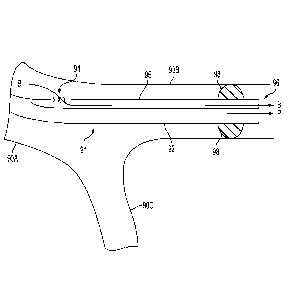
[0022] FIG. 9A depicts the distal end of an improved catheter that is
designed to
prevent reflux.
[0023] FIG. 9B depicts the distal end of another embodiment of an improved
catheter
that is designed to prevent reflux.
DETAILED DESCRIPTION OF THE PREFERRED EMBODIMENTS
[0024] Percutaneous endovascular modification of the function of the
gastric fundus
using particulate embolization of the distal portion of the left gastric
artery is less invasive
and more cost effective alternative to bariatric surgery for achieving weight
loss.
[0025] US Application 14/091,787 describes a novel approach which involves
modifying the arterial blood flow to the gastric fundus by means of
percutaneous
endovascular flow reduction (or interruption) in the distal portion of the
left gastric artery.
Experiments in humans (performed outside the U.S.) has demonstrated dramatic
weight loss
at one month after procedure and sustained for six months follow-up with no
reported adverse
effects. While reduction in the hunger-mediating peptide hormone ghrelin
(secreted in the
gastric fundus) has been identified as a one of possible mechanism, the
complete physiologic
mechanism is not yet clear and may well involve other hormones and/or changes
in gastric
motility with consequent reduction in hunger sensation in experimental
subjects.
6
CA 02936830 2016-07-12
WO 2015/109093 PCMJS2015/011600
[0026] The approach described herein achieves endovascular flow reduction
or
interruption by introducing a plurality of particles into the distal portion
of the subject's left
gastric artery. The particles, also referred to herein as microparticles,
preferably have sizes
between 300 and 500 ktm, and are delivered via a microcatheter. The particles
are preferably
compressible and spherical. They are preferably made of polyvinyl alcohol, and
more
preferably made of acrylamido polyvinyl alcohol. One suitable commercially
available
product for this purpose is BeadBlock Embolic Beads, 300-5001= compressible
microspheres (Biocompatibles UK Limited, Surrey, UK).
[0027] Alternative commercially available products for this purpose include
polyvinyl
alcohol (PVA foam embolization particles, Cook Medical, Bloomington, IN);
hydrogel core
with Polyzene-F coating (EmbozeneTM microspheres, CeloNova Biosciences, Inc.,
San
Antonio, TX); microspheres made from trisacryl cross linked with gelatin
(Embosphere
microspheres, Merit Medical Systems, Inc., South Jordan, UT); HepaSphercTM
Microspheres,
which are made from two monomers (vinyl acetate and methyl acrylate) that
combine to form
a copolymer (sodium acrylate alcohol copolymer); BearingTM nsPVA Embolization
Particles,
which are irregularly-shaped, biocompatible, hydrophilic, nonresorbable
particles produced
from polyvinyl alcohol; EmboGoldTM Microspheres, which are made from trisacryl
cross
linked with gelatin and impregnated with 2% elemental gold for visibility;
QuadraSphereTM
Microspheres, which are also made from two monomers (vinyl acetate and methyl
acrylate)
that combine to form a copolymer (sodium acrylate alcohol copolymer), and
Tcrumo Bead
BlockT microspheres. In alternative embodiments, other embolization materials
may be
used, including but not limited to coils, other microparticles, foams,
different synthetic or
organic gels, thrombin, fibrin, collagen, fibrinogen (liquid or powder), and
any other material
that can occlude blood vessel.
7
CA 02936830 2016-07-12
WO 2015/109093
PCMJS2015/011600
[0028]
Optionally, certain substances may be added to the particles (or to the other
embolization materials) to enhance the effect of the procedure. Examples
include, but are not
limited to: pharmaceuticals, genetic materials, or different types of cells
that also help to
decrease production of ghrelin and/or other hormones or other substances that
effect appetite
in humans.
[0029] One
procedure involves inserting a catheter into the left gastric artery, which
is the major vessel that supplies gastric fundus and modify blood flow. One
way to
accomplish this is to insert a guiding catheter via the femoral artery or
radial artery until the
left gastric artery is engaged (in other words, until the distal end of the
guiding catheter is
introduced into the subject's left gastric artery.) Although the inventor is
not aware of any
guiding catheters that are specially designed to engage the left gastric
artery, examples of
suitable guiding catheters for this step include catheters that are already
available for other
applications such as for coronary angiography and or coronary stenting. In one
preferred
embodiment, the guiding catheter is a 6 French Heartrail II JR-4.0 guiding
catheter (Terumo
Europe N.Y., Leuven, Belgium). That particular guiding catheter is a Judkins
Right type
catheter and has a JR-4.0 shape code. In alternative embodiments, a custom-
shaped guiding
catheter may be used for obtaining easy access to left gastric artery. An
example of one
suitable shape for such a guiding catheter is provided in FIG. 1, in which the
distal end 12 of
the custom-shaped guiding catheter 10 has an S-shaped bend. This shape is
similar to the
shape of the Surefire Axis Catheter (Surefire Medical Inc., Westminster CO),
but the distal-
most bend 14 is increased from about 45 to about 160 . Suitable dimensions
for the guiding
catheter 10 for accessing the left gastric artery are as follows: A between 3
and 4 inches; and
B between 1/2 and 1 inch.
8
CA 02936830 2016-07-12
WO 2015/109093 PCMJS2015/011600
[0030] After the guiding catheter is in position, a guidewire is then
guided through the
guiding catheter and introduced into the mid segment or distal portion of the
subject's left
gastric artery, and then a microcatheter is advanced over the guidewire. Once
the distal end
of the microcatheter has been inserted into the mid segment or distal portion
of left gastric
artery, the embolization material is delivered into the distal portion of left
gastric artery via
the microcatheter. The distal shaft of the microcatheter must be small, e.g.,
2 French in
diameter. One example of a commercially available microcatheter that is
suitable for this
purpose is the Excelsior 1018 Microcatheter (Boston Scientific Corp., Corck,
Ireland).
[0031] The presence of the embolization material in the distal portion of
left gastric
artery will reduce or interrupt the blood flow in the distal portion of left
gastric artery, which
will modify the blood supply to the fundus of stomach. More specifically, it
will reduce or
interrupt the blood supply to the fundus.
[0032] Using microparticles for the embolization material (as opposed to
other types
of embolization materials) is advantageous because they are inert,
biocompatible, and flow-
directed. Moreover, when used as described herein, they will not cause tissue
necrosis or
unwanted non-target embolization. In contrast, if a chemical-based
embolization material
such as sodium morrhuate is used instead of the preferred microparticles, deep
penetration
and or extravasation of this sclerotherapy agent into the gastric tissue may
lead to local
edema and/or extensive inflammation that results in gastric ulceration and
necrosis.
Chemical-based embolization material may also lead to systemic toxicity and
non-target
embolization that may damage the liver, spleen or other organs.
[0033] Using particles with sizes between 300 and 500 um is advantageous
because
using smaller particles (e.g., 50-100 um) can result in mucosal necrosis of
the fundus, and
gastric ulcers. It can also result in non-target embolization of, for example,
the esophagus,
9
CA 02936830 2016-07-12
WO 2015/109093 PCMJS2015/011600
the liver, and/or the spleen because the small particles can penetrate very
deep into tissue and
destroy gastric mucosa. Animal experiments have shown that such smaller
particles may also
end up in structures other than the fundus. In addition, using larger
particles (e.g., 700-1000
[tm) can result in gastric ulcers, and non-target embolization of, for
example, the esophagus,
the liver, and/or the spleen. This may be due to deformation of the particles
during injections
and the formation of larger clusters, which can lead to more proximal
embolization. It may
also be due to reflux of the particles due to the Venturi effect. In contrast,
when particles
with sizes between 300 and 500 gm are used, these problems are avoided or at
least
minimized.
[0034] Limiting the delivery of the particles to the distal portion of the
subject's left
gastric artery is advantageous because when the proximal portion of the left
gastric artery is
also filled with particles, the risk of esophageal and nonfundus gastric
ulcers is very high.
More specifically, it was observed in three out of three subjects in animal
studies, when
tested in pigs. In contrast, these problems were not observed in any of the
three pig subjects
in which the delivery of the particles was limited to the distal portion of
the test subject's left
gastric artery.
[0035] Thus, by using the correct size of the correct material and
delivering it to the
correct location, many of the problems associated with other approaches are
avoided, and the
procedure can be made safe.
[0036] EXAMPLE 1
[0037] A study was done on five obese subjects to determine the
feasibility, safety,
and efficacy of embolization of the distal portion of the left gastric artery
to reduce plasma
ghrelin levels and body weight.
CA 02936830 2016-07-12
WO 2015/109093 PCMJS2015/011600
[0038] All subjects underwent gastroscopy prior the embolization to assess
for the
presence of peptic ulcer or gastritis. Gastritis was found in two subjects who
subsequently
underwent medical treatment. Embolization was performed only after follow-up
gastroscopy
showed significant improvement in mucosal irritation.
[0039] Weights were measured and routine blood samples obtained including a
complete blood count, electrolytes, and creatinine prior to embolization.
[0040] In the procedure, 6-Fr femoral access was obtained. More
specifically, a 6-Fr
Heartrail II JR-4.0 guiding catheter (Terumo Europe N.V., Leuven, Belgium) was
used to
engage the celiac trunk ostium and angiography performed in different
projections in order to
identify the origin and anatomy of left gastric artery. In some cases, a 0.35"
guidewire was
advanced into the common hepatic or splenic arteries to stabilize the guiding
catheter
position.
[0041] The left gastric artery, a branch of the celiac trunk, was wired
with a 0.014"
Runthrough NS PTCA Guide Wire (Terumo Europe N.V., Leuven, Belgium) and an
Excelsior 1018 Microcatheter (Boston Scientific Corp., Corck, Ireland)
advanced over the
guide wire into the mid segment of the left gastric artery. Subsequently, the
guide wire was
removed while maintaining the microcatheter position in the left gastric
artery and selective
angiography performed to ensure proper catheter position and define the
anatomy and course
of the left gastric artery. FIG. 5 is an angiography of the left gastric
artery 50 and the
surrounding anatomy after a radio-opaque material was injected into the left
gastric artery,
but prior to the injection of any particles. The dark artifacts in the circle
52 reveal that blood
is flowing in the distal portion of the left gastric artery.
[0042] Repeat injections of small amounts of BeadBlock Embolic Bead, 300-
500 m
compressible microspheres (Biocompatibles UK Limited, Surrey, UK) mixed with
contrast
11
CA 02936830 2016-07-12
WO 2015/109093 PCMJS2015/011600
agent (1:1 ratio) were then performed. Angiography was performed between
injections of the
microspheres to assess left gastric artery flow characteristics. The injection
of the
micro spheres was continued until distal portions of artery branches were no
longer visible
during radio-opaque contrast injection. This is shown in FIG. 6, which depicts
the left gastric
artery 50 and the surrounding anatomy. Note the absence of dark artifacts in
the circle 52',
which indicates that blood is no longer flowing in the distal portion of the
left gastric artery.
[0043] The guiding and microcatheter were then withdrawn and subjects
transferred
to a ward, where the introducer sheath was removed and manual pressure applied
to obtain
hemostasis.
[0044] Esophagogastroscopy was performed in all subjects before and after
the
procedure gastroscopy. A second follow-up gastroscopy was performed one week
after the
procedure. Weight and fasting plasma ghrelin levels were obtained at baseline
and the 1, 3,
and 6-month follow-up visits. To obtain the ghrelin levels, clotted blood
samples were
centrifuged to separate out blood plasma. Fasting levels of ghrelin, ALT, AST,
urea and uric
acid were then measured. Ghrelin was measured using the Human Ghrelin (TOTAL)
RIA
KIT (Merck Millipore). Subject's weight and body mass index (BMI) was also
calculated at
each of the visits.
[0045] Data for the study is presented below. Table 1 shows the subject
data, Table 2
shows the weight at each visit for each subject, Table 3 shows the
corresponding BMI, and
Table 4 shows the ghrelin levels at each visit for each subject. FIGS. 2, 3,
and 4 depict the
data in Tables 2, 3, and 4, respectively, in a graphical format.
12
CA 02936830 2016-07-12
WO 2015/109093
PCMJS2015/011600
Parameter Value
_
Number of participants 5
Gender female/male (%) 20/80%
Age female/male (years) 44.7 7.4
Weight (kg) 128.1 +24.4
BMI (kg/m2) 42.2 6.8
Ghrelin (pg/ml) 473.4+ 189.11
TABLE 1
Subject Initial weight Weight at Weight at Weight at
# (kg) 1 month FU 3 month FU 6 month FU
1 119 102 99 94
2 165 146 143 140
3 98 90 85 80
4 131 120 116 117
127 117 114 107
Mean 128 24 115 21 111 22 108 23
p Value 0.0032 0.0012 0.0008
TABLE 2
Subject Initial BMI at BMI at BMI at
# BMI 1 month FU 3 month FU 6 month FU
1 42 36 35
2 53 47 46 45
3 34 31 30 28
4 41 38 37 37
5 41 38 38 34
Mean 42 7 38 6 37 6 36 6
p Value 0.0033 0.0012 0.001
TABLE 3
13
CA 02936830 2016-07-12
WO 2015/109093 PCMJS2015/011600
Subject Initial Ghrelin Ghrelin level at Ghrelin level at
Ghrelin level at
level (pg/ml) 1 month FU 3 month FU 6 month FU
1 459.6 313.4 301.3 325.5
2 486.1 325.9 323.6 410.9
3 445.5 380.7 315.8 389.1
4 501.2 341.6 299.7 388.6
478.3 342.2 325.5 391.3
Mean 470.54 340.76 314.18 381.08
p Value 0.0015 0.0002 0.0042
TABLE 4
[0046] STATISTICAL ANALYSIS: Statistical analysis was performed using
computer software (SPSS 12.0 for Windows, Lead Technologies Inc. 2003.
Chicago, II.).
All values were presented as the mean standard deviation ( SD). Comparison
of weights
and plasma ghrelin levels between different time points were done with the
paired t-test. A
p-value of <0.05 was considered to determine statistical significance
[0047] RESULTS: There were no procedural complications. Three of the five
subjects described mild transient epigastric pain after the procedure.
However, follow-up
gastroscopies on the day after embolization and at 1-week follow-up did not
reveal any
abnormalities. All subjects reported a significantly decreased appetite in the
first days after
the procedure.
[0048] Significant progressive weight loss accompanied by reductions in
plasma
ghrelin levels was observed in all subjects at all follow-ups: Mean weight and
BMI was
reduced by 10%, 13%, and 16% at 1-, 3- and 6-month follow-up, respectively
(Table 2 and3).
Mean initial weight (128.12 24.4kg) decreased to 108 23kg (p<0.001). Blood
plasma
ghrelin levels (initially 473+189) were significantly lower at 1- and 6-month
follow-up (by
14
CA 02936830 2016-07-12
WO 2015/109093 PCMJS2015/011600
29% and 36% from baseline, p < 0.05) and increased slightly at the 6-month
follow-up
compared with 3-month follow-up while remaining 18% lower from the baseline
(p> 0.05).
[0049] FIG. 7 is a CT angiograph the left gastric artery 70 and the
surrounding
anatomy that was taken 3 months after procedure. In this figure, portions
where blood is
flowing are indicated in white. Because the distal portion of the left gastric
artery is not
visible in region 72, it is apparent that the distal portion remains occluded
3 months after
procedure.
[0050] The data above demonstrates that embolization of the distal portion
of the left
gastric artery using microparticles is associated with significant reductions
in plasma ghrelin
levels and weight loss in humans. It should be noted, however, that after an
initial
pronounced decline in ghrelin levels after the procedure, the levels did
increase at the last
follow-up visit (i.e., at the 6 month visit), Although the levels were still
lower than the pre-
procedure baseline, a long-term study may be warranted to further investigate
this increase.
[0051] The procedure described above appears to be safe. Specifically,
there were no
incidences of ulcer formation or injury to remote structures. This may be
related to the
selective injection into the left gastric artery of beads that are large
enough in size as to not
allow systemic or remote toxicity, yet small enough to avoid the potential
problems described
above. Note that with more extensive embolization of arteries other than the
left gastric
artery, the ulcer risk may be higher. For example, 40% of animals that
underwent
embolization of the left, short, and accessory gastric arteries developed
gastric ulcers in a
study by Paxton et al. These ulcers were located at the lesser curvature,
suggesting a
watershed effect. In addition, using the correct embolic materials as
described herein
apparently minimizes the extent and likelihood of injury to adjacent or remote
tissue.
CA 02936830 2016-07-12
WO 2015/109093 PCMJS2015/011600
[0052] It should be noted that this example was a non-randomized single-arm
feasibility, safety, and efficacy trial with all its inherent limitations.
First, the absence of a
control group does not allow definitive conclusions regarding efficacy. It is
possible that the
procedure and study participation led to a higher motivation for diet control
and exercise.
However, in this case, a decrease in plasma ghrelin levels should not be
expected. Second,
the intermediate-term follow-up (i.e., 6 months) is too short to make
conclusions regarding
long-term weight loss, as a rebound phenomenon with recurrent weight gain is
conceivable.
Third, though not observed in a study by the inventor, a risk of gastric ulcer
formation may
be significant but too small to have been observed in the study.
[0053] It can therefore be concluded that Percutaneous embolization of the
distal
portion of the left gastric artery with embolic beads is feasible and appears
to be safe. It leads
to a reduction in plasma ghrelin levels and is accompanied by a significant
weight loss at
intermediate term follow-up. It may be a good tool to enhance weight loss in
subjects with
morbid obesity who cannot achieve weight loss by conventional means (diet and
exercise)
and an alternative to or complimentary to bariatric surgery.
[0054] Although the procedure described above in has many benefits, a
potential
problem exists: if too many particles are delivered through the catheter,
reflux of the particles
may occur. More specifically, even though the distal end of the catheter might
be properly
positioned in the mid segment or distal portion of left gastric artery, when
too many particles
arc injected through the catheter, the particles can back up to more proximal
portions of the
left gastric artery. And if the number of particles is even larger, the
particles could back up
all the way to the celiac artery. This could be dangerous because the
particles could then
travel forward to the liver, spleen, or pancreas. It is therefore preferable
to make special
16
CA 02936830 2016-07-12
WO 2015/109093 PCMJS2015/011600
provisions to prevent reflux of the particles, so that the particles do not
travel to other parts of
the body.
[0055] FIG. 8 depicts the distal end of a commercially available catheter
(made by
Surefire Medical Inc.) that can be used to prevent reflux. In this embodiment,
the wall 82 of
the catheter defines an internal lumen, and the particles are delivered
through that lumen, as
indicated by the arrow labeled P. The distal end 84 of the catheter flares out
and preferably
touches the inner walls of the artery 80 on all sides of the catheter. At
least a portion of the
flared distal end 84 is made of mesh, indicated by the "xxx" marking in FIG.
8. The mesh
size is selected so that all types of blood component cells can pass
(including red blood cells,
white blood cells, etc.), but the microparticles cannot pass. The mesh at the
flared distal end
84 therefore prevents the particles from traveling backwards. A suitable
spacing for the mesh
for this purpose is between 150 and 250 microns, and preferably about 200
microns. As a
result, even though the flared distal end 84 of the catheter touches the walls
of the artery 80,
blood can still flow as indicated by the arrows B. The flow of blood is
desired because the
blood flow helps to carry the particles along to their destination in the
distal portion of the left
gastric artery. However, because the left gastric artery has a relatively
small diameter, the
body of the catheter will block a large portion of the artery, which will
reduce the amount of
blood that can flow past the catheter. In this embodiment, the blood flow
could be reduced to
the point where the blood flow will not adequate to direct the particles to
their desired
destination.
[0056] FIG. 9A depicts the distal end of a novel catheter that is designed
to overcome
this problem by maintaining significant blood flow that is sufficient to carry
the particles to
their desired destination, while still preventing reflux of the particles. In
this embodiment,
the wall 92 of the catheter 91 defines a first internal lumen, and the
particles are delivered
17
CA 02936830 2016-07-12
WO 2015/109093 PCMJS2015/011600
through that first internal lumen, as indicated by the arrow labeled P. This
lumen provides a
fluid-tight path for particles to flow between the proximal end of the
catheter (not shown) and
the distal end of the catheter. The outer diameter of the catheter 91 is
preferably between
0.038 and 0.064 inches, and is more preferably about 0.05 inches. The diameter
of the first
internal lumen is preferably between 0.012 and 0.020 inches, and is more
preferably about
0.016 inches. The first internal lumen is preferably tracked over a guidewire
(not shown) to
facilitate safe positioning of the catheter 91 to the desired location.
Optionally, to facilitate
catheter advancement in the blood vessel and to prevent kinking, the wall 92
of the catheter
91 may contain a longitudinally embedded core-wire (not shown) which may be
tapered to
impart varying degrees of longitudinal flexibility.
[0057] A balloon 98 located near the distal end of the catheter can be
inflated in a
conventional manner (e.g., via a dedicated inflation lumen, not shown, that
has a distal end
that is in fluid communication with an interior of the balloon), and the
balloon will prevent
any particles from refluxing. The balloon is preferably between 2 and 15 mm
long in a
proximal to distal direction, and is more preferably between 5 and 10 mm long.
The balloon
is preferably located between 2 and 20 mm from the distal end of the catheter
91, and is more
preferably about 10 mm. The balloon 98 is preferably inflated with very low
pressure (less
than 1 atm), and is preferably designed to fail at low pressure (greater than
2 atm) in order to
prevent barotrauma to the blood vessel. Optionally, the balloon and catheter
preferably may
have a hydrophilic-heparin coating to further minimize vascular trauma. The
balloon 98 may
be made from a compliant or semi-compliant material, such as polyurethane,
polyimide,
polyolefins, silicone, or copolymers thereof. The balloon 98 is preferably
designed such that
the diameter may be adjusted in a slow, continuous manner by varying the
volume of
inflation media in order to occlude varying vessel diameters.
18
CA 02936830 2016-07-12
WO 2015/109093 PCMJS2015/011600
[0058] When the balloon 98 is inflated, the natural flow of blood through
the left
gastric artery is blocked (i.e., the balloon prevents blood from flowing
through the target
artery), which would ordinarily not be desirable because the blood flow help
carry the
particles along to their destination. To remedy this issue, a second lumen 95
is provided in
this embodiment. The second lumen 95 has an input port 94 that is proximal to
balloon 98,
and an output port 96 that is distal to the balloon 98. This second lumen
provides a fluid-
tight path for blood to flow from the input port to the output port. Blood
will enter the second
lumen 95 through the input port 94 and exit the second lumen 95 through output
port 96, as
indicated by the arrow labeled B---B. The diameter of second lumen is
preferably between
0.006 and 0.014 inches, and is more preferably about 0.010 inches. Preferably,
at least one of
the ports 94, 96 is fitted with a strainer that prevents the particles from
flowing through the
second lumen. A preferred approach for implementing the strainer is to use a
mesh with a
mesh size that is coarse enough to permit all types of blood component cells
to pass
(including red blood cells, white blood cells, etc.), but fine enough to
prevent the
microparticles from passing. A suitable spacing for the mesh for this purpose
is between 150
and 250 microns, and preferably about 200 microns. Alternatively, a finer mesh
that only
permits the red blood cells to pass may be used. Preferably, the mesh includes
filaments in
two perpendicular directions (i.e., arranged like the wires in a conventional
window screen).
In alternative embodiments, the strainer may be made of filaments that
intersect at non-
perpendicular angles. In other alternative embodiments, the strainer may be
made of a
plurality of filaments that are all parallel (i.e., arranged like the strings
of a harp), the strainer
may also be configured like a colander, or the strainer may be implemented
using a
semipermeable membrane with a suitable pore size.
[0059] Due to the second lumen 95, blood from an artery in the patient's
body can
enter the second lumen via the input port, flow through the second lumen, exit
the second
19
CA 02936830 2016-07-12
WO 2015/109093 PCMJS2015/011600
lumen via the output port, and flow from the output port into the left gastric
artery. Thus,
blood can flow forward through the artery 90B even though the balloon 98 is
inflated. The
flow of blood through the second lumen 95 will be sufficient to carry the
particles along to
their destination in the distal portion of the left gastric artery. In some
preferred
embodiments, the length of the second lumen 95 is long enough so that the
input port 94 is
disposed in a relatively wide portion of the vasculature, such as the celiac
artery 90A (i.e.,
before the left gastric artery 90B branches off from the splenic and common
hepatic arteries,
both illustrated schematically as 90C), or even the aorta (not shown). This
arrangement will
make it even easier for the blood to flow into the input port 94, so that the
blood flow can
direct the particles to their desired destination.
[0060] FIG. 9B depicts the distal end of an alternative novel catheter that
is designed
to maintain sufficient blood flow to carry the particles to their desired
destination, while
preventing reflux of the particles. In this embodiment, the wall 192 of the
catheter 191
defines an internal lumen, and the particles are delivered through that
internal lumen, as
indicated by the arrow labeled P. The internal lumen provides a path for
particles to flow
between the proximal end of the catheter and the distal end of the catheter.
The outer
diameter of the catheter 191 is preferably between 0.026 and 0.052 inches, and
is more
preferably about 0.04 inches. The diameter of the internal lumen is preferably
between 0.012
and 0.020 inches, and is more preferably about 0.016 inches. The internal
lumen is
preferably tracked over a guidewire (not shown) to facilitate safe positioning
of the catheter
191 to the desired location. Optionally, longitudinally embedded core-wire
(not shown) may
be used as described above in connection with the FIG. 9A embodiment.
[0061] A balloon 98 that is similar to the balloon 98 of the FIG. 9A
embodiment is
also used in this FIG. 9B embodiment. When the balloon 98 is inflated, the
natural flow of
CA 02936830 2016-07-12
WO 2015/109093 PCMJS2015/011600
blood through the left gastric artery is blocked, which would ordinarily not
be desirable
because the blood flow help carry the particles along to their destination. To
remedy this
issue, an opening 194 into the first lumen is provided in the sidewall 192 of
the catheter 191.
This opening provides a path for blood to flow from the artery directly into
the internal lumen
of the catheter 191. The opening 194 is proximal to balloon 98, and the
opening 194 permits
blood to enter the internal lumen 191. The opening is disposed at a position
such that when
the distal end of the catheter is positioned in the target artery, blood from
an artery in the
patient's body can flow into the opening, through the internal lumen, and into
the left gastric
artery. Once inside the internal lumen 191, the blood will flow forward and
will exit the
internal lumen through output port 196, as indicated by the arrow labeled B---
B. A strainer is
disposed at the opening, and the strainer is configured to prevent the
particles from exiting
the first lumen via the opening. One preferred way to implement this strainer
is to cover the
opening 194 with a mesh similar to the mesh describe above in connection with
FIG. 9A.
Due to the mesh-covered opening 194, blood can flow forward through the artery
even when
the balloon 98 is inflated. This flow of blood will be sufficient to carry the
particles along to
their destination in the distal portion of the left gastric artery. In some
preferred
embodiments, the opening 194 is disposed far back enough along the catheter so
that it will
be disposed in a relatively wide portion of the vasculature, such as the
celiac artery or even
the aorta (not shown). This arrangement will make it even easier for the blood
to flow into
the opening 194 so that the blood flow can carry the particles to their
desired destination in
the left gastric artery. Note that any of the alternative strainers described
above in connection
with the FIG. 9A embodiment may also be used in this FIG. 9B embodiment.
[0062] Note that while the embodiments described above are described in the
context
of the left gastric artery, similar techniques may be used in other arteries
to embolize different
21
CA 02936830 2016-07-12
WO 2015/109093 PCMJS2015/011600
portions of a patient's anatomy. The artery into which the embolization
material is delivered
is referred to herein as the target artery.
[0063] While the present invention has been disclosed with reference to
certain
embodiments, numerous modifications, alterations, and changes to the described
embodiments are possible without departing from the sphere and scope of the
present
invention, as defined in the appended claims. Accordingly, it is intended that
the present
invention not be limited to the described embodiments, but that it has the
full scope defined
by the language of the following claims, and equivalents thereof.
22