Note: Descriptions are shown in the official language in which they were submitted.
CA 02937882 2016-07-25
1
DESCRIPTION
METHOD FOR GENERATING CELL CONDENSATE FOR SELF-ORGANIZATION
TECHNICAL FIELD
The present invention relates to a method of preparing a cell condensate for
self-
organization. More specifically, the present invention relates to a method of
preparing a cell
condensate that is necessary for directing self-organization into a tissue or
an organ of
interest.
BACKGROUND ART
Recently, methods using the self-organization capacity of cells which they
inherently
possess have been attracting attention as methods of forming tissues/organs
with complex
structures (Non-Patent Documents Nos. 1 and 2). Self-organization is a process
in which one
or a few elements construct complex higher structures by exerting intrinsic
properties of their
own without receiving specific "instructions" (information) from the outside.
For example,
natural phenomena in which spontaneous order arises from patternless
aggregates to form
patterns, as in crystallization of snow, are observed. Self-organization is
also used in the field
of engineering, e.g. in nanotechnology or in preparing optical crystals.
For inducing self-organization, it is necessary to form an aggregate
consisting of
homogeneous cells in a high-density environment. Studies have been reported in
which
aggregates were prepared from cultured ES/iPS cells to generate brain, optic
cup, pituitary
gland, teeth, etc. (Non-Patent Documents Nos. 3 to 6). As a technique for
preparing such
aggregates, a method is mainly used in which tissues of several hundred trn
level are formed
from cell aggregates of a small number of cells (about several thousand) by
using a substrate
such as a 96-well plate with U- or V-shaped bottoms that permits cells to
gather in the
bottom. However, it has been difficult to achieve formation of larger size
(200 1,tni or more)
cell condensates from a large number of cells (several ten thousand to several
million cells).
Therefore, it has been difficult to apply conventional methods to preparation
of cell
aggregates consisting of diverse cells.
Under these circumstances, it has been desired to develop a self-organization
based
technique for preparing cell condensates for generating large and complex
tissues/organs (as
CA 02937882 2016-07-25
2
from humans) compared to tissues/organs of small animals like mouse.
PRIOR ART LITERATURE
Non-Patent Documents
Non-Patent Document No. 1: Camazine, S., Deneubourg, J. -L., Franks, N. R.,
Sneyd, J.,
Theraulaz, G. & Bonabeau, E. Self-Organization in Biological Systems
(Princeton Univ.
Press, 2001).
Non-Patent Document No. 2: Takeichi, M. Self-organization of animal tissues:
cadherin-
mediated processes. Dev. Cell 21, 24-26 (2011).
Non-Patent Document No. 3: Eiraku, E. et al. Self-organizing optic-cup
morphogenesis in
three-dimensional culture. Nature 472, 51-56 (2011).
Non-Patent Document No. 4: Eiraku, M. et al. Self-organized formation of
polarized cortical
tissues from ESCs and its active manipulation by extrinsic signals. Cell Stern
Cell 3, 519-532
(2008).
Non-Patent Document No. 5: Suga, H. et al. Self-formation of functional
adenohypophysis in
three-dimensional culture. Nature 480, 57-62 (2011).
Non-Patent Document No. 6: Sato, T. et al. Single Lgr5 stem cells build crypt-
villus
structures in vitro without a mesenchymal niche. Nature 459, 262-265 (2009).
DISCLOSURE OF THE INVENTION
PROBLEM FOR SOLUTION BY THE INVENTION
The present inventors have already established a groundbreaking three-
dimensional
culture technique using spatiotemporal interactions of three different cell
lineages; this
technique has realized "directed differentiation of organ cells based on
reconstitution of
organs". Briefly,
the present inventors have established a platform technology which
recapitulates interactions among organ cells, vascular cells and mesenchymal
cells that are
essential for early processes of organogenesis, to thereby induce 3D organ
primordia (starting
material for organs) and enable generation of vascularized functional organs
(Nature, 499
(7459), 481-484; PCT/JP2012/074840 Method for Preparing Tissue and Organ).
On the other hand, for the development of drugs or realization of regenerative
medicine for diseases in kidney, liver, lung, etc., it is essential to
recapitulate three-
CA 02937882 2016-07-25
3
dimensional complex structures (integrating not only a vasculature but also
higher structures
such as ureteral structure, biliary structure, tracheal structure, etc.) and
cell polarity.
Moreover, induction of an organ of interest is achieved through interactions
with other
organs.
Therefore, in order to maximize the function of tissues induced from
pluripotent stem
cells or tissues isolated from individuals, three-dimensional tissue
constructs should be
formed which enable reconstitution of continuity with diverse higher
structures and other
organs. According to conventionally devised methods, however, only tissue
constructs
having a vascular structure alone have been prepared from the three types of
cells or tissues.
No technique has been invented for preparing more complex, higher structures
(such as
ureteral structure, biliary structure and tracheal structure).
It is an object of the present invention to find out the requirements
necessary for
preparing a cell condensate in vitro from a large number of cells (several ten
thousand to
several million cells). It is another object of the present invention to
provide a method of
forming a cell condensate for self-organization which is capable of realizing
complex higher
structures (such as liver and kidney) and interactions with other organs.
MEANS TO SOLVE THE PROBLEM
The present inventors have succeeded in preparing three-dimensional
tissues/organs
having complex higher structures from isolated, multiple types of cells or
tissues by the
operations 1 to 4 described below. Thus, the present invention has been
achieved.
I . Preparation of Necessary Cells/Tissues
A) Cells/tissues of a desired type or types that are necessary for self-
organization into tissues
with complex structures are prepared. The types or numbers to be combined do
not matter.
B) A mixture in solution that consists of a desired type or types of
cells/tissues in a total
number of approximately 2 million is mixed with approximately 100,000 to
400,000 isolated
mesenchymal cells.
2. Preparation of Support
A) A support with an appropriate stiffness is formed and solidified on a cell
culture dish.
Preferable materials for the support include, but are not limited to,
hydrogels (such as
polyacrylamide gel).
B) Chemical/physical modifications are provided on the prepared support.
Giving such
=
CA 02937882 2016-07-25
4
modifications, however, is not an essential requirement. Preferable chemical
factors include,
but are not limited to, Matrigel and laminin.
C) The stiffness of the support need not be uniform and may vary depending on
the shape,
size and quantity of an condensate of interest. The stiffness of the support
may be provided
with aspatial/temporal gradient or patterned, for use in subsequent
experiments.
3. Preparation and Culture of Cell Condensates
A) The cell/tissue mixture in solution as prepared in 1 above is plated on the
support
prepared in 2 above to form condensates. The thus formed condensates may be
cultured for
an elongated period so that it can be used for self-organization into organs
of interest in
vitro.
By combining mesenchymal cells with a culture substrate that permets cells to
gather
in the bottom, condensates can also be prepared from the cells if they arc
small in number.
4. Transplantation of Cell Condensates
By subjecting the condensates prepared in 3 above to long-term culture or
transplanting them into living bodies to induce blood perfusion and allow self-
organization
into higher tissues with a complex structure, tissues/organs can be prepared
that have a highly
ordered tissue structure comparable to that of adult tissues.
The above-described technique which prepares a complex cell condensate
consisting
of cells of a desired type or types by combining mesenchymal cells with
physicochemical
properties of a support has not existed to date and is believed to provide a
method that is
extremely high in novelty.
The gist of the present invention is as described below.
( I) A method of preparing a cell condensate in vitro, comprising culturing a
mixture of cells
and/or tissues of a desired type and mesenchymal cells to form a cell
condensate.
(2) The method of (1) above, wherein the cell condensate is capable of forming
a three-
dimensional tissue structure that has been provided with higher structures by
self-
organization.
(3) The method of (1) or (2) above, wherein the mixture of cells and/or
tissues of a desired
type and mesenchymal cells is cultured on a gel-like support on which the
mesenchymal
cell is capable of contraction.
(4) The method of (3) above, wherein the culture is two-dimensional culture.
(5) The method of (3) or (4) above, wherein the gel-like support is planar or
the side of the
CA 02937882 2016-07-25
gel-like support on which culture is performed has a U- or V-shaped cross-
section.
(6) The method of any one of (3) to (5) above, wherein the stiffness of the
central part of the
support is greater than the stiffness of the peripheral part thereof.
(7) The method of any one of (3) to (5) above, wherein the stiffness of the
peripheral part of
the gel-like support is greater than the stiffness of the central part
thereof.
(8) The method of any one of (3) to (5) above, wherein the gel-like support is
patterned and
has one or more patterns in which the stiffness of the central part is greater
than the
stiffness of the peripheral part.
(9) The method of any one of (3) to (5) above, wherein the gel-like support is
patterned and
has one or more patterns in which the stiffness of the peripheral part is
greater than the
stiffness of the central part.
(10) The method of any one of (1) to (9) above, wherein the cells and/or
tissues of a desired
type have a total cell count of 400,000 or more and the mesenchymal cells are
100,000 to
400,000 in number.
(11) The method of any one of (1) to (10) above, wherein the size of the cell
condensate is 1
mm or more.
(12) The method of any one of (1) to (11) above, wherein the cell condensate
is formed
autonomously.
(13) The method of any one of (1) to (12) above, wherein the mixture of cells
and/or tissues
of a desired type and mesenchymal cells is cultured without using scaffold
materials.
(14) The method of any one of (1) to (13) above, wherein the cells and/or
tissues mixed with
the mesenchymal cells are derived from liver, pancreas, intestine, lung,
kidney, heart,
brain or cancer.
(15) The method of any one of (1) to (13) above, wherein the cells mixed with
the
mesenchymal cells are pluripotent cells.
(16) The method of any one of (1) to (13) above, wherein the tissues mixed
with the
mesenchymal cells are tissues induced from pluripotent cells.
(17) The method of (15) or (16) above, wherein the pluripotent cell is a
pluripotent cell
obtained from a living body, a pluripotent cell obtained by induction from
reprogramming or a mixture thereof.
(18) A cell condensate prepared by the method of any one of (1) to (17) above.
(19) A method of preparing a three-dimensional tissue structure, comprising
allowing self-
CA 02937882 2016-07-25
6
organization of a cell condensate prepared by the method of any one of (1) to
(17) above
to form a three-dimensional tissue structure that has been provided with
higher
structures.
(20) A gel-like culture support wherein the side on which culture is performed
has a U- or V-
shaped cross-section.
(21) A gel-like culture support wherein the stiffness of the central part
thereof is greater than
the stiffness of the peripheral part thereof
(22) A gel-like culture support wherein the stiffness of the peripheral part
thereof is greater
than the stiffness of the central part thereof.
(23) A gel-like support having one or more patterns in which the stiffness of
the central part
is greater than the stiffness of the peripheral part.
(24) A gel-like support having one or more patterns in which the stiffness of
the peripheral
part is greater than the stifthess of the central part.
(25) A method of preparing a cell condensate in vitro, comprising culturing a
mixture of cells
and/or tissues of a desired type and mesenchymal cells on the gel-like culture
support of
any one of (20) to (24) above to thereby form a cell condensate.
EFFECT OF THE INVENTION
According to the present invention, a cell condensate of theoretically any
complex
composition can be formed by combining a mesenchymal stem cell and a support
or a
substrate that will allow cells to gather in the bottom. According to the
present invention,
tissues and organs can be constructed without using scaffolds.
First, the cell condensate of the present invention is expected to find use as
an
artificial constitution system for more complex tissues and organs. For
example, with the
cell condensate of the present invention, it may be possible to prepare three-
dimensional
complex structures that are provided with not only a vascular network but also
higher
structures such as ureteral structure, biliary structure, tracheal structure,
etc. Further, a great
number of organs essentially require that reconstitution associated with other
organs be
realized in order to exhibit their functions; e.g., in liver, reconstitution
of junctions with bile
duct and pancreatic duct and connection to duodenum is essential for
exhibiting its Function.
According to the present invention, a cell condensate which recapitulates
interactions with
other organs is prepared. This cell condensate is expected to find use as a
system for
CA 02937882 2016-07-25
7
inducing self-organization into complex organs existing in the body.
Secondly, since the present invention uses an inexpensive and comparatively
easy-to-
process support, its industrial applicability toward mass production of
tissues is high. Mass
production of tissues of a desired shape, size and number can be realized at
low cost by
combining the cell condensate with the multi-patterning of the support or
other techniques.
The technique of generating a 3D tissue construct self-organized from a cell
condensate prepared from stem cells such as iPS cells is applicable to
generation of human
functional cells which has been difficult to achieve to date; transplantation
of tissues and
organs; screening in drug discovery; a novel analysis system for evaluating
the relationships
between development of drug effects and supporting tissues (blood vessels,
nerves, stroma,
etc.) and so on.
The present specification encompasses the contents disclosed in the
specification
and/or drawings of Japanese Patent Application No. 2014-037341 based on which
the present
application claims priority.
BRIEF DESCRIPTION OF THE DRAWINGS
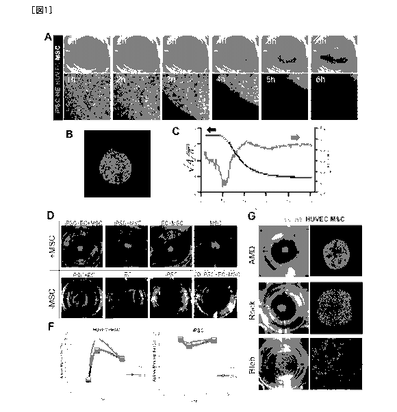
[Fig. 1] Preparation of cell condensates via contraction of mesenchymal cells
(A) Time-dependent changes in the process of formation of cell condensates.
(Green) iPSC-
hepatic endoderm cells; (Light red) human vascular endothelial cells;
(Colorless)
mesenchymal cells.
(B) Formation of self-organized, iPSC or iPS cell-derived liver buds
(C) Temporal development of dynamics of cell condensate formation. (Red)
square root of
the projected area of condensate. This can be used as an indicator showing the
location of the
edge of condensate. After about 13 hr, an exponential function provides good
approximation
(black dotted line). ; (Blue) circularity of condensate calculated from the
projected area and
the contour line length of condensate.
(D) Necessity of mesenchymal cells in cell condensate formation
(L) Inhibitory experiment against cell condensate formation process using
various chemical
substances.
(F) Time-dependent changes in the content of active form of myosin and the
inhibition
thereof
[Fig. 2] Optimization of stiffness environment in cell condensate formation
CA 02937882 2016-07-25
8
(A,B) Cell condensate formation experiments under various stiffness
conditions. (A)
Macroscopic observation after 48 hr of culture. (B) Time-dependent changes in
cell
movement under confocal laser microscope. (C-G) Characterization of MSCs in
cell
condensate. trajectories (C); time dependency of velocity and order parameter
(D, E); and
dependency on substrate stiffness (F, G).
[Fig. 3] Experiments on the formation of condensates for self-organization
using
diverse tissue-derived cells
(A,B) Cell condensate formation using pancreatic i cells (A) and self-
organization (B).
(C,D) Cell condensate formation experiments using other organ cells/tissues.
[Fig. 4] In vivo self-organization of diverse tissue-derived cell condensates
and
development of their function
(A) Functional vascularization occurs in 2 to 3 days after transplantation.
(B) Comparison between the conventional and invention methods of the time
required for
blood perfusion.
(C) Direct anastomosis of mouse and human blood vessels.
(D) Glomeruli and renal tubules formed by cell condensates prepared from
embryonic renal
cells.
(E) Islet-like tissues formed by cell condensates prepared from 13 cells.
(F) Model for evaluating the therapeutic effect of cell condensates prepared
from 13 cells.
(G) Time-dependent changes in blood glucose level in diabetic model mice
transplanted with
cell condensates prepared from [3 cells.
[Fig.5] Time-dependent changes in the trajectory, velocity and order parameter
of
MSCs in cell condensates under various stiffness conditions
[Fig. 6] Chronological observation of cell condensate formation processes
using
various inhibitors.
[Fig. 7] In vivo self-organization of cell condensate using adult kidney
tissue.
[Fig. 81 In vivo self-organization of cell condensate using embryonic lung
tissue.
[Fig. 9] Tracing of in vivo vascularization process in cell condensate using
[3 cells.
[Fig. 101 Observation of in vivo junctions with host blood vessels in cell
condensate
using [3 cells.
[Fig. 11] 1Iistological analysis of tissues generated from cell condensate
using (3 cells.
[Fig. 12] Cross section of U-bottom gel.
CA 02937882 2016-07-25
9
[Fig. 13] (A) Formation of cell condensates containing no vascular endothelial
cells.
(B) Formation of cell condensates using human or mouse mesenehymal cells.
[Fig. 14] Formation of cell condensates using U-bottom gel.
[Fig. 151 Reconstitution of a functional vascular network by transplantation
of a
kidney primordium prepared on a support.
[Fig. 16] Maturation of transplanted kidney primordium.
[Fig. 17] Structural analysis of kidney primordium that matured after
transplantation.
[Fig. 18] Live imaging of the capacity of the transplanted kidney primordium
to
produce primitive urine.
[Fig. 19] Measurements of stiffness properties before and after coating with a
biochemical substance.
[Fig. 20] Preparation of supports having multiple patterns of stiffness.
[Fig. 2111 Preparation of cell condensates on supports having multiple
patterns of
sti ffness.
[Fig. 2211 Preparation of supports having complex multiple patterns.
BEST MODES FOR CARRYING OUT THE INVENTION
Hereinbelow, the present invention will be described in detail.
The present invention provides a method of preparing a cell condensate in
vitro,
comprising culturing a mixture of cells and/or tissues of a desired type and
mesenchymal
cells to form a cell condensate.
Mesenchymal cells are connective tissue cells that are mainly located in
mesoderm-
derived connective tissues and which form support structures for cells that
function in tissues.
In the present specification, the term "mesenchymal cell" is a concept that
encompasses those
cells which are destined to, but are yet to, differentiate into mesenchymal
cells.
Mesenchymal cells to be used in the present invention may be either
differentiated or
undifferentiated. Preferably, undifferentiated mesenchymal cells are used.
Whether a cell is
an undifferentiated mesenchymal cell or not can be determined by checking to
see lithe cell
expresses marker proteins such as Stro-1, CD29, CD44, CD73, CD90, CD105. CD I
33,
CD271 or Nestin (if any one or more of the above-listed marker proteins are
expressed. the
cell can safely be regarded as an undifferentiated mesenchymal cell). A
mesenchymal cell in
which none of the above-listed markers is expressed can be regarded as a
differentiated
CA 02937882 2016-07-25
mesenchymal cell. Among the terms used by those skilled in the art, the
following are
included in the "mesenchymal cell" of the present invention: mesenchymal stem
cells,
mesenchymal progenitor cells, mesenchymal cells (R. Peters et al. PLoS One.
30;
5(12):e15689 (2010)) and so on. As mesenchymal cells, human-derived ones are
mainly
used. However, mesenchymal cells derived from non-human animals (e.g., animals
used, for
example, as experimental animals, pet animals, working animals, race horses or
lighting
dogs; more specifically, mouse, rat, rabbit, pig, dog, monkey, cattle. horse,
sheep. chicken,
shark, devilfish, raffish, salmon, shrimp, crab or the like) may also be used.
The cells and/or tissues of a desired type to be mixed with mesenchymal cells
are
independent of the types or numbers to be combined and may be any cells and/or
tissues.
Moreover, the origin of such cells and/or tissues also does not matter and
they may be derived
from any organ (e.g. liver, pancreas, intestine, lung, kidney, heart and
brain) or any tissue;
alternatively, they may be derived from cancer. Cells to be mixed with
mesenchymal cells
may be functional cells which constitute organs or tissues, or
undifferentiated or pluripotent
cells which will differentiate into functional cells. Further,
tissues to be mixed with
mesenchymal cells may be tissues isolated from individuals, or tissues induced
from
functional cells which constitute organs or tissues, or tissues induced from
undifferentiated or
pluripotent cells which will differentiate into functional cells.
Undifferentiated cells may be cells capable of differentiating into an organ
such as
kidney, heart, lung, spleen, esophagus, stomach, thyroid, parathyroid, thymus,
gonad, brain or
spinal cord; cells capable of differentiating into an ectodermal organ such as
brain, spinal
cord, adrenal medulla, epidermis, hair/nail/dermal gland, sensory organ,
peripheral nerve or
lens; cells capable of differentiating into a mesodermal organ such as kidney,
urinary duct,
heart, blood, gonad, adrenal cortex, muscle, skeleton, dermis, connective
tissue or
mesothelium: and cells capable of differentiating into an endodermal organ
such as liver.
pancreas, intestine, lung. thyroid, parathyroid or urinary tract. Whether or
not a cell is
capable of differentiating into an ectodermal organ, mesodermal organ or
endodermal organ
can be determined by checking for the expression of marker proteins (if any
one or a plurality
of marker proteins are expressed, the cell can be regarded as a cell capable
of differentiating
into an endodermal organ). For example, cells capable of differentiating into
liver have such
markers as HHEX, SOX2, HNF4A, AFP and ALB; cells capable of differentiating
into
pancreas have such markers as PDX1, SOX17 and SOX9; cells capable of
differentiating into
CA 02937882 2016-07-25
ii
intestine have such markers as CDX2 and SOX9; cells capable of differentiating
into kidney
have such markers as SIX2 and SALL ; cells capable of differentiating into
heart have such
markers as NKX2-5, MY116, ACTN2, MYL7 and NITA; cells capable of
differentiating into
blood have such markers as C-KIT. SCA I, TER 119 and HOXB4; and cells capable
of
differentiating into brain or spinal cord have such markers as HNKI. AP2 and
NESTIN.
Among the terms used by those skilled in the art, the following are included
in the
-undifferentiated cell" of the present invention: hepatoblast, hepatic
progenitor cells,
pancreatoblast, hepatic precursor cells, pancreatic progenitors, pancreatic
progenitor cells,
pancreatic precursor cells, endocrine precursors, intestinal progenitor cells,
intestinal
precursor cells, intermediate mesoderm, metanephric mesenchymal precursor
cells,
multipotent nephron progenitor, renal progenitor cells, cardiac mesoderm,
cardiovascular
progenitor cells, cardiac progenitor cells (JR. Spence et al. Nature.;
470(7332):105-9.(2011):
Self et al. EMBO J.: 25(21): 5214-5228.(2006): J. Zhang et al. Circulation
Research.; 104:
e30-e41(2009); G. Lee et al. Nature Biotechnology 25, 1468 - 1475 (2007)) and
so on.
Examples of pluripotent cells include pluripotent cells obtained from living
bodies (e.g., ES
cells), pluripotent cells obtained by induction from reprogramming [e.g., iPS
cells. STAP
cells (Stimulus-triggered fate conversion of somatic cells into pluripotency.
Nature, 2014),
MUSE cells (Multilineage-differentiating stress-enduring (Muse) cells are a
primary source
of induced pluripotent stem cells in human fibroblasts. PNAS, 2011), iMPC
cells (induced
multipotent progenitor cell; Mouse liver repopulation with hepatocytes
generated from
human fibroblasts. Nature, 2014)] and combinations thereof. Undifferentiated
cells may be
prepared from pluripotent stem cells such as induced pluripotent stem cells
(iPS cells) or
embryonic stem cells (ES cells) according to known methods. For example, cells
capable of
differentiating into liver may be prepared as previously described (K.Si-
Taiyeb et al.
Ilepatology. 51 (1): 297- 305(2010); T. Touboul et al. Hepatology. 51 (5):1754-
65 (2010));
cells capable of differentiating into pancreas may be prepared as previously
described (1).
Zhang et al. Cell Res.;19(4):429-38 (2009)); cells capable of differentiating
into intestine may
be prepared as previously described (J. Cai et al. J Mol Cell Biol.; 2(1):50-
60 (2010); R.
Spence et al. Nature.; 470 (7332):105-9 (2011)); cells capable of
differentiating into heart
may be prepared as previously described (J. Zhang et al. Circulation
Research.; 104: e30-
e41(2009); and cells capable of differentiating into brain or spinal cord may
be prepared as
previously described (G. Lee et al. Nature Biotechnology 25, 1468 - 1475
(2007)). Examples
CA 02937882 2016-07-25
12
of functional cells that constitute organs or tissues include endocrine cells
in pancreas.
pancreatic duct epithelial cells in pancreas, hepatocytes in liver, epithelial
cells in intestine,
tubular epithelial cells in kidney, glomerular epithelial cells in kidney,
cardiomyocytes in
heart, lymphocytes, granulocytes and erythrocytes in blood, neurons and glial
cells in brain,
as well as neurons and Schwan cells in spiral cord. Human-derived cells are
mainly used, but
cells derived from non-human animals (e.g., animals used, for example, as
experimental
animals, pet animals, working animals, race horses or fighting dogs; more
specifically,
mouse, rat, rabbit, pig, dog, monkey, cattle, horse, sheep, chicken, shark,
devilfish, raffish,
salmon, shrimp, crab or the like) may also he used.
When a cell condensate need be provided with a vascular system, vascular cells
may
be added to a mixture of cells and/or tissues of a desired type with
mesenchymal cells.
Vascular cells may be isolated from vascular tissues but they are in no way
limited to those
isolated from vascular tissues. Vascular cells may be derived from totipotent
or pluripotent
cells (such as il'S cells and ES cells) by directed differentiation. As
vascular cells, vascular
endothelial cells are preferable. In the present specification, the term
"vascular endothelial
cells" means cells that constitute vascular endothelium or cells that are
capable of
differentiating into such cells (for example, vascular endothelial progenitor
cells and vascular
endothelial stem cells). Whether a cell is a vascular endothelial cell or not
can be determined
by checking to see if it expresses marker proteins such as TIE2, VEGFR-1.
VIHGFR-2.
VEGFR-3, VE-cadherin and CD31 (if any one or more of the above-listed marker
proteins
are expressed, the cell can safely be regarded as a vascular endothelial
cell). Further, as
markers for vascular endothelial progenitor cells, c-kit. Sea-I, etc. have
been reported. If
these markers are expressed, the cell of interest can be identified as a
vascular endothelial
progenitor cell (S. Fang et al., PLOS Biology, 2012; 10(10): el 001407). Among
the terms
used by those skilled in the art, the following are included in the "vascular
endothelial cell"
of the present invention: endothelial cells, umbilical vein endothelial cells,
endothelial
progenitor cells, endothelial precursor cells, vasculogenic progenitors,
hemangioblast
JO() et al. Blood. 25; 118(8): 2094-104 (2011)) and so on. As vascular cells,
human-derived
cells are mainly used. However, vascular cells derived from non-human animals
(e.g.,
animals used. for example, as experimental animals, pet animals, working
animals, race
horses or fighting dogs; more specifically, mouse, rat, rabbit, pig, dog,
monkey, cattle, horse,
sheep, chicken, shark, devilfish, ratfish, salmon, shrimp, crab or the like)
may also be used.
CA 02937882 2016-07-25
13
Vascular cells may be obtained from umbilical cord blood, umbilical cord
vessels, neonatal
tissues, liver, aorta, brain, bone marrow, adipose tissues, and so forth.
In the present specification, the term "vascular system" refers to a structure
composed
of vascular endothelial cells and their supporting cells. Vascular systems not
only maintain
tissues but also play an important role in the maturation process of tissues.
Vascular
structures have such a role that. once transplanted, they supply the interior
of tissues with
oxygen and nutrients that are necessary for their survival. What is more, it
is believed that
even before blood flows into the tissue, recapitulating three-dimensional
tissue structures and
cell polarities that are accompanied by blood vessels is important for the
differentiation.
proliferation and maintenance of cells. Therefore, avascular tissues not only
fail to engraft
upon transplantation, resulting in necrosis of their interior, but at the same
time, tissue
maturation associated with vascularization is not achieved. It has, therefore,
been difficult for
avascular tissues to exhibit adequate functions.
In the present specification, the terms "providing a vasculature system" and
"vascularization" mean that a vascular system composed of vascular endothelial
cells and
their supporting cells is made directly integral with a target tissue. When a
biological tissue
that has been provided with a vascular system is transplanted into a living
body, maturation of
blood vessels is observed and upon connecting to the host blood vessels, blood
perfusion
starts, enabling the transplanted biological tissue to be directed to a
functional tissue/organ
having vascular networks.
In the present invention, a mixture of cells and/or tissues of a desired type
(in a total
cell count of 400,000 or more, preferably 400,000 to 4,400,000, and more
preferably about
2,000,000) and mesenchymal cells (40,000 or more, preferably 50,000 to
1,000,000, and
more preferably 100,000 to 400,000 cells) may be cultured. According to the
method of the
present invention, a cell condensate is formed autonomously and. Cell
condensates of
various sizes can be formed. e.g., in sizes of 1 mm or more (preferably 1-20
mm and more
preferably 1-8 mm). The ratio between the cells and/or tissues of a desired
type and the
mescnchymal cells is not particularly limited as long as it falls within a
range which permits
formation of cell condensates of a desired size. An advantageous cell count
ratio between the
cells and/or tissues of a desired type and the mesenchymal cells is 10 : 0.5-
3.
When vascular cells are added, 4,000 or more (preferably 20,000 to 400,000,
more
preferably about 40,000 to 280,000) vascular cells may be added to cells
and/or tissues of a
CA 02937882 2016-07-25
14
desired type (in a total cell count of 400,000 or more, preferably 400,000 to
4,400,000, and
more preferably about 2,000,000) and mesenchymal cells (40,000 or more,
preferably 50,000
to 1.000,000, and more preferably 100,000 to 400,000 cells). The ratio between
the cells
and/or tissues of a desired type, mesenehymal cells and vascular cells is not
particularly
limited as long as it falls within a range which permits formation of cell
condensates of a
desired size. An advantageous cell count ratio between the cells and/or
tissues of a desired
type. mesenchymal cells and vascular cells is 10 : 1-3 : 0.1-7.
The mixture of the cells and/or tissues of a desired type and the mesenchymal
cells is
capable of forming cell condensates in two-dimensional culture. The medium
used for
culture may be any medium that enables the formation of cell condensates.
Preferably, the
medium has a composition that promotes induction of self-organization into a
tissue of
interest. For example, when self-organization is to be induced by
transplantation into a living
body. a medium prepared by mixing a vascular endothelial cell culture medium
and a
medium for culturing the organ of interest at 1:1 may be used. Preferable
examples of
vascular endothelial cell culture media include, but are not limited to, EGMTm
BulletKitim
(Lonza CC-4133) and EGM-2Tm, BulletKit (Lonza CC-3162), EGM-21m and MV (Lonza
CC-3156). Examples
of media for culturing organs include, but are not limited to,
RPMI1640 (Wako) supplemented with 20% fetal bovine serum (BWT Lot.S-1560), 100
ug/m1 penicillin/streptomycin (Gibco) and Insulin-Transferrin-Selenium X
(GIBC0), which
may be used for adult renal cells. For culturing embryonic renal cells, D-MEM
High-
Glucose (Wako 043-30085), 10% fetal bovine serum (BWT Lot.S-1560), 100 ug/m1
penicillin/streptomycin (Gibco), and the like may be preferably used.
The mixture of the cells and/or tissues of a desired type and the mesenchymal
cells
may be cultured on a gel-like support on which the mesenchymal cells arc
capable of
contraction.
Contraction of mesenchymal cells may be confirmed, for example, by
microscopically
or macroscopically noting the formation of a 3D tissue morphologically or by
ShOWina that
the tissue has such a strength that it retain its shape as it is collected as
with a spatula (Takebe
et al. Nature 499 (7459), 481-484, 2013).
The support may be a gel-like substrate having an appropriate stiffness [e.g.,
a
Young's modulus of 200 kPa of less (in the case of a Matrigel-coated gel of a
flat shape);
however, the appropriate stiffness of the support may vary depending on the
coating and
CA 02937882 2016-07-25
shape]. Examples of such substrates include, but are not limited to, hydrogels
(such as
acrylamide gel, gelatin and Matrigel). The stiffness of the support need not
be uniform and
may vary depending on the shape, size and quantity of an condensate of
interest. It is
possible to provide the stiffness with a spatial/temporal gradient (as in
Example 6 to be
described later) or a pattern (as in Example 7 to be described later). When
the stiffness of the
support is uniform, it is preferably 100 kPa or less, more preferably 1-50
kPa. The gel-like
support may be planar, or the side on which culture is to be performed may
have a U- or V-
shaped cross section. If the side of the gel-like support on which culture is
to be performed
has a U- or V-shaped cross section, cells tend to gather on the culture
surface and a cell
condensate can advantageously be formed from a smaller number of cells and/or
tissues.
Further, the support may be modified chemically or physically. Examples of
modifying
substances include, but are not limited to. Matrigel, laminin, entactin,
collagen, fibronectin
and vitronectin.
One example of the gel-like culture support that is provided with a spatial
gradient of
stiffness is a gel-like culture support whose stiffness in the central part is
greater than the
stiffness in the peripheral part (see Example 6 to be described later and
Figs. 20 and 21).
Appropriately, the stiffness of the central part is 200 kPa or less and it
suffices that the
peripheral part is softer than the central part. Appropriate values for the
stiffness of the
central and peripheral parts of the substrate are variable depending on the
coating and the
shape. Another example of the gel-like culture support that is provided with a
spatial gradient
of stiffness is a gel-like culture support whose stiffness in the peripheral
part is greater than
the stiffness in the central part.
One example of the patterned, gel-like culture support is a gel-like culture
support
having one or more patterns in which the stiffness of the central part is
greater than the
stiffness of the peripheral part (see Example 7 to be described later; Fig.
22, left panel:
positive pattern). Appropriately, the stiffness of the central part is 200 kPa
or less; it suffices
that the peripheral part is softer than the central part. Appropriate values
for the stiffness of
the central and peripheral parts of the substrate are variable depending on
the coating and the
shape. Another example of the patterned, gel-like culture support is a gel-
like culture support
having one or more patterns in which the stiffness of the peripheral part is
greater than the
stiffness of the central part (see Example 7 to be described later; Fig. 22,
right panel: negative
pattern). Appropriately, the stiffness of the peripheral part is 200 Oa or
less; it suffices that
CA 02937882 2016-07-25
16
the central part is softer than the peripheral part. Appropriate values for
the stiffness of the
central and peripheral parts of the substrate are variable depending on the
coating and the
shape.
The temperature at the time of culture is not particularly limited but it is
preferably
30-40 C and more preferably 37 C. When a larger tissue is to be cultured, an
increased
amount of oxygen is preferably supplied into the incubator. The amount of
oxygen supply is
appropriately 4-50%, preferably 10-30%, and more preferably 18-25%.
The culture period is not particularly limited but it is preferably 12-144 hr.
For
example, when formation of cell condensates 0.4-10 mm in size from cells
and/or tissues
isolated from liver is intended, the culture period is preferably 12-48 hr.
When formation of
cell condensates 0.4-10 mm in size from cells and/or tissues isolated from
pancreas is
intended, the culture period is preferably 12-144 hr. When formation of cell
condensates 0.4-
3 mm in size from cells and/or tissues isolated from intestine is intended,
the culture period is
preferably 12-96 hr. When formation of cell condensates 0.4-1 mm in size from
cells and/or
tissues isolated from lung is intended, the culture period is preferably 12-96
hr. When
formation of cell condensates 0.4-10 mm in size from cells and/or tissues
isolated from heart
is intended, the culture period is preferably 12-96 hr. When formation of cell
condensates
0.4-5 mm in size from cells and/or tissues isolated from kidney is intended,
the culture period
is preferably 12-144 hr. When formation of cell condensates 0.4-10 mm in size
from cells
and/or tissues isolated from brain is intended, the culture period is
preferably 12-144 hr.
When formation of cell condensates 0.4-10 mm in size from cells and/or tissues
isolated from
cancer is intended, the culture period is preferably 12-144 hr. Further, when
formation of cell
condensates 0.4-10 mm in size from pluripotent cells such as iPS cells is
intended, the culture
period is preferably 48-144 hr.
In the cell condensates prepared by the method of the present invention, cell-
cell
interactions have taken place in such a close manner that a biological
environment as occurs
in the womb is recapitulated. As a consequence, induction of early
differentiation into organ
progenitor cells occurs efficiently and this would improve the frequency and
number of such
cells. Further, in the cell condensates prepared by the method of the present
invention, cells
adhere to each other so strongly that they can be collected in a non-
destructive manner.
The cell condensate described in the present application is a concept
typically
encompassing organ buds and organoids [organ bud (W02013/047639), liver bud,
liver
CA 02937882 2016-07-25
1.7
diverticula, liver organoid, pancreatic (dorsal or ventral) buds, pancreatic
diverticula,
pancreatic organoid, intestinal bud, intestinal diverticula, intestinal
organoid (K. Matsumoto
et al. Science.19; 294 (5542): 559-63 (2001)]. The cell condensates are
independent of the
types of constituent cells and the number of such types. However, organ buds
correspond to
cell condensates that are formed at an early stage of organogenesis and are in
principle
composed of the following three types of cells: functional cells that
constitute organs or
tissues (or undifferentiated cells which will differentiate into functional
cells): vascular cells;
and mesenchymal cells. Organoids are solely composed of cells that constitute
epithelial
tissues and they are basically of a small size (1 mm or less).
Cell condensates undergo self-organization to form three-dimensional tissue
structures provided with higher structures, whereby progenitor cells can be
directed to
terminal differentiation. Self-organization may be performed either in vivo or
in vi/m. For
example, when a cell condensate prepared by the method of the present
invention is
transplanted into a living body, vascular networks are formed, blood perfusion
is induced, and
self-organization into a higher tissue with a complex structure occurs,
enabling the
preparation of tissues/organs that have a highly ordered tissue structure
comparable to that of
adult tissues. With the cell condensate of the present invention, it may be
possible to prepare
a higher tissue that is provided with not only a vascular network but also
higher structures
such as ureteral structure, biliary structure, tracheal structure, etc.
Further, a great number of
organs essentially require that reconstitution associated with other organs be
realized in order
to exhibit their functions; e.g., in liver, reconstitution of junctions with
bile duct and
pancreatic duct and connection to duodenum is essential for exhibiting its
function.
According to the present invention, a cell condensate which recapitulates
interactions with
other organs is prepared. This cell condensate is expected to find use as a
system for
inducing self-organization into complex organs existing in the body.
'file present invention also provides a cell condensate prepared by the above-
described method.
Further, the present invention also provides a method of three-dimensional
tissue
structure, comprising allowing self-organization of a cell condensate prepared
by the above-
described method to form a three-dimensional tissue structure that has been
provided with
higher structures.
Further, the present invention also provides a gel-like culture support
wherein the side
CA 02937882 2016-07-25
18
on which culture is performed has a U- or V-shaped cross-section. The gel-like
culture
support of the present invention, having a U- or V-shaped cross-section on the
side where
culture is performed, allows cells to gather on the culture surface to ensure
that a cell
condensate is advantageously formed from a smaller number of cells and/or
tissues. The gel-
like culture support wherein the side on which culture is performed has a U-
or V-shaped
cross-section is as defined above.
The present invention also provides a gel-like culture support wherein the
stillness of
the central part thereof is greater than the stiffness of the peripheral part
thereof. One
embodiment of such culture support is shown in Example 6 to be described later
(Figs. 20
and 21). Appropriately, the stiffness of the central part is 200 Oa or less;
it suffices that the
peripheral part is softer than the central part. Appropriate values for the
stillness of the
central and peripheral parts of the support are variable depending on the
coating and the
shape.
"[he present invention also provides a gel-like culture support in which the
stiffness of
the peripheral part thereof is greater than the stiffness of the central part
thereof
The present invention also provides a gel-like culture support having one or
more
patterns in which the stiffness of the central part is greater than the
stiffness of the peripheral
part. One embodiment of such culture support is given in Example 7 to be
described later
(Fig. 22, left panel: positive pattern). Appropriately, the stiffness of the
central part is 200
Oa or less; it suffices that the peripheral part is softer than the central
part. Appropriate
values for the stiffness of the central and peripheral parts of the support
are variable
depending on the coating and the shape.
The present invention also provides a gel-like culture support having one or
more
patterns in which the stiffness of the peripheral part is greater than the
stiffiless of the central
part. One embodiment of such culture support is given in Example 7 described
later (Fig. 22,
right panel: negative pattern). Appropriately, the stiffness of the peripheral
part is 200 Oa or
less: it suffices that the central part is softer than the central part.
Appropriate values for the
stillness of the central and peripheral parts of the support are variable
depending on the
coating and the shape.
Further, the present invention also provides a method of preparing a cell
condensate in
vitro, comprising culturing a mixture of cells and/or tissues of a desired
type and
mesenchymal cells on the above-described gel-like culture support to thereby
form a cell
CA 02937882 2016-07-25
19
condensate. Culturing of the mixture of the cells and/or tissues of a desired
type and the
mcsenchymal cells is as defined above.
EXAMPLES
Hereinbelow, the present invention will be described in more detail with
reference to
the following Examples.
[Example ]
It has been long held that formation of cell aggregation is an important
principle for
isolated immature cells to form a three-dimensional, complex organ via self-
organization.
The present inventors had found that liver primordia (of millimeter scale)
were autonomously
formed from isolated human liver progenitor cells in vitro by recapitulating
the cell-cell
interactions which would occur at organogenesis stages. However,
the mechanism
underlying such dynamic three-dimensional organization were totally unknown.
The present
inventors revealed that this 3D tissue formation started from self-assembly
behavior of
multiple cell units and that the presence of the cytoskeletal contractile
force of myosin II
occurring in mesenchymal stem cells was crucial for the progress of such
behavior. This
dynamic cell collective behavior is regulated by the stiffness conditions of
substrate matrix.
Further, the present inventors succeeded under optimized substrate conditions
in preparing
three-dimensional organ primordia from cells/tissues isolated from diverse
organs including
liver, pancreas, intestine, lung, heart, kidney, brain and even cancer. The
thus prepared three-
dimensional primordia were immediately vascularized upon transplantation
(since vascular
endothelial cells had been incorporated therein), followed by autonomous
formation of sell
organized three-dimensional tissue structures having therapeutic effects.
Toward the goal of
regenerative medicine in future, this principle will serve to establish a
highly versatile
platform for reconstituting a plurality of vascularized, complex organ systems
from stem cells
via dynamic cell condensation and the subsequent self-organization.
It is known that liver is formed from a condensed tissue mass termed "liver
bud- at
week 5-8 of gestation in human during physiological organogenesis. Cell-cell
interactions
between mesenchymal stem cells, undifferentiated vascular endothelial cells
and anterior
visceral endoderm cells are required for the initiation of liver regeneration
termed "liver
budding" (also called "liver bud") in the foregut (1). In
parallel with these basic
understandings in organogenesis, recent advances in regenerative medicine have
also
CA 02937882 2016-07-25
demonstrated that this dynamic three-dimensional (3-D) rearrangement can be
mimicked by
recapitulating cellular interactions at organogenesis stages in culture using
pluripotent stem
cells (PSCs). When plated on a solidified soft matrix gel, single PSC-derived
hepatocytes
autonomously form 3-D condensates by co-culture with endothelial cells and
mesenchymal
cells (2). Once condensates are established, they continue to self-organize
after several days
under complete in vitro conditions into liver bud tissues having a structure
resembling the
organs that exist in the womb (3). The in vitro grown organ bud is
transplanted into a living
body, where it undergoes further self-organization (is matured) to eventually
become a
vascularized and functional liver. This method opens a new road for artificial
reconstitution
of vascularized organ systems (4). The most attractive aspect of these
previous observations
was that, in spite of the culture on a flat two-dimensional culture plate,
considerably great
morphogenetic changes were found in the cocultured cells. In the preceding
studies of sell-
organization, condensates of micron scale were generally produced in 96-well
plates with
steep bottoms. In th system under consideration, however, condensates are
capable of
growing up to millimeter or even centimeter scale (5, 6). It was therefore the
principal object
of the present study to analyze the mechanism working at the center of this
surprisingly
dynamic assembling behavior and to elucidate crucial factors for
recapitulating the
phenomenon of interest. And under optimized conditions, the present inventors
assessed the
expandability of this approach ultimately aiming to reconstitute other organ
systems.
First, the present inventors performed a time-lapse imaging analysis to track
cellular
movements during organoid formation. Hepatic endoderm cells derived from human
induced
pluripotent stem cells (iPSCs), umbilical cord-derived endothelial cells
(HUVECs), and
mesenchymal stem cells (MSCs) were labeled with distinctive fluorescent
markers and
cocultured on a solidified matrix gel which was already described. Live cell
tracking
revealed that after rapid cell convergence, the assembly of vascularized
organoids was
initiated; this was followed by spatial rearrangements via self-organization
as demonstrated
by the formation of an endothelial-like network (Fig. 1). Briefly, during the
initial self-
convergent phase, it was discovered that cells behave as a cohesive
multicellular unit and
quickly travel to a single center (Fig. 1). To elucidate dynamics of such
condensate formation
in more detail, the present inventors examined the temporal development of the
position of
the edge of the cell condensate (square root of cell area) and circularity by
image analysis
(Fig. lb). The results showed that cell condensates contracted gently at 10
.tin/h or less up to
CA 02937882 2016-07-25
21
about 7 hr after seeding, and then the contraction accelerated to about 500
..ttn/h at naxunyn
over the next several hours and finally decreased exponentially to converge.
On the other
hand, its circularity decreased almost monotonically right after cell seeding
and reached a
minimal value of about 0.5 in 10-13 hr. The circularity then increased and
finally achieved
an almost constant value (0.85) at 20 hr after seeding.
The results described so for suggest that the formation of the condensate in
the present
study is based not on cell migration but on cell tissue contraction. First,
the maximum
velocity of the condensate edge reached as high as about 500 um/h at 10-15 hr
after cell
seeding which is much higher than general cell migration velocity. Finally,
the velocity
decreased exponentially, but this suggests that the condensate is contracting
in line with
Kelvin-Voigt model, a dynamic model shown by an exponential function. Indeed,
it has been
shown that contraction of diverse cell tissues and stress fibers can be
approximated with
Kelvin-Voigt model. About 10 hr was required for the initiation of large-scale
contraction of
the condensate, which is assumed to be reasonable as a time for the progress
of cell-cell
adhesion and formation of stress fiber necessary for contraction. Indeed, the
circularity
results indicated that the shape of the condensates deviated from an exact
circle during the
early 10 hr, causing them to contract in distorted forms. This is believed to
suggest that the
contraction force at early stages of condensate formation is equal to or below
the adhesion
strength of cell-extracellular environment (cell-substrate and cell-container
wall).
To identify the cell types which are critical for initiating this dynamic and
directed
cell condensation phenomenon, the present inventors examined all the possible
combinations
of the three cell lineages in coculture. As a result, it was found that lack
of mesenchymal
stem cells (MSCs) leads to a failure in condensate formation (iPSC+EC. EC,
iPSC in Fig. 1).
On the other hand, combination with MSCs is a sufficient condition for cell
condensate
formation, but the presence of vascular endothelial cells is not essential.
For example, cell
condensate formation was possible in coculture of iPSC-derived hepatic
endoderm cells and
MSC (iPSC+MSC) or coculture of vascular endothelial cells and MSC (EC+MSC)
(Fig. 1).
Although condensates were formed even in single MSC culture, culture groups
without MSC
simply produced sheet-like fragile tissues in any of the following groups: EC
alone, iPSC
alone, and iPSC+EC. Since it was impossible to collect such fragile tissues in
a non-
destructive manner, no condensates were formed. Condensate formation was not
recognized
also when cells were not cultured on a support (2-D, iPSC+EC+MSC). To
elucidate the
CA 02937882 2016-07-25
22
**contraction mechanisms" implied by the above-described observation, the
present inventors
subsequently assessed the contributions of the contraction force of MSCs at
the molecular
level against their substratum and the surrounding cells. During embryonic
invagination in
early developmental process, a group of cells undergoes contraction and it is
known that the
drastic inward displacement of cell-cell junctions is driven by myosin II (Mu)
activity,
allowing cells to invaginate during embryonic gastrulation. The present
inventors therefore
assessed MII activity by measuring time-course-dependent changes in MIIA
phosphorylation
with MIIA inactivating S1943 (pS1943) through decomposition of myofilament by
phosphate-specific antibodies (7) and intracellular flow cytometry. Based on
the formula
reported to estimate MIIA activity (8), the present inventors showed that
active MIIA was
remarkably up-regulated in stromal cells during condensate formation and
reached its peak at
6 hr, which corresponds to the time at which cells moved at maximum velocity
(1). On the
other hand, it is seen that activated MIIA is almost constant throughout
condensate formation
in iPSC-derived hepatocytes. This suggests that the MSC-driven activation of
MIIA is
responsible for this strong three-dimensional rearrangement. As data
indicating direct
evidence for the decrease of this activated MBA, the present inventors showed
that this
condensate formation could be completely antagonized by treatment with
blebbistatin (an
MII ATPase inhibitor) (9). Similarly, it was found that addition of Rho kinase
inhibitor Y-
27632 to the cocultures partially delayed condensate formation (Fig. 1). On
the other hand,
with respect to the recently reported collective cell migration mechanism by
an autonomously
generated ehemokine gradient during organogenesis (1), it was assumed that
such mechanism
is hard to apply because pharmacological inhibition of chemokine receptor
pathways by
addition of AMD3100 could not hinder condensate formation (10). These results
revealed
that the contraction force produced by actomyosin cytoskeleton plays an
important role in the
directed and drastic movements of cell condensates.
It is suggested at the single cell level that such cellular cytoskeletal
contraction in
culture is balanced by the degree of attachment to the anchoring matrix (11).
Briefly, recent
studies measuring the traction force of single cells have shown that
cytoskeletal tension can
be modulated by the biochemical and biophysical parameters of the substratum
(12).
Therefore, the present inventors assumed that the modulation of substratum
hardness
conditions could alter the collective behavior of the cultured cells if this
process is also
applicable to the contraction mechanism in cell condensation . In their
preceding studies, the
CA 02937882 2016-07-25
23
inventors tested various biochemical conditions using hydrogels, collagens,
laminin, entactin,
and combinations thereof and showed that a basement membrane composite, such
as
Matrigel, is the most efficient matrix. To further clarify the essential
parameters, the present
inventors assessed the effect of the biophysical stiffness of substrate.
Specifically, to assess
the effect of the outer environment on cell response, hydrogels were prepared
whose
biochemical/dynamic conditions were freely tunable (Fig. 2) (13). Cells were
plated on the
above-prepared substrates with diverse stiffness conditions. After incubation
for about 24 hr.
significant differences in collective behavior were already discernible.
Briefly, when the
movement of MSCs during condensate formation were traced to analyze velocity
and order
parameter, it became clear that both the velocity and order parameters
exhibited maxima at
E-17 kPa. These results clearly show that the stiffness of the extracellular
environment is
one of the critical parameters in condensate formation. Indeed, MSCs that are
the key cell in
condensate formation in the system of the present invention are known to
exhibit mechano-
response in diverse processes including differentiation and attachment.
Generally, for the
formation of condensates such as spheroids, cell-cell interactions must exceed
cell-
extracellular interactions and this condition may have been realized in the
present system by
the extracellular stiffness environment of E-17 Oa. Considering the necessity
of MSC (Fig.
1), the present inventors have concluded that contraction of mesenchymal cells
against softer
substrate might have caused these collective behaviors in coculture systems.
Considering that the MSC-derived contraction force plays a central role in the
above-
described self-assembly behavior, it may be assumed that the proposed
principle can be
expanded to self-organization systems for other organs irrespective of the
origin of germ
layers that arc to be used in the future for the purpose of regenerative
medicine. To validate
this hypothesis, the present inventors first selected pancreatic cells and
subjected them to
coculture. since there is increasing evidence that pancreas follows a
developmental program
relatively close to that of liver. When
isolated mouse pancreas 13 cells (MIN6) were
cocultured with flUVEC and MSC, a similar formation of cell condensate was
observed (Fig.
3). To visualize the internal structure of the generated organoids, confocal
microscopic
analyses were performed with fluorescence-labeled cells. 3-D Z-stack images
revealed that
kusabira Orange (KO)-labeled MIN6 self-organized in 72 hr after
transplantation to form
islet-like tissues, whereas green fluorescence protein (EGFP)-labeled IniVEC
formed a
network structure covering the MIN6-derived islets inside the organoids. These
results
CA 02937882 2016-07-25
24
indicated a possibility that the operating principle found in liver might be
extended to
pancreas.
Next, to assess further versatility of this approach, the present inventors
isolated
multiple cells or tissue fragments (up to 200 lam) from embryonic or adult
mice. Surprisingly,
the directed and autonomic assembling phenomenon was retained in all the
cell/tissue types
tested, including pancreas, liver, intestine, lung, heart, kidney, brain, and
even cancer (Fig. 3).
Time-lapse imaging analyses revealed that both the embryonic and adult
cells/tissues
successfully resisted additional manipulations (including surgical
transplantation) to form
single 3D organoids autonomously (Fig. 3). Condensates as designed to contain
cultured
endothelial cells (HUVECs) turned out to permit a much more rapid perfusion
with recipient
circulation after transplantation (average perfusion time: ¨72 hr) compared
with reliable
conventional tissue engineering approaches (average perfusion time: ¨192 hr).
These results
suggest that scaffold-free and self-assembly approaches are superior in terms
of
vasculogenesis (Fig. 4). Although the presence of endothelial cells is
dispensable for the
generation of condensates, the post-transplant outcomes are clearly
disappointing in the
absence of HUVECs because no signs of functional vascularization are observed
in vivo (Fig.
Interestingly, most of adult organ cell-derived condensates, although
retaining
functional vascularization, failed to reconstitute tissues resembling the
original tissues after
transplantation (Fig. 7). However,
embryonic cell-derived condensates efficiently
reconstituted functional tissue units through self-organization. For example,
transplantation
of embryonic kidney-derived organoids reconstituted glomerular-like
microtissues with signs
of blood filtration (Fig. 4D), whereas adult kidney- or lung-derived
condensates failed to
produce such tissues (Figs. 7 and 8). These results raise a question to the
dominant paradigm
in regenerative medicine that mature cell transplantation using cells directly
differentiated
from PSC might be effective for treating organ failures, because terminally
differentiated
cells have only poor ability to reconstitute functional tissues upon
transplantation, even under
well vascularized conditions.
Subsequently, the present inventors selected pancreatic cells for in-depth
characterization. The transplantation of 3-D pancreatic organoids resulted in
rapid (-48 hr)
reperfusion and successful (3 cell engraftment. These were confirmed by live
imaging
analysis. After 14 days, the transplants developed islet-like structures (Fig.
4, E) with
CA 02937882 2016-07-25
functional microvascular networks that connected to the recipient circulatory
system (Fig. 4
C). Such blood perfusion was not recognized when condensates not containing
vascular
endothelial cells were transplanted (Fig. 9). The reconstituted islets
directly connected to
peripheral mouse blood vessels to be highly vascularized with a tight network
of
microvessels (Fig. 10). The capillary network in the islet in a living body is
known to be
approximately 5 times as dense as the capillary network surrounding exocrine
secretion
tissues. Consistent with this, intravital quantification of the functional
vascular density
showed that the capillary network was much denser (by 4.2 times) in the
reconstituted islet-
like tissues than in the areas surrounding the normal tissues (Fig.4. Fig. 9).
Histological
analysis also showed that the islet-like tissues had a structure resembling
the adult islet.
suggesting the reconstitution of a mature tissue via self-organization (Fig.
11). Further, to
evaluate their therapeutic efficiency, in vitro-derived 13 cell organoids were
transplanted into
kidney subcapsule of type 1 fulminant diabetic model mouse. As the diabetic
model, the
present inventors used a toxin receptor-mediated cell knockout (TREK) Tg mouse
having a
diphtheria toxin (DT) receptor cDNA transgene in insulin promoter. While mice
in non-
transplantation group died at day 6 of DT administration-mediated induction of
diabetes.
those mice which received transplantation of {3 cell organoids maintained
normal blood
glucose levels and survived (Fig. 4. G). Thus, the present inventors have
demonstrated the
applicablility of the foregoing principle to other organ systems by
experimentally
recapitulating vascularization and reconstituting a functional three-
dimensional tissue in vivo.
In the 1960s. aggregates of dissociated embryonic cells were shown to
reconstitute
tissues with a structure resembling that of the original tissue via self-
organization. Once the
required small numbers of various cells have aggregated to become capable of
close
interactions, individual cells are able to self-organize to form functional
tissues in vitro (14).
This classic knowledge about self-organization is capable of bringing about a
technical
revolution in the field of regenerative medicine which designs a principle for
growing organs
from PSC, one substantial challenge in this field. Now, this principle has
been reinforced
with observations of brain, optic cup, kidney and liver from PSC-derived cell
aggregates by
the present inventors and other researchers (15). In this
context, the present inventors
demonstrate one promising principle. Briefly, in contrast with conventional
methods each
enabling the formation of only small-size condensates (aggregates), the
principle under
consideration ensures that starting with larger numbers of the desired
cells/tissues, sell-
CA 02937882 2016-07-25
26
organized organoids can be designed via condensation. The condensates may be
used for
examining the subsequent self-organization capacity both in vitro and in vivo.
In the
foregoing study, rapid vasculogenesis and subsequent functionalization were
evaluated by
incorporating endothelial cells experimentally. For more precise
reconstruction of tissues,
evaluating the contribution of undeveloped supporting cells such as neurons is
also an
interesting topic for the present inventors and other research groups.
Although further
improvement is necessary for determining optimal conditions for self-
organization of tissues
of interest, the present inventors believe that the culture principle
described above not only
provides a powerful tool for studying human biology and pathology using
pluripotent stem
cells but also enables realization of regenerative medicine of the next
generation for currently
untreatable patients by using in vitro grown. complex tissue structures.
References
1. K. Matsumoto, H. Yoshitomi, J. Rossant, K. S. Zaret, Liver organogenesis
promoted
by endothelial cells prior to vascular function. Science 294, 559 (Oct 19,
2001).
2. T. Takebe et al., Self-organization of human hepatic organoid by
recapitulating
organogenesis in vitro. Transplant Proc 44, 1018 (May, 2012).
3. T. Takebe et al., Generation of a vascularized and functional human
liver from an
iPSC-derived organ bud transplant. Nature protocols 9, 396 (Feb, 2014).
4. T. Takebe et al., Vascularized and functional human liver from an iPSC-
derived organ
bud transplant. Nature 499, 481 (Jul 25, 2013).
5. M. Eiraku et al., Self-organizing optic-cup morphogenesis in three-
dimensional
culture. Nature 472, 51 (Apr 7, 2011).
6. T. Nakano et al., Self-formation of optic cups and storable stratified
neural retina from
human ESCs. Cell stem cell 10, 771 (Jun 14, 2012).
7. N. G. Dulyaninova, R. P. House, V. Betapudi, A. R. Bresnick, Myosin-IIA
heavy-
chain phosphorylation regulates the motility of MDA-MB-231 carcinoma cells.
Molecular
biology of the eel] 18, 3144 (Aug, 2007).
8. J. W. Shin et al., Contractile forces sustain and polarize hematopoiesis
from stem and
progenitor cells. Cell stem cell 14, 81 (Jan 2, 2014).
9. A. F. Straight et al., Dissecting temporal and spatial control of
cytokinesis with a
myosin 11 Inhibitor. Science 299, 1743 (Mar 14, 2003).
CA 02937882 2016-07-25
27
10. 11'. Dona et al., Directional tissue migration through a self-generated
chemokine
gradient. Nature 503, 285 (Nov 14, 2013).
11. D. E. Discher, P. Jamey, Y. L. Wang, Tissue cells feel and respond to
the stiffness of
their substrate. Science 310, 1139 (Nov 18, 2005).
12. Z. Liu et al., Mechanical tugging force regulates the size of cell-cell
junctions.
Proceedings of the National Academy of Sciences of the United States of
America 107, 9944
(Jun 1, 2010).
13. H. Y. Yoshikawa et al., Quantitative evaluation of mechanosensing of
cells on
dynamically tunable hydrogels. Journal of the American Chemical Society 133,
1367 (Feb 9,
2011).
14. M. Takeichi, Self-organization of animal tissues: cadherin-mediated
processes.
Developmental cell 21, 24 (Jul 19, 2011).
15. Y. Sasai, Cytosystems dynamics in self-organization of tissue
architecture. Nature
493, 318 (Jan 17, 2013).
Materials and Methods
- Preparation of mesenchymal cells (MCs)
As for MCs. any of the following cells was used: cells isolated from human
bone marrow,
cells isolated from umbilical cord stroma (Wharton's sheath), cells isolated
from human
auricle, cells isolated from mouse bone marrow, human fibroblast cells or the
like. The
mesenchymal stem cells isolated from human bone marrow (hMSCs) that were
mainly used
in this experiment had been cultured using MSCGMThi BulletKitIm (Lonza PT-
3001), a
medium prepared exclusively for hMSC culture.
- Preparation of various cells
After anesthetization with diethyl ether (Wako), the abdomens of C57131,/6-Tg
(CAG-
EGET) mice (Nippon SLC) at days 12-17 of gestation were disinfected with 70%
ethanol and
incised to remove embryos. Brain, heart, lung, liver, metanephros or intestine
was removed
From the embryos. 13rain, heart, lung, liver, kidney or intestine was also
removed from
C5713L/6-BAL13/c RFP hairy mice 6 or more weeks of age (purchased from
Anticancer Inc.).
When cells isolated from these removed tissues were used, they were put in 200
)..L1 of 0.05%
Tryspin-EMA (G113C0) and incubated for 20 min at 37 C. Subsequently, the
tissues were
disrupted with a pipette and added to 4.8 ml of a medium. After
centrifugation, medium was
CA 02937882 2016-07-25
28
added and the number of cells was counted. Then, enzyme treatment was
conducted to give
single cells, which were subsequently used for coculture. When the removed
tissues were to
be used in a state of small tissues, the removed embryonic tissues were minced
with scissors,
put in 10 ml of 0.05% Tryspin-EDTA and shaken for 20 min at 37 C. After
addition of
medium, the resultant cells were passed through a 100 um cell strainer and
centrifuged. After
centrifugation, medium was added for use in cell culture. The brain, heart,
lung and kidney
of the adult mice were minced with scissors and passed through a 100 tum cell
strainer. The
resultant flow-through was filtered with a 40 um cell strainer. The cell mass
remaining on
this cell strainer was collected with medium for use in coculture of cells.
With respect to the
small intestine of adult mice, the contents were washed with physiological
saline. The
washed small intestine was cut lengthwise at intervals of 4 cm. The resultant
sections were
put in 2 mM EDTA, 0.5 mM OTT in PBS and shaken for 20 min at 37 C.
Subsequently, the
cells were passed through a 100 j.im cell strainer, followed by addition of
PBS. After
centrifugation, the supernatant was suctioned and PBS was added for washing.
Then the cells
were centrifuged, and medium was added for use in cell culture.
With respect to normal umbilical vein endothelial cells (HUVECs), either cells
isolated
from the umbilical cords provided by maternal women at the time of delivery
after informed
consent or purchased cells were cultured in EGMTM BulletKitTm (Lonza CC-4133)
through
no more than 5 passages. Either type of the cells were fluorescence-labeled
with retrovirus
vector when necessary. IlepG2 was cultured in DMEM supplemented with 10% F13S.
Each
type of cells were cultured in a 37 C, 5%C07 incubator.
- Preparation of cell condensates for self-organization
On each well of a 24-well plate on which PA gel planar substrate was placed or
to which
Matrigel coating [Matrigel (BD) either in stock solution or as a mixture with
medium at 1:1
was poured at 300 p1/well and left to stand in a 37 C, 5% CO2 incubator for 10
min until it
solidified] was applied, 2x10⁶ cells or more of multiple types in any
combination (tissues
isolated from embryo or adult, or KO-IlepG2, HUVEC, etc.) as mixed with 2x10)
MSCs
were seeded. In order to form small-sized cell condensates, 4x10³ cells or
more of multiple
types in any combination (tissues isolated from embryo or adult, or KO-HepG2,
HUVEC)
were mixed with 5x1 03 or more MSCs and seeded. In
either case, the cells were
subsequently cultured in a 37 C incubator for a day. After seeding,
chronological observation
of cell coculture was performed with a stereomicroscope or a confocal
microscope. There is
CA 02937882 2016-07-25
29
no limitation of the cell types in "any combination". For example, cells
derived from
different tissues such as pancreas, liver, intestine, nerve, etc. may be mixed
and used.
Subsequently, an optimal composition for self-organization of an organ of
interest could be
used.
In experiments performing cell image analyses using plates on which PA gel
planar
substrate was placed, evaluation was made based on movies of the process of
condensate
formation as realized by seeding the two types of cells, HUVEC (2-4x10⁶ cells)
and hMSC
(2-4x10⁵ cells).
- Preparation of PA gel planar substrate (uniform in-plane stiffness)
As a gel reaction solution, a 10 ml solution was prepared by mixing aqueous
acrylamide
solution (40% w/v, A4058, Sigma), aqueous his-acrylamide solution (2% w/v.
M1533,
Sigma) and distilled water. In the process, the Young's modulus of the gel was
adjusted by
changing th0, mixing ratio of the individual solutions. The resultant reaction
solution was
bumped using a vacuum chamber. Then, 100 ul of APS (5 g/DW 50 ml, 01307-00,
KANTO,
0.20 mm filtered) and 10 ul of TEMED (T9281. Sigma) were sequentially added to
the
reaction solution. Subsequently, the reaction solution (25 El) was dripped
onto a hydrophobic
glass slide (S2112, MATSUNAMI) functionalized with dichlorodimethylsilane
(DCDMS.
D0358, 'ICI) and then a round glass coverslip ((p-12-25 mm, MATSUNAMI) treated
with
allytricholorosilane (ATCS, 107778-5g, Sigma) was placed on top to provide a
sandwich
structure, which was then left to stand for 30 min. Subsequently, distilled
water was added to
the sandwiched sample, which was then left to stand overnight. Then, the glass
coverslip was
peeled off from the glass slide, leaving a gel coat on the former. Phosphate
buffer was added
to the resultant glass coverslip, which was then left to stand for one day to
remove the
unreacted monomers. Table 1 below shows representative mixing ratios for the
reaction
solution and the Young's moduli of gels. The Young's moduli of gels were
determined by
nanoindentation measurements performed with an atomic force microscope
(Nanowizard 3.
.11)K Instruments, Germany).
Coating of adhesion molecules (Matrigel or laminin) onto the PA gel surface
was
performed by the procedures described below. First, 0.2 mg/ml N-
sullosuccinimidy1-6-(4'-
azido-2'-nitrophenylamino)hexanoate (Sullo-SANPAH, 22589, Pierce) in 20 mM
HETES
(p1-1 8.5) was dripped onto the PA gel substrate, followed by irradiation with
a UV lamp
(Z169633-1EA, Sigma) for 20 min. Subsequently, 4.4 mg/m1 Matrigel solution
[stock
CA 02937882 2016-07-25
solution diluted 227-fold in 20 mM I IEPES (pH 8.5), 354234, BD] or 10.0 mg/ml
laminin
solution [stock solution diluted 227-fold in 20 mM 1-IEPES (pH 8.5), 354232,
RD] was
dripped in several milliliters onto the gel surface. The resultant gel was
left to stand in a
37 C incubator for 16 hr. Finally, the PA gel was thoroughly rinsed with
phosphate buffer to
remove the uncrosslinked Matrigel or laminin.
Table 1. Mixing Ratio for Solution and Young's Modulus in Representative Cases
of Gel Synthesis
Sample Acr y I ami de Bis-acrylamide Diet i I led
Young' smodulus
ID 4 0% s o I [m 2% o [mLl Water [mL] [k Pa]
1. 5 It 0 .
1
Si 0 75 0. 5 8. 75
1 3. 6 - 1 . 2
S2 1 . 25 . 75 8
S3 1 . 25 1. 1 2 5 7. 6 2 5 1 7 . 0 0. 3
54 . 3 A= 1 . 2
S4 2 1 . 32 6. 68
1 0 5 33 . 7
S5 2 2. 4 5. 6
[Example 2]
On each well of a 24-well plate to which Matrigel coating [Matrigel (130)
either in
stock solution or as a mixture with medium at 1:1 was poured at 300 ul/well
and left to stand
in a 37 C, 5% CO? incubator for 10 min until it solidified] was applied, 2x10⁶
iPS cell-
derived hepatic endoderm cells or human adult hepatocytes as mixed with 5x10)
human or
mouse MSCs were seeded. In either case, the cells were subsequently cultured
in a 37 C
incubator for a day. After seeding, chronological observation of cell
coculture was performed
with a stereomicroscope or a confocal microscope. As shown in Fig. 13, even in
the absence
of vascular endothelial cells, cell condensates could be formed as long as
mesenchymal cells
were present.
I Example 3]
- Preparation of U-bottom PA gel
As a gel reaction solution, a 10 ml solution was prepared by mixing aqueous
acrylamide
solution (40% w/v, A4058, Sigma), aqueous bis-acrylamide solution (2% w/v,
M1533,
Sigma) and distilled water. In the process, the Young's modulus of the gel was
adjusted by
CA 02937882 2016-07-25
31
changing the mixing ratio of the individual solutions. This reaction solution
(500 .3) was
added to a 24-well tissue culture plate (353047, 1.3D). Then, 0.5 p.1 of'
TEMED (T9281,
Sigma) and 5111 of APS (5 g/DW 50 ml, 01307-00, KANTO, 0.20 mm filtered) were
added to
the reaction solution in this order, immediately followed by thorough mixing.
Then, the plate
was left to stand on a 50 C hot plate for 15 min. Phosphate buffer was then
added and the
plate was left to stand for one day to remove the unreacted monomers. A cross
section of the
resultant U-bottom gel is shown in Fig. 12.
In cell condensate formation experiments using this gel (having a constant
stiffness of
about 30 kPa at depths of 3 microns and more), 2x10⁶ iPS cell-derived hepatic
endoderm
cells. 7x10' 1-IUVECs and 2x10 human MSCs were mixed and seeded on each well
of 24-
well plate. Then, the cells were incubated in a 37 C incubator for one day.
After seeding,
chronological observation of' cell coculture was performed with a
stereomicroscope or a
confocal microscope. The results revealed that cell condensates were formed
(Fig. 14).
[Example 4]
(Methods and Results)
After anesthetization with diethyl ether (Wako), the abdomens of C57BL/6-Tg
(CAG-
EGFP) mice (Nippon SEC) at days 12.5 and 13.5 of gestation were disinfected
with 70%
ethanol and incised to remove embryos. Metanephros was removed from the
embryos, put in
200 ..t1 of 0.05% Tryspin-EMA (GIBCO) and incubated for 20 min at 37 C.
Subsequently,
the tissues were disrupted with a pipette and added to 4.8 ml of a medium.
After
centrifugation, medium was added and the number of cells was counted. Then,
enzyme
treatment was conducted to give single cells, which were subsequently used for
coculturc.
Thereafter, the cells were mixed with mesenchymal stem cells isolated from
human bone
marrow (hMSCs) and normal umbilical vein endothelial cells (11UVECs) and
seeded on wells
where a solution obtained by mixing Matrigel (the stock solution of Matrigel
(BD) used in
Example 2) and a medium for vascular endothelial cells (EGM BulletKitim, Lonza
CC-4133)
at 1:1 had been solidified. In the case of a 24-well plate, cells in any types
of combinations
were seeded in each Well for a total cell count of about 2x10⁶. The mixing
ratio of' embryonic
renal cells, MSC's and HUVECs was 10:2:0.1-1 but this is not the sole case of
the applicable
mixing ratio. Thereafter, the cells were cultured in a 37 C incubator for one
day. As a result,
three-dimensional tissues formed autonomously. The result shown in the lower
left panel of
CA 02937882 2016-07-25
32
Fig. 16 were obtained from tissues formed by pellet culture. To prepare pellet
tissues, the
method described in Christodoulos Xinaris et al. (In Vivo Maturation of
Functional Renal
Organoids Formed from Embryonic Cell Suspensions. J Am Soc Nephrol. 2012 Nov;
23(11):
1857-1868.) was essentially adopted using a technique in which isolated cells
as collected
simultaneously were allowed to assemble in the bottom of a tube by centrifugal
force to form
tissues for transplantation.
The renal primordium formed was transplanted into the wombs of
immunodeficiency
mice. As a result of macroscopic observation, blood perfusion was recognized
in two to three
days after transplantation (Fig. 15, upper row). The white dotted lines in
Fig. 15 indicate the
transplantation areas.
Scattered cells formed spherical, glomerular tissues at day 8 of
transplantation (lig. IS, bottom row). The results of fluorescence observation
as shown in
the left panel of Fig. 16 revealed that a great number of glomerular
structures were formed by
culturing on a support but that this was not the case when the conventional
method (pellet
transplantation group) was applied. The results of comprehensive gene
expression analyses
as shown in Fig. 16, right panel, revealed that the transplants at one month
after
transplantation had matured to a degree equivalent to that of 0-8 weeks after
birth. As shown
in the three left columns of Fig. 17, the results of electron microscopy
targeting tissues at
week 4 of transplantation revealed that the resulting tissues formed normal
nephron structures
comprising podocytes, slit membranes, endothelial cells, proximal tubules,
mesangial cells
and the like. As shown in the rightmost panel of Fig. 17, the results of
immunostaining
confirmed the presence of podocytes and slit membranes. Further, the results
of fluorescence
live observation as performed after administration of low molecular weight
fluorescence
dextran at week 3 of transplantation are also shown (Fig. 18). The tissues
formed first flowed
into blood vessels, were filtered inside glomeruli, and collected in proximal
tubules.
indicating that they had the primitive urine producing function of the kidney
(Fig. 18). As
described above. by transplanting into a living body the renal primordium
artificially
prepared according to the present invention, autonomous maturation could
successfully be
induced to prepare functional renal tissues.
I Example 51
(Methods and Results)
Changes in the Young's Modulus of supports (see "preparation of PA gel planar
CA 02937882 2016-07-25
33
substrate") with different stiffness conditions (Samples A, B and C) before
(oblique lines) and
alter (solid black) Matrigel coating. It was shown that the stiffness
conditions can be strictly
controlled regardless of the presence or absence of the coating (Fig.19). The
gel substrate
used in Example 5 was prepared according to the method described in Example 1.
[Example 6]
(Methods and Results)
Gels having multiple stiffness patterns providing different stiffness
conditions could
successfully be prepared on one substrate (Fig. 20). According to pattern 1, a
gel with a hard
central part was prepared and according to pattern 2, a gel with a less hard
central part was
prepared (Fig. 20, left panel). The right panel of Fig. 20 shows the results
of measurement of
stillness conditions along the major axis, indicating that the intended
stiffness conditions
could be achieved.
Gel substrates having spatial patterns of stiffness were prepared by the
method
described below. As a gel reaction solution, a 10 ml solution was prepared by
mixing
aqueous acrylamide solution (40% w/v, A4058, Sigma), aqueous bis-acrylamide
solution (2%
w/v, MI533, Sigma) and distilled water. Subsequently, with light shielded, 50
mg of lrgacure
2959 (0.5 % w/v, DY15444, Ciba) was added and dissolved in a hot water bath at
37"C, The
resultant reaction solution was bumped in a vacuum chamber. Then, 10 u.1 of
this reaction
solution was dripped onto a hydrophobic glass slide (S2112, MATSUNAM1)
functionalized
with dichlorodimethylsilane (DCDMS, D0358, TCI) and then a round glass
coverslip (9=12
mm, MATSUNAMI) treated with allytricholorosilane (ATCS, 107778-5g, Sigma) was
placed
on top to provide a sandwich structure. A photomask was placed on the
sandwiched sample
and irradiated with UV light at 254 nm. The photomask was made from acetyl
cellulose
(G254B, Agar) by printing with a laser printer (MC860, OKI). Using Adobe
Photoshop as a
mask pattern designer, a circular mask (12 mm o.d. and 2-4 mm id.) was
prepared. For
irradiation, a mercury lamp (C - 1-IGF1, Nikon) was used as a light source and
fiber optics
was combined with a light projection tube to ensure uniform irradiation of the
reaction
solution. The time of UV irradiation was adjusted by minutes depending on the
desired
stiffness. Subsequently, distilled water was added to the sandwiched sample
and the glass
coverslip was peeled off from the glass slide, leaving a gel coat on the
former. Phosphate
buffer was added to the resultant glass coverslip, which was then left to
stand for one day to
CA 02937882 2016-07-25
34
remove the unreacted monomers. The Young's moduli of gels were determined by
nanoindentation measurements performed with an atomic force microscope
(Nanowizard 3,
.11)K Instruments, Germany). Coating of adhesion molecules (Matrigel or lam
mm) onto the
PA gel surface was performed by the procedures described below. First, 0.2
mg/ml N-
sulfosuccinimidy1-6-(4'-azido-21-nitrophenylamino)hexanoate (Sulfo-
SANPAH, 22589,
Pierce) in 20 mM HEPES (pH 8.5) was dripped onto the PA gel substrate,
followed by
irradiation with a UV lamp (Z169633-IEA, Sigma) for 20 min. Subsequently, 1 ml
of 4.4
mg/ml Matrigel solution [stock solution diluted 227-fold in 20 mi\4 1-111-3PES
(pH 8.5),
354234, 13D] or 10.0 mg/ml laminin solution [stock solution diluted 227-fold
in 20 mM
HEPES (pli 8.5), 354232, RI)] was dripped in one milliliter onto the gel
surface. The
resultant gel was left to stand in a 37cC incubator for 16 hr. Finally, the PA
gel was
thoroughly rinsed with phosphate buffer to remove the uncrosslinked Matrigel
or laminin.
Subsequently, iPS cell-derived hepatic endoderm cells, HUVECs and MSCs were
mixed at a ratio of 10:7:2 and the mixture was seeded on the patterned gels to
give a total cell
count of about 2x10⁶ cells (Fig. 21). As a result, in the gel with pattern 1,
cells gathered in
the central hard area within 30 hr after seeding to rapidly form condensates
in the gel with
pattern 2, formation of condensates was recognized but the velocity of cell
movement toward
the central part was slightly delayed. It was therefore suggested that the
optimal condition for
the stiffness of the central part was 100 kPa.
[ Example 7]
(Methods and Results)
A positive pattern was so designed that individual circular parts were hard
and their
periphery was soft (Fig. 22, left panel). A negative pattern was so designed
that individual
circular parts were soft and their periphery was hard (Fig. 22, right panel).
Gel substrates
with such multiple patterns of stiffness were prepared based on the technique
described in
Example 6 and by exposing a gel substrate (25 mm in diameter) through a
photomask with a
4x4 pattern of circles (diameter: about 2 mm; center-to-center distance
between circles: about
2.7 mm). It is believed that by using these patterned supports, cell
condensates of any size
may be formed at any place.
All publications, patents and patent applications cited herein are
incorporated herein
CA 02937882 2016-07-25
by reference in their entirety.
INDUSTRIAL APPLICABILITY
The present invention is applicable in various fields including search for new
drugs
and evaluation of their efficacy, regenerative medicine, diagnosis of diseases
and pathology,
and production of useful substances.