Language selection

Search

Patent 2938109 Summary

Third-party information liability

Some of the information on this Web page has been provided by external sources. The Government of Canada is not responsible for the accuracy, reliability or currency of the information supplied by external sources. Users wishing to rely upon this information should consult directly with the source of the information. Content provided by external sources is not subject to official languages, privacy and accessibility requirements.

Claims and Abstract availability

Any discrepancies in the text and image of the Claims and Abstract are due to differing posting times. Text of the Claims and Abstract are posted:

  • At the time the application is open to public inspection;
  • At the time of issue of the patent (grant).
(12) Patent: (11) CA 2938109
(54) English Title: ULTRASONIC DEBRIDER PROBE
(54) French Title: SONDE DE DEBRIDEMENT A ULTRASONS
Status: Granted
Bibliographic Data
(51) International Patent Classification (IPC):
  • A61B 17/22 (2006.01)
(72) Inventors :
  • VOIC, DAN (United States of America)
(73) Owners :
  • MISONIX, INCORPORATED (United States of America)
(71) Applicants :
  • MISONIX, INCORPORATED (United States of America)
(74) Agent: WILSON LUE LLP
(74) Associate agent:
(45) Issued: 2023-03-21
(86) PCT Filing Date: 2015-01-22
(87) Open to Public Inspection: 2015-08-13
Examination requested: 2020-01-21
Availability of licence: N/A
(25) Language of filing: English

Patent Cooperation Treaty (PCT): Yes
(86) PCT Filing Number: PCT/US2015/012413
(87) International Publication Number: WO2015/119778
(85) National Entry: 2016-07-27

(30) Application Priority Data:
Application No. Country/Territory Date
14/172,566 United States of America 2014-02-04

Abstracts

English Abstract

An ultrasonic surgical instrument has a shaft and a probe head, the shaft and the probe head have a lumen or channel and a longitudinal axis and the probe head having a distal end face. The probe head has at least one operative surface engageable with organic tissues for the application of ultrasonic vibratory energy to the tissues. The end face is oriented at least partially transversely to the axis. The lumen or channel has a first outlet opening in the end face and at least one second outlet opening in a lateral surface of either the shaft or the probe head at a position spaced from the end face. The probe head has a plurality of teeth extending laterally from the end face, in two rows on opposing side of the end face.


French Abstract

La présente invention concerne un instrument chirurgical à ultrasons présentant un arbre et une tête de sonde, l'arbre et la tête de sonde présentant une lumière ou un canal et un axe longitudinal et la tête de sonde présentant une face frontale distale. La tête de sonde présente au moins une surface opératoire pouvant entrer en prise avec des tissus organiques pour l'application d'une énergie vibratoire à ultrasons sur les tissus. La face frontale est orientée au moins partiellement transversalement par rapport à l'axe. La lumière ou le canal présente une première ouverture d'orifice de sortie dans la face frontale et au moins une seconde ouverture d'orifice de sortie dans une surface latérale de l'arbre ou de la tête de sonde dans une position espacée de la face frontale. La tête de sonde présente une pluralité de dents s'étendant latéralement depuis la face frontale, dans deux rangées du côté opposé de la face frontale.

Claims

Note: Claims are shown in the official language in which they were submitted.


10
WHAT IS CLAIMED IS:
1. An ultrasonic surgical instrument comprising:
a shaft having a lumen or channel and a longitudinal axis; and
a probe head disposed at a distal end of said shaft, said probe head having a
distal end
face and a plurality of teeth extending laterally from said end face, said
probe head having at
least one operative surface engageable with organic tissues for the
application of ultrasonic
vibratory energy to said tissues, said end face being oriented at least
partially transversely to said
axis, said lumen or channel having a first opening or port in said end face,
said lumen or channel
having at least one second opening or port in a lateral surface of one of said
shaft and said probe
head at a position spaced from said end face,
wherein said probe head is wider than said shaft and extends to opposite sides
of said
axis, said probe head being formed with a pair of opposed lateral surfaces
extending from a distal
end of said shaft to said end face, said probe head being further formed with
a plurality of
concave ramp surfaces equal in number to said teeth and each extending from
one of said
opposed lateral surfaces to a straight edge of a respective one of said teeth.
2. The instrument defined in claim 1 wherein said teeth are disposed in two
rows each along
an opposite edge of said end face, the teeth of one of said rows projecting in
a direction opposed
to the teeth in the other of said rows.
3. The instrument defined in claim 2 wherein said end face is slanted or
inclined relative to
said axis so that the teeth in any one of said rows are disposed at different
distances from said
shaft.
4. The instrument defined in claim 3 wherein said teeth have operative
surfaces at free ends
spaced laterally from said axis, said operative surfaces being defined on a
proximal side by
respective sharp edges.
5. The instrument defined in claim 4 wherein said sharp edges are straight
edges.


11
6. The instrument defined in claim 4 wherein said teeth each have a pair of
planar lateral
surfaces disposed parallel to one another, said lateral surfaces extending in
planes oriented at a
common acute angle relative to said axis.
7. An ultrasonic surgical instrument comprising:
a shaft having a longitudinal axis, said shaft being provided at a proximal
end with a
connector for coupling said shaft to an ultrasonic signal generator; and
a flattened probe head disposed at a distal end of said shaft, said probe head
being
laterally enlarged with respect to said shaft and having a major lateral face
extending in a plane
parallel to said axis, said probe head having a plurality of teeth extending
in a linear array at and
along a distal edge of said probe head, laterally away from said major lateral
face and said plane
generally orthogonally thereto, said shaft being provided with a lumen or
channel having an
opening or port in at least one of said probe head and said shaft.
8. The instrument defined in claim 7 wherein said flattened probe head
extends to opposite
sides of said longitudinal axis, said major lateral face extending from a
distal end of said shaft.
9. The instrument defined in claim 7 wherein said teeth are disposed in two
linear arrays
each along a respective distal edge of said flattened probe head, the teeth of
one of said linear
arrays projecting in a direction at least partially opposed to the teeth in
the other of said linear
arrays.
10. The instrument defined in claim 9 wherein each said distal edge of said
flattened probe
head is slanted or inclined relative to said longitudinal axis so that the
teeth in any one of said
linear arrays are disposed at different distances from said shaft.
11. The instrument defined in claim 10 wherein said teeth have operative
sur bees at free
ends spaced laterally from said major lateral face, said operative surfaces
being defined on a
proximal side by respective sharp edges.
12. The instrument defined in claim 11 wherein said sharp edges are
straight edges.
Date Recue/Date Received 2022-03-14

12
13. The instrument defined in claim 12 wherein said flattened probe head is
further fanned
with a plurality of concave ramp surfaces equal in number to said teeth and
each extending the
straight edge of a respective one of said teeth.
14. The instrument defined in claim 9 wherein said teeth each have a pair
of planar lateral
surfaces disposed parallel to one another.
15. The instrument defined in claim 7 wherein said teeth have respective
operative surfaces
engageable with organic tissues for the application of ultrasonic vibrator
energy to said tissues,
said opening or port positioned in a lateral surface of one of said shaft and
said probe head at a
position longitudinally spaced from said distal edge of said flattened probe
head.
16. An ultrasonic surgical instrument comprising:
a shaft having a lumen or channel and a longitudinal axis; and
a probe head disposed at a distal end of said shaft, said probe head being
laterally
enlarged with respect to said shaft and extending in a plane containing said
axis, said probe head
having a distal end face, said probe head having at least one operative
surface engageable with
organic tissues for the application of ultrasonic vibratory energy to said
tissues, said end face
being oriented at least partially transversely to said axis, said lumen or
channel having a first
opening or port in said end face, said lumen or channel having at least one
second opening or
port in a lateral surface of one of said shaft and said probe head at a
position spaced from said
end face, said probe head having two linear arrays of teeth, said arrays
extending parallel to one
another, said teeth extending in part laterally from said end face and away
from said plane
generally orthogonally thereto, said arrays each being located at and along an
edge of said end
face.
17. The instrument defined in claim 16 wherein said arrays are disposed
along opposite edges
of said end face, the teeth of one of said arrays projecting in a direction
opposed to the teeth in
the other of said arrays.
Date Recue/Date Received 2022-03-14

13
18. The instrument defined in claim 17 wherein said end face is slanted or
inclined relative to
said axis so that the teeth in any one of said arrays are disposed at
different distances from said
shaft.
19. The instrument defined in claim 18 wherein said teeth have operative
surfaces at free
ends spaced laterally from said axis, said teeth having respective sharp
edges, said operative
surfaces being defined on a proximal side by respective ones of said sharp
edges.
20. The instrument defined in claim 19 wherein said sharp edges are
straight edges.
21. The instrument defined in claim 20 wherein said probe head extends to
opposite sides of
said axis, said probe head being formed with a pair of opposed lateral
surfaces extending from a
distal end of said shaft to said end face, said probe head being further
formed with a plurality of
concave ramp surfaces equal in number to said teeth and each extending from
one of said
opposed lateral surfaces to the straight edge of a respective one of said
teeth.
22. The instrument defined in claim 21 wherein said teeth each have a pair
of planar lateral
surfaces disposed parallel to one another, said lateral surfaces extending in
planes oriented at a
common acute angle relative to said axis.
23. The instrument defined in claim 22 wherein said probe head extends to
opposite sides of
said axis, said probe head being formed with a pair of opposed lateral
surfaces extending from a
distal end of said shaft to said end face, said probe head being further
formed with a plurality of
concave ramp surfaces equal in number to said teeth and each extending from
one of said
opposed lateral surfaces to the sharp edge of a respective one of said teeth.
24. An ultrasonic surgical instrument comprising:
a shaft having a longitudinal axis; and
a flattened probe head disposed at a distal end of said shaft, said probe head
having a
distal end face oriented at least partially transversely to said axis, said
probe head being laterally
enlarged with respect to said shaft and having two major lateral faces
extending in respective
planes parallel to one another and said axis, said probe head having a
plurality of teeth extending
Date Recue/Date Received 2022-03-14

14
in a linear array at and along an edge of said end face, in part laterally
from said end face and
away from a respective one of said major lateral faces and the respective one
of said planes
generally orthogonally thereto, said shaft being provided with a lumen or
channel having an
opening or port in said end face.
25. The instrument defined in claim 24 wherein said probe head extends to
opposite sides of
said axis, said major lateral surfaces extending from a distal end of said
shaft to said end face.
26. The instrument defined in claim 25 wherein said teeth are disposed in
two linear arrays
each along an opposite edge of said end face, the teeth of one of said arrays
projecting in a
direction at least partially opposed to the teeth in the other of said arrays.
27. The instrument defined in claim 26 wherein said end face is slanted or
inclined relative to
said axis so that the teeth in any one of said arrays are disposed at
different distances from said
shaft.
28. The instrument defined in claim 27 wherein said teeth have operative
surfaces at free
ends spaced laterally from both of said planes, said operative surfaces being
defined on a
proximal side by respective sharp edges.
29. The instrument defined in claim 28 wherein said sharp edges are
straight edges.
30. The instrument defined in claim 29 wherein said probe head is further
formed with a
plurality of concave ramp surfaces equal in number to said teeth and each
extending from one of
said major lateral surfaces to the straight edge of a respective one of said
teeth.
31. The instrument defined in claim 26 wherein said teeth each have a pair
of planar lateral
surfaces disposed parallel to one another, the teeth in either one of said
linear arrays extending at
a common acute angle relative to said major lateral surfaces and said planes.
32. The instrument defined in claim 31 wherein said teeth have respective
operative surfaces
engageable with organic tissues for the application of ultrasonic vibratory
energy to said tissues,
Date Recue/Date Received 2022-03-14

15
said lumen or channel having an opening or port in a lateral surface of one of
said shaft and said
probe head at a position spaced from said end face.
33. An ultrasonic surgical instrument comprising:
a shaft having a longitudinal axis; and
a flattened probe head disposed at a distal end of said shaft, said probe head
having a
distal end face oriented at least partially transversely to said axis, said
shaft being provided at a
proximal end with a connector for coupling said shaft to an ultrasonic signal
generator, said
probe head being laterally enlarged with respect to said shaft and having two
major lateral faces
extending in respective planes parallel to one another and said axis, said
probe head having a
plurality of teeth extending in a linear array at and along an edge of said
end face, in part
laterally from said end face and away from a respective one of said major
lateral faces and the
respective one of said planes generally orthogonally thereto.
Date Recue/Date Received 2022-03-14

Description

Note: Descriptions are shown in the official language in which they were submitted.


1
ULTRASONIC DEBRIDER PROBE
BACKGROUND OF THE INVENTION
This invention relates to an ultrasonic surgical instrument. More
particularly, this
invention relates to a high-efficiency medical treatment probe for use in
wound debridement.
Over the past 30 years, several ultrasonic tools have been invented which can
be used to
ablate or cut tissue in surgery. Such devices are disclosed by Wuchinich et
al. in U.S. Patent No.
4,223,676 and Idemoto et al in U.S. Patent No. 5,188,102.
In practice, these surgical devices include a blunt tip hollow probe that
vibrates at
frequencies between 20 kc and 100 kc, with amplitudes up to 300 microns or
more. Such devices
ablate tissue by either producing cavitation bubbles which implode and disrupt
cells, tissue
compression and relaxation stresses (sometimes called the jackhammer effect)
or by other forces
such as micro streaming of bubbles in the tissue matrix. The effect is that
the tissue becomes
liquefied and separated. It then becomes emulsified with the irrigant
solution. The resulting
emulsion is then aspirated from the site. Bulk excision of tissue is possible
by applying the
energy around and under the target tissue to separate it from the surrounding
structure. The
surgeon can then lift the tissue out using common tools such as forceps.
The probe or tube is excited by a transducer of either the piezoelectric or
magnetostrictive
type that transforms an alternating electrical signal within the frequencies
indicated into a
longitudinal or transverse vibration. When the probe is attached to the
transducer, the two
become a single element with series and parallel resonances. The designer will
try to tailor the
mechanical and electrical characteristics of these elements to provide the
proper frequency of
operation. Most of the time, the elements will have a long axis that is
straight and has the tip
truncated in a plane perpendicular to the long axis. This is done for
simplicity and economic
considerations. In almost all applications, whether medical or industrial,
such an embodiment is
practical and useful. However, in applications such as the debridement of
burns, wounds, diabetic
ulcers or ulcers induced by radiation treatments, the blunt straight probe has
CA 2938109 2020-01-21

CA 02938109 2016-07-27
WO 2015/119778 PCT/US2015/012413
2
been shown to be less effective in removing the hard eschar buildup that
occurs when the wound
is healing. This eschar buildup must be removed so that the healthy tissue is
exposed and
allowed to close the wound to provide complete healing with minimal scar
tissue formation.
Also, the small diameter tip, since it is cannulated, has a small annular area
with limits energy
transmission into the wound. This extends the length of the procedure and
causes operator fatigue
and patient discomfort.
U.S. Patent No. 7,931,611 discloses an ultrasonic wound debrider probe with a
central
bore coincident with the longitudinal axis. The proximal end of said bore
communicates with a
bore in the ultrasonic handpiece using methods well known to the art, such as
a male/female
thread combination. The probe is shaped such as to provide both a resonant
frequency of
operation in the range for which the electronic generator was designed and an
amplitude of
vibration at the distal face which is desired for proper tissue ablation. Such
amplitudes have
generally been shown to be in the range of 30 to 300 microns. Probe heads or
ends as disclosed in
U.S. Patent No. 7,931,611 incorporate either a substantially symmetrical
distal end or a distal end
with a pronounced asymmetry. Each end has attributes that increase its
effectiveness on varying
tissue pathologies. Probe ends may be further modified to improve the liquid
flow to the
probe/tissue interface such as to reduce the bulk temperature rise of the
tissue and prevent
clogging of the liquid passageway. Probe ends may also be modified to produce
energy directors
that impart energy from the sides of the probes instead of only at the distal
face of the probe.
Such energy directors, when contacting skin or tissue, will increase volume of
tissue treated per
unit time and thereby reduce the operating time of the procedure. In one
specific embodiment of
U.S. Patent No. 7,931,611, an ultrasonic medical probe has an elongate shaft
which is formed
integrally with a head portion having a distal end face oriented at least
partially transversely to a
longitudinal axis of the shaft. The shaft is provided with an internal
longitudinal channel or bore
extending to the end face. The end face is formed with an indentation
communicating with the
channel or bore at a distal end thereof, whereby liquid is guided over an
extended surface of the

CA 02938109 2016-07-27
WO 2015/119778 PCT/US2015/012413
3
end face relative to the channel or bore. The head portion may be enlarged in
a transverse
direction relative to the shaft. In that event, the end face has an elongated
shape, while the
indentation is elongate and forms a groove in the end face of the head
portion. This groove may
extend parallel to or in a length dimension of the end face.
A problem that sometimes arises with convention wound debrider and other
ultrasonic
surgical tools is that the irrigation channel or bore, which may be
intermittently connected to a
suction source, sometimes becomes clogged with tissue, thus reducing the
effectiveness of
irrigation and/or aspiration.
SUMMARY OF THE INVENTION
The present invention aims to provide an improved ultrasonic surgical
instrument for use
in debridement of wounds, particularly in the form of a probe that may be used
in conjunction
with ultrasonic surgical aspirators to debride wounds. More specifically, the
present invention
contemplates a ultrasonic surgical instrument with improved irrigation or
suction capability
and/or improved tissue removal capability.
A probe for use as an ultrasonically vibrating tool in accordance with the
present invention
has a central bore coincident with a longitudinal axis of the probe shaft. The
proximal end of the
bore communicates with a bore in an ultrasonic handpiece using methods well
known to the art,
such as a male/female thread combination. The probe is shaped such as to
provide both a
resonant frequency of operation in the range for which the electronic
generator was designed and
an amplitude of vibration at the distal face which is desired for proper
tissue ablation. Such
amplitudes have generally been shown to be in the range of 30 to 300 microns.
Again, the
technique needed for calculating said shapes is well known to the art and
outside the scope of this
disclosure.
An ultrasonic surgical instrument in accordance with the present invention
comprises
(a) a shaft having a lumen or channel and a longitudinal axis and (b) a probe
head disposed at
a distal end of the shaft, the probe head having a distal end face. The probe
head has at least

CA 02938109 2016-07-27
WO 2015/119778 PCT/US2015/012413
4
one operative surface engageable with organic tissues for the application of
ultrasonic
vibratory energy to the tissues. The end face is oriented at least partially
transversely to the
axis and may be oriented perpendicularly to the axis. The lumen or channel has
a first
opening or port in the end face and at least one second opening or port in a
lateral surface of
either the shaft or the probe head at a position spaced from the end face.
Also in accordance with the present invention, an ultrasonic surgical
instrument
comprises a shaft having a longitudinal axis and a probe head disposed at a
distal end of the
shaft, the probe head having a distal end face oriented at least partially
transversely to the
axis, the probe head having a plurality of teeth extending laterally from the
end face.
Preferably, the probe head is wider than the shaft, extends to opposite sides
of the
axis, and is formed with a pair of opposed lateral surfaces extending from a
distal end of the
shaft to the end face. The instrument axis is disposed in a plane bisecting
the end face, while
the lateral surfaces of the probe head are spaced from that plane. The teeth
extend away from
the plane generally orthogonally thereto.
Pursuant to another feature of the present invention, the teeth are disposed
in two
rows each along an opposite edge of the end face, the teeth of one of the rows
projecting in a
direction opposed to the teeth in the other of the rows.
Preferably, the end face is slanted or inclined relative to the axis so that
the teeth in
any one of the rows are disposed at different distances from the shaft. The
teeth of either row
are thus disposed in a linear array that is slanted or inclined relative to
any plane that is
perpendicular to the axis.
In accordance with another feature of the present invention, the teeth have
operative
surfaces at free ends that are spaced laterally from the axial plane (and may
be oriented
parallel thereto), the operative surfaces being defined on a proximal side by
respective sharp
edges. Where the end face of the probe head is slanted or inclined relative to
the shaft axis so
that the teeth are disposed in respective linear arrays each slanted or
inclined relative to any

CA 02938109 2016-07-27
WO 2015/119778
PCT/US2015/012413
plane perpendicular to the axis, the sharp edges of the teeth of either row
are likewise linearly
arranged.
In operation of the instrument, the operative surfaces or the teeth of on or
the other
row are placed into contact with a tissue surface at a surgical site. During
subsequent
5 ultrasonic vibration of the instrument, the sharp edges of the teeth are
raked back and forth
across the tissue surface, shaving tissue layers off from the operative site.
The slanting of the
teeth, relative to the direction of ultrasonic reciprocation (parallel to the
axis of the
instrument) results in a camming action that moves the shaved tissue fragments
towards the
gaps between the teeth where the tissue fragments are subjected to suction
force or aspiration
applied through the channel port or opening in the end face of the probe head.
That opening
is preferably located centrally between the rows of teeth.
The sharp proximal edges of the teeth are preferably straight edges. It is to
be noted
that the teeth may be formed with sharp cutting edges alternatively or
additionally on a distal
side, opposite the hand piece and the probe shaft.
Pursuant to a more specific feature of the present invention, the probe head
is further
formed with a plurality of concave ramp surfaces equal in number to the teeth
and each
extending from one of the opposed lateral surfaces of the probe head to the
sharp edge on the
proximal side of a respective one of the teeth. Each tooth may be formed with
a pair of
planar lateral surfaces disposed parallel to one another and transversely to
the sharp tooth
edge(s). Those lateral tooth surfaces extend in planes oriented at a common
acute angle
relative to the axial plane that bisects the end face of the probe head.
The present invention contemplates a surgical method utilizing an ultrasonic
instrument having a head with a plurality of mutually spaced teeth along a
distal end face of
the head. The method generally comprises manipulating the instrument to press
the teeth
against organic tissue at a surgical site. While one maintains the teeth in
contact with the
tissue at the surgical site, vibratory energy is applied to the instrument and
the instrument is

CA 02938109 2016-07-27
WO 2015/119778
PCT/US2015/012413
6
pulled across the surgical site, whereby tissue fragments are shaved from the
surgical site in a
raking action.
The method typically includes delivering irrigation fluid via a channel in the

instrument to the tissues at the surgical site at least in part during the
applying of ultrasonic
.. vibratory energy to the instrument and the pulling of the instrument across
the surgical site.
Subsequently severed tissue fragments are aspirated through the channel from
the surgical
site. The shaved tissue fragments are moved towards gaps between the teeth
during the
pulling of the instrument across the surgical site, where the tissue fragments
are subjected to
suction force or aspiration via a port or opening in the distal end face of
the instrument.
BRIEF DESCRIPTION OF THE DRAWINGS
FIG. 1 is a schematic partial perspective view of an ultrasonic surgical
instrument,
probe or tool in accordance with the present invention.
FIG. 2 is a side elevation view of a probe head of the instrument or tool of
FIG. 1.
FIG. 3 is a front or distal end elevational view of the instrument or tool of
FIGS. 1 and
2.
FIG. 4 is a longitudinal cross-sectional view, taken along line IV-IV in FIG.
3.
DETAILED DESCRIPTION
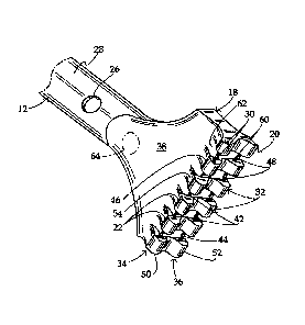
An ultrasonic surgical instrument 10, particularly utilizable in wound
debridement,
includes a shaft or shank 12 having a longitudinal axis 14 and a lumen or
channel 16 (FIGs.
4) coaxial with the axis. Instrument 10 further includes a probe head 18
disposed at a distal
end of shaft 12, the probe head having a distal end face 20. Probe head 18 has
a plurality of
operative surfaces 22 engageable with organic tissues for the application of
ultrasonic
vibratory energy to the tissues. End face 20 is oriented at least partially
transversely to
longitudinal axis 14. Lumen or channel 16 extends through probe head 18 and
has a first
opening or port 24 (FIG. 4) in probe head end face 20. Lumen or channel 16 has
at least one
second opening or port 26 preferably in a lateral surface 28 of shaft 12 at a
position spaced

7
from probe head 18 and particularly end face 20 thereof. Openings or ports 24
and 26 are
outlets when irrigation is applied via lumen or channel 16 and inlets in when
aspiration or
suction is applied.
As depicted in FIGS, 1 and 4, probe head 18 is wider than shaft 12 and is thus
laterally enlarged relative to shaft 12. Probe head 18 extends to opposite
sides of axis 14, and
is bisected by a longitudinal plane PI (FIG. 3) containing axis 14. Probe head
18 has a
plurality of teeth 30 and 32 extending laterally from end face 20. Teeth 30
and 32 are
disposed in respective rows or linear arrays 34 and 36 extending in parallel
to one another as
well as in parallel with plane Pl. Tooth arrays 34 and 36 are spaced from
plane P1 on
opposite sides thereof The teeth extend away from plane P1 generally
orthogonally thereto.
Probe head 18 is mirror symmetric about plane PI and may additionally be
mirror symmetric
about a second longitudinal plane P2 (FIG. 3) containing axis 14 and oriented
orthogonally or
perpendicularly to place Pl.
Probe head 18 is defined in part by a pair of opposed major lateral surfaces
38 and 40
extending from a distal end of shaft 12 to end face 20 and more particularly
to teeth 30 and
32. More particularly, major lateral surfaces 38 and 40 flare outwardly from
the distal end of
shaft 12 to end face 20 and extend at their distal ends to teeth 30 and 32 and
to a plurality of
edges 42 and 44 of end face 20 which are located between adjacent teeth 30 and
32 at the
bases or roots thereof
As shown in FIG. 2, end face 20 is disposed in a plane P3 that is slanted or
inclined at
an angle al relative to a transverse plane P4 that is perpendicular to axis
14. The different
teeth 30 of row or array 34, as well as the teeth 32 or row or array 36, are
disposed at
different distances dl, d2, dn from shaft 12. Tooth rows or arrays 34 and 36
are slanted or
inclined relative to any plane (like plane P4) that is perpendicular to axis
14.
CA 2938109 2020-01-21

8
It is to be noted that angle al may be zero, in which case teeth 30 are all
disposed at
the same distance from the distal end of shaft 12. In general, angle al may
take on any value
between 0 and about 30 degrees.
Operative surfaces 22 and 22' are located at free ends of teeth 30, 32 and are
spaced
laterally from axial plane Pl. Operative surfaces 22 and 22' are exemplarily
oriented parallel
to plane Pl. Operative surfaces 22, 22' are defined on a proximal side by
respective sharp
edges 46 and 48. Opposed major lateral surfaces 38 and 40 of probe head 18 are
contiguous
with and defined at their distal end by edges 46 and 48. Owing to the slanting
or inclination
of end face 20 (as plane P3) relative to shaft axis 14, edges 46 and 48 of
teeth 30 and 32 are
likewise linearly arranged.
In operation of the instrument, irrigation fluid such as a saline solution is
delivered to
tissues at a surgical site via channel 16, and openings or ports 24 and 26, as
indicated by
arrows 162, 164, and 166 when either teeth 30 or teeth 32, and more
particularly when either
operative surfaces 22 or 22', are placed into contact with a tissue surface at
a surgical site.
During subsequent ultrasonic vibration of the instrument 10, sharp edges 46 or
48 of teeth 30
or 32 are raked back and forth across the tissue surface, shaving tissue
layers off from the
operative site. The slanting of the teeth, relative to the direction of
ultrasonic reciprocation
(parallel to the axis of the instrument) results in a camming action that
pushes the shaved
tissue fragments towards the gaps (not separately designated) between the
teeth, defined by
inter-tooth edges 42 or 44, where the tissue fragments are subjected to
suction force or
aspiration via port or opening 24 in probe end face 20. Owing to the
symmetries of probe
head 18 and more specifically end face 20, opening 24 is located at the
geometrical center, on
axis 14, between the rows 34 and 36 of teeth 30 and 32.
Cutting edges 46 and 48 of teeth 30 and 32 are preferably, but not
necessarily, straight
edges. Teeth 30 and 32 may be alternatively or additionally formed with sharp
cutting edges
on their distal sides 50 and 52, opposite the hand piece (not shown) and probe
shaft 12.
CA 2938109 2020-01-21

CA 02938109 2016-07-27
WO 2015/119778
PCT/US2015/012413
9
Probe head 18 includes a plurality of concave ramp surfaces 54 and 56 each
partially
defining a respective tooth 30 or 32 and each extending from one of the
opposed lateral
surfaces 38 and 40 of probe head 18 to the sharp edge 46 or 48 on the proximal
side of the
respective tooth 30 or 32. Each tooth 30 and 32 may be formed with a
respective pair of
planar lateral surfaces 58 and 60, all disposed parallel to each other and
transversely to the
sharp tooth edges 46 and 48. Lateral tooth surfaces 58 and 60 extend in planes
P5 oriented
at a common acute angle a2 relative to axial plane P2 (which bisects end face
20).
Probe head 18 further includes a plurality of extension surfaces 62 which are
interleaved or alternating with concave tooth-defining surfaces 54 and 56 and
which are
contiguous with, or ending at, respective inter-tooth edges 42 and 44 of end
face 20.
In being provided with an ancillary irrigation and suction port 26 disposed in
shaft 12
or possibly lateral surface 38 and/or 40, instrument 10 is more likely to
enable continued
irrigation of a surgical site if the main irrigation port 24 is blocked or
occluded by severed
organic tissue.
It is to be noted that an opening or port 64 of channel 16 could be located in
a lateral
surface of probe head 18, rather than or in addition to port 26 in shaft 12.

Representative Drawing
A single figure which represents the drawing illustrating the invention.
Administrative Status

For a clearer understanding of the status of the application/patent presented on this page, the site Disclaimer , as well as the definitions for Patent , Administrative Status , Maintenance Fee  and Payment History  should be consulted.

Administrative Status

Title Date
Forecasted Issue Date 2023-03-21
(86) PCT Filing Date 2015-01-22
(87) PCT Publication Date 2015-08-13
(85) National Entry 2016-07-27
Examination Requested 2020-01-21
(45) Issued 2023-03-21

Abandonment History

There is no abandonment history.

Maintenance Fee

Last Payment of $210.51 was received on 2023-12-30


 Upcoming maintenance fee amounts

Description Date Amount
Next Payment if small entity fee 2025-01-22 $125.00
Next Payment if standard fee 2025-01-22 $347.00

Note : If the full payment has not been received on or before the date indicated, a further fee may be required which may be one of the following

  • the reinstatement fee;
  • the late payment fee; or
  • additional fee to reverse deemed expiry.

Patent fees are adjusted on the 1st of January every year. The amounts above are the current amounts if received by December 31 of the current year.
Please refer to the CIPO Patent Fees web page to see all current fee amounts.

Payment History

Fee Type Anniversary Year Due Date Amount Paid Paid Date
Application Fee $400.00 2016-07-27
Maintenance Fee - Application - New Act 2 2017-01-23 $100.00 2017-01-20
Maintenance Fee - Application - New Act 3 2018-01-22 $100.00 2018-01-19
Registration of a document - section 124 $100.00 2018-03-12
Maintenance Fee - Application - New Act 4 2019-01-22 $100.00 2019-01-22
Maintenance Fee - Application - New Act 5 2020-01-22 $200.00 2020-01-17
Request for Examination 2020-01-22 $800.00 2020-01-21
Maintenance Fee - Application - New Act 6 2021-01-22 $204.00 2021-01-11
Maintenance Fee - Application - New Act 7 2022-01-24 $203.59 2022-01-14
Final Fee 2023-01-09 $306.00 2023-01-04
Maintenance Fee - Application - New Act 8 2023-01-23 $210.51 2023-01-04
Maintenance Fee - Patent - New Act 9 2024-01-22 $210.51 2023-12-30
Owners on Record

Note: Records showing the ownership history in alphabetical order.

Current Owners on Record
MISONIX, INCORPORATED
Past Owners on Record
None
Past Owners that do not appear in the "Owners on Record" listing will appear in other documentation within the application.
Documents

To view selected files, please enter reCAPTCHA code :


To view images, click a link in the Document Description column. To download the documents, select one or more checkboxes in the first column and then click the "Download Selected in PDF format (Zip Archive)" or the "Download Selected as Single PDF" button.

List of published and non-published patent-specific documents on the CPD .

If you have any difficulty accessing content, you can call the Client Service Centre at 1-866-997-1936 or send them an e-mail at CIPO Client Service Centre.


Document
Description 
Date
(yyyy-mm-dd) 
Number of pages   Size of Image (KB) 
Maintenance Fee Payment 2020-01-17 1 33
Request for Examination / Amendment 2020-01-21 14 412
Description 2020-01-21 9 402
Claims 2020-01-21 4 106
Maintenance Fee Payment 2021-01-11 1 33
Examiner Requisition 2021-04-01 3 172
Amendment 2021-08-03 13 404
Claims 2021-08-03 8 262
Examiner Requisition 2021-11-15 4 210
Maintenance Fee Payment 2022-01-14 1 33
Amendment 2022-03-14 9 337
Amendment 2022-03-14 13 443
Claims 2022-03-15 6 247
Claims 2022-03-14 8 262
Maintenance Fee Payment 2023-01-04 1 33
Final Fee 2023-01-04 4 98
Representative Drawing 2023-02-28 1 12
Cover Page 2023-02-28 1 45
Electronic Grant Certificate 2023-03-21 1 2,526
Abstract 2016-07-27 1 53
Claims 2016-07-27 5 150
Drawings 2016-07-27 3 43
Description 2016-07-27 9 415
Cover Page 2016-08-16 1 31
Maintenance Fee Payment 2018-01-19 1 33
Maintenance Fee Payment 2019-01-22 1 33
Maintenance Fee Payment 2023-12-30 4 96
International Search Report 2016-07-27 1 51
National Entry Request 2016-07-27 2 68
Prosecution-Amendment 2016-07-27 6 176
Correspondence 2016-11-03 3 149
Correspondence 2016-12-01 3 110
Office Letter 2016-12-20 2 233
Office Letter 2016-12-20 2 237
Fees 2017-01-20 1 33