Note: Descriptions are shown in the official language in which they were submitted.
OPTOACOUSTIC IMAGE MAPPING OF TISSUE TEMPERATURE
Cross-Reference to Related Applications
This international application claims benefit of priority of provisional
application U.S.
Serial No. 61/934,529, filed January 31, 2014.
BACKGROUND OF THE INVENTION
Field of the Invention
The present invention generally relates to the fields of biomedical
optoacoustic imaging.
Particularly, the present invention relates to real-time imaging systems that
visualize maps of
temperature in a human or animal body noninvasively and provide independent
images of
tissue anatomy co-registered with images of temperature variations.
Description of the Related Art
Many in the filed of biomedical science have recognized that accurate
noninvasive
temperature mapping in vivo in the depth of human (animal) body could lead to
ground
breaking advances in the thermal therapy and cryotherapy. Hence, in the past
few decades,
significant efforts have been made to create a device that could achieve this
goal.
Optoacoustic imaging and sensing represent a novel biomedical monitoring
technologies with contrast based on optical absorption in tissues. Previously,
sensing of
optoacoustic signals has been proposed for monitoring tissue properties and
temperature. It
is known that the magnitude of optoacoustic response is sensitive to the local
temperature.
The phenomenon is attributed to temperature dependent behavior of
thermodynamic and
mechanical properties, which comprise thermoacoustic efficiency of the tissue,
also known as
Gruneisen parameter. The presence of temperature dependent optoacoustic
response
(ThOR) measured as signals generated by laser pulses in biological tissues
provided the
foundation for non-invasive temperature monitoring. However, currently, when
considering in
vivo applications of optoacoustic sensing, sample-to-sample and spatial
variations of
Gruneisen parameter for different tissues remains as the major issue. In other
words, under
currently technology of optoacoustic imaging, each calibration method only
remains valid if
the temperature measurement is conducted in the same particular tissue.
Therefore, when a
population of live biological subjects was studied with prior optoacoustic-
based methods, it
becomes obvious that the measured temperature accuracy is far from ideal.
Furthermore, current optoacoustic imaging technology only provides the
temperature information. To obtain more comprehensive information of a
patient, which could
1
Date Revue/Date Received 2021-09-17
allow a medical professional to identify the exact temperature in a particular
anatomical
locations of interest, a combined image of anatomical structures with
corresponding
temperatures are highly desirable. It would substantially increase the
efficiency of thermal
(and cryo) therapy by directly monitoring the treatment of abnormal human
tissues and
ensuring the safety for surrounding normal tissues. So far, there is no
technology that could
achieve such an objective.
Thus, there is a recognized need in the art for improved devices and methods
for
accurate noninvasive temperature mapping, and preferably providing images of
anatomical
structures co-registered with corresponding temperatures. Particularly, the
prior art is deficient
in these aspects. The present invention fulfills this longstanding need and
desire in the art.
SUMMARY OF THE INVENTION
The present invention is directed to an imaging system for visualization and
accurate
mapping of temperature in absolute values in the region of interest of live
human or animal
tissue independently on spatial distribution of the optical fluence in the
body and independently
on spatial distribution of the tissue optical properties. The imaging system
comprises an
optoacoustic imaging module that uses pulsed optical illumination at preferred
wavelength
around 800 nm or around 1300 nm, an image processing and calibration module
connected
to the optoacoustic imaging module and an operating and controlling module
electronically
connected with said image processing module and configured to control and
manipulate all
components of the imaging system. The present invention is directed to another
imaging
system further comprising an ultrasound imaging module having an ultrasonic
probe
communicably connected to an electronics system that also serves as a probe
and to an
electronics system for the optoacoustic imaging module.
The present invention also is directed to an imaging system for monitoring and
guiding
thermal therapy procedures within a human or animal tissue. The system
comprises the
imaging system for visualization and accurate mapping of temperature in
absolute values as
described herein, a therapeutic module configured to apply thermal treatment
to a subject and
an operating controlling module connected with said processing module and
configured to
manipulate at least one of said therapeutic module, ultrasound imaging module
or
optoacoustic imaging module.
The present invention is directed further to a user-implemented method for
calibrating a
temperature-structure imaging system. The method comprises the steps of (a)
illuminating a
tissue with the laser pulses of the optoacoustic imaging module and acquiring
optoacoustic
signals from the illuminated tissue to generate a first optoacoustic image at
human
2
Date Revue/Date Received 2021-09-17
physiological temperature; (b) applying an automatic self-focusing algorithm
in the image
processing module to determine the temperature dependent speed of sound in a
region of
interest of a patient's body and obtain the optimal resolution for the first
optoacoustic image
and (c) turning on the temperature cooling source and allow time for the
temperature of region
of interest to change and create gradient of the spatial distribution of
temperature, T(r). Step
(d) applies step (a) at a changed temperature and a second optoacoustic image
is acquired.
Step (e) applies step (b) and optimizes resolution of the second optoacoustic
Image to achieve
matching between localization of tissue structures in the first image and the
second
optoacoustic image. Step f) normalizes the second optoacoustic image to the
first
optoacoustic image by dividing every pixel of the second optoacoustic image
intensity to that
of corresponding pixel of the first optoacoustic image, and thereby produce a
normalized
image of the optoacoustic image intensity ratio proportional to temperature
ratio. In step (g)
temperature is measured with thermocouples placed in the region of interest
along
temperature gradient to calibrate the map generated in step (g) in absolute
temperature value.
In Step (h) steps (d) through (g) are repeated to acquire a sequence of
optoacoustic images
and display of temperature distribution maps, which undergoes changes in the
course of
calibration procedure and, in step (i), calibration curve data is recorded
from images of spatial
distribution of the temperature in the calibration tissues or phantoms that
resemble properties
of the region of interest in the human body;
The present invention is also directed to a method for mapping the temperature
of a
tissue in the course of thermal therapy procedure. The method comprises in
step (a)
illuminating a tissue inside a region of interest of a subject using laser
pulses of the
optoacoustic imaging module as described herein at a wavelength within
preferred spectral
range and safe optical fluence and in step (b) measuring an optoacoustic
response of the
tissue by using the ultrasonic probe. In step (c) constructing a first
optoacoustic image at a
physiological temperature inside said subject. In step (d) an automatic self-
focusing algorithm
is applied for the first optoacoustic image to determine the temperature
dependent speed of
sound in the region of interest of a subject and achieve an optimal resolution
for the first
optoacoustic image. In step (e) a spatial distribution for temperature in the
subject is created
by performing thermal therapy on the subject. In step (f) the tissue is
illuminated in the same
region of interest at the second temperature point, in the same position of
the subject, using
laser pulses at the same preferred laser wavelength and the same optical
fluence and in step
(g) a second optoacoustic image at the second temperature is constructed. In
step (h) the
automatic self-focusing algorithm is applied for the second optoacoustic image
to determine
the temperature dependent speed of sound in the region of interest of a
subject and achieve
an optimal resolution for the second optoacoustic image at the second
temperature. In step
3
Date Revue/Date Received 2021-09-17
(i) a normalized image of the optoacoustic image intensity ratio is generated
by dividing every
pixel value of the second optoacoustic image to corresponding pixel value on
the first
optoacoustic image and in step (j) calibrating the normalized optoacoustic
image is calibrated
using the calibration curve described herein. In step (k) a map of temperature
distribution on
the tissues inside the region of interest of the subject is produced. In step
(I) steps f) to step
k) are repeated for generating a map of absolute temperature distribution in
real time and in
step (m) the map of the temperature distribution inside the region of interest
of the subject
isused to guide the thermal therapy procedure.
In another aspect, there is provided an imaging system for visualization and
accurate
mapping of temperature distribution in absolute values in a region of interest
and anatomical
structures of live human or animal tissue independently on spatial
distribution of the optical fluence
in the body and independently on spatial distribution of the tissue optical
properties, comprising: an
optoacoustic imaging module that uses pulsed optical illumination at a
preferred wavelength around
800 nm or around 1300 nm; an ultrasound imaging module having an ultrasonic
probe
communicably connected to an electronics system that also serves as a probe
and to an electronics
system for the optoacoustic imaging module and is configured to emit and to
detect ultrasonic waves
in an ultrasound imaging mode and to detect optoacoustic signals of thermal
conditions dependent
optoacoustic response of tissue in an optoacoustic imaging mode; an image
processing and
calibration module connected to the optoacoustic imaging module and to the
ultrasound imaging
module and configured to generate an image co-registered from an image
generated of the
temperature distribution and an image generated of the anatomical structures
of the live human or
animal tissue; and an image display module programmed to display either image
of anatomical
structure or temperature or both; a therapeutic module configured to apply a
cryoablation treatment
to the live human or animal tissue; and an operating and controlling module
electronically connected
with said image processing module and configured to control and manipulate at
least one of the
modules of the imaging system.
BRIEF DESCRIPTIONS OF THE DRAWINGS
So that the matter in which the above-recited features, advantages and objects
of the
invention, as well as others that will become clear, are attained and can be
understood in detail,
more particular descriptions of the invention briefly summarized above may be
had by reference to
certain embodiments thereof that are illustrated in the appended drawings.
These drawings form a
part of the specification. It is to be noted, however, that the appended
drawings illustrate preferred
embodiments of the invention and therefore are not to be considered limiting
in their scope.
FIG. 1 demonstrates how optoacoustic signals and images change in the process
of
4
Date Recue/Date Received 2022-06-29
temperature decreasing from physiological temperature to the temperature zone
where the
optoacoustic response is zero in the blood of a subject.
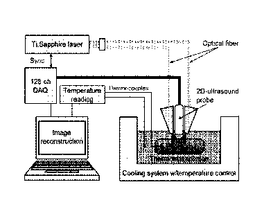
FIG. 2 demonstrates experimental block diagram of the calibration system of
the present
invention as applied to laboratory calibration procedure using phantoms.
FIGS. 3A-3D illustrate the temperature dependence of optoacoustic image
intensity in
a region of interest for aqueous solution of CuSO4=5H20 and calculated
Gruneisen parameter
for water after normalization at 37 C (FIG. 3A); the concentration dependence
of 1 C, at which
thermal condition dependent optoacoustic response of tissue is equal to zero
(FIG. 3B);
temperature dependence of relative density for aqueous solution of CuSO4.5H20
(240 mM)
with the second order polynomial regression (FIG. 3C); temperature of maximum
relative
density as a function of CuSO4=5H20 concentration (FIG. 3D).
FIGS. 4A-4B demonstrate results from image data matching experiments conducted
using two tubes filled with the same solution but placed at different
distances between the light
emitting fiber bundles and the ultrasonic probe. FIG. 4A illustrates
temperature dependence
of optoacoustic image intensity in a region of interest for the two tubes.
FIG. 4B demonstrates
that optoacoustic image intensity of every pixel normalized to that at 37 C
leads to complete
match of data for both tubes. Time interval between image recordings was about
30 seconds.
The total duration of the cooling procedure was about 180 minutes.
FIGS. 5A-5B depict optoacoustic image intensity as a function of temperature
measured
from the nickel sulfate and cupric sulfate solutions with the same molar
concentrations (FIG.
5A); optoacoustic image intensity data measured at gradually changing
temperature
normalized to the OA image of each pixel intensity measured at 37 C (FIG. 5B).
FIGS. 6A-6B show temperature dependence of the optoacoustic image intensity
normalized at 20 C for nickel sulfate solution at different concentrations
with water as an
acoustically coupling liquid (FIG. 6A); temperature of zero thermal conditions
dependent
optoacoustic response of tissue as a function of solution concentration
measured for NiSO4
solution in different optoacoustic coupling media and its linear fit. This
graph demonstrates
that the temperature of zero thermal conditions dependent optoacoustic
response of tissue is
independent on optoacoustic coupling media (FIG. 6B).
FIG. 7 illustrates that optoacoustic imaging intensity is a nonlinear function
of
temperature, but it may be approximated with a linear function with sufficient
accuracy. The
range of temperature monitoring is mathematically determined with the value of
maximum
nonlinear temperature deviation ATmax.
FIGS. 8A-8C illustrate that volumetric fraction of erythrocytes (hematocrit,
Ht)
significantly varies through the entire vascular network, decreasing from
systemic blood
vessels down to capillaries (FIG. 8A); experiments with whole and diluted
blood demonstrating
5
Date Revue/Date Received 2021-09-17
that the optoacoustic temperature dependent response (ThOR) is scaled
proportionally to
hematocrit (FIG. 8B); when normalized at 37 C, the thermal conditions
dependent
optoacoustic response of tissue becomes invariant as the curves representing
whole and
diluted blood coincide (FIG.8C).
FIG. 9A-9B show optoacoustic image intensity normalized at 37 C as a function
of
temperature for three different concentrations of hemoglobin. Dash dotted line
marks zero
optoacoustic response (FIG. 9A) and temperature To of zero thermal conditions
dependent
optoacoustic response of tissue as a function of hemoglobin concentration
(FIG. 9B).
FIG. 10A-10B depict ThOR in porcine blood samples collected from eight animals
(4
males and 4 females). At least three tubes positioned at different distances
from the probe
were filled with each blood sample. Measured optoacoustic response data were
averaged
over different tubes (FIG. 10A) and the thermal conditions dependent
optoacoustic response
of tissue of blood in scattering medium in comparison to that in transparent
medium.
optoacoustic image intensity normalized at 27 C (FIG. 10B).
FIG. 11A-11B show zoomed in temperature-dependent optoacoustic response of
blood
in scattering medium comparing to averaged ThOR function in transparent
surrounding. OA
image intensity normalized at 27 C. Accuracy of an individual temperature
reading in milk
surrounding is 1.5 C (FIG. 11A); and the temperature monitoring function T of
normalized
optoacoustic image intensity at 37 C for whole pigs blood is presented after
median filtration
and characterized by polynomial fit of second order (FIG. 11B).
FIGS. 12A-12D show photographs of tissue-mimicking optoacoustic phantom
(optically
scattering PVCP background and seven tubes LJ0.635 mm filled with live blood)
preheated to
36.5 C (left upper) (FIG. 12A). FIGS. 12B-12D show 3 sample frames of a movie
recorded
with video rate. The frames present temperature images of tube cross-sections
(circles)
changing their intensity converted into color from red (FIG. 12B) to yellow
(FIG. 12C) to blue
(FIG. 12D) depending on gradually decreasing local temperature ( C) mapped
using the
method of the present invention. A square on images represent a tube filled
with cold water at
-11 C (refrigerated NaCI solution was circulated in the tube).
FIG. 13 depicts a clinical cryoablation procedure with optoacoustic
temperature
monitoring. Ultrasound imaging shows anatomy of region of interest and allows
precise
insertion of cryoablation needles. Transrectal ultrasonic probe is designed to
include fiberoptic
bundles for optical illumination with NIR laser pulses. Deep penetration of
NIR light at
preferred wavelengths through the scattering medium allows non-invasive
temperature
monitoring with clinical significance.
FIGS. 14A-14D depict temperature monitoring during clinical image-guided
cryoablation
of prostate cancer. FIG. 14A shows ultrasound image of the prostate with
inserted 4 cryo-
6
Date Revue/Date Received 2021-09-17
needles. Arrows point to the locations of cryogenic needles, the small arrow
shows urethra,
which is being kept warm with a warm liquid. The arc at the bottom indicates
position of the
rectal wall. FIG. 14B shows coalesced ice-balls created around the cryo-
needles and visible
at bottom as a crescent-shaped line. Sharp change of the normalized
optoacoustic image
intensity also permits tracking of the ice-ball boundary with real-time
optoacoustic image as
shown in FIG. 14C. FIG. 14D shows a contour map of isotherms revealing
distribution of
temperature generated with a system of the present invention.
DETAILED DESCRIPTION OF THE INVENTION
As used herein, the following terms and phrases shall have the meanings set
forth
below. Unless defined otherwise, all technical and scientific terms used
herein have the same
meaning as commonly understood to one of ordinary skill in the art.
As used herein, the term, "a" or "an" may mean one or more. As used herein in
the claim(s),
when used in conjunction with the word "comprising", the words "a" or "an" may
mean one or more
than one. As used herein "another" or "other" may mean at least a second or
more of the same or
different claim element or components thereof. The terms "comprise" and
"comprising" are used in
the inclusive, open sense, meaning that additional elements may be included.
As used herein, the term "or" in the claims refers to "and/or" unless
explicitly indicated to refer
to alternatives only or the alternatives are mutually exclusive, although the
disclosure supports a
definition that refers to only alternatives and "and/or".
As used herein, the term "about" refers to a numeric value, including, for
example, whole
numbers, fractions, and percentages, whether or not explicitly indicated. The
term "about" generally
refers to a range of numerical values (e.g., +1- 5-10% of the recited value)
that one of ordinary skill in
the art would consider equivalent to the recited value (e.g., having the same
function or result). In
some instances, the term "about" may include numerical values that are rounded
to the nearest
significant figure.
As used herein, the term "computer" or "computer system" refer to one or more
machines that comprise at least a memory, a processor, a display, one or more
interfaces and
at least one wired and/or wireless network connection. A computer may be a
desktop or
laptop machine or other electronic media, for example, a smartphone or tablet,
as are standard
and currently known in the art. As such computer may comprise a user input
device such as
a keyboard, keypad, touch screen, mouse, trackball, camera, microphone, and/or
other like
user input device. Without being limiting, any software, modules,
applications, add-ons, plug-
ins, programs and/or databases, etc. and associated instructions and/or
functions necessary
for implementation of any imaging system or dual modality imaging system or
subsystems or
7
Date Revue/Date Received 2021-09-17
means comprising the same may be programmed into the computer, may be
retrieved over
the network connection or may be retrieved from a non-transitory machine-
readable media,
such as computer readable media or storage device tangibly storing the same,
may be
tangibly stored in computer memory or other electronic media memory and are
executable by
the processor comprising the computer.
As used herein, the term "subject" refers to an animal or human, particularly
a patient.
As used herein, the term "ThOR" refers to Thermal conditions dependent
Optoacoustic
Response of tissue, i.e. optically induced temperature dependent pressure wave
propagating
as ultrasound.
As used herein, the term "ROI" refers to a region of interest within
biological tissue in
which temperature distribution is being monitored
As used herein, the term "Preferred Wavelength" refers to the laser
illumination
wavelength at which the optical absorption coefficient of the dominating
tissue chromophore
is constant and independent on changing tissue properties. For hemoglobin of
blood as the
dominating tissue chromophore the preferred wavelength is selected at which
the optical
absorption is independent on blood oxygenation and temperature. For cases of
water being
the dominating tissue chromophore, the optical absorption coefficient must be
stronger than
that of other tissue constituents and independent on temperature. A contrast
agent can be
used as a dominating exogenous chromophore.
As used herein, the term Dominating Chromophore refers to a molecule or
substance
with such a strong optical absorption, so that optical absorption of all other
tissue
chromophores can be neglected
As used herein, the term "Ultrasonic Probe" refers to an array of ultrasonic
transducers
capable of properly detecting optoacoustic signals
As used herein, the term "SoS" refers to the speed of sound
As used herein, the term "Optoacoustic Image" refers to the image that
displays pixel
intensity value as the product of Gruneisen parameter, optical absorption
coefficient and
optical fluence.
As used herein, the term "Normalized Optoacoustic Image" refers to the image
that
displays ratio of pixel intensity at unknown temperature to the pixel
intensity at a well-known
temperature. This ratio image is independent on the distributions of the
optical absorption and
the optical fluence, and thus, can be calibrated in values (units) of
temperature.
As used herein, the term "PVCP" refers to the poly(vinyl chloride) plastisol,
a tissue
phantom material.
In one embodiment of the present invention, there is provided an imaging
system for
visualization and accurate mapping of temperature in absolute values in the
region of interest
8
Date Revue/Date Received 2021-09-17
of live human or animal tissue independently on spatial distribution of the
optical fluence in
the body and independently on spatial distribution of the tissue optical
properties, comprising
an optoacoustic imaging module that uses pulsed optical illumination at
preferred wavelength
around 800 nm or around 1300 nm; an image processing and calibration module
connected
to the optoacoustic imaging module; and an operating and controlling module
electronically
connected with said image processing module and configured to control and
manipulate all
components of the imaging system.
Further to this embodiment the imaging system
comprises an ultrasound imaging module having an ultrasonic probe communicably
connected to an electronics system that also serves as a probe and to an
electronics system
for the optoacoustic imaging module.
In another embodiment of the present invention, there is provided an imaging
system
for visualization of tissue anatomical structures and mapping of temperature
distribution within
a region of interest in human or animal tissue, comprising the optoacoustic
imaging and
temperature mapping system as described supra; an ultrasound imaging module
for imaging
tissue anatomical structures; an image processing module connected to both
ultrasound and
optoacoustic imaging module; and an image display module programmed to display
either
image of anatomical structure or temperature or both. In this embodiment, the
system is
configured to generate two types of images, temperature and anatomical
structure,
coregistered in space and time for the same tissues in a patient's body.
In this embodiment the optoacoustic imaging module may integrate a pulsed
laser
connected with an imaging module through a light delivery subsystem configured
to deliver
the laser pulses to the region of interest. Also in this embodiment the system
may be
configured to generate two types of images that are temperature and anatomical
structure
images which are coregistered in space and time for the same tissues in a
patient's body.
In yet another embodiment of the present invention, there is provided an
imaging system
for monitoring and guiding thermal therapy procedures within a human or animal
tissue,
comprising the imaging system for visualization of tissue anatomical
structures and mapping
of temperature distribution within a region of interest in human or animal
tissue as described
supra; a therapeutic module configured to apply thermal treatment to a
subject; and an
operating controlling module connected with said processing module and
configured to
manipulate at least one of the therapeutic module, ultrasound imaging module
or optoacoustic
imaging module.
In this embodiment the processing module may comprise a calculation module
configured to calculate the location and temperature within specific
anatomical tissue
structures based on the information received in the processing module; an
image constructing
module that generate images based on the calculation from the calculation
module and the
9
Date Revue/Date Received 2021-09-17
signals received in the processing module; and an user interface communicably
connected to
said calculation module, said image constructing module. Particularly, the
operating and
controlling module is configured to manipulate at least one of the therapeutic
module, the
ultrasound imaging module, the optoacoustic imaging module, or the image
processing
module.
In another embodiment of the present invention, there is provided a (a)
illuminating a
tissue with the laser pulses of the optoacoustic imaging module and acquiring
optoacoustic
signals from the illuminated tissue to generate a first optoacoustic image at
human
physiological temperature; (b) applying an automatic self-focusing algorithm
in the image
processing module to determine the temperature dependent speed of sound in a
region of
interest of a patient's body and obtain the optimal resolution for the first
optoacoustic image;
(c) turning on the temperature cooling source and allow time for the
temperature of ROI to
change and create gradient of the spatial distribution of temperature, T(r);
(d) applying step
(a) at a changed temperature and acquiring a second optoacoustic image; (e)
applying step
(b) and optimizing resolution of the second OA Image to achieve matching
between
localization of tissue structures in the first image and the second
optoacoustic image; (f)
normalizing the second optoacoustic image to the first optoacoustic image by
dividing every
pixel of the second optoacoustic image intensity to that of corresponding
pixel of the first
optoacoustic image, and thereby produce a normalized image of the optoacoustic
image
intensity ratio proportional to temperature ratio; (g) measuring temperature
with
thermocouples placed in the region of interest along temperature gradient to
calibrate the map
generated in step (g) in absolute temperature value; (h) repeating steps d)
through g) to
acquire a sequence of optoacoustic images and display of temperature
distribution maps,
which undergoes changes in the course of calibration procedure; and (i)
recording a calibration
curve data from images of spatial distribution of the temperature in the
calibration tissues or
phantoms that resemble properties of the region of interest in the human body.
In this embodiment in tissue with greatly varying speed of sound, the method
may
comprise replacing step 9b with speed of sound tomography to generate the map
of speed of
sound in the region of interest and then to generate the most accurate high
resolution
optoacoustic image. Also in this embodiment accuracy of absolute calibration
of temperature
may be increased by expanding the range of temperatures to include two
characteristic points
of well-known temperature, such as (i) temperature at which Gruneisen
parameter becomes
zero at 4 C for water and at -12 C for blood and the optoacoustic image
disappears and (ii)
the physiological temperature of a human body about 36.5 C.
In yet another embodiment of the present invention there is provided a method
for
mapping the temperature of a tissue in the course of a thermal therapy
procedure, comprising
Date Revue/Date Received 2021-09-17
the steps of (a) illuminating a tissue inside a region of interest of a
subject using laser pulses
of the optoacoustic imaging module, at a wavelength within preferred spectral
range and safe
optical fluence; (b) measuring an optoacoustic response of the tissue by using
the ultrasonic
probe; (c) constructing a first optoacoustic image at a physiological
temperature inside the
subject; (d) applying an automatic self-focusing algorithm for the first
optoacoustic image to
determine the temperature dependent speed of sound in the region of interest
of a subject
and achieve an optimal resolution for the first optoacoustic image; (e)
creating a spatial
distribution for temperature in the subject by performing thermal therapy on
said subject; (f)
illuminating the tissue in the same region of interest at the second
temperature point, in the
same position of the subject, using laser pulses at the same preferred laser
wavelength and
the same optical fluence; (g) constructing a second optoacoustic image at the
second
temperature; (h) applying the automatic self-focusing algorithm for the second
optoacoustic
image to determine the temperature dependent speed of sound in the region of
interest of a
subject and achieve an optimal resolution for the second optoacoustic image at
the second
temperature; (i) generating a normalized image of the optoacoustic image
intensity ratio by
dividing every pixel value of the second optoacoustic image to corresponding
pixel value on
the first optoacoustic image; (j) calibrating the normalized optoacoustic
image using a
calibration curve; (k) producing a map of temperature distribution on the
tissues inside the
region of interest of the subject; (I) repeating step f) to step k) generating
a map of absolute
temperature distribution in real time; (m) using the map of the temperature
distribution inside
the region of interest of the subject to guide the thermal therapy procedure.
In this embodiment the system may generate coregistered overlaid ultrasound
and
temperature images, displays the temperature map within anatomical tissue
structures in the
region of interest and uses real time overlaid images to guide thermal therapy
procedure. Also,
in this embodiment the absolute measurement of temperature may be conducted
within a
temperature range that includes two characteristic temperatures, one of which
is physiological
temperature of about 36.6 C and the second is the protein denaturation
temperature of about
52 C. In addition, blood may be the dominating tissue chromophore and the
preferred spectral
range of laser wavelengths is about 795 nm to about 805 nm and, as such, the
absolute
measurement of temperature is conducted within a temperature range that
includes two
characteristic temperatures, one of which is physiological temperature of
about 36.6 C and
the second is the temperature about -10 C at which blood reaches its maximum
density and
optoacoustic image intensity flips its polarity. Furthermore, water may be the
dominating
tissue chromophore and the preferred spectral range of laser wavelengths is
from about 1300
nm to about 1305 nm and, as such, the absolute measurement of temperature may
be
conducted within a temperature range that includes two characteristic
temperatures, one of
11
Date Revue/Date Received 2021-09-17
which is physiological temperature of about 36.6 C and the second is the
temperature about
4 C at which water reaches its maximum density and optoacoustic image
intensity flips its
polarity.
In this embodiment imaging system may be configured to generate real-time two-
dimensional and three-dimensional images of tissues in a patient's body.
Particularly, three-
dimensional images may be generated by assembling two-dimensional slices
though the
depth of tissues, said two-dimensional slices are obtained by scanning a hand-
held ultrasound
probe on the surface of an area of a patient's body. Also in this embodiment
the method may
provide guidance for cryotherapy based on the phenomenon of change of sign of
the
optoacoustic signal from positive to negative when temperature in the
specified region of
interest reaches and surpasses the point of maximum density and zero thermal
expansion.
The following examples are included to demonstrate preferred embodiments of
the
invention. It should be appreciated by those of skill in the art that the
techniques disclosed in
the examples which follow represent techniques discovered by the inventor to
function well in
the practice of the invention, and thus can be considered to constitute
preferred modes for its
practice. However, those of skill in the art should, in light of the present
disclosure, appreciate
that many changes can be made in the specific embodiments which are disclosed
and still
obtain a like or similar result without departing from the spirit and scope of
the invention.
EXAMPLE 1
Measurements of Temperature Dependence of Gruneisen Parameter
Optoacoustic (OA) thermography is a promising method for temperature
monitoring in
hypo- and hyperthermal medical treatment. A basic phenomenon associated with
the invented
method of temperature measurements is presented in FIG. 1. This method is
based on high
temperature sensitivity of the Gaineisen parameter. OA signal amplitude VOA
can be
expressed as: VOA Li rpaF, where r is thermoelastic efficiency or Gaineisen
parameter of
light-absorbing material, pa is optical absorption coefficient, F is local
optical fluence.
Gaineisen parameter incorporates three components6: volumetric thermal
expansion (13),
speed of sound for longitudinal waves (V), and specific (per mass) heat
capacity at constant
pressure (Cr): r = 13V2/C.
The method is validated using optically absorbing aqueous solutions of nickel
and cupric
sulfate. Two-dimensional optoacoustic imaging is employed to improve
sensitivity and
precision by measurements with high signal-to-noise ratio (SNR). The
experimental setup is
designed for simultaneous studies of multiple samples, which allowed
confinement of
systematic errors caused by spatial fluctuations of laser fluence and
distortions of propagating
12
Date Revue/Date Received 2021-09-17
optoacoustic waves. Optical absorbance of the studied solutions has negligible
dependence
on temperature. The studied aqueous solutions have thermodynamic properties
and
Gruneisen parameter, which are different from pure water and are dependent on
concentration. The method provides data insensitive to spatial variations of
laser fluence and
optical absorbance. Therefore, temperature-dependent changes of the Gaineisen
parameter
can be reliably evaluated by this method. The proposed methodology by
estimating
temperature dependence of Gaineisen parameter for different concentrations of
hexahydrate
nickel sulfate in the range of temperatures from 4 to 40 C is validated. This
range is important
in future applications of optoacoustic imaging for non-invasive monitoring of
tissue
hypothermia.
Image intensity of aqueous solution samples was gradually decreasing following
the
local temperature trend, and eventually became indistinguishable from
background. Further
cooling resulted in re-appearance and growth of the optoacoustic response from
the sample,
but now registered as the image with opposite (negative) polarity. FIG. 1
provides the first
direct observation of the change in polarity of optoacoustic image. Such a
positive-negative
transition of optoacoustic intensity is expected from aqueous compounds due to
nil volumetric
thermal expansion coefficient achieved at the extremum of the compound's
density. For
example, such an effect is predicted for water with maximum density at 3.98 C.
It is provided
below an example showing direct experimental evidence that optoacoustic
response
completely disappears at the temperature of maximum density.
EXAMPLE 2
Experimental Calibration System
The laboratory calibration procedure is conducted using phantoms. The system
comprises the following components: Ti:Sapphire pulsed laser that emits in the
preferred
range of wavelengths 800-805 nm for a live biological tissue containing blood.
This laser also
has preferred pulse duration of 5 to 10 ns for maximum efficiency of thermal
conditions
dependent optoacoustic response of tissue generation in tissue. Optical fiber
bundles are
used for light delivery to tissue phantom, however other means to deliver
light also can be
used. Ultrasound probe is used for two purposes: it emits and detects
ultrasonic waves in the
ultrasound imaging mode, and it also detects optoacoustic signals of thermal
conditions
dependent optoacoustic response of tissue. The probe is connected to a
multichannel
electronic system, which amplifies, records and processes signals and
transmits the
processed signals to a computer, which reconstructs images and also controls
the whole
system. The electronic system can also be used for at least partial image
reconstruction.
Thermocouples provide absolute temperature readings for calibration purposes.
A thermostat
13
Date Revue/Date Received 2021-09-17
system provides conditions similar to thermal therapy.
In this exmperiment, Ti-Sapphire output of the laser unit was tuned to 800 nm
and
produced 6 ns, 16 mJ per pulse laser radiation with pulse repetition rate of
10 Hz. Two optical
fiber bundles delivered light to the samples. Rectangular output apertures
were 1.5 mm x 50
mm each and produced laser fluence about 2 mJ/cm2 at 20 mm depth. The probe
and
fiberoptic outputs were hermetically sealed to enable operation in liquid
environment.
A chest freezer was employed for cooling of a thermostat tank. The temperature
was
measured and logged by digital thermometer with precision of 0.1 C. The 1.5 L
tank was filled
with coupling solution and was subject to 0.2 C/min cooling rate. The cooling
from 40 to 4 C
took about 3 hours. Simultaneously laser power was registered by pyroelectric
energy meter
to record potential laser fluence fluctuations caused by long time operation.
EXAMPLE 3
The Temperature for the Maximum Density of a Sample Solution (To)
In this set of experiments, a cupric sulfate model is used to elucidate
physical meaning
of the parameter To in temperature dependent optoacoustic response. Normalized
optoacoustic intensity and density of aqueous cupric sulfate solutions were
measured as a
function of temperature. The cupric sulfate was preferred over hemoglobin,
since it produces
larger variation of To for the set of achievable concentrations. To eliminate
possible effects of
the acoustic coupling medium, experiments were performed using distilled water
and sodium
chloride (23wt /0). FIG. 3A shows normalized optoacoustic intensity for two
concentrations of
cupric sulfate and calculated normalized Gruneisen parameter of water as a
control. The
Gruneisen parameter of water was calculated with 1 C intervals using
temperature
dependences of speed of sound, specific heat capacity, and thermal expansion
coefficient.
FIG. 3B shows To directly measured as a temperature at zero optoacoustic
intensity and its
linear regression as a function of concentration. Data matches previous
results obtained by
extrapolation of the fitted data. The measurements were not affected by using
different
surrounding media, implying that the entire optoacoustic stress generation
happens inside the
tubes with sample solutions. FIG. 3C shows two temperature dependent curves of
density.
The lower one demonstrates known relationship for water. The top one measured
relationship
for 240 mM cupric sulfate. Arrows indicate maxima of the fitted parabolic
functions. Consistent
with the Despretz's law, maximum density for cupric sulfate is shifted towards
more negative
temperatures. FIG. 3D summarizes measured temperatures of the maximum density
for
different concentrations of cupric sulfate. When fitted with a linear
regression model, the
resultant equation effectively matches the one obtained for To measured via
normalized
optoacoustic imaging. The equivalence of two relationships allows one skilled
in the art to
14
Date Revue/Date Received 2021-09-17
conclude that To represents the temperature of maximum density of a sample
solution, which
is manifested by the absence of thermal expansion, and therefore optoacoustic
response.
Note, that the data fits in the FIGS. 3B and 3D intercept the ordinate axis at
about 4 C, which
corresponds to the temperature of maximum density of the pure solvent, i.e.
distilled water.
EXAMPLE 4
One-valued normalization of temperature-dependent optoacoustic response (ThOR)
Independence of the method on laser fluence
This experiment demonstrats one-valued normalization of temperature-dependent
optoacoustic response (ThOR) at certain temperature caused by existence of
zero
optoacoustic signal in aqueous solutions. It provides independence of the
method on laser
fluence.
Median intensity of the optoacoustic image was measured in each pixel of
region of
interest as a function of temperature for multiple consecutive frames. To
evaluate spatial
confinement of the laser fluence, samples with the same salt solutions located
at different
distances (Z) from the light illuminators and US probe are visualized. Tubes
filled with 240
mM cupric sulfate solution were placed at the distances of 15 and 25 mm from
the probe. Due
to optical scattering and laser beam divergence, the laser fluence decreased
with depth
resulting in reduced optoacoustic intensity for the lower tube (FIG. 4A).
Temperature
dependences of both samples exhibited linear trend in the temperature range
from 4 to 40 C.
When normalized optoacoustic image intensity values to those measured at
physiologically
relevant 37 C, perfectly coinciding straight lines were obtained (FIG 4B). The
fluctuation of
laser energy in the course of the experiment was about 4%, but the averaged
results of
multiple laser pulses, which render the measurement accurate.
Independence of the method on optical absorption coefficient
This set of experiments explore one-valued normalization of temperature-
dependent
optoacoustic response (ThOR) at certain temperature caused by existence of
zero OA signal
in aqueous solutions, which provides independence of the method the optical
absorption
coefficient. Direct comparison of temperature functions for OA response from
samples with
different optical absorbance and equal or almost equal thermodynamic
parameters was
challenging. Variation of optical absorbance due to salt concentration was
unacceptable as it
could change thermodynamic properties of the solution as well. Therefore, two
different salts
¨ cupric sulfate and nickel sulfate were used. With the same concentrations,
the
thermodynamic characteristics of the two solutions are expected to be very
similar. These
Date Revue/Date Received 2021-09-17
compounds have the same anionic group and their cations are close by weight
and radius.
This is the reason why expected similar thermodynamic behavior of these
solutions are
expected. At the same molar concentration aqueous solution of cupric and
nickel sulfates
have one order difference in optical absorption at the wavelength of 800 nm.
There are
¨800nm
= 10.57 0.13 M-1cm-1 in CuS0405H20 and
¨800nm = 0.95 0.04 M-1cm-1 in NiS0406H20. The
ratio of intensities of OA images for nickel and cupric solutions placed at
the same distance
from the probe was proportional to the difference in optical absorbance (FIG.
5A). After
normalization of OA image intensity to that measured at 37 C, curves in FIG.
5B coincide with
each other. Note, that the sample of lower absorbance revealed higher
sensitivity to laser
energy fluctuations. Experimental evidence of FIGS. 4A-4B and FIGS. 5A-5B
indicates that
the method allows indirect measurements of the relative temperature changes of
the
Gaineisen parameter.
EXAMPLE 5
Correlations between Thermodynamic Properties and the Gaineisen parameter
In this experiment, the effects of the thermodynamics properties on the
Gaineisen
parameter are explored. The datasets from nickel sulfate solutions at
different concentrations
were plotted on the same graph (FIG. 6A). The plots have different linear
slopes due to
different concentration of NiSO4 salt. Their zero optoacoustic signal
temperature decreases
with increased concentration of salt (FIG. 6A). The graphs indicate that the
temperature of
zero optoacoustic signal can be considered an important physical parameter of
a particular
solution. On the other hand, through thermal conditions dependent optoacoustic
response
measurements in tubes filled with nickel sulfate solution placed various
optoacoustic coupling
media, it is proved that the parameter To is independent on optoacoustic
coupling medium
that surrounded the tubes (FIG. 6B). FIG. 6B presents the results for the
experiments with de-
ionized water and aqueous solutions of ethanol (40 Illy%) and sodium chloride
(23 wt%) as
different coupling liquids. Similar to deionized-water, NaCI solution is
characterized by its
speed of sound increasing with temperature. In the contrast, the ethanol
solution has its speed
of sound reducing with temperature. Change of the surrounding solution
requires
corresponding adjustment of speed of sound during the optoacoustic image
reconstruction,
but the temperature dependence of optoacoustic image intensity is not
affected.
Concentration dependence of To is still linear and agrees well with the
results for deionized
water as an optoacoustic coupling medium.
EXAMPLE 6
Accuracy of Processing Thermal Conditions Dependent Optoacoustic Response Data
16
Date Revue/Date Received 2021-09-17
The normalized Thermal Conditions Dependent Optoacoustic Response IThOR)
data was fitted with a second order polynomial function consistent with the
prior art. According
to the experimental methodology, the function is expressed by in the following
equation:
= T TaXt +LA;
where is the normalized optoacoustic intensity; T- temperature ( C), Ti -fixed
normalization
temperature, where . In biological applications, it is prudent to select T1 as
a normal
physiological temperature, for humans Ti = 37 C; To is the temperature of zero
optoacoustic
response; ATmax is a maximum nonlinear temperature deviation in the
temperature range [To
Ti]. If ATmax = 0, the function becomes linear, identical to the one described
in previous studies
of the aqueous cupric sulfate in the smaller temperature range. FIG. 7 helps
to understand
the mathematical meaning of ATmax Temperature dependent behavior of the
normalized
optoacoustic response can be represented as a sum of its linear and nonlinear
components.
The linear component connects the points (To, 0) and (Ti, 1) with a straight
line:
UAL =
71-70:
The nonlinear component is represented by the parabolic portion:
Nonlinear temperature deviation AT= T-T* could be calculated by assuming
OANE er) =
441Tme,
414(.6 = ¨ Cr ¨ roXT
.011-Tcõ:
with maximum ATmax at T= (To- Ti)/2.
The procedure to find the parameters To and A Tmax for each sample was as
following:
(i) To was estimated directly for each sample as a temperature where polarity
of the
normalized optoacoustic intensity changed from positive to negative. Due to
very small noise,
zero transition of the normalized optoacoustic intensity is determined with
accuracy limited by
individual temperature measurements.
(ii) Not-normalized optoacoustic intensity data was fitted with a parabolic
function, with
fixed parameters To and T1, and unknown ATmax and the normalization scaling
factor.
EXAMPLE 7
Red Blood Cells as a Universal Optoacoustic Sensor
In live organisms, the hemoglobin, which under normal physiological conditions
is
exclusively compartmentalized inside red blood cells (RBCs), is the only
chemical tissue
component with significant optical absorption at 805 nm, which was also
reported to be
17
Date Revue/Date Received 2021-09-17
independent of oxygenation status and temperature. The intracellular
concentration of
hemoglobin is a part of broad homeostasis and is relatively constant for
individual species.
For example, for adult humans it varies in the range 330-360 mg/ml or 5.1-5.6
mM. Therefore,
it is expected that despite significant spatial variations of hemoglobin
concentrations caused
by hematocrit differences between major blood vessels and capillaries and
tissue-specific
density of vascularization, in vivo optoacoustic response at 805 nm will be
defined by physical
properties of intracellular hemoglobin. It is showed in FIG 8 that the
intensity-normalized 2D
optoacoustic imaging could be reliably used for remote temperature monitoring
inside optically
absorbing solutions, if a solution- and concentration-specific parameter To is
known. The
material parameter To was extracted from linear fit of the measured data as a
temperature at
zero optoacoustic response. The implemented normalization of the optoacoustic
image
intensity at initial temperature provided spatial confinement of optical
fluence and absorption,
which is necessary for potential in vivo applications. Here the same imaging
approach is used
to study temperature dependent behavior of optoacoustic response in whole and
diluted
.. porcine blood contained inside ultrathin-wall plastic tubes. Blood dilution
was implemented in
order to simulate conditions of physiological variability of hematocrit across
systemic
vasculature and capillary networks. Phosphate buffered saline (PBS, pH 7.4)
was used to
dilute the whole blood while preserving the integrity of red blood cells.
Optoacoustic imaging
was performed while slowly decreasing the temperature from +37 to -15 C.
Aqueous solution
of sodium chloride at concentration of its eutectic point (23wt%) with
freezing temperature at
about -21 C was used as an acoustically coupling medium. FIG. 8B shows
optoacoustic
response from diluted blood samples simulating physiological range of
hematocrit across the
entire vasculature (from systemic blood vessels down to capillaries, FIG. 8A.
While dilution
of blood samples resulted in proportional decrease of the optoacoustic
intensity measured at
a particular temperature, the entire data ensemble still intersected in the
same point of zero
optoacoustic response at To = -13.1 0.3 C (N = 4). Here and anywhere else
below, if not
explicitly stated, statistical data is presented as average standard
deviation with number of
samples indicated in parenthesis. After normalization at physiological 37 C
the graphs merge
into a universal calibration curve (FIG. 8C), which can be accurately
approximated by a
second order polynomial. The second order approximation is consistent with
thermal behavior
of Gruneisen parameter for water and optoacoustic response measured from in
vitro retina
tissue and turkey breast in a wide range of temperatures. Data from whole
blood samples
obtained from eight animals were analyzed and the temperature of zero
optoacoustic
response for the porcine blood was estimated as To = -12.8 0.5 C. It is
found that the thermal
expansion coefficient of erythrocyte's cytoplasm is the factor dominating in
the observed
temperature-dependent optoacoustic response of blood samples. The functional
trend and
18
Date Revue/Date Received 2021-09-17
the measured temperature of zero optoacoustic response are in agreement with
those of
thermal expansion coefficient estimated for erythrocyte concentrates in the
temperature range
from 4 to 48 C. However, To extrapolated from the data reported on plasma
ultrafiltrate is
much higher, and is rather close to the one measured in pure PBS.
To prove that the universal temperature dependent optoacoustic response
observed in
blood is confined within the stable internal environment of erythrocytes, a
control imaging of
hemoglobin solutions is performed (FIG. 9A-9B). The hemoglobin powder was
dissolved in
PBS to keep physical and chemical properties of hemoglobin within
physiological range. The
solutions were prepared at different concentrations from highly diluted 12
mg/ml or 0.186 mM
to the concentration mimicking whole blood at average hematocrit (120 mg/ml or
1.860 mM).
As presented in FIG. 9A, all normalized optoacoustic imaging intensities for
different Hb
concentrations including the one that matches whole blood, cross the zero
intensity line at
temperatures substantially different from that of whole blood with intact red
blood cells (FIG.
9A). It was found that in contrast to blood, there is a linear decrease of
temperature To (at
which one can observe zero value of ThOR) with hemoglobin concentration from
about +3 C
at low concentrations to about -3 C for 1.86 mM solutions (FIG. 9B).
EXAMPLE 8
Normalized Optoacoustic Image Intensity as a Function of Temperature
FIG. 10A shows normalized OA image intensity as a function of temperature for
two
groups of blood (4 subjects in each group) representing male and female blood.
One again it
is verified that the system of the present invention performs well giving
accurate
measurements in male and female blood, being independent on the fact that the
samples
differ in their hematocrit and associated optical absorption coefficients.
FIG. 10B demonstrates the effects of optically scattering compared to clear
media. The
transparent surrounding was replaced by scattering medium to study behavior of
thermal
conditions dependent optoacoustic response for blood in conditions closed to a
potential
medical application. For this purpose, fat free milk was employed as a
coupling liquid. The
experiment was performed at the temperature range from 30 to 5 C to avoid milk
freezing.
The curves of temperature dependence for optoacoustic image intensity in
scattering medium
replicate the previous result in transparent medium (FIG. 10B). Thus, it is
revealed that whole
and diluted blood has the same thermal conditions dependent optoacoustic
response. The
integrity of erythrocytes during performed experiments was confirmed. The
found
phenomenon was observed in both, transparent and scattering media.
EXAMPLE 9
19
Date Revue/Date Received 2021-09-17
The Temperature Calibration Curve
The temperature calibration curve is made from individual thermal conditions
dependent optoacoustic response (ThOR) and normalized optoacoustic imaging
intensity.
FIG. 11A shows sample-to-sample variation of ThOR magnitude as a function of
temperature
variations. Depending on the temperature range, the accuracy varied from 2.3
C to 0.4 C.
The accuracy averaged over the entire temperature range was about 1.3 C. A
dramatic
improvement in the accuracy of temperature measurement was achieved with
measurements
of the normalized optoacoustic image intensity. The error of measuring image
intensity in
each pixel is at least an order of magnitude higher than that of each sample
of optoacoustic
signals, i.e. ThOR magnitude, because many optoacoustic signal samples
contribute to one
image pixel. FIG. 11B shows that the accuracy of temperature measurement from
the two-
dimensional map of the temperature distribution, i.e. the accuracy the method
can achieve is
about 0.1 C.
EXAMPLE 10
Temperature Mapping
Temperature mapping was conducted using tissue-mimicking optoacoustic phantom
made of optically scattering PVCP background with inserted seven tubes filled
with live blood
preheated to 36.5 C. FIGS. 12B-12D show 3 sample frames of a movie recorded
with video
rate. The frames present temperature images of tube cross-sections (circles)
changing their
intensity converted into color from red (image frame #1, right upper) to
yellow (image frame
#20, left lower) to blue (image frame #56, right lower) depending on gradually
decreasing local
temperature mapped using the system of the present invention. A blue square on
images
represent is a tube filled with cold solution of NaCI at -11 C circulated in
the tube. This video
demonstrates that the image guided system can acquire optoacoustic images and
normalize
them to the first image obtained at the physiological body temperature in real
time thereby
generating and displaying a temperature map.
EXAMPLE 11
Clinical Application
FIG. 13 demonstrates clinical application of the invented system for the
thermal therapy
procedure of prostate cancer cryoablation with optoacoustic temperature
monitoring.
Ultrasound imaging shows anatomy of region of interest and allows precise
insertion of 3 or
more cryoablation needles. One thermocouple needle may be used for control.
Transrectal
ultrasonic probe is designed to include fiberoptic bundles for optical
illumination with NIR laser
pulses. Deep penetration of NIR light at preferred wavelengths through the
scattering medium
Date Revue/Date Received 2021-09-17
allows non-invasive temperature monitoring with clinical significance. Mapping
of distribution
of tissue temperature allows doctors to monitor temperature in multiple
pivotal locations, such
as rectal wall, nerves and urethra and modify the procedure in real time to
avoid side effects
of damaged normal tissue.
FIGS. 14A-14D shows clinical images that can be obtained with the invented
system.
Based on the reported findings, the following procedure for non-invasive
monitoring of
temperature using 2D optoacoustic imaging at 805 nm laser wavelength were
performed: (1)
Prior to any thermal intervention record optoacoustic image from the
vascularized tissue
regions of interest at a normal local physiological temperature, e.g. 37 C;
(2) At any
subsequent moment, obtain intensity-normalized optoacoustic response and
convert it to the
local temperature via the universal blood calibration curve measured for a
particular biological
population. Since in vivo optoacoustic response is generated predominantly
within blood
vessels and normalization makes it independent of hematocrit and local
fluence, the
calibration should remain valid across the entire field of view.
Cryoablation involves rapid localized temperature decrease, and there is a
crucial
requirement to minimize collateral thermal damage in the innervation areas
near rectal wall,
which cannot be addressed by direct invasive temperature measurements with the
needle
probes. On the other hand, two-dimensional optoacoustic imaging of temperature
could be
implemented in this case using a modified transrectal linear ultrasound probe,
which has
imaging characteristics similar to the general-purpose clinical probe used in
this studies. It
was expected that the normalized optoacoustic imaging technique shows better
accuracy
when monitoring lower temperatures due to non-linearity of the temperature
calibration curve,
which decreases sensitivity for higher temperatures (FIG. 7). Full applicable
range of
temperatures measured in blood is constrained by thermal stability of
hemoglobin within red
blood cells to preserve intact near-infrared spectral properties. Another
critical requirement of
the technique is hemoglobin compartmentalization inside erythrocytes. Blood
samples that
underwent the cooling procedure down to -15 C were examined under 40x light
microscope
with additional digital zoom and did not observe any morphological changes in
red blood cells,
which indirectly confirms that the hemoglobin compartmentalization was
maintained during
the experiments. However, cryoablation is known to produce disruptive effects
within cell
membranes, caused by repetitive cycles of fast freezing followed by slow
thawing, an
indication that the rate of temperature change could be another important
factor to consider
in development of the optoacoustic temperature mapping technology.
Tissue thermal coagulation that occurs at about 52 C represents a limitation
of the
method on the other end of the temperature curve. Statistical variance of To
is another
important characteristic that will affect accuracy of the technique and should
be estimated for
21
Date Revue/Date Received 2021-09-17
the entire clinical population. So far, according to the experimental results,
subject-to-subject
variations in To that could be caused by differences in cytoplasmic
composition including
hemoglobin concentration inside red blood cells are not substantial. To of
blood samples from
8 animals were measured with standard deviation of 0.5 C. Depending on the
clinical
application the variance of the To could be further minimized by categorizing
subjects based
on sex, age, etc. Prior to performing clinical procedures of image guided
thermal therapy
procedures with temperature mapping, one needs to take into account
potentially changing
hemostasis of blood vessels, which can effect accuracy of the optoacoustic
temperature
measurements in vivo. Therefore, a coefficient can be introduced into the
calibration curve to
account for gradually changing blood flow and hemostasis.
While the present invention is described with reference to one or more
particular 15
embodiments, those skilled in the art will recognize that many changes may be
made thereto
without departing from the spirit and scope of the present invention. Each of
these
embodiments and obvious variations thereof is contemplated as falling within
the spirit and
scope of the claimed invention set forth in the following claims.
The following references are cited herein.
1. A.A. Oraevsky and S.L. Jacques, R.O. Esenaliev, U.S. Patent No. 5,840,023
2. Esenaliev et al., U.S. Patent No. 6,309,352
3. Esenaliev etal., Proceedings SPIE 3601:268-275 (1999).
4. Shah etal., Journal of Biomedical Optics 13:034024 (2008).
5. Pramanik, M. and Wang, L.V., Journal of Biomedical Optics 14:054024 (2009).
6. Nikitin etal., Journal of Biomedical Optics 17:061214 (2012).
7. Yao, J etal., Patent application US 13/190,334, July 23, 2010.
9. Petrova etal., A., Optics Express 25077-25090 (2013).
10. N. Bilaniuk and G. S. K. Wong, J. Acoust. Soc. Am. 93:1609-1612 (1993).
11. Zhmakin Al., "Fundamentals of Cryobiology: Physical Phenomena and
Mathematical
Models", Springer, 2010, pp. 278.
12. Pennes H.H. J. Appl. Physiol. 1:93-122 (199).
13. Kolios etal., Phys. Med. Biol. 40:477-494 (1995).
14. Rivens etal., mt. J. Hyperthermia 23(2):121-39 (2007).
15. Saccomandi etal., Int. J. Hyperthermia 29(7):609-19 (2013).
16. C. D. Arvanitis and N. McDannold, Med. Phys. 40(11):112901 (2013).
17. Ke et al., J. Blamed. Opt. 19(2):26003 (2014).
18. Shah etal., J. Blamed. Opt. 13(3):034024 (2008).
19. Yao etal., Opt. Lett. 38(24):5228-31 (2013).
20. Chen etal., J. Biophotonics 6(6-7):534-42 (2013).
22
Date Revue/Date Received 2021-09-17
21. Nikitin eta!, J. Biomed. Opt. 17(6):061214 (2012).
22. Petrova et al., Opt. Express 21(21):25077-25090 (2013).
23. Yao etal., J. Biomed. Opt. 19(1):17007 (2014).
24. Esenaliev etal., Proc. SPIE 3601:268-275 (1999).
25. Larin etal., J. Phys. D 38(15):2645-2653 (2005).
26. Petrova etal., Proc. SPIE 8943:89430S (2014).
27. M. Pramanik and L. V. Wang, J. Biomed. Opt. 14(5):054024 (2009).
28. Gao etal., J. Biomed, Opt. 18(2):26003 (2013).
29. Gao etal., AppL Phys. Lett. 102(19):193705 (2013).
30. Brinkmann etal., J. Biomed. Opt. 17(6):061219 (2012).
31. Roggan etal., J. Blamed. Opt. 4(1):36-46 (1999).
32. Cordone etal., Biophys. Chem. 24(3):259-275 (1986).
33. Steinke and A. P. Shepherd, Clin. Chem. 38(7):1360-1364 (1992).
34. Brix et al., Radiology 210(1):269-76 (1999).
35. Chen etal., Small 8:47 (2012).
23
Date Revue/Date Received 2021-09-17