Note: Descriptions are shown in the official language in which they were submitted.
CA 02939969 2016-08-17
WO 2015/123767
PCT/CA2015/050115
COMPOSITIONS AND METHODS FOR TREATING OR PREVENTING
CLOSTRIDIUM INFECTION
CROSS REFERENCE TO RELATED APPLICATIONS
[0001] This application claims the benefit of priority of U.S. Provisional
Patent
Application No. 61/941,090 entitled TRANSLOCATI ON-DEFECTIVE RECOMBINANT
HOLOTOXINS OF CLOSTREDEUrvl DIFFICILE AS IMMUNOGENS filed on February 18, 2014
and U.S. Provisional Patent Application No. 62/076,689 entitled CLOSTRIDIUM
TOXIN
PORE FORMING DOMAIN AND ASSOCIATED VACCINES AND NEUTRALIZING
ANTIBODIES filed on November 7, 2014, which are hereby incorporated by
reference.
FIELD
[0002] The present disclosure relates generally to compositions for
reducing
incidence or severity of Clostridium difficile C. difficile) infection. More
particularly, the
present disclosure relates to atoxic mutated forms of C. difficile toxin
proteins.
BACKGROUND
[0003] C. difficile is a leading cause of antibiotic-associated infection
in hospitals
worldwide, including such infections as hospital-acquired diarrhea and
pseudonnembranous
colitis. Disease symptoms are caused by homologous toxins A and B (TcdA and
TcdB),
which form membrane-spanning translocation pores through which associated
cytotoxic
enzymatic domains are delivered into target cells of the colonic epithelium.
This leads to
cellular death and tissue damage. Binding to target cells triggers toxin
internalization into
acidified vesicles, whereupon cryptic segments from within the translocation
domain unfurl
and insert into the membrane of the epithelial cell, creating a transnnembrane
passageway to
the cytosol. Despite a wealth of information for the enzymatic domains that
act once inside
the cell, little is known about the translocation pore and its role disease
pathogenesis.
- 1 -
CA 02939969 2016-08-17
WO 2015/123767
PCT/CA2015/050115
[0004] TcdA and TcdB are the primary virulence determinants of pathogenic
C.
difficife. TcdA and TcdB are responsible for the symptoms associated with
infection,
including diarrhea and pseudomembranous colitis. TcdA and TcdB are large
(approximately
308 kDa and 270 kDa, respectively), homologous toxins sharing 48% sequence
identity. An
amino acid sequence representing TcdB is provided in Figure 1 (SEO ID NO:1).
TcdA and
TcdB appear to intoxicate target cells using a strategy that is similar to a
number of smaller
A-B toxins, such as anthrax toxin and diphtheria toxin (DT). En addition to a
cytotoxic enzymic
A-domain and receptor binding B-domain responsible for binding and
translocating the A-
domain into cells. TcdA and TcdB are equipped with an internal autoprocessing
domain that
proteolytically cleaves and releases the N-terminal glucosyltransferase domain
in response
to intracellular inositol hexakisphosphate.
[0005] The series of events leading to the delivery of the A-domain into
cells begins
with toxin binding to an as yet identified receptor on target cells via the C-
terminal receptor-
binding domain (the B-domain), which triggers toxin internalization into
acidified vesicles via
clathrin-mediated endocytosis. In the endosome, cryptic regions from within
the large ¨1000
amino acid translocation domain emerge and insert into the endosornal
membrane, creating
a pore that is believed to enable translocation of the N-terminal
glucosyltransferase (the A-
domain) into the cytosol. Processed and released A-chains enzymatically
glucosylate and
thereby inactivate intracellular Rho and Ras family GTPases, leading first to
cytopathic
effects (such as cell rounding), and later cytotoxic effects, such as
apoptosis and necrosis.
[0006] Like many other A-B toxins that mediate their own delivery into
cells, high-
resolution structures of the enzymic A-domains and the receptor-binding
portion of the B-
domains of glucosylating toxin family members are known, while the structure
and
mechanism of the pore-forming translocation domain remains poorly
characterized. These
inter-connected processes have been proposed to be mediated by the central
¨1000 amino
acid D-domain (at about aa 801-1850). However, absent structural information
for this
domain in either the pre-pore or pore state, no framework exists for resolving
the functional
determinants for this large domain that govern pore formation and
translocation. It is
established that in response to acidic pH, the D-domain undergoes a
conformational change
that results in the formation of ion-conductive pores in both biological
membranes and
artificial lipid bilayers.
- 2 -
CA 02939969 2016-08-17
WO 2015/123767
PCT/CA2015/050115
[0007] Ft has been hypothesized that the cluster of 172 hydrophobic,
highly
conserved amino acids in the middle of the translocation domain (i.e.,
residues 958-1130 in
TcdA; and, 956 to 1128 in TcdB) comprised some, if not all, of the segments
that form the
translocation pore. See von Eichei-Streiber et al. (1992), Mal Gen Genet 233(1-
2):260-268.
Demonstrating this, however, has been challenging, in large part due to
difficulties
associated with manipulating Clostridial toxin genes at the genetic level. The
recent
availability of clones of both TcdA and TcdB in Bacillus megateriurn
expression plasmids,
which enable the high-Level production of stably folded toxin, has facilitated
research in this
direction, however studies specifically addressing the structure and function
of the
translocation domain have been limited to large-fragment deletions to probe
function
(Genisyuerek et al. (2011) Mol Microbiol 79(6)1643-1654; Zhang et al. (2013)
PLoS One
8(3):e58634).
[0008] U.S. Patent Application No. 13/486,550, filed June 1, 2012 and
given US
Patent Publication No. US 2012/0276132 Al, describes a recombinant toxin of C.
difficile
which is based on a 97 amino acid deletion in the transmembrane domain.
[0009] There is a need for a vaccine to C. difficile that can target toxic
proteins, and
that could elicit adequate systemic or mucosal immunity to prevent infection
or reduce the
severity of infection. It is desirable to provide a protein that is highly
similar to the native toxin
of C. difficile, in which toxicity is reduced or eliminated.
SUMMARY
[0010] It is an object of the present disclosure to obviate or mitigate at
least one
disadvantage of previous efforts to reduce incidence or severity of
Clostridium infection, such
as C. difficile infection.
[0011] There is provided herein a recombinant Clostridium toxin protein
comprising a
mutant of SE() ID NO: 11 (AGISAGIPSLVNNEL).
[0012] Further, there is provided herein a recombinant protein which is a
Clostridium
TcdA toxin protein comprising a L1108K mutation; a recombinant protein which
is a
Clostridium TadB toxin protein comprising a L1106K mutation; a recombinant
protein which is
a Clostridium TecIA toxin protein comprising mutations V1109S, N1110A, and
N1111S; and a
- 3 -
CA 02939969 2016-08-17
WO 2015/123767
PCT/CA2015/050115
recombinant protein which is a Clostridium TcdB toxin protein comprising
mutations Vi 107S,
N1108A, and N1109S.
[0013] There is also provided herein an immunogenic composition comprising
one or
more of the above recombinant proteins and a pharmaceutically acceptable
excipient. The
composition may be a vaccine, and may comprise an adjuvant.
[0014] A nucleic acid is provided herein encoding one or more of the above-
noted
recombinant Clostridium toxin proteins. A vector comprising such a nucleic
acid, and a cell
comprising such a vector are provided herein.
[0015] There is provided herein a kit comprising the composition described
herein,
together with instructions for use in treating or preventing Clostridium
infection.
[0016] Further, a method is provided herein for eliciting an immune
response to
Clostridium in a subject comprising administration of the recombinant
Clostridium toxin
protein described herein to the subject.
[0017] There is provided herein an antibody or antigen-binding fragment
thereof
which specifically binds to a modified Clostridium difficile toxin A (TccIA)
comprising a
L1108K mutation, a modified Clostridium difficile toxin B (TcdB) protein
comprising a L1108K
mutation; a modified Clostridium difficile toxin A (TcdA) comprising Vii 09S,
N1110A, and
N11113 mutations; or a modified Clostridium difficile toxin B (TcdB) protein
comprising
V11073, N1108A, and N11095 mutations. Such an antibody or antigen-binding
fragment
thereof may be used to prevent infection from Clostridium in a subject, or
used to prepare a
medicament for such a purpoase.
[0018] There is also provided herein a method of identifying an antibody
or antigen-
binding fragment thereof which specifically binds to a portion of an epitope
defined by SEQ
ID NO: 11 (AGISAGIPSLVNNEL), SEQ ID NO:12 (VPLAGISAGIPSLVNNELVL), or SEQ ID
NO: 13 (LPIAGISAGIPSLVNNELIL) in a Clostridium TodA and/or TcdB toxin,
comprising the
steps of: a) immunizing an animal with a Clostridium TcdA or TcdB toxin
comprising SEQ ID
NO: 11, SEQ ID NO: 12, or SEQ ID NO: 13; b) obtaining sera from the immunized
animal
subsequent to immunization; and c) screening the sera for an antibody or
antigen-binding
fragment thereof which specifically binds to a portion of an epitope defined
by SEQ ID NO:
11, SEQ ID NO: 12, or SEQ ID NO: 13 in the Clostridium TcdA andior TcdB toxin.
- 4 -
CA 02939969 2016-08-17
WO 2015/123767
PCT/CA2015/050115
[0019] Other aspects and features of the present disclosure will become
apparent to
those ordinarily skilled in the art upon review of the following description
of specific
embodiments in conjunction with the accompanying figures.
BRIEF DESCRIPTION OF THE DRAWINGS
[0020] Embodiments of the present disclosure will now be described, by way
of
example only, with reference to the attached Figures.
[0021] Figure 1 shows an amino acid sequence of wild-type (WT) TcdB (SEC/
ID
NO:1).
[0022] Figure 2 shows an amino acid sequence of wild-type (WT) TcdB (SEQ
ID
NO:2).
[0023] Figure 3A is a first portion of SEG ID NO:3, of a nucleic acid
sequence
encoding wild-type TcdB, showing codon CTG, encoding leucine (L) at position
1106.
[0024] Figure 3B is a second portion of SEG ID NO:3, a nucleic acid
sequence
encoding wild-type TcdB when taken together with the portion shown in Figure
3A.
[0025] Figure 4A is a first portion of SEC) ID NO:4, of a nucleic acid
sequence
encoding a mutant of wild-type TcdB showing codon AAG encoding lysine (K) at
residue
1106 (thus, representing "L11 06K').
[0026] Figure 4B is a second portion of SEG ID NO:4, a nucleic acid
sequence
encoding mutant TcdB when taken together with the portion shown in Figure 4A.
[0027] Figure 5 shows a hydropathy analysis of the translocation domain.
In the top
panel, hydropathy analysis of the entire translocation domain of TcdB is shown
from a
membrane protein topology prediction method. The bottom panel shows hydropathy
analysis of the 172 residue hydrophobic region, indicating predicted
hydrophobic helices
(HH1-HH5). The inset shows hydropathy analysis of the 173 amino acid
diphtheria toxin
translocation domain.
[0028] Figure 6 shows high throughput mapping of the functional
determinants in
the translocation domain of TcdB, illustrating functional consequences on
toxicity (EC50) of
Cys and Lys substitutions in the hydrophobic region of TcdB.
- 5 -
CA 02939969 2016-08-17
WO 2015/123767
PCT/CA2015/050115
[0029] Figure 7 provides characterizations of defective purified TcdB
mutants. Panel
A shows pore-formation on biological membranes for different. Panel B shows
pore formation
on planar lipid bilayers. Panel C shows effect of pore-formation on enzyme-
independent
cytotoxicity.
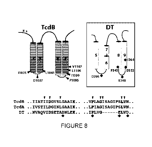
[0030] Figure 8 shows a mapping of the functional determinants of pore-
formation
and translocation onto a working model of the TcdB transiocation pore. A model
of TcdA and
of the "double-dagger" DT pore are also shown. TcdB sequences shown here are
SEG ID
NO:5 IIATIIDGvSLGAAIK and SEG ID NO:6 VPLAGISAGIPSLVN TcdA sequences
shown here are SEG ID NO:7 IVST I LDG INLGAAI K and SEG ID NO:8 LP IAGI SAGI
PSLVN
DT sequences shown here are SEG ID NO:9 uvAQVIDSETADDILEK and SEC ID NO: 10
I PLVGXXXXXXELVD
[0031] Figure 9 shows the structure of TcdA.
[0032] Figure 10 provides data to illustrate that InsP6 binding induces a
significant
structural change in the CPD.
[0033] Figure 11 provides data to illustrate that the delivery domain
provides an
extended scaffold for an alpha-helical hydrophobic stretch involved in pore
formation.
[0034] Figure 12 shows that the TcdA1831 structure can rotate about the
delivery
domain-CROPS junction upon exposure to low pH.
[0035] Figure 13 shows Rb86 release for Vero cells at pH 4.8, with TcdA
and
mutants thereof.
[0036] Figure 14 shows a dose response curve for CHO cell viability for
TcdA and
mutants.
DETAILED DESCRIPTION
[0037] Generally, the present disclosure provides recombinant Clostridium
toxin
proteins which comprise one or more mutations, and are thus said to be
"mutant" sequences
containing SEG ID NO: 11 (AGISAGIPSLVNNEL), which is a portion of a highly
conserved
region of native TcdA and TcdB. The one or more mutations in this region
render the protein
a mutant of SEG ID NO: 11. The protein need only contain this sequence, and is
thus said to
comprise the sequence within the length of the protein, but need not be
limited to this
sequence only. The mutant of SEC) ID NO: 11 may, for example, have from 1 to 4
mutations
- 6 -
CA 02939969 2016-08-17
WO 2015/123767
PCT/CA2015/050115
with reference to altered amino acid residues. Exemplary mutations may occur
in LVNN of
SEQ ID NO: 11, which are located at the positions 10 to 13 of SEQ ID NO: 11.
When the N
at position 12 is referred to, the term the first N" may be used, whereas when
the N at
position 13 is referred to, it may be referred to as "the second N", as a
skilled person would
understand that within the portion of the protein defined by SEQ ID NO: 11,
only these two N
residues are present. The mutation may be L to K at the 10th amino acid
position of SEQ ED
NO: 11. Further, the mutation may be of VNN to SAS at positions 11 to 13 of
SEQ ID NO:
11.
[0038] For example, mutant may contain the mutations within of SEQ ID NO:
12
(VPLAGISAGIPSLVNNELVL) from TcdB or SEQ ID NO: 13 (LPIAGISAGlPSLVNNELIL) from
TcdB, both of which contain SEQ ID NO: 11.
[0039] The mutant may be mutated at particular residues, and thus may
comprise
SEQ ID NO: 14 (VPLAGISAGIPSKVNNELVL) for TcdB; SEQ ID NO: 15
(LPIAGISAGIPSKVNNELIL) for TcdA; SEQ ID NO: 16 (VPLAGISAGIPSKSASELVL) for
TcdB; or SEC; ID NO: 17 (LPIAGISAGIPSKSASELIL) for TcdA. However, the mutants
are
not limited to these.
[0040] A recombinant protein mutant is described which is comparable to
native
Clostridium TcdA toxin protein, except that it comprises a Li 108K mutation,
or which is
comparable to Clostridium TcdB toxin protein, except that it comprises a
L1106K mutation.
Further, a recombinant protein is described which is a Clostridium TcdA toxin
protein
comprising mutations V11095, N1110A, and N11115; or a Clostridium TcdB toxin
protein
comprising mutations V11075, N1108A, and N11095. These proteins retain
conformational
properties of the native TcdA and TcdB toxins, but do not possess toxic
effects.
[0041] The proteins described herein may have an epithelial cell toxicity
that is
reduced by 100-fold or greater, or 1000-fold or greater compared to wild-type
Clostricflurn
toxin.
[0042] The proteins described herein may comprise a sequence of 75% or
greater,
80% or greater, 85% or greater, 90% or greater, 95% or greater, or 99% or
greater identity to
residues 958 to 1130 of TcdA; or to residues 956 to 1128 of TcdB, which are
hydrophobic
and highly conserved residues within the middle of the translocation domain of
these
Clostridium toxins.
- 7 -
CA 02939969 2016-08-17
WO 2015/123767
PCT/CA2015/050115
[0043] The proteins described may be encoded by a sequence of 75% or
greater,
80% or greater, 85% or greater, 90% or greater. 95% or greater. or 99% or
greater identity to
SEQ ID NO:4.
[0044] The proteins described herein may be produced recombinantly in a
Bacillus
host. For example, the Bacillus host may be Bacillus megaterium.
[0045] Immunogenic compositions are described herein which comprise one or
more
of the recited recombinant proteins having the described mutations, together
with one or
more pharmaceutically acceptable exdpients. Such a composition may, for
example, be a
vaccine, which may be combined with or administered together, in serial, or in
parallel with a
pharmaceutically acceptable adjuvant. The vaccine may have a toxicity,
attributable to the
recombinant Clostridium toxin protein, which is reduced by 100-fold or
greater, or 1000-fold
or greater compared to the wild-type Clostridium toxin(s).
[0046] Nucleic acids are described which encode the subject recombinant
Clostridium toxin proteins. Such a nucleic acid may encode a modified
Clostridium difficile
toxin A (TccIA) protein comprising a Li 108K mutation, or may encode a
modified Clostridium
diffici(e toxin B (TcdB) protein comprising a L1106K mutation. Further, such
nucleic acids
may encode a modified TodA with VNN mutated to SAS at residues 1109-1111
(TcdA) or at
1107-1109 (Tcd13). A vector is described herein which comprises such nucleic
acids as
these, or others which may encode the subject proteins. There is also provided
herein a cell
which comprises such a vector.
[0047] A kit comprising the composition of which contains the protein is
described,
which kit includes instructions for use of the composition in treating or
preventing Clostridium
infection.
[0048] A method of eliciting an immune response to Clostridium is
described. The
method is intended for use by such subjects in need of prevention or treatment
for
Clostridium infection. The method involves administration to the subject of
the recombinant
Clostridium toxin protein described herein. The method may be used to treat,
prevent, or
otherwise counter Clostridium infections such as Clostridium difficile,
Clostridium sordellii,
Clostridium novyi, or Clostridium perfringens. The administration of the
protein to the subject
may elicit an effective immune response before the subject has had any
exposure to
Clostridium toxin, and in this way, the method may be said to be prophylactic.
The method
- 8 -
CA 02939969 2016-08-17
WO 2015/123767
PCT/CA2015/050115
may involve any variety of routes for administration of the protein to the
subject, such as
intravenous, ntramuscukar, subcutaneous, intraperitonea intradermal,
ransdermal, mucosaF,
sublingual. intranasal or oral administration.
[0049] The method for eliciting an immune response may include an
assessment of
antibody titer in the subject, so as to check for antibodies specifically
binding to wild-type
Clostridium toxin B or toxin A. This may be used for comparison with a control
value to
determine the immune response of the subject. The control value used may be
selected as a
level of antibody titer for TcdB, measured in the subject prior to
administering the protein. In
this way, the subject can serve as her own control. Optionally, when the
protein is
administered to the subject, it may be in combination with a pharmaceutically
acceptable
adjuvant in an appropriate manner to heighten or otherwise encourage immune
response.
[0050] There is described herein an antibody or antigen-binding fragment
thereof
which specifically binds to a modified Clostridium difficile toxin A (TcdA)
comprising a
L1108K mutation, a modified Clostridium difficile toxin B (TcdB) protein
comprising a L1106K
mutation; a modified Clostridium difficile toxin A (TcdA) comprising Vii 09S,
N1110A, and
N1111S mutations; or a modified Clostridium difficile toxin B (TcdB) protein
comprising
V11075, N1108A, and N11095 mutations. For example, the antibody or antigen-
binding
fragment thereof may specifically bind to a protein having a sequence
according to SEQ ID
NO:2. Further, the antibody or antigen-binding fragment thereof may be one
which
specifically binds to a portion of an epitope comprising AGISAGIPSLVNNEL (SEC;
ID NO:
11) in a Clostridium TcdA or TcdB toxin, or may be one which specifically
binds to SEQ ID
NO:12 (VPLAGISAGIPSLVNNELVL) or SEQ ID NO: 13 (LPIAGISAGIPSLVNNELIL). The
antibody or antigen-binding fragment thereof may specifically bind to the TcdA
and the TcdB
toxin. The antibody may be a monoclonal antibody, or may be a humanized
antibody.
[0051] There is described herein a method of preventing infection from
Clostridium in
a subject, comprising administering to the subject an effective amount of the
antibody or
antigen-binding fragment thereof.
[0052] The described proteins may be used for eliciting an immune response
to
Clostridium difficile toxin A (TcdA) or toxin B (TcdB) in a subject. Further,
the use of the
antibody or antigen-binding fragment thereof may be for preventing Clostridium
infection in a
subject. The described proteins may be used for preparation of a medicament
for eliciting an
- 9 -
CA 02939969 2016-08-17
WO 2015/123767
PCT/CA2015/050115
immune response to Clostridium difficile toxin A (TcdA) or toxin B (TcdB) in a
subject.
Further, the use of the antibody or antigen-binding fragment thereof may be
for preparation
of a medicament for preventing Clostridium infection in a subject.
[0053] A method is described herein for identifying an antibody or antigen-
binding
fragment thereof which specifically binds to a portion of an epitope defined
by SEQ ID NO:
11 (AGISAGIPSLVNNEL), SEQ ID NO:12 (VPLAGISAGIPSLVNNELVL), or SEQ ID NO: 13
(LPIAGISAGIPSLVNNELIL) in a Clostridium TcdA and/or TcdB toxin, comprising the
steps
of: a) immunizing an animal with a Clostridium TcdA or TcdB toxin comprising
SEQ ID NO:
11, SEQ ID NO: 12, or SEQ ID NO: 13; b) obtaining sera from the immunized
animal
subsequent to immunization; and c) screening the sera for an antibody or
antigen-binding
fragment thereof which specifically binds to a portion of an epitope defined
by SEQ ID NO:
11; SEQ ID NO: 12, or SEQ ID NO: 13 in the Clostridium TcdA andior TcdB toxin.
[0054] In such a method, the antibody or antigen-binding fragment thereof
may
specifically bind to both the TcdA and TcdB toxin. Further, the screening may
be conducted
using a high-throughput screening method. The sera may be screened by ELISA.
The
method may comprise the step of adding Clostridium toxin to cell culture and
determining if
the sera decreases the cytotoxic effect of the Clostridium toxin on the cells.
[0055] A protein, a nucleic acid, and an antibody are described herein, as
well as an
immunogenic composition or vaccine composition, based on a C. difficile toxin
protein TcdA
and/or TcdB, which contain one or more mutations so as to be rendered less
toxic or ataxic.
For Example, TcdB comprising the mutation L1106K is rendered less toxic than
native TcdB.
Similarly, the mutation of TcdA with Li 108K renders a protein less toxic than
native TcdA.
TcdA mutated from VNN to SAS at residues 1107-1109 (TcdB) or residues 1109-
1111
(TcdA) are less toxic than wild-type. The use of these proteins, nucleic acid
encoding for
them, or antibodies based upon these for immunizing a subject against
Clostridium infection
are described herein. Despite the mutation and reduced toxicity, the mutated
TcdA and
TcdB proteins described herein retain native protein conformation comparable
to wild-type
TcdB.
[0056] An amino acid sequence of a Li 106K protein is provided in SEQ ID
NO:2. An
exemplary nucleotide sequence encoding a wild-type TcdB protein is found in
SEQ ID NO: 3.
- 10-
CA 02939969 2016-08-17
WO 2015/123767
PCT/CA2015/050115
An exemplary nucleic acid sequence encoding a protein sequence having the
L1106K
mutation is provided in SEO ID NO:4.
[0057] There is also described herein a translocation-defective
recombinant hoiotoxin
proteins of Clostridium difficile toxin TcdB, for use as imnriunogens. The
proteins have a
L1 106K mutation. Protein sequences, nucleotide sequences, and antibodies are
described
as well as compositions, vaccines, uses and methods pertaining to treatment or
prevention of
C. difficile infection. The L1 106K mutation blocks pore-formation within the
translocation
domain and thus reduces toxicity.
[0058] It would be advantageous to develop neutralizing antibodies against
homologous toxins from organisms associated with rare infections, such as
those involving
Clostridium sortlellii and Clostridurn navy'. It is possible that neutralizing
antibodies against
Clostridium that are currently in development may not be effective against all
strains, since
the TcdB sequence can vary significantly. Efforts have been made to develop
non-toxic
vaccines against Clostridium toxin using fornrialin-treated TcdA and TcdB
proteins, but
formalin-treatment results in significant disruption of the toxin structure.
[0059] There is described herein a 3.25 A crystal structure for residues 1-
1832 of C.
difficile TcdA. The sequence of TcdA is known, for example, GenBank describes
a 2710 aa
sequence for TcdA in accession number CAA63564, hereby incorporated by
reference, as
described by Hundsberger et al., Eur. J. Biochem. 244 (3), 735-742 (1997). The
structure
reveals a novel epitope to be targeted for pan-toxin neutralization. Mutation
of the epitope,
representing a pore forming domain as revealed by the crystal structure, can
provide a toxin
protein with decreased toxicity which can be used as a vaccine antigen. The
pore forming
domain is a hydrophobic helical element, the sequence of which strictly
conserved in 6
homologous toxins: TcdA and TcdB from C. difficile, TosH and TcsL from C.
&ardent', Tcna
from C. novyi, and TpeL from C. petfringens. It is shown herein that mutation
of the
conserved loop removes or decreases all or essentially all of the toxicity
associated with the
toxin in cell culture. The strictly conserved amino acid sequence is
LPIAGISAGIPSLVNNELIL (SEQ ID NO: 13). In TcdA, this typically corresponds to
amino
acids 1096 to 1115, while in TcdB this typically corresponds to amino acids
1094 to 1113,
taking into account the slightly different sequences present in different
strains of C. difficile.
A mutation tested involved changing VNN of SEQ ID NO: 13 to SAS in both TcdA
and TcdB.
- 11 -
CA 02939969 2016-08-17
WO 2015/123767
PCT/CA2015/050115
[0060] Identification of this novel epitope permits the preparation
neutralizing
antibodies against homologous toxins from organisms associated with rare
infections such
as C. sorclellii and C. novyi, and can be used to provide a common
neutralizing antibody
against TccIA and TcdB that would be effective across many, and possibly ail
strain variants.
Mutation of the novel epitope can provide safe vaccine antigens.
[0061] A modified Clostridium difficile toxin B (TcdB) protein is
described herein
comprising a L1106K mutation. The protein may comprise a sequence of 75% or
greater
identity to SEQ ID NO:2, or may comprise a sequence of 80% or greater, 85% or
greater,
90% or greater, 95% or greater, or 99% or greater identity to SEQ ID NO:2. The
protein may
have the sequence according to SEQ ID NO:2, and encompasses equivalent
proteins which
have deletions or substitutions while generally maintaining the properties of
SEC; ID NO:2.
The protein may be one encoded by a nucleic acid sequence of 75% or greater
identity to
SEQ ID NO:4. Specifically, the protein may be encoded by a sequence of 80% or
greater,
85% or greater, 90% or greater, 95% or greater, or 99% or greater identity to
SEQ ID NO:4.
The protein may be encoded by the nucleic acid sequence of SEQ ID NO:4, which
encompasses equivalents, having different codons encoding the same amino acids
or
different codons encoding conservatively substituted amino acids.
[0062] The protein described herein possesses epithelial cell toxicity
that is reduced
by 10-fold or greater, 100-fold or greater, or 1000-fold or greater when
compared to wild-type
(WT) TcdB.
[0063] In exemplary embodiments, the protein may be produced
reconnbinantly in a
Bacillus host. For example, the Bacillus host may be Bacillus megateriurrp.
[0064] A nucleic acid is described herein encoding a modified Clostridium
clifficlie
toxin B (Tcd13) protein comprising a Li 106K mutation. The nucleic acid may
comprise a
sequence of 75% or greater identity to SEQ ID NO:4. For example, the nucleic
acid may
comprise a sequence of 80% or greater, 85% or greater, 90% or greater, 95% or
greater, or
99% or greater identity to SEQ ID NO:4. The nucleic acid may comprise the
sequence
according to SEQ ID NO:4. The nucleic acid may encode a protein of 75% or
greater identity
to SEQ ID NO:2. For example, the nucleic acid may comprise a sequence of 80%
or greater,
85% or greater, 90% or greater, 95% or greater, or 99% or greater identity to
SEQ ID NO: 2,
and may specifically encode SEQ ID NO:2.
- 12-
CA 02939969 2016-08-17
WO 2015/123767
PCT/CA2015/050115
[0065] The nucleic acid may be one in which the epithelial cell toxicity
of the encoded
protein is reduced by 10-fold or greater, 100-fold or greater, or 1000-fold or
greater
compared to wild-type (WI) TcdB.
[0066] A vector is described herein comprising the nucleic acid described
above. A
cell comprising such a vector is also encompassed. The cell may thus express a
modified
Clostridium difficiie toxin B (TcdB) protein comprising the Li 106K mutation,
for example that
of SEQ ID NO:2, or a functional equivalent thereto having 75% or greater
identity to SEQ ID
NO:2.
[0067] An immunogenic composition is described herein comprising the
protein
described above. The composition may comprise a pharmaceutically acceptable
excipient.
The composition may be used as a vaccine to Clostridium difficile. A kit is
provided,
comprising such a composition together with instructions for use as a vaccine
to Clostridium
difficife.
[0068] A method of eliciting an immune response to Clostridium cificile
toxin B
(TcdB) in a subject is described comprising: delivering to the subject a
modified Clostridium
diffici(e toxin B (TcdB) protein comprising a L11 06K mutation or a nucleic
acid encoding the
modified Clostridium dificile toxin B (TcdB) protein comprising the L1106K
mutation. The
method may involve delivering the protein or the nucleic acid via intravenous,
intramuscular,
subcutaneous, intraperitoneal, intradermal, transderrnal, mucosal, sublingual,
intranasal or
oral administration to the subject. The method may further comprise assessing
antibody titer
in the subject for antibodies specifically binding to wild-type TcdB for
comparison with a
control value to determine immune response to the modified TcdB protein or
nucleic acid.
Such a control value may, for example, be a level of antibody titer for TcdB
in the subject
prior to delivering the modified protein or nucleic acid.
[0069] An antibody is provided herein which is raised to and/or
specifically binds to a
modified Clostridium cificile toxin B (TcdB) protein comprising a L1106K
mutation or an
equivalent mutation. Such an antibody may specifically bind to a protein
having a sequence
according to SEQ ID NO:2, or one at least 75% identical thereto. Such an
antibody may be
monoclonal, and/or may be humanized.
[0070] The use of a modified Clostridium difficile toxin B (TcdB) protein
is described
herein comprising a Li 106K mutation or a nucleic acid encoding the modified
Clostridium
- 13-
CA 02939969 2016-08-17
WO 2015/123767
PCT/CA2015/050115
difficile toxin B (TcdB) protein comprising the Li 106K mutation for eliciting
an immune
response to Clostridium difficile toxin B (TcdB) in a subject.
[0071] Further, as use is provided of a modified Clostridium difficile
toxin B (TcdB)
protein comprising a L1 106K mutation or a nucleic acid encoding the modified
Clostridium
difficile toxin B (TcdB) protein comprising the Li 106K mutation for
preparation of a
medicament for eliciting an immune response to Clostridium difficile toxin B
(TcdB) in a
subject.
[0072] Regarding substitutions within the described sequences, different
codons can
encode lysine, and thus it is understood that the nucleic acid sequence of SEQ
ID NO:4 is
merely one example of a sequence that can encode the amino acid sequence
L1106K of
SEQ ID NO:2. Codons MG and AAA encode lysine. Further, amino acids than lysine
may
be substituted for leucine at this position, provided a similar effect in
blocking pore-formation
within the translocation domain is achieved, and toxicity is appropriately
reduced. Such
residues would be considered equivalent, provided the atoxic effect is
maintained. Lysine is a
basic amino acid, and thus it may be considered an appropriate conservative
substitution to
utilize other basic amino acids, such as arginine and glutamine. Conservative
substitutions in
other residues of SEC ID NO:2 may be made, provided the desired properties of
the protein
are intact: preserving the intoxicating properties of wild-type TcdB, while
rendering the
protein atoxic. Deletions and substitutions which may be permitted while
maintaining these
properties may render a protein having 75% or greater identity to that of SEQ
ID NO:2, for
example 84% or greater, 85% or greater, 90% or greater, 95% or greater, or 99%
or greater
identity thereto.
[0073] Similarly, nucleic acid sequences utilizing equivalent codons, or
codons
encoding a conservative substitution of amino acids are considered equivalent
to SEQ ID
NO:4, provided the protein encoded maintains the properties described herein.
Nucleotide
sequences encoding for a protein having deletions and substitutions which may
be permitted
while maintaining these properties may be encoded by a sequence having 75% or
greater
identity to that of SEQ ID NO:4, for example 80% or greater, 85% or greater,
90% or greater,
95% or greater, or 99% or greater identity thereto.
[0074] Conservative amino acid substitutions which are known in the art
are as
follows with conservative substitutable candidate amino acids showing in
parentheses: Ala
- 14 -
CA 02939969 2016-08-17
WO 2015/123767
PCT/CA2015/050115
(Gly, Ser); Arg (Gly, Gin); Asn (Gin; His); Asp (Glu); Cys (Ser); Gin (Asn,
Lys); Glu (Asp); Gly
(Ala, Pro); His (Asn; Gin); lie (Leu; Val); Leu (He; Val); Lys (Arg; Gin); Met
(Leu, He); Phe
(Met, Leu, Tyr); Ser (Thr; Gly); Thr (Ser; Val); Trp (Tyr); Tyr (Trp: Phe);
Val (lle; Leu).
[0075] Compositions having immunogenic properties, such as a vaccine, are
described herein for use in treating or vaccinating against C. cfifficife. The
described
compositions elicit antibody production in a subject. The production of
antibodies to TcdB
can protect against or reduce the severity of C. cif/0e infection.
Advantageously, the
described mutation results in a protein that maintains wild-type
characteristics but has a
greatly reduced toxicity compared with the wild-type TcdB protein. A Bacillus
megaterium
expression system was used to generate mutant proteins, which were tested for
a reduction
in or absence of toxicity.
[0076] The protein or nucleic acid may be formulated in a composition with
a
pharmaceutically acceptable excipient, for delivery as a vaccine to a subject
at an effective
dose, optionally with an adjuvant. Such a vaccination would be utilized for
prevention of the
disease and symptoms associated with C. difficile infection in human or animal
subjects.
[0077] The subject may be a human, and may advantageously be a human at
high
risk for C. difficile infection, such as a hospitalized or immune compromised
human. Further,
the subject may be a non-human animal such as a livestock animal, a research
animal, or a
domesticated animal such as a companion animal at risk of infection. The
subject may be an
animal in a stressful circumstance, at risk for C. difficile infection.
[0078] Antibodies against C. difficile are typically present in the
general population.
Thus, antibody titer may involve assessing an individual's own base-line level
(as a control
value) of antibody before and after vaccination is used to elicit an immune
response.
[0079] Advantageously, the mutant toxin protein provided herein involves a
primary
point mutation that permits the protein to maintain similar properties to the
native toxin while
exhibiting a greatly reduced toxicity.
[0080] The composition may comprise the mutant protein as an antigen along
with
one or more pharmaceutically acceptable carrier, excipient or diluent.
Optionally, the
composition may further include an adjuvant. The composition may be used in
conjunction
with conventional treatments or prevention strategies for C. difficlle
infection, either delivered
- 15-
CA 02939969 2016-08-17
WO 2015/123767
PCT/CA2015/050115
separately or simultaneously. Such conventional treatments may encompass
antibiotic
treatment with nrietronidazole or vancomycin, or probiotic delivery.
[0081] in certain embodiments, the composition optionally further
comprises one or
more additional therapeutic agents. For example, an antibiotic compound, anti-
viral
compound, anti-fungal compound may be included. Other optional components of
the
composition include one or more growth factor, anti-inflammatory agent,
vasopressor agent,
collagenase inhibitor, topical steroid, matrix metalloproteinase inhibitor,
ascorbate,
angiotensin, caireticulin, tetracycline. flbronectin, collagen,
thrombospondin, transforming
growth factor (TGF), keratinocyte growth factor (KGF), fibroblast growth
factor (FGF), insulin-
like growth factor (lGF), epidermal growth factor (EGF), platelet derived
growth factor
(PDGF). neu differentiation factor (NDF), hepatocyte growth factor (HGF), or
hyaluronic acid.
[0082] Pharmaceutically acceptable carriers include solvents, diluents,
liquid
vehicles, dispersion or suspension aids, surface active agents, isotonic
agents, thickening or
emulsifying agents, preservatives, solid binders, or lubricants. Carriers may
be selected to
prolong dwell time for sustained release appropriate to the selected route of
administration.
Exemplary carriers include sugars such as glucose and sucrose, starches such
as corn
starch and potato starch, fibers such as cellulose and its derivatives, sodium
carboxymethyl
cellulose, ethyl cellulose, cellulose acetate, powdered tragacanth, malt,
gelatin, talc, cocoa
butter, suppository waxes, oils such as peanut oil, cottonseed oil, safflower
oil, sesame oil,
olive oil, corn oil, and soybean oil; glycols such as propylene glycol, esters
such as ethyl
oleate and ethyl laurate, agar, buffering agents such as magnesium hydroxide
and aluminum
hydroxide, alginic acid, pyrogen-free water, isotonic saline, Ringers
solution, ethyl alcohol,
phosphate buffer solutions, non-toxic compatible lubricants such as sodium
lauryl sulfate and
magnesium stearate, coloring agents, releasing agents, coating agents,
sweeteners, flavors,
perfuming agents, preservatives, and antioxidants.
[0083] Immunization of a subject, or eliciting an immune response may
involve
delivery of a therapeutically effective amount of the composition to a subject
in need or at risk
of C. difficile infection, in an appropriate amount and for an adequate time
as may be
necessary to achieve the goal. The composition can be used as a preventive or
therapeutic
measure to promote immunity to infection or re-infection by C. cliff/01e.
- 16-
CA 02939969 2016-08-17
WO 2015/123767
PCT/CA2015/050115
[0084] The therapeutically effective amount may be determined on an
individual
basis or on the basis of the established amount necessary for an effective
promotion of
antibody formation. Appearance of antibodies in serum, which are specific for
the toxins of
C. difficife, or disappearance of disease symptoms can be evaluated
clinically. The dosage
for an individual subject is chosen in view of the subject to be treated.
Dosage and
administration are adjusted to provide sufficient levels of the active
agent(s) or to maintain
the desired effect. Factors which may be taken into account include the
severity of the
disease state, contact with infectious agent in the past, potential future
contact; age, weight,
gender of the subject, diet, time and frequency of administration, drug
combinations, reaction
sensitivities, and tolerance/response to therapy. Sustained release
compositions might be
administered less frequently than fast-acting compositions.
[0085] A therapeutic dose may encompass from about 1 pg per kg, for
example,
about 5, 10, 50, 100, 500 pg per kg, at least about 1 mg/kg, 5, 10, 50 or 100
mg/kg body
weight of the purified toxin vaccine per body weight of the subject, although
the doses may
be more or less depending on age, health status, history of prior infection,
and immune
status of the subject as would be known by one of skill in the art of
immunization. Doses may
be administered as a bolus or repeated at appropriate intervals, or via an
infusion at a
constant or intermittent rate.
[0086] Compositions can be administered to subjects through any acceptable
route,
such as topically (as by powders, ointments, or drops), orally, rectally,
mucosally,
sublingually, parenterally, intracisternally, intravaginally,
intraperitoneally, bucally, ocularly, or
intranasally.
[0087] Liquid dosage forms for oral administration may include emulsions,
microemulsions, solutions, suspensions, syrups and elixirs. Liquid dosage
forms may contain
inert diluents such as water or other solvents, solubilizing agents and
emulsifiers such as
ethyl alcohol, isopropyl alcohol, ethyl carbonate, ethyl acetate, benzyl
alcohol, benzyl
benzoate, propylene glycol, 1,3-butylene glycol, dimethylformamide, oils such
as cottonseed,
groundnut, corn, germ, olive, castor, and sesame oils, glycerol,
tetrahydrofurfuryl alcohol,
polyethylene glycols and fatty acid esters of sorbitan, and mixtures thereof.
Besides inert
diluents, the oral compositions can also include adjuvants such as wetting
agents,
emulsifying and suspending agents, sweetening, flavoring, and perfuming
agents.
- 17-
CA 02939969 2016-08-17
WO 2015/123767
PCT/CA2015/050115
[0088] Dosage forms for topical or transderrnal administration of an
inventive
pharmaceutical composition include ointments, pastes, creams, lotions, gels,
powders,
solutions, sprays, inhalants, or patches. The active agent is admixed under
sterile conditions
with a pharmaceutically acceptable carrier and any needed preservatives or
buffers as may
be required.
[0089] Injectable preparations, such as sterile injectable aqueous or
oleaginous
suspensions may be formulated according to the known art using suitable
dispersing or
wetting agents and suspending agents. The sterile injectable preparation may
also be a
sterile injectable solution, suspension or emulsion in a non-toxic
parenterally acceptable
diluent or solvent, for example, as a solution in 1,3-butanedioi. Among the
acceptable
vehicles and solvents that may be employed are water, Ringer's solution,
U.S.P. and isotonic
sodium chloride solution. In addition, sterile, fixed oils are conventionally
employed as a
solvent or suspending medium. For this purpose any bland fixed oil can be
employed
including synthetic mono- or diglycerides. In addition, fatty acids such as
oleic acid are used
in the preparation of injectables. The injectable formulations can be
sterilized prior to addition
of spores, for example, by filtration through a bacterial-retaining filter, or
by incorporating
sterilizing agents in the form of sterile solid compositions which can be
dissolved or
dispersed in sterile water or other sterile injectable medium prior to use.
[0090] It is often desirable to slow the absorption of the agent from
subcutaneous or
intramuscular injection. Delayed absorption of a parenterally administered
active agent may
be accomplished by dissolving or suspending the agent in an oil vehicle.
Injectable depot
forms are made by forming nnicroencapsule matrices of the agent in
biodegradable polymers
such as polyladide-polyglycolide. Depending upon the ratio of active agent to
polymer and
the nature of the particular polymer employed, the rate of active agent
release can be
controlled. Examples of other biodegradable polymers include poly(orthoesters)
and
poly(anhydrides). Depot injectable formulations are also prepared by
entrapping the agent in
liposomes or nnicroemulsions which are compatible with body tissues.
[0091] Compositions for rectal or vaginal administration are preferably
suppositories
which can be prepared by mixing the active agent(s) of this invention with
suitable non-
irritating excipients or carriers such as cocoa butter, polyethylene glycol or
a suppository wax
- 18-
CA 02939969 2016-08-17
WO 2015/123767
PCT/CA2015/050115
which are solid at ambient temperature but liquid at body temperature and
therefore melt in
the rectum or vaginal cavity and release the active agent(s).
[0092] Solid dosage forms for oral, mucosal or sublingual administration
include
capsules, tablets, pills, powders, and granules. In such solid dosage forms,
the active agent
is mixed with at least one inert, pharmaceutically acceptable excipient or
carrier such as
sodium citrate or dicalcium phosphate, fliers or extenders such as starches,
sucrose,
glucose, mannitol, and silicic acid, binders such as, for example.
carboxymethylcellulose,
alginates, gelatin, polyvinylpyrrolidinone, sucrose, and acacia, humectants
such as glycerol.
disintegrating agents such as agar-agar, calcium carbonate, potato or tapioca
starch, alginic
acid, certain silicates, and sodium carbonate, solution retarding agents such
as paraffin,
absorption accelerators such as quaternary ammonium compounds, wetting agents
such as,
for example, cetyl alcohol and glycerol monostearate, absorbents such as
kaolin and
bentonite clay, and lubricants such as talc, calcium stearate, magnesium
stearate, solid
polyethylene glycols, sodium lauryl sulfate, and mixtures thereof.
[0093] Solid compositions of a similar type may also be employed as
fillers in soft
and hard-filled gelatin capsules using such excipients as milk sugar as well
as high molecular
weight polyethylene glycols and the like. The solid dosage forms of tablets,
capsules, pills,
and granules can be prepared with coatings and shells such as enteric
coatings, release
controlling coatings and other coatings well known in the pharmaceutical
formulating art. In
such solid dosage forms the active agent(s) may be admixed with at least one
inert diluent
such as sucrose or starch. Such dosage forms may also comprise, as is normal
practice,
additional substances other than inert diluents, such as tableting lubricants
and other
tableting aids such a magnesium stearate and microcrystalline cellulose. In
the case of
capsules, tablets and pills, the dosage forms may also comprise buffering
agents. They may
optionally contain pacifying agents and can also be of a composition that
they release the
active agent(s) only, or preferentially, in a certain part of the intestinal
tract, optionally, in a
delayed manner. Examples of embedding compositions which can be used include
polymeric
substances and waxes.
EXAMPLES
- 19-
CA 02939969 2016-08-17
WO 2015/123767
PCT/CA2015/050115
Example .1
[0094] Translocation Domain Mutations Affecting Cellular Toxicity Identify
the
Clostridium difficile Toxin B Pore
[0095] ENTRODUCTION
[0096] Homologous toxins TcdA and TcdB of C. difficile impact colonic
epithelial cells
upon infection. Binding to target cells triggers internalization of these
toxins into acidified
vesicles, whereupon cryptic segments from within the 1050 amino acid
translocation domain
unfurl and insert into the membrane of the epithelial cell, creating a
transmembrane
passageway to the cytosol. Current understanding of pore-formation and the
subsequent
translocation of the upstream cytotoxic domain to the cytosol is limited by
the lack of
information regarding the identity and architecture of the transmembrane pore.
In this
Example, through systematic perturbation of conserved sites within predicted
membrane-
insertion elements of the translocation domain, highly sensitive residues have
been found,
clustered between amino acids 1035 and 1107, that when individually mutated
reduce
cellular toxicity by as much as >1000-fold. It is shown that defective
variants are defined by
impaired pore-formation in planar lipid bilayers and biological membranes,
resulting in an
inability to intoxicate cells through either apoptotic or necrotic pathways.
Further, unexpected
similarities were uncovered between the pore-forming "hotspots" of TcdB and
the well-
characterized a-helical diphtheria toxin translocation domain. Together, there
is provided
insight into the structure and mechanism of formation of the translocation
pore for this
important class of pathogenic toxins.
[0097] The structural features of the pore are described, and mutants are
described
which prevent pore-formation, showing reduced toxicity to host cells. These
findings reveal
information about the translocation pore, and provide the basis for a strategy
to target toxins
therapeutically.
[0098] In this Example, the initial goal was to identify the determinants
of pore-
formation and translocation through a comprehensive mutagenesis study using
the B.
megaterium platform. It was found, early in this pursuit, that site-specific
mutagenesis of the
inherently AT-rich toxin sequence (i.e., G+C = 27%) using the B. megaterfum
system was
laborious and inefficient. To address this, a GC-enriched copy of TcdB was
generated
(approximately G+C = 45%) with codons optimized for E. coil expression. This
permitted
- 20 -
CA 02939969 2016-08-17
WO 2015/123767
PCT/CA2015/050115
high throughput probing of the translocation domain. Several single point
mutations were
identified, clustering to within the hydrophobic region of the delivery
domain, resulting in
major defects in pore-formation and translocation. The unexpected similarity
of the identified
pore-forming region to that of the translocation domain of DT is reported. An
a-helical model
is described for the translocation pore of TcdB and homologous pathogenic
toxins.
[0099] MATERIALS AND METHODS
[00100] Expression and purification of recombinant TcdB from Bacillus
megaterium. Recombinant TcdB will-type was a 8,r:water/urn expression vector
pHis1522
encoding the strain VPI10463 obtained from Dr. Hangping Feng. Proteins were
expressed
and purified as previously described (Yang et al. (2008) BMC Microbial 8:192).
[00101] Expression and purification of recombinant codon-optimized
TcdB
constructs. Codon-optimized TcdB sequence was synthesized (GenScriptim) to
increase GC
percentage to 45%. The codon-optimized gene was cloned into an E. coil
expression vector
pET28a and transformed into E. calf BL21 0E3 competent cells and expressed as
C-terminal
His-tagged proteins.
[00102] Mutagenesis of TcdB mutants. Single point mutations were made
in
the TcdB codon-optimized sequence using QuickChangeTM lightning multi-
nnutagenesis kit
(Agilent Technologies, Santa Clara, CA). Sequenced plasmids with confirmed
mutations
were transformed and expressed using the same conditions as wild-type.
[00103] Small-scale expression of TcdB mutants. Plasmids expressing
TcdB
mutants were transformed into E. col, BL21 DE3 cells. Overnight cultures were
prepared in a
24-well block (BD biosciences) in 5 mL. Cells were harvested by centrifugation
and
resuspended in buffer (20 mM Tris, 500 mM NaCI pH 8.0 and protease inhibitor)
and lysed
by lysozyme (BioShopTM) as manufacturer's instructions followed by
centrifugation at 4,000 g
for 20 min. Supernatants were collected. The concentration of each full-length
mutant protein
in the lysates was determined by densitometry (Image Lab 3.0).
[00104] Cell viability assay. TcdB variants were added to CHO-K1 cells
at a
serial dilution of 1/3 starting at latl. Cell viability was assessed after 48
h by PrestoBlue
Cell Viability Reagent (Life technology). Fluorescence was read on a
Spectramax M5 plate
reader (Molecular Devices).
- 21 -
CA 02939969 2016-08-17
WO 2015/123767
PCT/CA2015/050115
[00105] Rubidium release assay. 86Rb+ release assay was performed as
previously reported by Genisyuerek S, et al. (2011). Briefly, CHO-K1 cells
were seeded in
96-well plates supplemented with I pCiiml 86Rb+ (PerkinElmer) at a density of
1 104 cells
per well. 66Rbi- released was determined by liquid scintillation counting with
TopCount NXT
(PerkinElmer).
[00106] TNS fluorescence assay. pH-induced conformational changes of
TcdB
were assessed as described previously (Lanis et al. (2010) PLoS Pathog 6(8):el
001061).
Assay plates were read in Spectramax M5 plate reader (Molecular Devices).
[00107] Black lipid bilayer experiments. Lipid bilayer experiments
were
performed essentially as described previously (ivielnyk & Collier (2006) Proc
Nati Acad Sci
USA 103(26):9802-9807). Both cis and trans compartments contained 1 nil of
solutions
containing 1 M KCI; 10 mM Tris pH 7.4. Pore-formation was initiated by adding
appropriate
amounts of 2 M HCI to the cis compartment to lower the pH to 4.5.
[00108] CellTfterGio TM ATP assay. Cell death assay was performed as
previously described (Chunibler et al. (2012) PLoS Pathog 8(12):e1003072.).
Assay plates
were read in Spectramax M5 plate reader (Molecular Devices).
[00109] Expression and purification of recombinant TcdB from Bacillus
megaterium. The template used for mutagenesis and clone for production of
recombinant
TcdB wild-type and mutant was a B. megaterium expression vector pHisl 522
encoding the
strain VPI10463 obtained from Dr. Hangping Feng. Proteins were expressed and
purified as
described by Yang et al. (2008) BMC ivlicrobiol 8:192.
[00110] Expression and purification of recombinant codon-optimfzed
TcdB
constructs. Codon-optimized TcdB sequence was synthesized (GenScriptim) to
increase GC
percentage to 47%. The codon-optimized gene was cloned into an E. coil
expression vector
pET28a and transformed into E. coil BL21 DE3 competent cells and expressed as
C-terminal
His-tagged proteins. 50 nn L of overnight culture was inoculated into 1L of LB
with 50.g/ml
Kanamycin and induced at 0D600 of 0.6 with 0.5mM IPTG at 37 C for 4 h. Cells
were
harvested by centrifugation and re-suspended with lysis buffer (20mM Tris pH
8.0, 0.5M
NaCI, protease inhibitor) and lysed by an EmulsiFlexTm 03 microfluidizer
(Avestin) at 15,000
psi. After lysing, lysate were centrifuged at 18,000 g for 20 min. Proteins
were purified by Ni-
affinity chromatography using HisTrap FF column (GE Healthcare). Fractions
containing
- 22 -
CA 02939969 2016-08-17
WO 2015/123767
PCT/CA2015/050115
TcdB were verified and pooled with a 100,0001v1WCO ultrafiltration device. 10%
of glycerol
was added; protein concentration was calculated by densitometry (Image Lab
3.0).
[00111] Iviutagenesis of TcdB mutants. Single point mutations were
made in
the TcdB codon-optimized sequence using QuickChange"' lightning multi-
rnutagenesis kit
(Agilent technologies). Plasmids with correct mutations were transformed and
expressed
using the same conditions as wild-type.
[00112] Small-scale expression of TcdB mutants. Plasrnids expressing
TcdB
mutants were transformed into E. coil BL21 DE3 cells. Overnight culture were
prepared in 24
well block (BD biosciences) in 5 mL. 250 pi of overnight culture were
inoculated into 5 ml of
LB with Kanannycin and induced at OD600 of 0.6 with 0.5 mM IPTG at 37 C for
4h. Cells
were harvested by centrifugation and resuspended in buffer (20mM Tris, 500mM
NaCI pH
8.0 and protease inhibitor (Sigma)) and lysed by lysozynne (Bioshop) as
manufacturer's
instructions followed by centrifugation at 4,000 g for 20 min. Supernatants
were collected.
The concentration of each full-length mutant protein in the lysates was
determined by
densitometry (Image Lab 3.0).
[00113] Details of Cell viability assay. CHO-K1 Cells (Chinese hamster
ovary
cells) were cultured in Ham's F-12 medium (Wisent) with 10% fetal calf serum
(FBS, Wisent)
and 1% penicillin and streptomycin (Wisent). CHO-K-1 cells were seeded at a
concentration
of 8,000 cells/well in 96-well CellBind plates (Corning). The next day, medium
was
exchanged with serum free medium and cells were intoxicated by adding TcdB
toxins at a
serial dilution of 1/3 starting at la/ After intoxication, cells were
incubated at 37C, 5%
CO2 for 48 h. Serum (FES) was added back to cells 24 h after intoxication to a
final
concentration of 10%. The Cell viability after 48 h was assessed by PrestoBlue
Cell
Viability Reagent (Life technology). Fluorescence was read on a Spectramax M5
plate reader
(Molecular Devices).
[00114] Details of Rubidium release assay. 86Rb+ release assay was
performed as previously reported (2) with slight modifications. Briefly, CHO-
K1 cells were
seeded in 96-well plates in the medium (Ham's F-12 with 10 % FBS),
supplemented with 1
CiJml86Rb+ (PerkinElmer) at a density of 1 104 cells per well. Cells were
incubated at 37
C. 5% CO2 overnight. Medium was exchanged with fresh growth medium with 100 nM
bafilomycin Al (Sigma) and continued to incubate for another 20 nnin. Then,
cells were
- 23 -
CA 02939969 2016-08-17
WO 2015/123767
PCT/CA2015/050115
chilled on ice and ice-cold medium containing TcdB mutants (10 nlv!) was
added. Cells were
kept on ice for toxin binding for 1 h at 4 C before they were washed with ice-
cold PBS twice
to remove unbound toxins, pH-dependent insertion into the plasma membrane was
induced
by warm, acidified growth medium (37 C, pH 4.5 or pH 7.5) for 5 min at 37 C.
After 1 hour
of further incubation on ice, medium containing released 86Rb+ was removed
from cell plate
and amount of 8ÃRb+ released was determined by liquid scintillation counting
with
TopCountm NXT (PerkinElmer).
[00115] in vitro giucosyltransferase assay. 10 nM of TcdB mutants were
incubated with 0.8 pM GST-Racl (0.2 pgipl, Sigma) in 25 pM UDPglucose in
glucosyiation
buffer (50 Wit HEPES, 100 mM KCI, 2 mM MgCl2 and 1 mM MnCi2, pH 7.5) for 60
min. The
reaction was stopped by addition of Laennnnli loading buffer with 8-
mercaptoethanol and
boiling at 95 C for 5 min. The proteins were separated on a 5%-12% gradient
polyacrylamide
gel by SDS-PAGE and then proteins were transferred to nitrocellulose with the
iBlot device
(Invitrogen). Glucosylated GST-Racl was detected by standard western blotting
with an
antibody that specifically recognizes the non-glucosylated form of Racl (1v1ab
142, ED
Biosciences), anti-GST antibody (GenScript) and HRP-conjugated anti-mouse-19G
(GE
healthcare).
[00116] TNS fluorescence assay pH-induced conformational changes of
TcdB
were assessed as described by Lanis et al. (2010) PLoS Pathog 6(8):e1001061. 2
pg of
TcdB was prepared in buffer having a pH ranging from 4 to 7. 2-(p-toluidiny)-
naphthalene-6-
sulfonic acid, sodium salt (2,6- TNS, lnvitrogen) was added at a final
concentration of 150
M. The final volume was 250 pl and mixed in 96-well black plate (Corning).
Mixtures were
incubated at 37 C for 20 min. The plate was analyzed in SpectrarnaxTM M5 plate
reader
(Molecular Devices) with excitation of 366 nm and an emission scan of 380 to
50Gnm.
[00117] Black lipid bilayer experiments. Lipid bilayer experiments
were
performed as described previously with modifications (Melnyk & Collier (2006)
Proc Nail
Acad Sci U S A 103(26):9802-9807). Briefly, membranes were made by painting
diphytanoyl
phosphatidylcholine (Avanti Polar Lipids) in decane across a 200-pm aperture
in a Delrin cup
by using the brush technique. Both cis and trans compartments contained 1 nil
of solutions
containing universal bilayer buffer as described by Kreimeyer et al. (2011)
Naunyn
Schmiedebergs Arch Pharmacol 383(3):253-262 (1 M KCI; 10 mM Tris pH 7.4).
- 24 -
CA 02939969 2016-08-17
WO 2015/123767
PCT/CA2015/050115
Translocation was initiated by adding appropriate amounts of 2 M HCI to the
cis
compartment to lower the pH to 4.5. Each compartment was stirred continuously
throughout
the experiment with a small stir bar. Agar salt bridges linked AgtAgCI
electrodes in 3 M KCI.
The current was amplified through a BC-525C integrating bilayer clamp
amplifier (Warner
Instruments, Hamden, CT), filtered at a frequency of 0.1 kHz by a low-pass
eight-pole Bessel
filter and computer-displayed through an analog/digital converter.
[00118] Further details of cell death assay. Cell death assay was
performed as
previously described (Chunribler et al. (2012 PLoS Pathog 8(12):e1003072.).
Briefly, iMR-90
Cells (cultured in EMEM, 10% FBS, 5%CO2) were seeded in 96-well Cel!bindTM
plate at a
concentration of 8,000 cells/well. The next day, the growth medium was
exchanged with
serum free EMEM and incubated at 37 C, 5% CO2 for 60 min. TcdB toxins were
added to
cells in dilutions starting at 30 nM. After intoxication, cells were incubated
at 37 C, 5% CO2
for 3 h. The amount of ATP was assessed with CellTiterGlolm as per the
manufacturer's
instructions (Promega). Plates were read in SpectrarnaxTM M5 plate reader
(Molecular
Devices).
[00119] RESULTS
[00120] Patterns of Hydrophobicity and Secondary Structure Suggest a
Helical
Pore for TcdB
[00121] To begin to unravel the determinants of pore formation and
translocation,
hydrophobicity, sequence conservation and predicted secondary structure
elements of the
1050 amino acid translocation domain were analyzed. Seven stretches of
hydrophobicity
were identified in TcdB: 985-1005, 1018-1036, 1037-1056, 1064-1089, 1091-1112,
1261-
1281, and 131C-1330 as shown in the top panel of Figure 5. The latter two
regions were
excluded since neither was predicted to be hydrophobic in the homologue from
Clostridium
novyi (TcnA).
[00122] Figure 5 shows a hydropathy analysis of the translocation domain.
In the top
panel, hydropathy analysis of the entire translocation domain of TcdB was
performed using a
membrane protein topology prediction method (TIVIHMM v2.0) that uses a hidden
Markov
model to predict transmembrane helices (Krogh et al. (2001) J Mol Biol
305(3):567-580).
Seven distinct peaks of hydrophobicity are evident; five within the previously
described
- 25 -
CA 02939969 2016-08-17
WO 2015/123767
PCT/CA2015/050115
hydrophobic region, along with two smaller regions of hydrophobicity between
1280-1350,
which were poorly conserved among homologous toxins and thus not pursued. In
the bottom
panel, hydropathy analysis of the 172 residue hydrophobic region is
illustrated, showing
predicted hydrophobic helices (HH1-HH5). The inset shows a hydropathy analysis
of the 173
amino acid diphtheria toxin translocation domain with established a-helical
segments
predicted to comprise the translocation pore of DT.
[00123] Alignment of the translocation domain of the large Clostridia'
Toxin family
using ClustalX2.1 was conducted. Residues 800-1880 were evaluated using TcdB
numbering. Only HH1-HH5 were predicted to be hydrophobic, whereas HH6 and HH7
were
predicted in all homologues except for TcnA from Clostridium t7ovy;r.
[00124] Notably, the former five hydrophobic segments fell within the
"hydrophobic
region" of the translocation domain (i.e., 956-1128). The length of the four
hydrophobic
segments that all were between 18 to 25 amino acids, combined with the absence
of any
alternating hydrophobic-hydrophilic up-barrel" motifs in this region, suggest
that the
membrane-inserted form of these segments adopt an a-helical conformation. When
the
primary sequence of the hydrophobic region was analyzed using secondary
structure
propensity algorithms, five a-helical structural elements with four
intervening disordered
loops were predicted. Secondary structure prediction for the translocation
domain of TcdB
was undertaken using JPRED3, and predicted helical regions were observed.
[00125] Positing an a-helical mode of membrane insertion, the well-
characterized a-
helical diphtheria toxin (DT) translocation domain was evaluated for
comparison. The
hydrophobic helices (TH5-6/7 and TH8-TH9) previously shown to be involved in
pore-
formation and translocation of DT were correctly mapped by this analysis
(Figure 5, bottom
panel). Unexpectedly, it was found that the general pattern of hydrophobicity
was strikingly
similar for the 173-residue translocation and the 172-residue hydrophobic
region of TcdB.
Three peaks of similar length and amplitude were predicted in both toxins. The
functional link
between DT translocation and the hydrophobic region of TcdB is considered
further, below.
[00126] Validation of a GC-Enriched Toxin B Gene for Mutagenesis and
Expression in E. coil
[00127] The observation that the putative pore-forming hydrophobic
regions of
the large translocation domain were localized within the 172 amino acid window
led to the
- 26 -
CA 02939969 2016-08-17
WO 2015/123767
PCT/CA2015/050115
investigation of which specific amino acids in this region were involved in
pore-formation and
translocation. To circumvent experimental barriers associated with generating
many mutants
to the AT-rich Clostridial toxin gene (i.e., G+C = 27%), a copy of the 7,098
base pair TcdB
gene was synthesized in which the G+C content was increased to 45%. This
mutagenesis-
competent copy of TcdB was then cloned into an E. coil expression plasmid in
order to
enable expression in a host that is more amenable for high throughput
characterization than
the existing Bacillus megaterium expression system.
[00128] To validate the newly constructed GC-enhanced copy of TcdB, the
structure
and function of E. coil produced TcdB was characterized and compared it to
benchmark
standards. TcdB produced in E. coil was indistinguishable from TcdB produced
in the well-
validated B. megaterium system showing equal potency on CHO cells toxicity.
Validation of
GC-enriched, codon-optimized TcdB protein was evaluated based on E. coil
expression.
Autoprocessing activity of recombinant toxins was evaluated. Recombinant TcdB
variants
were treated with 100 pM InsP6 (+) or PBS (-) for 3h and cleavage was
visualized by
Western blot by probing with an anti-GTD antibody. Further. GTD activity of
recombinant
toxins was evaluated. GST-Racl was treated with recombinant toxins, and the
level of
glucosylation was determined by Western blot analysis using Mab102 that
recognizes
unglucosyated Racl and an anti-Racl antibody to determine total Rac1.
Functional activity
of recombinant toxins was assessed. Recombinant TcdB constructs were added to
CHO
cells over a range of concentrations. Cellular viability was quantitated 48h
later by measuring
the fluorescence of cells treated with the cell viability reagent (PrestoBlue
).
[00129] The cytotoxicity of both purified toxin and soluble clarified
lysate from induced
E. coil on Chinese Hamster Ovary (CHO) cells was next measured, and compared
this to
uninduced controls, a glucosyltransferase defective mutant (D270A), and WT
TcdB produced
in B. megaterium. Purified WT toxins produced in either system yielded similar
potency on
CHO cells, whereas D270A was similarly inactive when produced in either
system.
Importantly, WT toxin produced in E. coil clarified soluble lysate was
equipotent with the
purified toxins (after normalizing toxin concentration of toxin in crude
lysates using
densitometry). This set of experiments showed that E. coil-produced toxin is
functional and
further that there is no confounding contaminant in the E. coil preparations
as evidenced by
the complete lack of toxicity of the uninduced control on CHO cell viability.
- 27 -
CA 02939969 2016-08-17
WO 2015/123767
PCT/CA2015/050115
[00130] High-Throughput Mapping of the Functional Determinants in the
Transiocation Domain
[00131] With a robust system to probe toxin function in place, residues
that were
absolutely conserved among the LCT family members were probed, reasoning that
functionally important residues would be conserved in homologous toxins. A
double-mutant
strategy was used in which each of the residues was mutated to both a highly
disruptive
residue (Lysine), and to a more conservative residue (Cysteine). The highly
polar Lysine side
chain was selected to increase the probability of identifying a membrane-
spanning segment;
introducing the polar and charged Lys side chain into a marginally hydrophobic
membrane-
inserting segment could be expected to prevent insertion of this segment and
thus pore-
formation. On the other hand. Cysteine, like Alanine, is a relatively benign
substitution that
can both help identify key functional residues and has the added downstream
benefit of
offering the possibility of attaching sulthydryl probes in TcdB for
structure/function studies.
[00132] The impact of each mutation on TcdB function was quantified by
measuring
the dose-dependent reduction in cell viability 48 hours post-toxin addition
relative to wild-type
TcdB, and results are shown in Figure 6.
[00133] Figure 6 shows high throughput mapping of the functional
determinants in the
translocation domain of TcdB. Functional consequences of Cys and Lys
substitutions in the
hydrophobic region of TcdB. Mutant soluble lysates were titrated onto CHO
cells (using 3-
fold dilutions) in 96-well plates and incubated for 48h at 37 C (n=4). In
parallel, an aliquot of
each mutant was used to measure the concentration using band densitometry
after SDS-
PAGE. 48h later cell viability of treated cells was quantitated by measuring
PrestoBlue
fluorescence using a SpectraMax IVI2 fluorescence microplate reader, as shown
in the inset
chart of sample titration curves of WT TcdB and L1041K mutant TcdB. Grey
shading in
Figure 6 represents the wild-type-like range of activity (i.e., 5-fold wild-
type -MB).
[00134] Of the nearly 90 mutants generated, only Y971 and L1048 were not
expressed upon mutation, and thus were not evaluated further. Of the remaining
mutants,
eight residues that when mutated, gave rise to a greater than a 90% reduction
in toxicity
relative to WT (Figure 6). As expected, for virtually all sites tested,
mutation to Lysine was
more detrimental to function than mutation to Cysteine. Four positions (i.e.,
D1037, G1098,
- 28 -
CA 02939969 2016-08-17
WO 2015/123767
PCT/CA2015/050115
11099, and L1106) displayed a greater than a 99% reduction in toxicity of
which L1106K was
the most defective with an observed shift of over 1000-fold relative to wild-
type TcdB. This
surprising result was highly indicative of an atoxic holotoxin.
[00135] Figure 3A and Figure 3B, when taken together, show SEQ ID NO: 3, a
nucleic acid sequence encoding wild-type TcdB (SE() ID NO:1), showing codon
CTG (bold
and underlined in Figure 3A), encoding L1106.
[00136] Figure 4A and Figure 4B, when taken together, show SEQ ID NO: 4, a
nucleic acid sequence encoding a mutant of wild-type TcdB (SEQ ID NO:2)
showing codon
AAG (bold and underlined in Figure 4A) encoding lysine at residue 1106 (thus,
representing
"L1106K").
[00137] Mutants displaying greater than a 10-fold reduction in toxicity
were re-
expressed in large scale and purified using Ni+-affinity chromatography and/or
anion
exchange chromatography and re-tested in triplicate. Excellent overall
correlation was
observed in potency between the soluble lysates and purified toxins, which
both confirmed
the screening results and further validated the soluble lysate screening
approach.
[00138] Pinpointing the Nature of the Defects in TcdB that Diminish
Function
[00139] Defective TcdB mutants were studied in detail to determine the
mechanistic
basis for their defects. To rule out the possibility that point mutations were
causing gross and
global misfolding of toxins, thus rendering them unable to intoxicate cells,
the state of folding
for each mutant was evaluated using the hydrophobic dye TNS, which displays
increased
fluorescence when binding to hydrophobic patches of polypeptide (i.e.,
unfolded proteins). In
addition to confirming that all mutants tested were folded, these studies show
that the pH
dependence of unfolding was preserved for mutant toxins. Defective TcdB
mutants are
folded and display a similar pH-induced unfolding by INS fluorescence.
Defective mutants
were all folded at neutral pH and began unfolding at pH <5. The two most
defective mutants
(G1098K and L1106K) show a similar unfolding profile to WT TcdB.
[00140] To address this further, the enzymatic activity of the
glucosyltransferase
domain was evaluated for activity in each defective mutant. All mutants tested
showed
comparable activity to WT TcdB with specific activities that were within f 2-
fold wild-type
levels, further suggesting that mutant toxins were otherwise folded.
- 29 -
CA 02939969 2016-08-17
WO 2015/123767
PCT/CA2015/050115
[00141] Defective mutants were then tested for translocation domain-
specific
functions. The ability of each of the defective mutants and control toxins to
release
8ÃRubidium ions from CHO-K1 cells upon binding to the cell surface and
acidification of the
medium to trigger insertion into the plasma membrane was tested as shown in
Figure 7,
panel A.
[00142] Figure 7 shows a characterization of defective purified TcdB
mutants. Panel A
shows the characterization of pore-formation on biological membranes. Pore-
formation of
purified mutant toxins was tested on CHO cells pre-Loaded with 86Rb+. Pore-
formation was
induced by acidification of the externai medium (control pH 7.0; black bars.
pH 4.5; grey
bars) - see Methods for details of assay (n=5). Panel B shows pore formation
on planar lipid
bilayers. Approximately 100 pM of WT or L1106K toxin was added to the cis
chamber of
planar DiPhPC/n-decane membranes in buffer containing 1M KCI. After 2 minutes
the pH of
the cis chamber was dropped to pH 4.5 using a defined concentration of dilute
HCI.
Measurements were performed with +50mV (k-P = +50mV, cis-positive) at room
temperature.
Panel C shows the effect of pore-formation on enzyme-independent cytotoxicity.
The high-
dose acute cytotoxicity of purified WT TcdB, a glucosyltransferase-defective
mutant (D270A),
and a pore-formation defective mutant (L1106K). Constructs were tested on
human IMR-90
fibroblasts under necrosis-like conditions as described previously (Churribler
et al. (2012)
PLoS Pathog 8(12):e1003072).
[00143] Wild-type TcdB from all sources was able to form pores and release
rubidium
upon acidification to levels comparable to the detergent controls. As
expected, the
glucosyltransferase inactive mutant D270A, and a construct in which the entire
GTD was
removed (i.e.. AGTD) were also able to form pores at low pH. In contrast, all
of the defective
mutant toxins tested showed defects in pore-formation, with most showing
levels comparable
to the no toxin control. Interestingly, two mutant toxins, 1037K and 1038K
showed
intermediate levels of pore-formation, suggesting that pore-formation was
reduced, but not
ablated under these conditions. Similarly, the previously identified mutant
E970K/E976K,
which is the only other mutant reported to date that affects pore-formation,
showed a partial,
but not complete reduction in pore-formation.
[00144] In parallel, pore-formation in synthetic lipid bilayers was tested
using
electrophysiological methods. As shown in Figure 7, panel B (top trace), WT
TcdB induced
- 30 -
CA 02939969 2016-08-17
WO 2015/123767
PCT/CA2015/050115
an increase in membrane activity at acidic pH (pH 4.5) with a holding
potential of +50rnV (Ay
= +50mV, cis-positive). The observed Large and transient currents were
reproducible and
similar in character to previous planar lipid bilayer experiments of
Genisyuerek et al. (2011,
B. Mel lvticrobiol 79(6):1643-1654). By contrast, despite several attempts (n
= 8), the use of
higher concentrations and extended traces, no channel activity was detected
for the Li 106K
mutant, consistent with an inability to form ion-conductive pores as seen in
Figure 7, panel
B.
[00145] Examining the Role of Pore-Formation on TcdB-induced Necrosis
[00146] The experimental conditions used in this study to measure TcdB
cytotoxicity
depend on functional autoprocessing and glucosyltransferase domain functions.
A second,
alternative mode of cytotoxicity for TcdB that was found to be independent of
autoprocessing
and glucosyltransferase functions. See Chumbler et al. (2012). PLoS Pathog
8(12):e1003072. At higher doses of TcdB ¨ which may be possible during
infection ¨ cells
undergo a rapid necrotic-like cell death via an NADPH oxidase pathway,
characterized by a
rapid depletion of ATP. The pore-defective mutants identified here were
utilized to examine
the importance of pore-formation on this alternative mode of TcdB-mediated
cell death.
Whereas both WT TcdB, and the glucosyltransferase inactive D270A mutant
equally induced
a rapid depletion of ATP at 100 pM, no reduction was observed in cellular ATP
for Li 106K
up to 100 nM, indicating that pore-formation is indeed required for the TcdB-
mediated
necrosis, see Figure 7, panel C. In support of this, Donald et al., recently
showed using the
milder double mutant E970K/E976K, which partially reduces pore-formation,
shifted the
potency of a TcdB variant with attenuated GTD and CPD activity by ¨100-fold.
Under
equivalent conditions. Li 106K has a greater impact on pore formation than
E970K/E976K.
[00147] Mapping the Determinants of Pore formation onto a Model of the TcdB
Pore
[00148] To place these functional data onto a structural framework, a model
of the
membrane-inserted form of TcdB was used, based on the predicted hydrophobicity
and
secondary structure of the hydrophobic region and using knowledge of DT to
help orient the
model.
- 31 -
CA 02939969 2016-08-17
WO 2015/123767
PCT/CA2015/050115
[00148] Figure 8 provides a mapping of the functional determinants of pore-
formation
and translocation onto a working model of the TcdB translocation pore and the
model of the
"double-dagger" DT pore proposed originally by Choe et al (1992) Nature
357(6375):216-
222.) and later supported by Wang & London (2009), Biochemistry 48(43):10446-
10456.
Four hydrophobic segments of ¨20 amino acids are proposed to span the lipid
bilayer in the
pore state as two helical hairpins - similar to DT. Functionally important
residues affecting
membrane-insertion/pore-formation map to the cytosolic face of the membrane in
TcdB and
DT. Below the model is shown a ClustaIX alignment of TcdB, TcdA and DT showing
regions
of conservation in the two pore-formation "hotspots" with defective mutants
indicated as
arrowheads for TcdB) or diamonds (for DT). Residues within the boxed region
(i.e.. S1105-
N1108 for TcdB, 51107-N1110 for TcdA and E349-D352 for DT) are highly
susceptible to
nnutagenesis.
[00150] Mapping the residues that were identified as being sensitive to
nnutagenesis
onto the TcdB model revealed two "hotspots" that conspicuously localized to
the distal side of
the membrane (Figure 8). In forming the pore, these residues would be expected
to traverse
the greatest distance into the membrane relative to the proximal side of the
membrane where
the pre-pore residues before insertion. Intriguingly, when the four residues
were mapped in
the DT translocation domain previously shown to reduce DT-mediated toxicity by
more than
100-fold, a similar phenomenon was observed; defective mutants localize to the
distal end of
the pore. In support of this model, shared residues were found within the
hotspots that
appear to be functionally important in both toxins. Asp-1037 in TcdB and Asp-
295 in DT,
though offset slightly in primary sequence alignments are positioned in the
loop between the
first two membrane-spanning helices and mutations to Lysine in both cases as
shown here
for TcdB and previously with DT resulting in a >100-fold shifted relative to
WT toxin. In the
loop region intervening the second helical hairpin hotspot. Pro-345 in TcdB is
aligned with
Pro-1095 in DT. Pro-345 in DT was previously shown to prevent pore formation
and
membrane insertion resulting in a 99% reduction in toxicity to Vero cells.
Mutagenesis
studies were conducted, showing that the P1095K mutation also prevents pore
formation
similarly giving rise to a ¨95% reduction in toxicity (Figure 6).
[00151] Finally, at the heart of the membrane-insertion region are the well-
studied
pore-formation / translocation defective DT mutants E349K and D352K, which
were shown
- 32 -
CA 02939969 2016-08-17
WO 2015/123767
PCT/CA2015/050115
previously to reduce functional toxicity in Vero cells by >100-fold due to an
impairment in
membrane-insertion of the TH8-TH9 helical hairpin. Sandwiched between these
two residues
are L350 and V351 in DT, corresponding to the two defective mutants. L1106 and
V1107
uncovered here in TcdB.
[00152] DISCUSSION
[00153] Research over the past decade has provided tremendous insight into
the
structure and function of the pathogenic toxins of Clostridium difficile.
Despite these
significant advances, there remains a large gap in our understanding of the
underlying
structural and functional features that explain how the central translocation
domain mediates
the critical step of delivering the cytotoxic glucosyltransferase domains
across the endosomal
membrane into the cytosol. In this study we provide the first comprehensive
analysis of the
functional determinants of the translocation domain and offer a model of the
TcdB pore.
Using a synthetic GC-enriched copy of TcdB, we identified eight residues,
between residues
1035 and 1107 that when mutated resulted in a greater than 90% decrease in
function. One
of these pore-formation defective mutants, Li 106K, strikingly reduced TcdB
toxicity by
greater than 1000-fold relative to \NT toxin. These key functional
determinants are conserved
in the members of the Large Clostridial Toxin family as well as among the
different variants
of TcdB. Alignment of the hydrophobic region of different forms/strains of
TcdB was
undertaken. Specifically, five TcdB strains were aligned as follows (with
accession codes
shown in parentheses): R20291 (YP_003217086), CD196 (YP_003213639), 8864
(CAC19891), 630 (YP_001087135) and 1470 (CAA80815). The eight residues
resulted in a
greater than 90% decrease in activity upon mutation. That these studies were
conducted
under conditions where autoprocessing and glucosyltransferase functions are
required for
cytotoxicity, argues that formation of this pore is required for translocation
of the
glucosyltransferase effector into cells.
[00154] Genisyuerek et al. (2011. Mol Ivlicrobiol 79(6):1643-1654)
generated a series
of internal-, amino-, and carboxy-terminal truncations of the translocation
domain in order to
delineate the determinants of pore formation and translocation for TcdB. They
concluded that
residues 1-1500 (with a receptor-binding domain) encompassed the translocation
machinery
enabling functional toxicity. They showed that a small fragment (i.e., 830-
1025) was able to
- 33 -
CA 02939969 2016-08-17
WO 2015/123767
PCT/CA2015/050115
form channels in planar lipid bilayers. The appearance of ion channel activity
in a synthetic
lipid bilayer with fragments of a toxin, does not necessarily indicate the
existence of the
minimal translocation domain. Distinctions between the minimal channel forming
region and
the minimal translocation domain have been made previously for DT. For
example, a C-
terminal fragment of DT, containing only half of TH8 and all of TH9 formed
channels in lipid
bilayers that had similar electrophysiological properties as those formed by
wild-type toxin,
despite lacking the necessary determinants for translocation.
[00155] The data obtained in this example supports a model for the TodB
pore that is
similar in principle to the double-hairpin model has been suggested previously
for DT.
Introducing a positive charge in regions that must travel furthest across the
apolar bilayer
during insertion to be the most detrimental to function.
[00156] The observation made in this Example, that a single point mutant
within the
translocation domain of Tcd13 can prevent pore formation and translocation,
highlights the
importance of this domain in pathogenesis.
[00157] Given the magnitude of the reduction in toxicity combined with the
subtly of a
single point mutation on holotoxin folding demonstrated in this Example, the
defective single-
point mutants described herein are useful for vaccine and/or monoclonal
antibody
development to neutralize these potent toxins.
[00158] Example 2
[00159] Crystal structure of Clostridium difficile Toxin A
[00160] OVERVIEW
[00161] Clostridium difficfle infection (CD!) is the leading cause of
hospital-acquired
diarrhea and pseudomembranous colitis. Disease is mediated by the actions of
two toxins.
TcdA and TcdB, which cause diarrhea, inflammation, and necrosis within the
colon. The
toxins are large (308 and 270 kDa, respectively), homologous (47% amino acid
identity),
glucosyltransferases that target and inactivate small GTPases within the host.
The multi-
domain toxins enter cells by receptor-mediated endocytosis and form pores upon
exposure
to the low pH of the endosonne. Eukaryotic inositol-hexakisphosphate (InsP8)
binds a
cysteine protease domain (CPD) to activate an autoprocessing event that
releases the N-
terminal glucosyltransferase domain (GTD) into the cytosol. The molecular
details of how low
- 34 -
CA 02939969 2016-08-17
WO 2015/123767
PCT/CA2015/050115
pH and InsP6 regulate the delivery of the GTD into the cell have not been
defined. The
crystal structure of a fully-toxic 1831 amino acid fragment of TcdA (TcdA1831)
is described
herein, which reveals a role for zinc in the regulation of toxin
autoprocessing and an
extended delivery domain that serves as a scaffold for the hydrophobic a-
helices involved in
pH-dependent pore formation. A surface loop of the delivery domain comprises a
sequence
that is strictly conserved among all large clostridial toxins. This loop and
the sequence
thereof is described herein for use in vaccines and therapeutics.
[00162] METHODS
[00163] In general, TcdA1831 was expressed in Bacillus rnegaterium and
purified to
homogeneity by affinity chromatography and gel filtration. The protein was
crystallized and
diffraction data were collected on LS-CAT beamline 21-D at the Advanced Photon
Source
(USA). The structure was determined with phases from mercury using heavy atom
methods.
Data collection and refinement statistics not shown. XAS and ICP-MS were used
to detect
zinc bound to the toxin in solution. Viability, rubidium release, and Rac1
glucosylation
assays were conducted in a cell culture model to assess toxin activity.
[00164] Plasmid Construction and Point Mutants. Previously described
plasmids for
the recombinant expression of TcdA, TcdA1831, and TcdB (Pruitt et al., 2010 &
Pruitt et al.,
2012) were used as templates for mutant proteins generated. Mutations were
introduced by
site-directed mutagenesis using the QuickChange protocol. TcdB C698A was
previously
described (Churnbler et al., 2012). The plasmid encoding TcdAnxc was provided
by Ralf
Gerhard (Kreimeyer et al., 2011). TcdAt,x1) is a known TcdA protein with two
point mutations:
D286N and D288N. This renders the toxin inactive in its gluoosyltransferase
activity.
[00165] Protein Expression and Purification. GST-Rac1 was expressed and
purified
as previously described (Pruitt et al., 2012). Toxin expression plasmids were
transformed
into Bacillus megaterium protoplasts according to the manufacturer's protocol
(1V1oBiTec).
Transformants were grown in LB containing 10 ugiml tetracycline at 37 C, 220
rpm overnight
to produce a seed culture. To 1L of LB, 30 rnL of the overnight seed was used
as inoculum.
The inoculated cultures were grown at 37 C until their A6Da = 0.3-0.4. Protein
expression
was induced using 5 WI_ of D-xylose solid (TCI, X0019). After approximately 4
more hours at
37 C, 220 rpm, the cells were harvested into 1L bottles at 4 C and 5000 x g
for 30 minutes.
Pellets were resuspended in buffer containing 20 mM Tris, pH 8.0, 300 rnlvl
NaCI, 10 pgiml
- 35 -
CA 02939969 2016-08-17
WO 2015/123767
PCT/CA2015/050115
DNasel, protease cocktail (Sigma, P8849), and 20 pgirril lysozyme. The
suspensions were
homogenized using a dounce homogenizer and then lysed at room temperature at
25,000
psi (Constant Cell Disruption Systems). The lysates were placed on ice then
centrifuged at
18,000 rpm in a JA-20 fixed-angle rotor for 25 minutes at 4 C. After filtering
the chilled
supernatants through 0.22 mm filters, the proteins were purified using nickel
affinity
chromatography at 4 C. Further purification was performed at room temperature
using anion
exchange chromatography followed by gel-filtration chromatography into 20 mM
Tris, pH 8.0,
100 mM NaCi.
[00166] Crystallization. TcdA1831 and S1329C TcdA1831 were concentrated to
10 mg/mil
in 20 mkt! Iris, pH 8.0, 100 nriM NaCI. Crystallization was performed using
the hanging drop
method at 21 C with a 1:1 ratio of protein to mother liquor. The mother liquor
formulation for
WT crystals was 100 mM Bis-Tris, pH 6, 11% PEG 4000, 30-50 mM guanidium
chloride
(GuCI). The mother liquor formulation for the S1329C crystals was 100 mMBis-
Tris, pH 5.8,
8% PEG 4000, 50 mM GuCl. Crystals were exchanged into appropriate mother
liquor
containing 20% glycerol, mounted on cryo loops, and flash cooled in liquid
nitrogen.
[00167] Heavy atom derivatives of TcdAism were prepared by soaking
crystals in the
appropriate mother liquor containing either 5 mivi mercuric chloride for 90
minutes, 5 mrvl
mercuric chloride for 3 days, 1 mM gold (III) chloride hydrate for 40 min, or
1 mM K2PtC12 for
40 minutes. Heavy atom derivatives of S1329C TcdA1831 were prepared by soaking
crystals
in 5 mM mercuric chloride for 3 days.
[00168] Structure Determination and Refinement. X-ray data were collected
from
single crystals on LS-CAT beamline 21 ID-D at the Advanced Photon Source
(Argonne, IL) at
100 K. Diffraction data were indexed, integrated, and scaled using XDS
(Kabsch, 2010) or
HKL2000. The two mercury datasets were compared to the native dataset using
multiple
isomorphous replacement with anomalous scattering in SHARP. The analysis
revealed five
mercury sites in the two mercury datasets, differing only in their
occupancies, and was
consistent with the expectation that each protein monomer would have five free
cysteine
residues. The heavy atom positions were used to calculate initial phases,
which were
included in an auto-building protocol in PHENIX. The fragments generated by
auto-building
guided manual placement of the apo-GTD structure (PDB ID 3SS1) (Pruitt, 2012).
Phases
from the GTD model were combined with the phases from SHARP to calculate a new
map
- 36 -
CA 02939969 2016-08-17
WO 2015/123767
PCT/CA2015/050115
and initiate a new round of autobuilding. The fragments generated through
autobuilding
allowed for manual placement of the CPD (PDB ID 3H06). Phases from the
combined GTD
and CPD model were combined with the phases from SHARP to calculate a new map
and
initiate new rounds of automated and manual building. Further phase
improvement came
from multi-crystal averaging.
[00169] The working model (consisting of the GTD, most of the CPD, and a
series of
unconnected fragments from the delivery domain) was used as search model for
molecular
replacement into the native, platinum, and gold datasets. The models and
phases from each
dataset were subjected to multi crystal averaging and density modification in
PHENIX and
resulted in excellent quality maps. One area of ambiguity was resolved through
site specific
introduction of a Hg atom: crystals of a S1 329C TcdA1_1832 mutant were
derivitized with
mercuric chloride, and the sixth heavy atom site was identified using PHENIX.
The model
was generated through an iterative process of manual building in Coot and
refinement using
Phenix.
[00170] The final model reflects the 50-3.25 A native dataset (Rõy,t = 22%
, Re=
26%) with 92.6% of the residues in the most favored regions of the
Rarnachandran plot with
0.3% outliers. The model contains residues 4-944 and 951-1806 along with 1
zinc atom.
[00171] X-ray absorption spectroscopy. X-ray absorption spectroscopy (XAS)
experiments were carried at beamline X33 of the National Synchrotron Light
Source, which
was equipped with a sagitally focusing Si(111) double crystal monochromator
and a Ni-
coated mirror for harmonic rejection. A He Displex cryostat was used for
temperature control
(-15K typical sample temperatures). Fluorescence detection was provided by a
31-element
solid-state germanium detector array (Canberra Industries, Inc., Meriden, CT,
USA).
[00172] Samples of TcdA (10 mgimL) and buffer blanks were loaded into 30 pL
polycarbonate cuvettes wrapped in 1 mil Kapton tape and then frozen by
immediate
immersion in liquid nitrogen. The Ka fluorescence emission spectra from TcdA
and buffer
samples in the X-ray beam (incident energy = 10 keV) were examined. There was
a
significant increase in the total Zn fluorescence counts for the TcdA sample
compared to
buffer, while fluorescence for the Mn - Cu series was unchanged. XAS
measurements were
therefore carried out at the Zn K-edge on TcdA, over an energy range of 9.46 ¨
10.3 keV.
Internal energy calibration was provided by simultaneous measurement of a Zn
metal foil,
- 37 -
CA 02939969 2016-08-17
WO 2015/123767
PCT/CA2015/050115
with the first inflection point of the edge set to a reference energy of 9659
eV. Calibration and
averaging of XAS data was carried out using Athena.
[00173] 1CP-MS. Proteins were prepared as described above and dialyzed
overnight
into metal free buffers. Tcd13 proteins were maintained in 20 mfvf HEPES pH
6.9, 50 rnivl
NaCI, whereas TcdA1831 proteins were dialyzed into 100 rnIVI BisTris pH 6.0,
50 mM NaCI to
reflect the crystallization conditions. Protein samples were analyzed for
metal content by
utilizing 50 pL of the protein solution and diluting in 2.5% (v/v) nitric acid
(Sigma-Aldrich,
TraceSELECT quality) to a final volume of 3 mL for ICP-MS analysis. In samples
with
significant precipitation after acidification, the samples were centrifuged at
15,000 x g for 20
minutes to pellet any precipitate, and the solution transferred to a fresh
tube for
measurement. The diluted samples were analyzed for 55Zn.55rvin, Cu.65 and 6
Ni using a 1 ¨
30 ppb standard curve utilizing stock solutions (Perkin Elmer). Analyses were
performed
using a PerkinElmer ELAN DRCII ICP-MS. The instrument was equipped with a
Microflow
PFA-ST concentric nebulizer with a 100 pl./min self-aspiration capillary, a
cyclonic spray
chamber, a quartz torch and nickel sampler/skimmer cones. Germanium at 50 ppb
was
added as an internal standard using an EzyFitTM glass mixing chamber.
Concentrations in
ppb) were corrected for the dilution factor and the molar concentrations and
molar ratios
(6Zn/protein) were determined for each sample.
[00174] Viability assays. Chinese hamster ovary cells CHO-K1 cells were
cultured in
Ham's F-12 medium (Wisent) with 10% FCS (FBS; Wisent) and 1% penicillin and
streptomycin (Wisent). CHO-K1 cells were seeded at a concentration of 8,000
cells per well
in 96-well CellBind plates (Corning). The next day, medium was exchanged with
serum-free
medium and cells were intoxicated by adding TcdA toxins at a serial dilution
of 1/3 starting at
nM. After intoxication, cells were incubated at 37 C. 5% CO2 for 48 h. Serum
(FBS) was
added back to cells 24 h after intoxication to a final concentration of 10%
(vol/vol). The cell
viability after 48 h was assessed by PrestoBlue Cell Viability Reagent (Life
Technologies).
Fluorescence was read on a Spectramax M5 plate reader (Molecular Devices).
[00175] Rubidium release assays. 86Rb+ release assay was performed
described
briefly, herein. Vero cells were seeded in 24-well plates in the medium
(DMEIVI with 10%
FBS), supplemented with 1 uCi/mL 86Rb+ (PerkinElnner) at a density of lx 105
cells per well.
Cells were incubated at 37 C, 5% CO2 overnight. Medium was exchanged with
fresh growth
- 38 -
CA 02939969 2016-08-17
WO 2015/123767
PCT/CA2015/050115
medium with 100 nM bafilomycin Al (Sigma) and continued to incubate for
another 20 min.
Then, cells were chilled on ice and ice-cold medium containing TcdA mutants
(10 nM) was
added. Cells were kept on ice for toxin binding for I h at 4 C before they
were washed with
ice-cold PBS twice to remove unbound toxins, pH-dependent insertion into the
plasma
membrane was induced by warm, acidified growth medium (37 C, pH 4.8 or pH
7.5) for 5
min at 37 C. Cells were incubated further on ice, medium containing released
86Rb+ was
removed from the cell plate at different time points, and the amount of 86Rb+
released was
determined by liquid scintillation counting with TopCount NXT (PerkinElmer).
The
percentage of 8aRb release was calculated by subtracting the signal from
untreated controls
from each time point and dividing this difference by the signal from cells
treated with 0.1%
Triton.
[00176] Rac-I glucosylation in cells. HeLa cells were synchronized by
cooling to 4 C
and then intoxicated with 10 nM toxin or buffer. The cells were returned to 4
C for 1 h, and
then shifted to 37 C for 3 hours. The cells were harvested and lysed (250
mIV1 sucrose, 10
mM Tris pH 7.5, 3 mM imidazole), samples were boiled, and proteins were
separated by
SDS-PAGE. Samples were analyzed by Western with primary antibodies specific
for
unglucosylated Racl (BD, 610650) and total Racl (Millipore, clone 23A8).
Binding of an
anti-mouse, HRP-conjugated secondary antibody (Jackson ImmunoResearch
Laboratories,
115-035-174) was detected with a LumiGLOTM kit (Cell Signaling) according to
manufacturer's instructions.
[00177] Raci glucosylation in vitro. 100 nlvl toxin was added to 0.8 pM
GST-Racl
and 25 pM UDP-glucose (Sigma) in glucosylation buffer (50 mM HEPES pH 6.9, 100
nriM
KCI, 2 mM MgC12, 1 mMivinC12) for 3 hours. The reactions were stopped by
adding Laemnnli
buffer and boiling. Samples were separated by SDS-PAGE and analyzed by Western
using
antibodies specific for glucosylated and total Racl (see above).
[00178] RESULTS
[00179] Figure 9 shows the Structure of TcdA. In panel (a), the TcdA
primary structure
can be divided into four functional domains: the glucosyltransferase domain
(GTD) 901
initially indicated in red; the cysteine protease domain (CPD) 903 initially
in blue; the delivery
domain 905 initially in yellow; the CROPS domain 907 in white. In panel (b), a
cartoon
representation of the TcdA1831 crystal structure shaded according to panel (a)
with a zinc
- 39 -
CA 02939969 2016-08-17
WO 2015/123767
PCT/CA2015/050115
atom indicated. In panel (c), the structure in panel (a) was rotated 90 and
the GTD is now
shown as a surface view with the UDP-glucose binding site depicted.
[00180] Figure 10 shows that lnsP6 binding induces a significant
structural change in
the CPD. Shading is illustrated in shades of grey, which were initially
represented in color.
In panel (a), the CPD along with a small portion of the GTD and the three-
helix bundle from
the TcdA1831 structure (oriented as in Figure 9, panel (a)) is depicted with
residues 522-542
(initially indicated in red), 543-745 initially in white, the 746-765 13-flap
initially in light blue,
and the three helix bundle (766-841) initially indicated in dark blue and
yellow. A zinc atom
(Initially shown in green) is bound in the CPD active site by H655, C700, and
H759. Four
iysines form the initial binding site for InsP6: K602, K649, K754, and K777.
Comparison to
panel (b), the InsP6-bound structure of the TcdA CPD suggests significant
structural
changes occur with InsP6 binding: the accumulation of 8 lysines and 1 arginine
in the InsP6-
binding site, a rearrangement of the p-flap and elements of the three-helix
bundle, and
displacement of H759 from the active site.
[00181] Figure 11 shows that the delivery domain provides an extended
scaffold for
an alpha-helical hydrophobic stretch involved in pore formation. In panel (a),
it is shown that
most of the TcdAini crystal structure (residues 1-1025 and 1136-1802) is
depicted as a
transparent surface with the GTD in light shading, initially white, and the
CPD in slightly
darker shading, initially light blue. The delivery domain is also visible as a
cartoon to
highlight discrete structural elements: the three-helix bundle (initially
shown in blue), the
globular sub-domain (initially shown in green), the alpha-helical hydrophobic
stretch
(residues 1026-1135, initially shown in purple), and the beta-scaffold
(initially shown in
yellow). The locations of residues that have been implicated in TodB pore
formation were
initially depicted in orange or red sticks, now shades of medium grey.
[00182] In panel (b), representative sequences are provided from the six
large
clostridial glycosylating toxins were aligned and scored with a Risler matrix
according to the
extent of sequence variation. Scores were displayed on the Tod/kiwi structure
surface with a
color ramp (initially utilizing red, orange, yellow, green, light blue, and
dark blue coloring for
differentiation) in which strictly conserved residues were initially colored
red, and the most
variable residues were initially colored dark blue. The most conserved surface
region (boxed)
is at the end of the alpha-helical hydrophobic stretch: the 1098-1118 loop and
p-hairpin.
- 40 -
CA 02939969 2016-08-17
WO 2015/123767
PCT/CA2015/050115
Within this region, the V1109, N1110, and N1111 residues are notable in their
accessibility to
solvent.
[00183] in panel (c), the average EC 50 and standard deviation calculated
from five
independent viability assays. Toxins were applied to CHO cells in duplicate
over a range of
concentrations (10 fIVI ¨ 20 nM) and incubated for 48 hours at 37 C. Cell
viability was
quantitated with PrestoBlue by measuring fluorescence and normalizing to cells
that had not
been exposed to toxin. A representative dose response curve was evaluated.
EC50's were
calculated in Prism using a four-parameter dose-response curve.
[00184] in panel (d), pore formation on biological membranes was evaluated.
TcdA,
TcdApxo, TcdAKK, TodAsAs and TcdAL1108K were applied to Vero cells preloaded
with 86Rb+ and
then subjected to external medium at pH 4.8. The data represent the averages
and standard
deviations associated with four experiments for the five proteins. TcdA--,
(circles); TcdADxD,
(squares); TccIAKK, (upward triangles); TcdAsAs, (downward triangles);
TcdALllosk,
(diamonds).
[00185] In panel (e), it is shown that the TcdAsAs and TedAL1108K proteins
are impaired
in their capacity to glucosylate Racl when applied to the exterior to cells.
Toxins (10 nIVI)
were applied to HeLa cells and incubated for 3 hours at 37 C. Proteins were
separated by
SDS-PAGE and probed with antibodies that recognize either non-glucosylated or
total
quantities of Racl .
[00186] In panel (f), it is shown that the TcdAsAs and TcdALlia5K proteins
are not
impaired in their capacity to glucosylate Racl in vitro. Toxins (100 nrv1)
were mixed with
purified GST-Racl, incubated for 3 hours at 37 C. and analyzed by Western as
in panel (e).
[00187] Figure 12 shows that the TccIA1831 structure can rotate about the
delivery
domain-CROPS junction upon exposure to low pH. The TcdAia,31 structure was
placed in the
20 A EM structures of TcdA holotoxin at neutral (upper) and acidic (lower) pH.
The EM
structures were calculated by single particle averaging and random conical
tilt as previously
described (Pruitt et al., 2010). The crystal structure (initially colored as
in Figure 9) was fit in
each map using Chimera.
[00188] DISCUSSION
- 41 -
CA 02939969 2016-08-17
WO 2015/123767
PCT/CA2015/050115
[00189] Analysis of TcdA holotoxin by electron microscopy (EM) shows that
the C-
terminal CROPS (combined repetitive oligopeptides) domain (1832-2710) can
serve as an
impediment to crystallization. The TcdA construct described herein, encoding
residues 1-
1831 (Figure 9, panel a), illustrates that this protein was as active as full-
length toxin in cell-
based viability and Racl glucosylation assays (Figure 11, panels c and e),
consistent with
data showing that the CROPS modulates the cytopathic potency of the toxin but
is not
essential for uptake. The structure of TcdA1831 was determined and refined to
3.25 A
resolution. The structure (Figure 9 panel b) reveals significant interactions
between the GTD
and CPD and an extended and topologically complex delivery domain.
[00190] The GTD aligns to structures of the isolated TcdA GTD in the
presence or
absence of UDP-glucose with rmsd's of 0.92 and 0.66 A', respectively. In the
context of
TcdA1831, the GTD is oriented such that the proposed GTPase binding site is
occluded by the
presence of the CPD (Figure 9, panel c). This explains data indicating that
glucosyltransferase efficiency is enhanced after the GTD is released by
autoprocessing
(Pruitt et al., 2012). The C-terminus of the GTD emerges in proximity to the
CPD, with
residues 538-557 forming an extended loop that spans the CPD active site
(Figure 10, panel
a).
[00191] Autoprocessing in TcdA and TcdB has previously been ascribed to an
InsP6-
dependent cysteine protease activity that results in cleavage after L542 (L543
for TcdB) and
release of the GTD. Structures of the isolated TcdA and TcdB CPDs have shown
that InsP6
binds a positively charged pocket, distal from the active site (Shen et al.,
2011). While
structures in the absence of InsP6 have heretofore been unavailable,
mutational studies in
TcdB have revealed an allosteric circuit where InsP6 binding is functionally
coupled to the
active site through a central 'I3-flap' structure (Figure 10, panel b).
[00192] While the N-terminal portion (547-741) of the TcdA1831CPD
(crystallized in the
absence of InsP6) aligns to the InsP6-bound CPD with an rmsd of 0.67 A, the C-
terminal
portion of the domain is significantly different. The 13-f1ap (residues 746-
765) separating the
InsP6 binding site and the catalytic dyad (C700 and H655) has rotated ¨90
degrees, and the
sequence that follows (766-802) is significantly repositioned (Figure 10). One
effect of this
conformational change is an increase in positively charged residues at the
InsP6 binding site.
The pocket transitions from four lysine residues in the TcdAini structure
(K602, K649, K754,
- 42 -
CA 02939969 2016-08-17
WO 2015/123767
PCT/CA2015/050115
and K777) to include 7 lysines and 1 arginine in the InsP6 bound structure.
The largest
change is evident in Lys766 as its NZ atom moves 21 A as a result of
rearrangements in the
j3-f1ap. The change also results in a 19 A movement of H759 out of the active
site
(comparison of cp atoms).
[00193] Unexpectedly, anomalous scattering data reveal a zinc atom, bound
at H759
and the catalytic dyad of the CPO (Figure 10). While zinc was not observed in
the InsP6-
bound structures of the TcdA and TcdB CPDs solved previously, TcdA binds zinc
in solution
as indicated by both X-ray absorption spectroscopy (XAS) and inductively
coupled plasma-
mass spectrometry (ICP-MS) experiments. A zinc atom is present at the same
site in TcdB
as indicated by ECP-MS analysis of TcdB and a TcdB C698A mutant. Zinc is known
to act as
a negative regulator in cysteine proteases such as papain and has been
proposed to protect
the active-site sulfhydryl of caspases from oxidative stress. In the toxins,
the zinc may also
aid in coupling the structural change of InsP6-binding to the positioning of
substrate within
the active site.
[00194] A three-helix bundle (767-841) is located at the GTD-CPD interface
and
serves as a transition into the delivery domain (Figure 11, panel a). The
three-helix bundle
is followed by a small globular sub-domain (850-1025) and then an elongated
'hydrophobic
helical stretch' containing four a-helices (1026-1135) that extends to the
other end of the
molecule. The delivery domain then adopts a series of 13-sheet structures as
it returns to the
base of the CPD. Clear placement of the TcdAisai structure into holotoxin EM
structures
indicates that the CROPS will extend from the base of the CPD and that
exposure to low pH
causes a rotation around the CROPS-delivery domain junction (Figure 12)
(Pruitt et al,
2010). A conformational change at the CROPS junction fully exposes the
delivery domain
and could facilitate pore formation, autoprocessing, and/or GTD delivery
across the
endosomal membrane.
[00195] Primary sequence analysis has revealed the presence of conserved
hydrophobic sequences in TcdA (958-1130) and TcdB (956-1128) (von Eichel-
Streiber et al.,
1992). These sequences have been predicted to insert into the endosomal
membrane with
acidic pH to form the pore that allows for translocation of the GTD into the
cytosol. A
combination of site-directed nnutagenesis and whole cell rubidium release is
used to identify
residues required for pore formation in TcdB, as described above in Example 1.
One pair of
- 43 -
CA 02939969 2016-08-17
WO 2015/123767
PCT/CA2015/050115
mutants E970K/E976K maps to D9720978 in the globular sub-domain of the TcdA
delivery
domain, while another set of mutants align to residues along the 'hydrophobic
helical stretch'
(Figure 11, panel a). Among this second set. TcdB 1_11D6K was notable in that
it eliminated
Rb+ release and cellular toxicity, as outlined above.
[00196] To test whether the mutations that confer defects in TcdB pore
formation also
confer defects in the context of TcdA, equivalent mutations were constructed:
TatAKK
(D972K, D978K) and TccIA
-L11 DEIK= While both TcdA mutants were unperturbed in their in vitro
glucosyitransfer activity (Figure 11, panel f), TcdAula8K was devoid of Rb+
release activity
and TcdAKK was impaired relative to wild-type TcdA (Figure 11, panel d). The
impact of the
mutations on viability differed significantly, however: relative to wild-type
TcdA, the EC5D of
TcdAKK was unchanged whereas the EC50 of TcdALi 108K was increased over 100-
fold (Figure
11, panel c). Similarly, differences were observed in a cell-based Racl
glucosylation assay:
TcdAKK was able to access and glucosylate all of the detectable Racl while
TcdA.
-._1108K was
impaired in this activity.
[00197] It was concluded that the pore-forming defect in TcdAKK is modest
and that a
significant amount of GTD delivery still occurs with this protein. The
location of D972 and
D978 is in the small globular sub-domain between two loops of the delivery
domain, 1658-
1669 and 945-950 (a loop that is likely to be flexible since it was not
visible in the electron
density maps). This suggests a role for the small globular sub-domain in the
cellular
intoxication mechanism of TcdA. The significant defects in Rb+ release,
viability, and cellular
Racl modification observed with Tcdiki D8K support a model in which the
hydrophobic helical
stretch is involved in pore formation in both TcdA and TcdB.
[00198] In addition to the homology with TcdB, TcdA shares homology with
large
glucosylating toxins from C. sorde(lii (Tc,sH and TcsL), C. navy, (Tcna), and
C. perfringens
(TpeL). Sequences from these six large clostridial toxins (LCTs) were aligned,
and the
sequence conservation was mapped onto the TcdA1831 structure (Figure 11, panel
b). The
largest area of strict conservation that mapped to the surface of the
structure was located in
a portion of the 'hydrophobic helical stretch': a 1096-1115 loop and [3-
hairpin. To test the
hypothesis that a highly conserved sequence on the surface of the molecule is
important for
the function of the molecule, we mutated the [3-hairpin turn from VNN to SAS.
TcdAsAs
showed no defect in its in vitro glucosyltransfer activity (Figure 11, panel
f) but was impaired
- 44 -
CA 02939969 2016-08-17
WO 2015/123767
PCT/CA2015/050115
in its capacity to kill cells (Figure 11, panel c). The impaired toxicity is
likely due to a defect
in pore formation; TcdAsAs is impaired in a cell-surface Rbi- release assay
(Figure 11, panel
d) and its capacity to glucosylate Racl in a cell-based intoxication assay
(Figure 11 r panel
e). The identification of a conserved surface feature with function in
toxicity indicates that
antibodies specific for this feature can protect against multiple toxin-
mediated clostridial
infections and indicates that a generalizable strategy can generate safe
vaccine antigens for
this class of toxins.
[00199] Recognizing that hydrophobic helical elements resemble motifs
present in the
pore-forming domain of diphtheria toxin (DT) (Choe et al., 1992; Wang et al.,
2009), the
'double-dagger' model described in Example 1 above, is described. In this
model, TcdB
inserts two pairs of helical hairpins into the membrane to form a pore. While
TcdA and TcdB
are homologs and predicted to form similar tertiary structures, DT is
different in both size and
structure. The pore-forming domain of DT consists of 180 amino acids that form
a globular
10-helix bundle with the most hydrophobic sequences shielded within the core
of the soluble
toxin structure. In contrast, the delivery domains of TcdA and TcdB are 982
amino acids
long. Pore formation is but one function established for this domain, and the
reason why so
much protein is dedicated to this function is not known. The structure of
TcdA1831 reveals
that, rather than shield its hydrophobic sequences within a compact interior,
TcdA has
stretched its 173 amino acid hydrophobic sequence across the surface of an
elongated
scaffold of p-sheets. This large delivery domain scaffold can provide an
alternative
structural solution to maintaining hydrophobic segments that are destined for
the membrane
in a soluble, but readily accessible conformation. This Example shows a
paradigm: that pH-
dependent toxins and viruses exist in two distinct environments during their
pathogenic
lifecycle.
[00200] Example 3
[00201] Cell Response to TcdA Mutants
[00202] Homologous toxins TcdA, TcdAuxo, TcdAKK, TcdAsAs, TcdA0 low and
TcdAks
were applied to cells to observe associated effects. Methods used are as
described above in
Example 1 and Example 2. The ability of each of mutants and control toxins to
release
'Rubidium ions from Vero cells upon binding to the cell surface and
acidification of the
- 45 -
CA 02939969 2016-08-17
WO 2015/123767
PCT/CA2015/050115
medium to trigger insertion into the plasma membrane was tested. Further,
mutant toxicity
was evaluated by dose response with CHO cells. A double mutant was made
prepared for
TcdA, having both Li 108K. V11096.
[00203] Figure 13 shows Rb86 release, indicative of pore formation on
biological
membranes. TcdAr TcclAuxD, TcdAKK, TcdAsAs, TcdALliaBK, and TcdAKs were
applied to Vero
cells preloaded with 86Rif and cells were then subjected to external medium at
pH 4.8. The
data represent the averages and standard deviations associated with four
experiments for
the 6 proteins. TcdA-r. (small circles); TcdAuxu, (squares); TcdAKK, (upward-
pointing
triangles); TcdAsAs, (downward-pointing triangles); TcdAL1108k, (diamonds);
TcdAKs, (large
circles). These data indicate that TcdA, (small circles); TcdADxD, (squares)
steadily
increased in percentage Rb86 release over a time period of 1 hour, while
others toxins
showed little release or change over time (with a low but steady increase in
release from
TcdAKK).
[00204] Figure 14 shows a dose response curve for cell viability
attributable to the
presence of these toxins. TcdA. (small circles); TcdADxu, (large circles);
TedA.1831,
(upward-pointing triangles); TcdAKK, (downward-pointing triangles); TcdAvNri,
(diamonds);
TcdAL1103K, (small squares); TcdAK,s, (large squares). Toxins were applied to
CHO cells in
duplicate over the indicated concentrations (ranging from 10 Mil ¨20 nM) and
incubated for
48 hours at 37 C. Cell viability was quantitated by measuring PrestoBlue
fluorescence and
normalized to cells that had not been exposed to toxin. TcdADx0 illustrated
low toxicity,
followed by TcdAulogic, TcdAks and TodAvNN. Whereas TcdA1831 and TcdAKK showed
more
similarity to the dose response curve of wildtype TcdA.
[00205] TcdA uxD was used herein, as described above in Example 2. As
expected,
TcdADXD showed wild-type ability in its capacity to form pores (Figure 13) but
is not able to
kill cells (Figure 14).
[00206] In the preceding description, for purposes of explanation,
numerous details
are set forth in order to provide a thorough understanding of the embodiments.
However, it
will be apparent to one skilled in the art that these specific details are not
required.
[00207] The above-described embodiments are intended to be examples only.
Alterations, modifications and variations can be effected to the particular
embodiments by
- 46 -
CA 02939969 2016-08-17
WO 2015/123767
PCT/CA2015/050115
those of skill in the art without departing from the scope, which is defined
solely by the claims
appended hereto.
[00208] REFERENCES
[00209] All references cited herein are incorporated by reference in their
entirety.
[00210] Choe S. et al. (1992) The crystal structure of diphtheria toxin.
Nature
357(6375):216-222.
[00211] Chumbler NM, et al. (2012) Clostridium difficile Toxin B causes
epitheliai cell
necrosis through an autoprocessing-independent mechanism. PLoS Pathog
8(12):e1003072.
[00212] Genisyuerek S. et al. (2011) Structural determinants for membrane
insertion,
pore formation and translocation of Clostridium difficile toxin B. Mol
Microbial 79(6)1643-
1654.
[00213] Hundsberger et al., Eur. J. Biochem. 244 (3), 735-742 (1997).
[00214] Kreimeyer I, et al. (2011) Autoproteolytic cleavage mediates
cytotoxicity of
Clostridium difficile toxin A. Naunyn Schmiedebergs Arch Pharmacol 383(3):253-
262.
[00215] Lanis JM, Barua S, & Ballard JD (2010) Variations in Mc:1B activity
and the
hypervirulence of emerging strains of Clostridium difficile. PLoS Pathog
6(8):e1001061.
[00216] Kabsch, W. Xds. Acta Crystallogr D Biol Crystallogr 66, 125-132,
doi:10.1107/S0907444909047337 (2010).
[00217] Krogh A, Larsson B, von Heijne G, & Sonnhammer EL (2001) Predicting
transmembrane protein topology with a hidden Markov model: application to
complete
genomes. J Mol Biol 305(3):567-580.
[00218] Melnyk RA & Collier RJ (2006) A loop network within the anthrax
toxin pore
positions the phenylalanine clamp in an active conformation. Proc Natl Acad
Sci U S A
103(26):9802-9807.
[00219] Pruitt, R. N., Chambers, M. G., Ng, K. K., Ohi, M. D. & Lacy, D. B.
Structural
organization of the functional domains of Clostridium difficile toxins A and
B. Proc Natl Acad
Sci U S A 107, 13467-13472, doi:10.1073/pnas.1002199107 (2010).
[00220] Pruitt, R. N. et al. Structural determinants of Clostridium
difficile toxin A
glucosyltransferase activity. J Biol Chem 287, 8013-8020,
doi:10.1074/jbc.IV1111.298414
(2012).
- 47 -
CA 02939969 2016-08-17
WO 2015/123767
PCT/CA2015/050115
[00221] Shen, A. et al. Defining an allosteric circuit in the cysteine
protease domain of
Clostridium difficile toxins. Nat Struct 11.4o1 Blot 18, 364-371,
doi:10.1038/nsmb.1990 (2011).
[00222] von Eichel-Streiber C. Laufenberg-Feldmann R, Sartingen S, Schulze
J, &
Sauerborn M (1992) Comparative sequence analysis of the Clostridium difficile
toxins A and
B. Mol Gen Genet 233(1-2):260-268.
[00223] Wang J & London E (2009) The membrane topography of the diphtheria
toxin
T domain linked to the a chain reveals a transient transmembrane hairpin and
potential
translocation mechanisms. Biochemistry 48(43):10446-10456.
[00224] Yang G. et al. (2008) Expression of recombinant Clostridium
difficile toxin A
and B in Bacillus megaterium. BMC Microbiol 8:192.
[00225] Zhang Y, et al. (2013) A segment of 97 amino acids within the
translocation
domain of Clostridium clifficile toxin B is essential for toxicity. PLoS One
8(3):e58634.
[00226] US Patent Publication No. US 2012/0276132 Al.
- 48 -