Note: Descriptions are shown in the official language in which they were submitted.
INJECTATE DELIVERY DEVICES, SYSTEMS AND METHODS
[001]
TECHNICAL FIELD
[002] The embodiments disclosed herein relate generally to systems, devices
and methods
for delivering injectate, particularly for delivering injectate to expand one
or more layers of
gastrointestinal tissue.
BACKGROUND OF THE INVENTION
[003] The field of gastrointestinal endoscopy has for many years focused on
diagnostic
and therapeutic techniques to observe, modify and remove tissues located in
the digestive
tract. For example, prior to a procedure to remove or otherwise modify tissue,
a method
referred to in the art as "lift and cut" involves the injection of saline or
other biocompatible
solution beneath the submucosa in an attempt to elevate and/or expand the
submucosa,
thereby changing the geometry to make it suitable for treatment, for example
resection of
tissue. In some cases, an injection catheter is used to deliver the fluid
within the submucosal
layer, which does not readily dissipate, throughout the target area, and once
the target
resection area has been elevated and/or expanded, the tissue can be treated.
[004] However, the current devices, systems and methods for expanding
submucosal and
other tissue layers are cumbersome, inaccurate, and have a limited effected
tissue area.
-1-
Date Recue/Date Received 2021-09-03
CA 02941414 2016-08-31
WO 2015/148541 PCT/US2015/022293
Therefore, there is a need for improved devices, systems and methods for
expanding
submucosal and other tissue layers that provide simplified use, larger
expansion areas, and
reduced procedure time.
BRIEF SUMMARY OF THE INVENTION
[005] According to one aspect of the present inventive concepts, an
injectate delivery
device for expanding tissue comprises: at least one fluid delivery tube
comprising a proximal
end, a distal end and a lumen therebetween; at least one fluid delivery
element in fluid
communication with the at least one fluid delivery tube lumen; and at least
one control. The
at least one control can be constructed and arranged to perform one or more
functions, such
as a function selected from the group consisting of: advance the at least one
fluid delivery
element while limiting force applied to fluid delivery element; activate a
supply of vacuum
constructed and arranged to move tissue toward the at least one fluid delivery
element;
manipulate tissue toward the fluid delivery element such that the fluid
delivery element
penetrates the tissue; initiate the flow of injectate through the at least one
fluid delivery
element and into tissue; modify the flow of injectate into tissue; expand a
radially expandable
element comprising the at least one fluid delivery element; compact a radially
compactable
element comprising the at least one fluid delivery element; control a separate
device; and
combinations thereof. The injectate delivery device can be constructed and
arranged to
deliver an injectate to target tissue through the at least one fluid delivery
element.
[006] In some embodiments, the at least one control comprises multiple
controls.
[007] In some embodiments, the injectate delivery device further comprises
a handle, and
the handle comprises the at least one control. The at least one control can
comprise one or
more controls selected from the group consisting of: electrical control;
mechanical control;
button; knob; switch; lever; touchscreen; and combinations thereof. The
injectate delivery
device can further comprise a fluid delivery assembly, and the at least one
control can be
configured to control a fluid delivery assembly parameter. The at least one
control can be
configured to at least one of: initiate; regulate; modify; or stop injectate
delivery from the
fluid delivery assembly. The controlled fluid delivery assembly parameter can
comprise a
parameter selected from the group consisting of: injectate flow rate;
injectate flow duration;
volume of injectate delivered; injectate temperature; injectate pressure; a
threshold parameter;
injectate type; and combinations thereof. The fluid delivery assembly can
comprise a source
of ablation energy, and the controlled fluid delivery assembly parameter can
comprise a
parameter selected from the group consisting of: flow rate of ablative fluid;
volume of
-2-
CA 02941414 2016-08-31
WO 2015/148541 PCT/US2015/022293
ablative fluid; pressure of ablative fluid; temperature of ablative fluid;
type of energy
delivered; type of RF energy delivered such as monopolar, bipolar or both;
amount of RF
energy delivered such as voltage, current and/or power delivered; and
combinations thereof.
[008] In some embodiments, the injectate delivery device further comprises
a second
device, and the at least one control controls the second device. The second
device can
comprise an endoscope. The at least one control can be constructed and
arranged to control
insufflation delivered with the endoscope. The second device can comprise an
energy
delivery device. The at least one control can be constructed and arranged to
modify energy
delivered by the energy delivery device. The second device can comprise a
fluid delivery
assembly. The at least one control can be constructed and arranged to modify
injectate or
other fluid delivered by the fluid delivery assembly.
[009] In some embodiments, the injectate delivery device further comprises
a fluid
delivery assembly, and the fluid delivery assembly can comprise the at least
one control.
[010] In some embodiments, the at least one control is constructed and
arranged to
advance the at least one fluid delivery element. The at least one control can
be constructed
and arranged to advance the at least one fluid delivery tube. The injectate
delivery device can
be constructed and arranged to limit the force applied to the at least one
fluid delivery tube
during advancement. The injectate delivery device can further comprise a
compression
element operably connecting the at least one control to the at least one fluid
delivery tube.
The compression element can comprise a spring. The compression element can be
constructed and arranged to avoid full compression. The at least one control
can be
constructed and arranged to advance the at least one fluid delivery element
approximately
4mm. The at least one control can be constructed and arranged to advance the
at least one
fluid delivery element at least 1mm. The at least one control can be
constructed and arranged
to advance the at least one fluid delivery element at least 2mm. The at least
one control can
be constructed and arranged to advance the at least one fluid delivery element
no more than
6mm. The at least one control can be constructed and arranged to advance the
at least one
fluid delivery element no more than 5mm. The at least one fluid delivery tube
can comprise
multiple fluid delivery tubes and the at least one fluid delivery element can
comprise multiple
fluid delivery elements each attached to a fluid delivery tube, and the at
least one control can
be constructed and arranged to advance the multiple fluid delivery tubes. The
at least one
control can comprise a single control constructed and arranged to advance the
multiple fluid
delivery tubes simultaneously. The injectate delivery device can be
constructed and arranged
to limit the force applied to each of the multiple fluid delivery tubes. The
injectate delivery
-3-
CA 02941414 2016-08-31
WO 2015/148541
PCT/US2015/022293
device can be constructed and arranged to independently limit the force
applied to each of the
multiple fluid delivery tubes. The injectate delivery device can further
comprise multiple
compression elements, and each compression element can operably connect one of
the
multiple fluid delivery tubes to the at least one control. The multiple
compression elements
can comprise multiple springs. The multiple compression elements can each be
constructed
and arranged to avoid full compression.
[011] In some embodiments, the injectate delivery device further comprises
at least one
vacuum lumen, and the at least one control can be constructed and arranged to
initiate a
vacuum to be present in the at least one vacuum lumen. The at least one vacuum
lumen can
be constructed and arranged to cause tissue to tend toward the at least one
fluid delivery
element. The at least one vacuum lumen can comprise multiple vacuum lumens,
and the at
least one control can comprise multiple controls constructed and arranged to
independently
initiate a vacuum to be present in each of the multiple vacuum lumens. The at
least one
control can be further constructed and arranged to apply a positive pressure
to the at least one
vacuum lumen. The at least one control can comprise a first control for
initiating the vacuum
and a second control for initiating the positive pressure. The positive
pressure can be
constructed and arranged to flush material from the at least one vacuum lumen.
The at least
one vacuum lumen can comprise multiple vacuum lumens. The at least one control
can
comprise multiple controls constructed and arranged to independently flush the
multiple
vacuum lumens. The injectate delivery device can further comprise at least one
tissue
capture port fluidly attached to the at least one vacuum lumen, and the at
least one tissue
capture port can be constructed and arranged to cause tissue to tend toward
the at least one
fluid delivery element when the vacuum is applied, and the positive pressure
can be
constructed and arranged to cause the tissue to tend away from the at least
one fluid delivery
element.
[012] In some embodiments, the at least one control comprises a control
biased in an off
state. The at least one control can comprise a spring-biased control
mechanism. The at least
one control can be constructed and arranged to advance the at least one fluid
delivery
element. The at least one control can be constructed and arranged to initiate
delivery of
injectate through the at least one fluid delivery element into tissue. The at
least one control
can be constructed and arranged to activate a vacuum.
[013] In some embodiments, the injectate delivery device further comprises
a sensor. The
sensor can comprise multiple sensors. The sensor can comprise a sensor
selected from the
group consisting of: pressure sensor; temperature sensor; impedance sensor; pH
sensor; flow
-4-
CA 02941414 2016-08-31
WO 2015/148541 PCT/US2015/022293
sensor; ultrasonic sensor; optical sensor; magnetic sensor; hall effect
sensor; osmolarity
sensor; strain gauge; gas bubble sensor; and combinations thereof. The
injectate delivered by
the at least one fluid delivery element can comprise a dye, and the sensor can
comprise a
camera constructed and arranged to image the tissue being expanded and produce
a signal
correlating to the amount of tissue expansion based on the amount of dye
present in the
expanded tissue. The dye can comprise a material selected from the group
consisting of:
visible dye; ultrasonically reflective material; radiopaque dye; and
combinations thereof. The
injectate delivered by the at least one fluid delivery element can comprise a
temperature
different than body temperature, and the sensor can comprise a temperature
sensor
constructed and arranged to measure the temperature proximate the tissue being
expanded
and produce a signal correlating to the amount of tissue expansion based on
the difference
between the measured temperature and body temperature. The injectate delivered
by the at
least one fluid delivery element can comprises a pH different than the pH of
the target tissue,
and the sensor can comprise a pH sensor constructed and arranged to measure
the pH
proximate the tissue being expanded and produce a signal correlating to the
amount of tissue
expansion based on a change in the measured pH. The sensor can comprise an
ultrasound
transducer directed at the tissue being expanded, and the sensor can be
constructed and
arranged to produce a signal correlating to the amount of tissue expansion
based on an
analysis of an image of the expanding tissue produced by the ultrasound
transducer. The
sensor can be positioned in fluid communication with at least one of the at
least one fluid
delivery tube or the at least one fluid delivery element. The at least one
fluid delivery
element can comprise multiple fluid delivery elements attached to an
expandable element,
and the sensor can be in fluid communication with the expandable element. The
injectate
delivery device can further comprise at least one vacuum lumen, and the sensor
can be
positioned in fluid communication with the at least one vacuum lumen. The
sensor can be
constructed and arranged to detect an occlusion. The sensor can be constructed
and arranged
to detect an occlusion within the at least one fluid delivery lumen. The at
least one fluid
delivery lumen can comprise multiple fluid delivery lumens and the sensor can
be
constructed and arranged to detect an occlusion in two or more of the fluid
delivery lumens
independently from one another. The injectate delivery device can further
comprise at least
one vacuum lumen, and the sensor can be constructed and arranged to detect an
occlusion
within the at least one vacuum lumen. The at least one vacuum lumen can
comprise multiple
vacuum lumens, and the sensor can comprise multiple sensors constructed and
arranged to
detect an occlusion in two or more of the vacuum lumens independently. The
sensor can be
-5-
CA 02941414 2016-08-31
WO 2015/148541 PCT/US2015/022293
constructed and arranged to detect presence of a vacuum. The injectate
delivery device can
further comprise at least one tissue capture port, and the sensor can be
constructed and
arranged to detect a vacuum present proximate the at least one tissue capture
port. The at
least one fluid delivery element can comprise multiple fluid delivery elements
attached to an
expandable element, and the sensor can be constructed and arranged to detect
radial
expansion of the expandable element. The expandable element can comprise a
balloon. The
sensor can be constructed and arranged to detect the delivery of injectate
into the tissue. The
sensor can be constructed and arranged to detect when the at least one fluid
delivery element
is in an advanced position. The injectate delivery device can further comprise
at least one
advanceable tube, and the sensor can be constructed and arranged to detect
when the at least
one advanceable tube is in an advanced position. The at least one advanceable
tube can
comprise the at least one fluid delivery tube. The at least one fluid delivery
element can
comprise multiple fluid delivery elements attached to an expandable balloon,
and the sensor
can be constructed and arranged to measure the balloon pressure. The injectate
delivery
device can be constructed and arranged to stop injectate infusion when the
balloon pressure
reaches or exceeds a pressure threshold. The injectate delivery device can be
constructed and
arranged to stop injectate infusion when the balloon pressure is below a
pressure threshold.
The injectate delivery device can be constructed and arranged to expand the
balloon until it
reaches a pressure threshold. The pressure threshold can be at least 0.4psi,
or at least 0.8psi.
The injectate delivery device can be constructed and arranged to maintain the
balloon at a
pre-determined pressure level for a pre-determined time period prior to
beginning delivery of
injectate to tissue by the at least one fluid delivery element. The at least
one fluid delivery
element can be constructed and arranged to be translated to an advanced
position and the
sensor can be constructed and arranged to detect the at least one fluid
delivery element in the
advanced position. The injectate delivery device can further comprise a second
sensor
configured to produce a signal corresponding to flow through the fluid
delivery element, and
the injectate delivery device can be constructed and arranged to enter an
alarm state or other
alert state when the at least one fluid delivery element is advanced and the
flow through the
fluid delivery element is below a threshold. The injectate delivery device can
further
comprise an expandable element attached to the at least one fluid delivery
element and a
second sensor configured to produce a signal corresponding to expansion of the
expandable
element, and the injectate delivery device can be constructed and arranged to
enter an alert
state when the at least one fluid delivery element is advanced and the
diameter of the
expandable element is below a threshold. The injectate delivery device can
further comprise
-6-
CA 02941414 2016-08-31
WO 2015/148541 PCT/US2015/022293
a vacuum location and a second sensor configured to produce a signal
corresponding to the
vacuum level at the vacuum location, and the injectate delivery device can be
constructed and
arranged to enter an alert state when the at least one fluid delivery element
is advanced and
the vacuum level is below a threshold. The injectate delivery device can
comprise: a vacuum
location; a vacuum sensor configured to produce a signal correlating to the
vacuum level in
the vacuum location; a balloon attached to the at least one fluid delivery
element; and a
balloon pressure sensor configured to produce a signal correlating to the
pressure in the
balloon. The injectate delivery device can be configured to enter an alert
state when the
balloon pressure is below a first threshold and the vacuum level is above a
second threshold.
The expandable assembly can comprise a balloon, the sensor can comprise a
first sensor
configured to monitor pressure within the balloon and a second sensor
configured to monitor
flow through the at least one fluid delivery element, and the injectate
delivery device can be
constructed and arranged to enter an alert state when the pressure in the
balloon is above a
threshold and injectate is flowing (e.g. at a sufficient flow rate) through
the at least one fluid
delivery element. The expandable assembly can comprise a balloon, the sensor
can comprise
a first sensor configured to monitor pressure within the balloon and a second
sensor
configured to monitor flow through the at least one fluid delivery element,
and the injectate
delivery device can be constructed and arranged to enter an alert state when
the pressure in
the balloon is below a threshold and injectate is flowing (e.g. at a
sufficient flow rate) through
the at least one fluid delivery element.
[014] In some embodiments, the injectate delivery device further comprises
a transducer.
The transducer can comprise an element selected from the group consisting of:
heating
element; audio transducer; vibrational transducer; light transducer; magnetic
transducer;
visual transducer; ultrasound sensor; camera; and combinations thereof. The
injectate
delivery device can further comprise a handle, and the handle can comprise the
transducer.
The injectate delivery device can comprise a shaft, and the shaft can comprise
the transducer.
The transducer can be constructed and arranged to provide an alarm or other
alert signal. The
alert signal can comprise at least one of an audible alert or a tactile alert.
The injectate
delivery device can further comprise at least one tissue capture port, and the
injectate delivery
device can be constructed and arranged to activate the alert signal when
vacuum is applied to
the tissue capture port. The injectate delivery device can further comprise an
expandable
element, and the injectate delivery device can be constructed and arranged to
activate the
alert signal when the expandable element is radially expanded. The injectate
delivery device
can be constructed and arranged to activate the alert signal when injectate is
being delivered
-7-
CA 02941414 2016-08-31
WO 2015/148541 PCT/US2015/022293
into tissue. The at least one fluid delivery element can be constructed and
arranged to be
placed in an advanced position, and the injectate delivery device can be
constructed and
arranged to activate the alert signal when the at least one fluid delivery
element is in the
advanced position. The transducer can comprise a pressure regulator. The
transducer can
comprise a pressure relief valve.
[015] In some embodiments, the injectate delivery device further comprises
a tissue
capture port surrounding the at least one fluid delivery element. The tissue
capture port can
comprise an opening, and the opening can comprise a dimension selected from
the group
consisting of: length of at least 0.1"; length of between 0.14" and 0.20";
length of
approximately 0.16"; width of at least 0.4"; width of between 0.05" and 0.08";
width of
approximately 0.06"; and combinations thereof. The tissue capture portion can
comprise a
depth with a dimension selected from the group consisting of: at least 0.05";
between 0.06"
and 0.10"; approximately 0.08"; and combinations thereof. The tissue capture
port can be in
fluid communication within a vacuum source such that tissue enters the tissue
capture port
when vacuum is applied. The tissue capture port can be constructed and
arranged such that
tissue exits the port when positive pressure is applied. The at least one
fluid delivery element
can be constructed and arranged to travel from a retracted position to an
advanced and remain
within the tissue capture port for the length of travel. The injectate
delivery device can
further comprise a second tissue capture port surrounding a second fluid
delivery element.
The tissue capture port can comprise at least a radiopaque portion.
[016] The injectate delivery device can further comprise a handle including
a user
interface, wherein the user interface comprises the at least one control. The
handle user
interface can comprise a user output component selected from the group
consisting of:
screen; touchscreen; light; tactile transducer; audio transducer; and
combinations thereof.
The handle user interface can comprise a user input component selected from
the group
consisting of: touchscreen; keyboard: mouse; joystick: switch; and
combinations thereof.
The handle user interface can be constructed and arranged to display
information selected
from the group consisting of: fluid delivery element position; vacuum status;
occlusion status;
expandable element status; volume of injection from the at least one fluid
delivery element;
total injected volume of injectate; pressure of injection; catheter position,
such as catheter
position relative to the papilla; number of completed injections; and
combinations thereof.
The handle user interface can be constructed and arranged to display a visual
image. The
visual image can comprise an image of the gastrointestinal lumen. The visual
image can
comprise an image provided by an endoscope. The handle user interface can be
configured to
-8-
CA 02941414 2016-08-31
WO 2015/148541
PCT/US2015/022293
control a second device. The second device can comprise a device selected from
the group
consisting of: endoscope: fluid delivery device; energy delivery device;
visualization device;
and combinations thereof.
[017] In some embodiments, the injectate delivery device further comprises
a handle with
a first portion constructed and arranged for use in a plurality of medical
procedures, and a
second portion constructed and arranged for fewer uses than the first. The
second portion can
be constructed and arranged for use in a single clinical procedure. The first
portion can
comprise a component selected from the group consisting of: printed circuit
board;
transducer; audible transducer; tactile transducer; light; LED; sensor;
magnetic sensor; hall
effect sensor; and combinations thereof.
[018] In some embodiments, the injectate delivery device further comprises
a handle
comprising an attachment element constructed and arranged to removably attach
to an
endoscope. The attachment element can be constructed and arranged to removably
attach to
a biopsy port of an endoscope. The attachment element can comprise a component
selected
from the group consisting of: clip; clamp; strap; electromagnetic coupler such
as a solenoid-
based clamp; adhesive strip; and combinations thereof. The injectate delivery
device can be
constructed and arranged to operably connect to an endoscope and to remotely
control the
endoscope. The injectate delivery device can further comprise a handle and a
control
positioned on at least one of the handle or the attachment element, and the
injectate delivery
device can be constructed and arranged to remotely control the endoscope via
the control.
The injectate delivery device can be constructed and arranged to control a
function of the
endoscope selected from the group consisting of: activating a camera;
modifying flow of
insufflation fluid or flushing fluid; advancing or retracting a shaft;
delivering energy; and
combinations thereof. The injectate delivery device can be constructed and
arranged to
control a component of the endoscope selected from the group consisting of:
suction valve;
vent hole; air or water valve; channel opening such as a biopsy channel
opening; suction
connector; air supply connector; water supply connector; and combinations
thereof.
[019] In some embodiments, the injectate delivery device further comprises
at least one
tissue capture port including an opening, and the at least one fluid delivery
element can
comprise a needle oriented toward the opening such that when vacuum is applied
to the tissue
capture port, tissue is drawn into the tissue capture port through the opening
and is penetrated
by the needle.
[020] In some embodiments, the injectate delivery device further comprises
at least one
tissue capture port including a translatable carriage positioned slidingly
therein. The at least
-9-
CA 02941414 2016-08-31
WO 2015/148541 PCT/US2015/022293
one fluid delivery element can comprise a needle, and translation of the
carriage proximally
causes tissue captured within the carriage to be penetrated by the needle. The
injectate
delivery device can be constructed and arranged to capture tissue within the
at least one tissue
capture port through application of vacuum to the tissue capture port. The
carriage can be
constructed and arranged to translate proximate by application of vacuum to
the at least one
tissue capture port. The carriage can be constructed and arranged to translate
distally by
removal of vacuum from the at least one tissue capture port. The carriage can
be constructed
and arranged to translate distally by application of positive pressure to the
at least one tissue
capture port. The injectate delivery device can further comprise a biasing
spring attached to
the carriage. The biasing spring can be constructed and arranged to bias the
carriage in a
distal position. The injectate delivery device can further comprise a control
rod attached to
the carriage, and the carriage can be translated proximally by retraction of
the control rod.
The carriage can be translated distally by advancement of the control rod.
[021] In some embodiments, the at least one fluid delivery element
comprises one or more
elements selected from the group consisting of: needle; fluid jet;
iontophoretic element; a
porous element; and combinations thereof.
[022] In some embodiments, the at least one fluid delivery element
comprises one or more
needles. The at least one fluid delivery element can comprise a needle with a
diameter
greater than 30ga. The at least one fluid delivery element can comprise a
needle with a
diameter greater than 27ga. The at least one fluid delivery element can
comprise a curved
needle.
[023] In some embodiments, the at least one fluid delivery element
comprises multiple
fluid delivery elements. The multiple fluid delivery elements can comprise
multiple elements
disposed in a circumferential array. The multiple fluid delivery elements can
comprise at
least three fluid delivery elements. The multiple fluid delivery elements can
comprise three
fluid delivery elements separated by approximately 120 along a circumference.
[024] In some embodiments, the injectate delivery device further comprises
a radially
expandable element, and the at least one fluid delivery element can comprise
multiple fluid
delivery elements positioned on the radially expandable element. The radially
expandable
element can comprise an element selected from the group consisting of:
balloon; cage;
radially deployable arm; and combinations thereof. The radially expandable
element can
comprise a balloon. The radially expandable element can be constructed and
arranged to
apply a force to luminal tissue at a pressure of no more than 2.0psi. The
radially expandable
element can be constructed and arranged to apply a force to luminal tissue at
a pressure no
-10-
CA 02941414 2016-08-31
WO 2015/148541
PCT/US2015/022293
more than 1.2psi. The radially expandable element can be constructed and
arranged to
contact luminal tissue at a pressure of at least 0.6psi as the injectate is
delivered to the target
tissue. The radially expandable element can be constructed and arranged to
expand to a
target diameter of between 20mm and 35mm. The radially expandable element can
be
constructed and arranged to expand to a target diameter of between 20mm and
27.5mm. The
radially expandable element can be constructed and arranged to expand to a
target diameter in
less than 60 seconds. The radially expandable element can be constructed and
arranged to
expand to a target diameter in less than 30 seconds. The radially expandable
element can be
constructed and arranged to expand to a target diameter in less than 15
seconds. The
expandable element can be constructed and arranged to expand with injectate
maintained at a
pressure of approximately 0.7psi until a target diameter is reached. The
radially expandable
element can be constructed and arranged to expand to a target diameter that is
less than the
diameter of the lumen in which it is positioned. The injectate delivery device
can be
constructed and arranged to deliver a vacuum that tends tissue toward the at
least one fluid
delivery element. The radially expandable element can comprise a proximal
portion attached
to multiple fluid delivery tubes, and the multiple fluid delivery tubes can
define an opening
positioned proximate the radially expandable element proximal portion and
sized to receive
the distal end of an elongate device positioned within 9cm of the radially
expandable element
proximal portion. The opening can be sized to receive the distal end of an
elongate device
positioned within 1.5cm, within 2.0cm or within 3.0cm of the radially
expandable element
proximal portion. The elongate device can comprise an endoscope or other
elongate
visualization device. The injectate delivery device can comprise a guidevvire
lumen
positioned such that an inserted guidewire does not pass through the proximal
end of the
radially expandable element. The multiple fluid delivery tubes can each
comprise a distal
portion, and the distal portions can be arranged to receive the elongate
device.
[0251 In some
embodiments, the injectate delivery device further comprises the injectate
delivered by the at least one fluid delivery element to the target tissue. The
injectate can
comprise a material selected from the group consisting of: water; saline;
fluid with a dye such
as a visible dye such as indigo carmine; methylene blue; India ink; SPOTTm
dye; a gel; a
hydrogel; a protein hydrogel; a fluid containing a visualizable media such as
a media
visualizable under X-ray; ultrasound and/or magnetic resonance imaging; and
combinations
thereof. The injectate can be constructed and arranged to remain in place in
tissue for an
extended period of time. The injectate can be constructed and arranged to
remain in place for
a time period selected from the group consisting of: at least one day; at
least one week; at
-11-
CA 02941414 2016-08-31
WO 2015/148541 PCT/US2015/022293
least one month; at least 3 months; at least 6 months; or combinations
thereof. The injectate
can comprise a material selected from the group consisting of: biopolymer such
as ethylene
vinyl alcohol; adhesive such as cyanoacrylate; and combinations thereof.
[026] In some embodiments, the injectate delivery device further comprises
a mechanical
stop constructed and arranged to limit the advancement of the at least one
fluid delivery
element.
[027] In some embodiments, the injectate delivery device comprises a distal
end and a
bulbous tip positioned on the distal end. The bulbous tip can comprise a
diameter between
approximately 2mm and 9mm. The bulbous tip can comprise a diameter between
approximately 4mm and 6mm. The ball tip can comprise at least a radiopaque
portion.
[028] In some embodiments, the at least one fluid delivery tube is
constructed and
arranged to avoid radial expansion. The at least one fluid delivery tube can
comprise a
braided tube. The at least one fluid delivery tube can comprise a braided
polyimide tube.
[029] In some embodiments, the injectate delivery device is constructed and
arranged to
limit the force applied to a component selected from the group consisting of:
the at least one
fluid delivery tube; the at least one fluid delivery element; and combinations
thereof.
[030] In some embodiments, the at least one fluid delivery tube comprises a
proximal
portion, and the injectate delivery device further comprises a compression
element operably
attached to the at least one fluid delivery tube proximal portion. The
compression element
can comprise a spring. The compression element can be constructed and arranged
to limit the
force applied to the at least one fluid delivery tube. The injectate delivery
device can be
constructed and arranged to prevent full compression of the compression
element.
[031] In some embodiments, the injectate delivery device further comprises
an elongate
shaft with a proximal end and a distal portion. The elongate shaft can
comprise multiple
shafts. The multiple shafts can each comprise a proximal portion, and the
multiple shafts'
proximal portions can diverge. The multiple shafts can each comprise a distal
portion, and
the multiple shafts' distal portions can diverge. The multiple shafts can
comprise a helical
arrangement along at least a portion of the elongate shaft. The helical
arrangement can be
positioned proximate the at least one fluid delivery element. The helical
arrangement can
comprise uniform pitch. The helical arrangement can comprise non-uniform
pitch. The
helical arrangement can comprise between 360 and 1440 of twist. The helical
arrangement
can comprise approximately 540 of twist. The injectate delivery device can
further comprise
an expandable assembly, and a first shaft can comprise an inflation lumen
constructed and
arranged to deliver injectate to the expandable assembly, and a second shaft
can surround the
-12-
CA 02941414 2016-08-31
WO 2015/148541
PCT/US2015/022293
at least one fluid delivery tube. The at least one fluid delivery tube can
comprise three fluid
delivery tubes, and the multiple shafts can comprise three shafts, each
surrounding a fluid
delivery tube. The at least one fluid delivery element can comprise three
fluid delivery
elements each fluidly attached to a separate fluid delivery tube, and the
three fluid delivery
elements can be separated by approximately 1200. The expandable assembly can
be
constructed and arranged to expand to a diameter selected from the group
consisting of: at
least 20mm; between 25mm and 36mm; between 28mm and 36mm; approximately 32mm;
and combinations thereof. The at least one fluid delivery tube can comprise
the elongate
shaft and the at least one fluid delivery lumen can comprise a first lumen of
the shaft. The at
least one fluid delivery lumen can comprise a second lumen and a third lumen
of the shaft.
The at least one fluid delivery tube can comprise a first fluid delivery tube
slidingly received
by the elongate shaft. The at least one fluid delivery tube can further
comprise a second fluid
delivery tube and a third fluid delivery tube each slidingly received by the
elongate shaft.
The elongate shaft can comprise a first vacuum lumen. The elongate shaft can
further
comprise a second vacuum lumen and a third vacuum lumen. The first, second and
third
vacuum lumens can travel from the elongate shaft proximal end to the distal
portion. The
elongate shaft can comprise a guidewire lumen. The guidewire lumen can
comprise a
diameter between approximately 0.040" to 0.050". The guidewire lumen can be
positioned
about a central axis of the shaft along a majority of the length of the shaft.
[032] In some embodiments, the injectate delivery device further comprises
a functional
element. The functional element can comprise an element selected from the
group consisting
of: a sensor; a transducer; an ablation element such as one or more electrodes
configured to
deliver electrical energy such as radiofrequency (RF) energy; a fluid delivery
element such as
a needle, a fluid jet, a permeable membrane and/or an exit port; a heating
element; a cooling
element; and combinations thereof. The functional element can be positioned
proximate a
component selected from the group consisting of: the at least one fluid
delivery tube; the at
least one fluid delivery element; and combinations thereof.
[033] In some embodiments, the injectate delivery device further comprises
a steering
mechanism positioned within the shaft.
[034] In some embodiments, the injectate delivery device further comprises
an elongate
shaft and a camera positioned within the elongate shaft.
[035] In some embodiments, the injectate delivery device is constructed and
arranged to
deliver insufflation fluid.
-13-
CA 02941414 2016-08-31
WO 2015/148541
PCT/US2015/022293
[036] In some embodiments, the expanded tissue comprises a tissue layer of
the
gastrointestinal tract. The expanded tissue layer can comprise one or more
layers of
submucosal tissue. The expanded tissue layer can comprise one or more layers
of duodenal
submucosal tissue.
[037] In some embodiments, the injectate delivery device is constructed and
arranged to
perform a near full circumferential expansion of luminal wall tissue.
[038] In some embodiments, the injectate delivery device is constructed and
arranged to
create a therapeutic restriction in the gastrointestinal tract.
[039] In some embodiments, the injectate delivery device is constructed and
arranged to
deliver injectate to submucosal vessels. The injectate delivery device can be
constructed and
arranged to deliver injectate to submucosal vessels to treat mucosal tissue.
[040] In some embodiments, the injectate delivery device is constructed and
arranged to
cause a reduction in cross sectional area of a gastrointestinal lumen. The
reduction in cross
sectional area can comprise a reduction of between 80% and 85% of the pre-
expansion cross
sectional area. The reduction in cross sectional area can comprise reducing a
pre-expansion
cross sectional diameter of approximately 25mm to 28mm by approximately
between 2mm
and 4mm.
[041] According to another aspect of the inventive concepts, a system
comprises an
injectate delivery device as described hereabove and a component selected from
the group
consisting of: an endoscope; injectate for delivery through the at least one
fluid delivery
element; an ablation catheter comprising a treatment element for treating
target tissue
proximate the expanded tissue layer; a sizing device constructed and arranged
to provide
lumen diameter information; a guidewire; and combinations thereof.
[042] In some embodiments, the system is constructed and arranged to treat
a disease or
disorder selected from the group consisting of: diabetes; obesity or otherwise
being
overweight; hypercholesterolemia; exercise intolerance; psoriasis;
hypertension; metabolic
syndrome; and combinations thereof.
[043] In some embodiments, the system is constructed and arranged to ablate
tissue distal
to the ampulla of Vater. The system can be constructed and arranged to ablate
at least 50%
of the duodenal mucosal distal to the ampulla of Vater.
[044] According at another aspect of the inventive concepts, a method
comprises selecting
an injectate delivery device as describe hereabove, and delivering injectate
through the at
least one fluid delivery element into target tissue to expand tissue proximate
the target tissue.
-14-
CA 02941414 2016-08-31
WO 2015/148541 PCT/US2015/022293
[045] In some embodiments, the method is constructed and arranged to treat
a disease or
disorder selected from the group consisting of: diabetes; obesity or otherwise
being
overweight; hypercholesterolemia; exercise intolerance; psoriasis;
hypertension; metabolic
syndrome; and combinations thereof.
[046] In some embodiments, the expanded tissue comprises a cumulative axial
length of
duodenal mucosa selected from the group consisting of: at least 5cm of axial
length; at least
10cm of axial length; and at least 15cm of axial length.
[047] In some embodiments, a first axial length of approximately between
4cm and 5cm is
expanded, and subsequently at least 3cm of the first axial length is ablated.
[048] In some embodiments, the method is constructed and arranged to ablate
tissue distal
to the ampulla of Vater. The method can be constructed and arranged to ablate
at least 50%
of the duodenal mucosa] distal to the ampulla of Vater.
BRIEF DESCRIPTION OF THE DRAWINGS
[049] The foregoing and other objects, features and advantages of
embodiments of the
present inventive concepts will be apparent from the more particular
description of preferred
embodiments, as illustrated in the accompanying drawings in which like
reference characters
refer to the same or like elements. The drawings are not necessarily to scale,
emphasis
instead being placed upon illustrating the principles of the preferred
embodiments.
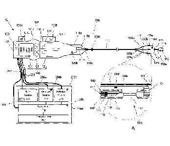
[050] Fig. 1 is a side view of an injectate delivery system comprising a
fluid delivery
assembly and an injectate delivery device, wherein the injectate delivery
device includes a
proximal handle with operator activated controls and a distal array of fluid
delivery elements,
consistent with the present inventive concepts.
[051] Fig. 1A is a magnified side sectional view of a tissue port of the
injectate delivery
device of Fig. 1, consistent with the present inventive concepts.
[052] Fig. 2A is a side view of a force limiting assembly, consistent with
the present
inventive concepts.
[053] Fig. 2B is a side sectional view of a segment of shaft of an
injectate delivery device
oriented in a curved geometry, consistent with the present inventive concepts.
[054] Fig. 2C is an end sectional view of a portion of a shaft of an
injectate delivery
device, consistent with the present inventive concepts.
[055] Fig. 3 is a side sectional view and a magnified side sectional view
of the proximal
and distal portions, respectively, of an injectate delivery device, consistent
with the present
inventive concepts.
-15-
CA 02941414 2016-08-31
WO 2015/148541 PCT/US2015/022293
[056] Figs. 4A-4D are a series of side sectional anatomical views of the
distal portion of
an injectate delivery device delivering injectate into tissue that has been
captured by a tissue
port, consistent with the present inventive concepts.
[057] Figs. 5A, 5B and 5C are a series of side sectional and end anatomical
views of a
segment of luminal wall tissue, prior to, during and after full
circumferential tissue
expansion, respectively, consistent with the present inventive concepts.
[058] Figs. 6A and 6B are side sectional and end sectional views,
respectively, of the
distal portion of an injectate delivery device including a quadrifurcated
shaft, consistent with
the present inventive concepts.
[059] Fig. 7 is a schematic view of an injectate delivery system,
consistent with the
present inventive concepts.
[060] Fig. 8 is a side view of the distal portion of an injectate delivery
device including
multiple shafts arranged in a helix, consistent with the present inventive
concepts.
[061] Fig. 9 is a side sectional view of the distal portion of an injectate
delivery device
including a fluid delivery element positioned and oriented to penetrate tissue
as tissue is
captured within a tissue port, consistent with the present inventive concepts.
[062] Fig. 9A is a side sectional anatomical view of the injectate delivery
device of Fig. 9
after tissue has been captured into the tissue port and the fluid delivery
element has
penetrated the tissue, consistent with the present inventive concepts.
[063] Fig. 10A and 10B are side sectional anatomical views of the distal
portion of an
injectate delivery device prior to and after translation of a tissue port
carriage via applied
vacuum, consistent with the present inventive concepts.
[064] Fig. HA and 11B are side sectional anatomical views of the distal
portion of an
injectate delivery device prior to and after translation of a tissue port
carriage via retraction of
a control rod, consistent with the present inventive concepts.
[065] Fig. 12 is a side view of a portion of a handle of an injectate
delivery device that is
operably attached to a separate device and configured to control one or more
functions of the
separate device, consistent with the present inventive concepts.
DETAILED DESCRIPTION OF THE DRAWINGS
[066] The terminology used herein is for the purpose of describing
particular
embodiments and is not intended to be limiting of the inventive concepts.
Furthermore,
-16-
CA 02941414 2016-08-31
WO 2015/148541 PCT/US2015/022293
embodiments of the present inventive concepts may include several novel
features, no single
one of which is solely responsible for its desirable attributes or which is
essential to
practicing an inventive concept described herein. As used herein, the singular
forms "a,"
"an" and "the" are intended to include the plural forms as well, unless the
context clearly
indicates otherwise.
[067] It will be further understood that the words "comprising" (and any
form of
comprising, such as "comprise" and "comprises" ), "having" (and any form of
having, such as
"have" and "has"), "including" (and any form of including, such as "includes"
and "include")
or "containing" (and any form of containing, such as "contains" and "contain")
when used
herein, specify the presence of stated features, integers, steps, operations,
elements, and/or
components, but do not preclude the presence or addition of one or more other
features,
integers, steps, operations, elements, components, and/or groups thereof.
[068] It will be understood that, although the terms first, second, third
etc. may be used
herein to describe various limitations, elements, components, regions, layers
and/or sections,
these limitations, elements, components, regions, layers and/or sections
should not be limited
by these terms. These terms are only used to distinguish one limitation,
element, component,
region, layer or section from another limitation, element, component, region,
layer or section.
Thus, a first limitation, element, component, region, layer or section
discussed below could
be termed a second limitation, element, component, region, layer or section
without departing
from the teachings of the present application.
[069] It will be further understood that when an element is referred to as
being "on",
"attached", "connected" or "coupled" to another element, it can be directly on
or above, or
connected or coupled to, the other element, or one or more intervening
elements can be
present. In contrast, when an element is referred to as being -directly on",
"directly
attached", -directly connected" or "directly coupled" to another element,
there are no
intervening elements present. Other words used to describe the relationship
between
elements should be interpreted in a like fashion (e.g., "between" versus
"directly between,"
"adjacent" versus "directly adjacent," etc.).
[070] It will be further understood that when a first element is referred
to as being "in",
"on" and/or "within" a second element, the first element can be positioned:
within an internal
space of the second element, within a portion of the second element (e.g.
within a wall of the
second element); positioned on an external and/or internal surface of the
second element; and
combinations of one or more of these.
-17-
CA 02941414 2016-08-31
WO 2015/148541
PCT/US2015/022293
[071] Spatially relative terms, such as "beneath." "below," "lower,"
"above," "upper" and
the like may be used to describe an element and/or feature's relationship to
another element(s)
and/or feature(s) as, for example, illustrated in the figures. It will be
understood that the
spatially relative terms are intended to encompass different orientations of
the device in use
and/or operation in addition to the orientation depicted in the figures. For
example, if the
device in a figure is turned over, elements described as "below" and/or
"beneath" other
elements or features would then be oriented "above" the other elements or
features. The
device can be otherwise oriented (e.g., rotated 90 degrees or at other
orientations) and the
spatially relative descriptors used herein interpreted accordingly.
[072] The term "and/or" where used herein is to be taken as specific
disclosure of each of
the two specified features or components with or without the other. For
example "A and/or
B" is to be taken as specific disclosure of each of (i) A, (ii) B and (iii) A
and B. just as if each
is set out individually herein.
[073] It is appreciated that certain features of the invention, which are,
for clarity,
described in the context of separate embodiments, may also be provided in
combination in a
single embodiment. Conversely, various features of the invention which are,
for brevity,
described in the context of a single embodiment, may also be provided
separately or in any
suitable sub-combination. For example, it will be appreciated that all
features set out in any
of the claims (whether independent or dependent) can be combined in any given
way.
[074] As described herein, "room pressure" shall mean pressure of the
environment
surrounding the systems and devices of the present inventive concepts.
Positive pressure
includes pressure above room pressure or simply a pressure that is greater
than another
pressure, such as a positive differential pressure across a fluid pathway
component such as a
valve. Negative pressure includes pressure below room pressure or a pressure
that is less
than another pressure, such as a negative differential pressure across a fluid
component
pathway such as a valve. Negative pressure can include a vacuum but does not
imply a
pressure below a vacuum. As used herein, the term "vacuum" can be used to
refer to a full or
partial vacuum, or any negative pressure as described hereabove. As used
herein, the term
"vacuum level" refers to a measure of a vacuum wherein the lower the pressure,
the greater
the vacuum level.
[075] The term "diameter" where used herein to describe a non-circular
geometry is to be
taken as the diameter of a hypothetical circle approximating the geometry
being described.
For example, when describing a cross section, such as the cross section of a
component, the
-18-
CA 02941414 2016-08-31
WO 2015/148541 PCT/US2015/022293
term "diameter" shall be taken to represent the diameter of a hypothetical
circle with the
same cross sectional area as the cross section of the component being
described.
[076] It is an object of the present inventive concepts to provide devices,
systems, and
methods to safely and effectively expand an area of tissue, such as one or
more layers of a
portion of tubular or solid tissue, such as tissue of an organ or tissue of
the gastrointestinal
(GI) tract of a patient. The expanded tissue can comprise one or more
submucosal layers of
tissue, such as one or more full or partial circumferential submucosal layers
of one or more
segments (e.g. one or more axial segments) of the duodenum. The devices and
systems of the
present inventive concepts include one or more fluid delivery elements, such
as needles or
water jets configured to deliver one or more fluids to target tissue, to
expand the target tissue
and/or tissue proximate the target tissue (hereinafter "target tissue").
Needles can comprise
hollow or partially hollow needles, such as needles with one or more openings
at the distal
end and/or at a side wall location. One or more visualization assemblies (e.g.
an endoscope
camera or other camera, an ultrasound imager, and the like) can be included,
such as to allow
an operator to visualize or otherwise assess the tissue expansion or other
injectate delivery
procedure (e.g. when the delivered fluid includes a dye or is otherwise
visible). One or more
tissue manipulation assemblies can be included, such as to apply a force to
enhance or
otherwise modify the injectate delivery.
[077] In some embodiments, a vacuum or other negative pressure can be used
to
manipulate tissue and/or to maintain proximity between a portion of an
injectate delivery
device or assembly, and tissue. This vacuum or other negative pressure can
comprise a
pressure below another pressure, such as a pressure below the pressure of the
environment
surrounding the patient, hereinafter referred to as a "vacuum" or "vacuum
pressure". The
vacuum can be provided by one or more vacuum sources, such as via one or more
operator
adjustable vacuum sources.
[078] In some embodiments, the injectate delivery is performed prior to
treatment of
tissue, such as a tissue treatment comprising an ablation of a target volume
of tissue. The
devices and systems of the present invention can further include one or more
ablation
devices, such as ablation devices configured to treat a layer of tissue
proximate (e.g. above or
below) a previously expanded tissue layer, such as to prevent damage to one or
more tissue
layers below or above the expanded tissue layer. In these embodiments, the
expanded tissue
layer acts as a safety volume of tissue, reducing the specificity of the
ablation required and/or
the need to protect the underlying tissue from damage.
-19-
CA 02941414 2016-08-31
WO 2015/148541 PCT/US2015/022293
[079] The injectate delivery systems of the present inventive concepts can
include an
injectate delivery device constructed and arranged for insertion into a
patient, as well as a
fluid delivery assembly operably (e.g. fluidly and/or electrically) attached
to the injectate
delivery device. The injectate delivery device can include one or more fluid
delivery
elements. The handle of the injectate delivery device can comprise one or more
controls
configured to control the injectate delivery device and/or the fluid delivery
assembly, such as
via a wired or wireless connection. The injectate delivery system can further
include a tissue
ablation device, such as a hot fluid or radiofrequency (RF) ablation device.
[080] Referring now to Fig. 1, a side view of an injectate delivery system
comprising a
fluid delivery assembly and an injectate delivery device is illustrated,
wherein the injectate
delivery device includes a proximal handle with operator activated controls
and a distal array
of fluid delivery elements, consistent with the present inventive concepts.
System 10
comprises an injectate delivery device, device 100, and an assembly for
delivering one or
more fluids, at positive or negative pressure, to device 100, fluid delivery
assembly 200.
Device 100 can be constructed and arranged for insertion into the body of a
patient, such as
through a channel of an endoscope (e.g. an endoscope inserted through the
mouth of a patient
and accessing a GI location such as the duodenum), through the channel of a
laparoscopic
port (e.g. a laparoscopic port accessing the GI tract or an organ of the
patient), and/or over a
guidewire (e.g. over a guidewire placed outside of but parallel to an
endoscope accessing a
GI location). Body-contacting and/or body-inserted components of device 100
can be
constructed of one or more biocompatible materials. System 10 and/or device
100 can be
constructed and arranged to deliver fluid to tissue to perform one or more
functions. In some
embodiments, system 10 and/or device 100 is constructed and arranged to
deliver injectate to
expand one or more layers of tissue prior to a tissue treatment procedure. For
example,
submucosal tissue of the duodenum or other GI tract location can be expanded
prior to
ablating neighboring mucosal tissue, such as is described herebelow in
reference to Fig. 7.
Alternatively or additionally, system 10 and/or device 100 can be constructed
and arranged to
deliver fluid to submucosal blood vessels to damage, denature or otherwise
treat mucosa'
tissue to cause a therapeutic benefit. Alternatively or additionally, system
10 and/or device
100 can be constructed and arranged to create a therapeutic restriction, such
as a restriction
configured to treat a disease or disorder such as obesity, such as is
described in applicant's
co-pending International Patent Application Serial Number PCT/US2014/066829,
entitled
"Systems, Devices and Methods for the Creation of a Therapeutic Restriction in
the
-20-
Gastrointestinal Tract", filed November 21, 2014.
[081] Device 100 includes shaft 110, which can comprise a single shaft
including one or
more lumens, or multiple shafts (e.g. each including one or more lumens) whose
external
walls can be attached along at least a portion of the length of shaft 110. At
the proximal end
of shaft 110 is handle 101. On the distal end or on a distal portion of shaft
110 is expandable
assembly 130. In the embodiment of Fig. 1, shaft 110 comprises 4 shafts,
shafts 110a, 110b,
110c and 110d, whose proximal portions diverge from each other at a location
proximate
handle 101 as shown. The distal portions of shaft 110a, 110b, 110c and 110d
can also
diverge from each other. Shafts 110a-c of Fig.. 1 extend in a curved,
diverging arrangement
to attach to the surface of expandable assembly 130, such as in an arrangement
with equal
spacing (e.g. 120 apart for three shafts 110a-c). Shaft 110d diverges from
shafts 110a-c but
continues in a relatively straight direction attaching to the proximal end of
expandable
assembly 130 (distal portion of shaft 110d not shown as it is hidden by the
distal portion of
shaft 110b.
[082] In some embodiments, system 10 and/or device 100 are of similar
construction
and arrangement to the system and device of applicant's co-pending United
States Patent
Application Serial Number 14/515,324, entitled "Tissue Expansion Devices,
Systems and
Methods", filed October 15, 2014.
In some embodiments. system 10 and/or device 100 are of similar
construction and arrangement to system 10 and/or device 100 described
herebelow in
reference to Fig. 7.
[083] Expandable assembly 130 comprises an expandable element 131, such as
a balloon,
deployable cage, or set of radially deployable arms. Expandable assembly 130
can comprise
one or more tissue capture ports, such as the three ports 135a, 135b and 135c
(singly or
collectively port 135) shown in Fig. 1 with relatively equivalent (e.g. 120 )
spacing.
Expandable assembly 130 can comprise a single tissue capture port 135, or it
can comprise
between two and ten tissue capture ports 135. One or more portions of each
port 135 can
comprise a radiopaque portion. Shaft 110 can further comprises a distal
segment, shaft 110e,
attached to a distal portion of expandable assembly 130 as shown. An
atraumatic tip,
bulbous tip 115, can be mounted to the distal end and/or a distal portion of
shaft 110e. In
some embodiments, bulbous tip 115 comprises a diameter between 4mm and 9mm,
such as a
diameter between 4mm and 6mm. In some embodiments, bulbous tip 115 comprises
at least
a radiopaque portion. Bulbous tip 115 can comprise a passageway, guidewire
lumen 116,
-21-
Date Recue/Date Received 2021-09-03
CA 02941414 2016-08-31
WO 2015/148541 PCT/US2015/022293
passing from a proximal to distal portion of bulbous tip 115, such that device
100 can be
advanced over a guidewire passing through lumen 116. In some embodiments,
lumen 116
comprises a diameter of approximately 0.040" to 0.050" (e.g. to accommodate a
0.035" or
0.038" diameter guidewire). System 10 can include a guidewire for insertion
through lumen
116 and over-the-wire advancement of device 100, such as a guidewire selected
from the
group consisting of: an 0.35" guidewire; an 0.038" guidewire; a guidewire
relatively similar
to an Amplatz Super Stiff guidewire; a guidewire relatively similar to a
Wallstent Super Stiff
guidewire; a guidewire relatively similar to a Dreamwire Stiff Shaft
guidewire; and
combinations of these. In some embodiments, guidewire lumen 116 is parallel to
and off
center from the central axis of the distal portion of shaft 110e. In other
embodiments,
guidewire lumen 116 is not parallel to the central axis of the distal portion
of shaft 110e. In
some embodiments, guidewire lumen 116 passes through one or more portions of
shaft 110,
such as a guidewire lumen 116 which is in the relative center of shaft 110
and/or travels
proximally to exit a port positioned on handle 101.
[084] Referring additionally to Fig. 1A, a magnified view of tissue capture
port 135c of
expandable assembly 130 is illustrated, consistent with the present inventive
concepts. Port
135c can be positioned in and/or on a distal portion of shaft 110c as shown.
The distal
portion of shaft 110c can be attached to expandable element 131, such as via
adhesive or
other attachment element (e.g. a flexible attachment element). Shaft 110c can
comprise one
or more lumens, such as lumen 111 constructed and arranged for attachment to a
vacuum
source, and lumen 112 constructed and arranged to slidingly receive a fluid
delivery tube
(e.g. fluid delivery tube 137 described herebelow). Lumens 111 and 112 can
each comprise a
cross sectional profile as described herebelow in reference to Fig. 2C.
[085] Port 135c comprises an opening 136 in the wall of shaft 110c, which
is in fluid
communication with vacuum lumen 111. An advanceable needle or other fluid
delivery
element, fluid delivery element 132, is constructed and arranged to be
advanced into opening
136 as described herebelow in reference to Figs. 4A-4D. Fluid delivery element
132 can
comprise a fluid delivery element selected from the group consisting of:
needle; water jet;
iontophoretic fluid delivery element; and combinations of these. In some
embodiments, one
or more fluid delivery elements 132 comprise a needle, such as a curved or
relatively straight
needle with a diameter greater than 30ga, or greater than 27ga. In some
embodiments, fluid
delivery element 132 can remain stationary while tissue is brought toward
fluid delivery
element 132, such as is described herebelow in reference to Figs. 9, 9A, 10A,
10B. 11A and
11C.
-22-
CA 02941414 2016-08-31
WO 2015/148541 PCT/US2015/022293
[086] Fluid delivery element 132 includes lumen 138 which is fluidly
attached to fluid
delivery tube 137. In some embodiments, fluid delivery element 132 comprises a
needle with
an outer diameter of approximately 0.016" and lumen 138 comprises an outer
diameter of
approximately 0.008". In some embodiments, fluid delivery tube 137 comprises a
polyimide
tube, such as a tube with an outer diameter of approximately 0.022" and/or an
inner diameter
of approximately 0.016". Fluid delivery tube 137 is slidingly received by
lumen 111 of shaft
110c, and travels proximally to handle 101. Fluid delivery element 132 and
fluid delivery
tube 137 can be fluidly attached at any location within shaft 110c or handle
101. Fluid
delivery tubes 137 can be constructed and arranged to avoid or at least
minimize radial
expansion, such as when fluid delivery tube 137 comprises a braided tube such
as a braided
polyimide tube.
[087] Fluid delivery element 132 and/or fluid delivery tube 137 can be
surrounded by
collar 133, as shown. Lumen 112 comprises two projections which extend into
lumen 112,
proximal stop 134a and distal stop 134b. Device 100 is constructed and
arranged such that
fluid delivery element 132 and the distal end of fluid delivery tube 137 can
advance distally
until collar 133 contacts distal stop 134b, and each can retract proximally
until collar 133
contacts proximal stop 134a.
[088] In some embodiments, tissue capture ports 135a and/or 135b can be of
similar
construction and arrangement and/or include similar components to tissue
capture port 135c
as described hereabove, such as to include an opening 136 which is fluidly
attached to a
corresponding vacuum lumen 111 and can be constructed and arranged to receive
a
corresponding fluid delivery element 132 whose travel is limited by contact of
a collar 133
with a mechanical stop 134a and/or 134b.
[089] In some embodiments, one or more of tissue capture ports 135a-c
comprise an
opening 136 with a length of at least 0.1", such as a length between 0.14" and
0.20", such as
a length of approximately 0.16". In some embodiments, one or more tissue
capture ports
135a-c comprise an opening with a width of at least 0.04", such as a width
between 0.05" and
0.08", such as a width of approximately 0.06". In some embodiments, one or
more of tissue
capture ports 135a-c comprise a tissue-capture depth of at least 0.05", such
as a depth
between 0.06" and 0.10", such as a depth of approximately 0.08".
[090] Fluid delivery assembly 200 comprises a controller 210 and one or
more fluid
transfer mechanisms (e.g. mechanisms to transfer fluid in and/or out of device
100), such as
fluid source 220, vacuum source 230 and/or inflation source 240. Controller
210 comprises
one or more electronic modules, power sources and/or fluid control components
(e.g. valves
-23-
CA 02941414 2016-08-31
WO 2015/148541 PCT/US2015/022293
and/or pumps) configured to initiate, regulate, modify, stop and/or otherwise
control fluid
source 220, vacuum source 230 and/or inflation source 240. In some
embodiments,
expandable element 131 of expandable assembly 130 comprises a balloon, and
inflation
source 240 is constructed and arranged to inflate and/or deflate expandable
element 131. In
these embodiments, inflation source 240 can comprise a source of fluid such as
a liquid (e.g.
saline or water) and/or gas (e.g. air) that is fluidly attached to one or more
tubes 201 which is
in turn fluidly attached to an inflation lumen of shaft 110d, which is fluidly
attached to a
balloon-based expandable element 131. In some embodiments, shaft 110d
comprises an
inflation lumen with a cross sectional area of between 1.5mm2 and 1.9mm2, such
as an
inflation lumen with a cross sectional area of approximately 1.7mm2 when shaft
110d
comprises a diameter of approximately 0.090". Controller 210 can operably
attach to one or
more components of device 100 via cable 202, such that user interface 205 can
be used to
control one or more components of device 100.
[091] Vacuum source 230 is fluidly attached via one or more tubes 201 to
one or more
vacuum lumens 111 of shafts 110a, 110b and 110c as described hereabove. Vacuum
source
230 can be constructed and arranged to manipulate tissue into one or more of
tissue capture
ports 135a, 135b and/or 135c (e.g. to cause tissue to tend toward the
associated fluid delivery
element 132) as described herebelow in reference to Figs. 4A-4D. In some
embodiments,
vacuum source 230 provides a vacuum at a pressure between 22mmHg and 27mmHg.
In
some embodiments, vacuum source 230 provides a vacuum to multiple tissue
capture ports
135 individually, such as via individual tubes 201 connected to independent
lumens 111.
Alternatively, multiple tissue capture ports 135 can be fed by a single tube
201 and/or a
single lumen 111. In some embodiments, vacuum source 230 is constructed and
arranged to
apply a reduced vacuum pressure or a positive pressure to one or more tissue
capture ports
135, such as to discharge or at least release tissue from within tissue
capture port 135 and/or
to flush any material from lumen 111 and/or tissue capture port 135. In some
embodiments,
the positive pressure can be applied (e.g. via a control of user interface 105
and/or 205), to
multiple tissue capture ports 135 independently. In some embodiments, a first
control of user
interface 105 and/or 205 is used to initiate a vacuum and a second, separate
control is used to
initiate the positive pressure.
[092] Fluid source 220 is fluidly attached via one or more tubes 201 to the
lumen of
one or more fluid delivery tubes of device 100, such as a lumen of a fluid
delivery tube 137
positioned within shaft 110a, 110b and/or 110c, which is fluidly attached to a
corresponding
fluid delivery element 132. Fluid source 220 is constructed and arranged to
deliver fluid or
-24-
other injectate to one or more fluid delivery elements 132, such as to expand
tissue, as
described herein. In some embodiments, fluid source 220 provides fluid to
multiple fluid
delivery elements 132 individually, such as via individual tubes 201 connected
to
independent fluid delivery tubes 137. Alternatively, multiple fluid delivery
elements 132 can
be fed by a single tube 201 and/or a single fluid delivery tube 137. In some
embodiments,
system 10 comprises one or more fluids, injectate 221, to be delivered by
fluid source 220 to
one or more fluid delivery elements 132 to expand tissue. Injectate 221 can
include one or
more fluids selected from the group consisting of: water; saline; fluid with a
dye such as a
visible dye such as indigo carmine; methylene blue; India ink; SPOTTm dye; a
gel; a
hydrogel; a protein hydrogel; a fluid containing a visualizable media such as
a media
visualizable under X-ray, ultrasound and/or magnetic resonance imaging; and
combinations
of these. In some embodiments, injectate 221 can comprise a material
constructed and
arranged to cause a narrowing or other restriction that results in a
therapeutic benefit to the
patient, such as is described in applicant's co-pending International Patent
Application Serial
Number PCT/US2014/066829, entitled "Systems, Devices and Methods for the
Creation of a
Therapeutic Restriction in the Gastrointestinal Tract", filed November 21,
2014.
In these embodiments,
injectate 221 can comprise a material configured to remain in place (e.g.
within one or more
tissue layers of the GI tract) for an extended period of time, such as at
least 1 day, 1 week, 1
month, 3 months or 6 months. Injectate 221 can comprise a biopolymer (e.g.
ethylene vinyl
alcohol) and/or an adhesive (e.g. cyanoacrylate).
[093] Handle 101 can comprise user interface 105 comprising one or more
controls for
initiating, modifying, stopping and/or otherwise operating one or more
functions of device
100 and/or fluid delivery assembly 200. User interface 105 can include a
control, slide 102,
constructed and arranged to advance and retract fluid delivery elements 132
into, out of,
and/or within the respective tissue capture ports 135. In some embodiments,
device 100 is
constructed and arranged to advance one or more fluid delivery elements 132
approximately
4rnm. In some embodiments, device 100 is constructed and ananged to advance
one or more
fluid delivery elements 132 at least lmm or at least 2mm. In some embodiments,
device 100
is constructed and arranged to advance one or more fluid delivery elements 132
a distance of
no more than 6mm or no more than 5mm. User interface 105 can include one or
more
electrical and/or mechanical controls, such as buttons 103 (3 shown in Fig.
1), configured to
initiate, regulate modify, stop and/or otherwise control one or more functions
of device 100
and/or fluid delivery assembly 200. User interface 105 can include a display
104, such as an
-25-
Date Recue/Date Received 2021-09-03
CA 02941414 2016-08-31
WO 2015/148541 PCT/US2015/022293
LCD display, video display and/or touchscreen configured to provide
information to an
operator of system 10 and/or receive instructions (e.g. commands) from an
operator of system
10. User interface 105 can include numerous user input components, such as a
user input
component selected from the group consisting of: touchscreen; keyboard; mouse;
joystick;
switch; and combinations thereof. In some embodiments, display 104 comprises a
touchscreen or other user input component configured to allow an operator to
initiate,
regulate, modify, stop and/or otherwise control one or more functions of
device 100 and/or
fluid delivery assembly 200. User interface 105 can further comprise one or
more user output
components, such as a component selected from the group consisting of:
display; light such
as an LED; tactile transducer such as a vibrational transducer; audio
transducer; and
combinations of these. In some embodiments, user interface 105 can include a
user output
component configured to display information selected from the group consisting
of: fluid
delivery element 132 position (e.g. advanced or retracted); vacuum status
(e.g. vacuum level
or pressure within lumen 111 and/or tissue capture port 135); occlusion status
such as
occlusion present in fluid delivery tube 137, lumen 111 and/or tissue capture
port 135;
expandable element status (e.g. radially compacted, partially expanded, fully
expanded or
expansion level); volume of fluid injected by one or more individual fluid
delivery elements
132; total injected volume of fluid; pressure of injection; catheter position
(such as catheter
position relative to the papilla); number of completed injections; and
combinations thereof.
In some embodiments, user interface 105 comprises one or more user output
components
used to display a visual image, such as an image of the GI lumen, such as an
image provided
by an endoscope or camera assembly 119 of device 100, described herebelow. In
some
embodiments, buttons 103 and/or display 104 are used to control fluid source
220, vacuum
source 230, inflation source 240 and/or another component of fluid delivery
assembly 200.
[094] In some embodiments, buttons 103, display 104 and/or another control
of user
interface 105 are configured to allow an operator to activate a supply of
vacuum provided by
vacuum source 230, such as to cause tissue to move or otherwise tend toward a
tissue capture
port 135 and/or a fluid delivery element 132 as described in detail herebelow.
In some
embodiments, buttons 103, display 104 and/or another control of user interface
105 are
configured to allow an operator to initiate, regulate, modify, stop and/or
otherwise control the
flow of fluid through one or more fluid delivery elements 132. In some
embodiments,
buttons 103, display 104 and/or another control of user interface 105 are
configured to allow
an operator to radially expand and/or radially compact expandable assembly
130. In some
embodiments, buttons 103, display 104 and/or another control of user interface
105 are
-26-
CA 02941414 2016-08-31
WO 2015/148541 PCT/US2015/022293
configured to allow an operator to modify a fluid delivery parameter selected
from the group
consisting of: flow rate of tissue expanding fluid; duration of tissue
expanding fluid flow;
volume of tissue expanding fluid; temperature of tissue expanding fluid;
pressure of tissue
expanding fluid; a tissue expanding fluid threshold parameter level (e.g.
maximum or
minimum flow rate, duration, volume, temperature and/or pressure); type of
tissue expanding
fluid; and combinations thereof. In some embodiments, buttons 103. display 104
and/or
another control of user interface 105 are configured to allow an operator to
modify a
parameter related to one or more of: fluid source 220 (e.g. fluid flow rate,
fluid volume or
fluid pressure); vacuum source 230 (e.g. vacuum pressure); and/or inflation
source 240 (e.g.
inflation flow rate; inflation volume or inflation pressure). In some
embodiments, fluid
delivery assembly 200 is further constructed and arranged to provide ablation
energy to treat
tissue, and user interface 105 comprises one or more controls to adjust one or
more ablation
parameters, such as is described herebelow in reference to Fig. 7.
[095] In some embodiments, one or more controls of user interface 105 is
biased to tend
towards one state, such as a bias towards a state selected from the group
consisting of: on
state such as a state in which fluid is flowing and/or vacuum is applied; off
state such as a
state in which fluid is not flowing and/or vacuum is not applied; advanced
state such as a
state in which one or more fluid delivery elements are advanced into tissue
capture port 135;
retracted state such as a state in which one or more fluid delivery elements
are retracted from
tissue capture port 135; and combinations of these. The bias to one or more
controls of user
interface 105 can be a mechanical bias (e.g. via a spring as described
herebelow in reference
to Fig. 2A) or an electronic bias (e.g. via a pre-determined state in memory
of electronics
module 107). In some embodiments, a mechanical control or electronic control
of user
interface 105 is biased in an off state, such that fluid delivery from fluid
source 220 is not
initiated until the control is activated by an operator of system 10. In some
embodiments, a
mechanical control or electronic control of user interface 105 is biased in an
off state, such
that application of vacuum to one or more lumens 111 via vacuum source 230 is
not initiated
until the control is activated by an operator of system 10. In some
embodiments, a
mechanical control or electronic control of user interface 105 is biased in an
off state, such
that inflation of expandable assembly 130 via inflation source 240 is not
initiated until the
control is activated by an operator of system 10.
[096] In some embodiments, display 104 is configured to provide status
information
regarding one or more parameters of device 100 and/or fluid delivery assembly
200. In these
embodiments, parameter information can comprise information selected from the
group
-27-
CA 02941414 2016-08-31
WO 2015/148541 PCT/US2015/022293
consisting of: flow rate such as flow rate delivered from fluid source 220 or
inflation source
240 and/or to one or more fluid delivery elements 132 or expandable assembly
130; pressure
such as pressure of fluid delivered from fluid source 220 or inflation source
240 or pressure
of fluid within a lumen 138 or expandable assembly 130; temperature such as
temperature of
fluid delivered from fluid source 220 or inflation source 240; volume such as
volume of fluid
within fluid source 220, within inflation source 240, delivered by a fluid
delivery element 132
or contained within expandable assembly 130; and combinations of these.
Buttons 103
and/or display 104 can be electrically or otherwise operably attached to cable
202 which can
comprise one or more electrical wires, optical fibers and/or hollow tubes
(e.g. hydraulic or
pneumatic control tubes) that operably attach to controller 210, fluid source
220, vacuum
source 230 and/or inflation source 240.
[097] In some embodiments, fluid delivery assembly 200 comprises one or
more operator
controls and/or information display elements, such as when fluid delivery
assembly 200
comprises user interface 205 comprising one or more components selected from
the group
consisting of: an electrical control; a mechanical control; a switch such as
an electrical switch
or a mechanical switch; a button; a knob; a lever; a display; a touchscreen;
and combinations
of these. Information provided by user interface 205 and/or controls
accessible via user
interface 205 can be separate from or similar to (e.g. redundant with) the
information
displayed and control provided by buttons 103 and/or display 104 of device
100.
[098] In some embodiments, slide 102 can be attached to the fluid delivery
tubes 137 via
force-limiting assembly 140. Force limiting assembly 140 can be constructed
and arranged to
limit the force applied by slide 102 onto the fluid delivery tubes 137 and/or
to limit the travel
(e.g. forward and/or reverse travel) of at least the proximal portion of fluid
delivery tubes
137. In some embodiments, force-limiting assembly 140 is constructed and
arranged as
described herebelow in reference to force limiting assembly 140 of Fig. 2A
and/or force
limiting assembly 340 of Fig. 3.
[099] In some embodiments, handle 101 comprises one or more attachment
elements,
such attachment element 106a and/or 106b. Attachment elements 106a and/or 106b
can be
constructed and arranged to attach handle 101 to another device, such as to
the proximal end
of an endoscope, such as to the biopsy port of an endoscope. Attachment
elements 106a
and/or 106b can comprise one or more mechanical and/or electromechanical
attachment
elements, such as an element selected from the group consisting of: clip;
clamp; strap;
electromagnetic coupler such as a solenoid-based clamp; adhesive strip; and
combinations
thereof. In some embodiments, attachment elements 106a and/or 106b can be
operably
-28-
CA 02941414 2016-08-31
WO 2015/148541 PCT/US2015/022293
connected (e.g. mechanically linked), with one or more controls of the
attached device. In
these embodiments, attachment elements 106a and/or 106b can comprise a control
121a
and/or 12 lb. respectively. Control 121a and/or 121b can be operably connected
to an
insufflation vacuum control knob and/or a flush control knob of the attached
device, such as
to activate insufflation or flushing functions of the attached device. Control
121a and/or
121b can comprise a knob, push button, lever or other user input component and
an electrical
and/or mechanical mechanism, such as a solenoid, a cam and/or a linkage which
activates a
control of the attached device. Alternatively or additionally. control 121a
and/or 121b can be
positioned within handle 101, such as is described herebelow in reference to
Fig. 12.
[0100] Handle 101 can surround various electrical and mechanical components
and
mechanisms, such as force-limiting assembly 140 described hereabove, as well
as electronics
module 107. In some embodiments, electronics module 107 comprises a component
selected
from the group consisting of: battery; microcontroller; memory circuitry;
wireless transmitter;
wireless receiver; camera such as a CCD camera; optical lens assembly; and
combinations
thereof. In some embodiments, electronics module 107 comprises a wireless
transceiver
configured to send or receive communications (e.g. Bluetooth communications)
with
controller 210 (e.g. to avoid the need for cable 202) and/or with another
device of system 10.
[0101] In some embodiments, handle 101 comprises two connectable portions,
such as
distal portion 101a and proximal portion 101b shown in Fig. 1. In these
embodiments,
system 10 can comprise one or more reusable proximal portions 101b, each of
which that can
be attached to two or more portions 101a, such as when each portion 101a is
used during a
single clinical procedure or at least fewer clinical procedures than its
attached portion 101b.
In these embodiments, certain components (e.g. more expensive components) can
be
positioned in the reusable portion 101b, such as one or more components
selected from the
group consisting of: buttons 103, display 104; electronics module 107; a
printed circuit board;
a transducer such as an audible transducer or a tactile transducer; a light;
an LED; a sensor
such as a magnetic sensor or a hall effect transducer; and combinations
thereof.
[0102] In some embodiments, expandable assembly 130 comprises one or more
camera
components, such as camera assembly 119 shown positioned on the distal end of
expandable
assembly 130 and oriented toward the proximal end of expandable assembly 130.
Camera
assembly 119 can comprise one or more components selected from the group
consisting of: a
camera such as a CCD camera; a lens; a filter; a minor; and combinations
thereof. Camera
assembly 119 can be constructed and arranged to collect an image of tissue
contacted or
otherwise proximate to expandable element 131, and/or an image of one or more
fluid
-29-
CA 02941414 2016-08-31
WO 2015/148541 PCT/US2015/022293
delivery elements 132. In some embodiments, fluid source 220 delivers a fluid
with a visible
agent, such as a dye, such that camera assembly 119 collects an image of
delivered fluid
and/or expanding tissue that is enhanced with the dye. Camera assembly 119 can
be operably
attached to electronics module 107 such as via one or more wires and/or
optical fibers, not
shown but traveling proximally through one or more shafts of shaft 110 and
into handle 101.
In some embodiments, camera assembly 119 is positioned on the proximal end of
expandable
assembly 130 and oriented toward the distal end of expandable assembly 130.
[0103] System 10 can comprise one or more functional elements, such as one or
more of
functional elements 109a-109f (singly or collectively, functional element 109)
and/or
functional elements 209a-209d (single or collectively, functional element 209)
shown in Fig.
1. Each functional element 109 can comprise a sensor, a transducer and/or
other functional
element. In some embodiments, a functional element 109 comprises one or more
sensors
selected from the group consisting of: pressure sensor; temperature sensor;
impedance sensor;
pH sensor; flow sensor; ultrasonic sensor; optical sensor; magnetic sensor;
hall effect sensor;
osmolarity sensor; strain gauge; gas bubble sensor; and combinations of these.
Alternatively
or additionally, a functional element 109 can comprise one or more transducers
selected from
the group consisting of: heating element; audio transducer; vibrational
transducer; light
transducer; magnetic transducer; visual transducer; ultrasound sensor; camera;
and
combinations of these. In some embodiments, a functional element 109 comprises
a pressure
regulator and/or a pressure relief valve, such as when functional element 109
is in fluid
communication with one or more fluid delivery tubes 137 and/or an inflation
lumen of shaft
110d. In some embodiments, a functional element 109 comprises an element
selected from
the group consisting of: a sensor; a transducer; an ablation element such as
one or more
electrodes configured to deliver electrical energy such as radiofrequency (RF)
energy; a fluid
delivery element such as a needle, a fluid jet, a permeable membrane; an exit
port; an
insufflation port; a heating element; a cooling element; and combinations of
these. In some
embodiments, one or more functional elements 109 comprise a visualization
element, such as
to reduce or avoid the need for a separate visualization device such as an
endoscope. In these
embodiments, functional element 109d can comprise a camera and/or a lens
configured to
provide an image of one or more of: expandable assembly 130; one or more
tissue ports 135;
one or more fluid delivery elements 132; shaft 110; tissue proximate
expandable assembly
135 and/or tissue ports 135; and combinations of these. Alternatively or
additionally,
functional element 109d or another functional element 109 can comprise a fluid
delivery
element constructed and arranged to provide and/or remove insufflation fluids.
-30-
CA 02941414 2016-08-31
WO 2015/148541 PCT/US2015/022293
[0104] One or more functional elements 109 can comprise a sensor configured to
detect
occlusion, such as an occlusion in a lumen or other location of device 100. In
some
embodiments, multiple functional elements 109 each comprise a sensor
configured to detect
an occlusion (e.g. via low flow, low pressure, etc.) in one or more fluid
delivery tubes 137
(e.g. collectively or independently), one or more vacuum lumens 111 (e.g.
collectively or
independently), or an occlusion in one or more of tubes 201. In some
embodiments, one or
more functional elements 109 can be configured to detect and/or confirm
adequate flow (e.g.
within one or more fluid delivery tubes 137) and/or adequate vacuum (e.g.
within one or
more lumens 111 and/or tissue capture ports 135), collectively or
independently (e.. via
multiple independent functional elements 109).
[0105] Device 100 can include one or more sensors, transducers and/or other
functional
elements as described hereabove. Device 100 can include one or more functional
elements
positioned in, on and/or within handle 101, such as functional element 109a
positioned in
handle portion 101b (e.g. a reusable portion as described hereabove) and
functional element
109b positioned in handle portion 101a (e.g. a portion of handle 101 used in
fewer clinical
procedures than portion 101b). In some embodiments, functional elements 109a
and/or 109b
comprise a sensor configured to monitor a voltage or current, such as the
voltage or current of
a power supply of electronics module 107. Functional elements 109a and/or 109b
can
comprise a sensor (e.g. an ultrasonic sensor) configured to monitor fluid
flowing through one
or more tubes passing through handle 101, such as to produce a signal
correlating to flow
rate, temperature and/or the presence of one or more gas bubbles present in
fluid passing
through a portion of handle 101. Functional elements 109a and/or 109b can
comprise a
transducer, such as a vibrational or audible transducer used to alert an
operator of an alert or
other condition of system 10. In some embodiments, device 100 and/or system 10
is
constructed and arranged to activate an alert signal delivered by a functional
element 109a
and/or 109b, when one or more of the following conditions occur: vacuum is
applied to one
or more tissue capture ports 135; expandable assembly 130 is radially
expanded; fluid is
being delivered into tissue; and one or more fluid delivery elements 132 are
in an advanced
position.
[0106] Device 100 can further include functional element 109c comprising one
or more
functional elements positioned on, in and/or within shaft 110 as shown.
Functional element
109c can comprise a sensor such as a sensor configured to provide a signal
correlating to one
or more of: flow rate; pressure; presence of a gas bubble; temperature; and
combinations of
-31-
CA 02941414 2016-08-31
WO 2015/148541 PCT/US2015/022293
these. Alternatively or additionally, functional element 109c can comprise a
transducer, such
as a vibrational transducer, a pressure regulator and/or a pressure relief
valve.
[0107] Device 100 can further include functional element 109d, positioned on,
in and/or
within expandable assembly 130 as shown. Functional element 109d can comprise
a sensor
configured to produce a signal correlating to one or more of: pressure;
volume; temperature;
and combinations of these. Functional element 109d can comprise a sensor
configured to
produce a signal correlating to adequate expansion of expandable assembly 130
(e.g.
adequate expansion of expandable element 131 such as when expandable element
131
comprises a balloon). In some embodiments, functional element 109d comprises a
sensor
configured to produce a signal correlating to balloon expansion and/or balloon
pressure and
controller 210 is configured to perform a function based on the produced
signal, the function
selected from the group consisting of: stop fluid infusion when the balloon
pressure reaches
or exceeds a pressure threshold; stop fluid infusion when the balloon pressure
is below a
pressure threshold; expand the balloon until it reaches a pressure threshold,
such as a pressure
of at least 0.4psi or at least 0.8psi; maintain the balloon at a pre-
determined pressure level for
a pre-determined time period prior to beginning delivery of fluid to tissue by
one or more
fluid delivery elements 132; and combinations of these.
[0108] Device 100 can further comprise functional element 109e, positioned on,
in and/or
within lumen 111 as shown. Functional element 109e can comprise a sensor
configured to
produce a signal correlating to one or more of: occlusion; pressure; flow
rate; and
combinations of these.
[0109] Device 100 can further comprise functional element 109f, positioned on,
in and/or
within lumen 112 and/or fluid delivery element 132 as shown. Functional
element 109f can
comprise a sensor configured to produce a signal correlating to one or more
of: flow rate;
pressure; presence of one or more gas bubbles; osmolarity; occlusion;
temperature; and
combinations of these. Functional element 109f can comprise a sensor to
produce a signal
correlating to one or more of: fluid being delivered from fluid delivery tube
137 and/or fluid
delivery element 132 into tissue; fluid delivery tube 137 and/or fluid
delivery element 132 in
an advanced and/or retracted position; position of fluid delivery tube 137
and/or fluid deliver
element 132 such as is described in reference to Fig. 3 herebelow; and
combinations of these.
[0110] In some embodiments, controller 210 and/or electronics module 107 are
configured
to enter an alarm or other alert state when two conditions that are
incompatible occur, such as
when signals provided by one or more functional elements 109 indicate that one
or more of
the following incompatible conditions are present: fluid delivery element 132
is retracted or
-32-
CA 02941414 2016-08-31
WO 2015/148541 PCT/US2015/022293
retracting while fluid is flowing through fluid delivery element 132; fluid
delivery element
132 is advanced and a diameter of expandable assembly 130 is below a
threshold; fluid
delivery element 132 is advanced and vacuum level in tissue capture port 135
and/or lumen
111 is below a threshold; balloon pressure of expandable assembly 130 is below
a first
threshold and vacuum level in a vacuum location such as tissue capture port
135 and/or
lumen 111 is above a second threshold; balloon pressure of expandable assembly
130 is
above a threshold and fluid is flowing (e.g. at a sufficient flow rate)
through fluid delivery
element 132; balloon pressure of expandable assembly 130 is below a threshold
and fluid is
flowing (e.g. at a sufficient flow rate) through fluid delivery element 132;
and combinations
of these.
[0111] Fluid delivery assembly 200 can comprise one or more functional
elements, such as
one or more sensors, transducers and/or other functional elements as described
hereabove.
Fluid delivery assembly 200 can comprise functional elements 209a, 209b and
209c
positioned in, on and/or within fluid source 220, vacuum source 230 and/or
inflation source
240, respectively. In some embodiments, one or more of functional elements
209a-c are
positioned in, on and/or within one or more tubes 201.
[0112] Fluid delivery assembly 200 can comprise functional element 209d
positioned in, on
and/or within controller 210.
[0113] In some embodiments, device 100 further comprises a treatment element,
such as an
ablation element or other treatment element such as is described herebelow in
reference to
Fig. 7. In some embodiments, shaft 110 comprises one or more working channels
or other
lumens, such as a lumen configured to provide insufflation as described
herein. In some
embodiments, device 100 comprises a steering mechanism for deflecting or
otherwise
steering shaft 110, such as is also described herebelow in reference to Fig.
7. In some
embodiments, shaft 110 comprises multiple shafts arranged in a helical
geometry along at
least a portion of the length of shaft 110, such as is described herebelow in
reference to Fig.
8.
[0114] In some embodiments, one or more fluid delivery elements 132 are
positioned and
oriented such that when tissue is drawn into the associated tissue capture
port 135, the tissue
can be penetrated by the fluid delivery element 132 (e.g. without advancement
of the fluid
delivery element 132), such as described herebelow in reference to Figs. 9 and
9A. In these
embodiments, slide 102 and its associated mechanism can be avoided or their
function
reduced. Alternatively or additionally, one or more tissue capture ports 135
can be
constructed and arranged to translate (e.g. be translated by an operator) to
cause tissue
-33-
CA 02941414 2016-08-31
WO 2015/148541 PCT/US2015/022293
captured within port 135 to be penetrated by fluid delivery element 132, such
as is described
herebelow in reference to Figs. 10A, 10B, 11A and 11B.
[0115] Referring now to Fig. 2A, a side view of a particular embodiment of the
force
limiting assembly of Fig. 1 is illustrated, consistent with the present
inventive concepts.
Force limiting assembly 140 comprises one or more compression elements, such
as springs
142a-c. Force limiting assembly 140 further comprises blocks 143a-c, channels
146a-c and
mechanical stops 141 and 148. Device 100 comprises fluid delivery tubes 137a-
c, vacuum
lumens 1 1 la-c, vacuum ports 118a-c, seals 117a-c, ports 144a-c and openings
145a-c. For
illustrative clarity, only reference designations 142a, 143a, 146a, 1 1 la,
118a, 117a, 144a and
145a are listed on Fig. 2A.
[0116] Springs 142a-c can comprise a coil spring and/or other compression
spring.
Channels 146a-c can comprise a relatively uniform recess in handle 101 sized
to allow blocks
143a-c to move within channel 146a-c (e.g. move back and forth within channel
146a-c),
respectively.
[0117] Slide 102 operably attaches to fluid delivery tubes 137a-c via force
limiting
assembly 140, such that slide 102 can translate along axis 147. Slide 102 is
attached to
springs 142a-c. Springs 142a-c are attached to blocks 143a-c, respectively.
Fluid delivery
tubes 137a-c are attached to blocks 143a-c, respectively. When slide 102 is
advanced (i.e.
moved to the right of the page), springs 142a-c elongate and apply a pulling
force to blocks
143a-c, respectively, such that blocks 143a-c translate (to the right) within
channels 146a-c,
respectively, and fluid delivery tubes 137a-c, respectively advance. When
slide 102 is
retracted (i.e. moved to the left of the page), springs 142a-c compress and
apply a pushing
force to blocks 143a-c, respectively, such that blocks 143a-c translate (to
the left) within
channels 146a-c, respectively, and fluid delivery tubes 137a-c, respectively
retract.
[0118] Mechanical stop 141 is positioned to limit the proximal travel of slide
102, while
mechanical stop 148 is positioned to limit the distal travel of slide 102.
Channels 146a-c can
have sufficient length and can be positioned relative to stops 141 and 148
such that blocks
143a-c, respectively, never reach either end of channels 146a-c. Stops 141 and
148 can be
positioned such that springs 142a-c never fully compress (i.e. when slide 102
contacts stop
141) and springs 142a-c never plastically deform (i.e. when slide 102 contacts
stop 148). In
this configuration, the force applied to each of fluid delivery tubes 137a-c
is limited to a
small range of forces applied by springs 142a-c, respectively, experienced
throughout the
travel of slide 102 from stop 141 to stop 148. In this configuration, the
force applied to each
fluid delivery tube 137 via slide 102 is force limited (e.g. to prevent damage
to fluid delivery
-34-
CA 02941414 2016-08-31
WO 2015/148541 PCT/US2015/022293
tube 137 and/or any component attached thereto and/or interfacing therewith),
and
individually compensated (e.g. when shaft 110 is in a curvilinear geometry
such as that
described herebelow and shown in Fig. 2B). In some embodiments, one or more of
springs
142a-c comprise an effective length of between 17mm and 37mm, such as a length
of
approximately 27mm. In some embodiments, one or more of springs 142a-c
comprise a
spring rate of between 2.1 lb/in and 3.1 lb/in, such as a spring rate of
approximately 2.6 lb/in.
In some embodiments, springs 142a-c, blocks 143a-c and/or channels 146a-c are
similar. In
other embodiments, springs 142a-c, blocks 143a-c and/or channels 146a-c are
dissimilar.
[0119] Fluid delivery tubes 137a-c fluidly attach to ports 144a-c,
respectively. Ports 144a-
c also fluidly attach to one or more tubes 201, not shown but described in
reference to Fig. 1
hereabove, fluidly connecting fluid source 220 to fluid delivery tubes 137a-c.
Handle 101
can comprise openings 145a-c which each allow an individual tube 201 to pass
therethrough,
and accommodate translation of blocks 143a-c as slide 102 is moved back and
forth.
[0120] Also shown in Fig. 2A are ports 118a-c, which fluidly attach to lumens
111a-c
respectively. Ports 118a-c also fluidly attach to one or more tubes 201, not
shown but
described in reference to Fig. 1 hereabove, fluidly connecting vacuum source
230 to lumens
111a-c. Handle 101 can comprise one or more openings configured to allow the
associated
tubes 201 to pass therethrough. Lumens 111a-c can each comprise a sealing
element, seal
117a-c, respectively, on the proximal end of lumens 111a-c.
[0121] As described above in reference to Fig. 1, one or more controls of the
devices and
systems of the present inventive concepts can be biased to a particular state,
such as an on
state, an off state, an advanced state and/or a retracted state. In the
embodiment of Fig. 2A, a
control, slide 102 is biased such that fluid delivery tubes 137 and fluid
delivery elements 132
are in the retracted state (i.e. an "off' state) until a force is applied to
slide 102. The bias is
provided by spring 149 which is attached at one end to slide 102 and at the
opposite end to
handle 101 such that when no external force is applied to slide 102, fluid
delivery elements
132 are in the retracted state.
[0122] Referring additionally to Fig. 2B, a sectional side view of a segment
of shaft 110
in a curved geometry is illustrated, consistent with the present inventive
concepts. Fluid
delivery tube 137a is on the inside of the curve shown, while fluid delivery
tube 137c is on
the outside of the curve shown. In this configuration, fluid delivery tube
137c travels a
greater distance to accommodate the curve than does fluid delivery tube 137a.
Force limiting
assembly 140 is constructed and arranged such that the varied distance
traveled can be
-35-
CA 02941414 2016-08-31
WO 2015/148541 PCT/US2015/022293
accommodated, such as when block 143a advances (e.g. to the right of the page)
less than
block 143c to accommodate the curve shown in Fig. 2B.
[0123] Referring additionally to Fig. 2C, an end sectional view of a portion
of shaft 110a
is illustrated, consistent with the present inventive concepts. Shaft 110a
comprises lumen
112a and lumen 111a. Lumen 112a surrounds fluid delivery tube 137a. Lumen 111a
is
fluidly attached to tissue port 135a on its distal end, and sealing element
117a can be
positioned on its proximal end (as shown in Fig. 2A). Lumen 112a can comprise
a relatively
circular geometry as shown, and slidingly receive fluid delivery tube 137a.
Lumen Illa can
comprise a geometry to maximize cross-sectional area of lumen 111 a (e.g. a
non-circular
geometry), such as when shaft 110a comprises a relatively circular outer wall.
Lumen ilia
can comprise a cross sectional area between 0.8mm2 and 2.0mm2, such as a cross
sectional
area of 1.0mm2 when shaft 110a comprises a diameter of approximately 0.090".
[0124] Referring now to Fig. 3, side sectional and magnified side sectional
views of the
proximal and distal portions, respectively, of an injectate delivery including
a force limiting
assembly are illustrated, consistent with the present inventive concepts.
Device 100 includes
force limiting assembly 340 as shown. Device 100 can comprise a single fluid
delivery
element 132 as shown, or multiple fluid delivery elements as described
hereabove in
reference to Figs. 1, 2A and 2B. Force limiting assembly 340 can be positioned
in, on and/or
within handle 101. Force limiting assembly 340 operably attaches to fluid
delivery tube 137
which in turn is fluidly attached to fluid delivery element 132. Fluid
delivery tube 137 is
attached to block 343. Block 343 is positioned in channel 346, typically an
elongate recess in
handle 101 sized to slidingly receive block 343 such that block 343 can
translate back and
forth in direction 347 as shown. Block 343 frictionally engages lead screw 349
such that
rotation of lead screw 349 in a first direction causes block 343 to advance in
channel 346
(and correspondingly advance fluid delivery tube 137 and fluid delivery
element 132), and
rotation of the lead screw 349 in the opposite direction causes lead screw 349
to retract in
channel 346 (and correspondingly retract fluid delivery tube 137 and fluid
delivery element
132).
[0125] Lead screw 349 is driven by motor 350 via clutch 351. Clutch 351 is
constructed
and arranged to limit the force applied to lead screw 349, and thus limit the
push and/or pull
force applied to fluid delivery tube 137 and fluid delivery element 132, such
as to prevent
damage to fluid delivery tube 137, fluid delivery element 132 and/or any
components
attached thereto or interfacing therewith. Electronics module 107 is attached
to motor 350
via cable 352. User interface 105 is configured to control motor 350 such as
to operably
-36-
CA 02941414 2016-08-31
WO 2015/148541 PCT/US2015/022293
advance and/or retract one or more fluid delivery elements 132. This
advancement and/or
retraction can be performed automatically or at least semi-automatically. User
interface 105
and/or electronics module 107 can be of similar construction and arrangement
as
corresponding components of device 100 described hereabove in reference to
Fig. 1.
[0126] Device 100 can further include stops 134a and 134b. Stops 134a and 134b
can be
configured to provide proximal and distal stops which engage collar 133, which
surrounds
fluid delivery tube 137 and/or fluid delivery element 132. In some
embodiments, stops 134a
and/or 134b comprise a functional element 109, such as a sensor configured to
produce a
signal corresponding to proximity of collar 133 to stops 134a and/or 134b
and/or produce a
signal corresponding to force applied by collar 133 to stops 134a and/or 134b.
In these
embodiments, advancement of fluid delivery tube 137 and/or fluid delivery
element 132 can
be made based on the position of collar 133 (e.g. which corresponds to the
position of fluid
delivery element 132). Alternatively or additionally, advancement of fluid
delivery tube 137
and/or fluid delivery element 132 can be made using a force feedback signal
and/or proximity
signal provided by stops 134a and/or 134b (e.g. via one or more wires not
shown but
operably connected to electronics module 107). The force feedback information
can be used
dynamically to adjust the position of block 343 and correspondingly translate
fluid delivery
tube 137 and/or fluid delivery element 132 based on a force measured at a
distal location in
shaft 110a (i.e. at a location proximate port 135a).
[0127] Although the device 100 shown in Fig. 3 comprises a single fluid
delivery element
132 and a force limiting assembly 340 that limits the force applied to a
single fluid delivery
tube 137, device 100 can comprise multiple fluid delivery elements 132 and
force limiting
assembly 340 can be constructed and arranged to limit force applied to
multiple fluid delivery
tubes 137. For example, force limiting assembly 340 can comprise multiple lead
screws 349
which attach to one or more motors 350 via one or more clutches 351.
[0128] Referring now to Figs 4A-4D, a series of steps for delivering fluid
into tissue
captured by a tissue port is illustrated, consistent with the present
inventive concepts. In Figs.
4A-4D, the distal portion of a fluid expanding device 100 is illustrated. In
some
embodiments, fluid expanding device 100 is of similar construction and
arrangement as
device 100 of Figs. 1 and 1A.
[0129] In Fig. 4A, tissue capture port 135 has been positioned proximate a
surface of tissue
T, such as proximate the mucosal layer of a portion of the gastrointestinal
tract, such as the
mucosal layer of the duodenum of a patient. Device 100 can be inserted over a
guidevvire
and/or through a body access device such as a laparoscopic port or endoscope.
Positioning of
-37-
CA 02941414 2016-08-31
WO 2015/148541 PCT/US2015/022293
port 135 at a desired axial location of the GI tract can be accomplished by
advancing or
retracting shaft 110 while using a visualization device such as a camera (e.g.
the camera of an
endoscope) or an imaging instrument such as a fluoroscope, ultrasound imager.
MRI or the
like. Radial positioning of port 135 proximate the surface of tissue can
comprise expanding a
component onto which one or more ports 135 are attached. such as a balloon or
other
expanding element. In some embodiments, one or more tissue capture ports 135
are attached
to an expanding element similar to expanding element 131 described hereabove
in reference
to Fig. 1. Device 100 includes fluid delivery element 132 fluidly attached to
fluid delivery
tube 137. Collar 133 surrounds fluid delivery element 132 and/or fluid
delivery tube 137
which resides in lumen 112. Stops 134a and 134b can be included to limit the
travel of fluid
delivery element 132, all as is described hereabove in reference to Figs. 1,
lA and/or 3.
Tissue capture port 135 includes opening 136 which is in fluid communication
with lumen
11l.
[0130] In Fig. 4B, vacuum has been applied to opening 136 via lumen 111 such
that a
portion of tissue T is captured by (e.g. drawn into or otherwise tends toward)
tissue capture
port 135. Fluid delivery element 132 has been advanced into tissue T, such as
when fluid
delivery element 132 comprises a sharpened needle. Subsequently, one or more
injectates
can be delivered into tissue T. In some embodiments, fluid delivery element
132 comprises a
water jet or iontophoretic element, such that fluid delivery element 132 can
penetrate into
tissue T or simply reside proximate but external to tissue T during delivery
of the injectate.
In some embodiments, advancement of one or more fluid delivery elements 132 is
performed
with a force-limiting mechanism, such as is described hereabove in reference
to force limiting
assembly 140 of Figs. 1 or 2A, or force limiting assembly 340 of Fig. 3. In
some
embodiments, fluid delivery element 132 is advanced at least semi-
automatically, such as via
lead screw 349 and/or motor 350 described hereabove in reference to Fig. 3.
[0131] In Fig. 4C, the injectate has been delivered into tissue T, and fluid
delivery element
132 has been retracted. In some embodiments, retraction of one or more fluid
delivery
elements 132 is performed with a force-limiting mechanism, such as is
described hereabove
in reference to force limiting assembly 140 of Figs. 1 or 2A, or force
limiting assembly 340
of Fig. 3. In some embodiments, fluid delivery element 132 is retracted at
least semi-
automatically, such as via lead screw 349 and/or motor 350 described hereabove
in reference
to Fig. 3.
[0132] In Fig. 4D. the vacuum is released from lumen 111 such that tissue T
evacuates
tissue capture port 135. In some embodiments, a positive pressure is applied
to lumen 111,
-38-
CA 02941414 2016-08-31
WO 2015/148541 PCT/US2015/022293
such as via the fluid delivery assembly 200 of Fig. 1, to discharge tissue T
from tissue capture
port 135. In some embodiments, the circumferential span of tissue expanded in
the steps
illustrated in Figs. 4A-D comprises a circumferential span of approximately
360 of an axial
segment, and/or an axial length of approximately between 2cm and 5cm, such as
between
2cm and 4cm or between 3cm and 5cm.
[0133] Subsequently, the distal portion of device 100 can be repositioned
(e.g. advanced,
retracted and/or rotated), and the steps shown in Figs. 4A-D repeated one or
more additional
times, such as to expand tissue in multiple locations of the gastrointestinal
tract, such as to
substantially expand a submucosal layer of the duodenum comprising a
cumulative axial
length of at least 5cm, at least 10cm, or at least 15cm. In some embodiments,
a cumulative
axial of at least 4cm or at least 5cm is expanded, followed by an ablation of
tissue with an
axial length of at least 3cm. The cumulative axial length of expanded tissue
can comprise a
relatively continuous axial length of the GI tract or a series of two or more
discrete segments.
[0134] Although the device 100 shown in Figs. 4A-D comprises a single fluid
delivery
element 132 and a single tissue capture port 135, device 100 can comprise
multiple fluid
delivery elements 132 and multiple tissue capture ports 135, such as a
construction
comprising a circumferential array of tissue capture ports 135, such as two
tissue capture
ports 135 arranged along a circumference with 180 spacing, two tissue capture
ports 135
arranged along a circumference with 120 spacing, or four tissue capture ports
135 arranged
along a circumference with 90 spacing. In these embodiments, the application
and/or
release of vacuum applied to the multiple tissue capture ports 135, and/or the
advancement
and/or retraction of the multiple fluid delivery elements 132 (e.g. one or
more of the steps
shown in Figs. 4A-D), can be performed simultaneously or sequentially.
[0135] Referring now to Figs. 5A, 5B and 5C, side and end sectional anatomical
views of
a segment of luminal wall tissue are illustrated, prior to, during and after
full circumferential
tissue expansion, respectively, consistent with the present inventive
concepts. In Fig. 5A, a
side and end sectional view of a segment of lumina] wall tissue includes inner
layer Ll, mid
layer L2 and outer layer L3, prior to any expansion by an injectate delivery
device of the
present inventive concepts. In Fig. 5B, a tissue expansion has occurred at a
single location
toward the top of the page as shown, within tissue layer L2. In Fig. 5C, a
tissue expansion
has occurred for a full 360 segment of layer L2. In some embodiments, a full
or near full
circumferential expansion (e.g. greater than approximately 300 of tissue
expansion, greater
than approximately 320 of tissue expansion, or greater than approximately 330
of tissue
expansion), is performed in a relatively single step, such as from multiple
fluid delivery
-39-
CA 02941414 2016-08-31
WO 2015/148541 PCT/US2015/022293
elements. In other embodiments, a full or near full circumferential expansion
is performed in
multiple steps, such as from one or more fluid delivery elements that are
configured to inject
or otherwise deliver fluid in a first step and be rotated in one or more
subsequent steps, each
rotation followed by a delivery of fluid into tissue.
[0136] The expansion of a tissue layer, such as layer L2 of Figs. 5A through
5C, can be
performed to cause a reduction in cross sectional area of the lumen, such as a
reduction to
between 80% and 85% of the pre-expansion cross sectional area (e.g. a 30mm
lumen reduced
to a 25mm lumen), or a reduction to 75% of the pre-expansion cross-sectional
area. In some
embodiments, a pre-expansion cross sectional diameter of approximately 25mm to
28mm is
reduced by between 2mm and 4mm. Some body lumens comprise an inner layer
including a
non-smooth surface, such as the lining of the duodenum or jejunum including
one or more
folds known as the plicae. In some embodiments, the tissue expansion causes
folds such as
plicae to be smoothed and/or widened. This modification can be useful in
subsequent
treatments of the lumen's inner lining, such as to improve the results of one
or more tissue
ablation procedures.
[0137] Numerous forms and locations of patient tissue can be expanded by the
devices,
systems and methods of the present inventive concepts. In some embodiments,
the tissue to
be expanded comprises submucosal tissue, such as submucosal tissue of the
duodenum. The
devices systems and methods of the present inventive concepts can be
constructed and
arranged to avoid expanding one or more layers of tissue, such as when the
muscularis or
serosal layer of the duodenum is prevented from being expanded. Applicable
tissue can
comprise luminal wall tissue or other tissue layers. Applicable tissue
locations to be
expanded can include luminal wall tissue selected from the group consisting
of: a
gastrointestinal tissue layer; a duodenal tissue layer; an esophageal tissue
layer; a jejunal
tissue layer; an ileal tissue layer; a colonic tissue layer; and combinations
of these.
Alternatively or additionally, tissue to be expanded can comprise tissue
selected from the
group consisting of: a stomach tissue layer; a bladder tissue layer; an oral
cavity tissue layer;
a uterine tissue layer; and combinations of these.
[0138] Referring now to Figs. 6A and 6B, side and end sectional views of the
distal
portion of an injectate delivery device including a quadrifurcated shaft is
illustrated,
consistent with the present inventive concepts. Device 100 can have similar
construction and
arrangement to device 100 of Fig. 1, with similar components sharing the same
or like
reference numbers. As shown in Figs. 6A and 6B, the distal portions of shafts
110a, 110b,
110c, and 110d diverge from each other (e.g. the separation beginning at a
location
-40-
CA 02941414 2016-08-31
WO 2015/148541 PCT/US2015/022293
approximately 140mm from the distal end of device 100), creating a space which
is sized and
configured to allow an elongate device, such as an elongate visualization
device, such as
endoscope 50, to be positioned in between at least two of shafts 110a, 110b
and 110c without
applying significant force and/or significantly deflecting any of shafts 110a,
110b and/or
110c and/or expandable assembly 130. Shafts 110a, 110b and/or 110c are
attached to
expandable element 131 of expandable assembly 130. Expandable element 131 can
be a
balloon or other expandable element constructed and arranged to radially
expand to position
ports 135a, 135b and 135c in a circumferential geometry with a diameter of at
least 20mm,
such as a diameter between 25mm and 36mm, a diameter between 28mm and 36mm, or
a
diameter of approximately 32mm.
[0139] In some embodiments, shafts 110a, 110b and/or 110c are oriented such as
to enable
the distal end of endoscope 50 (e.g. a scope with a distal portion diameter
between 7mm and
llmm) to be within 1.5cm, 2.0cm, 3.0cm, or within 9.0cm of ports 135a, 135b
and/or 135c,
such as to provide a visual or other image of ports 135a-c and/or tissue
proximate ports 135a-
c. In some embodiments, shaft 110 comprises four or more separate shafts, and
the geometric
arrangement of two or more of the shafts 110 is sufficient to allow the distal
portion of
endoscope 50 to be positioned therein, similar to the arrangement shown in
Figs. 6A and 6B.
[0140] Referring now to Fig. 7, a schematic view of a system for expanding
tissue is
illustrated, consistent with the present inventive concepts. System 10 is
configured to deliver
an injectate into tissue, to expand one or more layers of tissue (e.g. to
perform or full or
partial circumferential expansion of one or more layers of submucosal tissue
of an axial
segment of the GI tract). System 10 can be further configured to treat one or
more layers of
tissue, such as to treat one or more corresponding inner layers of tissue
(e.g. the mucosal
layer of the same axial segment of the GI tract). Target tissue TT of the
embodiment of Fig.
7 collectively includes portions of tissue to be expanded and/or portions of
tissue to be
treated. Target tissue TT shown includes tissue of an axial segment of the GI
tract
comprising submucosal tissue to be expanded and mucosal tissue to be
subsequently treated.
In some embodiments, one or more layers (e.g. one or more inner layers) of
submucosal
tissue that are expanded are also treated or otherwise affected by a treatment
performed by
system 10, as described herebelow. Expansion and/or treatment of all or a
portion of target
tissue TT (hereinafter "target tissue TT") by system 10 can be configured to
treat one or more
patient diseases or disorders selected from the group consisting of: diabetes;
obesity or
otherwise being overweight; hypercholesterolemia; exercise intolerance;
psoriasis;
hypertension; metabolic syndrome; and combinations of these. Tissue expansion
of a first
-41-
CA 02941414 2016-08-31
WO 2015/148541 PCT/US2015/022293
portion of target tissue TT by device 100 can greatly alleviate the need for
precision of
treatment, such as precision of delivery of energy and/or precision of
delivery of an ablative
fluid, due to the increased size (e.g. increased depth) of the to-be-treated
portion of target
tissue TT which can include an associated safety-margin of tissue to which
treatment causes
no significant adverse event (e.g. an expanded submucosal layer prior to a
mucosal layer
ablation). In the embodiment of Fig. 7, target tissue TT includes one or more
tubular tissue
segments, such as one or more axial tissue segments within a body lumen of a
mammalian
patient. In some embodiments. target tissue TT that is expanded and/or treated
comprises a
continuous segment (e.g. a continuous, full-circumferentially treated segment)
and/or
multiple discontinuous segments (e.g. multiple full-circumferentially treated
segments) of a
duodenum, such as a volume of tissue comprising at least 50% of the duodenal
mucosa, or at
least 67% of the duodenal mucosa. The entirety of tissue treated can comprise
tissue distal to
the ampulla of Vater, such as in a procedure in which at least 50% of post-
ampullary
duodenal mucosa is treated. In some embodiments, the target tissue TT
comprises a
treatment portion including duodenal mucosal tissue and a safety-margin
portion comprising
at least an innermost layer of the duodenal submucosa (e.g. an innermost layer
of duodenal
submucosa expanded by a device of the present inventive concepts). System 10
can be
configured to treat the duodenal mucosa while avoiding damage to duodenal
adventitial tissue
(e.g. non-target tissue), such as by avoiding damage to: tissue beyond the
mucosa; tissue
beyond the superficial submucosa; and/or tissue beyond the deep submucosa.
[0141] System 10 can include one or more injectate delivery devices such as
first injectate
delivery device 100 and second injectate delivery device 100' (singly or
collectively, device
100). First device 100 and/or second device 100' can be further constructed
and arranged to
treat target tissue, as described in detail herein. Alternatively or
additionally, system 10 can
include a separate treatment device 500. First device 100 can be used in a
first clinical
procedure comprising expansion and/or treatment of target tissue TT, and
second device 100'
can be used in a second clinical procedure comprising expansion and/or
treatment of target
tissue TT. In some embodiments, the second clinical procedure is performed at
least twenty-
four hours after the first clinical procedure. Target tissue TT expansions
performed in the
second clinical procedure can be constructed and arranged based on one or more
outcomes of
the first clinical procedure. Additional target tissue TT expansion and/or
treatment devices
can be included in system 10, such as to perform a third or other subsequent
clinical
procedures including target tissue TT expansion and/or treatments.
-42-
[0142] First device 100 and second device 100' can be similar or dissimilar
devices, and
can be constructed and arranged to perform similar or dissimilar tissue
expansions and/or
treatments to similar or dissimilar volumes of tissue. Differences between
first device 100
and second device 100' can include but are not limited to: type of fluid
delivery element; type
of fluid delivered to expand tissue; type of ablative treatment provided such
as type of energy
delivered; type of non-ablative treatment provided; type of treatment
assembly; type of
treatment element; length of the device; diameter of a portion of the device;
and combinations
of these. In some embodiments, first device 100 comprises a first treatment
element
constructed and arranged to deliver a different form of energy than a second
treatment
element of second device 100'. Alternatively or additionally, first device 100
can comprise a
first treatment element with a different geometry (e.g. different diameter,
length and/or tissue
contact surface area or shape), than a second treatment element of second
device 100'.
[0143] System 10 can include one or more body introduction devices, such as
endoscope
50. Endoscope 50 can comprise a standard GI endoscope such as an endoscope
with one or
more working channels configured to slidingly receive first device 100 (as
shown), second
device 100' and/or another elongate device of system 10. Additionally or
alternatively,
system 10 can include other body introduction devices, such as a laparoscopic
port, vascular
introducer and/or other introducer.
[0144] System 10 includes fluid delivery assembly 200, which includes user
interface
205, controller 210, fluid source 220, vacuum source 230 and inflation source
240. Fluid
delivery assembly 200 is connected to handle 101 of device 100 via tubes 201
and cable 202.
User interface 205, controller 210, fluid source 220, vacuum source 230,
inflation source 240.
tubes 201 and cable 202 can be of similar construction and arrangement to
similar
components of device 100 of Fig. 1. System 10 can include injectate 221, which
is delivered
to device 100 by fluid source 220. Injectate 221 can comprise a fluid selected
from the group
consisting of: water; saline; fluid with a dye such as a visible dye such as
indigo carmine;
methylene blue; India ink; SPOTTm dye; a gel; a hydrogel; a protein hydrogel;
a fluid
containing a visualizable media such as a media visualizable under X-ray;
ultrasound and/or
magnetic resonance imaging; and combinations of these. In some embodiments,
injectate
221 can comprise a material constructed and arranged to cause a narrowing or
other
restriction that results in a therapeutic benefit to the patient, such as is
described in
applicant's co-pending International Patent Application Serial Number
PCT/US2014/066829,
entitled "Systems, Devices and Methods for the Creation of a Therapeutic
Restriction in the
Gastrointestinal Tract", filed November 21, 2014.
-43-
Date Recue/Date Received 2021-09-03
In these embodiments, injectate 221 can comprise a
material configured to remain in place (e.g. within one or more tissue layers
of the GI tract)
for an extended period of time, such as at least 1 day, 1 week, 1 month, 3
months or 6
months. Injectate 221 can comprise a biopolymer (e.g. ethylene vinyl alcohol)
and/or an
adhesive (e.g. cyanoacrylate)
[0145] In some embodiments, fluid delivery assembly 200 comprises an energy
delivery
unit, EDU 250. EDU 250 can be constructed and arranged to deliver ablative
fluids or other
ablative energy to one or more components of device 100, such as treatment
assembly 160
described herebelow, or to a separate treatment device, such as treatment
device 500 also
described herebelow. In some embodiments, fluid delivery assembly 200
comprises a motion
control mechanism, motion transfer assembly 260. Motion transfer assembly 260
can be
constructed and arranged to rotate, translate, vibrate and/or otherwise move
one or more
components of device 100, such as expandable assembly 130 and/or treatment
assembly 160.
In some embodiments, motion transfer assembly 260 is constructed and arranged
to rotate
another device or component of system 10, such as a treatment element or other
component
of treatment device 500. In some embodiments, motion transfer assembly 260 is
constructed
and arranged to steer a shaft of one or more components of system 10, such as
shaft 110 of
device 100 and/or a shaft of treatment device 500.
[0146] Device 100 can comprise one or more shafts 110 (e.g. a single shaft or
multiple
elongate shafts) which attach on their proximal end to handle 101. A distal
portion of one or
more shafts 110 include radially expandable assembly 130 comprising one or
more fluid
delivery elements 132, each attached to a fluid delivery tube 137 as described
hereabove in
reference to device 100 of Fig. 1. Fluid delivery tubes 137 travel proximally
within one or
more shafts 110 and into handle 101. Handle 101 fluidly attaches (e.g. via one
or more ports
and/or via tubes 201) to fluid delivery assembly 200 such that injectate 221
and/or another
fluid can be provided to fluid delivery element 132 via fluid source 220, such
as is described
hereabove in reference to handle 101 and tubes 201 of Fig. 1. In some
embodiments, two
fluid delivery elements 132 are included (e.g. mounted 180 apart on
expandable element
131). In some embodiments, three fluid delivery elements 132 are included
(e.g. mounted
120 apart on expandable element 131). In some embodiments, four or more fluid
delivery
elements 132 are included (e.g. four elements mounted 90'apart on expandable
element 131).
In some embodiments, three or more fluid delivery tubes 137 are attached to
expandable
element 131 with spacing to accommodate advancement of endoscope 50 proximate
to
expandable element 131, as is described hereabove in reference to Figs. 6A and
6B. In some
-44-
Date Recue/Date Received 2021-09-03
CA 02941414 2016-08-31
WO 2015/148541 PCT/US2015/022293
embodiments, a distal portion of one or more shafts 110 further include
treatment assembly
160 as shown. Treatment assembly 160 can be positioned distal or proximal (as
shown) to
expandable assembly 130.
[0147] Motion transfer assembly 260 can be configured to rotate treatment
assembly 160
and/or expandable assembly 130 independently or in unison. Motion transfer
assembly 260
can be configured to translate treatment assembly 160 as treatment is applied
to a portion of
target tissue TT. In some embodiments, contiguous tissue segments are treated
by device 100
continuously as motion transfer assembly 260 causes treatment assembly 160 to
translate at a
rate of at least 10cm/minute, or at a rate of least 20cm/minute. In some
embodiments,
treatment assembly 160 is manually translated, such as at a rate of at least
10cm/minute, or at
least 20cm/minute. Motion transfer assembly 260 can be configured to translate
treatment
assembly 160 between a first tissue treatment and a second tissue treatment.
Motion transfer
assembly 260 can include one or more rotational and/or linear drive
assemblies, such as those
including rotational motors, magnetic drives, lead screws and/or other linear
actuators, and
the like which are operably connected to shaft 110a and/or 110b. Shafts 110a
and/or 110b
are constructed with sufficient column strength and/or torque transfer
properties to
adequately rotate and/or translate treatment assembly 160 and/or expandable
assembly 130,
respectively. Motion transfer assembly 260 can be in communication with
controller 210,
such as to activate, adjust and/or otherwise control motion transfer assembly
260 and thus the
motion of treatment assembly 160 and/or expandable assembly 130. Motion
transfer
assembly 260 can be manually driven and/or automatically (e.g. motor) driven.
Alternatively
or additionally, motion transfer assembly 260 can be used to advance and/or
retract treatment
assembly 160 and/or expandable assembly 130 from a first position to treat a
first portion of
target tissue, to a second position to treat a second portion of target
tissue. In these
embodiments, repositioning of treatment assembly 160 and/or expandable
assembly 130 can
be configured to provide overlapping treatment.
[0148] Shafts 110a and l 10b can include one or more lumens passing
therethrough,
and can comprise wires and/or optical fibers for transfer of data and/or
energy such as RF
energy to a functional element 109. Shafts 110b and/or 110a can comprise one
or more
shafts, such as one or more concentric shafts configured to deliver and/or
recirculate hot
and/or cold fluid through expandable assembly 130 and/or treatment assembly
160,
respectively. In some embodiments, a heated fluid is used to pre-heat one or
more device 100
components and/or to deliver a bolus of hot fluid energy, each as described in
applicant's co-
pending United States Patent Application Serial Number 14/470,503, entitled
"Heat Ablation
-45-
Systems, Devices and Methods for the Treatment of Tissue, filed August 27,
2014.
Device 100 can comprise
multiple expandable assemblies 130, such as a first expandable assembly
positioned proximal
to treatment assembly 160 (not shown) and a second expandable assembly
positioned distal
to treatment assembly 160 (expandable assembly 130 as shown in Fig. 7).
[0149] The distal end of shaft 110 (e.g. the distal end of shaft 110b) can
comprise a bulbous
element, bulbous tip 115. In these embodiments, bulbous tip 115 can be sized
to fit through a
working channel of endoscope 50, such as when bulbous tip 115 has a diameter
less than
6mm or less than 4mm. Alternatively, bulbous tip 115 can have a larger
diameter, such as a
diameter or other geometry configured to assist in smoothly traversing plicae,
such as a
diameter of at least 8mm. In some embodiments, bulbous tip 115 comprises a
diameter
between 4mm and 9mm, such as a diameter between 4mm and 6mm. In some
embodiments,
bulbous tip 115 comprises at least a radiopaque portion.
[0150] Shafts 110a and 110b of Fig. 7 are sized and configured such that shaft
110a
slidingly receives shaft 110b, such that they can be advanced and/or retracted
in unison or
independently. Differential motion between shafts 110a and 110b can be used to
change the
distance between expandable assembly 130 and treatment assembly 160. In some
embodiments, motion transfer assembly 260 is configured to rotate and/or
axially translate
shafts 110a and/or 110b such that treatment assembly 160 and/or expandable
assembly 130,
respectively, are rotated and/or translated. In some embodiments, device 100
comprises a
flexible portion (e.g. a portion of shafts 110a and 110b, such as a distal
portion of shaft 110b)
with a diameter less than 6mm. In some embodiments, the flexible portion of
device 100 is
configured to pass through a working channel of an endoscope with a diameter
of less than or
equal to 6.0mm, 4.2mm, 3.8mm, 3.2mm or 2.8mm. In some embodiments, device 100
comprises a shaft length of 100cm or longer, or otherwise comprises a length
sufficient to be
orally and/or nasally inserted into a patient, and subsequently advanced to
reach the
esophagus, stomach, duodenum and/or jejunum; and/or rectally inserted into a
patient, and
subsequently advanced to reach the terminal ileum of that patient. In Fig. 7,
shafts 110a and
110b have been inserted through a working channel (e.g. a 6mm working
channel), lumen 51,
of endoscope 50, typically a GI endoscope. Shafts 110a and/or 110b can be
inserted over a
standard interventional guidewire, such as guidewire 60 shown exiting the
distal end of shaft
110b. In an alternative embodiment, shafts 110a and 110b are positioned in a
side-by-side
configuration, such as to be placed in two separate lumens of endoscope 50 or
in two other
non-coaxial locations. In some embodiments, one or both of shafts 110a or 110b
passes
-46-
Date Recue/Date Received 2021-09-03
CA 02941414 2016-08-31
WO 2015/148541 PCT/US2015/022293
through a body lumen or other internal body location alongside endoscope 50
(i.e. not
through lumen 51, traveling relatively parallel with but external to endoscope
50). Shaft 110a
and/or 110b can include a manipulating element constructed and arranged to
deflect and/or
steer a distal portion of the shaft, such as via one or more proximal handle
controlled and/or
motion transfer assembly 260 controlled pull wires that extend and are
attached to a distal
portion of the shaft (handle and pull wires not shown but well known to those
of skill in the
art). such as to deflect and/or steer treatment assembly 160 and/or expandable
assembly 130
towards and/or away from tissue and/or assist in navigating treatment assembly
160 and/or
expandable assembly 130 through tortuous anatomy.
[0151] Handle 101 can comprise one or more controls included in user interface
105 (such
as are described hereabove in reference to user interface 105 of Fig. 1). In
some
embodiments, user interface 105 comprises one or more controls selected from
the group
consisting of: electrical control; mechanical control; button; knob; switch;
lever; touchscreen;
and combinations of these. In some embodiments, a mechanical control is
operably attached
to a mechanical mechanism, such as a cam or other mechanical advantage
mechanism used to
transmit a force. In some embodiments, an electrical control is used to attach
one or more
components of system 10 to power and/or to activate an electrically powered
mechanical
mechanism such as a solenoid or an electronic valve. User interface 105 can be
configured to
allow an operator to initiate, regulate, modify, stop and/or otherwise control
one or more
functions of fluid delivery assembly 200 and/or device 100.
[0152] In some embodiments, user interface 105 comprises one or more knobs or
other
controls used to advance and/or retract one or more fluid delivery elements
132, positioned
on expandable element 131 of expandable assembly 130, each described in detail
herebelow.
In some embodiments, one or more fluid delivery elements 132 are advanced
and/or retracted
via a force limiting assembly 140. Force limiting assembly 140 can be of
similar
construction and arrangement to force limiting assembly 140 of Figs. 1 and 2A
and/or force
limiting assembly 340 of Fig. 3. Force limiting assembly 140 can be
constructed and
arranged to allow a single control (e.g. a sliding knob) to advance multiple
fluid delivery
elements 132 simultaneously, also as described hereabove in reference to Figs.
1 and 2A. In
some embodiments, advancement and/or retraction of one or more fluid delivery
elements
132 is limited by one or more mechanical stops, such as are described herein.
[0153] In some embodiments, user interface 105 comprises a button, touch
screen display
and/or other control used to initiate, regulate, modify, stop and/or otherwise
control one or
more parameters of fluid delivery assembly 200, such as a tissue expanding
fluid parameter
-47-
CA 02941414 2016-08-31
WO 2015/148541 PCT/US2015/022293
selected from the group consisting of: flow rate of tissue expanding fluid;
duration of tissue
expanding fluid flow; volume of tissue expanding fluid; temperature of tissue
expanding
fluid; pressure of tissue expanding fluid; a tissue expanding fluid threshold
parameter level
(e.g. maximum or minimum flow rate, duration, volume, temperature and/or
pressure); type
of tissue expanding fluid; and combinations thereof. In some embodiments, user
interface
105 comprises a button, touch screen display and/or other control used to
initiate, regulate,
modify, stop and/or otherwise control one or more parameters of energy
delivery unit 250,
such as an ablation parameter selected from the group consisting of: flow rate
of ablative
fluid; volume of ablative fluid; pressure of ablative fluid; temperature of
ablative fluid; type
of energy delivered; type of RF energy delivered (e.g. monopolar, bipolar or
both); amount of
RF energy delivered (e.g. voltage, current and/or power delivered); and
combinations of
these.
[0154] Device 100 of Fig. 7 can include an outer shaft 110a and an inner shaft
110b
(generally shaft 110 or shafts 110). Expandable assembly 130 can be mounted to
shaft 110b,
and an optional treatment assembly 160 can be mounted proximal to expandable
assembly
130 on shaft 110a. In some embodiments, device 100 comprises a single shaft,
and both
treatment assembly 160 and expandable assembly 130 are mounted to that single
shaft.
Expandable assembly 130 is constructed and arranged to deliver fluid, via one
or more fluid
delivery elements 132, into target tissue TT, such as to expand tissue
proximate target tissue
TT (e.g. tissue proximate target tissue TT including target tissue TT). In
some embodiments,
expandable assembly 130 can be configured in one or more various forms to
treat, modify,
manipulate, measure and/or diagnose target tissue TT and/or other tubular
tissue. Expandable
assembly 130 can comprise one or more expandable elements 131, such as one or
more
expandable elements selected from the group consisting of: an inflatable
balloon; a radially
expandable stent or cage; an array of splines; one or more radially deployable
arms; a spiral
or other helical structure; a furlable structure such as a furlable sheet; an
unfurlable structure
such as an unfurlable sheet; a foldable structure such as a foldable sheet: an
unfoldable
structure such as an unfoldable sheet; and combinations of these. In some
embodiments,
expandable assembly 130 is inflatable (e.g. an inflatable balloon), and
inflation fluid can be
delivered into expandable assembly 130 via an inflation tube 139. Inflation
tube 139 can
comprise a lumen of shaft 110b (or a tube within shaft 110b) that travels
proximally through
shaft 110b and shaft 110a, such as to receive inflation fluid delivered by
inflation source 240.
Expandable assembly 130 can be positioned distal to treatment assembly 160 as
shown in
Fig. 7, or alternatively, expandable assembly 130 can be positioned proximal
to treatment
-48-
CA 02941414 2016-08-31
WO 2015/148541 PCT/US2015/022293
assembly 160, such as when treatment assembly 160 is mounted to shaft 110b and
expandable assembly 130 is mounted to shaft 110a.
[0155] Treatment assembly 160 can be radially expandable, similar to
expandable assembly
130 and/or it can include one or more radially expandable elements, such as
those described
hereabove in reference to expandable assembly 130 and/or expandable element
131. System
can be configured to allow expansion of treatment assembly 160 to cause one or
more
treatment elements 165 to approach and/or contact a tissue wall such as a
duodenal wall, such
as when one or more treatment elements 165 comprise a balloon configured to
ablate tissue
with a contained hot or cold fluid, or when one or more treatment elements 165
comprise an
electrode configured to deliver RF energy to ablate tissue. Treatment assembly
160 can be
configured to expand to a diameter less than the diameter of the target tissue
TT, such as
when a vacuum is applied to cause the target tissue TT diameter to decrease
sufficiently to
make contact with one or more treatment elements 165 (e.g. in a desufflation
procedure).
System 10 can be configured to allow expansion of treatment assembly 160 to
cause one or
more treatment elements 165 to be positioned at a fixed distance from the
luminal wall of
tubular tissue, such as a positioning at a fixed distance of at least 250
microns, at least 500
microns, or at least lmm from a tissue wall, such as when one or more
treatment elements
165 are configured to deliver ablative fluid to the target tissue TT and/or to
deliver light
energy to the target tissue TT. In addition to treating target tissue TT,
treatment assembly
160 and/or one or more treatment elements 165 can be configured in one or more
various
forms to modify, manipulate, measure and/or diagnose target tissue TT and/or
other tubular
or non-tubular tissue. Expansion of treatment assembly 160 can occur prior to,
during and/or
after treatment of target tissue TT by treatment element 165. Treatment
element 165 can be
mounted on, within and/or inside of an expandable assembly, such as on, within
and/or inside
of an expandable balloon. Treatment assembly 160 can be constructed and
arranged to
expand and contact luminal wall tissue without applying an undesired force to
the luminal
wall tissue, such as by applying a pressure of less than 2.0psi or less than
1.2psi. Treatment
assembly 160 can be constructed and arranged to expand to a diameter between
20rnm and
35mm, such as to a diameter between 20mm and 27.5mm. Treatment assembly 160
can be
constructed and arranged to contact luminal wall tissue with a pressure of at
least 0.6psi.
[0156] In some embodiments, expandable assembly 130 and/or treatment assembly
160
comprise inflatable or otherwise expandable balloons, such as one or more of:
a compliant
balloon; a non-compliant balloon; a balloon with a pressure threshold; a
balloon with
compliant and non-compliant portions; a balloon with a fluid entry port; a
balloon with a
-49-
CA 02941414 2016-08-31
WO 2015/148541 PCT/US2015/022293
fluid exit port; and combinations of these. In some embodiments, expandable
assembly 130
and/or treatment assembly 160 comprise a balloon which is fluidly attached to
an inflation
tube, such as inflation tube 139 which travels proximally through shaft 110a
and/or 110b and
is attached to one or more tubes 201 and/or an inflation port on handle 101.
[0157] In some embodiments, expandable assembly 130 is constructed and
arranged to
exert no more than a maximum threshold force on tissue, such as luminal wall
tissue. The
threshold force can comprise a force less than 2.0psi, such as a force less
than 1.2psi.
Expandable assembly 130 can be constructed and arranged to contact luminal
wall tissue with
sufficient force to maintain a pressure of at least 0.6psi. Expandable
assembly 130 can be
constructed and arranged to expand to a target diameter, such as a diameter of
at least lOmm,
at least 15mm, at least 25mm, at least 30mm or at least 40mm. In some
embodiments,
expandable assembly 130 is constructed and arranged to expand to a diameter
between 20mm
and 35mm, such as a diameter between 20mm and 27.5mm. In some embodiments,
expandable assembly 130 has its diameter controlled by a component of system
10 (e.g.
controller 210 and/or inflation source 240), such as to control the diameter
to at least lOmm,
at least 15mm, at least 20mm, at least 25mm, at least 30mm, or at least 40mm,
or to control
the diameter to a diameter between 20mm and 35mm. In some embodiments,
expandable
assembly 130 is constructed and arranged to expand to its target diameter in
less than 60
seconds, such as less than 30 seconds or less than 15 seconds. In other
embodiments,
expandable assembly 130 is expanded to a target diameter by inflating with
fluid delivered at
a constant pressure (e.g. approximately 0.7psi) until the target diameter is
reached. In some
embodiments, expandable assembly 130 is constructed and arranged to expand to
a diameter
less than the diameter of the lumen of the GI tract proximate expandable
assembly 130. In
these embodiments, vacuum can be applied (e.g. gas or other fluid removed via
an endoscope
50 or device 100 insufflation port), which brings the tissue of the luminal
wall toward a tissue
capture port 135 and/or a fluid delivery element 132.
[0158] In some embodiments, treatment assembly 160 is constructed and arranged
to exert
no more than a maximum threshold force on tissue, such as luminal wall tissue.
Treatment
assembly 160 can be constructed and arranged to treat tissue while maintaining
a pressure of
at least 0.6psi. Treatment assembly 160 can be constructed and arranged to
expand to a target
diameter, such as a diameter of at least lOmm, at least 15mm, at least 25mm,
at least 30mm
or at least 40mm. In some embodiments, treatment assembly 160 is constructed
and arranged
to expand to a diameter between 20mm and 35mm, such as a diameter between 20mm
and
27.5mm. In some embodiments, treatment assembly 160 has its diameter
controlled by a
-50-
CA 02941414 2016-08-31
WO 2015/148541 PCT/US2015/022293
component of system 10 (e.g. controller 210, inflation source 240 and/or EDU
250), such as
to control the diameter to at least lOmm, at least 15mm, at least 20mm, at
least 25mm, at least
30mm, or at least 40mm, or to control the diameter to a diameter between 20mm
and 35mm.
In some embodiments, treatment assembly 160 is constructed and arranged to
expand to a
diameter less than the diameter of the lumen of the GI tract proximate
treatment assembly
160. In these embodiments, vacuum can be applied (e.g. gas or other fluid
removed via an
endoscope 50 or device 100 insufflation port), which brings the tissue of the
luminal wall
toward treatment assembly 160 and/or treatment element 165.
[0159] In some embodiments, expandable assembly 130 and/or treatment assembly
160
comprise a length of at least lOmm, such as a length between lOmm and 40mm, a
length
between 15mm and 30mm, or a length between 20mm and 25mm. In some embodiments,
expandable assembly 130 and/or treatment assembly 160 comprise a length less
than or equal
to 15mm, such as when configured to treat curvilinear portions of the GI
tract. Multiple
assemblies positioned on shafts 110a and/or 110b (e.g. between two and twenty
treatments
and/or expandable assemblies), such as expandable assembly 130 and treatment
assembly
160, can be separated along a shaft by a distance less than or equal to 25mm,
such as a
distance less than or equal to 20mm. This separation distance can comprise the
distance
between a distal end of a tissue contacting portion of a first expandable
element, and the
neighboring proximal end of a tissue contacting portion of a second expandable
element. In
some embodiments, expandable assembly 130 comprises a length, and the
separation distance
between expandable assembly 130 and treatment assembly 160 is less than or
equal to the
expandable assembly 130 length. In these embodiments, treatment assembly 160
can
comprise a similar length to that of expandable assembly 130, such as when
both expandable
assembly 130 and treatment assembly 160 comprise an ablation element as is
described
herebelow. Treatment assembly 160 and/or expandable assembly 130 can be sized,
constructed and/or arranged to expand tissue and/or ablate tissue, or
otherwise perform a
function, while positioned in a curved segment of the GI tract.
[0160] Expandable assembly 130 and/or treatment assembly 160 can be
resiliently biased,
such as in a radially expanded or radially compacted state. In some
embodiments,
expandable assembly 130 and/or treatment assembly 160 are expanded and/or
compacted by
a control shaft, such as control shaft included in conduit 161 or another
conduit of device 100
and manipulatable by an operator of system 10 and/or by motion transfer
assembly 260.
Expandable assembly 130 and/or treatment assembly 160 can be constructed and
arranged to
achieve a round or non-round shape (e.g. a football shape) when expanded.
Expandable
-51-
CA 02941414 2016-08-31
WO 2015/148541 PCT/US2015/022293
assembly 130 and/or treatment assembly 160 can approximate a tubular shape
when
expanded, such as a relatively constant diameter or varying diameter tubular
shape.
Expandable assembly 130 can be configured to un-fold to a radially expanded
state, or to fold
to a radially compacted state.
[0161] Expandable assembly 130 and at least one fluid delivery element 132 are
configured
to expand or otherwise modify tissue, such as to expand one or more layers of
tissue. One or
more fluid delivery elements 132 can comprise a needle, water jet and/or
iontophoretic fluid
delivery element configured to deliver injectate 221 into target tissue, such
as to expand
submucosal or other tissue of the GI tract. Fluid delivery assembly 200 can
comprise a
reservoir or control means for delivering a pre-determined amount of injectate
221 to tissue
by device 100, such as a volume of fluid of at least lml, or a volume of fluid
of at least 2m1,
5m1, 10m1 or 25m1. Device 100 can be configured to inject fluid into multiple
injection sites
(e.g. simultaneously or sequentially), such as a set of multiple injection
sites selected from
the group consisting of: at least 3 injection sites along a circumference of
tubular tissue, a
first circumferential injection site separated from a second circumferential
injection site by
approximately lcm, or between 0.5cm to 5cm, or between lcm and 3cm, or between
lcm and
2cm; two or more injection sites that are axially and/or radially spaced; two
or more
injections sites that are separated based on the diameter of the tubular
tissue into which they
are injected; and combinations of these. Fluid can be injected with the
assistance of one or
more vacuum applying elements positioned on or near fluid delivery elements
132, such as
tissue capture ports 135 shown. Tissue capture ports 135 can be of similar
construction and
arrangement to tissue capture ports 135 of Fig. 1 described hereabove. Tissue
capture ports
135 are configured to apply negative pressure proximate the injection site,
such as to capture
tissue within the port and avoid the fluid delivery element 132 from having to
radially exit
tissue capture port 135 to penetrate the tissue. Tissue capture ports 135 can
comprise one or
more portions that are radiopaque. Fluid delivery assembly 200 and/or tissue
capture ports
135 can be configured to discharge or otherwise release tissue from tissue
capture port 135,
such as by applying a positive pressure to tissue capture port 135. Device 100
can comprise
one or more sensors configured to monitor the vacuum level in tissue capture
port 135 and/or
a fluidly connecting lumen, such as is described in detail hereabove in
reference to Fig. 1.
[0162] As described hereabove, system 10 can be constructed and arranged to
both expand
tissue and treat tissue. In some embodiments, one or more devices 100 can be
constructed
and arranged to both expand tissue and treat tissue, such as via treatment
assembly 160.
Alternatively or additionally, system 10 can comprise a separate device for
tissue treatment,
-52-
treatment device 500. Device 500 can comprise one or more treatment elements
configured
to treat target tissue TT, such as a treatment assembly similar to treatment
assembly 160
described herein. Fluid delivery assembly 200 can further include an energy
delivery unit,
EDU 250, which can be operably attached to first device 100 (as shown), second
device 100'
and/or device 500. EDU 250 can be configured to provide numerous forms of
energy to one
or more treatment elements of device 100 and/or device 500, such as an energy
form selected
from the group consisting of: RF energy; microwave energy; laser energy; sound
energy such
as subsonic sound energy or ultrasound energy; chemical energy; thermal energy
such as heat
energy or cryogenic energy provided by an ablative fluid; and combinations of
these.
[0163] In some embodiments, system 10 and/or device 500 can be constructed and
arranged as is described in applicant's co-pending United States Patent
Application Serial
Number 13/945,138, entitled "Devices and Methods for the Treatment of Tissue",
filed July
18,2013. In
some embodiments, device 100 can be constructed and arranged to ablate tissue
with an
ablation treatment selected from the group consisting of: delivery of thermal
energy from a
balloon filled with fluid at an ablative temperature; RF energy ablation such
as monopolar
and/or bipolar RF energy ablation; delivery of an ablative fluid directly to
tissue;
cryoablation; delivery of laser energy; delivery of sound energy such as
subsonic sound
energy or ultrasonic sound energy; plasma energy delivery; argon plasma
coagulation;
microwave energy delivery; delivery of non-laser light energy; and
combinations of these. In
some embodiments, device 100 and/or device 500 can be constructed and arranged
to
perform a non-ablative treatment of target tissue, such as with a non-ablative
treatment
selected from the group consisting of: mechanical removal of mucosal tissue;
chemical,
sclerosant or pharmaceutical injection into the submucosa; radioactive seed
deposition;
chemical spray such as an acid spray; pharmacologic administration such as
drug delivery via
an agent-eluting balloon; and combinations of these. Device 100 and/or device
500 can be
constructed and arranged to resect tissue, such as to resect tissue selected
from the group
consisting of: plicae tissue; mucosal tissue; submucosal tissue; and
combinations of these.
[0164] One or more components of fluid delivery assembly 200 can include a
pump and/or
reservoir which can provide and/or remove one or more fluids to and/or from
one or more
devices of system 10, such as device 100, device 500 and/or endoscope 50.
Fluids can be
provided (e.g. by EDU 250) to thermally prime (e.g. hot or cold priming) one
or more
components of system 10, as described in detail herebelow. Tissue ablating
fluids can be
provided, such as hot or cold ablative fluids provided by EDU 250 to treatment
assembly 160
-53-
Date Recue/Date Received 2021-09-03
CA 02941414 2016-08-31
WO 2015/148541 PCT/US2015/022293
of device 100. Tissue neutralizing fluids can be provided (e.g. by EDU 250)
such as cooling
fluids provided after elevated temperature ablation or warming fluids provided
after
cryogenic ablation. Fluids can be provided (e.g. a gas) to insufflate a
portion of the GI tract,
such as fluids provided through a lumen of endoscope 50 or a lumen of device
100. Fluid
delivery assembly 200 can include one or more fluid reservoirs (e.g. one or
more reservoirs
included in fluid source 220, vacuum source 230, inflation source 240 and/or
energy delivery
unit 250) constructed and arranged to supply or receive fluids to or from
device 100. In some
embodiments, fluid delivery assembly 200 includes one or more reservoirs, one
or more
pumps, and one or more cooling or heating units such that fluid delivery
assembly 200
recirculates or otherwise continuously provides one or more hot and/or cold
fluids through a
device of system 10, such as to recirculate fluid through one or more portions
of device 100,
device 500 and/or endoscope 50.
[0165] Treatment assembly 160 can include one or more elements constructed and
arranged
to ablate or otherwise treat target tissue TT, such as tissue treatment
element 165 shown.
Treatment element 165 can comprise one or more elements selected from the
group
consisting of: a bolus of ablative fluid; recirculating ablative fluid;
continuously replenished
ablative fluid; an electrical energy delivery element such as one or more
electrodes
constructed and arranged to deliver RF energy; a fluid delivery element such
as a nozzle or
permeable surface constructed and arranged to deliver ablative fluid directly
in contact with
target tissue TT; a balloon such as a balloon constructed and arranged to
receive a bolus of
ablative fluid and deliver hot or cold thermal energy to ablate target tissue
TT; a balloon such
as a balloon constructed and arranged to receive a recirculating ablative
fluid and deliver hot
or cold thermal energy to ablate target tissue TT; a laser energy delivery
element such as an
optical fiber, a focusing lens and/or other optical component; a sound energy
delivery
element such as a piezo-based element configured to deliver ultrasonic and/or
subsonic
energy; a tissue abrading element; and combinations of these. Treatment
element 165 can be
positioned on, in, within and/or passing through one or more components of
treatment
assembly 160, such as a balloon, cage, spline or other component as are
described herein. In
some embodiments, treatment assembly 160 and treatment element 165 are the
same
component, such as when treatment assembly 160 comprises a balloon constructed
and
arranged to receive hot or cold ablative fluid to treat target tissue.
Treatment assembly 160
can comprise an energy distribution element, such as one or more optical
components
configured to rotate. translate and/or otherwise distribute laser or other
light energy to target
tissue. In some embodiments, treatment assembly 160 and/or treatment element
165
-54-
comprise an energy distribution element including a rotating element such a
rotating mirror; a
rotating prism and/or a rotating diffractive optic. In some embodiments,
device 100
comprises one or more fibers that deliver laser or other light energy to a
treatment element
165 comprising a balloon filled with light-scattering material.
[0166] In some embodiments, device 100 and/or device 500 delivers thermal
(e.g. heat or
cryogenic) energy to tissue, such as when treatment assembly 160 and/or
treatment element
165 comprises a balloon constructed and arranged to be filled with an ablative
fluid
comprising a hot or cold volume of fluid at a temperature sufficient to ablate
tissue when the
balloon contacts the tissue. The hot or cold volume of fluid can be provided
to treatment
assembly 160 and/or treatment element 165 via EDU 250. System 10 can be
configured to
deliver thermal energy to tissue as is described in applicant's co-pending
United States Patent
Application Serial Number 14/470,503, entitled "Heat Ablation Systems, Devices
and
Methods for the Treatment of Tissue, filed August 27, 2014, or as is described
in applicant's
co-pending International Patent Application Serial Number PCT/US2014/055514,
entitled
"Systems. Methods and Devices for Treatment of Target Tissue", filed September
12, 2104.
[0167] In some embodiments, device 100 and/or device 500 delivers RF energy to
tissue,
such as when treatment element 165 comprises one or more electrodes
constructed and
arranged to receive RF energy provided by EDU 250. In these embodiments, the
one or more
electrodes can comprise one or more conductive dots or other conductive
elements positioned
on an expandable element such as a balloon. In some embodiments, EDU 250 is
configured
to deliver RF energy to one or more electrodes of device 100 and/or device
500, such as in a
monopolar mode through a grounding pad such as ground pad 70 and/or in a
bipolar mode
between two or more electrodes of device 100 or device 500. System 10 can be
configured to
deliver RF energy to tissue as is described in applicant's co-pending United
States Patent
Application Serial Number 14/609,332, entitled "Electrical Energy Ablation
Systems,
Devices and Methods for the Treatment of Tissue", filed January 29, 2015.
[0168] In some embodiments, device 100 and/or device 500 delivers ablative
fluid directly
to tissue, such as when treatment element 165 comprises one or more ablative
fluid delivery
elements. In these embodiments, treatment element 165 can be constructed and
arranged to
ablate target tissue TT by delivering ablative fluid provided by EDU 250.
Treatment element
165 can include one or more fluid delivery elements selected from the group
consisting of:
nozzle such as a nozzle configured to deliver a cone or other shaped spray of
fluid; needle;
-55-
Date Recue/Date Received 2021-09-03
opening; hole; slit; permeable membrane; misting element; vaporizer; and
combinations of
these. Ablative fluid can comprise one or more liquids or gases that are
delivered to target
tissue TT at a temperature above or below a threshold that would ablate
tissue. In some
embodiments, the ablative fluid delivered by treatment element 165 comprises
steam, such as
steam at a temperature of 100 C or above. In some embodiments, the ablative
fluid delivered
by treatment element 165 comprises a vaporized fluid at a temperature below
100 C, such as
a vaporized fluid at a temperature between 70 C and 90 C. In some embodiments,
the
ablative fluid delivered by treatment element 165 comprises a gas, such as a
gas between
60 C and 99cC, such as a gas delivered to tissue at a temperature between 70 C
and 90 C. In
some embodiments, the ablative fluid delivered by treatment element 165
comprises a
vaporized liquid, such as a vaporized liquid delivered to tissue at a
temperature below 100 C,
such as at a temperature between 70 C and 90 C. Alternatively or additionally,
an ablative
fluid delivered by treatment element 165 can comprise one or more liquids or
gases that
cause tissue necrosis or otherwise treat target tissue TT using one or more
chemically active
agents (e.g. ablation not primarily caused by delivery or removal of heat from
tissue). In
these embodiments, the agent can comprise an agent selected from the group
consisting of:
sclerotic agent; acid; base; saline; alcohol; carbon dioxide; nitrous oxide;
nitrogen; acetic
acid; glycerol; and combinations of these. In these embodiments, a counter-
acting agent can
be included, such as a counter-acting agent delivered by device 100 or another
device or
component of system 10 that is used to neutralize, impede, reduce and/or limit
tissue ablation
caused by the delivery of a necrotic agent-based ablative fluid. The counter-
acting agent can
be delivered by treatment element 165 or another component of device 100 or
system 10.
The counter-acting agent can comprise an agent selected from the group
consisting of: anti-
sclerotic agent; base; acid; buffer solution; saline; water; and combinations
of these. System
can be configured to deliver ablative fluid directly to tissue as is described
in applicant's
co-pending United States Patent Application Serial Number 14/609,334, entitled
"Ablation
Systems, Devices and Methods for the Treatment of Tissue", filed January 29,
2015.
[0169] Treatment assembly 160 can be positioned on shaft 110a as shown.
Treatment
element 165 is electrically, fluidly, mechanically and/or otherwise operably
connected to
conduit 161. Conduit 161 comprises one or more elongate filaments selected
from the group
consisting of: a wire such as one or more wires configured to deliver
electrical or other power
and/or transmit electrical or other data signals; an optical fiber such as one
or more optical
fibers configured to deliver power and/or transmit data signals; a tube such
as a fluid delivery
-56-
Date Recue/Date Received 2021-09-03
CA 02941414 2016-08-31
WO 2015/148541 PCT/US2015/022293
or a vacuum supplying tube; a lumen such as a fluid delivery lumen or a vacuum
supplying
lumen; a control rod such as an advanceable and/or retractable control rod;
and combinations
of these. Conduit 161 travels proximally through shaft 110a and operably
attaches to fluid
delivery assembly 200, such as to operably attach to one or more of: fluid
source 220;
vacuum source 230; inflation source 240; EDU 250; motion transfer assembly
260; and/or
combinations of these, and/or to attach to another component, assembly or
device of system
10. In some embodiments, one or more portions (e.g. one or more filaments) of
conduit 161
extend to expandable assembly, such as one or more filaments selected from the
group
consisting of: a control rod; an inflation tube; an inflation lumen; a fluid
delivery tube; a wire;
an optical fiber; and combinations of these.
[0170] In some embodiments, conduit 161 comprises one or more fluid delivery
tubes
and/or lumens constructed and arranged to deliver and/or recirculate heated or
chilled fluid
into treatment assembly 160, such as heated or chilled fluid received from EDU
250 and
delivered into treatment element 165, such as when treatment element 165
comprises a
balloon or other fluid reservoir configured to receive ablative fluid at a
temperature sufficient
to ablate tissue when treatment element 165 contacts the tissue. Alternatively
or additionally,
conduit 161 can comprise one or more fluid delivery tubes constructed and
arranged to
deliver an ablative fluid to treatment assembly 160, such as ablative fluid
provided by EDU
250 and delivered directly to target tissue TT by one or more treatment
elements 165, such as
when treatment element 165 comprises a fluid delivery element such as a
nozzle. Conduit
161 can further comprise one or more insulating layers configured to prevent
transfer of heat
into and/or out of conduit 161. Conduit 161 can include a surrounding lumen
which receives
a circulating fluid configured to provide an insulating, warming and/or
cooling effect on
conduit 161 and/or any fluid contained within conduit 161. Conduit 161 and/or
another fluid
delivery tube of system 10 can comprise one or more elongate hollow tubes,
such as a hollow
tube positioned within shaft 110a. Alternatively, conduit 161 and/or another
fluid delivery
tube of system 10 can comprise a lumen within a shaft, such as a lumen within
shaft 110a. In
some embodiments, conduit 161 and/or another fluid delivery tube of system 10
comprises
both a lumen and a hollow tube, such as when the lumen and hollow tube are
fluidly
connected in an end-to-end configuration. Conduit 161 typically attaches to
fluid delivery
assembly 200 with one or more operator attachable fluid connection ports (e.g.
attaching to
tubes 201), such as a fluid connection port included in handle 101 positioned
on the proximal
end of shaft 110a. Conduit 161 can comprise one or more fluid delivery tubes
including one
-57-
CA 02941414 2016-08-31
WO 2015/148541 PCT/US2015/022293
or more valves, not shown but such as a duck-bill or other valve used to
regulate flow within
conduit 161, such as to regulate flow pressure and/or direction.
[0171] In some embodiments, conduit 161 comprises one or more elongate
filaments
constructed and arranged to transmit energy and/or data. Conduit 161 can
comprise one or
more wires constructed and arranged to deliver RF energy to one or more
electrode-type
treatment elements 165, such as when the treatment elements 165 are configured
to ablate
target tissue TT in monopolar and/or bipolar modes as described herein.
Conduit 161 can
comprise one or more filaments constructed and arranged to deliver laser
energy, such as one
or more optical fibers constructed and arranged to deliver laser energy to one
or more lenses
or other optical component-type treatment elements 165, such as to ablate
target tissue TT
with laser or other light energy. Conduit 161 can comprise one or more wires
or other energy
transfer filaments constructed and arranged to allow a sound producing-type
treatment
element to ablate target tissue TT with sound energy such as ultrasonic or
subsonic sound
energy. Conduit 161 can comprise one or more wires or optical fibers
configured to transmit
information, such as information received from a sensor of system 10 as
described
herebelow.
[0172] In some embodiments, conduit 161 comprises one or more control rods
constructed
and arranged to cause one or more treatment elements 165 and/or fluid delivery
elements 132
to rotate and/or translate, such as when conduit 161 is operably attached to
motion transfer
assembly 260, such as prior to, during and/or after expansion of a tissue
layer and/or delivery
of energy to target tissue. In some embodiments, one or more treatment
elements 165
comprise a surface configured to abrade or otherwise disrupt tissue as it is
rotated and/or
translated by movement of conduit 161. Alternatively or additionally, one or
more fluid
delivery elements 132 and/or treatment elements 165 can deliver energy and/or
fluid to tissue,
and movement of one or more control rods of conduit 161 changes the location
of the tissue
segment receiving the energy and/or fluid. Motion of one or more fluid
delivery elements
132 and/or treatment elements 165 can be configured to expand and/or treat a
full
circumferential (i.e. 360 ) segment of tubular tissue, or a partial
circumferential (e.g. 45 -
350 ) segment of tubular tissue. Motion of one or more treatment elements 165
can be
configured to expand and/or treat a particular axial length of tubular tissue,
such as an axial
length comprising at least 25% of the axial length of the duodenum, or at
least 35% of the
axial length of the duodenum, or at least 50% of the axial length of the
duodenum, or at least
66% of the axial length of the duodenum; or at least 75% of the axial length
of the
duodenum.
-58-
CA 02941414 2016-08-31
WO 2015/148541 PCT/US2015/022293
[0173] EDU 250 can comprise multiple heat or cold sources used to modify the
temperature of one or more fluids provided by and/or passing through EDU 250,
fluid
delivery assembly 200, device 100 and/or device 500. The heat or cold sources
can be at a
fixed temperature or they can be variable. In some embodiments, a first heat
or cold source is
at a fixed temperature and a second heat or cold source is at a variable
temperature.
[0174] In some embodiments, a cooling fluid is delivered, prior to, during
and/or after a
heat ablation treatment of target tissue TT, such as to precisely control
target tissue ablation
and avoid ablation of non-target tissue. The cooling fluid can be provided by
EDU 250 or
another component of fluid delivery assembly 200, and it can be delivered to
tissue, such as
target or non-target tissue, and/or it can be delivered to a component of
system 10 such as to
reduce the temperature of a component of treatment assembly 160 or a component
of device
500. Treatment element 165, fluid delivery element 132 and/or another
component of system
can be constructed and arranged to deliver the cooling fluid to one or more
tissue surfaces,
such as a cooling fluid delivered to treatment element 165 via conduit 161 and
configured to
reduce the temperature of one or more volumes of tissue. In some embodiments.
system 10 is
configured to deliver fluid at a sufficiently high temperature to ablate
target tissue TT, after
which a cooling fluid is automatically and/or semi-automatically delivered to
remove thermal
energy from target tissue TT and/or other tissue, such as cooling fluid
delivered for a time
period of at least 2 seconds, at least 5 seconds, at least 10 seconds or at
least 20 seconds.
[0175] Ablation provided by system 10 can comprise a non-desiccating or a
desiccating
ablation. In some embodiments, a non-desiccating ablation is performed for a
first portion of
target tissue TT such as in a first tissue treatment, and a desiccating
ablation is performed for
a second portion of target tissue TT such as in a second tissue treatment. Non-
desiccating
ablations can be performed to treat over-lapping portions of target tissue TT,
and/or to avoid
creation of tissue debris if desired. Desiccating ablations can be performed
to achieve a
higher thermal gradient, to remove excess tissue, and/or to ablate rapidly if
desired. Fluid
delivery assembly 200, treatment element 165 and/or other components of system
10 can be
configured to treat target tissue TT with a non-desiccating ablation, such as
by avoiding
tissue temperatures above 100 C, avoiding the creation of steam, or otherwise
avoiding
deleterious desiccation of tissue. System 10 can be configured to minimize
heat production
in the outermost 50% of a mucosal layer, such as to ablate the outermost 50%
of the mucosal
layer via thermal conduction. System 10 can be configured to minimize heat
production in
the outermost 80% of a mucosal layer, such as to ablate the outermost 80% of
the mucosal
layer via thermal conduction. System 10 can be configured to maximize the flow
of electrical
-59-
CA 02941414 2016-08-31
WO 2015/148541 PCT/US2015/022293
current, such as through the innermost 50% of a mucosal layer, or through the
innermost 20%
of a mucosal layer. In some embodiments, system 10 can be configured to avoid
detachment
of tissue particles.
[0176] EDU 250 can be configured to deliver a hot fluid to thermally prime
(i.e. pre-heat or
pre-chill) one or more components of system 10. In some embodiments, the one
or more
components include conduit 161; a fluid delivery tube such as a tube within
shaft 110a, a
fluid delivery lumen such as a lumen within shaft 110a; shaft 110b; fluid
delivery element
132; treatment element 165; and combinations of these. System 10 can be
configured to
thermally prime one or more components by circulating or recirculating hot
fluid (pre-heat)
or cold fluid (pre-chill), such as a hot or cold liquid or gas. In some
embodiments, treatment
assembly 160 contains and/or treatment element 165 delivers a hot fluid, and
one or more
components of system 10 are pre-treated with a hot gas. Alternatively or
additionally, system
can comprise one or more insulators surrounding one or more conduits, lumens
and/or
shafts of device 100 and/or system 10, such as an insulator surrounding
conduit 161 and
configured to prevent transfer of heat across (e.g. into or out of) conduit
161.
[0177] Fluid delivery assembly 200, treatment element 165 and/or other
components of
system 10 can be configured to treat target tissue TT such that the
temperature of at least a
portion of the target tissue TT rises rapidly, such as at a rate of greater
than or equal to
17.5 C per second. Treatment can be delivered to cause the temperature of at
least a portion
of the target tissue TT to reach a setpoint temperature between 60 C and 90 C,
such as a
setpoint temperature between 65 C and 85 C. System 10 can be configured to
cause the
target tissue TT to elevate to a setpoint temperature and maintain that
setpoint temperature,
such as by maintaining the setpoint temperature for a time period between 2
and 40 seconds.
In these embodiments, the setpoint temperature can be between 60 C and 90 C,
such as a
setpoint temperature between 65 C and 85 C that is maintained for between 5
and 15
seconds. In some embodiments, after a setpoint temperature is achieved and/or
maintained,
the treatment can be adjusted (e.g. by adjusting energy delivery from EDU 250)
such that
tissue temperature decreases over time, such as to match a tissue response of
the target tissue
TT.
[0178] System 10 can be configured to maintain target tissue TT or other
tissue under a
threshold (e.g. below a maximum temperature of a heat ablation or above a
minimum
temperature of a cryogenic ablation) and/or within a temperature range, such
as in a closed-
loop configuration through the use of one or more sensors such as functional
element 109 of
treatment assembly 160 or functional element 109 of expandable assembly 130,
each
-60-
CA 02941414 2016-08-31
WO 2015/148541 PCT/US2015/022293
described in detail herebelow. In some embodiments, tissue temperature is
maintained below
100 C, such as between 60 C and 90 C, such as between 65 C and 85 C. In some
embodiments, system 10 is configured to maintain the temperature of target
tissue TT at a
setpoint temperature. The setpoint temperature can vary over time. System 10
can be
configured to deliver energy at a level that increases and/or decreases over
time. In some
embodiments, treatment element 165 is constructed and arranged to cause the
temperature of
at least a portion of target tissue TT to rapidly rise to a setpoint (e.g. a
setpoint between 60 C
and 75 C). After the target tissue TT reaches the setpoint temperature, system
10 can deliver
energy or otherwise treat the target tissue TT to maintain the setpoint
temperature for an
extended time period.
[0179] In some embodiments, EDU 250 is configured to heat or chill one or more
fluids,
such as one or more ablative fluids provided by EDU 250, or other fluids. In
some
embodiments, treatment assembly 160 is configured to heat or chill one or more
fluids, such
as when functional element 109 comprises a heating ancUor cooling element.
Applicable
heating and cooling elements include but are not limited to heat exchangers,
heating coils,
peltier components, refrigeration assemblies, gas expansion coolers, and the
like. Heating
and cooling can be applied to a source of fluid (e.g. a reservoir of fluid
delivery assembly
200), or to fluid that is withdrawn from device 100 (e.g. a recirculating
fluid and/or a body
extracted fluid such as recovered, previously delivered, ablative or
insufflating fluid). EDU
250 can include one or more pumps configured to deliver and/or extract fluid
at a particular
flow rate, pressure, or other fluid delivery parameter.
[0180] Expandable assembly 130 and/or treatment assembly 160 can be configured
to seal
a body lumen location, such as to create a full or partial occlusive barrier
at a location within
the duodenum or other location in the GI tract. System 10 can be configured to
cause a fluid
or other seal comprising an occlusive barrier selected from the group
consisting of: a pressure
seal; a cryogenically applied seal such as an ice ball seal: a vacuum seal; a
full
circumferential seal; a partial circumferential seal; and combinations of
these. In some
embodiments, treatment element 165 treats a portion of target tissue TT
located proximal or
distal to the occlusive barrier. System 10 can include multiple expandable
assemblies
configured to seal a body lumen location, such as first expandable assembly
which provides a
seal at a proximal end of a segment of tubular tissue, and a second expandable
assembly
which provides a seal at a distal end of the tubular tissue segment. In some
embodiments,
treatment element 165 treats a portion of target tissue TT located between the
two sealed
locations, such as between two locations of the duodenum, each duodenal
location sealed by
-61-
CA 02941414 2016-08-31
WO 2015/148541 PCT/US2015/022293
an expandable component or assembly of device 100. One or more expandable
assemblies
can be configured to occlude a first location of a body lumen, followed by
subsequent
occlusions of one or more different locations within the body lumen. System 10
can be
configured to apply a vacuum between two occlusive elements, such as a vacuum
applied by
one or more treatment elements 165, via one or more functional elements 109
(e.g. functional
elements 109 of expandable assembly 130 and/or treatment assembly 160, as
described in
detail herebelow) and/or by another device or component of system 10. Applied
vacuum can
be used to modify (e.g. change the shape of) the tubular tissue between the
two occlusive
elements and/or to increase the sealing force and/or the circumferentiality of
the seal. In
some embodiments, system 10 is configured to deploy a detached-balloon
configured to
occlude a body lumen, where the detached-balloon can later be punctured or
otherwise
deflated for physiologic removal by the GI tract. Deployed balloons or other
occlusive
elements of system 10 can be positioned to protect tissue, such as to protect
the ampulla of
Vater and/or the pylorus from adverse effects that can be caused by treatment
of target tissue
TT by treatment element 165.
[0181] Expandable assembly 130 can comprise at least one functional element
109, and
treatment assembly 160 can comprise at least one functional element 109.
Functional
elements 109 can be elements selected from the group consisting of: a sensor;
a transducer;
an ablation element such as one or more electrodes configured to deliver
electrical energy
such as radiofrequency (RF) energy; a fluid delivery element such as a needle,
a fluid jet, a
permeable membrane and/or an exit port; a heating element; a cooling element;
and
combinations of these.
[0182] In some embodiments, expandable assembly 130 is configured to ablate
tissue, such
as via functional element 109. Functional element 109 of expandable assembly
130 can
comprise one or more ablation elements, such as those described herein. In
some
embodiments, functional element 109 comprises an ablation element selected
from the group
consisting of: an RF energy delivery element such as one or more electrodes,
each
comprising one or more elongate conductors; an ultrasonic transducer such as
one or more
piezo crystals configured to ablate tissue; a laser energy delivery element
such as one or more
optical fibers and/or laser diodes; a heat delivery element such as a hot
fluid filled balloon; a
rotating ablation element; a circumferential array of ablation elements; and
combinations of
these. In these embodiments, either or both expandable assembly 130 or
treatment assembly
160 can be used to ablate target tissue TT. EDU 250 or another component of
system 10 can
be configured to deliver RF or other energy to any functional element 109.
System 10 can
-62-
CA 02941414 2016-08-31
WO 2015/148541 PCT/US2015/022293
include ground pad 70, such as a standard RF energy delivery ground pad
typically placed on
the patient's back, such that EDU 250 can supply RF energy to a functional
element 109
and/or any other electrodes of system 10 in monopolar, bipolar and/or combined
monopolar-
bipolar energy delivery modes.
[0183] In some embodiments, functional element 109 of expandable assembly 130
and/or
treatment assembly 160 comprises an abrasive element configured for abrading
target tissue,
such as an abrasive element attached to a balloon or expandable cage.
[0184] In some embodiments, expandable assembly 130 is further configured to
perform at
least one non-tissue expanding function. In some embodiments, expandable
assembly 130 is
configured to ablate tissue, as described hereabove. Alternatively or
additionally, expandable
assembly 130 can be configured to occlude or partially occlude a lumen
surrounded by tissue
(as described hereabove), such as a lumen of the GI tract to be occluded
during an
insufflation procedure, also as described hereabove. Expandable assembly 130
can be
configured to manipulate tissue, such as to linearize and/or distend GI tissue
by frictionally
engaging (e.g. when expanded) and applying forces to the tissue (e.g. by
advancing and/or
retracting shaft 110b). In some embodiments, one or more expandable assemblies
130 can
perform a function selected from the group consisting of: linearizing
curvilinear tissue;
distending tissue; expanding tissue; occluding a body lumen; and combinations
of these.
Expandable assembly 130 can be configured to test and/or diagnose tissue, such
as when
expandable assembly 130 is used to measure a diameter of tubular tissue into
which it has
been inserted. Diameter measurements can be performed in various ways,
including but not
limited to: injection of a radiopaque fluid into expandable assembly 130 and
fluoroscopic
measurement of the injected fluid; controlled inflation of expandable assembly
130 to a
pressure whose level corresponds to a luminal diameter; and combinations of
these. In some
embodiments, device 100 includes an expandable assembly that can be expanded
with one or
more control rods (e.g. one or more control rods of conduit 161), such as to
perform a
diametric measurement of tubular tissue by precision measurement of control
rod
advancement (e.g. when control rod position correlates to expandable assembly
diameter).
Alternatively or additionally, tubular tissue diameter can be determined by
measuring the
diameter of an expandable assembly when it initially, circumferentially
contacts the wall of
tubular tissue (e.g. when a specific radial force is achieved and/or when
contact is observed
such as using fluoroscopy or ultrasound visualization devices). In some
embodiments,
system 10 includes a separate device, such as sizing device 430 described in
detail herebelow,
used to perform a diameter measurement. One or more energy delivery or other
ablation
-63-
CA 02941414 2016-08-31
WO 2015/148541 PCT/US2015/022293
parameters can be adjusted based on the measured diameter of target tissue TT
and/or a target
tissue segment.
[0185] Treatment element 165 can be configured to treat various thicknesses of
GI tissue,
such as at least the innermost 500 microns of duodenal tissue, or at least the
innermost lmm
of duodenal tissue. In some embodiments, treatment element 165 can be
configured to ablate
or otherwise treat a thickness of at least 600 microns, at least lmm or at
least 1.25mm, such
as when treating the mucosa of the stomach. Treatment element 165 can be
configured to
treat a volume of tissue comprising a surface area and a depth, where the
ratio of magnitude
of the depth to the magnitude of the surface area is less than or equal to 1
to 100 (e.g. less
than 1%), or less than or equal to 1 to 1000 (e.. less than 0.1%). In some
embodiments,
expandable assembly 130 and/or treatment assembly 160 are configured to be in
a relatively
rigid state, such as during treatment of target tissue TT.
[0186] Treatment element 165 and/or other treatment elements of the present
inventive
concepts can be arranged in an array of elements, such as a circumferential or
linear array of
elements. The circumferential array can comprise a partial circumferential
array of treatment
elements 165, such as an array covering approximately 45 to 300 of
circumferential area.
Partial circumferential arrays of treatment elements 165 can treat a first
target tissue segment
and a second target tissue segment in two sequential steps, where the array is
rotated between
treatments (e.g. energy deliveries). The circumferential array can comprise a
full 360 array
of treatment elements 165, such that a full circumferential volume of target
tissue TT can be
treated in single or multiple treatments (e.g. energy deliveries) that do not
require
repositioning of treatment assembly 160. In some embodiments, less than 360
of tubular
tissue is treated, such as by treating a circumferential portion of tissue
comprising less than or
equal to a 350 , or between 300 and 350 , such as to prevent a full
circumferential scar from
being created.
[0187] Two or more treatment elements 165 can be arranged in a helical array.
In some
embodiments, at least three, four or five treatment elements independently
treat target tissue,
in similar or dissimilar treatments (e.g. similar or dissimilar amounts of
energy, provided
simultaneously and/or sequentially by EDU 250).
[0188] In some embodiments, fluid delivery assembly 200, EDU 250 and/or
another device
or component of system 10 provides electrical or other energy to a component
of device 100,
such as electrical energy provided to a heating coil in a distal portion of
device 100, now
shown but typically connected to one or more wires of conduit 161 that travel
proximally
through shaft 110a to handle 101. Fluid delivery assembly 200, EDU 250 and/or
another
-64-
CA 02941414 2016-08-31
WO 2015/148541 PCT/US2015/022293
device or component of system 10 can provide energy such as electrical energy
to one or
more functional elements 109 such as when a functional element 109 comprises a
transducer
or other powered component.
[0189] In some embodiments, treatment element 165 comprises one or more
treatment
elements that are constructed and arranged to treat the entire amount of
tissue to be treated
("desired treatment area") with a single energy delivery and/or at least
without having to
reposition device 100. In these embodiments, treatment element 165 can
comprise an array
of treatment elements positioned along substantially the entire desired
treatment area of the
target tissue, or treatment element 165 can comprise one or more treatment
elements
configured to rotate and/or translate along substantially the entire desired
treatment area of
tissue. Treatment element 165 and/or other tissue treatment elements of the
present inventive
concepts can be configured to treat at least 25% of the desired treatment area
of the
duodenum simultaneously and/or without having to reposition device 100.
Alternatively,
treatment element 165 and/or other ablation elements of the present inventive
concepts can be
configured to treat a first portion of the desired treatment area followed by
a second portion
of the desired treatment area. The first and second treated tissue segments
can be overlapping
and they can have non-parallel central axes (e.g. tissue segments in a curved
portion of the
duodenum). Three or more target tissue segments can be treated, such as to
cumulatively
ablate at least 25% or at least 50% of the duodenal mucosa.
[0190] System 10 can be configured to ablate or otherwise treat target tissue
TT, such as
duodenal mucosal tissue, while avoiding damaging non-target tissue, such as
the GI
adventitia. Target tissue TT can include at least a portion of safety-margin
tissue comprising
tissue whose ablation causes minimal or no adverse effect to the patient, such
as sub-mucosal
tissue of the GI tract. Target tissue TT can comprise one or more portions of
tissue that are
treated simultaneously or sequentially. In some embodiments, the target tissue
TT comprises
at least 25% or at least 50% of the duodenal mucosa. In some embodiments, the
target tissue
TT includes the full mucosal thickness of at least a portion of duodenal
tissue, as well as at
least the innermost 100 microns of submucosal duodenal tissue, or at least the
innermost 200
microns of submucosal duodenal tissue. The target tissue TT can include at
least one of ileal
mucosal tissue or gastric mucosal tissue.
[0191] Endoscope 50 can be a standard endoscope, such as a standard GI
endoscope, or a
customized endoscope, such as an endoscope including sensor 53 configured to
provide
information related to the tissue expansion and/or tissue treatment of the
present inventive
concepts. Endoscope 50 can include camera 52, such as a visible light,
ultrasound and/or
-65-
CA 02941414 2016-08-31
WO 2015/148541 PCT/US2015/022293
other visualization device used by the operator of system 10 prior to, during
and/or after the
expansion and/or treatment of target tissue TT, such as during insertion
and/or removal of
endoscope 50 and/or shafts 110a and 110b of device 100. Camera 52 can provide
direct
visualization of internal body spaces and tissue, such as the internal organs
of the GI tract.
Endoscope 50 can be coupled with or otherwise include a guidewire, e.g.
guidewire 60, such
as to allow insertion of endoscope 50 into the jejunum and/or advancement of
device 100.
Device 100 can be constructed and arranged such that endoscope 50 can be
advanced within
5cm of treatment assembly 160 and/or expandable assembly 130, such as is
described
hereabove in reference to Figs. 6A and 6B.
[0192] System 10 can be constructed and arranged to perform insufflation of a
body lumen,
such as insufflation of a segment of the GI tract. The body lumen can be
pressurized, such as
by using one or more standard insufflation techniques. Insufflation fluid can
be introduced
through second lumen 54 of endoscope 50. Second lumen 54 travels proximally
and
connects to a source of insufflation liquid and/or gas, such as fluid delivery
assembly 200,
and typically a source of air, carbon dioxide, water and/or saline.
Alternatively or
additionally, insufflation fluid can be delivered by device 100, such as
through shaft 110a
and/or 110b. and/or through a port in expandable assembly 130 and/or treatment
assembly
160, such as when an associated functional element 109 comprises a fluid
delivery port
attached to a source of insufflation liquid and/or gas (e.g. provided by fluid
delivery assembly
200). Alternatively or additionally, a separate device configured to be
inserted through
endoscope 50 and/or to be positioned alongside endoscope 50, can have one or
more lumens
configured to deliver the insufflation fluid. System 10 can include one or
more occlusive
elements and/or devices, such as expandable assembly 130, treatment assembly
160 and/or
another expandable device configured to radially expand such as to fully or
partially occlude
a body lumen, such that insufflation pressure can be achieved and/or
maintained over time
(e.g. reduce or prevent undesired migration of insufflation fluid). The one or
more occlusive
elements and/or devices can be positioned proximal to and/or distal to the
lumina] segment to
be insufflated.
[0193] Fluid delivery assembly 200 can be configured to remove fluid from a
body lumen
such as a segment of the GI tract. Removed fluids include but are not limited
to: tissue
expansion fluid; ablative fluid; condensate of delivered ablative fluid;
insufflation fluids;
excess bodily fluids; chyme; digestive fluids; gas; and combinations of these.
Fluids can be
removed prior to, during and/or after expansion of target tissue TT by one or
more fluid
delivery elements 132 and/or treatment of target tissue TT by treatment
element 165.
-66-
CA 02941414 2016-08-31
WO 2015/148541 PCT/US2015/022293
Treatment element 165, fluid delivery element 132 and/or a functional element
109 can be
constructed and arranged to remove fluid from a body lumen. Fluid delivery
assembly 200
can be configured to apply a vacuum (e.g. suction), such as to remove fluid
via at least one
treatment element 165, fluid delivery element 132, an outflow drain, or other
fluid extraction
port of system 10. In some embodiments, extracted fluids are recycled, such as
for
subsequent delivery by at least one treatment element 165 to target tissue TT.
[0194] Fluid delivery assembly 200 can be configured to deliver one or more
gases (e.g.
carbon dioxide, nitrogen, nitrous oxide and/or air) to at least one treatment
element 165, fluid
delivery element 132 and/or another gas delivering component of system 10. In
some
embodiments, at least one treatment element 165 and/or fluid delivery element
132 comprises
a gas jet nozzle configured to deliver gas to target tissue, such as a gas
than has been
processed to remove moisture or otherwise is relatively dry (e.g. less than
the dew point of
air, or at a relative humidity less than 20% or less than 10%). In some
embodiments, system
is configured to deliver gas to cause agitation of an ablative fluid
previously delivered
within a body lumen. System 10 can be configured to deliver relatively dry or
other gas to
move ablative fluid in a body lumen. The delivered gas can comprise a cooling
gas, such as a
gas below 37 C, a gas between 0 C and 7 C such as a gas between 2 C and 7 C,
and/or a gas
at approximately 4 C. System 10 can deliver cooling gas for a time period of
at least 10
seconds, at least 20 seconds or at least 30 seconds. In some embodiments,
system 10 delivers
cooling gas at a temperature less than 0 C for a time period less than or
equal to 20 seconds,
less than or equal to 10 seconds, or less than or equal to 5 seconds. In some
embodiments,
system 10 is configured to deliver gas at a temperature at or above 42 C, such
as to remove
moisture or otherwise dry a tissue wall of the GI tract. System 10 can be
configured to
deliver carbon dioxide gas.
[0195] Functional elements 109 can comprise a sensor. In some embodiments,
functional
element 109, sensor 53 and/or another sensor of system 10, such as functional
element 109
positioned on expandable assembly 130 and/or functional element 109 positioned
on
treatment assembly 160, can comprise a sensor selected from the group
consisting of:
temperature sensors such as thermocouples, thermistors, resistance temperature
detectors and
optical temperature sensors; strain gauges; impedance sensors such as tissue
impedance
sensors; pressure sensors; blood sensors; optical sensors such as light
sensors; sound sensors
such as ultrasound sensors; electromagnetic sensors such as electromagnetic
field sensors;
visual sensors; and combinations of these. The sensors can be configured to
provide
information to one or more components of system 10, such as to controller 210
and/or fluid
-67-
CA 02941414 2016-08-31
WO 2015/148541 PCT/US2015/022293
delivery assembly 200, such as to monitor the expansion and/or treatment of
target tissue TT
and/or to expand and/or treat target tissue TT in a closed loop configuration.
Fluid delivery
by fluid source 220 and/or energy delivery from EDU 250 can be initiated,
regulated,
modified, stopped and/or otherwise controlled based on one or more sensor
readings.
[0196] Controller 210 can comprise one or more algorithms 211, which can be
constructed
and arranged to automatically and/or manually control and/or monitor one or
more devices,
assemblies and/or components of system 10. Algorithm 211 of controller 210 can
be
configured to determine one or more tissue expansion and/or tissue treatment
parameters. In
some embodiments, algorithm 211 processes one or more functional element 109
sensor
signals to modify one or more of: volume of tissue expansion fluid delivered;
rate of tissue
expansion fluid delivery; temperature of tissue expansion fluid delivery;
amount of ablative
fluid delivered; rate of ablative fluid delivery; energy delivered; power of
energy delivered;
voltage of energy delivered; current of energy delivered; and/or temperature
of ablative fluid
or energy delivered. Treatment assembly 160 can deliver energy to a surface of
tissue, an
"energy delivery zone", which is a subset of the target tissue TT treated by
that energy
delivery (i.e. due to the conduction of heat or other energy to neighboring
tissue). Algorithm
211 can comprise an algorithm configured to determine an energy delivery zone
parameter
such as an energy delivery zone parameter selected from the group consisting
of: anatomical
location of an energy delivery zone; size of energy delivery zone; percentage
of energy
delivery zone to receive energy; type of energy to be delivered to an energy
delivery zone;
amount of energy to be delivered to an energy delivery zone; and combinations
of these.
Information regarding the energy delivery zone parameter can be provided to an
operator of
system 10. This information can be employed to set an energy delivery zone
parameter, assist
the operator in determining the completion status of the procedure (e.g.
determining when the
procedure is sufficiently complete) and/or to advise the operator to continue
to complete a
pre-specified area or volume of target tissue. The total area of treatment or
number of energy
delivery zones or number of treatments during a particular procedure (any of
which can be
employed in algorithm 211) can be defined by patient clinical or demographic
data.
[0197] Functional elements 109, such as functional element 109 of treatment
assembly 160,
can comprise a gravimetric sensor. In these embodiments, functional element
109 can
comprise an accelerometer or other sensor configured to provide a signal
representing the
orientation of treatment assembly 160 and/or treatment element 165 as it
relates to the force
of earth's gravity. In embodiments in which treatment element 165 delivers
ablative fluid to
target tissue TT, the signal provided by functional element 109 can provide
information for
-68-
CA 02941414 2016-08-31
WO 2015/148541 PCT/US2015/022293
manual and/or automated control of ablative fluid delivery direction. In some
embodiments,
gravimetric orientation of device 100 is provided to an operator, such as via
a screen on user
interface 205 of fluid delivery assembly 200 and/or user interface 105 of
handle 101. In
some embodiments, the signal from functional element 109 is recorded by
controller 210,
such as to adjust a spray pattern delivered by treatment assembly 160 and/or
treatment
element 165, such as via algorithm 211. Based on a signal from functional
element 109,
treatment element 165 and/or shaft 110a can be positioned to deliver ablative
fluid in upward
and/or side-ways (i.e. horizontal) directions. such as to allow delivered
fluid to flow across
the walls of a lumen in a downward direction. Controller 210 and/or algorithm
211 can be
configured to adjust the flow pattern of ablative fluid delivery by adjusting
the rotation and/or
translation of treatment assembly 160 (e.g. by creating an asymmetric
movement). Controller
210 can be configured to adjust the flow pattern of ablative fluid delivery by
adjusting which
of multiple treatment elements 165 deliver ablative fluid (e.g. by turning on
one or more
electronic fluid valves) or by adjusting a nozzle direction or nozzle flow
path geometry of
treatment element 165 (e.g. when treatment element 165 comprises a rotatable
nozzle and/or
a nozzle with an adjustable orifice). In some embodiments, controller 210
utilizes a signal
from functional element 109 to manipulate one or more treatment elements 165
to deliver
fluid in a relatively upward direction. In some embodiments, system 10
includes a fluid
removal element as described hereabove, such as a treatment element 165
configured to
remove fluid by an outflow drain, and the fluid removal element is
gravimetrically oriented
by a signal provided by functional element 109.
[0198] Functional elements 109 can comprise a chemical detection sensor, such
as a
chemical detection sensor to confirm proper apposition of expandable assembly
130 and/or
treatment assembly 160. In this configuration, a chemical sensor such as a
carbon dioxide
sensor can be placed distal to expandable assembly 130 and/or treatment
assembly 160, and a
fluid such as carbon dioxide gas can be introduced proximal to the expandable
assembly 130
and/or treatment assembly 160. Detection of the introduced fluid by a
functional element 109
can indicate inadequate apposition of expandable assembly 130 and/or treatment
assembly
160, respectively. Readjustment to achieve sufficient apposition can prevent
inadequate
expansion and/or treatment of target tissue TT (e.g. inadequate delivery of
fluid and/or
inadequate transfer of energy) and/or prevent inadequate measurement,
modification,
manipulation and/or diagnosis of target tissue TT.
[0199] Functional elements 109, sensor 53 and/or another sensor of system 10
can be a
sensor configured to provide information related to the tissue treatment
and/or expansion
-69-
CA 02941414 2016-08-31
WO 2015/148541 PCT/US2015/022293
performed by treatment assembly 160 and/or expandable assembly 130,
respectively, such as
a visual sensor mounted to treatment assembly 160 and/or expandable assembly
130 that is
configured to differentiate tissue types that are proximate treatment assembly
160 and/or
expandable assembly 130. In some embodiments. system 10 is constructed and
arranged to
differentiate mucosal and submucosal tissue, such as to adjust one or more
treatment
parameters (e.g. to stop treatment and/or modify the temperature of treatment)
based on the
differentiation. Applicable visible sensors include but are not limited to:
visible light camera;
infrared camera; CT Scanner; MRI; and combinations of these. In some
embodiments,
energy provided by EDU 250 is based on one or more signals from the visible
sensor, such as
a sensor providing a signal correlating to tissue color wherein the energy
delivered is
modified based on a tissue color change and/or tissue expansion injectate 221
comprise a
visible dye or other visualizable marker used to assess tissue expansion.
[0200] One or more functional elements 109 can comprise a temperature sensor
configured
to monitor the temperature of treatment provided by treatment assembly 160
and/or
expandable assembly 130 and/or tissue proximate treatment assembly 160 and/or
expandable
assembly 130. Functional elements 109 can each comprise multiple temperature
sensors,
such as multiple temperature sensors positioned on treatment assembly 160
and/or
expandable assembly 130, respectively, with a spacing of at least one sensor
per square
centimeter. Energy delivered by EDU 250 can be based on signals recorded by
the multiple
temperature sensors.
[0201] Fluid delivered by fluid source 220 (e.g. injectate 221) can be based
on signals
recorded by one or functional elements 109. One or more functional elements
109 can
comprise one or more sensors, such as one or more of: a visual sensor such as
a camera; a
temperature sensor; a pH sensor; an ultrasound transducer; and combinations of
these. In
some embodiments, injectate 221 comprises one or more dyes (e.g. visible dye,
ultrasonically
reflective material and/or radiopaque dye), and functional element 109
comprises one or
more cameras (e.g. visible light camera, ultrasound imager and/or x-ray
camera) that image
the tissue being expanded and produce a signal correlating to the amount of
tissue expansion
based on the amount of dye present in the expanded tissue. In some
embodiments, injectate
221 is delivered at a temperature different than the temperature of the tissue
being expanded
(e.g. above or below body temperature), and functional element 109 comprises a
sensor that
measures the temperature proximate the tissue being expanded and produces a
signal
correlating to the amount of tissue expansion based on the measured
temperature (e.g. based
on the difference between the measured temperature and body temperature). In
some
-70-
CA 02941414 2016-08-31
WO 2015/148541 PCT/US2015/022293
embodiments, injectate 221 comprises a pH different than the pH of the tissue
being
expanded, and functional element 109 comprises a sensor that measures the pH
proximate the
tissue being expanded and produces a signal correlating to the amount of
tissue expansion
based on the measured pH (e.g. based on a change in the measured pH that
occurs during
tissue expansion). In some embodiments, functional element 109 comprises an
ultrasound
transducer directed at the tissue being expanded and produces a signal
correlating to the
amount of tissue expansion based on an analysis of an image of the expanding
tissue
produced by the ultrasound transducer.
[0202] A functional element 109 can comprise a transducer. In these and other
embodiments, functional element 109 and/or another transducer of system 10 can
be a
transducer selected from the group consisting of: a heat generating element; a
drug delivery
element such as an iontophoretic drug delivery element; a magnetic field
generator; an
ultrasound wave generator such as a piezo crystal; a light producing element
such as a visible
and/or infrared light emitting diode; a motor; a vibrational transducer; and
combinations of
these.
[0203] In some embodiments, fluid delivery assembly 200 and/or another device
of
component of system 10 is configured to deliver a visualizable material, such
as when
injectate 221 and/or another fluid of system 10 includes a visualizable
material delivered to
one or more fluid delivery elements 132 and/or one or more treatment elements
165. In some
embodiments, visualizable material is delivered by fluid delivery element 132
onto and/or
beneath the surface of tissue, to assist in the tissue expansion of target
tissue TT, such as to
assess the status of tissue expansion as described hereabove. In some
embodiments,
visualizable material is delivered by treatment element 165 onto and/or
beneath the surface of
tissue, to assist in the treatment of target tissue TT, such as to assess the
status of tissue
ablation, such as via a camera-based functional element 109. In some
embodiments, the
visualizable material is selected from the group consisting of; colored dye;
radiopaque agent;
ultrasonically visible material; magnetically visible material; and
combinations of these. An
imaging device of system 10, such as a camera based functional element 109
and/or imaging
device 410 described herebelow, can be used to create an image of the
visualizable material
during and/or after delivery of the visualizable material.
[0204] In some embodiments, fluid delivery assembly 200 or another device of
component
of system 10 is configured to deliver abrasive particles, such as abrasive
particles delivered to
one or more treatment elements 165 and/or fluid delivery elements 132. In some
embodiments, visualizable material is also delivered by fluid delivery
assembly 200 to assist
-71-
CA 02941414 2016-08-31
WO 2015/148541 PCT/US2015/022293
in the treatment of tissue, such as to improve cellular disruption caused by a
mechanical
abrasion treatment by visualizing the treatment in real time.
[0205] In some embodiments, EDU 250 is configured to deliver at least RF
energy, and
system 10 includes ground pad 70 configured to be attached to the patient
(e.g. on the back of
the patient), such that RF energy can be delivered in monopolar delivery mode
to one or more
electrode-based treatment elements 165 of device 100 or to one or more
electrodes of another
device of system 10 (e.g. second device 100' and/or device 500). Alternatively
or
additionally, EDU 250 can be configured to deliver energy in a bipolar RF
mode, such as
bipolar energy delivered between any two electrode-based treatment elements
165 of device
100 or between any other two electrodes of another treatment device of system
10.
Alternatively or additionally, EDU 250 can be configured to deliver energy in
a combined
monopolar-bipolar mode.
[0206] EDU 250 can be configured to deliver RF and/or other forms of energy to
one or
more treatment elements 165 of treatment assembly 160 and/or a treatment
element
expandable assembly 130. In some embodiments, EDU 250 delivers energy selected
from
the group consisting of: RF energy; microwave energy; plasma energy;
ultrasound energy:
light energy; and combinations of these. Energy can be continuous and/or
pulsed, and can be
delivered in a closed-loop fashion as described hereabove. Energy delivery
parameters such
as power, voltage, current and frequency can be held relatively constant or
they can be varied
by EDU 250, such as in a closed loop fashion based on one or more signals
provided by a
sensor-based functional element 109. Energy delivery can be varied from a
first tissue
location (e.g. a first portion of target tissue TT) to a second location (e.g.
a second portion of
target tissue TT), such as a decrease in energy from a first treated location
to a second treated
location when the second treated location is thinner than the first treated
location.
Alternatively or additionally, energy delivery can be varied during a single
application of
energy to a single tissue location, such as by adjusting one or more energy
delivery
parameters during a continuous energy delivery. Alternatively or additionally,
one or more
energy delivery parameters can be varied between a first treatment of target
tissue and a
second treatment of target tissue, for example a first treatment performed
during a first
clinical procedure and a second treatment performed during a second clinical
procedure, such
as when the second treatment is performed at least twenty-four hours after the
first treatment.
[0207] As described hereabove, fluid delivery assembly 200 typically includes
one or more
fluid pumps, such as one or more peristaltic, displacement and/or other fluid
pumps; as well
as one or more heat exchangers and/or other fluid heating elements internal
and/or external to
-72-
CA 02941414 2016-08-31
WO 2015/148541 PCT/US2015/022293
device 100. EDU 250 and/or another component of fluid delivery assembly 200 or
system 10
can be configured to rapidly deliver and/or withdraw fluid to and/or from
treatment assembly
160 and/or expandable assembly 130 via one or more fluid transport means.
Fluid transport
means can include a pump configured to deliver fluid at a flow rate of at
least 50 ml/min
and/or a pump and/or vacuum source configured to remove fluid at a flow rate
of at least 50
ml/min. In some embodiments, fluid delivery assembly 200 is configured to
deliver fluid,
such as a liquid, at a flow rate of at least 500 ml/min, or at least 750
ml/min. A pump and/or
vacuum source can be configured to continuously exchange hot fluid and/or to
perform a
negative pressure priming event to remove fluid from one or more fluid
pathways of device
100. Fluid delivery assembly 200, device 100 and/or device 500 can include one
or more
valves in the fluid delivery and/or fluid withdrawal pathways or one or more
other valves in
the fluid pathway within treatment assembly 160 and/or expandable assembly
130. Valves
can be configured to control entry of fluid into an area and/or to maintain
pressure of fluid
within an area. Valves can be used to transition from a heating fluid, such as
a fluid of 90 C
maintained in a treatment assembly for approximately 12 seconds, to a cooling
fluid, such as
a fluid between 4 C and 10 C maintained in the assembly element for
approximately 30 to 60
seconds. Typical valves include but are not limited to: duck-bill valves; slit
valves;
electronically activated valves; pressure relief valves; and combinations of
these. Fluid
delivery assembly 200 can be configured to rapidly inflate and/or deflate
treatment assembly
160 and/or expandable assembly 130. Fluid delivery assembly 200 can be
configured to
purge the fluid pathways of device 100 and/or device 500 with a gas such as
air, such as to
remove cold and/or hot fluid from the devices and/or to remove gas bubbles
from the devices.
[0208] User interface 205 of fluid delivery assembly 200 and/or user interface
105 of
handle 101 can include a graphical user interface configured to allow one or
more operators
of system 10 to perform one or more functions such as entering of one or more
system input
parameters and visualizing and/or recording of one or more system output
parameters. User
interface 205 and/or user interface 105 can include one or more user input
components (e.g.
touch screens, keyboards, joysticks, electronic mice and the like), and one or
more user
output components (e.g. video displays; liquid crystal displays; alphanumeric
displays; audio
devices such as speakers; lights such as light emitting diodes; tactile alerts
such as assemblies
including a vibrating mechanism; and the like). Examples of system input
parameters include
but are not limited to: volume of tissue expanding fluid to be delivered; flow
rate of tissue
expanding fluid; temperature of tissue expanding fluid; type of tissue
expanding fluid to be
delivered; temperature of ablative fluid to be delivered such as temperature
of fluid to be
-73-
CA 02941414 2016-08-31
WO 2015/148541 PCT/US2015/022293
delivered to a nozzle or to an expandable reservoir such as a balloon; type of
ablative fluid to
be delivered; rate of ablative fluid to be delivered; volume of ablative fluid
to be delivered;
type of energy to be delivered such as RF energy, thermal energy and/or
mechanical energy;
quantity of energy to be delivered such as a cumulative number of joules of
energy to be
delivered and/or peak amount of energy to be delivered; types and levels of
combinations of
energies to be delivered; energy delivery duration; pulse width modulation
percentage of
energy delivered; temperature of a cooling fluid to be delivered; temperature
of a priming
fluid to be delivered; flow rate of a fluid to be delivered; volume of a fluid
to be delivered;
number of reciprocating motions for an energy delivery element to transverse;
temperature
for a treatment assembly such as target temperature and/or maximum
temperature;
insufflation pressure; insufflation duration; and combinations of these.
System input
parameters can include information based on patient anatomy and/or conditions
such as pre-
procedural and/or pen -procedural parameters selected from the group
consisting of: mucosa]
density and/or thickness; mucosa' "lift" off of subtnucosa after a submucosal
injection;
longitudinal location of target tissue within the GI tract; and combinations
of these. Examples
of system output parameters include but are not limited to: temperature
information such as
tissue and/or treatment assembly temperature information; pressure information
such as
balloon pressure information and/or insufflation pressure information; force
information such
as level of force applied to tissue information; patient information such as
patient physiologic
information recorded by one or more sensors; and combinations of these.
[0209] Fluid delivery assembly 200, device 100 and/or one or more other
components of
system 10 can include an electronics module (e.g. similar to electronics
module 107 of Fig.
1), such as an electronics module including a processor, memory, software, and
the like.
User interface 205 and/or user interface 105 are typically configured to allow
an operator to
initiate, regulate, modify, stop and/or otherwise control expansion and/or
treatment of target
tissue TT by the various components of system 10, such as by controlling fluid
source 220
and/or EDU 250. User interface 205 and/or user interface 105 can be configured
to modify
one or more tissue treatment parameters, such as a parameter selected from the
group
consisting of: volume of tissue expanding fluid to be delivered; flow rate of
tissue expanding
fluid; temperature of tissue expanding fluid; type of tissue expanding fluid
to be delivered;
temperature of an ablative fluid to be delivered directly to tissue or to an
expandable reservoir
such as a balloon; type of ablative fluid to be delivered; rate of ablative
fluid to be delivered;
volume of ablative fluid to be delivered; pulse width modulation on-time
and/or off-time; a
time division multiplexing parameter; and combinations of these. System 10 can
be
-74-
CA 02941414 2016-08-31
WO 2015/148541 PCT/US2015/022293
configured for manual control, so that the operator first initiates the tissue
treatment, then
allows the treatment element 165 and/or another associated treatment element
to treat the
target tissue TT for some time period, after which the operator terminates the
treatment.
[0210] System 10 can be configured to treat target tissue TT in constant,
varied, continuous
and discontinuous energy delivery or other treatment delivery profiles. Pulse
width
modulation and/or time division multiplexing (TDM) can be incorporated to
achieve
precision of an ablative treatment, such as to ensure ablation of target
tissue TT while leaving
non-target tissue intact.
[0211] In some embodiments, where system 10 is configured to perform hot fluid
ablation,
controller 210 can be configured to adjust the temperature, flow rate and/or
pressure of fluid
delivered to an expandable reservoir, such as when treatment assembly 160
and/or
expandable assembly 130 comprise a balloon. Controller 210 can be configured
to receive
commands from user interface 205 or user interface 105 of device 100. In some
embodiments, controller 210 receives wireless (e.g. Bluetooth) commands from
user device
100 via user interface 105. Controller 210 can be configured to initiate
insufflation and/or to
adjust insufflation pressure. Controller 210 can be configured to deliver
energy or otherwise
treat target tissue in a closed-loop fashion, such as by modifying one or more
tissue treatment
parameters based on signals from one or more sensors of system 10, such as
those described
hereabove. Controller 210 can be programmable such as to allow an operator to
store
predetermined system settings for future use. Controller 210 can comprise
memory
configured to store one or more system or patient parameters.
[0212] Controller 210 can comprise an impedance monitoring assembly, such as
an
impedance monitoring assembly that receives impedance information from one or
both of
functional element 109 of expandable assembly 130 and/or functional element
109 of
treatment assembly 160. EDU 250 can deliver RE energy to one or more electrode-
based
treatment elements of system 10 based on the impedance determined by the
impedance
monitoring assembly.
[0213] Numerous embodiments of the systems, methods and devices for treating
target
tissue TT described hereabove include controlling and/or monitoring the change
in target
tissue temperature to cause its ablation, such as a temperature increase above
43 C, typically
above 60 C, 70 C or 80 C, to ablate at least a portion of the target tissue
TT. One or more
cooling fluids can be delivered to limit or otherwise control ablation, such
as to prevent
damage to non-target tissue, such as the duodenal adventitia. Fluid delivery
assembly 200
can be configured to deliver a fluid to tissue and/or a component and/or
assembly of system
-75-
CA 02941414 2016-08-31
WO 2015/148541 PCT/US2015/022293
10, such as to warm and/or cool the tissue, component and/or assembly. Fluid
delivery
assembly 200 can be configured to deliver a cooling fluid to a luminal wall
such as the
duodenal wall, such as prior to a delivery of energy, during a delivery of
energy and/or after a
delivery of energy. In some embodiments, a chilled fluid is used to cool
tissue prior to,
during and/or after a high temperature ablation of tissue. System 10 can be
configured to
deliver a fluid at a temperature below 37 C or below 20 C. The chilled fluid
can be
delivered at a temperature between 0 C and 7 C, and in some embodiments, the
chilled fluid
is delivered at a temperature less than 0 C. System 10 to can be configured to
deliver chilled
fluid at multiple temperatures to target tissue TT and/or other tissue. System
10 can be
configured to deliver a first chilled fluid at a first temperature for a first
time period, followed
by a second chilled fluid delivered at a second temperature for a second time
period. The
first and second chilled fluids can be similar or dissimilar fluids, such as
similar or dissimilar
liquids and/or gases. In some embodiments, the first chilled fluid is colder
than the second
chilled fluid, such as a first chilled fluid delivered at approximately 4 C
for a time period of
approximately 5 seconds, followed by fluid delivered at a higher temperature
(e.g. a
temperature between 10 C and 37 C) for a time period of at least 5 seconds.
The chilled
fluid can be delivered between treatment of a first portion of target tissue
and a second
portion of target tissue (e.g. to the same or different tissue), such as to
remove residual heat
remaining after the first treatment. The cooling fluid can be delivered
through functional
element 109 of expandable assembly 130 and/or functional element 109 of
treatment
assembly 160, such as when functional elements 109 comprise a fluid delivery
element such
as a nozzle, an exit hole, a slit, or a permeable membrane. The cooling fluid
can be supplied
to a location within expandable assembly 130 and/or treatment assembly 160,
such as when
expandable assembly 130 and/or treatment assembly 160 comprises a balloon or
other
expandable reservoir configured to contact tissue. Alternatively or
additionally, fluid
delivery assembly 200 can be fluidly attached to another component of device
100 and/or
system 10, the attached component not shown but configured to deliver fluid to
tissue and/or
a component of system 10 such as to add and/or absorb heat. Fluid delivery
assembly 200
can comprise a cryogenic source used to deliver fluids at low temperatures,
such as
temperatures below 0 C. Typical fluids delivered include but are not limited
to: liquids such
as water and/or saline; gases such as carbon dioxide, nitrogen, nitrous oxide
and/or air; and
combinations of these.
[0214] In some embodiments, fluid delivery assembly 200 includes a desiccant
and/or
drying assembly configured to dehydrate or otherwise remove moisture from one
or more
-76-
CA 02941414 2016-08-31
WO 2015/148541 PCT/US2015/022293
delivered gases prior to their delivery by device 100, device 500 and/or
another device of
system 10.
[0215] In some embodiments, system 10. device 100 and/or device 500 are
constructed and
arranged to perform a fractional treatment of tissue. Device 100 and/or device
500 can be
constructed and arranged to treat target tissue with a fractional delivery of
RF energy, such as
monopolar and/or bipolar RF energy delivered from an array of electrodes
positioned on an
expandable element. In some embodiments, device 100 and/or device 500 are
configured as
a laser or other light energy delivery device constructed and arranged to
provide a fractional
energy delivery to target tissue. In some embodiments, device 100 and/or
device 500 are
configured to vaporize at least a portion of target tissue.
[0216] As described hereabove, system 10 can include one or more additional
tissue
expanding and/or tissue treating devices, such as second injectate delivery
device 100' and/or
treatment device 500. Device 500 and/or other treatment devices of the present
inventive
concepts can be configured to treat expand and/or target tissue TT in the same
clinical
procedure, or in a clinical procedure performed at least twenty-four hours
after the first
clinical procedure. Second device 100' can be of similar or dissimilar
construction to device
100. In some embodiments, second device 100' comprises an expandable assembly
with a
different diameter than expandable assembly 130 of device 100. In some
embodiments,
second device 100' comprises a treatment element with a different construction
and
arrangement than treatment element 165 of device 100. In some embodiments,
second device
100' comprises a device selected from the group consisting of: injectate
delivery device;
tissue expansion device; hot fluid filled balloon device; RF energy delivery
device; vapor
ablation device; cryoablation device; laser ablation device; ultrasound
ablation device;
mechanical abrasion device; and combinations of these. Second device 100' can
comprise at
least one fluid delivery element selected from the group consisting of:
needle; water jet;
iontophoretic element; and combinations of these. Second device 100' can
comprise at least
one ablation element selected from the group consisting of: an RF energy
delivery element
such as one or more electrodes, each comprising one or more elongate
conductors; an
ultrasonic transducer such as one or more piezo crystals configured to ablate
tissue; a laser
energy delivery element such as one or more optical fibers and/or laser
diodes; a heat
delivery element such as a hot fluid filled balloon; a rotating ablation
element; a
circumferential array of ablation elements; and combinations of these.
[0217] System 10 can further include one or more imaging devices, such as
imaging device
410. Imaging device 410 can be configured to be inserted into the patient and
can comprise a
-77-
CA 02941414 2016-08-31
WO 2015/148541 PCT/US2015/022293
visual light camera; an ultrasound imager; an optical coherence domain
reflectometry
(OCDR) imager; and/or an optical coherence tomography (OCT) imager, such as
when
integral to, attached to, contained within and/or proximate to shaft 110a
and/or 110b.
Imaging device 410 can be inserted through a separate working channel of
endoscope 50,
such as lumen 51. In one embodiment, imaging device 410 is an ultrasound
transducer
connected to a shaft, not shown but surrounded by shaft 110a and typically
rotated and/or
translated to create a multi-dimensional image of the area surrounding imaging
device 410.
Alternatively or additionally, imaging device 410 can be external to the
patient, such as an
imaging device selected from the group consisting of: an X-ray; a fluoroscope;
an ultrasound
image; an MRI; a PET Scanner; a near-infrared imaging camera; a fluorescence
imaging
camera; and combinations of these. Image and other information provided by
imaging device
410 can be provided to an operator of system 10 and/or used by a component of
system 10,
such as controller 210, to automatically or semi-automatically adjust one or
more system
parameters such as one or more energy delivery parameters.
[0218] System 10 can further include protective element 191, configured to be
positioned
proximate tissue to prevent damage to certain tissue during tissue ablative
fluid delivery,
other energy delivery, tissue expansion and/or other tissue treatment event.
Protective
element 191 can comprise an element selected from the group consisting of: a
deployable
and/or recoverable cap and/or covering; an advanceable and/or retractable
protective sheath;
and combinations of these. Protective element 191 can be delivered with
endoscope 50
and/or another elongate device such that protective element 191 can be placed
over or
otherwise positioned to protect non-target tissue, such as tissue selected
from the group
consisting of: ampulla of Vater; bile duct; pancreas; pylorus; muscularis
externae; serosa; and
combinations of these. In some embodiments, protective element 191 is placed
prior to
treatment of at least a portion of target tissue TT, and removed in the same
clinical procedure.
In other embodiments, protective element 191 is implanted in a first clinical
procedure, and
removed in a second clinical procedure, such as a second clinical procedure as
described
herein. System 10 can be configured to identify non-target tissue, such as via
a camera used
to identify the ampulla of Vater.
[0219] System 10 can be configured to prevent excessive or otherwise undesired
distension
of the duodenum such as distension that could cause tearing of the serosa. In
some
embodiments, system 10 is configured such that all tissue contacting
components and/or
fluids delivered by system 10 maintain forces applied on a GI wall below
2.0psi, such as less
than 1.2psi. System 10 can be configured to avoid or otherwise minimize damage
to the
-78-
CA 02941414 2016-08-31
WO 2015/148541 PCT/US2015/022293
muscularis layer of the GI tract, such as by controlling pressure of target
tissue treatment (e.g.
via controlling expansion force of treatment assembly 160 and or expandable
assembly 130)
and/or by otherwise minimizing trauma imparted on any tissue by one or more
components of
system 10.
[0220] System 10 can further include one or more pharmaceutical and/or other
agents 420,
such as an agent configured for systemic and/or local delivery to a patient.
Agents 420 can
be delivered pre-procedurally, pen-procedurally and/or post-procedurally.
Agents 420 can
comprise one or more imaging agents, such an imaging agent used with imaging
device 410.
Agents 420 can be one or more pharmaceutical or agents configured to improve
healing, such
as agents selected from the group consisting of: antibiotics; steroids;
mucosal cytoprotective
agents such as sucralfate, proton pump inhibitors and/or other acid blocking
drugs; and
combinations of these. Alternative or in addition to agents 420, pre-
procedural and/or post-
procedural diets can be employed. For example, pre-procedural diets can
include food intake
that is low in carbohydrates and/or low in calories, and post-procedural diets
can include food
intake that comprise a total liquid diet and/or a diet that is low in calories
and/or low in
carbohydrates.
[0221] In some embodiments, system 10 does not include a chronically implanted
component and/or device, only body inserted devices that are removed at the
end of the
clinical procedure or shortly thereafter, such as devices removed within 8
hours of insertion,
within 24 hours of insertion and/or within one week of insertion. In an
alternative
embodiment, implant 192 can be included. Implant 192 can comprise at least one
of: a stent;
a sleeve; and/or a drug delivery device such as a coated stent, a coated
sleeve and/or an
implanted pump. Implant 192 can be inserted into the patient and remain
implanted for a
period of at least one month, at least 6 months or at least 1 year. In some
embodiments, a
first clinical procedure is performed treating target tissue, and a subsequent
second clinical
procedure is performed, as is described herein. In these two clinical
procedure embodiments,
a device can be implanted in the first clinical procedure, and removed in the
second clinical
procedure.
[0222] System 10 can include sizing device 430 which is constructed and
arranged to be
placed into one or more locations of the gastrointestinal tract or other
internal location of the
patient and measure the size or other geometric parameter of tissue. In some
embodiments,
sizing device 430 comprises a balloon, expandable cage or other sizing element
constructed
and arranged to measure the inner surface diameter of a tubular tissue such as
duodenal
and/or jejunal tissue. A diameter measurement can be performed by inflating a
balloon of
-79-
CA 02941414 2016-08-31
WO 2015/148541 PCT/US2015/022293
sizing device 430 to one or more predetermined pressures, or pressure
profiles, and
performing a visualization procedure or other procedure to determine balloon
diameter.
Alternatively or additionally, a balloon can be filled with a fluid and one or
more of fluid
volume or fluid pressure is measured to determine balloon diameter and
subsequently
diameter of tubular tissue proximate the balloon. In some embodiments,
subsequent selection
(e.g. size selection) and/or expansion diameter (e.g. sized for apposition) of
expandable
assembly 130, treatment assembly 160 and/or a treatment assembly of treatment
device 500
can be determined using these tissue geometry measurements. Alternatively or
additionally,
an expandable element such as a balloon or cage can comprise two or more
electrodes
configured to provide a tissue impedance measurement whose value can be
correlated to a
level of apposition of the expandable element, and whose expanded diameter
(e.g. visually
measured) subsequently correlated to a diameter of tubular tissue proximate
the expandable
element. In some embodiments, treatment assembly 160 and/or expandable
assembly 130
comprise sizing device 430, such as when treatment assembly 160 and/or
expandable
assembly 130 comprise a balloon or other sizing element used to measure a
diameter of the
inner surface of tubular tissue.
[0223] System 10 can be constructed and arranged to control one or more system
parameters, such as controlling one or more system parameters prior to. during
or after the
delivery of a thermal dose of energy, during a priming procedure, during a
sizing procedure
and/or during a tissue expansion procedure. System 10 can be constructed and
arranged to
control a system parameter selected from the group consisting of: a priming
procedure
parameter such as priming temperature or priming duration; a target tissue
treatment
parameter such as target tissue temperature or target tissue treatment
duration; fluid flow rate
such as treatment fluid flow rate; a pressure parameter such as a treatment
element pressure
maintained during treatment of target tissue; a treatment element diameter
such as a treatment
element diameter maintained during treatment of target tissue; and
combinations thereof.
System 10 can be constructed and arranged to control the size of an expandable
reservoir,
such as by controlling the diameter of expandable assembly 130, treatment
assembly 160
and/or another expandable reservoir as described herein. In some embodiments,
a user of
system 10 selects a size of an expandable reservoir, such as by selecting the
size from a range
of available sizes of expandable assembly 130 and/or treatment assembly 160
provided to the
user in a kit.
[0224] Any of the components of system 10 can include a coating, such as a
lubricious
coating. In some embodiments, expandable assembly 130, treatment elements 165
and/or
-80-
CA 02941414 2016-08-31
WO 2015/148541 PCT/US2015/022293
other radially expandable elements such as balloons include a lubricious or
other material
property modifying coating. In some embodiments, a radially expandable
treatment
assembly 160 and/or expandable assembly 130 comprise a hydrophilic coating,
for example
configured to disperse or otherwise move an ablative fluid.
[0225] Each of the components and/or devices of system 10 can be removably
attached to
another component, particularly device 100, treatment device 500, fluid
delivery assembly
200, EDU 250, motion transfer assembly 260, ground pad 70, endoscope 50 and/or
second
device 100'. Typical attachment means include but are not limited to
mechanical or
electromechanical connectors providing an electrical, optical and/or fluidic
connection
between the attached components.
[0226] Referring now to Fig. 8, a side view of the distal portion of an
injectate delivery
device including multiple shafts arranged in a helix is illustrated,
consistent with the present
inventive concepts. Device 100 comprises shaft 110 and expandable assembly
130, which
comprises expandable element 131 (e.g. one or more balloons). Shaft 110
comprises multiple
shafts, such as shafts 110a, 110b, 110c, and 110d shown. Shafts 110a-c are
each arranged in
a helical, spiral and/or otherwise twisted-shaft geometry (hereinafter helix
or helical) about
shaft 110d. Shaft 110d comprises one or more lumens, such as a lumen
constructed and
arranged to inflate expandable element 131. Tissue capture ports 135a, 135b,
and 135c
(singly or collectively port 135) are attached to expandable element 131, such
as with equal
120 spacing along a circumference of expandable element 131 and positioned at
a relative
mid-portion of expandable element 131. Shafts 110a-c are operably attached to
tissue capture
ports 135a-c, respectively. Shafts 110a-c can each comprise multiple lumens,
such as a
vacuum lumen configured to deliver a vacuum to an attached tissue capture port
135 and a
lumen configured to slidingly receive a fluid delivery tube 137 which includes
a fluid
delivery element 132 (for example a needle, not shown) at its distal end, such
as is described
hereabove in reference to Fig. 1.
[0227] As described above, in the embodiment of Fig. 8, shafts 110a-c are
arranged in a
helical arrangement along at least a portion of the length of shaft 110. In
this helical
arrangement, relatively similar advancement of the proximal ends of multiple
fluid delivery
tubes 137 causes relatively similar advancement of the distal ends of multiple
fluid delivery
tubes 137 (i.e. relatively similar advancement of multiple fluid delivery
elements 131), even
when shaft 110 is in a curvilinear geometry. This equilibration is due to the
helix causing
each shaft 110a-c to transition between the inner and outer radii of one or
more curves when
device 100 has been inserted through tortuous or otherwise curvilinear
anatomy. If the shafts
-81-
CA 02941414 2016-08-31
WO 2015/148541 PCT/US2015/022293
110a-c were arranged in a relatively co-linear, non-helical arrangement, a
lumen on the inside
of a curve would traverse a shorter path length than a lumen on the outside of
the curve. The
helical arrangement of shafts 110a-c ensures that no lumen (or filament within
the lumen) is
consistently on either the inside or outside of a curved portion of shaft 110.
[0228] Shafts 110a-c can be arranged in a helix with a uniform or non-uniform
pitch. In
some embodiments, shafts 110a-c are arranged with a pitch such that each shaft
spiral (e.g.
rotates) between 360 (1 turn) and 1440 (4 turns) about a central axis (e.g.
shaft 110d) along
at least a portion of the length of shaft 110. In some embodiments, one or
more continuous
segments of shaft 110 comprise a helical portion. In some embodiments, shaft
110 comprises
an arrangement of shafts 110a-c which spiral approximately 540 (1.5 turns)
about shaft 110d
along at least a portion of the length of shaft 110. In some embodiments, the
helical portion
of shaft 110 is a segment proximate expandable assembly 130 (e.g. in a distal
portion of shaft
110). This helical arrangement of shafts 110a-c ensures that if shaft 110 is
coiled in one or
more directions, none of the lumens of shafts 110a-c are always on the inside
or outside of a
curved portion of shaft 110, minimizing differences in the lumen path lengths
caused by
shortening of a lumen in compression (inside of a curve) and/or extending of a
lumen in
tension (outside of a curve). Similar lumen path lengths result in similar
travel distances in
one or more filaments within the lumens, such as similar travel distances of
fluid delivery
tubes 137 during advancement and/or retraction of the associated fluid
delivery element 132
into and/or out of tissue capture ports 135. The one or more helical portions
of shaft 110
described hereabove enable the translation provided by a control on a proximal
handle (e.g.
slide 102 of handle 101 of Fig. 1) to accommodate shaft 110a-c lumen path
length variations
that result when shaft 110 is in a curved geometry.
[0229] Referring now to Fig. 9, a side sectional view of the distal portion of
an injectate
delivery device including a fluid delivery element positioned and oriented to
penetrate tissue
as tissue is captured within a tissue capture port is illustrated, consistent
with the present
inventive concepts. A distal portion of shaft 110 comprises a tissue capture
port 135, which
includes an opening 136. Positioned proximate opening 136 is the distal end of
fluid delivery
element 132, for example a sharpened needle. Fluid delivery element 132 is
fluidly attached
to fluid delivery tube 137. Tissue capture port 135 and opening 136 are in
fluid
communication with vacuum lumen 111. Shaft 110, tissue capture port 135,
opening 136,
fluid delivery tube 137, fluid delivery element 132, and/or vacuum lumen 111
can be of
similar construction and arrangement to similar components described hereabove
in reference
-82-
CA 02941414 2016-08-31
WO 2015/148541 PCT/US2015/022293
to Fig. 1. The distal portion of fluid delivery element 132 is positioned and
supported by
block 151 and oriented such that the distal end faces opening 136.
[0230] Referring now to Fig. 9A, a side sectional anatomical view of the
distal portion of
the injectate delivery device of Fig. 9 is shown, after positioning proximate
tissue T and
application of a vacuum via lumen 111. The applied vacuum has caused a portion
of tissue T
to enter tissue capture port 135 via opening 136 and has caused the distal end
of fluid
delivery element 132 to penetrate tissue T. In a subsequent step, fluid can be
delivered to
tissue T via fluid delivery tube 137 and fluid delivery element 132 as has
been described
hereabove. In the embodiment of Figs. 9 and 9A, fluid can be delivered to
tissue T while
avoiding advancement of fluid delivery tube 137 and fluid delivery element 132
(e.g.
avoiding the need for separate controls and other mechanisms to translate
fluid delivery tube
137 and fluid delivery element 132). Positive pressure can be introduced via
lumen 111 to
eject tissue from tissue capture port 135 (e.g. after fluid is delivered to
achieve sufficient
tissue expansion).
[0231] Referring now to Figs. 10A and 10B, side sectional anatomical views of
the distal
portion of an injectate delivery device prior to and after translation of a
tissue port carriage
via applied vacuum is illustrated, consistent with the present inventive
concepts. A distal
portion of shaft 110 comprises a tissue capture port 135, which includes an
opening 136.
Positioned proximate opening 136 is the distal end of fluid delivery element
132, for example
a sharpened needle. Fluid delivery element 132 is fluidly attached to fluid
delivery tube 137.
Opening 136 is in fluid communication with vacuum lumen 111. Shaft 110, tissue
capture
port 135, opening 136, fluid delivery tube 137, fluid delivery element 132,
and/or vacuum
lumen 111 can be of similar construction and arrangement to similar components
described
hereabove in reference to Fig. 1.
[0232] Positioned within tissue capture port 135 is carriage 152. Carriage 152
is slidingly
positioned within tissue capture port 135 as shown. Carriage 152 is
constructed and arranged
to receive tissue T through opening 156 when vacuum is applied via lumen 111,
such as is
shown in Fig. 10A. Carriage 152 is biased toward the distal end of shaft 110
(i.e. biased
toward the right of the page) by spring 153. Once tissue T fills opening 156
(i.e. forms a
relatively seal about opening 156), the applied vacuum causes carriage 152 to
translate
proximally (i.e. to the left of the page), which causes the distal end of
fluid delivery element
132 to penetrate the captured tissue T. In a subsequent step, fluid can be
delivered to tissue T
via fluid delivery tube 137 and fluid delivery element 132 as has been
described hereabove.
-83-
CA 02941414 2016-08-31
WO 2015/148541 PCT/US2015/022293
[0233] When vacuum is removed from lumen 111, spring 153 translates carriage
152
distally such that fluid delivery element 132 is removed from tissue T.
Removal of vacuum
from lumen 111 can cause tissue T can evacuate carriage 152. In some
embodiments, a
positive pressure is applied via lumen 111 to remove tissue T from carriage
152 (e.g. after
fluid is delivered to achieve sufficient tissue expansion). In the embodiment
of Figs. 10A and
10B, fluid can be delivered to tissue T while avoiding advancement of fluid
delivery tube 137
and fluid delivery element 132 (e.g. avoiding the need for separate controls
and other
mechanisms to translate fluid delivery tube 137 and fluid delivery element
132).
[0234] Referring now to Figs. HA and 11B, side sectional anatomical views of
the distal
portion of an injectate delivery device prior to and after translation of a
tissue port carriage
via retraction of a control rod is illustrated, consistent with the present
inventive concepts. A
distal portion of shaft 110 comprises a tissue capture port 135, which
includes an opening
136. Positioned proximate opening 136 is the distal end of fluid delivery
element 132, for
example a sharpened needle. Fluid delivery element 132 is fluidly attached to
fluid delivery
tube 137. Opening 136 is in fluid communication with vacuum lumen 111. Shaft
110, tissue
capture port 135, opening 136, fluid delivery tube 137, fluid delivery element
132, and/or
vacuum lumen 111 can be of similar construction and arrangement to similar
components
described hereabove in reference to Fig. 1.
[0235] Positioned within tissue capture port 135 is carriage 152. Carriage 152
is slidingly
positioned within a distal portion of shaft 110 as shown. Carriage 152 is
constructed and
arranged to receive tissue T through opening 156 when vacuum is applied via
lumen 111,
such as is shown in Fig. 11A. Carriage 152 is attached to control rod 154,
such that
advancement and retraction of control rod 154 causes subsequent distal and
proximal
translation, respectively, of carriage 152. Control rod 154 travels proximally
within shaft
110, such as to attach to one or more controls of a proximal handle, not shown
but such as is
described hereabove in reference to Fig. 1. Carriage 152 can be biased in a
distal position by
control rod 154 and/or a biasing mechanism of a proximal handle. Alternatively
or
additionally, carriage 152 can include spring 153, such as to bias carriage
152 distally. Once
tissue T has been captured within carriage 152 via vacuum applied via lumen
111, control rod
154 can be retracted to cause the distal end of fluid delivery element 132 to
penetrate the
captured tissue T. In a subsequent step, fluid can be delivered to tissue T
via fluid delivery
tube 137 and fluid delivery element 132 as has been described hereabove.
[0236] Advancement of control rod 154 causes translation of carriage 152
distally, such
that fluid delivery element 132 is removed from tissue T. Removal of vacuum
from lumen
-84-
CA 02941414 2016-08-31
WO 2015/148541 PCT/US2015/022293
111 can cause tissue T can evacuate carriage 152. In some embodiments, a
positive pressure
is applied (e.g. via lumen 111) to remove tissue T from carriage 152. In the
embodiment of
Figs. 11A and 11B, fluid can be delivered to tissue T while avoiding
advancement of fluid
delivery tube 137 and fluid delivery element 132. In an alternative
embodiment, control rod
154 comprises a hydraulic or pneumatic tube used to translate carriage 152.
[0237] Referring now to Fig. 12, a side view of a portion of a handle of an
injectate
delivery device that is operably attached to a separate device and configured
to control one or
more functions of the separate device is illustrated, consistent with the
present inventive
concepts. Handle 101 can be a portion of handle 101 of Fig 1 described
hereabove, such as to
advance and retract one or more fluid delivery elements of the present
inventive concepts, to
apply a vacuum, to control delivery of fluids, and/or to control a separate
device, all as have
been described in detail hereabove. Handle 101 includes attachment elements
106a and
106b, which can be constructed and arranged to attach to a separate device
such as endoscope
50 shown. Attachment elements 106a and/or 106b can comprise an element
selected from
the group consisting of: clip; clamp; strap; electromagnetic coupler such as a
solenoid-based
clamp; adhesive strip; and combinations thereof.
[0238] Attachment elements 106a and/or 106b, and/or another portion (e.g. a
control) of
handle 101 can be operably connected (e.g. mechanically linked), with one or
more controls
of the attached device, such as to allow a clinician to control each device
simply by accessing
handle 101. Handle 101 of Fig. 12 comprises controls 121a, 121b and 121c. One
or more
of controls 121a-c can be positioned on attachment element 106a or 106b, as
shown in Fig. 1,
such as to allow a clinician or other operator to remotely control endoscope
50. Control 121a
comprises a depressible button which is biased in the up position (e.g. off
position) by spring
122a. Pressing of control 121a activates depressible button 55 of endoscope
50, such as a
button used to perform a function selected from the group consisting of:
activating a camera;
modifying flow of insufflation fluid or flushing fluid; advancing or
retracting a shaft;
delivering energy; and combinations of these. Control 121b comprises a
depressible button
which is biased in the up position (e.g. off position) by spring 122b.
Pressing of control 121b
covers and seals port 56 of endoscope 50, such as an opening used to activate
a vacuum when
covered and sealed. Control 121c comprises an electrical switch which is
electrically attached
to electronics module 107. Activation (e.g. pressing) of control 121c causes
activation of
solenoid 108 which in turns activates control 57 of endoscope 50. Control 57
can be used to
activate and/or modify one or more functions of endoscope 50 such as have been
described in
reference to button 55 and port 56 of endoscope 50. One or more of controls
121a, 121b and
-85-
CA 02941414 2016-08-31
WO 2015/148541 PCT/US2015/022293
121c can be used to control various elements of the attached device, such as
an element
selected from the group consisting of: suction valve; vent hole; air or water
valve; channel
opening such as a biopsy channel opening; suction connector; air supply
connector; water
supply connector; and combinations of these.
[0239] While the preferred embodiments of the devices and methods have been
described
in reference to the environment in which they were developed, they are merely
illustrative of
the principles of the inventions. Modification or combinations of the above-
described
assemblies, other embodiments, configurations, and methods for carrying out
the invention,
and variations of aspects of the invention that are obvious to those of skill
in the art are
intended to be within the scope of the claims. In addition, where this
application has listed
the steps of a method or procedure in a specific order, it may be possible, or
even expedient
in certain circumstances, to change the order in which some steps are
performed, and it is
intended that the particular steps of the method or procedure claim set forth
herebelow not be
construed as being order-specific unless such order specificity is expressly
stated in the claim.
-86-