Note: Descriptions are shown in the official language in which they were submitted.
CA 02942820 2016-09-14
WO 2015/148806
PCT/US2015/022758
ANTI-INFLUENZA B VIRUS HEMAGGLUTININ ANTIBODIES AND
METHODS OF USE
RELATED APPLICATIONS
This application claims the benefit of U.S. Provisional Application No.
61/971,123, filed on
27 March 2014, which is incorporated by reference herein in its entirety.
SEQUENCE LISTING
The instant application contains a Sequence Listing which has been submitted
electronically in
ASCII format and is hereby incorporated by reference in its entirety. Said
ASCII copy, created
on March 18, 2014, is named P05794R1-WO SL.txt and is 96,612 bytes in size.
FIELD OF THE INVENTION
The present invention provides anti-influenza B virus hemagglutinin
antibodies, compositions
comprising anti-influenza B virus hemagglutinin antibodies, and methods of
using the same.
BACKGROUND
Influenza virus infection causes between three and five million cases of
severe illness and
between 250,000 and 500,000 deaths every year around the world. In the United
States alone,
5% to 20% of the population becomes infected with influenza virus each year,
with the
majority of these infections caused by influenza A virus. (See, e.g., Dushoff
et at., (2006) Am
J Epidemiology 163:181-187; Thompson et at., (2004) JAMA 292:1333-1340;
Thompson et
at., (2003) JAMA 289:179-186.) Influenza B virus infections, however, account
for
approximately 10,000-100,000 hospitalized influenza cases per year in the
United States alone,
displaying a high year-to-year variability (1%-40% of all hospitalized
influenza virus cases are
influenza B virus infections, with a mean of 17%). (See Zhou et al (2012) Clin
Inf Dis
54:1427-1436.) The burden associated with influenza virus infection on health
care costs and
lost productivity is extensive. Hospitalization and deaths mainly occur in
high-risk groups,
such as the elderly, children, and chronically ill.
Neuraminidase inhibitors are approved for outpatient treatment and prophylaxis
for influenza
A and B virus infection. Oseltamivir (TamifluO) is a widely used prophylactic
and early
therapeutic treatment option for influenza A and B virus infection. (See,
e.g., Kandel and
1
CA 02942820 2016-09-14
WO 2015/148806
PCT/US2015/022758
Hartshorn (2001) BioDrugs: Clinical Immunotherapy, Biopharmaceuticals and Gene
Therapy
15:303-323; Nicholson et at., (2000) Lancet 355:1845-1850; Treanor et at.,
(2000) JAMA
283:1016-1024; and Welliver et at., (2001) JAMA 285:748-754.) However,
oseltamivir
treatment must begin within 48 hours of symptom onset to provide a significant
clinical
benefit. (See, e.g., Aoki et al (2003) J Antimicrobial Chemotherapy 51:123-
129.) This
liability compromises oseltamivir's ability to treat severely ill patients,
who are typically
beyond the optimal 48-hour treatment window at the time of seeking treatment.
Additionally,
oseltamivir is less effective at treating influenza B virus infection compared
to treating
influenza A virus infection, perhaps due in part to its 10-fold higher IC50
value for influenza B
neuraminidase compared to that for influenza A neuraminidase. Therefore,
significant focus
has recently been placed on identifying influenza B virus therapeutics to
treat hospitalized
influenza B virus infected patients.
During 1988-1989, two highly distinct antigenic variants of influenza B virus
emerged from
ancestral influenza B virus lineages. These viruses were antigenically related
to either
influenza B virus B/Victoria/2/87 or B/Yamagata/16/88. (See, e.g., Rota et al.
(1990) Virology
175:59-68.) It is therefore desirable to develop a therapy for influenza B
virus infection that is
effective against ancestral, Victoria, and Yamagata lineages of influenza B
virus.
Recent reports have described monoclonal antibodies (mAb) that bind
hemagglutinin and
neutralize influenza B virus. (See Kubota-Koketsu et al. (2009) Biochem
Biophys Res Comm
387:180-185; Yasugi et al. (2013) PLOS Pathogens 9:e1003150, 1-12; Dreyfus et
al. (2012)
Science Express 337:1343-1348; International application publication numbers
WO
2013/007770, WO 2013/132007, WO 2013/114885, WO 2010/073647, and U.S.
application
publication numbers US 2009/0092620, US 2011/0319600, and US 2011/0319660.)
Despite these reports, a need still exists in the art for novel influenza B
virus therapies effective
against a broad range of influenza B virus strains, including influenza B
virus therapies
effective at treating or preventing influenza B virus infection of ancestral,
Yamagata, and
Victoria lineages. The present invention meets this need and provides other
benefits for the
treatment and prevention of influenza B virus infection.
2
CA 02942820 2016-09-14
WO 2015/148806
PCT/US2015/022758
SUMMARY OF THE INVENTION
The present invention provides anti-influenza B virus hemagglutinin antibodies
(i.e., anti-
hemagglutinin antibodies, anti-influenza B virus antibodies), compositions
comprising anti-
influenza B virus hemagglutinin antibodies, and methods of using the same.
In some embodiments, an isolated anti-hemagglutinin antibody of the present
invention
comprises three heavy chain hypervariable regions (HVR-H1, HVR-H2, and HVR-H3)
and
three light chain hypervariable regions (HVR-L1, HVR-L2, and HVR-L3), wherein:
(a) HVR-H1 comprises the amino acid sequence of SEQ ID NO :61;
(b) HVR-H2 comprises an amino acid sequence selected from the group consisting
of
SEQ ID NOs:64 and 65;
(c) HVR-H3 comprises the amino acid sequence of SEQ ID NO:75;
(d) HVR-L1 comprises the amino acid sequence of SEQ ID NO:55;
(e) HVR-L2 comprises the amino acid sequence of SEQ ID NO:57; and
(f) HVR-L3 comprises the amino acid sequence of SEQ ID NO:59.
In some embodiments, the invention provides an isolated anti-hemagglutinin
antibody
comprising: at least one, two, three, four, five and/or six hypervariable
region (HVR)
sequences, wherein:
(a) HVR-H1 comprises the amino acid sequence of SEQ ID NO :61;
(b) HVR-H2 comprises an amino acid sequence selected from the group consisting
of
SEQ ID NOs:64 and 65;
(c) HVR-H3 comprises the amino acid sequence of SEQ ID NO:75;
(d) HVR-L1 comprises the amino acid sequence of SEQ ID NOs:55;
(e) HVR-L2 comprises the amino acid sequence of SEQ ID NO:57; and
(f) HVR-L3 comprises the amino acid sequence of SEQ ID NO:59.
In some embodiments, the invention provides an isolated anti-hemagglutinin
antibody
comprising three light chain hypervariable regions (HVR-L1, HVR-L2, and LVR-
L3), wherein:
(a) HVR-L1 comprises the amino acid sequence of SEQ ID NO:55;
(b) HVR-L2 comprises the amino acid sequence of SEQ ID NO:57; and
(c) HVR-L3 comprises the amino acid sequence of SEQ ID NO:59.
3
CA 02942820 2016-09-14
WO 2015/148806
PCT/US2015/022758
In some embodiments, the invention provides an isolated anti-hemagglutinin
antibody
comprising three heavy chain hypervariable regions (HVR-H1, HVR-H2, and HVR-
H3),
wherein:
(a) HVR-H1 comprises the amino acid sequence of SEQ ID NO :61;
(b) HVR-H2 comprises an amino acid sequence selected from the group consisting
of
SEQ ID NOs:64 and 65; and
(c) HVR-H3 comprises the amino acid sequence of SEQ ID NO:75.
In some embodiments, the invention provides an isolated anti-hemagglutinin
antibody
comprising: at least one, two, and/or three light chain hypervariable region
(HVR) sequences,
wherein:
(a) HVR-L1 comprises the amino acid sequence of SEQ ID NO:55;
(b) HVR-L2 comprises the amino acid sequence of SEQ ID NO:57; and
(c) HVR-L3 comprises the amino acid sequence of SEQ ID NO:59.
In some embodiments, the invention provides an isolated anti-hemagglutinin
antibody
comprising: at least one, two, and/or three heavy chain hypervariable region
(HVR) sequences,
wherein:
(a) HVR-H1 comprises the amino acid sequence of SEQ ID NO :61;
(b) HVR-H2 comprises an amino acid sequence selected from the group consisting
of
SEQ ID NOs:64 and 65; and
(c) HVR-H3 comprises the amino acid sequence of SEQ ID NO:75.
In some embodiments, an isolated anti-hemagglutinin antibody of the present
invention
comprises a heavy chain variable region and a light chain variable region,
wherein the heavy
chain variable region comprises an amino acid sequence selected from the group
consisting of
SEQ ID NOs:79 and 83, and the light chain variable region comprises an amino
acid sequence
selected from the group consisting of SEQ ID NOs:78, 82, and 86.
In some embodiments, an isolated anti-hemagglutinin antibody of the present
invention
comprises a light chain variable region comprising an amino acid sequence
selected from the
group consisting of SEQ ID NOs:78, 82, and 86.
4
CA 02942820 2016-09-14
WO 2015/148806
PCT/US2015/022758
In some embodiments, an isolated anti-hemagglutinin antibody of the present
invention
comprises a heavy chain variable region comprises an amino acid sequence
selected from the
group consisting of SEQ ID NOs:79 and 83.
In some embodiments, an isolated anti-hemagglutinin antibody of the present
invention
comprises a heavy chain and a light chain, wherein the heavy chain comprises
an amino acid
sequence selected from the group consisting of SEQ ID NOs:81, 85, and 88, and
the light chain
comprises an amino acid sequence selected from the group consisting of SEQ ID
NOs:80, 84,
and 87.
In some embodiments, an isolated anti-hemagglutinin antibody of the present
invention
comprises a light chain comprising an amino acid sequence selected from the
group consisting
of SEQ ID NOs:80, 84, and 87.
In some embodiments, an isolated anti-hemagglutinin antibody of the present
invention
comprises a heavy chain comprising an amino acid sequence selected from the
group
consisting of SEQ ID NOs:81, 85, and 88.
In some embodiments, an isolated anti-hemagglutinin antibody of the present
invention
comprises three heavy chain hypervariable regions (HVR-H1, HVR-H2, and HVR-H3)
and
three light chain hypervariable regions (HVR-L1, HVR-L2, and HVR-L3), wherein:
(a) HVR-H1 comprises the amino acid sequence of SEQ ID NO:62;
(b) HVR-H2 comprises the amino acid sequence of SEQ ID NO:66;
(c) HVR-H3 comprises the amino acid sequence of SEQ ID NO:76;
(d) HVR-L1 comprises the amino acid sequence of SEQ ID NO:55;
(e) HVR-L2 comprises the amino acid sequence of SEQ ID NO:57; and
(f) HVR-L3 comprises the amino acid sequence of SEQ ID NO:59.
In some embodiments, the invention provides an isolated anti-hemagglutinin
antibody
comprising: at least one, two, three, four, five and/or six hypervariable
region (HVR)
sequences, wherein:
(a) HVR-H1 comprises the amino acid sequence of SEQ ID NO:62;
(b) HVR-H2 comprises the amino acid sequence of SEQ ID NO:66;
(c) HVR-H3 comprises the amino acid sequence of SEQ ID NO:76;
5
CA 02942820 2016-09-14
WO 2015/148806
PCT/US2015/022758
(d) HVR-L1 comprises the amino acid sequence of SEQ ID NO:55;
(e) HVR-L2 comprises the amino acid sequence of SEQ ID NO:57; and
(f) HVR-L3 comprises the amino acid sequence of SEQ ID NO:59.
In some embodiments, the invention provides an isolated anti-hemagglutinin
antibody
comprising three light chain hypervariable regions (HVR-L1, HVR-L2, and LVR-
L3), wherein:
(a) HVR-L1 comprises the amino acid sequence of SEQ ID NO:55;
(b) HVR-L2 comprises the amino acid sequence of SEQ ID NO:57; and
(c) HVR-L3 comprises the amino acid sequence of SEQ ID NO:59.
In some embodiments, the invention provides an isolated anti-hemagglutinin
antibody
comprising three heavy chain hypervariable regions (HVR-H1, HVR-H2, and HVR-
H3),
wherein:
(a) HVR-H1 comprises the amino acid sequence of SEQ ID NO:62;
(b) HVR-H2 comprises the amino acid sequence of SEQ ID NO:66; and
(c) HVR-H3 comprises the amino acid sequence of SEQ ID NO:76.
In some embodiments, the invention provides an isolated anti-hemagglutinin
antibody
comprising: at least one, two, and/or three light chain hypervariable region
(HVR) sequences,
wherein:
(a) HVR-L1 comprises the amino acid sequence of SEQ ID NO:55;
(b) HVR-L2 comprises the amino acid sequence of SEQ ID NO:57; and
(c) HVR-L3 comprises the amino acid sequence of SEQ ID NO:59.
In some embodiments, the invention provides an isolated anti-hemagglutinin
antibody
comprising: at least one, two, and/or three heavy chain hypervariable region
(HVR) sequences,
wherein:
(a) HVR-H1 comprises the amino acid sequence of SEQ ID NO:62;
(b) HVR-H2 comprises the amino acid sequence of SEQ ID NO:66; and
(c) HVR-H3 comprises the amino acid sequence of SEQ ID NO:76.
In some embodiments, an isolated anti-hemagglutinin antibody of the present
invention
comprises a heavy chain variable region and a light chain variable region,
wherein the heavy
6
CA 02942820 2016-09-14
WO 2015/148806
PCT/US2015/022758
chain variable region comprises the amino acid of SEQ ID NO:89, and the light
chain variable
region comprises the amino acid sequence of SEQ ID NO:78.
In some embodiments, an isolated anti-hemagglutinin antibody of the present
invention
comprises a light chain variable region comprising the amino acid sequence of
SEQ ID NO:78.
In some embodiments, an isolated anti-hemagglutinin antibody of the present
invention
comprises a heavy chain variable region comprises the amino acid sequence of
SEQ ID NO:89.
In some embodiments, an isolated anti-hemagglutinin antibody of the present
invention
comprises a heavy chain and a light chain, wherein the heavy chain comprises
the amino acid
sequence of SEQ ID NO:90, and the light chain comprises the amino acid
sequence of SEQ ID
NO:80.
In some embodiments, an isolated anti-hemagglutinin antibody of the present
invention
comprises a light chain comprising the amino acid sequence of SEQ ID NO:80.
In some embodiments, an isolated anti-hemagglutinin antibody of the present
invention
comprises a heavy chain comprising the amino acid sequence of SEQ ID NO:90.
In some embodiments, an isolated anti-hemagglutinin antibody of the present
invention
comprises three heavy chain hypervariable regions (HVR-H1, HVR-H2, and HVR-H3)
and
three light chain hypervariable regions (HVR-L1, HVR-L2, and HVR-L3), wherein:
(a) HVR-H1 comprises the amino acid sequence of SEQ ID NO:63;
(b) HVR-H2 comprises an amino acid sequence selected from the group consisting
of
SEQ ID NOs:67, 68, 69, 70, 71, 72, 73, and 74;
(c) HVR-H3 comprises the amino acid sequence of SEQ ID NO:77;
(d) HVR-L1 comprises the amino acid sequence of SEQ ID NO:56;
(e) HVR-L2 comprises the amino acid sequence of SEQ ID NO:58; and
(f) HVR-L3 comprises the amino acid sequence of SEQ ID NO:60.
In some embodiments, the invention provides an isolated anti-hemagglutinin
antibody
comprising: at least one, two, three, four, five and/or six hypervariable
region (HVR)
sequences, wherein:
7
CA 02942820 2016-09-14
WO 2015/148806
PCT/US2015/022758
(a) HVR-H1 comprises the amino acid sequence of SEQ ID NO:63;
(b) HVR-H2 comprises an amino acid sequence selected from the group consisting
of
SEQ ID NOs:67, 68, 69, 70, 71, 72, 73, and 74;
(c) HVR-H3 comprises the amino acid sequence of SEQ ID NO:77;
(d) HVR-L1 comprises the amino acid sequence of SEQ ID NO:56;
(e) HVR-L2 comprises the amino acid sequence of SEQ ID NO:58; and
(f) HVR-L3 comprises the amino acid sequence se of SEQ ID NO:60.
In some embodiments, the invention provides an isolated anti-hemagglutinin
antibody
comprising three light chain hypervariable regions (HVR-L1, HVR-L2, and LVR-
L3), wherein:
(a) HVR-L1 comprises the amino acid sequence of SEQ ID NO:56;
(b) HVR-L2 comprises the amino acid sequence of SEQ ID NO:58; and
(c) HVR-L3 comprises the amino acid sequence of SEQ ID NO:60.
In some embodiments, the invention provides an isolated anti-hemagglutinin
antibody
comprising three heavy chain hypervariable regions (HVR-H1, HVR-H2, and HVR-
H3),
wherein:
(a) HVR-H1 comprises the amino acid sequence of SEQ ID NO:63;
(b) HVR-H2 comprises an amino acid sequence selected from the group consisting
of
SEQ ID NOs:67, 68, 69, 70, 71, 72, 73, and 74; and
(c) HVR-H3 comprises the amino acid sequence of SEQ ID NO:77.
In some embodiments, the invention provides an isolated anti-hemagglutinin
antibody
comprising: at least one, two, and/or three light chain hypervariable region
(HVR) sequences,
wherein:
(a) HVR-L1 comprises the amino acid sequence of SEQ ID NO:56;
(b) HVR-L2 comprises the amino acid sequence of SEQ ID NO:58; and
(c) HVR-L3 comprises the amino acid sequence of SEQ ID NO:60.
In some embodiments, the invention provides an isolated anti-hemagglutinin
antibody
comprising: at least one, two, and/or three heavy chain hypervariable region
(HVR) sequences,
wherein:
(a) HVR-H1 comprises the amino acid sequence of SEQ ID NO:63;
8
CA 02942820 2016-09-14
WO 2015/148806
PCT/US2015/022758
(b) HVR-H2 comprises an amino acid sequence selected from the group consisting
of
SEQ ID NOs:67, 68, 69, 70, 71, 72, 73, and 74; and
(c) HVR-H3 comprises the amino acid sequence of SEQ ID NO:77.
In some embodiments, an isolated anti-hemagglutinin antibody of the present
invention
comprises a heavy chain variable region and a light chain variable region,
wherein the heavy
chain variable region comprises an amino acid sequence selected from the group
consisting of
SEQ ID NOs:92, 95, 97, 99, 101, 103, 105, and 107, and the light chain
variable region
comprises the amino acid sequence of SEQ ID NO:91.
In some embodiments, an isolated anti-hemagglutinin antibody of the present
invention
comprises a light chain variable region comprising the amino acid sequence of
SEQ ID NO:91.
In some embodiments, an isolated anti-hemagglutinin antibody of the present
invention
comprises a heavy chain variable region comprises an amino acid sequence
selected from the
group consisting of SEQ ID NOs: 92, 95, 97, 99, 101, 103, 105, and 107.
In some embodiments, an isolated anti-hemagglutinin antibody of the present
invention
comprises a heavy chain and a light chain, wherein the heavy chain comprises
an amino acid
sequence selected from the group consisting of SEQ ID NOs:94, 96, 98, 100,
102, 104, 106,
and 108, and the light chain comprises the amino acid sequence of SEQ ID
NO:93.
In some embodiments, an isolated anti-hemagglutinin antibody of the present
invention
comprises a light chain comprising the amino acid sequence of SEQ ID NO:93.
In some embodiments, an isolated anti-hemagglutinin antibody of the present
invention
comprises a heavy chain comprising an amino acid sequence selected from the
group
consisting of SEQ ID NOs:94, 96, 98, 100, 102, 104, 106, and 108.
In some embodiments, the isolated anti-hemagglutinin antibody of the present
invention is a
monoclonal antibody. In some embodiments, the isolated anti-hemagglutinin
antibody of the
present invention specifically binds influenza B virus hemagglutinin. In some
embodiments,
the isolated anti-hemagglutinin antibody is an isolated anti-hemagglutinin
monoclonal antibody
that specifically binds influenza B virus hemagglutinin.
9
CA 02942820 2016-09-14
WO 2015/148806
PCT/US2015/022758
The invention also provides isolated nucleic acids encoding an anti-
hemagglutinin antibody of
the present invention. The invention also provides vectors comprising a
nucleic acid encoding
an anti-hemagglutinin antibody of the present invention. The invention also
provides host cells
comprising a nucleic acid or a vector of the present invention. A vector can
be of any type, for
example, a recombinant vector such as an expression vector. Any of a variety
of host cells can
be used. In one embodiment, a host cell is a prokaryotic cell, for example, E.
coli. In another
embodiment, a host cell is a eukaryotic cell, for example, a mammalian cell,
such as a Chinese
Hamster Ovary (CHO) cell.
The invention further provides a method of producing an anti-hemagglutinin
antibody of the
present invention. For example, the invention provides methods for making an
anti-
hemagglutinin antibody (which, as defined herein, includes full length
antibody and fragments
thereof), the method comprising expressing in a suitable host cell a
recombinant vector of the
invention encoding the anti-hemagglutinin antibody or fragments thereof so
that the antibody
or fragments thereof are produced. In some embodiments, the method comprises
culturing a
host cell comprising nucleic acid encoding an anti-hemagglutinin antibody of
the present
invention (or fragments thereof) so that the nucleic acid is expressed. The
method may further
comprise recovering the anti-hemagglutinin antibody or fragments thereof from
the host cell
culture or the host cell culture medium.
The invention also provides a pharmaceutical formulation comprising an anti-
hemagglutinin
antibody of the present invention and a pharmaceutically acceptable carrier.
The
pharmaceutical formulation may further comprise an additional therapeutic
agent (e.g., a
neuraminidase inhibitor, such as oseltamivir or zanamivir; another antibody,
such as another
anti-hemagglutinin antibody or an anti-M2 antibody; etc).
The invention also provides compositions comprising an anti-hemagglutinin
antibody of the
present invention. The composition may further comprise an additional
therapeutic agent (e.g.,
a neuraminidase inhibitor, such as oseltamivir or zanamivir; another antibody,
such as another
anti-hemagglutinin antibody or an anti-M2 antibody; etc).
The invention also provides a composition comprising an anti-hemagglutinin
antibody of the
present invention for use in preventing influenza B virus infection. In some
embodiments, the
CA 02942820 2016-09-14
WO 2015/148806
PCT/US2015/022758
invention provides a pharmaceutical composition comprising an anti-
hemagglutinin antibody
of the present invention for use in preventing influenza B virus infection.
The invention further
provides a composition comprising an anti-hemagglutinin antibody of the
present invention for
use in treating influenza B virus infection. In some embodiments, the
invention provides a
pharmaceutical composition comprising an anti-hemagglutinin antibody of the
present
invention for use in treating influenza B virus infection. The invention
further provides a
composition comprising an anti-hemagglutinin antibody of the present invention
for use in
inhibiting influenza B virus infection. In some embodiments, the invention
provides a
pharmaceutical composition comprising an anti-hemagglutinin antibody of the
present
invention for use in inhibiting influenza B virus infection.
Compositions comprising an anti-hemagglutinin antibody of the present
invention may also be
used in the manufacture of a medicament. The medicament may be for use in the
inhibition,
treatment, or prevention of influenza B virus infection. In certain
embodiments, the
medicament may further comprise an additional therapeutic agent (e.g., a
neuraminidase
inhibitor, such as oseltamivir or zanamivir; another antibody, such as another
anti-
hemagglutinin antibody or an anti-M2 antibody; etc).
The invention also provides a method for inhibiting influenza B virus
infection, the method
comprising administering to a subject in need thereof an effective amount of a
composition
comprising an anti-hemagglutinin antibody of the present invention, thereby
inhibiting
influenza B virus infection. The invention also provides a method for treating
influenza B
virus infection, the method comprising administering to a subject in need
thereof an effective
amount of a composition comprising an anti-hemagglutinin antibody of the
present invention,
thereby treating influenza B virus infection. The invention also provides a
method for
preventing influenza B virus infection, the method comprising administering to
a subject in
need thereof an effective amount of a composition comprising an anti-
hemagglutinin antibody
of the present invention, thereby preventing influenza B virus infection.
The invention also provides a method for inhibiting, treating, or preventing
influenza B virus
infection, the method comprising administering to a patient in need thereof an
effective amount
of a composition comprising an anti-hemagglutinin antibody of the present
invention, and
administering to the patient an effective amount of an additional therapeutic
agent, thereby
inhibiting, treating, or preventing influenza B virus infection. In some
embodiments, the
11
CA 02942820 2016-09-14
WO 2015/148806
PCT/US2015/022758
additional therapeutic agent is a neuraminidase inhibitor, such as oseltamivir
or zanamivir.
In other embodiments, the additional therapeutic agent is another anti-
hemagglutinin antibody.
In yet other embodiments, the additional therapeutic agent is an anti-M2
antibody. In various
aspects of such combination treatments, the therapeutic agents are
administered at about the
same time, are administered together, or are administered sequentially or
consecutively. In
particular embodiments, an anti-neuraminidase inhibitor is administered prior
to the
administration of an anti-hemagglutinin antibody of the present invention. In
some
embodiments, the anti-influenza B virus hemagglutinin antibodies of the
present invention are
effective at neutralizing, inhibiting, treating, or preventing influenza B
virus infection from
influenza B virus strains of different lineages, including ancestral,
Yamagata, and Victoria
lineages.
In another aspect, the invention provides use of an anti-hemagglutinin
antibody of the present
invention in the manufacture of a medicament. The medicament may be for use in
the
inhibition, treatment, or prevention of influenza B virus infection. In
certain embodiments, the
medicament may further comprise an additional therapeutic agent (e.g., a
neuraminidase
inhibitor, such as oseltamivir or zanamivir; another antibody, such as another
anti-
hemagglutinin antibody or an anti-M2 antibody; etc).
In another aspect, the invention provides use of a nucleic acid of the
invention in the
manufacture of a medicament. The medicament may be for use in the inhibition,
treatment, or
prevention of influenza B virus infection. In certain embodiments, the
medicament may further
comprise an additional therapeutic agent (e.g., a neuraminidase inhibitor,
such as oseltamivir or
zanamivir; another antibody, such as another anti-hemagglutinin antibody or an
anti-M2
antibody; etc).
In another aspect, the invention provides use of an expression vector of the
invention in the
manufacture of a medicament. The medicament may be for use in the inhibition,
treatment, or
prevention of influenza B virus infection. In certain embodiments, the
medicament may further
comprise an additional therapeutic agent (e.g., a neuraminidase inhibitor,
such as oseltamivir or
zanamivir; another antibody, such as another anti-hemagglutinin antibody or an
anti-M2
antibody; etc).
12
CA 02942820 2016-09-14
WO 2015/148806
PCT/US2015/022758
In another aspect, the invention provides use of a host cell of the invention
in the manufacture
of a medicament. The medicament may be for use in the inhibition, treatment,
or prevention of
influenza B virus infection. In certain embodiments, the medicament may
further comprise an
additional therapeutic agent (e.g., a neuraminidase inhibitor, such as
oseltamivir or zanamivir;
another antibody, such as another anti-hemagglutinin antibody or an anti-M2
antibody; etc).
In another aspect, the invention provides use of an article of manufacture of
the invention in the
manufacture of a medicament. The medicament may be for use in the inhibition,
treatment, or
prevention of influenza B virus infection. In certain embodiments, the
medicament may further
comprise an additional therapeutic agent (e.g., a neuraminidase inhibitor,
such as oseltamivir or
zanamivir; another antibody, such as another anti-hemagglutinin antibody or an
anti-M2
antibody; etc).
In another aspect, the invention provides use of a kit of the invention in the
manufacture of a
medicament. The medicament may be for use in the inhibition, treatment, or
prevention of
influenza B virus infection. In certain embodiments, the medicament may
further comprise an
additional therapeutic agent (e.g., a neuraminidase inhibitor, such as
oseltamivir or zanamivir;
another antibody, such as another anti-hemagglutinin antibody or an anti-M2
antibody; etc).
In various aspects, an anti-hemagglutinin antibody of the present invention
binds
hemagglutinin of influenza B virus. In other aspects, an anti-hemagglutinin
antibody of the
present invention binds hemagglutinin and neutralizes influenza B virus. In
some
embodiments, an anti-hemagglutinin antibody of the present invention
neutralizes influenza B
virus in vitro, in vivo, or in vitro and in vivo.
BRIEF DESCRIPTION OF THE FIGURES
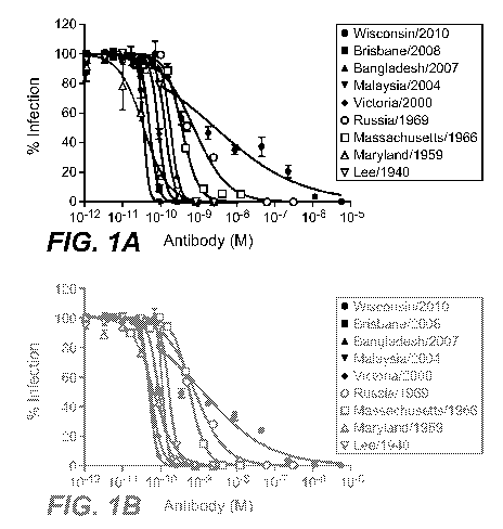
Figures 1A and 1B set forth data showing in vitro neutralization of various
influenza B virus
isolates by monoclonal antibody 34B5A and monoclonal antibody 33F8,
respectively.
Figure 2 sets forth data showing in vitro neutralization of various influenza
B virus isolates by
monoclonal antibody 46B8A.
Figure 3 sets forth data showing the effect of monoclonal antibody 34B5C and
monoclonal
antibody 46B8C on hemagglutination inhibition.
13
CA 02942820 2016-09-14
WO 2015/148806
PCT/US2015/022758
Figure 4 sets forth data showing neutralization of various influenza B virus
isolates by
monoclonal antibody 46B8C by in vitro plaque inhibition assay.
Figure 5 sets forth data showing the effects of monoclonal antibody 34B5C and
monoclonal
antibody 46B8C on hemagglutinin-mediated cell-cell fusion.
Figures 6A and 6B set forth data showing percent survival of mice infected
with influenza B
virus BNictoria/2000 and administered various amounts of monoclonal antibody
34B5A
(Figure 6A) compared to that of mice administered oseltamivir (Tamiflu)
(Figure 6B).
Figures 7A and 7B set forth data showing percent survival of mice infected
with influenza B
virus B/Wisconsin/2000 and administered various amounts of monoclonal antibody
34B5C at
48 hours post-infection or 72 hours post-infection, respectively.
Figures 8A, 8B, 8C, and 8D set forth data showing percent survival of mice
infected with
influenza B viruses B/Wisconsin/2010, BNictoria/2000, B/Russia/1969, and
B/Massachusetts/1966, respectively, and administered monoclonal antibody 46B8C
at 24, 48,
or 72 hours post-infection.
Figures 9A and 9B set forth data showing percent survival of mice infected
with influenza B
virus B/Wisconsin/2010 and BNictoria/2000, respectively, and administered
various amounts
of monoclonal antibody 46B8C at 72 hours post-infection.
Figures 10A and 10B set forth data showing percent survival and percent body
weight (BW)
change, respectively, of mice infected with influenza B virus BNictoria/2000
and administered
either monoclonal antibody 46B8C or oseltamivir (Tamiflu).
Figures 11A and 11B set forth data showing the effect of administration of
monoclonal
antibody 46B8C and oseltamivir (Tamiflu) alone or in combination on percent
survival and
viral lung titer, respectively, in mice.
Figures 12A and 12B set forth data showing the effect of co-administration of
monoclonal
antibody 46B8C and oseltamivir (Tamiflu)
14
CA 02942820 2016-09-14
WO 2015/148806
PCT/US2015/022758
Figure 13 sets forth the amino acid sequences of light chain and heavy chain
hypervariable
regions of anti-influenza B virus antibodies of the present invention.
Figure 14 sets forth the amino acid sequences of light chain variable region,
heavy chain
variable region, light chain, and heavy chain of mAb 34B5A.
Figure 15 sets forth the amino acid sequences of light chain variable region,
heavy chain
variable region, light chain, and heavy chain of mAb 34B5B.
Figure 16 sets forth the amino acid sequences of light chain variable region,
heavy chain
variable region, light chain, and heavy chain of mAb 34B5C.
Figure 17 sets forth the amino acid sequences of light chain variable region,
heavy chain
variable region, light chain, and heavy chain of mAb 33F8.
Figure 18 sets forth the amino acid sequences of light chain variable region,
heavy chain
variable region, light chain, and heavy chain of mAb 46B8A.
Figure 19 sets forth the amino acid sequences of light chain variable region,
heavy chain
variable region, light chain, and heavy chain of mAb 46B8B.
Figure 20 sets forth the amino acid sequences of light chain variable region,
heavy chain
variable region, light chain, and heavy chain of mAb 46B8C.
Figure 21 sets forth the amino acid sequences of light chain variable region,
heavy chain
variable region, light chain, and heavy chain of mAb 46B8D.
Figure 22 sets forth the amino acid sequences of light chain variable region,
heavy chain
variable region, light chain, and heavy chain of mAb 46B8E.
Figure 23 sets forth the amino acid sequences of light chain variable region,
heavy chain
variable region, light chain, and heavy chain of mAb 46B8F.
CA 02942820 2016-09-14
WO 2015/148806
PCT/US2015/022758
Figure 24 sets forth the amino acid sequences of light chain variable region,
heavy chain
variable region, light chain, and heavy chain of mAb 46B8G.
Figure 25 sets forth the amino acid sequences of light chain variable region,
heavy chain
variable region, light chain, and heavy chain of mAb 46B8H.
Figures 26A and 26B set forth data showing percent survival and percent body
weight (BW)
change, respectively, of mice infected with influenza B virus B/Brisbane/2008
and
administered monoclonal antibody 46B8C.
DETAILED DESCRIPTION OF EMBODIMENTS OF THE INVENTION
I. DEFINITIONS
An "acceptor human framework" for the purposes herein is a framework
comprising the amino
acid sequence of a light chain variable domain (VL) framework or a heavy chain
variable
domain (VH) framework derived from a human immunoglobulin framework or a human
consensus framework, as defined below. An acceptor human framework "derived
from" a
human immunoglobulin framework or a human consensus framework may comprise the
same
amino acid sequence thereof, or it may contain amino acid sequence changes. In
some
embodiments, the number of amino acid changes are 10 or less, 9 or less, 8 or
less, 7 or less, 6
or less, 5 or less, 4 or less, 3 or less, or 2 or less. In some embodiments,
the VL acceptor
human framework is identical in sequence to the VL human immunoglobulin
framework
sequence or human consensus framework sequence.
"Affinity" refers to the strength of the sum total of noncovalent interactions
between a single
binding site of a molecule (e.g., an antibody) and its binding partner (e.g.,
an antigen). Unless
indicated otherwise, as used herein, "binding affinity" refers to intrinsic
binding affinity which
reflects a 1:1 interaction between members of a binding pair (e.g., antibody
and antigen). The
affinity of a molecule X for its partner Y can generally be represented by the
dissociation
constant (Kd). Affinity can be measured by common methods known in the art,
including
those described herein. Specific illustrative and exemplary embodiments for
measuring
binding affinity are described in the following.
16
CA 02942820 2016-09-14
WO 2015/148806
PCT/US2015/022758
An "affinity matured" antibody refers to an antibody with one or more
alterations in one or
more hypervariable regions (HVRs), compared to a parent antibody which does
not possess
such alterations, such alterations resulting in an improvement in the affinity
of the antibody for
antigen.
The terms "anti-hemagglutinin antibody" and "an antibody that binds to
hemagglutinin" refer
to an antibody that binds hemagglutinin with sufficient affinity such that the
antibody is useful
as a diagnostic and/or therapeutic agent in targeting hemagglutinin, including
targeting
hemagglutinin of influenza virus. In one embodiment, the extent of binding of
an anti-
hemagglutinin antibody to an unrelated, non-hemagglutinin protein is less than
about 10% of
the binding of the antibody to hemagglutinin as measured, e.g., by a
radioimmunoassay (RIA).
In certain embodiments, an antibody that binds to hemagglutinin has a
dissociation constant
(Kd) of < liAM, < 100 nM, < 10 nM, < 1 nM, < 0.1 nM, < 0.01 nM, or < 0.001 nM
(e.g., 10-8
M or less, e.g., from 10-8M to 10-13M, e.g., from 10-9M to 10-13 M). In
certain embodiments,
an anti-hemagglutinin antibody binds to an epitope of hemagglutinin of
influenza B virus that
is conserved among hemagglutinin from different strains, subtypes, and
isolates of influenza B
viruses, such as that of hemagglutinin of influenza B viruses of ancestral,
Victoria, or
Yamagata lineages.
The term "antibody" herein is used in the broadest sense and encompasses
various antibody
structures, including but not limited to monoclonal antibodies, polyclonal
antibodies,
multispecific antibodies (e.g., bispecific antibodies), and antibody fragments
so long as they
exhibit the desired antigen-binding activity.
An "antibody fragment" refers to a molecule other than an intact antibody that
comprises a
portion of an intact antibody that binds the antigen to which the intact
antibody binds. An
antibody fragment also refers to a molecule other than an intact antibody that
comprises a
portion of an intact antibody that binds hemagglutinin and neutralizes
influenza A virus.
Examples of antibody fragments include but are not limited to Fv, Fab, Fab',
Fab'-SH, F(ab')2;
diabodies; linear antibodies; single-chain antibody molecules (e.g., scFv);
and multispecific
antibodies formed from antibody fragments.
An "antibody that binds to the same epitope" as a reference antibody refers to
an antibody that
blocks binding of the reference antibody to its antigen in a competition assay
by 50% or more,
17
CA 02942820 2016-09-14
WO 2015/148806
PCT/US2015/022758
and conversely, the reference antibody blocks binding of the antibody to its
antigen in a
competition assay by 50% or more. An exemplary competition assay is provided
herein.
The term "chimeric" antibody refers to an antibody in which a portion of the
heavy and/or light
chain is derived from a particular source or species, while the remainder of
the heavy and/or
light chain is derived from a different source or species.
The "class" of an antibody refers to the type of constant domain or constant
region possessed
by its heavy chain. There are five major classes of antibodies: IgA, IgD, IgE,
IgG, and IgM,
and several of these may be further divided into subclasses (isotypes), e.g.,
IgGi, IgG2, IgG3,
Igat, IgAi, and IgA2. The heavy chain constant domains that correspond to the
different
classes of immunoglobulins are called a, 6, 8, y, and it, respectively.
The term "cytotoxic agent" as used herein refers to a substance that inhibits
or prevents a
cellular function and/or causes cell death or destruction. Cytotoxic agents
include, but are not
211 /131, /125, y90, Re 186, Re 188, sm153, Bi212, p32,
limited to, radioactive isotopes (e.g., At,
Pb212 and radioactive isotopes of Lu); chemotherapeutic agents or drugs (e.g.,
methotrexate,
adriamicin, vinca alkaloids (vincristine, vinblastine, etoposide),
doxorubicin, melphalan,
mitomycin C, chlorambucil, daunorubicin or other intercalating agents); growth
inhibitory
agents; enzymes and fragments thereof such as nucleolytic enzymes;
antibiotics; toxins such as
small molecule toxins or enzymatically active toxins of bacterial, fungal,
plant or animal
origin, including fragments and/or variants thereof; and the various antitumor
or anticancer
agents disclosed below.
"Effector functions" refer to those biological activities attributable to the
Fc region of an
antibody, which vary with the antibody isotype. Examples of antibody effector
functions
include: Clq binding and complement dependent cytotoxicity (CDC); Fc receptor
binding;
antibody-dependent cell-mediated cytotoxicity (ADCC); phagocytosis; down
regulation of cell
surface receptors (e.g., B cell receptor); and B cell activation.
An "effective amount" of an agent, e.g., a pharmaceutical formulation, refers
to an amount
effective, at dosages and for periods of time necessary, to achieve the
desired therapeutic or
prophylactic result.
18
CA 02942820 2016-09-14
WO 2015/148806
PCT/US2015/022758
The term "Fe region" herein is used to define a C-terminal region of an
immunoglobulin heavy
chain that contains at least a portion of the constant region. The term
includes native sequence
Fe regions and variant Fe regions. In one embodiment, a human IgG heavy chain
Fe region
extends from Cys226, or from Pro230, to the carboxyl-terminus of the heavy
chain. However,
the C-terminal lysine (Lys447) of the Fe region may or may not be present.
Unless otherwise
specified herein, numbering of amino acid residues in the Fe region or
constant region is
according to the EU numbering system, also called the EU index, as described
in Kabat et al.,
Sequences of Proteins of Immunological Interest, 5th Ed. Public Health
Service, National
Institutes of Health, Bethesda, MD, 1991.
"Framework" or "FR" refers to variable domain residues other than
hypervariable region
(HVR) residues. The FR of a variable domain generally consists of four FR
domains: FR1,
FR2, FR3, and FR4. Accordingly, the HVR and FR sequences generally appear in
the
following sequence in VH (or VL): FR1-H1(L1)-FR2-H2(L2)-FR3-H3(L3)-FR4.
The terms "full length antibody," "intact antibody," and "whole antibody" are
used herein
interchangeably to refer to an antibody having a structure substantially
similar to a native
antibody structure or having heavy chains that contain an Fe region as defined
herein.
The terms "host cell," "host cell line," and "host cell culture" are used
interchangeably and refer
to cells into which exogenous nucleic acid has been introduced, including the
progeny of such
cells. Host cells include "transformants" and "transformed cells," which
include the primary
transformed cell and progeny derived therefrom without regard to the number of
passages.
Progeny may not be completely identical in nucleic acid content to a parent
cell, but may
contain mutations. Mutant progeny that have the same function or biological
activity as
screened or selected for in the originally transformed cell are included
herein.
A "human antibody" is an antibody which possesses an amino acid sequence which
corresponds to that of an antibody produced by a human or a human cell or
derived from a non-
human source that utilizes human antibody repertoires or other human antibody-
encoding
sequences. This definition of a human antibody specifically excludes a
humanized antibody
comprising non-human antigen-binding residues.
19
CA 02942820 2016-09-14
WO 2015/148806
PCT/US2015/022758
A "human consensus framework" is a framework which represents the most
commonly
occurring amino acid residues in a selection of human immunoglobulin VL or VH
framework
sequences. Generally, the selection of human immunoglobulin VL or VH sequences
is from a
subgroup of variable domain sequences. Generally, the subgroup of sequences is
a subgroup as
in Kabat et at., Sequences of Proteins of Immunological Interest, Fifth
Edition, NIH
Publication 91-3242, Bethesda MD (1991), vols. 1-3. In one embodiment, for the
VL, the
subgroup is subgroup kappa I as in Kabat et al., supra. In one embodiment, for
the VH, the
subgroup is subgroup III as in Kabat et at., supra.
A "humanized" antibody refers to a chimeric antibody comprising amino acid
residues from
non-human HVRs and amino acid residues from human FRs. In certain embodiments,
a
humanized antibody will comprise substantially all of at least one, and
typically two, variable
domains, in which all or substantially all of the HVRs (e.g., CDRs) correspond
to those of a
non-human antibody, and all or substantially all of the FRs correspond to
those of a human
antibody. A humanized antibody optionally may comprise at least a portion of
an antibody
constant region derived from a human antibody. A "humanized form" of an
antibody, e.g., a
non-human antibody, refers to an antibody that has undergone humanization.
The term "hypervariable region" or "HVR" as used herein refers to each of the
regions of an
antibody variable domain which are hypervariable in sequence ("complementarity
determining
regions" or "CDRs") and/or form structurally defined loops ("hypervariable
loops") and/or
contain the antigen-contacting residues ("antigen contacts"). Generally,
antibodies comprise
six HVRs: three in the VH (H1, H2, H3), and three in the VL (L1, L2, L3).
Exemplary HVRs
herein include:
(a) hypervariable loops occurring at amino acid residues 26-32 (L1), 50-52
(L2), 91-96
(L3), 26-32 (H1), 53-55 (H2), and 96-101 (H3) (Chothia and Lesk, J. Mot. Biol.
196:901-917
(1987));
(b) CDRs occurring at amino acid residues 24-34 (L1), 50-56 (L2), 89-97 (L3),
31-35b
(H1), 50-65 (H2), and 95-102 (H3) (Kabat et al., Sequences of Proteins of
Immunological
Interest, 5th Ed. Public Health Service, National Institutes of Health,
Bethesda, MD (1991));
(c) antigen contacts occurring at amino acid residues 27c-36 (L1), 46-55 (L2),
89-96
(L3), 30-35b (H1), 47-58 (H2), and 93-101 (H3) (MacCallum et al. J. Mol. Biol.
262: 732-745
(1996)); and
CA 02942820 2016-09-14
WO 2015/148806
PCT/US2015/022758
(d) combinations of (a), (b), and/or (c), including HVR amino acid residues 46-
56 (L2),
47-56 (L2), 48-56 (L2), 49-56 (L2), 26-35 (H1), 26-35b (H1), 49-65 (H2), 93-
102 (H3), and
94-102 (H3).
Unless otherwise indicated, HVR residues and other residues in the variable
domain (e.g., FR
residues) are numbered herein according to Kabat et at., supra.
An "immunoconjugate" is an antibody conjugated to one or more heterologous
molecule(s),
including but not limited to a cytotoxic agent.
An "individual" or "subject" is a mammal. Mammals include, but are not limited
to,
domesticated animals (e.g., cows, sheep, cats, dogs, and horses), primates
(e.g., humans and
non-human primates such as monkeys), rabbits, and rodents (e.g., mice and
rats). In certain
embodiments, the individual or subject is a human.
An "isolated" antibody is one which has been separated from a component of its
natural
environment. In some embodiments, an antibody is purified to greater than 95%
or 99% purity
as determined by, for example, electrophoretic (e.g., SDS-PAGE, isoelectric
focusing (IEF),
capillary electrophoresis) or chromatographic (e.g., ion exchange or reverse
phase HPLC). For
review of methods for assessment of antibody purity, see, e.g., Flatman et
al., J. Chromatogr. B
848:79-87 (2007).
An "isolated" nucleic acid refers to a nucleic acid molecule that has been
separated from a
component of its natural environment. An isolated nucleic acid includes a
nucleic acid
molecule contained in cells that ordinarily contain the nucleic acid molecule,
but the nucleic
acid molecule is present extrachromosomally or at a chromosomal location that
is different
from its natural chromosomal location.
"Isolated nucleic acid encoding an anti-hemagglutinin antibody" refers to one
or more nucleic
acid molecules encoding antibody heavy and light chains (or fragments
thereof), including such
nucleic acid molecule(s) in a single vector or separate vectors, and such
nucleic acid
molecule(s) present at one or more locations in a host cell.
21
CA 02942820 2016-09-14
WO 2015/148806
PCT/US2015/022758
The term "monoclonal antibody" as used herein refers to an antibody obtained
from a
population of substantially homogeneous antibodies, i.e., the individual
antibodies comprising
the population are identical and/or bind the same epitope, except for possible
variant
antibodies, e.g., containing naturally occurring mutations or arising during
production of a
monoclonal antibody preparation, such variants generally being present in
minor amounts. In
contrast to polyclonal antibody preparations, which typically include
different antibodies
directed against different determinants (epitopes), each monoclonal antibody
of a monoclonal
antibody preparation is directed against a single determinant on an antigen.
Thus, the modifier
"monoclonal" indicates the character of the antibody as being obtained from a
substantially
homogeneous population of antibodies, and is not to be construed as requiring
production of
the antibody by any particular method. For example, the monoclonal antibodies
to be used in
accordance with the present invention may be made by a variety of techniques,
including but
not limited to the hybridoma method, recombinant DNA methods, phage-display
methods, and
methods utilizing transgenic animals containing all or part of the human
immunoglobulin loci,
such methods and other exemplary methods for making monoclonal antibodies
being described
herein.
A "naked antibody" refers to an antibody that is not conjugated to a
heterologous moiety (e.g.,
a cytotoxic moiety) or radiolabel. The naked antibody may be present in a
pharmaceutical
formulation.
"Native antibodies" refer to naturally occurring immunoglobulin molecules with
varying
structures. For example, native IgG antibodies are heterotetrameric
glycoproteins of about
150,000 daltons, composed of two identical light chains and two identical
heavy chains that are
disulfide-bonded. From N- to C-terminus, each heavy chain has a variable
region (VH), also
called a variable heavy domain or a heavy chain variable domain, followed by
three constant
domains (CH1, CH2, and CH3). Similarly, from N- to C-terminus, each light
chain has a
variable region (VL), also called a variable light domain or a light chain
variable domain,
followed by a constant light (CL) domain. The light chain of an antibody may
be assigned to
one of two types, called kappa (x) and lambda (X), based on the amino acid
sequence of its
constant domain.
The term "package insert" is used to refer to instructions customarily
included in commercial
packages of therapeutic products, that contain information about the
indications, usage, dosage,
22
CA 02942820 2016-09-14
WO 2015/148806
PCT/US2015/022758
administration, combination therapy, contraindications and/or warnings
concerning the use of
such therapeutic products.
"Percent (%) amino acid sequence identity" with respect to a reference
polypeptide sequence is
defined as the percentage of amino acid residues in a candidate sequence that
are identical with
the amino acid residues in the reference polypeptide sequence, after aligning
the sequences and
introducing gaps, if necessary, to achieve the maximum percent sequence
identity, and not
considering any conservative substitutions as part of the sequence identity.
Alignment for
purposes of determining percent amino acid sequence identity can be achieved
in various ways
that are within the skill in the art, for instance, using publicly available
computer software such
as BLAST, BLAST-2, ALIGN or Megalign (DNASTAR) software. Those skilled in the
art
can determine appropriate parameters for aligning sequences, including any
algorithms needed
to achieve maximal alignment over the full length of the sequences being
compared. For
purposes herein, however, % amino acid sequence identity values are generated
using the
sequence comparison computer program ALIGN-2. The ALIGN-2 sequence comparison
computer program was authored by Genentech, Inc., and the source code has been
filed with
user documentation in the U.S. Copyright Office, Washington D.C., 20559, where
it is
registered under U.S. Copyright Registration No. TXU510087. The ALIGN-2
program is
publicly available from Genentech, Inc., South San Francisco, California, or
may be compiled
from the source code. The ALIGN-2 program should be compiled for use on a UNIX
operating
system, including digital UNIX V4.0D. All sequence comparison parameters are
set by the
ALIGN-2 program and do not vary.
In situations where ALIGN-2 is employed for amino acid sequence comparisons,
the % amino
acid sequence identity of a given amino acid sequence A to, with, or against a
given amino acid
sequence B (which can alternatively be phrased as a given amino acid sequence
A that has or
comprises a certain % amino acid sequence identity to, with, or against a
given amino acid
sequence B) is calculated as follows:
100 times the fraction X/Y
where X is the number of amino acid residues scored as identical matches by
the sequence
alignment program ALIGN-2 in that program's alignment of A and B, and where Y
is the total
number of amino acid residues in B. It will be appreciated that where the
length of amino acid
sequence A is not equal to the length of amino acid sequence B, the % amino
acid sequence
23
CA 02942820 2016-09-14
WO 2015/148806
PCT/US2015/022758
identity of A to B will not equal the % amino acid sequence identity of B to
A. Unless
specifically stated otherwise, all % amino acid sequence identity values used
herein are
obtained as described in the immediately preceding paragraph using the ALIGN-2
computer
program.
The term "pharmaceutical formulation" refers to a preparation which is in such
form as to
permit the biological activity of an active ingredient contained therein to be
effective, and
which contains no additional components which are unacceptably toxic to a
subject to which
the formulation would be administered.
A "pharmaceutically acceptable carrier" refers to an ingredient in a
pharmaceutical
formulation, other than an active ingredient, which is nontoxic to a subject.,
A
pharmaceutically acceptable carrier includes, but is not limited to, a buffer,
excipient,
stabilizer, or preservative.
The term "hemagglutinin," as used herein, refers to any native hemagglutinin
from any
influenza virus source, unless otherwise indicated. The term encompasses "full-
length,"
unprocessed hemagglutinin as well as any form of hemagglutinin that results
from processing
in an influenza virus or an influenza virus-infected cell. The term also
encompasses naturally
occurring variants of hemagglutinin, e.g., splice variants or allelic
variants. The amino acid
sequences of exemplary hemagglutinin proteins from various influenza B virus
strains or
lineages are readily available in the art.
As used herein, "treatment" (and grammatical variations thereof such as
"treat" or "treating")
refers to clinical intervention in an attempt to alter the natural course of
the individual being
treated, and can be performed either for prophylaxis or during the course of
clinical pathology.
Desirable effects of treatment include, but are not limited to, preventing
occurrence or
recurrence of disease (e.g., preventing occurrence or recurrence of influenza
B virus infection),
reduction (e.g., reducing) or alleviation of symptoms, diminishment of any
direct or indirect
pathological consequences of the disease, decreasing the rate of disease
progression,
amelioration or palliation of the disease state, and remission or improved
prognosis. In some
embodiments, antibodies of the invention are used to delay development of a
disease or to slow
the progression of a disease.
24
CA 02942820 2016-09-14
WO 2015/148806
PCT/US2015/022758
The term "variable region" or "variable domain" refers to the domain of an
antibody heavy or
light chain that is involved in binding the antibody to antigen. The variable
domains of the
heavy chain and light chain (VH and VL, respectively) of a native antibody
generally have
similar structures, with each domain comprising four conserved framework
regions (FRs) and
three hypervariable regions (HVRs). (See, e.g., Kindt et al. Kuby Immunology,
6t1 ed., W.H.
Freeman and Co., page 91 (2007).) A single VH or VL domain may be sufficient
to confer
antigen-binding specificity. Furthermore, antibodies that bind a particular
antigen may be
isolated using a VH or VL domain from an antibody that binds the antigen to
screen a library of
complementary VL or VH domains, respectively. See, e.g., Portolano et al., J.
Immunol.
150:880-887 (1993); Clarkson et al., Nature 352:624-628 (1991).
The term "vector," as used herein, refers to a nucleic acid molecule capable
of propagating
another nucleic acid to which it is linked. The term includes the vector as a
self-replicating
nucleic acid structure as well as the vector incorporated into the genome of a
host cell into
which it has been introduced. Certain vectors are capable of directing the
expression of nucleic
acids to which they are operatively linked. Such vectors are referred to
herein as "expression
vectors."
II. COMPOSITIONS AND METHODS
In one aspect, the invention is based, in part, on anti-hemagglutinin
antibodies and uses thereof.
In certain embodiments, antibodies that bind to hemagglutinin are provided.
Antibodies of the
invention are useful, e.g., for the diagnosis, treatment, or prevention of
influenza A virus
infection.
A. Exemplary Anti-Hemagglutinin Antibodies
In one aspect, the invention provides isolated antibodies that bind to
hemagglutinin. In certain
embodiments, an anti-hemagglutinin antibody of the present invention binds
hemagglutinin,
binds hemagglutinin from influenza B virus, binds hemagglutinin from the
Yamagata lineage
of influenza B viruses, binds hemagglutinin from the Victoria lineage of
influenza B viruses,
binds hemagglutinin from ancestral lineages of influenza B virus, or binds
hemagglutinin from
the Yamagata lineage, the Victoria lineage, and ancestral lineages of
influenza B virus. In
other embodiments, an anti-hemagglutinin antibody of the present invention
neutralizes
influenza B virus in vitro. In other embodiments, an anti-hemagglutinin
antibody of the
present invention neutralizes influenza B virus in vivo. In yet other
embodiments, an anti-
CA 02942820 2016-09-14
WO 2015/148806
PCT/US2015/022758
hemagglutinin antibody of the present invention reduces influenza B virus
infection, prevents
influenza B virus infection, inhibits influenza B virus infection, or treats
influenza B virus
infection. In some embodiments, an anti-hemagglutinin antibody of the present
invention
prevents, inhibits, or reduces hemagglutinin-mediated fusion between influenza
virus
membrane and infected cell endosomal membranes (thus preventing, inhibiting,
or reducing
viral RNA entry into the infected cell cytoplasm, thus preventing, inhibiting,
or reducing
further propagation of influenza virus infection.)
In one aspect, the invention provides an anti-hemagglutinin antibody
comprising at least one,
two, three, four, five, or six HVRs selected from (a) HVR-H1 comprising the
amino acid
sequence of SEQ ID NO :61; (b) HVR-H2 comprising the amino acid sequence of
SEQ ID
NO:64; (c) HVR-H3 comprising the amino acid sequence of SEQ ID NO:75; (d) HVR-
L1
comprising the amino acid sequence of SEQ ID NO:55; (e) HVR-L2 comprising the
amino
acid sequence of SEQ ID NO:57; and (f) HVR-L3 comprising the amino acid
sequence of SEQ
ID NO:59.
In one aspect, the invention provides an anti-hemagglutinin antibody
comprising at least one,
two, three, four, five, or six HVRs selected from (a) HVR-H1 comprising the
amino acid
sequence of SEQ ID NO :61; (b) HVR-H2 comprising the amino acid sequence of
SEQ ID
NO:65; (c) HVR-H3 comprising the amino acid sequence of SEQ ID NO:75; (d) HVR-
L1
comprising the amino acid sequence of SEQ ID NO:55; (e) HVR-L2 comprising the
amino
acid sequence of SEQ ID NO:57; and (f) HVR-L3 comprising the amino acid
sequence of SEQ
ID NO:59.
In one aspect, the invention provides an antibody comprising at least one, at
least two, or all
three VH HVR sequences selected from (a) HVR-H1 comprising the amino acid
sequence of
SEQ ID NO :61; (b) HVR-H2 comprising the amino acid sequence of SEQ ID NO :64;
and (c)
HVR-H3 comprising the amino acid sequence of SEQ ID NO:75.
In one aspect, the invention provides an antibody comprising at least one, at
least two, or all
three VH HVR sequences selected from (a) HVR-H1 comprising the amino acid
sequence of
SEQ ID NO :61; (b) HVR-H2 comprising the amino acid sequence of SEQ ID NO :65;
and (c)
HVR-H3 comprising the amino acid sequence of SEQ ID NO:75.
26
CA 02942820 2016-09-14
WO 2015/148806
PCT/US2015/022758
In another aspect, the invention provides an antibody comprising at least one,
at least two, or
all three VL HVR sequences selected from (a) HVR-L1 comprising the amino acid
sequence of
SEQ ID NO:55; (b) HVR-L2 comprising the amino acid sequence of SEQ ID NO:57;
and (c)
HVR-L3 comprising the amino acid sequence of SEQ ID NO:59.
In another aspect, the invention provides an antibody comprising (a) HVR-H1
comprising the
amino acid sequence of SEQ ID NO :61; (b) HVR-H2 comprising the amino acid
sequence of
SEQ ID NO:64; (c) HVR-H3 comprising the amino acid sequence of SEQ ID NO:75;
(d)
HVR-L1 comprising the amino acid sequence of SEQ ID NO:55; (e) HVR-L2
comprising the
amino acid sequence of SEQ ID NO:57; and (f) HVR-L3 comprising the amino acid
sequence
selected from SEQ ID NO:59.
In another aspect, the invention provides an antibody comprising (a) HVR-H1
comprising the
amino acid sequence of SEQ ID NO :61; (b) HVR-H2 comprising the amino acid
sequence of
SEQ ID NO:65; (c) HVR-H3 comprising the amino acid sequence of SEQ ID NO:75;
(d)
HVR-L1 comprising the amino acid sequence of SEQ ID NO:55; (e) HVR-L2
comprising the
amino acid sequence of SEQ ID NO:57; and (f) HVR-L3 comprising the amino acid
sequence
selected from SEQ ID NO:59.
In another aspect, the invention provides an antibody comprising a heavy chain
variable region
comprising an amino acid sequence selected from the group consisting of SEQ ID
NOs:79 and
83.
In another aspect, the invention provides an antibody comprising a light chain
variable region
comprising an amino acid sequence selected from the group consisting of SEQ ID
NOs:78, 82,
and 86.
In another aspect, the invention provides an antibody comprising a heavy chain
variable region
comprising an amino acid sequence selected from the group consisting of SEQ ID
NOs:79 and
83 and a light chain variable region comprising an amino acid sequence
selected from the
group consisting of SEQ ID NOs:78, 82 and 86.
27
CA 02942820 2016-09-14
WO 2015/148806
PCT/US2015/022758
In one embodiment, the invention provides an antibody comprising a heavy chain
variable
region comprising the amino acid sequence of SEQ ID NO:79 and a light chain
variable region
comprising the amino acid sequence of SEQ ID NO:78.
In one embodiment, the invention provides an antibody comprising a heavy chain
variable
region comprising the amino acid sequence of SEQ ID NO:83 and a light chain
variable region
comprising the amino acid sequence of SEQ ID NO:82.
In one embodiment, the invention provides an antibody comprising a heavy chain
variable
region comprising the amino acid sequence of SEQ ID NO:83 and a light chain
variable region
comprising the amino acid sequence of SEQ ID NO:86.
In another aspect, the invention provides an antibody comprising a heavy chain
comprising an
amino acid sequence selected from the group consisting of SEQ ID NOs:81, 85,
and 88.
In another aspect, the invention provides an antibody comprising a light chain
comprising an
amino acid sequence selected from the group consisting of SEQ ID NOs:80, 84,
and 87.
In another aspect, the invention provides an antibody comprising a heavy chain
comprising an
amino acid sequence selected from the group consisting of SEQ ID NOs:81, 85,
and 88 and a
light chain comprising an amino acid sequence selected from the group
consisting of SEQ ID
NOs:80, 84, and 87.
In one embodiment, the invention provides an antibody comprising a heavy chain
comprising
the amino acid sequence of SEQ ID NO:81 and a light chain comprising the amino
acid
sequence of SEQ ID NO:80.
In one embodiment, the invention provides an antibody comprising a heavy chain
comprising
the amino acid sequence SEQ ID NO:85 and a light chain comprising the amino
acid sequence
selected from the group consisting of SEQ ID NO:84.
In one embodiment, the invention provides an antibody comprising a heavy chain
comprising
the amino acid sequence of SEQ ID NO:88 and a light chain comprising the amino
acid
sequence of SEQ ID NO:87.
28
CA 02942820 2016-09-14
WO 2015/148806
PCT/US2015/022758
In one aspect, the invention provides an anti-hemagglutinin antibody
comprising at least one,
two, three, four, five, or six HVRs selected from (a) HVR-Hl comprising the
amino acid
sequence of SEQ ID NO:62; (b) HVR-H2 comprising the amino acid sequence of SEQ
ID
NO:66; (c) HVR-H3 comprising the amino acid sequence of SEQ ID NO:76; (d) HVR-
L1
comprising the amino acid sequence of SEQ ID NO:55; (e) HVR-L2 comprising the
amino
acid sequence of SEQ ID NO:57; and (f) HVR-L3 comprising the amino acid
sequence of SEQ
ID NO:59.
In one aspect, the invention provides an antibody comprising at least one, at
least two, or all
three VH HVR sequences selected from (a) HVR-Hl comprising the amino acid
sequence of
SEQ ID NO:62; (b) HVR-H2 comprising the amino acid sequence of SEQ ID NO:66;
and (c)
HVR-H3 comprising the amino acid sequence of SEQ ID NO:76.
In another aspect, the invention provides an antibody comprising at least one,
at least two, or
all three VL HVR sequences selected from (a) HVR-L1 comprising the amino acid
sequence of
SEQ ID NO:55; (b) HVR-L2 comprising the amino acid sequence of SEQ ID NO:57;
and (c)
HVR-L3 comprising the amino acid sequence of SEQ ID NO:59.
In another aspect, the invention provides an antibody comprising (a) HVR-Hl
comprising the
amino acid sequence of SEQ ID NO:62; (b) HVR-H2 comprising the amino acid
sequence of
SEQ ID NO:66; (c) HVR-H3 comprising the amino acid sequence of SEQ ID NO:76;
(d)
HVR-L1 comprising the amino acid sequence of SEQ ID NO:55; (e) HVR-L2
comprising the
amino acid sequence of SEQ ID NO:57; and (f) HVR-L3 comprising the amino acid
sequence
selected from SEQ ID NO:59.
In another aspect, the invention provides an antibody comprising a heavy chain
variable region
comprising the amino acid sequence of SEQ ID NO:89.
In another aspect, the invention provides an antibody comprising a light chain
variable region
comprising the amino acid sequence of SEQ ID NO:78.
29
CA 02942820 2016-09-14
WO 2015/148806
PCT/US2015/022758
In one embodiment, the invention provides an antibody comprising a heavy chain
variable
region comprising the amino acid sequence of SEQ ID NO:89 and a light chain
variable region
comprising the amino acid sequence of SEQ ID NO:78.
In one embodiment, the invention provides an antibody comprising a heavy chain
comprising
the amino acid sequence of SEQ ID NO:90 and a light chain comprising the amino
acid
sequence of SEQ ID NO:80.
In one aspect, the invention provides an anti-hemagglutinin antibody
comprising at least one,
two, three, four, five, or six HVRs selected from (a) HVR-H1 comprising the
amino acid
sequence of SEQ ID NO:63; (b) HVR-H2 comprising an amino acid sequence
selected from
the group consisting of SEQ ID NOs:67, 68, 69, 70, 71, 72, 73, and 74; (c) HVR-
H3
comprising the amino acid sequence of SEQ ID NO:77; (d) HVR-L1 comprising the
amino
acid sequence of SEQ ID NO:56; (e) HVR-L2 comprising the amino acid sequence
of SEQ ID
NO:58; and (f) HVR-L3 comprising the amino acid sequence of SEQ ID NO:60.
In one aspect, the invention provides an anti-hemagglutinin antibody
comprising at least one,
two, three, four, five, or six HVRs selected from (a) HVR-H1 comprising the
amino acid
sequence of SEQ ID NO:63; (b) HVR-H2 comprising the amino acid sequence of SEQ
ID
NO:67; (c) HVR-H3 comprising the amino acid sequence of SEQ ID NO:77; (d) HVR-
L1
comprising the amino acid sequence of SEQ ID NO:56; (e) HVR-L2 comprising the
amino
acid sequence of SEQ ID NO:58; and (f) HVR-L3 comprising the amino acid
sequence of SEQ
ID NO:60.
In one aspect, the invention provides an anti-hemagglutinin antibody
comprising at least one,
two, three, four, five, or six HVRs selected from (a) HVR-H1 comprising the
amino acid
sequence of SEQ ID NO:63; (b) HVR-H2 comprising the amino acid sequence of SEQ
ID
NO:68; (c) HVR-H3 comprising the amino acid sequence of SEQ ID NO:77; (d) HVR-
L1
comprising the amino acid sequence of SEQ ID NO:56; (e) HVR-L2 comprising the
amino
acid sequence of SEQ ID NO:58; and (f) HVR-L3 comprising the amino acid
sequence of SEQ
ID NO:60.
In one aspect, the invention provides an anti-hemagglutinin antibody
comprising at least one,
two, three, four, five, or six HVRs selected from (a) HVR-H1 comprising the
amino acid
CA 02942820 2016-09-14
WO 2015/148806
PCT/US2015/022758
sequence of SEQ ID NO:63; (b) HVR-H2 comprising the amino acid sequence of SEQ
ID
NO:69; (c) HVR-H3 comprising the amino acid sequence of SEQ ID NO:77; (d) HVR-
L1
comprising the amino acid sequence of SEQ ID NO:56; (e) HVR-L2 comprising the
amino
acid sequence of SEQ ID NO:58; and (f) HVR-L3 comprising the amino acid
sequence of SEQ
ID NO:60.
In one aspect, the invention provides an anti-hemagglutinin antibody
comprising at least one,
two, three, four, five, or six HVRs selected from (a) HVR-H1 comprising the
amino acid
sequence of SEQ ID NO:63; (b) HVR-H2 comprising the amino acid sequence of SEQ
ID
NO:70; (c) HVR-H3 comprising the amino acid sequence of SEQ ID NO:77; (d) HVR-
L1
comprising the amino acid sequence of SEQ ID NO:56; (e) HVR-L2 comprising the
amino
acid sequence of SEQ ID NO:58; and (f) HVR-L3 comprising the amino acid
sequence of SEQ
ID NO:60.
In one aspect, the invention provides an anti-hemagglutinin antibody
comprising at least one,
two, three, four, five, or six HVRs selected from (a) HVR-H1 comprising the
amino acid
sequence of SEQ ID NO:63; (b) HVR-H2 comprising the amino acid sequence of SEQ
ID
NO :71; (c) HVR-H3 comprising the amino acid sequence of SEQ ID NO:77; (d) HVR-
L1
comprising the amino acid sequence of SEQ ID NO:56; (e) HVR-L2 comprising the
amino
acid sequence of SEQ ID NO:58; and (f) HVR-L3 comprising the amino acid
sequence of SEQ
ID NO:60.
In one aspect, the invention provides an anti-hemagglutinin antibody
comprising at least one,
two, three, four, five, or six HVRs selected from (a) HVR-H1 comprising the
amino acid
sequence of SEQ ID NO:63; (b) HVR-H2 comprising the amino acid sequence of SEQ
ID
NO:72; (c) HVR-H3 comprising the amino acid sequence of SEQ ID NO:77; (d) HVR-
L1
comprising the amino acid sequence of SEQ ID NO:56; (e) HVR-L2 comprising the
amino
acid sequence of SEQ ID NO:58; and (f) HVR-L3 comprising the amino acid
sequence of SEQ
ID NO:60.
In one aspect, the invention provides an anti-hemagglutinin antibody
comprising at least one,
two, three, four, five, or six HVRs selected from (a) HVR-H1 comprising the
amino acid
sequence of SEQ ID NO:63; (b) HVR-H2 comprising the amino acid sequence of SEQ
ID
NO:73; (c) HVR-H3 comprising the amino acid sequence of SEQ ID NO:77; (d) HVR-
L1
31
CA 02942820 2016-09-14
WO 2015/148806
PCT/US2015/022758
comprising the amino acid sequence of SEQ ID NO:56; (e) HVR-L2 comprising the
amino
acid sequence of SEQ ID NO:58; and (f) HVR-L3 comprising the amino acid
sequence of SEQ
ID NO:60.
In one aspect, the invention provides an anti-hemagglutinin antibody
comprising at least one,
two, three, four, five, or six HVRs selected from (a) HVR-Hl comprising the
amino acid
sequence of SEQ ID NO:63; (b) HVR-H2 comprising the amino acid sequence of SEQ
ID
NO:74; (c) HVR-H3 comprising the amino acid sequence of SEQ ID NO:77; (d) HVR-
L1
comprising the amino acid sequence of SEQ ID NO:56; (e) HVR-L2 comprising the
amino
acid sequence of SEQ ID NO:58; and (f) HVR-L3 comprising the amino acid
sequence of SEQ
ID NO:60.
In one aspect, the invention provides an antibody comprising at least one, at
least two, or all
three VH HVR sequences selected from (a) HVR-Hl comprising the amino acid
sequence of
SEQ ID NO:63; (b) HVR-H2 comprising the amino acid sequence of SEQ ID NO:67;
and (c)
HVR-H3 comprising the amino acid sequence of SEQ ID NO:77.
In one aspect, the invention provides an antibody comprising at least one, at
least two, or all
three VH HVR sequences selected from (a) HVR-Hl comprising the amino acid
sequence of
SEQ ID NO:63; (b) HVR-H2 comprising the amino acid sequence of SEQ ID NO:68;
and (c)
HVR-H3 comprising the amino acid sequence of SEQ ID NO:77.
In one aspect, the invention provides an antibody comprising at least one, at
least two, or all
three VH HVR sequences selected from (a) HVR-Hl comprising the amino acid
sequence of
SEQ ID NO:63; (b) HVR-H2 comprising the amino acid sequence of SEQ ID NO:69;
and (c)
HVR-H3 comprising the amino acid sequence of SEQ ID NO:77.
In one aspect, the invention provides an antibody comprising at least one, at
least two, or all
three VH HVR sequences selected from (a) HVR-Hl comprising the amino acid
sequence of
SEQ ID NO:63; (b) HVR-H2 comprising the amino acid sequence of SEQ ID NO:70;
and (c)
HVR-H3 comprising the amino acid sequence of SEQ ID NO:77.
In one aspect, the invention provides an antibody comprising at least one, at
least two, or all
three VH HVR sequences selected from (a) HVR-Hl comprising the amino acid
sequence of
32
CA 02942820 2016-09-14
WO 2015/148806
PCT/US2015/022758
SEQ ID NO:63; (b) HVR-H2 comprising the amino acid sequence of SEQ ID NO :71;
and (c)
HVR-H3 comprising the amino acid sequence of SEQ ID NO:77.
In one aspect, the invention provides an antibody comprising at least one, at
least two, or all
three VH HVR sequences selected from (a) HVR-H1 comprising the amino acid
sequence of
SEQ ID NO:63; (b) HVR-H2 comprising the amino acid sequence of SEQ ID NO:72;
and (c)
HVR-H3 comprising the amino acid sequence of SEQ ID NO:77.
In one aspect, the invention provides an antibody comprising at least one, at
least two, or all
three VH HVR sequences selected from (a) HVR-H1 comprising the amino acid
sequence of
SEQ ID NO:63; (b) HVR-H2 comprising the amino acid sequence of SEQ ID NO:73;
and (c)
HVR-H3 comprising the amino acid sequence of SEQ ID NO:77.
In one aspect, the invention provides an antibody comprising at least one, at
least two, or all
three VH HVR sequences selected from (a) HVR-H1 comprising the amino acid
sequence of
SEQ ID NO:63; (b) HVR-H2 comprising the amino acid sequence of SEQ ID NO:74;
and (c)
HVR-H3 comprising the amino acid sequence of SEQ ID NO:77.
In another aspect, the invention provides an antibody comprising at least one,
at least two, or
all three VL HVR sequences selected from (a) HVR-L1 comprising the amino acid
sequence of
SEQ ID NO:56; (b) HVR-L2 comprising the amino acid sequence of SEQ ID NO:58;
and (c)
HVR-L3 comprising the amino acid sequence of SEQ ID NO:60.
In another aspect, the invention provides an antibody comprising (a) HVR-H1
comprising the
amino acid sequence of SEQ ID NO:63; (b) HVR-H2 comprising the amino acid
sequence of
SEQ ID NO:67; (c) HVR-H3 comprising the amino acid sequence of SEQ ID NO:77;
(d)
HVR-L1 comprising the amino acid sequence of SEQ ID NO:56; (e) HVR-L2
comprising the
amino acid sequence of SEQ ID NO:58; and (f) HVR-L3 comprising the amino acid
sequence
selected from SEQ ID NO:60.
In another aspect, the invention provides an antibody comprising (a) HVR-H1
comprising the
amino acid sequence of SEQ ID NO:63; (b) HVR-H2 comprising the amino acid
sequence of
SEQ ID NO:68; (c) HVR-H3 comprising the amino acid sequence of SEQ ID NO:77;
(d)
HVR-L1 comprising the amino acid sequence of SEQ ID NO:56; (e) HVR-L2
comprising the
33
CA 02942820 2016-09-14
WO 2015/148806
PCT/US2015/022758
amino acid sequence of SEQ ID NO:58; and (f) HVR-L3 comprising the amino acid
sequence
selected from SEQ ID NO:60.
In another aspect, the invention provides an antibody comprising (a) HVR-H1
comprising the
amino acid sequence of SEQ ID NO:63; (b) HVR-H2 comprising the amino acid
sequence of
SEQ ID NO:69; (c) HVR-H3 comprising the amino acid sequence of SEQ ID NO:77;
(d)
HVR-L1 comprising the amino acid sequence of SEQ ID NO:56; (e) HVR-L2
comprising the
amino acid sequence of SEQ ID NO:58; and (f) HVR-L3 comprising the amino acid
sequence
selected from SEQ ID NO:60.
In another aspect, the invention provides an antibody comprising (a) HVR-H1
comprising the
amino acid sequence of SEQ ID NO:63; (b) HVR-H2 comprising the amino acid
sequence of
SEQ ID NO:70; (c) HVR-H3 comprising the amino acid sequence of SEQ ID NO:77;
(d)
HVR-L1 comprising the amino acid sequence of SEQ ID NO:56; (e) HVR-L2
comprising the
amino acid sequence of SEQ ID NO:58; and (f) HVR-L3 comprising the amino acid
sequence
selected from SEQ ID NO:60.
In another aspect, the invention provides an antibody comprising (a) HVR-H1
comprising the
amino acid sequence of SEQ ID NO:63; (b) HVR-H2 comprising the amino acid
sequence of
SEQ ID NO :71; (c) HVR-H3 comprising the amino acid sequence of SEQ ID NO:77;
(d)
HVR-L1 comprising the amino acid sequence of SEQ ID NO:56; (e) HVR-L2
comprising the
amino acid sequence of SEQ ID NO:58; and (f) HVR-L3 comprising the amino acid
sequence
selected from SEQ ID NO:60.
In another aspect, the invention provides an antibody comprising (a) HVR-H1
comprising the
amino acid sequence of SEQ ID NO:63; (b) HVR-H2 comprising the amino acid
sequence of
SEQ ID NO:72; (c) HVR-H3 comprising the amino acid sequence of SEQ ID NO:77;
(d)
HVR-L1 comprising the amino acid sequence of SEQ ID NO:56; (e) HVR-L2
comprising the
amino acid sequence of SEQ ID NO:58; and (f) HVR-L3 comprising the amino acid
sequence
selected from SEQ ID NO:60.
In another aspect, the invention provides an antibody comprising (a) HVR-H1
comprising the
amino acid sequence of SEQ ID NO:63; (b) HVR-H2 comprising the amino acid
sequence of
SEQ ID NO:73; (c) HVR-H3 comprising the amino acid sequence of SEQ ID NO:77;
34
CA 02942820 2016-09-14
WO 2015/148806
PCT/US2015/022758
(d) HVR-L1 comprising the amino acid sequence of SEQ ID NO:56; (e) HVR-L2
comprising
the amino acid sequence of SEQ ID NO:58; and (f) HVR-L3 comprising the amino
acid
sequence selected from SEQ ID NO:60.
In another aspect, the invention provides an antibody comprising (a) HVR-H1
comprising the
amino acid sequence of SEQ ID NO:63; (b) HVR-H2 comprising the amino acid
sequence of
SEQ ID NO:74; (c) HVR-H3 comprising the amino acid sequence of SEQ ID NO:77;
(d)
HVR-L1 comprising the amino acid sequence of SEQ ID NO:56; (e) HVR-L2
comprising the
amino acid sequence of SEQ ID NO:58; and (f) HVR-L3 comprising the amino acid
sequence
selected from SEQ ID NO:60.
In another aspect, the invention provides an antibody comprising a heavy chain
variable region
comprising an amino acid sequence selected from the group consisting of SEQ ID
NOs:92, 95,
97, 99, 101, 103, 105, and 107.
In another aspect, the invention provides an antibody comprising a light chain
variable region
comprising the amino acid sequence of SEQ ID NO:91.
In another aspect, the invention provides an antibody comprising a heavy chain
variable region
comprising an amino acid sequence selected from the group consisting of SEQ ID
NOs: 92, 95,
97, 99, 101, 103, 105, and 107 and a light chain variable region comprising
the amino acid
sequence of SEQ ID NO:91.
In one embodiment, the invention provides an antibody comprising a heavy chain
variable
region comprising the amino acid sequence of SEQ ID NO:92 and a light chain
variable region
comprising the amino acid sequence of SEQ ID NO:91.
In one embodiment, the invention provides an antibody comprising a heavy chain
variable
region comprising the amino acid sequence of SEQ ID NO:95 and a light chain
variable region
comprising the amino acid sequence of SEQ ID NO:91.
In one embodiment, the invention provides an antibody comprising a heavy chain
variable
region comprising the amino acid sequence of SEQ ID NO:97 and a light chain
variable region
comprising the amino acid sequence of SEQ ID NO:91.
CA 02942820 2016-09-14
WO 2015/148806
PCT/US2015/022758
In one embodiment, the invention provides an antibody comprising a heavy chain
variable
region comprising the amino acid sequence of SEQ ID NO:99 and a light chain
variable region
comprising the amino acid sequence of SEQ ID NO:91.
In one embodiment, the invention provides an antibody comprising a heavy chain
variable
region comprising the amino acid sequence of SEQ ID NO:101and a light chain
variable region
comprising the amino acid sequence of SEQ ID NO:91.
In one embodiment, the invention provides an antibody comprising a heavy chain
variable
region comprising the amino acid sequence of SEQ ID NO:103 and a light chain
variable
region comprising the amino acid sequence of SEQ ID NO:91.
In one embodiment, the invention provides an antibody comprising a heavy chain
variable
region comprising the amino acid sequence of SEQ ID NO:105 and a light chain
variable
region comprising the amino acid sequence of SEQ ID NO:91.
In one embodiment, the invention provides an antibody comprising a heavy chain
variable
region comprising the amino acid sequence of SEQ ID NO:107 and a light chain
variable
region comprising the amino acid sequence of SEQ ID NO:91.
In another aspect, the invention provides an antibody comprising a heavy chain
comprising an
amino acid sequence selected from the group consisting of SEQ ID NOs:94, 96,
98, 100, 102,
104, 106, and 108.
In another aspect, the invention provides an antibody comprising a light chain
comprising the
amino acid sequence of SEQ ID NO:93.
In another aspect, the invention provides an antibody comprising a heavy chain
comprising an
amino acid sequence selected from the group consisting of SEQ ID NOs: 94, 96,
98, 100, 102,
104, 106, and 108 and a light chain comprising the amino acid sequence of SEQ
ID NO:93.
36
CA 02942820 2016-09-14
WO 2015/148806
PCT/US2015/022758
In one embodiment, the invention provides an antibody comprising a heavy chain
comprising
the amino acid sequence of SEQ ID NO:94 and a light chain comprising the amino
acid
sequence of SEQ ID NO:93.
In one embodiment, the invention provides an antibody comprising a heavy chain
comprising
the amino acid sequence of SEQ ID NO:96 and a light chain comprising the amino
acid
sequence of SEQ ID NO:93.
In one embodiment, the invention provides an antibody comprising a heavy chain
comprising
the amino acid sequence of SEQ ID NO:98 and a light chain comprising the amino
acid
sequence of SEQ ID NO:93.
In one embodiment, the invention provides an antibody comprising a heavy chain
comprising
the amino acid sequence of SEQ ID NO:100 and a light chain comprising the
amino acid
sequence of SEQ ID NO:93.
In one embodiment, the invention provides an antibody comprising a heavy chain
comprising
the amino acid sequence of SEQ ID NO:102 and a light chain comprising the
amino acid
sequence of SEQ ID NO:93.
In one embodiment, the invention provides an antibody comprising a heavy chain
comprising
the amino acid sequence of SEQ ID NO:104 and a light chain comprising the
amino acid
sequence of SEQ ID NO:93.
In one embodiment, the invention provides an antibody comprising a heavy chain
comprising
the amino acid sequence of SEQ ID NO:106 and a light chain comprising the
amino acid
sequence of SEQ ID NO:93.
In one embodiment, the invention provides an antibody comprising a heavy chain
comprising
the amino acid sequence of SEQ ID NO:108 and a light chain comprising the
amino acid
sequence of SEQ ID NO:93.
In any of the above embodiments, an anti-hemagglutinin antibody of the present
invention is
humanized. In one embodiment, an anti-hemagglutinin antibody comprises HVRs as
in any of
37
CA 02942820 2016-09-14
WO 2015/148806
PCT/US2015/022758
the above embodiments, and further comprises an acceptor human framework,
e.g., a human
immunoglobulin framework or a human consensus framework.
In another aspect, an anti-hemagglutinin antibody of the present comprises a
heavy chain
variable domain (VH) sequence having at least 90%, 91%, 92%, 93%, 94%, 95%,
96%, 97%,
98%, 99%, or 100% sequence identity to an amino acid sequence selected from
the group
consisting of SEQ ID NOs:79, 83, 89, 92, 95, 97, 99, 101, 103, 105, and 107.
In certain
embodiments, a VH sequence having at least 90%, 91%, 92%, 93%, 94%, 95%, 96%,
97%,
98%, or 99% identity contains substitutions (e.g., conservative
substitutions), insertions, or
deletions relative to the reference sequence, but an anti-hemagglutinin
antibody comprising that
sequence retains the ability to bind to hemagglutinin. In certain embodiments,
a total of 1 to 10
amino acids have been substituted, inserted and/or deleted in SEQ ID NO:79,
83, 89, 92, 95,
97, 99, 101, 103, 105, or 107. In certain embodiments, substitutions,
insertions, or deletions
occur in regions outside the HVRs (i.e., in the FRs). Optionally, the anti
hemagglutinin
antibody comprises the VH sequence in SEQ ID NO:79, 83, 89, 92, 95, 97, 99,
101, 103, 105,
or 107, including post-translational modifications of that sequence.
In another aspect, an anti-hemagglutinin antibody is provided, wherein the
antibody comprises
a light chain variable domain (VL) having at least 90%, 91%, 92%, 93%, 94%,
95%, 96%,
97%, 98%, 99%, or 100% sequence identity to an amino acid sequence selected
from the group
consisting of SEQ ID NOs:78, 82, 86, and 91. In certain embodiments, a VL
sequence having
at least 90%, 91%, 92%, 93%, 94%, 95%, 96%, 97%, 98%, or 99% identity contains
substitutions (e.g., conservative substitutions), insertions, or deletions
relative to the reference
sequence, but an anti-hemagglutinin antibody comprising that sequence retains
the ability to
bind to hemagglutinin. In certain embodiments, a total of 1 to 10 amino acids
have been
substituted, inserted and/or deleted in SEQ ID NO:78, 82, 86, or 91. In
certain embodiments,
the substitutions, insertions, or deletions occur in regions outside the HVRs
(i.e., in the FRs).
Optionally, the anti-hemagglutinin antibody comprises the VL sequence in SEQ
ID NO:78, 82,
86, or 91, including post-translational modifications of that sequence.
In another aspect, an anti-hemagglutinin antibody is provided, wherein the
antibody comprises
a VH as in any of the embodiments provided above, and a VL as in any of the
embodiments
provided above. In one embodiment, the antibody comprises the VH and VL
sequences in
38
CA 02942820 2016-09-14
WO 2015/148806
PCT/US2015/022758
SEQ ID NO:79, 83, 89, 92, 95, 97, 99, 101, 103, 105, or 107 and SEQ ID NO:78,
82, 86, or 91,
respectively, including post-translational modifications of those sequences.
In a further aspect, the invention provides an antibody that binds to the same
epitope as an anti-
hemagglutinin antibody provided herein. For example, in certain embodiments,
an antibody is
provided that binds to the same epitope as an anti-hemagglutinin antibody
comprising a VH
sequence of SEQ ID NO:79 and a VL sequence of SEQ ID NO:78; a VH sequence of
SEQ ID
NO:83 and a VL sequence of SEQ ID NO:82; a VH sequence of SEQ ID NO:83 and a
VL
sequence of SEQ ID NO:86; a VH sequence of SEQ ID NO:89 and a VL sequence of
SEQ ID
NO:78; a VH sequence of SEQ ID NO:92 and a VL sequence of SEQ ID NO:91; a VH
sequence of SEQ ID NO:95 and a VL sequence of SEQ ID NO:91; a VH sequence of
SEQ ID
NO:97 and a VL sequence of SEQ ID NO:91; a VH sequence of SEQ ID NO:99 and a
VL
sequence of SEQ ID NO:91; a VH sequence of SEQ ID NO:101 and a VL sequence of
SEQ ID
NO:91; a VH sequence of SEQ ID NO:103 and a VL sequence of SEQ ID NO:91; a VH
sequence of SEQ ID NO:105 and a VL sequence of SEQ ID NO:91; a VH sequence of
SEQ ID
NO:107 and a VL sequence of SEQ ID NO:91.
In a further aspect of the invention, an anti-hemagglutinin antibody according
to any of the
above embodiments is a monoclonal antibody, including a chimeric, humanized,
or human
antibody. In one embodiment, an anti-hemagglutinin antibody is an antibody
fragment, e.g., a
Fv, Fab, Fab', scFv, diabody, or F(ab')2 fragment. In another embodiment, the
antibody is a
full length antibody, e.g., an intact, e.g., IgG1 antibody or other antibody
class or isotype as
defined herein.
In a further aspect, an anti-hemagglutinin antibody according to any of the
above embodiments
may incorporate any of the features, singly or in combination, as described in
Sections 1-7
below:
1. Antibody Affinity
In certain embodiments, an antibody provided herein has a dissociation
constant (Kd) of
< liAM, < 100 nM, < 10 nM, < 1 nM, < 0.1 nM, < 0.01 nM, or < 0.001 nM (e.g.,
10-8M or less,
e.g., from 10-8M to 10-13M, e.g., from 10-9M to 10-13 M).
39
CA 02942820 2016-09-14
WO 2015/148806
PCT/US2015/022758
In one embodiment, Kd is measured by a radiolabeled antigen binding assay
(RIA). In one
embodiment, an RIA is performed with the Fab version of an antibody of
interest and its
antigen. For example, solution binding affinity of Fabs for antigen is
measured by
equilibrating Fab with a minimal concentration of (125I)-labeled antigen in
the presence of a
titration series of unlabeled antigen, then capturing bound antigen with an
anti-Fab antibody-
coated plate (see, e.g., Chen et al., J. Mot. Biol. 293:865-881(1999)). To
establish conditions
for the assay, MICROTITER multi-well plates (Thermo Scientific) are coated
overnight with
5 jig/ml of a capturing anti-Fab antibody (Cappel Labs) in 50 mM sodium
carbonate (pH 9.6),
and subsequently blocked with 2% (w/v) bovine serum albumin in PBS for two to
five hours at
room temperature (approximately 23 C). In a non-adsorbent plate (Nunc
#269620), 100 pM or
26 pM [1251]-antigen are mixed with serial dilutions of a Fab of interest
(e.g., consistent with
assessment of the anti-VEGF antibody, Fab-12, in Presta et at., Cancer Res.
57:4593-4599
(1997)). The Fab of interest is then incubated overnight; however, the
incubation may continue
for a longer period (e.g., about 65 hours) to ensure that equilibrium is
reached. Thereafter, the
mixtures are transferred to the capture plate for incubation at room
temperature (e.g., for one
hour). The solution is then removed and the plate washed eight times with 0.1%
polysorbate
(TWEEN-20 ) in PBS. When the plates have dried, 150 pl/well of scintillant
(MICROSCINT-20 TM; Packard) is added, and the plates are counted on a TOPCOUNT
TM
gamma counter (Packard) for ten minutes. Concentrations of each Fab that give
less than or
20 equal to 20% of maximal binding are chosen for use in competitive
binding assays.
According to another embodiment, Kd is measured using a BIACORE surface
plasmon
resonance assay. For example, an assay using a BIACOREc)-2000 or a BIACORE c)-
3000
(BIAcore, Inc., Piscataway, NJ) is performed at 25 C with immobilized antigen
CM5 chips at
¨10 response units (RU). In one embodiment, carboxymethylated dextran
biosensor chips
(CM5, BIACORE, Inc.) are activated with N-ethyl-N'- (3-dimethylaminopropy1)-
carbodiimide
hydrochloride (EDC) and N-hydroxysuccinimide (NHS) according to the supplier's
instructions. Antigen is diluted with 10 mM sodium acetate, pH 4.8, to 5
jig/ml (-0.2 [tM)
before injection at a flow rate of 5 pi/minute to achieve approximately 10
response units (RU)
of coupled protein. Following the injection of antigen, 1 M ethanolamine is
injected to block
unreacted groups. For kinetics measurements, two-fold serial dilutions of Fab
(0.78 nM to 500
nM) are injected in PBS with 0.05% polysorbate 20 (TWEEN-20Tm) surfactant
(PBST) at 25 C
at a flow rate of approximately 25 pl/min. Association rates (kon) and
dissociation rates (koff)
are calculated using a simple one-to-one Langmuir binding model (BIACORE
Evaluation
CA 02942820 2016-09-14
WO 2015/148806
PCT/US2015/022758
Software version 3.2) by simultaneously fitting the association and
dissociation sensorgrams.
The equilibrium dissociation constant (Kd) is calculated as the ratio
koff/kon. See, e.g., Chen
et al., J. Mol. Biol. 293:865-881 (1999). If the on-rate exceeds 106 M-1 54 by
the surface
plasmon resonance assay above, then the on-rate can be determined by using a
fluorescent
quenching technique that measures the increase or decrease in fluorescence
emission intensity
(excitation = 295 nm; emission = 340 nm, 16 nm band-pass) at 250C of a 20 nM
anti-antigen
antibody (Fab form) in PBS, pH 7.2, in the presence of increasing
concentrations of antigen as
measured in a spectrometer, such as a stop-flow equipped spectrophometer (Aviv
Instruments)
or a 8000-series SLM-AMINCO TM spectrophotometer (ThermoSpectronic) with a
stirred
cuvette.
2. Antibody Fragments
In certain embodiments, an antibody provided herein is an antibody fragment.
Antibody
fragments include, but are not limited to, Fab, Fab', Fab'-SH, F(a1302, Fv,
and scFv fragments,
and other fragments described below. For a review of certain antibody
fragments, see Hudson
et at., Nat. Med. 9:129-134 (2003). For a review of scFy fragments, see, e.g.,
Pluckthiin, in The
Pharmacology of Monoclonal Antibodies, vol. 113, Rosenburg and Moore eds.,
(Springer-
Verlag, New York), pp. 269-315 (1994); see also WO 93/16185; and U.S. Patent
Nos.
5,571,894 and 5,587,458. For discussion of Fab and F(ab)2 fragments comprising
salvage
receptor binding epitope residues and having increased in vivo half-life, see
U.S. Patent No.
5,869,046.
Diabodies are antibody fragments with two antigen-binding sites that may be
bivalent or
bispecific. See, for example, EP 404,097; WO 1993/01161; Hudson et at., Nat.
Med. 9:129-
134 (2003); and Hollinger et at., Proc. Natl. Acad. Sci. USA 90: 6444-6448
(1993). Triabodies
and tetrabodies are also described in Hudson et at., Nat. Med. 9:129-134
(2003).
Single-domain antibodies are antibody fragments comprising all or a portion of
the heavy chain
variable domain or all or a portion of the light chain variable domain of an
antibody. In certain
embodiments, a single-domain antibody is a human single-domain antibody
(Domantis, Inc.,
Waltham, MA; see, e.g., U.S. Patent No. 6,248,516 B1).
41
CA 02942820 2016-09-14
WO 2015/148806
PCT/US2015/022758
Antibody fragments can be made by various techniques, including but not
limited to proteolytic
digestion of an intact antibody as well as production by recombinant host
cells (e.g., E. coli or
phage), as described herein.
3. Chimeric and Humanized Antibodies
In certain embodiments, an antibody provided herein is a chimeric antibody.
Certain chimeric
antibodies are described, e.g., in U.S. Patent No. 4,816,567; and Morrison et
al., Proc. Natl.
Acad. Sci. USA, 81:6851-6855 (1984)). In one example, a chimeric antibody
comprises a non-
human variable region (e.g., a variable region derived from a mouse, rat,
hamster, rabbit, or
non-human primate, such as a monkey) and a human constant region. In a further
example, a
chimeric antibody is a "class switched" antibody in which the class or
subclass has been
changed from that of the parent antibody. Chimeric antibodies include antigen-
binding
fragments thereof
In certain embodiments, a chimeric antibody is a humanized antibody.
Typically, a non-human
antibody is humanized to reduce immunogenicity to humans, while retaining the
specificity and
affinity of the parental non-human antibody. Generally, a humanized antibody
comprises one
or more variable domains in which HVRs, e.g., CDRs, (or portions thereof) are
derived from a
non-human antibody, and FRs (or portions thereof) are derived from human
antibody
sequences. A humanized antibody optionally will also comprise at least a
portion of a human
constant region. In some embodiments, some FR residues in a humanized antibody
are
substituted with corresponding residues from a non-human antibody (e.g., the
antibody from
which the HVR residues are derived), e.g., to restore or improve antibody
specificity or
affinity.
Humanized antibodies and methods of making them are reviewed, e.g., in Almagro
and
Fransson, Front. Biosci. 13:1619-1633 (2008), and are further described, e.g.,
in Riechmann et
al., Nature 332:323-329 (1988); Queen et al., Proc. Nat'l Acad. Sci. USA
86:10029-10033
(1989); US Patent Nos. 5, 821,337, 7,527,791, 6,982,321, and 7,087,409;
Kashmiri et al.,
Methods 36:25-34 (2005) (describing specificity determining region (SDR)
grafting); Padlan,
Mot. Immunol. 28:489-498 (1991) (describing "resurfacing"); Dall'Acqua et at.,
Methods
36:43-60 (2005) (describing "FR shuffling"); and Osbourn et al., Methods 36:61-
68 (2005) and
Klimka et at., Br. J. Cancer, 83:252-260 (2000) (describing the "guided
selection" approach to
FR shuffling).
42
CA 02942820 2016-09-14
WO 2015/148806
PCT/US2015/022758
Human framework regions that may be used for humanization include but are not
limited to:
framework regions selected using the "best-fit" method (see, e.g., Sims et al.
J. Immunol.
151:2296 (1993)); framework regions derived from the consensus sequence of
human
antibodies of a particular subgroup of light or heavy chain variable regions
(see, e.g., Carter et
al. Proc. Natl. Acad. Sci. USA, 89:4285 (1992); and Presta et al. J. Immunol.,
151:2623
(1993)); human mature (somatically mutated) framework regions or human
germline
framework regions (see, e.g., Almagro and Fransson, Front. Biosci. 13:1619-
1633 (2008)); and
framework regions derived from screening FR libraries (see, e.g., Baca et at.,
J. Biol. Chem.
272:10678-10684 (1997) and Rosok et at., J. Biol. Chem. 271:22611-22618
(1996)).
4. Human Antibodies
In certain embodiments, an antibody provided herein is a human antibody. Human
antibodies
can be produced using various techniques known in the art or using techniques
described
herein. Human antibodies are described generally in van Dijk and van de
Winkel, Curr. Opin.
Pharmacol. 5: 368-74 (2001) and Lonberg, Curr. Opin. Immunol. 20:450-459
(2008).
Human antibodies may be prepared by administering an immunogen to a transgenic
animal that
has been modified to produce intact human antibodies or intact antibodies with
human variable
regions in response to antigenic challenge. Such animals typically contain all
or a portion of
the human immunoglobulin loci, which replace the endogenous immunoglobulin
loci, or which
are present extrachromosomally or integrated randomly into the animal's
chromosomes. In
such transgenic mice, the endogenous immunoglobulin loci have generally been
inactivated.
For review of methods for obtaining human antibodies from transgenic animals,
see Lonberg,
Nat. Biotech. 23:1117-1125 (2005). See also, e.g., U.S. Patent Nos. 6,075,181
and 6,150,584
describing XENOMOUSETm technology; U.S. Patent No. 5,770,429 describing HuMABO
technology; U.S. Patent No. 7,041,870 describing K-M MOUSE technology, and
U.S. Patent
Application Publication No. US 2007/0061900, describing VELociMousE0
technology).
Human variable regions from intact antibodies generated by such animals may be
further
modified, e.g., by combining with a different human constant region.
Human antibodies can also be made by hybridoma-based methods. Human myeloma
and
mouse-human heteromyeloma cell lines for the production of human monoclonal
antibodies
have been described. (See, e.g., Kozbor J. Immunol., 133: 3001 (1984); Brodeur
et at.,
43
CA 02942820 2016-09-14
WO 2015/148806
PCT/US2015/022758
Monoclonal Antibody Production Techniques and Applications, pp. 51-63 (Marcel
Dekker,
Inc., New York, 1987); and Boerner et al., J. Immunol., 147: 86 (1991).) Human
antibodies
generated via human B-cell hybridoma technology are also described in Li et
at, Proe,
Acad, Sci. USA, 103:3557-3562 (2000). Additional methods include those
described, for
example, in U.S. Patent No. 7,189,826 (describing production of monoclonal
human IgM
antibodies from hybridoma cell lines) and Ni, Xiandai Mianyixue, 26(4):265-268
(2006)
(describing human-human hybridomas). Human hybridoma technology (Trioma
technology) is
also described in Vollmers and Brandlein, Histology and Histopathology,
20(3):927-937
(2005) and Vollmers and Brandlein, Methods and Findings in Experimental and
Clinical
Pharmacology, 27(3):185-91 (2005).
Human antibodies may also be generated by isolating Fv clone variable domain
sequences
selected from human-derived phage display libraries. Such variable domain
sequences may
then be combined with a desired human constant domain. Techniques for
selecting human
antibodies from antibody libraries are described below.
5. Library-Derived Antibodies
Antibodies of the invention may be isolated by screening combinatorial
libraries for antibodies
with the desired activity or activities. For example, a variety of methods are
known in the art
for generating phage display libraries and screening such libraries for
antibodies possessing the
desired binding characteristics. Such methods are reviewed, e.g., in
Hoogenboom et al. in
Methods in Molecular Biology 178:1-37 (O'Brien et al., ed., Human Press,
Totowa, NJ, 2001)
and further described, e.g., in the McCafferty et al., Nature 348:552-554;
Clackson et al.,
Nature 352: 624-628 (1991); Marks et al., J. Mol. Biol. 222: 581-597 (1992);
Marks and
Bradbury, in Methods in Molecular Biology 248:161-175 (Lo, ed., Human Press,
Totowa, NJ,
2003); Sidhu et al., J. Mol. Biol. 338(2): 299-310 (2004); Lee et al., J. Mol.
Biol. 340(5): 1073-
1093 (2004); Fellouse, Proc. Natl. Acad. Sci. USA 101(34): 12467-12472 (2004);
and Lee et
al., J. Immunol. Methods 284(1-2): 119-132(2004).
In certain phage display methods, repertoires of VH and VL genes are
separately cloned by
polymerase chain reaction (PCR) and recombined randomly in phage libraries,
which can then
be screened for antigen-binding phage as described in Winter et al., Ann. Rev.
Immunol., 12:
433-455 (1994). Phage typically display antibody fragments, either as single-
chain Fv (scFv)
fragments or as Fab fragments. Libraries from immunized sources provide high-
affinity
44
CA 02942820 2016-09-14
WO 2015/148806
PCT/US2015/022758
antibodies to the immunogen without the requirement of constructing
hybridomas.
Alternatively, the naive repertoire can be cloned (e.g., from human) to
provide a single source
of antibodies to a wide range of non-self and also self antigens without any
immunization as
described by Griffiths et at., EMBO J, 12: 725-734 (1993). Finally, naive
libraries can also be
made synthetically by cloning unrearranged V-gene segments from stem cells,
and using PCR
primers containing random sequence to encode the highly variable CDR3 regions
and to
accomplish rearrangement in vitro, as described by Hoogenboom and Winter, J.
Mot. Biol.,
227: 381-388 (1992). Patent publications describing human antibody phage
libraries include,
for example: US Patent No. 5,750,373, and US Patent Publication Nos.
2005/0079574,
2005/0119455, 2005/0266000, 2007/0117126, 2007/0160598, 2007/0237764,
2007/0292936,
and 2009/0002360.
Antibodies or antibody fragments isolated from human antibody libraries are
considered human
antibodies or human antibody fragments herein.
6. Multispecific Antibodies
In certain embodiments, an antibody provided herein is a multispecific
antibody, e.g., a
bispecific antibody. Multispecific antibodies are monoclonal antibodies that
have binding
specificities for at least two different sites. In certain embodiments, one of
the binding
specificities is for hemagglutinin and the other is for any other antigen. In
certain
embodiments, bispecific antibodies may bind to two different epitopes of
hemagglutinin.
Bispecific antibodies may also be used to localize cytotoxic agents to cells
which express
hemagglutinin. Bispecific antibodies can be prepared as full length antibodies
or antibody
fragments.
Techniques for making multispecific antibodies include, but are not limited
to, recombinant co-
expression of two immunoglobulin heavy chain-light chain pairs having
different specificities
(see Milstein and Cuello, Nature 305: 537 (1983)), WO 93/08829, and Traunecker
et at.,
EMBO J. 10: 3655 (1991)), and "knob-in-hole" engineering (see, e.g., U.S.
Patent No.
5,731,168). Multi-specific antibodies may also be made by engineering
electrostatic steering
effects for making antibody Fc-heterodimeric molecules (WO 2009/089004A1);
cross-linking
two or more antibodies or fragments (see, e.g., US Patent No. 4,676,980, and
Brennan et at.,
Science, 229: 81(1985)); using leucine zippers to produce bi-specific
antibodies (see, e.g.,
Kostelny et al., J. Immunol., 148(5):1547-1553 (1992)); using "diabody"
technology for
CA 02942820 2016-09-14
WO 2015/148806
PCT/US2015/022758
making bispecific antibody fragments (see, e.g., Hollinger et al., Proc. Natl.
Acad. Sci. USA,
90:6444-6448 (1993)); and using single-chain FAT (sFy) dimers (see,e.g. Gruber
et at., J.
Immunol., 152:5368 (1994)); and preparing trispecific antibodies as described,
e.g., in Tutt et
at. J. Immunol. 147: 60 (1991).
Engineered antibodies with three or more functional antigen binding sites,
including "Octopus
antibodies," are also included herein (see, e.g., US 2006/0025576A1).
The antibody or fragment herein also includes a "Dual Acting FAb" or "DAF"
comprising an
antigen binding site that binds to hemagglutinin as well as another, different
antigen (see,
US 2008/0069820, for example).
7. Antibody Variants
In certain embodiments, amino acid sequence variants of the antibodies
provided herein are
contemplated. For example, it may be desirable to improve the binding affinity
and/or other
biological properties of the antibody. Amino acid sequence variants of an
antibody may be
prepared by introducing appropriate modifications into the nucleotide sequence
encoding the
antibody, or by peptide synthesis. Such modifications include, for example,
deletions from,
and/or insertions into and/or substitutions of residues within the amino acid
sequences of the
antibody. Any combination of deletion, insertion, and substitution can be made
to arrive at the
final construct, provided that the final construct possesses the desired
characteristics, e.g.,
antigen-binding.
a) Substitution, Insertion, and Deletion Variants
In certain embodiments, antibody variants having one or more amino acid
substitutions are
provided. Sites of interest for substitutional mutagenesis include the HVRs
and FRs.
Conservative substitutions are shown in Table 1 under the heading of
"preferred substitutions."
More substantial changes are provided in Table 1 under the heading of
"exemplary
substitutions," and as further described below in reference to amino acid side
chain classes.
Amino acid substitutions may be introduced into an antibody of interest and
the products
screened for a desired activity, e.g., retained/improved antigen binding,
decreased
immunogenicity, or improved ADCC or CDC.
46
CA 02942820 2016-09-14
WO 2015/148806
PCT/US2015/022758
TABLE 1
Original Exemplary
Preferred
Residue Substitutions
Substitutions
Ala (A) Val; Leu; Ile Val
Arg (R) Lys; Gin; Asn Lys
Asn (N) Gin; His; Asp, Lys; Arg Gin
Asp (D) Glu; Asn Glu
Cys (C) Ser; Ala Ser
Gin (Q) Asn; Glu Asn
Glu (E) Asp; Gin Asp
Gly (G) Ala Ala
His (H) Asn; Gin; Lys; Arg Arg
Ile (I) Leu; Val; Met; Ala; Phe; Norleucine Leu
Leu (L) Norleucine; Ile; Val; Met; Ala; Phe Ile
Lys (K) Arg; Gin; Asn Arg
Met (M) Leu; Phe; Ile Leu
Phe (F) Trp; Leu; Val; Ile; Ala; Tyr Tyr
Pro (P) Ala Ala
Ser (S) Thr Thr
Thr (T) Val; Ser Ser
Trp (W) Tyr; Phe Tyr
Tyr (Y) Trp; Phe; Thr; Ser Phe
Val (V) Ile; Leu; Met; Phe; Ala; Norleucine Leu
Amino acids may be grouped according to common side-chain properties:
(1) hydrophobic: Norleucine, Met, Ala, Val, Leu, Ile;
(2) neutral hydrophilic: Cys, Ser, Thr, Asn, Gin;
(3) acidic: Asp, Glu;
(4) basic: His, Lys, Arg;
(5) residues that influence chain orientation: Gly, Pro;
(6) aromatic: Trp, Tyr, Phe.
Non-conservative substitutions will entail exchanging a member of one of these
classes for
another class.
47
CA 02942820 2016-09-14
WO 2015/148806
PCT/US2015/022758
One type of substitutional variant involves substituting one or more
hypervariable region
residues of a parent antibody (e.g. a humanized or human antibody). Generally,
the resulting
variant(s) selected for further study will have modifications (e.g.,
improvements) in certain
biological properties (e.g., increased affinity, reduced immunogenicity)
relative to the parent
antibody and/or will have substantially retained certain biological properties
of the parent
antibody. An exemplary substitutional variant is an affinity matured antibody,
which may be
conveniently generated, e.g., using phage display-based affinity maturation
techniques such as
those described herein. Briefly, one or more HVR residues are mutated and the
variant
antibodies displayed on phage and screened for a particular biological
activity (e.g., binding
affinity).
Alterations (e.g., substitutions) may be made in HVRs, e.g., to improve
antibody affinity. Such
alterations may be made in HVR "hotspots," i.e., residues encoded by codons
that undergo
mutation at high frequency during the somatic maturation process (see, e.g.,
Chowdhury,
Methods Mol. Biol. 207:179-196 (2008)), and/or residues that contact antigen,
with the
resulting variant VH or VL being tested for binding affinity. Affinity
maturation by
constructing and reselecting from secondary libraries has been described,
e.g., in Hoogenboom
et at., in Methods in Molecular Biology 178:1-37 (O'Brien et at., ed., Human
Press, Totowa,
NJ, (2001).) In some embodiments of affinity maturation, diversity is
introduced into the
variable genes chosen for maturation by any of a variety of methods (e.g.,
error-prone PCR,
chain shuffling, or oligonucleotide-directed mutagenesis). A secondary library
is then created.
The library is then screened to identify any antibody variants with the
desired affinity. Another
method to introduce diversity involves HVR-directed approaches, in which
several HVR
residues (e.g., 4-6 residues at a time) are randomized. HVR residues involved
in antigen
binding may be specifically identified, e.g., using alanine scanning
mutagenesis or modeling.
CDR-H3 and CDR-L3 in particular are often targeted.
In certain embodiments, substitutions, insertions, or deletions may occur
within one or more
HVRs so long as such alterations do not substantially reduce the ability of
the antibody to bind
antigen. For example, conservative alterations (e.g., conservative
substitutions as provided
herein) that do not substantially reduce binding affinity may be made in HVRs.
Such
alterations may, for example, be outside of antigen contacting residues in the
HVRs. In certain
embodiments of the variant VH and VL sequences provided above, each HVR either
is
unaltered, or contains no more than one, two or three amino acid
substitutions.
48
CA 02942820 2016-09-14
WO 2015/148806
PCT/US2015/022758
A useful method for identification of residues or regions of an antibody that
may be targeted
for mutagenesis is called "alanine scanning mutagenesis" as described by
Cunningham and
Wells (1989) Science, 244:1081-1085. In this method, a residue or group of
target residues
(e.g., charged residues such as arg, asp, his, lys, and glu) are identified
and replaced by a
neutral or negatively charged amino acid (e.g., alanine or polyalanine) to
determine whether the
interaction of the antibody with antigen is affected. Further substitutions
may be introduced at
the amino acid locations demonstrating functional sensitivity to the initial
substitutions.
Alternatively, or additionally, a crystal structure of an antigen-antibody
complex to identify
contact points between the antibody and antigen. Such contact residues and
neighboring
residues may be targeted or eliminated as candidates for substitution.
Variants may be
screened to determine whether they contain the desired properties.
Amino acid sequence insertions include amino- and/or carboxyl-terminal fusions
ranging in
length from one residue to polyp eptides containing a hundred or more
residues, as well as
intrasequence insertions of single or multiple amino acid residues. Examples
of terminal
insertions include an antibody with an N-terminal methionyl residue. Other
insertional variants
of the antibody molecule include the fusion to the N- or C-terminus of the
antibody to an
enzyme (e.g., for ADEPT) or a polypeptide which increases the serum half-life
of the antibody.
b) Glycosylation variants
In certain embodiments, an antibody provided herein is altered to increase or
decrease the
extent to which the antibody is glycosylated. Addition or deletion of
glycosylation sites to an
antibody may be conveniently accomplished by altering the amino acid sequence
such that one
or more glycosylation sites is created or removed.
Where the antibody comprises an Fc region, the carbohydrate attached thereto
may be altered.
Native antibodies produced by mammalian cells typically comprise a branched,
biantennary
oligosaccharide that is generally attached by an N-linkage to Asn297 of the
CH2 domain of the
Fc region. See, e.g., Wright et at., TIB TECH 15:26-32 (1997). The
oligosaccharide may
include various carbohydrates, e.g., mannose, N-acetyl glucosamine (G1cNAc),
galactose, and
sialic acid, as well as a fucose attached to a GlcNAc in the "stem" of the
biantennary
oligosaccharide structure. In some embodiments, modifications of the
oligosaccharide in an
49
CA 02942820 2016-09-14
WO 2015/148806
PCT/US2015/022758
antibody of the invention may be made in order to create antibody variants
with certain
improved properties.
In one embodiment, antibody variants are provided having a carbohydrate
structure that lacks
fucose attached (directly or indirectly) to an Fc region. For example, the
amount of fucose in
such antibody may be from 1% to 80%, from 1% to 65%, from 5% to 65% or from
20% to
40%. The amount of fucose is determined by calculating the average amount of
fucose within
the sugar chain at Asn297, relative to the sum of all glycostructures attached
to Asn 297 (e.g.,
complex, hybrid and high mannose structures) as measured by MALDI-TOF mass
spectrometry, as described in WO 2008/077546, for example. Asn297 refers to
the asparagine
residue located at about position 297 in the Fc region (Eu numbering of Fc
region residues);
however, Asn297 may also be located about 3 amino acids upstream or
downstream of
position 297, i.e., between positions 294 and 300, due to minor sequence
variations in
antibodies. Such fucosylation variants may have improved ADCC function. See,
e.g., US
Patent Publication Nos. US 2003/0157108 (Presta, L.); US 2004/0093621 (Kyowa
Hakko
Kogyo Co., Ltd). Examples of publications related to "defucosylated" or
"fucose-deficient"
antibody variants include: US 2003/0157108; WO 2000/61739; WO 2001/29246; US
2003/0115614; US 2002/0164328; US 2004/0093621; US 2004/0132140; US
2004/0110704;
US 2004/0110282; US 2004/0109865; WO 2003/085119; WO 2003/084570; WO
2005/035586; WO 2005/035778; W02005/053742; W02002/031140; Okazaki et at., J.
Mot.
Biol. 336:1239-1249 (2004); Yamane-Ohnuki et at., Biotech. Bioeng. 87: 614
(2004).
Examples of cell lines capable of producing defucosylated antibodies include
Lec13 CHO cells
deficient in protein fucosylation (Ripka et at., Arch. Biochem. Biophys.
249:533-545 (1986);
US Pat Appl No US 2003/0157108 Al, Presta, L; and WO 2004/056312 Al, Adams et
at.,
especially at Example 11), and knockout cell lines, such as alpha-1,6-
fucosyltransferase gene,
FUT8, knockout CHO cells (see, e.g., Yamane-Ohnuki et at., Biotech. Bioeng.
87: 614 (2004);
Kanda, Y. et al., Biotechnol. Bioeng., 94(4):680-688 (2006); and
W02003/085107).
Antibodies variants are further provided with bisected oligosaccharides, e.g.,
in which a
biantennary oligosaccharide attached to the Fc region of the antibody is
bisected by GlcNAc.
Such antibody variants may have reduced fucosylation and/or improved ADCC
function.
Examples of such antibody variants are described, e.g., in WO 2003/011878
(Jean-Mairet et
al.); US Patent No. 6,602,684 (Umana et al.); and US 2005/0123546 (Umana et
al.). Antibody
variants with at least one galactose residue in the oligosaccharide attached
to the Fc region are
CA 02942820 2016-09-14
WO 2015/148806
PCT/US2015/022758
also provided. Such antibody variants may have improved CDC function. Such
antibody
variants are described, e.g., in WO 1997/30087 (Patel et al.); WO 1998/58964
(Raju, S.); and
WO 1999/22764 (Raju, S.).
c) Fc region variants
In certain embodiments, one or more amino acid modifications may be introduced
into the Fc
region of an antibody provided herein, thereby generating an Fc region
variant. The Fc region
variant may comprise a human Fc region sequence (e.g., a human IgGl, IgG2,
IgG3 or IgG4 Fc
region) comprising an amino acid modification (e.g. a substitution) at one or
more amino acid
positions.
In certain embodiments, the invention contemplates an antibody variant that
possesses some
but not all effector functions, which make it a desirable candidate for
applications in which the
half life of the antibody in vivo is important yet certain effector functions
(such as complement
and ADCC) are unnecessary or deleterious. In vitro and/or in vivo cytotoxicity
assays can be
conducted to confirm the reduction/depletion of CDC and/or ADCC activities.
For example,
Fc receptor (FcR) binding assays can be conducted to ensure that the antibody
lacks FcyR
binding (hence likely lacking ADCC activity), but retains FcRn binding
ability. The primary
cells for mediating ADCC, NK cells, express FcyRIII only, whereas monocytes
express FcyRI,
FcyRII and FcyRIII. FcR expression on hematopoietic cells is summarized in
Table 3 on page
464 of Ravetch and Kinet, Annu. Rev. Immunol. 9:457-492 (1991). Non-limiting
examples of
in vitro assays to assess ADCC activity of a molecule of interest is described
in U.S. Patent No.
5,500,362 (see, e.g. Hellstrom, I. et at. Proc. Nat'l Acad. Sci. USA 83:7059-
7063 (1986)) and
Hellstrom, I et al., Proc. Nat'l Acad. Sci. USA 82:1499-1502 (1985); 5,821,337
(see
Bruggemann, M. et at., J. Exp. Med. 166:1351-1361 (1987)). Alternatively, non-
radioactive
assays methods may be employed (see, for example, ACTITm non-radioactive
cytotoxicity
assay for flow cytometry (CellTechnology, Inc. Mountain View, CA; and CytoTox
96 non-
radioactive cytotoxicity assay (Promega, Madison, WI). Useful effector cells
for such assays
include peripheral blood mononuclear cells (PBMC) and Natural Killer (NK)
cells.
Alternatively, or additionally, ADCC activity of the molecule of interest may
be assessed in
vivo, e.g., in a animal model such as that disclosed in Clynes et at. Proc.
Nat'l Acad. Sci. USA
95:652-656 (1998). Clq binding assays may also be carried out to confirm that
the antibody is
unable to bind Clq and hence lacks CDC activity. See, e.g., Clq and C3c
binding ELISA in
WO 2006/029879 and WO 2005/100402. To assess complement activation, a CDC
assay may
51
CA 02942820 2016-09-14
WO 2015/148806
PCT/US2015/022758
be performed (see, for example, Gazzano-Santoro et at., J. Immunol. Methods
202:163 (1996);
Cragg, M.S. et at., Blood 101:1045-1052 (2003); and Cragg, M.S. and M.J.
Glennie, Blood
103:2738-2743 (2004)). FcRn binding and in vivo clearance/half life
determinations can also
be performed using methods known in the art (see, e.g., Petkova, S.B. et at.,
Int?. Immunol.
18(12):1759-1769 (2006)).
Antibodies with reduced effector function include those with substitution of
one or more of Fc
region residues 238, 265, 269, 270, 297, 327 and 329 (U.S. Patent No.
6,737,056). Such Fc
mutants include Fc mutants with substitutions at two or more of amino acid
positions 265, 269,
270, 297 and 327, including the so-called "DANA" Fc mutant with substitution
of residues 265
and 297 to alanine (US Patent No. 7,332,581).
Certain antibody variants with improved or diminished binding to FcRs are
described. (See,
e.g., U.S. Patent No. 6,737,056; WO 2004/056312, and Shields et at., J. Biol.
Chem. 9(2):
6591-6604 (2001).)
In certain embodiments, an antibody variant comprises an Fc region with one or
more amino
acid substitutions which improve ADCC, e.g., substitutions at positions 298,
333, and/or 334
of the Fc region (EU numbering of residues).
In some embodiments, alterations are made in the Fc region that result in
altered (i.e., either
improved or diminished) Clq binding and/or Complement Dependent Cytotoxicity
(CDC),
e.g., as described in US Patent No. 6,194,551, WO 99/51642, and Idusogie et
at. J. Immunol.
164: 4178-4184 (2000).
Antibodies with increased half lives and improved binding to the neonatal Fc
receptor (FcRn),
which is responsible for the transfer of maternal IgGs to the fetus (Guyer et
at., J. Immunol.
117:587 (1976) and Kim et at., J. Immunol. 24:249 (1994)), are described in
U52005/0014934A1 (Hinton et al.). Those antibodies comprise an Fc region with
one or more
substitutions therein which improve binding of the Fc region to FcRn. Such Fc
variants
include those with substitutions at one or more of Fc region residues: 238,
256, 265, 272, 286,
303, 305, 307, 311, 312, 317, 340, 356, 360, 362, 376, 378, 380, 382, 413, 424
or 434, e.g.,
substitution of Fc region residue 434 (US Patent No. 7,371,826).
52
CA 02942820 2016-09-14
WO 2015/148806
PCT/US2015/022758
See also Duncan & Winter, Nature 322:738-40 (1988); U.S. Patent No. 5,648,260;
U.S. Patent
No. 5,624,821; and WO 94/29351 concerning other examples of Fc region
variants.
411 Cysteine engineered antibody variants
In certain embodiments, it may be desirable to create cysteine engineered
antibodies, e.g.,
"thioMAbs," in which one or more residues of an antibody are substituted with
cysteine
residues. In particular embodiments, the substituted residues occur at
accessible sites of the
antibody. By substituting those residues with cysteine, reactive thiol groups
are thereby
positioned at accessible sites of the antibody and may be used to conjugate
the antibody to
other moieties, such as drug moieties or linker-drug moieties, to create an
immunoconjugate, as
described further herein. In certain embodiments, any one or more of the
following residues
may be substituted with cysteine: V205 (Kabat numbering) of the light chain;
A118 (EU
numbering) of the heavy chain; and S400 (EU numbering) of the heavy chain Fc
region.
Cysteine engineered antibodies may be generated as described, e.g., in U.S.
Patent No.
7,521,541.
e) Antibody Derivatives
In certain embodiments, an antibody provided herein may be further modified to
contain
additional nonproteinaceous moieties that are known in the art and readily
available. The
moieties suitable for derivatization of the antibody include but are not
limited to water soluble
polymers. Non-limiting examples of water soluble polymers include, but are not
limited to,
polyethylene glycol (PEG), copolymers of ethylene glycol/propylene glycol,
carboxymethylcellulose, dextran, polyvinyl alcohol, polyvinyl pyrrolidone,
poly-1, 3-
dioxolane, poly-1,3,6-trioxane, ethylene/maleic anhydride copolymer,
polyaminoacids (either
homopolymers or random copolymers), and dextran or poly(n-vinyl
pyrrolidone)polyethylene
glycol, propropylene glycol homopolymers, prolypropylene oxide/ethylene oxide
co-polymers,
polyoxyethylated polyols (e.g., glycerol), polyvinyl alcohol, and mixtures
thereof.
Polyethylene glycol propionaldehyde may have advantages in manufacturing due
to its stability
in water. The polymer may be of any molecular weight, and may be branched or
unbranched.
The number of polymers attached to the antibody may vary, and if more than one
polymer are
attached, they can be the same or different molecules. In general, the number
and/or type of
polymers used for derivatization can be determined based on considerations
including, but not
limited to, the particular properties or functions of the antibody to be
improved, whether the
antibody derivative will be used in a therapy under defined conditions, etc.
53
CA 02942820 2016-09-14
WO 2015/148806
PCT/US2015/022758
In another embodiment, conjugates of an antibody and nonproteinaceous moiety
that may be
selectively heated by exposure to radiation are provided. In one embodiment,
the
nonproteinaceous moiety is a carbon nanotube (Kam et at., Proc. Natl. Acad.
Sci. USA 102:
11600-11605 (2005)). The radiation may be of any wavelength, and includes, but
is not
limited to, wavelengths that do not harm ordinary cells, but which heat the
nonproteinaceous
moiety to a temperature at which cells proximal to the antibody-
nonproteinaceous moiety are
killed.
B. Recombinant Methods and Compositions
Antibodies may be produced using recombinant methods and compositions, e.g.,
as described
in U.S. Patent No. 4,816,567. In one embodiment, isolated nucleic acid
encoding an anti-
hemagglutinin antibody described herein is provided. Such nucleic acid may
encode an amino
acid sequence comprising the VL and/or an amino acid sequence comprising the
VH of the
antibody (e.g., the light and/or heavy chains of the antibody). In a further
embodiment, one or
more vectors (e.g., expression vectors) comprising such nucleic acid are
provided. In a further
embodiment, a host cell comprising such nucleic acid is provided. In one such
embodiment, a
host cell comprises (e.g., has been transformed with): (1) a vector comprising
a nucleic acid
that encodes an amino acid sequence comprising the VL of the antibody and an
amino acid
sequence comprising the VH of the antibody, or (2) a first vector comprising a
nucleic acid that
encodes an amino acid sequence comprising the VL of the antibody and a second
vector
comprising a nucleic acid that encodes an amino acid sequence comprising the
VH of the
antibody. In one embodiment, the host cell is eukaryotic, e.g. a Chinese
Hamster Ovary (CHO)
cell or lymphoid cell (e.g., YO, NSO, Sp20 cell). In one embodiment, a method
of making an
anti-hemagglutinin antibody is provided, wherein the method comprises
culturing a host cell
comprising a nucleic acid encoding the antibody, as provided above, under
conditions suitable
for expression of the antibody, and optionally recovering the antibody from
the host cell (or
host cell culture medium).
For recombinant production of an anti-hemagglutinin antibody, nucleic acid
encoding an
antibody, e.g., as described above, is isolated and inserted into one or more
vectors for further
cloning and/or expression in a host cell. Such nucleic acid may be readily
isolated and
sequenced using conventional procedures (e.g., by using oligonucleotide probes
that are
capable of binding specifically to genes encoding the heavy and light chains
of the antibody).
54
CA 02942820 2016-09-14
WO 2015/148806
PCT/US2015/022758
Suitable host cells for cloning or expression of antibody-encoding vectors
include prokaryotic
or eukaryotic cells described herein. For example, antibodies may be produced
in bacteria, in
particular when glycosylation and Fc effector function are not needed. For
expression of
antibody fragments and polypeptides in bacteria, see, e.g.,U U.S. Patent Nos.
5,648,237,
5,789,199, and 5,840,523. (See also Charlton, Methods in Molecular Biology,
Vol. 248
(B.K.C. Lo, ed., Humana Press, Totowa, NJ, 2003), pp. 245-254, describing
expression of
antibody fragments in E. coli.) After expression, the antibody may be isolated
from the
bacterial cell paste in a soluble fraction and can be further purified.
In addition to prokaryotes, eukaryotic microbes such as filamentous fungi or
yeast are suitable
cloning or expression hosts for antibody-encoding vectors, including fungi and
yeast strains
whose glycosylation pathways have been "humanized," resulting in the
production of an
antibody with a partially or fully human glycosylation pattern. See Gerngross,
Nat. Biotech.
22:1409-1414 (2004), and Li et al., Nat. Biotech. 24:210-215 (2006).
Suitable host cells for the expression of glycosylated antibody are also
derived from
multicellular organisms (invertebrates and vertebrates). Examples of
invertebrate cells include
plant and insect cells. Numerous baculoviral strains have been identified
which may be used in
conjunction with insect cells, particularly for transfection of Spodoptera
frugiperda cells.
Plant cell cultures can also be utilized as hosts. See, e.g., US Patent Nos.
5,959,177, 6,040,498,
6,420,548, 7,125,978, and 6,417,429 (describing PLANTIBODIES TM technology for
producing
antibodies in transgenic plants).
Vertebrate cells may also be used as hosts. For example, mammalian cell lines
that are adapted
to grow in suspension may be useful. Other examples of useful mammalian host
cell lines are
monkey kidney CV1 line transformed by 5V40 (COS-7); human embryonic kidney
line (293 or
293 cells as described, e.g., in Graham et al., J. Gen Virol. 36:59 (1977));
baby hamster kidney
cells (BHK); mouse sertoli cells (TM4 cells as described, e.g., in Mather,
Biol. Reprod. 23:243-
251(1980)); monkey kidney cells (CV1); African green monkey kidney cells (VERO-
76);
human cervical carcinoma cells (HELA); canine kidney cells (MDCK; buffalo rat
liver cells
(BRL 3A); human lung cells (W138); human liver cells (Hep G2); mouse mammary
tumor
(MMT 060562); TRI cells, as described, e.g., in Mather et al., Annals N.Y.
Acad. Sci. 383:44-
68 (1982); MRC 5 cells; and F54 cells. Other useful mammalian host cell lines
include
CA 02942820 2016-09-14
WO 2015/148806
PCT/US2015/022758
Chinese hamster ovary (CHO) cells, including DHFR- CHO cells (Urlaub et at.,
Proc. Natl.
Acad. Sci. USA 77:4216 (1980)); and myeloma cell lines such as YO, NSO and
Sp2/0. For a
review of certain mammalian host cell lines suitable for antibody production,
see, e.g., Yazaki
and Wu, Methods in Molecular Biology, Vol. 248 (B.K.C. Lo, ed., Humana Press,
Totowa, NJ),
pp. 255-268 (2003).
C. Assays
Anti-hemagglutinin antibodies provided herein may be identified, screened for,
or
characterized for their physical/chemical properties and/or biological
activities by various
assays known in the art.
1. Binding assays and other assays
In one aspect, an antibody of the invention is tested for its antigen binding
activity, e.g., by
known methods such as ELISA, Western blot, etc.
In another aspect, competition assays may be used to identify an antibody that
competes for
binding of hemagglutinin with any anti-hemagglutinin antibody described
herein. In certain
embodiments, such a competing antibody binds to the same epitope (e.g., a
linear or a
conformational epitope) that is bound by an anti-hemagglutinin antibody
described here (e.g.,
an anti-hemagglutinin antibody comprising a VH sequence of SEQ ID NO:79 and a
VL
sequence of SEQ ID NO:78; a VH sequence of SEQ ID NO:83 and a VL sequence of
SEQ ID
NO:82; a VH sequence of SEQ ID NO:83 and a VL sequence of SEQ ID NO:86; a VH
sequence of SEQ ID NO:89 and a VL sequence of SEQ ID NO:78; a VH sequence of
SEQ ID
NO:92 and a VL sequence of SEQ ID NO:91; a VH sequence of SEQ ID NO:95 and a
VL
sequence of SEQ ID NO:91; a VH sequence of SEQ ID NO:97 and a VL sequence of
SEQ ID
NO:91; a VH sequence of SEQ ID NO:99 and a VL sequence of SEQ ID NO:91; a VH
sequence of SEQ ID NO:101 and a VL sequence of SEQ ID NO:91; a VH sequence of
SEQ ID
NO:103 and a VL sequence of SEQ ID NO:91; a VH sequence of SEQ ID NO:105 and a
VL
sequence of SEQ ID NO:91; a VH sequence of SEQ ID NO:107 and a VL sequence of
SEQ ID
NO:91. Detailed exemplary methods for mapping an epitope to which an antibody
binds are
provided in Morris (1996) "Epitope Mapping Protocols," in Methods in Molecular
Biology vol.
66 (Humana Press, Totowa, NJ).
56
CA 02942820 2016-09-14
WO 2015/148806
PCT/US2015/022758
In an exemplary competition assay, immobilized hemagglutinin is incubated in a
solution
comprising a first labeled antibody that binds to hemagglutinin and a second
unlabeled
antibody that is being tested for its ability to compete with the first
antibody for binding to
hemagglutinin. The second antibody may be present in a hybridoma supernatant.
As a control,
immobilized hemagglutinin is incubated in a solution comprising the first
labeled antibody but
not the second unlabeled antibody. After incubation under conditions
permissive for binding
of the first antibody to hemagglutinin, excess unbound antibody is removed,
and the amount of
label associated with immobilized hemagglutinin is measured. If the amount of
label
associated with immobilized hemagglutinin is substantially reduced in the test
sample relative
to the control sample, then that indicates that the second antibody is
competing with the first
antibody for binding to hemagglutinin. See Harlow and Lane (1988) Antibodies:
A
Laboratory Manual ch.14 (Cold Spring Harbor Laboratory, Cold Spring Harbor,
NY).
2. Activity assays
In one aspect, assays are provided for identifying anti-hemagglutinin
antibodies and fragments
thereof having biological activity. Biological activity may include, e.g.,
specifically binding to
influenza B virus hemagglutinin, neutralizing influenza B virus, etc.
Antibodies and
compositions comprising antibodies or fragments thereof having such biological
activity in
vivo and/or in vitro are also provided.
In certain embodiments, an antibody of the invention is tested for such
biological activity. See
Examples 3-16 for exemplary descriptions of such assays.
D. Immunoconjugates
The invention also provides immunoconjugates comprising an anti-hemagglutinin
antibody
herein conjugated to one or more cytotoxic agents, such as chemotherapeutic
agents or drugs,
growth inhibitory agents, toxins (e.g., protein toxins, enzymatically active
toxins of bacterial,
fungal, plant, or animal origin, or fragments thereof), or radioactive
isotopes.
In one embodiment, an immunoconjugate is an antibody-drug conjugate (ADC) in
which an
antibody is conjugated to one or more drugs, including but not limited to a
maytansinoid (see
U.S. Patent Nos. 5,208,020, 5,416,064 and European Patent EP 0 425 235 B1); an
auristatin
such as monomethylauristatin drug moieties DE and DF (MMAE and MMAF) (see U.S.
Patent
Nos. 5,635,483 and 5,780,588, and 7,498,298); a dolastatin; a calicheamicin or
derivative
57
CA 02942820 2016-09-14
WO 2015/148806
PCT/US2015/022758
thereof (see U.S. Patent Nos. 5,712,374, 5,714,586, 5,739,116, 5,767,285,
5,770,701,
5,770,710, 5,773,001, and 5,877,296; Hinman et al., Cancer Res. 53:3336-3342
(1993); and
Lode et at., Cancer Res. 58:2925-2928 (1998)); an anthracycline such as
daunomycin or
doxorubicin (see Kratz et al., Current Med. Chem. 13:477-523 (2006); Jeffrey
et al.,
Bioorganic & Med. Chem. Letters 16:358-362 (2006); Torgov et at., Bioconj.
Chem. 16:717-
721 (2005); Nagy et at., Proc. Natl. Acad. Sci. USA 97:829-834 (2000);
Dubowchik et at.,
Bioorg. & Med. Chem. Letters 12:1529-1532 (2002); King et al., J. Med. Chem.
45:4336-4343
(2002); and U.S. Patent No. 6,630,579); methotrexate; vindesine; a taxane such
as docetaxel,
paclitaxel, larotaxel, tesetaxel, and ortataxel; a trichothecene; and CC1065.
In another embodiment, an immunoconjugate comprises an antibody as described
herein
conjugated to an enzymatically active toxin or fragment thereof, including but
not limited to
diphtheria A chain, nonbinding active fragments of diphtheria toxin, exotoxin
A chain (from
Pseudomonas aeruginosa), ricin A chain, abrin A chain, modeccin A chain, alpha-
sarcin,
Aleurites fordii proteins, dianthin proteins, Phytolaca americana proteins
(PAPI, PAPII, and
PAP-S), momordica charantia inhibitor, curcin, crotin, sapaonaria officinalis
inhibitor, gelonin,
mitogellin, restrictocin, phenomycin, enomycin, and the tricothecenes.
In another embodiment, an immunoconjugate comprises an antibody as described
herein
conjugated to a radioactive atom to form a radioconjugate. A variety of
radioactive isotopes
are available for the production of radioconjugates. Examples include At211,
1131, 1125, y90,
Re186, Re188, sm153, Bi212, P32, Pb 212
and radioactive isotopes of Lu. When the radioconjugate
is used for detection, it may comprise a radioactive atom for scintigraphic
studies, for example
tc99m or 1123, or a spin label for nuclear magnetic resonance (NMR) imaging
(also known as
magnetic resonance imaging, mri), such as iodine-123 again, iodine-131, indium-
111, fluorine-
19, carbon-13, nitrogen-15, oxygen-17, gadolinium, manganese or iron.
Conjugates of an antibody and cytotoxic agent may be made using a variety of
bifunctional
protein coupling agents such as N-succinimidy1-3-(2-pyridyldithio) propionate
(SPDP),
succinimidy1-4-(N-maleimidomethyl) cyclohexane-l-carboxylate (SMCC),
iminothiolane (IT),
bifunctional derivatives of imidoesters (such as dimethyl adipimidate HC1),
active esters (such
as disuccinimidyl suberate), aldehydes (such as glutaraldehyde), bis-azido
compounds (such as
bis (p-azidobenzoyl) hexanediamine), bis-diazonium derivatives (such as bis-(p-
diazoniumbenzoy1)-ethylenediamine), diisocyanates (such as toluene 2,6-
diisocyanate), and
58
CA 02942820 2016-09-14
WO 2015/148806
PCT/US2015/022758
bis-active fluorine compounds (such as 1,5-difluoro-2,4-dinitrobenzene). For
example, a ricin
immunotoxin can be prepared as described in Vitetta et at., Science 238:1098
(1987). Carbon-
14-labeled 1-isothiocyanatobenzy1-3-methyldiethylene triaminepentaacetic acid
(MX-DTPA) is
an exemplary chelating agent for conjugation of radionucleotide to the
antibody. See
W094/11026. The linker may be a "cleavable linker" facilitating release of a
cytotoxic drug in
the cell. For example, an acid-labile linker, peptidase-sensitive linker,
photolabile linker,
dimethyl linker or disulfide-containing linker (Chari et at., Cancer Res.
52:127-131(1992);
U.S. Patent No. 5,208,020) may be used.
The immunuoconjugates or ADCs herein expressly contemplate, but are not
limited to such
conjugates prepared with cross-linker reagents including, but not limited to,
BMPS, EMCS,
GMBS, HBVS, LC-SMCC, MBS, MPBH, SBAP, SIA, SLAB, SMCC, SMPB, SMPH, sulfo-
EMCS, sulfo-GMBS, sulfo-KMUS, sulfo-MBS, sulfo-SIAB, sulfo-SMCC, and sulfo-
SMPB,
and SVSB (succinimidy1-(4-vinylsulfone)benzoate) which are commercially
available (e.g.,
from Pierce Biotechnology, Inc., Rockford, IL., U.S.A).
E. Methods and Compositions for Diagnostics and Detection
In certain embodiments, any of the anti-hemagglutinin antibodies provided
herein is useful for
detecting the presence of hemagglutinin or influenza B virus in a biological
sample. The term
"detecting" as used herein encompasses quantitative or qualitative detection.
In certain
embodiments, a biological sample comprises a cell or tissue, such as, for
example, lung, upper
respiratory tract, nasal canal, blood, sputum, or comprises a biological
sample obtained by
nasal or throat swab.
In one embodiment, an anti-hemagglutinin antibody for use in a method of
diagnosis or
detection is provided. In a further aspect, a method of detecting the presence
of hemagglutinin
or influenza B virus in a biological sample is provided. In certain
embodiments, the method
comprises contacting the biological sample with an anti-hemagglutinin antibody
as described
herein under conditions permissive for binding of the anti-hemagglutinin
antibody to
hemagglutinin, and detecting whether a complex is formed between the anti-
hemagglutinin
antibody and hemagglutinin. Such method may be an in vitro or in vivo method.
In one
embodiment, an anti-hemagglutinin antibody is used to select subjects eligible
for therapy with
an anti-hemagglutinin antibody, e.g., where hemagglutinin is a biomarker for
selection of
patients.
59
CA 02942820 2016-09-14
WO 2015/148806
PCT/US2015/022758
Exemplary disorders that may be diagnosed using an antibody of the invention
include
influenza A virus infection, including influenza B virus infection in
children, infants, adults,
and the elderly.
In certain embodiments, labeled anti-hemagglutinin antibodies are provided.
Labels include,
but are not limited to, labels or moieties that are detected directly (such as
fluorescent,
chromophoric, electron-dense, chemiluminescent, and radioactive labels), as
well as moieties,
such as enzymes or ligands, that are detected indirectly, e.g., through an
enzymatic reaction or
molecular interaction. Exemplary labels include, but are not limited to, the
radioisotopes 32P,
14C, 12515 3-.- 1-1-.-5
and 1311, fluorophores such as rare earth chelates or fluorescein and its
derivatives,
rhodamine and its derivatives, dansyl, umbelliferone, luceriferases, e.g.,
firefly luciferase and
bacterial luciferase (U.S. Patent No. 4,737,456), luciferin, 2,3-
dihydrophthalazinediones,
horseradish peroxidase (HRP), alkaline phosphatase, 13-galactosidase,
glucoamylase, lysozyme,
saccharide oxidases, e.g., glucose oxidase, galactose oxidase, and glucose-6-
phosphate
dehydrogenase, heterocyclic oxidases such as uricase and xanthine oxidase,
coupled with an
enzyme that employs hydrogen peroxide to oxidize a dye precursor such as HRP,
lactoperoxidase, or microperoxidase, biotin/avidin, spin labels, bacteriophage
labels, stable free
radicals, and the like.
F. Pharmaceutical Formulations
Pharmaceutical formulations of an anti-hemagglutinin antibody as described
herein are
prepared by mixing such antibody having the desired degree of purity with one
or more
optional pharmaceutically acceptable carriers (Remington's Pharmaceutical
Sciences 16th
edition, Osol, A. Ed. (1980)), in the form of lyophilized formulations or
aqueous solutions.
Pharmaceutically acceptable carriers are generally nontoxic to recipients at
the dosages and
concentrations employed, and include, but are not limited to: buffers such as
phosphate, citrate,
and other organic acids; antioxidants including ascorbic acid and methionine;
preservatives
(such as octadecyldimethylbenzyl ammonium chloride; hexamethonium chloride;
benzalkonium chloride; benzethonium chloride; phenol, butyl or benzyl alcohol;
alkyl parabens
such as methyl or propyl paraben; catechol; resorcinol; cyclohexanol; 3-
pentanol; and m-
cresol); low molecular weight (less than about 10 residues) polypeptides;
proteins, such as
serum albumin, gelatin, or immunoglobulins; hydrophilic polymers such as
polyvinylpyrrolidone; amino acids such as glycine, glutamine, asparagine,
histidine, arginine,
CA 02942820 2016-09-14
WO 2015/148806
PCT/US2015/022758
or lysine; monosaccharides, disaccharides, and other carbohydrates including
glucose,
mannose, or dextrins; chelating agents such as EDTA; sugars such as sucrose,
mannitol,
trehalose or sorbitol; salt-forming counter-ions such as sodium; metal
complexes (e.g. Zn-
protein complexes); and/or non-ionic surfactants such as polyethylene glycol
(PEG).
Exemplary pharmaceutically acceptable carriers herein further include
insterstitial drug
dispersion agents such as soluble neutral-active hyaluronidase glycoproteins
(sHASEGP), for
example, human soluble PH-20 hyaluronidase glycoproteins, such as rHuPH20
(HYLENEX ,
Baxter International, Inc.). Certain exemplary sHASEGPs and methods of use,
including
rHuPH20, are described in US Patent Application Publication Nos. 2005/0260186
and
2006/0104968. In one aspect, a sHASEGP is combined with one or more additional
glycosaminoglycanases such as chondroitinases.
Exemplary lyophilized antibody formulations are described in US Patent No.
6,267,958.
Aqueous antibody formulations include those described in US Patent No.
6,171,586 and
W02006/044908, the latter formulations including a histidine-acetate buffer.
The formulation herein may also contain more than one active ingredients as
necessary for the
particular indication being treated, preferably those with complementary
activities that do not
adversely affect each other. For example, it may be desirable to further
provide a
neuraminidase inhibitor, an anti-hemagglutinin antibody, an anti-M2 antibody,
etc. Such
active ingredients are suitably present in combination in amounts that are
effective for the
purpose intended.
Active ingredients may be entrapped in microcapsules prepared, for example, by
coacervation
techniques or by interfacial polymerization, for example,
hydroxymethylcellulose or gelatin-
microcapsules and poly-(methylmethacylate) microcapsules, respectively, in
colloidal drug
delivery systems (for example, liposomes, albumin microspheres,
microemulsions, nano-
particles and nanocapsules) or in macroemulsions. Such techniques are
disclosed in
Remington's Pharmaceutical Sciences 16th edition, Osol, A. Ed. (1980).
Sustained-release preparations may be prepared. Suitable examples of sustained-
release
preparations include semipermeable matrices of solid hydrophobic polymers
containing the
antibody, which matrices are in the form of shaped articles, e.g. films, or
microcapsules.
61
CA 02942820 2016-09-14
WO 2015/148806
PCT/US2015/022758
The formulations to be used for in vivo administration are generally sterile.
Sterility may be
readily accomplished, e.g., by filtration through sterile filtration
membranes.
G. Therapeutic Methods and Compositions
Any of the anti-hemagglutinin antibodies provided herein may be used in
therapeutic methods.
In one aspect, an anti-hemagglutinin antibody for use as a medicament is
provided. In further
aspects, an anti-hemagglutinin antibody for use in treating, preventing, or
inhibiting influenza
B virus infection is provided. In certain embodiments, an anti-hemagglutinin
antibody for use
in a method of treatment is provided. In certain embodiments, the invention
provides an anti-
hemagglutinin antibody for use in a method of treating an individual having
influenza B virus
infection comprising administering to the individual an effective amount of
the anti-
hemagglutinin antibody. In one such embodiment, the method further comprises
administering
to the individual an effective amount of at least one additional therapeutic
agent, e.g., as
described below. In further embodiments, the invention provides an anti-
hemagglutinin
antibody for use in preventing, inhibiting, or reducing hemagglutinin-mediated
fusion between
influenza B virus viral membrane and infected cell endosomal membranes, thus
preventing
viral RNA entry into the infected cell cytoplasm and preventing further
propagation of
infection. In certain embodiments, the invention provides an anti-
hemagglutinin antibody for
use in a method of preventing, inhibiting, or treating influenza B virus
infection in an
individual comprising administering to the individual an effective amount of
the anti-
hemagglutinin antibody to prevent, inhibit, or treat influenza B virus
infection. An
"individual" according to any of the above embodiments is preferably a human.
In a further aspect, the invention provides for the use of an anti-
hemagglutinin antibody in the
manufacture or preparation of a medicament. In one embodiment, the medicament
is for
treatment of influenza B virus infection. In a further embodiment, the
medicament is for use in
a method of treating influenza B virus infection comprising administering to
an individual
having influenza B virus infection an effective amount of the medicament. In
one such
embodiment, the method further comprises administering to the individual an
effective amount
of at least one additional therapeutic agent, e.g., as described below. In a
further embodiment,
the medicament is for preventing, inhibiting, or reducing hemagglutinin-
mediated fusion
between influenza B virus viral membrane and infected cell endosomal
membranes, thus
preventing viral RNA entry into the infected cell cytoplasm and preventing
further propagation
62
CA 02942820 2016-09-14
WO 2015/148806
PCT/US2015/022758
of infection. In a further embodiment, the medicament is for use in a method
of preventing,
inhibiting, or treating influenza B virus infection in an individual
comprising administering to
the individual an amount effective of the medicament to prevent, inhibit, or
reduce, influenza B
virus infection. An "individual" according to any of the above embodiments may
be a human.
In a further aspect, the invention provides a method for treating influenza B
virus infection. In
one embodiment, the method comprises administering to an individual having
such influenza B
virus infection an effective amount of an anti-hemagglutinin antibody. In one
such
embodiment, the method further comprises administering to the individual an
effective amount
of at least one additional therapeutic agent, as described herein. An
"individual" according to
any of the above embodiments may be a human.
The present invention provides anti-hemagglutinin antibodies effective at
inhibiting,
preventing, or treating influenza B virus infection in an individual (e.g., a
subject or a patient).
In some aspects, an anti-hemagglutinin antibody of the present invention is
effective at
prophylactically treating an individual in order to prevent influenza B virus
infection of the
individual.
In some aspects, an individual suitable for treatment with an anti-
hemagglutinin antibody of the
present invention is an individual having or suspected having influenza B
virus infection. In
some embodiments, such individuals include infants, children, adults, and the
elderly. In some
embodiments, the individual is hospitalized with influenza B virus infection.
In other
embodiments, the individual having influenza B virus infection has one or more
co-
morbidities, such as, for example, immunodeficiency, pregnancy, lung disease,
heart disease,
renal disease, or co-infection (e.g., a bacterial infection or a viral
infection, such as bacterial or
viral pneumonia).
In some aspects, treatment of an individual with an anti-hemagglutinin
antibody of the present
invention reduces influenza B virus infection severity, reduces the length of
influenza B virus
infection, or reduces influenza B virus infectivity. In other aspects,
treatment of influenza B
virus infection with an anti-hemagglutinin antibody of the present invention
provides
additional benefit, including a reduction in the length of hospital stay,
reduction or prevention
of the need for intensive care unit (ICU) use, reduction or prevention of the
need for assisted or
mechanical ventilation, reduction or prevention of the need for supplemental
oxygen use, and
63
CA 02942820 2016-09-14
WO 2015/148806
PCT/US2015/022758
reduction of mortality. In some aspects, the reduction in the length of
hospital stay is 1 day, 2
days, 3 days, 4 days, 5 days, or longer than 5 days. In some aspects, the
reduction in the need
for intensive care unit use is 1 day, 2 days, 3 days, 4 days, 5 days, or
longer than 5 days. In
some aspects, the reduction in need for assisted or mechanical ventilation is
1 day, 2 days, 3
days, 4 days, 5 days, or longer than 5 days. In some aspects, the reduction in
the need for
supplemental oxygen is 1 day, 2 days, 3 days, 4 days, 5 days, or longer than 5
days. In some
aspects, treatment of an individual with an anti-hemagglutinin antibody of the
present
invention reduces influenza B virus infection disease symptoms, such as, for
example, fever,
coryza, chills, sore throat, muscle pain, body aches, headache, cough, nasal
congestion,
weakness or fatigue, irritated or watering eyes, and general discomfort.
In some aspects, treatment of an individual with an anti-hemagglutinin
antibody of the present
invention reduces the time to normalization of respiratory function, such as a
reduction of time
to normalization of respiratory rate, or a reduction of time to normalization
of oxygen
saturation. In some aspects, treatment of an individual with an anti-
hemagglutinin antibody of
the present invention reduces the time to return to normal oxygen saturation,
e.g., to an oxygen
saturation of about 92% or greater, as measured over a 24 hour period without
supplemental
oxygen administration. In other aspects, treatment of an individual with an
anti-hemagglutinin
antibody of the present invention reduces the time to normalization of vital
signs, such as heart
rate, blood pressure, respiratory rate, and temperature.
In some aspects, treatment of an individual with an anti-hemagglutinin
antibody of the present
invention improves virologic endpoints, such as, for example, influenza virus
titer. Virus titer
can be measured by various ways known to one of skill in the art, such as, for
example, viral
area under the curve (AUC), as measured by, for example, qPCR or tissue
culture infective
dose (TCID50). In some aspects, the treatment results in greater than or equal
to 50%
reduction in viral AUC as measured by qPCR or TCID50.
In various aspects of the present invention, an anti-hemagglutinin antibody
provided herein is
effective at treating influenza B virus infection when administered at about
12 hours, at about
24 hours, at about 36 hours, at about 48 hours, at about 60 hours, at about 72
hours, at about
84 hours, and at about 96 hours after onset of symptoms (e.g., onset of
illness). In other
aspects, an anti-hemagglutinin antibody provided herein is effective at
treating influenza B
virus infection when administered between about 24 hours and 48 hours after
onset of
64
CA 02942820 2016-09-14
WO 2015/148806
PCT/US2015/022758
symptoms (e.g., the individual has been symptomatic for between 24 and 48
hours), when
administered between about 48 hours and 72 hours after onset of symptoms, or
when
administered between about 72 hours and 96 hours after onset of symptoms. In
certain
embodiments of the present invention, an anti-hemagglutinin antibody of the
present invention
is effective at treating or reducing influenza B virus infection and extends
the treatment
window of current standard of care (e.g., oseltamivir) beyond 48 hours after
onset of
symptoms.
In a further aspect, the invention provides pharmaceutical formulations
comprising any of the
anti-hemagglutinin antibodies provided herein, e.g., for use in any of the
above therapeutic
methods. In one embodiment, a pharmaceutical formulation comprises any of the
anti-
hemagglutinin antibodies provided herein and a pharmaceutically acceptable
carrier. In
another embodiment, a pharmaceutical formulation comprises any of the anti-
hemagglutinin
antibodies provided herein and at least one additional therapeutic agent,
e.g., as described
below.
Antibodies of the invention can be used either alone or in combination with
other agents in a
therapy. For instance, an antibody of the invention may be co-administered
with at least one
additional therapeutic agent. In certain embodiments, an additional
therapeutic agent is a
neuraminidase inhibitor (e.g., zanamivir, oseltamivir phosphate, amantadine,
rimantadine), an
anti-M2 antibody, an anti-hemagglutinin antibody, etc. In some aspects,
treatment of an
individual having influenza B virus infection with an anti-hemagglutinin
antibody of the
present invention co-administered with a neuraminidase inhibitor provides a
synergistic
therapeutic effect compared to treatment with either agent alone.
Such combination therapies noted above encompass combined administration
(where two or
more therapeutic agents are included in the same or separate formulations),
and separate
administration, in which case, administration of the antibody of the invention
can occur prior
to, simultaneously, and/or following, administration of the additional
therapeutic agent or
agents. In one embodiment, administration of the anti-hemagglutinin antibody
and
administration of an additional therapeutic agent occur within about one
month, or within
about one, two, or three weeks, within about one, two, three, four, five, or
six days, or within
about one, two, three, four, five, six, eight, ten, twelve, sixteen, twenty,
or twenty-four hours of
each other.
CA 02942820 2016-09-14
WO 2015/148806
PCT/US2015/022758
An antibody of the invention (and any additional therapeutic agent) can be
administered by any
suitable means, including parenteral, intrapulmonary, and intranasal, and, if
desired for local
treatment, intralesional administration. Parenteral infusions include
intramuscular, intravenous,
intraarterial, intraperitoneal, or subcutaneous administration. Dosing can be
by any suitable
route, e.g. by injections, such as intravenous or subcutaneous injections,
depending in part on
whether the administration is brief or chronic. Various dosing schedules
including but not
limited to single or multiple administrations over various time-points, bolus
administration,
and pulse infusion are contemplated herein.
Antibodies of the invention would be formulated, dosed, and administered in a
fashion
consistent with good medical practice. Factors for consideration in this
context include the
particular disorder being treated, the particular mammal being treated, the
clinical condition of
the individual patient, the cause of the disorder, the site of delivery of the
agent, the method of
administration, the scheduling of administration, and other factors known to
medical
practitioners. The antibody need not be, but is optionally formulated with one
or more agents
currently used to prevent or treat the disorder in question. The effective
amount of such other
agents depends on the amount of antibody present in the formulation, the type
of disorder or
treatment, and other factors discussed above. These are generally used in the
same dosages and
with administration routes as described herein, or about from 1 to 99% of the
dosages
described herein, or in any dosage and by any route that is
empirically/clinically determined to
be appropriate.
For the prevention or treatment of disease, the appropriate dosage of an
antibody of the
invention (when used alone or in combination with one or more other additional
therapeutic
agents) will depend on the type of disease to be treated, the type of
antibody, the severity and
course of the disease, whether the antibody is administered for preventive or
therapeutic
purposes, previous therapy, the patient's clinical history and response to the
antibody, and the
discretion of the attending physician. The antibody is suitably administered
to the patient at
one time or over a series of treatments. Depending on the type and severity of
the disease,
about 1 g/kg to about 45 mg/kg (e.g., about 1.0 mg/kg to about 15 mg/kg) of
antibody can be
an initial candidate dosage for administration to the patient, whether, for
example, by one or
more separate administrations, or by continuous infusion. One typical daily
dosage might
range from about 1 g/kg to 100 mg/kg or more, depending on the factors
mentioned above.
66
CA 02942820 2016-09-14
WO 2015/148806
PCT/US2015/022758
For repeated administrations over several days or longer, depending on the
condition, the
treatment would generally be sustained until a desired suppression of disease
symptoms occurs.
Exemplary dosages of the antibody would be in the range from about 1.0 mg/kg
to about 45
mg/kg, from about 1.0 mg/kg to about 30 mg/kg, from about 1.0 mg/kg to about
15 mg/kg,
from about 1.0 mg/kg to about 10 mg/kg, or from about 1.0 mg/kg to about 5
mg/kg. Thus,
one or more doses of about 1.0 mg/kg, 2.5 mg/kg, 5.0 mg/kg, 10 mg/kg, 15
mg/kg, 30 mg/kg,
or 45 mg/kg (or any combination thereof) may be administered to the patient.
Such doses may
be administered intermittently, e.g., every day, every two days, every three
days, etc. An initial
higher loading dose, followed by one or more lower doses may be administered.
Dosing can
also be at a fixed dose, such as, for example, 200 mg, 400 mg, 600 mg, 800 mg,
1000 mg, 1200
mg, 1400 mg, 1500 mg, 1600 mg, 1800 mg, 2000 mg, 2200 mg, 2400 mg, 2500 mg,
2600 mg,
2800 mg, 3000 mg, 3200 mg, 3400 mg, 3600 mg, etc. The progress of this therapy
is easily
monitored by conventional techniques and assays.
It is understood that any of the above formulations or therapeutic methods may
be carried out
using an immunoconjugate of the invention in place of or in addition to an
anti-hemagglutinin
antibody.
H. Articles of Manufacture
In another aspect of the invention, an article of manufacture containing
materials useful for the
treatment, prevention and/or diagnosis of the disorders described above is
provided. The
article of manufacture comprises a container and a label or package insert on
or associated with
the container. Suitable containers include, for example, bottles, vials,
syringes, IV solution
bags, etc. The containers may be formed from a variety of materials such as
glass or plastic.
The container holds a composition which is by itself or combined with another
composition
effective for treating, preventing and/or diagnosing the condition and may
have a sterile access
port (for example the container may be an intravenous solution bag or a vial
having a stopper
pierceable by a hypodermic injection needle). At least one active agent in the
composition is
an antibody of the invention. The label or package insert indicates that the
composition is used
for treating the condition of choice. Moreover, the article of manufacture may
comprise (a) a
first container with a composition contained therein, wherein the composition
comprises an
antibody of the invention; and (b) a second container with a composition
contained therein,
wherein the composition comprises a further cytotoxic or otherwise therapeutic
agent. The
article of manufacture in this embodiment of the invention may further
comprise a package
67
CA 02942820 2016-09-14
WO 2015/148806
PCT/US2015/022758
insert indicating that the compositions can be used to treat a particular
condition.
Alternatively, or additionally, the article of manufacture may further
comprise a second (or
third) container comprising a pharmaceutically-acceptable buffer, such as
bacteriostatic water
for injection (BWFI), phosphate-buffered saline, Ringer's solution and
dextrose solution. It
may further include other materials desirable from a commercial and user
standpoint, including
other buffers, diluents, filters, needles, and syringes.
It is understood that any of the above articles of manufacture may include an
immunoconjugate
of the invention in place of or in addition to an anti-hemagglutinin antibody.
III. EXAMPLES
The following are examples of methods and compositions of the invention. It is
understood
that various other embodiments may be practiced, given the general description
provided
above.
Example 1. Plasmablast enrichment and expansion
To discover and identify rare antibodies against influenza B virus
hemagglutinin, the following
plasmablast enrichment and expansion technique was developed. (See co-pending
patent
application U.S. patent application serial number 14/077,414 and International
patent
application number PCT/U52013/69567, both filed 12 November 2013, and Nakamura
et al.
(2013) Cell Host & Microbe, 14:93-103, each of which is incorporated by
reference herein in
its entirety.)
Leukopacs from normal human donors that received the seasonal influenza
Fluvirin0 vaccine
(Novartis Lot #111796P1) 7 days prior to their blood donation were obtained
from Blood
Centers of the Pacific (San Francisco, CA). Peripheral blood mononuclear cells
(PBMCs)
were isolated from the leukopacs using standard methodologies. Six- to eight-
week old female
SCID/beige mice were purchased from Charles River Laboratories (Hollister, CA)
and housed
and maintained at Genentech in accordance with American Association of
Laboratory Animal
Care guidelines. All experimental studies were conducted under the approval of
the
Institutional Animal Care and Use Committees of Genentech Lab Animal Research
in an
AAALACi-accredited facility in accordance with the Guide for the Care and Use
of Laboratory
Animals and applicable laws and regulations. Leukopac or blood from healthy
human donors
68
CA 02942820 2016-09-14
WO 2015/148806
PCT/US2015/022758
was obtained after written informed consent was provided and ethical approval
granted from
the Western Institutional Review Board.
In vivo antigen-driven plasmablast enrichment and expansion was performed
using intraspenic
transplantation of PBMCs as follows. Isolated PBMCs were resuspended with
hemagglutinin
antigens (see below) (0.1-2 ng antigen for each one million B cells) and
incubated for 30
minutes at 37 C (PBMC/antigen pre-mix). Following this incubation, the PBMCs
were
washed to remove unbound antigens. To enrich for plasmablasts that produced
cross-reactive
hemagglutinin antibodies specific to influenza B virus, the hemagglutinin
antigen variants used
for PBMC/antigen pre-mix and single cell sorting were specifically chosen to
differ from the
hemagglutinin antigen variants contained within the influenza Fluvirin0
vaccine.
Hemagglutinin antigens used in this study, therefore, included hemagglutinin
from influenza B
virus isolates: B/HongKong/1973 (used in antigen-pre-mix and FACS);
B/Maryland/1/1959
and B/Wisconsin/2010 (used in ELISA screen); and B/Brisbane/2008 (in vaccine
and used in
ELISA screen). The hemagglutinin antigens were produced at Genentech using
standard
molecular biology techniques.
6-8 week old female SCID/beige mice (Charles River Laboratories, Hollister,
CA) were sub-
lethally irradiated with 350 rads using a Cesium-137 source. Polymyxin B (110
mg/L) and
neomycin (1.1 g/L) were added to the drinking water for 7 days following
irradiation. Four
hours after irradiation, the left flank of each mouse was shaved and prepped
with Betadine0
(Purdue Pharma, Stamford, CT) and 70% alcohol. Surgical procedures were
performed under
anesthesia using aseptic surgical procedures. A 1-cm skin incision was made
just below the
costal border of each mouse, followed by an incision of the abdominal wall and
the
peritoneum. The spleen of each mouse was carefully exposed and injected with
50x106 human
PBMCs resuspened in 30 iut PBS. The incisions were closed in the muscular
layer and in the
skin using 5-0 Vicry10 sutures (Ethicon, Somerville, NJ) and surgical staples,
respectively.
For antigen-specific cell sorting experiments, mice were sacrificed at 8 days
post-
transplantation, and their spleens harvested.
Single cell suspensions of spleen cells obtained from the mice were stained
with a cocktail of
anti-human monoclonal antibodies CD38 PECy7 (BD Biosciences, San Jose, CA) and
IgG
Dylight (Jackson ImmunoResearch Laboratories, Inc., West Grove, PA) which
define human
IgG+ plasmablasts as CD38111gh/IgG+ expression. To identify influenza B virus
hemagglutinin
69
CA 02942820 2016-09-14
WO 2015/148806
PCT/US2015/022758
cross-reactive plasmablasts within the suspension of isolated spleen cells,
the cells were stained
with hemagglutinin from influenza B virus strain B/HongKong/1973, which was
previously
conjugated with FITC or PE, respectively, using Lightning-Link labeling kits
(Innova
Biosciences, Cambridge, UK).
Of approximately 2,018 antigen-specific plasmablasts identified using the
methods described
above, seven mAbs showed viral neutralization against at least one influenza B
virus strain,
and three mAbs displayed viral neutralization against all influenza B virus
strains tested,
including influenza B virus strains from ancestral, Yamagata, and Victoria
lineages.
Example 2. IgG cloning from single plasmablasts
Influenza B virus hemagglutinin cross-reactive human plasmablasts (described
above) were
single-cell sorted, resulting in approximately 2,018 antigen-specific
plasmablasts. Single
plasmablasts were sorted directly into U-bottom 96-well micro-well plates
containing 50 ul
RPMI containing 5% Low IgG fetal bovine serum. The plates were centrifuged for
5 minutes
at 600 x g (Beckman Coulter, Brea, CA) and the media was carefully removed by
aspiration.
The cells were re-suspended and washed twice in 90 n1 of PBS following the
same procedure.
To generate cDNA encoding the variable heavy chains and light chains, each
cell was re-
suspended in 6 ill of Reverse Transcriptase (RT) reaction mixture containing 2
units RNaseout
(Invitrogen, Grand Island, NY), 0.5 mM 4dNTP (Perkin Elmer, Waltham, MA), 1.5
mM
MgC12, 37.5 mM KC1, 10 mM DTT (dithiothreitol), 0.25% Nonidet P40 (US
Biological,
Marblehead, MA), 0.1 mg/ml bovine serum albumin (Sigma-Aldrich), 25 mM Tris pH
8.3,
0.25 pmol of IgGi_4 constant, kappa chain constant, and lambda chain constant
region specific
oligonucleotides (shown below) and 40 U Superscript III (Invitrogen, Grand
Island, NY).
IgG1_4 constant: GAAGTAGTCCTTGACCAGGCAG (SEQ ID NO:1)
Kappa constant: CTCAGCGTCAGGGTGYTGCTGAG (SEQ ID NO:2)
Lambda constant: GGGTKTGGTSGTCTCCAC (SEQ ID NO:3)
The reaction was incubated for 3 x 30-minute intervals at 45 C, 50 C, and 55
C each.
Following the incubation, the reaction mixture was diluted to 15 ul with TE
buffer (10 mm
Tris HC1, 1 mM EDTA). Initial polymerase chain reactions (PCR) were performed
to amplify
CA 02942820 2016-09-14
WO 2015/148806
PCT/US2015/022758
IgG heavy chains, kappa chains, and lambda chains using 2 ill of the diluted
RT cocktail from
above and Advantage-GC 2 Polymerase Mix (Clontech, Mountain View, CA),
following
protocols provided by the manufacturers. The PCR amplifications were performed
using
degenerate oligonucleotides based on variable heavy chain and light chain
germline and
constant region sequences shown below.
IGVH1a CAGGTGCAGCTGGTGCAGTCTGGGGC (SEQ ID NO:4)
IGVH1b CAGGTCCAGCTGGTGCAGTCTGGGGC (SEQ ID NO:5)
IGVH2 CAGGTCACCTTGAAGGAGTCTGGTCC (SEQ ID NO:6)
IGVH3 GAGGTGCAGCTGGTGGAGTCTGGGGG (SEQ ID NO:7)
IGVH4 CAGGTGCAGCTGCAGGAGTCGGGCCC (SEQ ID NO:8)
IGVH5 GAGGTGCAGCTGGTGCAGTCTGG (SEQ ID NO:9)
IGVH6 CAGGTACAGCTGCAGCAGTCAGGTCC (SEQ ID NO:10)
IGVH7 CAGGTGCAGCTGGTGCAATCTGG (SEQ ID NO:11)
IGKV1 GHCATCCRGWTGACCCAGTCTC (SEQ ID NO:12)
IGKV2 GATRTTGTGATGACYCAGWCTC (SEQ ID NO:13)
IGKV3 GAAATWGTRWTGACRCAGTCTC (SEQ ID NO:14)
IGKV4 GACATCGTGATGACCCAGTCTCC (SEQ ID NO:15)
IGKV5 GAAACGACACTCACGCAGTCTC (SEQ ID NO:16)
IGKV6 GAWRTTGTGMTGACWCAGTCTC (SEQ ID NO:17)
IGLV1 CAGTCTGTGYTGACKCAGCCRCCCTC (SEQ ID NO:18)
IGLV2 CAGTCTGCCCTGACTCAGCCT (SEQ ID NO:19)
IGLV3 TCCTATGAGCTGACWCAGSHVCCCKC (SEQ ID NO:20)
IGLV4 CAGCCTGTGCTGACTCARTCVCCCTC (SEQ ID NO:21)
IGLV5 CAGCCTGTGCTGACTCAGCCAACTTC (SEQ ID NO:22)
IGLV6 AATTTTATGCTGACTCAGCCCCAC (SEQ ID NO:23)
IGLV7 CAGGCTGTGGTGACTCAGGAGCCC (SEQ ID NO:24)
IGLV8 CAGACTGTGGTGACCCAGGAGCC (SEQ ID NO:25)
IGLV9 CAGCCTGTGCTGACTCAGCCACC (SEQ ID NO:26)
HC301.5 constant GCAGCCCAGGGCSGCTGTGC (SEQ ID NO:27)
Kappa102constant GCACACAACAGAGGCAGTTCCAG (SEQ ID NO :28)
Lambda202constant CTTGRAGCTCCTCAGAGGAG (SEQ ID NO :29)
Heavy chain and light chain PCR amplification reactions were each divided into
two reactions
as follows: heavy chain families VH.1,2,3 (primers IGVH1a, IGVH1b, IGVH2,
IGVH3) and
71
CA 02942820 2016-09-14
WO 2015/148806
PCT/US2015/022758
VH.4,5,6,7 (primers IGVH4, IGVH5, IGVH6, and IGVH7); kappa chain families
VK.1,2,3
(primers IGKV1, IGKV2, and IGKV3) and VK.4,5,6 (primers IGVK4, IGVK5, and
IGVK6);
and lambda chain families VL.1,2,3,4,5 (IGLV1, IGLV2, IGLV3, IGLV4, and IGLV5)
and
VL.6,7,8,9 (primers IGLV6, IGLV7, IGLV8, and IGLV9). A touchdown PCR
amplification
protocol was used for temperature cycling.
Following the reaction, PCR amplification products were treated with
Exonucleasel (Exo) and
Shrimp Alkaline Phosphatase (SAP) to remove excess nucleotides and primers
from each of
the PCR amplification reactions (U.S. Biologicals, Marblehead, MA). Initial
PCR
amplification products were directly sequenced to determine the variable
sequences of both the
heavy chains and light chains using Sanger sequencing. Second nested PCR
amplifications
were performed using germline-matched heavy chain and light chain variable
oligonucleotides
in order to insert a mammalian signal and constant region cloning sequences
using the
following oligonucleotide primers.
sVH1a :
CCACCATGGGATGGTCATGTATCATCCTTTTTCTAGTAGCAACTGCAACTGGAGTA
CATTCACAGGTGCAGCTGGTGCAGTCTGGGGCTGAGGTGAAG (SEQ ID NO :30)
sVH2:
CCACCATGGGATGGTCATGTATCATCCTTTTTCTAGTAGCAACTGCAACTGGAGTA
CATTCACAGATCACCTTGAAGGAGTCTGGTCCTACGCTGGTG (SEQ ID NO:31)
sVH3vv:
CCACCATGGGATGGTCATGTATCATCCTTTTTCTAGTAGCAACTGCAACTGGAGTA
CATTCACAGGTGCAGCTGGTGGAGTCTGGGGGAGGCGTGGTC (SEQ ID NO :32)
sVH3g1:
CCACCATGGGATGGTCATGTATCATCCTTTTTCTAGTAGCAACTGCAACTGGAGTA
CATTCAGAGGTGCAGCTGGTGGAGTCTGGGGGAGGCTTG (SEQ ID NO:33)
sVH4:
CCACCATGGGATGGTCATGTATCATCCTTTTTCTAGTAGCAACTGCAACTGGAGTA
CATTCACAGGTGCAGCTGCAGGAGTCGGGCCCAGGACTGG (SEQ ID NO :34)
sVH5:
CCACCATGGGATGGTCATGTATCATCCTTTTTCTAGTAGCAACTGCAACTGGAGTA
CATTCAGAGGTGCAGCTGGTGCAGTCTGGAGCAGAGGTGAAAAAG (SEQ ID
NO:35)
sVH6:
CCACCATGGGATGGTCATGTATCATCCTTTTTCTAGTAGCAACTGCAACTGGAGTA
CATTCACAGGTACAGCTGCAGCAGTCAGGTCCAGGACT (SEQ ID NO :36)
72
CA 02942820 2016-09-14
WO 2015/148806
PCT/US2015/022758
sVH7:
CCACCATGGGATGGTCATGTATCATCCTTTTTCTAGTAGCAACTGCAACTGGAGTA
CATTCACAGGTGCAGCTGGTGCAATCTGGGTCTGAGTTG (SEQ ID NO :37)
sVK1 :
CCACCATGGGATGGTCATGTATCATCCTTTTTCTAGTAGCAACTGCAACTGGAGTA
CATTCAGACATCCAGATGACCCAGTCTCCATCCTCCCTG (SEQ ID NO:38)
sVK2:
CCACCATGGGATGGTCATGTATCATCCTTTTTCTAGTAGCAACTGCAACTGGAGTA
CATTCAGATATTGTGATGACTCAGTCTCACTCTCCCTGC (SEQ ID NO :39)
sVK3:
CCACCATGGGATGGTCATGTATCATCCTTTTTCTAGTAGCAACTGCAACTGGAGTA
CATTCAGAAATTGTGTTGACACAGTCTCCAGCCACCCTGTCTTTG (SEQ ID NO:40)
sVK4:
CCACCATGGGATGGTCATGTATCATCCTTTTTCTAGTAGCAACTGCAACTGGAGTA
CATTCAGACATCGTGATGACCCAGTCTCCAGACTCCCTGGCTGTG (SEQ ID NO :41)
sVK5:
CCACCATGGGATGGTCATGTATCATCCTTTTTCTAGTAGCAACTGCAACTGGAGTA
CATTCAGAAACGACACTCACGCAGTCTCCAGC (SEQ ID NO:42)
sVK6:
CCACCATGGGATGGTCATGTATCATCCTTTTTCTAGTAGCAACTGCAACTGGAGTA
CATTCAGAAATTGTGCTGACTCAGTCTCCAGACTTTCG (SEQ ID NO :43)
sVL1 :
CCACCATGGGATGGTCATGTATCATCCTTTTTCTAGTAGCAACTGCAACTGGAGTA
CATTCACAGTCTGTGYTGACKCAGCCRCCCTC (SEQ ID NO:44)
sVL2:
CCACCATGGGATGGTCATGTATCATCCTTTTTCTAGTAGCAACTGCAACTGGAGTA
CATTCACAGTCTGCCCTGACTCAGCCT (SEQ ID NO:45)
sVL3:
CCACCATGGGATGGTCATGTATCATCCTTTTTCTAGTAGCAACTGCAACTGGAGTA
CATTCATCCTATGAGCTGACWCAGSHVCCCKC (SEQ ID NO:46)
sVL4:
CCACCATGGGATGGTCATGTATCATCCTTTTTCTAGTAGCAACTGCAACTGGAGTA
CATTCACAGCCTGTGCTGACTCARTCVCCCTC (SEQ ID NO:47)
sVL5:
CCACCATGGGATGGTCATGTATCATCCTTTTTCTAGTAGCAACTGCAACTGGAGTA
CATTCACAGCCTGTGCTGACTCAGCCAACTTC (SEQ ID NO:48)
sVL6:
CCACCATGGGATGGTCATGTATCATCCTTTTTCTAGTAGCAACTGCAACTGGAGTA
CATTCAAATTTTATGCTGACTCAGCCCCAC (SEQ ID NO:49)
73
CA 02942820 2016-09-14
WO 2015/148806
PCT/US2015/022758
sVL7:
CCACCATGGGATGGTCATGTATCATCCTTTTTCTAGTAGCAACTGCAACTGGAGTA
CATTCACAGGCTGTGGTGACTCAGGAGCCC (SEQ ID NO:50)
sVL8:
CCACCATGGGATGGTCATGTATCATCCTTTTTCTAGTAGCAACTGCAACTGGAGTA
CATTCACAGACTGTGGTGACCCAGGAGCC (SEQ ID NO:51)
wVL9:
CCACCATGGGATGGTCATGTATCATCCTTTTTCTAGTAGCAACTGCAACTGGAGTA
CATTCACAGCCTGTGCTGACTCAGCCACC (SEQ ID NO:52)
Heavy constant: GCCAGGGGGAAGACCGATG (SEQ ID NO:53)
Kappa constant:
CTGGGATAGAAGTTATTCAGCAGGCACACAACAGAAGCAGTTCCAGATTTCAACT
GCTC (SEQ ID NO:54)
Lambda constant: CTTGRAGCTCCTCAGAGGAG (SEQ ID NO:29)
PCR amplification reactions were set up using PrimeStar HS DNA Polymerase with
GC
(Takara Bio, Shiga, Japan) according to the manufacturer's recommendation.
Following the
PCR amplification reactions, the amplification products were treated with
Exo/SAP as
described above. Heavy variable chain and light variable chain encoding PCR
amplification
products were inserted into a mammalian expression vector using restriction
endonuclease free
procedures. 20 ill of the PCR amplification products were annealed onto single
stranded DNA
human templates for IgGi, kappa, and lambda chain using the Kunkel mutagenesis
protocol.
(See Kunkel (1985) PNAS 82:488-492.) Correctly inserted constructs were
confirmed by
DNA sequencing. Plasmids containing nucleic acids encoding heavy chains and
light chains
were co-transfected into 293T human embryonic kidney cells using Fugene
transfection reagent
(Roche Diagnostic, Indianapolis, IN) for transient expression, and analyzed
for expression and
binding as described below.
Example 3. Hemagglutinin ELISA screening assay
The ability of each monoclonal anti-hemagglutinin antibody (i.e., anti-
influenza B virus
antibody) obtained as described above to bind various hemagglutinin subtypes
from different
influenza B virus isolates was examined by ELISA as follows. Various
hemagglutinin-
expressing plasmids were transfected into 293T cells; these included
hemagglutinin from
influenza B virus strains B/Maryland/1/1959, BNictoria/2000, and
B/Brisbane/2008.
74
CA 02942820 2016-09-14
WO 2015/148806
PCT/US2015/022758
After two days, cells were lysed in 50 mM Tris, pH 8, 5 mM EDTA, 150 mM NaC1,
1% Triton
X-100 plus protease inhibitor cocktail (Roche). Nuclei were cleared by
centrifugation and the
resulting lysates were stored at -80 C.
For ELISA screening, 384-well plates (Nunc MaxiSorp) were coated with 5 lg/m1
Galanthus
nivalis lectin (Sigma) in PBS. The plates were washed and then coated with
dilutions of the
cell lysates containing various expressed hemagglutinins. The plates were
washed and
incubated with various dilutions of the anti-hemagglutinin antibodies and
subsequently with a
goat-anti-human-HRP secondary antibody (Jackson). Plates were washed and
processed for
TMB (3,3',5,5'-tetramethylbenzidine) substrate detection.
Approximately 2,018 plasmablasts were obtained from single-cell sorting
described above in
Example 2. Of this, 98 monoclonal antibodies transiently expressed in 293T
cells and screened
by ELISA displated binding to hemagglutinin from influenza B virus strains
B/Maryland/1/1959, BNictoria/2000, and B/Brisbane/2008.
Example 4. In vitro influenza B virus neutralization
The ability of the anti-influenza B virus hemagglutinin antibodies of the
present invention to
elicit broad hemagglutinin subtype binding and neutralization of a panel of
influenza B virus
isolates in vitro was examined as follows. MDCK cells were grown in DMEM media
supplemented with 10% FBS as a 25% confluent monolayer in 96-well black-wall
with clear-
bottom imaging plates (Costar 3904). Each Influenza B virus subtype was
diluted in influenza
media (DMEM, 0.2%BSA from Gibco Cat# 15260, 10 mM HEPES,
Penicillin/Streptomycin/Glutamin from Gibco Cat# 10378, 2 ug/mL TPCK treated
Trypsin
from Sigma Cat# T1426) to an MOI of 1 and incubated for 1 hour at 37 C with
varying
concentrations of mAb 34B5A and mAb 33F8 ranging from 0.02 to 1,600 nM. Each
antibody/influenza virus cocktail was then allowed to infect the MDCK cells
for 16 hours at
37 C in a 5% CO2 incubator prior to fixation of the cells with cold 100%
ethanol. The fixed
cells were then stained with Hoechst 33342 (Invitrogen Cat# H3570) to
visualize cell nuclei
and determine total cell number. The cells were also stained sequentially with
a broadly
reactive monoclonal antibody (Millipore Cat# MAB8258) specific for the
influenza B virus
nucleoprotein (NP) and an Alexa Fluor 488-conjugated goat anti-mouse secondary
antibody
(Invitrogen Cat# A11029) to determine the number of infected cells. Cells were
imaged using
CA 02942820 2016-09-14
WO 2015/148806
PCT/US2015/022758
an Image Express Micro instrument (Molecular Devices) and data images were
analyzed using
MetaXpress 3.1 software.
The percentage of infected cells was determined and plotted on the Y-axis
versus the antibody
concentration (Logio) on the X-axis. All neutralization assays were completed
in triplicate and
data is reported as IC50 values in nM with 95% confidence intervals (95% CI).
The data were
fit with a nonlinear regression dose response curve to generate the IC50 and
95% CI.
In vitro neutralization dose-response curves were generated using various
concentrations of the
monoclonal antibodies described herein against a broad panel of influenza B
virus strains.
Figures lA and 1B show neutralization curves of mAb 34B5A and mAb 33F8 against
a panel
of influenza B virus strains, respectively. As shown in Figures lA and 1B, mAb
34B5A and
mAb 33F8 were effective at in vitro neutralization of a broad panel of
influenza B virus strains,
including in vitro neutralization activity against ancestral influenza B virus
lineages, as well as
influenza B viruses from Yamagata and Victoria lineages.
Table 2 below shows in vitro neutralization activity calculations from the
experiments
described above for mAb 34B5A.
TABLE 2
Influenza B Virus Lineage IC50 (nM) 95% CI (nM)
Strain
B/Wisconsin/1/2010 Yamagata 2.9 1.3 ¨ 6.4
B/Brisbane/2008 Victoria 0.054 0.047 ¨ 0.063
B/Bangladesh/2007 Victoria 0.19 0.17 ¨ 0.21
B/Malaysia/2004 Victoria 0.038 0.024 ¨ 0.059
B/Victoria/504/2000 Yamagata 0.092 0.088 ¨ 0.096
B/Russia/1969 Ancestral 0.79 0.61 ¨ 1.0
B/Massachusettes/3/1966 Ancestral 0.38 0.33 ¨ 0.44
B/Maryland/1/1959 Ancestral 0.038 0.029 ¨ 0.050
B/Lee/10/1940 Ancestral 0.14 0.13 ¨0.15
76
CA 02942820 2016-09-14
WO 2015/148806
PCT/US2015/022758
Figure 2 shows in vitro neutralization curves of mAb 46B8A against a panel of
influenza B
virus strains. Additionally, as shown in Figure 2, mAb 46B8A was effective at
in vitro
neutralization of a broad panel of influenza B virus strains, including in
vitro neutralization
activity against ancestral influenza B virus lineages, as well as influenza B
viruses from
Yamagata and Victoria lineages.
These results showed that monoclonal antibodies of the present invention were
able to
neutralize in a dose-dependent manner various influenza B virus
isolates/strains in vitro.
Additionally, these results showed that monoclonal antibodies of the present
invention were
able to neutralize ancestral influenza B virus isolates as well as influenza B
virus isolates from
post-divergence of Yamagata and Victoria lineages, including neutralization of
influenza B
virus strains Wisconsin/2010, Brisbane/2008, Bangladesh/2007, Malaysia/2004,
Victoria/2000,
Russia/1969, Massachusettes/1966, Maryland/1959, and Lee/1940.
These results indicated that monoclonal antibodies of the present invention
are effective in the
treatment and prevention of influenza B virus infection and influenza B virus
strains from
ancestral, Yamagata, and Victoria lineages.
Example 5. Influenza B virus hemagglutination inhibition assay
To examine the mechanism of neutralization by mAb 46B8C and mAb 34B5C,
hemagglutination inhibition (HI) assays were performed using two influenza B
viruses:
BNictoria/504/2000 and B/Wisconsin/1/2010. For each influenza B virus, eight
serial
dilutions in 5-fold steps were made in duplicate in phosphate buffered saline
(PBS), starting at
1:5. Fifty ill of each dilution was transferred into V-bottom 96-well plate
(Costar 3894).
Turkey red blood cells (TRBCs, from Lampire Biological Laboratories Cat#
7249408) were
diluted to 0.5% in PBS and 50 ill was added to each well containing virus. The
plate was
incubated at room temperature for 1 hour. The last virus dilution
(corresponding to the lowest
virus concentration) that prevented TRBC aggregation was determined by direct
visualization
and used for hemagglutination inhibition (HI) assay.
HI assay was performed with mAb 46B8C and mAb 34B5C, two human monoclonal
antibodies (huMab) with broad influenza B virus hemagglutinin subtype binding,
and a control
huMab gD5237, which is specific for the glycoprotein D of Herpes Simplex Virus
(HSV).
Eight serial dilutions of each antibody in 5-fold steps ranging from 0.0032 ¨
250 lg/m1 (in
77
CA 02942820 2016-09-14
WO 2015/148806
PCT/US2015/022758
triplicate) were mixed with pre-determined amount of B/Victoria/504/2000 or
B/Wisconsin/1/2010 virus in PBS and incubated at 37 C for 1 hour, as described
above. Fifty
ill of the virus-antibody mixture was transferred into V-bottom 96-well plate.
TRBCs were
diluted to 0.5% in PBS and 50 ill was added to each well containing 50 ill
virus-antibody
mixtures. Each plate was incubated at room temperature for 1 hour and HI
titers (i.e., lowest
antibody concentration effective at inhibition of hemagglutination) were
determined for each
antibody by direct visualization.
As shown in Figure 3, mAb 34B5C was effective at inhibition of
hemagglutination of turkey
red blood cells (TRBCs) by both BNictoria/504/2000 and B/Wisconsin/1/2010
influenza B
viruses. In contrast, neither mAb 46B8C nor the control gD5237 antibody showed
inhibition
of hemagglutination by either influenza B virus, even at the highest antibody
concentration
tested. These results suggested that mAb 34B5C binds to the receptor-binding
domain in the
head group of influenza B virus hemagglutinin and thus prevents the binding of
the viruses to
the sialic acid receptor on TRBC. These results also suggested that mAb 46B8C
binds to an
area on influenza B virus hemagglutinin that is outside the receptor-binding
domain (e.g., the
stalk (or stem) region).
Example 6. In vitro influenza B virus neutralization by plaque inhibition
assay
The ability of anti-influenza B virus hemagglutinin antibodies of the present
invention to
neutralize various influenza B virus isolates was further analyzed as follows.
Influenza B virus
titer was determined by plague assay as follows. MDCK cells were grown in DMEM
media
supplemented with 10% FBS as a confluent monolayer in 6-well tissue culture
plates (Costar
3516). All influenza B virus strains used in these studies were purchased from
ViraPur (Dan
Diego, CA). For virus titer determination, each virus stock was diluted in
influenza media
(DMEM, 0.2%BSA from Gibco Cat# 15260, 10 mM HEPES,
Penicillin/Streptomycin/Glutamin from Gibco Cat# 10378, 2 ug/mL TPCK treated
trypsin from
Sigma Cat# T1426). Six serial dilutions in 10-fold steps were made for each
virus, from 1:102
to 1:107, and lml of each was used to infect MDCK cells in 6-well plate. Two
hours after
infection, virus was removed and cells were overlaid with 2m1 of a 1:1 mixture
of 2X influenza
media : 2% agarose. The plates were kept at room temperature for 30 minutes
and then
incubated at 37 C in a 5% CO2 incubator. Three days later, plaques were
counted by direct
visualization under opaque light, and titer of each virus was determined in
plaque forming units
(PFU)/ml.
78
CA 02942820 2016-09-14
WO 2015/148806
PCT/US2015/022758
The effect of monoclonal antibodies of the present invention on influenza B
virus
neutralization by a plaque inhibition assay was then examined as follows. MDCK
cells were
grown in DMEM media supplemented with 10% FBS as a confluent monolayer in 6-
well tissue
culture plates (Costar 3516). For each influenza B virus, the amount of virus
that resulted in 20
to 200 plaques per well in a 6-well plate (determined as described above) was
used in the
plaque inhibition assay. Six serial dilutions of mAb 46B8C in 3-fold steps
ranging from 0.16
to 38.4 nM were mixed with each virus in influenza media and incubated at 37 C
for 1 hour.
One ml of the virus-antibody mixture was used to infect MDCK cells in 6-well
plates, and each
infection was carried out in 3 triplicate plates. The same serial dilutions of
mAb 46B8C were
made in 2X influenza media and mixed at 1:1 with 2% agarose. Two hours after
infection,
virus-antibody mixture was removed and cells were overlaid with 2 ml of the
antibody-agarose
mixture. The plates were kept at room temperature for 30 minutes and then
incubated at 37 C
in a 5% CO2 incubator. Five to six days later, plaques were counted by direct
observation
under opaque light. The percentage of infection was determined by normalizing
to the highest
plaque number (at the lowest antibody concentration) and plotted on the Y-axis
versus the Log
10 antibody concentration on the X-axis. The data were fit with a nonlinear
regression dose
response curve to generate the IC50 (concentration that gave 50% inhibition)
values with 95%
confidence intervals (95% CI).
As shown in Figure 4, mAb 46B8C blocked in vitro plaque formation against all
influenza B
virus strains tested in a dose-dependent manner. IC50 values calculated from
the data
generated in the plaque formation assay with mAb 46B8C described above is
shown in Table 3
below. As seen in Table 3, mAb 46B8C blocked plaque formation at very low
concentrations,
exhibiting IC50 values less than 1 nM.
79
CA 02942820 2016-09-14
WO 2015/148806
PCT/US2015/022758
TABLE 3
Influenza Strain Lineage IC50 (nM) 95% CI (nM)
B/Wisconsin/1/2010 Yamagata 0.64
0.53 ¨ 0.76
B/Brisbane/2008 Victoria 0.86 0.72 ¨
1.0
B/Bangladesh/2007 Victoria 0.75 0.62 ¨
0.92
B/Malaysia/2004 Victoria 0.58 0.36 ¨
0.91
BNictoria/504/2000 Yamagata 0.67
0.62 ¨ 0.73
B/Russia/1969 Ancestral 0.73 0.61 ¨ 0.86
B/Massachusettes/3/1966 Ancestral 0.80 0.65 ¨ 0.98
B/Maryland/1/1959 Ancestral 0.95
0.71 ¨ 1.3
B/Lee/10/1940 Ancestral 0.68
0.55 ¨ 0.85
Taken together, these data indicated that monoclonal antibodies of the present
invention are
effective at inhibiting and neutralizing influenza B virus in vitro plaque
formation using a
plaque neutralization assay, including influenza B virus isolates from
ancestral, Yamagata, and
Victoria lineages. Additionally, these results showed that monoclonal
antibodies of the present
invention inhibit influenza B virus in vitro plaque formation at IC50 values
below 1 nM.
Example 7. Influenza B virus hemagglutinin fusion inhibition assay
To further explore the mechanism of neutralization by mAb 46B8C and mAb 34B5C,
the
inhibitory effect of these antibodies in a hemagglutinin-mediated cell-cell
fusion assay that
bypasses the initial receptor binding step during virus entry was examined as
follows. HeLa
cells were grown in DMEM + 10% FBS to ¨ 40% confluent in 6-well tissue culture
plates
(Costar 3516). Cells in each well were transfected with 10 mg of a plasmid
expressing the
B/Wisconsin/1/2010 hemagglutinin. Seventeen hours later, transfection mix was
removed
from the cells and fresh media containing 10 mM sodium butyrate was added to
cells. Media
was replaced again 6 hours later and cells were allowed to grow overnight to ¨
80% confluent,
after which a fusion inhibition assay was performed.
Cells were washed in PBS and treated with 5 mg/ml TPCK trypsin (Sigma Cat#
T1426) in PBS
for 7 minutes at 37 C. Trypsin was removed, culture media containing 50 mg/ml
soybean
CA 02942820 2016-09-14
WO 2015/148806
PCT/US2015/022758
trypsin inhibitor (CalBiochem Cat# 65035) was added and cells were incubated
for 10 minutes
at 37 C. Cells were then incubated at 37 C in culture media containing 20
mg/ml or 200
mg/ml of mAb 46B8C, mAb 34B5C, or control human mAb gD5237, specific for the
glycoprotein D of Herpes Simplex Virus (HSV). After 1 hour, antibody was
removed and the
cells were incubated in influenza media (pH 4.85) for 5 minutes at 37 C. Low-
pH media was
removed and cells were incubated in growth media overnight at 37 C to allow
full formation of
syncytia. Phase images of the cells were taken under 10X objective with a
Nikon Eclipse
TE2000-E microscope and an NIS-Elements AR3.2 software.
HeLa cells expressing influenza B virus B/Wisconsin/1/2010 hemagglutinin were
incubated
with 20 i.tg/m1 or 200 i.tg/m1 of mAb 46B8C, mAb 34B5C, or control mAb gD5237
before
exposure to low-pH media. Syncytia appeared within a few hours of the pH drop
and fully
developed after overnight culture. mAb 46B8C inhibited syncytia formation at
both 20 i.tg/m1
and 200 lg/m1; in contrast, neither mAb 34B5C nor control mAb gD5237 blocked
cell-cell
fusion, at either concentration examined. (See Figure 5.)
Consistent with the results obtained in the hemagglutination inhibition (HI)
assay described
above in Example 5, these results suggested that mAb 46B8C is a hemagglutinin
stalk-binding
antibody and thus able to block the pH-induced conformational change in the
hemagglutinin
stalk required for influenza B virus membrane fusion. These results also
suggested that mAb
34B5C likely binds to the head group of hemagglutinin and neutralizes
influenza B virus by
blocking the initial receptor binding step, a step which is bypassed in the
cell-cell fusion assay
described herein.
Example 8. Affinity of mAb 46B8C to various influenza B virus hemagglutinins
The affinity of mAb46B8 to various influenza B virus hemagglutinins was
determined as
follows.
Competition reaction mixtures of 50 ilL containing a fixed concentration of
iodinated anti-
influenza B virus antibody (mAb 46B8C) and serially diluted concentrations of
unlabeled anti-
influenza B virus antibody (mAb 46B8C) in binding buffer (DMEM with 2%FBS, 50
mM
HEPES, pH 7.2 and 0.1% sodium azide) were placed into 96-well plate. 293 cells
transiently
expressing influenza B viruses of various strains were added to the
competition reaction
mixtures at a density of 50,000 cells per 0.2 ml in binding buffer.
Competition reactions with
81
CA 02942820 2016-09-14
WO 2015/148806
PCT/US2015/022758
cells were incubated for 2 hours at room temperature. After the 2-hour
incubation, the
competition reactions were transferred to a Millipore Multiscreen filter plate
and washed four
times with binding buffer to separate the free from bound iodinated antibody.
The filters were
counted on a Wallac Wizard 1470 gamma counter (PerkinElmer Life and Analytical
Sciences;
Wellesley, MA). The binding data were evaluated using New Ligand software
(Genentech),
which uses the fitting algorithm of Munson and Rodbard to determine the
binding affinity
(Munson and Rodbard (1980) Anal Biochem 7:2239).
Table X below shows Scatchard binding analysis of mAb 46B8C to hemagglutinin
trimers
from various influenza B viruses recombinantly expressed on the surface of
293T cells. As
shown in Table 4 below, mAb 46B8C displayed low-nM affinity to various
influenza B virus
hemagglutinins.
TABLE 4
Influenza B Virus Strain mAb 46B8C Antigen
Density
KD, nM (% error) Sites/Cell
Massachusetts/1966 2.5 (16%)
776,000
Russia/1969 2.9 (10%)
540,000
Wisconsin/2010 4.9 (14%)
1.98e6
Brisbane/2008 5.6 (12%)
1.40e6
Victoria/2000 3.5 (12%)
331,000
Example 9. In vivo efficacy of mAb 34B5A in mice against BNictoria/2000 and
B/Wisconsin/2010
The in vivo efficacy of mAb 34B5A to influenza B virus infection in mice was
performed as
follows. DBA/2J mice (Jackson Lab, Bar Harbor, ME) were infected intra-nasally
with 50 ill
of influenza B virus strain BNictoria/2000 diluted in influenza media (DMEM,
0.2% BSA,
2 ,g/mL TPCK-treated trypsin) at the minimum LDioo dose (1 x 10⁴
virus/mouse). Influenza
virus infection was allowed to progress for 72 hours (for BNictoria/2000
infection) prior to
the intravenous administration of mAb 34B5A.
After 72 hours post influenza B virus BNictoria/2000 infection, various
amounts of
mAb 34B5A were administered intravenously to the mice at a dose of 15 mg/kg, 3
mg/kg, 0.6
mg/kg, and 0.12 mg/kg in 200 ill PBS. Control treated animals were
administered mAb
82
CA 02942820 2016-09-14
WO 2015/148806
PCT/US2015/022758
gD5237 at the highest tested equivalent dose of mAb 34B5A (i.e., approximately
15 mg/kg).
Mice were monitored daily for body conditioning and survival, and also weighed
daily, until 21
days after infection.
Figure 6A shows percent survival (over time, in days) of mice administered
various amounts of
mAb 34B5A 72 hours after infection with influenza B virus BNictoria/2000. As
shown in
Figure 6A, 100% mortality was observed by day 10 in infected mice administered
control
antibody. However, infected mice administered monoclonal antibody of the
present invention
showed increased survival. In particular, 100% survival was observed in mice
infected with
influenza B virus BNictoria/2000 at a treatment does of 15 mg/kg of mAb 34B5A.
Parallel experiments were performed to compare efficacy of mAb 34B5A with that
of Tamiflu
(oseltamivir). Tamiflu was administered at 10 mg/kg, 30 mg/kg, or 100 mg/kg
BID, beginning
72 hours post-virus infection. While Tamiflu did provide some protection to
mice infected
with influenza B virus BNictoria/2000 compared to that of vehicle control
treated animals,
100% mortality was observed by day 11, even at the highest dose administered.
(See Figure
6B.)
These results showed that monoclonal antibodies of the present invention are
effective at
treating influenza B virus infection in vivo. Additionally, these data showed
that monoclonal
antibodies of the present invention were effective at treating influenza B
virus infection in vivo
when administered up to at least 72 hours post influenza B virus infection.
Taken together,
these results additionally showed that monoclonal antibodies of the present
invention displayed
better in vivo efficacy in mice compared to that of Tamiflu when administered
72 hours post-
virus infection.
Example 10. In vivo efficacy of mAb 34B5C in mice against influenza B virus
B/Wisconsin/2010
The in vivo efficacy of mAb 34B5C to influenza B virus infection in mice was
performed as
follows. DBA/2J mice (Jackson Lab, Bar Harbor, ME) were infected intra-nasally
with 50 ill
of influenza B virus strain B/Wisconsin/2010 diluted in influenza media (DMEM,
0.2% BSA,
2 ilg/mL TPCK-treated trypsin) at the minimum LDioo dose (1 x 10⁶
virus/mouse). Influenza
virus infection was allowed to progress for either 48 hours or 72 hours prior
to the intravenous
administration of mAb 34B5C.
83
CA 02942820 2016-09-14
WO 2015/148806
PCT/US2015/022758
After 48 hours or 72 hours post influenza B virus B/Wisconsin/2010 infection,
various
amounts of mAb 34B5C were administered intravenously to the mice at a dose of
15 mg/kg, 5
mg/kg, and 1.7 mg/kg in 200 ill PBS. Control treated animals were administered
mAb gD5237
at the highest tested equivalent dose of mAb 34B5C (i.e., approximately 15
mg/kg). Mice were
monitored daily for body conditioning and survival, and also weighed daily,
until 21 days after
infection.
Figures 7A and 7B show percent survival (over time, in days) of mice
administered various
amounts of mAb 34B5C at 48 or 72 hours after infection with influenza B virus
B/Wisconsin/2010, respectively. As shown in Figures 7A and 7B, 100% mortality
was
observed by day 9 or day10 in infected mice administered control antibody.
However, infected
mice administered mAb 34B5C showed increased survival. (See Figures 7A and
7B.)
These results showed that monoclonal antibodies of the present invention are
effective at
treating various influenza B virus infections. Additionally, these data
indicated that
monoclonal antibodies of the present invention were effective at treating
influenza B virus
infection when administered up to at least 72 hours post influenza B virus
infection.
Example 11. In vivo efficacy of mAb 46B8C in mice against influenza B virus
B/Wisconsin/2010
To test the in vivo efficacy of mAb 46B8C in mice, the antibody was
administered i.v. to mice
infected with four different influenza B virus strains (B/Wisconsin/2010,
BNictoria/2000,
B/Russia/1969, and B/Mass/1966). DBA/2J mice (Jackson Lab , Bar Harbor, ME)
were
infected intranasally with 50 ill of different influenza B virus strains
diluted into influenza
media (DMEM, 0.2% BSA, 2 ug/mL TPCK treated trypsin) at 1 x LDioo dose.
In one set of experiments, the following influenza B virus isolates were used:
B/Wisconsin/2010, B/Victoria/2000, B/Russia/1969, and B/Mass/1966. At 24, 48,
or 72 hours
post infection, anti-hemagglutinin mAb 46B8C was administered intravenously at
approximately 15 mg/kg in 200 ill PBS. Control treated animals were given mAb
gD5237 (15
mg/kg). Mice were monitored for body conditioning and survival, and weighed
until 21 days
after infection.
84
CA 02942820 2016-09-14
WO 2015/148806
PCT/US2015/022758
As shown in Figures 8A and 8B, 100% mortality was observed in the control
treatment group
by day 10 and day 9 in mice administered influenza B virus strain
B/Wisconsin/2010 and
BNictoria/2000, respectively. A single dose of mAb 46B8C at 15 mg/kg
administered at 24,
48, or 72 hours following infection with either BNictoria/2000 or B/Mass/1966
resulted in
100% survival of the mice. (See Figures 8B and 8D.) A single dose of mAb 46B8C
at 15
mg/kg administered at 24 or 48 hours after infection with either
B/Wisconsin/2010 or
B/Russia/1969 (as well as either BNictoria/2000 or B/Mass/1966) resulted in
100% survival of
the mice. (See Figures 8A, 8B, 8C, and 8D.)
These results showed that mAb 46B8C was effective at treating infection of
various strains of
influenza B virus in vivo. In particular, these results showed that mAb 46B8C
was effective at
treating influenza B virus infection and improving survival when administered
at 24, 48, or 72
hours post-infection. Taken together, there results showed that monoclonal
antibodies of the
present invention were effective at treating influenza B virus isolates from
ancestral,
Yamagata, and Victoria lineages, even when administered up to at least 72
hours post-
infection.
Example 12. In vivo efficacy of mAb 46B8C in mice when administered 72 hours
post
influenza B virus infection
The in vivo efficacy of various doses of mAb 46B8C to influenza B virus
infection in mice was
performed as follows. DBA/2J mice (Jackson Lab, Bar Harbor, ME) were infected
intranasally
with 50 ill of influenza B virus strain B/Wisconsin/2010 or BNictoria/2000
diluted in
influenza media (DMEM, 0.2% BSA, 2 ilg/mL TPCK-treated trypsin) at the minimum
LDioo
dose. Influenza virus infection was allowed to progress for 72 hours prior to
the intravenous
administration of various doses of mAb 46B8C.
After 72 hours post influenza virus B infection, various amounts of mAb 46B8C
were
administered intravenously to the mice at a dose of 45 mg/kg, 15 mg/kg, or 5
mg/kg in 200 ill
PBS. Control treated animals were administered mAb gD5237 at the highest
tested equivalent
dose of approximately 45 mg/kg. Mice were monitored daily for body
conditioning and
survival, and also weighed daily, until 21 days after infection.
As shown in Figures 9A and 9B, administration of mAb 46B8C at either 45 mg/kg
or 15 mg/kg
at 72 hours post influenza B virus infection resulted in 100% survival of mice
infected with
CA 02942820 2016-09-14
WO 2015/148806
PCT/US2015/022758
either B/Wisconsin/2010 or BNictoria/2000, respectively. Even at a dose of 5
mg/kg,
administration of mAb 46B8C showed therapeutic treatment efficacy against
influenza B virus
B/Wisconsin/2010 and B/Victoria/2000 as measured by percent survival of the
mice, as
compared to control-treated animals. This data indicated that mAb 46B8C was
effective at
treating influenza B virus infection when administered at least up to 72 hours
post influenza B
virus infection.
Example 13. Comparison of in vivo efficacy of mAb 46B8C and oseltamivir in
severe
influenza B virus infection in mice
To compare the efficacy of anti-influenza B virus hemagglutinin antibodies of
the present
invention to that of oseltamivir phosphate (TamifluO) in mice, the following
studies were
performed. Balb/c mice (Charles River Laboratories, Hollister, CA) at 6-weeks
old were
infected intranasally with 50 ill influenza B virus strain BNictoria/2000 at
4x LDioo. At 48
hours post infection, anti-hemagglutinin antibody mAb 46B8C was administered
as a single
dose of 45 mg/kg or control IgG in 200 ill PBS intravenously. In these
experiments, an
oseltamivir dosing regimen consisting of 2 mg dosed twice daily (BID) for five
days was
compared with a single i.v. does of ¨15 mg/kg of mAb 46B8C. (Oseltamivir
(i.e., TamifluO)
used in any of the Examples described herein was obtained from Toronto
Research Chemicals,
Cat. No. 0701000.)
As shown in Figure 10A, 100% mortality was observed by day 9 in control-IgG
(mAb gD5237)
treated animals, and 100% mortality was observed by day 11 in Tamiflu-treated
animals.
However, a single 15 mg/kg dose of mAb 46B8C protected approximately 75% of
the infected
animals from the lethal influenza B virus challenge. Additionally, animals
treated with
mAb 46B8C showed a recovery in % body weight change. (See Figure 10B.)
Example 14. mAb 46B8C is safe in combination with Tamiflu and reduces lung
titer
To further examine the use and efficacy of anti-influenza B virus
hemagglutinin antibodies of
the present invention to that of oseltamivir phosphate (TamifluO) in mice, the
following
studies were performed. Balb/c mice (Charles River Laboratories, Hollister,
CA) at 6-weeks
old were infected intranasally with 50 ill influenza B virus strain
BNictoria/2000 at lx LDioo=
At 48 hours post infection, anti-hemagglutinin antibody mAb 46B8C was
administered as a
single dose of 15 mg/kg or control IgG in 200 ill PBS intravenously. In these
experiments, an
oseltamivir dosing regimen consisting of 2 mg dosed twice daily (BID) for five
days (100
86
CA 02942820 2016-09-14
WO 2015/148806
PCT/US2015/022758
mg/kg) was compared with a single i.v. does of ¨15 mg/kg of mAb 46B8C.
Combination
treatment was also performed.
As shown in Figure 11A, 100% mortality was observed by day 11 in control-IgG
(mAb
gD5237) treated animals. However, a single 15 mg/kg dose of mAb 46B8C resulted
in 100%
survival of mice, when administered alone or in combination with Tamiflu.
Additionally,
treatment of animals with a combination of mAb 46B8C and Tamiflu was both safe
and
effective in this in vivo influenza B virus infection model.
As shown in Figure 11B, administration of mAb 46B8C, either alone or in
combination with
Tamiflu, showed a reduction of influenza B virus lung titer compared to that
observed in
control-treated animals or in animals treated with Tamiflu alone.
These results indicated that monoclonal antibodies of the present invention
are safe and
effective when used in combination with neuraminidase inhibitors (e.g.,
oseltamivir).
Example 15. Synergy of mAb 46B8C with oseltamivir in severe influenza B virus
infection model in mice
To further examine the efficacy of co-administration of anti-influenza B virus
hemagglutinin
antibodies of the present invention and oseltamivir phosphate (Tamiflu ) in
mice, the
following studies were performed. Balb/c mice (Charles River Laboratories,
Hollister, CA) at
6-weeks old were infected intranasally with 50 ill influenza B virus strain
BNictoria/2000 at
4x LDioo. At 48 hours post infection, anti-hemagglutinin antibody mAb 46B8C
was
administered as a single dose of either 15 mg/kg or 5 mg/kg, or control IgG in
200 ill PBS
intravenously. In these experiments, an oseltamivir dosing regimen consisting
of 2 mg dosed
twice daily (BID) for five days was compared with a single i.v. does of ¨15
mg/kg of mAb
46B8C. In these experiments, an oseltamivir dosing regimen consisting of 2 mg
dosed twice
daily (BID) for five days (100 mg/kg). Animals were treated with either
control antibody, mAb
46B8C alone, oseltamivir alone, or a combination of mAb 46B8C and oseltamivir.
As shown in Figure 12A and 12B, animals administered with either control
antibody or
Tamiflu alone showed 100% mortality by day 9 or 10. Additionally, animals
administered
mAb 46B8C at 5 mg/kg showed 100% mortality by day 9 in this severe influenza B
virus
87
CA 02942820 2016-09-14
WO 2015/148806
PCT/US2015/022758
infection model. However, combination treatment of mAb 46B8C and Tamiflu
resulted in
increased survival at a dose of either 5 mg/kg or 15 mg/kg.
These results indicated that combination treatment using an antibody of the
present invention
together with oseltamivir provides some degree of synergy in treatment outcome
compared to
either treatment alone.
Example 16. Competition ELISA
Competition ELISA assays are developed using hemagglutinin influenza B virus
(e.g.,
BNictoria/2000, B/Wisconsin/2010, etc.). Hemagglutinin-coated ELISA plates are
allowed to
bind test antibody at various concentrations (X-axis) prior to the addition of
saturating
concentrations of biotin labeled monoclonal antibody of the present invention
(e.g., mAb
48B8C, etc). If the test antibody competed for the influenza B virus
hemagglutinin epitope of a
monoclonal antibody of the present invention, the biotin ELISA signal (Y-axis)
is decreased as
a function of increasing test antibody concentration. The binding data are fit
with a non-linear
dose response curve to determine the EC50 value given in nM.
Monoclonal antibody of the present invention is biotinylated through amine
coupling according
to the manufacturer's recommended protocol (Sulfo-NHS-LC-LC, Pierce, Rockford,
IL). Final
stock concentration of the biotinylated mAb is, for example, 13.2 mM. To
determine the
optimal concentration for usage, the biotinylated mAb is serially titrated
against immobilized
hemagglutinin from influenza B virus. Recombinant hemagglutinin proteins are
diluted to 2
ug/m1 in phosphate buffered saline (PBS) and dispensed (100 ul) onto 96-well
Nunc Maxisorp
plates (Nunc, Rochester, NY). The plates are coated overnight at 4 C, rinsed
in PBS, and then
blocked for 1-hour at room temperature with PBS containing 1% bovine serum
albumin (BSA,
Sigma-Aldrich, St. Louis, MO).
Each plate then receives 100 ill of serially diluted biotinylated mAb starting
at an initial
concentration of 88 nM with 1/3 dilutions in PBS containing 1.0% BSA and 0.05%
Polysorbate 20 (Sigma-Aldrich). After one hour incubation, the plates are
washed and then
incubated with 100 ill of a 1:5000 dilution of streptavidin-conjugated
horseradish peroxidase
(Caltag Laboratories, Carlsbad, CA) for 30 minutes at room temperature.
Following the
incubation, the plates are washed and developed with 100 ill of TMB substrate
(Kirkegaard and
88
CA 02942820 2016-09-14
WO 2015/148806
PCT/US2015/022758
Perry Laboratories, Inc. Gaithersburg, MD). Plates are read on a SpectraMax
plate reader
(Molecular Devices, Sunnyvale, CA.) at O.D. 450 nM. The optimal concentration
of
biotinylated mAb is determined to be, for example, 1 nM.
Example 17. In vivo efficacy of mAb 46B8C in mice against influenza B virus
B/Brisbane/2008
To test the in vivo efficacy of mAb 46B8C in mice against influenza B virus
B/Brisbane/2008,
an influenza B virus of Victoria lineage (Viapur, LLC, San Diego, CA), the
following studies
were performed. DBA/2J mice were infected intranasally with a minimum of 1 x
LDioo dose
(1X10⁴ PFU/mouse) of influenza B virus B/Brisbane/2008. At 24, 48, and 72
hours
post-infection, 8 female mice/group were administered intravenously mAb 46B8C
at 15 mg/kg
in 0.1 mL PBS. Mice were monitored for survival, and weighed until 21 days
post-infection.
As a control, a group of infected mice were treated with human IgG1 antibody
anti-gD (no
known target in mouse) at 15 mg/kg 72 hours post-infection.
As shown in Figure 26A, 100% mortality was observed in the control treatment
group by day
11 in mice administered influenza B virus B/Brisbane/2008. A single dose of
mAb 46B8C at
15 mg/kg administered at 24, 48, or 72 hours following infection with
influenza B virus
B/Brisbane/2008 resulted in 100% survival of the mice. (See Figure 26A.)
Figure 26B shows
changes in body weight in the mice under the various conditions and treatments
described
above.
These results showed that mAb 46B8C was effective at treating infection of
influenza B virus
B/Brisbane/2008, from the Victoria lineage.
Statistical analyses
Statistics were calculated using JMP version 9Ø2 software (SAS Institute).
Survival
experiments were compared using log-rank test. P values<0.05 were considered
significant.
IC50 curves and values were plotted and calculated using Graphpad Prism
version 5.0 software.
Although the foregoing invention has been described in some detail by way of
illustration and
example for purposes of clarity of understanding, the descriptions and
examples should not be
construed as limiting the scope of the invention. The disclosures of all
patent and scientific
literature cited herein are expressly incorporated in their entirety by
reference.
89