Note: Descriptions are shown in the official language in which they were submitted.
CA 02943308 2016-09-19
WO 2015/168504 PCT/US2015/028711
ANASTOMOSIS DEVICES
FIELD
[0001] The present disclosure relates generally to implantable medical
devices, and
more specifically, to implantable devices for connecting tissue layers to
create an
anastornosis. Methods for using the implantable medical devices are also
disclosed.
BACKGROUND
[0002] An anastomosis is a cross-connection between two tissue structures,
such as
blood vessels or intestines. For example, in the context of coronary artery
bypass graft
surgery, a graft vessel is anastornosed to a native coronary artery so that
blood can flow
through the graft vessel,
[0003] Anastomoses can be created in various manners including, but not
limited to:
end-to-end, end-to-side, and side-to-side anastomoses. Often, suturing is used
to
create such anastomoses,
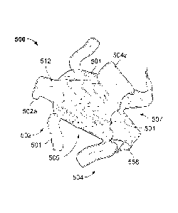
SUMMARY
[0004] One aspect of the invention relates to a medical device that
includes (1) an
expandable frame having a first end, a second end, and a middle portion
between the
first end and the second end, (2) a first apposition portion including a
plurality of first
apposition members, each of the first apposition members extending toward the
middle
portion, and (3) a second apposition portion including a plurality of second
apposition
members each of the second apposition members extending toward the middle
portion.
A first portion of each of the first apposition members may be oriented at a
first angle in
relation to a surface of the middle portion and a second portion of each the
first
apposition members may be oriented at a second angle in relation to the
surface of the
middle portion. In exemplary embodiments, the first angle is acute and is less
than the
second angle. Also, a first portion of each of the second apposition members
may be
oriented at a third angle in relation to the surface of the middle portion and
a second
portion of each of the second apposition members may be oriented at a fourth
angle in
relation to the surface of the middle portion. In exemplary embodiments, the
third angle
1
CA 02943308 2016-09-19
WO 2015/168504
PCT/US2015/028711
is acute and is less than the fourth angle. In some embodiments, at least one
the first
apposition members is longer than one or more others of the first apposition
members.
Additionally, at least one of the first apposition members may be longer than
at least
one of the second apposition members. In one or more embodiment, all of the
first
apposition members are longer than all of the second apposition members, The
first
apposition members may or may not be in axial alignment with the second
apposition
members. In another embodiment, one or more of the first apposition members
may
longitudinally overlap with one or more of the second apposition members. A
cover
material may be positioned on at least a portion of the frame,
[0005] A
second aspect of the invention relates to a medical device that includes a
frame including an elongate member defining (1) a first apposition portion
that includes
one or more first flange members configured to contact a first tissue surface
and to
provide an apposition force against the first tissue surface, (2) a second
apposition
portion that includes one or more second flange members configured to contact
a
second tissue surface and to provide an apposition force against the second
tissue
surface, and (3) a central portion having a first end and a second end where
the central
portion defines a longitudinal axis and the central portion is disposed
between and
interconnects the first apposition portion and the second apposition portion.
In
exemplary embodiments, at least one of the first flange members and at least
one of the
second flange members include a radius portion and a descending portion that
extends
longitudinally toward the central portion. At least one of the radius portions
of the first
flange members extends longitudinally beyond the first end and at least one of
the
radius portions of the second flange members extends longitudinally beyond the
second
end. In at least one embodiment, at least one of the first flange members or
at least
one of the second flange members further includes a horizontal portion that
extends
from the descending portion. The radius portions may extend from the first end
or the
second end of the central portion. Additionally, the descending portion may be
a
linearly descending portion. Further, the central portion may be configured to
longitudinally extend and retract to maintain contact, over a range of tissue
thicknesses,
CA 02943308 2016-09-19
WO 2015/168504 PCT/US2015/028711
of the first and second apposition portions with the first and second tissue
surfaces,
respectively.
[0006] A third aspect of the invention relates to a method of implanting an
anastomosis device in a patient that includes (1) positioning a delivery
sheath
containing the anastomosis device at a target location within the patient and
(2)
deploying the anastomosis device out from the delivery sheath such that at
least one
layer of tissue is between a first apposition portion and a second apposition
portion of
the device. The anastomosis device includes (1) an expandable frame having a
first
end, a second end, and a middle portion between the first end and the second
end, (2)
a first apposition portion including a plurality of first apposition members,
each of the
first apposition members extending toward the middle portion, and (3) a second
apposition portion including a plurality of second apposition members
extending toward
the middle portion. A first portion of each of the first apposition members
may be
oriented at a first angle in relation to a surface of the middle portion and a
second
portion of each the first apposition members may be oriented at a second angle
in
relation to the surface of the middle portion. In exemplary embodiments, the
first angle
is acute and is less than the second angle. Also, a first portion of each of
the second
apposition members may be oriented at a third angle in relation to the surface
of the
middle portion and a second portion of each of the second apposition members
may be
oriented at a fourth angle in relation to the surface of the middle portion.
In exemplary
embodiments, the third angle is acute and is less than the fourth angle. In at
least one
exemplary embodiment, a tip portion of the plurality of first apposition
members or the
plurality of second apposition members is spaced apart from the tissue. In
other
exemplary embodiments, two layers of tissue are between the first apposition
portion
and the second apposition portion.
DESCRIPTION OF DRAWINGS
[0007] The accompanying drawings are included to provide a further
understanding of
the disclosure and are incorporated in and constitute a part of this
specification,
illustrate embodiments, and together with the description serve to explain the
principles
of the disclosure.
3
CA 02943308 2016-09-19
WO 2015/168504 PCT/US2015/028711
[0008] FIG. 1 is a cutaway perspective view of an exemplary anastomosis
device,
that has been implanted within a patient to act as a shunt between the
patient's
gallbladder and intestine according to some embodiments;
[0009] FIG. 2 is a perspective view of an exemplary anastomosis device in
accordance with some embodiments;
[0010] FIGS, 3-6 are perspective views of exemplary apposition members in
accordance with some embodiments;
[0011] FIG. 7 is graphical illustration showing the relationship between
force and
displacement for each of the apposition members shown in FIGS. 3-6;
[0012] FIG. 8 is a schematic illustration of another exemplary anastomosis
device in
accordance with some embodiments;
[0013] FIG. 9 is a schematic illustration of exemplary apposition members
in
accordance with some embodiments;
[0014] FIG. 10 is a perspective view of yet another exemplary anastomosis
device in
accordance with some embodiments;
[0015] FIG. 11 is an end view of the anastomosis device of FIG. 10:
[0016] FIG. 12 is an alternative embodiment of the anastomosis device of
FIG. 10;
and
[0017] FIG, 13 is a side view of a central portion of another anastomosis
device that
includes expansion members in accordance with some embodiments,
DETAILED DESCRIPTION
[0018] Persons skilled in the art will readily appreciate that various
aspects of the
present disclosure can be realized by any number of methods and apparatus
configured
to perform the intended functions. It should also be noted that the
accompanying
4
CA 02943308 2016-09-19
WO 2015/168504 PCT/US2015/028711
drawing figures referred to herein are not necessarily drawn to scale, but may
be
exaggerated to illustrate various aspects of the present disclosure, and in
that regard,
the drawing figures should not be construed as limiting,
[0019] The present invention is directed to implantable devices for
connecting tissue
layers, for example, to circumvent a conduit or organ blockage, such as by
creating a
direct passage between tissue structures (e.g. connecting a gallbladder and a
portion
of a gastrointestinal tract) to create an anastomosis that facilitates
material flow
therebetween. The devices described herein may be endoscopically deployable or
deliverable via a catheter and may include self-expanding apposition
mechanisms that
facilitate a secure connection between the tissue structures (such a
connection may
also be referred to herein as a "shunt," "passageway," "shunt passageway," or
"tunnel").
Such design features simplify implantation and reduce the likelihood of
complications.
In some embodiments, the devices provided herein are configured to be
removable
after implantation. As one example, the device is implanted and remains in
place until
the gallbladder and/or its associated ducts are cleared of blockages, after
which the
device is removed. In another example, the device remains implanted until the
body
grows a tissue-anastomosis around the device, and then the device is removed.
In
other embodiments, tissue ingrowth into and/or around the device permanently
implants
the device, and the device is not removed. The devices described herein can
provide
an alternative treatment for patients who are not suitable candidates for
other types of
treatments (e.g., gallbladder removal surgery) and/or to avoid known
complications of
other types of treatments (e.g., external biliary drainage).
[0020] This document refers to anastomosis devices in an exemplary fashion.
That
is, it should be understood that the inventive concepts disclosed in this
document can
also be applied to other types of devices. For example, this document also
provides
implantable devices that, in some embodiments, can be used for occluding
tissue
structures, organs, body conduits, blood vessels, the GI tract, and the like.
For
example, in some embodiments the devices provided herein can be used to
occlude
septal defects. In some embodiments, the devices provided herein can be used
to
occlude a patient's vasculature or GI tract. In some such embodiments, the
device
CA 02943308 2016-09-19
WO 2015/168504 PCT/US2015/028711
does not include a tunnel through the device. Rather, in some embodiments a
covering
material seals the device to inhibit, modulate, or substantially prevent
material from
flowing through the device.
[0021] Referring to FIG, 1, an exemplary anastomosis device 40 in
accordance with
one or more provided herein can be implanted in a patient to create a fluidic
connection
between two organs, spaces, tissue structures, conduits, and the like, and
combinations
thereof. For example, in the depicted implementation the anastomosis device 40
is
connecting a gallbladder 10 (that defines an internal gallbladder space 12)
with an
intestine 20 (that defines an internal intestinal space 22). Hence, the
anastomosis
device 40 is acting as a fluidic shunt device between the internal gallbladder
space 12
and the internal intestinal space 22. Such an implementation may provide a
beneficial
treatment to the patient when, for example, a flow blockage exists in the
native
anatomical conduits connecting the internal gallbladder space 12 and the
internal
intestinal space 22. For example, in some instances the patient may have one
or more
gallstones that cause a blockage of the patient's cystic duct 14 and/or common
bile duct
16. In such a case, the anastomosis device 40 can provide a fluidic passageway
such
that bile from the gallbladder 10 can flow into the intestine 20. If not for
the anastomosis
device 40, when bile is blocked from flowing out of the gallbladder 10
cholecystitis
(inflammation of the gallbladder 10) may result.
[0022] While the anastomosis devices provided herein can be used in some
implementations to relieve or prevent cholecystitis as described above, it
should be
understood that the anastomosis devices provided herein can also be used in
many
other types of implementations within a patient. For example, the anastomosis
devices
provided herein can be used in conjunction with various body tissue structures
and
organs such as, but not limited to, stomachs, colons, small intestines,
pancreases,
blood vessels, bladders, kidneys, conduits, and the like.
[0023] In general, some embodiments of the anastomosis devices provided
herein
(of which anastomosis device 40 is one type of example), include a first
tissue
apposition portion 42a, a second tissue apposition portion 42b, and a central
portion 44
6
CA 02943308 2016-09-19
WO 2015/168504 PCT/US2015/028711
between the first and second tissue apposition portions 42a and 42b. The
central
portion 44 defines a lumen 46 that extends longitudinally from a first end of
the
anastomosis device 40 to a second end of the device 40. The lumen 46 acts as a
connection (e.gõ a shunt passageway) between the internal gallbladder space 12
and
the internal intestinal space 22, such that the internal gallbladder space 12
is in fluid
communication with the internal intestinal space 22 via the anastomosis device
40.
[0024] Referring to FIG. 2, an example anastomosis device 500 includes a
framework of one or more elongate elements 501 that defines a first apposition
portion
502, a second apposition portion 504, and a central portion 506 is depicted.
The central
portion 506 is disposed between and interconnects the first apposition portion
502 and
the second apposition portion 504. In some embodiments, the central portion
506 is
essentially cylindrical (although other geometries are also contemplated and
are
considered to be within the purview of the invention).
[0025] In some embodiments, a covering material 512 is disposed on at least
some
portions of the anastomosis device 500, As described further below, the
covering
material 512 can be disposed on some portions or on all of the first
apposition portion
502, the second apposition portion 504, and/or the central portion 506. In
some
embodiments, portions of the first apposition portion 502, the second
apposition portion
504, and/or the central portion 506 can remain free of the covering material
512.
[0026] In some embodiments, the central portion 506 defines a lumen 507
that
extends between the first apposition portion 502 and the second apposition
portion 504.
In some implementations, the lumen 507 provides an anastomosis passageway or
tunnel through which biological materials and/or fluids can pass. The device
500 is
shown in an expanded configuration. The expanded configuration is the
configuration
that the device 500 naturally exhibits in the absence of external forces
acting upon the
device 500. In should be understood that when the anastomosis device 500 is
implanted in a patient, the configuration of the device 500 may be somewhat
different
than shown because of the external forces from the patient's anatomy that are
exerted
on the device 500.
7
CA 02943308 2016-09-19
WO 2015/168504 PCT/US2015/028711
[0027] The anastomosis device 500 is shown in a deployed or expanded
configuration. In some embodiments, the framework of the anastomosis device
500, as
described further below, can be made of a variety of metallic shape memory
materials
and super-elastic alloys. Thus, in some embodiments the central portion 506
(and/or
the apposition portions 502 and 504) can be configured to self-expand to the
deployed
configuration. In some embodiments, the central portion 506 is balloon
expandable to
the deployed configuration, or supplemental expansion forces can be applied to
a self-
expandable device by balloon dilation. The diameter of the central portion 506
can be
made in any size as desired in order to suit the intended use and/or delivery
system of
the anastomosis device 500.
[0028] When the anastomosis device 500 is configured in its expanded
deployed
configuration as shown, the diameter of the central portion 506 increases to a
deployed
diameter. The diameter of the central portion 506 can be made in any dimension
as
desired in order to suit the intended use and/or delivery system of the
anastomosis
device 500. In some implementations, the deployed outer diameter of the
central
portion 506 is configured to at least partially anchor the device 500 via an
interference fit
with the tissue aperture in which the central portion 506 resides.
Additionally, when the
central portion 506 and the tissue aperture have an interference fit
relationship, para-
device leakage may be reduced or minimized. In such a case, leakage of the
contents
of the organs, conduits, and other types of tissue structures in which the
anastomosis
device 500 may be deployed can be substantially prevented. For example, when
the
anastomosis device 500 is used between a gallbladder and GI tract (e.g., refer
to FIG.
1), leakage into the abdominal cavity can be substantially prevented.
[0029] In some implementations the deployed outer diameter of the central
portion
506 is slightly less than the diameter of the tissue aperture in which the
central portion
506 resides, and the apposition portions 502 and 504 compress the tissue to
provide
the migration resistance. In some embodiments, the fully expanded diameter of
the
central portion 506 is about 30 mm, or about 25 mm, or about 20 mm, or about
15 mm,
or about 12 mm, or about 10 mm, or about 8 mm, or about 6 mm, or about 4 mm,
and
the like. In some embodiments, the fully expanded diameter of the central
portion 506
8
CA 02943308 2016-09-19
WO 2015/168504 PCT/US2015/028711
is in a range between about 20 mm to about 30 mm, or about 15 mm to about 25
mm,
or about 10 mm to about 20 mm, or about 5 trim to about 15 mm, or about 4 mm
to
about 8 mm, and the like.
[0030] The length of the central portion 506 can be made in any dimension
as
desired in order to suit the intended use and/or delivery system of the
anastomosis
device 500. For instance, in one exemplary embodiment the central portion 506
is
about 13.5 mm in length and about 15 mm in diameter. In some embodiments, the
length of the central portion 506 can be in a range from about 5 mm to about
10 mm, or
about 8 mm to about 13 mm, or about 11 mm to about 16 mm, or about 14 mm to
about
19 mm, or about 17 mm to about 22 mm, or greater than 22 mm.
[0031] In some embodiments, the anastomosis device 500 has a framework that
comprises one or more elongate elements 501. In some embodiments, the one or
more
elongate elements 501 are wound into the framework configuration. In some
embodiments, a single elongate element 501 is wound to form the framework of
the
anastomosis device 500. In some embodiments, two or more elongate elements 510
are cooperatively wound to form the framework of the anastomosis device 500.
[00321 In some embodiments, the framework of the first apposition portion
502, the
second apposition portion 504, and the central portion 506 are formed of one
or more
elongate elements 501 made of materials such as, but not limited to, spring
wire (e.g.,
L605 steel or stainless steels), shape memory alloy wire (e.g., nitinol or
nitinol alloys),
super-elastic alloy wire (e.g., nitinol or nitinol alloys), other suitable
types of elongate
elements or wires, or combinations thereof. In some embodiments, the first
apposition
portion 502, the second apposition portion 504, and the central portion 506
are formed
from a precursor material that is cut to create the framework of elongate
elements 501.
In some such embodiments, the precursor material is a single piece of
precursor
material. In some embodiments, one or more elongate elements 501 are wound
into a
configuration to form the framework. In some embodiments, different types of
elongate
elements 501 are used at different locations of the first apposition portion
502, the
second apposition portion5, and/or the central portion 506. In some
embodiments, the
9
CA 02943308 2016-09-19
WO 2015/168504 PCT/US2015/028711
elongate elements 501 of the first apposition portion 502, the second
apposition portion
504, and/or the central portion 506 (or portions thereof) may be constructed
of
polymeric materials.
[0033] Suitable materials for the elongate elements 501 of the anastomosis
device
500 and/or other devices provided herein include a variety of metallic
materials
including alloys exhibiting, shape memory, elastic and super-elastic
characteristics.
Shape memory refers to the ability of a material to revert to an originally
memorized
shape after plastic deformation by heating above a critical temperature.
Elasticity is the
ability of a material to deform under load and return to its original shape
when the load
is released. Most metals will deform elastically up to a small amount of
strain. Super-
elasticity refers to the ability of a material to deform under strain to much
larger degree
than typical elastic alloys, without having this deformation become permanent.
For
example, the super-elastic materials included in the frames of some
anastomosis device
embodiments provided herein are able to withstand a significant amount of
bending and
flexing and then return to or substantially to the frame's original form
without
deformation. In some embodiments, suitable elastic materials include various
stainless
steels which have been physically, chemically, and otherwise treated to
produce a high
springiness, metal alloys such as cobalt chrome alloys (e.g., ELGILOYTM,
MP35N,
1.605), platinum/tungsten alloys. Embodiments of shape memory and super-
elastic
alloys include the NiTi alloys, ternary shape memory alloys such as NiTiPt,
NiTiCo,
NiTiCr, or other shape memory alloys such as copper-based shape memory alloys.
Additional materials could combine both shape memory and elastic alloys such
as
drawn filled tube where the outer layer is constructed of nitinol and the
inner core is a
radiopaque material such as platinum or tantalum. In this construct, the outer
layer
provides the super-elastic properties and the inner core remains elastic due
to lower
bending stresses.
[0034] In some embodiments, the elongate elements 501 used to construct the
anastomosis device 500 and/or other devices provided herein can be treated in
various
ways to increase the radiopacity of the devices for enhanced radiographic
visualization.
In some embodiments, the devices are least partially a drawn-filled type of
NiTi
CA 02943308 2016-09-19
WO 2015/168504 PCT/US2015/028711
containing a different material at the core, such as a material with enhanced
radiopacity.
In some embodiments, the devices include a radiopaque cladding or plating on
at least
portions of the first apposition portion, the second apposition portion, and
the central
portion. In some embodiments, one or more radiopaque markers are attached to
the
devices. In some embodiments, the elongate elements and/or other portions of
the
devices provided herein are also visible via ultrasound, and may include
portions with
enhanced echogenicity.
[0035] In some embodiments, the materials and configuration of the
anastomosis
device 500 (and the other anastomosis device embodiments provided herein)
allow the
devices to be elastically crushed, folded, and/or collapsed into a low-profile
delivery
configuration for containment within a lumen for transcatheter or
endoscopicithorascopic delivery, and to self-expand to an operative size and
configuration once positioned at a desired target site within a body and
deployed from
the lumen. For example, in the low-profile delivery configuration the
anastomosis
device 500 can be disposed within a delivery sheath that has about a 15 Fr. (5
mm)
outer diameter. However, in some embodiments, sheaths that are smaller or
larger than
15 Fr. can be used. For example, sheaths that have outer diameters of 6 Fr., 7
Fr., 8
Fr., 9 Fr., 10 Fr., 11 Fr., 12 Fr., 13 Fr., 14 Fr., 16 Fr., 17 Fr., 18 Fr., 19
Fr., 20 Fe, and
larger than 20 Fr., can be used in some embodiments. While the anastomosis
device
500 is configured in a collapsed delivery configuration, in some embodiments
the
framework of one or more elongate elements 501 is radially compressed such
that the
elongate elements 501 are forced to extend substantially parallel to axis of
the central
portion 506, and the diameter of the central portion 506 is crushed to become
smaller.
[0036] The anastomosis device 500 also includes the covering material 512
(which
may also be referred to herein as a "covering"). In some embodiments, the
covering
material 512 is disposed on at least some portions (or on all) of the first
apposition
portion 502, the second apposition portion 504, and the central portion 506.
In some
embodiments, some portions of the first apposition portion 502, the second
apposition
portion 504, and/or the central portion 506 are not covered by the covering
material 512.
CA 02943308 2016-09-19
WO 2015/168504 PCT/US2015/028711
[0037] In some embodiments, the covering material 512 is generally fluid
impermeable. That is, in some embodiments the covering material 512 is made of
a
material that inhibits or reduces passage of blood, bile and/or other bodily
fluids and
materials through the covering material 512 itself, In some embodiments, the
covering
material 512 has a material composition and configuration that inhibits or
prevents
tissue ingrowth andibr endothelialization or epithelialization into the
covering material
512. Some such embodiments that are configured to inhibit or prevent tissue
ingrowth
and/or endothelialization can be more readily removed from the patient at a
future date
if so desired. In some embodiments, the covering material 512, or portions
thereof, has
a microporous structure that provides a tissue ingrowth scaffold for durable
sealing
and/or supplemental anchoring strength of the anastomosis device 500.
[0038] In some embodiments, the covering material 512 comprises a
fluoropolymer,
such as an expanded polytetrafluoroethylene (ePTFE) polymer, polyvinylidene
fluoride
(PVDF), or PVDA. In some embodiments, the covering material 512 comprises a
polyester, a silicone, a urethane, biocompatible polymer(s), polyethylene
terephthalate
(e,g., Dacrond4), bioabsorbable materials, copolymers, or combinations
thereof, In
some embodiments, the covering material 512 comprises a bioabsorbable web. In
other embodiments, the bioabsorbable material may also provide an anti-
migration
feature by promoting attachment between the device 500 and tissue until the
bioabsorbable material is absorbed.
[0039] In some embodiments, the covering material 512 (or portions thereof)
is
modified by one or more chemical or physical processes that enhance one or
more
properties of the material 512. For example, in some embodiments, a
hydrophilic
coating may be applied to the covering material 512 to improve the wettability
and echo
translucency of the material 512. In some embodiments, the covering material
512, or
portions thereof, may be modified with chemical moieties that facilitate one
or more of
endothelial cell attachment, endothelial cell migration, endothelial cell
proliferation, and
resistance to or promotion of thrombosis. In some embodiments, the covering
material
512, or portions thereof, may be modified to resist biofouling. In some
embodiments,
the covering material 512, or portions thereof, may be modified with one or
more
CA 02943308 2016-09-19
WO 2015/168504 PCT/US2015/028711
covalently attached drug substances (e.g., heparin, antibiotics, and the like)
or
impregnated with the one or more drug substances. The drug substances can be
released in situ to promote healing, reduce tissue inflammation, reduce or
inhibit
infections, and to promote various other therapeutic treatments and outcomes.
In some
embodiments, the drug substance may be, but is not limited to a
corticosteroid, a
human growth factor, an anti-mitotic agent, an antithrombotic agent, a stem
cell
material, or dexamethasone sodium phosphate. In some embodiments, a
pharmacological agent is delivered separately from the covering material 512
to the
target site to promote tissue healing or tissue growth.
[0040] Coatings and treatments may be applied to the covering material 512
before
or after the covering material 512 is joined or disposed on or around the
framework of
the anastomosis device 500. Additionally, one or both sides of the covering
material
512, or portions thereof, may be coated, In some embodiments, certain coatings
and/or
treatments are applied to the covering material(s) 512 located on some
portions of the
anastomosis device 500, and other coatings and/or treatments are applied to
the
material(s) 512 located on other portions of the anastomosis device 500. In
some
embodiments, a combination of multiple coatings and/or treatments are applied
to the
covering material 512, or portions thereof. In some embodiments, certain
portions of
the covering material 512 are left uncoated and/or untreated. In some
embodiments,
the device 500 is fully or partially coated to facilitate or frustrate a
biological reaction,
such as, but not limited to, endothelial cell attachment, endothelial cell
migration,
endothelial cell proliferation, and resistance to or promotion of thrombosis.
[0041] In some embodiments, a first portion of the covering material 512 is
formed of
a first material and a second portion of the covering material 512 is formed
of a second
material that is different than the first material. In some embodiments, the
covering
material 512 is comprised of multiple layers of materials, which may be the
same or
different materials. In some embodiments, portions of the covering material
512 have
one or more radiopaque markers attached thereto to enhance in viva
radiographic
visualization of the anastomosis device 500, or one or more echogenic areas to
enhance ultrasonic visibility.
1.)
CA 02943308 2016-09-19
WO 2015/168504 PCT/US2015/028711
[0042] In some embodiments, one or more portions of the covering material
512 are
attached to the framework of the device 500, such as the central portion 506
and/or the
apposition portions 502 and 504. The attachment can be accomplished by a
variety of
techniques such as, but not limited to, stitching the covering material 512 to
the
framework of the device 500, adhering the covering material 512 to the
framework of
the device 500, laminating multiple layers of the covering material 512 to
encompass
portions of the elongate members of the device 500, using clips or barbs,
laminating
multiple layers of the covering material together through openings in the
framework of
the device 500. In some embodiments, the covering material 512 is attached to
the
framework of the device 500 at a series of discrete locations, thereby
facilitating the
flexibility of the framework. In some embodiments, the covering material 512
is loosely
attached to the framework of the device 500. It is to be appreciated that the
covering
material 512 may be attached to the framework using other techniques or
combinations
of techniques described herein.
[0043] In some embodiments, the framework of the device 500 (or portions
thereof)
is coated with a bonding agent (e.g., fluorinated ethylene propylene or other
suitable
adhesive) to facilitate attachment of the covering material 512 to the
framework. Such
adhesives may be applied to the framework using contact coating, powder
coating, dip
coating, spray coating, or any other appropriate means.
[0044] The covering material 512 can adapt to changes in the length and/or
diameter
of the central portion 506 in a variety of manners. In a first example, the
covering
material 512 can be elastic such that the covering material 512 can stretch to
accommodate changes in the length and/or diameter of the device 500. In a
second
example, the covering material can include slackened material in the low-
profile delivery
configuration that becomes less slackened or totally unslackened when the
device 500
is in the expanded configuration. In a third example, the covering material
512 can
include folded portions (e.g., pleats) that are folded in the low-profile
configuration and
less folded or totally unfolded when the device 500 is in the expanded
configuration. In
some embodiments, combinations of such techniques, and/or other techniques can
be
14
CA 02943308 2016-09-19
WO 2015/168504
PCT/US2015/028711
used whereby the covering material 512 can adapt to changes in the length
and/or
diameter of the central portion 506,
[00451 The
one or more elongate element(s) 501 of the central portion 506 can be
configured in various ways to define a generally cylindrical framework. In the
embodiment depicted in FIG. 2, the elongate element(s) 501 of the central
portion 506
are wound circumferentially around the central portion 506. In addition to the
circumferential winding, the elongate element(s) 501 can exhibit other winding
paths,
such as the wavy or serpentine path shown (e.g., approximately sinusoidal) and
other
paths, In the depicted embodiment, the winding path of the elongate element(s)
501 in
the central portion 506 has eight apices per circumference, and an apical
length of
about 3,5 mm. In some embodiments, the elongate element(s) 501 of the central
portion 506 can be made to have more or less than eight apices per
circumference, and
can be made to have an apical length of more than or less than 3.5 mm, as
desired to
suit a particular application. For example, in some embodiments the elongate
element(s) 501 of the central portion 506 can be made to have three, four,
five, six,
seven, nine, ten, eleven, twelve, thirteen, fourteen, fifteen, sixteen, or
more than sixteen
apices per circumference. In some embodiments, the elongate element(s) 501 of
the
central portion 506 can be made to have an apical length in a range of about 1
mm to
about 2 mm, or about 2 mm to about 3 mm, or about 3 mm to about 4 mm, or about
4
mm to about 5 mm, or about 5 mm to about 6 mm, or about 6 mm to about 7 mm, or
greater than 7 mm.
[0046] In
some embodiments, the apposition portions 502 and 504 include one or
more flange components 502a and 504a, respectively, Such flange components
(e.g.,
flange components 502a and 504a) may also be referred to herein as "fins,'
"petals," or
"fingers." The flange components 502a and 504a are configured to contact
tissues and
to exert an apposition pressure thereto. While the depicted embodiment
includes four
flange components 502a and four flange components 504a, other quantities of
flange
components 502a and 504a can be included. For example, in some embodiments
one,
two, three, five, six, seven, eight, or more than eight flange components 502a
and/or
CA 02943308 2016-09-19
WO 2015/168504 PCT/US2015/028711
504a may be included. In some embodiments, unequal numbers of flange
components
502a and flange components 504a are included.
[0047] The flange components 502a and 504a can be configured to exert a
predictable and desired apposition force when in contact with tissue. For
example, the
material(s), the diameter, and other properties of the elongate element can be
selected
to attain a desired apposition force. Elongate elements (e.g,, nitinol
elongate members)
can be made to have a particular diameter as desired. Elongate elements made
of
other suitable materials and with larger or smaller diameters can be selected
as desired.
The geometry of the flange components 502a and 504a can also affect the
apposition
force exerted by the flange components 502a and 504a. That is, geometry
aspects
such as, but not limited to, the length, width, radii, angles, arcs (and the
like) of the
flange components 502a and/or 504a can be selected to attain a desired
apposition
force.
[0048] In some embodiments, the flange components 502a and 504a can be
configured to have an offset orientation between the opposite end portions of
the
anastomosis device 500. That is, the axes of one or more of the individual
flange
components 502a may be offset (e.g., skewed, or out of alignment) from the
axes of
one or more of the individual flange components 504a. In some such
embodiments,
some or all of the flange components 502a and 504a can be configured to cross
each
other (e.g, overlap each other in an interposing arrangement). In some such
embodiments, some or all of the flange components 502a and 504a may be offset
from
each other but not crossing each other. However, in some embodiments the axes
of
one or more of the individual flange components 502a may be generally in
alignment
(e.g., substantially parallel) with the axes of one or more of the individual
flange
components 504a, In some such embodiments, some or all of the flange
components
502a and 504a can be configured to abut each other, In some such embodiments,
some or all of the flange components 502a and 504a may be in alignment with
each
other but not abutting each other.
16
CA 02943308 2016-09-19
WO 2015/168504 PCT/US2015/028711
[0049] In some embodiments, one or more of the flange components 502a
and/or
504a may vary in configuration in comparison to one or more others of the
flange
components 502a and/or 504a. For example, the flange components 502a can
protrude farther towards the central portion 506 than the flange components
504a (or
vice versa). Or, one or more of the flange components 502a or 504a can
protrude
farther towards the central portion 506 than others of the flange components
502a or
504a respectively.
[0050] In some embodiments, one or more of the flange components 502a
and/or
504a may have two or more portions with differing curvatures (radii). For
example, in
the depicted embodiment, at least some of the flange components 502a and/or
504a
extend from the central portion 506 at a first radius, and then straighten to
a generally
linear portion, and then curve along a second radius after which the flange
components
502a and/or 504a terminate. In some embodiments, the first radius is unequal
to the
second radius. In some embodiments, the first and second radii are curved in
opposite
directions from each other.
[0051] In some embodiments, a radius 558 of the flange components 502a and
504a
protrudes beyond the central portion 506 of the device. Therefore, the force
applied by
the flange components 502a and 504a may push some tissue into the radius 558,
thereby making a longer and potentially stronger or less leak-prone
anastomosis. In
some embodiments, the radius 558 of curvature is determined by the allowed
strain of
the nitinol material when loaded into a delivery system (e.g., sheath). For
example, in
some embodiments a strain of about 6.4% may result. However, other strain
levels of
less than or more than about 6.4% are used in some embodiments.
[0052] In some implementations, including multiple flange components 502a
and
504a may tend to reduce the potential for causing tissue ischemia. In some
embodiments, individual flange components 502a are configured differently from
each
other and/or individual flange components 504a are configured differently from
each
other. In some embodiments, the flange components 502a and 504a can remain
discrete from each other (as shown), or in some embodiments the flange
components
17
CA 02943308 2016-09-19
WO 2015/168504 PCT/US2015/028711
502a and 504a are interconnected to each other by, for example, the covering
512. In
some embodiments, the flange components 502a and 504a can oppose or not oppose
each other, can crisscross over each other, can have different geometries
(e.g., lengths,
widths, angles, radii, shapes, etc.). All combinations of such design features
can be
combined to create anastomosis devices of a wide variety of configurations. In
some
embodiments, one or both of the flange components 502a and 504a protrude from
the
central portion 506 at an axial orientation and shape to achieve a specific
desired
apposition pressure on the tissue.
[0053] In some embodiments, one or more of the shape of the flange
components,
the number of flange components, the elongate element size, and the tissue
thickness
are factors that are selectable to achieve a specific force profile vs,
displacement. For
example, referring to FIGS. 3-6, various exemplary flange component designs
510,
520, 530, and 540 are shown. The free ends shown in FIGS, 3-6 are where the
flange
components design 510, 520, 530, and 540 would extend from the device body
(e.g.,
anastomosis device 500). The force vs, displacement curves for each is shown
in FIG.
7,
[0054] The exemplary flange component 510 includes a sharply descending
region
511 extending to the edge of the central portion (not shown), and a
substantially
horizontal region 513 extending away from the device. The exemplary flange
component 520 includes a moderately sharp descending region 514 connected to
and a
sloping region 515 extending away from the device, The exemplary flange
component
520 includes a linearly descending region 522 extending away from the device.
The
exemplary flange component 530 includes a gradual sloping curved region 532
extending away from the device. In some embodiments, one or more regions of
the
flange components longitudinally extend towards the central portion of the
device of
which the flange components are part of (e.g, towards central portion 506 of
anastomosis device 500),
18
CA 02943308 2016-09-19
WO 2015/168504 PCT/US2015/028711
[0055] Particular flange component force vs. displacement profiles (e.g.,
flange
components 510, 520, 530, and 540) may be advantageous for achieving a desired
apposition pressure and/or other performance characteristics. For example,
referring to
FIG. 7; a graph of force vs. displacement shows the apposition force that can
be applied
by each flange component 510 (5100, 520 (520f), 530 (530f), and 540 (540f).
The force
vs. displacement profile of flange component 510 can include a steep linear
slope, as
shown by 510f. Curve 510f may be of particular benefit if the organs to be
apposed are
not close together. The linear and quickly increasing force would resist
separation of
the organs. In some embodiments, the force vs. displacement profile of flange
component 520 can include a shallow linear slope that abruptly changes to a
steep
linear slope (520f). Curve 520f may be of particular benefit for creating high
apposition
force during the initial healing phase while the anastomosis is being created.
During
this time the tissue may be thicker and inflamed utilizing the steep linear
profile of
520f. As the tissue inflammation and resulting tissue thickness reduced, the
shallow
part of curve 5201 would be employed helping to avoid necrosis of the tissue.
The force
vs. displacement profile of flange component 530 produces a shallow linear
slope
(5300. Curve 530f provides a shallow linear increase in force with respect to
displacement and may be particularly useful for tissue that is considered
friable and
prone to perforation. In other embodiments, the force vs. displacement profile
of flange
component 540 can include a continuously increasing slope (5400. Such
variations of
flange components (and other variations also contemplated within the scope of
this
disclosure) can be selected for a particular application as desired. For
example, the
force vs. displacement curve achieved by flange component 540 may be
advantageous
in particular applications because the curve smoothly increases and the design
allows
for a broad area of contact over a large range of displacement.
[0056] FIGS. 8 and 9 illustrate another example anastomosis device 1200.
Anastomosis device 1200 is an example variation of the anastomosis device 500
described above. In particular, the anastomosis device 1200 has first and
second
apposition portions 1202 and 1204 that are designed differently than the first
and
second apposition portions 502 and 504 of anastomosis device 500. As described
19
CA 02943308 2016-09-19
WO 2015/168504 PCT/US2015/028711
further below, the one or more apposition members 1208 and 1210 that make up
the
first and second apposition portions 1202 and 1204 can be configured to
provide
functional properties that are desirable in some implementations.
[0057] In some embodiments, the framework of the device 1200 or any portion
thereof can comprise elongate elements such as a spring wire (e.g,, L605 steel
or
stainless steels), shape memory alloy wire (e.g., nitinol or nitinol alloys),
super-elastic
alloy wire (e.g., nitinol or nitinol alloys), other suitable types of wire, or
combinations
thereof. In the depicted embodiment of device 1200, the framework includes an
elongate element that is formed by winding, for example. In some embodiments,
different types of wires are used at different locations of the device 1200.
Alternatively,
device 1200 or portions thereof can be formed from the same piece of precursor
material that is cut to create the elongate element framework structure as
desired. In
some embodiments, the device 1200 or portions thereof may be constructed of
polymeric materials. The device 1200 is shown with a covering material, as
described
above. It should be understood that anastomosis device 1200 can be constructed
using
any of the materials and techniques described in reference to any and all
other
anastomosis devices described herein.
[0058] The central portion 1206 of the device can be constructed to have a
tailored
radial strength by, for example, varying the elongate element's sine wave
amplitude,
angle, number of apices per row, number of rows, wire diameter, and by
selecting (or
not selecting) a covering material. For anastomosis device applications, the
radial
strength of the central portion 1206 may be designed to resist circumferential
loading
from the surrounding tissue. Therefore, in some embodiments the radial
strength of the
central portion 1206 is configured to facilitate remodeling of the tissue
external to the
central portion 1206 to become approximate in size to the outer diameter of
the central
portion 1206. When the anastomosis device 1200 (and the other anastomosis
devices
provided herein) is implanted to form an anastomosis, the radial strength of
the central
portion 1206 provides resistance to the hoop force applied by the surrounding
tissue.
Therefore, an anastomosis device with strong radial strength in the central
portion (e.g_
central portion 1206) will substantially maintain an open lumen at a desired
dimension.
CA 02943308 2016-09-19
WO 2015/168504 PCT/US2015/028711
In addition, a device with strong radial strength can advantageously act as a
scaffold for
tissue to grow around the device.
[0059] In some embodiments, the materials and configuration of the
anastomosis
device 1200 allow the device 1200 to be elastically crushed, folded, and/or
collapsed
into a low-profile configuration for containment within a lumen for
transcatheter or
endoscopicithorascopic delivery, and to self-expand to an operative size and
configuration once positioned at a desired target site within a body and
deployed from
the lumen.
[0060] The first apposition portion 1202 and the second apposition portion
1204 are
configured to engage one or more layers of tissue between the first and second
apposition portions 1202 and 1204, and to provide apposition forces against
the tissue
surfaces. The apposition forces provided by the first and second apposition
portions
1202 and 1204 can facilitate fixation of the device 1200 to the tissue, and
provide
migration resistance such that the device 1200 can reliably remain positioned
at a target
site in a patient as desired.
[0061] The first apposition portion 1202 and the second apposition portion
1204
each include one or more apposition members 1208 and 1210 respectively. The
anastomosis device 1200 can be configured in a collapsed low-profile delivery
configuration in which the apposition members 1208 and 1210 are radially
compressed
such that they extend substantially parallel to the longitudinal axis of the
device. In the
deployed or expanded configuration, the apposition members 1208 and 1210
protrude
outwardly from the central portion 1206.
[0062] In some embodiments, at least one of the apposition members 1208
and/or
1210 is orientated to have a first angle 1216 in relation to the central
portion 1206 and
to have a second angle 1214 in relation to the central portion 1206 (as shown
in FIG. 9).
In other words, in some embodiments at least one apposition member 1208 and/or
1210 is non-planar. In the depicted embodiment the apposition member 1210 is
orientated at a first angle 1216 in relation to the central portion 1206, and
the apposition
member 1210 is also oriented at a second angle 1214 in relation to the central
portion
CA 02943308 2016-09-19
WO 2015/168504 PCT/US2015/028711
1206. In some embodiments, the first angle 1216 is a shallow angle. For
example, in
some embodiments the angle 1216 is acute, e.g., less than about 90 , or less
than
about 75 , or less than about 600, or less than about 45 , or less than about
30', or less
than about 25 , or less than about 20 , or less than about 15 , or less than
about 10 , or
less than about 5'. In some embodiments, the angle 1216 is between about 15
and
about 20 , or between about 10 and about 30', or between about 5 and about
45 .
[0063] Because in some embodiments the angle 1216 is relatively shallow,
and
because the apposition member 1210 extends towards the central portion 1206, a
portion of the apposition member 1210 is therefore orientated relatively close
to the
access location (i.e., the incision location in which the anastomosis device
1200 will be
deployed). Hence, this configuration facilitates an effective and sustainable
apposition
of tissue.
[0064] In some embodiments, the second angle 1214 is a larger angle than
the first
angle 1216. For example, in some embodiments the angle 1214 is greater than
about
90 , less than about 45 , less than about 25 , less than about 20 , less than
about 15 ,
less than about 10 , or less than about 5'. In some embodiments, the angle
1214 is
between about 30 and about 40 , or about 20 and about 45 , or about 30 and
about
50 , or about 40 and about 60', or about 50 and about 70', or about 60' and
about
80 , or about 70 and about 90 . In some embodiments the second angle 1214 is
larger
than the first angle 1216 such that a portion of the apposition member is
orientated
farther away from the access location. While, the shallow angle of the first
angle 1216
permits tissue contact resulting in apposition force near the access location,
the larger
angle of the second angle 1214 permits tissue contact away from the access
location,
providing anti-migration forces to keep the device 1200 in place. Moreover, in
some
embodiments the larger angle of the second angle 1214 permits the terminal
ends of
the apposition members 1208 and/or 1210 to be out of contact with tissue in
situ. In
some such implementations, by having fins pointing away from the apposed
tissue, the
potential for tissue over-growth on fins can be delayed or avoided. By
delaying or
avoiding tissue overgrowth the device can be easily removed when/if needed. In
some
22
CA 02943308 2016-09-19
WO 2015/168504 PCT/US2015/028711
embodiments, a single apposition member of this design can provide the
benefits that
are associated with having dual length apposition members.
[0065] In some embodiments, the apposition members 1208 and 1210 are in
axial
alignment with each other such that the positions of the apposition members
1208 and
1210 around the periphery of the central portion 1206 longitudinally coincide
with each
other. In some embodiments, the apposition members 1208 and 1210 are out or
axial
alignment with each other such that the positions of the apposition members
1208 and
1210 around the periphery of the central portion 1206 do not longitudinally
coincide with
each other. In some such embodiments, one or more of the apposition members
1208
of the first apposition portion 1202 longitudinally overlap (e.g., criss-
crossed in an
interposing arrangement) with one or more of the apposition members 1210 of
the
second apposition portion 1204, as depicted in FIGS. 8 and 9. In some
embodiments,
some or all of the apposition members 1208 and 1210 may be offset from each
other, or
in alignment with each other, while not crossing each other. In some
embodiments,
some or all of the apposition members 1208 and 1210 may be in alignment with
each
other and abutting each other.
[0066] Referring to FIGS. 10-12, an anastomosis device 1300 is shown having
a
central portion 1306 that is interchangeable with any other central portion
described
herein, a first apposition portion 1302, and a second apposition portion 1304.
In some
embodiments, the framework of device 1300 or any portion thereof can comprise
one or
more elongate elements such as a spring wire (e.g., L605 steel or stainless
steels),
shape memory alloy wire (e.g., nitinol or nitinol alloys), super-elastic alloy
wire (e.g.,
nitinol or nitinol alloys), other suitable types of wire, or combinations
thereof (as
described above in reference to elongate element 501).
[0067] In the depicted embodiment of device 1300, the framework is
comprised of a
single elongate element that is formed by winding and shape-setting, for
example. In
some embodiments, different types of elongate elements are used at different
locations
of the device 1300. Alternatively, the anastomosis device 1300 (or portions
thereof) can
be formed from the same piece of precursor material that is cut and expanded
to create
CA 02943308 2016-09-19
WO 2015/168504 PCT/US2015/028711
the elongate element framework structure as desired. In some embodiments, the
device 1300 (or portions thereof) may be constructed of polymeric materials.
It should
be understood that anastomosis device 1300 can be constructed using any of the
materials and techniques described in reference to any and all other
anastomosis
devices described herein.
[0068] In some embodiments, the framework of the central portion 1306 can
be
configured such that the central portion 1306 is longitudinally extendable.
For example,
in the depicted embodiment of anastomosis device 1300 the framework of the
central
portion 1306 includes serpentine-wound portions that allow for longitudinal
extension
and retraction (like a spring). Accordingly, the longitudinal length of the
central portion
1306 can self-adjust based on in vivo loading forces. This feature can be
advantageous, for example, in maintaining apposition when the tissue thickness
changes during the healing process (e.g,, in at least some cases of acute
cholecystitis).
It should be understood that this feature can be incorporated into any of the
device
embodiments described herein.
[0069] The device 1300 may include a covering material 1312. In some
embodiments, the covering 1312 is made of stretchable (elastic-like) material
that can
have a high percentage of recoverable strain (e.g., recoverable strain in the
magnitude
of 100s of percentage per unit length). Some embodiments of such covering
materials
may include, but are not limited to, pure silicone, urethane material, or such
materials
that are imbibed in or laminated to other materials including, but not limited
to,
flouropolymers such as ePTFE. In some embodiments, the covering material 1312
is
as described herein (e.g., similar or identical to covering material 512).
[00701 The first apposition portion 1302 and the second apposition portion
1304 are
configured to engage one or more layers of tissue between the first and second
apposition portions 1302 and 1304, and to provide apposition forces against
the tissue
surfaces. The apposition forces provided by the first and second apposition
portions
1302 and 1304 can facilitate attachment of the device 1300 to the tissue and
provide
CA 02943308 2016-09-19
WO 2015/168504 PCT/US2015/028711
displacement resistance such that the device 1300 can reliably remain
positioned at a
target site in a patient as desired,
[0071] The first apposition portion 1302 and the second apposition portion
1304
each include one or more apposition members 1302a or 1302a and 1304a or 1304a1
(also referred to herein as anchor members, fins, flange portions, petals,
etc.). In some
embodiments, the apposition members 1302a and 1304a are bare elongate elements
(without a covering). In some embodiments, the apposition members 1302a' and
1304a' have the covering material 1312 disposed on at least some areas of
their
elongate elements.
[0072] In some embodiments, one or more of the apposition members 1302a or
1302a' and/or 1304a or 1304a' have different configurations (e.g., geometries,
lengths,
widths, shapes, etc.) than one or more other apposition members 1302a or
1302a'
and/or 1304a or 1304a.
[0073] In some embodiments, the materials of the anastomosis device 1300
allow
the device 1300 to be elastically crushed, folded, and/or collapsed into a low-
profile
configuration for containment within a lumen for transcatheter or
encloscopicithorascopic delivery, and to self-expand to an operative size and
configuration once positioned at a desired target site within a body and
deployed from
the lumen. For example, the anastomosis device 1300 can be configured in a
collapsed
delivery configuration in which the framework is compressed to a low-profile
such that
the apposition members 1302a or 1302a' and 1304a or 1304a' extend
substantially
parallel to the longitudinal axis of the device 1300, In the deployed or
expanded
configuration, the apposition members 1302a or 1302a' and 1304a or 1304a'
extend
outwardly from the central portion 1306.
[0074] In some embodiments, the lengths of some of the apposition members
1302a
or 1302a' and 1304a or 1304a' are dissimilar to provide both sufficient
apposition forces
near the periphery of the access location or hole where access is created, and
anti-
migration forces farther away therefrom. For example, in some embodiments one
or
more of the apposition members 1302a or 1302a' is longer than one or more of
the
CA 02943308 2016-09-19
WO 2015/168504 PCT/US2015/028711
apposition members 1304a or 130421. In some embodiments, the apposition
members
1302a or 1302a1 and/or 1304a or 130481 have varying lengths and are
alternated, or
arranged around the periphery of the first apposition portion 1302 and/or the
second
apposition portion 1304 in a pattern. In some embodiments, the apposition
members
1302a or 130281 and/or1304a or 130421 withineach apposition portion 1102
and/or
1104 are uniform in length.
[0075] In some embodiments, the apposition members 1302a or 1302a' and/or
1304a or 1304a' have lengths that are selected based at least in part on the
size of
tissue structures that the device 1300 is to be implanted into. For example,
if a first
tissue structure generally includes smaller geometry than the second tissue
structure,
having differing lengths of the apposition members 1302a or 1302a1 versus
1304a or
1304a1can be advantageous. In this example, the apposition portion entering
the
smaller tissue structure may beneficially have apposition members with a
shorter length,
while longer apposition members may be better-suited in the larger tissue
structure. In
some such implementations, the shorter length apposition members provide an
appropriate fit for the smaller tissue structure thus ensuring sufficient
tissue contact
necessary for an anastomosis device, while the longer apposition members
provide
anti-migratory forces that help to retain the device in place. In some
embodiments,
such short and long apposition members 1302a or 1302a and/or 1304a or 1304a1
are
staggered, nested, or separated around a periphery of a particular apposition
portion
1302 and/or 1304.
[0076] The anastomosis device 1300 (and other embodiments that share design
features of the anastomosis device 1300) can exhibit the following advantages.
Having
varying lengths of apposition members 1302a or 130221 and/or 1304a or 1304a1
can
provide apposition at various target tissue locations. Having one or more such
specific
apposition zones may minimize or eliminate leakage of fluid or other contents
that pass
through the device lumen. Discrete apposition members 1302a or 130221 and/or
1304a
or 1304a1 that move independently of each other can provide advantageous
apposition
member conformability to non-planar tissue topography. Better conformability
can
minimize tissue injury especially when used in a diseased tissue bed, The
flexible
CA 02943308 2016-09-19
WO 2015/168504 PCT/US2015/028711
discrete design of the apposition members 1302a or 1302a and/or 1304a or
1304a' can
facilitate device 1300 removal by folding the apposition members 1302a or
1302a'
and/or 1304a or 1304a' parallel to the lumen of the device 1300. This
flexibility of the
apposition members 1302a or 1302a' and/or 1304a or 1304a' may help to minimize
tissue injury during removal of the anastomosis device 1300,
[0077] Referring to FIG. 13, another example central portion 206 can be
included as
part of any the anastomosis devices provided herein. As with the central
portions 506,
1206, and 1306, the central portion 206 is comprised of a framework of one or
more
elongate elements. For enhanced visibility, the central portion 206 is shown
without a
covering material, however the covering material(s) described elsewhere herein
can be
applied to the central portion 206 in some embodiments. In this example, the
central
portion 206 is shown in an undeployed or low-profile delivery configuration.
When
deployed in a patient, the central portion 206 can self-expand or be forced to
expand to
an expanded configuration.
[0078] The one or more elongate elements of the central portion 206 can be
constructed from the same types of materials and can be constructed using the
same
types of techniques as described above in reference to the elongate elements
of
anastomosis device 500. In some embodiments, the central portion 210 is formed
by
one or more wound wires. In some embodiments, the central portion 206 is
formed
from a unitary piece of precursor material that is cut to create the elongate
element
framework structure as desired. In some such embodiments, the precursor
material is a
tube (e.g., a nitinol tube) that is laser cut to form the desired elongate
element
framework structure. In some such embodiments, the precursor material is a
sheet
(e.g., a nitinol sheet) that is laser cut to form the desired elongate element
framework
structure. In some embodiments, different types of elongate elements are used
at
different portions of the central portion 206. For example, in some
embodiments the
central portion 206 or portions thereof may be constructed of polymeric
materials.
CA 02943308 2016-09-19
WO 2015/168504 PCT/US2015/028711
[0079] The central portion 206 includes one or more axial adjustment
members 218
extending along the longitudinal axis of the central portion 206. The axial
adjustment
members 218 allow the axial length (also referred to herein as longitudinal
length") of
the central portion 206 to elastically extend or contract (as described above
in reference
to the framework of the central portion 1306 of anastomosis device 1300),
[0080] The central portion 206 also includes one or more cells 212. In some
embodiments, the one or more cells 212 interconnect the axial adjustment
members
218. The cells 212 allow for the radial expansion and contraction of the
central portion
206, and provide radial strength to the device to maintain its shape/size
while resisting
external compression forces. During radial expansion of the central portion
206, the
cells 212 expand in the circumferential direction and collapse in the
longitudinal
direction. While the cells 212 are shown as having a diamond-like shape, other
geometries may be used. In the depicted embodiment, pairs of cells 212 are
interconnected to each other in the circumferential direction by a bridge
member 213.
However, in some embodiments a single cell 212, or more than two cells 212,
may be
included (rather than the pair shown).
[0081] In some embodiments, the cells 212 are unconnected to adjacent cells
212
along the longitudinal axis of the central portion 206. As such, the cells 212
connecting
to the axial adjustment members 218 do not substantially constrain the axial
expansion
or contraction of the axial adjustment members 218 and/or the central portion
206. In
some embodiments, the cells 212 provide radial strength to the central portion
206. That
is, in some embodiments the cells 212 tend to self-expand into an expanded
configuration. Such self-expansion can provide radial forces from the central
portion
206 to the tissues in that are contacted by the central portion 206.
[0082} In some embodiments, axial length of the central portion 206 is
adjusted
before or during deployment, e.g,, by a clinician to accommodate differences
in tissue
thicknesses. In other embodiments, the axial adjustment member 218 self-
responds to
mechanical forces exerted on the deployed anastomosis device in situ. For
example,
the axial adjustment member 218 permits the axial length of the device to
dynamically
28
CA 02943308 2016-09-19
WO 2015/168504 PCT/US2015/028711
adjust during the healing process (as described above in reference to central
portion
1306).
[0083] The anastomosis devices provided herein are deployable to a target
site
within a patient using one or more catheters, delivery sheaths, and other
suitable
devices and techniques. In some implementations, the anastomosis devices
provided
herein are deployable using an endoscopic or laparoscopic approach.
[0084] It should be understood that one or more design features of the
anastomosis
devices provided herein can be combined with other features of other
anastomosis
devices provided herein. In effect, hybrid designs that combine various
features from
two or more of the anastomosis device designs provided herein can be created,
and are
within the scope of this disclosure.
[0085] In some such embodiments, the device does not include a tunnel or
central
aperture through the device.
[0086] In some embodiments the devices provided herein can be used for
sealing or
anchoring a heart valve implant. A heart valve implant enables one-way flow of
blood
from a heart chamber and usually has a first inflow end and a second outflow
end. The
contractions of the heart cause flow of blood through the valve from the
inflow end to
the outflow end. Between the inflow and outflow ends, a valve assembly within
the
heart valve implant provides for one way flow, opening to allow flow from the
inflow to
the outflow end when the pressure of the blood is higher on the inflow end,
and closing
to prevent flow when the pressure on the outflow end is higher than the inflow
end. In
some embodiments, the device includes a tunnel or central aperture through the
device
with apposition portions to anchor a valve assembly and seal against backward
flow. A
valve assembly can be attached in the tunnel or central aperture. The
apposition
portions of the device can be configured to be highly conformable to the
topography of
the heart chambers or blood vessels, and compliant with the beating movements
of the
heart In some embodiments, a covering material is configured to allow flow
through a
valve assembly in the tunnel or aperture while preventing flow around the
apposition
portions.
29
CA 02943308 2016-09-19
WO 2015/168504 PCT/US2015/028711
[0087] The invention of this application has been described above both
generically and
with regard to specific embodiments. It will be apparent to those skilled in
the art that
various modifications and variations can be made in the embodiments without
departing
from the scope of the disclosure, Thus, it is intended that the embodiments
cover the
modifications and variations of this invention provided they come within the
scope of the
appended claims and their equivalents.