Note: Descriptions are shown in the official language in which they were submitted.
CA 02943597 2016-09-22
WO 2015/150831
PCT/GB2015/051049
1
ECG Evaluation
The present invention relates to a method for evaluating cardiac function, in
particular a method that utilises the information provided by
electrocardiography. The invention also relates to an apparatus in which the
aforementioned method may be practised, including a computer program.
The intrinsic conducting system of the heart permits electrical impulses
originating from the sinoatrial node to travel through the cardiac tissue in a
controlled manner. The passage of this electrical impulse through the heart
tissue produces a wave of contraction through the cardiac tissue. The wave of
contraction is followed by a period of relative electrical calm in the heart
tissue,
which corresponds to relaxation of the cardiac tissue. Arrhythmias occur when
this normal, organised electrical activity of the heart becomes disrupted.
Worldwide 3 million people a year die from sudden cardiac death. In most cases
there is no warning and the heart is stopped by a sudden arrhythmia. Some
people are at high risk of sudden cardiac death, but this can be prevented by
an
implantable cardioverter defibrillator, which is implanted in a minor
operation.
In the UK, subjects are screened for risk of sudden cardiac death using the
National Institute for Health and Clinical Excellence (NICE) guidelines (a
screening that is based on a mixture of physiological and electro-
physiological
measurements and an understanding of the subject's clinical history). However,
most of the people who die from sudden cardiac death are not identified by
these guidelines.
Assessment of the health of the heart by measuring its electrical activity is
known. For example, one can measure the electrical activity of the heart with
the use of intra-cardiac electrodes that are directly applied to the cardiac
tissue.
SUBSTITUTE SHEET (RULE 26)
CA 02943597 2016-09-22
WO 2015/150831
PCT/GB2015/051049
2
This is however a particularly invasive technique that is not preferable for
the
routine assessment of subjects and that has not been clearly shown to
demonstrate any clinical relevance for assessing cardiac function such as the
risk
of arrhythmia. Electrocardiography (ECG) has been developed as a non-invasive
procedure for studying the electrical activity of the heart. ECG involves the
placing of a plurality of electrodes on the skin surface of a subject. An
understanding of the electrical activity of the heart may be identified from
the
potential difference (i.e. leads) between combinations of the plurality of
electrodes. Conventionally, a collective assessment of ECG leads provide a
classic ECG tracing which comprises a P wave, a QRS complex and a T wave, and
which demonstrate periods of electrical activity that vary from the
isoelectric
line. It has been suggested that ECGs may be useful for identifying arrhythmia
of
the heart by measuring the dispersion of QT durations on an ECG tracing.
Measuring changes in this QT duration as an indicator of cardiac arrhythmia
has
however since been discredited; to the degree that the cardiology community no
longer view the QT dispersion assessment as a clinically relevant way to
establish arrhythmia risk (see, for example, Malik et aL; JACC; 2000;36:1749-
66).
The present inventors have identified a further means of analysis of ECG
output
that has proved useful in evaluating cardiac function. The analysis, termed
"Regional Restitution Instability Index or R2I2", essentially evaluates the
between lead differences in ECG output as an indicator of cardiac function
(see
International Patent Publication No. WO 2011/117608 Al and Nicolson et al. "A
Novel Surface Electrocardiogram-Based Marker of Ventricular Arrhythmia Risk
in Patients with Ischemic Cariomyopathy" J. American Heart Association, 2012).
There however remains a need for further methods and apparatus capable of
identifying the risk of sudden cardiac death due to arrhythmia. Such methods
and apparatus would be particularly useful for identifying those individuals
who
are most likely to benefit from the implantation of an implantable
cardioverter
defibrillator or from treatment with anti-arrhythmic therapeutics.
CA 02943597 2016-09-22
WO 2015/150831
PCT/GB2015/051049
3
It has surprisingly been found by the present inventors that analysis of the
change in relationship between action potential duration and the diastolic
interval taken over multiple time periods by each lead of an ECG presents
result
that can indicate whether or not an individual being analysed is likely to go
on to
develop an arrhythmia.
Accordingly, in the first aspect of the present invention, there is provided a
method for assessing the electrical function of a heart, comprising the steps
of:
a. for each of a plurality of leads of an ECG, at multiple time points,
determining a value derived from the output of that lead and
which corresponds to an action potential duration;
b. for each of the plurality of leads of the ECG, at the multiple time
points, determining a value derived from the output of that lead
and which corresponds to a diastolic interval;
c. for each of the plurality of leads of the ECG, using the determined
values for action potential duration and diastolic interval across
the multiple time points to determine a relationship between
action potential duration and diastolic interval as seen by that
lead;
d. defining at least one characteristic of interest of each of the
determined relationships between action potential duration and
diastolic interval and combining information on that
characteristic from the relationships seen by the plurality of leads
to derive a combined value;
e. deriving an assessment result by analysing the combined value.
An ECG provides a cutaneous electrocardiagraphic measurement of the electrical
functioning of the heart. As would be known to the skilled person, an ECG
includes a plurality of electrodes that are placed on specific external
positions of
the body. A lead of an ECG is the potential difference between two or more of
CA 02943597 2016-09-22
WO 2015/150831
PCT/GB2015/051049
4
these electrodes. Consequently, a lead provides an electrical output that
corresponds to a changing potential difference between the electrodes that
form
the lead.
The plurality of leads available in an ECG would be known to the skilled
person
(see, for example, "The ECG made easy", 4th edition, John R. Hampton,
Churchill
Livingstone, 1997). For example, the leads may comprise or consist: limb
leads,
chest leads, posterior leads, anterior leads, lateral leads, inferior leads,
or any
combination thereof. For example, the limb leads may comprise or consist:
right
arm (Red), left arm (Yellow), left leg (Green), right leg (Black), or any
combination thereof. For example, the chest leads may comprise or consist: V1
(right sternal edge, 4th intercostal space), V2 (left sternal edge, 4th
intercostal
space), V3 (halfway between V2 and V4), V4 (position of the apex beat - e.g.
intersection of the 5th intercostal space and mid-clavicular line), VS
(anterior
axillary line), V6 (mid-axillary line), or any combination thereof For
example,
the posterior leads may comprise or consist of V7 (left posterior axillary
line,
straight line from V6), V8 (left midscapular line, straight line from V7) and
V9
(left paraspinal line, straight line from V8). For example, the anterior leads
may
comprise or consist: V1, V2, V3, V4, or any combination thereof For example,
the lateral leads may comprise or consist: VS, V6, I, aVL, or any combination
thereof For example, the inferior leads may comprise or consist: II, III, aVF,
or
any combination thereof
The number of leads used in the method according to the present invention must
exceed 2, and may be S or more, 10 or more, or 12 or more. Optionally, the
number of leads do not exceed 4096. The plurality of leads of the present
method may be 5, 12, 128 or 256 lead configurations.
The reference to "multiple time points" requires that the steps carried out at
multiple time points are repeated a plurality of times. In other words, the
methods of the present invention require more than one instance of the
CA 02943597 2016-09-22
WO 2015/150831
PCT/GB2015/051049
determining of a value derived from the output of a lead and which corresponds
to an action potential (or diastolic interval). As it may be beneficial to
evaluate
changes in the relationship between action potential duration and diastolic
interval achieved by the heart beating at different rates, each time point may
be
5 taken during a period in which the heart rate changes. The heart rate may
be
induced to change by:- the application of a chronotropic therapeutic agent to
the
subject in possession of the heart under analysis (ie medications that alter
heart
rate); by physically exercising the subject in possession of the heart under
analysis; by providing electrical pacing impulses to the heart of varying
frequency (often called "pacing spikes"). Alternatively, the electrical
function of
the heart may be analysed over a period (eg 48 hours) and the multiple time
points chosen for analysis under the method of the present application as
being
those instances where extremes of heart rate are experienced (eg during a
priod
of arrhythmia).
The action potential duration is the period of myocyte electrical activity,
which
would be understood to consist of the initial depolarisation, a plateau phase
and
finally repolarisation phase. The diastolic interval is the interval between
action
potentials, when the myocyte is electrically quiescent. The output from each
lead of an ECG provides sufficient information concerning the electrical
activity
of the heart for a skilled person to derive therefrom a value for both the
action
potential duration and the diastolic interval. For example, the output of ECG
leads may be converted into an ECG tracing, e.g. comprises a P wave, a QRS
complex and a T wave. The skilled person would have no difficulty in
preselecting the relevant portion of the ECG tracing that corresponds to the
action potential duration and to the diastolic interval. By measurement of the
duration of these preselected portions one can determine a value from the
output of the lead and which corresponds to the action potential duration and
to
the diastolic interval.
CA 02943597 2016-09-22
WO 2015/150831
PCT/GB2015/051049
6
The preselected portion that corresponds to the action potential duration can,
for example, be the QT or the IT interval. The preselected portion that
corresponds to the diastolic interval can, for example, be the TQ interval.
The
process of determining the value for each lead in step a. should be
consistent.
The process of determining the value for each lead in step b. should be
consistent.
It should be understood that how one precisely calculates the beginning and
end
of each of these intervals (in order to identify their duration) is of less
significance than the fact that the value for the JT, QT and TQ intervals is
measured for each in the methods of the present invention in a consistent
manner. For example, the QT interval may be measured:- from the beginning of
the QRS complex to the end of the T wave; from the onset of the R wave to the
end of the T wave; from the beginning of the QRS complex to the peak of the T
wave, or; from the onset of the R wave to the peak of the T wave. For example,
the IT interval may be measured:- from the point of separation between the QRS
complex and the end of the T wave, or; from the point of separation between
the
QRS complex and the peak of the T wave. For example, the TQ interval may be
measured:- from the end of the T wave to the beginning of the QRS complex;
from the end of the T wave to the onset of the R wave; from the peak of the T
wave to the beginning of the QRS complex, or; from the peak of the T wave to
the
onset of the R wave. (see, for example, Malik et aL; JACC; 2000;36:1749-66).
When the heart rate is under the influence of electrical pacing impulses,
during
capture of values for the present method, the pacing spikes that appear in an
ECG and that correspond to each pacing impulse may be taken to be the
beginning of the action potential duration and/or the end of the diastolic
interval.
Determining a relationship between the determined values for action potential
duration and diastolic interval across the multiple time points may be
achieved
in a number of ways, which would be understood by the skilled person. For
CA 02943597 2016-09-22
WO 2015/150831
PCT/GB2015/051049
7
example, a restitution curve for each lead may be plotted on a graph
(conventionally, the Y-axis corresponds to the action potential duration,
whilst
the x-axis corresponds to the diastolic interval). A restitution curve for a
individual heart cell describes the nonlinear relationship between the
Diastolic
Interval and Action Potential Duration. Establishing a restitution curve is
well
within the ordinary skill in the art. For example, an explanation as to how to
establish a restitution curve suitable for use in the present invention may be
found in Taggart et al. "Effect of Adrenergic Stimulation on Action Potential
Duration Restitution in Humans" Circulation, 30 December 2002 (incorporated
herein by reference), ie a method that uses least squares regression to fit a
linear
gradient within a 40ms segment of the data. This segment is then moved in
10ms increments along the x-axis (TpQ).
The characteristic of interest is optionally any characteristic that defines
the
change in the aforementioned relationship between action potential duration
and diastolic interval as seen by each lead. Consequently, the characteristic
of
interest may be one or more gradient of the restitution curve for each lead,
optionally the gradient of the restitution curve at each time point on that
curve
and for each lead. Each point plotted to establish a restitution curve for
each lead
represents the relationship between action potential duration and diastolic
interval at the time-point when the output was received from that lead. The
gradient of the curve passing that point may be calculated as the
characteristic of
interest. Rather than analysing each time point on that curve for each lead,
time
points on only a portion of the curve for each lead may instead be analysed.
For
example, analysing only the first quarter of time points, the second quarter
of
time points, the third quarter of time points and/or the fourth quarter of
time
points. Rather than analysing each time point on that curve, or portion of
that
curve, a representative selection of time points instead may be only analysed.
For example, only every 2nd, 3rd, 4th, 5th, or 6th time point in the curve or
portion of the curve may be analysed. Indeed, the method does not have to be
restricted to time-points on the curve for each lead being time points that
are
CA 02943597 2016-09-22
WO 2015/150831
PCT/GB2015/051049
8
directly derived from the diastolic interval and action potential duration
values
derived from the output of the leads. Once the curve has been established,
every
point along the curve may be taken as a time point (including those in the
curve
that are positioned between points directly derived from the action potential
duration and diastolic interval values derived from the output of leads).
The combined value may be a plurality of combined values, each combined value
being a combining of information for the characteristic for each time point.
The
combined value may be an average of the characteristic of interest across the
leads for each time point, and so a combined value for one or more time point
may be derived. This is quite distinct from establishing the amount of
difference
between the characteristics for each time-point. For example, when the
characteristic of interest is a gradient of the restitution curve at a time
point, the
combined value, or one of the combined values, is obtained by establishing the
average value for the gradient across the leads, optionally the combined value
is
obtained by establishing, for each time point, the average value for the
gradient
across the leads for one time point. This would, for example, require
establishing the gradient for the time-point established by each lead, the
average
of those gradients is then established. Establishing an average is well within
the
skill of the ordinary person. Any method may be used as long as it is
consistently
used. For example, all values may be added together and the total is divided
by
the number of values in order to identify the average value.
The analysis of step d) may comprise the identification of the steepest
gradient
from the combined values. For example, if multiple time points are analysed
and
so multiple combined values are determined, the value of the steepest of the
gradients (ie the highest combined value) may be the combined value used in
step e.
It has been found that the steeper the gradient the more likely the subject is
going to progress to cardiac failure, and so be more likely to require a
implantable cardioverter defibrillator or the need for administration of anti-
CA 02943597 2016-09-22
WO 2015/150831
PCT/GB2015/051049
9
arrhythmic agents. For example a gradient of greater than 1.21 is considered
to
be indicative of an elevated risk of developing a cardiac arrhythmia or
cardiac
failure compared to normal (as analysed by the methods of the present
invention), for example analysed according to the Peak Electrocardiogram
Restitution Gradient (PERG) as described later in the methods section.
It has been found that analysis using the aforementioned method when
combined with the R2I2 method results in a more accurate assessment of
cardiac function. R2I2 identifies the amount of difference between the
relationships identified in step c, and the greater the amount of difference
between each lead, the greater the risk of developing cardiac arrhythmia.
Consequently, the methods of the present invention may include the additional
steps of carrying out analysis according to R2I2 (as described in WO
2011/117608 Al or Nicolson et al. "A Novel Surface Electrocardiogram-Based
Marker of Ventricular Arrhythmia Risk in Patients with Ischemic
Cariomyopathy" J. American Heart Association, 2012 and incorporated herein by
reference) and combining that analysis with that carried out in step e. of the
present invention.
For example, the method of the present invention may additionally comprising
the steps of:-
f. assessing the differences between the determined relationships
from step c) for each of the plurality of leads.
g. assessing the cardiac function of the heart, determining the
subject's need for the implantation of an implantable cardioverter
defibrillator, or the need for administration of an anti-arrhythmic
agent by analysing the combined assessment of step e. and step f.
There are many ways in which the difference between the relationships
identified
in step c. may be assessed in step f For example, the relationship between
single
action potential duration and a single diastolic interval may be determined as
a
CA 02943597 2016-09-22
WO 2015/150831 PCT/GB2015/051049
ratio of the two for each lead, the difference between the ratios for each
lead may
be assessed numerically. For example, when a number of action potential
durations and diastolic intervals are determined for each lead, the
differences
between the determined relationships may be assessed by identifying or
5 quantifying the difference in the gradient or gradients of the curves
established by
plotting the values for action potential duration against diastolic interval
(or vice
versa) for each lead on a graph (ie a restitution curve). This difference may
be
visually apparent from degree of separation of the curves for each lead over
the
length of the curves, or by changes in the degree of separation of the curves
for
10 each lead over the length of the curves.
Numerical analysis of the curves may also be used to quantify the differences.
For example, the following process may be applied:- (1) application of
logistic
regression to the data set to derive a polynomial equation, (2) application of
this
polynomial equation, adjusting the linear constant to achieve best fit, to
each lead
in turn, (3) using logistic regression to calculate the residuals this
technique
produces for each lead, (4) Summing the residuals will produce a measure of
the
differences between the relationships. At point (1) a spline could be used in
place
of the polynomial equation. At point (1) linear regression could be used
separately on groups of leads from each cardiac region, the resulting
equations
could then be applied to the leads from their corresponding regions as
described in
steps (2), (3) and (4). In a further example, the following process may be
applied:- (1) the standard deviation of the action potential difference from
all
leads is calculated for each determined diastolic interval length, (2) the
mean of
this value is taken as a marker of heterogeneity of the data
For example, assessing the difference in step f may comprise, for each time
point.:-
(i) establishing the mean point between the relationships
determined in step c. for each of the plurality of leads,
CA 02943597 2016-09-22
WO 2015/150831
PCT/GB2015/051049
11
(ii) for each lead, calculating the square of the residual from the
mean point to the relationship determined for that lead (e.g. the
square of the variation from the mean);
Assessing the difference in step f may further comprise:-
(iii) for each lead, calculating the mean value of the square of the
residuals calculated in step (ii) for each time point.
Assessing the difference in step f may further comprise:-
(iv) calculating the normalised mean value by dividing the mean
value calculated in step (iii) by the same mean value when
calculated from the assessment of subjects at normal risk of
developing cardiac arrhythmia, or by the mean of the values of
step (iii) for all of the plurality of leads.
Assessing the difference in step f, may further comprise:-
(v) identifying the largest normalised mean value calculated in step
(iv) out of the normalised mean values calculated for each of the
plurality of leads.
The values calculated in step (v) have been designated the Regional
Repolarisation Instability Index (R212).
It has been found that the greater the difference between the relationships
identified for each lead (which can be demonstrated by a relatively large
R212),
the greater the risk that the heart being assessed is abnormal, eg will
develop a
cardiac arrhythmia. Thus, the method of the present invention, when applied to
the outputs derived from an ECG applied to a subject, may be used as in a
method
of prognosis to assess the risk of the subject developing arrhythmia.
Essentially,
therefore, an increased level of heterogeneity between the relationships
CA 02943597 2016-09-22
WO 2015/150831
PCT/GB2015/051049
12
determined for each lead (which can be demonstrated by a relatively large
R212)
results in an increased risk of cardiac arrhythmia.
Consequently, in one embodiment of the present invention, the steps of the
invention may be carried out on the output derived from an ECG applied to a
subject to be examined for the risk of developing cardiac arrhythmia. The
method
may further comprise the carrying out of the steps on the output derived from
an
ECG applied to a subject that has been determined to have normal risk of
developing cardiac arrhythmia, and comparing the differences in step f
assessed
for the output from the subject to be examined with the differences in step f.
assessed for the output from the subject determined to be at normal risk of
developing cardiac arrhythmia (or a predetermined value that corresponds to
the
differences in step d. assessed for the output from subjects determined to be
at
normal risk of developing cardiac arrhythmia). When the differences are
determined to be greater for the subject to be examined than those of the
subject
determined to be at normal risk (or than the predetermined value), the subject
to
be examined is at increased risk of developing a cardiac arrhythmia
(increased,
being at greater risk than normal or vice versa). Similar analyses with
respect to a
normal subject may be carried out with respect to step e.
The predetermined value is derived from the assessment of subjects determined
to
be at normal risk of developing cardiac arrhythmia (i.e. the mean value for a
group of normal subjects). Normal subjects therefore represent a control
group.
Determining whether or not an individual subject is normal with respect to
their
risk of cardiac arrhythmia is a clinical question well within the abilities of
the
skilled person. However, in the interests of clarity, but not wishing to be
restricted
further, individuals in such a group will be characterised by structurally
normal
heart, as determined by echocardiography, and no history of palpitation,
syncope
or other cardiac problems. Optionally a normal subject has no family history
of
cardiac death.
CA 02943597 2016-09-22
WO 2015/150831
PCT/GB2015/051049
13
If both steps e and g conclude that there is an increased risk of the heart
having
less than normal function (ie the subject for the heart is at increased risk
of
developing a cardiac arrhythmia than normal) the risk is determined to be
greater than if only steps e or g had that conclusion.
In a second aspect of the present invention there is provided a method for
determining a subject's need for the implantation of an implantable
cardioverter
defibrillator or the need for administration of an anti-arrhythmic agent,
comprising the steps of:-
a. for each of a plurality of leads of an ECG directed to the subject, at
multiple time points, determining a value derived from the output
of that lead and which corresponds to an action potential
duration;
b. for each of the plurality of leads of the ECG directed to the subject,
at the multiple time points, determining a value derived from the
output of that lead and which corresponds to a diastolic interval;
c. for each of the plurality of leads of the ECG directed to the subject,
using the determined values for action potential duration and
diastolic interval across the multiple time points to determine a
relationship between action potential duration and diastolic
interval as seen by that lead;
d. defining at least one characteristic of interest of each of the
determined relationships between action potential duration and
diastolic interval and combining information on that
characteristic from the relationships seen by the plurality of leads
to derive a combined value;
e. deriving an assessment of the subject's need for the implantation
of an implantable cardioverter defibrillator or the administration
CA 02943597 2016-09-22
WO 2015/150831
PCT/GB2015/051049
14
of an anti-arrhythmic agent based on the analysis of the combined
value.
Such a method can be used in a method for directing subjects determined to
need treatment to be treated with by administering of an effective amount of
one
or more anti-arrhythmic agent, and/or to be treated by implanting a
cardioverter defibrillator.
Such a method can be used in a method of treating a subject with cardiac
arrhythmia and further comprises the step of administering an effective amount
of one or more anti-arrhythmic agent to a subject, and/or implanting a
cardioverter defibrillator if the subject is assessed by step d. to require
such
treatment.
Any clinically relevant anti-arrhythmic agent may be used, for example
amiodarone.
In order to monitor the efficacy of any anti-arrhythmic agent, the methods of
the
present invention may be carried out first in the absence of treatment with an
anti-arrhythmic agent and then repeated one or more times after the
administration of one or more doses of anti-arrhythmic agent. In this way the
methods of the present invention may be used to track the efficacy of
treatment
using the agent.
All optional features of the first aspect of the present invention maybe
included
in the second aspect of the present invention. For the avoidance of doubt, it
should be understood that when the method identifies that the subject is at
increased risk of developing cardiac arrhythmia, there is an increased need
for
the implantation of an implantable cardioverter defibrillator in the subject
or the
administration of an anti-arrhythmic agent to the subject (e.g. compared to an
individual at normal risk of developing cardiac arrhythmia).
CA 02943597 2016-09-22
WO 2015/150831
PCT/GB2015/051049
In a third aspect of the present invention there is provided Apparatus for
assessing the function of the heart, comprising a computer arranged to
receive input from each of a plurality of leads of an ECG and arranged to:-
5 a. for each of a plurality of leads of an ECG, at multiple time
points,
determining a value derived from the output of that lead and
which corresponds to an action potential duration;
b. for each of the plurality of leads of the ECG, at the multiple time
points, determining a value derived from the output of that lead
10 and which corresponds to a diastolic interval;
c. for each of the plurality of leads of the ECG, using the determined
values for action potential duration and diastolic interval across
the multiple time points to determine a relationship between
action potential duration and diastolic interval as seen by that
15 lead;
d. defining at least one characteristic of interest of each of the
determined relationships between action potential duration and
diastolic interval and combining information on that
characteristic from the relationships seen by the plurality of leads
to derive a combined value ;
e. deriving an assessment result by analysing the combined value.
The apparatus according to the third aspect of the present invention is
arranged
so as to be capable of operating the methods according to the earlier aspects
of
the present invention. Consequently, all features of the first and second
aspects
of the present invention maybe included in the third aspect of the present
invention. For example:-
The apparatus may include an ECG device. The ECG device may include a
plurality of electrodes configured to provide any of the lead combinations
described for the first aspect of the present invention.
CA 02943597 2016-09-22
WO 2015/150831
PCT/GB2015/051049
16
The output from each lead of an ECG provides sufficient information concerning
the electrical activity of the heart for the computer to derive therefrom a
value
for both the action potential duration and the diastolic interval. For
example, the
computer may be configured to convert the output of ECG leads into an ECG
tracing e.g. comprises a P wave, a QRS complex and a T wave. The computer
may be configured to preselect the relevant portion of the ECG tracing that
corresponds to the action potential duration and to the diastolic interval.
Appropriate pre-selection criteria are discussed above with respect to the
first
aspect of the present invention.
The computer may be arranged to determine the relationship between the
action potential duration and the diastolic interval in a number of ways, see
for
example the determination discussed in the first aspect of the present
invention
The apparatus of the present invention, when applied to the outputs derived
from an ECG applied to a subject, may be used in a method of prognoses of the
risk of that subject developing cardiac arrhythmia.
The apparatus may further comprise an electrophysiological catheter capable of
providing an electrical provocation to the cardiac tissue.
The apparatus may further comprise a computer program product that when
run on the computer causes it to be configured in the aforementioned manners.
In a fourth aspect of the present invention, there is provided a computer
program product when run on a computer arranged to receive input
from each of a plurality of leads of an ECG causes the computer to:-
a. for each of a plurality of leads of an ECG, at multiple time
points,
determining a value derived from the output of that lead and
which corresponds to an action potential duration;
CA 02943597 2016-09-22
WO 2015/150831
PCT/GB2015/051049
17
b. for each of the plurality of leads of the ECG, at the multiple time
points, determining a value derived from the output of that lead
and which corresponds to a diastolic interval;
c. for each of the plurality of leads of the ECG, using the determined
values for action potential duration and diastolic interval across
the multiple time points to determine a relationship between
action potential duration and diastolic interval as seen by that
lead;
d. defining at least one characteristic of interest of each of the
determined relationships between action potential duration and
diastolic interval and combining information on that
characteristic from the relationships seen by the plurality of leads
to derive a combined value ;
e. deriving an assessment result by analysing the combined value.
The computer program according to the fourth aspect of the present invention
may be included in the apparatus of the third aspect of the present invention.
Consequently, all features of the previous aspects of the present invention
maybe included in the fourth aspect of the present invention.
In yet a further aspect of the present invention, there is provided a method
as
substantially hereinbefore described and with reference to the figures.
In yet a further aspect of the present invention, there is provided apparatus
as
substantially hereinbefore described and with reference to the figures.
In yet a further aspect of the present invention, there is provided a computer
program as substantially hereinbefore described and with reference to the
figures.
CA 02943597 2016-09-22
WO 2015/150831
PCT/GB2015/051049
18
The present invention will now be described, by way of example, with reference
to accompanying figures, in which:-
Figure la shows a cutaneous APD restitution graph from a subject suffering
from arrhythmia.
Figure lb shows a cutaneous APD restitution graph from a subject that does not
suffer from arrhythmia.
Figure 2a shows a continuous cutaneous APD restitution graph from a subject
suffering from arrhythmia.
Figure 2b shows a cutaneous APD restitution graph from a subject that does not
suffer from arrhythmia.
Figure 3 shows analog digitized and recorded at 1000 Hz with 12-bit resolution
data from ECG (expanded from portion of that shown in Figure 4).
Figure 4 shows analog digitized and recorded at 1000 Hz with 12-bit resolution
data from ECG.
Figure 5 shows the technique by which TpQ and QTp measurements are made:
when an S2 arrives after the T wave peak the TpQ and QTp are measured as
shown on the left of the diagram. However, if the S2 occurs before the T wave
peak the TpQ is effectively negative. In this case it is measured by
subtracting the
QTpl interval (QTp for drive cycle beat) from the QTp2, in the example above
this would give a TpQ close to zero.
Figure 6 is a graph that illustrates the dynamic relationship between QTp
interval and TpQ interval for 12 leads, marked to show the 4 lateral leads, 3
CA 02943597 2016-09-22
WO 2015/150831
PCT/GB2015/051049
19
inferior leads, 4 anterior leads and 1 aVR lead. Results for population mean
values of all patients in the study shown on the graph.
Figure 7 is a graph that illustrates the dynamic relationship between QTp
interval and TpQ interval for 12 leads prepared for the assessment of R2I2 of
a
single patient.
Figure 8 provides a selection of only the anterior leads of the graph of
Figure 7,
prepared for the assessment of R2 12.
Figure 9 provides a blown up image of the box provided in the graph of Figure
8.
This figure also illustrates how to establish the mean point between the
relationships determined for this repetition for each of the anterior leads,
and
then how to calculate the square of the residual from the mean point to the
relationship determined for each lead (e.g. the square of the variation from
the
mean);
Figure 10 represents the graph of Figure 8 with the mean points for each
repetition provided in the graph, with figures provided below.
Figure 11 provides an explanation of the R2I2 calculation: the graph in A
shows
the anterior, inferior and lateral leads for a patient who reached the
endpoint of
ventricular arrhythmia (VA) / death. Each region is analysed separately as
seen
for the anterior leads in B; the points are grouped by the Si S2 coupling
interval
that produced them and the square of the residuals (narrow black lines) from
best fit points (black dots) is calculated for each lead at each 51 S2
coupling
interval. The mean of these residuals is then taken for each lead. There were
differences in the spread of the leads, in particular the lateral leads tended
to be
more widely spaced than the anterior and inferior leads. A proportion was
therefore taken: each lead's value was divided by the population mean value
for
that lead. The R2I2 is then taken as the mean of the maximum anterior,
inferior
CA 02943597 2016-09-22
WO 2015/150831
PCT/GB2015/051049
and lateral values. The LGE CMR scan for this patient (C) showed a large
anteroseptal and apical myocardial infarction with 16% pen-infarct zone (PIZ)
anteriorly, 13% inferiorly and 4% laterally corresponding with the R2I2
components: anterior 3.6, inferior 1.3 and lateral 0.25.
5
Figure 12 shows a cardiac magnetic resonance scan. A) First endocardial and
epicardial borders are drawn; then a large representative area of "normal
myocardium" and a small area of "peak scar" are selected. B) Software analysis
identifies all voxels with signal intensity >2 standard deviations (SD) above
10 "normal myocardium" mean intensity and voxels with signal intensity >SO%
of
the "peak scar" are subtracted from this to obtain the PIZ. Identified voxels
that
are not in the region of an infarct are discarded. The example in B shows an
infarct with relatively small PIZ compared with the example in Figure 11C.
15 Figure 13 shows a Kaplan-Meier curve of the probability of survival free
of
ventricular arrhythmia (VA) / death in the "high risk" group with R2I2 >
median
and the "low risk" group with R2I2 <= median. The difference in VA / death was
significant (p = 0.017, log rank test).
20 Figure 14 shows a plot of R2I2 against PIZ in each of the 22 patients
for whom
paired data was available. Lines are drawn at the median values for both
parameters. A least-squares regression line demonstrates a degree of
correlation
(r = 0.41 p=0.057).
Figure 15 shows a diagram of the last beat of the drive train and the 51 S2
coupling interval at 400, 380, 360 and 340ms for leads V2 and III.
Demonstration
of regional heterogeneity in repolarisation: little change is seen in V2 and
the
QTp is stable, while lead III is seen to fragment with two peaks and variable
QTp.
This gross change was seen in 2/4 of the patients who developed VA during
follow up.
CA 02943597 2016-09-22
WO 2015/150831
PCT/GB2015/051049
21
Figure 16 shows a Kaplan-Meier curve of probability of survival free of VA/
death in "high risk" group R2I2maxR > median and the "low risk" group with
R2I2maxR <= median. The difference in VA/death was significant (P = 0.051 log
rank test). Here the R2I2maxR has been calculated using TpS in place of the
TpQ
and JTe in place of the QTp. Additionally the maximum normalised mean value
has been taken rather than the mean of the regional normalised mean maxima.
Figure 17 shows measurement of R212. Figure 17A: Lead III example of
Identification of ECG fiducial points. Last 3 beats of drive train (Si) and
extrastimulus (S2) beats shown. Paired surrogates for APD/DI shown; both QTe
and TpTe are paired with TeQ. Abbreviations: TpQ T wave peak to QRS onset,
TeQ T wave end to QRS onset, QTp QRS onset to T wave peak, QTe QRS onset to
T wave end, TpTe T wave peak to T wave end. Figure 1B: QTp/TpQ plot for
representative ECG leads: V2 (anterior), II (inferior) and aVL (lateral) to
explain
the Regional Restitution Instability Index (R2I2) calculation in a typical
study
patient. For each lead, the QTp / TpQ gradient (least squares regression) was
calculated over a 40 ms segment of TpQ range. This segment was then scanned
over the range of TpQ with available data to produce gradients at 10 ms
intervals (numerical gradients are shown in bars adjacent to the corresponding
lines, note on the far left gradients are available for lead II but not for V2
and
aVL). The difference of the gradient from the mean gradient in each 40ms
segment was calculated. The standard deviation of these values was taken as a
measure of action potential duration restitution heterogeneity within each
lead.
The mean of this was then taken as the R2I2.
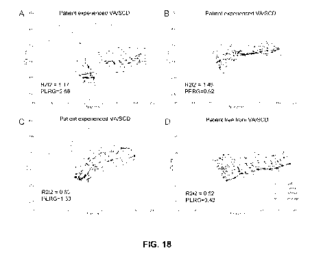
Figure 18 shows QTp/TpQ plots for 4 patients with A. high R2I2 and high PERG,
B. high R2I2 and low PERG, C. low R2I2 and high PERG, D. low R2I2 and Low
PERG. For each example patient the 12 ECG leads have separate lines styles
coded by ECG region. The lines are drawn point-to-point rather than as
gradients
to allow differentiation of the ECG leads. R2I2 is higher in the patients
whose
CA 02943597 2016-09-22
WO 2015/150831
PCT/GB2015/051049
22
ECG leads follow dissonant QTp/TpQ paths. PERG reflects the steepest gradient
taken as a mean for all 12 ECG leads.
Figure 19 shows association of R212 with risk of ventricular arrhythmia /
sudden cardiac death. Figure 3A: Kaplan Meier curve showing significant
separation of the curves for survival free of ventricular arrhythmia / sudden
cardiac death for patients partitioned by an R2I2 value of 1.03 (p<0.0001, log
rank). Figure 3B. Receiver operating characteristic curve for Regional
Restitution
Instability Index (R2I2): VA/SCD vs. event free survival. (Area under curve =
0.770)
Figure 20 shows a plot of Regional Restitution Instability Index against Peak
ECG
Restitution gradient. Lines are drawn at the pre-selected cut-off value for
R2I2
and at the optimal cut-off of 1.21 for PERG. Spearman rank correlation
analysis
minimal correlation between 12 lead mean peak restitution gradient and R2I2
(r=0.290, p=0.025).
Figure 21 shows a Kaplan Meier curve showing significant separation of the
curves for survival free of ventricular arrhythmia / sudden cardiac death for
patients partitioned by: both R2I2<1.03 and PERG<1.21 / either R2I21.03 or
PERG1.21 / both value R2I21.03 and PERG1.21 (p<0.0001, log rank).
1. Example 1: Inclusion criteria:
= Patients being considered for new ICD implantation with NYHA class II-
III symptoms of heart failure and documented left ventricular
dysfunction.
2. Example: Exclusion criteria
CA 02943597 2016-09-22
WO 2015/150831
PCT/GB2015/051049
23
= Unstable coronary heart disease, likely to need percutaneous or surgical
intervention
= Requirement for constant cardiac pacing (such as high grade AV block or
for cardiac resynchronisation)
= Recent coronary artery bypass graft surgery (within 3 months)
= Recent valvular surgery (within 3 months)
= Recent myocardial infarction (as documented by appropriate ECG &
biochemical analysis) (within 3 months)
2.1 Primary Outcome measure: ICD therapy for ventricular arrhythmia or
death within a 2 year follow up period
3. Example 3: Study Practiced on Patients included after analysis
from
Examples 1 and 2
A) Subjects were separated into two groups (the first group being patients
determined to at high risk of cardiac arrhythmia; the second group being
patients determined to be at low risk of cardiac arrhythmia) studied in the
post
absorptive state.
B) Appropriate aseptic technique employed throughout.
C) Cutaneous ECG leads were applied in the standard positions and connected to
an appropriate electrophysiological recorder. (Bard system used for study
standard 12 lead ECG positions)
D) An appropriate transvenous route was selected and the Seldinger technique
employed to insert a 6F venous sheath.
E) An appropriate electrophysiology catheter, for example the 6F Josephson
Quadripolar catheter, was inserted through the sheath.
F) Fluoroscopic guidance was used to manipulate the catheter into the right
ventricular apex, where a stable position was obtained.
G) The ventricular stimulation threshold was obtained, preferably via the
diastolic approach.
CA 02943597 2016-09-22
WO 2015/150831
PCT/GB2015/051049
24
H) An appropriate pacing protocol was delivered with rectangular pulses of 2
ms
duration set sufficiently greater than the threshold to achieve reliable
stimulation with a preferred value of 3 times the diastolic threshold. The
pacing
protocol used was the same for each patient in the study.
I) Analog data were digitized and recorded at 1000 Hz with 12-bit resolution,
shown in Figures 3 and 4. Low pass filter was set to 50Hz and high pass filter
set
to 0.01Hz.
J) Data analysis was performed with custom-written analysis programs in the
MATLAB 2009a language.
K) For consistency QT measurements were taken as from the start of the pacing
spike to the peak of the T wave and TQ measurements were taken as from the
peak of the T wave to the start of the pacing spike.
L) The QT / TQ restitution graphs were determined by plotting QT as a function
of preceding TQ and by plotting QT as a function of S2 coupling interval (see
figures la, lb, 2a and 2b).
4. Example 4: Pilot Study Exploring the Regional Repolarisation
Instability Index in relation to Myocardial heterogeneity and prediction of
Ventricular Arrhythmia and Death
4,1 Methods
4.1.1. Subjects were identified by screening the department audit databases
for
patients with a history of IHD who had undergone programmed electrical
stimulation (PES) between 1st January 2005 and 31st July 2009 as part of
clinical risk stratification for ICD implantation and who had had a CMR scan
within 6 months of their PES. This identified 43 patients. PES recordings were
unavailable for 9 patients and 4 more patients were excluded because only 6
lead ECGs had been recorded. Of the 30 patients whose PES data were available
1 could not be analysed because their drive cycle length (DCL) was changed
midway through the protocol. CMR data was then sought for these 30 patients.
CA 02943597 2016-09-22
WO 2015/150831
PCT/GB2015/051049
LGE images were not acquired for 3 patients because of difficulties gating (1)
and breath holding (2) and 4 patients could not be analysed because of an
incompatibility between the acquisition and pen-infarct zone analysis
software.
LGE CMR images were available for 23/30 patients.
5
4.2 Electrophysiological Study
4.2.1. Studies were performed as per the standard departmental protocol
which did not change for the duration of the study. Fasting subjects were
studied
10 with minimal sedation and with antiarrhythmic drug cessation 4-5 half-
lives
prior to the procedure. A 6F Josephson quadripolar catheter was advanced
transvenously first to the right ventricular apex (RVA) and then the right
ventricular outflow tract (RVOT). Electrocardiograms were recorded using
LabSystem Pro (BARD, Lowell) at 1 kHz sampling rate with a low pass filter set
1 5 to 50Hz and high pass filter set to 0.01Hz. The ventricular stimulation
test
followed a modified Wellens protocol with two 8 beat drive trains at the RVA
with drive cycle length (DCL) 600ms and 400ms and one 8 beat RVOT drive train
with DCL 400ms. If breakthrough beats were seen in the drive train the DCL was
reduced. Up to 3 extrastimuli were used with each drive train; the
extrastimulus
20 was typically started at 500/360ms and reduced in 20ms steps.
Monomorphic
VT of duration greater than 30 seconds or associated with haemodynamic
compromise was recorded as positive; the test was otherwise recorded as
negative. The 51 S2 coupling interval is the period between the last beat of
the
drive train and the first extrastimulus, this part of the PES was used to
derive the
25 R2I2.
4.3 Analysis of the R2I2
4.3.1. The electrocardiograms were exported at 16-bit digital resolution for
analysis in bespoke software written in MatLab (Mathworks, Natick). The timing
of the QRS onset (QRSo) and T wave peak (Tp) were analysed automatically and
CA 02943597 2016-09-22
WO 2015/150831
PCT/GB2015/051049
26
all data points were manually verified, a senior electrophysiology research
fellow blinded to the CMR data, the PES result and endpoint data. The Tp was
chosen in preference to the end of the T wave (Te) because of the known
difficulties in measuring Te.
Intra and inter-operator reproducibility (8 cardiology specialists mean 10.1
years of cardiology training) were assessed using a representative sample of
48
paced ECG points from the dataset. Mean intra-operator variability for
measurement of the QRSo and Tp was 6.3ms (SD 16.3ms) vs. inter-operator
6.4ms (SD 16.7ms).
4.3.2. Data points were censored according to predetermined rules: 1.
Breakthrough beat occurring after beat 6 of the drive train (51/316 drive
trains
censored), 2. Point indeterminate due to artefact, baseline wander or unclear
morphology (256/3089 points censored). For each 51 S2 coupling interval the DI
was taken as the period from Tp on the last beat of the DCL to the S2 QRSo as
detailed in Figure 5 and is referred to as the TpQ interval, note the
possibility for
negative TpQ as measured in this way. The cutaneous surrogate for the APD was
taken as the period from S2 QRSo to the S2 Tp (QTp). The TpQ interval and QTp
were measured at each S2 performed at the RVA; where possible the DCL 600ms
drive train was used but if it was not present or unusable due to breakthrough
beats an alternative DCL was selected.
4.3.3. Figure 6 shows a representative plot of the dynamic relationship of TQ
interval and QT interval for a number of lead types. The focus of the study
was
on regional electrical heterogeneity and as such the ECG leads were divided
into
regions based on anterior (V1-4), inferior (II,III,aVF) and lateral
(I,aVL,V5,V6)
leads. For each lead QTp was plotted as a function of TpQ, points were then
grouped by ECG region and 51 S2 coupling interval and for each lead the mean
of
the squared residuals from best fit points was recorded (Figure 11). This
number was then expressed as a proportion of the mean value for each lead
CA 02943597 2016-09-22
WO 2015/150831
PCT/GB2015/051049
27
across all patients to account for differences in lead distribution. The mean
of the
maximum regional values was taken as the R2I2 and investigated as a marker of
VA or death. Figures 7 to 10 illustrate further how this analysis is
calculated,
with Table 3 providing the final analysis of the study shown in Figures 7 and
10
where normalised values of the results are calculated.
4.4. Late Gadolinium Enhanced Cardiac Magnetic Resonance Imaging
Protocol
4.4.1. Patients underwent LGE CMR as per departmental protocol within 63
63 days of their PES study (in all but one patient the CMR was performed
before
the PES study) as per the retrospective criteria used to select patients.
Comprehensive CMR imaging was performed using a 1.5-T scanner (Siemens
Magnetom, Avanto) with ECG triggering and a 6 channel phased array cardiac
1 5 coil. After scout imaging, steady-state free precession (TrueFISP) cine
images
were acquired in 4, 3 and 2 chamber-views and a series of short axis slices
were
obtained using SSFP cine imaging covering the LV from base to apex, with 1
slice
every 10mm . A gadolinium-based contrast agent(0.1-0.2mmol/kg) was
administered intravenously as a bolus and (LGE) images were obtained
approximately 10 minutes later with the use of an inversion-recovery,
segmented gradient echo sequence.
4.5. CMR analysis
4.5.1. All analysis was performed offline blinded to patient details using
commercially available software. Volumetric analysis was performed by manual
tracing of endocardial and epicardial contours; LV end-diastolic volume
(LVEDV), end-systolic volume (LVESV), stroke volume (SV), LV ejection fraction
(LVEF) and LV end-diastolic mass (LVM) were calculated. LGE images were
analysed for scar and PIZ mass using a modification of the Schmidt et al
technique. All voxels with signal intensity greater than SO% of peak infarct
core
CA 02943597 2016-09-22
WO 2015/150831
PCT/GB2015/051049
28
were recorded as scar. PIZ was defined as all pixels in the region of the MI
with
signal intensity >2 standard deviations (SD) above mean intensity in an area
of
normal myocardium and below SO% of the peak intensity (Figure 12).
CMR volumes and mass were indexed to height. Scar size is presented as % of LV
mass and PIZ as mass in grams, % of LV mass and % of infarct size.
4.6. Statistical analysis
4.6.1. The primary endpoint was time to VA or death. Parametric data are
expressed as mean standard deviation (SD) and analysed using Student's t-
test; non-parametric data as median [inter-quartile range] (IQR) and analysed
using Mann-Whitney U test; proportions were analysed using a one sided
Fisher's exact test. The population R2I2 median value was used to separate
"high
risk" and "low risk" results for the R2I2 and a Kaplan-Meier survival curve
was
drawn for R2I2 > median vs. R2I2 median with comparison of cumulative VA/
death based on logarithmic transformations. Pearson rank correlation was used
to look for correlation between the R2I2 and PIZ. A single Cox proportional
hazards model was used to look for independence of the R2I2 > median, PES
result, LVEF and QRS duration (QRSD). A p-value <0.05 was considered
statistically significant. All analyses were performed using STATA(StataCorp
LP,
College Station).
4.7. Results
4.7.1. The clinical characteristics, R2I2 and PIZ data for the 30 patients are
summarised in Table 1. R2I2 data and CMR volumetric analysis, were available
for 29 of the patients and LGE CMR data were available for 23, both were
available for 22 patients. R2I2max3 and R2I2maxRdata for each patient can be
found in Table 2. R2I2max3 being a measurement based on analysis of TpQ and
QTp and calculated as the mean of the maximum regional normalised mean
values. R2I2maxR being a measurement based on analysis of TpS and JTe and
CA 02943597 2016-09-22
WO 2015/150831
PCT/GB2015/051049
29
calculated as the largest normalised mean value. Fourteen patients had a
positive PES of whom 13 had ICD implantation, no patients with negative PES
had ICD implantation during the study follow up period. Median follow up
duration was 725 days (IQR 553 days). Seven patients reached the primary
endpoint of VA / death during follow up, 4 VA and 4 deaths (1 patient had
successful ICD therapy for VA and subsequently died). Survival was recorded as
time to first endpoint / the end of follow up.
4.7.2. When data was analysed using the population median R2I2max3value,
patients with R2I2 > median have a significantly higher VA / death rate than
those with R2I2 median (6/14 vs. 1/15 p=0.031). Kaplan-Meier survival
curves for the 2 groups are shown in Figure 13 , with the populations
diverging
significantly (p =0.017, log rank test). As would be expected age and PES
result
were close to being significantly related to outcome but were not correlated
with
R2I2. The extent of PIZ showed a trend towards an association with VA / death
(13.59, IQR 8.51 vs. 7.51, IQR 8.39, p= 0.093) and modest correlation with the
R2I2 (r = 0.41 p=0.057), Figure 14. Cox multivariate analysis of R2I2 median,
PES result, LVEF and QRSD showed that R2I2 median was an independent
predictor of VA/death (p=0.032). Kaplan-Meier survival curves for the same
group analysed as R2I2maxR are shown in figure 16.
Table 1
Variable Whole group No VA / VA / Death P
(n=30) Death (n=7)
(n=23)
Age (years) 67 9 65 9 72 8 0.055
Sex (% male) 97 96 100 ...
DCL (ms) 23x600, 1x550, 16x600, A11600 ...
5x400 1x550,
5x400
CA 02943597 2016-09-22
WO 2015/150831
PCT/GB2015/051049
QRSD (ms) 107 20 107 21 106 15 0.95
LVEF (%) 31 14 32.4 15 27 7.5 0.34
PES result 12/30 7/23 5/7 0.068
(positive/total)
R212 1.38 [0.88] 1.22 [0.90] 1.76 [0.58]
0.075
R2I2 > median 14/29 8/22 6/7 0.031
(positive/total)
EDV index (ml/cm) 1.48 0.41 1.49 0.41 1.45 0.45
0.84
SV index (ml/cm) 0.42 0.14 0.43 0.14 0.39 0.15
0.47
Mass index(gm/cm) 0.78 0.17 0.75 0.23 0.77 0.15
0.81
Height (cm) 170 7 169 8 173 5 0.24
Follow up (months) 24 [18] 24 [16] 16 [16] 0.088
PIZ % 7.8 [10.7] 7.5 [8.4] 13.6 [8.5] 0.093
PIZ mass (gm) 10.3 [15.8] 7.8 [9.7] 16.7 [12.8]
0.161
PIZ mass/Scar Mass 0.67 [0.66] 0.67 [0.64] 0.67 [0.53]
0.78
Scar % 10.9 [16.5] 9.67 [13.5] 21.9 [17.8]
0.16
Table 2
Time to
Dead / AT Death/AT R2I2max3 R2I2maxR
1 492 1.5713 1.3815
1 1046 2.0153 1.4117
1 122 1.1857 1.0557
1 384 1.436 2.3839
1 865 1.8388 2.4571
1 631 1.7603 1.208
1 502 4.3956 2.5317
0 361 1.144 0.9638
0 601 1.0352 0.5599
0 1456 1.0228 1.0991
0 795 0.7533 0.5867
0 1376 1.0829 1.2713
0 655 2.3692 0.9575
0 1247 1.0118 1.0043
CA 02943597 2016-09-22
WO 2015/150831
PCT/GB2015/051049
31
0 578 2.2275 2.6992
0 874 0.379 0.6112
0 473 3.842 4.3457
0 1069 0.9167 0.8627
0 742 1.3929 2.6172
0 522 1.0024 0.324
0 1054 1.3069 1.1769
0 1306 1.3781 0.7677
0 732 1.6938 2.502
0 942 0.8577 2.4208
0 718 1.9053 1.9395
0 1350 2.9189 1.2323
0 354 0.5353 12.0136
0 391 3.3542 0.5685
0 624 1.2884 0.9892
Table 3
Anterior Lateral
Inferior
V1 V2 V3 V4 I avL V5 V6 II III avF
Patient x 749 181 98 111 3330 1603 600 1912 44 58 67
Mean
Population 596 279 357 848 1440 875 1383 1846 180 132 72
Mean
Normalised 1.3 0.6 0.3 0.1 2.3 1.8 0.4 1.0 0.2 0.4 0.9
values for
patient x
4.8. Discussion
4.8.1. This pilot investigation suggests that R2I2 may be a useful prognostic
marker stratifier in patients with IHD at risk of SCD. Patients with ischaemic
cardiomyopathy who subsequently had a VA or died had higher R2I2 than those
CA 02943597 2016-09-22
WO 2015/150831
PCT/GB2015/051049
32
without an event. The R2I2 electrical measure of risk shows a moderately
strong
correlation with an anatomic measure of arrhythmic substrate, the extent of
PIZ.
Conceptually the R2I2 has superficial similarities to QTp dispersion as both
involve measurement of inter-lead differences in the QTp interval duration..
The
R2I2 has been developed with the weaknesses of QTp dispersion in mind. Firstly
it is a dynamic measure: as the Si S2 coupling interval shortens the complex
interplay of restitution and anatomical factors will influence the QRS and T
loops, the ECG resulting from this will in part reflect the projection of the
changing QRS and T loops but the effects of this are likely to be separate
from the
changes due to repolarisation heterogeneity. Figure 1 5 shows an example of i2
regional differences in repolarisation developing as the Si S2 coupling
interval
shortens in a patient who went on to develop VA. Secondly the R2I2 is based on
regional QTp variation and is designed to minimise influence by the baseline
QTp dispersion. Thirdly the R2I2 measurements are made from paced ECGs and
the T wave peak has been used for optimal reproducibility.
Abbreviations
CMR Cardiac magnetic resonance
CV Conduction velocity
DCL Drive cycle length
DI Diastolic interval
ECG Electrocardiogram
EP Electrophysiological
ICD Implantable cardioverter defibrillator
IHD Ischaemic heart disease
IQR Inter-quartile range
JTe J point to end of the T wave
LGE Late gadolinium enhancement
LVEDV Left ventricular end-diastolic volume
CA 02943597 2016-09-22
WO 2015/150831
PCT/GB2015/051049
33
LVEF Left ventricular ejection fraction
LVESV Left ventricular end-systolic volume
LVM Left ventricular end-diastolic mass
MI Myocardial infarction
PES Programmed electrical stimulation
PIZ Pen-infarct-zone
QRSo QRS onset
R2I2 Regional repolarisation instability
index
RVA right ventricular apex
RVOT Right ventricular outflow tract
SCD Sudden cardiac death
SD Standard deviation
SI Signal intensity
SV Stroke volume
Te End of the T wave
Tp T wave peak
TpS T wave peak to pacing spike
VA Ventricular arrhythmia
5. PERG Methods
Study population and Protocol
This was a prospective, single centre study that enrolled 62 consecutive
patients
with ischaemic cardiomyopathy (ICM) between January 2010 and March 2012.
The study was blinded in that analysis of electrical data on all subjects was
performed prior to ascertaining the endpoint of VA/SCD. Inclusion criteria
were
patients over age 18 referred for ICD implantation or SCD risk stratification
with
CA 02943597 2016-09-22
WO 2015/150831
PCT/GB2015/051049
34
programmed electrical stimulation. Exclusion criteria were: indication for
cardiac resynchronisation therapy, less than 28 days since an acute coronary
syndrome / cardiac surgery, pregnancy, unable to give informed consent and
contraindication to electrophysiological study (e.g. haemodynamic
instability).
Ethical approval was granted by the Derbyshire Research Ethics Committee
(09/H0401/70) and the study protocol was approved by the Research and
Development Office of the University Hospitals of Leicester National Health
Service Trust (UHL-10824) (Leicester, UK). All patients gave written, informed
consent. Following recruitment, two patients were excluded: 1 patient did not
have electrophysiology data collected because he declined ICD implant after
recruitment (electrophysiology data was typically acquired during ICD implant)
and 1 patient's electrophysiological data were corrupted and not analysable.
The
primary endpoint was ventricular arrhythmia / sudden cardiac death (VA/SCD).
Ventricular arrhythmia was taken to be ventricular fibrillation or ventricular
tachycardia of duration greater than 30 seconds or terminated appropriately by
ICD shock / antitachycardia pacing. For the purposes of this study the
ACC/AHA/ESC 2006 definition for SCD was taken: "death from an unexpected
circulatory arrest, usually due to a cardiac arrhythmia occurring within an
hour
of the onset of symptoms".(12) Endpoints were assigned by a three member
independent committee with access to clinical records. 8
Electrophysiological Study
Fasting subjects were studied with minimal sedation. The Electrophysiological
study (EPS) protocol was performed with programmed electrical stimulation at
the RV apex through either a 6F quadripolar catheter (St Jude Medical,
Minnesota, USA) or a 65cm 7F Durata ICD lead (St Jude Medical, Minnesota,
USA). Standard 12 lead ECG was recorded with signals recorded at 1 kHz
sampling rate with a low pass filter set to 50Hz and high pass filter set to
0.01Hz.
Bipolar or unipolar stimulation protocols were delivered through either the
proximal two poles of the quadripolar catheter or using the proximal pole of
the
ICD lead respectively. Rectangular pulses of 2ms duration at 3 times the
diastolic
CA 02943597 2016-09-22
WO 2015/150831
PCT/GB2015/051049
threshold were delivered according to the following protocol. A 10 beat train
at
drive cycle lengths of 600ms and 400ms followed by a single extrastimulus at
SOO / 360ms with decrements of 20ms to 300ms and 10ms to effective
refractory period. If breakthrough beats were seen in the drive train the
drive
5 cycle length was reduced to SOOms and the extrastimulus started at 460ms.
The
S1-S2 coupling interval is the period between the last beat of the drive train
and
the first extrastimulus, the R2I2 was derived from measurements taken from the
last Si and the S2 beats. Programmed electrical stimulation was performed
using a modified Wellens protocol at the right ventricular apex (two drive
trains,
1 0 drive cycle length 600ms and 400ms, up to 3 extrastimuli).(13)
Monomorphic
ventricular tachycardia of duration greater than 30 seconds or associated with
haemodynamic compromise was recorded as positive; the test was otherwise
recorded as negative. It was necessary to delay the EPS in 7 patients due to
anticoagulation requirement; these patients had the same protocols as detailed
1 5 above delivered through their ICD with bipolar pacing set as close to
three times
the diastolic threshold as the programmer allowed. 9
Analysis of the R212
The surface electrocardiograms were exported at 16-bit digital resolution for
20 analysis in custom software written in MATLAB version R2 009a
(Mathworks,
Natick, USA) by WBN with further work to refine the software by Madeiro et
al.(14) The timing of the QRS onset, T wave peak (Tp) and T wave end (Te) were
analysed automatically and all data points were manually verified by WBN. The
R2I2 is derived using ECG surrogates for the APD (i.e. QRS onset to T wave
peak
25 (QTp) and DI (i.e. T wave peak to QRS onset (TpQ)). Published R2I2
analysis has
used QTp/TpQ and this was used as our primary measure with additional
assessment made of QRS onset to T wave end (QTe)/ T wave end to QRS onset
((TeQ) (QTe/TeQ) to see if this provided equivalent or better discrimination
(Figure 17A and see below). For each lead of the ECG the APD surrogate was
30 plotted as a function of DI surrogate and gradients were fitted using
40ms
overlapping least squares linear segments as described previously by Taggart
et
CA 02943597 2016-09-22
WO 2015/150831
PCT/GB2015/051049
36
al. (Figure 17B).(15) The difference of the gradient from the mean gradient
was
calculated across the ECG leads in each 40ms segment. The standard deviation
of
these values within each ECG lead was taken as a measure of APD heterogeneity
in each lead. The mean of this was then taken as the R2I2 (no units).(11) A
fully
worked example of the R2I2 calculation is shown in the supplementary file.
Data points were censored out according to predetermined rules: 1.
Breakthrough beat occurring after beat 8 of the drive train or a repetitive
ventricular response beat interfering with measurement of the Tp/Te (73/859
drive train beats censored), 2. Point indeterminate due to low amplitude T
wave,
low signal to noise ratio, baseline wander, artefact or unclear morphology
(340/9432 points). A small number of non-physiologically steep gradients
result
from points that have near or identical TpQ (measured to the nearest
millisecond). To avoid skewing of the data, gradients exceeding 10 were
censored out, 1.6% (198/12511) of gradients were censored out. For 10
consistency in comparison between Tp and Te the same dataset was used for
both fiducial points: ECG complexes in which both Tp and Te were measureable
were analysed
Intra-ob server and inter-operator variability of R2I2 was assessed using a
representative sample of 5 patients from the dataset (856 QTp and TpQ
intervals) and was performed independently by two electrophysiology research
fellows (WBN and MIS). The intra-class correlation coefficient was 0.86 and
0.93
respectively for intra-observer and inter-observer agreement (p<0.05). Intra-
observer variability of TpQ values was mean -1.2ms (standard deviation 5.5ms)
compared with inter-operator mean 2.8ms (standard deviation 6.1ms); Intra-
observer variability of QTp values was mean -0.9ms (standard deviation 6.0ms)
compared with inter-operator mean -2.6ms (standard deviation 6.7ms).
Choice of Electrocardiogram Surrogate for Action Potential Duration
Electrocardiogram surrogates for APD/DI are used in the R2I2 calculation. To
date R2I2 research has favoured use of QTp/TpQ over the more natural choice of
CA 02943597 2016-09-22
WO 2015/150831
PCT/GB2015/051049
37
QTe/TeQ because of known challenges in accurate, reproducible identification
of
Te.(16) There is no definitive ECG surrogate for the APD/DI but there is a
strong
theoretical basis, although with conflicting viewpoints, to suggest that the
TpTe
portion of the QTe interval reflects dispersion of repolarisation.(17,18) It
could
be argued that QTe is more reflective of APD and therefore might improve R2I2.
Substituting QTe/TeQ for QTp/TpQ in the R2I2 calculation did not discriminate
VA/SCD endpoints and did not appear to offer additional value to standard
R2I2.
11
Calculation of peak electrocardiogram restitution gradient
The mean gradient at each S1-S2 coupling interval was calculated across the 12
ECG leads from the gradients used in the R2I2 and the peak value was then
taken
as the PERG. Example QTp/TpQ plots for patients with low and high R2I2 and
low and high PERG are given in Figure 18. In each of these examples 12 lines
are
plotted, each connects the QTp/TpQ points for one ECG lead. This allows
differentiation of the different ECG leads and is visually clearer than a plot
containing all of the gradients (an example of a gradient plot is given in the
supplementary file). The timing of Tp varies across the 12 ECG leads and this
results in TpQ and QTp offsetting of the different ECG leads, this effect is
best
seen with the lateral leads (lead I particularly) in Figure 18A and C. In
patients
with low R2I2 (Figure 18C and D) the ECG leads run relatively parallel courses
compared to patients with high R2I2 (Figure 18A and B) whose ECG leads follow
inharmonious, erratic courses. In patients with high PERG (Figure 18A and C)
the gradient steepens at shorter TpQ intervals compared with patients with low
PERG (Figure 18B and D) who have more horizontal ECG lead paths with little
decrease in QTp at shorter TpQ intervals.
Sample size and Statistical analysis
The sample size was informed by a two sample t-test power calculation using
the
Satterthwaite approximation for unequal variances and using R2I2 data from
our previous retrospective study (R2I2 in VA/death group compared with No
CA 02943597 2016-09-22
WO 2015/150831
PCT/GB2015/051049
38
VA/death group (mean SD: 1.30 0.25 versus 1.03 0.27)).(11) To achieve 80%
power at a 5% significance level, to show that R2I2 was significantly higher
in
ICM patients reaching the endpoint of VA/SCD versus those not, required 10
patients reaching endpoint. Audit of our ICD service found a rate of
appropriate
ICD therapy of 15% per year. Therefore to achieve sufficient events (> 10
events) in a 12-18 month period, a sample size of--'60 patients was
determined.
12
Parametric data are expressed as mean SEM and analysed using Student's t-test;
non-parametric data as median [inter-quartile range] and analysed using the
Mann-Whitney U test. Proportions were analysed using a two-sided Fisher's
exact test. A receiver operator characteristic curve using the R2I2 was
constructed in the study cohort and the area under the curve calculated. The
retrospective study of R2I2 has previously found a cut-off R2I2 value of 1.03
provided the best discrimination of endpoint versus not reaching endpoint.(11)
An optimal peak ECG restitution gradient (PERG) cut-off of 1.21 was selected
to
partition patients into "high" and "low" risk groups. Kaplan-Meier survival
curves were drawn for patient sub-groups partitioned by this R2I2 cut-off and
for patient sub-groups partitioned by combinations of R2I2 and PERG cut-offs;
comparison of cumulative endpoints was based on logarithmic transformations.
Survival was recorded as time to first endpoint or the end of follow up.
Piecewise Poisson models were used to estimate an incidence rate ratio (IRR,
equivalent of hazard ratio) for the R2I21.03/PERG1.21 and to assess
independence of R2I21.03/PERG1.21 from programmed electrical stimulation
result, left ventricular ejection fraction and QRS duration in the study
group. A
Piecewise Poisson model was also used to compare and assess independence of
standard R2I21.03 with PERG1.21. The Piecewise Poisson model is a
generalised linear model and is equivalent to a Cox proportional hazards model
but allows control of time dependant effects due to non-proportional hazards.
Analysis was performed across the whole time frame splitting the data at 9
months and then averaging the estimates for each time period using the inverse
CA 02943597 2016-09-22
WO 2015/150831
PCT/GB2015/051049
39
variance as weights. For R2I2 using QTe/TeQ the median value was used to
partition patients into "high" and "low" risk groups. Pearson rank correlation
was used to look for correlation between parametric data and Spearman rank
correlation was used for non-parametric data. Intra-ob server 13 and inter-
observer agreement for the R2I2 was calculated using the intra-class
correlation
coefficient for absolute agreement. A p-value <0.05 was considered
statistically
significant. All analyses were performed using STATA version 11 (StataCorp LP,
College Station, USA).
Results
Regional Restitution Instability Index
Median follow up was 22 months (range 3-34 months) during which 16 patients
reached the endpoint of VA/SCD, 15 patients had VA and 2 patients had SCD (1
patient had VA and SCD). Other deaths, not counted as endpoints, were due to:
1
ruptured aortic aneurysm, 1 heart failure and 1 ventricular tachycardia storm
(counted as VA, this patient had VA prior to the terminal admission). ICDs
were
fitted in 51/60 patients; there were no endpoints reached in patients who did
not have ICDs fitted.
Patient characteristics partitioned on the basis of the primary endpoint are
shown in Table 1. Patients who reached the endpoint were more likely to have a
secondary prevention ICD indication (p=0.04) but had otherwise similar
clinical
characteristics to those not reaching endpoint. Patients that reached the
endpoint of VA/SCD (16/60) had significantly higher mean R2I2 than those that
did not (1.11 0.09vsØ84 0.04, p=0.003). Patients were partitioned into "high
risk" and "low risk" groups on the basis of the predefined R2I2 value of 1.03
and
a Kaplan Meier curve constructed (Figure 19A). Patients with R2I2 1..03 had a
significantly higher rate of VA/SCD than patients with R2I2<1.03 (p<0.0001).
A receiver operating characteristic analysis found that R2I2 significantly
discriminated between those experiencing VA/SCD and those without during
CA 02943597 2016-09-22
WO 2015/150831
PCT/GB2015/051049
follow-up (area under curve of 0.770, Figure 19B). A Piecewise Poisson model
of
R2I2 found that patients with R2I21.03 had a VA/SCD incidence rate ratio 7.5
times that of patients with R2I2<1.03 (p=0.004). A second Piecewise Poisson
model that included R2I2, programmed electrical stimulation result, left
5 ventricular ejection fraction and QRS duration showed that the R2I21.03
was
an independent predictor of VA/SCD with a incidence rate ratio of 6.5
(p=0.008).
An R2I2 cut-off value of 1.03 gave sensitivity 63%, specificity 82%, positive
predictive value 56%, negative predictive value 86%.
10 Peak electrocardiogram restitution gradient (PERG - also may be referred
to as PERS; Peak Electrocardiogram Restitution Slope)
Peak ECG restitution gradient was significantly higher in patients
experiencing
VA/SCD than in patients not (1.35[0.60]vs.1.08[0.52], p=0.014). A Piecewise
Poisson model of PERG found that patients with PERG1.21 had a VA/SCD
15 incidence rate ratio 4.1 times that of patients with PERG<1.21
(p=0.017). A
second Piecewise Poisson model that included PERG, programmed electrical
stimulation result, left ventricular ejection fraction and QRS duration showed
that PERG1.21 was an independent predictor of VA/SCD with a incidence rate
ratio of 4.9 (p=0.006).
Spearman rank correlation analysis found minimal positive correlation between
peak ECG restitution gradient and R2I2 (r=0.290, p=0.025, Figure 20).
Piecewise
Poisson models that included R2I21.03 and PERG1.21 showed that R2I21.03
(IRR 5.8, p=0.001) and PERG1.21 (IRR 3.7, p=0.027) were independent
predictors of VA/SCD. A Kaplan Meier curve was constructed for patients
partitioned by both R2I21.03 and PERG1.21 and showed significant
separation (p<0.0001, Figure 21). A Piecewise Poisson model found that
patients
with R2I21.03 and PERG1.21 had a VA/SCD incidence rate ratio 21.6 times
that of patients with R2I2<1.03 and PERG<1.21 (p<0.0001). Combining
R2I21.03 and PERG1.21 gave sensitivity 50%, specificity 95%, positive
predictive value 80%, negative predictive value 84%. 15
CA 02943597 2016-09-22
WO 2015/150831
PCT/GB2015/051049
41
Discussion
This blinded study successfully replicated the findings of a retrospective
study of
R2I2 that used the same technique developed for R2I2 analysis and the same
endpoints.(11) The relative risk of VA/SCD in patients with high R2I2 values
was
6.5 that of low risk R2I2 patients. PERG was also found to be significantly
steeper in patients experiencing VA/SCD than in those not; the relative risk
of
VA/SCD in patients with PERG1.2 1 was 4.9 times that of PERG<1.2 1 patients.
Importantly both R2I2 and PERG were independent of the programmed
electrical stimulation result, left ventricular ejection fraction and QRS
duration
suggesting that they may add value to existing markers of VA/SCD risk.
Furthermore, in a combined model the strong association with VA/SCD was
retained for both R2I2 (IRR 5.8) and PERG (IRR 3.7). In patients positive for
both
R2I2 1..03 and PERG1.21 the relative risk of VA/SCD was 21.6 times that of
patients negative for both. The positive predictive value for VA/SCD in
patients
with both R2I21.03 and PERG1.21 is 80% with a specificity of 95%.
The strength of R2I2 and PERG is in their identification of a very high risk
group,
even amongst ICM patients in whom an ICD is currently recommended by
guidelines. Using the previously determined R2I2 cut-off of 1.03, the rate of
VA/SCD in those above this threshold was 63% at 18 months; for 17 patients
with R2I21.03 and PERG1.21 the rate of VA/SCD was 82% at 18 months. This
is considerably higher than patients recruited to the MADIT II and SCD-heft
trials
who had a rate of appropriate ICD therapy of less than 10% per year.(2 5)
Individuals with high R2I2 and/or PERG therefore represent a particular but
substantial group amongst those receiving ICDs where further research should
be focused to try and reduce risk. In addition, the findings raise the
possibility
that R2I2 and PERG might retain sufficient positive predictive value for
clinical
utility when applied to lower risk populations such as patients with LVEF over
35% for whom risk stratification is currently very limited.(2 6) This requires
further evaluation.
CA 02943597 2016-09-22
WO 2015/150831
PCT/GB2015/051049
42
Conclusions
R2I2 and PERG are independent biomarkers of VA/SCD risk in patients with ICM.
In combination, in this study, they provided an 80% positive predictive value
and 95% specificity.