Note: Descriptions are shown in the official language in which they were submitted.
1
BAND TRANSDUCER ULTRASOUND THERAPY
Field
[0002]
Several embodiments of the present invention generally relate to
noninvasive, semi-invasive, and/or invasive energy-based treatments to achieve
cosmetic
and/or medical effects. For example, some embodiments generally relate to
devices, systems
and methods with linear, curved, planar, and/or three-dimensional ultrasound
treatment focus
zones for performing various treatment procedures safely and effectively.
Various
embodiments of a treatment system can improve cosmetic results and patient
outcomes
through reduced treatment time and/or reduced treatment energy, which can
increase comfort
and cosmetic outcomes. In various embodiments, ultrasound transducers have
treatment
focus zones in the form of one or more lines, belts, bands, and/or planes.
Description of the Related Art
[0003]
Many cosmetic procedures involve invasive procedures that may require
invasive surgery, which can places more requirements on biocompatibility and
sterility.
Patients not only have to endure weeks of recovery time, but also are
frequently required to
undergo risky anesthetic procedures for aesthetic treatments.
Traditional cosmetic
procedures involving piercing or cutting the skin surface to access target
tissue under the skin
surface tend to involve higher requirements on biocompatibility and sterility.
Certain
traditional energy based treatments, such as with radio-frequency (RF) and
laser treatments
must heat or treat tissue starting from the skin surface affecting all the
intermediary tissue
between the skin surface and a target tissue at a depth under the skin
surface.
SUMMARY
[0004]
Although energy-based treatments have been disclosed for cosmetic and
medical purposes, no procedures are known to Applicant, other that Applicant's
own work,
that successfully achieve an aesthetic tissue heating and/or treatment effect
using targeted and
Date Recue/Date Received 2021-07-14
CA 02944707 2016-09-30
WO 2015/160708 PCT/US2015/025581
2
precise ultrasound to cause a visible and effective cosmetic results via a
thermal pathway by
using band shaped treatment focus zone techniques to expand the area and
volume of tissue
treated at a specific, targeted area. Treatment can include heating,
coagulation, and/or
ablation (including, for example, hyperthermia, thermal dosimetry, apoptosis,
and lysis). In
various embodiments, band treatment provides improved thermal heating and
treatment of
tissue compared to diathermy or general bulk heating techniques. In various
embodiments,
band treatment provides the capability of heating and/or treating tissue at
specific depth
ranges without affecting proximal tissues. In general, diathermy and bulk
heating techniques
usually involve heating a skin surface and conducting the heat through the
skin surface and
all underlying tissue to reach a tissue at a target depth below the skin
surface. In various
embodiments, band treatment provides targeted heating and treatment at a
specific,
prescribed depth range below the skin surface without heating the skin surface
and/or
intermediary tissue between the skin surface and the target tissue. This
offset band treatment
reduces damage and associated pain at the skin surface, and treats tissue only
at the
prescribed, targeted tissue depth. Thus, embodiments of the present invention
can be used to
treat tissue in a specific range of depths below the skin surface without
heating the skin
surface. In some embodiments, band treatment can also be used to prepare
tissue at target
depths for a second, ultrasound treatment by pre-heating the target tissue to
an elevated
temperature so the secondary treatment can be performed with reduced time
and/or energy
and increased comfort.
[0005] In accordance with various embodiments, a cosmetic ultrasound
treatment
system and/or method can non-invasively produce single or multiple cosmetic
treatment
zones and/or thermal treatment points, lines, bands, belts, planes, areas,
volumes, and/or
shapes, where ultrasound is focused in one or more locations in a region of
treatment in tissue
at one or more depths under a skin surface. Some systems and methods provide
cosmetic
treatment at different locations in tissue, with treatment areas at various
depths, heights,
widths, and/or positions. hi one embodiment, a method and system comprise a
transducer
system configured for providing ultrasound treatment to more than one region
of interest,
such as between at least two treatment positions and/or regions of interest.
In one
embodiment, a method and system comprise a transducer system configured for
providing
3
ultrasound treatment to more than one region of interest, such as between at
least two lines in
various locations (e.g. at a fixed or variable depth, height, width,
orientation, etc.) in a region
of interest in tissue. In various embodiments, lines can be straight, curved,
continuous,
and/or non-continuous. In some embodiments, the energy beam is split to focus
at two, three,
four, or more focal zones (e.g., multiple focal lines, multi-focal lines) for
cosmetic treatment
zones and/or for imaging in a region of interest in tissue. Position of the
focal zones can be
positioned axially, laterally, or otherwise within the tissue. Some
embodiments can be
configured for spatial control, such as by the location of a focus line,
changing the distance or
angle between a transducer and an optional motion mechanism, and/or changing
the angles of
energy focused or unfocused to the region of interest, and/or configured for
temporal control,
such as by controlling changes in the frequency, drive amplitude and timing of
the transducer.
In some embodiments the position of multiple treatment zones can be enabled
through
poling, phasic poling, biphasic poling, and/or multi-phasic poling. As a
result, changes in the
location of the treatment region, the number, shape, size and/or volume of
treatment zones,
heating zones, and/or lesions in a region of interest, as well as the thermal
conditions, can be
dynamically controlled over time. Additional details regarding poling and
modulation are
disclosed in U.S. Application No. 14/193,234 filed on February 28, 2014 and
published as
U.S. Publication No. 2014-0257145.
[0006]
In one embodiment, an aesthetic imaging and treatment system includes a
hand held probe with a housing that encloses an ultrasound transducer
configured to apply
ultrasound therapy to tissue at a focal zone. In one embodiment, the focal
zone is a line. In
one embodiment, the focal zone is a two dimensional region or plane. In one
embodiment,
the focal zone is a volume. In various embodiments, the focal zone treats a
treatment area
that is linear, curved, rectangular, and/or planar. In various embodiments,
the size of the
treatment area depends on the size of the transducer. The treatment can be
performed in lines
and/or planes. In various embodiments, the width of the treatment focal zone
is 5 ¨ 50 mm, 5
¨ 30 mm, 5 ¨ 25 mm, 10 ¨ 25 mm, 10 mm ¨ 15 mm, 15 mm ¨ 20 mm, 10 mm, 15 mm, 20
mm, 25 mm, or any range therein (including but not limited to 12 mm ¨ 22 mm).
In various
embodiments, a focal zone can be moved to sweep a volume between a first
position and a
second position. In various embodiments, one or more a focal zone locations
are positioned
Date Recue/Date Received 2021-07-14
CA 02944707 2016-09-30
WO 2015/160708 PCT/US2015/025581
4
in a substantially linear sequence within a cosmetic treatment zone. In
various embodiments,
one or more a focal zone locations are positioned with one, two, or more
motion mechanisms
to form any shape for a treatment area within a cosmetic treatment zone. In
one embodiment,
a first set of locations is positioned within a first cosmetic treatment zone
and a second set of
locations is positioned within a second cosmetic treatment zone, the first
zone being different
from the second zone. In one embodiment, the first cosmetic treatment zone
includes a
substantially linear sequence of the first set of locations and the second
cosmetic treatment
zone includes a substantially linear sequence of the second set of locations.
In some non-
limiting embodiments transducers can be configured for a treatment zone at a
tissue depth
below a skin surface of 1.5 mm, 3 mm, 4.5 mm, 6 mm, less than 3 mm, between
1.5 mm and
3 mm, between 1.5 mm and 4.5 mm, more than more than 4.5 mm, more than 6 mm,
and
anywhere in the ranges of 0.1 mm - 3 mm, 0.1 mm - 4.5 mm, 3 mm ¨ 7 mm, 3 mm ¨
9 mm,
0.1 mm - 25 mm, 0.1 mm - 100 mm, and any depths therein (including, for
example, 4.5 mm
¨ 6 mm, 1 mm ¨ 20 mm, 1 mm ¨ 15 mm, 1 mm ¨ 10 mm, 5 mm ¨ 25 mm, and any depths
therein). In one embodiment, cosmetic treatment zones are continuous. In one
embodiment,
cosmetic treatment zones have no spacing. In one embodiment, a sequence of
individual
cosmetic treatment zones with a treatment spacing in a range from about 0.05
mm to about
25 mm (e.g., 0.05 ¨ 0.1 mm, 0.05 ¨ 1 mm, 0.2 ¨ 0.5 mm, 0.5 ¨ 2 mm, 1 ¨ 10 mm,
0.5 ¨ 3
mm, 5 ¨ 12 mm). In various embodiments, the treatment spacing has a constant
pitch, a
variable pitch, an overlapping pitch, and/or a non-overlapping pitch.
[0007] In one embodiment, the ultrasonic transducer is configured to
provide
therapeutic intensity on the transducer surface in a range of between about 1
W/cm2 to 100
W/cm2 (e.g., 1 ¨ 50, 10 ¨ 90, 25 ¨ 75, 10 ¨ 40, 50 ¨ 80 W/cm2 and any ranges
and values
therein). In one embodiment, the ultrasonic transducer is configured to
provide an acoustic
power of the ultrasonic therapy in a range of between about 1W to about 100W
and a
frequency of about 1 MHz to about 10 MHz to thermally heat the tissue. In
various
embodiments, the transducer module is configured to provide an acoustic power
of the
ultrasonic therapy in a range of between about 1W to about 100W (e.g., 5 - 40
W, 10 - 50 W,
25 - 35 W, 35 ¨ 60 W, 35 W, 40 W, 50 W, 60 W) and a frequency of about 1 MHz
to about
MHz to thermally heat the tissue. In one embodiment, the acoustic power can be
from a
CA 02944707 2016-09-30
WO 2015/160708 PCT/US2015/025581
range of 1 W to about 100 W in a frequency range from about 1 MHz to about 12
MHz (e.g.,
3.5 MHz, 4MHz, 4.5MHz, 7 MHz, 10MHz, 3 - 5MHz), or from about 10 W to about 50
W at
a frequency range from about 3 MHz to about 8 MHz. In one embodiment, the
acoustic
power and frequencies are about 40 W at about 4.3 MHz and about 30 W at about
7.5 MHz.
In various embodiments, the transducer module is configured to deliver energy
with no pitch
or a pitch of 0.1 - 2 mm (e.g., 0.4, 0.5, 0.6, 0.7, 0.8, 0.9, 1.0, 1.1, 1.2,
1.5 mm). In various
embodiments, the pitch is constant or variable, in various embodiments, the
transducer
module is configured to deliver energy with an on-time of 10 - 500 ms (e.g.,
30 - 100, 90 -
200, 30, 32, 35, 40, 50, 60, 64, 75, 90, 100, 112, 200, 300. 400 ms and any
range therein). In
various embodiments, the transducer module is configured to deliver energy
with an off-time
of 1- 200 ms (e.g., 4, 10, 22, 45, 60, 90, 100, 150 ms and any range therein).
In one
embodiment, an acoustic energy produced by this acoustic power can be between
about 0.01
joule ("J") to about 10 J or about 2 J to about 5 J. In one embodiment, the
acoustic energy is
in a range less than about 3 J. In various embodiments, an acoustic energy
produced by this
acoustic power in a single dose pass can be between about 1 - 500 J (e.g., 20 -
310, 70, 100,
120, 140, 150, 160, 200, 250, 300, 350, 400, 450 J and any range therein). In
various
embodiments, a treatment can involve 1, 2, 3, 4, 5, 10 or more dose passes.
[0008] In several embodiments disclosed herein, non-invasive ultrasound
is used
to achieve one or more of the following effects: tissue heating, tissue pre-
heating, a face lift, a
brow lift, a chin lift, an eye treatment, a wrinkle reduction, a scar
reduction, a burn treatment,
a tattoo removal, a vein removal, a vein reduction, a treatment on a sweat
gland, a treatment
of hyperhidrosis, a fat or adipose and/or cellulite reduction, a sun spot
removal, an acne
treatment, a pimple reduction. Treatment of the décolletage is provided in
several
embodiments. In another embodiment the system, device and/or method may be
applied in
the genital area (e.g., vaginal rejuvenation and/or vaginal tightening, such
as for tightening
the supportive tissue of the vagina). In several of the embodiments described
herein, the
procedure is entirely cosmetic and not a medical act. For example, in one
embodiment, the
methods described herein need not be performed by a doctor, but at a spa or
other aesthetic
institute. In some embodiments, a system can be used for the non-invasive
cosmetic
treatment of skin.
CA 02944707 2016-09-30
WO 2015/160708 PCT/US2015/025581
6
[0009] In one embodiment, a method of reducing variance in focal gain of
a
cylindrical ultrasound transducer includes providing a cylindrical
transduction element
comprising a convex surface and a concave surface, wherein one of the surfaces
(e.g., the
concave surface) comprises a plurality of electrodes (or e.g., electrical
conductor or electrical
material), and subsequently applying a current to the electrode, thereby
directing ultrasound
energy to a linear focal zone at a focal depth. The ultrasound energy produces
a reduced
variance in focal gain at the linear focal zone. The concave surface can be
plated with silver.
The convex surface can include an uncoated region and a plurality of coated
regions. The
plurality of coated regions can include fired silver to form the plurality of
electrodes. The
features on the convex surface can instead be on the concave surface.
[0010] In one embodiment, the reduction of edge noise facilitates the
efficient and
consistent treatment of tissue, wherein the cylindrical transduction element
is configured to
apply ultrasonic therapy to a linear tissue thermal treatment zone at a focal
depth.
[0011] In one embodiment, the reduction of edge noise facilitates the
efficient and
consistent heating of a material, wherein the material is any one of the group
consisting of a
compound, an adhesive, and food.
[0012] In one embodiment, an ultrasound transduction system for reducing
edge
noise at a focal line includes a cylindrical transduction element and a power
source
configured to drive the cylindrical transduction element. The cylindrical
transduction
element is configured to apply ultrasonic energy to a linear focal zone at a
focal depth. The
cylindrical transduction element includes a convex surface and a concave
surface. The
concave surface is plated with an electrical conductor, such as silver. The
convex surface
includes an uncoated region and one or more coated regions, wherein the one or
more coated
regions includes silver to form an electrode. The power source is in electric
communication
with the electrode. The coated regions are configured to reduce variance in
focal gain at the
linear focal zone at the focal depth.
[0013] In one embodiment, an ultrasound transduction system for reducing
edge
noise at a focal line includes a cylindrical transduction element and a power
source
configured to drive the cylindrical transduction element. The cylindrical
transduction
element is configured to apply ultrasonic energy to a linear focal zone at a
focal depth. The
CA 02944707 2016-09-30
WO 2015/160708 PCT/US2015/025581
7
cylindrical transduction element includes a convex surface and a concave
surface. The
convex surface plated with silver. The concave surface includes an uncoated
region and one
or more coated regions, wherein the one or more coated regions includes silver
to form an
electrode. The power source is in electric communication with the electrode.
The coated
regions are configured to reduce variance in focal gain at the linear focal
zone at the focal
depth.
[0014] In one embodiment, a coated transducer for reducing variance in
focal gain
at a focal zone includes a cylindrical transduction element comprising a
convex surface and a
concave surface. The concave surface is plated with silver. The convex surface
includes an
uncoated region and a plurality of coated regions. The plurality of coated
regions includes
silver to form a plurality of electrodes. The cylindrical transduction element
is configured to
apply ultrasonic therapy to a linear focal zone at a focal depth. The coated
regions are
configured to reduce variance in focal gain at the linear focal zone.
[0015] In one embodiment, a coated transducer for reducing variance in
focal gain
at a focal zone includes a cylindrical transduction element comprising a
convex surface and a
concave surface. In one embodiment the convex surface is plated. In one
embodiment the
concave surface is plated. In one embodiment the concave surface includes an
uncoated
region and a plurality of coated regions. In one embodiment the convex surface
includes an
uncoated region and a plurality of coated regions. The plurality of coated
regions includes a
conductor to form a plurality of electrodes. The cylindrical transduction
element is
configured to apply ultrasonic therapy to a linear focal zone at a focal
depth. The coated
regions are configured to reduce variance in focal gain at the linear focal
zone.
[0016] In one embodiment, an aesthetic treatment system includes a
cylindrical
transduction element comprising a convex surface and a concave surface. In one
embodiment the concave surface is plated with silver to form an electrode. In
one
embodiment the convex surface is plated with silver to form an electrode. In
one
embodiment the convex surface includes an uncoated region and one or more
coated regions,
wherein the one or more coated regions includes silver to form an electrode.
In one
embodiment the concave surface includes an uncoated region and one or more
coated
regions, wherein the one or more coated regions includes silver to form an
electrode. The
CA 02944707 2016-09-30
WO 2015/160708 PCT/US2015/025581
8
cylindrical transduction element is configured to apply ultrasonic therapy to
a linear tissue
thermal treatment zone at a focal depth. The coated regions are configured to
reduce variance
in focal gain at the thermal treatment zone. The cylindrical transduction
element is housed
within an ultrasonic hand-held probe. In one embodiment, the ultrasonic probe
includes a
housing, the cylindrical transduction element, and a motion mechanism. The
ultrasound
transducer is movable within the housing. The motion mechanism is attached to
the
ultrasound transducer and configured to move the ultrasound transducer along a
linear path
within the housing.
[0017] In one embodiment,
an aesthetic imaging and treatment system includes an
ultrasonic probe that includes a housing, a coated ultrasound transducer, and
a motion
mechanism. The ultrasound transducer is movable within the housing, the
ultrasound
transducer including a cylindrical transduction element and an imaging
element. The
cylindrical transduction element is configured to apply ultrasonic therapy to
a linear tissue
thermal treatment zone at a focal depth. The cylindrical transduction element
has an opening
configured for placement of the imaging element. The cylindrical transduction
element
includes a convex surface and a concave surface. In one embodiment, the entire
concave
surface is plated with silver. In one embodiment, the entire convex surface is
plated with
silver. In one embodiment, the convex surface includes an uncoated portion and
one or more
coated regions. In one embodiment, the concave surface includes an uncoated
portion and
one or more coated regions. The coated region includes silver to form an
electrode. The
coated regions are configured to reduce variance in focal gain at the thermal
treatment zone.
The motion mechanism is attached to the ultrasound transducer and configured
to move the
ultrasound transducer along a linear path within the housing.
[0018] As provided herein,
one of the surfaces of the transduction element (either
the convex or the concave surface) is fully coated (or at least 90% coated)
with an electrically
conductive material (including but not limited to silver or another metal or
alloy) and the
other surface (either the convex
or the concave surface) has regions (or a pattern or
patchwork) of coated and uncoated portions that are coated with an
electrically conductive
material (including but not limited to silver or another metal or alloy).
This, in several
embodiments, can be advantageous because it facilitates uniform heating (e.g.,
reducing
CA 02944707 2016-09-30
WO 2015/160708 PCT/US2015/025581
9
temperature spikes or fluctuations). In some embodiments, both surfaces
(convex and
concave surfaces) contain regions (or a pattern or patchwork) of coated and
uncoated
portions. Although convex and concave surfaces are described herein, one or
both of these
surfaces may be planar in some embodiments. Additionally, convex or concave
surfaces as
described herein may be multi-faceted (e.g., with multiple convexities and/or
concavities)
and also include surfaces with a curvature (e.g., one or more angles less than
180 degrees).
In several embodiments, the pattern of coated and uncoated regions can include
one, two or
more coated regions and one, two or more uncoated regions, wherein the coated
regions
cover at least 60%, 70%, 80%, or 90% of the surfaces. Further, the uncoated
region may be
considered uncoated to the extent it does not have an electrically conductive
coating ¨ the
uncoated region may have other types of surface coatings in certain
embodiments.
[0019] In various embodiments, an ultrasound system includes a
transducer with a
transduction element (e.g., a flat, round, circular, cylindrical, annular,
have rings, concave,
convex, contoured or other shaped transduction element).
[0020] In various embodiments, an ultrasound transduction system,
includes a
transduction element (e.g., a cylindrical transduction element), and a power
source configured
to drive the transduction element, wherein the transduction element is
configured to apply
ultrasonic energy to a linear focal zone at a focal depth, wherein the
transduction element
comprises a first surface and a second surface, wherein the first surface
comprises an
electrically conductive coating, wherein the second surface comprises at least
one electrically
conductive coated region and at least one uncoated region that is not coated
with an
electrically conductive coating, wherein the at least one coated region on the
second surface
comprises a conductive material that forms an electrode when the power source
is in electric
communication with the at least one coated region, wherein the at least one
coated region on
the second surface is configured to reduce edge noise at the linear focal zone
at the focal
depth.
[0021] In various embodiments, an ultrasound transduction system
includes a
cylindrical transduction element and a power source configured to drive the
cylindrical
transduction element, wherein the cylindrical transduction element is
configured to apply
ultrasonic energy to a linear focal zone at a focal depth. In some
embodiments, the cylindrical
CA 02944707 2016-09-30
WO 2015/160708 PCT/US2015/025581
transduction element comprises a first surface and a second surface, wherein
the first surface
comprises a coating, wherein the second surface comprises at least one coated
region and at
least one uncoated region, wherein the at least one coated region on the
second surface
comprises a conductive material that forms an electrode when the power source
is in electric
communication with the at least one coated region, wherein the at least one
coated region on
the second surface is configured to reduce edge noise at the linear focal zone
at the focal
depth.
[0022] in an embodiment, the uncoated region does not comprise a
conductive
material. In an embodiment, the conductive material is a metal (e.g., silver,
gold, platinum,
mercury, and/or copper, or an alloy). In an embodiment, the first surface is a
concave surface
and the second surface is a convex surface. In an embodiment, the first
surface is a convex
surface and the second surface is a concave surface. In an embodiment, the
cylindrical
transduction element is housed within an ultrasonic hand-held probe, wherein
the ultrasonic
probe includes a housing, the cylindrical transduction element, and a motion
mechanism,
wherein the ultrasound transducer is movable within the housing, wherein the
motion
mechanism is attached to the ultrasound transducer and configured to move the
ultrasound
transducer along a linear path within the housing. In an embodiment, the
motion mechanism
automatically moves the cylindrical transduction element to heat a treatment
area at the focal
depth to a temperature in a range between 40 ¨ 65 degrees Celsius (e.g., 40 ¨
45, 40 ¨ 50, 40-
55, 45 ¨ 60, 45 ¨ 55, 45 ¨ 50 degrees Celsius, and any values therein). In an
embodiment, the
reduction of edge noise facilitates the production of a uniform (e.g.,
completely uniform,
substantially uniform, about uniform) temperature in a treatment area. in an
embodiment, the
reduction of edge noise facilitates the efficient and consistent treatment of
a tissue, wherein
the cylindrical transduction element is configured to apply ultrasonic therapy
to a treatment
zone at the focal depth in the tissue. In an embodiment, the reduction of edge
noise reduces a
peak such that a variance around the focal depth is reduced by 75 ¨ 200%
(e.g., 75 ¨ 100, 80
¨ 150, 100 ¨ 150, 95 ¨ 175%, and any values therein). In an embodiment, the
reduction of
edge noise reduces a peak such that a variance of an intensity around the
focal depth is 5 mm
or less (e.g., 4.5, 4, 3.5, 3, 2.5, 2, 1.5, 1, 0.5 or less). In an embodiment,
the reduction of edge
noise reduces a variance in focal gain in a range of 0.01 ¨ 10 (e.g., 1 ¨ 5, 2
¨ 8, 0.5 ¨ 3, and
CA 02944707 2016-09-30
WO 2015/160708 PCT/US2015/025581
11
any any values therein). In an embodiment, the power source is configured to
drive the
cylindrical transduction element to produce a temperature in a range of 42 ¨
55 degrees
Celsius (e.g.. 43 ¨ 48, 45 ¨ 53, 45 ¨ 50 degrees Celsius, and any values
therein) in a tissue at
the focal depth. In an embodiment, a temperature sensor is located on the
housing proximate
an acoustic window in the housing configured to measure a temperature at a
skin surface. In
an embodiment, a system includes one or more imaging elements, wherein the
cylindrical
transduction element has an opening configured for placement of the one or
more imaging
elements. In an embodiment, the imaging element is configured to confirm a
level of
acoustic coupling between the system and a skin surface. In an embodiment, the
imaging
element is configured to confirm a level of acoustic coupling between the
system and a skin
surface via any one of the group consisting of: defocused imaging and Voltage
Standing
Wave Ratio (VSWR). In an embodiment, the imaging element is configured to
measure a
temperature at a target tissue at the focal depth below a skin surface. In an
embodiment, the
imaging element is configured to measure a temperature at a target tissue at
the focal depth
below a skin surface with any one of the group of Acoustic Radiation Force
Impulse (ARFI),
Shear Wave Elasticity Imaging (SWEI), and measurement of attenuation.
[0023] In several embodiments, a method of heating tissue with a
cylindrical
ultrasound transducer includes providing a cylindrical transduction element
comprising a first
surface, a second surface, a coated region, and an uncoated region. In some
embodiments,
the coated region comprises an electrical conductor. In some embodiments, the
uncoated
region does not comprise an electrical conductor. In some embodiments, the
first surface
comprises at least one coated region, wherein the second surface comprises the
uncoated
region and a plurality of coated regions, applying a current to the coated
region, thereby
directing ultrasound energy to a linear focal zone at a focal depth, wherein
the ultrasound
energy produces a reduction in focal gain at the linear focal zone.
[0024] In several embodiments, a cosmetic method of non-invasively and
non-
ablatively heating tissue with a heating source (e.g., a cylindrical
ultrasound transducer) to
heat the region under a subject's skin by between 5-25 degrees Celsius) while
causing the
temperature at the skin surface to stay the same or increases to a temperature
that does not
causing discomfort (e.g., by 1-5, 1-10, 1-15 degrees Celsius). This
differential aids in the
CA 02944707 2016-09-30
WO 2015/160708 PCT/US2015/025581
12
comfort of the subject. The heating, in one embodiment, occurs in increments
over a period
of 5-120 minutes with a graded or gradual increase in temperature. The heating
can be
performed by the cylindrical ultrasound transducer systems described herein.
Optionally, an
ablative or coagulative energy can subsequently be applied by increasing the
temperature by
another 5-25 degrees Celsius. The initial pre-heating step or bulk heating is
advantageous
because it allows less energy to be applied to achieve the
coagulative/ablative state. In one
embodiment, the initial pre-heating step is performed with a heating source
other than an
ultrasound transducer. For
example, radiofrequency, microwave, light, convective,
conversion, and/or conductive heat sources can he used instead of or in
addition to
ultrasound.
[0025] In
several embodiments, a non-invasive, cosmetic method of heating tissue
includes applying a cosmetic heating system to a skin surface, wherein the
cosmetic heating
system comprises a hand-held probe. In some embodiments, the hand-held probe
comprises
a housing that encloses an ultrasound transducer configured to heat tissue
below the skin
surface to a tissue temperature in the range of 40 ¨ 50 degrees Celsius (e.g.,
44 - 47, 41 ¨ 49,
45 ¨ 50 degrees Celsius, and any values therein). In some embodiments, the
ultrasound
transducer comprises a cylindrical transduction element comprising a first
surface, a second
surface, a coated region, and an uncoated region, wherein the coated region
comprises an
electrical conductor, wherein the first surface comprises at least one coated
region, wherein
the second surface comprises the uncoated region and a plurality of coated
regions. In some
embodiments, the method includes applying a current to the plurality of coated
regions,
thereby directing ultrasound energy to a linear focal zone at a focal depth,
wherein the
ultrasound energy produces a reduction in focal gain at the linear focal zone,
thereby heating
the tissue at the focal depth in the linear focal zone to the tissue
temperature in the range of
40 ¨ 50 degrees Celsius for a cosmetic treatment duration of less than 1 hour
(e.g., 1 ¨ 55, 10
¨ 30, 5 ¨ 45, 15 ¨ 35, 20 ¨ 40 minutes and any values therein), thereby
reducing a volume of
an adipose tissue in the tissue.
[0026] In an
embodiment, the reduction of focal gain facilitates the efficient and
consistent treatment of tissue, wherein the cylindrical transduction element
is configured to
apply ultrasonic therapy to a thermal treatment zone at a focal depth. In an
embodiment, the
CA 02944707 2016-09-30
WO 2015/160708 PCT/US2015/025581
13
reduction of focal gain reduces a peak such that a variance around the focal
depth is reduced
by 25 ¨ 100% (e.g., 30 ¨ 50, 45 ¨ 75, 50 ¨ 90%, and any values therein). In an
embodiment,
the reduction of focal gain reduces a peak such that a variance of an
intensity around the focal
depth is 5 mm or less (e.g., 1, 2, 3, 4 mm or less). In an embodiment, the
reduction of focal
gain reduces a variance in focal gain in a range of 0.01 ¨ 10 (e.g., 0.06, 3,
4.5, 8, or any
values therein). ln an embodiment, the electrical conductor is a metal. In an
embodiment,
the first surface is a concave surface and the second surface is a convex
surface. In an
embodiment, the first surface is a convex surface and the second surface is a
concave surface.
In an embodiment, the cylindrical transduction element is housed within an
ultrasonic hand-
held probe, wherein the ultrasonic probe includes a housing, the cylindrical
transduction
element, and a motion mechanism, wherein the ultrasound transducer is movable
within the
housing, wherein the motion mechanism is attached to the ultrasound transducer
and
configured to move the ultrasound transducer along a linear path within the
housing. In an
embodiment, the motion mechanism automatically moves the cylindrical
transduction
element to heat a treatment area at the focal depth to a temperature in a
range between 40 ¨
65 degrees Celsius. In an embodiment, the cylindrical transduction element
produces a
temperature in a range of 42 ¨ 55 degrees Celsius in a tissue at the focal
depth. In an
embodiment, the method also includes imaging tissue with one or more imaging
elements,
wherein the cylindrical transduction element has an opening configured for
placement of the
one or more imaging elements. In an embodiment, the method also includes
confirming a
level of acoustic coupling between the system and a skin surface with an image
from the
imaging element. In an embodiment, the method also includes confirming a level
of acoustic
coupling between the system and a skin surface with the imaging element using
any one of
the group consisting of: defocused imaging and Voltage Standing Wave Ratio
(VSWR). In
an embodiment, the method also includes measuring a temperature at a target
tissue at the
focal depth below a skin surface with the imaging element. In an embodiment,
the method
also includes measuring a temperature with the imaging element at a target
tissue at the focal
depth below a skin surface with any one of the group of Acoustic Radiation
Force Impulse
(ARFI), Shear Wave Elasticity Imaging (SWEI), and measurement of attenuation.
CA 02944707 2016-09-30
WO 2015/160708 PCT/US2015/025581
14
[0027] The methods summarized above and set forth in further detail
below
describe certain actions taken by a practitioner; however, it should be
understood that they
can also include the instruction of those actions by another party. Thus,
actions such as
"applying an ultrasound energy" include "instructing the application of
ultrasound energy."
[0028] Further, areas of applicability will become apparent from the
description
provided herein. It should be understood that the description and specific
examples are
intended for purposes of illustration only and are not intended to limit the
scope of the
embodiments disclosed herein.
BRIEF DESCRIPTION OF THE DRAWINGS
[0029] The drawings described herein are for illustration purposes only
and are
not intended to limit the scope of the present disclosure in any way.
Embodiments of the
present invention will become more fully understood from the detailed
description and the
accompanying drawings wherein:
[0030] FIG. 1 is a schematic illustration of an ultrasound system
according to
various embodiments of the present invention.
[0031] FIG. 2 is a schematic illustration of an ultrasound system
coupled to a
region of interest according to various embodiments of the present invention.
[0032] FIG. 3 illustrates a schematic cross-sectional side view of a
cylindrical
transducer in a cosmetic treatment system according to an embodiment. Although
a cylinder
transducer is illustrated here, the transducer need not be cylindrical. In
several embodiments,
the transducer has one or more shapes or configurations that cause edge
effects, such as
variance, spikes or other inconsistencies in the delivery of ultrasound. For
example, the
transducer may have one or more non-linear (e.g., curved) portions.
[0033] FIG. 4 illustrates a schematic isometric side view of a sectioned
cylindrical
transducer of FIG. 3;
[0034] FIGS. 5A ¨ 5B illustrate a schematic isometric side view of a
cylindrical
transducer being moved by a motion mechanism in a cosmetic treatment system,
wherein the
thermal treatment zone (TTZ) sweeps a treatment area, according to an
embodiment.
[0035] FIG. 6 illustrates a schematic exploded isometric view of a
cylindrical
transduction element in a cosmetic treatment system according to an
embodiment.
CA 02944707 2016-09-30
WO 2015/160708 PCT/US2015/025581
[0036] FIG. 7 illustrates a schematic isometric view of the cylindrical
transduction element of FIG. 6 with a motion mechanism in a cosmetic treatment
system
according to an embodiment.
[0037] FIG. 8 illustrates a schematic isometric view of the cylindrical
transduction element with a motion mechanism of FIG. 7 in a probe housing of a
cosmetic
treatment system according to an embodiment.
[0038] FIG. 9 is a schematic partial cut away illustration of a portion
of a
transducer according to various embodiments of the present invention.
[0039] FIG. 10 is a partial cut away side view of an ultrasound system
according
to various embodiments of the present invention.
[0040] FIGS. 11A-11B are schematic illustrations and plots illustrating
normalized pressure intensity distributions at a depth of 20 mm according to
an embodiment
of a transducer comprising a cylindrical transduction element.
[0041] FIGS. 12A-12B are schematic illustrations and plots illustrating
normalized pressure intensity distributions at a depth of 15 mm according to
the embodiment
of a transducer comprising a cylindrical transduction element of FIG. 11A-11B.
[0042] FIGS. 13A-13B are schematic illustrations and plots illustrating
normalized pressure intensity distributions at a depth of 13 mm according to
the embodiment
of a transducer comprising a cylindrical transduction element of FIG. 11A-11B.
[0043] FIGS. 14A-14B are schematic plots illustrating normalized
pressure
intensity distributions at a depth of 20 mm according to an embodiment of a
transducer
comprising a cylindrical transduction element.
[0044] FIGS. 15A-15B are schematic plots illustrating normalized
pressure
intensity distributions at a depth of 15 mm according to the embodiment of a
transducer
comprising a cylindrical transduction element of FIG. 11A-11B.
[0045] FIGS. 16A-16B are schematic plots illustrating normalized
pressure
intensity distributions at a depth of 13 mm according to the embodiment of a
transducer
comprising a cylindrical transduction element of FIG. 11A-11B.
CA 02944707 2016-09-30
WO 2015/160708 PCT/US2015/025581
16
[0046] FIG. 17 is a plot illustrating temperature in porcine muscle over
time at
different power levels for an embodiment of a transducer comprising a
cylindrical
transduction element.
[0047] FIG. 18 is a photograph of porcine muscle after experimental
treatment
confirming confirmed line and plane heating with an embodiment of a transducer
comprising
a cylindrical transduction element.
[0048] FIG. 19 is a cross-section cut through the porcine muscle in FIG.
18
showing a linear thermal treatment zone.
[0049] FIG. 20 is an orthogonal cross-section cut through the porcine
muscle in
FIG. 19 showing a planar thermal treatment zone.
[0050] FIG. 21 is a cross-section view of a combined imaging and
cylindrical
therapy transducer according to an embodiment of the present invention.
[0051] FIG. 22 is a side view of a combined imaging and cylindrical
therapy
transducer according to FIG. 21.
[0052] FIG. 23 is a plot illustrating harmonic pressure across an
azimuth of an
embodiment of a cylindrical element with an imaging element.
[0053] FIG. 24 is a plot illustrating harmonic pressure across an
azimuth of an
embodiment of a coated cylindrical element with an imaging element.
[0054] FIG. 25 is a plot illustrating harmonic pressure across an
azimuth of an
embodiment of a cylindrical element with an imaging element compared to an
embodiment
of a coated cylindrical element with an imaging element.
[0055] FIG. 26 is a side view of a coated transducer comprising a
cylindrical
transduction element with one or more coated regions according to an
embodiment of the
present invention.
[0056] FIG. 27 is a plot illustrating focal gain across the azimuth of
two
embodiments of cylindrical transduction elements.
[0057] FIG. 28 is a schematic plot illustrating normalized pressure
intensity
distributions at a depth distal to the focal zone by about 5 mm according to
an embodiment of
a coated transducer comprising a cylindrical transduction element with one or
more coated
regions.
CA 02944707 2016-09-30
WO 2015/160708 PCT/US2015/025581
17
[0058] FIG. 29 is a schematic plot illustrating normalized pressure
intensity
distributions at a focal depth according to the embodiment of the coated
transducer of FIG.
28.
[0059] FIG. 30 is a schematic plot illustrating normalized pressure
intensity
distributions at a depth proximal to the focal depth by about 2 mm according
to the
embodiment of the coated transducer of FIG. 28.
[0060] FIG. 31 is a side view of a coated transducer according to an
embodiment
of the present invention.
[0061] FIG. 32 is a side view of a coated transducer according to an
embodiment
of the present invention.
[0062] FIG. 33 is a side view of a coated transducer according to an
embodiment
of the present invention.
[0063] FIG. 34 is a side view of a coated transducer according to an
embodiment
of the present invention.
[0064] FIG. 35 is a side view of a coated transducer according to an
embodiment
of the present invention.
[0065] FIG. 36 is a side view of a coated transducer according to an
embodiment
of the present invention.
[0066] FIG. 37 is a side view of a coated transducer according to an
embodiment
of the present invention.
[0067] FIG. 38 is a side view of a coated transducer according to an
embodiment
of the present invention.
[0068] FIG. 39 illustrates a charts relating time and temperature to
attain various
theoretical cell kill fractions according to an embodiment of the present
invention.
[0069] FIG. 40 illustrates charts relating time and temperature to
attain various
theoretical cell kill fractions according to an embodiment of the present
invention.
[0070] FIG. 41 is a table listing isoeffective dosages to theoretically
achieve 1%
survival fraction in tissue, listing temperature and time, according to an
embodiment of the
present invention.
CA 02944707 2016-09-30
WO 2015/160708 PCT/US2015/025581
18
[0071] FIG. 42 is a chart relating time and temperature for isoeffective
doses
applied for surviving fraction of cells according to an embodiment of the
present invention.
[0072] FIG. 43 illustrates simulations of cylindrical transducer output
showing
linear superposition of multiple pulses according to an embodiment of the
present invention.
[0073] FIG. 44 is a top view of an apodized transducer according to an
embodiment of the present invention.
[0074] FIG. 45 illustrates acoustic pressure profiles with an apodized
transducer
according to the embodiment of FIG. 44.
[0075] FIG. 46 is a chart illustrating temperature profiles from an
embodiment of
an in-vivo porcine model treatment dosage study according to an embodiment of
the present
invention.
[0076] FIG. 47 is a chart for setting for an isoeffective dosage study
according to
an embodiment of the present invention.
[0077] FIG. 48 illustrates cumulative dose relating time, temperature,
and pass
count of a treatment study according to an embodiment of the present
invention.
[0078] FIG. 49 is a table with target temperatures and time for a
treatment study
according to an embodiment of the present invention.
[0079] FIG. 50 is a table with various embodiments of transducers
treatments
settings for an isoeffective thermal dosage treatment study according to an
embodiment of the
present invention.
[0080] FIG. 51 is an image of a thermally overdosed site with a
transducer
according to an embodiment of the present invention.
[0081] FIG. 52 is chart relating time and temperature with target goal
temperatures according to an embodiment of the present invention.
[0082] FIG. 53 is an isometric side view of a transducer and treatment
area
according to an embodiment of the present invention.
[0083] FIG. 54 is a chart illustrating velocity and position along an
axis according
to an embodiment of the present invention.
[0084] FIG. 55 is a chart illustrating velocity and position along an
axis according
to an embodiment of the present invention.
CA 02944707 2016-09-30
WO 2015/160708 PCT/US2015/025581
19
[0085] FIG. 56 is a chart illustrating amplitude and position along an
axis
according to an embodiment of the present invention.
[0086] FIG. 57 is a chart illustrating velocity and position along an
axis according
to an embodiment of the present invention.
[0087] FIG. 58 is a chart illustrating velocity and position along an
axis according
to an embodiment of the present invention.
[0088] FIG. 59 illustrates a non-overlapping treatment according to an
embodiment of the present invention.
[0089] FIG. 60 illustrates a partially overlapping and a partially non-
overlapping
treatment according to an embodiment of the present invention.
[0090] FIG. 61 illustrates a treatment area according to various
embodiments of
the present invention.
[0091] FIG. 62 is a chart illustrating intensity and depth according to
an
embodiment of the present invention.
[0092] FIG. 63 is an isometric side view of a transducer and treatment
area with
multiple thermal treatment zones according to an embodiment of the present
invention.
[0093] FIG. 64 is a schematic side view of a system comprising a
plurality of
ultrasound elements on a motion mechanism according to an embodiment of the
present
invention.
DETAILED DESCRIPTION
[0094] The following description sets forth examples of embodiments, and
is not
intended to limit the present invention or its teachings, applications, or
uses thereof. It should
be understood that throughout the drawings, corresponding reference numerals
indicate like
or corresponding parts and features. The description of specific examples
indicated in
various embodiments of the present invention are intended for purposes of
illustration only
and are not intended to limit the scope of the invention disclosed herein.
Moreover,
recitation of multiple embodiments having stated features is not intended to
exclude other
embodiments having additional features or other embodiments incorporating
different
combinations of the stated features. Further, features in one embodiment (such
as in one
figure) may be combined with descriptions (and figures) of other embodiments.
CA 02944707 2016-09-30
WO 2015/160708 PCT/US2015/025581
[0095] In various embodiments, systems and methods for ultrasound
treatment of
tissue are configured to provide cosmetic treatment. Various embodiments of
the present
invention address potential challenges posed by administration of ultrasound
therapy. In
various embodiments, the amount of time and/or energy to create a thermal
treatment zone
(also referred to herein `7-17") for a desired cosmetic and/or therapeutic
treatment for a
desired clinical approach at a target tissue is reduced. In various
embodiments, tissue below
or at a skin surface such as epidermis, dermis, platysma, lymph node, nerve,
fascia, muscle,
fat, and/or superficial muscular aponeurotic system ("SMAS"), are treated non-
invasively
with ultrasound energy. In various embodiments, tissue below or at a skin
surface such as
epidermis, dermis, platysma, lymph node, nerve, fascia, muscle, fat, and/or
SMAS are not
treated. The ultrasound energy can be focused at one or more treatment zones,
can be
unfocused and/or defocused, and can be applied to a region of interest to
achieve a cosmetic
and/or therapeutic effect. In various embodiments, systems and/or methods
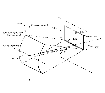
provide non-
invasive dermatological treatment to tissue through heating, thermal
treatment, coagulation,
ablation, and/or tissue tightening (including, for example, hyperthermia,
thermal dosimetry,
apoptosis, and lysis). In one embodiment, dermal tissue volume is increased.
In one
embodiment, fat tissue volume is reduced, or decreased.
[0096] In various embodiments, target tissue is, but is not limited to,
any of skin,
eyelids, eye lash, eye brow, caruncula lacrimalis, crow's feet, wrinkles, eye,
nose, mouth,
tongue, teeth, gums, ears, brain, chest, back, buttocks, legs, arms, hands,
arm pits, heart,
lungs, ribs, abdomen, stomach, liver, kidneys, uterus, breast, vagina, penis,
prostate, testicles,
glands, thyroid glands, internal organs, hair, muscle, bone, ligaments,
cartilage, fat, fat lobuli,
adipose tissue, cellulite, subcutaneous tissue, implanted tissue, an implanted
organ, lymphoid,
a tumor, a cyst, an abscess, or a portion of a nerve, or any combination
thereof. In several
embodiments disclosed herein, non-invasive ultrasound is used to achieve one
or more of the
following effects: a face lift, a brow lift, a chin lift, an eye treatment, a
wrinkle reduction, a
scar reduction, a fat reduction, a reduction in the appearance of cellulite, a
décolletage
treatment, a burn treatment, a tattoo removal, a vein reduction, a treatment
on a sweat gland,
a treatment of hyperhidrosis, sun spot removal, an acne treatment, and a
pimple removal. In
21
some embodiments, two, three or more beneficial effects are achieved during
the same
treatment session, and may be achieved simultaneously.
[0097]
Various embodiments of the present invention relate to devices or methods
of controlling the delivery of energy to tissue. In various embodiments,
various forms of
energy can include acoustic, ultrasound, light, laser, radio-frequency (RF),
microwave,
electromagnetic, radiation, thermal, cryogenic, electron beam, photon-based,
magnetic,
magnetic resonance, and/or other energy forms. Various embodiments of the
present
invention relate to devices or methods of splitting an ultrasonic energy beam
into multiple
beams. In various embodiments, devices or methods can be used to alter the
delivery of
ultrasound acoustic energy in any procedures such as, but not limited to,
therapeutic
ultrasound, diagnostic ultrasound, non-destructive testing (NDT) using
ultrasound, ultrasonic
welding, any application that involves coupling mechanical waves to an object,
and other
procedures.
Generally, with therapeutic ultrasound, a tissue effect is achieved by
concentrating the acoustic energy using focusing techniques from the aperture.
In some
instances, high intensity focused ultrasound (HIFU) is used for therapeutic
purposes in this
manner. In one embodiment, a tissue effect created by application of
therapeutic ultrasound
at a particular location (e.g., depth, width) to can be referred to as
creation of a thermal
treatment zone. It is through creation of thermal treatment zones at
particular positions that
thermal and/or mechanical heating, coagulation, and/or ablation of tissue can
occur non-
invasively or remotely offset from the skin surface.
System Overview
[0098]
Various embodiments of ultrasound treatment and/or imaging devices are
described in U.S. Publication No. 2011-0112405, which is a national phase
publication from
International Publication WO 2009/149390.
[0099]
With reference to the illustration in FIG. 1, an embodiment of an
ultrasound system 20 includes a hand wand 100, module 200, and a controller
300. The hand
wand 100 can be coupled to the controller 300 by an interface 130, which may
be a wired or
wireless interface. The interface 130 can be coupled to the hand wand 100 by a
connector
145. The distal end of the interface 130 can be connected to a controller
connector on a
Date Recue/Date Received 2021-07-14
CA 02944707 2016-09-30
WO 2015/160708 PCT/US2015/025581
22
circuit 345. In one embodiment, the interface 130 can transmit controllable
power from the
controller 300 to the hand wand 100. In various embodiments, the controller
300 can be
configured for operation with the hand wand 100 and the module 200, as well as
the overall
ultrasound system 20 functionality. In various embodiments, a controller 300
is configured
for operation with a hand wand 100 with one or more removable modules 200,
200, 200,
etc. The controller 300 can include an interactive graphical display 310,
which can include a
touchscreen monitor and Graphic User Interface (GUI) that allows the user to
interact with
the ultrasound system 20. As is illustrated, the graphical display 315
includes a touchscreen
interface 315. In various embodiments, the display 310 sets and displays the
operating
conditions, including equipment activation status, treatment parameters,
system messages and
prompts, and ultrasound images. In various embodiments, the controller 300 can
be
configured to include, for example, a microprocessor with software and
input/output devices,
systems and devices for controlling electronic and/or mechanical scanning
and/or
multiplexing of transducers and/or multiplexing of transducer modules, a
system for power
delivery, systems for monitoring, systems for sensing the spatial position of
the probe and/or
transducers and/or multiplexing of transducer modules, and/or systems for
handling user
input and recording treatment results, among others. In various embodiments,
the controller
300 can include a system processor and various analog and/or digital control
logic, such as
one or more of microcontrollers, microprocessors, field-programmable gate
arrays, computer
boards, and associated components, including firmware and control software,
which may be
capable of interfacing with user controls and interfacing circuits as well as
input/output
circuits and systems for communications, displays, interfacing, storage,
documentation, and
other useful functions. System software running on the system process may be
configured to
control all initialization, timing, level setting, monitoring, safety
monitoring, and all other
ultrasound system functions for accomplishing user-defined treatment
objectives. Further,
the controller 300 can include various input/output modules, such as switches,
buttons, etc.,
that may also be suitably configured to control operation of the ultrasound
system 20. In one
embodiment, the controller 300 can include one or more data ports 390. In
various
embodiments, the data ports 390 can be a USB port, Bluetooth port, IrDA port,
parallel port,
serial port, and the like. The data ports 390 can be located on the front,
side, and/or back of
CA 02944707 2016-09-30
WO 2015/160708 PCT/US2015/025581
23
the controller 300, and can be used for accessing storage devices, printing
devices, computing
devices, etc. The ultrasound system 20 can include a lock 395. In one
embodiment, in order
to operate the ultrasound system 20, the lock 395 should be unlocked so that a
power switch
393 may be activated. In one embodiment, the lock 395 can be connectable to
the controller
300 via a data port 390 (e.g., a USB port). The lock 395 could be unlocked by
inserting into
the data port 390 an access key (e.g., USB access key), a hardware dongle, or
the like. The
controller 300 can include an emergency stop button 392, which can be readily
accessible for
emergency deactivation.
[0100] As is illustrated in FIG. 1, in one embodiment, the hand wand 100
includes one or more finger activated controllers or switches, such as 150 and
160. In one
embodiment, the hand wand 100 can include a removable module 200. In other
embodiments, the module 200 may be non-removable. The module 200 can be
mechanically
coupled to the hand wand 100 using a latch or coupler 140. An interface guide
235 can be
used for assisting the coupling of the module 200 to the hand wand 100. The
module 200 can
include one or more ultrasound transducers 280. In some embodiments, an
ultrasound
transducer 280 includes one or more ultrasound elements 281. The module 200
can include
one or more ultrasound elements 281. The elements 281 can be therapy elements,
and/or
imaging elements. The hand wand 100 can include imaging-only modules 200,
treatment-
only modules 200, imaging-and-treatment modules 200, and the like. In one
embodiment,
the imaging is provided through the hand wand 100. In one embodiment, the
control module
300 can be coupled to the hand wand 100 via the interface 130, and the graphic
user interface
310 can be configured for controlling the module 200. In one embodiment, the
control
module 300 can provide power to the hand wand 100. In one embodiment, the hand
wand
100 can include a power source. In one embodiment, the switch 150 can be
configured for
controlling a tissue imaging function and the switch 160 can be configured for
controlling a
tissue treatment function
[0101] In one embodiment, the module 200 can be coupled to the hand wand
100.
The module 200 can emit and receive energy, such as ultrasonic energy. The
module 200 can
be electronically coupled to the hand wand 100 and such coupling may include
an interface
which is in communication with the controller 300. In one embodiment, the
interface guide
CA 02944707 2016-09-30
WO 2015/160708 PCT/US2015/025581
24
235 can be configured to provide electronic communication between the module
200 and the
hand wand 100. The module 200 can comprise various probe and/or transducer
configurations. For example, the module 200 can be configured for a combined
dual-mode
imaging/therapy transducer, coupled or co-housed imaging/therapy transducers,
separate
therapy and imaging probes, and the like. In one embodiment, when the module
200 is
inserted into or connected to the hand wand 100, the controller 300
automatically detects it
and updates the interactive graphical display 310.
[0102] In various embodiments, tissue below or even at a skin surface
such as
epidermis, dermis, hypodermis, fascia, and SMAS, and/or muscle are treated non-
invasively
with ultrasound energy. Tissue may also include blood vessels and/or nerves.
The
ultrasound energy can be focused, unfocused or defocused and applied to a
region of interest
containing at least one of epidermis, dermis, hypodermis, fascia, and SMAS to
achieve a
therapeutic effect. FIG. 2 is a schematic illustration of the ultrasound
system 20 coupled to a
region of interest 10, such as with an acoustic gel. With reference to the
illustration in FIG.
2, an embodiment of the ultrasound system 20 includes the hand wand 100, the
module 200,
and the controller 300. In various embodiments, tissue layers of the region of
interest 10 can
be at any part of the body of a subject. In various embodiments, the tissue
layers are in the
head, face, neck and/or body region of the subject. The cross-sectional
portion of the tissue
of the region of interest 10 includes a skin surface 501, an epidermal layer
502, a dermal layer
503, a fat layer 505, a SMAS 507, and a muscle layer 509. The tissue can also
include the
hypodermis 504, which can include any tissue below the dermal layer 503. The
combination
of these layers in total may be known as subcutaneous tissue 510. Also
illustrated in FIG. 2
is a treatment zone 525 which is the active treatment area below the surface
501. In one
embodiment, the surface 501 can be a surface of the skin of a subject 500.
Although an
embodiment directed to therapy at a tissue layer may be used herein as an
example, the
system can be applied to any tissue in the body. In various embodiments, the
system and/or
methods may be used on muscles (or other tissue) of the face, neck, head,
arms, legs, or any
other location in the body. In various embodiments, the therapy can be applied
to a face,
head, neck, submental region, shoulder, arm, back, chest, buttock, abdomen,
stomach, waist,
flank, leg, thigh, or any other location in or on the body.
CA 02944707 2016-09-30
WO 2015/160708 PCT/US2015/025581
Band Therapy Using A Cylindrical Transducer
[0103] In various embodiments, a transducer 280 can comprise one or more
therapy elements 281 that can have various shapes that correspond to various
focal zone
geometries. in one embodiment, the transducer 280 comprises a single therapy
element 281.
In one embodiment, the transducer 280 does not have a plurality of elements.
In one
embodiment, the transducer 280 does not have an array of elements. In several
embodiments,
the transducers 280 and/or therapy elements 281 described herein can be flat,
round, circular,
cylindrical, annular, have rings, concave, convex, contoured, and/or have any
shape. In some
embodiments, the transducers 280 and/or therapy elements 281 described herein
are not flat,
round, circular, cylindrical, annular, have rings, concave, convex, and/or
contoured. In one
embodiment, the transducers 280 and/or therapy elements 281 have a mechanical
focus. In
one embodiment, the transducers 280 and/or therapy elements 281 do not have a
mechanical
focus. In one embodiment, the transducers 280 and/or therapy elements 281 have
an
electrical focus. In one embodiment, the transducers 280 and/or therapy
elements 281 do not
have an electrical focus. Although a cylinder transducer and/or a cylindrical
element is
discussed here, the transducer and/or element need not be cylindrical. In
several
embodiments, the transducer and/or element has one or more shapes or
configurations that
cause edge effects, such as variance, spikes or other inconsistencies in the
delivery of
ultrasound. For example, the transducer and/or element may have one or more
non-linear
(e.g., curved) portions. A transducer may be comprised of one or more
individual transducers
and/or elements in any combination of focused, planar, or unfocused single-
element, multi-
element, or array transducers, including 1-fl, 2-1), and annular arrays;
linear, curvilinear,
sector, or spherical arrays; spherically, cylindrically, and/or electronically
focused, defocused,
and/or lensed sources. In one embodiment, the transducer is not a multi-
element transducer.
In one embodiment, a transducer 280 can include a spherically shaped bowl with
a diameter
and one or more concave surfaces (with respective radii or diameters)
geometrically focused
to a single point TTZ 550 at a focal depth 278 below a tissue surface, such as
skin surface
501. In one embodiment, a transducer 280 may be radially symmetrical in three
dimensions.
For example, in one embodiment, transducer 280 may be a radially symmetrical
bowl that is
configured to produce a focus point in a single point in space. In some
embodiments, the
CA 02944707 2016-09-30
WO 2015/160708 PCT/US2015/025581
26
transducer is not spherically shaped. In some embodiments, the element is not
spherically
shaped.
[0104] In various embodiments, increasing the size (e.g. width, depth,
area)
and/or number of focus zone locations for an ultrasonic procedure can be
advantageous
because it permits treatment of a patient at varied tissue widths, heights
and/or depths even if
the focal depth 278 of a transducer 280 is fixed. This can provide synergistic
results and
maximizing the clinical results of a single treatment session. For example,
treatment at larger
treatment areas under a single surface region permits a larger overall volume
of tissue
treatment, which can heat larger tissue volumes, and which can result in
enhanced collagen
formation and tightening. Additionally, larger treatment areas, such as at
different depths,
affects different types of tissue, thereby producing different clinical
effects that together
provide an enhanced overall cosmetic result. For example, superficial
treatment may reduce
the visibility of wrinkles and deeper treatment may induce skin tightening
and/or collagen
growth. Likewise, treatment at various locations at the same or different
depths can improve
a treatment. In various embodiments, a larger treatment area can be
accomplished using a
transducer with a larger focus zones (e..(4., such as a linear focus zone
compared to a point
focus zone).
[0105] In one embodiment, as illustrated in FIGS. 3 and 4, a transducer
280
comprises a cylindrical transduction element 281. In FIG. 4, the view of the
cylindrical
transduction element 281, which has a concave surface 282 and a convex surface
283, is
sectioned to show energy emission from the concave surface to a linear TTZ
550. The
cylindrical transduction element 281 extends linearly along its longitudinal
axis (X-axis,
azimuth) with a curved cross section along a Y-axis (elevation). In one
embodiment, the
cylindrical surface has a radius at a focal depth (z-axis) at the center of
the curvature of the
cylindrical surface, such that the TTZ 550 is focused at the center of the
radius. For example,
in one embodiment, cylindrical transduction element 281 has a concave surface
that extends
like a cylinder that produces a focus zone that extends along a line, such as
a therapy line,
such as TTZ 550. The focus zone TTZ 550 extends along the width (along the X-
axis,
azimuth) of the cylindrical transduction element 281, in a line parallel to
the longitudinal axis
of the cylindrical transduction element 281. As illustrated in FIG. 3, the TTZ
550 is a line
CA 02944707 2016-09-30
WO 2015/160708 PCT/US2015/025581
27
extending in and/or out of the page. In various embodiments of the cylindrical
transduction
element 281, a concave surface directs ultrasound energy to a linear TTZ 550.
Cylindrical
transduction element 281 need not be cylindrical; in some embodiments. element
281 is a
transduction element having one or more curved or non-linear portions.
[0106] In various embodiments, transducers 280 can comprise one or more
transduction elements 281. The transduction elements 281 can comprise a
piezoelectrically
active material, such as lead zirconante titanate (PZT), or any other
piezoelectrically active
material, such as a piezoelectric ceramic, crystal, plastic, and/or composite
materials, as well
as lithium niobate, lead titanate, barium titanate, and/or lead metaniobate.
In various
embodiments, in addition to, or instead of, a piezoelectrically active
material, transducers can
comprise any other materials configured for generating radiation and/or
acoustical energy. In
one embodiment, when cylindrical transduction element 281 comprises a
piezoelectric
ceramic material that is excited by an electrical stimulus, the material may
expand or
contract. The amount of expansion or contraction is related to boundary
conditions in the
ceramic as well as the magnitude of the electric field created in the ceramic.
In some
embodiments of conventional HIFU design, the front surface (e.g. subject side)
is coupled to
water and the back surface of a transducer 280 is coupled to a low impedance
medium which
is typically air. In some embodiments, although the ceramic is free to expand
at the back
interface, essentially no mechanical energy is coupled from the ceramic to the
air because of
the significant acoustic impedance disparity. This results in this energy at
the back of the
ceramic reflecting and exiting the front (or subject side) surface. As
illustrated in an
embodiment in FIGS. 3 - 5B, the focus is created by forming, casting, and/or
machining the
ceramic to the correct radius-of-curvature. In one embodiment, a flat
transducer material is
bent to form a cylindrical transducer. In various embodiments, transducers 280
and/or
therapy elements 281 can be configured to operate at different frequencies and
treatment
depths. Transducer properties can be defined by a focal length (FL), sometimes
referred to as
a focal depth 278. The focal depth 278 is the distance from the concave
cylindrical surface to
the focal zone TTZ 550. In various embodiments, the focal depth 278 is the sum
of a
standoff distance 270 and a treatment depth 279 when the housing of a probe is
placed
against a skin surface. In one embodiment, the standoff distance 270, or
offset distance 270,
CA 02944707 2016-09-30
WO 2015/160708 PCT/US2015/025581
28
is the distance between the transducer 280 and a surface of an acoustically
transparent
member 230 on the housing of a probe. The treatment depth 279 is a tissue
depth 279 below
a skin surface 501, to a target tissue. In one embodiment, the height of the
aperture in the
curved dimension is increased or maximized to have a direct effect on overall
focal gain,
which correlates to the ability to heat tissue. For example, in one
embodiment, the height of
the aperture in the curved dimension is maximized for a treatment depth of 6
mm or less. In
one embodiment, as the aperture is increased (e.g. decreasing the f#), the
actual heating zone
gets closer to the surface.
[0107] In one embodiment, a transducer can be configured to have a focal
depth
278 of 6 mm, 2 ¨ 12 mm, 3 ¨ 10 mm, 4 ¨ 8 mm, 5 ¨ 7 mm. In other embodiments,
other
suitable values of focal depth 278 can be used, such as focal depth 278 of
less than about 15
mm, greater than about 15 mm, 5 ¨ 25 mm, 10 ¨ 20 mm, etc. Transducer modules
can be
configured to apply ultrasonic energy at different target tissue depths. In
one embodiment, a
therapy of 20 mm or less (e.g., 0.1 mm ¨ 20 mm, 5 ¨ 17 mm, 10 ¨ 15 mm). In one
embodiment, a devices that goes to 6 mm or less has a radius of curvature
(ROC) of 13.6
mm, with a ratio of treatment depth to ROC at approximately 44%. In one
embodiment, the
height of the element is 22 mm. In one embodiment, using an aspect ratio for a
treatment
depth of 20 mm, the aperture height would be 74.5 mm with a ROC of 45 mm.
[0108] As illustrated in FIGS. 5A-5B, 7. 9 and 10 in several
embodiments, a
system may comprise a movement mechanism 285 configured to move a transducer
280
comprising a cylindrical transduction element 281 in one, two, three or more
directions. In
one embodiment, a motion mechanism 285 can move in a linear direction, one or
both ways,
denoted by the arrow marked 290 in order move a TTZ 550 through tissue. In
various
embodiments, the motion mechanism 285 can move the transducer in one, two,
and/or three
linear dimensions and/or one, two, and/or three rotational dimensions. In one
embodiment, a
motion mechanism 285 can move in up to six degrees of freedom. Movement of the
TTZ
550 can be with the transducer continuously delivering energy to create a
treatment area 552.
In one embodiment, a movement mechanism 285 can automatically move the
cylindrical
transduction element 281 across the surface of a treatment area so that the
TTZ 550 can form
a treatment area 552.
CA 02944707 2016-09-30
WO 2015/160708 PCT/US2015/025581
29
[0109] As indicated in FIGS. 6, 7, and 8, a cylindrical transduction
element 281
can be connected to a motion mechanism 285 and placed inside a module 200 or a
probe. In
various embodiments, a movement mechanism 285, or a motion mechanism 285,
moves the
transducer 280 and/or treatment element 281 such that the corresponding 'FEZ
550 moves to
treat a larger treatment area 552. In various embodiments, a movement
mechanism 285 is
configured to move a transducer within a module or a probe. In one embodiment,
a
transducer is held by a transducer holder. In one embodiment, the transducer
holder includes
a sleeve which is moved along motion constraining bearings, such as linear
bearings, namely,
a bar (or shaft) to ensure a repeatable linear movement of the transducer. In
one embodiment,
sleeve is a spline bushing which prevents rotation about a spline shaft, but
any guide to
maintain the path of motion is appropriate. In one embodiment, the transducer
holder is
driven by a motion mechanism 285, which may be located in a hand wand or in a
module, or
in a probe. In one embodiment, a motion mechanism 285 includes any one or more
of a
scotch yoke, a movement member, and a magnetic coupling. In one embodiment,
the
magnetic coupling helps move the transducer. One benefit of a motion mechanism
285 is
that it provides for a more efficient, accurate and precise use of an
ultrasound transducer, for
imaging and/or therapy purposes. One advantage this type of motion mechanism
has over
conventional fixed arrays of multiple transducers fixed in space in a housing
is that the fixed
arrays are a fixed distance apart. By placing transducer on a track (e.g.,
such as a linear track)
under controller control, embodiments of the system and device provide for
adaptability and
flexibility in addition to efficiency, accuracy and precision. Real time and
near real time
adjustments can be made to imaging and treatment positioning along the
controlled motion
by the motion mechanism 285. In addition to the ability to select nearly any
resolution based
on the incremental adjustments made possible by the motion mechanism 285,
adjustments
can be made if imaging detects abnormalities or conditions meriting a change
in treatment
spacing and targeting. In one embodiment, one or more sensors may be included
in the
module. In one embodiment, one or more sensors may be included in the module
to ensure
that a mechanical coupling between the movement member and the transducer
holder is
indeed coupled. In one embodiment, an encoder may be positioned on top of the
transducer
holder and a sensor may be located in a portion of the module, or vice versa
(swapped). In
CA 02944707 2016-09-30
WO 2015/160708 PCT/US2015/025581
various embodiments the sensor is a magnetic sensor, such as a giant
magnetoresistive effect
(GMR) or Hall Effect sensor, and the encoder a magnet, collection of magnets,
or multi-pole
magnetic strip. The sensor may be positioned as a transducer module home
position. In one
embodiment, the sensor is a contact pressure sensor. In one embodiment, the
sensor is a
contact pressure sensor on a surface of the device to sense the position of
the device or the
transducer on the patient. In various embodiments, the sensor can be used to
map the
position of the device or a component in the device in one, two, or three
dimensions. In one
embodiment the sensor is configured to sense the position, angle, tilt,
orientation, placement,
elevation, or other relationship between the device (or a component therein)
and the patient.
In one embodiment, the sensor comprises an optical sensor. In one embodiment,
the sensor
comprises a roller ball sensor. In one embodiment, the sensor is configured to
map a position
in one, two and/or three dimensions to compute a distance between areas or
lines of treatment
on the skin or tissue on a patient.
[0110] In various embodiments, a motion mechanism 285 can be any
mechanism
that may be found to be useful for movement of the transducer. In one
embodiment, the
motion mechanism 285 comprises a stepper motor. In one embodiment, the motion
mechanism 285 comprises a worm gear. In various embodiments, the motion
mechanism
285 is located in a module 200. In various embodiments, the motion mechanism
285 is
located in the hand wand 100. In various embodiments, the motion mechanism 285
can
provide for linear, rotational, multi-dimensional motion or actuation, and the
motion can
include any collection of points, lines and/or orientations in space. Various
embodiments for
motion can be used in accordance with several embodiments, including but not
limited to
rectilinear, circular, elliptical, arc-like, spiral, a collection of one or
more points in space, or
any other l-D, 2-D, or 3-D positional and attitudinal motional embodiments.
The speed of
the motion mechanism 285 may be fixed or may be adjustably controlled by a
user. One
embodiment, a speed of the motion mechanism 285 for an image sequence may be
different
than that for a treatment sequence. In one embodiment, the speed of the motion
mechanism
285 is controllable by a controller.
[0111] In some embodiments, the energy transmitted from the transducer
is turned
on and off, forming a non-continuous treatment area 552 such that the TTZ 550
moves with a
CA 02944707 2016-09-30
WO 2015/160708 PCT/US2015/025581
31
treatment spacing between individual TTZ 550 positions. For example, treatment
spacing
can be about 1 mm, 1.5 mm, 2 mm, 5mm, 10 mm, etc. In several embodiments, a
probe can
further comprise a movement mechanism configured to direct ultrasonic
treatment in a
sequence so that TTZs 550 are formed in linear or substantially linear
sequences. For
example, a transducer module can be configured to form TTZs 550 along a first
linear
sequence and a second linear sequence separated by treatment spacing between
about 2 mm
and 3 mm from the first linear sequence. In one embodiment, a user can
manually move the
transducer modules across the surface of a treatment area so that adjacent
linear sequences of
11 __ Zs are created.
[0112] In one embodiment, a TTZ can be swept from a first position to a
second
position. In one embodiment, a TTZ can be swept from the first position to the
second
position repeatedly. In one embodiment, a TTZ can be swept from the first
position, to the
second position, and back to the first position. In one embodiment, a TTZ can
be swept from
the first position, to the second position, and back to the first position,
and repeated. In one
embodiment, multiple sequences of TTZs can be created in a treatment region.
For example,
1'1Zs can be formed along a first linear sequence and a second linear sequence
separated by a
treatment distance from the first linear sequence.
[0113] In one embodiment, TTZs can be created in a linear or
substantially linear
zone or sequence, with each individual TTZ separated from neighboring TTZs by
a treatment
spacing, such as shown in FIG. 9. FIG. 9 illustrates an embodiment of an
ultrasound system
20 with a transducer 280 configured to treat tissue at a focal depth 278. In
one embodiment,
the focal depth 278 is a distance between the transducer 280 and the target
tissue for
treatment. In one embodiment, a focal depth 278 is fixed for a given
transducer 280. In one
embodiment, a focal depth 278 is variable for a given transducer 280. As
illustrated in FIG.
9, in various embodiments, delivery of emitted energy 50 at a suitable focal
depth 278,
distribution, timing, and energy level is provided by the module 200 through
controlled
operation by the control system 300 to achieve the desired therapeutic effect
of controlled
thermal injury to treat at least one of the epidermis layer 502, dermis layer
503, fat layer 505,
the SMAS layer 507, the muscle layer 509, and/or the hypodermis 504. FIG. 9
illustrates one
embodiment of a depth that corresponds to a depth for treating muscle. In
various
CA 02944707 2016-09-30
WO 2015/160708 PCT/US2015/025581
32
embodiments, the depth can correspond to any tissue, tissue layer, skin,
epidermis, dermis,
hypodermis, fat, SMAS, muscle, blood vessel, nerve, or other tissue. During
operation, the
module 200 and/or the transducer 280 can also be mechanically and/or
electronically scanned
along the surface 501 to treat an extended area. Before, during, and after the
delivery of
ultrasound energy 50 to at least one of the epidermis layer 502, dermis layer
503, hypodermis
504, fat layer 505, the SMAS layer 507 and/or the muscle layer 509, monitoring
of the
treatment area and surrounding structures can be provided to plan and assess
the results
and/or provide feedback to the controller 300 and the user via a graphical
interface 310. In
one embodiment, an ultrasound system 20 generates ultrasound energy which is
directed to
and focused below the surface 501. This controlled and focused ultrasound
energy 50 creates
the thermal treatment zone (TTZ) 550. In one embodiment, the ITZ 550 is a
line. In one
embodiment, the TTZ 550 is a point. In one embodiment, the TTZ 550 is a two
dimensional
region or plane. In one embodiment, the TTZ 550 is a volume. In one
embodiment, the
ultrasound energy 50 heat treats the subcutaneous tissue 510. In various
embodiments, the
emitted energy 50 targets the tissue below the surface 501 which heats, cuts,
ablates,
coagulates, micro-ablates, manipulates, and/or causes a lesion in the tissue
portion 10 below
the surface 501 at a specified focal depth 278. In one embodiment, during the
treatment
sequence, the transducer 280 moves in a direction denoted by the arrow marked
290 to move
the TTZ 550.
[0114] In various embodiments, an active TTZ can be moved (continuously,
or
non-continuously) through tissue to form a treatment area 552, such as shown
in FIG. 10.
With reference to the illustration in FIG. 10, the module 200 can include a
transducer 280
which can emit energy through an acoustically transparent member 230. In
various
embodiments, a depth may refer to the focal depth 278. In one embodiment, the
transducer
280 can have an offset distance 270, which is the distance between the
transducer 280 and a
surface of the acoustically transparent member 230. In one embodiment, the
focal depth 278
of a transducer 280 is a fixed distance from the transducer. In one
embodiment, a transducer
280 may have a fixed offset distance 270 from the transducer to the
acoustically transparent
member 230. In one embodiment, an acoustically transparent member 230 is
configured at a
position on the module 200 or the ultrasound system 20 for contacting the skin
surface 501.
CA 02944707 2016-09-30
WO 2015/160708 PCT/US2015/025581
33
In various embodiments, the focal depth 278 exceeds the offset distance 270 by
an amount to
correspond to treatment at a target area located at a tissue depth 279 below a
skin surface
501. In various embodiments, when the ultrasound system 20 placed in physical
contact with
the skin surface 501, the tissue depth 279 is a distance between the
acoustically transparent
member 230 and the target area, measured as the distance from the portion of
the hand wand
100 or module 200 surface that contacts skin (with or without an acoustic
coupling gel,
medium, etc.) and the depth in tissue from that skin surface contact point to
the target area.
In one embodiment, the focal depth 278 can correspond to the sum of an offset
distance 270
(as measured to the surface of the acoustically transparent member 230 in
contact with a
coupling medium and/or skin 501) in addition to a tissue depth 279 under the
skin surface
501 to the target region. In various embodiments, the acoustically transparent
member 230 is
not used.
[0115] In various embodiments, therapeutic treatment advantageously can
be
delivered at a faster rate and with improved accuracy by using a transducer
configured to
deliver energy to an expanded TTZ. This in turn can reduce treatment time and
decrease pain
experienced by a subject. In several embodiments, treatment time is reduced by
creating a
TTZ and sweeping the TTZ through an area or volume for treatment from a single
transducer.
In some embodiments, it is desirable to reduce treatment time and
corresponding risk of pain
and/or discomfort experienced by a patient. Therapy time can be reduced by
treating larger
areas in a given time by forming larger a TTZ 550, multiple TTZs
simultaneously, nearly
simultaneously, or sequentially, and/or moving the 117 550 to form larger
treatment areas
552. In one embodiment, a reduction in treatment time is reduced by treating a
given area or
volume with multiple TTZs reduces the overall amount of movement for a device.
In some
embodiments, overall treatment time can be reduced 10%, 20%, 25%, 30%, 35%,
40%, 4%,
50%, 55%, 60%, 65%, 70%, 75%, 80% or more by through creation of continuous
treatment
areas 552 or discrete, segmented treatment areas 552 from a sequence of
individual TTZs. In
various embodiments, therapy time can be reduced by 10-25%, 30-50%, 40-80%, 50-
90%, or
approximately 40%, 50%, 60%, 70%, and/or 80%. Although treatment of a subject
at
different locations in one session may be advantageous in some embodiments,
sequential
treatment over time may be beneficial in other embodiments. For example, a
subject may be
CA 02944707 2016-09-30
WO 2015/160708 PCT/US2015/025581
34
treated under the same surface region at one depth in time one, a second depth
in time two,
etc. In various embodiments, the time can be on the order of nanoseconds,
microseconds,
milliseconds, seconds, minutes, hours, days, weeks, months, or other time
periods. For
example, in some embodiments, the transducer module is configured to deliver
energy with
an on-time of 10 ms ¨ 100 minutes (e.g., 100 ms, 1 second, 1 ¨ 60 seconds, 1
minute ¨ 10
minutes, 1 minute ¨ 60 minutes, and any range therein). The new collagen
produced by the
first treatment may be more sensitive to subsequent treatments, which may be
desired for
some indications. Alternatively, multiple depth treatment under the same
surface region in a
single session may be advantageous because treatment at one depth may
synergistically
enhance or supplement treatment at another depth (due to, for example,
enhanced blood flow,
stimulation of growth factors, hormonal stimulation, etc.). In several
embodiments, different
transducer modules provide treatment at different depths. In one embodiment, a
single
transducer module can be adjusted or controlled for varied depths.
[0116] In one embodiment, an aesthetic treatment system includes an
ultrasonic
probe with a removable module that includes an ultrasound transducer
configured to apply
ultrasonic therapy to tissue at in a focal zone. In one embodiment, the focal
zone is a point.
In one embodiment, the focal zone is a line. In one embodiment, the focal zone
is a two
dimensional region or plane. In one embodiment, the focal zone is a volume. In
various
embodiments, a focal zone can be moved to sweep a volume between a first
position and a
second position. In various embodiments, one or more a focal zone locations
are positioned
in a substantially linear sequence within a cosmetic treatment zone. In one
embodiment, a
first set of locations is positioned within a first cosmetic treatment zone
and a second set of
locations is positioned within a second cosmetic treatment zone, the first
zone being different
from the second zone. In one embodiment, the first cosmetic treatment zone
includes a
substantially linear sequence of the first set of locations and the second
cosmetic treatment
zone includes a substantially linear sequence of the second set of locations.
[0117] In one embodiment, the transducer module 280 can provide an
acoustic
power in a range of about 1 W or less, between about 1 W to about 100 W, and
more than
about 100 W. In one embodiment, the transducer module 280 can provide an
acoustic power
at a frequency of about 1 MHz or less, between about 1 MHz to about 10 MHz,
and more
CA 02944707 2016-09-30
WO 2015/160708 PCT/US2015/025581
than about 10 MHz. In one embodiment, the module 200 has a focal depth 278 for
a
treatment at a tissue depth 279 of about 4.5 mm below the skin surface 501.
Some non-
limiting embodiments of transducers 280 or modules 200 can be configured for
delivering
ultrasonic energy at a tissue depth of 3 mm, 4.5 mm, 6 mm, less than 3 mm,
between 3 mm
and 4.5 mm, between 4.5 mm and 6 mm, more than more than 4.5 mm, more than 6
mm, etc.,
and anywhere in the ranges of 0.1 - 3 mm, 0.1 - 4.5 mm, 0.1 - 6 mm, 0.1 - 25
mm, 0.1 - 100
mm, etc. and any depths therein. In one embodiment, the ultrasound system 20
is provided
with two or more removable transducer modules 280. In one embodiment, a
transducer 280
can apply treatment at a tissue depth (e.g., about 6 mm). For example, a first
transducer
module can apply treatment at a first tissue depth (e.g., about 4.5 mm) and a
second
transducer module can apply treatment at a second tissue depth (e.g., of about
3 mm), and a
third transducer module can apply treatment at a third tissue depth (e.g., of
about 1.5 - 2 mm).
In one embodiment, at least some or all transducer modules can be configured
to apply
treatment at substantially same depths. In various embodiments, the tissue
depth can be 1.5
mm, 2 mm, 3 mm, 4.5 mm, 7 mm, 10 mm, 12 mm, 14 mm, 15 mm, 17 mm. 18 mm, and/or
20 mm, or any range therein (including but not limited to 12-20 mm, or
higher).
[0118] In one embodiment, a transducer module permits a treatment
sequence at a
fixed depth at or below the skin surface. In one embodiment, a transducer
module permits a
treatment sequence at a range of depths below the skin surface. In several
embodiments, the
transducer module comprises a movement mechanism configured to move the
ultrasonic
treatment at the TTY. In one embodiment, the linear sequence of individual
flYs has a
treatment spacing in a range from about 0.01 mm to about 25 mm. For example,
the spacing
can be 1.1 mm or less, 1.5 mm or more, between about 1.1 mm and about 1.5 mm,
etc. In
one embodiment, the individual TTZs are discrete. In one embodiment, the
individual TTZs
are overlapping. In one embodiment, the movement mechanism is configured to be
programmed to provide variable spacing between the individual TTZs. In several
embodiments, a transducer module comprises a movement mechanism configured to
direct
ultrasonic treatment in a sequence so that TTZs are formed in linear or
substantially linear
sequences separated by a treatment distance. For example, a transducer module
can be
configured to form TTZs along a first linear sequence and a second linear
sequence separated
CA 02944707 2016-09-30
WO 2015/160708 PCT/US2015/025581
36
by a treatment distance from the first linear sequence. In one embodiment,
treatment distance
between adjacent linear sequences of individual TTZs is in a range from about
0.01 mm to
about 25 mm. For example, the treatment distance can be 2 mm or less, 3 mm or
more,
between about 2 mm and about 3 mm, etc. In several embodiments, a transducer
module can
comprise one or more movement mechanisms configured to direct ultrasonic
treatment in a
sequence so that rliZs are formed in linear or substantially linear sequences
of individual
thermal lesions separated by a treatment distance from other linear sequences.
In one
embodiment, the treatment distance separating linear or substantially linear
TTZs sequences
is the same or substantially the same. In one embodiment, the treatment
distance separating
linear or substantially linear TTZs sequences is different or substantially
different for various
adjacent pairs of linear TTZs sequences.
Band Therapy Using A Cylindrical Transducer with An Imaging Element
[0119] In various embodiments, including an imaging transducer or
imaging
element with a cylindrical transduction element 281 can be used to improve
safety and/or
efficacy of a treatment. In one embodiment, an imaging element can be used to
confirm
acceptable coupling between the ultrasound therapy transducer and/or identify
target tissue
below the skin surface. As illustrated at FIGS. 21 and 22, in various
embodiments, a
transducer 280 comprises a cylindrical transduction element 281 and one or
more imaging
elements 284. The imaging element 284 is configured to image a region of
interest at any
suitable tissue depths 279. In one embodiment, an imaging element is centered
on a therapy
element. In one embodiment, an imaging element is axis symmetric with a
therapy element.
In one embodiment, an imaging element is not axis symmetric with a therapy
element. In one
embodiment, the imaging axis may be pointed in a completely different
direction and
translated from the therapy beam axis. In one embodiment, the number of
imaging elements
in the aperture may be greater than one. For example, in one embodiment, the
imaging
elements may be located on each corner of a cylinder pointed straight ahead
and/or in the
middle. In one embodiment, a combined imaging and cylindrical therapy
transducer 280
comprises a cylindrical transduction element 281 and one or more imaging
elements 284. In
one embodiment, a combined imaging and cylindrical therapy transducer 280
comprises a
cylindrical transduction element 281 with an opening 285 through which one
imaging
CA 02944707 2016-09-30
WO 2015/160708 PCT/US2015/025581
37
element 284 is configured to operate. In one embodiment, the opening 284 is a
circular hole
through the wall thickness of the cylindrical transduction element 281 at the
center of the X-
axis (azimuth) and Y-axis (elevation) of the cylindrical transduction element
281. In one
embodiment, the imaging element 284 is circular in cross-section and fits in
the opening 284.
[0120] In one embodiment, first and second removable transducer modules
are
provided. In one embodiment, each of the first and second transducer modules
are
configured for both ultrasonic imaging and ultrasonic treatment. In one
embodiment, a
transducer module is configured for treatment only. In one embodiment, an
imaging
transducer may be attached to a handle of a probe or a hand wand. The first
and second
transducer modules are configured for interchangeable coupling to a hand wand.
The first
transducer module is configured to apply ultrasonic therapy to a first
treatment area, while the
second transducer module is configured to apply ultrasonic therapy to a second
treatment
area. The second treatment area can be at a different depth, width, height,
position, and/or
orientation than the first treatment area.
Band Therapy Using A Coated Transducer Configured to Reduce Edge Effects
[0121] In various embodiments, treatment advantageously can be delivered
with
improved accuracy. Further, efficiency, comfort and safety can be increased if
variance is
reduced in a treatment area. This in turn can reduce treatment time and
decrease pain
experienced by a subject. In some instances, non-uniform heating at a focal
zone can result
from geometric aspects of a transducer. Inconsistencies in pressure or
temperature profiles
can be attributed to edge effects, which can cause spikes in pressure or
temperature around
the focal zone of a transducer. Thus, with edge effects, instead of achieving
a uniform line
segment of heating, the segment is broken into many isolated hot spots which
may fail to
meet an objective a more uniform heat distribution at the focal zone. This
phenomenon is
further exacerbated at high heating rates which relate to elevated acoustic
pressures. This is
due to the generation of nonlinear harmonics created especially in areas of
high pressure.
Energy at harmonic frequencies is more readily absorbed than energy at the
fundamental
frequency. In one embodiment, energy absorption is governed by the following
equation:
II = 2*a*f*p2/Z (I)
CA 02944707 2016-09-30
WO 2015/160708 PCT/US2015/025581
38
[0122] where alpha is the absorption constant in nepers per MHz cm, f is
frequency in MHz, p is the pressure at that frequency, Z is the acoustic
impedance of tissue,
and H is the heating rate in Watt/cm3. In one embodiment, the amount of
harmonics
produced is proportional to the intensity. FIG. 23 shows the normalized
harmonic pressure at
the focal depth across an azimuth of one embodiment of a cylindrical element
with an
imaging element. FIG. 23 shows the rapid swings in harmonic pressure at this
depth which
causes hot spots and non-uniform heating.
[0123] In one embodiment, a way to combat these hot and cold spots that
are the
result from edge effects is to reduce the average intensity at the focal depth
and/or increase
the heating time. These two processes can reduce the amount on nonlinear
heating as well as
allow for the conduction of the heat away from the hot spot into the cold
areas. The thermal
conduction of tissue effectively acts as a low pass filter to the acoustic
intensity distribution
as the heating time increases. Although these methods may reduce the non-
uniform heating
issues, they can also reduce the localization of the heating zone and can also
increase the
treatment time. Therefore, three performance areas of ultrasound therapy, e.g.
efficacy,
comfort, and treatment time, are adversely affected. In one embodiment, a more
normalized
pressure profile results in more consistent therapy, such that temperature
increase through
heating, coagulation, and/or ablation is more predictable and can better
ensure the desired or
targeted temperature profiles are obtained in the TTZ 550. In various
embodiments,
apodization of edge effects is accomplished with transducers coated in
specific regions.
[0124] In one embodiment, use of coatings, or shadings, can help
circumvents
these issues such that efficacy, comfort and treatment time are optimized.
FIG. 24 shows a
harmonic pressure distribution from an embodiment of a shaded aperture, or a
coated
element, that has an imaging transducer. In one embodiment, the coated element
is a coated
cylindrical element with an imaging element. The variation in harmonic
pressure across the
treatment line varies by less the 1.5 dB with the highest intensity near the
center and sharp
edges at -10 mm and +10 mm. In one embodiment, the coated element design does
not
require the conduction of heat away from hot spots since the tissue along the
focused line has
a uniform temperature increase during the absorption. Therefore, the amount of
intensity at
the focus can be increased to localize the heating zone and reduce treatment
time.
CA 02944707 2016-09-30
WO 2015/160708 PCT/US2015/025581
39
[0125] In one embodiment, the coated element is a shaded therapeutic
cylinder.
In one embodiment, a coated element also has benefits outside the intended
heating zone. In
one embodiment, the boundary between the heated and unheated junction is
vastly improved
when compared to an uncoated element. HG. 25 shows a comparison of harmonic
pressure
across an azimuth of an embodiment of a cylindrical element 280 compared to an
embodiment of a coated cylindrical element 600 at this boundary. FIG. 25 shows
that, in one
embodiment, the possible harmonic pressures are approximately 20 dB lower for
the shaded
aperture with a coated cylindrical element 600, which helps confine the
heating zone and
maximize comfort. In one embodiment, areas of plating or non-plating are
initially used to
define regions where the piezoelectric material will be poled or not poled.
Regions where
there is plating define regions that will be poled or actually mechanically
vibrating. In one
embodiment, a cylindrical element 280 can be uncoated. Further, an uncoated
region may be
considered uncoated to the extent it does not have an electrically conductive
coating ¨ the
uncoated region may have other types of surface coatings in certain
embodiments. In one
embodiment, a cylindrical element is completely coated. For example, in one
embodiment, a
first transducer 280 includes a first coated region 287 that fully plates the
concave surface
282 of the cylindrical transduction element and a second coated region 287
that fully plates
the convex surface 283 of the cylindrical transduction element. A second
coated transducer
600 includes a first coated region 287 that fully plates the concave surface
282 of the
cylindrical transduction element and at least a second coated region 287 that
partially plates
the convex surface 283 of the cylindrical transduction element. As shown in
HG. 27, the
fully coated first transducer 281 demonstrates the spikes in focal gain due to
edge effects.
[0126] Referring to FIGS. 11A-13B, in one embodiment, transducer
treatment
profiles were plotted based on theoretical and experimental performance with a
cylindrical
transduction element 281 that was coated on the entire concave surface 282 and
the entire
convex surface 283 with a coating. In one embodiment, the coating is a metal.
In one
embodiment, the coating is a conductive metal. In one embodiment, the coating
is an
electrical conductor. In various embodiments, the coating is plated with any
one or more of
silver, gold, platinum, mercury, copper or other materials. In one embodiment,
a coating
comprises fired silver. In one embodiment, a surface is fully coated. In one
embodiment, a
CA 02944707 2016-09-30
WO 2015/160708 PCT/US2015/025581
surface is fully non-coated. In one embodiment, a surface is partially coated
and partially
non-coated. The normalized pressure is proportional to a thermal heating
measure at the
specified depth. The discontinuous spikes (pointed regions at the top of the
plots) plots
indicate pressure and/or temperature peaks that occur as a result of the
geometric edge effects
of the geometry of the cylindrical transduction element 281. In various
embodiments, the
spikes, or peaks, can be reduced with a coated transducer 600 comprising one
or more coated
regions 287. In one embodiment, the coated region 287 only partially coats a
transducer
surface. In one embodiment, the coated region 287 does not completely coat a
transducer
surface.
[0127] As shown in FIG. 26, in various embodiments, a coated transducer
600
comprises a cylindrical transduction element 281 with one or more coated
regions 287. In
various embodiments, the coated region 287 coats part, a portion, and/or all
of a surface of
the transducer 600. In various embodiments, the coated region 287 coats part
or all of a
surface of the cylindrical transduction element 281. In various embodiments, a
coated
transducer 600 comprises one or more imaging elements 284. In some
embodiments, one,
two, three or more imaging element(s) are placed in 'unused regions' of
coatings/shading for
the purpose of imaging.
[0128] The edge effects from the geometry of one embodiment of a
combined
imaging and cylindrical therapy transducer comprising a cylindrical
transduction element 281
with an opening 285 through it are more pronounced due to the additional edges
of the
opening 285. FIG. 27 is a plot illustrating focal gain across the azimuth of
two embodiments
of combined imaging and cylindrical therapy transducers with different
coatings. A first
transducer 280 includes a first coated region 287 that fully plates the
concave surface 282 of
the cylindrical transduction element and a second coated region 287 that fully
plates the
convex surface 283 of the cylindrical transduction element. Both the first and
the second
coated regions 287 of the first transducer 280 are plated with silver. A
second coated
transducer 600 includes a first coated region 287 that fully plates the
concave surface 282 of
the cylindrical transduction element and at least a second coated region 287
that partially
plates the convex surface 283 of the cylindrical transduction element. Both
the first and the
second coated regions 287 of the second transducer 600 are plated with silver.
As shown in
CA 02944707 2016-09-30
WO 2015/160708 PCT/US2015/025581
41
FIG. 27, the fully coated first transducer 281 demonstrates the spikes in
focal gain due to
edge effects. The partially coated second transducer 600 has a more
consistent, normalized
performance output with the spikes substantially reduced and/or removed. In
various
embodiments, a coated transducer 600 reduces the peaks such that variance
around the focal
depth is reduced by 1 ¨ 50%, 25 ¨ 100%, 75 ¨ 200%, and/or 10 ¨ 20%, 20 ¨ 40%
and 60 ¨
80%. In various embodiments, a coated transducer 600 reduces the peaks such
that variance
of the intensity in a location around the focal depth is +/- 0.01 ¨ 5 mm, 5 mm
or less, 4 mm
or less, 3 mm or less, 2 mm or less, 1 mm or less, 0.5 mm or less, 0.25 mm or
less, 0.1 mm or
less, 0.05 mm or less, or any range therein. In various embodiments, a coated
transducer 600
reduces the peaks in focal gain such that variance in focal gain is 0.01 ¨
0.1, 0.01 ¨ 1.0, 0.01
¨5,0.01-10,1-10,1-5,10,9,8,7,6,5,4,3,2,1 or less, or any range therein.
[0129] As described in Example 2 below, FIGS. 28, 29, and 30 illustrate
the
embodiment of the performance of the partially coated second transducer 600 in
FIG. 27 at
different depths. In the illustrated embodiment, the partially coated second
transducer 600
has a focal depth of 15 mm. In various embodiments, the focal depth can be at
any depth. In
various embodiments, the focal depth is at 7, 8, 9, 10, 12, 13, 13.6, 14, 15,
16, 17, 18, or any
depth therein.
[0130] In one embodiment, the coated region 287 is plating. In one
embodiment,
the coated region 287 is a conductive material. In one embodiment, the coated
region 287 is
a semi-conductive material. In one embodiment, the coated region 287 is an
insulator
material. In various embodiments, the coated region 287 is silver, copper,
gold, platinum,
nickel, chrome, and/or any conductive material that will adhere with the
surface of a
piezoelectric material, or any combinations thereof. In one embodiment, the
coated region
287 is silver plating.
[0131] In various embodiments, a cylindrical transduction element 281
has an
azimuth (x-axis) dimension in the range of 1 ¨ 50 mm, 5 ¨ 40 mm, 10 ¨ 20 mm,
15 ¨ 25 mm,
and/or 15 mm, 16 mm, 17 mm, 18 mm, 19 mm, 20 mm, 21 mm, 22 mm, 23 mm, 24 mm,
and
25 mm. In various embodiments, a cylindrical transduction element 281 has an
elevation (y-
axis) dimension in the range of 1 ¨ 50 mm, 5 ¨ 40 mm, 10 ¨ 20 mm, 15 ¨ 25 mm,
and/or 15
mm, 16 mm, 17 mm, 18 mm, 19 min, 20 mm, 21 mm, 22 mm, 23 mm, 24 mm, and 25 mm.
CA 02944707 2016-09-30
WO 2015/160708 PCT/US2015/025581
42
In various embodiments, a cylindrical transduction element 281 has focal depth
(z-axis)
dimension in the range of 1 ¨ 50 mm, 5 ¨ 40 mm, 10 ¨ 20 mm, 15 ¨ 25 mm, 12 ¨
17 mm, 13
¨ 15 mm, and/or lOmm. 1 1mm, 12mm, 13mm, 13.6mm, 14mm, 15 mm, 16 mm, 17 mm, 18
mm, 19 mm, 20 mm, 21 mm, 22 mm, 23 mm, 24 mm, and 25 mm. In some non-limiting
embodiments transducers can be configured for a treatment zone at a tissue
depth below a
skin surface of 1.5 mm, 3 mm, 4.5 mm, 6 mm, less than 3 mm, between 1.5 mm and
3 mm,
between 1.5 mm and 4.5 mm, more than more than 4.5 mm, more than 6 mm, and
anywhere
in the ranges of 0.1 mm ¨ 3 mm, 0.1 mm ¨4.5 mm, 3 mm ¨ 7 mm, 3 mm ¨ 9mm, 0.1
mm -
25 mm, 0.1 mm ¨ 100 mm, and any depths therein.
[0132] In various embodiments, a coated transducer 600 comprising a
cylindrical
transduction element 281 has one, two, three, four, or more coated regions
287. In one
embodiment, a coated region 287 covers an entire surface of the element. In
one
embodiment, a coated region 287 covers a portion of a surface of the element.
In various
embodiments, the coated region 287 includes a conductive plating. In one
embodiment, a
coated region 287 includes a silver plating to form an electrode. When an
electrical signal is
applied to an electrode at a coated region 287, the coated region 287 expands
and/or contracts
the corresponding portion of the cylindrical transduction element 281. In
various
embodiments, the coated region 287 has a shape or border that is a complete or
a partial
point, edge, line, curve, radius, circle, oval, ellipse, parabola, star,
triangle, square, rectangle,
pentagon, polygon, a combination of shapes, or other shape. In various
embodiments, a
coated transducer 600 can also comprise an opening 285.
[0133] In one embodiment illustrated at FIG. 31, a partially coated
transducer 600
comprising a cylindrical transduction element 281 has one, two, three, four,
or more coated
regions 287 of one or more shapes on a convex 283 surface. In one embodiment,
a partially
coated transducer 600 comprising a cylindrical transduction element 281 has
one, two, three,
four, or more coated regions 287 of one or more shapes on a concave 282
surface. In various
embodiments, the coated region 287 has a lateral edge 293, a side edge 290,
and a medial
edge 291. The various edges can be straight, curved, and/or have a radius, and
the sizes can
be modified to result in various performance profiles.
CA 02944707 2016-09-30
WO 2015/160708 PCT/US2015/025581
43
[0134] In one embodiment illustrated at FIG. 32, a partially coated
transducer 600
comprising a cylindrical transduction element 281 has one, two, three, four,
or more circular,
round, curved and/or elliptical coated regions 287. In various embodiments,
the coated
region 287 has a lateral edge 293, a side edge 290, and a medial edge 291. The
various edges
can be straight, curved, and/or have a radius, and the sizes can be modified
to result in
various performance profiles.
[0135] In one embodiment illustrated at FIG. 33, a partially coated
transducer 600
comprising a cylindrical transduction element 281 has one, two, three, four,
or more
triangular coated regions 287. In various embodiments, the coated region 287
has a lateral
edge 293, a side edge 290, and a medial edge 291. The various edges can be
straight, curved,
and/or have a radius, and the sizes can be modified to result in various
performance profiles.
[0136] In one embodiment illustrated at FIG. 34, a partially coated
transducer 600
comprisimg a cylindrical transduction element 281 has one, two or more square,
rectangular,
and/or polygon coated regions 287. In various embodiments, the coated region
287 has a
lateral edge 293, a side edge 290, and a medial edge 291. The various edges
and/or sizes can
be modified to result in various performance profiles.
[0137] In one embodiment illustrated at FIG. 35, a partially coated
transducer 600
comprising a cylindrical transduction element 281 has one, two or more
combined and/or
mixed shape coated regions 287. In one embodiment illustrated at FIG. 35, a
partially coated
transducer 600 is a combined imaging and cylindrical therapy transducer
comprising a
cylindrical transduction element 281 with an opening 285 for an imaging
element 284. In
one embodiment, the coated transducer 600 includes a concave surface 282 that
is fully
plated with fired silver, and has a convex surface 283 with two coated regions
287 that are
plated with fired silver to form electrodes. When an electrical signal is
applied to an
electrode at a coated region 287, the coated region 287 expands and/or
contracts the
corresponding portion of the cylindrical transduction element 281. In some
embodiments,
the shape may be applied before or after the poling process, as vibration will
occur where the
electrode is located. In various embodiments, an electrode could be defined
before or after
poling. In various embodiments, a coating pattern may be on the concave or
convex surface.
In one embodiment, the coated region 287 has a lateral edge 293, a first and
second side edge
44
290, and a medial edge 291 with a central edge 297. The various edges can be
straight,
curved, and/or have a radius. Various dimensions 294, 295, 296, and the
various edges can
be modified to result in various performance profiles. In one embodiment, the
medial edge
291 along the curved dimension (elevation) is a portion of an ellipse. In one
embodiment, the
medial edge 291 along the curved dimension (elevation) is a portion of a
parabola. In one
embodiment, the first and second side edge 290 along the uncurved dimension
(azimuth) is a
portion of a parabola. In one embodiment, the first and second side edge 290
along the
uncurved dimension (azimuth) is a portion of an ellipse.
[0138]
In one embodiment illustrated at FIG. 36, a partially coated transducer 600
comprising a cylindrical transduction element 281 has one, two, three, four,
or more
diamond, rhombus, and/or other polygon coated regions 287. In various
embodiments, the
coated region 287 has a lateral edge 293, a side edge 290, and a medial edge
291. The
various edges and/or sizes can be modified to result in various performance
profiles.
[0139]
In one embodiment illustrated at FIGS. 37 and 38, a partially coated
transducer 600 comprising a cylindrical transduction element 281 has one, two,
three, four or
more coated regions 287. In various embodiments, the coated region 287 has a
lateral edge
293, a side edge 290, and a medial edge 291. In some embodiments, the coated
region 287 is
configured to position one, two, three, four, or more (e.g., multiple) thermal
treatment zones
through poling, phasic poling, biphasic poling, and/or multi-phasic poling.
Various
embodiments of ultrasound treatment and/or imaging devices with of multiple
treatment
zones enabled through poling, phasic poling, biphasic poling, and/or multi-
phasic poling are
described in U.S. Application No. 14/193,234 filed on February 28, 2014.
Non-Therapeutic Uses of a Coated Cylindrical Transducer With Reduced Edge
Effects
[0140]
In various embodiments, a coated cylindrical transducer 600 comprising
one or more coated regions 287 is configured for non-therapeutic use.
[0141]
In one embodiment, a coated cylindrical transducer 600 comprising one or
more coated regions 287 is configured for materials processing. In one
embodiment, a coated
cylindrical transducer 600 comprising one or more coated regions 287 is
configured for
Date Recue/Date Received 2021-07-14
CA 02944707 2016-09-30
WO 2015/160708 PCT/US2015/025581
ultrasonic impact treatment for the enhancement of properties of a material,
such as a metal,
compound, polymer, adhesive, liquid, slurry, industrial material.
[0142] In one embodiment, a coated cylindrical transducer 600 comprising
one or
more coated regions 287 is configured for material heating. In various
embodiments, the
cylindrical transducer 600 is configured for cooking, heating, and/or warming
materials, food,
adhesives or other products.
heating Tissue and Quantification of Thermal Dose for Ultrasound Band Therapy
[0143] As described above, in various embodiments, systems and/or
methods
provide non-invasive dermatological treatment to tissue through heating,
hyperthermia,
thermal dosimetry, thermal treatment, coagulation, ablation, apoptosis, lysis,
increasing tissue
volume, decreasing or reducing tissue volume, and/or tissue tightening. In one
embodiment,
dermal tissue volume is increased. In one embodiment, fat tissue volume is
reduced, or
decreased.
[0144] In various embodiments, band treatment involves metrics that
quantify the
magnitude of adipocyte death with heat. For example, in one embodiment,
thermal dosage in
a heat treatment relates time-temperature curves back to a single reference
temperature, e.g.
T=43 degrees Celsius, using the Arrhenius equation. In one embodiment, a band
treatment is
configured under a relationship that that for every I degree Celsius increase
in tissue
temperature above in a range above body temperature, the rate of cell death
doubles. A
theoretical survival fraction can then be determined by comparing the thermal
dose to
empirical data from the literature.
[0145] In various embodiments, band treatment provides improved thermal
heating and treatment of tissue compared to diathermy or general bulk heating
techniques. In
general, noimal body temperatures tend to range between about 33 ¨ 37 degrees
Celsius. In
various embodiments, as tissue is heated in a range of about 37 ¨ 43 degrees
Celsius,
physiological hyperthermia can take place, and exposure to this temperature
range on order
of, for example, a few hours, can result in increased normal tissue metabolism
and/or
increased normal tissue blood flow, and in some embodiments, accelerated
noimal tissue
repair. As temperature in the tissues reaches the higher ¨ 43 degrees Celsius
range and/or the
tissue is subject to the temperature for longer periods of time (e.g., 2
hours, 3, hours or more)
CA 02944707 2016-09-30
WO 2015/160708 PCT/US2015/025581
46
the tissue can experience acute tissue metabolism and/or acute tissue blood
flow, and in some
embodiments, accelerated normal tissue repair. In one embodiment, heating
(e.g., bulk
heating) of tissue to a range of about 42 - 55 degrees Celsius is performed.
In various
embodiments, heating of tissue to about 43 - 50 degrees Celsius can be
considered adjuvant
synergistic hyperthermia, and exposure to this temperature range on order of,
for example, a
few minutes, can result in immediate or delayed cell death, apoptosis,
decreased tumor
metabolism, increased tissue oxygen levels, increased tissue damage, increased
sensitivity to
therapy, vascular status, DNA damage, cell reproductive failure, and/or cell
destruction. In
various embodiments, heating of tissue to about 50 - 100 degrees Celsius can
be considered
surgical hyperthermia, and exposure to this temperature range on order of, for
example, a few
seconds or fractions of a second, can result in coagulation, ablation,
vaporization, and
immediate cell destruction.
[0146] In some embodiments of the invention, the temperature of the
tissue
treatment site (e.g., the adipocytes) is elevated to 38 - 43 degrees Celsius,
and according to
one embodiment, thereby increasing tissue metabolism and perfusion and
accelerating tissue
repair mechanisms. In other embodiments, the temperature of the tissue
treatment site (e.g.,
the adipocytes) is elevated to 43 - 50 degrees Celsius, which in one
embodiment can
increase cell damage starts and result in immediate cell death, particularly
when the
temperature remains elevated on the order of several minutes to an hour (or
longer). In yet
other embodiments, the temperature of the tissue treatment site (e.g., the
adipocytes) is
elevated to above 50 degrees Celsius, which in one embodiment results in
protein coagulation
on the order of seconds and less and can lead to immediate cell death and
ablation. In various
embodiments, the temperature of the tissue treatment site is heated to 40, 41,
42, 43, 44, 45,
46, 47, 48, 49, 50, 51, 52, 53, 54, 55, 56, 57, 58, 59, 60, 61, 62, 63, 64,
65, 70, 75, 80, 90, or
100 degrees Celsius, and/or any range therein. In various embodiments, a
treatment area has
uniform temperature, a variance of 1%, 2%, 3%, 4%, 5%, 6%, 7 %, 8%, 9%, 10%,
12%,
15%, 20%, 25%, 30%, 40%, 50% or more. In various embodiments, a treatment area
has a
variance of +/- 0, 1, 2, 3, 4, 5, 6, 7, 8, 9, 10, 12, 15, 20, 25 degrees
Celsius or more.
[0147] In several embodiments, the invention comprises elevating the
temperature
of the tissue treatment site (e.g., the adipocytes) is elevated to 38 - 50
degrees Celsius for a
CA 02944707 2016-09-30
WO 2015/160708 PCT/US2015/025581
47
time period between 1 ¨ 120 minutes, and then optionally increasing the
temperature in one,
two, three, four five or more increments by 10-50%. As an example using three
increments,
the target temperatures may be increased as follows: (i) elevate temperature
to about 40-42
degrees Celsius for 10-30 minutes, (ii) then optionally increase temperature
by about 20% to
elevate temperature to about 48-51 degrees Celsius for 1-10 minutes, and (iii)
then optionally
increase by about 10-50% for a shorter time frame. As another example, the
target
temperature may be increased as follows: (i) elevate temperature to about 50
degrees Celsius
for 30 seconds to 5 minutes (e.g., about 1 minute) to destroy over 90%, 95% or
99% of target
(e.g., adipose) cells, with an optional pre-heating step of raising the
temperature to 38 ¨ 49
degrees Celsius for a period of 10-120 minutes prior to the elevation to 50
degrees Celsius.
As yet another example, in some embodiments, a non-invasive, cosmetic method
of heating
tissue, comprises applying a cosmetic heating system to a skin surface,
wherein the cosmetic
heating system comprises a hand-held probe, wherein the hand-held probe
comprises a
housing that encloses an ultrasound transducer configured to heat tissue below
the skin
surface to a tissue temperature in the range of 40 ¨ 50 degrees Celsius,
wherein the
ultrasound transducer comprises a cylindrical transduction element comprising
a first surface,
a second surface, a coated region, and an uncoated region, wherein the coated
region
comprises an electrical conductor, wherein the first surface comprises at
least one coated
region, wherein the second surface comprises the uncoated region and a
plurality of coated
regions, applying a current to the plurality of coated regions, thereby
directing ultrasound
energy to a linear focal zone at a focal depth, wherein the ultrasound energy
produces a
reduction in focal gain at the linear focal zone, thereby heating the tissue
at the focal depth in
the linear focal zone to the tissue temperature in the range of 40 ¨ 50
degrees Celsius for a
cosmetic treatment duration of less than 1 hour, thereby reducing a volume of
an adipose
tissue in the tissue.
[0148] In one embodiment, a band therapy system uses a relationship
between cell
death and time-temperature dosages as quantified using the Arrhenius equation.
The
Arrhenius equation shows an exponential relationship exists between cell death
and exposure
time and temperature. Above a certain break temperature, the increase in the
rate of cell
killing with temperature is relatively constant. Time-temperature
relationships to achieve
CA 02944707 2016-09-30
WO 2015/160708 PCT/US2015/025581
48
isoeffective dose in several types of tissue appears to be conserved both in
vitro and in-vivo
across multiple cell types.
[0149] In some embodiments, clinical situations involve ramp-up of
temperatures,
cooling, and fluctuations when approaching and maintaining a steady state
temperature. In
various embodiments, different thermal profiles can produce the same thermal
dose. In order
to estimate the thermal dosage from a time-varying thermal profile, a
temperature curve is
discretized into small time steps, and the average temperature during each
time step is
calculated. The thermal dosage is then calculated as an equivalent exposure
time at the break
temperature (43 degrees Celsius) by integrating these temperatures according
to equation (2):
t '4Var' p(.43-1)
0..5 õT 43*(.7
R OM.
.3ete.
t43 Equivalent time at 43'e t0.2,5T.< 430:
Averoge temperature during At
(2)
[0150] Equation (2) suggests that the increase in the rate of killing
with
temperature is relatively constant. In some embodiments, a 1 degree Celsius
increase above a
break point results in the rate of cell death doubles. FIGs. 39 and 40
illustrate theoretical cell
death fractions over time depending on tissue temperature, with higher
theoretical cell killing
fractions at higher temperatures and/or higher periods of time. The higher a
kill fraction
(such as shown with kill fractions of 99%, 80%, 50%, 40%, and 20%) the higher
a
temperature and/or a time is used in an embodiment of a treatment.
[0151] Once a thermal dose has been calculated, a dose survival response
can be
estimated from empirical data. In one embodiment, an isoeffective dose of 43
degrees
Celsius for 100 minutes theoretically yields a cell survival fraction of 1%.
Based on the
Arrhenius relationship, a similar surviving fraction can be obtained with an
isoeffective dose
of 44 degrees Celsius for 50 minutes, or 25 minutes at 45 degrees Celsius,
etc. as tabulated in
the table listing isoeffective dosages to theoretically achieve 1% survival
fraction at FIG. 41,
according to embodiments of the present invention.
[0152] In various embodiment, simulations of various embodiments of band
therapy using a cylindrical transducer source conditions linked to the
relationship between
CA 02944707 2016-09-30
WO 2015/160708 PCT/US2015/025581
49
tissue and heat equation showed that successive treatment pulses obey linear
superposition,
which allows for simplification of the heat transfer physics so that the
heating rate may be
described as a temperature rise per time (degrees Celsius/sec), and as a
temperature rise per
pass (degrees Celsius/button push).
Heating Tissue via Ultrasound Band Therapy
[0153] In various embodiments, a band therapy system is configured for
treating
the tissue. For example, in one embodiment, a band treatment is configured for
treatment of
supraplatysmal submental fat. In one embodiment, a treatment of fat includes
selectively
causing thermal heat shock followed by apoptosis to a fat layer, at a depth of
about 2.5 - 6.0
mm, without causing any major skin surface effects. In one embodiment, the
treatment
involves exposing fat to a bulk heating treatment with a temperature of 42-55
degrees Celsius
for 1-5 minutes without exceeding 41 degrees Celsius on the skin surface, with
physiologic/biologic effect (e.g. one or more of coagulation, apoptosis, fat
cell lysis, etc.). In
various embodiments, treatment with a band transducer treats tissue with
isoeffective doses,
as shown in a graph representing various levels of theoretical cell kill
fractions in FIG. 42.
[0154] In various embodiments, a theoretical review of the effect of
stacking
multiple treatment pulses using the Khokhlov¨Zabolotskaya¨Kuznetsov (KZK)
Equation was
implemented with cylindrical source acoustic geometry, linked to a bioheat
equation (e.g., in
one embodiment, using the Arrhenius equation). HG. 43 shows the results of a
KZK
simulation of cylindrical transducer output showing linear superposition of
multiple pulses;
approximately the same temperatures are reached when treating with 3 pulses of
0.45 J or 1
pulse of 1.35 J (3 * 0.45 J). The results of a theoretical experiment with one
embodiment of a
band therapy system as shown in FIG. 43, suggest non-linear acoustics are not
a major
contributor to the final temperature for the energies, and suggests that body
tissue acts as a
linear time-invariant system, which allows for simplification of the heat
transfer physics, and
the heating and cooling rates to be described in relatively few parameters. In
various
embodiments, a therapy system with a hand wand 100 includes a module 200 with
one or
more ultrasound transducers 280. In some embodiments, an ultrasound transducer
280
includes one or more cylindrical ultrasound elements 281, as shown in FIGs. 5A
¨ 8. The
CA 02944707 2016-09-30
WO 2015/160708 PCT/US2015/025581
cylindrical transducer element 281 is configured for bulk heating treatments
with its linear
focus along an axis, resulting in a continuous line that can be moved with an
automated
motion mechanism to treat a rectangular plane. In one embodiment, lines of
treatment are
deposited perpendicular to the direction of motor movement in a single
direction. A single
"pass" of treatment creates a number of therapy lines equal to {Length}/{
Spacing}.
[0155] In various embodiments, various cylindrical geometries were
tested from
the first build (4.5 MIIz ¨ 12 mm width at 4.5 mm and 6.0 mm depths); however,
acoustic
tank testing showed higher acoustic pressures (and therefore heating rates) at
the each edge of
the therapy line. In one embodiment, a ceramic transducer was apodized to
produce a flat
thermal profile, as shown in FIGs. 44 and 45. In various embodiments,
different cylindrical
geometries based on two operating frequencies, two treatment widths, and two
treatment
depths were built: (1) 3.5MHz - 22mm Width - 4.5mm Depth; (2) 3.5MHz - 22mm
Width -
6.0mm Depth; (3) 4.5MHz - 22mm Width - 4.5mm Depth; (4) 4.5MHz - 22mm Width -
6.0mm Depth; (5) 3.5MHz - 12mm Width - 4.5mm Depth; (6) 4.5MHz - 12mm Width -
4.5mm Depth; (7) 3.5MHz - 12mm Width - 6.0mm Depth: and (8) 4.5MHz - 12mm
Width -
6.0mm Depth. In various embodiments, a tissue temperature measurement system
included
one or more of including IR thermography, temperature strips, and resistance
temperature
detectors (RTDs), and thermocouples. IR thermography can be used to read skin
surface
temperatures. Temperature strips are able to provide peak temperature reached.
RTD
sheaths have a large thermal mass and may have a slow response time. In
various
embodiments, thermocouples have a response time less than a second, which is
helpful for
measuring the heating and cooling phase of a single treatment pass.
Thermocouples also
have the advantage of being small enough that they can be positioned through a
large bore
needle to the desired tissue depth. In one embodiment, a particular
isoeffective dose is
attached via the heating phase followed by a maintenance phase in which the
system or an
operator pulses treatment at an interval to sustain a steady state
temperature. A parameter of
interest during this phase is the average pulse period needed to maintain the
steady state
temperature.
CA 02944707 2016-09-30
WO 2015/160708 PCT/US2015/025581
51
Body Contouring via Ultrasound Band Therapy
[0156] In various embodiments, a band therapy system is configured for
body
contouring. In one embodiment, body contouring treatment involves thermal heat
shock
concurrent with, and/or followed by apoptosis. In one embodiment, body
contouring
treatment involves exposing fat to 42-55 degrees Celsius for 1-5 minutes to
induce delayed
apoptosis. In one embodiment, body contouring treatment involves exposing fat
at a focus
depth of at least 13 mm below the skin surface.
Temperature and Dose Control
[0157] In various embodiments, one or more sensors may be included in
the
module 200 or system 20 to measure a temperature. In one embodiment, methods
of
temperature and/or dose control are provided. In one embodiment, temperature
is measured
to control dosage of energy provided for a tissue treatment. In various
embodiments, a
temperature sensor is used to measure a tissue temperature to increase,
decrease, and/or
maintain the application of energy to the tissue in order to reach a target
temperature or target
temperature range. In some embodiments, a temperature sensor is used for
safety, for
example, to reduce or cease energy application if a threshold or maximum
target temperature
is reached. In one embodiment, a cooling device or system can be employed to
cool a tissue
temperature if a certain temperature is reached. In some embodiments, a
temperature sensor
is used to modulate an energy dose, for example, via modulation, termination
of amplitude,
power, frequency, pulse, speed, or other factors.
[0158] In one embodiment, a temperature sensor is used to measure a skin
surface
temperature. In one embodiment, a temperature sensor may be positioned on top
of the
transducer holder and a sensor may be located in a portion of the module, or
vice versa
(swapped). In various embodiments, a temperature sensor is positioned on a
system or
module housing, such as in one embodiment, near or on an acoustic window, such
as an
acoustically transparent member 230. In one embodiment, one or more
temperature sensors
are positioned around or proximate an acoustically transparent member 230. In
one
embodiment, one or more temperature sensors are positioned in or on an
acoustically
transparent member 230. In one embodiment, a temperature sensor measure from a
skin
surface can be used to calculate a temperature in a tissue at the focus depth
of the energy
CA 02944707 2016-09-30
WO 2015/160708 PCT/US2015/025581
52
application. In various embodiments, a target tissue temperature can be
calculated and/or
correlated to the depth in tissue, type of tissue (e.g. epidermis, dermis,
fat, etc.) and relative
thickness of tissue between the skin surface and the focus depth. In some
embodiments, a
temperature sensor provides a temperature measurement for a signal to a
control system. In
some embodiments, a temperature sensor provides a temperature measurement for
visual
and/or auditory feedback to a system operator, such as a text, color, flash,
sound, beep, alert,
alarm, or other sensory indicator of a temperature state.
[0159] In some embodiments, imaging can be used to control energy dose.
In one
embodiment, a thermal lens effect can be used to account for speckle shift
and/or feature shift
to indicate a temperature of a tissue at a target location, such as at a focus
depth in tissue
below the skin surface. In one embodiment. Acoustic Radiation Force Impulse
(ARFI)
imaging is used to calculate a tissue temperature. In one embodiment, Shear
Wave Elasticity
Imaging (SWEI) is used to calculate a tissue temperature. In one embodiment,
attenuation is
used to calculate a tissue temperature.
[0160] In various embodiments, a variable dose delivery technique is
used to
attain a target temperature in a tissue and maintain that target temperature.
The body
temperature at a depth in tissue surrounds a thermal treatment zone (TTZ). In
one
embodiment, to overcome the body temperature, a treatment focuses energy at
the TTZ at a
first rate to bring the tissue temperature in the TTZ to a target temperature.
Once that target
temperature is attained, the second rate can be reduced or stopped to maintain
the tissue at the
target temperature.
[0161] In some embodiments, energy is focused at a depth or position in
tissue at
the TTZ, such that the temperature in the focal zone is increased. However, at
the edges
(e.g., ends, top, bottom, sides, etc.) of the focal zone, a boundary condition
at body
temperature can result in temperature fluctuations at the boundaries of the
treatment area 552.
In various embodiments, movement of the TTZ 550 can be with the transducer
delivering
energy to create a treatment area 552. In one embodiment, a movement mechanism
285 can
automatically move the cylindrical transduction element 281 across the surface
of a treatment
area so that the TTZ 550 can form a treatment area 552. In FIG. 53, the
treatment area 552 is
surrounded at the edges by body temperature, or approximately body
temperature. In some
CA 02944707 2016-09-30
WO 2015/160708 PCT/US2015/025581
53
embodiments, the temperature in the treatment area 552 along the
edges/boundary are lower
than the desired, target temperature.
[0162] In various embodiments, mechanical velocity modulation is used to
attain
a specific thermal distribution in the treatment area 552. In one embodiment,
in order to
attain a more uniform temperature in the treatment area 552, the applied
temperature at the
edges/boundaries is increased to counteract the surrounding body temperature
difference.
FIG. 54 illustrates an embodiment of mechanical velocity modulation in which
the velocity,
or speed of the automatic motion of the motion mechanism moving the transducer
along
direction 290 (along the elevation direction), is varied to provide a more
uniform temperature
in the treatment area 552 by slowing near the boundaries, resulting in
increased temperature
at the boundaries (start and stop position, such as along a 25 mm travel
distance, in one
embodiment). The increased velocity near the middle delivers a lower
temperature than the
decreased velocity.
[0163] In various embodiments, amplitude modulation is used to attain a
specific
thermal distribution in the treatment area 552. In one embodiment, in order to
attain a more
uniform temperature in the treatment area 552, the applied temperature at the
edges/boundaries is increased to counteract the surrounding body temperature
difference.
FIG. 55 illustrates an embodiment of amplitude modulation in which the
amplitude
(correlates to power) of the energy delivered by the transducer as the
automatic motion of the
motion mechanism moves along direction 290 (along the elevation direction), is
varied to
provide a more uniform temperature in the treatment area 552 by increasing
amplitude near
the boundaries, resulting in increased temperature at the boundaries (start
and stop position,
such as along a 25 mm travel distance, in one embodiment). The lower amplitude
near the
middle delivers a lower temperature than the higher amplitude near the
boundaries.
[0164] In various embodiments, aperture apodization is used to attain a
specific
thermal distribution in the treatment area 552. In one embodiment, aperture
apodization
along the non-focused dimension (such as along TTZ 550 and/or the azimuth
direction) is
used in order to attain a more uniform temperature in the treatment area 552.
The applied
temperature at the end points, along the edges/boundaries is increased to
counteract the
surrounding body temperature difference. FIG. 56 illustrates an embodiment of
aperture
CA 02944707 2016-09-30
WO 2015/160708 PCT/US2015/025581
54
apodization in which the amplitude of the energy delivered by the transducer
along the TTZ
550 is varied to provide a more uniform temperature in the treatment area 552
by increasing
amplitude near the end points near the boundaries, resulting in increased
temperature at the
boundaries (with L as a length of the focused line TTZ 550, L/2 from center is
the end point).
The lower amplitude near the middle delivers a lower temperature than the
higher amplitude
near the boundaries. In various embodiments, a temperature profile can be
generated along
the -11Z with embodiments of a coated transduction element 600, such as
illustrated in FIGs.
31-38.
[0165] In various embodiments, pulsing and/or duty cycles are controlled
to attain
a specific thermal distribution in the treatment area 552. At FIG. 57, in
various
embodiments, treatment patterns can have a consistent or a constant pulsing or
duty cycle. At
FIG. 58, in various embodiment, treatment patterns can have variable pulsing
or a variable
duty cycle, with variations in any of peak amplitude, spacing of application,
duration of
application. As shown in FIG. 58, the application of energy is longer and
covers more area
near the boundary of the treatment area 552, while the internal region has
less power
application for a corresponding lower temperature application in the internal
region.
[0166] In various embodiments, treatment patterns are used to attain a
specific
thermal distribution in the treatment area 552. In some embodiments the TTZ
550 has a
dimension (e.g., width, height, thickness, etc.). In some embodiments, the
pulsed application
of "1"fZ 550 is non-overlapping, as shown in HG. 59. In some embodiments, the
pulsed
application of TTZ 550 is overlapping, as is shown near a boundary in FIG. 60,
where the
amount of overlapping can be constant or vary. As shown in the embodiment in
FIG. 60, the
amount of overlap varies and includes a non-overlapping portion. In various
embodiments, a
cross hatching pattern is used, wherein the system hand piece is rotated about
90 degrees, or
orthogonally, and the motion mechanism is operated in one or more additional
passes over a
target tissue region in an orthogonal direction to a prior treatment pass.
[0167] In various embodiments, a specific thermal distribution in the
treatment
area 552 comprises treatment with a tissue temperature of 37 ¨ 50 degrees
Celsius for a
duration of minutes to hours to cause a targeted percentage of cell death
(such as fat cell
death) which a relationship can be determined via Arrhenius equation, such as
is shown on
CA 02944707 2016-09-30
WO 2015/160708 PCT/US2015/025581
the left side of HG. 61. In various embodiments, a specific thermal
distribution in the
treatment area 552 comprises treatment with a tissue temperature of over 60
degrees Celsius
for a duration of seconds to fractions of a second (or near instantaneous)
coagulation,
ablation, and/or cell death (such as fat cell death) at the elevated
temperature, such as shown
on the right side of FIG. 62. In various embodiments, a treatment can be
either one, or both
in sequence and/or simultaneous treatments.
[0168] In some embodiments, one, two, three, four, or more of mechanical
velocity modulation, amplitude modulation, aperture apodization, pulsing duty
cycles, and/or
treatments at different temperatures can be used to achieve a desired
temperature profile
across the treatment area 552. In various embodiments, one or more of
mechanical velocity
modulation, amplitude modulation, aperture apodization, pulsing duty cycles,
and/or
treatments at different temperatures is used to create a temperature profile,
wherein the
temperature profile can include areas for increased, decreased, and/or uniform
temperatures.
In some embodiments, one, two, or more types of treatment are applied in one,
two, or three
dimensions (along any of the azimuth, elevation, and/or depth directions) and
is configured
for treatment in any of one, two, or three dimensions to create a one, two, or
three
dimensional temperature profile.
[0169] In some embodiments, a compound lens system produces various peak
intensities and different depths. In various embodiments, a mechanical and/or
electronic
focus lens can be used in any one or more of the azimuth, elevation, and/or
depth directions.
As illustrated in HG. 62 and FIG. 63, a compound lens system can create two or
more focal
lines 550 and 550a.
[0170] In various embodiments, an ultrasound system 20 comprises a
motion
mechanism 285 configured for moving a plurality of ultrasound transducers 280
and/or a
plurality of ultrasound elements 281. In some embodiments, such as illustrated
in an
embodiment at FIG. 64, the motion mechanism 285 is configured to minimize heat
fluctuation in treated tissue and reduce treatment time by presenting the
plurality of elements
281 on a conveyor system, such as with a belt and/or pulley system that can
move the
plurality of elements 281 at a velocity v. In various embodiments, velocity
can be constant,
variable, zero (e.g., stopped), reversible (e.g., forward and backward, left
and right, first
CA 02944707 2016-09-30
WO 2015/160708 PCT/US2015/025581
56
direction and second direction, etc.) and/or have values in the range 0 ¨ 100
RPM, 1 RPM ¨
50 RPM, or other velocities. In various embodiments, the velocity is any value
1 ¨ 1,000
cm/second (e.g., 10, 20, 50, 100, 200, 500. 1000 cm/sec, and any other values
therein). In
various embodiments, the motion mechanism 285 moves one, two, three, four,
five, six,
seven, eight, or more ultrasound elements 281. In various embodiments,
ultrasound elements
281 are connected, or spaced at a distance of 0.01 ¨ 10 cm apart, (e.g.. 0.1,
0.5, 1,2, 5 cm and
any values therein), such that one, two, or more ultrasound elements 281 are
configured to
treat a treatment area.
[0171] In some embodiments, imaging is used to confirm the quality of
the
acoustic coupling between a treatment device and the skin. In one embodiment,
clarity of an
ultrasound image along a treatment area, line, or point is used to determine
the extent to
which a device is acoustically coupled to a skin surface. In one embodiment,
defocused
imaging and/or Voltage Standing Wave Ratio (VSWR) from backscatter is used to
check
acoustic coupling for a treatment.
[0172] In some embodiments, a treatment is automated. In one embodiment,
a
treatment is set up by acoustically coupling a system to a skin surface, and
the movement
mechanism and treatment is automated to function. In various embodiments, the
system is
coupled to a skin surface via suction. In various embodiments, a system
operator couples the
system to a skin surface, activates the system, and can leave the system to
automatically
perform a treatment, or a portion of a treatment. In one embodiment, a system
uses suction
and/or vacuum pressure to hold a probe or portion of the system to a skin
surface, allowing
the system user to initiate treatment and leave the system to automatically
perform a
treatment or a portion of a treatment for a period of time. In some
embodiments, a treatment
system includes a TENS stimulation device to reduce pain at a skin treatment
site.
Theoretical and Experimental Treatments with A Cylindrical Transducer
[0173] The following examples illustrate various non-limiting
embodiments.
EXAMPLE 1
[0174] The following example is intended to be a non-limiting embodiment
of the
invention.
CA 02944707 2016-09-30
WO 2015/160708 PCT/US2015/025581
57
[0175] As illustrated at FIGS. 11A ¨ 20, it was experimentally verified
that an
embodiment of a transducer 280 comprising a cylindrical transduction element
281, which
was applied to a simulated target tissue, an artificial tissue, and to porcine
tissue sample,
formed localized, linear thermal treatment zone (11Z 550) in a targeted focal
area 552. In
the experiment, the single cylindrical transduction element 281 was
constructed with a radius
and focal depth of 15 mm. The size of the cylindrical transduction element 281
was 20 mm
(azimuth) by 17 mm (elevation). Additional focal gain could be achieved with a
larger
aperture. Depth is limited by frequency and focal gain, and was set to 6 mm
below a
simulated tissue surface.
[0176] In FIGS. 11A-13B, treatment profiles were plotted based on
theoretical
and experimental performance with a cylindrical transduction element 281. The
normalized
pressure is proportional to a thermal heating measure at the specified depth.
The spikes
(pointed regions at the top of the plots) plots indicate pressure peaks that
occur as a result of
the geometric edge effects of the geometry of the cylindrical transduction
element 281. The
spikes are visible in both the theoretical and the experimental performance
results. The
software simulated experiments reflect the theoretical performance of the 15
mm cylindrical
transduction element 281 in FIGS. 11A, 12A, 13A, 14A, 15A, and 16A. The
physical
experiments in simulated tissue were performed and measured, with results in
FIGS. 11B,
12B, 13B, 14B, 15B and 16B.
[0177] In FIGS. 11A ¨ 11B and 14A-14B, the depth is 20 mm, where the
normalized pressure peaks at a value of roughly 0.15. As shown in FIG. 14A-
14B, the
normalized pressure is not visible. In FIGS. 12A ¨ 12B and 15A-15B, the depth
is the
designed, optimal 15 mm, where the normalized pressure peaks at a value of
roughly 0.8. As
shown in FIG. 15A-15B, the normalized pressure is clearly visible, with peak
normalized
pressures at approximately 0.9 ¨ 1Ø The size of the cylindrical transduction
element 281
was 20 mm (azimuth) by 17 mm (elevation). The size of the TTZ 550 at a depth
of 15 mm
was about 0.5 mm thick (along azimuth) by 17 mm width (along elevation). In
FIGS. 13A ¨
13B and 16A-16B, the depth is 13 mm, where the normalized pressure peaks at a
value of
roughly 0.25. As shown in FIG. 16A-16B, the normalized pressure is barely
visible. As
shown through both the theoretical and experimental data, the normalized
pressure
CA 02944707 2016-09-30
WO 2015/160708 PCT/US2015/025581
58
corresponding to the TTZ 550 for a 15 mm focal depth cylindrical transduction
element 281
is at the 15 mm depth, with a linear TTZ 550.
[0178] As illustrated at FIGS. 17 - 20, it was experimentally verified
that the
embodiment of a transducer 280 comprising a cylindrical transduction element
281, which
was applied to a porcine tissue sample (muscle tissue), formed localized,
linear thermal
treatment zone (TTZ 550) in a targeted focal area 552. In the experiment, an
embodiment of
a transducer 280 comprising a cylindrical transduction element 281 was passed
over the
porcine muscle tissue with three passes in 20 seconds, operating at 4.5 MHz
and a tissue
depth of 6 mm. As shown in FIG. 17, the three passes (shown with the three
spikes in
temperature) increased the temperature of the porcine muscle. Two power levels
are shown.
The 40 W porcine muscle started at 30 degrees Celsius, and over the course of
20 seconds
(between the 20 and 40 second marks) of heating through three passes of the
cylindrical
transduction element 281 over the target tissue region, the temperature spiked
to a maximum
of about 55 degrees Celsius, then gradually cooled to about 32 degrees Celsius
100 seconds
after the start of the treatment. The 60 W porcine muscle started at about 24
degrees Celsius,
and over the course of 20 seconds (between the 40 and 60 second marks) of
heating through
three passes of the cylindrical transduction element 281 over the target
tissue region, the
temperature spiked to a maximum of about 59 degrees Celsius, then gradually
cooled to
about 40 degrees Celsius about 80 seconds after the start of the treatment.
[0179] HG. 18 is a photograph of the porcine muscle after treatment
confirming
line and plane heating. In one embodiment, the coagulation was dependent on
time-off
between lines, time-off between passes, and number of passes. Slower
temperature rise than
thermal coagulation points. FIG. 19 is a cross-section cut through the porcine
muscle in FIG.
18 showing a linear thermal treatment zone. FIG. 20 is an orthogonal cross-
section cut
through the porcine muscle in FIG. 19 showing a planar thermal treatment zone.
EXAMPLE 2
[0180] The following example is intended to be a non-limiting embodiment
of the
invention.
[0181] As illustrated at FIGS. 28 - 30, it was experimentally verified
that an
embodiment of a partially coated transducer 600 comprising a cylindrical
transduction
CA 02944707 2016-09-30
WO 2015/160708 PCT/US2015/025581
59
element 281, which was applied to a simulated target tissue, formed a
localized, linear
thermal treatment zone (TTZ 550) in a targeted focal area 552. The partially
coated
transducer 600 includes a first coated region 287 that fully plates the
concave surface 282 of
the cylindrical transduction element and at least a second coated region 287
that partially
plates the convex surface 283 of the cylindrical transduction element. Both
the first and the
second coated regions 287 of the partially coated transducer 600 are plated
with silver. In the
experiment, the single cylindrical transduction element 281 was constructed
with a radius and
focal depth of 15 mm. The size of the cylindrical transduction element 281 was
20 mm
(azimuth) by 17 mm (elevation). The cylindrical transduction element 281 had
an opening
285 in the center of 4mm in diameter.
[0182] In FIGS. 28, 29 and 30, treatment profiles were plotted based on
theoretical performance with a cylindrical transduction element 281. The
theoretical
performance is proportional the thermal heating at the specified depth. The
software
simulated experiment reflects the theoretical performance of the 15 mm
partially coated
transducer 600, showinL, a consistent linear thermal treatment zone 550 at the
15 mm depth.
EXAMPLE 3
[0183] The following example is intended to be a non-limiting embodiment
of the
invention.
[0184] Multiple in-vivo porcine studies and multiple cadaver studies
were
conducted to evaluate various embodiments of hardware to perform bulk heating
treatments.
Early studies focused on specifying and improving the instrumentation
necessary to measure
subdermal temperatures. In some embodiments, insulated wire thermocouples were
placed at
focal and subfocal depths by snaking the thermocouple through a needle-bored
hole in the
skin and verifying the depth with a Siemens s2000 ultrasound device.
Temperature profiles
were collected using a high sampling DAQ card. Once the measurement setup was
defined, a
replicated 3-factor 3-level design of experiments was performed in the in-vivo
porcine model
to determine energy settings that could safely reach isoeffective dosages
without causing skin
surface damage. In one embodiment, a mean temperature differential of 10
degrees Celsius
was observed, with a mean focal heating rate of ¨1.2 degrees Celsius/pass.
Safe heating rates
appear to be similar across transducer.
CA 02944707 2016-09-30
WO 2015/160708 PCT/US2015/025581
[0185] A thermal dosage study was performed in the in-vivo porcine model
after
safe heating rates were determined. The study demonstrated an embodiment of
the system is
capable of reaching isoeffective dosages such as 47 degrees Celsius for 3
minutes, 48 degrees
Celsius for 1 minute, and 50 degrees Celsius for 1 minute without exceeding 41
degrees
Celsius on the skin surface. In some embodiments, use of higher temperature,
shorter
exposure time treatments may have the potential to overshoot the target
temperature and
could overheat the skin surface. In various embodiments, the longer it takes
to perform an
isoeffective dose, the more heat diffuses to the surrounding tissue and less
selective the
treatment becomes with depth. Additionally, the longer the isoeffective
exposure time, the
more impractical the treatment becomes from an operator and ergonomics point
of view. For
these reasons, in some embodiments, use of higher isoeffective temperatures
and shorter
exposure times were preferred.
[0186] In-vivo porcine tests were conducted to determine if the
candidate
treatment settings for submental could cause adverse surface skin effects. The
animals
procured for these studies were light skinned, 120-140 pound castrated male
Yucatan
miniature pigs, selected due to its skin characteristics being similar to that
of human tissue.
Skin surface data was evaluated by monitoring the animal for evidence of
erythema, edema,
and contusion on the skin surface after treatment. Photographs of each
treatment area were
taken prior to and following treatment (Cannon G9 and Cannon VIXIA HF 510). In
one
embodiment, a thermal dosage study using a cylindrical element transducer was
performed on
in-vivo porcine models. In several embodiments, test sites were able to
achieve a significant
temperature differential between the focus tissue site and the skin surface
without causing
damage to the skin surface. FIG. 46 shows the temperature profiles from an
embodiment of
an in-vivo porcine model treatment in which the temperature profile reached 50
degrees
Celsius for several seconds without the skin surface exceeding 41 degrees
Celsius, and shows
a temperature differential of as much as 15 degrees Celsius between the focus
tissue site and
the skin surface. The temperature change accrued from a single pass of
treatment is
sufficiently small (approximately 0.9 degrees Celsius/pass or 0.13 degrees
Celsius/sec) to
perform corrective action and maintain a target temperature within +/-1
degrees Celsius. A
modified 3-factor 3-level design of experiments was performed in the in-vivo
porcine model
CA 02944707 2016-09-30
WO 2015/160708 PCT/US2015/025581
61
to determine a range of energy settings that could safely reach the
isoeffective dosages
temperatures shown in FIG. 42. The settings, according to various embodiments,
are
tabulated in the table at FIG. 47. The Design of Experiments (DOE) tests an
acoustic power
range of 10-20 W, exposure times of 20-40 ms, and spacings in the range of 0.1
- 0.3 mm.
FIG. 48 shows an embodiment of a treatment setting that was able to achieve a
relatively high
thermal dosage at the focus with little to no dose or temperature increase at
the skin surface.
The focus achieves a thermal dose of 100 equivalent minutes (red-dashed line)
at T=43
degrees Celsius on the 24th pass, which corresponds to a theoretical survival
fraction of 1%
according to FIG. 42. In various embodiments, similar temperature rises and
heating rates
were achieved at the focus and surface across various embodiments of
transducers for
treatments that did not cause significant skin surface damage. A mean
temperature
differential of 10 degrees Celsius was observed, with a mean focal heating
rate of ¨1.2
degrees Celsius/pass. The largest temperature differential between the focus
and the skin was
achieved by the 3.5MHz, 22mm width, 6.0 depth design which had an average
difference of
12 degrees Celsius across treatments. Since the heating rates that produce
little to no surface
effects are similar across transducer, the 3.5MHz, 22mm width, 6.0mm depth
transducer was
selected to be assessed in a thermal dosage study.
[0187] In various embodiments, thermal dosage studies were performed on
in-
vivo porcine and cadaver models to determine safe isoeffective dosages, and
the geometry of
adipocyte death through histological evaluation. The Table at FIG. 49
tabulates the target
time-temperature exposures to achieve different levels of adipocyte death.
According to the
empirical data in FIG. 42, Site 2 and 5 should achieve little to no adipocyte
death. Sites 3, 6
and 7 should achieve a high degree of adipocyte death. Sites 1 and 4 are
within the transition
region and should achieve a moderate amount of adipocyte death. The table at
FIG. 50 lists
the energy settings used to approach each isoeffective dose using a 3.5 MHz,
22 mm width,
6.0 mm depth transducer. In various embodiments, treatments were active for 2-
3 minutes
with 20-30 pulses to reach the target temperature with a 1 degrees
Celsius/pass ramp
followed by maintenance pulses ever 3-5 seconds. A few test sites showed mild
surface
effects the day of treatment, only to become more pronounced as the injury
rose to the skin
surface. FIG. 51 shows one site that was treated aggressively for the purpose
of coagulating
CA 02944707 2016-09-30
WO 2015/160708 PCMJS2015/025581
62
tissue for histological control through overdosing. In the embodiment in FIG.
51, the
dimension of the lesion represents a an example of the spread of thermal
energy. measuring
12.6 x 19.9 mm on the skin surface with a depth of edema that can be detected
up to 12 mm
from the skin surface. A visual representation of the time-temperature goals
listed in the
table at FIG. 49 is shown in FIG. 52 (triangle marks), with six isoeffective
dosages achieved
in the lab are overlayed in FIG. 52 (square marks). Two of these isoeffective
dosages fall in
the coagulative region, two fall in the transition region, and two in the
hyperthermia region.
[0188] Some embodiments and the examples described herein are examples
and
not intended to be limiting in describing the full scope of compositions and
methods of these
invention(s). Equivalent changes, modifications and variations of some
embodiments,
materials, compositions and methods can be made within the scope of the
embodiments
herein. In various embodiments, a device or method can combine features or
characteristics
of any of the embodiments disclosed herein.
[0189] While the invention is susceptible to various modifications, and
alternative
forms, specific examples thereof have been shown in the drawings and are
herein described
in detail. It should be understood, however, that the invention is not to be
limited to the
particular forms or methods disclosed, but to the contrary, the invention is
to cover all
modifications, equivalents, and alternatives falling within the spirit and
scope of the various
embodiments described and the appended claims. Any methods disclosed herein
need not be
performed in the order recited. The methods disclosed herein include certain
actions taken by
a practitioner; however, they can also include any third-party instruction of
those actions,
either expressly or by implication. For example, actions such as "coupling an
ultrasound
probe to a skin surface" include "instructing the coupling of an ultrasound
probe to a skin
surface." The ranges disclosed herein also encompass any and all overlap, sub-
ranges, and
combinations thereof. Language such as "up to," "at least," "greater than,"
"less than,"
"between," and the like includes the number recited. Numbers preceded by a
term such as
"about" or "approximately" include the recited numbers. For example, "about 25
mm"
includes "25 mm." The terms "approximately". "about", and "substantially" as
used herein
represent an amount or characteristic close to the stated amount or
characteristic that still
performs a desired function or achieves a desired result. For example, the
terms
CA 02944707 2016-09-30
WO 2015/160708 PCT/US2015/025581
63
"approximately", "about". and "substantially" may refer to an amount that is
within less than
10% of, within less than 5% of, within less than 1% of, within less than 0.1%
of, and within
less than 0.01% of the stated amount or characteristic.