Note: Descriptions are shown in the official language in which they were submitted.
CA 02947498 2016-10-28
WO 2015/200807
PCT/US2015/038005
ENDOSPORE DETECTION USING HYDROPHOBIC COLLECTION
MATERIAL
TECHNICAL FIELD
[0001] This disclosure relates to bacterial endospore isolation and analysis.
BACKGROUND
[0002] Bacterial spores are generally accepted to be indicator species for
validating
sterility since they are the most resilient form of life against sterilization
regimens.
Traditional bacterial spore analysis is a labor intensive and time consuming
process. For
example, spore analysis may involve heat activation, serial dilution, plating
on a suitable
growth medium, and incubation for two to three days until enumeration can be
performed. This analysis process can take several days, requiring
manufacturers of
product undergoing analysis to hold significant quantities of the product
before receiving
sterility test results that allow them to release product to the marketplace.
Moreover, in
instances where there is a sterility issue, the lack of real-time information
can result in
several days' worth of production being deemed out of specification and
needing to be
discarded or repurposed.
[0003] In an attempt to provide faster analysis, designers have utilized
optical analysis
techniques that detect optical emission signals associated with bacterial
spores and then
correlate these signals with spore count. These techniques are of limited use,
however,
for many categories of materials desirably analyzed for bacterial spore count.
For
example, materials that contain a low number of bacterial spores or contain
bacterial
spores within a surrounding fluid that optically interferes with the emissions
associated
with the spores can be difficult to evaluate using optical analysis
techniques. As one
example, dairy production facilities monitoring bacterial spore counts in
their products
typically cannot use optical emission analysis techniques. This is because
proteins and
other molecules within the dairy products can optically interfere with
emissions produced
by bacterial spores.
SUMMARY
[0004] In general, this disclosure relates to techniques and systems for
analyzing bacterial
endospores within fluids. Depending on the application, a fluid containing the
bacterial
endospores may or may not be optically active such that the fluid surrounding
the
1
CA 02947498 2016-10-28
WO 2015/200807
PCT/US2015/038005
endospores optically interferes with optical emissions corresponding to the
endospores.
In either case, the fluid may be passed through a hydrophobic sieve to help
isolate the
endospores from the surrounding fluid. The hydrophobic sieve may be fabricated
from a
hydrophobic material and have a porous structure allowing substantially all of
the fluid to
pass through the hydrophobic sieve. As the endospores contained within the
fluid come
into proximity of the hydrophobic material while the fluid is passing through
the sieve,
the endospores may adhere to the surface of the hydrophobic sieve via
hydrophobic
attraction forces. The fluid surrounding the endospores may continue passing
through the
hydrophobic sieve. In this manner, the bacterial endospores can be
substantially isolated
from the carrier fluid.
[0005] Once isolated from the surrounding carrier fluid, the bacterial
endospores captured
on the surface of the hydrophobic sieve via hydrophobic attraction forces can
be
processed to qualitatively and/or quantitatively evaluate the characteristics
of the
endospores within the fluid. In some examples, the hydrophobic sieve is
flushed with an
optically inert flushing fluid such as water to remove residual carrier fluid
from the
bacterial endospores. In addition or alternatively, a germination fluid may be
added to the
hydrophobic sieve to germinate the captured bacterial endospores and release
dipicolinic
acid (DPA) from the core of the spores. When a lanthanide ion source is added
to the
germination fluid, the lanthanide ion can bind with the DPA to form a
lanthanide-DPA
complex that fluoresces when optically excited. This fluorescence can be
detected and
correlated to the concentration of bacterial endospores captured by the
hydrophobic sieve
which, in turn, can be correlated to the concentration of bacterial endospores
in the
original carrier fluid.
[0006] Using a hydrophobic material, such as a hydrophobic sieve, to help
isolate
bacterial endospores from a surrounding fluid can be useful for a variety of
reasons. In
instances where the surrounding fluid is optically active, the hydrophobic
material can
help separate the bacterial endospores from the optically active fluid. This
can remove a
source of optical emissions that may otherwise interfere with the emissions
produced by
the lanthanide-DPA complex during optical analysis of the bacterial
endospores. As
another example, the hydrophobic material can be used to increase the
concentration of
bacterial endospores available for analysis, which may increase the range of
bacterial
endospore concentrations that can be analyzed and/or reduce error effects
associated with
low endospore concentration fluids. For example, the number of endospores
captured by
the hydrophobic material may be proportional to the volume of fluid passed
across the
2
CA 02947498 2016-10-28
WO 2015/200807
PCT/US2015/038005
material. In such applications, the concentration of bacterial endospores
available for
analysis can be increased by increasing the volume of carrier fluid passed
over the
hydrophobic material. The concentration of bacterial endospores available for
subsequent
optical analysis can then be controlled by controlling the amount of
germination fluid
added to the hydrophobic material. Where the volume of germination fluid added
to the
hydrophobic material is less than the volume of carrier fluid passed across
the material,
the concentration of bacterial endospores may be increased for subsequent
optical
analysis as compared to the concentration in the original carrier fluid. This
can be helpful
when analyzing fluids having comparatively low concentrations of bacterial
endospores.
[0007] In one example a method is described that includes passing a fluid
containing
bacterial endospores across a hydrophobic collection material and thereby
collecting
bacterial endospores on the hydrophobic collection material. The method also
includes
releasing dipicolinic acid (DPA) from the collected bacterial endospores and
adding a
lanthanide source to the dipicolinic acid released from the collected
bacterial endospores
to form a lanthanide-dipicolinic acid complex. The method further includes
determining
a concentration of the bacterial endospores in the fluid based on an optical
response of the
lanthanide-dipicolinic acid complex.
[0008] In another example a system is described that includes an aqueous
liquid to be
evaluated, a germination fluid, a lanthanide source, a hydrophobic collection
material,
and an optical sensor. The hydrophobic collection material is configured to
receive the
aqueous liquid and capture therefrom bacterial endospores, receive the
germination fluid
so as to release dipicolinic acid (DPA) from the captured bacterial
endospores, and
receive the lanthanide source so as to form a lanthanide-dipicolinic acid
complex in the
germination fluid. The optical sensor is configured to emit optical energy
into the
germination fluid and thereby generate optical emissions from the lanthanide-
dipicolinic
acid complex, detect the optical emissions emitted by the lanthanide-
dipicolinic acid
complex, and determine therefrom a concentration of bacterial endospores in
the aqueous
liquid.
[0009] In another example a method is described that includes passing an
aqueous liquid
through a hydrophobic sieve and thereby capturing bacterial endospores present
in the
aqueous liquid on a surface of the hydrophobic sieve via hydrophobic
attraction between
the hydrophobic sieve and the bacterial endospores. The method includes adding
a
germination fluid to the hydrophobic sieve so as to release dipicolinic acid
(DPA) from
bacterial endospores captured on the hydrophobic sieve and providing a
lanthanide source
3
CA 02947498 2016-10-28
WO 2015/200807
PCT/US2015/038005
to form a lanthanide-dipicolinic acid complex in the germination fluid with
the dipicolinic
acid released from the bacterial endospores captured on the hydrophobic sieve.
The
method further includes fluorometrically analyzing the germination fluid to
determine a
concentration of bacterial endospores in the aqueous liquid.
[0010] The details of one or more examples are set forth in the accompanying
drawings
and the description below. Other features, objects, and advantages will be
apparent from
the description and drawings, and from the claims.
BRIEF DESCRIPTION OF DRAWINGS
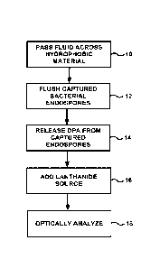
[0011] FIG. 1 is a flow diagram illustrating an example process for capturing
and
analyzing bacterial endospores from a fluid under evaluation.
[0012] FIG. 2 is a conceptual illustration of an example system that can be
used to
determine the bacterial endospore count in a fluid according to the example
technique of
FIG. 1.
[0013] FIG. 3 is a plot showing example optical responses of fluids prepared
by passing
milk samples across a hydrophobic material.
[0014] FIG. 4 is a plot showing an example number of endospores captured
versus
surface area of an example hydrophobic material.
[0015] FIG. 5 is a micrograph showing one example hydrophobic material at a
resolution
of 1 millimeter after passing milk through the material.
[0016] FIG. 6 is a 100 time magnification of the micrograph of FIG. 5 showing
endospores adhered to the surface of the material.
DETAILED DESCRIPTION
[0017] This disclosure generally relates to the isolation, concentration, and
analysis of
bacterial endospores from fluids in which the endospores are carried. In some
examples,
the bacterial endospores are contained within an optically active fluid that
emits optical
emissions within wavelengths overlapping with the wavelengths at which a
lanthanide-
dipicolinic acid complex liberated from the bacterial endospores emits. This
can prevent
direct in-situ optical analysis of the bacterial endospores within the carrier
fluid because
of optical interference. In some examples in accordance with the disclosure,
the carrier
fluid is passed across a hydrophobic collection material to help separate the
bacterial
endospores from the remaining carrier fluid. The hydrophobic collection
material can be
a porous sieve that the carrier fluid flows through, a sheet of hydrophobic
material that is
4
CA 02947498 2016-10-28
WO 2015/200807
PCT/US2015/038005
positioned in contact with the carrier fluid, or yet another structure that
attracts and holds
the bacterial endospores, e.g., via hydrophobic attraction forces. Regardless
of the
configuration, the hydrophobic collection material can capture and collect
bacterial
endospores out of the carrier fluid.
[0018] Bacterial endospores collected out of the surrounding carrier fluid can
be analyzed
using a variety of different techniques. In some examples, a germination fluid
is added to
the hydrophobic collection material to germinate the collected bacterial
endospores and
release dipicolinic acid (DPA) from the endospores. A lanthanide ion reagent
can be
added to the germination fluid to form a lanthanide-dipicolinic acid complex.
When
irradiated with light of appropriate wavelengths, this lanthanide-dipicolinic
acid complex
may emit fluorescent light that can be detected and quantified. The magnitude
and/or
wavelength of light emitted by the lanthanide-dipicolinic acid complex
corresponds to the
concentration of the lanthanide-dipicolinic acid complex in the germination
fluid which,
in turn, corresponds to the concentration of bacterial endospores in the
carrier fluid.
[0019] The disclosed systems and techniques for isolating, concentrating,
and/or
analyzing bacterial endospores can be utilized with any desired fluids that
may contain
bacterial endospores. Example industries that may use the systems and
techniques
include the food industry, the beverage industry, the pharmaceutical industry,
the
chemical industry, the healthcare industry, the water purification industry,
and other
industries where material is inspected for bacterial endospores. In the case
of the food
and beverage industry, fluids that may be analyzed for bacterial endospores
include those
obtained from mammalian-consumable (e.g., human-consumable) food and beverages
such as, but not limited to, dairy products (e.g., raw milk, whole and skimmed
milk,
condensed milk, cream, whey and whey derivatives, buttermilk), juices (e.g.,
fruit juices
such as orange and other citrus juices, apple juice and other pomaceous
juices, red berry
juice, coconut milk, and tropical fruit juices, vegetable juices such as
tomato juice,
beetroot juice, carrot juice, and grass juice), and canned foods (e.g., canned
fruits, canned
vegetables, canned meat). Types of bacteria that may produce endospores
desired to be
analyzed include, but are not limited to, Bacillus subtilis, Bacillus cereus,
Bacillus
amyloliquefaciens, Bacillus atrophaeus, Bacillus megaterium, Bacillus
coagulans,
Bacillus pumilus, Bacillus mycoides, Bacillus licheniformis, Bacillus
sporothermodurans,
Bacillus thuringensis, Bacillus weihenstephanensis, Geobacillus
stearothermophilus,
Clostridium tyrobutyricum, Alicyclobacillus, Clostridium botulinum,
Clostridium difficile,
and Bacillus anthracis.
CA 02947498 2016-10-28
WO 2015/200807
PCT/US2015/038005
[0020] In general, the fluid containing bacterial endospores for analysis will
be a liquid,
such as an aqueous liquid (e.g., water-based liquid), although gaseous carrier
fluids
containing bacterial endospores can also be evaluated using the systems and
techniques.
The fluid may be obtained directly from a product sample under analysis or may
be
formed by combining the product sample with a carrier fluid to extract
bacterial
endospores from the sample. For example, in instances where the product sample
potentially containing bacterial endospores is a solid, the solid may be
ground or mixed
with a carrier fluid to extract the bacterial endospores for analysis.
[0021] The term bacterial endospore generally refers to an endospore produced
within a
bacterium. The term endospore does not include conidio spores and ascospores
of fungi.
Endospores typically are non-reproductive structures whose primary function is
to ensure
survival of the bacterium through periods of environmental stress. Endospores
can
tolerate extreme drought, heat, and starvation. They are protected by a
hardened shell of
protein and carbohydrates and produced by a form of binary fission in
bacteria. An
endospore can comprise the DNA of its parent bacterium, ribosomes, and large
amounts
of dipicolinic acid. For example, dipicolinic acid may compose greater than 5
weight
percent of an endospore's dry weight, such as at least 10 weight percent or at
least 15
weight percent, such as from 5 weight percent to 20 weight percent, or from 7
weight
percent to 16 weight percent. Dipicolinic acid is a chemical that is believed
to help
endospores maintain their dormancy.
[0022] Because endospores can survive harsh environments and are resilient to
sterilization regimes, endospores can be a good indicator species for
validating the
sterility of a product. The absence of endospores within a product sample
and/or the
detection of low levels of endospores within the product can indicate that the
product is
suitably sterile for market release. While such information is useful for a
variety of
products, the information may be especially valuable for consumable products,
such as
ingestible foods and beverages. Indeed, governmental regulatory regimes may
mandate
compliance with certain sterility standards before a product can be sold to
the public. The
ability to measure endospore count within a product in substantially real-time
in
accordance with some examples of the present disclosure can enable
manufacturers to
monitor the quality of their products in substantially real-time and rapidly
respond if a
sterility issue is detected.
[0023] FIG. 1 is a flow diagram illustrating an example process for capturing
and
analyzing bacterial endospores from a fluid under evaluation. The process
includes
6
CA 02947498 2016-10-28
WO 2015/200807
PCT/US2015/038005
passing a fluid to be evaluated across a hydrophobic material (10) to collect
bacterial
endospores from the fluid, for example, by capturing the endospores on the
surface of the
hydrophobic material via hydrophobic attraction forces. After capturing the
bacterial
endospores from the fluid, the bacterial endospores are optionally flushed
(12) to remove
optically interfering carrier fluid and then processed to release dipicolinic
acid (14). In
addition, a lanthanide ion source is added to the dipicolinic acid (16) to
form a lanthanide-
dipicolinic complex that can then be optically analyzed (18) to determine the
concentration of the bacterial endospores in the fluid under evaluation. As
described in
greater detail below, a variety of different processing parameters and
conditions can be
used to capture and analyze bacterial endospores utilizing the example process
of FIG. 1.
[0024] To concentrate and/or isolate bacterial endospores from a fluid under
evaluation
the fluid may be passed over a hydrophobic material (10). Example hydrophobic
materials that can be used to concentrate and/or isolate endospores are
described in
greater detail with respect to FIG. 2. In general, however, the hydrophobic
material may
be a material or combination of materials chosen to selectively bind with
endospores in
the fluid under analysis while allowing the surrounding fluid carrying the
endospores to
pass across the hydrophobic material without binding. When so configured,
endospores
present in the fluid under analysis may attach to the surface of the
hydrophobic material
while a remainder of the fluid continues to pass across the hydrophobic
material without
adhering. This can allow the endospores present in the fluid under analysis to
collect on
the surface of the hydrophobic material, concentrating and isolating the
endospores from
the surrounding carrier fluid.
[0025] The hydrophobic collection material may be hydrophobic in that it does
not bind
or absorb water. For example, the hydrophobic collection material may repel
polar
molecules, such as water and molecules soluble in water, while allowing at
least some
types of non-polar molecules to bind to the surface of the material. The non-
polar
molecules may adhere to the surface of the hydrophobic material via
hydrophobic
attraction. Molecules having both polar and non-polar functional groups may or
may not
adhere to the hydrophobic material, e.g., depending on the hydrophobicity of
the material
being used, the length of the carbon chain separating a polar functional group
from a non-
polar functional group, and/or net polarity of the molecule.
[0026] When used, the hydrophobic collection material can capture and collect
endospores out of a fluid passing over the material via hydrophobic
attraction. As a fluid
containing endospores passes across the hydrophobic collection material, the
endospore
7
CA 02947498 2016-10-28
WO 2015/200807
PCT/US2015/038005
may pass adjacent to and, in some examples, in contact with the surface of
hydrophobic
collection material. Since bacterial endospores present within the fluid may
be non-polar
and/or contain non-polar functional groups, the endospores can attract and
attach to the
surface of the hydrophobic material. These endospores can bind to the surface
of the
hydrophobic material via hydrophobic attraction forces, such as entropic
interfacial
forces. The magnitude of these forces may depend on the hydrophobicity of the
interacting groups (e.g., the endospore and the hydrophobic material) as well
as the
distance separating them. In some examples, the hydrophobic forces increase
approximately exponentially with decreasing separation distance.
[0027] The carrier fluid surrounding the endospores may continue to pass
across the
hydrophobic collection material without binding to the surface of the
material. For
example, when the carrier fluid is an aqueous fluid, the hydrophobic material
can repel
the polar water molecules present in the carrier fluid, causing the carrier
fluid to flow past
the hydrophobic material without attaching to the surface of the material. In
this manner,
the hydrophobic material can capture endospores out of the fluid sample
passing across
the material and collect the endospores on the surface material, generating an
accumulated mass of endospores for subsequent analysis. In different examples,
the fluid
having passed across the hydrophobic material can be disposed or recycled and
again
passed across the hydrophobic material. For example, the fluid may be passed
across the
hydrophobic material two, three, or more times to increase the number of
endospores
collected out of the fluid by the hydrophobic material.
[0028] The volume of fluid passed across the hydrophobic material can vary,
e.g.,
depending on the size of the hydrophobic material being used and the expected
concentration of endospores in the sample under analysis. In some examples,
the volume
of fluid passed across the hydrophobic material is equal to or greater than
the volume of
the hydrophobic material itself. For example, the volume of fluid containing
endospores
that is passed across the hydrophobic material may be at least 10 times the
volume of the
hydrophobic material itself, such as at least 100 times the volume of the
material, at least
1000 times the volume of the material or at least 10,000 times the volume of
the material.
In general, increasing the volume of fluid passed across the hydrophobic
material
increases the number of endospores captured on the surface of the material and
available
for analysis.
[0029] A fluid under analysis may be passed across the hydrophobic collection
material
(10) using a variety of different techniques. In some examples, the
hydrophobic
8
CA 02947498 2016-10-28
WO 2015/200807
PCT/US2015/038005
collection material is dipped or immersed into a reservoir containing the
fluid under
analysis and then pulled out of the reservoir to provide endospores
hydrophobically-
bound on the surface of the material and available for analysis. In other
examples, a
moving stream of fluid is flowed across the surface of the hydrophobic
material, allowing
endospores in the stream of fluid to attach to the surface of the material
while a remainder
of the fluid continues flowing past and away from the hydrophobic material. In
such
examples, the flow of fluid under analysis may be directed generally parallel
to a planar
surface of the hydrophobic material and/or generally transverse (e.g.,
perpendicular) to
the planar surface of the hydrophobic material. For example, in some
configurations
described in greater detail with respect to FIG. 2, the hydrophobic material
may be a
porous material with pores sized to allow substantially all of the fluid under
analysis to
flow through the material. When so configured, the fluid under analysis may
flow
generally transverse to an external face of the hydrophobic material.
Endospores may
collect within the pores of the material while a remainder of the fluid
continues flowing
through the material and discharges on an opposite side of the hydrophobic
material.
[0030] Independent of the specific configuration of the hydrophobic material
utilized to
collect bacterial endospores, the example technique of FIG. 1 includes
optionally flushing
the collected endospores with a flushing fluid (12). After passing the fluid
to be
evaluated across the hydrophobic material (10), the flow of fluid (in instance
in which the
fluid is flowed across the material) may be terminated to leave a hydrophobic
material
having collected endospores on its surface. This hydrophobic material may also
contain
residual fluid carrying the endospores, e.g., trapped between and around
collected
endospores on the surface of the collection material and/or within the pores
of the
material (in instances in which the material is porous). In some applications,
this residual
fluid may optically interfere with subsequent optical analysis of the
endospores, if the
fluid is not removed from the endospores before analysis. For example, the
fluid carrying
the endospores may emit fluorescent emissions when light within a wavelength
ranging
from 250 nanometers (nm) to 300 nm impinges upon the fluid. These emissions
emitted
by the carrier fluid may be within a wavelength ranging from 300 nm to 700 nm,
which
can overlap with a wavelength at which a lanthanide-dipicolinic acid complex
fluoresces
of, e.g., 450 nm to 650 nm. As a result, these emissions from the carrier
fluid can obscure
and/or distort emissions associated with the endospores, inhibiting accurate
quantification
of the endospores.
9
CA 02947498 2016-10-28
WO 2015/200807
PCT/US2015/038005
[0031] To help remove this contaminating fluid from the endospores, the
endospores
attached to the hydrophobic material may be flushed with a flushing fluid. The
flushing
fluid may be optically inert, e.g., such that residual flushing fluid
remaining on the
endospores does not interfere with subsequent optical analysis of the
endospores. In one
example, the flushing fluid is or includes liquid water (e.g., distilled
water).
[0032] The hydrophobic material can be flushed, in different examples, by
immersing the
hydrophobic material containing the spores in a reservoir containing the
flushing fluid
and/or passing a moving stream of flushing fluid across the surface of the
hydrophobic
material and the endospores contained thereon. When the hydrophobic material
is
configured as a porous material, a pressurized stream of flushing fluid may be
passed
through the material (e.g., generally transversely), rinsing residual carrier
fluid from
around the endospores and from the pores of the material. In practice, some of
the
endospores captured on the surface of the hydrophobic material may be released
into the
flushing fluid. However, these lost endospores can be accounted for when
calibrating the
technique, e.g., by using a consistent amount of flushing fluid under
consistent pressure
conditions between a calibration run and subsequent operation.
[0033] When flushing is performed, the amount of flushing fluid passed over
the
collected endospores and/or through the hydrophobic material can vary, e.g.,
based on the
concentration of the endospores in the sample under analysis and the optical
interference
impact of the carrier fluid. In some examples, the volume of flushing fluid
passed across
the hydrophobic material and the endospores collected thereon is at least
equal to the
volume of fluid under evaluation initially passed across the hydrophobic
material. For
example, if a volume of liquid containing endospores equal to "X" is passed
across the
hydrophobic collection material, the hydrophobic material may subsequently be
flushed
with a volume of flushing fluid greater than or equal to "X," such as a volume
of flushing
fluid greater than or equal to 1.5 times "X," greater than or equal to 2 times
"X," or
between "X" and 5 times "X." As one specific and non-limiting example, if 50
milliliters
of fluid containing endospores was passed across the hydrophobic material, the
material
may subsequently be flushed with 100 milliliters of flushing fluid when using
a flushing
fluid ratio of 2 times "X." In some examples, a volume of flushing fluid
sufficient to
remove substantially all (and, in other examples, all) optically interfering
carrier fluid
from the endospores is used.
[0034] After capturing the bacterial endospores from the fluid under
evaluation and
optionally flushing the hydrophobic material containing captured endospores to
remove
CA 02947498 2016-10-28
WO 2015/200807
PCT/US2015/038005
optically interfering carrier fluid (12), the technique of FIG. 1 includes
processing the
endospores to release dipicolinic acid (14). Dipicolinic acid (DPA, 2,6-
pyridinedicarboxylic acid) is typically present in high concentrations (e.g.,
approximately
1 molar percent or approximately 15% dry weight percent) in the core of
endospores,
typically as a 1:1 complex with Ca2 . Dipicolinic acid is also a commercially
available
product having the following characteristics: CAS #: 499-83-2, Synonyms: 2,6
Pyridine
Dicarboxylic Acid, Molecular Formula: C7H5N04, Molecular Weight: 167.12,
Description: White crystalline powder, Sulphated Ash: 0.3% max, Moisture
Content:
0.5% max, Melting Point: 242.0 to 245Ødegree Celsius. Because dipicolinic
acid is an
indicator uniquely associated with bacterial endospores, the concentration of
dipicolinic
acid in a sample can be indicative of the number of endospores in the sample.
[0035] To detect and quantify the dipicolinic acid present in a sample
according to the
example technique of FIG. 1, the bacterial endospores collected on the
surface(s) of the
hydrophobic material can be processed in situ to release the dipicolinic acid
from the
cores of the spores. In some examples, a germination fluid is added to the
endospores
collected on the surface of the hydrophobic material, e.g., causing the
endospores to
transform to a vegetative cell and release dipicolinic acid. Germination
involves the
dormant endospore starting metabolic activity and thus breaking hibernation.
It
commonly includes rupture or absorption of the spore coat, swelling of the
endospore, an
increase in metabolic activity, and loss of resistance to environmental
stress. The addition
of the germination fluid, alone or in combination with heating, can initiate
germination of
the endospores.
[0036] In some examples, the technique of FIG. 1 includes adding a germination
fluid to
the hydrophobic material containing captured endospores to release dipicolinic
acid (14).
The germination fluid can be added to the endospores captured on the
hydrophobic
material, in different examples, by immersing the hydrophobic material
containing the
spores in a reservoir containing the germination fluid and/or passing a moving
stream of
germination fluid across the surface of the hydrophobic material and the
endospores
contained thereon. When the hydrophobic material is configured as a porous
material, a
pressurized stream of germination fluid may be passed through the material
(e.g.,
generally transversely), exposing the endospores collected on the surface of
the
hydrophobic material (e.g., external surfaces and pore walls of the material)
to
germination fluid. Example germinating liquids that be used to release
dipicolinic acid
include, but are not limited to, L-alanine, L-asparagine, D-glucose, L-
cysteine,
11
CA 02947498 2016-10-28
WO 2015/200807
PCT/US2015/038005
dodecylamine, and the cocktail AGFK (containing D-glucose, D-fructose, KC1,
and L-
asparagine).
[0037] When used, the amount of germination fluid added to the hydrophobic
material to
initiate germination of the endospores captured on the material can vary. In
some
examples, the volume of germination fluid added to the hydrophobic material
and the
endospores collected thereon is less than the volume of fluid under evaluation
initially
passed across the hydrophobic material. For example, if a volume of liquid
containing
endospores equal to "X" is passed across the hydrophobic collection material,
the amount
of germination fluid subsequently added to the hydrophobic material and the
endospores
contained thereon may be less than "X," such as a volume of flushing fluid
less than 0.5
times "X," less than 0.2 times "X," or less than 0.05 times "X." For example,
the volume
of germination fluid added to the hydrophobic material may range from 0.001
times "X"
to 0.5 times "X," such as from 0.01 time "X" to 0.2 times "X," or from 0.025
times "X"
to 0.1 times "X." As one specific and non-limiting example, if 50 milliliters
of fluid
containing endospores was passed across the hydrophobic material, 5
milliliters of
germination fluid may then be added to the hydrophobic material (e.g., after
optionally
flushing the material with any of the previously mentioned volumes of flushing
fluid)
when using a germination fluid ratio of 0.1 times "X."
[0038] The germination fluid added to the hydrophobic material and endospores
collected
thereon may be captured and/or retained for subsequent optical analysis. By
contrast,
residual sample fluid passing over and/or through the hydrophobic material
after
depositing endospores and/or flushing fluid may be disposed after crossing the
hydrophobic material. Accordingly, adding a smaller volume of germination
fluid to the
hydrophobic material can increase the concentration of endospores and/or
dipicolinic acid
in the germination fluid for subsequent analysis as compared to using a larger
volume of
germination fluid. This can be helpful when analyzing fluids having
comparatively low
concentrations of bacterial endospores.
[0039] In some examples, the germination fluid is heated prior to being added
to the
hydrophobic material and/or while the hydrophobic material containing
endospores is
immersed in the germination fluid. For example, the hydrophobic material
containing
captured endospores may be submerged in the germination fluid or the pore
space of the
hydrophobic material containing endospores may be saturated with germination
fluid and
then subject to heat shock. Heating the germination fluid can help initiate
germination of
the endospores and accelerate release of dipicolinic acid. In some examples,
the
12
CA 02947498 2016-10-28
WO 2015/200807
PCT/US2015/038005
germination material is heated to a temperature ranging from 75 degrees
Celsius to 90
degrees Celsius, for example, for a time ranging from 15 minutes to 30
minutes.
[0040] In addition to or in lieu of adding a germination fluid to the
bacterial endospores,
the endospores may be destructively lysed by impacting the endospores with a
core wall
rupturing force. Example lysing methods include but are not limited to:
microwaving,
plasma cleaning, dry heating, autoclaving, sonicating and hydrogen chloride
gassing.
Lysing may destroy the core of the endospores, releasing dipicolinic acid.
[0041] The example technique of FIG. 1 also includes adding a lanthanide ion
source to
the dipicolinic acid released from the captured endospores to form a
lanthanide-
dipicolinic complex (16). Dipicolinic acid is a chemistry ligand that binds
metal ions
with high affinity. Once bound, the dipicolinic acid complex can emit an
intense green
fluorescence under UV excitation. The magnitude of this fluorescence can be
correlated
to the concentration of dipicolinic acid which, in turn, can be correlated
with the number
of endospores present in the sample under analysis.
[0042] A lanthanide ion source can be added to the dipicolinic acid released
from the
endospores captured on the hydrophobic material to form a fluorescing
lanthanide-
dipicolinic complex. Example lanthanide ions that can be added to the
dipicolinic acid
include, but are not limited to: terbium (Tb3+), europium (Eu3+), and
dysprosium. In one
example, terbium (Tb3+) ions are used resulting in the formation of a terbium-
dipicolinic
acid complex. Typically, an excess amount of lanthanide ions are added to the
dipicolinic
acid that is sufficient to complex all dipicolinic acid molecules present in
the sample.
[0043] To add lanthanide ions to the dipicolinic acid released from the
endospores
collected on the hydrophobic material, the lanthanide ions can be introduced
to the
germination fluid or other fluid (e.g., when destructive lysing in performed)
that contains
or will contain the dipicolinic acid. In one example, the lanthanide ions are
added to the
germination fluid prior to introducing the germination fluid to the endospores
captured on
the hydrophobic material. In such an application, the lanthanide ions can
complex with
the dipicolinic acid as the acid is being released from the endospores during
germination.
In another example, the lanthanide ions are added to the germination fluid
after the
endospores have germinated and released their dipicolinic acid, e.g., forming
a
germination fluid containing released dipicolinic acid. Therefore, although
FIG. 1
illustrates the example technique as adding lanthanide ions (16) as a separate
step after
releasing dipicolinic acid from the captured endospores (14), it should be
appreciated that
the lanthanide ions may be present in the germination fluid such that the ions
are added
13
CA 02947498 2016-10-28
WO 2015/200807
PCT/US2015/038005
and combine with the dipicolinic acid as it is released from the endospores in
a single
step.
[0044] The technique of FIG. 1 also includes optically analyzing a fluid
containing the
lanthanide-dipicolinic acid complex and determining therefrom a concentration
of
bacterial endospores in the fluid under evaluation (18). The fluorescence of
lanthanide
ions is characterized by long lifetimes (e.g., 0.1 to 1 milliseconds), small
extinction
coefficients, and narrow emission bands. Narrow emission bands arise because
the
valence f-orbitals are shielded from the environment by the outer s and p
electrons, and
long lifetimes/small extinction coefficients arise because the transition
between the
emitting excited state and ground state is prevented. Thus, direct excitation
of lanthanide
ions leads to weak fluorescence due to the small absorption cross section.
However,
coordination of organic chromophores, like dipicolinic acid, with the
lanthanide ions
triggers intense lanthanide fluorescence. The dipicolinic acid serves as a
light-harvesting
center, and strong electronic coupling with downhill energetics allows the
dipicolinic acid
centered excitation energy to be efficiently transferred to the lanthanide
ion, which
subsequently fluoresces bright green.
[0045] In one example, the germination fluid or other fluid containing the
lanthanide-
dipicolinic acid complex is optically analyzed using a fluorometer. The
fluorometer
emits light into the fluid under analysis and, in response to receiving the
emitted light, the
lanthanide-dipicolinic complex emits fluorescent emissions. The fluorescent
emissions
may be at a different wavelength than the wavelength(s) of light emitted by
the
fluorometer into the fluid containing the lanthanide-dipicolinic acid complex.
The
fluorometer can detect the fluorescent emissions and determine the
concentration of
dipicolinic acid based on the optical response.
[0046] In another example, the germination fluid or other fluid containing the
lanthanide-
dipicolinic acid complex is optically analyzed using a spectrophotometer. The
spectrophotometer emits light at one or more specific wavelength(s) into the
fluid
containing the lanthanide-dipicolinic acid complex and detects that amount of
light
passing through the fluid at those one or more wavelength(s). The amount of
light
absorbed by the fluid sample may be proportional to the concentration of the
lanthanide-
dipicolinic acid complex in the sample.
[0047] Independent of the technique used, the optical response of the fluid
containing the
lanthanide-dipicolinic acid complex may be proportional to the concentration
of
dipicolinic acid in the fluid. In turn, this dipicolinic acid is proportional
to the
14
CA 02947498 2016-10-28
WO 2015/200807
PCT/US2015/038005
concentration of endospores captured on the hydrophobic material, which is
further
correlated to the concentration of endospores in the original fluid sample
under analysis.
Accordingly, the optical response of the fluid containing the lanthanide-
dipicolinic acid
complex can be used with calibration data to determine the concentration of
endospores
in the original fluid sample under analysis.
[0048] FIG. 2 is a conceptual illustration of an example system 20 that can be
used to
determine the bacterial endospore count in a fluid according to the example
technique of
FIG. 1. System 20 can be implemented as an on-line monitoring station to
continuously
monitor the bacterial endospore count in a fluid being processed.
Alternatively, system
20 can be implemented off-line, e.g., in a laboratory environment to
periodically
determine bacterial endospore counts in discrete fluid samples. System 20 is
illustrated as
including a source of aqueous liquid to be evaluated 22, an optional flushing
fluid source
24, a germination fluid source 26, and a lanthanide ion source 28. System 20
also
includes a hydrophobic material 30. Hydrophobic material 30 can receive
aqueous liquid
from liquid source 22, flushing fluid from source 24, germination fluid from
source 26,
and lanthanide ions from source 28. For example, hydrophobic material 30
and/or a
housing containing the material may be fluidly connected to the different
sources via fluid
conduits conveying the fluids to the hydrophobic material. In other examples,
hydrophobic material 30 and/or a housing containing the material may receive
the fluids
via manual transfer, e.g., performed by a laboratory technician.
[0049] In operation, hydrophobic material 30 can receive liquid from aqueous
liquid
source 22 and capture bacterial endospores out of the liquid. The bacterial
endospores
may hydrophobically bind and collect on the surfaces of the hydrophobic
material 30 as
the liquid flows over and/or through the material. As a result, the residual
liquid having
passed across hydrophobic material 30 may have a lower concentration of
endospores
(e.g., may be substantially devoid of endospores) than the liquid from source
22. This
residual liquid having a reduced quantity of endospores can be disposed to
drain 32 after
passing across hydrophobic material 30 or recycled back across the material.
[0050] Hydrophobic material 30 may have endospores collected and held on its
surface
after terminating the flow of liquid from liquid source 22. If desired,
hydrophobic
material 30 can subsequently receive flushing liquid from flushing fluid
source 24. The
flushing liquid can flow across the surface of hydrophobic material 30 and
over the
endospores captured on the surface of the material, e.g., helping to remove
optically
interfering liquid from the endospores in preparation for subsequent optical
analysis. The
CA 02947498 2016-10-28
WO 2015/200807
PCT/US2015/038005
flushing liquid having passed across hydrophobic material 30 and containing
residual
carrier liquid (e.g., optically interfering liquid) can be disposed to drain
32. In other
applications, hydrophobic material 30 is not flushed with a flushing liquid
before optical
analysis and, accordingly, system 20 does not include flushing fluid source
24.
[0051] After terminating the flow of flushing fluid from flushing fluid source
24 (when
used), hydrophobic material 30 can receive germination fluid from germination
fluid
source 26. In one example, the germination fluid flows across the surface of
hydrophobic
material 30 and over the endospores captured on the surface of the material,
e.g.,
collecting in a reservoir downstream of the hydrophobic material. In another
example,
the germination fluid is introduced into a housing containing hydrophobic
material 30,
resulting in the hydrophobic material and endospores collected thereon being
partially or
fully immersed in the germination fluid. This can allow the endospores
captured by
hydrophobic material 30 to germinate in situ, while being retained on the
surface of the
material. As discussed above with respect to FIG. 1, the endospores can
release
dipicolinic acid during germination, increasing the concentration of
dipicolinic acid in the
germination fluid (e.g., from an original concentration of zero).
[0052] To determine the concentration of dipicolinic acid in the germination
fluid added
to hydrophobic material 30 and, as a result, the concentration of endospores
in aqueous
liquid source 22, lanthanide ions are added to the dipicolinic-rich
germination fluid from
lanthanide ion source 28. In one example, the lanthanide ions are added to the
dipicolinic-rich germination fluid while hydrophobic material 30 is partially
or fully
immersed in the germination fluid. In another example, the germination fluid
is added to
the dipicolinic-rich germination fluid after removing hydrophobic material 30,
e.g., by
discharging the fluid downstream of the material. In yet another example, the
lanthanide
ions are not added separately from germination fluid but instead are mixed
with the
germination fluid prior to being received by hydrophobic material 30 so as to
provide a
single source of both germination fluid and lanthanide ions. The lanthanide
ions can
combine with the dipicolinic acid released from the endospores to form an
optically
active lanthanide-dipicolinic acid complex.
[0053] System 20 in the example of FIG. 2 also includes an optical sensor 34
and a
controller 36. Optical sensor 34 is configured to receive and optically
analyze the
germination fluid containing the lanthanide-dipicolinic acid complex generated
from the
endospores captured by hydrophobic material 30. Optical sensor 34 may include
one or
more optical emitters and one or more optical detectors. In operation, the one
or more
16
CA 02947498 2016-10-28
WO 2015/200807
PCT/US2015/038005
optical emitters direct light into fluid containing the lanthanide-dipicolinic
acid complex
and the one or more optical detectors detect fluorescent emissions generated
by the fluid
and, in particular, the lanthanide-dipicolinic acid complex contained within
the fluid. The
light directed into the fluid may generate fluorescent emissions by exciting
electrons of
the lanthanide-dipicolinic acid complex, causing the molecules to emit energy
(i.e.,
fluoresce) that can be detected by the optical detectors. In some examples,
the one or
more optical emitters direct light at one frequency (e.g., ultraviolet
frequency) into the
fluid and cause the lanthanide-dipicolinic acid complex to emit light energy
at a different
frequency (e.g., visible light frequency, a different ultraviolet frequency).
[0054] For example, optical sensor 34 may include one or more optical emitters
that emit
light within the ultraviolet (UV) spectrum, such as within wavelengths in the
range from
approximately 250 nm to approximately 300 nanometers. For example, the one or
more
optical emitters may emit within a wavelength from 270 nm to 280 nm. In
response to
this emitted light, the lanthanide-dipicolinic acid complexes within the fluid
may generate
fluorescent emissions at a wavelength from 450 nm to 650 nm. The one or more
optical
detectors of optical sensor 34 can detect the energy emitted by the
fluorescing lanthanide-
dipicolinic acid complexes at these wavelengths.
[0055] Controller 36 can control the one or more optical emitters to direct
radiation into
the fluid containing the lanthanide-dipicolinic acid complexes and also
control the one or
more detectors to detect fluorescent emissions emitted by the fluid. In some
examples,
controller 36 (or another controller within system 20) processes the light
detection
information to determine a concentration or count of endospores in the
original aqueous
liquid under analysis. For example, controller 36 can determine a
concentration or count
of the endospores in the aqueous liquid under analysis by comparing the
magnitude of
fluorescent emissions detected by the one or more optical detectors from a
germination
fluid prepared from a source liquid having an unknown concentration or count
of
endospores to the magnitude of the fluorescent emissions detected by the
optical detectors
from a germination fluid prepared having a known concentration or count of
endospores.
The unknown fluid may be prepared using the same or substantially same process
as was
used to prepare the calibration fluid. For example, the same volume of unknown
source
fluid may be passed across hydrophobic material 30 as was used to generate the
calibration fluid, followed by use of the same volumes of flushing fluid,
germination
fluid, and lanthanide ion source. Calibration information can be stored in a
non-transitory
computer readable storage medium associated with controller 36.
17
CA 02947498 2016-10-28
WO 2015/200807
PCT/US2015/038005
[0056] In some examples, controller 36 manages the overall operation of system
20.
Controller 36 may be communicatively coupled to various components within
system 20,
for example via a wired or wireless connection, so as to send and receive
electronic
control signals and information between controller 36 and the communicatively
coupled
components. For example, controller 36 may electronically actuate valves
and/or control
pumps within system 20 to control movements of the different fluids to and
from
hydrophobic material 30. Controller 36 can include a processor and memory. The
memory may store software and data used or generated by controller. The memory
may
comprise a computer-readable medium, such as random access memory (RAM), read-
only memory (ROM), non-volatile random access memory (NVRAM), electrically
erasable programmable read-only memory (EEPROM), embedded dynamic random
access memory (eDRAM), static random access memory (SRAM), flash memory,
magnetic or optical data storage media. The memory may or may not be
removable. The
processor can run software stored in the memory to perform functions
attributed to optical
sensor 34 and controller 36 in this disclosure. The processor can include one
or more
processors, such as one or more microprocessors, digital signal processors
(DSPs),
application specific integrated circuits (ASICs), field programmable gate
arrays (FPGAs),
programmable logic circuitry, or the like, either alone or in any suitable
combination.
[0057] As discussed above with respect to FIG. 1, hydrophobic material 30 can
hydrophobically bind with endospores present in the fluid under analysis to
concentrate
and/or isolate the endospores from the remainder of the fluid. Hydrophobic
material 30
can have a variety of different configurations and can function as a support
surface on
which endospores are captured and germinated. In some examples, hydrophobic
material
30 is a non-porous (e.g., substantially non-porous) material. When so
configured, fluid
containing endospores can pass across the external surface(s) of the non-
porous material
and be collected on the enteral surface. In other examples, hydrophobic
material 30 is a
porous material having void spaces through which liquid can travel across the
cross-
section of the material. In these configurations, fluid containing endospores
can pass
across the external surface(s) of the hydrophobic material and/or internal
surfaces of the
material that bound and define the void spaces providing the material's
porosity.
[0058] A porous hydrophobic material may be useful to increase the amount of
surface
area available for capturing endospores out of the fluid under analysis via
hydrophobic
attraction. In general, increasing the surface area of hydrophobic material 30
over which
the fluid containing endospores passes increases the likelihood that
endospores in the
18
CA 02947498 2016-10-28
WO 2015/200807
PCT/US2015/038005
fluid will hydrophobically bond to the material and separate from the
remaining fluid.
The remainder of the fluid carrying the endospores, which may be composed of
water and
other polar molecules, can continue flowing through the pores or void spaces
of the
material and discharge on an opposite side of the material. In this way, the
endospores
can adhere to the wall surfaces bounding the various pores of hydrophobic
material 30
while the remaining carrier fluid flows through the material, isolating and
concentrating
the endospores.
[0059] When hydrophobic material 30 is implemented using a porous material,
the pores
of the material may be sized large enough to allow substantially all (and, in
other
examples, all particles) in the fluid under analysis to flow through the pores
of the
material. For example, the pores of hydrophobic material 30 may be larger than
the
endospores being captured out of the fluid under analysis via hydrophobic
attraction. In
other words, instead of functioning as a filter that separates endospores via
size exclusion,
the pores of hydrophobic material 30 may be large enough to allow the
endospores to
pass through the hydrophobic material. The hydrophobic attraction forces
between
hydrophobic material 30 and the endospores may bind the endospores to the
material and
prevent the endospores from flowing out through the pores of the material,
even though
the endospores are smaller than the pores of the material. When so configured,
hydrophobic material 30 may provide torturous (e.g., non-linear) fluid flow
paths
extending through the material and have a comparatively high surface area
across which
the fluid flows during endospore capture.
[0060] In some examples, hydrophobic material 30 has an average pore size
greater than
the average size of the bacterial endospores in the fluid under analysis, such
as an average
pore size at least 5 times larger than the average size of the bacterial
endospores, at least
times larger, or at least 100 times larger, or at least 1000 times larger. In
some
additional examples, the distribution of pore sizes is such that at least 75%
of the pores
are at least 5 times larger (and in some examples at least 10 times larger)
than the average
size of the bacterial endospores in the fluid under analysis, such as at least
90% of the
pores, at least 95% of the pores, or at least 99% of the pores.
[0061] The size of the pores of hydrophobic material 30 can also vary
depending on the
size of particles other than the endospores within the fluid under analysis,
which will
depend on the composition of the fluid under analysis. In the case of milk,
for example,
milk may contain protein micelles having a size of approximately 0.1
micrometers, fat
globules ranging from 0.2 micrometers to 15 micrometers, endospores ranging
from 0.6
19
CA 02947498 2016-10-28
WO 2015/200807
PCT/US2015/038005
micrometers to 1 micrometer, bacteria ranging from 1 micrometer to 5
micrometers, and
somatic cells ranging from 10 micrometers to 15 micrometers, among other
particles.
Hydrophobic material 30 may have pores sized larger than substantially all
particles in
the fluid in an attempt to allow the non-endospore particles to flow through
the material
without blocking the pores. In some examples, hydrophobic material 30 has an
average
pore size greater than the average size of the particles in the fluid under
analysis, such as
an average pore size at least 5 times larger than the average size of the
smallest particles
(e.g., class of particles) in the fluid, at least 10 times larger, or at least
100 times larger, or
at least 1000 times larger. In some additional examples, the distribution of
pore sizes is
such that at least 75% of the pores are at least 5 times larger (and in some
examples at
least 10 times larger) than the average size of the smallest particles in the
fluid under
analysis, such as at least 90% of the pores, at least 95% of the pores, or at
least 99% of
the pores. In one example, at least 95% of the pores range from 10 times
larger to 1000
times larger than the smallest sized class of particles in the fluid under
analysis.
[0062] While the absolute size of the pores of hydrophobic material 30 can
vary based on
the desired application, in some examples, the hydrophobic material has an
average pore
size greater than 25 micrometers, such as greater than 50 micrometers, greater
than 100
micrometers, or greater than 125 micrometers. In some examples, the
hydrophobic
material has a porosity ranging from 20 percent of the total volume of the
hydrophobic
material to 70 percent of the total volume of the material.
[0063] Hydrophobic material 30 can capture and collect endospores out of a
fluid passing
over the material via hydrophobic attraction. Accordingly, hydrophobic
material 30 can
exhibit a sufficient hydrophobicity to selectively bind with endospores in the
fluid under
analysis while allowing the surrounding fluid carrying the endospores to pass
across the
hydrophobic material without binding. In some examples, hydrophobic material
30 is
sufficiently hydrophobic such that substantially all endospores present in the
fluid under
analysis bind to the hydrophobic material while passing across the material.
In different
configurations, hydrophobic material 30 may capture greater than 30% of the
endospores
present in the fluid under analysis on the surface of the material, such as
greater than 50%
of the endospores, greater than 70% of the endospores, greater than 90% of the
endospores, or greater than 95% of the endospores. The resulting fluid having
passed
across hydrophobic material 30 may contain less than 50% of the endospores
present in
the original fluid initially contacting the material, such as less than 40% of
the
endospores, less than 30% of the endospores, or less than 10% of the
endospores. The
CA 02947498 2016-10-28
WO 2015/200807
PCT/US2015/038005
percentage of endospores captured by the hydrophobic material may be modified
by
adjusting the hydrophobicity of the material and/or the amount of hydrophobic
material
the fluid encounters.
[0064] Because hydrophobic materials repel water, the hydrophobicity of a
material can
be characterized by the contact angle between a water droplet placed on the
material and
the surface of the material. For example, the water contact angle of
hydrophobic material
30 can be determined by placing a droplet of water on the surface of the
material and
taking measurements of the drop and/or measuring the wetting angle that forms
at the
liquid-surface interface. One standard method used to measure the water
contact angle is
by measuring the maximum height and width of a sessile drop. Based on a ratio
of the
drop height to the drop width, the contact angle between the drop and the
surface is
calculated according to known equations. In some examples, hydrophobic
material 30
exhibits a contact angle with water greater than 80 degrees, such as a contact
angle
greater than 90 degrees.
[0065] Hydrophobic material 30 can be constructed from a variety of different
materials.
Example materials that can be used to fabricate hydrophobic material 30
include, but are
not limited to, polyethylene, polypropylene, polyvinylchloride, polyamide,
polystyrene,
polytetrafluoroethylene (e.g., Teflon ), ethylene propylene diene (EPDM),
nitrite rubber
(e.g., Buna-N), polyethylene terephthalate, aliphatic polyamides (e.g.,
Nylon), aramid
fabric (e.g., Kevlar ), and stainless steel. For example, hydrophobic material
30 may be
formed from a sintered metal powder or porous plastic. Hydrophobic material 30
may or
may not be surface treated to increase the hydrophobicity of the material.
[0066] Hydrophobic material 30 can have any desired size and shape. Typically,
the size
of hydrophobic material 30 will vary based on the amount of fluid intended to
be
analyzed, with larger volumes of fluid using larger volumes of hydrophobic
material.
Hydrophobic material 30 can be implemented as a planar sheet of material
(e.g., a disc), a
sphere of material, a cylinder of material (e.g., an annulus), or any other
desired shape. In
some examples, hydrophobic material 30 is implemented as a single structure
(e.g., sheet)
that fluid containing endospores passes over and/or through. In other
examples, a
plurality of hydrophobic structures may be used in close proximity, e.g., to
provide a
packed bed, column, or cartridge of hydrophobic material that fluid containing
endospores flows through.
[0067] In general, the foregoing examples describe the use of a hydrophobic
material to
concentrate and/or isolate bacterial endospores from a fluid sample followed
by
21
CA 02947498 2016-10-28
WO 2015/200807
PCT/US2015/038005
germination and optical analysis to determine the concentration or count of
endospores in
the fluid sample. Hydrophobic materials described as being suitable for
capturing and
collecting endospores out of a fluid may be used in other applications beyond
quantifying
the endospores via optical analysis. For example, the hydrophobic materials
can be used
in a manufacturing facility to purify a fluid containing endospores by passing
the fluid
across the hydrophobic material. The hydrophobic material can capture the
endospores in
the fluid and purify the fluid for downstream processing or sale. For example,
the
hydrophobic material may remove greater than 80% of the endospores present in
the
fluid, such as greater than 90% of the endospores, or greater than 99% of the
endospores.
In such applications, the hydrophobic material may be periodically flushed to
remove
captured endospores and to reopen surface area for endospore bonding. In still
other
examples, the hydrophobic material may be used in a laboratory setting to
concentrate
and/or isolate bacterial endospores from a fluid sample without subsequently
germinating
and/or optically analyzing the captured endospores. Rather, the concentrated
and/or
isolated endospores can be used to perform other types of laboratory analyses.
[0068] Further, although the foregoing examples describe the use of a
hydrophobic
material to capture and collect endospores out of a fluid passing over the
material via
hydrophobic attraction, in other examples, endospores can be isolated from a
fluid sample
without relying on hydrophobic attraction forces. For example, a mechanical
filter may
be used to separate endospores from a surrounding fluid based on size
exclusion. In these
examples, the mechanical filter may or may not be fabricated from a
hydrophobic
material.
[0069] The following examples may provide additional details about systems and
techniques in accordance with this disclosure.
EXAMPLE 1
[0070] A variety of milk solutions containing different bacterial endospores
counts were
passed across a hydrophobic material having a porosity of approximately 50
percent (e.g.,
ranging from 25% void space to 75% void space). Fifty milliliters of milk were
passed
across the hydrophobic material followed by two successive flushes of 50
milliliters each
of water. Subsequently, 2 milliliters of L-alanine (10 mM) type germination
fluid were
added to the hydrophobic material and heated at a temperature of 75 degrees
Celsius for
15 minutes. A terbium reagent was then added to the germinate fluid to form a
terbium-
22
CA 02947498 2016-10-28
WO 2015/200807
PCT/US2015/038005
dipicolinic acid complex. The germinate fluid containing the terbium-
dipicolinic acid
complex was fluorometrically analyzed by emitting light into the fluid samples
and
generating and detecting fluorescent emissions from the fluids.
[0071] FIG. 3 is a plot showing the optical response of the example milk
samples. The
X-axis of the plot is the endospore level in the milk samples in counts. The Y-
axis is the
optical response of the germinant fluid containing terbium-dipicolinic acid
complex that
was generated from the milk samples, in relative fluorescence units.
EXAMPLE 2
[0072] To evaluate the effect of hydrophobic material pore size on endospore
capture,
hydrophobic materials were fabricated having different average pore sizes,
including one
having an average pore size of 65 micrometers and one having an average pore
size of
125 micrometers. Subsequently, 50 milliliters of milk having an endospore
count of 1 x
102 was passed through each hydrophobic collection material. The endospores
were
believed to have ranged in size from approximately 0.7 micrometers to
approximately 1
micrometer. The residual milk discharged from the hydrophobic collection
material was
recycled back through the material such that the milk passed through
hydrophobic
material three times. FIG. 4 is a plot showing the number of endospores
captured
following the process versus the surface area of the hydrophobic material. The
larger
surface areas correspond to the hydrophobic materials having the small average
pore
sizes. The testing showed the hydrophobic material captured from about 50% of
the
spores to about 70% of the spores following the third pass of the milk.
[0073] FIG. 5 is a micrograph of one of the hydrophobic materials at a
resolution of 1
millimeter after passing the milk through the material three times. The figure
shows the
overall structure of the material and the relative size of an example pore
structure. FIG. 6
is a 100 time magnification of the micrograph of FIG. 5 showing an endospore
50
adhered to the surface of material. The images indicate the endospores were
captured via
hydrophobic attraction and not mechanical size exclusion (e.g., filtering).
[0074] In the example, the hydrophobic materials were subsequently flushed
with 50
milliliters of water. Testing showed that greater than from approximately 99%
to
approximately 99.9% of the endospores remained adhered to the surface of the
hydrophobic material after flushing.
23
CA 02947498 2016-10-28
WO 2015/200807
PCT/US2015/038005
24