Note: Descriptions are shown in the official language in which they were submitted.
OPTICAL COHERENCE TOMOGRAPHY-AUGMENTED SURGICAL
INSTRUMENTS AND SYSTEMS AND METHODS FOR CORRECTING UNDESIRED
MOVEMENT OF SURGICAL INSTRUMENTS
Field of the Disclosure
[0001] The present disclosure relates to optical coherence tomography (OCT)-
augmented surgical
instruments and to systems and methods for correcting for undesired movement
of surgical
instruments using OCT.
Background
[0002] Surgery often involves precise removal of tissue or placement of
incisions. In
microsurgery, in particular, accurate positioning in all three dimensions as
well as precise motion
control is critical to avoid unintended effects, such as an inability to
complete the surgery or even
damage. This is especially true for ophthalmic surgeries. For example, in
vitreoretinal surgeries,
tool-tip positioning accuracy of around 10 gm is desired. Hand tremor is a
common problem in
surgeries and is difficult to avoid, yet it can regularly cause movements of a
much as 50 urn. This
is well outside of the desired range for vitreoretinal surgery and other
microsurgeries and reduces
the quality of these surgeries.
[0003] Other surgeries, such as treatment of retinal vein occlusion, are
impossible to perform
because movement of the surgical instruments cannot be properly controlled.
Retinal vein
occlusion affect 1.6% of people aged 49 and older and can be treated
surgically, but currently the
procedure is considered too risky to perform.
[0004] Prior approaches to combat undesired movement such as accidental
movement in
microsurgical procedures actively compensate for tremors. For example, one
approach places a
magnetometer-aided accelerometer on the surgical instrument to detect and
compensate for
tremors. Other approaches using proximal-end accelerometers to sense
-1-
CA 2947626 2018-11-23
CA 02947626 2016-10-31
WO 2016/014289
PCT/US2015/040360
distal-end motion require complex data filtering and processing and add
significant weight to
the surgical instrument, which tends to cause more tremors.
[0005] Microsurgical instruments, systems, and methods that can compensate for
tremors or
other undesired movement in a more practical fashion are still needed.
-2-
SUMMARY
[0005a] Certain exemplary embodiments can provide an optical coherence
tomography
(OCT) system comprising: an OCT source; a beam splitter coupled to the OCT
source via
a first OCT transmission medium; a reference arm coupled to the beam splitter
via a
second OCT transmission medium; an OCT-augmented surgical instrument coupled
via a
third OCT transmission medium to the beam splitter, the surgical instrument
comprising: an
OCT focusing element; an actuator; and a surgical component, wherein the
surgical
component performs a surgical operation; and a detector coupled via a fourth
OCT
transmission medium to the beam splitter, wherein the detector receives an OCT
beam
containing a component from the reference arm and a component from the OCT-
augmented
surgical instrument; and a computer electrically or wirelessly coupled to the
detector and
the actuator, wherein an undesired movement of the surgical instrument results
in a change
in an interference pattern detected by the detector, which is communicated to
the computer,
which sends an electrical or wireless signal to the actuator to cause a
corrective movement
of the surgical instrument, the corrective movement being in at least two
directions.
[0005b] Certain exemplary embodiments can provide an optical coherence
tomography
(OCT)-augmented surgical instrument comprising: an OCT transmission medium; an
OCT
focusing element; an actuator; and a surgical component, wherein the actuator
is operable
to cause corrective movement of the surgical instrument in response to an OCT
beam that
travels through the OCT transmission medium and OCT focusing element, the
corrective
movement being in at least two directions.
[0006] In another embodiment an OCT system containing an OCT source coupled
via
an OCT transmission medium to a beam splitter coupled via one OCT path to a
reference arm; and via a second OCT transmission medium to an OCT-augmented
surgical instrument is described. The OCT-augmented surgical instrument
contains an
OCT focusing element, an actuator, and a surgical component that performs a
surgical operation. The OCT system also contains a detector coupled via an OCT
transmission medium to the beam splitter. The detector receives an OCT beam
containing a
component from the reference arm and a component from the OCT-augmented
surgical
-3-
CA 2947626 2018-11-23
instrument. The OCT system additionally contains a computer electrically or
wirelessly
coupled to the detector and the actuator. An undesired movement of the
surgical
instrument results in a change in an interference pattern detected by the
detector, which
is communicated to the computer, which sends an electrical or wireless signal
to the
actuator to cause a corrective movement of the surgical instrument.
[0007] In another embodiment an OCT-augmented surgical instrument containing
an
OCT transmission medium, an OCT focusing element, an actuator, and a surgical
component is described. The actuator is able to cause corrective movement of
the
surgical instrument in response to an OCT beam that travels through the OCT
transmission medium and OCT focusing element.
[0008] In another embodiment a method of correcting undesired movement of a
surgical instrument is described by sending an OCT beam from an OCT source via
an
OCT transmission medium to a beam splitter, which splits the beam into an OCT
beam
that travels to and is reflected by a reference arm and an OCT beam that
travels to and is
reflected by a tissue being operated on by the surgical instrument, detecting
an
interference pattern for the OCT beams reflected by the reference arm and
tissue,
determining whether an undesired movement occurred and an appropriate
corrective
movement based on the interference pattern, and causing the appropriate
corrective
movement in the surgical instrument using an actuator located in the surgical
instrument.
-3 a-
CA 2947626 2018-01-25
CA 02947626 2016-10-31
WO 2016/014289
PCT/US2015/040360
BRIEF DESCRIPTION OF THE DRAWINGS
[0009] For a more complete understanding of the present invention and its
features and
advantages, reference is now made to the following description, taken in
conjunction with the
accompanying drawings, which are not drawn to scale, and in which:
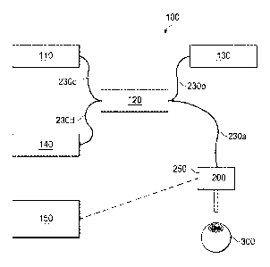
[0010] FIG. 1 is an OCT system containing an OCT-augmented surgical
instrument;
[0011] FIG. 2 is an OCT-augmented surgical instrument;
[0012] FIG. 3 is another embodiment of an OCT-augmented surgical instrument;
[0013] FIG. 4 is a beam-splitting and focusing unit in an OCT-augmented
surgical
instrument;
[0014] FIG. 5 is another embodiment of a beam-splitting and focusing unit in
an OCT-
augmented surgical instrument;
[0015] FIG. 6 is a coupler in an OCT-augmented surgical instrument; and
[0016] FIG 7 is diagram depicting the relationship of measured vectors in
space using an
OCT system containing an OCT-augmented surgical instrument.
-4-
CA 02947626 2016-10-31
WO 2016/014289
PCT/US2015/040360
DESCRIPTION OF PARTICULAR EMBODIMENT(S)
[0017] In the following description, details are set forth by way of example
to facilitate
discussion of the disclosed subject matter. It should be apparent to a person
of ordinary skill
in the field, however, that the disclosed embodiments are exemplary and not
exhaustive of all
possible embodiments.
[0018] As used herein, a reference numeral followed by a letter refers to a
specific instance
of an element and the numeral only form of the reference numeral refers to the
collective
element. Thus, for example, device '12a' refers to an instance of a device
class, which may
be referred to collectively as devices '12' and any one of which may be
referred to
generically as a device '12'.
[0019] Referring now to the drawings, FIG. 1 is an OCT system 100 with OCT-
augmented
surgical instrument 200. Optical coherence tomography (OCT) is an
interferometric analysis
technique for structural examination of a sample material, such as a tissue
that is at least
partially reflective to light. It can also be used for functional examination
of a sample
material, such as the motion and velocity of the sample material or blood flow
of the tissue.
In OCT, light in the form on an OCT beam is used to measure distances and
depth profiles
based on optical interference that arises between a reference beam and a
sample beam that
interacts with the sample material, such as a biological tissue. In some
embodiments, the
OCT beam may be supplied in pulses, sweeping wavelengths or a broad band
light.
[0020] OCT system 100 may make measurements of both the relative motion and
velocity
between surgical instrument 200 and tissue 300 (represented as an eye in this
example
diagram).
[0021] OCT system 100 additionally includes OCT source 110, which produces an
OCT
beam (not shown) that travels through OCT transmission medium 230c to beam
splitter 120
where it is split so that a portion of the beam travels through OCT
transmission medium 230b
to reference arm 130 and a portion of the beam travels through OCT
transmission medium
230a to surgical instrument 200. After hitting reference arm 130 or tissue
300, the OCT
beams travel back through OCT transmission mediums 230b and 230a,
respectively, to beam
splitter 120, where they are directed via OCT transmission medium 230d to
detector 140.
Detector 140 sends a signal to computer 150, which includes a processor able
to determine
the relative motion and velocity of surgical instrument 200 with respect to
tissue 300 based
on the signal received from detector 140. Computer 150 determines if
corrective movement
-5-
CA 02947626 2016-10-31
WO 2016/014289
PCT/US2015/040360
of surgical instrument 200 is needed and, if so, sends a signal to an actuator
250 in surgical
instrument 200. Actuator 250 responds to the signal and causes corrective
movement of
surgical instrument 200 in real-time.
[0022] In some embodiments, OCT transmission medium 230 is an optical fiber.
[0023] In the embodiment shown in FIG. 1, reference arm 130 is located close
to tissue 300
in terms of optical delay and is in a pre-determined position that is an
acceptable distance
from the OCT source 110. The OCT beam from tissue 300 traveling back through
surgical
instrument 200 to detector 140, interferes with the OCT beam from reference
arm 130 and
generates an interference pattern, As a result, the motion characteristics
(such as the gap,
displacement and velocity) of the surgical instrument 200 relative to tissue
300 can be
determined.
[0024] In one embodiment, reference arm 130 includes a mirror to reflect the
OCT beam.
[0025] In one embodiment, detector 140 is a spectrometer. In another
embodiment, detector
140 includes a photodiode or similar device that generates an electrical
signal indicative of
incident light intensity at detector 140.
[0026] Detector 140 may output an electrical signal to computer 150. In such
an
embodiment, computer 150 may include circuitry for signal conditioning,
demodulation,
digitization, and digital signal processing. In another embodiment, detector
140 outputs a
wireless signal to computer 150.
[0027] In one embodiment, computer 150 additionally includes memory media,
which store
instructions (i.e., executable code) that are executable by the processor
having access to the
memory media. The processor may execute instructions that cause actuator 250
in surgical
instrument 200 to activate and which control the parameters of such activation
to allow
compensation for undesired movement of surgical instrument 200. For the
purposes of this
disclosure, the memory media may include non-transitory computer-readable
media that
stores data and instructions for at least a period of time. The memory media
may comprise
persistent and volatile media, fixed and removable media, and magnetic and
semiconductor
media. The memory media may include, without limitation, storage media such as
a direct
access storage device (e.g., a hard disk drive or floppy disk), a sequential
access storage
device (e.g., a tape disk drive), compact disk (CD), random access memory
(RAM), read-
only memory (ROM), CD-ROM, digital versatile disc (DVD), electrically erasable
-6-
CA 02947626 2016-10-31
WO 2016/014289
PCT/US2015/040360
programmable read-only memory (EEPROM), flash memory, non-transitory media,
and
various combinations of the foregoing.
[0028] FIG. 2 depicts surgical instrument 200a, which may be used in an OCT
system, and
which includes handle 210 and cannula 220. In some embodiments, cannula 220
may be
replaced with a different surgical component that performs a surgical
operation. OCT
transmission medium 230a travels through handle 210 into cannula 220, where it
terminates
with focusing element (e.g., lens, curved mirror) 240. Handle 210 also
contains actuator 250,
which is operable to receive a signal from a computer (not shown) and to cause
movement of
surgical instrument 200a in response to the signal. Because only one OCT beam
travels
through focusing element 240, the instrument in FIG. 2 provides one-
dimensional OCT
measurements. A surgical instrument may include multiple OCT transmission
mediums and
focusing lenses similar to those depicted in FIG. 2. These multiple OCT
transmission
mediums may be OCT fibers coupled via a coupler as shown in FIG. 6.
[0029] In the example shown, in which surgical instrument 200 contains cannula
220,
actuator 250 moves cannula 220 in and out of handle 210 to compensate for
undesired
movement. For example, if the OCT system determines that cannula 220 is too
close to the
tissue (not shown), actuator 250 moves cannula 220 into handle 210 to
compensate.
[0030] In some embodiments, actuator 250 moves cannula 220 or another surgical
component at a speed that matches the speed of the undesired movement of
surgical
instrument 200. Actuator 250 may also move cannula 220 or another surgical
component in a
direction opposite the direction of the undesired movement, or a component
direction of the
undesired movement.
[0031] Actuator 250 may be any surgical actuator capable of moving surgical
instrument 200
in real-time in response to undesired movement. In some embodiments, actuator
250 may
constitute a small proportion of the weight of surgical instrument 200 in
order to avoid
introducing additional tremor. For example, actuator 250 may be less than 25%
of the weight
of surgical instrument 200. In one embodiment, actuator 250 is a piezoelectric
actuator. In
another embodiment, actuator 250 is a voice coil actuator. In still another
embodiment,
actuator 250 is an electromagnetic actuator. In another embodiment, actuator
250 is an
ultrasonic actuator. Actuator 250 may be capable of movement in only one
direction as
shown in FIG. 2 or in two, three, or more directions as shown in FIG. 3, which
illustrates
movement in three directions. In embodiments where actuator 250 is capable of
movement
-7-
CA 02947626 2016-10-31
WO 2016/014289
PCT/US2015/040360
in two or more directions, actuator 250 may contain multiple components or sub-
actuators,
each capable of movement in one direction.
[0032] FIG. 3 depicts an alternative surgical instrument 200b, which, instead
of focusing
element 240, contains beam splitting and focusing unit 260, which splits the
OCT beam
traveling along OCT transmission medium 230a into two or more separate OCT
beams
focused in two or more directions. In one embodiment, the beam is split into
three or more
OCT beams focused in three or more directions. The multiple split beams
produced by beam
splitting and focusing unit 260 have slightly different optical path length
delay, so that OCT
information from different beams will be separated in depth in corresponding
OCT images.
Using these different images, the multi-dimensional displacement and velocity
of the tissue
relative to surgical instrument 200, and vice versa, is calculated.
[0033] FIG. 4 depicts a unified beam splitting and focusing unit 260a. The
beam splitting
and focusing unit both splits the OCT beam into multiple beams and focuses
those beams on
the tissue (not shown).
[0034] FIG. 5 depicts an alternative beam splitting and focusing unit 260b,
which contains a
separate beam splitting unit 270 and beam focusing unit 280. Beam splitting
unit 270, in
some embodiments, is a fiber splitter or a multi-cladding fiber. Beam focusing
unit 280, in
some embodiments is a graded index (GRIN) lens. In other embodiments, beam
splitting and
focusing unit 260b is a multiple faceted ball, such as a sapphire ball.
[0035] FIG. 6 depicts a coupler 400 for splitting the OCT beam into multiple
OCT fibers
410a, 410b, and 410c, which terminate in focusing elements 240a, 240b, and
240c,
respectively. The multiple OCT fibers and focusing elements may be located in
surgical
instrument 200 in a manner similar to that depicted in FIG. 2. OCT fibers 410
may be of
slightly different lengths to cause different optical path delays.
[0036] In another embodiment, the detector is able to detect polarization of
light and the
OCT beam through the surgical instrument is split by polarization into
different orientations.
This also allows multi-dimensional measurements of motion and velocity of the
tissue and
surgical instrument with respect to one another.
[0037] In still another embodiment, not shown, the OCT beam may be split into
different
spectral bands in different orientations using one or more dispersive optical
elements or
dichroic beam splitting optical elements. In this embodiment, the detector is
able to detect
-8-
CA 02947626 2016-10-31
WO 2016/014289
PCT/US2015/040360
the different spectral bands. This embodiment also allows multi-dimensional
measurements
of motion and velocity of the tissue and surgical instrument with respect to
one another.
[0038] In a specific example embodiment, supplying three separate OCT beams to
a tissue,
FIG. 7 depicts the relationship of the beam vectors, V1, V2, and V3, in space,
which
represents the orientation of each beam, as well as a combined vector,
V_total. The beam
vectors may be displacement vectors or velocity vectors representative of a
tissue with
respect to a surgical instrument, and vice versa. The orientations of the beam
vectors to the
surgical instrument are known because they are based on instrument design or
prior
calibration. Note that the beam vectors, VI, V2, and V3 are the projections of
the overall
motion vector V_total on each beam directions. The OCT system measures the
magnitude of
each beam vector and calculates the overall motion vector V_total. In order to
compensate
the undesired motion, the surgical instrument uses multiple actuators for
active motion
compensation. For three-dimensional motion compensation, normally three
actuators are
required. Note that the orientation of the actuator motion vectors Ml, M2, M3
are considered
known parameters as well, but they may be not overlapping with those of the
OCT beam
vectors V1, V2, V3. The projection of the overall motion vector V_total onto
those actuator
motion directions, Ml, M2, M3 can be easily calculated, and used to guide the
actual motion
compensation of the surgical tool.
[0039] In one embodiment, the OCT system corrects for undesired movement in
the surgical
instrument by sending an OCT beam from the OCT source through an OCT
transmission
medium to the beam splitter, which splits the OCT beam to a beam that travels
to the
reference arm and a beam that travels to the surgical instrument. The OCT beam
in the
reference arm is reflected back and travels to a detector via an OCT
transmission medium, for
example through the beam splitter, which may recombine it with a beam from the
surgical
instrument. The OCT beam in the surgical instrument is reflected back by the
tissue and also
travels to a detector via an OCT transmission medium, for example through the
beam splitter,
which may recombine it with a beam from the reference arm. The detector
detects the
reflected OCT beam, either as a combined beam or as components from the
reference arm
and surgical instrument. The detector typically detects an interference
pattern, which is
altered if the surgical instrument experiences a pre-determined degree of
undesired
movement. The detector sends an electrical or wireless signal to the computer,
which then
uses its processor to determine whether undesired movement has occurred and
the
-9-
CA 02947626 2016-10-31
WO 2016/014289
PCT/US2015/040360
appropriate corrective movement. The computer then sends an electrical or
wireless signal to
the actuator in the surgical instrument to cause corrective movement. This
corrective
movement may occur in real-time. For example, it may occur in less than a
millisecond.
[0040] OCT-augmented surgical instruments of the types described above may be
used in
microsurgeries, such as vitreoretinal surgeries, including intraoccular
cannulation, injection
of anticoagulants to treat occlusions, and atriovenous sheathotomy,
otorhinolaryngological
surgeries, neurological surgeries, laproscopic surgery, prostate surgery, and
microvascular
surgeries. Positioning of the surgical instrument tip may be controlled to an
accuracy of 10
1.tm or less.
[0041] The above disclosed subject matter is to be considered illustrative,
and not restrictive,
and the appended claims are intended to cover all such modifications,
enhancements, and
other embodiments which fall within the true spirit and scope of the present
disclosure. Thus,
to the maximum extent allowed by law, the scope of the present disclosure is
to be
determined by the broadest permissible interpretation of the following claims
and their
equivalents, and shall not be restricted or limited by the foregoing detailed
description. For
instance, many example embodiments herein are depicted and described using
three OCT
beams. It will be apparent to one of ordinary skill in the art that any
plurality of OCT beams,
such as three or more beams, may be used in such embodiments with
corresponding increases
in the complexity of calculations.
-10-