Note: Descriptions are shown in the official language in which they were submitted.
CA 02949393 2016-11-16
WO 2016/010725
PCT/US2015/038613
Movable Wide-Angle
Ophthalmic Surgical System
BACKGROUND
Technical Field
[0001] Embodiments disclosed herein are related to improved
visualization for
vitreo-retinal, glaucoma, or other ophthalmic surgeries. More specifically,
embodiments described herein relate to a movable wide-angle ophthalmic
surgical
system that can be implemented as a diagnostic imaging system and/or a
treatment
beam delivery system.
Related Art
[0002] Developing techniques to assist ophthalmic surgery with imaging
and
visualization is one of the hottest areas of development and innovation. One
class of
ophthalmic surgeries, the vitreo-retinal procedure, involves vitrectomy, the
removal of
the vitreous body from the posterior chamber to access the retina. The
successful
execution of vitrectomy requires an essentially complete removal of the
vitreous,
including the most challenging regions near the vitreous base. Using imaging
techniques and devices can be of substantial help to improve the efficiency of
the
vitreous removal.
[0003] However, assisting vitrectomy with imaging is particularly
challenging
for several reasons. One of them is that the vitreous is transparent. Another
challenge
is that visualization of the periphery requires imaging beams with a high
angle of
obliqueness. Wide angle contact-based or non-contact based lenses are commonly
used to address the latter challenge, with only limited success. There are
many other
reasons that surgeons need to have a wider field of view into the eye in
vitreoretinal
surgeries, such as for retinal break detection, photocoagulation, etc. Wide-
angle
contact based lenses can reach approximately 120 field of view, while non-
contact
based lenses offer an even narrower field of view. Sometimes, surgeons have to
rotate the patient's eyeball or perform sclera depression to move the eye into
the
microscope field of view for observation.
1
CA 02949393 2016-11-16
WO 2016/010725
PCT/US2015/038613
[0004] Improvement of the imaging can be achieved by using optical
coherence tomography (OCT), a technique that enables visualization of the
target
tissue in depth by focusing a laser beam onto the target, collecting the
reflected beam,
interfering the reflected beam with a reference beam and detecting the
interference,
and measuring the reflectance signature within the depth of focus of the beam.
The
result is a line scan in depth, a cross-sectional scan, or a volumetric scan.
[0005] OCT has become common practice in the clinic as a diagnostic
tool.
Surgeons take pre-op images into the operating room for reference. OCT
scanning is
currently not available in the operating room, and thus does not support
decision
making during surgery. Pre-op images have limited utility following
morphologic
modifications to the target during a procedure.
[0006] Efforts to develop real-time intra-surgical OCT systems are being
made by multiple companies ranging from startups to large corporations. The
approaches to intra-surgical OCT to date have been microscope-based, handheld
probe-based, or endoprobe-based. Microscope-based OCT systems have
conventionally mounted the OCT system to the microscope with a fixed
orientation
with respect to the microscope and/or a patient's eye. Accordingly,
integrating OCT
into standard surgical microscopes can require substantial modifications of
the
microscope. Further, even with these modifications, the scanning angle and/or
the
target location of the OCT beam into the eye is fixed and limited. Moving the
patient
and/or microscope, both of which can be impractical or infeasible, are the
only
options for change the scanning angle and/or the target location of the OCT
beam.
2
SUMMARY
[0007] The presented solution fills an unmet medical need with a unique
solution to provide movable wide-angle diagnostic imaging and/or treatment
beam
delivery system intra-surgically, without surgical overhead or disruption to
the
surgical workflow, with an adjustable beam scanning/delivery angle and/or
location in
the eye to maximize usability.
[0008] Consistent with some embodiments, an ophthalmic surgical system
comprising: at least one light source, configured to generate a light beam; a
beam
guidance system, configured to guide the light beam from the at least one
light source;
an optical block comprising: a beam scanner, configured to: receive the light
from the
beam guidance system, and generate a scanned light beam; and a beam coupler,
configured to redirect the scanned light beam, the beam scanner and the beam
coupler
are integrated into the optical block; and a wide field of view (WFOV) lens,
configured to guide the redirected scanned light beam into a target region of
a
procedure eye; wherein: the optical block is movable with six degrees of
freedom, a
first degree of freedom being a rotation about an axis of the surgical
microscope, a
second degree of freedom being a rotation about an axis of the optical block;
and the
beam coupler is rotatable about an axis of the beam coupler, the beam coupler
rotatable independent of movement of the optical block to change at least one
of an
incidence angle of the redirected scanned light beam into the procedure eye
and the
target region of the procedure eye.
[0009] Consistent with some embodiments, a method of operating a
surgical
optical coherence tomography (OCT) visualization comprises: generating an
imaging
light beam using a light source; guiding the imaging light beam from the light
source
to a beam scanner using a beam guidance system; generating a scanned imaging
light
beam using the beam scanner; redirecting the scanned imaging light beam using
a
beam coupler, including redirecting the scanned imaging light beam into the
optical
pathway of a surgical microscope; guiding the redirected scanned imaging light
beam
into a target region of a procedure eye using a wide field of view (WFOV)
lens; and
selectively moving the beam coupler to change at least one of an incidence
angle of
the redirected scanned imaging light beam into the procedure eye and the
target
location of the procedure eye.
-3-
CA 2949393 2017-11-24
[0010] Additional
aspects, features, and advantages of the present disclosure
will become apparent from the following detailed description.
-3a-
CA 2949393 2017-11-24
CA 02949393 2016-11-16
WO 2016/010725
PCT/US2015/038613
BRIEF DESCRIPTION OF THE DRAWINGS
[0011] FIG. 1 is a diagram illustrating a movable wide-angle ophthalmic
surgical system.
[0012] FIG. 2 is a diagram illustrating a movable wide-angle ophthalmic
surgical system.
[0013] FIG. 3 is a diagram illustrating a movable wide-angle ophthalmic
surgical system.
[0014] FIG. 4 is a diagram illustrating a movable wide-angle ophthalmic
surgical system.
[0015] FIG. 5 is a diagram illustrating a movable wide-angle ophthalmic
surgical system.
[0016] FIG. 6 is a diagram illustrating a movable wide-angle ophthalmic
surgical system.
[0017] FIG. 7 is a diagram illustrating a movable wide-angle ophthalmic
surgical system.
[0018] FIG. 8a is a diagram illustrating a movable wide-angle ophthalmic
surgical system.
[0019] FIG. 8b is a diagram illustrating a movable wide-angle ophthalmic
surgical system.
[0020] FIG. 9a is a diagram illustrating a movable wide-angle ophthalmic
surgical system.
[0021] FIG. 9b is a diagram illustrating a movable wide-angle ophthalmic
surgical system.
[0022] FIG. 10 is a flow diagram illustrating a method of operating a
surgical
visualization system.
[0023] In the drawings, elements having the same designation have the
same
or similar functions.
4
CA 02949393 2016-11-16
WO 2016/010725
PCT/US2015/038613
DETAILED DESCRIPTION
[0024] In the following description specific details are set forth
describing
certain embodiments. It will be apparent, however, to one skilled in the art
that the
disclosed embodiments may be practiced without some or all of these specific
details.
The specific embodiments presented are meant to be illustrative, but not
limiting.
One skilled in the art may realize other material that, although not
specifically
described herein, is within the scope and spirit of this disclosure.
[0025] The real-time, intra-surgical, adjustable wide-field of view
imaging
systems of the present disclosure provide numerous advantages relative to
microscope-based OCT systems, including (1) reduced complexity of usage with a
large number of different surgical microscopes; (2) optical access to large
variety of
laser scanning visualization techniques; and (3) wider scan angles, including
the
ability to scan in the periphery of the eye, by permitting rotational and
translation
motion that changes the incidence angle and/or incidence location of the
scanning
beam in the eye. The real-time, intra-surgical, adjustable wide-field of view
imaging
systems of the present disclosure also provide numerous advantages relative to
handheld probe-based OCT systems, including (1) hands-free imaging; (2)
simplified
surgical workflow; (3) more stabilized OCT imaging with fewer motion related
artifacts; and (4) simultaneous OCT imaging and microscope observation. The
real-
time, intra-surgical, adjustable wide-field of view imaging systems of the
present
disclosure also provide numerous advantages relative to endoprobe-based OCT
systems, including (1) non-invasive OCT imaging; (2) simplified surgical
workflow;
(3) volume scan ability; (4) more stabilized OCT imaging with fewer motion
related
artifacts; (5) improved lateral resolution; and (6) the ability to be combined
with
surgical microscope imaging. Many similar advantages can be realized using the
real-
time, intra-surgical, adjustable wide-angle treatment beam delivery systems of
the
present disclosure.
[0026] The ophthalmic surgical system of the present disclosure can be
configured to facilitate delivery of intra-surgical, adjustable wide angle
laser scanning
via a movable beam coupler. The beam coupler, together with one or more
optical
elements, can be part of an integrated optical block component. The entirety
of the
optical block can be rotated or translated, or the beam coupler can be rotated
CA 02949393 2016-11-16
WO 2016/010725
PCT/US2015/038613
independent of the optical block. A wide-field of view for laser scanning can
be
provided as selective movement of the beam coupler changes the angle of
incidence
of the scanning beam into the eye and/or the incidence location of the
scanning beam
in the eye. The movable wide-angle ophthalmic surgical system can be
implemented
as diagnostic imaging system(s) such as optical coherence tomography (OCT),
multispectral imaging, fluorescence imaging, photo-acoustic imaging, etc., as
well as
treatment beam delivery system(s) for laser treatment such as
photocoagulation. The
wide-angle laser scanning can be diagnostic and/or therapeutic in nature.
Diagnostic
laser scanning can include optical coherence tomography (OCT) imaging. For
example, such a system may provide adjustable, wide-field intra-surgical OCT
without disrupting the surgical workflow. The treatment laser scanning can
include
laser beam scanning. The scanning beam can be delivered into the eye through a
contact based or non-contact based surgical lens. If non-visible laser
wavelengths are
used, then the contact lens can also serve as a standard surgical contact
lens. A non-
contact WFOV lens can be implemented in a manner similar to a binocular
indirect
ophthalmomicroscope (BIOM). Coupled with a real-time acquisition and display
system, the diagnostic imaging and/or treatment beam delivery system can
improve
intra-surgical visualization. Further, the diagnostic imaging and/or treatment
beam
delivery system can be operable independent of a microscope, and can even be
used
without a microscope. The diagnostic imaging and/or treatment beam delivery
system
can also be coupled to a stereoscopic camera viewing system as a microscope
replacement technology and/or a surgical guidance technology for surgical
robots or
remote surgical systems.
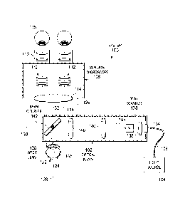
[0027] FIG. 1 illustrates a diagnostic imaging and/or treatment beam
delivery
system 100. The diagnostic imaging and/or treatment beam delivery system 100
can
include at least one light source 104 configured to generate a diagnostic
and/or
treatment light beam. For example, in some embodiments, diagnostic imaging
and/or
treatment beam delivery system 100 can include one light source to generate
the
diagnostic light beam and one light source to generate the treatment light
beam. In
some embodiments, the light source 104 can be configured to generate both the
diagnostic light beam and the treatment light beam. The light source 104 can
be part
of a diagnostic imaging system, such as an OCT imaging system, a multispectral
imaging system, a fluorescence imaging system, a photo-acoustic imaging
system,
6
CA 02949393 2016-11-16
WO 2016/010725
PCT/US2015/038613
etc. For example, the light beam can be part of an OCT scanning beam. The
light
source 104 can have an operating wavelength in the 0.2-1.8 micron range, the
0.7-1.4
micron range, and/or the 0.9-1.1 micron range. The light source 104 can be
part of a
treatment beam delivery system, such as a laser beam delivery system. The
diagnostic imaging system and/or the treatment beam delivery system can
include one
or more additional components (e.g., a beam guidance system, a beam scanner,
etc.).
[0028] The diagnostic imaging and/or treatment beam delivery system 100
can include a beam guidance system, including an optical fiber 106 and/or free
space,
configured to guide the light beam from the light source 104. The diagnostic
imaging
and/or treatment beam delivery system can include a collimator 136 that is
configured
to receive the light beam from the beam guidance system and collimate light.
[0029] The diagnostic imaging and/or treatment beam delivery system 100
can include an optical beam scanner 138 configured to receive the light beam
from the
collimator 136 and/or the beam guidance system, and generate a scanned light
beam
146. For example, the beam scanner 138 can be configured to receive the
diagnostic
light beam from the beam guidance system and to generate a scanned diagnostic
light
beam. The beam scanner 138 can be configured instead or additionally to
receive the
treatment light beam from the beam guidance system and generate a scanned
treatment light beam. The beam scanner 138 can be configured to generate the
scanned light beam 146 having any desired one-dimensional or two-dimensional
scan
patterns, including a line, a spiral, a raster, a circular, a cross, a
constant-radius
asterisk, a multiple-radius asterisk, a multiply folded path, and/or other
scan patterns.
The beam scanner 138 can include one or more of a pair of scanning mirrors, a
micro-
mirror device, a MEMS based device, a deformable platform, a galvanometer-
based
scanner, a polygon scanner, and/or a resonant PZT scanner.
[0030] The diagnostic imaging and/or treatment beam delivery system 100
can also include a beam coupler 142 configured to redirect the scanned light
beam
146 towards a wide field of view (WFOV) lens 120 configured to guide the
redirected
scanned light beam into a target region 124 of a procedure eye 122. The target
region
124 can include the retina, macula/fovea, optic disk, vitreous body, and/or
trabecular
meshwork/Schlemm's canal. The diagnostic imaging and/or treatment beam
delivery
7
CA 02949393 2016-11-16
WO 2016/010725
PCT/US2015/038613
system 100 can be configured to image these and other particular regions-of-
interest
with higher resolution.
[0031] The diagnostic imaging and/or treatment beam delivery system 100
can also include a surgical microscope 108 (FIGS. 1 and 7). An observer 118
can
view the procedure eye 122 through the eyepiece 110 of the surgical microscope
108.
An optical pathway 116 of the surgical microscope 108 can include one or more
focusing/zoom lenses of the eyepiece 110, one or more focusing/zoom lenses 112
of
the microscope body, and an objective lens 114.
[0032] The beam coupler 142 can be configured to redirect the scanned
light
beam 146 into the optical pathway 116 of the surgical microscope 108. To
redirect
the scanned light beam 146 into the target region 124 of the procedure eye 122
and/or
the optical pathway 116 of the surgical microscope, the beam coupler 142 can
include
a mirror. As shown in FIGS. 1-6 and 8a-9b, the mirror can be tilted such that
it is
oriented at an oblique angle with respect to each of the scanned light beam
146 and
the optical pathway 116 of the surgical microscope 108. The beam coupler 142
can
include a dichroic mirror, a notch filter, a hot mirror, a beamsplitter and/or
a cold
mirror. The beam coupler 142 can be configured to combine the visible beam of
the
microscope 108 with the scanned light beam 146. As a result, the field of view
of the
scanned light beam 146 and the microscope 108 can overlap completely, overlap
partially, or not overlap at all. The beam coupler 142 can be configured to
reflect the
scanned light beam 146 and/or reflections from the procedure eye 122 in the
wavelength range of the scanned light beam 142 while allowing the visible beam
of
the microscope 108 to pass therethrough.
[0033] The beam scanner 138 and/or the optical block 102 can also
include
focusing optics for defining a depth of focus of the scanned light beam 146.
For
example, one or more lenses 140 can be included within the optical block 102
(FIGS.
1-6 and 8a-9b). When present, the focusing optics of the beam scanner 138
and/or
the optical block 102 can be fixed or adjustable. Focusing optics or zoom
lenses
within the beam scanner 138 and/or optical block 102 can facilitate scanning
of a
region of interest with increased resolution and depth-of-field. The focusing
optics
and/or zoom lenses can be provided at one or more of the following locations:
between the beam coupler 142 and the surgical microscope 108; between the beam
8
CA 02949393 2016-11-16
WO 2016/010725
PCT/US2015/038613
coupler 142 and the WFOV lens 120; between the beam coupler 142 and the beam
scanner 138; and between the beam scanner 138 and the light source 104.
Focusing
optics and/or zoom lenses positioned between the beam coupler 142 and the
surgical
microscope 108 can be configured to adjust the focus of the optical pathway
116 of
the surgical microscope 108. Focusing optics and/or zoom lenses positioned
between
the beam coupler 142 and the beam scanner 138 or between the beam scanner 138
and
the light source 104 can be configured to adjust the focus of the scanned
light beam
146. Focusing optics and/or zoom lenses positioned between the beam coupler
142
and the WFOV lens 120 can be configured to adjust the focus of both the
optical
pathway 116 of the surgical microscope 108 and the scanned light beam 146.
[0034] The lens(es) 140 can be adjusted by a zoom-controller to adapt an
optical power of the diagnostic imaging and/or treatment beam delivery system
100 to
the desired target region 124 of the procedure eye 122. Further, the
adjustable zoom
lens(es) 140 can be controlled by the zoom-controller in real-time to adapt
the optical
power of the diagnostic imaging and/or treatment beam delivery system 100 to
keep
an aberration below a predetermined value as the scanned light beam 146 scans
across
the target region 124 of the procedure eye 122. In that regard, the zoom-
controller
can control each adjustable zoom lens 140 by adjusting a physical position of
the
zoom lens 140 (e.g., using piezo-electric or other suitable actuators) and/or
adjusting
an optical power of the zoom lens 140 without adjusting the physical position
of the
zoom lens 140 (e.g., by varying a voltage supplied to a liquid crystal zoom
lens).
[0035] In some embodiments, the diagnostic imaging and/or treatment beam
delivery system 100 can include a visible guidance beam, such as when the
scanned
light beam 146 is outside of the visible range. For example, the scanned light
beam
146 can be in the infrared range. As shown in FIG. 2, the diagnostic imaging
and/or
treatment beam delivery system 100 can include a guidance beam source 154
configured to generate the visible guidance beam. The visible guidance beam,
via an
optical fiber 106, can be coupled into the surgical imaging and/or beam
delivery
system 100 using a coupler, wavelength division multiplexer (WDM), or beam
splitter
156. The coupler, WDM, or beam splitter 156 can be positioned before the beam
scanner 138. The beam coupler 142 can be configured to reflect at least a
portion of a
visible guidance beam coincident with the scanned light beam 146 to facilitate
9
CA 02949393 2016-11-16
WO 2016/010725
PCT/US2015/038613
visualization of the scanned light beam 146. For example, the beam coupler 142
can
include a notch filter in the wavelength range of the visible guidance beam
such that
the visible guidance beam can be reflected by beam coupler 142 along with the
scanned light beam 146 to reach the procedure eye 122.
[0036] Referring again to FIG. 1, in some embodiments, the diagnostic
imaging and/or treatment beam delivery system 100 can include an integrated
optical
block component 102. The optical block 102 can include one or more optical
elements integrated into a common component, such as a hand-held device, a
lens
holder, an adapter, or other component. The optical block 102 can be a
consumable
product configured for use in a single surgical procedure or reusable in
multiple
surgical procedures. The optical block 102 can be independently positionable
relative
to the surgical microscope 108 and/or the procedure eye 122. FIGS. 1-7 and 8a-
9b
illustrate various embodiments of the optical block 102. For example, the
optical
block 102 can include the optical beam scanner 138, the beam coupler 142, one
or
more lenses 140, the collimator 136, etc. In various embodiments, the optical
block
102 can include more or fewer components. The optical block 102 can be in
optical
communication with the light source 104 via the optical fiber 106. The optical
block
102 can include a fiber holder 134 where the optical fiber 106 is mechanically
received in the optical block 102.
[0037] The beam coupler 142 and/or the optical block 102 can be operated
with or without a defined optical/optomechanical relationship to the surgical
microscope 108. For example, the beam coupler 142 or the optical block 102 can
be
maintained separate from and independently positionable relative to the
surgical
microscope 108. In such instances, the beam coupler 142 can be a hand-held
device,
a lens holder, a self-stabilized component or other component. As shown in
FIG. 3,
the optical block 102 can be coupled to a support arm 152. The support arm 152
can
be stationary, such as when the support arm 152 is wall-mounted. The support
arm
152 can be movable, such as when the support arm 152 is mounted on a movable
pole
or cart. As described with respect to FIGS. 7-9b, the optical block 102 can be
coupled to the support arm 152 such that optical block 102 is movable with six
degrees of freedom (e.g., three rotational degrees of freedom and three
translation
CA 02949393 2016-11-16
WO 2016/010725
PCT/US2015/038613
degrees of freedom) relative to the surgical microscope 108 and/or the
procedure eye
122.
[0038] Referring again to FIG. 1, the beam coupler 142 and/or the
optical
block 102 can be coupled to the surgical microscope 108, directly or
indirectly, such
that it has a defined optical/optomechanical relationship to the surgical
microscope.
For example, direct or indirect coupling 126 between the optical block 102 and
the
surgical microscope 108 can include one or more of a suspension system, a
mechanical frame, a protruding arm, a conical structure, a magnetic member, an
elastic member, and a plastic member. The WFOV lens 120 can be independently
manipulable relative to the procedure eye 122 by a lens-holder¨instead of the
beam
coupler 142¨when the beam coupler 142 is coupled to the surgical microscope in
a
defined optical/optomechanical relationship. As described with respect to
FIGS. 7-
9b, the optical block 102 can be coupled to the surgical microscope 108 such
that the
optical block 102 is movable with six degrees of freedom (e.g., three
rotational
degrees of freedom and three translation degrees of freedom) relative to the
surgical
microscope 108 and/or the procedure eye 122.
[0039] Referring again to FIG. 1, the WFOV lens 120 of the diagnostic
imaging and/or treatment beam delivery system 100 can be configured to provide
a
field of view of the procedure eye 122 greater than 15 degrees, greater than
30
degrees, greater than 45 degrees, greater than 60 degrees, greater than 80
degrees
and/or greater than 100 degrees. Accordingly, the diagnostic imaging and/or
treatment beam delivery system 100 can be configured to provide various field
of
view ranges, such as between 0 degrees and 30 degrees, between 15 degrees and
80
degrees, between 30 degrees and 120 degrees, and/or other desired ranges up to
ora
serrata within the field of view of the WFOV lens 120. The WFOV lens 120 can
be
configured to provide the desired refractive power for the diagnostic and/or
treatment
procedures to be performed on the procedure eye 122.
[0040] The WFOV lens 120 can be configured to operate in contact with
the
procedure eye 122, as a contact lens, or spaced from the procedure eye 122, as
a non-
contact lens. As shown in FIG. 4, the contact lens can be a macular lens 144
configured to be contacted to the procedure eye 122. A macular lens 144 can be
embedded in a stabilizing mechanism, where the stabilizing mechanism can be
11
CA 02949393 2016-11-16
WO 2016/010725
PCT/US2015/038613
configured to stabilize the macular lens 144 relative to the procedure eye
122. To that
end, the stabilizing mechanism can include one or more of a trocar, a counter
weight,
a friction-based system, and an elastic system. In some embodiments, the WFOV
lens
120 can be separate from, but attachable to the optical block 102.
[0041] As shown in FIGS. 5 and 6, the non-contact WFOV lens can be an
ocular lens 148 that is spaced from the procedure eye 122. An intermediate
image
plane 150 between the beam coupler 142 and the ocular lens 148 can be
generated in
embodiments of the diagnostic imaging and/or treatment beam delivery system
100
including the ocular lens 148. The ocular lens 148 can be configured to
operate in a
manner similar to a binocular indirect ophthalmomicroscope (BIOM). The ocular
lens 142 can be positioned by one or more of a mechanical coupling to the beam
coupler 142, the optical block 102, a mechanical coupling to the surgical
microscope
108, a suspension system, and a lens holder. In some embodiments, the WFOV
lens
120 can be one optical element of the optical block 102. The optical block
102, the
beam coupler 142, and/or the ocular lens 148 can be moved as described with
respect
to FIGS. 7-9b to change the incidence angle and/or the target location of the
scanned
light beam 146 in the procedure eye 122.
[0042] The light source 104, the beam guidance system, and the beam
scanner
138 can be part of an optical coherence tomographic (OCT) imaging system. To
that
end, the WFOV lens 120 and the beam coupler 142 can be configured to guide a
returned image light from the target region 124 of the procedure eye 122 back
to the
OCT imaging system. The returned image light can be interfered with a
reference
beam of the OCT imaging system, and from the interference an OCT image of the
target region in a range of depths can be generated and displayed to a user.
The
diagnostic imaging and/or treatment beam delivery system can be configured to
generate the imaging information based on processing the returned image light
in less
than 30 seconds, less than 10 seconds, and/or less than 5 seconds, including
in real
time. A single scanned light beam 152 or A-scan is shown in FIG. 6. The single
scanned light beam 152 can be focused at a particular location along the
target region
124 within the procedure eye 122. Multiple A-scans can be performed within the
target region 124 to generate the larger field of view illustrated FIGS. 1-5
and 8a-9b.
As described with respect to FIGS. 7-9b, the optical block 102, the beam
coupler 142,
12
CA 02949393 2016-11-16
WO 2016/010725
PCT/US2015/038613
and/or the WFOV lens 120 can be selectively moved such that the diagnostic
imaging
and/or treatment beam delivery system 100 has an adjustable field of view. The
returned image light from individual A-scans with different incidence angles
and/or
incidence locations can be processed and combined to generate combined imaging
formation (e.g., cross-sectional and/or volumetric OCT data).
[0043] FIG. 7 illustrates the diagnostic imaging and/or treatment beam
delivery system 100, including rotational and/or translational motion of the
optical
block 102. The beam coupler 142 and/or the optical block 102 can be movable
such
that the diagnostic imaging and/or treatment beam delivery system 100 has an
adjustable, wide angle scanning. For example, the field of view of the scanned
light
beam 146 can cover a changeable region of the procedure eye 122. The wide
angle
scanning or field of view can be adjusted as the incidence angle and/or
incidence
location of the scanned light beam 146 within the procedure eye 122 changes
based
on translation and/or rotation of the beam coupler 142 and/or the optical
block 102
relative to the surgical microscope 108 and/or the procedure eye 122. In some
embodiments, movement of the optical block 102 includes movement of the beam
coupler 142. In some embodiments, movement of the beam coupler 142 is
independent of movement of the optical block 102. In embodiments of the
diagnostic
imaging and/or treatment beam delivery system 100 including a non-contact WFOV
lens, the ocular lens 148 (FIGS. 5 and 6) can be moved along with the beam
coupler
142 and/or the optical block 102. The beam coupler 142 and/or the optical
block 102
can be selectively moved to scan in the periphery of the procedure eye 122.
The
beam coupler 142 and/or the optical block 102 can be selectively moved to scan
the
trabecular meshwork or Schlemm's canal of the procedure eye 122. Further, the
beam coupler 142 and/or the optical block 102 can be positioned such that the
field of
view of the scanned light beam 146 and the field of view of the visible beam
of the
microscope do not overlap, partially or entirely overlap.
[0044] Selective movement of the beam coupler 142 and/or the optical
block
102 can be configured to provide a field of view of the procedure eye 122
greater than
15 degrees, greater than 30 degrees, greater than 45 degrees, greater than 60
degrees,
greater than 80 degrees and/or greater than 100 degrees. Accordingly, the
diagnostic
imaging and/or treatment beam delivery system 100 can be configured to provide
13
CA 02949393 2016-11-16
WO 2016/010725
PCT/US2015/038613
various field of view ranges, such as between 0 degrees and 30 degrees,
between 15
degrees and 80 degrees, between 30 degrees and 120 degrees, and/or other
desired
ranges up to ora serrata.
[0045] In some embodiments, the optical block 102 can be movable with
one,
two, three, four, five, six, or more degrees of freedom. For example, the
optical block
102 can have one, two, three, or more rotational degrees of freedom. A first
rotational
degree of freedom can be about an axis 128 or z-axis (FIGS. 1 and 7). As shown
in
FIG. 1, the optical block 102 can rotate about the axis 128 into and out of
the plane of
the page. The axis 128 can be central axis of the surgical microscope 108. As
shown
in FIG. 7, the optical block 102 can rotate in directions 162 and 164 about
the axis
128. By itself, rotation about the axis 128 does not change the incidence
angle and/or
the incidence location of the scanned light beam 146 in the procedure eye 122.
However, rotation about the axis 128 provides flexibility to the observer 118,
such as
a surgeon, to move the optical block 102 to a more convenient orientation
during the
surgical procedure. For example, the observer 118 can rotate the optical block
102
based on the how the patient is positioned, which eye is being operated on,
etc.
[0046] A second rotational degree of freedom can be about an axis 132 or
y-
axis (FIGS. 1 and 7). Rotation about the axis 132 can be described as tilting
the
optical block 102. As shown in FIG. 1, the axis 132 extends into and out of
the page,
and the optical block 102 can rotate about the axis 132 in the plane of the
page. As
shown in FIG. 7, the optical block 102 can rotate in directions 182 and 184
about the
axis 132. While the axis 132 is shown as extending through the beam coupler
142,
the axis can be positioned anywhere along the optical block 102 such that the
axis is
parallel to the axis 132 shown in FIGS. 1 and 7. Rotation about the axis 132
can
change the incidence angle and/or the incidence location of the scanned light
beam
146 in the procedure eye 122. Depending on how the patient is positioned
relative to
the optical block 102, rotation about the axis 132 can shift the scanned light
beam 142
to the left, to the right, up, or down in the procedure eye 122. As shown in
FIG. 8a,
the optical block 102 can be rotated in the direction 182 about the axis 132.
As a
result, the scanned light beam 146 can be shifted to the left of the target
region 124.
As shown in FIG. 8b, the optical block 102 can be rotated in the direction 184
about
14
CA 02949393 2016-11-16
WO 2016/010725
PCT/US2015/038613
the axis 132. As a result, the scanned light beam 146 can be shifted to the
right of the
target region 124.
[0047] A third rotational degree of freedom can be about an axis 130 or
x-
axis (FIGS. 1 and 7). As shown in FIG. 7, the optical block 102 can rotate in
directions 172 and 174 about the axis 132. Rotation about the axis 130 can
change
the incidence angle and/or the incidence location of the scanned light beam
146 in the
procedure eye 122. Depending on how the patient is positioned relative to the
optical
block 102, rotation about the axis 130 can shift the scanned light beam 142 to
the left,
to the right, higher, or lower in the procedure eye 122. For example, rotation
in the
direction 172 can shift the scanned light beam 146 to the right in procedure
eye.
Rotation in the direction 174 can shift the scanned light beam 146 to the left
in the
procedure eye.
[0048] For example, the optical block 102 can have one, two, three, more
translational degrees of freedom. A first translational degree of freedom can
be along
the axis 128. As shown in FIG. 1, the optical block 102 can translate along
the axis
128 in the plane of the page. As shown in FIG.7, the optical block 102 can
translated
in the directions 166 and 168, along the axis 128. Translation along the axis
128 can
adjust a focusing depth of the scanned light beam 146 on the target region 124
of the
procedure eye 122.
[0049] A second translational degree of freedom can be along the axis
132.
As shown in FIG. 1, the optical block 102 can translate along axis 132 into
and out of
the page. As shown in FIG. 7, the optical block 102 can be translated in the
directions 186 and 188, along the axis 132. Translation along the axis 132 can
change
the incidence angle and/or the incidence location of the scanned light beam
146 in the
procedure eye 122. Depending on how the patient is positioned relative to the
optical
block 102, translation along the axis 132 can shift the scanned light beam 146
to the
left, to the right, higher, or lower in the procedure eye 122. For example,
translation
in the direction 186 can shift the scanned light beam 146 to the left in the
procedure
eye 122. Translation in the direction 188 can shift the scanned light beam 146
to the
right in the procedure eye 122.
[0050] A third translational degree of freedom can be along the axis
130. As
shown in FIG. 1, the optical block 102 can translate along axis 130 in the
plane of the
CA 02949393 2016-11-16
WO 2016/010725
PCT/US2015/038613
page. As shown in FIG. 7, the optical block 102 can be translated in the
directions
176 and 178, along the axis 130. Translation along the axis 130 can change the
incidence angle and/or the incidence location of the scanned light beam 146 in
the
procedure eye 122. Depending on how the patient is positioned relative to the
optical
block 102, translation along the axis 130 can shift the scanned light beam 142
to the
left, to the right, higher, or lower in the procedure eye 122. For example,
translation
in the direction 176 can shift the scanned light beam 146 to the left of the
target
region 124. Translation in the direction 178 can shift the scanned light beam
146 to
the right of the target region 124.
[0051] In some embodiments, movement of the optical block 102 can
include
only rotation or only translation. In some embodiments, movement of the
optical
block 102 can include both rotation and translation. The optical block 102 can
be
rotated about and/or translated along one or more of the axes 128, 130, and
132 to
provide an adjustable wide field of view for the diagnostic imaging and/or
treatment
beam delivery system 100. The optical block 102 can be translated in one or
more
directions and then rotated in one or more directions, or vice versa, in order
to direct
the scanned light beam 146 into the target region 124 (and prevent the scanned
light
beam 146 from encountering interference with, e.g., the iris). For example,
the
optical block 102 can be moved based on the visible guidance beam (FIG. 2) to
scan
desired locations of the target region 124. Rotation and/or translation of the
optical
block 102 can be achieved manually (e.g., by physical manipulation by the
surgeon)
or automatically (e.g., by one or more motorized actuators controlled by a
controller
of the surgical imaging and/or beam delivery system 100). A contact WFOV lens
(e.g., macular lens 144) can maintain a fixed orientation relative to the
procedure eye
114 during translation and/or rotation of the optical block 102. A non-contact
WFOV
lens (e.g., ocular lens 148) can translate and/or rotate along with optical
block 102.
[0052] In some embodiments, the beam coupler 142 can be rotatable
relative
to the procedure eye 122 and/or the microscope 108. Rotation of the beam
coupler
142 can be independent of movement of the optical block 102. In that regard,
rotation of the beam coupler 142 can be utilized to facilitate full
circumferential
scanning of the procedure eye 122 and/or to target a particular region of
interest
within the procedure eye 122. The beam coupler 142 can be rotatable about the
axis
16
CA 02949393 2016-11-16
WO 2016/010725
PCT/US2015/038613
132 (FIGS. 1 and 7) or an axis parallel to the axis 132. As shown in FIG. 9a,
rotation
of the beam coupler 142 in the direction 182 can shift the scanned light beam
146 to
the left of the target region 124. As shown in FIG. 9b, rotation of the beam
coupler
142 in the direction 184 can shift the scanned light beam 146 to the right of
the target
region 124. Rotation of the beam coupler 142 can be achieved manually (e.g.,
by
physical manipulation by the surgeon) or automatically (e.g., by one or more
motorized actuators controlled by a controller of the diagnostic imaging
and/or
treatment beam delivery system 100). A contact WFOV lens (e.g., macular lens
144)
can maintain a fixed orientation relative to the procedure eye 122 during
translation
and/or rotation of the beam coupler 142. A non-contact WFOV lens (e.g., ocular
lens
148) can translate and/or rotate along with the beam coupler 142.
[0053] FIG. 10 illustrates a method 200 of operating a wide-angle
ophthalmic
surgical system, such as a diagnostic imaging system and/or a treatment beam
delivery system. The diagnostic imaging system can be, for example, an optical
coherence tomography (OCT) visualization system. The method 200 can be further
understood with reference to FIGS. 1-9b. The method 200, at step 210, can
include
generating a diagnostic and/or treatment light beam using a light source. For
example, the light beam can be generated using the light source 104. The
method
200, at step 220, can include guiding the light beam from the light source to
a beam
scanner using a beam guidance system. The example, the beam guidance system
can
include the optical fiber 106 to guide the light beam from the light source
104 to the
beam scanner 138. The method 200, at step 230, can include generating a
scanned
light beam using the beam scanner. For example, the scanned light beam 146 can
be
generated using the beam scanner 138. The method 200, at step 240, can include
redirecting the scanned light beam using a beam coupler. Redirecting the
scanned
light beam can include redirecting the scanned light beam into an optical
pathway of a
surgical microscope. For example, the scanned light beam 146 can be redirected
using the beam coupler 142. The beam coupler 142 can redirect the scanned
light
beam 146 into the optical pathway 116 of the microscope 108. The method 200,
at
step 250, can include guiding the redirected scanned light beam into a target
region of
a procedure eye using a wide field of view (WFOV) lens. For example, the WFOV
lens 120 can be used to guide the scanned light beam 146 into the target
region 124 of
the procedure eye 122. The method 200, at step 260, can include selectively
moving
17
CA 02949393 2016-11-16
WO 2016/010725
PCT/US2015/038613
the beam coupler and/or the optical block to change at least one of an
incidence angle
of the redirected scanned light into the procedure eye and the target location
of the
procedure eye. For example, the beam coupler 142 and/or the optical block 102
can
be translated and/or rotated to change the incidence angle and/or target
location of the
scanned light beam 146 in the procedure eye 122.
[0054] In some embodiments, moving the beam coupler (step 260) can
include rotating the beam coupler. For example, the beam coupler 142 can be
rotated
about at least of one of a first axis, a second axis, and a third axis (e.g.,
axes 132,128
and 130). In some embodiments, moving the beam coupler (step 260) can include
rotating the optical block about at least one of a first axis, a second axis,
and a third
axis (e.g., axes 128, 130, and 132) and/or translating the optical block along
at least
one of the first axis, the second axis, and the third axis (e.g., axes 128,
1302, 132). In
some embodiments, the method 200 can include repeating the moving step to
generate
imaging information associated with different incidence angles and/or
different target
locations in the procedure eye and combining the imaging information
associated to
generate combined imaging information. For example, OCT data can be generated
at
various incidence angles and/or target locations. The OCT data from the
individual
angles and/or target locations can be combined or stitched together through
one or
more processing steps to generate OCT data for a wider field of view (e.g., a
cross-
sectional and/or volumetric scan). For example, a treatment beam can be
delivered to
various incidence angles and/or target locations.
[0055] Embodiments as described herein can provide devices, systems, and
methods that facilitate real-time, intra-surgical, adjustable wide-angle beam
scanning
for diagnostic imaging and/or treatment beam delivery. The examples provided
above
are exemplary only and are not intended to be limiting. One skilled in the art
may
readily devise other systems consistent with the disclosed embodiments which
are
intended to be within the scope of this disclosure. As such, the application
is limited
only by the following claims.
18