Note: Descriptions are shown in the official language in which they were submitted.
CA 02949778 2016-11-21
WO 2015/157447 PCT/US2015/024970
1
DEVICE FOR BIOSENSING WITH INDWELLING VENOUS CATHETER
Background
[1] Currently, sedation is monitored with vital signs and sometimes with
brain activity
(a BIS monitor ¨ bi-spectral index - measures awareness, but is not very
reliable).
Without knowing actual blood concentrations of anesthetics, anesthesiologists
tend to
give a bolus of anesthetics at the beginning of surgery. This sometimes
results in
patients being under for longer than necessary, which requires the patient to
stay in the
hospital for longer. Sometimes patients metabolize anesthetics quickly and
begin to
wake up during the surgery, also not ideal.
[2] Anesthesiologists regularly struggle with dosing decisions because, as
they
watch blood pressure as an indicator of dosing, it is unclear whether pressure
has
changed due to change in blood volume (blood loss or transfusion), or if
vasodilation or
vasoconstriction has occurred. During a surgery, significant blood is lost,
which is not
calculated (and is incalculable). The anesthesiologist estimates how many
units of
blood to give, and then watched the blood pressure to decide if blood supply
has been
adequately replenished. Deciding to dose anesthetics or give more blood based
on
blood pressure can be the most difficult and common judgment moment for
anesthesiologists. This decision is a judgment call because the total
circulating blood
volume is unknown. Accordingly, it would be a very significant value to know
total blood
volume at critical moments during surgery.
[3] Other possible applications exist in the ER and ICU, where a time-
sensitive
measurement of blood volume could add significant value to patient care.
Additionally,
there is an outpatient test that takes about 1 hour for the diagnosis of
chronic fatigue
syndrome, anemia, among other blood/RBC disorders. In this test a number of
blood
samples are collected over time and sent to the lab, resulting in inevitable
delays.
Patient care would be significantly improved by real-time monitoring.
[4] A typical IV catheter consists of a catheter (small flexible tube)
which is placed
into a vein using a needle. The catheter forms a sheath around the needle. The
needle
offers the rigidity and a sharp edge to introduce the catheter into the vein;
after which
SUBSTITUTE SHEET (RULE 26)
CA 02949778 2016-11-21
WO 2015/157447 PCT/US2015/024970
2
the needle is removed leaving the catheter (which is soft and flexible) in the
vein. Using
such a catheter, IV fluids can be pumped into the blood.
[5] A biosensor in the blood stream of a subject is subject to a number of
forces that
could cause it to fail: too rapid flow (not enough time for molecules to stick
to
biosensor), shear forces, biofouling by clotting factors, non-specific signal
due to large
proteins that stick to the surface and exclude the target molecule.
Brief Description of the Drawings
[6] Figure 1 shows a side view (A), cut-away (B) and cross-section (C and
D) views
of one embodiment of a needle or catheter having exclusionary slits and
sensors in
accordance with the present invention.
[7] Figure 2 is a side view of a tapered tip needle with built-in biosensor
(blue),
showing blood flow (red) and buffer flow (yellow).
[8] Figure 3 is a cross-section view of the needle of Figure 2.
[9] Figure 4 is a side view of a needle with retractable biosensor (blue),
showing
blood flow (red) and buffer flow (yellow).
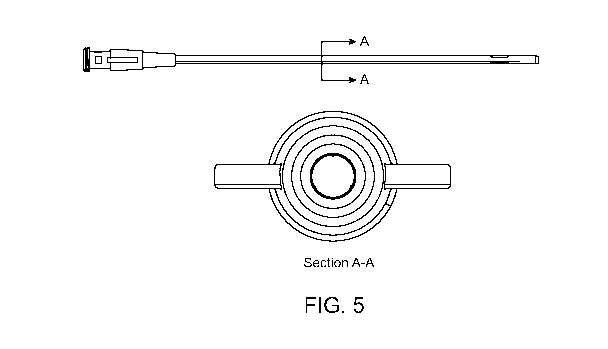
[10] Figure 5 shows a needle embodiment, and a cross-section showing possible
position of conductors within the needle.
[11] Figure 6 is a detail view of a Luer lock connector mechanism
comprising built-in
conductor pads.
Detailed Description
[12] Described herein is a biosensor that can be placed in the blood stream
while
protected from red blood cells, clotting factors, and shear force. This device
also allows
the flow past the sensor surface to be tuned to the requirements for the
binding kinetics
of the biosensor. Additionally, as some biosensing elements are degradable in
vivo
(due to innate immune response/encapsulation, nuclease degradation of
aptamers,
CA 02949778 2016-11-21
WO 2015/157447 PCT/US2015/024970
3
etc.), this device serves to exclude some of the potential biosensor-degrading
elements
in physiological samples.
[13] Also described herein is a tool that may be used to continuously monitor
the
concentrations of various drugs or biomarkers (e.g., chemotherapeutic levels)
in the
blood using catheter or needle bearing at least one aptamer biosensor as
described
herein. The catheter or needle is specifically designed to permit prolonged
monitoring in
blood while avoiding biofouling of the sensor through a boundary layer of
buffer flowing
past the sensor.
[14] In one embodiment, one or more conductors are placed on the inside of a
catheter or needle. These conductors can act as the electrodes for the
sensor(s) (e.g., a
counter, working and reference electrode). A buffer may be made to flow
through the
catheter or needle, thereby prolonging the working lifetime of the sensor.
There are thin
slits in the wall of the catheter or needle, which allow for the diffusion of
blood onto the
sensor. Such a device is shown in Figure 5. Other metal contacts located
elsewhere in
the body besides the catheter may also be used as the counter or reference
electrodes
for this sensor system.
[15] As used herein, the term "subject" means a human or other organism with a
circulatory system into which the device described herein may be inserted.
[16] The device described herein comprises three main elements. As shown in
Figure 1, the first main element is, one or more sensors 103 either placed or
imbedded
in either a plastic catheter (including catheters for IV administration,
peripherally
inserted central catheters 10 (PICC), or central venous line catheters) or a
stainless
steel needle 10 (or needle 10 of another material) or combination of the two.
The
catheter or needle will have a "distal" end, which is the end inserted in the
subject, and
a "proximal" end, which is the end to which tubing, a syringe, and/or wiring
may be
connected. A sensor 103 for use in the present device can be an aptasensor,
enzyme
sensor, antibody sensor, or may use an engineered protein, polymer or
biospecific
element. The readout of this sensor may be via an ion-sensitive field-effect
transistor
("ISFET"), impedimetric, amperometric or other electrochemical method, a
CA 02949778 2016-11-21
WO 2015/157447 PCT/US2015/024970
4
micromechanical or other sensor readout modality. The sensor(s) may be located
at or
near the distal end of the catheter or needle.
[17] The second main element, as shown in Figure 1, is a plurality of
exclusionary
slits 101 in the catheter, needle, or combination, upstream of the sensor(s),
and angled
so that fluid flows in through the slits and down past the sensor(s) 103 in
the direction of
the arrow. The size of these slits 101 will depend on the specific molecule(s)
to be
sensed. For example, if a small organic molecule (-500 Da) is the target
molecule, or
several are the targets, then the slits can be small enough to exclude all
proteins and
cells. If a protein is the target, then slit sizes can be adjusted to exclude
cells, and
possibly larger proteins. Slit dimensions, orientation, and placement can be
altered to
optimize the flow through the lumen 100 of the catheter 10. Slit design may
facilitate
reducing flow past the sensing element(s) 103 in the event that the kinetics
of the
sensor require incubation time. Slit design may be altered depending on
eventual in vivo
location of a given sensor. For example, the design for a central catheter may
be
distinct from the design for a peripheral catheter.
[18] The third main element is the wiring of the sensor(s). The wiring 110 may
be
embedded in the wall of the catheter or needle, or disposed along the inner
wall of the
catheter or needle, within the lumen. Wiring will allow for signal
transduction. Wiring
can either be traditional insulated wires, polyimide thin flex, or the like.
The use of a
flexible wiring connector allows fitting more connections in the catheter or
needle.
[19] Optionally, heparin, warfarin, low molecular weight heparin,
riveroxiban, or other
anticoagulant drugs can be impregnated in parts of the catheter 102 to reduce
the risk
of occlusion of the slits by clotting factors and/or proteins. Similarly,
catheter material
can be impregnated with antibiotics, such as rifampicin, clindamycin,
aminoglycosides,
or tetracycline, to reduce the risk of infection. Other options such as a
mechanical
cleaning device or delivery of current could be used to unclog the slits 101.
[20] An additional technique that may be used to prevent occlusion of the
slits and
exclude large molecules that might foul the biosensor is the placement of
microfilter
membrane over the slits 101. Semipermeable membranes and microfilters can
further
CA 02949778 2016-11-21
WO 2015/157447 PCT/US2015/024970
limit the size of molecules that enter the catheter beyond the size of the
slits. For
example, 5pm filter (has 5pm-sized holes) can exclude 8-10 pm red blood cells
from
entering into the catheter. The semipermeable membranes used on microdialysis
probes are examples of materials that could be used to modify the exclusionary
properties of the slits and membranes.
[21] Also disclosed herein is a method for detection of small molecules using
the
device described above.
[22] Injection of a very small amount of intravenous ("IV") fluid can be
used to act as a
fluid barrier to prevent blood cells and proteins from fouling the biosensor
surface.
Specifically, IV fluid flowing through the lumen 100 over the sensor(s) 103 at
a slow rate
(as low as about 0.25 mL/hour to about 5 mL/hour)) may be used to prevent the
buildup
of blood-borne biofouling agents. Fluid flow around the sensor(s) 103 would
still allow
for small molecules like drug, such as doxorubicin or aminoglycosides, to
diffuse to the
surface of the sensor. Similarly, small proteins could diffuse to the surface.
Other small
molecules (and other small proteins) would also diffuse to the sensor, but are
unlikely to
cause biofouling or nonspecific signal. The rate of IV fluid delivery can to
influence the
rate of diffusion and size of molecules allowed to diffuse to the sensor
surface.
[23] The method described herein can be used either with or without the
mechanical
protection of a catheter around the sensor. Fluid flow around the sensor can
be
adjusted such that the fluid acts as the only barrier between the sensor and
the external
environment. By modulating speed of fluid flow around the sensor, the sensor
can be
refreshed in the event that aptamer/enzyme/antibody kinetics do not allow for
rapid
enough equilibration.
[24] IV fluid may be injected either through the catheter that holds the
sensor
(catheter has a number of slits to allow influx of target molecule (see
above)) or IV fluid
is delivered directly next to a wire-like sensor so that fluid flows along the
wire, encasing
it. In addition, IV fluid may be designed for improved sensor function (i.e.,
ion
concentrations, pH, or other common additions such as glucose, within
clinically
accepted guidelines and commonly used IV solutions). While some IV fluids may
CA 02949778 2016-11-21
WO 2015/157447 PCT/US2015/024970
6
demonstrate preferable sensor performance, each sensor will be characterized
for the
range of clinically used fluids.
[25] The device may further comprise a null electrode sensor. The null
electrode
sensor is created by another conductor in the catheter or needle that is not
treated with
the sensor recognition element. This electrode will serve to calibrate the
functioning
biosensor for any degradation or change in baseline signal that may occur in
vivo, as a
result of a physiological change (such as blood pH shift) or biofouling. The
null sensor
may be covered in a similar bio-recognition material that is not sensitive to
the target (or
any molecule found in blood), but that indicates the baseline of a 0 M
concentration
signal for the target-binding sensor. It may also be bare or have a different
coating that
still serves as an indicator of behavior of the sensor. A null electrode may
be employed
to indicate any fluctuation in signal associated with changes in composition
of the IV
fluid being delivered and/or any changes in the baseline signal due to minor
degradation
caused by shear forces, biofouling, or other sensor degradation. An
alternative method
for monitoring changes in signal baseline comprises applying an
electrochemical
measurement method to the sensor electrode that is insensitive to changes in
target
molecule concentration. For example, in square wave voltammetry testing, some
frequencies demonstrate sensitivity to changes in concentration, whereas
others do not.
[26] Modulation of fluid flow may be used to "refresh" the sensor in the event
that
sensor does not release the target molecule well unless in a target-free
solution (i.e., by
speeding up fluid flow so that target diffusion is reduced during "refresh"
periods).
[27] The design of a catheter housing the sensor may be tuned and optimized in
order to introduce a boundary (sheath) layer of buffer that will be
immediately adjacent
to the sensor element, preventing biofouling by cells and large proteins and
other
molecules, while allowing the smaller analytes of interest to diffuse to the
sensor. For
example, the catheter may be gradually tapered using a variety of profiles (an
example
of which is shown in Figure 2) that result in a thin layer of laminar flow out
the distal
opening of the catheter. Such a design may be enhanced by features such as
guides or
internal features, reductions and enhancements in inner tube diameter, which
are
CA 02949778 2016-11-21
WO 2015/157447 PCT/US2015/024970
7
known to aid in the transition from turbulent to laminar flow, or to change
the cross
sectional profile of a laminar flow stream. The design of the exclusions slits
described
elsewhere might also be optimized to introduce desired characteristics (thin
laminar
flow).
[28] The use of a buffer fluid boundary layer to reduce the effects of
biofouling on a
biosensor relies on the careful control of flow conditions such as flow rate,
pressure,
and the degree of turbulence. Several design features in the subject invention
are
incorporated to exert control over these parameters. A miniature MEMS (micro-
electromechanical systems) regulator can be placed in-line with the flow, in
order to
control the flow rate through the device. Likewise a restrictor or flow
orifice may be used
for the same purpose. The degree of turbulence (less turbulent, or more
laminar, flow is
desired for effective diffusion control) in the device may be controlled not
only by setting
the flow rate to appropriate levels, but also by incorporating features into
the flow
channel that are specifically designed to produce laminar flow. Examples
include
converging and diverging flow areas, micro-structured surface features
incorporated into
the lumen sidewall, and bundles of parallel tubes, honeycomb structures,
meshes and
nozzles. Other features of the subject invention which are incorporated in
order to
control aspects of the desired flow include slots, slits, or one or more holes
in the tube
sidewall, allowing control of the way in which blood flow is introduced into
the laminar
buffer fluid stream. A multi-lumen tube could also be used where blood is
admitted into
the inner lumen (by strategically placed slits/slots/holes on the wall of the
tube) and a
sheath flow of buffer is then formed around the blood. This sheath flow of
buffer over
the sensor reduces biofouling because the blood will have to diffuse through
the sheath
flow and make it way to the sensor.
[29] A catheter or needle according to the present invention may have a single
lumen,
or two or more lumens. In multiple-lumen embodiments, the lumens may be
concentric,
or may divide the lumen into sections. For example, one such double lumen
design
divides a circular lumen into two half circles. A double-lumen design may be
used, for
example, to separate sensors that are measuring an analyte that is being
delivered in
the IV fluid. For example, in order to measure blood stream glucose
accurately, and
CA 02949778 2016-11-21
WO 2015/157447 PCT/US2015/024970
8
prevent the signal from being affected by the concentration of glucose in the
IV fluid, the
sensors may be isolated from the glucose IV fluid by being in a separate
lumen. Non-
glucose IV fluid would then be required to flow through the sensor-containing
lumen.
Additionally, a double semi-circle lumen catheter may be used to control the
fluid
dynamics of blood entering the catheter to promote improved laminar flow, or
to slow
down the flow rate sufficiently to detect the target molecule. In this
embodiment, slits
may be both on the outside of the catheter, as well as in the wall within the
catheter that
separates the two lumens.
[30] An alternative double-lumen embodiment of this device may include two or
more
concentric lumens. Similarly to above, such concentric lumens could be used to
separate a sensor from IV fluid containing the analyte or to further engineer
the fluid
dynamics of the system to promote laminar flow of blood next to the sensor. In
an
embodiment with concentric lumens, the inner and outer lumens may be defined
by
different materials; so, for example, the outer lumen may be a flexible
acrylic catheter,
while the inner lumen may end in a rigid metallic tip. Alternatively, the
outer lumen may
include a beveled tip configured to penetrate the skin and vasculature of a
subject, while
the inner lumen is defined by a flexible material throughout its length.
[31] A concentric double lumen design may be used to protect the sensor during
implantation into the body. The outside or leading lumen would take the brunt
of the
forces during implantation, leaving the inner lumen (and sensors) undisturbed.
A
concentric-lumen embodiment may include a traditional IV catheter that
includes a
plastic sheath lumen around a metal needle. In such an embodiment, the outside
lumen
is the plastic sheath (which remains in the body). The metal needle protrudes
past the
plastic, and so is used to penetrate the tissue. After placement in the vein,
the inner
metal needle is removed, leaving the plastic only. In such an embodiment the
sensors
would be included in the plastic (outer lumen).
[32] In some embodiments of this device, exposure of sensors may be controlled
to
either 1) protect the sensor during deployment into the body, or 2) to prolong
the
sensing ability of the device by sequentially exposing sensors. Methods for
covering the
CA 02949778 2016-11-21
WO 2015/157447 PCT/US2015/024970
9
sensors may include covering the sensor(s) with a degradable material which is
applied
to the sensor prior to implantation. The degradable material can be applied in
a manner
such that the degradable material will erode or degrade away, exposing the
sensor, in
response to the shearing force of the IV fluid or by other factors such as in
vivo pH or
enzyme degradation. Accordingly, the degradable material can be a hydrogel,
polymer,
peptide-based hydrogel, natural product such as chitosan, or other material.
In a multi-
sensor embodiment, varying thicknesses of degradable material can be applied
to
different sensors in order to exposing them in a predetermined sequence in
order to
extend the time in which data may be collected beyond the lifetime of a single
sensor.
[33] In an alternative embodiment, the sensor(s) can be recessed into the
catheter or
needle, and a covering comprising degradable material, or a thin metal film,
is applied
over the opening. This covering may be removed by applying a small current to
the
edges of the opening to dissipate the material, thus ensuring that sensing
(and
concomitant degradation of the sensor) does not occur immediately upon
implantation,
but instead can be delayed until a later time, e.g., during critical periods
of patient care.
[34] In some embodiments of the invention, active electronics may be
incorporated
into the device. For example, a hermetically sealed ASIC (application-specific
integrated
circuit) or multi-chip module may be integrated in order to drive and read out
the
biosensor signal. A potentiostat ASIC or multi chip module may be incorporated
to drive
and read out an electrochemical biosensor. Similarly, an ASIC could be used to
perform
signal processing functions such as digitization, self-test, offset
compensation and
calibration. In some embodiments it may be useful for data transmission to be
wireless,
in which case integrated electronics to transmit data to a nearby or distant
receiver may
be incorporated. In all cases, electronics may be integrated proximally to the
flow
device, or it may be packaged more distally with appropriate wiring
interconnect
between the biosensor and other elements of the device and the electronics
unit.
[35] In the embodiment as illustrated in Figure 3, diffusion of target
molecules from
blood into the buffer and eventually reaching the sensor is required. The
needle has an
extended tip with the biosensor patch at a specific distance from the main
body of the
CA 02949778 2016-11-21
WO 2015/157447 PCT/US2015/024970
needle. This distance may be engineered to be sufficient for enough
concentration of
target molecules to reach the sensor while blood flows through the blood
vessel
naturally and while buffer solution flows through the needle tip with a
controlled volume
flow rate.
[36] Overall catheter design and fluid dynamic design can be varied for
various types
of sensors. This design will be dependent on analyte properties such as size,
diffusivity,
and physiological concentrations). Design will also be dependent on the
binding
characteristics of the biorecognition element used to create the biosensor.
For example,
an aptamer with a slow Kon rate may require a slower moving sheath fluid layer
in order
to adequately bind to the target.
[37] In the embodiment shown in Figure 4, the sensor is placed on the tip of
an
inverted hook like structure, which can be retracted or advanced through the
needle tip.
The sensor may be fully retracted during insertion of needle into the blood
vessel and
can be advanced to a designed distance once the needle is fully inside the
blood
vessel. Buffer solution may flow through at a designed and controlled volume
flow rate.
The amount of advancement of the sensor structure and buffer flow rate will
specify the
concentration of target molecule that reaches the sensor. Adjustment for blood
flow rate
and type of sensor is possible through advancement/retraction of sensor
structure.
[38] Also described herein is a catheter-based system for placing the sensor
in the
subject's blood stream. This system may include an electrical connection that
can plug
into proprietary catheter tubing, which would convey signal out to a readout
device or
potentiostat. The proprietary catheter tubing may include electrical
connections that
connect the sensor to a readout box located near other catheter-related
medical
equipment. Additionally, the proprietary catheter tubing would include
connections that
allow for electrical contact to the sensor.
[39] As shown in Figure 10 and 1D, the sensor may be a gold surface, such as a
wire
111 or a microfabricated silicon piece with a gold site 112, that is covered
in a polymer
layer (or other layer for DNA/RNA attachment), and an aptamer layer that
interfaces
with the bloodstream but is not necessarily covered in polymer. Aptamers can
be
I
CA 02949778 2016-11-21
WO 2015/157447 PCT/US2015/024970
11
selected to be specific for the drug/dye/marker that is being used to
calculate blood
volume, if that is the use application for a particular sensor. Aptamers can
also be
selected for drugs or biomarkers for Therapeutic Drug Monitoring or as a
diagnostic/theranostic tool with respect to biomarker monitoring.
[40] The system may comprise multiple sensors with coverings, so that the same
catheter may be used for multiple detections over the course of a surgery,
recovery,
blood infusions, etc.
[41] An array of electrodes may be used to continuously monitor a panel.
Different
products/panels may be applicable in different clinical settings.
Method for the embedding of multiple electrodes inside an Intravenous
Catheter.
[42] The form and fit of the device is intended to be similar to existing
catheters so
that usability is not a challenge. In one embodiment, the catheter is intended
for both
adult and neonatal care, so the catheter dimensions may range from 28 or 24
gauge
(for infants) up to 14 gauge (for adults). For the smallest size required (28
gauge), a
typical 28 gauge needed has an ID of 184pm and a wall thickness of 89 pm. This
would
mean that the conductors used for this application would need to be at the
maximum ¨
50 pm in diameter These conductors may be coated in an insulating material
that allows
for the sensing area to be isolated to the tip of the catheter. The areas not
covered by
insulation become the sensing sites. The area of conductor that is exposed to
allow for
sensing may depend on the application (which target molecule, adult versus
child
patient, etc.).
Methods for sensor fabrication
[43] A sensor for use in the device described herein may be fabricated from a
variety
of materials. In one embodiment, a sensor is a thin flex-like sensor, which
may
comprise gold pads on polyimide. Such construction permits the user to roll up
the
sensors and insert them into a catheter or needle.
I
CA 02949778 2016-11-21
WO 2015/157447 PCT/US2015/024970
12
[44] A sensor for use in the device described herein need not be
microfabricated.
Instead, a wire, for example a gold wire, may be embedded into a catheter
through a
molding process This permits constructing a device that comprises multiple
parallel
wires for multiple sensors (which is capable of measuring multiple analytes,
or
alternatively may be used if multiple sensors are required for an average
measurement,
or for other purposes.
[45] Geometry of sensors. Where a device as described herein comprises
multiple
sensors, those sensors may be arranged in different geometries, such as an
array of
small squares (like probe sites); or long thin parallel lines of exposed gold
to reduce
variation across sensors due to fluid dynamics.
Fabrication of the catheter.
[46] Conventional catheters are made by drawing plastic to form a tube. This
tube is
then cut to size and the end is shaped by a hot forming process. The formed
and cut
tube is then assembled into standard medical fittings like a Luer lock.
[47] The fabrication of a catheter according to the present invention can be
achieved
in several ways. Interconnect and/or biosensing electrodes can be fabricated
by a co-
molding process along with the plastic catheter. Alternatively, leads and
electrodes can
be patterned onto a separate insert which can then be integrated or
incorporated with
the catheter. The catheter can be built photolithographically, with micro-
scale integrated
interconnect and a very small inner-diameter sealed micro-channel (lumen) on a
planar
micro-fabricated surface. This device, upon being released, can then be shaped
and or
be encapsulated to form the finished catheter. Designs include dimensions for
needle
gauges ranging from 14-28, in order to encompass all clinically relevant
sizes.
[48] Additive manufacturing. In an alternative embodiment, the conductors are
embedded in the catheter by an additive manufacturing process. For example,
the
conductors are mounted on a mandrel and plastic is added over the mandrel to
get the
desired wall thickness. This is an additive step. Once the plastic sets the
mandrel is
removed and the conductors will be partially embedded in the wall of the
catheter.
I
CA 02949778 2016-11-21
WO 2015/157447 PCT/US2015/024970
13
There is a possibility of insulating the entire length of the conductors and
then reflowing
the insulation to expose only a known length of the conductors if desired.
[49] Micro fabrication: In an alternative embodiment, gold is sputtered on a
thinplastic film. Following that the film is rolled over a mandrel and seam
welded to form
the catheter tube.
[50] Routing of the conductors: Two options for the routing of the conductors
include a Wye adapter, and a Luer lock connector. By using a Wye adapter the
conductors can be routed into one of the legs of the Wye and that leg is
potted with an
epoxy. The other leg is connected to the buffer solution. Alternatively, a
custom Luer
lock connector, as shown in Fig. 6, provides a smaller design profile. The
conductors
may be routed to pads on the barrel of the Luer lock connector as seen in Fig.
6. The
custom female Luer lock connector may have spring loaded pins which make
contact
with the pads on the barrel when the device is assembled.
[51] Method of Operation. At the beginning of surgery, drug infusion/dosing,
or ICU
admission, a catheter instrumented with aptamer-functionalized sensors can be
inserted
in the arm of the patient or through a central line or PICC line. This can be
the same
catheter that is used to deliver drugs and for all other purposes that an IV
catheter is
used in the OR. The sensor can either be continuously exposed to the blood
stream, or
have a covering layer that can be removed at the discretion of an operator
(who, for one
embodiment, may be the anesthesiologist). In the case of blood volume
calculation, at
the time when it is desired to measure the circulating blood volume, the
operator can
inject the marker intravenously. This marker can be any molecule that has no
undesired pharmacological effects and is quickly cleared from the blood.
[52] The readout may display a plot of instantaneous blood concentration of
the
marker. This may include the bolus phase and the plateau reached shortly after
injection (for example, approximately 15 seconds after). The readout can then
also
display the calculated circulating blood volume.
I
CA 02949778 2016-11-21
WO 2015/157447 PCT/US2015/024970
14
[53] In the case of biomarker monitoring, this molecule for measuring blood
volume
does not apply.
[54] The sensor may be covered, and the cover may be micro-spring loaded and
release with applied current to retract covering layer into the catheter.
[55] Method for calculating total circulating blood volume. For use in
calculating
blood volume, the sensor may comprise a microwire sensor or microfabricated
sensor,
functionalized (optionally with the use of an intermediate polymer layer) with
an aptamer
layer for the specific detection of whatever marker or dye is being use (among
many
options, some examples include hippuric acid, 1125-labeled human serum
albumin, and
iodinated-RISA). In order to determine the total blood volume, a known number
of moles
(and volume of dye) would be injected into the patient. The dye sensor would
capture
the concentration of the dye continuously, thus include the concentration when
the dye
is distributed through the entire circulatory system (several seconds to
minutes). The
total blood volume can be calculated using the equation: Ci x Vi = C2 X V2. In
other
words, if the initial concentration and volume of the dye injected (Ci x Vi)
is known, and
the sensor measured C2, then this equation can be solved for the total volume
(blood
total volume) throughout which the dye is distributed.
[56] Method for therapeutic drug monitoring (TDM). This device can also be
used
for therapeutic drug monitoring (TDM) in cases for which subjects require
close
monitoring for early doses. TDM is generally performed for drugs which have
significant
toxicities if overdosing occurs. TDM may also be used to achieve a specific
desired
dose, taking into account interindividual pharmacokinetic variability, which
may have
demonstrated improved benefit to the patient.
[57] It should be understood that the preceding is merely a detailed
description of
various embodiments of this invention and that numerous changes to the
disclosed
embodiments can be made in accordance with the disclosure herein without
departing
from the spirit or scope of the invention. The preceding description,
therefore, is not
meant to limit the scope of the invention. Rather, the scope of the invention
is to be
determined only by the appended claims and their equivalents.
I