Note: Descriptions are shown in the official language in which they were submitted.
CA 02950051 2016-11-23
WO 2015/193466 PCT/EP2015/063780
1
FLUORESCENT AMP-KINASE BIOSENSORS
Genetically encoded optical biosensors allow a real time readout of molecular
or
cellular events with potentially high spatial and temporal resolution. They
are the tool
of choice for high throughput screening and quantitative studies on
distribution and
concentration changes of ions and metabolites in living cells. Such sensors
allow multi-
scale analysis in space and time that is essential for modern systems biology
approaches and advanced understanding of both healthy and diseased
physiological
states. Optical biosensors are also adequate tools for high throughput
screening of
compounds and treatments in vitro or in cells in vivo.
AMP-activated protein kinase (AMPK) is a central energy sensor and regulator
that monitors and responds to variations in the cellular AMP:ATP and ADP:ATP
ratio
(Hardie et al., Annu Rev Biochem 67:821, 1998). Upon activation of AMPK, the
kinase
phosphorylates an ever increasing panel of substrates to decrease further ATP
usage
and to increase ATP generation by the cell. Structurally, AMPK forms a
heterotrimeric
complex consisting of a catalytic subunit (a) and two regulatory subunits (13
and y).
This AMPK complex is evolutionarily conserved in eukaryotes from yeast to
plants and
mammals. Mammalian AMPK is an obligatory heterotrimer composed of different
isoforms of subunits: al, a2, (31, 132, yl, y2, and y3 (Hardie and Hawley,
BioEssays
23:1112, 2001).
AMPK is activated by phosphorylation of the a-subunit on Thr172 and small
molecule activators like AMP or ADP. The latter follows three distinct,
additive
mechanisms; 1) direct allosteric activation (by AMP alone), 2) stimulation of
Thr172
phosphorylation via upstream kinases, and 3) inhibition of Thr172
dephosphorylation
via protein phosphatases (Hardie et al., Nat. Rev. Mol. Cell. Biol. 13:251,
2012).
The activation of AMPK results in many beneficial metabolic effects (Kahn et
al.,
Cell Metab 10:15, 2005). First of all, AMPK acts as a master regulator of fat
and
glucose metabolism. When activated, it decreases fatty acid and cholesterol
synthesis,
but increases fatty acid oxidation (Carling et al., FEBS Letters 223:217,
1987). Activated
AMPK also stimulates glucose transport into skeletal muscle and glycolysis,
and
regulates expression of key genes in fatty acid and glucose metabolism in
liver
(summarized in U.S. Pat. No. 7,119,205). Given these effects, pharmacological
AMPK
activators are intensely searched as drugs against the metabolic syndrome and
type 2
diabetes (Hardie et al., Annu. Rev. Pharmacol. Toxicol. 47:185, 2007). AMPK is
also
involved in a number of pathways that are important for some other diseases
like
CA 02950051 2016-11-23
WO 2015/193466 PCT/EP2015/063780
2
neurodegenerative disorders, fibrosis, osteoporosis, or heart failure
(Srivastava et at.,
J. Lipid Res. 53:2490, 2012; Inoki et al., Annu .Rev. Pharmacol. Toxicol.
52:381, 2012).
Further, several tumour suppressors are part of the AMPK pathway, and
activated AMPK negatively regulates mammalian target of rapamycin (mTOR), a
key
regulator of cell growth and proliferation. AMPK activators may therefore be
also
useful as anti-proliferative drugs (Motoshima et al., J. Physiol. 574:63,
2013). In
support, the anti-diabetic drug metformin, a weak AMPK activator, has also
anti-
tumor effects as revealed in meta-studies (Evans et al., BMJ 330:1304, 2005).
All current anti-diabetic drugs (e.g. metformin, glitazones) are known as
moderate AMPK activators, activating the kinase indirectly by inhibition of
mitochondrial activity and a subsequent drop in ATP/AMP ratios, and having a
number
of other pleiotropic effects. Compounds that activate AMPK directly may have
benefits for treating a variety of diseases mentioned above.
The activation mechanisms of AMPK are complex and not yet entirely
understood. However, it is accepted now that during allosteric activation,
binding of
AMP or ADP to the y subunit involves a conformational change within the AMPK
heterotrimer that activates the catalytic a subunit (Riek et al., J. Biol.
Chem. 283:283,
2008; Zhu et al., Structure 19:215, 2011; Chen et al., Nature Struct. Mol.
Biol. 19: 716,
2012).
The present invention deals with a heterotrimeric AMPK construct comprising a
fluorophore pair, allowing detection and/or measurement of conformational
changes
of the kinase complex. In a more specific aspect, the heterotrimeric AMPK
construct of
the invention allows the detection and/or the measurement of the allosteric
activation of the kinase.
The present invention describes the engineering and the use of heterotrimeric
AMPK constructs. Such fluorescent biosensors are able to monitor the direct
effect of
adenine nucleotides (AMP, ADP, ATP) and also other allosteric activators on
AMPK
conformation and activation.
These biosensors consist in AMPK subunits tagged directly or via intervening
spacer sequences to a fluorophore pair (Figure 1). A particular aspect of the
invention
is the way of placing the fluorescent tags on the AMPK heterotrimer to
translate
conformational changes induced by allosteric AMPK activators into an
exploitable
change in fluorescence resonance energy transfer (or Foerster resonance energy
transfer, FRET) or potentially in fluorescence quenching. As such, the present
invention deals with the use a FRET signal to detect conformational changes of
the
AMPK complex.
CA 02950051 2016-11-23
WO 2015/193466 PCT/EP2015/063780
3
The present invention deals in particular with a heterotrimeric AMP-activated
protein kinase (AMPK) construct comprising or consisting in an a-subunit, a [3-
subunit,
a y-subunit, mutants, or fragments thereof and a fluorescent dye pair tagging
at least
one of said a-subunit, [3-subunit or y-subunit, said fluorescent dye pair
being placed to
allow detection of conformational changes within the AMPK construct.
By "AMP-activated", it is meant in the sense of the present invention that the
AMPK responds to variations in the intracellular levels of adenosine
monophosphate.
AMP activation of an AMPK can be measured for example with the AMPK
constructs according to the present invention. In a cell in which the energy
reserves
are depleted, i.e. in which the concentration in AMP is high, the activation
of AMPK by
AMP or ADP will result in an increase in fluorescence resonance energy
transfer.
When the energy reserves of the cell increases, AMPK activation is decreased
and the
fluorescence resonance energy transfer signal declines proportionally to AMPK
inactivation.
It is now well-established that yeast SNF-1 is not activated by AMP, but
responds
to variations in the glucose levels in the microorganism. The AMP-activated
AMPK
constructs according to the present invention are therefore not yeast SNF-1.
By "mutant", it is meant in the sense of the present invention that the wild-
type
AMPK is engineered by deletion, addition of amino-acids and/or mutation in the
amino-acid sequence. Said a, 13 and/or y subunits mutants are chosen in such a
way
that the AMPK retains its kinase activity and the regulatory domains, such as
CBS1,
CBS3 or the alpha/beta activation site, still allow allosteric activation of
AMPK.
By "fragments", it is meant in the sense of the present invention that the a,
13
and/or y subunits do not contain the entire amino-acid sequence of the wild
type
subunit. Some parts of a protein can be removed as they are not involved in
regulation and/or activity. Said a, 13 and/or y subunits fragments are chosen
in such a
way that the AMPK retains its kinase activity and the regulatory domains, such
as CBS1
or CBS3, still allow allosteric activation of AMPK.
CA 02950051 2016-11-23
WO 2015/193466 PCT/EP2015/063780
4
By "detection of conformational changes within the AMPK construct", it is
meant in the sense of the present invention that the FRET signal resulting
from the
conformational changes induced by activation of the AMPK is measured using
appropriate devices, well known to those skilled in the art.
By "said fluorescent dye pair being placed to allow detection of
conformational
changes within the AMPK construct", it is meant in the sense of the present
invention
that the fluorophores of the fluorescent dye pair are localized on one or more
subunits in such a way that upon binding of AMP, ADP or any allosteric
activator to
the AMPK construct, a FRET signal is observed. Without wishing to being bound
by this
theory, it appears that the a-subunit C-terminus approaches the C-termini of
the 13-
and y-subunits upon allosteric activation. The fluorophores of the fluorescent
dye pair
may therefore be inserted in a position close to said C-termini or at said C-
termini,
either directly, or after engineering of said C-termini.
The AMP-activated AMPK according to the present invention is a metazoan
AMPK, in particular a mammalian AMPK. In particular, said mammalian AMPK is
selected from the group of murine, simian, equine, human, bovine and ovine
AMPK.
The AMP-activated AMPK according to the present invention may also be a
chimeric AMPK in which the a, 13 and y subunits come from two or more
different
metazoans, advantageously selected from mammals, in particular from mice, rat,
human, bovine and ovine.
In contrast to yeast SNF-1, AMP-activated AMPKs exist in the form of a stable
trimer, whether in activated or inactivated form. The AMPK constructs
according to
the present invention are constitutively stable heterotrimers.
By "constitutively stable heterotrimer", it is meant in the sense of the
present
invention that the AMPK construct is constitutively composed of three
subunits, under
any physiological condition, and irrespective of analyzed in native tissue or
as
recombinantly expressed in different hosts. The metazoan, in particular
mammalian
AMPK protein is such a stable (or constitutive) heterotrimer, since it is
purified from
tissue or recombinantly expressing bacteria (or other hosts) always in form of
heterotrimers which are stable during different purification steps and
extended
storage times. Metazoan AMPK differs from yeast SNF1 in that subunits of the
SNF1
5
complex can exist as monomers, can be expressed individually and assembled
into binary
complexes in vitro. (e.g., Elbling et al., 2006 Subunits of the Snfl Kinase
Heterotrimer Show
Interdependence for Association and Activity Journal of Biological Chemistry
281(36):26170-26180).
The heterotrimeric AMPK of the invention comprises one AMPK a subunit that is
either al or a2 or a fragment thereof, one 13 subunit that is either 131 or
132 or a fragment
thereof, and one y subunit that is either yl or y2, y3, or a fragment thereof.
The a, 13 and y subunits of the invention may originate from any organism such
as
mice, rat, human, bovine or ovine. Any combination of the subunits may be done
as long
as the subunits form a heterotrimeric functional AMPK.
An advantageous combination of subunits may be selected from the group
consisting of rat al, human 131 (SEQ ID NO: 99) and rat yl (SEQ ID NO: 15) ;
rat al, human
131 and rat y2; rat al, human 131 and rat y3 ; rat al, human 132 and rat yl;
rat al, human
132 and rat 72; rat al, human 132 and rat y3; rat x2, human 131 and rat y 1;
rat a2, human
131 and rat y 2; rat a2, human 131 and rat y 3; rat a2, human 132 and rat y 1;
rat a2, human
132 and rat 72; and rat a2, human 132 and rat y 3, mutants or fragments of any
of the
foregoing subunits.
In one particular embodiment, the AMP-activated AMPK is a chimeric AMPK
comprising or consisting in rodent, in particular rat, a and y subunits and
human p subunit.
Preferably, said chimeric AMPK consists of rat a2 and y1 subunits and human 32
subunit.
In another particular embodiment, the AMP-activated AMPK is a chimeric AMPK
comprising or consisting in rodent, in particular rat, a and y subunits and
human 13 subunit.
Preferably, said chimeric AMPK consists of rat a2 and y1 subunits and human
131 subunit.
The fluorescent dye pair may be any fluorescent dye pair known to those
skilled in
the art, advantageously chosen among Forster (or fluorescence) resonance
energy transfer
(FRET) pairs.
Within the meaning of the invention, the fluorophore pair (or donor and
acceptor
pair) may be a small molecule dye or chosen among genetically encoded
fluorescent
proteins, such as those derived from green fluorescent protein (GFP), like
Date Recue/Date Received 2021-07-07
CA 02950051 2016-11-23
WO 2015/193466 PCT/EP2015/063780
6
CFP (SEQ. ID NO: 53) / YFP (SEQ. ID NO: 51), mseCFPA11 (SEQ. ID NO: 67)
/cpVenus
(SEQ ID NO: 55) or derivatives thereof.
In a particular embodiment, the fluorescent dye pair consists in genetically
encoded fluorescent proteins.
The genetically encoded fluorescent proteins may in particular be chosen from
the group consisting in blue Fluorescent Proteins such as T-Sapphire, cyan
Fluorescent
Proteins such as eCFP, mCFP, CyPet, Cerulean, mTFP1 (Teal), green Fluorescent
Proteins such as EGFP, AcGFP, TurboGFP, Emerald, Azami Green, yellow
Fluorescent
Proteins such as EYFP, Topaz, Venus, mCitrine, YPet, PhiYFP and Orange and Red
Fluorescent Proteins, such as Kusabira Orange, mOrange, tdTomato, DsRed-
Monomer,
mTangerine, mStrawberry, mRFP1, mCherry, mRaspberry, mPlum, provided that the
actual emission and absorption spectra of the fluorophores overlap.
Advantageously, said genetically encoded fluorescent proteins consist in GFP
or
a GFP derived protein, or constructs thereof, preferably chosen from among CFP
/ YFP
and mseCFPAii cpVenus.
The fluorophore pair can be inserted in principle anywhere within the
heterotrimeric AMPK. This includes fusion of fluorescent protein derivatives
to the C-
or N-termini of the AMPK subunit coding sequence (genetically encoded sensor),
or
chemical addition of a fluorescent dye pair to reactive residues within the
subunits
(e.g. cystei nes).
In an advantageous embodiment, at least two of the a-subunit, 13-subunit and y-
subunit of the AMPK construct are tagged with the fluorescent dye pair. More
advantageously, two of the a-subunit, 13-subunit and y-subunit are tagged with
the
fluorescent dye pair.
By "two of the a-subunit, 13-subunit and y-subunit of the AMPK construct are
tagged with the fluorescent dye pair", it is meant in the sense of the present
invention
that one fluorophore of the fluorescent dye pair is attached to one subunit
and the
other fluorophore of the fluorescent dye pair is attached to one of the
remaining
subunits.
CA 02950051 2016-11-23
WO 2015/193466 PCT/EP2015/063780
7
In a particular embodiment, the a-subunit and either the 13 or the y subunit
are
tagged with the fluorescent dye pair.
It is advantageous that the a-subunit is tagged with one fluorophore of the
fluorescent dye pair at the C-terminus. It is also advantageous that the C-
terminus of
the 13 or the y subunit is tagged by the fluorophore.
Preferably, the a subunit is tagged at the C-terminus and either the 13 or the
y
subunit is tagged at the C-terminus.
More preferably, the a subunit is tagged at the C-terminus and either the p or
the y subunit is tagged at the C-terminus with genetically encoded fluorescent
proteins, such as those cited above, in particular derived from green
fluorescent
protein (GFP), like CFP /YFP, mseCFP All cpVenus or derivatives thereof. Even
more
preferably, said genetically encoded fluorescent protein consists in GFP or a
GFP
derived protein, or constructs thereof, preferably chosen from among CFP YFP
and
mseCFPAll / cpVenus. According to a specific aspect of the invention, the
heterotrimeric AMPK comprises a, 13 and y subunits, wherein the a subunit is
tagged
with CFP or mseGFPAll at the C-terminus and the (3 or the y subunit is tagged
with
YFP or cpVenus at the C-terminus.
The AMPK construct, i.e. one or more among the a, 13 and y subunits, can be
further engineered to improve the FRET response, for example by deletion,
addition
or substitution of sequences.
Further constructs wherein the linker sequence between AMPK subunit and dye
is altered in length or its flexibility is reduced are therefore also parts of
the invention.
Deletion includes the removal of amino-acids, in particular at the termini of
one
or more subunits.
In embodiments in which one fluorophore of the fluorescent dye pair is located
on a terminus of a subunit, i.e. on the C-terminus or the N-terminus, the
amino-acids
are preferably deleted between said terminus and the fluorophore. Said
deletion may
be done on one or two of the tagged subunits.
CA 02950051 2016-11-23
WO 2015/193466 PCT/EP2015/063780
8
Addition includes grafting additional amino-acids within one or more of the
subunits. Advantageously, amino-acids are added at the terminus of one or more
subunits, i.e. on the C-terminus or the N-terminus.
Amino-acids may be added for rigidifying the structure, in particular for
amplifying the FRET signal observed.
In embodiments where a fluorophore of the fluorescent dye pair is located on a
terminus of a subunit, i.e. on the C-terminus or the N-terminus, the amino-
acids are
preferably added between said terminus and the fluorophore.
In one particular embodiment, the amino-acids added between the terminus
and the fluorophore fold into a rigid a-helix. The amino-acid sequence is in
particular
SEQ-ID NO: 1.
Examples of an a subunit in which a rigid a-helix added between said a subunit
and the fluorophore are SEQ ID NO: 84 and SEQ ID NO: 85.
In a particular embodiment, the a subunit is engineered by deletion of the C-
terminal amino-acids and addition of a rigid a-helix. Preferably, the 132 or
y1 tagged
with the second fluorophore of the fluorescent dye pair is engineered by
deletion of
amino-acids at the terminus and said second fluorophore is attached to said
132 or y1
subunit at the new terminus.
Substitution in the amino-acid sequence includes replacement of an amino-acid
by another. Said modification may be introduced anywhere within one or more
subunits, provided that the AMPK retains its activity and that the allosteric
regulatory
domains remain functional.
By "the allosteric regulatory domains remain functional", it is meant in the
sense
of the present invention that the domain(s) of the subunits to which the
allosteric
interactor, such as e.g. AMP or ADP, bind enables an activation of AMPK.
The inventors have for example discovered that the CBS3 domain of the y1
subunit is mandatory for allosteric activation of AMPK. Indeed, replacing
valine 275
and leucine 276 by glycine residues (SEQ ID NO: 33 and SEQ ID NO: 37), thereby
CA 02950051 2016-11-23
WO 2015/193466 PCT/EP2015/063780
9
altering AMP binding to the CBS3 regulatory domain, results in a lack of
activation by
AMP, as evidenced by the absence of a FRET signal.
Such an AMPK in which the CBS3 regulatory domain has been inactivated can
however be used as a negative control, either in vitro or ex vivo, to validate
the
allosteric activation of AMPK via CBS3 binding.
Examples of such mutations are for example:
= the replacement of Theonine 172 of the a2 subunit, in particular with
alanine (such as SEQ ID NO: 25) or aspartic acid (such as SEQ ID NO: 27),
= the replacement of amino-acids in the CBS domains, such as, for example,
serine 315 of the yl subunit, in particular with proline (such as SEQ ID NO:
31
and SEQ ID NO 39), or
= replacement of leucine 128 with aspartic acid and valine 129 with
aspartic
acid in the 71 subunit (such as SEQ ID NO: 29 and SEQ ID NO 35).
In a particular embodiment, the AMPK construct consists in a2, 132 and y1
subunits.
In the AMPK in which the subunits are a2, 132 and yl:
= the a2 subunit may be tagged at its N-terminus, for example with eCFP
(such
as SEQ ID NO: 7) or mseCFPA11 (such as SEQ ID NO: 69) or at its C-terminus,
for example with eCFP (such as SEQ ID NO: 5) or mseCFPA11 (such as SEQ ID
NO: 65),
= the 132 subunit may be tagged at its N-terminus, for example with YFP (such
as SEQ ID NO: 13) or cpVenus (such as SEQ ID NO: 59), or at its C-terminus,
for example with cpVenus (such as SEQ ID NO: 63) or YFP (such as SEQ ID
NO: 23),
= the y1 subunit may be tagged at its N-terminus, for example with cpVenus
(such as SEQ ID NO: 63), YFP (such as SEQ ID NO: 47 and SEQ ID NO: 49),
eCFP (such as SEQ ID NO: 19 and SEQ ID NO: 45) or mseCFPA11 (such as SEQ
ID NO: 73) or at its C-terminus, for example with cpVenus (such as SEQ ID
NO: 61), eCFP (such as SEQ ID NO: 17), YFP (such as SEQ ID NO: 47) or
mseCFPA11 (such as SEQ ID NO: 71),
provided that the actual emission and absorption spectra of the fluorophores
overlap.
CA 02950051 2016-11-23
WO 2015/193466 PCT/EP2015/063780
In the AMPK in which the subunits are a2, 32 and y1, it is preferred that the
a2
subunit is tagged at the C-terminus and either the 132 or the y1 subunit is
tagged at the
C-terminus, in particular with genetically encoded fluorescent proteins, as
defined
5 above.
According to a specific aspect of the invention, the heterotrimeric AMPK
comprises a2, 132 and 71 subunits, wherein the a2 subunit is tagged with CFP
(such as
SEQ ID NO: 4) or mseGFPA11 (such as SEQ ID NO: 65) at the C-terminus and the
132 or
10 the y1 subunit is tagged with YFP (such as (32: SEQ ID NO: 11; 71: SEQ ID
NO: 21 and
SEQ ID NO: 47) or cpVenus (such as 132: SEQ ID NO: 57; 71: SEQ ID NO: 61) at
the C-
terminus.
In this specific aspect of the invention, the AMPK construct consists in a2,
132
and y1 subunits, the a2 subunit is tagged with mseCFPA11 at the C-terminus and
either the 132 or the y1 subunit is tagged with cpVenus at the C-terminus or
the a2
subunit is tagged with CFP at the C-terminus and either the 132 or the y1
subunit is
tagged with YFP at the C-terminus.
More specifically, said AMPK is selected from the following AMPK constructs:
- AMPK comprising or consisting in an a2 subunit tagged with CFP at the C-
terminus, a 132 subunit tagged with YFP at the C-terminus and an untagged
y1 subunit,
- AMPK comprising or consisting in an a2 subunit tagged with CFP at the C-
terminus, an untagged 132 subunit and a y1 subunit tagged with YFP at the C-
terminus,
- AMPK comprising or consisting in an a2 subunit tagged with mseCFPA11 at
the C-terminus, an untagged 132 subunit and a y1 subunit tagged with
cpVenus at the C-terminus.
The AMPK construct may be further engineered by deletion, addition or
substitution of sequences, as defined above.
In a particular embodiment, the AMPK construct consists in an a2 subunit
tagged at the C-terminus, a 132 subunit tagged at the C-terminus and an
untagged y1
subunit, or a mutated y1 subunit, as defined above wherein the C-terminus of
the a2
CA 02950051 2016-11-23
WO 2015/193466 PCT/EP2015/063780
11
subunit is connected to the fluorophore through a rigid a-helix,
advantageously
consisting of SEQ ID 1 (EEEEKKKK).
In another particular embodiment, the AMPK construct consists in an a2 subunit
tagged at the C-terminus, a 132 subunit tagged at the C-terminus and an
untagged y1
subunit, or a mutated y1 subunit, as defined above wherein the three C-
terminal
amino-acids of the 132 subunit are deleted and the fluorophore is connected to
the
engineered C-terminal amino-acid of the 132 subunit.
In a further particular embodiment, the AMPK construct consists in an a2
subunit tagged at the C-terminus, a 132 subunit tagged at the C-terminus and
an
untagged y1 subunit, or a mutated y1 subunit, as defined above wherein the C-
terminus of the a2 subunit is connected to the fluorophore through a rigid a-
helix,
advantageously consisting of SEQ ID 1 (EEEEKKKK) and the three C-terminal
amino-
acids of the 132 subunit are deleted and the fluorophore is connected to the
engineered C-terminal amino-acid of the 132 subunit.
In a more particular embodiment, the AMPK construct comprises at least the two
following subunits:
= SEQ ID NO: 75 and SEQ ID NO: 78,
= SEQ ID NO: 82 and SEQ ID NO: 89,
= SEQ ID NO: 75 and SEQ ID NO 80
= SEQ ID NO: 82 and SEQ ID NO 93
= SEQ ID NO: 85 and SEQ ID NO: 78
= SEQ ID NO: 84 and SEQ ID NO: 89,
= SEQ ID NO: 85 and SEQ ID NO: 80
= SEQ ID NO: 84 and SEQ ID NO 93.
In specific embodiments, the AMPK construct is chosen from the group
consisting of:
= SEQ ID NO: 75, SEQ ID NO: 78 and SEQ ID NO 15,
= SEQ ID NO: 82, SEQ ID NO: 89 and SEQ ID NO 15,
= SEQ ID NO: 75, SEQ ID NO: 9 and SEQ ID NO 80
= SEQ ID NO: 82, SEQ ID NO: 9 and SEQ ID NO 93
= SEQ ID NO: 85, SEQ ID NO: 78 and SEQ ID NO 15
= SEQ ID NO: 84, SEQ ID NO: 89 and SEQ ID NO 15
= SEQ ID NO: 85, SEQ ID NO: 9 and SEQ ID NO 80
= SEQ ID NO: 84, SEQ ID NO: 9 and SEQ ID NO 93
CA 02950051 2016-11-23
WO 2015/193466 PCT/EP2015/063780
12
In another particular embodiment, the AMPK construct consists in a2, 131 and
y1
subunits.
In the AMPK in which the subunits are a2, p1 and y1, it is preferred that the
a2
subunit is tagged at the C-terminus and either the 131 or the y1 subunit is
tagged at the
C-terminus, in particular with genetically encoded fluorescent proteins, as
defined
above.
According to a specific aspect of the invention, the heterotrimeric AMPK
comprises a2, 131 and y1 subunits, wherein the a2 subunit is tagged with CFP
or
mseGFPA11 at the C-terminus and the 131 or the y1 subunit is tagged with YFP
or
cpVenus at the C-terminus.
In this specific aspect of the invention, the AMPK construct consists in a2,
131
and y1 subunits, the a2 subunit is tagged with mseCFPA11 at the C-terminus and
either the 131 or the y1 subunit is tagged with cpVenus at the C-terminus or
the a2
subunit is tagged with CFP at the C-terminus and either the 131 or the y1
subunit is
tagged with YFP at the C-terminus.
More specifically, said AMPK is selected from the following AMPK constructs:
- AMPK comprising or consisting in an a2 subunit tagged with CFP at the C-
terminus, a 131 subunit tagged with YFP at the C-terminus and an untagged
y1 subunit,
- AMPK comprising or consisting in an a2 subunit tagged with CFP at the C-
terminus, an untagged 131 subunit and a y1 subunit tagged with YFP at the C-
terminus,
- AMPK comprising or consisting in an a2 subunit tagged with mseCFPA11 at
the C-terminus, an untagged 131 subunit and a y1 subunit tagged with
cpVenus at the C-terminus.
CA 02950051 2016-11-23
WO 2015/193466 PCT/EP2015/063780
13
The AMPK construct may be further engineered by deletion, addition or
mutations, as defined above.
In a particular embodiment, the AMPK construct consists in an a2 subunit
tagged at the C-terminus, a 131 subunit tagged at the C-terminus and an
untagged y1
subunit, or a mutated y1 subunit, as defined above wherein the C-terminus of
the a2
subunit is connected to the fluorophore through a rigid a-helix,
advantageously
consisting of SEQ ID 1 (EEEEKKKK).
In another particular embodiment, the AMPK construct consists in an a2 subunit
tagged at the C-terminus, a 131 subunit tagged at the C-terminus and an
untagged y1
subunit, or a mutated y1 subunit, as defined above wherein the five C-terminal
amino-
acids of the p1 subunit are deleted and the fluorophore is connected to the
engineered C-terminal amino-acid of the 131 subunit.
In a further particular embodiment, the AMPK construct consists in an a2
subunit tagged at the C-terminus, a 131 subunit tagged at the C-terminus and
an
untagged y1 subunit, or a mutated y1 subunit, as defined above wherein the C-
terminus of the a2 subunit is connected to the fluorophore through a rigid a-
helix,
advantageously consisting of SEQ ID 1 (EEEEKKKK) and the five C-terminal amino-
acids
of the 131 subunit are deleted and the fluorophore is connected to the
engineered C-
terminal amino-acid of the 131 subunit.
In a more particular embodiment, the AMPK construct comprises at least the two
following subunits:
= SEQ ID NO: 75 and SEQ ID NO: 97,
= SEQ ID NO: 85 and SEQ ID NO: 97.
In specific embodiments, the AMPK construct is chosen from the group
consisting of:
= SEQ ID NO: 75, SEQ ID NO: 97 and SEQ ID NO 15,
= SEQ ID NO: 85, SEQ ID NO: 97 and SEQ ID NO 15.
According to another aspect of the invention, the CFP and YFP can be replaced
by any one of said molecule variants.
The present invention also deals with the nucleic acid molecule encoding the
trimeric AMPK and a fluorescent dye pair. The nucleic acid can be DNA.
CA 02950051 2016-11-23
WO 2015/193466 PCT/EP2015/063780
14
The present invention deals particularly with the nucleic acid molecule
encoding
a heterotrimeric AMP-activated protein kinase (AMPK) construct comprising or
consisting in an a-subunit, a r3-subunit, a y-subunit, mutants, or fragments
thereof and
a genetically encoded fluorescent dye pair tagging at least one of said a-
subunit, 13-
subunit or y-subunit, said fluorescent dye pair being placed to allow
detection of
conformational changes within the AMPK construct.
The nucleic acid defined above can be inserted in a vector. The vector of the
invention may be an expression vector wherein the nucleic acid molecule is
operatively linked to one or more control sequences allowing expression in
prokaryotic and/or eukaryotic hosts. The hosts containing at least one vector
or at
least one nucleic acid molecule as described are further aspects of the
invention.
Said host can be a bacteria, an insect, fungal, plant or animal cell and more
preferably a human cell or human cell line.
In a particular embodiment, expression vectors comprising a nucleic acid
sequence encoding for the a-subunit, the 13-subunit or the y-subunit is
selected from
the group consisting in:
= a-subunit: SEQ ID NO: 2, SEQ ID NO: 4, SEQ ID NO: 6, SEQ ID NO: 24, SEQ ID
NO: 26, SEQ ID NO: 64, SEQ ID NO: 68, SEQ ID NO: 74, SEQ ID NO: 81, SEQ ID
NO: 76 and SEQ ID NO: 83,
= (3-subunit: SEQ ID NO: 12, SEQ ID NO: 56, SEQ ID NO: 58, SEQ ID NO: 77,
SEQ
ID NO: 86, SEQ ID NO: 88,
= y-subunit: SEQ ID NO: 14, SEQ ID NO: 16, SEQ ID NO: 18, SEQ ID NO: 20, SEQ
ID NO: 22, SEQ ID NO: 28, SEQ ID NO: 30, SEQ ID NO: 34, SEQ ID NO: 38, SEQ
ID NO: 40, SEQ ID NO: 42, SEQ ID NO: 44, SEQ ID NO: 46, SEQ ID NO: 48, SEQ
ID NO: 60, SEQ ID NO: 62, SEQ ID NO: 70, SEQ ID NO: 72, SEQ ID NO: 79, SEQ
ID NO: 90, SEQ ID NO: 92.
In an advantageous embodiment, the invention concerns expression vectors
encoding
for a pair of tagged subunits selected from the group consisting in:
= SEQ ID NO: 74 and SEQ ID NO: 77,
= SEQ ID NO: 81 and SEQ ID NO: 88,
= SEQ ID NO: 74 and SEQ ID NO 79
= SEQ ID NO: 81 and SEQ ID NO 92
CA 02950051 2016-11-23
WO 2015/193466 PCT/EP2015/063780
= SEQ ID NO: 76 and SEQ ID NO: 77
= SEQ ID NO: 83 and SEQ ID NO: 88,
= SEQ ID NO: 76 and SEQ ID NO: 79,
= SEQ ID NO: 83 and SEQ ID NO 92,
5 = SEQ ID NO: 74 and SEQ ID NO: 96, or
= SEQ ID NO: 76 and SEQ ID NO: 96.
In a more advantageous embodiment, the invention concerns expression vectors
encoding for an AMPK construct selected from the group consisting in:
= SEQ ID NO: 74, SEQ ID NO: 77 and SEQ ID NO 14,
= SEQ ID NO: 81, SEQ ID NO: 88 and SEQ ID NO 14,
= SEQ ID NO: 74, SEQ ID NO: 8 and SEQ ID NO 79,
= SEQ ID NO: 81, SEQ ID NO: 8 and SEQ ID NO 92,
= SEQ ID NO: 76, SEQ ID NO: 77 and SEQ ID NO 14,
= SEQ ID NO: 83, SEQ ID NO: 88 and SEQ ID NO 14,
= SEQ ID NO: 76, SEQ ID NO: 8 and SEQ ID N079,
= SEQ ID NO: 83, SEQ ID NO: 8 and SEQ ID NO 92,
= SEQ ID NO: 74, SEQ ID NO: 96 and SEQ ID NO 14, or
= SEQ ID NO: 76, SEQ ID NO: 96 and SEQ ID NO 14.
The fluorescent signal can be detected either with recombinant heterotrimeric
protein in vitro or with transfected AMPK tagged subunits in cells ex vivo.
The signals
can be used to screen AMPK interactors (in vitro or in cells ex vivo), or for
quantification of cellular AMP and ADP levels in the physiologically important
low
micromolar range (in cells ex vivo).
In another aspect, the present invention deals with a method for identifying
an
interactor of AMPK or its concentration in a sample.
The present invention also concerns a method for identifying an AMPK
allosteric
interactor and/or its concentration in a sample comprising the steps of:
a.
contacting the sample with an heterotrimeric AMPK construct as defined
above or a host cell as defined above,
b. detecting a
modification of the FRET fluorescence by fluorescence
techniques.
CA 02950051 2016-11-23
WO 2015/193466 PCT/EP2015/063780
16
In an advantageous embodiment, the in vitro method for identifying an
interactor of AMPK comprises the steps of:
a. contacting an AMPK with a candidate interactor,
b. detecting the fluorescence signal by fluorescence techniques,
c. comparing the fluorescence signal with the fluorescence signal under the
same
conditions in the absence of said candidate allosteric interactor.
Within the context of the invention, a "candidate interactor" is intended to
mean any natural or synthetic small molecule.
By comparing the fluorescence signal in the presence of the test compound with
the fluorescence signal in the absence of the candidate allosteric interactor,
it is
possible to determine if said candidate allosteric interactor interacts with
the AMPK.
A FRET signal indicates that the candidate allosteric interactor activates
AMPK.
The absence of the FRET signal indicates that the candidate allosteric
interactor does
not activate AMPK.
The in vitro method described above may also be used for quantifying the
concentration of an allosteric interactor in a sample. The intensity of the
FRET signal
measured by fluorescence techniques being proportional to the concentration of
the
allosteric interactor in the sample, comparing the intensity of the FRET
signal with
values of the fluorescence intensity determined under the same conditions with
determined concentrations of the allosteric interactor, one can thereby
determine the
concentration of the allosteric interactor in the sample.
For example, the fluorescence intensity of the sample of unknown concentration
in an allosteric interactor may be compared with a calibration curve
established with
samples of predetermined concentrations in said allosteric interactor.
A further aspect of the invention relates to an ex vivo method of screening an
AMPK interactor, the method comprising:
- providing a cell culture comprising cells expressing the heterotrimeric
AMPK
of the invention;
- providing candidate interactor;
- contacting the cells with said candidate interactor and
CA 02950051 2016-11-23
WO 2015/193466 PCT/EP2015/063780
17
- detecting a modification of the fluorescence by fluorescent techniques and
more particularly detecting a FRET signal.
If an AMPK candidate interactor results in the detection of a FRET signal,
either
in vitro upon contact with the AMPK construct or ex vivo with a cell culture
comprising
cells expressing the heterotrimeric AMPK construct, then said AMPK candidate
interactor can be considered an AMPK allosteric activator.Conversely, an
allosteric
interactor resulting in a decline of the FRET signal of the AMPK construct
activated for
example with AMP or ADP, can be considered an allosteric inhibitor of AMPK.
The present invention also deals with a kit for identifying the presence of an
interactor of AMPK in a sample, the kit comprising a trimeric AMPK as defined
above
or a microarray of the invention, the reagents and instructions for use.
The present invention also deals with a kit for transfecting a cell or cell
population, comprising the nucleic acid molecule encoding the trimeric AMPK
and the
fluorescent dye pair, the reagents and instructions for use.
Within the meaning of the invention, the allosteric interactor is a compound
that interacts with the heterotrimeric AMPK leading to a conformational change
that
can be detected and/or measured. Said interactor can be either an inhibitor or
an
activator of the AMPK. Examples of already known direct activators are A-
769662 or
the AICAR (5'-Aminoimidazole-4-carboxamide ribonucleoside)-derivative ZMP.
In a specific embodiment of the invention, the FRET signal is used to identify
an
interactor of AMPK. Within a cell, the FRET signal corresponds to a
physiological
increase in AMP and ADP concentrations, and can thus be used as a direct
readout of
the cellular energy state.
The present invention further deals with the use of the AMPK construct defined
above for the detection of conformational changes within AMPK.
Upon activation of the AMPK construct by an allosteric activator, such as AMP,
the induced faint conformational changes are translated into a FRET signal.
CA 02950051 2016-11-23
WO 2015/193466 PCT/EP2015/063780
18
The AMPK constructs according to the invention are particularly valuable, in
that
the FRET signal only reflects allosteric activation of the AMPK construct and
not
activation by other pathways, such as Thr172 phosphorylation.
In the sense of the present invention, allosteric activation is the regulation
of
enzymatic activity by binding of an effector molecule, such as adenine
nucleotides or
synthetic activators at a site different to the enzyme's active site. Known
regulatory
sites are located on the gamma subunit and at the alpha/beta interface.
In the sense of the present invention, the "conformational changes" are
considered faint, as they lie at the detection limits of other techniques such
as small
angle X-ray scattering. Previous studies have shown that the difference in
radius of
gyration and the particle volume of the whole heterotrimer is of less than 5 %
between the inactive form (no nucleotide) and the active form (AMP-bound) with
no
change in maximal intramolecular distance, indicating minimal changes in the
structure.
The liability of the AMPK construct to translate allosteric activation of AMPK
into
a FRET signal (or the deactivation of AMPK into fluorescence quenching) can
hence be
used for the identification of an allosteric interactor of AMPK.
The present invention also concerns a method for detecting conformational
changes within AMPK, comprising measuring a FRET signal.
The present invention also concerns a method for identifying an allosteric
interactor of AMPK comprising detecting conformational changes within AMPK by
fluorescence technique, in particular FRET.
AMPK being involved in several pathologies, the present invention also
concerns
the use of an AMPK construct as described above for the identification of a
drug
useful for the treatment of metabolic syndrome, type 2 diabetes,
neurodegenerative
disorders, fibrosis, osteoporosis, heart failure and proliferative diseases
such as
cancer.
Most pathologies are associated with bioenergetic dysfunction, mostly
concerning mitochondria! ATP production, whether this is part of the etiology
of the
CA 02950051 2016-11-23
WO 2015/193466 PCT/EP2015/063780
19
disease or just one of its consequences. These pathologies include, but are
not limited
to cardiovascular diseases, metabolic syndrome, (neuro)muscular diseases,
neurodegenerative diseases, and specific forms and stages of cancer
development.
Bioenergetic dysfunction is buffered to a certain degree, but beyond a
threshold, it
leads to decreased cellular energy state, i.e. decreased ATP and increased ADP
and
AMP concentrations. Exactly these early changes in AMP and ADP concentration
that
occur in the lower micromolar concentration range, are detected by AMPK via
conformational changes, leading to kinase activation that triggers
compensatory
adaptations of metabolism. Thus, AMPfret can detect very early a potential
onset of a
pathological development.
The present invention therefore also concerns the use of an AMPK construct as
defined above for detecting a pathological development, including, but not
limited to,
cardiovascular diseases, metabolic syndrome, (neuro)muscular diseases,
neurodegenerative diseases, and specific forms and stages of cancer
development.
Whether the AMPK construct is used for the detection of conformational
changes within AMPK construct or the identification of the interactor of AMPK,
change in fluorescence resonance energy transfer or in fluorescence quenching
is
measured.
By transfecting a cell with the AMPK constructs according to the invention, it
is
possible to obtain a real-time readout of the cellular energy state. In a cell
in which
the energy reserves are depleted, i.e. in which the concentration in AMP and
ADP is
high, this will result in an increase in fluorescence resonance energy
transfer.
Conversely, when the energy reserves of the cell increases, the fluorescence
resonance energy transfer signal decreases (fluorescence decline).
The AMPK constructs according to the present invention may therefore be used
to study the events occurring when a cell is subjected to an external stress,
for
example deprivation of oxygen or oxidative stress.
In a further embodiment, said AMPK constructs according to the present
invention may be transfected in a pathological cell for assessing the effect
of a
candidate compound on the cell.
CA 02950051 2016-11-23
WO 2015/193466 PCT/EP2015/063780
The present invention therefore also concerns the use of a pathological cell
culture comprising cells expressing the heterotrimeric AMPK of the invention
for
screening a drug liable of treating said pathological cell.
5 The present invention also concerns an ex vivo method of screening a drug
candidate against a pathology associated with AMPK, the method comprising:
- providing a pathological cell culture comprising cells expressing the
heterotrimeric AMPK of the invention;
- providing candidate drug;
10 - contacting the cells with said candidate drug and
- detecting a modification of the fluorescence by fluorescent techniques
and
more particularly detecting a FRET signal.
The FRET signal observed with a pathological cell differs from the one of a
15 healthy cell. By comparing the FRET signal of the pathological cell
incubated with a
candidate drug with the one of the healthy cell, one can determine if said
candidate
drug is liable to restore the normal functions of the cell and if said
candidate drug is
useful for treating and/or preventing said pathology.
20 The expression vectors, subunits and fluorophores according to the
present
invention are described thereafter:
Nucleic acid Protein Description
SEQ ID NO: 1 EEEEKKKK, rigid a-helix
SEQ ID NO: 2 SEQ ID NO: 3 Rat a2 subunit
SEQ ID NO: 4 SEQ ID NO: 5 Rat a2 subunit tagged with eCFP at its C-
terminus
SEQ ID NO: 6 SEQ ID NO: 7 Rat a2 subunit tagged with eCFP at its N-
terminus
SEQ ID NO: 8 SEQ ID NO: 9 Human 32 subunit
SEQ ID NO: 10 SEQ ID NO: 11 Human 132 subunit tagged with YFP at its
C-terminus
SEQ ID NO: 12 SEQ ID NO: 13 Human 132 subunit tagged with YFP at its
N-terminus
SEQ ID NO: 14 SEQ ID NO: 15 Rat y1 subunit
SEQ ID NO: 16 SEQ ID NO: 17 Rat y1 subunit tagged with eCFP at its C-
CA 02950051 2016-11-23
WO 2015/193466 PCT/EP2015/063780
21
terminus
SEQ ID NO: 18 SEQ ID NO: 19 Rat y1 subunit tagged with eCFP at
its N-
terminus
SEQ ID NO: 20 SEQ ID NO: 21 Rat y1 subunit tagged with YFP at
its C-
terminus
SEQ ID NO: 22 SEQ ID NO: 23 Rat y1 subunit tagged with YFP at
its N-
terminus
SEQ ID NO: 24 SEQ ID NO: 25 Rat a2 subunit tagged with eCFP at
its C-
terminus, threonine 172 replaced by
alanine
SEQ ID NO: 26 SEQ ID NO: 27 Rat a2 subunit tagged with eCFP at
its C-
terminus, threonine 172 replaced by
aspartic acid
SEQ ID NO: 28 SEQ ID NO: 29 Rat y1 subunit, leucine 128 and
valine
129 replaced by aspartic acid residues
SEQ ID NO: 30 SEQ ID NO: 31 Rat y1 subunit, serine 315 replaced
by
praline
SEQ ID NO: 32 SEQ ID NO: 33 Rat y1 subunit, valine 275 and
leucine
276 replaced by glycine residues.
Negative control
SEQ ID NO: 34 SEQ ID NO: 35 Rat y1 subunit tagged with YFP at
its C-
terminus, leucine 128 and valine 129
replaced by aspartic acid residues
SEQ ID NO: 36 SEQ ID NO: 37 Rat y1 subunit tagged with YFP at
its C-
terminus, valine 275 and leucine 276
replaced by glycine residues. Negative
control
SEQ ID NO: 38 SEQ ID NO: 39 Rat y1 subunit tagged with YFP at
its C-
terminus, serine 315 replaced by praline
SEQ ID NO: 40 SEQ ID NO: 41 Rat y1 subunit, T7 vector
SEQ ID NO: 42 SEQ ID NO: 43 Rat y1 subunit tagged with eCFP at
its C-
terminus, T7 vector
SEQ ID NO: 44 SEQ ID NO: 45 Rat y1 subunit tagged with eCFP at
its N-
terminus, T7 vector
SEQ ID NO: 46 SEQ ID NO: 47 Rat y1 subunit tagged with YFP at
its C-
terminus, T7 vector
CA 02950051 2016-11-23
WO 2015/193466 PCT/EP2015/063780
22
SEQ ID NO: 48 SEQ ID NO: 49 Rat y1 subunit tagged with YFP at
its N-
terminus, T7 vector
SEQ ID NO: 50 SEQ ID NO: 51 eYFP
SEQ ID NO: 52 SEQ ID NO: 53 eCFP
SEQ ID NO: 54 SEQ ID NO: 55 cpVenus
SEQ ID NO: 56 SEQ ID NO: 57 Human 132 subunit tagged with
cpVenus
at its C-terminus
SEQ ID NO: 58 SEQ ID NO: 59 Human 132 subunit tagged with
cpVenus
at its N-terminus
SEQ ID NO: 60 SEQ ID NO: 61 Rat y1 subunit tagged with cpVenus
at its
C-terminus
SEQ ID NO: 62 SEQ ID NO: 63 Rat y1 subunit tagged with cpVenus
at its
N-terminus
SEQ ID NO: 64 SEQ ID NO: 65 Rat a2 subunit tagged with mseCFPA11
at
its C-terminus
SEQ ID NO: 66 SEQ ID NO: 67 mseCFPA11
SEQ ID NO: 68 SEQ ID NO: 69 Rat a2 subunit tagged with mseCFPA11
at
its N-terminus
SEQ ID NO: 70 SEQ ID NO: 71 Rat y1 subunit tagged with mseCFRA11
at
its C-terminus
SEQ ID NO: 72 SEQ ID NO: 73 Rat y1 subunit tagged with mseCFPA11
at
its N-terminus
SEQ ID NO: 74 SEQ ID NO: 75 Rat a2 subunit tagged with eCFP at
its C-
terminus, C-terminal AR residues deleted
SEQ ID NO: 77 SEQ ID NO: 78 Human 132 subunit tagged with YFP at
its
C-terminus, C-terminal KPI residues
deleted
SEQ ID NO: 79 SEQ ID NO: 80 Rat y1 subunit tagged with YFP at
its C-
terminus, C-terminal LTGGEKKP residues
deleted
SEQ ID NO: 81 SEQ ID NO: 82 Rat a2 subunit tagged with mseCFPA11
at
its C-terminus, C-terminal AR residues
deleted
SEQ ID NO: 76 SEQ ID NO: 85 Rat a2 subunit-EEEEKKKK helix at its
C-
terminus, C-terminal AR residues deleted,
tagged with eCFP with MVSK N-terminal
CA 02950051 2016-11-23
WO 2015/193466 PCT/EP2015/063780
23
residues deleted
SEQ ID NO: 83 SEQ ID NO: 84 Rat a2 subunit EEEEKKKK helix at its
C-
terminus, C-terminal AR residues deleted,
tagged with mseCFPA11 with MVSK N-
terminal residues deleted
SEQ ID NO: 86 SEQ ID NO: 87 Human 132 subunit, pMDK vector
SEQ ID NO: 88 SEQ ID NO: 89 Human 132 subunit tagged with cpVenus
at its C-terminus, C-terminal KPI residues
deleted
SEQ ID NO: 90 SEQ ID NO: 91 Rat y1 subunit, pM DS vector
SEQ ID NO: 92 SEQ ID NO: 93 Rat y1 subunit tagged with cpVenus at
its
C-terminus, C-terminal LTGGEKKP
residues deleted
SEQ ID NO: 94 SEQ ID NO: 95 Rat y1 subunit, valine 275 and
leucine
276 replaced by glycine residues
V275G+L276G. Negative control
SEQ ID NO: 96 SEQ ID NO: 97 Human 131 subunit tagged with YFP at
its
C-terminus, C-terminal KPI residues
deleted
SEQ ID NO: 98 SEQ ID NO: 99 Human 131 subunit
The present invention further concerns the following embodiments:
(a) A heterotrimeric AMP-activated protein Kinase (AMPK) comprising a
fluorescent dye pair allowing detection and/or measurement of conformational
changes within the AMPK complex.
(b) A heterotrimeric AMP-activated protein Kinase (AMPK) comprising a
fluorescent dye pair allowing detection and/or measurement of conformational
changes within the AMPK complex, allowing the detection and/or measurement of
allosteric AMPK activation
(c) The heterotrimeric AMPK as defined in embodiments (a) or (b), wherein the
fluorescent dyes are chosen among Foerster transfer pairs, more particularly
genetically encoded fluorescent proteins such as GFP and GFP derived proteins
such
as CFP/YFP, mseCFPAii/cpVenus, or constructs thereof.
CA 02950051 2016-11-23
WO 2015/193466 PCT/EP2015/063780
24
(d) A trimeric AMPK as defined in embodiments (a), (b) or (c) characterized in
that it comprises an a-subunit, that is alor a2, a 3-subunit that is either
131 or 132, and
y-subunit that is either yl or y2 or y3, or fragments thereof, two of the
subunits being
tagged with one of the fluorescent dyes.
(e) An AMPK as defined in embodiments (a), (b), (c) or (d) comprising a2, 132
and
yl subunits, wherein the a2 subunit is tagged with CFP or mseCFPAll at the C-
terminus
and the 32 or the yl subunit is tagged with YFP or cpVenus at the C-terminus.
(f) A nucleic acid molecule encoding the trimeric AMPK as defined in
embodiments (a) to (e).
(g) A vector comprising the nucleic acid molecule of embodiment (f).
(h) The vector of embodiment (h) which is an expression vector wherein the
nucleic acid molecule of embodiment (h) is operatively linked to one or more
control
sequences allowing the expression in prokaryotic and/or eukaryotic hosts.
(i) A host containing at least one vector as defined in embodiments (g) or (h)
or
at least one nucleic acid molecule as defined in embodiment (f), the host
being
preferably a bacteria, an insect, fungal, plant or animal cell such as a
mammalian cell
and more preferably a human cell or human cell line.
(j) A method for identifying an AMPK allosteric interactor and/or its
concentration in a sample comprising contacting the sample with AMPK as
defined in
anyone of embodiments (a) to (e) or a host cell as defined in embodiment (i)
and
detecting a modification of the fluorescence by fluorescent techniques such as
FRET.
(k) An in vivo method of screening an AMPK allosteric interactor, the method
comprising:
- providing a cell culture comprising cells expressing the trimeric
AMPK as defined in anyone of embodiments (a) to (e);
- providing candidate allosteric interactors;
- contacting the cells with said candidate allosteric interactor;
and
CA 02950051 2016-11-23
WO 2015/193466 PCT/EP2015/063780
- detecting a modification of the fluorescence by fluorescent
techniques such as FRET.
(I) Use of FRET signal to detect a conformational change of the AMPK as
defined
5 in anyone of embodiments (a) to (e).
(m) Use of FRET signal as defined in embodiment (I) to identify an allosteric
interactor of the AMPK as defined in anyone of embodiments (a) to (e).
10 (n) Use of the AMPK as defined in anyone of embodiments (a) to (e) to
quantify
changes in cellular AMP and ADP levels.
(o) A kit for identifying the presence of an allosteric interactor of AMPK in
a
sample, the kit comprising a trimeric AMPK as defined in anyone of embodiments
(a)
15 to (e), the reagents and instructions for use.
Brief description of the figures
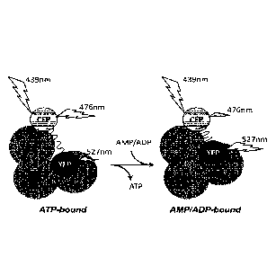
Figure 1: Conformational change model showing operating mode of AMPfret
20 sensors.
AMPfret sensors are constructed from an AMPK heterotrimer (consisting of a-,
p-, and y¨subunits) with two additional GFP-derived fluorescent proteins (CFP,
YFP)
fused to different N- and C-termini of AMPK subunits. Binding of AMP or ADP to
two
CBS domains in the AMPK y-subunit induces a conformational change which
reduces
25 the distance between the fluorophore couple. This increases fluorescence
(or
Foerster) resonance energy transfer (FRET) between the two fluorophores.
Experimentally, when CFP is excited at 439 nm, FRET reduces direct CFP
fluorescence
emission at 476 nm, while energy transferred to YFP increases YFP fluorescence
emission at 527 nm.
Figure 2: Initial AMPfret constructs.
AMPfret A and C exhibit variation of FRET ratio upon AMP binding. Top: Schema
showing structural organization of the sensors. CFP and YFP are respectively
represented as hatched- and dotted-circles. (a) Fluorescence emission spectra
of
AMPfret constructs excited at 430 nm. Spectra show fluorescence peaks of CFP
(476
nm) and YFP (527 nm), and their variation upon AMP binding (dotted line: 3 mM
ATP,
CA 02950051 2016-11-23
WO 2015/193466 PCT/EP2015/063780
26
continuous line: 20 p.M AMP). (b) FRET variation of AMPfret constructs
calculated
from data above (hatched column: 3 mM ATP, dotted column: 20 p.M AMP) and
autoradiograms of in vitro kinase activity assays with these constructs using
acetyl-
CoA carboxylase (ACC) as a substrate. Data correspond to mean SEM (AMPfret
A:
n=7; AMPfret C: n=10). Note: AMPfret constructs exhibit similar activity as
native
AMPK.
Figure 3: Optimized AM Pfret constructs.
Second generation of AMPfret constructs 1.1 and 2.1. based on constructs
AMPfret C and A, respectively. Top: Schema showing structural organization of
the
sensors. Both optimized constructs contain mseCFP
= All/cpVenus as GFP-derived
fluorescent couple instead of CFP/YFP. mseCFPAii and cpVenus are respectively
represented as hatched- and checkered circles. AMPfret 2.1 a- and 13-subunits
also
contain small deletions in their protein sequence to shorten C-terminal non-
folded
linker sequences (A551 and R552 in a and K270, P271 and 1272 in 13). In
addition, a putatively
rigid helix (7 amino acids) was inserted between the a-subunit C-terminus and
CFP
(see small box with curled lines). (a) Fluorescence emission spectra of
AMPfret
constructs excited at 430 nm. Spectra show fluorescence peaks of mseCFPAii
(476 nm)
and cpVenus (527 nm), and their variation upon AMP binding (dotted line: no
AMP,
continuous line: 20 iM AMP). (b) FRET variation of AMPfret constructs
calculated
from data above (hatched column: no AMP, dotted column: 20 p.M AMP) and
autoradiograms of in vitro kinase activity assays with these constructs using
acetyl-
CoA carboxylase (ACC) as a substrate. Data correspond to mean SEM (AMPfret
1.0:
n=10; AMPfret 1.1: n=7); *= p < 0,001 (significance assessed by a Student-
Newman-
Keuls test). Note: All AMPfret constructs exhibit similar activity as native
AMPK.
AMPfret 2.1 reveals improved FRET variation range as compared to AMPfret 1.1,
providing proof of principle that optimization of FRET is possible.
Figure 4: FRET response of AMPfret sensors correlates with the concentration
of AMPK activator AMP.
AMP concentration dependence of the normalized FRET ratio of AMPfret
sensors (a) AMPfret 1.1 and (b) AMPfret 2.1. The FRET ratio was calculated
from
fluorescence emission spectra excited at 430 nm. Data points correspond to
mean
SEM (n3). Data were fitted with Sigma Plot 1.1 software to single site binding
kinetics, yielding affinities of 1,8 M (AMPfret 1.1) and 1,5 p.M (AMPfret
2.1.). Note:
CA 02950051 2016-11-23
WO 2015/193466 PCT/EP2015/063780
27
AMPfret sensors are sensitive to AMP concentrations in a physiological range
(0-20
M)
Figure 5: FRET response of AMPfret sensors correlates with the concentration
of AMPK activator ADP.
ADP concentration dependence of the normalized FRET ratio of AMPfret sensors
(a) AMPfret 1.1 and (b) AMPfret 2.1. The FRET ratio was calculated from
fluorescence
emission spectra excited at 430 nm. Data points correspond to mean SEM
(n_.3).
Data were fitted with Sigma Plot 1.1 software to single site binding kinetics,
yielding
affinities of 5 iiM (AMPfret 1.1) and 7,4 p.M (AMPfret 2.1). Note: AMPfret
sensors are
sensitive to ADP concentrations in a physiological range (0-50 p.M for free
ADP).
Figure 6: AMPfret sensors as in vitro tools to identify AMPK allosteric
activators.
AMPfret 2.1 is incubated in absence (grey mesh bars ) or in presence (black
bars)
of (a) 20 p.M AMP, (b) 50 p.M A-769662 or (c) 500 M Metformin. Structure and
names of the molecules are given below the bars. Data correspond to mean SEM
(AMP: n=7; A-769662: n=4; Metformin: n=4); *= p < 0,001 (significance assessed
by
paired T-test).
Figure 7: AMPfret sensors as cellular in vivo tools to identify AMPK
allosteric
activators ¨ HeLa cells
HeLa cells transfected with AMPFret 2.1 were exposed for 60 min to 1 mM
AICAR. (a) Fluorescence emission spectra showing the increase of cpVenus peak
(527nm) over time; dotted black line: 0 min; dashed black line: 15 min and
solid black
line: 30 min. (b) Time course of the FRET signal. Normalized FRET ratio
determined
each 15 minutes (mean SEM; n=45; *= p < 0,001 according to the performed
Mann-
Whitney Rank Sum Test). (c) AMPK activation at t=0 min and t=60 min.
Phosphorylation of the AMPK substrate ACC as determined by immunoblotting
(lower
panel) and quantification of the resulting P-ACC/total ACC ratio (upper
panel). P-
ACC/total ACC ratios at t=0 and t=60min are respectively represented as a
white
dotted bar and black dotted bar. Data correspond to mean SEM (n=3).
Figure 8: AMPfret sensors as cellular in vivo tools to identify AMPK
allosteric
activators ¨ 3T3-L1 cells
28
HeLa cells transfected with AMPFret 2.1 were exposed for 60 min to 1 mM
AICAR. (a) Time course of the FRET signal. Normalized FRET ratio determined
each 15
minutes (mean SEM; n=9). (b) Time course of AMPK activation. Phosphorylation
of
the AMPK substrate ACC as determined by immunoblotting (lower panel) and
quantification of the resulting P-ACC/total ACC ratio (upper panel). Data
correspond to
mean SEM (n=3).
Figure 9: Effect of 1 hour ischemia followed by 1 hour reperfusion on HepG2
cell
followed by AMPfret.
AMPfret 2.1 normalized FRET ratio evolution during 1h ischemia (light grey
bar)
and 1h reperfusion (dark grey bar). Transfected HepG2 cells were cultured on a
glass
slide mountable onto the incubation flow-through chamber of our Leica TCS SP2
AOBS
confocal microscope. At t=0, the cell was placed under ischemia-like
conditions:
hypoxic conditions (2% 02) and glucose-free medium at 37 C. Deprived medium
was
previously bubbled with N2 for at least 10 minutes before its addition onto
the cells.
After 1hour of deprivation, started the 1 hour-reperfusion period with glucose-
rich
medium and 02 (21%). FRET values were record every minute from a single
isolated
cell using the Leica confocal software. The FRET ratio was followed by
recording
simultaneously nnseCFPAll (476 nm) and cpVenus (527 nm) fluorescence emitted
within 4 nm windows using two independent channels, under excitation set at
458 nm.
FRET ratio was normalized to 1 at t=0.
Figure 10: Strategy for optimizing the AMPK sensors according to the present
invention. Starting from the most promising original constructs, FRET signal
was
optimized by mutations, deletions and addition of sequences.
The compounds and processes of the present invention will be better
understood in connection with the following examples, which are intended as an
illustration only and not limiting of the scope of the invention
Example 1: AMPK constructs
AMPK constructs and protein preparation
The az, 132 and Vi AMPK subunits tagged or not with fluorescent protein, were
respectively cloned in the pACE, pDC and pDS vectors of the ACEMBL expression
system (Bieniossek et al., 2009, Automated unrestricted multigene
recombineering for
multiprotein complex production, Nat Meth 6(6):447-50) using SLIC (Li et al.,
Methods
Date Recue/Date Received 2021-07-07
29
Mol Biol 852:51, 2012) and conventional cloning. Created vectors, containing a
single
subunit fluorescently tagged or not, were fused via their Lox-P site using the
CRE-
recombinase (EMBL Heidelberg): a single expression vector coding for a
chimeric AMPK
that contains two of its three subunits flanked with the mseCFPAll/cpVenus
fluorescent proteins pair (respectively variant of cyan fluorescent protein
(CFP) and
yellow fluorescent protein (YFP) at their termini was obtained. A decaHis-tag,
cleavable
by the TEV protease, was inserted at the N-terminus of the a2 subunit in order
to purify
easily the heterotrimer.
BL21 (DE3) Star cells were transformed by electroporation and protein
expression was carried overnight at 18 C in autoinducing medium. Cells were
collected
by centrifugation at 6000 rpm for 20 min using a Beckman Coulter centrifuge
(rotor
JLA-8.1000) and wash with PBS. Cells were then suspended in lysis buffer (0,5
M
sucrose, 30% glycerol, 50 mM Tris pH8, 100 mM NaCI, 2 mM MgCl2, 2 mM [3-
mercaptoethanol, lysosyme 1 mg/mL, 20 mM imidazole, Complete EDTA free tablet
(Roche), leupeptin, pepstatin). 200 U Benzonase were added to the suspension,
and it
was gently stirred for 1 h in the cold room. Cells were then lysed by
sonication using a
MisonixSonicator 4000 (5 min total at 80% - 20 s ON / 1 min OFF).
Cell-free extract, obtained by centrifugation at 20'000 rpm for 80 min (rotor
JS
25.50) was applied on Ni-NTA Superflow resin (Qiagen) pre-equilibrated with
lysis
buffer. Resin was washed using washing buffer (50 mM Tris pH 8, 100 mM NaCl,
20
mM innidazole, 2 mM MgCl2, 2 mM P-nnercaptoethanol) and high salt buffer (wash
buffer + 1 M NaCI). Proteins were eluted by applying elution buffer (wash
buffer +
400 mM Imidazole). Imidazole was removed through an overnight dialysis in
buffer A
(50 mM Tris pH8, 100 mM NaCI, 2 mM MgCl2, 2 mM P-nnercaptoethanol). Eluted
proteins were passed over a 5 mL QXL column (GE Healthcare) in order to remove
proteins bound to nucleic acids and non-stoichiometric AMPK complexes.
Proteins
were eluted using a gradient of buffer B (50 mM Tris pH8, 1 M NaCI, 2 mM
MgC12,
2 mM P-nnercaptoethanol). Finally chimeric AMPK heterotrinners were applied to
a
SuperoseTM 6 gel filtration column (GE Healthcare) pre-equilibrated with SEC
buffer (50
mM Tris pH8, 200 mM NaCI, 2 mM MgCl2, 2 mM P-nnercaptoethanol, 5 mM
spermidine). Spermidine diminished concentration dependent AMPK oligomer
formation. After adding glycerol to a final concentration of 50%, the purified
AMPK
(untagged or AMPK 221W1) and AMPK heterotrimers of the invention (AMPK tagged
hereafter AMPFret or AMPFret sensors) were stored at -20 C for further
experiments.
Finally, combinations of AMPK tagged with mseCFPAn and cpVenus on two of the
termini of its 3 subunits were created in order to identify constructs that
show FRET
signal variation upon AMP binding (hereafter termed AMPFret).
Date Recue/Date Received 2021-07-07
30
Table 1¨ Overview of the AMPfret constructs containing two fluorescent protein
tags permutated at the N- and C-termini of the three AMPK subunits.
AMPfret
Vector name and composition
construct
AMPK 221 pACEMBL a2 _ [32_ yi
AMPfret A pACEMBL a2-CFP _ [32-YFP _ yi
AMPfret C pACEMBL a2-CFP _ 132 _ y1- YFP
Abbreviations: pACEMBL, plasmid resulting from the Cre-LoxP fusion of vectors
pACE, pDC and pDS of the MutliColi expression system; CFP, Cyan Fluorescent
Protein;
YFP, Yellow Fluorescent Protein; a2, [32, y1, AMPK subunits.
Characterization of AMPFret sensors in vitro
ATP containing buffers were always freshly prepared to limit AMP
contamination. Aqueous solutions of nucleotides (adenine nucleotides, NAD)
were
analyzed by HPLC (stationary phase: Polaris C18 / mobile phase: 60% CH3CN 40%
H20)
to evaluate spontaneous ATP and ADP hydrolysis and contaminations.
Enzymatic assay: AMPK 221WT and AMPfret constructs (3 pmol) were activated
by incubation with purified CannKK[3 (1 pmol) for 20 min at 30 C in kinase
buffer (200
p.M ATP, 40 p.M AMP, 5 mM MgCl2, 1 mM DTT and 10 mM Hepes pH 7.4). Purified
ACC
fragment targeted by AMPK (200 pmol) was then incubated for 20 min at 37 C in
presence or absence of pre-activated AMPK 221W1 or AM PFret sensor in kinase
buffer
containing [y-32NATP. Reaction mixtures were then load on SDS-PAGE gel, P-ACC
signals were revealed using a Typhoon' and activities were evaluated with
ImageJ.
FRET assay: FRET signal variation in presence of different compounds
(nucleotides, chemicals, ions) was measured using a fluorimeter (Photon
Technology
International). AMPfret constructs (20 pmol) were incubated in a quartz
cuvette in a
final volume of 150 pi (spectro buffer: 50 mM Tris pH8, 200 mM NaCI, 5 mM
MgCl2,
2 mM [3-nnercaptoethanol). Effects of nucleotides and others compounds
(previously
Date Recue/Date Received 2021-07-07
CA 02950051 2016-11-23
WO 2015/193466 PCT/EP2015/063780
31
prepared in the spectro buffer) on the FRET ratio given by AMPfret sensor was
determined by comparing FRET ratio (peak value at 527 nm / peak value at 476
nm) in
presence or absence of the compounds. Excitation wavelength was set to 430nm,
and
emission spectra were recorded from 450 to 600 nm with an integration time of
0,2 s.
Mg' effect on FRET was investigated in spectra buffer without Mg2+.
The two constructs, AMPFret A (a2-CFP-132-YFP-y1; CFP tagged at the a2 C-
terminus, and YFP at the 131 C-terminus) and AMPFret C (a2-CFP-132-yrYFP; CFP
tagged
at the a2 C-terminus, and YFP at the y1 C-terminus) showed both a significant
difference in their FRET signal (-10%) depending on the presence of AMP or ADP
(Figure 2). It appears that, during allosteric activation, the a-subunit C-
terminus
approaches the C-termini of the 13- and y-subunits.
Example 2: Optimized AMPK constructs
Constructs were optimized to achieve a superior FRET signal amplitude. The
construct AMPfret 1.1 is based on AMPfret C, containing full length AMPK
subunits
a2, 132 and y1. The a-subunit is tagged with mseCFPA11 at its C-terminus and
the y-
subunit is tagged at its C- terminus with cpVenus; the 13-subunit remains
untagged.
The construct AMPfret 2.1 is based on AMPfret A, where CFP/YFP were exchanged
for
the same different fluorophore pair as AMPfret 1.1. In addition, the sequence
of the
construct was modified. First, small truncations based on the crystal
structure (PDB
2Y94) and secondary structure prediction (nps@consensus (ucbl)) were inserted
via
PCR and "self SLIC" between the N-terminus of fluorescent protein tags and the
C-
terminus of the tagged AMPK subunits. Such shortening of the sequence between
AMPK core and tag may remove flexibility other than the conformational change
induced by AMP. Second, a short insert supposed to fold into a rigid a-helix
(Sivaramakrishnan et al., PNAS 105:13356, 2008 and 108:20467, 2011) was
inserted
between the a2 C-terminus and the CFP N-terminus to rigidify the AMPK backbone
of
the invention and to stabilize the CFP tag in a given position relative to
AMPK.
This engineering comprised the following mutations. The last 2 C-terminal
amino
acids (AR) of a2 and the first 3 N-terminal amino acids (MSK) of mseCFPA11
were
removed and the new termini linked via 8 amino acids insert supposed to fold
into an
a-helix (EEEEKKKK, SEQ ID No.1). Further, the last C-terminal (non-folded) 3
amino
acids (KPI) of 132 were also removed, and directly fused to the N-terminus of
YFP. Since
2 amino acids resulting from the restriction site previously used were also
removed by
the SLIC technique, this yielded a construct lacking in total 5 amino acids
between the
CA 02950051 2016-11-23
WO 2015/193466 PCT/EP2015/063780
32
132-subunit and YFP. The optimized AMPfret sensor showed an almost 100%
increased
FRET ratio (Figure 3).
The optimized AMPfret sensors allow titration of the allosteric AMPK-
activators
AMP and ADP, confirming that both induce conformational changes in the AMPK
heterotrimer. The affinity (Kd) for AMP and ADP could be determined as 1,5 p.M
and
7,4 p.M, respectively.
The AMPfret sensors thus represent a pioneering powerful and easy-to-use tool
to decipher the activation mechanisms of AMPK. They contain full length AMPK
heterotrimer that behaves the same way as native AMPK WT as judged by (i) its
kinase
activity after phosphorylation via CamK10 and allosteric activation by AMP and
(ii) its
affinities for adenine nucleotides. The AMPfret FRET signal is directly
dependent of
the AMP concentration; in a physiological range (1 ¨ 10 M) it shows almost
linear
relationship (Figures 4 and 5).
Example 3: in vitro interaction of optimized AMPfret with allosteric
activators
The optimized AMPfret sensors not only translate the adenylate-dependent
movements of the AMPK heterotrimer into a FRET signal, which are triggered by
adenylate binding to specific sites at the y-subunit. Their readout also
reports
conformational changes of other, pharmacological direct AMPK activators such
as the
compound A-769662 (Figure 6). This molecule interacts with the 13-subunit, but
clearly
induces a FRET signal comparable to AMP, even if the triggering conformational
change may be of different nature according the different binding mode.
Metformin, a widely used anti-diabetes drug, which was postulated to directly
interact with y-subunit (Zhang et al., Mol Cell Biochem 368:69, 2012) did not
induce
any FRET variation emission of AMPfret (Figure 6). This absence of
conformational
changes in vitro supports the generally accepted indirect mode of action,
whereby this
drug inhibits mitochondrial respiration and increases the AMP/ATP ratio, thus
activating AMPK by the canonical AMP binding mechanism at the y-subunit.
Taken together, AMPfret appears as a valuable and accurate tool for in vitro
applications, notably screening for AMPK interactors.
Example 4: Ex vivo experiments with the optimized AM Pfret constructs
For cellular ex vivo experiments, subunits of the optimized construct
AMPfret 2.1 were cloned in the vectors (pACEMam2, pMDS and pMDK) of the
33
Mu/Warn expression system. Created plasmids were fused via their Lox-P site to
yield
to a single mammalian expression vector coding for the sensor AM PFret 2.1
according
to well-known techniques to the skilled man in the art.
313-L1 and HeLa cells were cultivated in glucose containing DMEM (4,5 g/L)
supplemented with SVF, glutamine, penicillin and streptomycin. Once cells
reached
around 80% confluence, medium was replaced by Opti-MEMT" (Lifetechnologies)
and
AMPfret 2.1 coding plasmid was transfected using
Lipofectamine2000
(Lifetechnologies). After 5h, Opti-MEMT" was exchanged by complete DMEM and
cells
grew for >36h until their observations under the confocal microscope.
313-L1 or HeLa cells, cultivated in 8 wells LabTek cover glass plates (Nunc),
were
observed with a Leica TCS SP2 AOBS confocal microscope. LabTek plates were
placed
in an incubation chamber in which the temperature and 02 concentration were
maintained at 37 C and 21%, respectively. Without moving the Labtek, 200 1_
medium
was replaced by the same volume of complete medium containing 2 mM AICAR (1 mM
final). Excitation wavelength was set to 458 nm and emission spectra showing
FRET
signal were monitored through X scans from 463 nm to 600 nm every 15 nnin. ROI
(region of interest) were drawn in order to cover entire cells. FRET ratio
variations
were calculated from those measured emission spectra.
Under the microscope, cells were treated with 1 mM AICAR (AMPK allosteric
activator) to visualize the allosteric activation of AMPK through the FRET
signal of
AMPfret 2.1 and hence validate its use for ex vivo applications.
AMPfret 2.1 was excited using a 458 nm laser and emission spectra showing FRET
signal were monitored through X scans from 463 nm to 600 nm every 15 min. The
AMPfret 2.1 FRET signal increased with time upon AICAR addition, suggesting
that
AMPfret 2.1 can monitor allosteric activation of AMPK in cells (Figures 7 and
8).
More than half-maximal response was already reached after 15 min of
treatment, and the maximal effect reached after about 30 minutes.
The activation kinetics of AMPK upon AICAR addition was independently verified
by Western blotting for the AMPK-specific phosphosite in acetyl-CoA
carboxylase (ACC;
widely used as reporter for AMPK activity) in 31341 cells.
All the results presented above show that the AMPfret sensor can be used as a
suitable tool for cellular in vivo applications.
Example 5: AMPfret 2.1 during ischemia-reperfusion in a HepG2 single cell
Using an incubation flow-through chamber fitted to the confocal microscope
which permits to control temperature as well as 02 concentration, HepG2 cells
were
Date Recue/Date Received 2021-07-07
34
placed under ischemia-like conditions, comprising hypoxic conditions (2% 02)
and
glucose free medium at 37 C. ATP pools may not be affected when hypoxia is
applied
in a high nutrient containing medium since cells can adapt to hypoxia by
switching
their energy metabolism through anaerobic pathways to compensate for aerobic
ATP
production. The deprivation period was followed by 1 hour of reperfusion with
complete medium and 02 (21%). During the 2 hours of the ischemia-reperfusion
protocol, the FRET ratio was monitored every minute by recording
simultaneously
nnseCFPA11 and cpVenus fluorescence emitted within 4 nnn windows
(corresponding
to fluorescence emission) using two independent channels. Images were
collected and
processed using ImageJ in order to i) remove eventual background fluorescence
and ii)
isolate individual cells from acquired pictures. Then, the fluorescence
intensities were
extracted from single cell images using VolocityTM. Thus, the effect of
ischemia-
reperfusion on the AMPfret 2.1 signal in single cells was analyzed (Figure 9).
During ischemia in HepG2 cells, the FRET signal did not vary. Changes in
AMP/ATP ratio under such conditions were proposed to happen in the liver and
AMPK
becomes activated, but a recent study suggested that AMPK was activated during
ischemia through adenylate-independent pathways. Figure 9 shows results of a
single
cell.
During reperfusion of HepG2 cells, the FRET signal increases over the first 30
minutes indicating increased AMP and ADP concentrations. Subsequently, the
FRET
ratio remained at unchanged high values, suggesting that elevated AMP and ADP
concentrations were maintained. In fact, AMPfret should revert the FRET ratio
as soon
as AMP and ADP levels drop again. These results suggest that in HepG2 cells,
reperfusion represented a more drastic energy stress than ischemia regarding
adenylate concentrations and AMPK allosteric activation.
Through these experiments, using AMPfret 2.1 in HepG2 cells, we did not detect
any FRET signal changes during ischemia, suggesting that AMP and ADP
concentrations
remained unchanged. However, we showed an increase of AMPfret FRET signal
during
reperfusion, suggesting an elevation of intracellular AMP and ADP and
allosteric
activation of AMPK.
These experiments achieved in living cells using AMPfret 2.1 show that AMPfret
2.1 was properly transfected and its fluorescence monitored over time. These
results
Date Recue/Date Received 2021-07-07
CA 02950051 2016-11-23
WO 2015/193466 PCT/EP2015/063780
show that AMPfret 2.1 provides a readout of AMP/ZMP concentrations and AMPK
allosteric activation by reporting the related conformational changes.
Experiments
involving ischemia-reperfusion showed that AMPfret 2.1 can monitor endogenous
changes of adenylates and AMPK allosteric activation over time.
5
Monitoring of transient events related to AMPK allosteric activation is
promising
to decipher or unravel new aspects of its regulation.