Note: Descriptions are shown in the official language in which they were submitted.
CA 02951056 2016-12-02
WO 2015/184551 PCT/CA2015/050518
BIOPHOTONIC HYDROGELS
FIELD OF THE DISCLOSURE
The present disclosure generally relates to forming biophotonic hydrogels.
BACKGROUND OF THE DISCLOSURE
Phototherapy has recently been recognized as having wide range of applications
in both the
medical and cosmetic fields including use in surgery, therapy and diagnostics.
For example,
phototherapy has been used to treat cancers and tumors with lessened
invasiveness, to
disinfect target sites as an antimicrobial treatment, to promote wound
healing, and for facial
skin rejuvenation.
Hydrogels are materials which absorb solvents (such as water), undergo rapid
swelling
without discernible dissolution, and maintain three-dimensional networks
capable of
reversible deformation. Forming hydrogels has been proposed for use in a
number of
applications, including surgery, medical diagnosis and treatment, adhesives
and sealers. One
method for formation of hydrogels employs photopolymerization.
Photopolymerization
comprises using light to convert initiator molecules into free radicals that
can react with
monomers or macromers containing double bond and propagate radical chain
polymerization. Forming hydrogels intended for biomedical and tissue
engineering
applications should occur under mild conditions, for example neutral pH and
require non-
toxic photoinitiators.
Therefore, it is an object of the present disclosure to provide new and
improved formation of
hydrogel compositions and methods that are useful in phototherapy.
SUMMARY OF THE DISCLOSURE
The present disclosure provides biophotonic hydrogels and methods useful in
phototherapy.
In particular, biophotonic hydrogels of the present disclosure include a
polymerisable
monomer, and at least one chromophore. Preferably, the at least one
chromophore can
-1-
CA 02951056 2016-12-02
WO 2015/184551 PCT/CA2015/050518
absorb and/or emit light to initiate photopolymerization of the hydrogel, and
further wherein
the at least one chromophore is not fully photobleached after
photopolymerization.
In some embodiments, the biophotonic hydrogel composition further comprises a
cross
linker. In some embodiments, the cross linker is Poly(ethylene glycol)
diacrylate (PEGDA).
The composition may also include an initiator. The initiator may be TEA. The
composition
may also include a catalyst, and the catalyst may be 1-viny1-2 pyrrolidinone
(NVP).
In some embodiments, the catalyst may be polyvinyl pyrrolidone (PVP).
In some embodiments, the chromophore absorbs and/or emits visible light. In
some
embodiments, the chromophore absorbs and/or emits light within the range of
about 400
nm-750 nm or about 400-700 nm or about 400nm-800nm.
In some embodiments, the hydrogel composition further comprises a surfactant.
In some
embodiments, the surfactant is Pluronic F127. The surfactant may be present in
the
biophotonic hydrogel at between about 1-5 wt%, between about 2.5-7.5 wt%,
between about
5-10 wt%, between about 7.5-12.5 wt%, between about 10-15 wt%, between about
12.5-
17.5 wt%, between about 15-20 wt%, between about 20-25 wt% Pluronic F127. In
certain
embodiments, the biophotonic hydrogel comprises a further surfactant
comprising a cationic
surfactant. In certain other embodiments, the cationic surfactant is
cetyltrimethyl ammonium
bromide (CTAB). In certain embodiments, the CTAB may be present in the
biophotonic
hydrogel at a percentage concentration to allow for a formation of micelles by
the CTAB
(termed a critical micelle concentration). In certain embodiments, the
critical micelle
concentration may be increased with an increase in incubation temperature of
the
biophotonic hydrogel.
In some embodiments, the hydrogel composition further includes a stabilizer.
The stabilizer
may be gelatin, hydroxyethyl cellulose (HEC), carboxymethyl cellulose (CMC) or
any other
thickening agent.
-2-
CA 02951056 2016-12-02
WO 2015/184551 PCT/CA2015/050518
The chromophore of the present hydrogel composition may be a xanthene dye. The
xanthene dye may be fluorescein or eosin, or any other xanthene dye.
In some embodiments, the biophotonic hydrogel composition further comprises an
additional compound that may enhance the mechanical strength of the
biophotonic hydrogel.
In some embodiments, the additional compound may be a silica-based compound.
In certain
embodiments, the silica-based compound may be a silica clay or fumed silica
(Si02). In
certain embodiments, the silica clay may be bentonite. The bentonite may be
present in the
biophotonic hydrogel at between about 0.01-0.5 wt%, between about 0.25-0.75
wt%,
between about 0.5-0.75 wt%, between about 0.75-1.0 wt% of the biophotonic
hydrogel. The
fumed silica may be present in the biophotonic hydrogel at between about 0.01-
1.0 wt%,
between about 1.0-2.0 wt%, between about 2.0-3.0 wt%, between about 3.0-4.0
wt%,
between about 4.0-5.0 wt% of the biophotonic hydrogel.
In certain other embodiments, the biophotonic hydrogel comprises a combination
of the
further surfactant and the additional compound for enhancing the mechanical
strength of the
biophotonic hydrogel. In certain other embodiments, the combination of the
further
surfactant and the additional compound for enhancing the mechanical strength
in the
biophotonic hydrogel comprises CTAB and fumed silica, respectively.
The biophotonic hydrogel composition of any aspects or embodiments of the
disclosure may
be used for modulating a pro-inflammatory response in a cell or tissue type.
In some
embodiments, the biophotonic hydrogel composition of any aspects or
embodiments of the
disclosure may be used for stimulating an increase in collagen production in a
cell, or tissue
type, and in some embodiments, the biophotonic hydrogel composition of any
aspects or
embodiments of the disclosure may be used for stimulating fibroblast
proliferation.
The biophotonic hydrogel composition of any aspects or embodiments of the
disclosure may
be used for cosmetic or medical treatment of tissue. In some embodiments, the
cosmetic
treatment is skin rejuvenation and conditioning, and the medical treatment is
wound healing,
periodontal treatment or acne treatment or treatment of other skin conditions
including acne,
-3-
CA 02951056 2016-12-02
WO 2015/184551 PCT/CA2015/050518
eczema, psoriasis or dermatitis. In some aspects, the biophotonic hydrogel
composition is
used for modulating inflammation, modulating collagen synthesis or for
promoting
angio genesis.
The present disclosure also provides methods for promoting wound healing
comprising
applying a biophotonic hydrogel composition over a wound, wherein the hydrogel
composition comprises N-Hydroxyethyl acrylamide (HEAA) and at least one
chromophore;
and illuminating said biophotonic hydrogel composition with light having a
wavelength that
is absorbed by the at least one chromophore; wherein said method promotes
wound healing.
The present disclosure also provides methods for treating a skin disorder,
wherein the
method comprises applying a biophotonic hydrogel composition over a target
skin tissue,
wherein the hydrogel composition comprises N-Hydroxyethyl acrylamide (HEAA),
and at
least one chromophore; and illuminating said biophotonic hydrogel composition
with light
having a wavelength that is absorbed by the at least one chromophore; and
wherein said
method promotes healing of said skin disorder. In some embodiments, the skin
disorder is
selected from acne, eczema, proriasis and dermatitis.
The present disclosure also provides methods for treating acne comprising:
applying a
biophotonic hydrogel composition over a target skin tissue, wherein the
hydrogel
composition comprises N-Hydroxyethyl acrylamide (HEAA), and at least one
chromophore;
and illuminating said biophotonic hydrogel composition with light having a
wavelength that
is absorbed by the at least one chromophore; and wherein said method treats
the acne.
The present disclosure also provides methods for skin rejuvenation comprising
applying a
biophotonic hydrogel composition over a target skin tissue, wherein the
hydrogel
composition comprises N-Hydroxyethyl acrylamide (HEAA), and at least one
chromophore;
and illuminating said biophotonic hydrogel composition with light having a
wavelength that
is absorbed by the at least one chromophore; and wherein said method promotes
skin
rejuvenation.
-4-
CA 02951056 2016-12-02
WO 2015/184551 PCT/CA2015/050518
The present disclosure also provides methods for preventing or treating scars
comprising
applying a biophotonic hydrogel composition over a target skin tissue, wherein
the hydrogel
comprises N-Hydroxyethyl acrylamide (HEAA), and at least one chromophore; and
illuminating said biophotonic hydrogel composition with light having a
wavelength that is
absorbed the at least one chromophore; and wherein said method promotes
prevents or treats
scars.
BRIEF DESCRIPTION OF THE DRAWINGS
Further aspects and advantages of the present discosure will become better
understood with
reference to the description in association with the following in which:
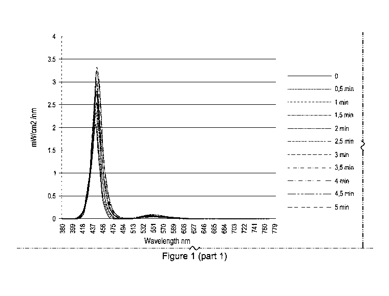
Figure 1 illustrates the light emission spectra of biophotonic
poly(hydroxyethyl acrylamide)
during 0-5 minutes of illumination, according to an embodiment of the present
disclosure.
Figure 2 illustrates the light emission spectra of biophotonic
poly(hydroxyethyl acrylamide)
during 5-10 minutes of illumination, according to an embodiment of the present
disclosure.
Figure 3 illustrates the light emission spectra of biophotonic
poly(hydroxyethyl
acrylamide)/gelatin during 0-5 minutes of illumination, according to an
embodiment of the
present disclosure.
Figure 4 illustrates the light emission spectra of biophotonic
poly(hydroxyethyl
acrylamide)/gelatin during 5-10 minutes of illumination, according to an
embodiment of the
present disclosure.
Figure 5 illustrates the light emission spectra of biophotonic
poly(hydroxyethyl
acrylamide)/HEC during 0-5 minutes of illumination, according to an embodiment
of the
present disclosure.
-5-
CA 02951056 2016-12-02
WO 2015/184551 PCT/CA2015/050518
Figure 6 illustrates the light emission spectra of biophotonic
poly(hydroxyethyl
acrylamide)/HEC during 5-10 minutes of illumination, according to an
embodiment of the
present disclosure.
Figure 7 illustrates the light emission spectra of biophotonic
poly(hydroxyethyl
acrylamide)/P1-F127 during 0-5 minutes of illumination, according to an
embodiment of the
present disclosure.
Figure 8 illustrates the light emission spectra of biophotonic
poly(hydroxyethyl
acrylamide)/P1-F127-CTAB during 0-5 minutes of illumination, according to an
embodiment of the present disclosure.
Figure 9 illustrates the light emission spectra of biophotonic
poly(hydroxyethyl
acrylamide)/P1-F127-Bentonite during 0-5 minutes of illumination, according to
an
embodiment of the present disclosure.
Figure 10 illustrates the light emission spectra of biophotonic
poly(hydroxyethyl
acrylamide)/P1-F127-Si02 during 0-5 minutes of illumination, according to an
embodiment
of the present disclosure.
Figure 11 illustrates the light emission spectra of biophotonic
poly(hydroxyethyl
acrylamide)/P1-F127-Si02-CTAB during 0-5 minutes of illumination, according to
an
embodiment of the present disclosure.
Figure 12 illustrates a graph indicating the modulation of collagen production
in Human
Dermal Fibroblasts (DHF) 48 hours after treatment with light from a blue light
and a
membrane according to one embodiment of the present disclosure.
Figure 13 illustrates a graph indicating the modulation of Human Dermal
Fibroblasts (DHF)
proliferation 24 hours after treatment with light from a blue light and a
membrane according
to one embodiment of the present disclosure.
-6-
CA 02951056 2016-12-02
WO 2015/184551 PCT/CA2015/050518
DETAILED DESCRIPTION
(1) Overview
The present disclosure provides biophotonic hydrogels and uses thereof.
Biophotonic
therapy using these materials would combine the beneficial effects of forming
hydrogels
with the photobiostimulation induced by the fluorescent light generated upon
illumination of
the materials. In certain embodiments of forming biophotonic hydrogels of the
present
disclosure are activated by visible light. Furthermore, in certain
embodiments, phototherapy
using the biophotonic hydrogels of the present disclosure will for instance
promote wound
healing, rejuvenate the skin by, e.g., promoting collagen synthesis, treat
skin conditions such
as acne, and treat periodontitis.
(2) Definitions
Before continuing to describe the present disclosure in further detail, it is
to be understood
that this disclosure is not limited to specific compositions or process steps,
as such may
vary. It must be noted that, as used in this specification and the appended
claims, the
singular form "a", "an" and "the" include plural referents unless the context
clearly dictates
otherwise.
As used herein, the term "about" in the context of a given value or range
refers to a value or
range that is within 20%, preferably within 10%, and more preferably within 5%
of the
given value or range.
It is convenient to point out here that "and/or" where used herein is to be
taken as specific
disclosure of each of the two specified features or components with or without
the other.
For example "A and/or B" is to be taken as specific disclosure of each of (i)
A, (ii) B and
(iii) A and B, just as if each is set out individually herein.
"Biophotonic" means the generation, manipulation, detection and application of
photons in
a biologically relevant context. In other words, biophotonic compositions and
materials
-7-
CA 02951056 2016-12-02
WO 2015/184551 PCT/CA2015/050518
exert their physiological effects primarily due to the generation and
manipulation of
photons.
"Hydrogel" refers to a material of solid or semi-solid texture that includes
water. Hydrogels
are formed by a three-dimensional network of molecular structures within which
water,
among other substances, may be held. The three-dimensional molecular network
may be
held together by covalent chemical bonds, or by ionic bonds, or by any
combination thereof
Some hydrogels may be formed through the mixture of two or more materials that
undergo
chemical or physical reactions with each other to create the three-dimensional
molecular
network that provides the hydro gel with a degree of dimensional stability.
"Topical application" or "topical uses" means application to body surfaces,
such as the skin,
mucous membranes, vagina, oral cavity, internal surgical wound sites, and the
like.
Terms "chromophore" and "photoactivator" are used herein interchangeably. A
chromophore means a chemical compound, when contacted by light irradiation, is
capable
of absorbing the light. The chromophore readily undergoes photoexcitation and
can transfer
its energy to other molecules or emit it as light (fluorescence).
"Photobleaching" or "photobleaches" means the photochemical destruction of a
chromophore. A chromophore may fully or partially photobleach.
The term "actinic light" is intended to mean light energy emitted from a
specific light source
(e.g. lamp, LED, or laser) and capable of being absorbed by matter (e.g. the
chromophore or
photoactivator). Terms "actinic light" and "light" are used herein
interchangeably. In a
preferred embodiment, the actinic light is visible light.
"Photopolymerization" herein refers to the use of visible or UV light to
interact with light-
sensitive compounds called "initiators" to create free radicals that can
initiate
polymerization of liquid or semi-liquid monomer or macromer to form a hydro
gel.
-8-
CA 02951056 2016-12-02
WO 2015/184551 PCT/CA2015/050518
"Skin rejuvenation" means a process of reducing, diminishing, retarding or
reversing one or
more signs of skin aging or generally improving the condition of skin. For
instance, skin
rejuvenation may include increasing luminosity of the skin, reducing pore
size, reducing
fine lines or wrinkles, improving thin and transparent skin, improving
firmness, improving
sagging skin (such as that produced by bone loss), improving dry skin (which
might itch),
reducing or reversing freckles, reducing or preventing the appearance of age
spots, spider
veins, rough and leathery skin, fine wrinkles that disappear when stretched,
reducing loose
skin, or improving a blotchy complexion. According to the present disclosure,
one or more
of the above conditions may be improved or one or more signs of aging may be
reduced,
diminished, retarded or even reversed by certain embodiments of the
compositions, methods
and uses of the present disclosure.
"Wound" means an injury to any tissue, including for example, acute, subacute,
delayed or
difficult to heal wounds, and chronic wounds. Examples of wounds may include
both open
and closed wounds. Wounds include, for example, amputations, burns, incisions,
excisions,
lesions, lacerations, abrasions, puncture or penetrating wounds, surgical
wounds,
amputations, contusions, hematomas, crushing injuries, ulcers (such as for
example
pressure, diabetic, venous or arterial), scarring (cosmesis), and wounds
caused by
periodontitis (inflammation of the periodontium).
Features and advantages of the subject matter hereof will become more apparent
in light of
the following detailed description of selected embodiments, as illustrated in
the
accompanying figures. As will be realized, the subject matter disclosed and
claimed is
capable of modifications in various respects, all without departing from the
scope of the
claims. Accordingly, the drawings and the description are to be regarded as
illustrative in
nature, and not as restrictive and the full scope of the subject matter is set
forth in the
claims.
(3) Biophotonic hydrogels
The present disclosure provides, in a broad sense, biophotonic hydrogels and
methods of
using the biophotonic hydrogels. Biophotonic hydrogels can be, in a broad
sense, activated
-9-
CA 02951056 2016-12-02
WO 2015/184551 PCT/CA2015/050518
by light (e.g., photons) of specific wavelength. Biophotonic hydrogel
according to various
embodiments of the present disclosure contains a polymerisable monomer, and at
least one
chromophore. The chromophore can absorb and/or emit light to initiate
photopolymerization
of the polymerisable monomer. In some embodiments, the chromophore is not
fully
photobleached after photopolymerization. Continued or repeated illumination of
the
biophotonic hydrogel can activate the at least one chromophore, which leads to
light
carrying on a therapeutic effect on its own, and/or to the photochemical
activation of other
agents contained in the composition.
When a chromophore absorbs a photon of a certain wavelength, it becomes
excited. This is
an unstable condition and the molecule tries to return to the ground state,
giving away the
excess energy. For some chromophores, it is favorable to emit the excess
energy as light
when returning to the ground state. This process is called fluorescence. The
peak
wavelength of the emitted fluorescence is shifted towards longer wavelengths
compared to
the absorption wavelengths due to loss of energy in the conversion process.
This is called
the Stokes' shift. In the proper environment (e.g., in a biophotonic hydrogel)
much of this
energy is transferred to the other components of the biophotonic hydrogel or
to the treatment
site directly.
Without being bound to theory, it is thought that fluorescent light emitted by
photoactivated
chromophores may have therapeutic properties due to its femto-, pico-, or nano-
second
emission properties which may be recognized by biological cells and tissues,
leading to
favourable biomodulation. Furthermore, the emitted fluorescent light has a
longer
wavelength and hence a deeper penetration into the tissue than the activating
light.
Irradiating tissue with such a broad range of wavelength, including in some
embodiments
the activating light which passes through the composition, may have different
and
complementary effects on the cells and tissues. In other words, chromophores
are used in
the biophotonic hydrogels of the present disclosure for therapeutic effect on
tissues.
The biophotonic hydrogels of the present disclosure may have topical uses such
as a mask
or a wound dressing, or as an attachment to a light source, as a waveguide or
as a light filter.
-10-
CA 02951056 2016-12-02
WO 2015/184551 PCT/CA2015/050518
In addition the biophotonic materials can limit the contact between the
chromophore and the
tissue. These materials may be described based on the components making up the
composition. Additionally or alternatively, the compositions of the present
disclosure have
functional and structural properties and these properties may also be used to
define and
describe the compositions. Individual components of the biophotonic hydrogels
of the
present disclosure, including chromophores, polymerisable monomers, cross
linkers,
initiators, catalysts, and other optional ingredients, such as thickening
agents and
surfactants, are detailed below.
The present disclosure also provides a premix composition to the material
described herein,
which will gel or polymerize upon light exposure. The premix composition
comprises at
least one chromophore and a polymerisable monomer, such as HEAA, which in its
polymerized form is referred to as "PHEAA".
(a) Chromophores
Suitable chromophores can be fluorescent compounds (or stains) (also known as
"fluorochromes" or "fluorophores"). Other dye groups or dyes (biological and
histological
dyes, food colorings, carotenoids, and other dyes) can also be used.
Suitable
photoactivators can be those that are Generally Regarded As Safe (GRAS).
Advantageously,
photoactivators which are not well tolerated by the skin or other tissues can
be included in
the biophotonic hydrogel of the present disclosure, as in certain embodiments,
the
photoactivators are encapsulated within the hydrogel and may not contact the
tissues.
In certain embodiments, the biophotonic hydrogel of the present disclosure
comprises a first
chromophore which undergoes partial or complete photobleaching upon
application of light.
In some embodiments, the first chromophore absorbs at a wavelength in the
range of the
visible spectrum, such as at a wavelength of about 380-800 nm, 380-700 nm, 400-
800 nm,
or 380-600 nm. In other embodiments, the first chromophore absorbs at a
wavelength of
about 200-800 nm, 200-700 nm, 200-600 nm or 200-500 nm. In some embodiments,
the
first chromophore absorbs at a wavelength of about 200-600 nm. In some
embodiments, the
-11-
CA 02951056 2016-12-02
WO 2015/184551 PCT/CA2015/050518
first chromophore absorbs light at a wavelength of about 200-300 nm, 250-350
nm, 300-400
nm, 350-450 nm, 400-500 nm, 450-650 nm, 600-700 nm, 650-750 nm or 700-800 nm.
It will be appreciated to those skilled in the art that optical properties of
a particular
chromophore may vary depending on the chromophore's surrounding medium.
Therefore,
as used herein, a particular chromophore's absorption and/or emission
wavelength (or
spectrum) corresponds to the wavelengths (or spectrum) measured in a
biophotonic
hydrogel of the present disclosure.
The biophotonic hydrogel disclosed herein may include at least one additional
chromophore.
Combining chromophores may increase photo-absorption by the combined dye
molecules
and enhance absorption and photo-biomodulation selectivity. Thus, in certain
embodiments,
biophotonic hydrogels of the disclosure include more than one chromophore.
When such
multi-chromophore materials are illuminated with light, energy transfer can
occur between
the chromophores. This process, known as resonance energy transfer, is a
widely prevalent
photophysical process through which an excited 'donor' chromophore (also
referred to
herein as first chromophore) transfers its excitation energy to an 'acceptor'
chromophore
(also referred to herein as second chromophore). The efficiency and
directedness of
resonance energy transfer depends on the spectral features of donor and
acceptor
chromophores. In particular, the flow of energy between chromophores is
dependent on a
spectral overlap reflecting the relative positioning and shapes of the
absorption and emission
spectra. More specifically, for energy transfer to occur, the emission
spectrum of the donor
chromophore must overlap with the absorption spectrum of the acceptor
chromophore.
Energy transfer manifests itself through decrease or quenching of the donor
emission and a
reduction of excited state lifetime accompanied also by an increase in
acceptor emission
intensity. To enhance the energy transfer efficiency, the donor chromophore
should have
good abilities to absorb photons and emit photons. Furthermore, the more
overlap there is
between the donor chromophore's emission spectra and the acceptor
chromophore's
absorption spectra, the better a donor chromophore can transfer energy to the
acceptor
chromophore.
-12-
CA 02951056 2016-12-02
WO 2015/184551 PCT/CA2015/050518
In certain embodiments, where the biophotonic hydrogels of the present
disclosure further
comprise a second chromophore, the first chromophore may have an emission
spectrum that
overlaps at least about 80%, about 75%, about 70%, about 65%, about 60%, about
55%,
about 50%, about 45%, about 40%, about 35%, about 30%, about 25%, about 20%,
about
15% or about 10% with an absorption spectrum of the second chromophore. In
some
embodiments, the first chromophore has an emission spectrum that overlaps at
least about
20% with an absorption spectrum of the second chromophore. In some
embodiments, the
first chromophore has an emission spectrum that overlaps at least between
about 1-10%,
between about 5-15%, between about 10-20%, between about 15-25%, between about
20-
30%, between about 25-35%, between about 30-40%, between about 35-45%, between
about 50-60%, between about 55-65% or between about 60-70% with an absorption
spectrum of the second chromophore.
% spectral overlap, as used herein, means the % overlap of a donor
chromophore's
emission wavelength range with an acceptor chromophore's absorption wavelength
rage,
measured at spectral full width quarter maximum (FWQM). In some embodiments,
the
second chromophore absorbs at a wavelength in the range of the visible
spectrum. In certain
embodiments, the second chromophore has an absorption wavelength that is
relatively
longer than that of the first chromophore within the range of about 50-250, 25-
150 or 10-
100 nm.
The chromophore can be present in an amount of about 0.001-40% per weight of
the
biophotonic hydrogel. In certain embodiments, the first chromophore is present
in an
amount of between about 0.001-3%, between about 0.001-0.01%, between about
0.005-
0.1%, between about 0.1-0.5%, between about 0.5-2%, between about 1-5%,
between about
2.5-7.5%, between about 5-10%, between about 7.5-12.5%, between about 10-15%,
between about 12.5-17.5%, between about 15-20%, between about 17.5-22.5%,
between
about 20-25%, between about 22.5-27.5%, between about 25-30%, between about
27.5-
32.5%, between about 30-35%, between about 32.5-37.5%, or between about 35-40%
per
weight of the biophotonic hydrogel.
-13-
CA 02951056 2016-12-02
WO 2015/184551 PCT/CA2015/050518
In embodiments comprising a second chromophore, the second chromophore can be
present
in an amount of about 0.001-40% per weight of the biophotonic hydrogel. In
some
embodiments, the second chromophore is present in an amount of between about
0.001-3%,
between about 0.001-0.01%, between about 0.005-0.1%, between about 0.1-0.5%,
between
about 0.5-2%, between about 1-5%, between about 2.5-7.5%, between about 5-10%,
between about 7.5-12.5%, between about 10-15%, between about 12.5-17.5%,
between
about 15-20%, between about 17.5-22.5%, between about 20-25%, between about
22.5-
27.5%, between about 25-30%, between about 27.5-32.5%, between about 30-35%,
between
about 32.5-37.5%, or between about 35-40% per weight of the biophotonic
hydrogel.
In certain embodiments, the total weight per weight of chromophore or
combination of
chromophores may be in the amount of between about 0.005-1%, between about
0.05-2%,
between about 1-5%, between about 2.5-7.5%, between about 5-10%, between about
7.5-
12.5%, between about 10-15%, between about 12.5-17.5%, between about 15-20%,
between
about 17.5-22.5%, between about 20-25%, between about 22.5-27.5%, between
about 25-
30%, between about 27.5-32.5%, between about 30-35%, between about 32.5-37.5%,
or
between about 35-40.001% per weight of the biophotonic hydrogel.
The concentration of the chromophore to be used can be selected based on the
desired
intensity and duration of the biophotonic activity from the biophotonic
hydrogel, and on the
desired medical or cosmetic effect. For example, some dyes such as xanthene
dyes reach a
'saturation concentration' after which further increases in concentration do
not provide
substantially higher emitted fluorescence. Further increasing the chromophore
concentration
above the saturation concentration can reduce the amount of activating light
passing through
the matrix. Therefore, if more fluorescence is required for a certain
application than
activating light, a high concentration of chromophore can be used. However, if
a balance is
required between the emitted fluorescence and the activating light, a
concentration close to
or lower than the saturation concentration can be chosen.
-14-
CA 02951056 2016-12-02
WO 2015/184551 PCT/CA2015/050518
Suitable chromophores that may be used in the biophotonic hydrogels of the
present
disclosure include, but are not limited to the following:
Chlorophyll dyes
Exemplary chlorophyll dyes include but are not limited to chlorophyll a;
chlorophyll
b; chlorophyllin; bacteriochlorophyll a; bacteriochlorophyll b;
bacteriochlorophyll c;
bacteriochlorophyll d; protochlorophyll; protochlorophyll a; amphiphilic
chlorophyll
derivative 1; and amphiphilic chlorophyll derivative 2.
Xanthene derivatives
Exemplary xanthene dyes include but are not limited to eosin, eosin B (4',5'-
dibromo,2',7'-dinitr- o-fluorescein, dianion); eosin Y; eosin Y (2',4',5',7'-
tetrabromo-
fluoresc- em, dianion); eosin (2',4',5',7'-tetrabromo-fluorescein, dianion);
eosin (2',4',5',7'-
tetrabromo-fluorescein, dianion) methyl ester; eosin (2',4',5',7-tetrabromo-
fluorescein,
monoanion) p-isopropylbenzyl ester; eosin derivative (2',7'-dibromo-
fluorescein, dianion);
eosin derivative (4',5'-dibromo-fluorescein, dianion); eosin derivative (2',7'-
dichloro-
fluorescein, dianion); eosin derivative (4',5'-dichloro-fluorescein, dianion);
eosin derivative
(2',7'-diiodo-fluorescein, dianion); eosin derivative (4',5'-diiodo-
fluorescein, dianion); eosin
derivative (tribromo-fluorescein, dianion); eosin derivative (2',4',5',T-
tetrachlor- o-
fluorescein, dianion); eosin dicetylpyridinium chloride ion pair; erythrosin B
(2',4',5',7'-
tetraiodo-fluorescein, dianion); erythrosin; erythrosin dianion; erythiosin B;
fluorescein;
fluorescein dianion; phloxin B (2',4',51,7'-tetrabromo-3,4,5,6-tetrachloro-
fluorescein,
dianion); phloxin B (tetrachloro-tetrabromo-fluorescein); phloxine B; rose
bengal (3,4,5,6-
tetrachloro-2',4',5',7'-tetraiodofluorescein, dianion); pyronin G, pyronin J,
pyronin Y;
Rhodamine dyes such as rhodamines that include, but are not limited to, 4,5-
dibromo-
rhodamine methyl ester; 4,5-dibromo-rhodamine n-butyl ester; rhodamine 101
methyl ester;
rhodamine 123; rhodamine 6G; rhodamine 6G hexyl ester; tetrabromo-rhodamine
123; and
tetramethyl-rhodamine ethyl ester.
Methylene blue dyes
-15-
CA 02951056 2016-12-02
WO 2015/184551 PCT/CA2015/050518
Exemplary methylene blue derivatives include, but are not limited to, 1-methyl
methylene blue; 1,9-dimethyl methylene blue; methylene blue; methylene blue
(16 ilM);
methylene blue (14 M); methylene violet; bromomethylene violet; 4-
iodomethylene violet;
1,9-dimethy1-3-dimethyl-amino-7-diethyl-a- mino-phenothiazine; and 1,9-
dimethy1-3-
diethylamino-7-dibutyl-amino-phenot- hiazine.
Azo dyes
Exemplary azo (or diazo-) dyes include but are not limited to methyl violet,
neutral
red, para red (pigment red 1), amaranth (Azorubine S), Carmoisine (azorubine,
food red 3,
acid red 14), allura red AC (FD&C 40), tartrazine (FD&C Yellow 5), orange G
(acid orange
10), Ponceau 4R (food red 7), methyl red (acid red 2), and murexide-ammonium
purpurate.
In some aspects of the disclosure, the one or more chromophores of the
biophotonic
hydrogels disclosed herein can be independently selected from any of Acid
black 1, Acid
blue 22, Acid blue 93, Acid fuchsin, Acid green, Acid green 1, Acid green 5,
Acid magenta,
Acid orange 10, Acid red 26, Acid red 29, Acid red 44, Acid red 51, Acid red
66, Acid red
87, Acid red 91, Acid red 92, Acid red 94, Acid red 101, Acid red 103, Acid
roseine, Acid
rubin, Acid violet 19, Acid yellow 1, Acid yellow 9, Acid yellow 23, Acid
yellow 24, Acid
yellow 36, Acid yellow 73, Acid yellow S, Acridine orange, Acriflavine, Alcian
blue,
Alcian yellow, Alcohol soluble eosin, Alizarin, Alizarin blue 2RC, Alizarin
carmine,
Alizarin cyanin BBS, Alizarol cyanin R, Alizarin red S, Alizarin purpurin,
Aluminon,
Amido black 10B, Amidoschwarz, Aniline blue WS, Anthracene blue SWR, Auramine
0,
Azocannine B, Azocarmine G, Azoic diazo 5, Azoic diazo 48, Azure A, Azure B,
Azure C,
Basic blue 8, Basic blue 9, Basic blue 12, Basic blue 15, Basic blue 17, Basic
blue 20, Basic
blue 26, Basic brown 1, Basic fuchsin, Basic green 4, Basic orange 14, Basic
red 2, Basic
red 5, Basic red 9, Basic violet 2, Basic violet 3, Basic violet 4, Basic
violet 10, Basic violet
14, Basic yellow 1, Basic yellow 2, Biebrich scarlet, Bismarck brown Y,
Brilliant crystal
scarlet 6R, Calcium red, Carmine, Carminic acid, Celestine blue B, China blue,
Cochineal,
Coelestine blue, Chrome violet CG, Chromotrope 2R, Chromoxane cyanin R, Congo
corinth, Congo red, Cotton blue, Cotton red, Croceine scarlet, Crocin, Crystal
ponceau 6R,
Crystal violet, Dahlia, Diamond green B, Direct blue 14, Direct blue 58,
Direct red, Direct
-16-
CA 02951056 2016-12-02
WO 2015/184551 PCT/CA2015/050518
red 10, Direct red 28, Direct red 80, Direct yellow 7, Eosin B, Eosin Bluish,
Eosin, Eosin Y,
Eosin yellowish, Eosinol, Erie garnet B, Eriochrome cyanin R, Erythrosin B,
Ethyl eosin,
Ethyl green, Ethyl violet, Evans blue, Fast blue B, Fast green FCF, Fast red
B, Fast yellow,
Fluorescein, Food green 3, Gallein, Gallamine blue, Gallocyanin, Gentian
violet,
Haematein, Haematine, Haematoxylin, Helio fast rubin BBL, Helvetia blue,
Hematein,
Hematine, Hematoxylin, Hoffman's violet, Imperial red, Indocyanin Green,
Ingrain blue,
Ingrain blue 1, Ingrain yellow 1, INT, Kermes, Kermesic acid, Kernechtrot,
Lac, Laccaic
acid, Lauth's violet, Light green, Lissamine green SF, Luxol fast blue,
Magenta 0, Magenta
I, Magenta II, Magenta III, Malachite green, Manchester brown, Martius yellow,
Merbromin, Mercurochrome, Metanil yellow, Methylene azure A, Methylene azure
B,
Methylene azure C, Methylene blue, Methyl blue, Methyl green, Methyl violet,
Methyl
violet 2B, Methyl violet 10B, Mordant blue 3, Mordant blue 10, Mordant blue
14, Mordant
blue 23, Mordant blue 32, Mordant blue 45, Mordant red 3, Mordant red 11,
Mordant violet
25, Mordant violet 39 Naphthol blue black, Naphthol green B, Naphthol yellow
S, Natural
black 1, Natural green 3(chlorophyllin), Natural red, Natural red 3, Natural
red 4, Natural
red 8, Natural red 16, Natural red 25, Natural red 28, Natural yellow 6, NBT,
Neutral red,
New fuchsin, Niagara blue 3B, Night blue, Nile blue, Nile blue A, Nile blue
oxazone, Nile
blue sulphate, Nile red, Nitro BT, Nitro blue tetrazolium, Nuclear fast red,
Oil red 0,
Orange G, Orcein, Pararosanilin, Phloxine B, Picric acid, Ponceau 2R, Ponceau
6R,
Ponceau B, Ponceau de Xylidine, Ponceau S, Primula, Purpurin, Pyronin B,
phycobilins,
Phycocyanins, Phycoerythrins. Phycoerythrincyanin (PEC), Phthalocyanines,
Pyronin G,
Pyronin Y, Quinine, Rhodamine B, Rosanilin, Rose bengal, Saffron, Safranin 0,
Scarlet R,
Scarlet red, Scharlach R, Shellac, Sirius red F3B, Solochrome cyanin R,
Soluble blue,
Solvent black 3, Solvent blue 38, Solvent red 23, Solvent red 24, Solvent red
27, Solvent red
45, Solvent yellow 94, Spirit soluble eosin, Sudan III, Sudan IV, Sudan black
B, Sulfur
yellow S, Swiss blue, Tartrazine, Thioflavine S, Thioflavine T, Thionin,
Toluidine blue,
Toluyline red, Tropaeolin G, Trypaflavine, Trypan blue, Uranin, Victoria blue
4R, Victoria
blue B, Victoria green B, Vitamin B, Water blue I, Water soluble eosin,
Xylidine ponceau,
or Yellowish eosin.
-17-
CA 02951056 2016-12-02
WO 2015/184551 PCT/CA2015/050518
In certain embodiments, the biophotonic hydrogel of the present disclosure
includes any of
the chromophores listed above, or a combination thereof, so as to provide a
synergistic
biophotonic effect at the application site.
Without being bound to any particular theory, a synergistic effect of the
chromophore
combinations means that the biophotonic effect is greater than the sum of
their individual
effects. Advantageously, this may translate to increased reactivity of the
biophotonic
hydrogel, faster or improved treatment time. Also, the treatment conditions
need not be
altered to achieve the same or better treatment results, such as time of
exposure to light,
power of light source used, and wavelength of light used. In other words, use
of synergistic
combinations of chromophores may allow the same or better treatment without
necessitating
a longer time of exposure to a light source, a higher power light source or a
light source with
different wavelengths.
In some embodiments, the biophotonic hydrogel includes Eosin Y as a first
chromophore
and any one or more of Rose Bengal, Fluorescein, Erythrosine, Phloxine B,
chlorophyllin as
a second chromophore. It is believed that these combinations have a
synergistic effect as
they can transfer energy to one another when activated due in part to overlaps
or close
proximity of their absorption and emission spectra. This transferred energy is
then emitted
as fluorescence and/or leads to production of reactive oxygen species. This
absorbed and re-
emitted light is thought to be transmitted throughout the composition, and
also to be
transmitted into the site of treatment.
In further embodiments, the material includes the following synergistic
combinations: Eosin
Y and Fluorescein; Fluorescein and Rose Bengal; Erythrosine in combination
with Eosin Y,
Rose Bengal or Fluorescein; Phloxine B in combination with one or more of
Eosin Y, Rose
Bengal, Fluorescein and Erythrosine. Other synergistic chromophore
combinations are also
possible.
By means of synergistic effects of the chromophore combinations in the
biophotonic
hydrogel, chromophores which cannot normally be activated by an activating
light (such as
-18-
CA 02951056 2016-12-02
WO 2015/184551 PCT/CA2015/050518
a blue light from an LED), can be activated through energy transfer from
chromophores
which are activated by the activating light. In this way, the different
properties of
photoactivated chromophores can be harnessed and tailored according to the
cosmetic or the
medical therapy required.
For example, Rose Bengal can generate a high yield of singlet oxygen when
activated in the
presence of molecular oxygen, however it has a low quantum yield in terms of
emitted
fluorescent light. Rose Bengal has a peak absorption around 540 nm and so can
be activated
by green light. Eosin Y has a high quantum yield and can be activated by blue
light. By
combining Rose Bengal with Eosin Y, one obtains a composition which can emit
therapeutic fluorescent light and generate singlet oxygen when activated by
blue light. In
this case, the blue light photoactivates Eosin Y which transfers some of its
energy to Rose
Bengal as well as emitting some energy as fluorescence.
In some embodiments, the chromophore or chromophores are selected such that
their
emitted fluorescent light, on photoactivation, is within one or more of the
green, yellow,
orange, red and infrared portions of the electromagnetic spectrum, for example
having a
peak wavelength within the range of about 490 nm to about 800 nm. In certain
embodiments, the emitted fluorescent light has a power density of between
0.005 to about
10 mW/cm2, about 0.5 to about 5 mW/cm2.
(b) Polymerisable monomers
The polymerisable monomers can be a hydrophilic monomer. As used herein, a
hydrophilic
monomer refers to any monomer which, when polymerized, yields a hydrophilic
polymer
capable of forming a hydrogel when contacted with an aqueous medium such as
water. In
some embodiments, a hydrophilic monomer can contain a functional group in the
polymer
backbone or as lateral chains. The term "functional group" as used herein
refers to a
chemical moiety which exhibits bond formation capability. Examples of
functional group
include, but are not limited to, hydroxyl (-OH), carboxyl (-COOH), amide (-
CONH-), thiol
(-SH), or sulfonic (-S03H) groups. Examples of hydrophilic monomers include,
but are not
limited to, hydroxyl-containing monomers such as 2-hydroxyethyl methacrylate,
2-
-19-
CA 02951056 2016-12-02
WO 2015/184551 PCT/CA2015/050518
hydroxyethyl acrylate, 2-hydroxyethyl methacrylamide, 2-hydroxyethyl
acrylamide, N-2-
hydroxyethyl vinyl carbamate, 2-hydroxyethyl vinyl carbonate, 2-hydroxypropyl
methacrylate, hydroxyhexyl methacryl ate and hydroxyoctyl methacrylate;
carboxyl-
containing monomers such as acrylic acid, methacrylic acid, itaconic acid,
fumaric acid,
crotonic acid, maleic acid and salts thereof, esters containing free carboxyl
groups of
unsaturated polycarboxylic acids, such as monomethyl maleate ester, monoethyl
maleate
ester, monomethyl fumarate ester, mono ethyl fumarate ester and salts thereof;
amide-
containing monomers such as (meth)acrylamide, crotonic amide, cinnamic amide,
maleic
diamide and fumaric diamide; thiol-containing monomers such as methanethiole,
ethanethiol, 1-propanethiol, butanethiol, tert-butyl mercaptan, and
pentanethiols; sulfonic
acid-containing monomers such as p-styrenesulfonic acid, vinylsulfonic acid, p-
a-
methylstyrenesulfonic acid, isoprene sulfonide and salts thereof.
In certain aspects of the present disclosure the polymerisable monomer is N-
Hydroxyethyl
acrylamide (HEAA). In certain embodiments of the disclosure the HEAA is
present in the
biophotonic hydrogel composition in the amount of about 1-50 wt%, or about 5-
50 wt%, or
about 5-40 wt%, or about 10-30 wt%, or about 15-25 wt% or about 20 wt% HEAA.
(c) Cross linkers
The cross-linking agent of the present disclosure is intended to form a cross-
linked structure
during the process of polymerization. Typical examples of cross-linking agents
include, but
are not limited to, compounds having at least two polymerizable unsaturated
double bonds
in the molecular unit thereof, compounds having at least two groups capable of
reacting
with a functional group such as acid group, hydroxyl groups, amino group. in
the molecule;
compounds having at least one double bond and at least one group capable of
reacting with
the functional group of the monomer compounds having at least two points
capable of
reacting with the functional group of monomer within the molecular unit; and
hydrophilic
polymers capable of forming a cross-linked structure as by graft bondage
during the process
of polymerization of the monomer component may be cited.
-20-
CA 02951056 2016-12-02
WO 2015/184551 PCT/CA2015/050518
Some embodiments of the biophotonic hydrogels of the present disclosure have a
cross-
linking agent comprised of:
poly(ethylene glycol) diacrylate, or
polyvalent(meth)acrylamide compounds such as N,N'-methylene
bis(meth)acrylamide; or
poly(meth)acrylate compounds such as poly(ethylene glycol) di(meth)acrylate,
poly(propylene) glycol di(meth)acrylate, glycerol di(meth)acrylate, glycerol
acrylate
methacrylate, trimethylolpropane di (meth) acrylate, trimethylol propane
acrylate
methacrylate, pentaerythritol di(meth)acrylate, glycerol tri (meth) acrylate,
trimethylolpropane tri (meth) acrylate, pentaerythritol tri(meth)acrylate, and
pentaerythritol
tetra-(meth)acrylate; or polyallyl compounds such as triallyl amine,
poly(allyloxy) alkane,
triallyl cyanurate, triallyl isocyanurate, and triallyl phosphate; or
polyglycidyl compounds
such as poly(ethylene glycol) diglycidyl ether, propylene glycol diglycidyl
ether, glycerol
diglycidyl ether, and glycerol triglycidyl ether; polyisocyanate compounds
such as 2,4-
toluylene diisocyanate and hexamethylene diisocyanate; polyoxazoline
compounds; or
reactive group-containing (meth)acryl amides or (meth)acrylates such as N-
methylol
(meth)acryl amide and glycidyl (meth)acrylate.
It is well known to persons of ordinary skill in the art that a decrease in
the density of cross-
links adds to the absorption capacity and, at the same time, increases the
content of soluble
component. The amount of cross-linking agent employed in the current
disclosure can be
varied. In certain embodiments of the present disclosure the cross-linking
agent is
poly(ethylene glycol) diacrylate (PEGDA). In further embodiments of the
present disclosure
the PEGDA is present in the biophotonic hydrogel composition in the amount of
0.1-10
wt%, or 1-5 wt% of the total composition.
(d) Initiators
Certain embodiments of biophotonic hydrogel of the present disclosure may also
comprise a
polymerization initiator. As used herein, an "initiator" for a polymerization
reaction refers to
a compound that can start a polymerization reaction, typically by providing a
free radical
species. The free radical species can be generated directly by the initiator
compound, or can
be abstracted from a compound that facilitates initiation of polymerization.
An initiator
molecule of the present disclosure may be a photoinitator, meaning it can be
activated by
-21-
CA 02951056 2016-12-02
WO 2015/184551 PCT/CA2015/050518
light. The free radicals generated or abstracted by the activated initator
compound can then
propagate radical chain polymerization. Initiator molecules of the present
disclosure may
include triethanolamine (TEA). In some embodiments of the biophotonic hydrogel
material
may comprise between about 0-1 wt%, between about 0.1-0.5 wt%, between about
0.2-1.0
wt%, between about 0.25-1.25 wt%, between about 0.1-2.0 wt%, between about 0.2-
4.0
wt% TEA.
(e) Catalysts
Certain embodiments of biophotonic hydrogel of the present disclosure may also
comprise a
catalyst. As used herein, a "catalyst" for a polymerization reaction refers to
a compound that
can assist the polymerization of polymerizable material following initiation
of the reaction.
Generally, a catalyst will promote completion of the polymerization reaction
and/or increase
the rate that the polymerizable material becomes incorporated into a
polymerized product.
Catalysts of the disclosure may be incorporated into the polymerized product
and provide
the product with (an) improved biocompatible feature(s). Suitable accelerators
are generally
lower molecular weight monomeric-type compounds that enhance matrix formation
when
added to and polymerized with a macromer-containing composition. A catalyst of
the
present disclosure may include 1-vinyl-2 pyrrolidinone (NVP). In certain
embodiments the
catalyst is NVP. In some embodiments of the biophotonic hydrogel material may
comprise
between about 0-1 wt%, between about 0.1-0.5 wt%, between about 0.2-1.0 wt%,
between
about 0.25-1.25 wt%, between about 0.1-2.0 wt%, between about 0.2-4.0 wt% NVP.
(f) Surfactants
The biophotonic hydrogel of the present disclosure may also comprise a
surfactant. The
surfactant may be present in an amount of about 5-10%, or about 10-15%, or
about 15-20%,
or about 20-25%, or about 25-30% of the total composition by weight. In
certain
embodiments the surfactant is a Poloxamer. Poloxamers are commercially
available from
BASF Corporation. Poloxamers produce reverse thermal gelatin compositions,
i.e., with the
characteristic that their viscosity increases with increasing temperature up
to a point from
which viscosity again decreases. In certain embodiments of the disclosure, the
surfactant is
Pluronic F127 (also known as Poloxamer 407). In some embodiments, the
biophotonic
-22-
CA 02951056 2016-12-02
WO 2015/184551 PCT/CA2015/050518
hydrogel material may comprise Pluronic F127 in the amount of 1-25 wt% of the
total
composition. In some embodiments, the biophotonic hydrogel material may
comprise
between about 1-5 wt%, between about 2.5-7.5 wt%, between about 5-10 wt%,
between
about 7.5-12.5 wt%, between about 10-15 wt%, between about 12.5-17.5 wt%,
between
about 15-20 wt%, between about 20-25 wt% Pluronic F127. In certain
embodiments, the
biophotonic hydrogel comprises a further surfactant comprising a cationic
surfactant. In
certain other embodiments, the cationic surfactant is cetyltrimethyl ammonium
bromide
(CTAB). In certain other embodiments, the cationic surfactant is
cetyltrimethyl ammonium
bromide (CTAB). In certain embodiments, the CTAB may be present in the
biophotonic
hydrogel at a percentage concentration to allow for a formation of micelles by
the CTAB
(termed a critical micelle concentration). In certain embodiments, the
critical micelle
concentration may be increased with an increase in incubation temperature of
the
biophotonic hydrogel.
(g) Thickening Agents
In certain embodiments, the biophotonic hydrogel may also include thickening
agents or
stabilizers such as gelatin and/or modified celluloses such as hydroxyethyl
cellulose (HEC)
and carboxymethyl cellulose (CMC), and/or polysaccharides such as xanthan gum,
guar
gum, and/or starches and/or any other thickening agent. In certain embodiments
of the
disclosure, the stabilizer or thickening agent may comprise gelatin. For
example, the
biophotonic hydrogel may comprise about 0-5 wt%, about 1-5 wt%, about 1.5-10
wt%, or
about 2-20 wt% gelatin. In other embodiments of the disclosure, the stabilizer
or thickening
agent may comprise HEC. For example, the biophotonic hydrogel may comprise
between
about 0-2.5 wt%, between about 1-5 wt%, between about 1.5-10 wt% HEC.
(h) Mechanical Strengtheners
In some embodiments, the biophotonic hydrogel composition further comprises an
additional compound that may enhance the mechanical strength of the
biophotonic hydrogel.
In some embodiments, the additional compound may be a silica-based compound.
In certain
embodiments, the silica-based compound may be a silica clay or fumed silica
(Si02). In
certain embodiments, the silica clay may be bentonite (B). The bentonite
surfactant may be
-23-
CA 02951056 2016-12-02
WO 2015/184551 PCT/CA2015/050518
present in the biophotonic hydrogel at between about 0.01-0.5 wt%, between
about 0.25-
0.75 wt%, between about 0.5-0.75 wt%, between about 0.75-1.0 wt% of the
biophotonic
hydrogel. The fumed silica surfactant may be present in the biophotonic
hydrogel at
between about 0.01-1.0 wt%, between about 1.0-2.0 wt%, between about 2.0-3.0
wt%,
between about 3.0-4.0 wt%, between about 4.0-5.0 wt% of the biophotonic
hydrogel.
In certain other embodiments, the biophotonic hydrogel comprises a combination
of the
further surfactant and the additional compound for enhancing the mechanical
strength of the
biophotonic hydrogel. In certain other embodiments, the combination of the
further
surfactant and the additional compound for enhancing the mechanical strength
in the
biophotonic hydrogel comprises CTAB and fumed silica.
(i) Antimicrobials
Antimicrobials kill microbes or inhibit their growth or accumulation, and are
optionally
included in the biophotonic hydrogels of the present disclosure. Exemplary
antimicrobials
(or antimicrobial agent) are recited in U.S. Patent Application Publication
Nos:
2004/0009227 and 2011/0081530. Suitable antimicrobials for use in the methods
and
compositions of the present disclosure include, but not limited to, hydrogen
peroxide, urea
hydrogen peroxide, benzoyl peroxide, phenolic and chlorinated phenolic and
chlorinated
phenolic compounds, resorcinol and its derivatives, bisphenolic compounds,
benzoic esters
(parabens), halogenated carbonilides, polymeric antimicrobial agents,
thazolines,
trichloromethylthioimides, natural antimicrobial agents (also referred to as
"natural essential
oils"), metal salts, and broad-spectrum antibiotics.
Hydrogen peroxide (H202) is a powerful oxidizing agent, and breaks down into
water and
oxygen and does not form any persistent, toxic residual compound. A suitable
range of
concentration over which hydrogen peroxide can be used in the biophotonic
hydrogel is
from about 0.1% to about 3%, about 0.1 to 1.5%, about 0.1% to about 1%, about
1%, less
than about 1%.
-24-
CA 02951056 2016-12-02
WO 2015/184551 PCT/CA2015/050518
Urea hydrogen peroxide (also known as urea peroxide, carbamide peroxide or
percarbamide) is soluble in water and contains approximately 35% hydrogen
peroxide. A
suitable range of concentration over which urea peroxide can be used in the
biophotonic
hydrogel of the present disclosure is less than about 0.25 %, or less than
about 0.3%, from
0.001 to 0.25%, or from about 0.3% to about 5%. Urea peroxide breaks down to
urea and
hydrogen peroxide in a slow-release fashion that can be accelerated with heat
or
photochemical reactions.
Benzoyl peroxide consists of two benzoyl groups (benzoic acid with the H of
the carboxylic
acid removed) joined by a peroxide group. It is found in treatments for acne,
in
concentrations varying from 2.5% to 10%. The released peroxide groups are
effective at
killing bacteria. Benzoyl peroxide also promotes skin turnover and clearing of
pores, which
further contributes to decreasing bacterial counts and reduce acne. Benzoyl
peroxide breaks
down to benzoic acid and oxygen upon contact with skin, neither of which is
toxic. A
suitable range of concentration over which benzoyl peroxide can be used in the
biophotonic
hydrogel is from about 2.5% to about 5%.
According to certain embodiments, the biophotonic hydrogel of the present
disclosure may
optionally comprise one or more additional components, such as oxygen-rich
compounds as
a source of oxygen radicals. Peroxide compounds are oxidants that contain the
peroxy group
(R-O-O-R), which is a chainlike structure containing two oxygen atoms, each of
which is
bonded to the other and a radical or some element. When a biophotonic material
of the
present disclosure comprising an oxidant is illuminated with light, the
chromophores are
excited to a higher energy state. When the chromophores' electrons return to a
lower energy
state, they emit photons with a lower energy level, thus causing the emission
of light of a
longer wavelength (Stokes' shift). In the proper environment, some of this
energy is
transferred to oxygen or the reactive hydrogen peroxide and causes the
formation of oxygen
radicals, such as singlet oxygen. The singlet oxygen and other reactive oxygen
species
generated by the activation of the biophotonic material are thought to operate
in a hormetic
fashion. That is, a health beneficial effect that is brought about by the low
exposure to a
normally toxic stimuli (e.g. reactive oxygen), by stimulating and modulating
stress response
-25-
CA 02951056 2016-12-02
WO 2015/184551 PCT/CA2015/050518
pathways in cells of the targeted tissues. Endogenous response to exogenous
generated free
radicals (reactive oxygen species) is modulated in increased defense capacity
against the
exogenous free radicals and induces acceleration of healing and regenerative
processes.
Furthermore, activation of the oxidant may also produce an antibacterial
effect. The extreme
sensitivity of bacteria to exposure to free radicals makes the biophotonic
hydrogel of the
present disclosure potentially a bactericidal composition.
Specific phenolic and chlorinated phenolic antimicrobial agents that can be
used in the
disclosure include, but are not limited to: phenol; 2-methyl phenol; 3-methyl
phenol; 4-
methyl phenol; 4-ethyl phenol; 2,4-dimethyl phenol; 2,5-dimethyl phenol; 3,4-
dimethyl
phenol; 2,6-dimethyl phenol; 4-n-propyl phenol; 4-n-butyl phenol; 4-n-amyl
phenol; 4-tert-
amyl phenol; 4-n-hexyl phenol; 4-n-heptyl phenol; mono- and poly-alkyl and
aromatic
halophenols; p-chlorophenyl; methyl p-chlorophenol; ethyl p-chlorophenol; n-
propyl p-
chlorophenol; n-butyl p-chlorophenol; n-amyl p-chlorophenol; sec-amyl p-
chlorophenol; n-
hexyl p-chlorophenol; cyclohexyl p-chlorophenol; n-heptyl p-chlorophenol; n-
octyl; p-
chlorophenol; o-chlorophenol; methyl o-chlorophenol; ethyl o-chlorophenol; n-
propyl o-
chlorophenol; n-butyl o-chlorophenol; n-amyl o-chlorophenol; tert-amyl o-
chlorophenol; n-
hexyl o-chlorophenol; n-heptyl o-chlorophenol; o-benzyl p-chlorophenol; o-
benxyl-m-
methyl p-chlorophenol; o-benzyl-m,m-dimethyl p-chlorophenol; o-phenylethyl p-
chlorophenol; o-phenylethyl-m-methyl p-chlorophenol; 3-methyl p-chlorophenol
3,5-
dimethyl p-chlorophenol, 6-ethyl-3 -methyl p-chlorophenol, 6-n-propy1-3-methyl
p-
chlorophenol; 6-iso-propy1-3-methyl p-chlorophenol; 2-ethyl-3,5-dimethyl p-
chlorophenol;
6-sec-butyl-3 -methyl p-chlorophenol; 2-iso-propy1-3,5-dimethyl p-
chlorophenol; 6-
diethylmethy1-3-methyl p-chlorophenol; 6-iso-propy1-2-ethyl-3-methyl p-
chlorophenol; 2-
sec-amyl-3,5-dimethyl p-chlorophenol; 2-diethylmethy1-3,5-dimethyl p-
chlorophenol; 6-
sec-octy1-3-methyl p-chlorophenol; p-chloro-m-cresol p-bromophenol; methyl p-
bromophenol; ethyl p-bromophenol; n-propyl p-bromophenol; n-butyl p-
bromophenol; n-
amyl p-bromophenol; sec-amyl p-bromophenol; n-hexyl p-bromophenol; cyclohexyl
p-
bromophenol; o-bromophenol; tert-amyl o-bromophenol; n-hexyl o-bromophenol; n-
propyl-
m,m-dimethyl o-bromophenol; 2-phenyl phenol; 4-chloro-2-methyl phenol; 4-
chloro-3-
methyl phenol; 4-chloro-3,5-dimethyl phenol; 2,4-dichloro-3,5-dimethylphenol;
3,4,5,6-
-26-
CA 02951056 2016-12-02
WO 2015/184551 PCT/CA2015/050518
tetabromo-2-methylphenol- ; 5-methyl-2-pentylphenol; 4-isopropyl-3-
methylphenol; para-
chloro-metaxylenol (PCMX); chlorothymol; phenoxyethanol; phenoxyisopropanol;
and 5-
chloro-2-hydroxydiphenylmethane.
Resorcinol and its derivatives can also be used as antimicrobial agents.
Specific resorcinol
derivatives include, but are not limited to: methyl resorcinol; ethyl
resorcinol; n-propyl
resorcinol; n-butyl resorcinol; n-amyl resorcinol; n-hexyl resorcinol; n-
heptyl resorcinol; n-
octyl resorcinol; n-nonyl resorcinol; phenyl resorcinol; benzyl resorcinol;
phenylethyl
resorcinol; phenylpropyl resorcinol; p-chlorobenzyl resorcinol; 5-chloro-2,4-
dihydroxydiphenyl methane; 4'-chloro-2,4-dihydroxydiphenyl methane; 5-bromo-
2,4-
dihydroxydiphenyl methane; and 4'-bromo-2,4-dihydroxydiphenyl methane.
Specific bisphenolic antimicrobial agents that can be used in the disclosure
include, but are
not limited to: 2,2'-methylene bis-(4-chlorophenol); 2,4,4'trichloro-2'-
hydroxy-diphenyl
ether, which is sold by Ciba Geigy, Florham Park, N.J. under the tradename
Triclosang;
2,2'-methylene bis-(3,4,6-trichlorophenol); 2,2'-methylene bis-(4-chloro-6-
bromophenol);
bis-(2-hydroxy-3 ,5 -dichlorop-henyl) sulphide; and bis-(2-hydroxy-5 -
chlorobenzyl)sulphide.
Specific benzoie esters (parabens) that can be used in the disclosure include,
but are not
limited to: methylparaben; propylparaben; butylparaben; ethylparaben;
isopropylparaben;
isobutylparaben; benzylparaben; sodium methylparaben; and sodium
propylparaben.
Specific halogenated carbanilides that can be used in the disclosure include,
but are not
limited to: 3 ,4,4'-trichlorocarbanilides, such
as 3 -(4- chloropheny1)- 1 -(3,4-
dichlorphenyl)urea sold under the tradename Triclocarban by Ciba-Geigy,
Florham Park,
N.J. ; 3 -trifluoromethyl-4,4'-dichlorocarbanilide; and 3,3 ',4-tri chloro
carbanilide.
Specific polymeric antimicrobial agents that can be used in the disclosure
include, but are
not limited to: polyhexamethylene biguanide hydrochloride; and
poly(iminoimidocarbonyl
iminoimidocarbonyl iminohexamethylene hydrochloride), which is sold under the
tradename Vantocil IB.
-27-
CA 02951056 2016-12-02
WO 2015/184551 PCT/CA2015/050518
Specific thazolines that can be used in the disclosure include, but are not
limited to that sold
under the tradename Micro-Check ; and 2-n-octy1-4-isothiazolin-3-one, which is
sold
under the tradename Vinyzene0 IT-3000 DIDP.
Specific trichloromethylthioimides that can be used in the disclosure include,
but are not
limited to: N-(trichloromethylthio)phthalimide, which is sold under the
tradename
Fungitrole; and N-trichloromethylthio-4-cyclohexene-1,2-dicarboximide, which
is sold
under the tradename Vancide .
Specific natural antimicrobial agents that can be used in the disclosure
include, but are not
limited to, oils of: anise; lemon; orange; rosemary; wintergreen; thyme;
lavender; cloves;
hops; tea tree; citronella; wheat; barley; lemongrass; cedar leaf; cedarwood;
cinnamon;
fleagyass; geranium; sandalwood; violet; cranberry; eucalyptus; vervain;
peppermint; gum
benzoin; basil; fennel; fir; balsam; menthol; ocmea origanuin; hydastis;
carradensis;
Berberidaceac daceae; Ratanhiae longa; and Curcuma longa. Also included in
this class of
natural antimicrobial agents are the key chemical components of the plant oils
which have
been found to provide antimicrobial benefit. These chemicals include, but are
not limited to:
anethol; catechole; camphene; thymol; eugenol; eucalyptol; ferulic acid;
farnesol; hinokitiol;
tropolone; limonene; menthol; methyl salicylate; carvacol; terpineol;
verbenone; berberine;
ratanhiae extract; caryophellene oxide; citronellic acid; curcumin; nerolidol;
and geraniol.
Specific metal salts that can be used in the disclosure include, but are not
limited to, salts of
metals in groups 3a-5a, 3b-7b, and 8 of the periodic table. Specific examples
of metal salts
include, but are not limited to, salts of: aluminum; zirconium; zinc; silver;
gold; copper;
lanthanum; tin; mercury; bismuth; selenium; strontium; scandium; yttrium;
cerium;
praseodymiun; neodymium; promethum; samarium; europium; gadolinium; terbium;
dysprosium; holmium; erbium; thalium; ytterbium; lutetium; and mixtures
thereof. An
example of the metal-ion based antimicrobial agent is sold under the tradename
HealthShield , and is manufactured by HealthShield Technology, Wakefield,
Mass.
-28-
CA 02951056 2016-12-02
WO 2015/184551 PCT/CA2015/050518
Specific broad-spectrum antimicrobial agents that can be used in the
disclosure include, but
are not limited to, those that are recited in other categories of
antimicrobial agents herein.
Additional antimicrobial agents that can be used in the methods of the
disclosure include,
but are not limited to: pyrithiones, and in particular pyrithione-including
zinc complexes
such as that sold under the tradename Octopirox8; dimethyidimethylol
hydantoin, which is
sold under the tradename Glydant0;
methylchloroisothiazolinone/methylisothiazolinone,
which is sold under the tradename Kathon CG ; sodium sulfite; sodium
bisulfite;
imidazolidinyl urea, which is sold under the tradename Germall 1158;
diazolidinyl urea,
which is sold under the tradename Germall 118; benzyl alcohol v2-bromo-2-
nitropropane-
1,3-diol, which is sold under the tradename Bronopol8; formalin or
formaldehyde;
iodopropenyl butylcarbamate, which is sold under the tradename Polyphase
P1008;
chloroacetamide; methanamine; methyldibromonitrile glutaronitrile (1,2-dibromo-
2,4-
dicyanobutane), which is sold under the tradename Tektamert; glutaraldehyde; 5-
bromo-5-
1 5 nitro-1,3-dioxane, which is sold under the tradename Bronidoxe,
phenethyl alcohol; o-
phenylphenol/sodium o-phenylphenol sodium hydroxymethylglycinate, which is
sold under
the tradename Suttocide AB; polymethoxy bicyclic oxazolidine; which is sold
under the
tradename Nuosept CS; dimethoxane; thimersal; dichlorobenzyl alcohol; captan;
chlorphenenesin; dichlorophene; chlorbutanol; glyceryl laurate; halogenated
diphenyl
ethers; 2,4,4'-trichloro-2'-hydroxy-diphenyl ether, which is sold under the
tradename
Triclosan8 and is available from Ciba-Geigy, Florham Park, N.J.; and 2,2'-
dihydroxy-5,5'-
dibromo-diphenyl ether.
Additional antimicrobial agents that can be used in the methods of the
disclosure include
those disclosed by U.S. Pat. Nos. 3,141,321; 4,402,959; 4,430,381; 4,533,435;
4,625,026;
4,736,467; 4,855,139; 5,069,907; 5,091,102; 5,639,464; 5,853,883; 5,854,147;
5,894,042;
and 5,919,554, and U.S. Pat. Appl. Publ. Nos. 20040009227 and 20110081530.
(4) Optical properties of the Biophotonic Materials
In certain embodiments, the biophotonic hydrogels of the present disclosure
are
substantially transparent or translucent. The % transmittance of the
biophotonic hydrogel
-29-
CA 02951056 2016-12-02
WO 2015/184551 PCT/CA2015/050518
can be measured in the range of wavelengths from 250 nm to 800 nm using, for
example, a
Perkin-Elmer Lambda 9500 series UV-visible spectrophotometer. In some
embodiments,
transmittance within the visible range is measured and averaged. In some other
embodiments, transmittance of the biophotonic hydrogel is measured with the
chromophore
omitted. As transmittance is dependent upon thickness, the thickness of each
sample can be
measured with calipers prior to loading in the spectrophotometer.
Transmittance values can
be normalized according to
t2 _t2
FT-corr(A, t2) = [e-crt (A)t1]11 = [1''T-corr().9 0] t1 9
where ti=actual specimen thickness, t2=thickness to which transmittance
measurements can
be normalized. In the art, transmittance measurements are usually normalized
to 1 cm.
In some embodiments, the biophotonic hydrogel has a transmittance that is more
than about
20%, 30%, 40%, 50%, 60%, 70%, or 75% within the visible range. In some
embodiments,
the transmittance exceeds 40%, 41%, 42%, 43%, 44%, or 45% within the visible
range.
(5) Methods of Use
The biophotonic hydrogels of the present disclosure may have cosmetic and/or
medical
benefits. They can be used to promote skin rejuvenation and skin conditioning,
promote the
treatment of a skin disorder such as acne, eczema, dermatitis or psoriasis,
promote tissue
repair, and modulate inflammation, modulate collagen synthesis, reduce or
avoid scarring,
for cosmesis, or promote wound healing including reducing the depth of
periodontitis
pockets. They can be used to treat acute inflammation. Acute inflammation can
present
itself as pain, heat, redness, swelling and loss of function, and includes
inflammatory
responses such as those seen in allergic reactions such as those to insect
bites e.g.; mosquito,
bees, wasps, poison ivy, or post-ablative treatment.
Accordingly, in certain embodiments, the present disclosure provides a method
for treating
acute inflammation.
-30-
CA 02951056 2016-12-02
WO 2015/184551 PCT/CA2015/050518
In certain embodiments, the present disclosure provides a method for providing
skin
rejuvenation or for improving skin condition, treating a skin disorder,
preventing or treating
scarring, and/or accelerating wound healing and/or tissue repair, the method
comprising:
applying a biophotonic hydrogel of the present disclosure to the area of the
skin or tissue in
need of treatment, and illuminating the biophotonic hydrogel premix with light
having a
wavelength that overlaps with an absorption spectrum of the chromophore(s)
present in the
biophotonic hydrogel to induce the formation of the hydrogel; and continued or
repeated
illumination of the biophotonic hydrogel with light having a wavelength that
overlaps with
an absorption spectrum of the chromophore(s) present in the biophotonic
hydrogel.
In the methods of the present disclosure, any source of actinic light can be
used. Any type of
halogen, LED or plasma arc lamp, or laser may be suitable. The primary
characteristic of
suitable sources of actinic light will be that they emit light in a wavelength
(or wavelengths)
appropriate for activating the one or more photoactivators present in the
composition. In one
embodiment, an argon laser is used. In another embodiment, a potassium-titanyl
phosphate
(KTP) laser (e.g. a GreenLightTM laser) is used. In yet another embodiment, a
LED lamp
such as a photocuring device is the source of the actinic light. In yet
another embodiment,
the source of the actinic light is a source of light having a wavelength
between about 200 to
800 nm. In another embodiment, the source of the actinic light is a source of
visible light
having a wavelength between about 400 and 600 nm. In another embodiment, the
source of
the actinic light is a source of visible light having a wavelength between
about 400 and 700
nm. In yet another embodiment, the source of the actinic light is blue light.
In yet another
embodiment, the source of the actinic light is red light. In yet another
embodiment, the
source of the actinic light is green light. Furthermore, the source of actinic
light should have
a suitable power density. Suitable power density for non-collimated light
sources (LED,
halogen or plasma lamps) are in the range from about 0.1 mW/cm2 to about 200
mW/cm2.
Suitable power density for laser light sources are in the range from about 0.5
mW/cm2 to
about 0.8 mW/cm2.
In some embodiments of the methods of the present disclosure, the light has an
energy at the
subject's skin surface of between about 0.1 mW/cm2 and about 500 mW/cm2, or
0.1-300
-31-
CA 02951056 2016-12-02
WO 2015/184551 PCT/CA2015/050518
mW/cm2, or 0.1-200 mW/cm2, wherein the energy applied depends at least on the
condition
being treated, the wavelength of the light, the distance of the skin from the
light source and
the thickness of the biophotonic material. In certain embodiments, the light
at the subject's
skin is between about 1-40 mW/cm2, or between about 20-60 mW/cm2, or between
about
40-80 mW/cm2, or between about 60-100 mW/cm2, or between about 80-120 mW/cm2,
or
between about 100-140 mW/cm2, or between about 30-180 mW/cm2, or between about
120-
160 mW/cm2, or between about 140-180 mW/cm2, or between about 160-200 mW/cm2,
or
between about 110-240 mW/cm2, or between about 110-150 mW/cm2, or between
about
190-240 mW/cm2.
The activation of the chromophore(s) within the biophotonic hydrogel may take
place
almost immediately on illumination (femto- or pico seconds). A prolonged
exposure period
may be beneficial to exploit the synergistic effects of the absorbed,
reflected and reemitted
light of the biophotonic hydrogel of the present disclosure and its
interaction with the tissue
being treated. In one embodiment, the time of exposure of the tissue or skin
or the
biophotonic hydrogel to actinic light is a period between .01 minutes and 90
minutes. In
another embodiment, the time of exposure of the tissue or skin or the
biophotonic hydrogel
to actinic light is a period between 1 minute and 5 minutes. In some other
embodiments, the
biophotonic hydrogel is illuminated for a period between 1 minute and 3
minutes. In certain
embodiments, light is applied for a period of about 1-30 seconds, about 15-45
seconds,
about 30-60 seconds, about 0.75-1.5 minutes, about 1-2 minutes, about 1.5-2.5
minutes,
about 2-3 minutes, about 2.5-3.5 minutes, about 3-4 minutes, about 3.5-4.5
minutes, about
4-5 minutes, about 5-10 minutes, about 10-15 minutes, about 15-20 minutes, or
about 20-30
minutes. The treatment time may range up to about 90 minutes, about 80
minutes, about 70
minutes, about 60 minutes, about 50 minutes, about 40 minutes or about 30
minutes. It will
be appreciated that the treatment time can be adjusted in order to maintain a
dosage by
adjusting the rate of fluence delivered to a treatment area. For example, the
delivered
fluence may be about 4 to about 60 J/cm2, 4 to about 90 J/cm2, 10 to about 90
J/cm2, about
10 to about 60 J/cm2, about 10 to about 50 J/cm2, about 10 to about 40 J/cm2,
about 10 to
about 30 J/cm2, about 20 to about 40 J/cm2, about 15 J/cm2 to 25 J/cm2, or
about 10 to about
20 J/cm2.
-32-
CA 02951056 2016-12-02
WO 2015/184551 PCT/CA2015/050518
In certain embodiments, the biophotonic hydrogel may be re-illuminated at
certain intervals.
In yet another embodiment, the source of actinic light is in continuous motion
over the
treated area for the appropriate time of exposure. In yet another embodiment,
the
biophotonic hydrogel may be illuminated until the biophotonic hydrogel is at
least partially
photobleached or fully photobleached.
In certain embodiments, the chromophore(s) in the biophotonic hydrogel can be
photoexcited by ambient light including from the sun and overhead lighting. In
certain
embodiments, the chromophore(s) can be photoactivated by light in the visible
range of the
electromagnetic spectrum. The light can be emitted by any light source such as
sunlight,
light bulb, an LED device, electronic display screens such as on a television,
computer,
telephone, mobile device, flashlights on mobile devices. In the methods of the
present
disclosure, any source of light can be used. For example, a combination of
ambient light and
direct sunlight or direct artificial light may be used. Ambient light can
include overhead
lighting such as LED bulbs, fluorescent bulbs, and indirect sunlight.
In the methods of the present disclosure, the biophotonic hydrogel may be
removed from the
skin following application of light. In other embodiments, the biophotonic
hydrogel is left
on the tissue for an extended period of time and re-activated with direct or
ambient light at
appropriate times to treat the condition.
In certain embodiments of the method of the present disclosure, the
biophotonic hydrogel
can be applied to the tissue, once, twice, three times, four times, five times
or six times a
week, daily, or at any other frequency. The total treatment time can be one
week, two
weeks, three weeks, four weeks, five weeks, six weeks, seven weeks, eight
weeks, nine
weeks, ten weeks, eleven weeks, twelve weeks, or any other length of time
deemed
appropriate.
-33-
CA 02951056 2016-12-02
WO 2015/184551 PCT/CA2015/050518
In certain embodiments, the biophotonic hydrogel can be used to promote wound
healing. In
this case, the biophotonic hydrogel may be applied at wound site as deemed
appropriate by
the physician or other health care providers.
In certain embodiments, the biophotonic hydrogel can be used following wound
closure to
optimize scar revision. In this case, the biophotonic hydrogel may be applied
at regular
intervals such as once a week, or at an interval deemed appropriate by the
physician or other
health care providers.
In certain embodiments, the biophotonic hydrogel can be used following acne
treatment to
maintain the condition of the treated skin. In this case, the biophotonic
hydrogel may be
applied at regular intervals such as once a week, or at an interval deemed
appropriate by the
physician or other health care providers.
In certain embodiments, the biophotonic hydrogel can be used following
ablative skin
rejuvenation treatment to maintain the condition of the treated skin. In this
case, the
biophotonic hydrogel may be applied at regular intervals such as once a week,
or at an
interval deemed appropriate by the physician or other health care providers.
In the methods of the present disclosure, additional components may optionally
be included
in the biophotonic hydrogel or used in combination with the biophotonic
hydrogel. Such
additional components include, but are not limited to, healing factors,
antimicrobials,
oxygen-rich agents, wrinkle fillers such as botox, hyaluronic acid and
polylactic acid,
fungal, anti-bacterial, anti-viral agents and/or agents that promote collagen
synthesis. These
additional components may be applied to the skin in a topical fashion, prior
to, at the same
time of, and/or after topical application of the biophotonic hydrogel of the
present
disclosure. Suitable healing factors comprise compounds that promote or
enhance the
healing or regenerative process of the tissues on the application site. During
the
photoactivation of a biophotonic hydrogel of the present disclosure, there may
be an
increase of the absorption of molecules of such additional components at the
treatment site
by the skin or the mucosa. In certain embodiments, an augmentation in the
blood flow at the
-34-
CA 02951056 2016-12-02
WO 2015/184551 PCT/CA2015/050518
site of treatment can observed for a period of time. An increase in the
lymphatic drainage
and a possible change in the osmotic equilibrium due to the dynamic
interaction of the free
radical cascades can be enhanced or even fortified with the inclusion of
healing factors.
Healing factors may also modulate the biophotonic output from the biophotonic
composition such as photobleaching time and profile, or modulate leaching of
certain
ingredients within the composition. Suitable healing factors include, but are
not limited to
glucosamines, allantoins, saffron, agents that promote collagen synthesis,
anti-fungal, anti-
bacterial, anti-viral agents and wound healing factors such as growth factors.
(i) Skin Rejuvenation
The biophotonic hydrogel of the present disclosure may be useful in promoting
skin
rejuvenation or improving skin condition and appearance. The dermis is the
second layer of
skin, containing the structural elements of the skin, the connective tissue.
There are various
types of connective tissue with different functions. Elastin fibers give the
skin its elasticity,
and collagen gives the skin its strength.
The junction between the dermis and the epidermis is an important structure.
The dermal-
epidermal junction interlocks forming finger-like epidermal ridges. The cells
of the
epidermis receive their nutrients from the blood vessels in the dermis. The
epidermal ridges
increase the surface area of the epidermis that is exposed to these blood
vessels and the
needed nutrients.
The aging of skin comes with significant physiological changes to the skin.
The generation
of new skin cells slows down, and the epidermal ridges of the dermal-epidermal
junction
flatten out. While the number of elastin fibers increases, their structure and
coherence
decreases. Also the amount of collagen and the thickness of the dermis
decrease with the
ageing of the skin.
Collagen is a major component of the skin's extracellular matrix, providing a
structural
framework. During the aging process, the decrease of collagen synthesis and
-35-
CA 02951056 2016-12-02
WO 2015/184551 PCT/CA2015/050518
insolubilization of collagen fibers contribute to a thinning of the dermis and
loss of the
skin's biomechanical properties.
The physiological changes to the skin result in noticeable aging symptoms
often referred to
as chronological-, intrinsic- and photo-aging. The skin becomes drier,
roughness and scaling
increase, the appearance becomes duller, and most obviously, fine lines and
wrinkles
appear. Other symptoms or signs of skin aging include, but are not limited to,
thinning and
transparent skin, loss of underlying fat (leading to hollowed cheeks and eye
sockets as well
as noticeable loss of firmness on the hands and neck), bone loss (such that
bones shrink
away from the skin due to bone loss, which causes sagging skin), dry skin
(which might
itch), an inability to sweat sufficiently to cool the skin, unwanted facial
hair, freckles, age
spots, spider veins, rough and leathery skin, fine wrinkles that disappear
when stretched,
loose skin and/or a blotchy complexion.
The dermal-epidermal junction is a basement membrane that separates the
keratinocytes in
the epidermis from the extracellular matrix, which lies below in the dermis.
This membrane
consists of two layers: the basal lamina in contact with the keratinocytes,
and the underlying
reticular lamina in contact with the extracellular matrix. The basal lamina is
rich in collagen
type IV and laminin, molecules that play a role in providing a structural
network and
bioadhesive properties for cell attachment.
Laminin is a glycoprotein that only exists in basement membranes. It is
composed of three
polypeptide chains (alpha, beta and gamma) arranged in the shape of an
asymmetric cross
and held together by disulfide bonds. The three chains exist as different
subtypes which
result in twelve different isoforms for laminin, including Laminin-1 and
Laminin-5.
The dermis is anchored to hemidesmosomes, specific junction points located on
the
keratinocytes, which consist of a-integrins and other proteins, at the basal
membrane
keratinocytes by type VII collagen fibrils. Laminins, and particularly Laminin-
5, constitute
the real anchor point between hemidesmosomal transmembrane proteins in basal
keratinocytes and type VII collagen.
-36-
CA 02951056 2016-12-02
WO 2015/184551 PCT/CA2015/050518
Laminin-5 synthesis and type VII collagen expression have been proven to
decrease in aged
skin. This causes a loss of contact between dermis and epidermis, and results
in the skin
losing elasticity and becoming saggy.
Recently another type of wrinkles, generally referred to as expression
wrinkles, received
general recognition. Expression wrinkles result from a loss of resilience,
particularly in the
dermis, because of which the skin is no longer able to resume its original
state when facial
muscles which produce facial expressions.
The biophotonic hydrogels of the present disclosure and methods of the present
disclosure
may be used to promote skin rejuvenation. In certain embodiments, the
biophotonic
hydrogels and methods of the present disclosure may be used to promote skin
luminosity,
reduction of pore size, reducing blotchiness, making even skin tone, reducing
dryness, and
tightening of the skin, thereby promoting skin rejuvenation. In certain
embodiments, the
biophotonic hydrogels and methods of the present disclosure promote collagen
synthesis. In
certain other embodiments, the biophotonic hydrogels and methods of the
present disclosure
may reduce, diminish, retard or even reverse one or more signs of skin aging
including, but
not limited to, appearance of fine lines or wrinkles, thin and transparent
skin, loss of
underlying fat (leading to hollowed cheeks and eye sockets as well as
noticeable loss of
firmness on the hands and neck), bone loss (such that bones shrink away from
the skin due
to bone loss, which causes sagging skin), dry skin (which might itch), an
inability to sweat
sufficiently to cool the skin, unwanted facial hair, freckles, age spots,
spider veins, rough
and leathery skin, fine wrinkles that disappear when stretched, loose skin, or
a blotchy
complexion. In certain embodiments, the biophotonic hydrogels and methods of
the present
disclosure may induce a reduction in pore size, enhance sculpturing of skin
subsections,
and/or enhance skin translucence.
In certain embodiments, the biophotonic hydrogel may be used in conjunction
with collagen
promoting agents. Agents that promote collagen synthesis (i.e., pro-collagen
synthesis
-37-
CA 02951056 2016-12-02
WO 2015/184551 PCT/CA2015/050518
agents) include amino acids, peptides, proteins, lipids, small chemical
molecules, natural
products and extracts from natural products.
For instance, it was discovered that intake of vitamin C, iron, and collagen
can effectively
increase the amount of collagen in skin or bone. See, e.g., U.S. Patent
Application
Publication 2009/0069217. Examples of the vitamin C include an ascorbic acid
derivative
such as L-ascorbic acid or sodium L-ascorbate, an ascorbic acid preparation
obtained by
coating ascorbic acid with an emulsifier or the like, and a mixture containing
two or more of
those vitamin Cs at an arbitrary rate. In addition, natural products
containing vitamin C such
as acerola or lemon may also be used. Examples of the iron preparation
include: an
inorganic iron such as ferrous sulfate, sodium ferrous citrate, or ferric
pyrophosphate; an
organic iron such as heme iron, ferritin iron, or lactoferrin iron; and a
mixture containing
two or more of those irons at an arbitrary rate. In addition, natural products
containing iron
such as spinach or liver may also be used. Moreover, examples of the collagen
include: an
extract obtained by treating bone, skin, or the like of a mammal such as
bovine or swine
with an acid or alkaline; a peptide obtained by hydrolyzing the extract with a
protease such
as pepsin, trypsin, or chymotrypsin; and a mixture containing two or more of
those
collagens at an arbitrary rate. Collagens extracted from plant sources may
also be used.
Additional pro-collagen synthesis agents are described, for example, in U.S.
Patents
7,598,291; 7,722,904; 6,203,805; 5,529,769; and U.S. Patent Application
Publications Nos:
2006/0247313; 2008/0108681; 2011/0130459; 2009/0325885; and 2011/0086060.
(ii) Skin disorders
The biophotonic hydrogels and methods of the present disclosure may be used to
treat skin
disorders that include, but are not limited to, erythema, telangiectasia,
actinic telangiectasia,
basal cell carcinoma, contact dermatitis, dermatofibrosarcoma protuberans,
genital warts,
hidradenitis suppurativa, melanoma, merkel cell carcinoma, nummular
dermatitis,
molloscum contagiosum, psoriasis, psoriatic arthritis, rosacea, scabies, scalp
psoriasis,
sebaceous carcinoma, squamous cell carcinoma, seborrheic dermatitis,
seborrheic keratosis,
shingles, tinea versicolor, warts, skin cancer, pemphigus, sunburn,
dermatitis, eczema,
-38-
CA 02951056 2016-12-02
WO 2015/184551 PCT/CA2015/050518
rashes, impetigo, lichen simplex chronicus, rhinophyma, perioral dermatitis,
pseudofolliculitis barbae, drug eruptions, erythema multiforme, erythema
nodosum,
granuloma annulare, actinic keratosis, purpura, alopecia areata, aphthous
stomatitis, dry
skin, chapping, xerosis, ichthyosis vulgaris, fungal infections, herpes
simplex, intertrigo,
keloids, keratoses, milia, moluscum contagiosum, pityriasis rosea, pruritus,
urticaria, and
vascular tumors and malformations. Dermatitis includes contact dermatitis,
atopic
dermatitis, seborrheic dermatitis, nummular dermatitis, generalized
exfoliative dermatitis,
and statis dermatitis. Skin cancers include melanoma, basal cell carcinoma,
and squamous
cell carcinoma.
(iii) Acne and Acne Scars
The biophotonic hydrogels and methods of the present disclosure may be used to
treat acne.
As used herein, "acne" means a disorder of the skin caused by inflammation of
skin glands
or hair follicles. The biophotonic hydrogels and methods of the disclosure can
be used to
treat acne at early pre-emergent stages or later stages where lesions from
acne are visible.
Mild, moderate and severe acne can be treated with embodiments of biophotonic
hydrogels
and methods. Early pre-emergent stages of acne usually begin with an excessive
secretion of
sebum or dermal oil from the sebaceous glands located in the pilosebaceous
apparatus.
Sebum reaches the skin surface through the duct of the hair follicle. The
presence of
excessive amounts of sebum in the duct and on the skin tends to obstruct or
stagnate the
normal flow of sebum from the follicular duct, thus producing a thickening and
solidification of the sebum to create a solid plug known as a comedone. In the
normal
sequence of developing acne, hyperkeratinazation of the follicular opening is
stimulated,
thus completing blocking of the duct. The usual results are papules, pustules,
or cysts, often
contaminated with bacteria, which cause secondary infections. Acne is
characterized
particularly by the presence of comedones, inflammatory papules, or cysts. The
appearance
of acne may range from slight skin irritation to pitting and even the
development of
disfiguring scars. Accordingly, the biophotonic hydrogels and methods of the
present
disclosure can be used to treat one or more of skin irritation, pitting,
development of scars,
comedones, inflammatory papules, cysts, hyperkeratinazation, and thickening
and hardening
of sebum associated with acne.
-39-
CA 02951056 2016-12-02
WO 2015/184551 PCT/CA2015/050518
Some skin disorders present various symptoms including redness, flushing,
burning, scaling,
pimples, papules, pustules, comedones, macules, nodules, vesicles, blisters,
telangiectasia,
spider veins, sores, surface irritations or pain, itching, inflammation, red,
purple, or blue
patches or discolorations, moles, and/or tumors.
The biophotonic hydrogels and methods of the present disclosure may be used to
treat
various types of acne. Some types of acne include, for example, acne vulgaris,
cystic acne,
acne atrophica, bromide acne, chlorine acne, acne conglobata, acne cosmetica,
acne
detergicans, epidemic acne, acne estivalis, acne fulminans, halogen acne, acne
indurata,
iodide acne, acne keloid, acne mechanica, acne papulosa, pomade acne,
premenstral acne,
acne pustulosa, acne scorbutica, acne scrofulosorum, acne urticata, acne
varioliformis, acne
venenata, propionic acne, acne excoriee, gram negative acne, steroid acne, and
nodulocystic
acne.
In certain embodiments, the biophotonic hydrogel of the present disclosure is
used in
conjunction with systemic or topical antibiotic treatment. For example,
antibiotics used to
treat acne include tetracycline, erythromycin, minocycline, doxycycline, which
may also be
used with the compositions and methods of the present disclosure. The use of
the
biophotonic hydrogel can reduce the time needed for the antibiotic treatment
or reduce the
dosage.
(iv) Wound Healing
The biophotonic hydrogels and methods of the present disclosure may be used to
treat
wounds, promote wound healing, promote tissue repair and/or prevent or reduce
cosmesis
including improvement of motor function (e.g. movement of joints). Wounds that
may be
treated by the biophotonic hydrogels and methods of the present disclosure
include, for
example, injuries to the skin and subcutaneous tissue initiated in different
ways (e.g.,
pressure ulcers from extended bed rest, wounds induced by trauma or surgery,
burns, ulcers
linked to diabetes or venous insufficiency, wounds induced by conditions such
as
periodontitis) and with varying characteristics. In certain embodiments, the
present
-40-
CA 02951056 2016-12-02
WO 2015/184551 PCT/CA2015/050518
disclosure provides biophotonic hydrogels and methods for treating and/or
promoting the
healing of, for example, burns, incisions, excisions, lesions, lacerations,
abrasions, puncture
or penetrating wounds, surgical wounds, contusions, hematomas, crushing
injuries,
amputations, sores and ulcers.
The biophotonic hydrogels and methods of the present disclosure may be used to
treat
and/or promote the healing of chronic cutaneous ulcers or wounds, which are
wounds that
have failed to proceed through an orderly and timely series of events to
produce a durable
structural, functional, and cosmetic closure. The vast majority of chronic
wounds can be
classified into three categories based on their etiology: pressure ulcers,
neuropathic (diabetic
foot) ulcers and vascular (venous or arterial) ulcers.
For example, the present disclosure provides the biophotonic hydrogels and
methods for
treating and/or promoting healing of a diabetic ulcer. Diabetic patients are
prone to foot and
other ulcerations due to both neurologic and vascular complications.
Peripheral neuropathy
can cause altered or complete loss of sensation in the foot and/or leg.
Diabetic patients with
advanced neuropathy lose all ability for sharp-dull discrimination. Any cuts
or trauma to the
foot may go completely unnoticed for days or weeks in a patient with
neuropathy. A patient
with advanced neuropathy loses the ability to sense a sustained pressure
insult, as a result,
tissue ischemia and necrosis may occur leading to for example, plantar
ulcerations.
Microvascular disease is one of the significant complications for diabetics
which may also
lead to ulcerations. In certain embodiments, the biophotonic hydrogels and
methods of
treating a chronic wound are provided herein, where the chronic wound is
characterized by
diabetic foot ulcers and/or ulcerations due to neurologic and/or vascular
complications of
diabetes.
In other examples, the present disclosure provides biophotonic hydrogels and
methods for
treating and/or promoting healing of a pressure ulcer. Pressure ulcers include
bed sores,
decubitus ulcers and ischial tuberosity ulcers and can cause considerable pain
and
discomfort to a patient. A pressure ulcer can occur as a result of a prolonged
pressure
applied to the skin. Thus, pressure can be exerted on the skin of a patient
due to the weight
-41-
CA 02951056 2016-12-02
WO 2015/184551 PCT/CA2015/050518
or mass of an individual. A pressure ulcer can develop when blood supply to an
area of the
skin is obstructed or cut off for more than two or three hours. The affected
skin area can turn
red, become painful and necrotic. If untreated, the skin can break open and
become infected.
A pressure ulcer is therefore a skin ulcer that occurs in an area of the skin
that is under
pressure from e.g. lying in bed, sitting in a wheelchair, and/or wearing a
cast for a prolonged
period of time. Pressure ulcers can occur when a person is bedridden,
unconscious, unable
to sense pain, or immobile. Pressure ulcers often occur in boney prominences
of the body
such as the buttocks area (on the sacrum or iliac crest), or on the heels of
foot.
Additional types of wounds that can be treated by the biophotonic hydrogels
and methods of
the present disclosure include those disclosed by U.S. Pat. Appl. Publ. No.
2009/0220450.
There are three distinct phases in the wound healing process. First, in the
inflammatory
phase, which typically occurs from the moment a wound occurs until the first
two to five
days, platelets aggregate to deposit granules, promoting the deposit of fibrin
and stimulating
the release of growth factors. Leukocytes migrate to the wound site and begin
to digest and
transport debris away from the wound. During this inflammatory phase,
monocytes are also
converted to macrophages, which release growth factors for stimulating
angiogenesis and
the production of fibroblasts.
Second, in the proliferative phase, which typically occurs from two days to
three weeks
from wound occurrence, granulation tissue forms, and epithelialization and
contraction
begin. Fibroblasts, which are key cell types in this phase, proliferate and
synthesize collagen
to fill the wound and provide a strong matrix on which epithelial cells grow.
As fibroblasts
produce collagen, vascularization extends from nearby vessels, resulting in
granulation
tissue. Granulation tissue typically grows from the base of the wound.
Epithelialization
involves the migration of epithelial cells from the wound surfaces to seal the
wound.
Epithelial cells are driven by the need to contact cells of like type and are
guided by a
network of fibrin strands that function as a grid over which these cells
migrate. Contractile
cells called myofibroblasts appear in wounds, and aid in wound closure. These
cells exhibit
collagen synthesis and contractility, and are common in granulating wounds.
-42-
CA 02951056 2016-12-02
WO 2015/184551 PCT/CA2015/050518
Third, in the remodeling phase, the final phase of wound healing which can
take place from
three weeks up to several years from wound occurrence, collagen in the scar
undergoes
repeated degradation and re-synthesis. During this phase, the tensile strength
of the newly
formed skin increases.
However, as the rate of wound healing increases, there is often an associated
increase in scar
formation. Scarring is a consequence of the healing process in most adult
animal and human
tissues. Scar tissue is not identical to the tissue which it replaces, as it
is usually of inferior
functional quality. The types of scars include, but are not limited to,
atrophic, hypertrophic
and keloidal scars, as well as scar contractures. Atrophic scars are flat and
depressed below
the surrounding skin as a valley or hole. Hypertrophic scars are elevated
scars that remain
within the boundaries of the original lesion, and often contain excessive
collagen arranged
in an abnormal pattern. Keloidal scars are elevated scars that spread beyond
the margins of
the original wound and invade the surrounding normal skin in a way that is
site specific, and
often contain whorls of collagen arranged in an abnormal fashion.
In contrast, normal skin consists of collagen fibers arranged in a basket-
weave pattern,
which contributes to both the strength and elasticity of the dermis. Thus, to
achieve a
smoother wound healing process, an approach is needed that not only stimulates
collagen
production, but also does so in a way that reduces scar formation.
The biophotonic hydrogels and methods of the present disclosure promote the
wound
healing by promoting the formation of substantially uniform epithelialization;
promoting
collagen synthesis; promoting controlled contraction; and/or by reducing the
formation of
scar tissue. In certain embodiments, the biophotonic hydrogels and methods of
the present
disclosure may promote wound healing by promoting the formation of
substantially uniform
epithelialization. In some embodiments, the biophotonic hydrogels and methods
of the
present disclosure promote collagen synthesis. In some other embodiments, the
biophotonic
hydrogels and methods of the present disclosure promote controlled
contraction. In certain
-43-
CA 02951056 2016-12-02
WO 2015/184551 PCT/CA2015/050518
embodiments, the biophotonic hydrogels and methods of the present disclosure
promote
wound healing, for example, by reducing the formation of scar tissue.
In the methods of the present disclosure, the biophotonic hydrogels of the
present disclosure
may also be used in combination with negative pressure assisted wound closure
devices and
systems.
In certain embodiments, the biophotonic hydrogel is kept in place for up to
one, two or 3
weeks, and illuminated with light which may include ambient light at various
intervals. In
this case, the composition may be covered up in between exposure to light with
an opaque
material or left exposed to light.
(6) Kits
The present disclosure also provides kits for preparing a biophotonic material
and/or
providing any of the components required for forming biophotonic materials of
the present
disclosure.
In some embodiments, the kit includes containers comprising the components or
compositions that can be used to make the biophotonic hydrogels of the present
disclosure.
In some embodiments, the kit includes biophotonic hydrogel material of the
present
disclosure. The different components making up the biophotonic hydrogel
materials of the
present disclosure may be provided in separate containers. For example, the
HEAA
polymerisable monomer may be provided in a container separate from the
chromophore.
Examples of such containers are dual chamber syringes, dual chamber containers
with
removable partitions, sachets with pouches, and multiple-compartment blister
packs.
Another example is one of the components being provided in a syringe which can
be
injected into a container of another component.
In other embodiments, the kit comprises a systemic drug for augmenting the
treatment of the
biophotonic hydrogels of the present disclosure. For example, the kit may
include a
-44-
CA 02951056 2016-12-02
WO 2015/184551 PCT/CA2015/050518
systemic or topical antibiotic, hormone treatment (e.g. for acne treatment or
wound healing),
or a negative pressure device.
In other embodiments, the kit comprises a means for applying the components of
the
biophotonic hydrogel materials.
In certain aspects, there is provided a container comprising a chamber for
holding a
biophotonic hydrogel material, and an outlet in communication with the chamber
for
discharging the biophotonic material from the container, wherein the
biophotonic material
comprises at least one chromophore.
In certain embodiments of the kit, the kit may further comprise a light source
such as a
portable light with a wavelength appropriate to activate the chromophore of
the biophotonic
hydrogel. The portable light may be battery operated or re-chargeable.
Written instructions on how to use the forming biophotonic hydrogels in
accordance with
the present disclosure may be included in the kit, or may be included on or
associated with
the containers comprising the compositions or components making up the
biophotonic
hydrogel materials of the present disclosure.
Identification of equivalent biophotonic hydrogels, methods and kits are well
within the skill
of the ordinary practitioner and would require no more than routine
experimentation, in light
of the teachings of the present disclosure.
Variations and modifications will occur to those of skill in the art after
reviewing this
disclosure. The disclosed features may be implemented, in any combination and
subcombinations (including multiple dependent combinations and
subcombinations), with
one or more other features described herein. The various features described or
illustrated
above, including any components thereof, may be combined or integrated in
other systems.
Moreover, certain features may be omitted or not implemented. Examples of
changes,
substitutions, and alterations are ascertainable by one skilled in the art and
could be made
-45-
CA 02951056 2016-12-02
WO 2015/184551 PCT/CA2015/050518
without departing from the scope of the information disclosed herein. All
references cited
herein are incorporated by reference in their entirety and made part of this
application.
Practice of the disclosure will be still more fully understood from the
following examples,
which are presented herein for illustration only and should not be construed
as limiting the
disclosure in any way.
EXAMPLES
Example 1: Hydrogel of poly(hydroxyethyl acrylamide)
An aqueous solution containing 2.025 g of HEAA (monomer), 0.274 g of PEGDA
(cross-
linker), 0.048 g of TEA (initiator) and 7.50 mL of H20 was prepared at room
temperature.
This solution was added with 0.1 mL of Eosin Y solution (10.9 mg/mL), 0.1 mL
of
fluorescein solution (10.9 mg/mL) and 0.1 mL of NVP solution (0.411 g/mL). The
final
concentration of Eosin Y in the hydrogel was 109 microgram per gram of
hydrogel. Then
the resulting mixture was vigorously homogenised and casted into petri dishes
to obtain stiff
hydrogels with a thickness of about 2 mm after illumination with blue light
(peak
wavelength between 400-470 nm and a power density of about 30-150 mW/cm2) for
2
minutes.
Light emitted through and by the membrane was measured using a SP-100
spectroradiometer (SP-100, ORB Optronix) whilst being illuminated with light
having a
peak emission wavelength of 450 nm (peak wavelength ranging between 400-470 nm
and a
power density of about 30-150 mW/cm2) for 5 minutes. Figures 1 and 2 show the
emission
spectra from the membrane after 5 and 10 minutes of illumination respectively.
As can be
seen, despite the loss in fluorescence, the chromophores did not fully
photobleach after 10
minutes of illumination. The biophotonic membrane retained about 35% of its
initial
fluorescence activity.
Example 2: Hydro gel of poly(hydroxyethyl acrylamide)/Gelatin
-46-
CA 02951056 2016-12-02
WO 2015/184551 PCT/CA2015/050518
In this experiment, 0.250 g of Gelatin was dissolved in 8.00 mL of H20
previously warmed
to around 40 C. Then, 2.024 g of HEAA, 0.253 g of PEGDA and 0.034 g of TEA
were
added to the gelatin solution and the mixture was left under stirring for
about 15 minutes at
room temperature. While maintaining stirring, 0.10 mL of Eosin Y solution
(10.9 mg/mL),
0.10 mL of fluorescein solution (10.9 mg/mL) and 0.10 mL of NVP solution were
added to
the resulting solution. Once homogenised, the solution was casted in petri
dishes and
illuminated during 2 minutes with blue light, as before, to photoinitiate
polymerisation and
cross-linking to form hydrogels incorporating chromophores. Here also, the
casted volume
was such as the thickness of the hydrogel is around 2 mm.
Activating blue light transmitted through the polymer and fluorescent light
emitted from the
polymer was measured as in Example 1. Figure 3, which displays the emission
spectrum
after exposure of the biophotonic membrane to the blue light during 5 minutes,
revealed a
partial photobleaching of the chromophores. It can be estimated that the
membrane lost
approximately 32% of its initial fluorescence activity after 5 minutes of
illumination and
about 50% after 10 minutes of illumination (See Figure 4).
Example 3: Hydro gel of poly(hydroxyethyl acrylamide)/HEC
Amounts of 2.007 g of HEAA, 0.256 g of PEGDA and 0.030 g of TEA were added and
thoroughly mixed to 7.522 g of aqueous solution (2%) of hydroxyethyl cellulose
(HEC). To
the resulting solution, 0.10 mL of Eosin Y solution (10.9 mg/mL), 0.10 mL of
fluorescein
solution (10.9 mg/mL) and 0.10 mL of NVP solution were added and homogenised
to obtain
photoactive solution. Then the solution was casted into petri dishes and
exposed to blue
light in order to photoinitiate polymerisation/crosslinking and form hydrogels
containing
chromophores after 2 minute light exposure as before.
Activating blue light transmitted through the polymer and fluorescent light
emitted from the
polymer was measured as in Example 1. Figures 5 and 6 show the spectra of
light detected
beneath the biophotonic membrane during 5 and 10 minutes respectively of light
activation.
Surprisingly, the data indicates a significant increase in the fluorescence
during the first 5
minutes of illumination. At 5 minutes the measured intensity of fluorescence
was more than
-47-
CA 02951056 2016-12-02
WO 2015/184551 PCT/CA2015/050518
twice that measured initially. Also, this increase in fluorescence was
associated with clear
coloration change of the biophotonic membrane, from pinkish to yellow,
suggesting nearly
complete photobleaching of eosin. Between 5 and 10 minutes of illumination, a
slight
decrease in the fluorescence was observed ending with a fluorescence
approximately 160%
higher than that recorded at time zero.
Example 4: Hydro gel of poly(hydroxyethyl acrylamide)/PL-F127
Amounts of 2.002 g of HEAA, 0.240 g of PEGDA and 0.035 g of TEA were added to
7.512
g of aqueous solution of thermosetting pluronic PL-F127 (25%). The mixture was
homogenised by vigorous stirring, and while maintaining stirring the resulting
solution was
added with 0.1 mL of Eosin Y solution (10.9 mg/mL), 0.1 mL of fluorescein
solution (10.9
mg/mL) and 0.1 mL of NVP solution. Then, the resulting mixture was casted in
petri dishes
and illuminated by the blue light of Example 1 for 2 minutes to form
poly(hydroxyethyl
acrylamide)/PL-F127 cross-linked with PEGDA. The volume casted was controlled
such as
the thickness of the formed hydrogel was around 2 mm.
Light emitted through the polymer and fluorescence emitted by the polymer was
measured
using a SP-100 spectroradiometer (SP-100, ORB Optronix).
Figure 7 shows the light spectrum recorded during exposure of the biophotonic
membrane
to blue light for 5 minutes. As can be seen, the emitted fluorescence in this
case was
significantly higher than that observed in the previous Examples, although all
the
membranes contained the same concentration of eosin y and fluorescein. While
not being
bound to theory, it is thought that this fluorescence enhancement can be
attributed to the
surfactant nature of Pluronic F-127.
Example 5: Hydro gel of poly(hydroxyethyl acrylamide)/PL-F127-CTAB
Amounts of 2.010 g of HEAA, 0.499 g of PEGDA, 0.081 g of cetyltrimethyl
ammonium
bromide (CTAB) and 0.0453 g of TEA were added to 8.00 g of aqueous solution of
thermosetting pluronic PL-F127 (25%). The mixture was homogenized by vigorous
stirring,
and while maintaining stirring the resulting solution was added with 0.1 mL of
Eosin Y
-48-
CA 02951056 2016-12-02
WO 2015/184551 PCT/CA2015/050518
solution (10.9 mg/mL), 0.1 mL of fluorescein solution (10.9 mg/mL) and 0.1 mL
of NVP
solution. Then, the resulting mixture was casted in petri dishes and
illuminated by the blue
light of Example 1 for 30 seconds to form poly(hydroxyethyl acrylamide)/PL-
F127-CTAB
cross-linked with PEGDA. The volume casted was controlled such as the
thickness of the
formed hydrogel was around 2 mm.
Light emitted through the polymer and fluorescence emitted by the polymer was
measured
using a SP-100 spectroradiometer (SP-100, ORB Optronix).
Figure 8 shows the light spectrum recorded during exposure of the biophotonic
membrane
to blue light for 5 minutes. As can be seen, the emitted fluorescence in this
case was
significantly higher than that observed in the previous Examples, although all
the
membranes contained the same concentration of Eosin Y and fluorescein. In
comparison to
the emitted fluorescence exhibited by the membrane observed in Example 4, the
membrane
in this Example 5 exhibited a decreased amount of purple and blue light being
emitted, and
with respect to the green, orange and red light emitted an approximate
doubling of each of
these respective light colors, and with respect to the amount of yellow light
emitted from the
biophotonic hydrogel of Example 5, an approximate tripling of this color of
light being
emitted.
Example 6: Hydro gel of poly(hydroxyethyl acrylamide)/PL-F127-Benonite
Amounts of 2.010 g of HEAA, 0.537 g of PEGDA, 0.021 g of bentonite (B) and
0.0453 g of
TEA were added to 7.500 g of aqueous solution of thermosetting pluronic PL-
F127 (25%).
The mixture was homogenised by vigorous stirring, and while maintaining
stirring the
resulting solution was added with 0.1 mL of Eosin Y solution (10.9 mg/mL), 0.1
mL of
fluorescein solution (10.9 mg/mL) and 0.1 mL of NVP solution. Then, the
resulting mixture
was casted in petri dishes and illuminated by the blue light of Example 1 for
30 seconds to
form poly(hydroxyethyl acrylamide)/PL-F127-B cross-linked with PEGDA. The
volume
casted was controlled such as the thickness of the formed hydrogel was around
2 mm.
-49-
CA 02951056 2016-12-02
WO 2015/184551 PCT/CA2015/050518
Light emitted through the polymer and fluorescence emitted by the polymer was
measured
using a SP-100 spectroradiometer (SP-100, ORB Optronix).
Figure 9 shows the light spectrum recorded during exposure of the biophotonic
membrane
to blue light for 5 minutes. As can be seen, the emitted fluorescence in this
case was not
lower than that observed in Example 4 (the membranes contained the same
concentration of
Eosin Y and fluorescein), however, comparison to the emitted fluorescence
exhibited by the
membrane observed in Example 4, the membrane in this Example 6 exhibited a
decreased
amount of purple and blue light being emitted, and with respect to the green,
yellow, orange
and red light emitted there was an increase in the amount of each of these
three colors
emitted from the biophotonic hydrogel of Example 6.
Example 7: Hydro gel of poly(hydroxyethyl acrylamide)/PL-F127-Si02
Amounts of 2.012 g of HEAA, 0.528 g of PEGDA, 0.150 g of fumed silica (Si02)
and
0.0453 g of TEA were added to 7.500 g of aqueous solution of thermosetting
pluronic PL-
F127 (25%). The mixture was homogenised by vigorous stirring, and while
maintaining
stirring the resulting solution was added with 0.1 mL of Eosin Y solution
(10.9 mg/mL), 0.1
mL of fluorescein solution (10.9 mg/mL) and 0.1 mL of NVP solution. Then, the
resulting
mixture was casted in petri dishes and illuminated by the blue light of
Example 1 for 30
seconds to form poly(hydroxyethyl acrylamide)/PL-F127-Si02 cross-linked with
PEGDA.
The volume casted was controlled such as the thickness of the formed hydrogel
was around
2 mm.
Light emitted through the polymer and fluorescence emitted by the polymer was
measured
using a SP-100 spectroradiometer (SP-100, ORB Optronix).
Figure 10 shows the light spectrum recorded during exposure of the biophotonic
membrane
to blue light for 5 minutes. As can be seen, the emitted fluorescence in this
case was not
lower than that observed in Example 4 (the membranes contained the same
concentration of
Eosin Y and fluorescein), however, comparison to the emitted fluorescence
exhibited by the
membrane observed in Example 4, the membrane in this Example 6 exhibited a
decreased
-50-
CA 02951056 2016-12-02
WO 2015/184551 PCT/CA2015/050518
amount of purple and blue light being emitted, and with respect to the yellow,
orange and
red light emitted there was a slight increase in the amount of each of these
three colors
emitted from the biophotonic hydrogel of Example 6.
Example 8: Hydro gel of poly(hydroxyethyl acrylamide)/PL-F127-Si02-CTAB
Amounts of 2.068 g of HEAA, 0.501 g of PEGDA, 0.081 g of CTAB, 0.151 g of
fumed
silica (Si02) and 0.0453 g of TEA were added to 7.500 g of aqueous solution of
thermosetting pluronic PL-F127 (25%). The mixture was homogenised by vigorous
stirring,
and while maintaining stirring the resulting solution was added with 0.1 mL of
Eosin Y
solution (10.9 mg/mL), 0.1 mL of fluorescein solution (10.9 mg/mL) and 0.1 mL
of NVP
solution. Then, the resulting mixture was casted in petri dishes and
illuminated by the blue
light of Example 1 for 30 seconds to form poly(hydroxyethyl acrylamide)/PL-
F127-Si02-
CTAB cross-linked with PEGDA. The volume casted was controlled such as the
thickness
of the formed hydrogel was around 2 mm.
Light emitted through the polymer and fluorescence emitted by the polymer was
measured
using a SP-100 spectroradiometer (SP-100, ORB Optronix).
Figure 11 shows the light spectrum recorded during exposure of the biophotonic
membrane
to blue light for 5 minutes. As can be seen, the emitted fluorescence in this
case was less
than that emitted by the biophotonic hydrogel of Example 5, but more than that
emitted by
the biophotonic hydrogel of Example 4. (the membranes contained the same
concentration
of Eosin Y and fluorescein). In comparison to the emitted fluorescence
exhibited by the
membrane observed in Example 4, the membrane in this Example 7 exhibited a
significantly
decreased amount of purple and blue light being emitted, and with respect to
the green,
yellow, orange and red light emitted there was a significant increase in the
amount of each
of these four colors emitted from the biophotonic hydrogel of Example 7,
however, in
comparison to the biophotonic membrane of Example 5, there was a lesser amount
of these
four colors emitted from the biophotonic membrane.
-51-
CA 02951056 2016-12-02
WO 2015/184551 PCT/CA2015/050518
Example 9: Modulation of IL6 and IL8 in HaCaT cells by biophotonic polymer
membranes
of Examples 1-4.
The biophotonic hydrogels of Examples 1-4 were evaluated for their ability to
modulate
inflammation, specifically cytokines IL6 and IL8. HaCaT human keratinocyte
cells were
used as an accepted in vitro model for assessing modulation of these
inflammatory
cytokines.
Excessive, uncontrolled inflammation is observed in many skin conditions as
well as in
wounds, and can be detrimental to a host such as by impairing wound healing
processes.
Therefore a down regulation of IL6 and IL8 secretion may be beneficial in
wound healing as
well as alleviating other conditions, such as eczema and psoriasis.
A non-toxic concentration of IFNy was used to modulate the secretion of IL6
and IL8 by the
HaCaT cells. Dexamethasone (final concentration of 5uM) was used as a positive
control
(strong inhibitor of pro-inflammatory cytokine production). The potential
toxic effect of
light on HaCaT cells was assessed using an in vitro toxicology assay kit, XTT
based, which
is a spectrophotometric evaluation of viable cell number.
Cells cultures were illuminated with light emitted by and transmitted through
the polymer
membranes of Examples 1-4. The membranes were positioned 5 cm above the cell
cultures
and the membranes were illuminated with blue light having a peak wavelength
between
400-470 nm and a power density of about 30-150 mW/cm2 for 90 seconds.
Cytokine quantification was performed by cytokine ELISA on the culture
supernatant 24
hours after illumination according to manufacturer instructions (DuoSet ELISA
development kit from R&D Systems). The quantity of cytokine secreted was
normalized to
cell viability. No toxic effect was observed for all the test samples as
measured by cell
viability using a spectrophotometric evaluation of viable cell number 24 hours
after
treatment. All samples were screened in quadruplets. Three repetitions were
performed for
each of the tested membranes.
-52-
CA 02951056 2016-12-02
WO 2015/184551 PCT/CA2015/050518
It was found that the light emitted by the eosin and fluorescein from the
biophotonic
hydrogels of Examples 1-4 produced a downward modulation of IL6 and IL8 on the
IFNy
stimulated HaCaT cells.
Table 1 summarizes the light treatment being received by the cultured cells
during the
illumination time from each polymer. Table 2 summarizes the IL6 and IL8
expression after
illumination with each of the polymers.
Table 1. Light treatment being received by the cultured HaCaT cells during the
illumination
time from each membrane
'1/cm2, for 90 sec exposure, THERA lamp at 5 cm
Violet Blue Green Yellow Orange WW1
Membrane l_PHEAA eosin/fluorescein (0.011% each) 3.57 1.28 0.3
0.07 0.04 0.04
Membrane 2 PHEAA/gelatine eosin/fluorescein (0.011% each) 0.84 0.29
0.21 0.08 0.05 0.08
Membrane 3 PHEANHEC eosin/fluorescein (0.011% each) 2.96 0.81 0.18
0.08 0.03 0.02
Membrane 4 PHEAA/PL-F127 eosin/fluorescein (0.011% each) 2.21 1.29
0.35 0.15 0.09 0.1
Table 2. IL6 and IL8 expression after illumination from each membrane.
Decrease of IL6 in Decrease of IL8 in
IFNgamma activated IFNgamma activated
HaCaT cells HaCaT cells
Membrane 1 +++
Membrane 2 ++
Membrane 3 +++
Membrane 4 ++
CONCLUSIONS
The results of the experiments revealed that matrices which allow blue light
penetration (up
to 5 J/cm2 of energy fluence delivered to the cells) are the most effective in
pro-
inflammatory cytokines IL-6 downregulation. 62% and 57% decrease in IL-6
production
was observed for PHEAA and PHEAA/HEC matrices, respectively;
-53-
CA 02951056 2016-12-02
WO 2015/184551 PCT/CA2015/050518
Matrices which generate the highest fluorescence within green and red light
spectrum are
the most effective in modulating IL-8 secretion. 24% and 28% reduction in IL-8
production
was observed for PHEAA and PHEAA/PL-F127 matrices, respectively. Interestingly
the
same matrices are potent at downmodulating IL-6 secretion, suggesting that the
combination
of blue, green and red fluorescence is required to achieve the optimal
therapeutic effect;
Possibly the generation of matrices with the ability to generate higher
fluorescence within
red light spectrum would enhance the downmodulatory effect on pro-inflammatory
cytokines during inflammatory phase of wound healing process.
Example 10: Modulation of collagen production by biophotonic polymer membranes
of
Examples 1-4.
Human Dermal Fibroblasts (DHF) cells were used as an in vitro model to study
the effect of
visible blue light in combination with embodiments of the biophotonic polymer
membranes
of the present disclosure on the secretion of one of the extracellular matrix
(ECM)
components, collagen.
Collagen production may be useful in wound healing, as well as other
indications such as
skin conditions and rejuvenation. In wound healing, within four-five days upon
injury,
matrix-generating cells (i.e. fibroblasts), move into the granulation tissue.
These fibroblasts
degrade the provisional matrix via matrix metalloproteinases (MMPs) and
respond to
cytokine/growth factors by proliferating and synthesizing new extracellular
matrix (ECM)
which is composed of collagen I, III, and V, proteoglycans, fibronectin and
other
components. TGF-beta concurrently inhibits proteases while enhancing protease
inhibitors,
favoring matrix accumulation.
A non-toxic concentration of TGFf3-1 was added to the cells to mimic
hyperproliferation
conditions. The potential toxic effect of light on the cells was assessed
using an in vitro
toxicology assay kit, XTT based, which is a spectrophotometric evaluation of
viable cell
number.
-54-
CA 02951056 2016-12-02
WO 2015/184551 PCT/CA2015/050518
Cell cultures were illuminated with light emitted by and transmitted through
the polymer
membranes of Examples 1-4. The membranes were positioned 5 cm above the cell
cultures
and the membranes were illuminated with blue light having a peak wavelength
between
400-470 nm and a power density of about 30-150 mW/cm2 for 5 minutes. Vitamin C
and
TGFf3-1 was used as a positive control.
Forty eight hours after treatment, collagen production was evaluated using the
Picro-Sirius
red method. In brief, collagen molecules being rich in basic amino acids
strongly react with
acidic dyes. Sirius red is an elongated dye molecule which reacts with
collagen (type I, II,
V), binds to it, and after several washes which remove free dye, the bound
Sirius red is
eluted with sodium hydroxide and quantified using a spectrophotometer. All
samples were
screened in quadruplets. Two repetitions were performed for each of the tested
matrices.
Table 3 summarizes the different lights and the radiant fluencies received by
the cultured
cells during the illumination time from each polymer.
Table 3. Light treatment being received by the cultured DHF during the
illumination time
from each
licm2, for 5min exposure, THERA lamp at 5 cm
Violet Blue Green Yellow Orange OM
Membrane 1 PHEAA eosin/fluorescein (0.011% each) 13.48 6.83_ 0.91
0.21 0.12 0.13
Membrane 2 PHEAA/gelatine eosin/fluorescein (0.011% each), 3.28, 1.29
0.67_ 0.26 0.16 0.25
Membrane 3 PHEAA/HEC eosin/fluorescein (0.011% each) 10.57 5.14 0.85
0.36 0.21 0.22
Membrane 4 PH EAA/PL-F127 eosin/fluorescein (0.011% each) 7.32 5.07
1.1 0.37 0.22 0.28
Table 4 shows the collagen production in TGF-betal stimulated DHF cells after
illumination
with each of the polymers of Examples 1-4.
Table 4. Collagen production in TGF fil-stimulated DHF cells after
illumination from each
membrane
Collagen concentration increase in DHF cultured supernatant
after illumination through biophotonic membrane
Membrane 1 +++
Membrane 2 ++
-55-
CA 02951056 2016-12-02
WO 2015/184551 PCT/CA2015/050518
Membrane 3 ++
Membrane 4 +++
CONCLUSIONS
The results of the Picro-Sirius red assay showed that matrices which generate
the highest
fluorescence within red light spectrum (up to 0.28 J/cm2 of energy fluence
delivered to the
cells) are the most effective in stimulating collagen production. 5-, 6,3-,
and 6,5-fold
increase in collagen production in DHF cell culture supernatant was observed
upon
illumination with PHEAA/Gelatin; PHEAA/HEC, and PHEAA/PL-F127 matrices,
respectively.
Interestingly the same matrices i.e. PHEAA/Gelatin; PHEAA/HEC, and PHEAA/PL-F
127
generate high fluorescence within green light spectrum, suggesting that deeper
penetrating
light such as green and red modulate together collagen synthesis in DHF
(Figure 12).
Example 11: Cytokines and growth factors in DHF.
In order to gain more detailed picture of the biological effect mediated by
tested matrices,
Human Cytokine Antibody Array (RayBio C-Series, RayBiotech, Inc.) was
performed.
Cytokines broadly defined as secreted cell-cell signaling proteins play
important roles in
inflammation, innate immunity, apoptosis, angiogenesis, cell growth and
differentiation.
Simultaneous detection of multiple cytokines provides a powerful tool to study
cell activity.
Regulation of cellular processes by cytokines is a complex, dynamic process,
often
involving multiple proteins. Positive and negative feedback loops, pleiotropic
effects and
redundant functions, spatial and temporal expression of or synergistic
interactions between
multiple cytokines, even regulation via release of soluble forms of membrane-
bound
receptors, are all common mechanisms modulating the effects of cytokine
signaling.
The effect of light/biophotonic membranes on cytokine secretion profile in the
culture
medium by DHF and THP-1 (Example 12 below) cells (prior to light illumination
the THP-
1 cells were differentiated in macrophages by adding phorbol 12-myristate 13-
acetate
(PMA)) was determined using Human Cytokine Antibody Array (RayBio C-series
from
-56-
CA 02951056 2016-12-02
WO 2015/184551 PCT/CA2015/050518
Raybiotech). In brief, a non-toxic concentration of TGF 13-1 was used to
stimulate DHF
cells. In case of differentiated THP-1 cells into macrophages (Example 8
below), IFN7 and
LPS were used to stimulate cells into an inflammatory phenotype. DHF and THP-1
cells
supernatants were collected 24h post-illumination and incubated with arrayed
antibody
membranes according to the manufacturer instructions. Obtained signals were
quantified
with ImageJ (U.S. National Institute of Health) software. For each experiment,
the XTT
assay was performed to normalize the quantity of cytokine secreted to the cell
viability (in
all cases the viability was over 90% showing a non-toxic effect of the
treatment). All
samples were done in quadruplets.
The effect of illuminated membrane on cytokines and growth factor secretion in
DHF and
THP-1 (Example 12 below) cells is summarized in the Tables 5 and 6,
respectively below.
Table 5: Modulation of protein expression in DHF activated by TGF181 24 hours
after
treatment with THERA lamp and matrices compared to control untreated cells.
Membrane 1 Membrane 4
Cytokines
1L2 1\ 1\
'
13 4,4,4, 4, 4, 4,
1L4 1\ 1\
1L6 4,
1L8 4, 4, 4, 4, 4, 4,
1L10 1\ 1\ 1\ 1\ 1\ '1\
IL12 p40/70
1L13 4, 4,
1L15 T t
TNFalpha 4. 4.
- TNFbeta 4, 4, 4, 4, 4,
IL 1 alpha 4,1,1, 4, 4, 4,
ILlbeta 4. 4, 4, 4,
IFNgamma 4, 4, 4, 4, 4, 4,
MCP 1 4, 4, 4, 4, 4, 4,
MCP2 4, 4,
MCP3 -
1' '1\
M-C SF 4, 4, 4, 4, 4, 4,
MDC i' t t
MIG 1'1 1' t t t
MIP-lbelta 4, 4, 4, 1, 4, 4,
_
-57-
CA 02951056 2016-12-02
WO 2015/184551 PCT/CA2015/050518
RANTES 1, 1'S'!'
TARC
Growth Factors
EGF
IG F-1 1\1\ '1\ 1\ 1\ 1\
ANG 1\ 1\ 1\
VEGF
PDGF-BB 1,1,1,
ENA-78
G-CSF 1\1\ 1\ 1\ 1\ 1\
GM-CSF
GRO 441'
GRO alpha
TGFbeta I
Leptin
less than 25% decrease I less than 25% increase
11 25-50% decrease 25-50% increase
114 more than 50% decrease tti more than 50% increase
No modulation
Example 12: Cytokines and growth factors in macrophages.
The methodology of Example 11 was carried out on macrophages which were
illuminated
using the method of Example 11 and using membrane 1.
Table 6 Modulation of protein expression in THP1 cells (differentiated into
macrophages)
24 hours after treatment with TheraTm lamp and matrices compared to control
untreated
cells.
Membrane 1
Cytokines
IL2
1L3
IL4
1L6
1L8
IL10 1\1\1\
1L12 p40/70
=
1L13
IL15
TNFlpha
TNFbeta
IL I alpha
-58-
CA 02951056 2016-12-02
WO 2015/184551 PCT/CA2015/050518
IL I beta 4,4,
IFNgamma
MCP I
MCP2
MCP3
M-C SF
MDC
MIG =
MIP-1 delta
RANTES
TARC
Growth Factors
EGF
IGF-1
ANG
VEGF
PDGF-BB
ENA-78
G-CSF
GM-CSF
GRO
GROalpha T
TGFbeta I
Leptin
less than 25% decrease less than 25% increase
25-50% decrease 25-50% increase
more than 50% decrease more than 50% increase
No modulation
The results of the experiments revealed that the biophotonic membranes 1 and 4
are
effective in pro-inflammatory cytokines (such as IL6, IL8, TNF alpha, IL1 beta
and IFN
gamma) downregulation in DHF and THP1 macrophages cells, respectively.
PHEAA and PHEAA/PL-F127 polymers proved to be efficient at down modulation of
cytokines (such as MCP land RANTES) involved in inflammatory conditions.
Example 13: Proliferation level in DHF cells upon biophotonic membrane
illumination.
Fibroblast migration to and proliferation within the wound site are
prerequisites for wound
granulation and healing. Fibroblasts then participate in the construction of
scar tissue and its
-59-
CA 02951056 2016-12-02
WO 2015/184551 PCT/CA2015/050518
remodeling. Thus viable, actively dividing fibroblasts are a crucial player in
healing
progression.
The XTT based method measures the mitochondrial dehydrogenase activity of
proliferating
cells. In brief, the mitochondrial dehydrogenases of viable cells reduce the
tetrazolium ring
of XTT, yielding an orange derivative, which is water soluble. The absorbance
of the
resulting orange solution is measured spectrophotometrically. An increase or
decrease in
cells number relative to control cells, results in an accompanying change in
the amount of
orange derivative, indicating the changes in the number of viable, dividing
cells.
DHF cells were illuminated for 5 min with biophotonic membrane 4 (PHEAA/PL-
F127) and
24h post-treatment an XTT solution was added to the cells. Four hours later
the absorbance
of orange supernatant was measured spectrophotometrically. The difference in
the number
of actively proliferating fibroblasts as compared to non-illuminated control
were calculated.
The data showed that the polymer membrane 4 induces proliferation of DHF
compared to a
control. In publications, proliferation of up to about 25-30% was seen. In the
present case,
an up to 50% proliferation was observed (Figure 13).
Example 14: Evaluation of biological properties of biophotonic PHEAA/Pl-F127
formulation in a wound healing process
Injury to the skin initiates a cascade of events that overlap in time and
space, including
inflammation, new tissue formation, and tissue remodeling, which finally lead
to at least
partial reconstruction of the wounded area. The repair process is initiated
immediately after
injury by release of various cytokines, growth factors, and low-molecular-
weight
compounds. During the early inflammation step, cells debris and bacteria are
eliminated by
the presence of phagocytic cells such as leukocytes and macrophage Ml. Later
inflammation response is essential for generating growth factor and cytokines
signals that
induce cell migration, proliferation, differentiation and ECM component
synthesis necessary
for tissue repair (Eming SA, Krieg T, Davidson JM. Inflammation in wound
repair:
-60-
CA 02951056 2016-12-02
WO 2015/184551 PCT/CA2015/050518
molecular and cellular mechanisms. J Invest Dermatol, 2007; 127:514-525).
Excessive
inflammation activity may have a profound impact on the quality of the
healing. Chronic
wounds are characterized by persistent inflammation, disturbed pattern of
growth factors
production and excessive proteinase activity of MMPs (; Eming et al., 2007;
Loots MA,
Lamme EN, Zeegelaar J, Mekkes JR, Bos JD, Middelkoop E. Differences in
cellular
infiltrate and extracellular matrix of chronic diabetic and venous ulcers
versus acute
wounds. J Invest Dermatol. 1998; 111:850-857, Schultz GS, Mast BA. Molecular
analysis
of the environments of healing and chronic wounds: cytokines, proteases and
growth
factors. Wounds, 1998; 10(suppl. F):1F-9F). A marked inflammation and
disturbed growth
factors production and enzymes activity is also found in other skin diseases
such as atopic
dermatitis and psoriasis.
The excessive, uncontrolled inflammation is detrimental to the host and
negatively influence
granulation, reepithalisation and scar formation process; therefore the
purpose of this study
was to evaluate the ability of the PHEAA/PLF127 hydrogel, on illumination with
a KLUX
Multi-LED light at 5 cm distance, to control and decrease the inflammation,
thus promoting
next phases of the wound healing. Without being bound to any particular
hypothesis, this
phenomenon could be achieved by induction of wide variety of different type of
mediators,
growth factors and enzymes accelerating resolution of the inflammation and
promoting
wound contraction and re-epithalisation.
The PHEAA/PLF127 hydrogel, which is a liquid formulation, is a vehicle that
may be used
with a broad variety of chromophores and such a chromophore-containing
formulation is
photo-polymerized within about 30 seconds upon being illuminated with a blue
light (such
as with a KLUX Multi-LED light) placed at 5 cm distance. The PHEAA/PLF127
hydrogel
can be applied in a liquid form and thereafter illuminated in order to photo-
polymerized,
after which the biophotonic treatment follows immediately. Alternatively, the
PHEAA/PLF127 hydrogel chromophore-containing formulation may be photo-
polymerized
for 30 sec before application, and this latter procedure was used in all
experiments described
in this Example 14. After treatment, the polymerized PHEAA/PLF127 hydrogel is
easily
removed as the polymerized polymer is pealable. During the polymerization
process, the
-61-
CA 02951056 2016-12-02
WO 2015/184551 PCT/CA2015/050518
formulation does not release any significant amount of heat, and post-
treatment, the
polymerized formulation may feel cool on the skin of a treated subject.
In the experiments presented below in this Example 14, the PHEAA/PLF127
hydrogel
contained two chromophores, eosin Y and fluorescein, in equal percentage-in-
weight
amount as between the two chromophores (109 micrograms per 1 gram of hydrogel
for each
chromophore). The pre-formed polymerized formulation may be sterilized by
autoclave, or
in its liquid form by filtration using a 0.22 um filter without any change in
both the
polymerization capacity
Experimental Design
a) Protein Secretion
Dermal Human Fibroblasts (DHF) and a 3D skin model were used as in vitro
models to
study the effect of PHEAA/PL-F127 in combination with blue light on the
secretion of
inflammatory mediators, growth factors, tissue remodeling proteins (such as
matrix
metalloproteinases (MMPs), and tissue inhibitors of matrix metalloproteinases
(TIMPs).
The cells were illuminated with different power densities using PHEAA/PL-F127
and
visible blue light (KLOX Multi-LED light) at the distance of 5 cm. Blue light
and
fluorescence dose received by the cells during the illumination time are shown
in Table 7.
Table 7: Dose (J/cm2) of blue light and fluorescence received by the cells
during each
treatment period.
Time 2,5 min 5 min 7 min 10 min
IMMIE 4.2 7.32 9.7 12.43
Blue 1.8 5.07 7.83 9.12
_ Green 0.4 1.10 1.17 1.55
Yellow
Oran_ e
0.09 0.28 0.26 0.35
Total 7 14.4 19.5 24
J/cm2
-62-
CA 02951056 2016-12-02
WO 2015/184551
PCT/CA2015/050518
DHF were cultured on glass bottom dish. One hour prior to illumination cells
were treated
with non-toxic concentration of IFN-7 (300U/m1) to induce the inflammatory
state observed
in acute and chronic wounds. IFNI, was maintained in the culture medium after
the
illumination to mimic the inflammatory condition through whole time during
which the
assay was performed. PHEAA/PL-F127 was applied on the other side of the glass
dish and
illuminated at 5 cm distance using blue visible light (KLOX TheraTm lamp).
Increasing
radiant fluencies (J/cm2) were used to illuminate DHF. Cells were also treated
with light
alone, which served as an internal control to ensure whether the combination
of light with
the PHEAA/PL-F127 exerted a significant biological effect compared to light
alone. At 24h,
48h, and 72h post-treatment, supernatant was collected and arrays were
performed to
evaluate the inflammatory cytokines, chemokines, growth factors, MMPs and
TIMPs
production profile upon PHEAA/PL-F127 treatment. The lists of proteins
analyzed for each
array are described below in Tables 8, 9 and 10:
Antibodies Array profiles
Table 8. Human Cytokine Antibody Array C3
A
1 POS POS NEG NEG ENA-78 G-CSF GM-CSF GRO GRO 1-309 IL-1 IL-1
alpha alpha
beta
2 POS POS NEG NEG ENA-78 G-CSF GM-CSF GRO GRO IL-1 1L-11-
309
alpha alpha
beta
12
3 IL-2 IL-3 IL-4 IL-5 IL-6 IL-7 IL-8 IL-
10 IL- IL-13 IL-15 IFN
P40/70
gamma
4 IL-2 IL-3 IL-4 IL-5 IL-6 IL-7 1L-8 1L-
10 IL-12 1L-13 IL-15 IFN
_______________________________________________________ P40170
gamma
S MCP-1 MCP-2 MCP-3 M-CSF MDC MIG MIP-1 RANTES SCF SDF-1 TARC TGF
delta
beta 1
6 MCP-1 MCP-2 MCP-3 M-CSF MDC MIG MIP-1 FtANTES SCF SDF-1 TARC TGF
delta
beta 1
TNF TNF PDGF
7 EGF IGF-1 ANG OSM THPO VEGF
Leptin NEG POS
alpha beta BB
TNF TNF
8 a EGF IGF-1 ANG OSM
THPO VEGF PDGF Leptin NEG POS
alph beta BB
POS = Positive Control Spot
NEG = Negative Control Spot
BLANK= Blank Spot
Table 9. Human Growth Factor Antibody Array Cl
-63-
CA 02951056 2016-12-02
WO 2015/184551 PCT/CA2015/050518
1 A B C I D E F . G H I 1 K L
1 POS POS NEG NEG AREG bFGF b-NGF EGF EGFR EGF-4 FGF-6 FGF-7
2 POS POS NEG NEG AREG bFGF b-NGF EGF EGFR FGF-4 FGF-6 FGF-7
GM HB
3 G-CSF GDNF HGF IGFBP IGFBP IGFBP IGFBP IGFBP IGF-1
IGF-1
CSF EGF 1 2 3 4 6 SR
-
GM HB
4 G-CSF GDNF HGF IGFBP IGFBP IGFBP
IGFBP IGFBP IGF-1 IGF-1
CSF EGF 1 2 3 4 6 sR
M-CSF PDGF R PDGF R PDGF PDGF
PDGF PLGF SCF
5 IGF-2 M-CSF NT-3 NT-4
R alpha beta AA AB BB
M-CSF PDGF R PDGF R PDGF PDGF PDGF
6 IGF-2 M-CSF NT-3 NT-4 PLGF SCF
R alpha beta M AB BB
SCF TGF TGF TGF TGF VEGF VEGF VEGF
7
R alpha beta beta 2 beta 3 VEGF R2 R3 D
BLANK BLANK POS
SCF TGF TGF TGF TGF VEGF VEGF VEGF
_LI
R alpha beta beta 2 beta 3 VEGF
R2 R3 D BLANK BLANK POS
POS = Positive Control Spot
NEG = Negative Control Spot
BLANK = Blank Spot
Table 10. Human Matrix Metalloproteinase Antibody Array Cl.
A B C . D E F G H
_____________________________________________________ _
1 POS POS NEG
NEG MMP-1 MMP-2 MMP-3 MMP-8
. .
2 POS POS NEG
NEG MMP-1 MMP-2 MMP-3 MMP-8
3 MMP-9 MMP-10 MMP-13 TIMP-1 TIMP-2 TIMP-4 NEG POS
4 MMP-9 MMP-10 MMP-13 TIMP-1 TIMP-2 11MP-4 NEG POS
..
POS = Positive Control Spot
NEG = Negative Control Spot
BLANK = Blank Spot
For the 3D skin model experiment, EpiDerm full thickness tissue (also referred
to as 3D
skin) that consists of Normal Human-derived Epidermal Keratinocytes (NHEK) and
Dermal
Fibroblasts (NHFB) was used. A wound was created inside the insert before
treatment, and
at 24 hours post-treatment, supernatants were collected for proteins arrays as
described
above. The polymerized membrane was placed above a nylon mesh itself layered
on the
surface of the 3D skin insert. The nylon mesh contains two notches for easy
removal of the
polymerized membrane after treatment because the 3D skin inserts were placed
deep in the
dish. The nylon mesh does not interfere with the radiant fluencies delivered
to the samples.
-64-
CA 02951056 2016-12-02
WO 2015/184551 PCT/CA2015/050518
To assess the potential cytotoxicity of the treatment, supernatants from the
treated cell
cultures and 3D skin inserts were also screened for lactate dehydrogenase
(LDH) activity.
LDH is an intracellular enzyme that is released in the culture medium when the
cell is
damaged. It is a marker of cytotoxicity. The assay quantifies the LDH activity
that reduces
NAD to NADH. NADH is specifically detected by colorimetry.
b) Cell proliferation
Prior to the treatment cells were undergoing starvation (medium deprived of
serum and
hormones) in order to be synchronised in G1 phase. Cells were monitored for
the
proliferation at 24h, 48h, and 72h post-treatment using CyQUANT direct cell
proliferation
assay.
c) Total collagen production
DHF cells were cultured to achieve logarithmic growth phase and subsequently
illuminated
with PHEAA/PL-F127 and visible blue light (KLOX Multi LED light) at power
density of
14.4 J/cm2, at 5 cm distance. TGF-131 and vitamin C were used as a positive
control for the
experiment purposes. At 48h post-illumination supernatants were collected and
screened for
total collagen content using SIRCOL total collagen assay.
Results
a)(i) PHEAA/PL-F127-mediated effect on inflammatory mediators production in
Dermal Human Fibroblast
At 24h, 48h, and 72h post-treatment supernatant was collected and inflammatory
cytokine
array was performed to evaluate the inflammatory cytokines production profile
upon
PHEAA/PL-F127 treatment in combination with KLOX Multi-LED light. The results
of the
array are summarized in Table 11.
-65-
CA 02951056 2016-12-02
WO 2015/184551 PCT/CA2015/050518
Analysis of LDH activity showed that no significant cytotoxic effect of the
treatment was
observed in all PHEAA/PL-F127 illuminated samples.
Table 11. Summary of significant up (i) and down-regulation (NG) observed in
inflammatory
mediators production (cytokines in red, chemokines in blue) compared to non-
treated
controls.
PHEAA/PLF127 with 1CLOX Multi LED light
7 J/cm2 14.4 J/cm2 19.5 .1/cm-2 24 3/cm2----
(2,5 min) (5 min) (7 min) (10 min)
IL3, IL8, IL4, IL6, IL3, IL4, IL4, IL6,
IL10, TNFa, TNFa, TNFP, IL6, TNFa, IL8, TNFa,
24h TNFP, MCP2, IL1 a, IL113; TNFP, IL1 a, TNFp, IL1 a,
M-CSF, GRO, IL8, IL10, IL113, MCP2, IL113,
MIP 1 -A, MCP2, GRO, MCP3, M- MCP2, MCP3,
THPO; THPO; CSF, ENA78, M-CSF,
TARC, ENA7 8,
RANTES, TARC,
MIP 1 -A; RANTES,
IL8, IL1 0; MIP1-A;
MCP1
IL4, IL5,
48h Not tested Not tested IL6, Not tested
IL12p40/70,
TNFa, TNFP,
ILla, IL1P,
IFNy;
IL4, IL5,
72h Not tested Not tested IL6, Not tested
IL 1 2p40/70,
TNFa, TNFP,
ILla, IL1P,
'FM;
TIL2;
Results from the inflammatory cytokines array analysis indicated that
biophotonic treatment
utilizing the PHEAA/PL-F127 membrane mediated an anti-inflammatory effect, as
observed
in IFNI, stimulated human fibroblasts as a majority of tested pro-inflammatory
cytokines
and chemokines was significantly down-regulated following the biophotonic
treatment.
-66-
CA 02951056 2016-12-02
WO 2015/184551 PCT/CA2015/050518
From four different intensities tested, a power density of 19.5 J/cm2 appeared
to be a most
effective at reducing production of pro-inflammatory cytokines (such as IL6,
TNFa, TNFI3,
ILI a, and IL1f3) which are hallmarks of inflammation. Along with these
cytokines, the level
of other chemokines (such as MCP2, MCP3, M-CSF, ENA78, TARC, RANTES, and MIP1-
A), which act as chemoattractant for the inflammatory cells to bring them to
the site of
inflammation, were also significantly reduced.
Under resting condition (no IFN-y stimulation) no variation in the cytokine
mediators level
was observed upon the biophotonic PHEAA/PL-F127 treatment.
By controlling the duration and extent of an inflammation phase along with the
level of
major cytokine players, the biophotonic PHEAA/PL-F127 treatment may facilitate
and
accelerate the resolution of inflammation and allows the wound healing process
to move to
the next phases, such as granulation, re-epithelialization and remodeling.
a)(ii) PHEAA/PL-F127-mediated effect on growth factors secretion in Dermal
Human
Fibroblast culture
At 48h, and 72h post-treatment, supernatant was collected and a growth factor
array was
performed to evaluate the growth factors production profile upon the
biophotonic
PHEAA/PL-F127 treatment. The results of the array are summarized in Table 12.
Table 12. Summary of significant up - (i) and down-regulation (N) observed in
growth
factors secretion compared to non-treated controls.
PHEAA/PL127 with KLOX Multi LED light
14.4 J/cm2 19.5 J/cm2 24 J/cm2
No significant 1' IGFBP4, IGF2, M- 1' IGFBP6, IGF2, M-
48h changes CSF, G-CSF, GM-CSF, CSF, M-CSFR, G-CSF,
observed Areg, bFGF, bNGF, HB, GM-CSF, Areg, bFGF,
compare to HGF, TGFI3, TGFI32, bNGF, EGF, FGF4,
untreated VEGF R3, PDGF AA, HB;
control PDGF BB;
-67-
CA 02951056 2016-12-02
WO 2015/184551 PCT/CA2015/050518
No significant 1' IGFBP1, IGFBP2, 1' IGFBP1, IGFBP2,
changes IGFBP3, IGFBP4, IGF1, IGFBP3, IGFBP4,
72h observed IGF1-sR, NT4, bFGF,
IGF2, M-CSF, M-CSF
compare to FGF6, bNGF, EGF, R, GM-CSF NT3, NT4,
untreated EGFR, TGFI32, TGFI33, SCF R, bFGF, bNGF,
control VEGF, VEGF R2, GDNF, HB, EGF,
VEGF R3, VEGF D, HGF, TGFa, TGFf32,
PDGF Ra, PDGF AA, TGE133, VEGF, VEGF
PDGF BB, PDGF AB, R2, VEGF R3, VEGF
PLGF D, PDGF AA, PDGF
BB, PDGF AB, PLGF
All stages of a tissue repair process are controlled by a wide variety of
different growth
factors, and it is known in the art that the tissue repair and healing process
is benefitted by
an increase production growth factors such as, e.g., insulin growth factors
(IGFs) and
insulin growth factor binding proteins family (IGF), nerve growth factor
(NGF), epidermal
growth factor (EGF) family comprising EGF, transforming growth factors a and
13 (TGFs),
and heparin binding EGF (HB-EGF), vascular endothelial growth factor family
(VEGF),
platelet-derived growth factors (PDGFs) family members, fibroblast growth
factors (FGFs)
family members, and granulocyte-macrophage colony stimulating factor (GM-CSF).
Interestingly, as indicated by the results shown in above in Table 12, upon
treatment with
the biophotonic PHEAA/PL-F127, a significant induction of a majority of growth
factors
was detected. Moreover, the effect of the biophotonic PHEAA/PL-F127-mediated
induction
of growth factors production was not only maintained over the time course over
which the
assays were taken, but also, more growth factors were detected at 72h versus
48h post-
treatment.
In the non-IFN-y stimulated DHF cells, no increase in growth factors
production was
detected, suggesting that PHEAA/PL-F127-mediated effect may be specific to the
inflammatory phenotype (triggered by IFN-y stimulation) only.
a)(iii) PHEAA/PL-F127-mediated effect on inflammatory mediators and growth
factors
production in wounded 3D skin inserts.
-68-
CA 02951056 2016-12-02
WO 2015/184551 PCT/CA2015/050518
Observations of cellular responses in monolayer of dermal human fibroblasts
upon
PHEAA/PL-F127 treatment prompted us to investigate the effect mediated by this
matrix on
more complex cellular system, such as 3D skin. EpiDerm full thickness is an in
vitro model
which has both epidermis and dermis, which closely resembles to human skin.
Taking
advantage of these features we were able to assess the effect mediated by
PHEAA/PL-F127
on the cytokines and growth factor profile in wounded skin inserts. Epidermal
full thickness
skin inserts were wounded using biopsy punch in order to induce acute
inflammation.
During the treatment PHEAA/PL-F127 was applied on the top of the skin insert.
Using
KLOX Thera lamp, skin inserts were illuminated with PHEAA/PL-F127 with the
intensity
of 14.4 J/cm2 at 5 cm distance. Fresh culture media was added to the wells and
3D skin
inserts were cultured at 37 C, in 5% CO2 atmosphere. Following treatment
supernatant was
collected at 24h and 72h post-illumination and screened to detect and quantify
the amount of
secreted inflammatory mediators and growth factors. The results of the protein
array using
collected supernatants are summarized in Table 13.
Table 13. Summary of significant up (i) and down-regulation (NJ) observed in
inflammatory
mediators and growth factors production (cytokines in red, chemokines in blue,
and growth
factors in green) in wounded 3D skin inserts treated with PHEAA/PL-F12 7 in
combination
with KLOX Multi LED light compared to untreated controls.
PHEAA/PL-F127 in combination with KLOX Multi LED light
14.4 J/cm2
4, IL6, TNFa, TNFf3, ILla, IL113, IL12 p40/70, MCP1, MCP2, TARC,
24h GROu
T IL3, EGF, IGF-1, ANG, VEGF
4, IL3, IL6, IL12p40/70, TNFa, TNFI3, IL1(3, IL12p40/70, IFNT,
72h GROu, MIP-1 A, TARC, PDGF-BB, TGF-131,
1IL8, ENA 78, EGF, IGF-1, ANG, VEGF
Inflammatory cytokines array analysis revealed that PHEAA/PL-F127 mediates
anti-
inflammatory effect on wounded EpiDerm full thickness skin inserts. The
pattern of up- and
-69-
CA 02951056 2016-12-02
WO 2015/184551 PCT/CA2015/050518
down-regulated mediators and growth factors resembles this one observed in
monolayer of
dermal human fibroblasts.
Tested radiant fluency of 14.4 J/cm2 proved to reduce production of pro-
inflammatory
cytokines (such as IL6, TNFa, TNF(3, ILla, and IL] 43) which are hallmarks of
inflammation.
Along with these cytokines, the level of certain chemokines (such as MCP1,
MCP2, MIP-
14, TARC, and GRO-a) which act as chemoattractant for the inflammatory cells
to bring
them to the site of inflammation, has also been significantly reduced.
Certain growth factors (such as EGF, IGF-1, ANG, and VEGF), which beneficial
effect on
the wound healing process has been proved, were significantly upregulated.
Interestingly,
the effect of PHEAA/PL-F127 illumination was maintained up to 72h post-
treatment.
EpiDerm full thickness skin system allowed us to confirm previous observations
made in
monolayer of dermal human fibroblasts and proved that treatment with PHEAA/PL-
F127
treatment could facilitate and accelerate the resolution of inflammation
By reducing the duration of inflammation along with the level of major
cytokine players,
PHEAA/PL-F127 treatment could accelerate the healing process and shorten the
recovery
process.
Supernatant from the treated Dermal Human Fibroblast cultures described above
were also
screened for lactate dehydrogenase (LDH) activity in order to assess the
cytotoxicity of the
treatment. No significant cytotoxic effect of the treatment was observed in
all PHEAA/PL-
F127 illuminated skin inserts.
b)(i) PHEAA/PL-F127-mediated proliferation of Dermal Human Fibroblast.
Significantly increased growth factors production upon PHEAA/PL-F127 treatment
correlates directly with the increased proliferation rate observed in DHF
cells. These
observations were made when DHF cells were illuminated with PHEAA/PL-F127 and
-70-
CA 02951056 2016-12-02
WO 2015/184551
PCT/CA2015/050518
visible blue light (KLOX TheraTm lamp). Fold increase in the proliferation
potential of DHF
upon treatment is summarized in Table 14.
Table 14. Cell proliferation expressed as fold increase compared to untreated
controls
.
Time post- Control KLOX Multi LED
PHEAA/PL-F127 in
treatment (non-treated) light only combination with
KLOX Multi LED light
24h 1 1 1
48h 1 1.4 2.82
72h 1 1.1 2.99
After 48 h the cultures were confluent with a 3-fold cell proliferation in
treated samples.
Proliferation assay performed on cells treated with PHEAA/PL-F127 revealed
nearly 3-fold
increase in the proliferation rate of DHF compare to untreated control cells.
This effect was
observed up to 72h post-illumination (longer time points were not tested under
these
experimental settings).
These data suggested that PHEAA/PL-F127 triggers the cellular mechanism(s)
responsible
for accelerated cellular growth and increased proliferation potential. These
observations
correlate with the previous results, which demonstrated significantly
increased production of
variety of growth factors implicated in the proliferation process.
b)(ii) PHEAA/PL-F127-mediated effect on matrix metalloproteinases (MMPs) and
tissue inhibitor
of matrix metalloproteinases (TIMPs) production in Dermal Human Fibroblast.
At 24h post-treatment supernatant was collected and MMPs and TIMPs level was
evaluated
by MMP and TIMPs antibody array. The results of the array are summarized in
Table 15.
Table 15. MMPs (in red) and TIMPs (in blue) level in PHEAA/PL-F127 treated
dermal
human fibroblasts.
PHEAA/PL127 with KLOX Multi LED light
14.4 J/cm2 19.5 J/cm2 24 J/cm2
-71-
CA 02951056 2016-12-02
WO 2015/184551 PCT/CA2015/050518
_________________________________________________________________________ 1
24h No changes L. MMP2, MMP3 1' MMP10, MMP13,
observed compare TIMP4;
to untreated
control
Performed analysis of MMPs and TIMPs level in DHF culture treated with
PHEAA/PL-
F127 revealed that majority of tested MMPs and TIMPs (i.e., MMP1, MMP9, MMP10,
TIMP1, and TIMP2) remains unchanged and no significant increase or decrease in
their
production was observed at 24h post-treatment.
The level of MMP2 (involved in collagen type IV and gelatin degradation) along
with
MMP3 (involved in MMP1, MMP7 and MMP9 activation and collagen type II, III and
IV
degradation) was decreased in DHF illuminated at 19,5 J/cm2 power density.
Interestingly, at the higher power density of 24 J/cm2 DHF produced increased
amount of
MMP10 (involved in proteoglycans and fibronectins degradation) and MMP13
(implicated
in type II collagen cleavage). This was accompanied by elevated TIMP4
(involved in the
regulation of the proteolytic activity of MMPs) production at 24h post-
treatment.
No significant changes in the MMPs and TIMPs level were detected in resting,
non IFN-7 -
stimulated fibroblast upon PHEAA/PL-F127 treatment.
c) PHEAA/PL-F127-mediated effect on total collagen production in Dermal Human
Fibroblast culture.
Unchanged level of MMPs in PHEAA/PL-F127 treated DHF at 14.4 J/cm2 as compared
to
untreated control cells correlates directly with the increased level of
collagen proteins
observed at the same dose. Collagens are crucial components of extracellular
matrix
involved in new tissue formation. Obtained results are summarized in Table 16.
Table 16. Total collagen (pg/ml) secreted by DHF upon PHEAA/PL-F127 in
combination
with light treatment (14.4 J/cm2).
Untreated Positive control ICLOX Multi PHEAA/PL-F127 in
-72-
CA 02951056 2016-12-02
WO 2015/184551 PCT/CA2015/050518
Control (Vit. C+TGfill) LED light combination with
only ICLOX Multi LED
light
48h 7.5 12.8 19.9 45.9
-
Total collagen production analysis revealed that PHEAA/PL-F127-treated dermal
human
fibroblast produced and secreted 6 times more collagen then untreated control
cells,
suggesting that PHEAA/PL-F127 possess the ability to trigger cellular
mechanism(s) which
leads to increased collagen production.
It should be appreciated that the disclosure is not limited to the particular
embodiments
described and illustrated herein but includes all modifications and variations
falling within
the scope of the disclosure as defined in the appended claims.
-73-