Note: Descriptions are shown in the official language in which they were submitted.
DEVICE, SYSTEM, AND METHOD FOR
TREATING A REGURGITANT HEART VALVE
Technical Field
[0001/2] The present disclosure relates generally to a devices, systems, and
methods for treating dysfunctional heart valves, and more particularly to a
device,
system, and method for preventing or mitigating heart valve regurgitation.
Background
[0003] The opening and closing of heart valves occur primarily as a
result
of pressure differences. For example, the opening and closing of the mitral
valve
occurs as a result of the pressure differences between the left atrium and the
left
ventricle. During ventricular diastole, the venous return of blood from the
pulmonary
veins into the left atrium causes the pressure in the atrium to exceed that in
the left
ventricle. As a result, the mitral valve opens and allows blood to enter the
left ventricle.
As the left ventricle contracts during ventricular systole, the
intraventricular pressure
rises above the pressure in the atrium and pushes the mitrel valve shut.
[0004] When the high pressure produced by contraction of the left
ventricle
pushes the valve leaflets too much, the leaflets become everted and prolapse
results.
This is normally prevented by contraction of the papillary muscles within the
left
ventricle, which are connected to the mitral valve leaflets by the chordae
tendineae
(chords). Contraction of the papillary muscles is simultaneous with the
contraction of
the left ventricle and serves to keep healthy mitrel valve leaflets tightly
shut at peak
contraction pressures.
[0005] Mitral valve malfunction can stem from a variety of
etiologies. For
example, the causes of mitrel regurgitation can range from intrinsic disease
of the
leaflets (e.g., mainly due to degenerative disease in patients with mitrel
valve prolapse),
to functional mitre! regurgitation (FMR), in which the valve is anatomically
1
CA 2951413 2018-04-09
CA 02951413 2016-12-06
WO 2015/191946 PCT/US2015/035459
normal but stretched due to tethering and annular dilatation. Although mitral
regurgitation in intrinsic disease occurs initially as leaflet disease,
secondary annular
dilatation occurs in the large majority of patients by the time they present
for
treatment. The larger proportion of patients with mitral regurgitation
includes those
without intrinsic disease of the leaflets, i.e., FMR.
[0006] Surgical correction of FMR is based upon overcorrection of
concomitant annular dilatation using an undersized, complete, and rigid
annuloplasty
ring that is intended to reduce the diameter of the mitral annulus and allow
for leaflet
coaptation. Although complete correction of mitral regurgitation has been
surgically
demonstrated, an important recurrence of mitral regurgitation after
annuloplasty
valve repair is common (25%) because the left ventricle continues to dilate or
remodel, thereby causing further tethering of the mitral leaflets.
Summary
[0007] In one aspect of the present disclosure, a device for treating a
regurgitant heart valve in a subject can comprise a flexible, elongated body
having a
central chordae support portion disposed between first and second arms. The
first
and second arms can include first and second lumens, respectively, extending
longitudinally therethrough.
[0008] In another aspect of the present disclosure, a system for treating a
regurgitant heart valve in a subject can comprise a flexible elongated body, a
first
anchoring catheter, and a second anchoring catheter. The elongated body can
comprise a central chordae support portion disposed between first and second
arms.
The first and second arms can include first and second lumens, respectively,
extending longitudinally therethrough. The first anchoring catheter can be
disposed
in, and at least partially extend through, the first lumen. The second
anchoring
catheter can be disposed in, and at least partially extend through, the second
lumen.
[0009] In another aspect of the present disclosure, a method is provided for
treating a heart valve in a subject. A device comprising a flexible, elongated
body
having a central chordae support portion disposed between first and second
arms is
provided. The first and second arms include first and second lumens,
respectively,
extending longitudinally therethrough. The device is positioned in a heart
chamber
2
of the subject so that at least a portion of the central chordae support
portion is in direct
contact with a chordae tendineae associated with the heart valve. The device
is
anchored to an anchor heart tissue of the subject so that the central chordae
support
member displaces the chordae tendineae associated with the heart valve, along
with an
affected heart tissue, toward the anchor heart tissue and thereby improves
cardiac
functioning by creating a reverse remodeling of the heart chamber and
improving valve
leaflet coaptation.
[0010] In another aspect of the present disclosure, a method is
provided for
treating a heart valve in a subject. A device comprises a flexible, elongated
body
having a central chordae support portion disposed between first and second
arms is
provided. The first and second arms include first and second lumens,
respectively,
extending longitudinally therethrough. The device is implanted in a heart
chamber of
the subject so that at least a portion of the central chordae support portion
is in direct
contact with a chordae tendineae associated with the heart valve. The central
chordae
support member displaces the chordae tendineae associated with the heart valve
toward an anchor heart tissue and improves cardiac functioning by creating a
reverse
remodeling of the heart chamber and improving valve leaflet coaptation.
[0010a] In another aspect of the present disclosure, a device for
treating a
regurgitant heart valve in a subject is provided. The device comprises a
flexible,
elongated body having a central chordae support portion and first and second
arms, the
central chordae support portion being disposed between, and connected directly
to, the
first and second arms, the first and second arms including first and second
elongate
lumens, respectively, extending longitudinally therethrough, wherein the body
is sling-
shaped, with the central chordae support portion being shorter than each of
the first and
second arms.
[0010b] In another aspect of the present disclosure, a system for
treating a
regurgitant heart valve in a subject is provided. The system comprises: a
flexible,
elongated body having a central chordae support portion and first and second
arms, the
central chordae support portion being disposed between, and connected directly
to, the
first and second arms, the first and second arms including first and second
lumens,
respectively, extending longitudinally therethrough; a first anchoring
catheter disposed
in, and at least partially extending through, the first lumen; and a second
anchoring
catheter disposed in, and at least partially extending through, the second
lumen,
wherein the body is sling-shaped, with the central chordae support portion
being shorter
than each of the first and second arms.
3
CA 2951413 2018-12-17
Brief Description of the Drawings
[0011] The foregoing and other features of the present disclosure
will
become apparent to those skilled in the art to which the present disclosure
relates upon
reading the following description with reference to the accompanying drawings,
in
which:
[0012] Figs. 1A-B illustrate a device for treating a regurgitant
heart valve in
a subject constructed in accordance with an aspect of the present disclosure;
[0013] Fig. 1C is a cross-sectional view taken along Line 1C-1C in
Fig. 1A;
[0014] Fig. 1D is a cross-sectional view taken along Line 1 D-1 Din
Fig.
1A;
[0015] Fig. 2 is a perspective front view showing a system for
treating a
regurgitant heart valve in a subject constructed in accordance with an aspect
of the
present disclosure;
3a
CA 2951413 2018-12-17
CA 02951413 2016-12-06
WO 2015/191946
PCT/US2015/035459
[0016] Fig. 3 is a process flow diagram illustrating a method for treating a
regurgitant mitral valve in a subject in accordance with an aspect of the
present
disclosure;
[0017] Fig. 4 is a schematic partial cross-sectional view of a human heart
showing a regurgitant mitral valve caused by asymmetrical leaflet tethering;
[0018] Fig. 5 is a schematic partial cross-sectional view of a human heart
showing a regurgitant mitral valve caused by symmetrical leaflet tethering;
[0019] Fig. 6 is a schematic illustration showing creation of a wire loop
within
the left ventricle of Fig. 4;
[0020] Fig. 7 is a schematic illustration showing the device of Figs. 1A-B
positioned in the left ventricle;
[0021] Fig. 8 is a schematic illustration showing first and second anchoring
catheters being mated with the device of Figs. 1A-B;
[0022] Fig. 9 is a schematic illustration showing the device of Figs. 1A-B
secured within the left ventricle;
[0023] Fig. 10 is a schematic plan view of the device of Figs. 1A-B;
[0024] Fig. 11 is a schematic illustration showing the device of Figs. 1A-B
displacing the chordae tendineae associated with the posterior mitral valve
leaflet,
along with the postero-lateral left ventricle wall, toward the
interventricular septum
and thereby improving cardiac functioning by creating a reverse remodeling of
a
posterior left ventricular wall and improving valve leaflet coaptation
[0025] Fig. 12 is a schematic side view of an alternative configuration of the
device of Figs. 1A-1B;
[0026] Fig. 13 is a schematic side view of an alternative configuration of the
device of Figs. 1A-1B;
[0027] Fig. 14 is a schematic side view of an alternative configuration of the
device of Figs. 1A-1B;
[0028] Figs. 15A-15E schematically depict a sequence of operation of the
device of Figs. 1A-1B; and
[0029] Fig 16 schematically depicts the device in Figs. 1A-1B in an
alternative
configuration.
4
CA 02951413 2016-12-06
WO 2015/191946 PCT/US2015/035459
Detailed Description
[0030] Definitions
[0031] Unless defined otherwise, all technical and scientific terms used
herein
have the same meaning as is commonly understood by one of skill in the art to
which
the present disclosure pertains.
[0032] As used herein, the term "subject" can be used interchangeably with
the term "patient" and refer to any warm-blooded organism including, but not
limited
to, human beings, pigs, rats, mice, dogs, goats, sheep, horses, monkeys, apes,
rabbits, cattle, farm animals, livestock, etc.
[0033] As used herein, the terms "treat" or "treating" can refer to
therapeutically regulating, preventing, improving, alleviating the symptoms of
and/or
reducing the effects of a regurgitant heart valve. As such, treatment also
includes
situations where a regurgitant heart valve, or at least a symptom associated
therewith, is completely inhibited, e.g., prevented from happening or stopped
(e.g.,
terminated) such that the subject no longer suffers from the regurgitant heart
valve,
or at least the symptom(s) associated therewith.
[0034] As used herein, the singular forms "a," "an" and "the" can include the
plural forms as well, unless the context clearly indicates otherwise. It will
be further
understood that the terms "comprises" and/or "comprising," as used herein, can
specify the presence of stated features, steps, operations, elements, and/or
components, but do not preclude the presence or addition of one or more other
features, steps, operations, elements, components, and/or groups thereof.
[0035] As used herein, the term "and/or" can include any and all combinations
of one or more of the associated listed items.
[0036] As used herein, phrases such as "between X and Y" and "between
about X and Y" can be interpreted to include X and Y.
[0037] As used herein, phrases such as "between about X and Y" can mean
"between about X and about Y."
[0038] As used herein, phrases such as "from about X to Y" can mean "from
about X to about Y."
[0039] It will be understood that when an element is referred to as being
"on,"
"attached" to, "connected" to, "coupled" with, "contacting," etc., another
element, it
CA 02951413 2016-12-06
WO 2015/191946
PCT/US2015/035459
can be directly on, attached to, connected to, coupled with or contacting the
other
element or intervening elements may also be present. In contrast, when an
element
is referred to as being, for example, "directly on," "directly attached" to,
"directly
connected" to, "directly coupled" with or "directly contacting" another
element, there
are no intervening elements present. It will also be appreciated by those of
skill in
the art that references to a structure or feature that is disposed "directly
adjacent"
another feature may have portions that overlap or underlie the adjacent
feature,
whereas a structure or feature that is disposed "adjacent" another feature may
not
have portions that overlap or underlie the adjacent feature.
[0040] Spatially relative terms, such as "under," "below," "lower," "over,"
"upper" and the like, may be used herein for ease of description to describe
one
element or feature's relationship to another element(s) or feature(s) as
illustrated in
the figures. It will be understood that the spatially relative terms can
encompass
different orientations of a device in use or operation, in addition to the
orientation
depicted in the figures. For example, if a device in the figures is inverted,
elements
described as "under" or "beneath" other elements or features would then be
oriented
"over" the other elements or features.
[0041] It will be understood that, although the terms "first," "second," etc.
may
be used herein to describe various elements, these elements should not be
limited
by these terms. These terms are only used to distinguish one element from
another.
Thus, a "first" element discussed below could also be termed a "second"
element
without departing from the teachings of the present disclosure. The sequence
of
operations (or steps) is not limited to the order presented in the claims or
figures
unless specifically indicated otherwise.
[0042] Overview
[0043] The invention comprises, consists of, or consists essentially of the
following features, in any combination.
[0044] The present disclosure relates generally to a devices, systems, and
methods for treating dysfunctional heart valves, and more particularly to a
device,
system, and method for preventing or mitigating heart valve regurgitation. The
present disclosure provides a trans-catheter device, system, and related
method for
percutaneous treatment of subjects suffering from a regurgitant heart valve
(e.g.,
6
CA 02951413 2016-12-06
WO 2015/191946 PCT/US2015/035459
ischemic mitral regurgitation and/or secondary tricuspid regurgitation)
without
removing leaflet tissue and/or placating/deforming the mitral valve annulus.
As
described in more detail below, the presently disclosed device, in some
instances,
pulls or displaces the posterior mitral leaflet subvalvular apparatus (e.g.,
the chordae
tendineae and papillary muscles associated with the posterior mitral leaflet)
toward
the interventricular septum. In such Instances, the presently disclosed device
advantageously provides a reverse-remodeling effect whereby the angle of
mitral
leaflet coaptation is normalized, the surface of mitral leaflet coaptation is
increased,
and the left ventricle is restored to a more normal size and shape to prevent
or
eliminate regurgitation.
[0045] In other words, the disclosed device may assist with remodeling a
mitral and/or tricuspid cardiac valve, supporting the leaflets as well as the
free-edge,
and sub-valvular apparatus, to correct and improve leaflet coaptation and to
resolve
valve regurgitation. For example, embodiments of the disclosed device include
a
free-edge leaflet and sub-valvular supporting mechanism that prevents valve
leaflets
tethering and the mitral and tricuspid valve regurgitation during systole, by
correcting
and normalizing the level and angle of leaflet coaptation.
[0046] The disclosed device can be introduced and delivered under
echocardiographic and/or fluoroscopic guidance through a transcatheter or
percutaneous approach with a flexible electromagnetic or mechanical adjustment
catheter antegrade transeptally or retrograde trans femoral, or by minimally
invasive
surgical procedure trans atrial , trans apical, trans aortic, trans carotid,
and/or trans
subclavian artery approaches.
[0047] The disclosed device can have at least two different stable positions
(e.g., related to the embodiments discussed in detail below), which can be
adjusted
depending on the anatomic leaflet and sub-valvular apparatus configuration,
and the
free-edge leaflet coaptation angle, to obtain normal correction such as by
mechanical or electromagnetic adjustment, optionally through a flexible
catheter by
echo guidance.
[0048] The disclosed device also can have at least two different stable
positions (e.g., related to the embodiments discussed in detail below), which
can be
adjusted depending on the anatomic leaflet and sub-valvular apparatus
7
CA 02951413 2016-12-06
WO 2015/191946 PCT/US2015/035459
configuration, and the free-edge leaflet coaptation angle, to obtain normal
correction
such as by transcatheter or percutaneous approach with a flexible
electromagnetic
or mechanical adjustment catheter, optionally by transseptal, transaortic,
transapical,
and/or transatrial approach, under echocardiographic guidance.
[0049] The disclosed device can correct the unbalance angle of leaflets
coaptation of the regurgitant valve. In other words, this device can operate
as a
"coaptation alignment support", and also, optionally, move the left
ventricular wall
more medially in order to obtain better mitral and/or tricuspid valve
competency and,
in some instances, may help to develop left and/or right ventricle reverse
remodeling.
[0050] Device
[0051] As representative of one aspect of the present disclosure, Figs. 1A-B
illustrate a device 10 for treating regurgitation of blood flow through a
diseased heart
valve, such as a regurgitant mitral valve. The device 10 can have a sling-
shaped
configuration (i.e., a short central strap portion attached to longitudinally
opposed
elongate support strands) and be sized and dimensioned for implantation into
the left
ventricle of a subject (e.g., via a percutaneous trans-catheter approach). The
device
can be made of one or a combination of biocompatible materials (e.g., PTFE,
ePTFE, PEEK) that impart all or only part of the device with flexible or
supple
properties. It will be appreciated, however, that certain portions of the
device 10 may
be constructed with a material (or materials) that impart certain portion(s)
of the
device with rigid or semi-rigid properties.
[0052] As shown in Figs. 1A-B, the device 10 can comprise an elongated body
12 having a central chordae support portion 14 disposed between first and
second
arms 16 and 18. The central chordae support portion 14 can be defined by
oppositely disposed first and second surfaces 20 and 22 that define a
thickness T.
In effect, the second surface 22 comprises a contact surface, at least a
portion of
which can be sized and dimensioned to directly contact the chordae tendineae.
The
second surface 22 can thus be made of one or more materials so that the second
surface is soft or atraumatic and does not damage the chordae tendineae when
the
device 10 is implanted.
[0053] In other words, the central chordae support portion 14 may include at
least one contact surface (e.g., first and/or second surfaces 20 and 22)
configured to
8
CA 02951413 2016-12-06
WO 2015/191946 PCT/US2015/035459
directly contact one or more chordae tendineae, the at least one contact
surface
having an area that is at least one of less than, about equal to, and greater
than the
footprint of the first and/or second arms 16 and 18. (The "footprint" is the
projection
or shadow of the three-dimensional subject structure onto a two-dimensional
underlying surface.) For many use environments of the disclosed device 10, the
contact surface will have an area that is greater than the footprint of a
corresponding
(to the length of the contact surface) length of both of the first and second
arms 16
and 18, as shown in the Figures.
[0054] The central chordae support portion 14 can also be defined by a length
Lc (Fig. 1B) and a height I-Ic (Fig. 1A). The height hIc can be uniform or
different
(asymmetrical) across the length Lc of the central chordae support portion 14.
As
shown in Figs. 1A-B, for example, the height H can be asymmetrical such that
the
central chordae support portion 14 has a chin strap-like (i.e., tapered-height
on both
sides, with a maximum height I-1, at approximately a midsection of the length
Lc)
configuration.
[0055] In some instances, the central chordae support portion 14 can include
one or more apertures 24 extending between the first and second surfaces 20
and
22. As shown in Figs. 1A-B, the apertures 24 can have a substantially
rectangular
shape and extend essentially parallel to one another. It will be appreciated
that the
apertures 24 can have any other shape (e.g., oval, circular, rectangular,
etc.) and be
arranged in a variety of locations relative to one another. In some instances,
the
central chordae support portion 14 can include a plurality of apertures 24 to
impart
the central chordae support portion with a mesh-like configuration.
Advantageously,
the apertures 24 minimize the amount of material in contact with blood while
also
maximizing blood flow through and around the device 10 when implanted. It will
also
be appreciated that, in other instances, the central chordae support portion
14 may
be free of any apertures 24.
[0056] The first and second arms 16 and 18 of the device 10 are connected to
the central chordae support portion 14. In some instances, the first and
second arms
16 and 18 are formed from the same material as the central chordae support
portion
14 and, thus, the elongated body 12 is a single, unitary structure. In other
instances,
the first and second arms 16 and 18 can be separate structures that are
securely
9
CA 02951413 2016-12-06
WO 2015/191946 PCT/US2015/035459
joined to the central chordae support portion 14 (e.g., by sutures, clips,
adhesive, or
the like). As shown in Fig. 1B, each of the first and second arms 16 and 18
includes
a thickness Ta, a height Ha, and a length La. The thickness Ta, the length La,
and the
height Ha of the first and second arms 16 and 18 can be the same or different.
In
some instances, the height Ha of the first and second arms 16 and 18 can be
the
same or different than the height Hc of the central chordae support portion
14. In
other instances, the second surface 22 of the central chordae support portion
14 can
have an area that is less than, about equal to, or greater than the surface
area of the
first arm 16 and/or the second arm 18.
[0057] The first and second arms 16 and 18 can include first and second
lumens 26 and 28, respectively, which extend longitudinally therethrough. As
discussed in more detail below, the first and second lumens 26 and 28 can be
configured to receive a variety of delivery devices, such as a guidewire or a
catheter.
Each of the first and second lumens 26 and 28 can extend between first and
second
openings 30 and 32. As shown in Figs. 1A-B, the first opening 30 of each of
the first
and second lumens 26 and 28 can be located immediately adjacent the central
chordae support portion 14. Additionally, the first opening 30 of each of the
first and
second lumens 26 and 28 can be located on a first major surface 34 that
partially
defines the first and second arms 16 and 18. Each of the second openings 30
and
32 can be located at a distal end 36 of each of the first and second arms 16
and 18.
Though the device is shown as including two lumens, it will be appreciated
that the
device 10 can include only one lumen or, alternatively, three or more lumens.
[0058] System
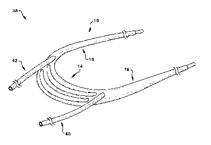
[0059] Another aspect of the present disclosure can include a system 38 (Fig.
2) for treating a regurgitant heart valve in a subject. The system 38 can
comprise a
device 10, a first anchoring catheter 40, and a second anchoring catheter 42.
The
device 10 can be identically or similarly constructed as the device shown in
Figs. 1A-
B and described above. Each of the first and second anchoring catheters 40 and
42
can have an elongated, tubular shape and be made of one or a combination of
biocompatible materials, such as PTFE, ePTFE, PEEK, etc. The first and second
anchoring catheters 40 and 42 can be sized and dimensioned for insertion into
the
first and second lumens 26 and 28 of the device 10, respectively. In the
assembled
CA 02951413 2016-12-06
WO 2015/191946 PCT/US2015/035459
configuration of the system 38 shown in Fig. 2, the first and second anchoring
catheters 40 and 42 can be disposed in, and at least partially extend through,
the
first and second lumens 26 and 28 (respectively) of the device 10. The first
and
second anchoring catheters 40 and 42 can be sized and dimensioned to suspend
and position the device 10 in the left ventricle when the device is implanted
in a
subject. As discussed in more detail below, the first and second anchoring
catheters
40 and 42 can be configured to receive a guidewire, which may then be used to
convey one or more anchoring elements therethrough.
[0060] Method
[0061] Another aspect of the present disclosure can include a method 44 (Fig.
3) for treating a regurgitant heart valve or a condition associated therewith,
such as,
in the depicted example, ischemic mitral regurgitation (IMR) or functional
mitral
regurgitation (FMR) associated with a regurgitant mitral valve. IMR and FMR
can
result from mitral leaflet tethering due to left ventricular remodeling.
Heterogeneity in
local or global left ventricular remodeling can result in differential
tethering patterns
and affect mitral valve function and the degree of mitral regurgitation.
Illustrating
examples of FMR are Figs. 4-5, which show regurgitant mitral valves 60 as a
result
of asymmetrical leaflet tethering and symmetrical tethering, respectively. The
method 44 of the present disclosure will be illustrated in terms of treating a
regurgitant mitral valve 60. It will be appreciated that even though the
method 44 is
described below in terms of treating a regurgitant mitral valve 60, the method
can
alternatively be used to treat another heart valve, such as a regurgitant
tricuspid
valve (not shown). Likewise, it will be appreciated that even though the
method 44 is
described below using a percutaneous approach from the right ventricle across
the
interventricular septum into the left ventricle, the method can alternatively
be used
with, for example, a transcatheter or percutaneous approach with a flexible
electromagnetic or mechanical adjustment catheter, optionally into any portion
of the
heart (e.g., a transseptal, transaortic, transapical, and/or transatrial
approach), such
as under echocardiographic guidance.
[0062] Referring to Fig. 3, Step 46 of the method 44 can include providing a
device 10. The device 10 can be configured in an identical or similar manner
as the
device described above. For example, the device 10 can comprise a flexible,
11
CA 02951413 2016-12-06
WO 2015/191946 PCT/US2015/035459
elongated body 12 having a central chordae support portion 14 disposed between
first and second arms 16 and 18, each of which includes first and second
lumens 26
and 28 (respectively) that extend longitudinally therethrough.
[0063] After selecting an appropriate device 10, a first guidewire 62 (Fig. 6)
can be inserted into the vasculature of the subject and then advanced into any
suitable heart chamber--here, for the sake of discussion, into the right
ventricle 64.
Using the Seldinger technique, for example, the first guidewire 62 can be
advanced
through the jugular vein (not shown) or a femoral vein (not shown) into the
right
ventricle 64. Next, the first guidewire 62 can be advanced across the
interventricular
septum 66 into the left ventricle 68. Once a distal end 70 of the first
guidewire 62 is
positioned in the left ventricle 68, a device delivery catheter 72 can be
advanced
over the first guidewire 62 into the left ventricle 68. The first guidewire
62/device
delivery catheter 72 can then be advanced behind and around the chordae
tendineae 74 associated with the posterior mitral valve leaflet 76. (It should
be
understood that no septal puncture is required for some other insertion paths,
and
that, regardless of the insertion path taken, the distal end 70 of the first
guidewire 62,
or any other desired structure of the system 38, can be placed into any
desired heart
chamber by one of ordinary skill in the art using the structures and
principles
described herein.)
[0064] At Step 48, a wire loop can be created in the left ventricle 68. As
shown in Fig. 6, a second guidewire 78 can be inserted into the right
ventricle 64
(e.g., using the Seldinger technique) and then advanced across the
interventricular
septum 66. A second catheter (not shown) can then be advanced over the second
guidewire 78 into the left ventricle 68. A distal end 80 of the second
guidewire 78
and the second catheter can be positioned immediately adjacent the distal end
70 of
the first guidewire 62. Next, a snare (not shown) can be threaded through the
second catheter and progressively fed therethrough until the snare catches the
distal
end 70 of the first guidewire 62, thereby forming a wire loop around the
chordae
tendineae 74 associated with the posterior mitral valve leaflet 76.
[0065] Once the wire loop has been formed, the device 10 can be loaded into
the device delivery catheter 72. The device 10 can then be advanced through
the
device delivery catheter 72, along the wire loop, until the device is
positioned within
12
CA 02951413 2016-12-06
WO 2015/191946 PCT/US2015/035459
the left ventricle 68 (Step 50). In particular, the device 10 can be advanced
along
the wire loop until the distal end 36 of the second arm 18 is located in the
right
ventricle 64 (as shown in Fig. 7). Positioning the device 10 at Step 50
results in the
central chordae support portion 14 of the device 10 partially encircling the
chordae
tendineae 74 associated with the posterior mitral valve leaflet 76, as well as
the
distal end 36 of each of the first and second arms 16 and 18 being located in
the
right ventricle 64. Sutures, hooks, barbs, screws, flexible discs, loop
members,
bands, rings, or any other aid mechanism may be provided to any structure of
the
device 10, and be used in cooperation with any patient tissue structure, to
affix the
device 10 to the patient tissue, whether or not the chordae are permitted to
slide on
these aid mechanisms and/or the central chordae support portion 14 of the
device
10. Similarly, it is contemplated that a material and/or coating/impregnate
could be
provided to at least the central chordae support portion 14 of the device 10
to either
encourage or discourage tissue ingrowth, as desired for a particular patient
treatment plan.
[0066] Optionally, anchors of any suitable type (omitted from Fig. 7) could be
provided at the distal end 36 of each of the first and second arms 16 and 18
within
the right ventricle, before or after a length of the first and second arms 16
and 18 are
optionally adjusted, to tension the first and second arms and thus place a
tensile
force on the chordae tendineae 74 via the central chordae support portion 14,
thus
completing the surgical procedure. However, it is also contemplated that
additional
structures could be installed, such as in the sequence described in Steps 52-
56 of
Fig. 3 and depicted in Figs. 7-11.
[0067] At Step 52, first and second anchoring catheters 40 and 42 (Fig. 8) can
be advanced into the right ventricle 64 and inserted into the first and second
lumens
26 and 28 of the first and second arms 16 and 18, respectively. As shown in
Fig. 8,
the first and second anchoring catheters 40 and 42 can be guided to (and
through)
the first and second lumens 26 and 28, respectively, using separate guidewires
82.
The first and second anchoring catheters 40 and 42 can then be advanced
through
the first and second lumens 26 and 28 (respectively) until a distal end 84 of
each of
the first and second anchoring catheters 40 and 42 extends beyond the
respective
first opening 30 of the device 10. The distal end 84 of each of the first and
second
13
CA 02951413 2016-12-06
WO 2015/191946 PCT/US2015/035459
anchoring catheters 40 and 42 can then be advanced until it is substantially
flush
with the endocardial surface of the left ventricle posterior-lateral wall.
Next, a distal
end 86 of each of the separate guidewires 82 can be advanced through the left
ventricle wall to reach the pericardial space, whereafter the first and second
anchoring catheters 40 and 42 are progressively fed over the guidewires 82.
[0068] The device can be anchored in the left ventricle 68 at Steps 54-56 of
the method 44. First, for example, separate external left ventricle anchors 88
(e.g.,
single or double titanium anchors) (Fig. 9) can be advanced through the first
and
second anchoring catheters 40 and 42. The external left ventricle anchors 88
can
then be deployed outside an affected heart tissue of the patient--here, the
left
ventricle posterior-lateral wall. Next, separate internal right ventricle
anchors 90
(e.g., single or double titanium anchors) can be advanced over the separate
guidewires 82 and then deployed to seat the internal right ventricle anchors
on an
anchor heart tissue of the patient--here, the right ventricle side of the
interventricular
septum 66. One example of an anchoring system that may be suitable for Steps
54-
56 of the method 44 is commercially available from BIOVENTRIX Inc. (San Ramon,
CA). The device 10, when properly anchored in the left ventricle 68, is
illustrated in
Figs. 9-10.
[0069] At Step 58, the position of the device 10 and, in particular, the
central
chordae support portion 14, can be adjusted as needed to ensure proper mitral
leaflet coaptation. For example, the external left ventricle anchors 88 and
the
internal right ventricle anchors 90 can be cinched (e.g., using
echocardiographic
guidance from the right ventricle 64) so that the central chordae support
portion 14
and the first and second anchoring catheters 40 and 42 pull the posterior
leaflet
subvalvular apparatus (e.g., the chordae tendineae 74) along with the left
ventricle
wall toward the interventricular septum 66. The device 10 may also or instead
be
adjusted by tightening the distal ends 36 of the first and second arms 16 and
18, at
the level of the sub-valvular apparatus. The valve competency can be tested by
fluoroscopy, injecting contrast solution through the valve, and/or by
echocardiographic guidance while the device 10 is percutaneously or surgically
tightened or loosened.
14
CA 02951413 2016-12-06
WO 2015/191946 PCT/US2015/035459
[0070] Consequently, as shown in Fig. 11, mitral regurgitation may be
reduced, prevented, or eliminated by normalizing the angle of mitral leaflet
coaptation, increasing the surface area of mitral leaflet coaptation, and
restoring the
left ventricle 68 to a more desired size and shape. Stated differently, the
device 10
helps to correct the unbalance angle of leaflets coaptation of the regurgitant
valve,
such as by working as a "coaptation alignment support", and optionally also by
moving the left ventricular wall more medially in order to obtain better
mitral or
tricuspid valve competency and/or by developing left and/or right ventricle
reverse
remodeling.
[0071] It is contemplated that portions of the device (e.g., the distal ends
36)
could include, or be attached to, a stent, hooks, barbs, screws, flexible
discs, loop
members, or any other desired aid mechanisms, to anchor the device 10 to
anatomic
heart structures such as, but not limited to, the ventricular wall,
interventricular septal
wall, interatrium septal wall, atrial wall, coronary sinus, pericardium, IVC,
SVC,
pulmonary veins, or any other desired patient tissue structures. For example,
and as
shown in Fig. 12, versions of the first and second anchoring catheters 40 and
42 are
seen as being anchored to the left atrium 92 wall, the interatrial septum 94,
and the
interventricular septum 66--these various anchoring locations could be used
together
(as shown), in any combination with or without other anchoring locations, or
singly.
The system 38, or portions thereof, can also or instead be supported by
additional
anchoring mechanisms that are suspended from any suitable patient tissue
structures.
[0072] Fig. 13 shows an alternate configuration of the device 10, wherein two
central chordae support portions 14A and 14B are used to mutually tension two
corresponding sets of chordae tendineae 74A and 74B, respectively. The first
and
second anchoring catheters 40 and 42 extending between the two central chordae
support portions 14A and 14B to draw the associates chordae tendineae 74A and
74B closer together, thus coapting the associated valve leaflets and reducing
or
preventing unwanted regurgitation.
[0073] In Fig. 14, another alternative configuration of the device 10 is
shown,
with a single central chordae support portion 14 being anchored directly to
the left
CA 02951413 2016-12-06
WO 2015/191946
PCT/US2015/035459
ventricle posterior-lateral wall by the first and second anchoring catheters
40 and 42,
with no other tensioning structures being provided within the left ventricle
68.
[0074] The sequence of Figs. 15A-15E schematically depicts placement of
any configuration of the device 10 via a trans interventricular septal
approach which
is otherwise similar to the sequence depicted in Figs. 6-8. In Fig. 15A, a
first
guidewire 62 is passed through the aorta 96 and into the left ventricle 68. In
Fig.
15B, a loop is made around the chordae tendineae 74 with the guidewire 62.
Fig.
15C depicts a second guidewire 78 approaching the interventricular septum 66
from
the right ventricle 64--the first guidewire 62 could still be maintained in
the loop,
and/or at least a portion of the device 10 may have already been passed over
the
first guidewire 62 to achieve the looped structure shown in Fig. 150.
[0075] Turning to Fig. 15D, the second guidewire 78 (optionally with the
assistance of another guidewire, not shown) "catches" two portions of the loop
formed by the first guidewire 62 and/or the device 10 in any suitable manner,
consecutively or concurrently. Fig. 15E, then, shows the device 10 as
installed with
one or more internal right ventricle anchors 90, similar to the arrangement
shown in
Fig. 9, with or without the addition of the external left ventricle anchors.
[0076] In Fig. 16, another alternative configuration of the device 10 is
shown,
with a single central chordae support portion 14 being anchored directly to
the left
ventricle posterior-lateral wall by the first and second anchoring catheters
40 and 42,
after the heart has been accessed pericardially.
[0077] In summary, the device 10, system 38, and/or method 44 described
and depicted herein can help normalize and remodel the leaflet shape and
function,
correct the leaflet mobility, coapt by improving the leaflet closure movement
during
systole, and/or corrects the unbalance angle of leaflet coaptation and sub-
valvular
apparatus position for valve regurgitation, without removing leaflet tissue,
chordal
shortening, transposing or replacement, placating and deforming the valve
annulus,
or using other surgical techniques or sophisticated procedures for making the
valve
competent. The device 100 can be adjustable depending on the anatomic leaflet
and
sub-valvular apparatus configuration, and the free-edge leaflet coaptation
angle, to
obtain normal correction by mechanical or electromagnetic adjustment through a
flexible catheter by echo guidance, or by transcatheter or percutaneous
approach
16
with a flexible electromagnetic or mechanical adjustment catheter by
transeptal, trans
atrial, trans apical, and/or trans ventricular approach under
echocardiographic
guidance.
[0078] While aspects of this disclosure have been particularly
shown and
described with reference to the example aspects above, it will be understood
by those
of ordinary skill in the art that various additional aspects may be
contemplated. For
example, the specific methods described above for using the apparatus are
merely
illustrative; one of ordinary skill in the art could readily determine any
number of tools,
sequences of steps, or other means/options for placing the above-described
apparatus,
or components thereof, into positions substantively similar to those shown and
described herein. In an effort to maintain clarity in the Figures, certain
ones of
duplicative components shown have not been specifically numbered, but one of
ordinary skill in the art will realize, based upon the components that were
numbered,
the element numbers which should be associated with the unnumbered components;
no differentiation between similar components is intended or implied solely by
the
presence or absence of an element number in the Figures. Any of the described
structures and components could be integrally formed as a single unitary or
monolithic
piece or made up of separate sub-components, with either of these formations
involving
any suitable stock or bespoke components and/or any suitable material or
combinations
of materials, such as, but not limited to, metal, plastic, ElgiloyTM, Nitinol,
stainless steel,
titanium, pyrrolitic carbon, and the like, or any combination thereof;
however, the
chosen material(s) should be biocompatible (and/or covered/coated with
synthetic or
natural biological and biocompatible materials) for many applications. Any
structure
described herein could be at least partially coated, impregnated with, or
otherwise
provided with pharmacologic and/or biologic agents, which may be permitted or
designed to leach or otherwise disperse into surrounding patient tissue
structures. Any
of the described structures and components could be disposable or reusable as
desired
for a particular use environment. Any component could be provided with a user-
perceptible marking to indicate a material, configuration, at least one
dimension, or the
like pertaining to that component, the user-perceptible marking aiding a user
in
selecting one component from an array of similar components for a particular
use
environment. A
17
CA 2951413 2018-04-09
CA 02951413 2016-12-06
WO 2015/191946 PCT/US2015/035459
"predetermined" status may be determined at any time before the structures
being
manipulated actually reach that status, the "predetermination" being made as
late as
immediately before the structure achieves the predetermined status. The term
"substantially" is used herein to indicate a quality that is largely, but not
necessarily
wholly, that which is specified--a "substantial" quality admits of the
potential for some
relatively minor inclusion of a non-quality item. Though certain components
described herein are shown as having specific geometric shapes, all structures
of
this disclosure may have any suitable shapes, sizes, configurations, relative
relationships, cross-sectional areas, or any other physical characteristics as
desirable for a particular application. Any structures or features described
with
reference to one aspect or configuration could be provided, singly or in
combination
with other structures or features, to any other aspect or configuration, as it
would be
impractical to describe each of the aspects and configurations discussed
herein as
having all of the options discussed with respect to all of the other aspects
and
configurations. A device or method incorporating any of these features should
be
understood to fall under the scope of this disclosure as determined based upon
the
claims below and any equivalents thereof.
[0079] Other aspects, objects, and advantages can be obtained from a study
of the drawings, the disclosure, and the appended claims.
18