Note: Descriptions are shown in the official language in which they were submitted.
CA 02951506 2016-12-07
WO 2015/189843
PCT/IL2015/050585
TISSUE REPAIR DEVICE AND METHOD
FIELD AND BACKGROUND OF THE INVENTION
The present invention relates to a device and method for guiding and anchoring
an implant to a tissue. Embodiments of the present invention relate to a
device and
method for guiding and anchoring a suture, suture anchor or mesh in a
sacrospinous
ligament for the purpose of repairing a pelvic floor disorder pelvic organ
prolapse
(POP).
Trans-vaginal pelvic floor repair is a surgical procedure which utilizes blunt
tissue dissection to provide access to the sacrospinous ligament from the
posterior
vaginal wall. A sling or mesh is then anchored to the sacrospinous ligament
and the
vaginal apex or the uterine isthmical fibrotic ring, cervix or body, to
thereby support
prolapsing tissues and/or organs.
Although pelvic floor repair is a common procedure, access to the sacrospinous
ligament is typically effected by improvised manual blunt dissection
techniques and/or
use of off the shelf instruments.
Centro-apical reconstruction is key for proper pelvic organ prolapse (POP)
repair. The premium supportive pelvic structure is the sacrospinous ligament
(SSL)
which is positioned at the posterior aspect of the pelvis. The SSL is a robust
ligament
and thus provides a long lasting solution. Since it is positioned high in the
pelvis and
medially the SSL provides a level 1 support (DeLancey) and reduces the
likelihood of
dyspareunia when utilized for prolapse repair.
Vaginal wall access to the SSL can be difficult and hazardous since organs and
tissues surrounding the access path can easily be injured during dissection.
Present day
approach for accessing the SSL starts with an incision at the mid-line of the
posterior or
anterior vaginal wall followed by lateral dissection under the sub-mucosal
fascia to the
pelvic side wall and dissection towards the ischial spine to the mid SSL
(MSSL).
This approach decreases risk of tissue injury by bypassing the bladder/rectum
while maintaining accurate navigation along the above mentioned landmarks.
Such an
approach requires a high degree of skill and as such can lead to a high rate
of
complications.
CA 02951506 2016-12-07
WO 2015/189843
PCT/IL2015/050585
2
While reducing the present invention to practice, the present inventors have
developed a device which can be used to deliver a tissue anchor to anatomical
landmarks
and structures such as the ischial spine and the sacrospinous ligament from
the vaginal
cavity.
SUMMARY OF THE INVENTION
According to one aspect of the present invention there is provided a surgical
device comprising: a housing adapted for mounting on a finger of a user; and
(b) at least
one guide tube attached along a length of the housing, the at least one guide
tube being
configured for guiding a tissue repair implant from a proximal opening to a
distal
opening thereof.
According to further features in preferred embodiments of the invention
described below, the guide tube is attached to the housing such that the
proximal
opening protrudes beyond a proximal end of the housing.
According to still further features in the described preferred embodiments the
guide tube is attached to the housing such that the proximal opening is
positioned above
a back of a hand of the user when the housing is mounted on the finger.
According to still further features in the described preferred embodiments the
housing is configured so as to enable the user to palpate the tissue via the
finger attached
to the housing.
According to still further features in the described preferred embodiments the
housing is open at a distal end thereof.
According to still further features in the described preferred embodiments the
guide tube is attached to the housing such that the distal opening abuts the
tissue when
the finger of the user contacts the tissue.
According to still further features in the described preferred embodiments the
guide tube is attached to the housing such that the distal opening is
displaced from the
tissue when the finger of the user contacts the tissue.
According to still further features in the described preferred embodiments the
suture end is attached to an anchor.
CA 02951506 2016-12-07
WO 2015/189843
PCT/IL2015/050585
3
According to still further features in the described preferred embodiments the
housing is configured for enabling flexion of the finger at a distal and/or
proximal
interphalangeal joint.
According to still further features in the described preferred embodiments the
.. housing is configured for attaching an imaging device thereto.
According to still further features in the described preferred embodiments the
imaging device is an ultrasound transducer.
According to still further features in the described preferred embodiments an
imaging head of the ultrasound transducer is capable of abutting the tissue
when the
1() ultrasound transducer is attached to the housing.
According to still further features in the described preferred embodiments the
imaging head abuts the tissue when the finger attached to the housing contacts
the tissue.
According to still further features in the described preferred embodiments the
tissue is a posterior-lateral vaginal wall and the housing is configured for
delivery into
the vaginal canal via the finger.
According to still further features in the described preferred embodiments
when
the housing is positioned within the vaginal canal with the finger in contact
with the
posterior-lateral vaginal wall, the proximal opening of the at least one guide
tube
extends out of the vaginal canal.
According to still further features in the described preferred embodiments the
tissue repair implant is a mesh, a sling, a suture or a suture-anchor.
According to another aspect of the present invention there is provided a
method
of repairing a pelvic floor disorder comprising: (a) positioning a surgical
device via a
finger within a vaginal cavity, the surgical device including a housing
adapted for
mounting on the finger and at least one guide tube attached along a length of
the
housing; (b) using the finger to palpate a posterior-lateral wall of the
vaginal cavity and
locate a sacrospinous ligament therethrough; and (c) advancing a tissue repair
implant
through the at least one guide tube and through a posterior-lateral wall to
thereby anchor
the tissue repair implant to the sacrospinous ligament.
According to still further features in the described preferred embodiments the
tissue repair implant is a mesh, a sling, a suture or a suture-anchor.
CA 02951506 2016-12-07
WO 2015/189843
PCT/IL2015/050585
4
According to still further features in the described preferred embodiments the
device further includes an ultrasound transducer attached to the housing and
further
wherein (b) is effected under ultrasound guidance.
According to still further features in the described preferred embodiments the
guide tube is attached to the housing such that a proximal opening of the
guide tube is
positioned outside the vaginal cavity.
According to another aspect of the present invention there is provided a
method
of repairing a pelvic floor disorder comprising (a) positioning a surgical
device via a
finger within a vaginal cavity, the surgical device including a housing
adapted for
mounting on the finger and at least one guide tube attached along a length of
the
housing; (b) using the finger to palpate a posterior-lateral wall of the
vaginal cavity and
locate a sacrospinous ligament therethrough; (c) advancing a suture-anchor
through the
at least one guide tube and through a posterior-lateral wall to thereby anchor
the suture-
anchor to the sacrospinous ligament; and (d) advancing a mesh over at least
one suture
thread of the suture-anchor; and (e) tying the at least one suture thread to
secure the
mesh in position.
According to another aspect of the present invention there is provided a
device
for delivering a mesh to an intrabody location using a suture anchor as a
guide and the
method described above. The device includes a hollow tube for accepting one or
more
suture threads, and a distal end for accepting a releasable cuff attached to
the mesh.
The present invention successfully addresses the shortcomings of the presently
known configurations by providing a device and method for tissue repair from
within a
body cavity.
Unless otherwise defined, all technical and scientific terms used herein have
the
same meaning as commonly understood by one of ordinary skill in the art to
which this
invention belongs. Although methods and materials similar or equivalent to
those
described herein can be used in the practice or testing of the present
invention, suitable
methods and materials are described below. In case of conflict, the patent
specification,
including definitions, will control. In addition, the materials, methods, and
examples are
illustrative only and not intended to be limiting.
CA 02951506 2016-12-07
WO 2015/189843
PCT/IL2015/050585
BRIEF DESCRIPTION OF THE SEVERAL VIEWS OF THE DRAWINGS
The invention is herein described, by way of example only, with reference to
the
accompanying drawings. With specific reference now to the drawings in detail,
it is
stressed that the particulars shown are by way of example and for purposes of
illustrative
5 discussion of the preferred embodiments of the present invention only,
and are presented
in the cause of providing what is believed to be the most useful and readily
understood
description of the principles and conceptual aspects of the invention. In this
regard, no
attempt is made to show structural details of the invention in more detail
than is
necessary for a fundamental understanding of the invention, the description
taken with
the drawings making apparent to those skilled in the art how the several forms
of the
invention may be embodied in practice.
In the drawings:
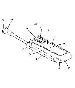
Fig. 1 illustrates an embodiment of the present device having a single guide
tube.
Fig. 2 illustrates an embodiment of the present device having two guide tubes.
Fig. 3 illustrates an embodiment of the present device having a single guide
tube
and an imaging device.
Fig. 4 illustrates an embodiment of the present device having two guide tubes
and an imaging device.
Fig. 5 illustrates the device of Figure 1 mounted on a finger of a user.
Figs. 6-7 illustrate insertion of the implant delivery device into the guide
tube.
Fig. 8 illustrates the device of Figure 1 mounted on a finger with the implant
delivery device positioned within the guide tube.
Figs. 9a-h illustrate mesh delivery and securement using the suture-anchor
delivered by the present device as a guide.
DESCRIPTION OF THE PREFERRED EMBODIMENTS
The present invention is of a device which can be used for tissue repair, and
specifically of a finger mounted device which can be used to deliver a tissue
repair
implant through a wall of a body cavity.
The principles and operation of the present invention may be better understood
with reference to the drawings and accompanying descriptions.
CA 02951506 2016-12-07
WO 2015/189843
PCT/IL2015/050585
6
Before explaining at least one embodiment of the invention in detail, it is to
be
understood that the invention is not limited in its application to the details
set forth in the
following description or exemplified by the Examples. The invention is capable
of other
embodiments or of being practiced or carried out in various ways. Also, it is
to be
understood that the phraseology and terminology employed herein is for the
purpose of
description and should not be regarded as limiting.
Pelvic organ prolapse (POP), and especially apical central supportive defect
(ACSD), significantly affects the quality of life of about 20% of the female
population.
POP is typically corrected via a transabdominal or a transvaginal surgical
procedure.
The transvaginal reconstruction approach is regarded as superior to the
transabdominal approach due to a shorter operative time and hospital stay and
quicker
rehabilitation. However, transvaginal procedures require advanced surgical
skill and as
such are performed by a rather small and highly qualified group of surgeons.
In the transvaginal procedure, a surgeon can elect to suspend the vaginal apex
(VA) or the uterine cervix (UC) to the sacrospinous-ligament (SSL), sacrum,
arcus
tendineus fascia pelvis (ATFP) or other potentially solid supportive pelvic
structures,
which are accessed via anterior or posterior vaginal wall incisions and blunt
dissection
of tissues.
Creating an access path to these tissues is a major challenge of transvaginal
procedures since it requires complicated navigation to the pelvic side wall
(PSW),
ischial spine (IS) and then to the mid SSL (MSSL) or the sacrum which carries
with it a
risk of damaging the bladder, rectum, blood vessels, nerves, ureters, etc.
Most POP procedure complications are attributed to the dissection necessary to
create the tissue path to the elected tissue support site.
In order to traverse these limitations of prior art transvaginal procedures,
the
present inventors have devised an approach which utilizes a finger mounted
intravaginal
device which enables the surgeon to palpate the SSL and deliver a tissue
repair implant
thereto.
As used herein, the phrase "pelvic floor disorder" refers to any disorder of
the
pelvic floor that is associated with prolapse, herniation or incorrect
anatomical
positioning of pelvic floor tissues.
CA 02951506 2016-12-07
WO 2015/189843
PCT/IL2015/050585
7
Thus, according to one aspect of the present invention there is provided a
device
for tissue repair, and in particular, repair of pelvic floor disorders. The
term "repair"
when used herein with reference to pelvic floor disorders refers to correction
(complete
or incomplete) of anatomy. via, a tissue repair implant such as a suture,
suture anchor,
mesh, sling and/or the like.
The present device includes a housing adapted for mounting on a finger of a
user,
preferably an index finger of the user. The housing can be configured for
mounting over
the finger tip or any portion of the finger (up to the distal or proximal
interphalangeal
joint or the entire finger). The device further includes at least one guide
tube (preferably
1 or 2) attached along a length of the housing. Thus, the guide tube runs
parallel or
substantially parallel to the finger of the user when the housing is mounted
thereupon.
The housing is substantially 'finger'-shaped (elongated slightly compressed
cylinder) with a longitudinal lumen mountable over a finger and smooth
external walls
(optionally having longitudinal apertures) for facilitating insertion of the
housing into a
body cavity (e.g. vaginal or anal cavity) when mounted over the finger. The
distal end
(away from user) includes an opening for the tip of the finger, such opening
can be
covered by an elastic this membrane.
The housing can be fabricated from a polymer or alloy (preferably
biocompatible) using molding or machining approaches. Typical dimensions for
housing
are 65 mm length, and 18 mm internal diameter. The housing includes a finger
adjusting
and retaining mechanism in order to ensure that the housing securely mounts
onto a
finger of any length or diameter without inadvertently detaching. Such a
mechanism can
include an elastic tab mounted within the housing lumen or an adjustment
mechanism
for adjusting the length, height and/or width (or diameter) of the housing.
One example
of such a mechanism is described below with reference to the Figures.
The guide tube(s) is attached to the housing or co-fabricated therewith and is
configured for guiding an implant from a proximal opening to a distal opening
thereof.
The guide tube is preferably attached along a side of the housing such that
its
runs along a side of a finger (when the hand is viewed from the top). A
detailed
description of one embodiment of the present device is provided hereinbelow
with
reference to Figures 1-8.
CA 02951506 2016-12-07
WO 2015/189843
PCT/IL2015/050585
8
As is mentioned hereinabove, the device of the present invention can be used
for
tissue repair by enabling delivery of a tissue repair implant from the guide
tube. Such
guiding can be effected with or without imaging guidance. When performed under
imaging, the present device is configured for attaching an imaging device
thereto. One
example of an imaging device is an ultrasound transducer which can be attached
to a
dedicated bracket provided on the housing.
The present device can further include irrigation lumens for attaching an
irrigation source and/or suction.
Referring now to the drawings, Figures 1-8 illustrate the present device which
is
.. referred to herein as device 10. Device 10 is configured for intravaginal
access and
pelvic floor repair, however, it should be noted that device 10 can be
modified for access
into other body cavities such as the anal canal to affect repair therein or
therethrough.
Device 10 includes a housing 12 which is substantially finger shaped and is 6
cm
in length and 1.8 cm in diameter. Housing can be fabricated from plastic,
metal or a
rubber like material. Housing 12 is configured for mounting over an entire
index finger
(see Figure 5) but will also provide the required functionality if configured
for mounting
over a portion of this finger. Mounting can be over a naked finger or one
covered by a
surgical glove. Housing 12 includes a proximal opening 14 and distal opening
16
forming lumen 18 surrounded by walls 20. Walls 20 can include several
apertures 22
(top aperture 22 shown) and a finger retaining mechanism 24 for elastically
engaging the
finger (Figure 5) to ensure that housing 12 is retained on a finger regardless
of its
dimensions. Finger retaining mechanism 24 can be designed to accommodate any
finger
size by providing an accommodative elastic force (downward) on the finger
surface.
Such a force would be enough to trap the finger within housing but would still
enable a
user to remove the housing by sliding it off the finger. The diameter of the
index finger
distal phalanx ranges between 13-18 mm for most individuals and thus a single
design
can be used to accommodate such a finger size range.
Distal opening 16 is sized and configured to enable a tip of the finger to
protrude
therethrough when housing 12 is mounted ion a finger. This enables a user to
palpate
.. tissue wall when device 10 is in use and positioned within the vaginal
cavity. Distal
opening 16 can be covered by a thin elastic membrane that enables palpation
and yet
CA 02951506 2016-12-07
WO 2015/189843
PCT/IL2015/050585
9
provides a barrier between the user's finger from the tissue in cases where
the user is not
wearing gloves.
Device 10 further includes at least one guide tube 28 (1 shown in Figure 1. 2
shown in Figure 2) Guide tube can be attached to housing 12 via brackets 29
(shown in
Figures 1-2). Guide tube 28 is configured as elongated tube having proximal
and distal
openings (30 and 32 respectively). Guide tube 28 has a length greater than
that of
housing 12 such that when housing 12 is positioned within the vaginal cavity
(with distal
opening at or near the lateral posterior wall), proximal opening 30 is
positioned outside
the vaginal cavity to allow access and delivery of a tissue repair implant
therethrough (as
is shown in Figures 6-8). The length of guide tube 28 can be anywhere from 70-
120
mm, while the outer and inner diameter can be anywhere from 2-8 mm and 1-3 mm
(respectively). Guide tube 28 can be fabricated from a substantially rigid
material such
as stainless steel or from an elastic material such as Nitinol or a polymer
such as
Polycarbonate. An elastic embodiment of guide tube 28 can be advantageous in
cases
where the distal opening of the tube is not aligned with the proximal opening.
Guide tube 28 includes a port 34 for allowing a delivery device 36 (shown in
Figures 6-8) to easily access the lumen of guide tube 28.
Delivery device 36 can be constructed from two coaxial tubes. An Internal tube
attached to a tissue anchor 38 (shown in Figure 6) and an external rigid tube
which is
coaxially disposed around the first tube. The tissue anchor 38 (which can be
attached to
a suture, mesh or sling) can be delivered from the rigid tube by advancing the
first tube
therewithin. To effect such delivery, delivery device 36 includes a handle 40
for
actuating forward movement (in a distal direction) of the first tube within
the rigid tube.
Delivery device 36 is preferably capable of puncturing the vaginal wall and
driving
tissue anchor 38 through the tissue and into the target site (e.g. MSSL). As
such, the
distal end of the first tube can be configured for tissue puncturing (beveled,
double
beveled or conical). Alternatively, tissue anchor 38 can be configured for
tissue
puncturing or still alternatively an initial incision in the vaginal wall can
be used to
deliver the first tube therethrough. The first and rigid tubes or anchor 38
can include an
imaging marker for identifying these elements within an imaging plane. An
example of
an echogenic marker which can be used along with an ultrasound probe is
provided in
US20050228288.
CA 02951506 2016-12-07
WO 2015/189843
PCT/IL2015/050585
As is shown in Figure 2, device 10 of the present invention can include 2
guide
tubes 28. Such a configuration allows a user to choose the best side for
delivery of a
tissue repair implant or to deliver two implants.
As is shown Figure 3, device 10 can also include an imaging device 50 which is
5 .. attached to housing 12 with an imaging head 52 positioned at a distal end
of house 12 on
a side opposite of guide tube 28 distal opening 32 or in the middle, as is
shown in Figure
4 (which depicts a device 10 having two guide tubes 28 and an imaging device
50).
One example of an imaging device which can be used in device 10 is an
ultrasound
imaging device. An ultrasound imaging device having a transducer head
positioned at
10 .. the distal end of housing near distal opening 32 of guide tube 28 can be
used to image
the SSL and surrounding structures/organs and provide additional guidance for
delivery
of the tissue repair implant.
Device 10 of the present invention can be used in a pelvic organ prolapse
(POP)
repair procedure as follows.
The site of anchoring is selected based on pre-palpation and/or pre-procedure
vaginal US.
The present device is positioned on an index finger of a dominant hand and a
delivery device (needle) is positioned within the guide tube such that a
distal end of the
external rigid tube of the delivery device is flush with the distal opening of
the guide
.. tube.
The present device is introduced into the vaginal cavity and the index finger
tip is
used to palpate the tissue target through the vaginal wall. The external rigid
tube of the
delivery device is then pressed against the vaginal wall at the region of the
ligament.
The internal tube of the delivery device is then actuated via the delivery
button of
the handle to deliver the anchor through the vaginal wall and into the
ligament. The
internal tube is then withdrawn leaving the anchor and attached suture/mesh in
position.
The suture end(s) are secured outside the vaginal cavity via forceps and the
initial pull
out force is verified by manually pulling on the suture ends. Optionally, the
procedure is
then repeated for the second side of the device thereby attaching a second
anchor-
suture/mesh to the ligament at a second site.
The suture ends are then attached to the uterine cervix fibrotic ring, the
serosa of
the vaginal apex, the utero-sacral ligaments, the vagina (in post hysterectomy
subjects),
CA 02951506 2016-12-07
WO 2015/189843
PCT/IL2015/050585
11
or any other appropriate centro-apical anchoring point of the pelvic floor as
is routine for
prolapse procedures.
As is described hereinabove, the present device can also be used to deliver a
mesh to the anchoring site. Figures 9a-h illustrate delivery of a mesh to the
anchoring
site using the suture anchor implanted by the present device as a guide.
Figure 9a illustrates anchor 38 positioned through the MSSL and attached to a
suture 38'.
A mesh delivery device 50 carrying mesh 52 having a distal cuff 54 (Figure 9c)
are threaded over one or both sutures 38'. Device 50 and attached cuff 54 are
advanced
over suture thread(s) (Figures 9b-d) by pushing device handle from outside the
vaginal
canal.
Cuff 54 is attached to the distal end of mesh 52 and is positioned around the
tip
of device 50. Cuff 54 can be fabricated from an alloy or polymer and can be
configured
to elastically constrict around sutures 38' when released from device 50 to
fixedly attach
to the sutures. Alternatively, cuff 54 can be rigid with one suture 38'
threaded through
the cuff and the other suture 38' around it (Figure 90. In any case, when in
position
close to or against the MSSL, cuff 54 is released from device 50 and device 50
is
removed (Figure 9g). Sutures 38' can then be tied around cuff 54 by running a
knot 56
from outside the body to cuff 54 (Figure 9h).
The proximal end of Mesh 52 can then be secured to anchor sutures using
approaches well known in the art.
As used herein the term "about" refers to 10 %.
Additional objects, advantages, and novel features of the present invention
will
become apparent to one ordinarily skilled in the art upon examination of the
following
example, which is not intended to be limiting.
EXAMPLE
Reference is now made to the following example. which together with the above
descriptions, illustrate the invention in a non limiting fashion.
A prototype of the device described herein was used in a pelvic repair
procedure.
Ten female subjects 42 to 76 years of age and having Centro-apical prolapse,
were
.. treated using the present device and a suture-anchor or mesh-anchor
implant.
12
Procedure
The patients were anesthetized and the present device was utilized to deliver
a
suture anchor with or without a mesh to the SSL after a posterior colpotomy
and
dissection were performed as described hereinabove. The suture was secured
with or
without a mesh and an initial pull out force was verified by manually pulling
on the
suture end. The suture end were then attached to the uterine cervix fibrotic
ring, the
serosa of the vaginal apex, or the utero-sacral ligaments, the vagina (in post
hysterectomy subjects), or any other appropriate centro-apical anchoring point
of the
pelvic floor as is routine for prolapse procedures. Both sutures were tied
while lifting
.. prolapsed uterus to its original location. The small colpotomy was then
closed to end the
procedure.
Two anchors were implanted in each patient (one per side). Two patients were
implanted with the suture-anchor and 8 with the suture + mesh anchor. Average
procedure time was 30 minutes.
Results
The pelvic organ prolapse quantification (POP-Q) score was 3-4 prior to the
procedure. Following the procedure the POP-Q score was 0/1. No device related
serious adverse events (SAE) or adverse events (AE) were observed immediately
following the procedure. A survey and examination conducted at 3 months post
.. operation, indicated patient satisfaction with no device related AE or
complaints from
the patients and no recurrence of prolapse.
It is appreciated that certain features of the invention, which are, for
clarity,
described in the context of separate embodiments, may also be provided in
combination
in a single embodiment. Conversely, various features of the invention, which
are, for
brevity, described in the context of a single embodiment, may also be provided
separately or in any suitable subcombination.
Although the invention has been described in conjunction with specific
embodiments thereof, it is evident that many alternatives, modifications and
variations
will be apparent to those skilled in the art. Accordingly, it is intended to
embrace all
such alternatives, modifications and variations that fall within the spirit
and broad scope
of the appended claims.
Date recue / Date received 2021-10-29
13
In addition, citation or identification of any i-eference in this application
shall not be
construed as an admission that such reference is available as prior art to the
present invention.
Date recue / Date received 2021-10-29