Note: Descriptions are shown in the official language in which they were submitted.
CA 02951683 2016-12-08
WO 2015/189646
PCT/GB2015/051752
1
Methodologies for measuring isopeptidase activity in biological samples in a
high
throughput manner
The present invention relates to materials and methods for high throughput
monitoring of
target engagement of isopeptidases, such as deubiquitylating enzymes by, inter
alia, small
molecule inhibitors. In particular the invention relates to development of
high throughput
assays to measure isopeptidase activity in biological samples, such as cells,
animal tissues,
animal tumours, human tissue or patient-derived biopsies. Furthermore, the
high
throughput assay can be used to measure isopeptidase in a biological sample
which contain
microorganisms with or without human or animal cells present. The invention
further
relates to methods for monitoring pharmaco-dynamic activities of isopeptidase
inhibitors in
biological samples. Furthermore, the invention relates to methods of
demonstrating the
activity status of isopeptidases under normal or pathological conditions and,
therefore,
methods of screening. The invention also provides assays that may be used as a
predictive,
diagnostic or prognostic tool for pathological conditions which are related
to, connected
with or due to defective isopeptidase activity. Such an assay may be used to
predict
pathological outcomes or treatment options. All methods and assays are
performed on
biological samples.
Background to the Invention
Ubiquitin, a 76 residue polypeptide is used as a posttranslational
modification to alter
intracellular protein functions in eukaryotic cells. Historically, the
ubiquitylation system was
identified as an ATP-dependent signal for targeting intracellular proteins for
proteasomal
degradation (Hershko, A. & Ciechanover, A., 1998, Ann. Rev. Biochem. 67, 425-
479;
Wilkinson, K. D., 2000, Sem in Cell & Dev. Bio., 11, 141-148 and Varshaysky,
A., 2012, Ann.
Rev. Biochem, 81, 167-176)).
Ubiquitylation of proteins is a multi-step process requiring the sequential
action of three
enzymes: ubiquitin-activating enzymes (Els) activate ubiquitin that is
subsequently loaded
onto ubiquitin-conjugating enzymes (E2s) and finally, the ubiquitin is
covalently linked to a
lysine side-chain from the E2s via specific recruitment of the target protein,
and facilitation
of the transfer by ubiquitin ligases (E3s). Ubiquitin can be linked to target
proteins singly, to
CA 02951683 2016-12-08
WO 2015/189646
PCT/GB2015/051752
2
form monoubiquitin adducts, however, in many cases, the initial ubiquitin is
then extended
by the covalent attachment (again by El, E2 and E3 proteins) of additional
ubiquitin
moieties to form poly-ubiquitin chains. Moreover, as any one of ubiquitin's
seven internal
lysine residues or its amino terminus can serve as sites for conjugation, the
resulting poly-
ubiquitin chains can have various, highly distinct topologies with different
biochemical and
biological functions. While Lys-48 (K48)-linked poly-ubiquitylation of
proteins is widely
recognised as a critical pathway for protein degradation, many additional
roles have been
attributed to either poly-ubiquitylation of proteins via non-K48 chains,
linear ubiquitin
chains as well as mono-ubiquitylation of proteins (Hicke, L., 2001, Nature
Reviews Mol Cell
Bio, 2, 195-201; Ikeda, F. & Dikic, I., 2008, EMBO Reports, 9, 536-542; lwai,
K., 2012, Trends
in Cell Biology, 22, 355-364 and Komander, D. & Rape, M., 2012, Ann. Rev.
Biochem, 81,
203-229). In addition to post-translational modification by ubiquitin, a whole
family of
ubiquitin-like (Ubl) modifications have been described. The degree of
conservation between
ubiquitin and ubiquitin like factors is somewhat limited at the protein
sequence level;
however, all members of the family share similar overall three-dimensional
structures and
highly related mechanisms of conjugation to their respective targets involving
El, E2 and E3
enzymes (Hay, R.T., 2007, Trends in Cell Biology, 17, 370-376; Hochstrasser,
M., 2009,
Nature 2009(458) 422¨ 499 and van der Veen, A.G., & Ploegh, H.L., 2012, Ann.
Rev.
Biochem, 81, 323-357).
Furthermore, since conjugation with ubiquitin or ubiquitin like molecules is a
crucial post-
translational modification that regulates cellular processes in eukaryotes, it
is a system that
pathogens encounter when attempting to infect humans and animals. Modification
with
ubiquitin or Ubl plays a central role in defence systems, for example. Thus,
pathogens such
as bacteria, viruses, fungi and parasites have evolved to exploit or evade the
host systems
for their own benefit, in order to maximise their chances of establishing a
successful
infection (Calistri et al, 2014, Cells, 386-417).
As for other protein post-translational modifications, conjugation of
ubiquitin or ubiquitin-
like factors to target protein is reversible, this being mediated by
isopeptidase enzymes that
are often collectively referred to as deubiquitylating enzymes or DUBs. DUBs
comprise a
large class of intra-cellular peptidases that cleave ubiquitin from
polypeptide substrates.
CA 02951683 2016-12-08
WO 2015/189646
PCT/GB2015/051752
3
Their substrates can be ubiquitin precursors, ubiquitin adducts, poly-
ubiquitin chains,
monoubiquitylated proteins or poly-mono-ubiquitylated proteins (lwai, K.,
2012, supra). If
ubiquitin-like peptidases are included, over a hundred DUBs are encoded by the
human
genome. DUBs can be classified into five families: ubiquitin carboxyl-terminal
hydrolases
(UCH), ubiquitin specific proteases (USPs), ovarian tumour proteases (OTU),
MJD (Josephins)
and MPN+/JAMM (JAB1/MPN/M0V34 metallo-enzymes). The first four families are
cysteine
peptidases, while MPN-F/JAMMs are metallopeptidases (Reyes-Turcu, F.E., eta!,
2009, Ann.
Rev. Biochem, 78, 363-397 and Sacco ii., eta!, 2010, IUBMB Life, 62, 140-157).
In addition
to processing ubiquitin and ubiquitin adducts, some USPs have been shown to
selectively
.. process specific ubiquitin-like proteins (for example, USP18 acts on the
ubiquitin-like protein
I5G15) (Zhang, D. & Zhang, D.E., 2011, J. Interferon and Cytokine Research,
31, 119-130) ). In
the case of the SUMO family of ubiquitin-like proteins, adducts are reversed
by a specialised
group of DUBs termed SENPs, all of which are cysteine peptidases, and some of
which may
also remove NEDD8. (Hay, 2007, supra and Dou, H., et al, 2010, Molecular Cell,
39, 333-345).
Similarly, microorganisms which have the ability to infect eukaryotic
organisms (pathogens)
have developed enzymes to reverse the conjugation of ubiquitin and Ubl
molecules to their
target protein, or have evolved strategies to affect the host enzymes. DUBs
have been
described for microorganisms.
While all DUBs are peptidases, there are considerable differences between
their precise
mechanisms of action, and there are also major differences in the regulatory
mechanisms
that modulate DUB selectivity and specificity (Komander, D., eta!, 2009,
Nature Reviews
Mol Cell Bio, 10, 550-563).). In this regard, DUBs can be classified into
three main categories
according to their type of substrate cleavage activity: some generate free
ubiquitin from
linear substrates, such as poly-ubiquitin chains or ribosomal protein fusions;
others liberate
ubiquitin from proteins modified post-translationally on lysine residues;
while, a third class
comprises DUBs that edit poly-ubiquitin chains (Komander eta!, 2009). For in
depth
discussions of DUB mechanism-of-action, we refer the reader to several
excellent reviews
on this subject (Reyes-Turcu, F.E., eta!, 2009, Ann. Rev. Biochem, 78, 363-
397; Linder, H.A.,
2007, Virology, 362, 245-256; Sun, S.C., 2008, Nature Reviews Immunology,
8(7), 501 ¨ 511;
CA 02951683 2016-12-08
WO 2015/189646
PCT/GB2015/051752
4
Hussaun, S. eta!, 2009, Cell Cycle, 8, 16884697 and Ramakrishna, S. et al,
2011, Cell and
Mol Life Sci, 68, 15-26).
Deubiquitylating enzymes may also be called deubiquitinating enzymes,
deubiquitinating
peptidases, deubiquitinases, ubiquitin isopeptidases, ubiquitin proteases,
ubiquitin
hydrolases, or DUBs.
The present invention relates to uses, methods and assays involving
isopeptidase enzymes.
An isopeptidase is an enzyme that catalyses the cleavage of an isopeptide
bond, especially
that between the terminal diglycine attached to ubiquitin, as well as cleavage
of ubiquitin
fusion or precursors through peptide bonds. As discussed above,
deubiquitylating enzymes
are isopeptidases. Other isopeptidases include SUMO (Small Ub modifier)
peptidases, ATG8
(Autophagy-related protein 8) peptidase, ISG15 (Interferon-stimulated gene 15)
peptidase,
NEDD8 (Neural precursor cell, developmentally down regulated 8) peptidase as
well as any
enzyme-cleaving adducts. Each isopeptidase catalyses the cleavage of an
isopeptide bond
involving a particular Ubl or Ub, and will be specific for the type of
reaction it catalyses.
In order to monitor the activity of isopeptidases, particularly
deubiquitylating enzymes, a
number of tools and assays have been developed. A number of in vitro assays
have been
designed to characterise both deubiquitylating enzymes and inhibitors: the
list includes
many covalent adducts to the carboxy-terminus of ubiquitin (Layfield, R. et al
,1999, Anal
Biochem., Oct 1999, 274(1): 40¨ 49; Lee, J.I., eta!, 1998, Bid Proced Online,
July 20; 1; 92-
99; Liu, C.C. et al, 1989, Dec5, J Biol Chem, 264(34): 20331-8; Falquet, L.,
et al, 1995, FEBS
Lett, Feb 6; 359(1) 73 ¨77; Larsen, C.N. et al, 1998, Biochemistry, Mar 10;
37(10): 3358-68;
Da ng L.C., et al, 1998, Feb 17, Biochemistry, 37(7):1868-79 and Tirat, A.,
eta!, 2005, Anal
Biochem, 343(2): 244-55 ). These assays have led to the biochemical and
biophysical
characterisation of a large number of DUBs.
A number of ubiquitin-based activity probe assays targeting the catalytic
sites of
deubiquitylating enzymes in cell extracts have been published in the last
decade
(Borodovsky, A., et al, 2001, EMBO J., 20(18):5187-96; Borodovsky, A. et al,
2002, Chem Biol,
9(10);1149-59; Ovaa, H. eta!, 2004, PNAS USA, 101(8):2253-8; Hemelarr, J.
eta!, 2004, J.
CA 02951683 2016-12-08
WO 2015/189646
PCT/GB2015/051752
Proteome Res., 3(2):268-76; Ovaa, H. et al, 2005, Methods Enzymol., 399: 468-
78; Galardy,
P. eta!, 2005, Methods Enzymol.,399:120-31, de Jong, A., eta!, 2012,
Chembiochem.,
13(15):2251-8 and Love K.R., et al, 2009, ACS Chem Biol, 4(4):275-87).
Ubiquitin-based
activity probe assays have been successfully used to characterise
deubiquitylating enzyme
5 inhibitors (Altun, M. eta!, 2011, Chem Biol, 18(11):1401-12 and Reverdy,
C., eta!, 2012,
Chem Biol, 19(4): 467-77). Similar activity-based probe assays have also been
developed for
the characterisation of the activity of ubiquitin-like peptidases as well as
non-human
ubiquitin-like peptidases (An, H. & Statsyuk, A.V.,2013, J Am Chem Soc,
135(45):16948-62
and Claessen, J.H. eta!, 2013, Chembiochem, 14(3):343-52). However, such probe
assays
are limited since they are low throughput, and as such are time and labour
intensive.
Furthermore, a number of high throughput assays have been developed to monitor
deubiquitylating enzyme activity in vitro. Many assays currently in use rely
on cleavage of
linear ubiquitin-fusions, (tetra-Ub, Ub-CEP52, Ub-GSTP1, Ub-DHFR, Ub-PESTc,
etc.) that are
synthesized recombinantly or chemically (Lee, JI, 1998, Larsen, C.N. 1998 and
Baker, R.T et
al, 1994, J Biol Chem, 269(41):25381-6). For small scale analysis of
deubiquitylating enzyme
activity, reaction products are analysed by gel electrophoresis, or are
selectively
precipitated and analysed by liquid scintillation spectrometry. Gel-based
procedures are
labour intensive and expensive, and while scintillation counting approaches
are quantitative
and allow processing of larger numbers of samples compared to gel-based
assays, they
require centrifugation and recovery of the supernatant. For higher throughput
assays,
fluorogenic substrates such as Ubiquitin-AMC (Ub-7-amino-4-methylcoumarin) or
Ubiquitin-
Rhodamine, have been employed, as well as the tetrapeptide z-LRGG-AMC,
corresponding
to the carboxyl terminus of ubiquitin (Dang, LC, 1998, Supra). Fluorescence
Resonance
Energy Transfer (FRET) has also been developed for high throughput
deubiquitylating
enzyme assays (Horton, R.A., eta!, 2007, Anal Biochem, 360(1): 138-43).
Bioluminescent and
fluorescent quenching assays for deubiquitylating enzymes have also been
recently
developed (Orcutt S. J, eta!, 2012, Biochim Biophys Acta., 1823(11): 2079-86
and Tian, X.,
2011, 9(2): 165-73 ). The assays as described use AMC and FRET as detection
means,
however both assays suffer from the need for specialized custom reagents and
equipment,
as well as difficulty in adapting to a multi-well plate format from which the
endpoints can be
read directly. Such assays are used in an in vitro setting, and are
biochemical. They do not
CA 02951683 2016-12-08
WO 2015/189646
PCT/GB2015/051752
6
measure the binding of an activity probe to an enzyme. Whilst these techniques
may be
suitable for high throughput biochemical assays in the in vitro setting they
cannot measure
the activity of isopeptidases in biological samples directly. These assays
rely on reactions
where the enzyme catalyses cleavage of a substrate mimic.
However, high throughput assays enabling the quantification of isopeptidase
activity,
particularly deubiquitylating enzyme activity in samples of cells or tissues
are highly
desirable. Such assays are highly advantageous, since they can provide
information on the
potential inhibition of the enzyme by an inhibitor, can monitor the cellular
selectivity of a
DUB, can monitor the pharmaco-dynamic activities of one or more enzymes and
can be
used as a diagnostic and prognostic tool. As such the assays can be used for
determining
and monitoring the development of one or more enzyme inhibitors, an important
step in
the drug discovery pathway. None of the currently available methods meets
these
requirements, and this has hindered or indeed prevented development of drugs
which
target isopeptidases, particularly deubiquitylating enzymes. The value of
using biological
samples comprising cellular materials, such as cells, tissues and biopsies is
that a more
robust picture of how the inhibitor in particular is working in situ and
offers a wealth of
information when compared to work on isolated enzymes.
The present inventors have recognised this unmet need in relation to the
development of
drugs that target isopeptidases, particularly deubiquitylating enzymes, and
assays that
provide diagnostic and prognostic information on isopeptidase activity,
particularly
deubiquitylating enzyme, activity. They have, therefore, developed a high
throughput cell
or tissue based assay that allows determination of the activity of
isopeptidases. This
determination can be performed for normal or pathological cellular material
(biological
sample). Such an assay has application in determining target engagement by
putative
inhibitors, monitoring pharmaco-dynamic activities of isopeptidases, and
performing
diagnostic and prognostic assays on patient-derived biopsies/biological
samples.
Summary of the Invention
In view of the above, it is an object of the present invention to provide a
high throughput
assay for determining or quantifying the activity of isopeptidases,
particularly
CA 02951683 2016-12-08
WO 2015/189646
PCT/GB2015/051752
7
deubiquitylating enzymes in biological samples such as cells or tissues. The
isopeptidase
(preferably deubiquitylating enzyme) may be endogenous to the biological
sample, or the
isopeptidase enzymes may be exogenously supplied, including by the presence of
microorganisms in the biological sample.
The present invention relates to the use of an activity probe for the
determination of target
engagement by isopeptidases in a biological sample in a high throughput
format.
The determination of target engagement of an activity probe by an isopeptidase
can allow
the exploration of various aspects of the isopeptidase in a biological sample.
For example, it
allows determination of biological activity of that isopeptidase, such that it
can be
determined if the isopeptidase is within normal parameters or is mutated
and/or not
functioning properly. These aspects could be used to diagnose or prognose a
disease or
disorder, including infection. Alternatively, it can be used to determine
whether a putative
inhibitor is active against an isopeptidase. In relation to an inhibitor, the
target engagement
can allow determination of potency and pharmacodynamics of an inhibitor of an
isopeptidase. The target engagement assay can therefore be put to numerous
uses, with
some examples listed here.
The present invention furthermore relates to the use of an activity probe for
the
determination of the effect of a putative inhibitor on an isopeptidase in a
biological sample
in a high throughput format.
The present invention moreover relates to the use of an activity probe for the
diagnosis
and/or prognosis of a disease, disorder or condition associated with a
defective
isopeptidase, in a biological sample in a high throughput manner.
The present invention also relates to the use of an activity probe for the
diagnosis and/or
prognosis of a disease, disorder or condition associated with the presence of
a
microorganism.
CA 02951683 2016-12-08
WO 2015/189646
PCT/GB2015/051752
8
According to a first aspect of the invention, there is provided a high
throughput method for
determining the activity of an isopeptidase enzyme in a biological sample,
comprising the
steps of:
i) preparing an extract of said sample
ii) contacting the extract with an activity probe
iii) including reagents which bind to or interact with the activity probe
and/or the
enzyme, and generate a detectable signal when the activity probe is bound to
the enzyme,
iv) measuring the detectable signal.
In order to be performed in a high throughput manner or format, it is
preferred that the
biological sample or the extracts thereof are included on a plate or other
suitable array.
More preferably, the biological sample or extract thereof are included on a
microtitre plate.
The method of this aspect of the invention may determine the activity of one
or more
isopeptidases present in the biological sample.
It is preferred that the natural substrate for the isopeptidase includes
ubiquitin or a
ubiquitin-like molecule. It is particularly preferred that the natural
substrate for the
isopeptidase include ubiquitin. The latter class of isopeptidases are
deubiquitylating
enzymes.
In one embodiment of any aspect of the invention, one or more isopeptidases
are
endogenous to the biological sample. Alternatively put, one or more
isopeptidases are
natural to the human or animal from which the biological sample is taken. In
another
embodiment, one or more isopeptidases are exogenously expressed in the
biological
sample. Alternatively put, one or more isopeptidases may be expressed by
foreign nucleic
acid such as DNA introduced to the biological sample or the animal (resulting
in a transgenic
non-human animal) from which the biological sample is derived. In yet a
further
embodiment, the one or more isopeptidases may be natural to a microorganism
which is
present in the biological sample, but not natural to the human or animal cells
which may or
may not be present in the biological sample.
CA 02951683 2016-12-08
WO 2015/189646
PCT/GB2015/051752
9
According to a second aspect of the invention, the present invention relates
to a high
throughput diagnostic or prognostic assay to determine the activity of one or
more
isopeptidase enzymes in a biological sample, comprising the step of:
i) preparing an extract of said sample
ii) contacting the extract with an activity probe
iii) including reagents which bind to or interact with the activity probe
and/or the
enzyme, and generate a detectable signal when the activity probe is bound to
the
enzyme,
iv) measuring the detectable signal.
According to a third aspect of the invention, there is provided a high
throughput method for
monitoring the target engagement of an isopeptidase enzyme by an inhibitor in
a biological
sample, said method comprising the steps of:
i) Treating an animal with said inhibitor and removing said biological
sample, or
treating said biological sample with said inhibitor
ii) preparing an extract of said sample
iii) contacting the extract with an activity probe
iv) including reagents which bind to or interact with the activity probe
and/or the
enzyme, and generate a detectable signal when the activity probe is bound to
the enzyme,
v) measuring the detectable signal.
According to a fourth aspect of the invention, there is provided a high
throughput method
for determining the activity of an isopeptidase in a biological sample in the
presence of a
putative inhibitor, comprising the steps of:
i) contacting said biological sample with a putative inhibitor
ii) preparing an extract of said sample
iii) contacting the extract with an activity probe
iv) including reagents which bind to or interact with the activity probe
and/or
and the isopeptidase, and generate a detectable signal when the activity
probe is bound to the enzyme,
CA 02951683 2016-12-08
WO 2015/189646
PCT/GB2015/051752
v) measuring the detectable signal.
According to a fifth aspect of the invention, there is provided a high
throughput method for
determining the pharmaco-dynamics of a putative inhibitor of an isopeptidase,
comprising
5 the steps of:
i) contacting an animal with said putative inhibitor,
ii) taking said biological sample from said animal,
iii) preparing an extract of said sample
iv) contacting the extract with an activity probe,
10 v) including reagents which bind to or interact with the activity
probe and/or
the isopeptidase, and generate a detectable signal when the activity probe is
bound to the enzyme
vi) measuring the detectable signal.
According to a sixth aspect of the invention, there is provided a high
throughput method for
determining the potency and/or pharmaco-dynamic properties of a putative
inhibitor of an
isopeptidase, comprising the steps of:
i) contacting an animal with said putative inhibitor and taking said
biological
sample from said animal, or contacting said biological sample with a putative
inhibitor,
ii) preparing an extract of said sample,
iii) contacting the extract with an activity probe,
iv) including reagents which bind to or interact with the activity probe
and/or
and the isopeptidase, and generate a detectable signal when the activity
probe is bound to the enzyme,
v) measuring the detectable signal.
According to a seventh aspect of the invention, there is provided a high
throughput
diagnostic or prognostic assay to determine the activity of one or more
deubiquitylating
enzymes in a biological sample, comprising the step of:
i) preparing an extract of said sample
ii) contacting the extract with an activity probe
84030232
11
iii) including reagents which bind to or interact with the activity probe
and/or
the enzyme, and generate a detectable signal,
iv) measuring the detectable signal.
According to an eigth aspect of the invention, there is provided a high
throughput diagnostic
or prognostic assay to determine the presence of a microorganism in a
biological sample,
comprising the step of:
i) preparing an extract of said sample
ii) contacting the extract with an activity probe
iii) including reagents which bind to or interact with the activity probe
and/or
the enzyme, and generate a detectable signal,
iv) measuring the detectable signal.
The biological sample may be a cells or tissue sample from a human or animal.
The assay
.. may be used to prognose the response to agents, such as anti-microbials, to
treat said
microorganism. Said microorganism may cause an infection in the human or
animal, and be
a pathogen.
According to a ninth aspect of the present invention, there is provided a kit
for performing
the methods of the invention, comprising an activity probe and detection
reagents.
Date Recue/Date Received 2021-07-13
84030232
1 1 a
The present invention as claimed relates to a high throughput method for
determining the
activity of an isopeptidase enzyme in a biological sample, comprising the
steps of:
i) preparing an extract of said sample, ii) contacting the extract with an
activity probe,
iii) adding reagents which bind to or interact with the activity probe and/or
the enzyme,
.. and generate a detectable signal, and iv) measuring the detectable signal;
wherein the
activity probe comprises a warhead and one or more of: a) Ubiquitin or a
Ubiquitin-like
molecule; and/or b) a tag; wherein the warhead binds to or interacts with the
active site
of the isopeptidase enzyme and is selected from an alkyl halide, a Michael
acceptor, and
propargyl; wherein the reagents which bind or interact with the activity probe
and/or the
enzyme comprise at least two separate binding agents, one of which binds to
the activity
probe, and one of which binds to the isopeptidase enzyme; and wherein the
detectable
signal is generated if the activity probe has bound to the isopeptidase
enzyme.
Brief description of the Figures
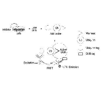
Figure 1 provides a schematic representation of the assay design for
monitoring
DUB activity and target engagement in cells expressing exogenously tagged
DUBs.
One possible method is depicted. A putative or known inhibitor ("inhibitor")
is added to
dose cells which are, in this depiction, expressing an exogenous tagged-DUB.
These
cells are subsequently lysed, and an activity probe as shown is added. This
probe will
bind to DUBs that are catalytically active and available for binding. The
binding of the
activity probe to the DUB is detected in this depiction by the use of two
antigens, one
which binds to the ubiquitin tag, and the other that binds to the DUB tag. The
first is
conjugated to a fluorescence donor such as Europium cryptate (Eu), and the
second to
a fluorescence acceptor (F), such as XL
Date Recue/Date Received 2021-07-13
CA 02951683 2016-12-08
WO 2015/189646
PCT/GB2015/051752
12
665. As shown in the figure, Eu is excited and transfers this to the
fluorochrome, F, which
emits a signal. This only occurs when the two are in close proximity.
Figure 2 provides a schematic representation of the assay design for
monitoring DUB
activity and target engagement in cells expressing endogenous DUBs. One
possible method
is depicted. A putative or known inhibitor ("inhibitor") is added to dose
cells which are, in
this depiction, expressing endogenous DUBS. These cells are subsequently
lysed, and an
activity probe as shown is added. This probe will bind to DUBs that are
catalytically active
and available for binding. The binding of the activity probe to the DUB is
detected in this
depiction by the use of two antigens, one which binds to the ubiquitin tag,
and the other
that binds to the DUB. The first is conjugated to Eu, and the second to F. As
shown in the
figure, Eu is excited and transfers this to F, which emits a signal. This only
occurs when the
two are in close proximity.
Figure 3 provides a schematic representation of the assay design for
monitoring DUB
activity and target engagement in animal tissues or tumours expressing
endogenous DUBs.
One particular method is depicted. A putative or known inhibitor ("inhibitor")
is added to
dose an animal, which contains cells which are expressing an endogenous DUB of
interest.
A sample of cells are taken (not shown) and these cells are subsequently
lysed, and an
activity probe as shown is added. This probe will bind to DUBs that are
catalytically active
and available for binding. The binding of the activity probe to the DUB is
detected in this
depiction by the use of two antigens, one which binds to the ubiquitin tag,
and the other
that binds to the DUB. The first is conjugated to Eu, and the second to F. As
shown in the
figure, Eu is excited and transfers this to F, which emits a signal. This only
occurs when the
two are in close proximity.
Figure 4 provides a schematic representation of the assay design for
monitoring DUB
activity and target engagement in human tissues or tumours expressing
endogenous DUBs.
One particular method is depicted. A sample or biopsy of cells from a human
are taken (not
shown) and these cells are subsequently lysed, and an activity probe as shown
is added.
This probe will bind to DUBs that are catalytically active and available for
binding. The
binding of the activity probe to the DUB is detected in this depiction by the
use of two
CA 02951683 2016-12-08
WO 2015/189646
PCT/GB2015/051752
13
antigens, one which binds to the ubiquitin tag, and the other that binds to
the DUB. The
first is conjugated to Eu, and the second to F. As shown in the figure, Eu is
excited and
transfers this to F, which emits a signal. This only occurs when the two are
in close
proximity. In this depiction, the quantification of the level of activity
probe binding can
indicate treatment options for this human patient.
Figure 5 shows examples of low-throughput activity probe assays developed in
human
cell lines to monitor inhibition of endogenous DUBs. Incubation of activity
probe results in a
complex with the isopeptidase, which results in slower migrating forms in SDS-
PAGE. It can
be seen that probe binding causes the position of the band to alter on the
western blot.
Unless stated, the activity probe used is Ub-VME and the cell line used is HEK
293. Key *=
U2OS cells and **= CAL51 cells. Figure 5A depicts Western Blot results of 14
activity probe
assays, each monitoring a different isopeptidase as indicated, with and
without the probe (+
and -, respectively). Figure 5B shows two separate Western Blot studies, one
each for USP14
and UCHL5. Various cell types (U20S, CAL51 and HEK) are tested, without an
activity probe,
or with the activity probes Ub-VME or Ub-PA. Thus, this shows the optimisation
of the
activity probe assay using different cell lines or probes.
Figure 6 is an example of Western blot ubiquitin activity probe assays
developed in
murine tissue models. Figure 6A depicts two Western blot photographs, the
first of which
relates to two tumour models H226 and A549. Various tumour samples are tested
using the
activity probe Ub-VME and its binding to UCHL1 is detected via increase in
molecular weight,
as shown. In the second western blot photograph, a third tumour model is
tested (H460),
this time for UCHL1 and USP11 using Ub-VME activity probe. These are xenograft
models to
monitor USP11 and UCHL1 activity in the tumours. Figure 6B shows three Western
blot
photographs, demonstrating the activities of UCHL1 or USP11 in mouse surrogate
tissues.
The first lane for each is a molecular weight marker. Various tissues are
lysed as indicated,
and increasing concentrations are used in the studies. Ub-VME is used as the
probe for
UCHL1 or Ub-PA is used as a probe for USP11 as indicated. Binding of the
activity probe is
demonstrated by a shift in the molecular weight. These are examples of low-
throughput
methods. UCHL1 and USP11 are detected using anti-UCHL1 and anti-USP11
antibodies,
respectively.
CA 02951683 2016-12-08
WO 2015/189646
PCT/GB2015/051752
14
Figure 7 shows assay optimisations for measuring on-target activity of
deubiquitylating
inhibitors in cells using activity probes. It also shows stability of lysates
and of compound
inhibition during freeze/thaw cycles. Photographs of Western blots are
provided, and as
indicated the cells were fresh, frozen or frozen in glycerol as a
cryopreservation agent.
U2OS osteosarcoma cells were incubated with DMSO or various concentrations of
USP11
inhibitor (depicted MTX) as indicated. Cells were washed and lysed. Lysates
were incubated
in the absence or presence of an ubiquitin-VME probe. Proteins were separated
by SDS-
PAGE electrophoresis and transferred to a nitrocellulose membrane. USP11
activity was
determined via measurement of a shifted (active, USP11-Ub) relative to non-
shifted
(inactive, USP11) form of USP11 as indicated by Western blotting with an anti-
USP11
antibody. The first lane for each is a molecular weight marker.
Figure 8 shows assay optimisation for a high throughput target engagement
assay using
exogenously expressed deubiquitylating enzymes. A plot of cell type including
level of
transient transfection of the indicated isopeptidase versus HTRF signal (Delta
F %) is
presented. Two cell types were used, HeLa and U2OS. The figure illustrates the
impact of
the lysates dilution on the HTRF signal and its reproducibility. The indicated
DUBs were
transiently transfected and expressed for 48h in HeLa or U2OS cells.
Figure 9 shows additional assay optimisation for a high throughput target
engagement assay using exogenously expressed deubiquitylating enzymes. Figure
9A
depicts the impact of expression duration of USP11 (24 vs 48h) on the overall
HTRF signal as
well as the effect of permuting the two detection antibodies. Furthermore, it
shows the
linearity of the HTRF signal with the amount of lysate used in each sample.
Figure 9B
depicts the relationship between the overall amount of lysates from CAL51
cells stably
expressing FLAG-UCHL1 and Ub-VME probe used in the assay. These optimisation
steps are
critical to ensure the best sensitivity and assay window in HTRF assays.
Figure 10 shows a comparison of high throughput target engagement assay
performed
in cells expressing exogenously tagged DUBs using antibodies targeting the tag
or specific
protein, thus the tagged DUB or endogenous, non-tagged and tagged DUB.
Putative
CA 02951683 2016-12-08
WO 2015/189646
PCT/GB2015/051752
inhibitors of the isopeptidase are added, these are represented by the MIX
number. Typical
ICso curves are generated, showing a variety of potencies against USP11. They
are obtained
by plotting the normalised HTRF signal (Delta F) against the concentration of
compound
used. The left hand side panel curves were generated using an anti-FLAG
antibody, the right
5 hand side panel curves were produced using an anti-USP11 antibody that
detects the
endogenous USP11 protein as well as the exogenously expressed FLAG-USP11. A
comparison of the curves obtained shows that they are remarkably similar.
Figure 11 shows a correlation between a high throughput biochemical assay and
high
10 .. throughput cellular target engagement assay. Figure 11A represents the
study of two small
molecule inhibitors, MTX084144 and MTX083568, in two different assays ¨ high
throughput
cellular target engagement assay (HTRF) and biochemical assay (FP). ICso
curves are
obtained by plotting the normalised HTRF signal (Delta F) against the
concentration of
compound used. The ICso value is the concentration at which only 50% of the
signal remains.
15 The cellular HTRF activity probe ICso matches the in vitro biochemical
fluorescence
polarisation (FP) ICso: 2 examples indicated. Figure 11B represents a plot of
the biochemical
assay IC50 versus cellular assay ICH. This depicts the correlation between the
in vitro
biochemical assay ICso and the cellular HTRF ¨this was obtained for 153
compounds tested
for UCHL1 inhibition.
Figure 12 illustrates the high throughput nature of the assays in various
types of
biological samples. Figure 12A depicts target engagement in cell lines, whilst
Figure 12B
depicts target engagement in biological samples. As depicted in Figure 12, the
detection of
the complex of the activity probe and the isopeptidase can be direct or
indirect. This may
depend upon whether the isopeptidase is endogenous or exogenous, but can be
varied to
suit the conditions of the assay.
Figure 13 shows the comparison between HTRF and Western blot activity probe
assays for USP11 target engagement by a small molecule inhibitor, MTX078594 in
U2OS
cells. Endogenous USP11 is tested. Figure 13A depicts an ICso plot using HTRF
and the
activity probe Ub-VME. Figure 13B depicts the Western blot assay, using anti-
USP11
antibodies for detection. The probe used was Ub-VME, and this was added to
cells to which
CA 02951683 2016-12-08
WO 2015/189646
PCT/GB2015/051752
16
either DMS0 or MTX078594 had been added in the noted concentrations. The shift
in the
position of USP11 on the gel indicates that the activity probe has bound.
These results are
also depicted as the plot of MTX concentration versus % inhibition.
Figure 14 shows an example of optimisation of the HTRF activity probe assay
for
endogenous USP5 in HEK 293 cells. In all HTRF assays, it is important to test
the
stoichiometry of the reagents in order to optimise the assay window and the
sensitivity. In
this experiment, the parameters tested were the Ub-VME activity probe final
concentration
(1.25p.M to 46nM), the amount of lysate (from 22.5p.g to 0 Lig) and the
concentration of the
secondary anti-rabbit antibody (1/5thiL to 1150th pL per sample). This
antibody recognises
the constant region of the primary antibody against USP5. The values shown are
the HTRF
signal (% Delta F). This demonstrates the importance of optimising the assay
conditions,
especially the assay window in this particular instance.
Figure 15 shows the target (USP5) engagement by two commercially available
inhibitors in mouse tissue lysates using HTRF: PR-619 (Sigma Aldrich -5ML0430)
(closed
triangles) and ubiquitin-aldehyde (Boston Biochem U-201) (open triangles). The
lysates were
freshly generated from the mouse brain (A) or 5W48 xenograft (B) tissues and
quantified. 10
pg of the lysates were incubated with the known inhibitors for 30 min before
adding the
activity probe for 60 more minutes after which the primary antibody (anti-
USP5), the
secondary antibody anti-rabbit-cryptate and the secondary antibody anti-HA-
XL665 were
added. As expected, ubiquitin-aldehyde is a much more potent inhibitor of USP5
than PR-
619.
Detailed Description of the Invention
According to a first aspect of the invention, there is provided a high
throughput method for
determining the activity of an isopeptidase enzyme in a biological sample,
comprising the
steps of:
i) preparing an extract of said sample
ii) contacting the extract with an activity probe
CA 02951683 2016-12-08
WO 2015/189646
PCT/GB2015/051752
17
iii) including reagents which bind to or interact with the activity probe
and/or the
enzyme, and generate a detectable signal
iv) measuring the detectable signal
It is preferred that a detectable signal is generated when the activity probe
is bound to the
isopeptidase enzyme. This happens where the reagents that bind to each of the
activity
probe and enzyme are brought into close proximity, due to the binding of the
activity probe.
As used herein, an isopeptidase is an enzyme that catalyses the cleavage of an
isopeptide
bond, especially that between the terminal diglycine attached to ubiquitin, as
well as
cleavage of ubiquitin fusion or precursors through peptide bonds.
lsopeptidases include
SUMO peptidases, ATG8 peptidase, ISG15 peptidase, and NEDD8 peptidase. A
particularly
preferred class of isopeptidases is deubiquitylating enzymes.
Examples of isopeptidase enzymes include the following:
Human enzymes include, but are not limited to:
The ubiquitin-specific protease (USP/UBP) superfamily; (USP1, USP2, USP3,
USP4, USP5,
USP6, USP7, USP8, USP9X, USP9Y, USP10, USP11, USP12, USP13, USP14, USP15,
USP16,
USP17, USP17L2, USP17L3, USP17L4, USP17L5, USP17L7, USP17L8, USP18, USP19,
USP20,
USP21, USP22, USP24, USP25, USP26, USP27X, USP28, USP29, USP30, USP31, USP32,
USP33,
USP34, USP35, USP36, USP37, USP38, USP39, USP40, USP41, USP42, USP43, USP44,
USP45,
USP46 USP47, USP48, USP49, USP50,USP51,USP52,USP53, USP54, USPL1, CYLD);
The ovarian tumour (OTU) superfamily: (Otubain-1 (OTUB1), Otubain-2 (OTUB2),
OTUD1,
OTUD3, OTUD4, OTUD5, OTUD6A, OTUD6B, OTUD7A, OTUD7B/Cezanne, A20, TRABID,
YOD1, VCIP1, HIN1L, FAM10SB/OTULIN);
The Machado-Josephin domain (MJD) superfamily: (ATXN3, ATXN3L, JOSD1, or
JOSD2);
The ubiquitin C-terminal hydrolase (UCH) superfamily: (BAP1, UCHL1, UCHL3,
UCHL5);
MPN-VJAMM (JAB1, MPN, M0V34) metallo-enzyme family: (BRCC36, MPND, MYSM1,
COPS5, PSMD14, ElF3H, COPS5, ElF3F, PSMD7, AMSH, AMSH-LP, PRPF8);
DeSUMOlyating enzymes (SENPS) family (ULP1, ULP2, SENP1, SENP2, SENP3, SENPS,
SENP6,
SENP7, DESI1, DESI2, SENP8, USPL1). SENPS are reviewed in Nayak and Muller,
2014,
Genome Biology, 15:422.
CA 02951683 2016-12-08
WO 2015/189646
PCT/GB2015/051752
18
Homologues of these human enzymes may exist in other organisms.
Fungi are eukaryotic, and therefore posses their own deconjugating enzymes for
Ub and
Ubl, such as DUBS. Yeast, for example, have numerous DUBs. Fungal DUBs
include, but are
not limited to:
Ulp1, Ulp2, Ulp3, Ulp4, Ulp5, Ulp6, Ulp7, Ulp8, Ulp9, Ulp10, Ulp11, Ulp12,
Ulp13, Ulp14,
Ulp15, Ulp16, Atg4, Otu1, 0tu2, Yuh1.
Bacterial isopeptidase enzymes include, but are not limited to:
TssM (Burkholderia pseudomallei), ChlaDub1, ChlaDUb2 (Chlamydia trachomatis),
YopP
(Yersina enterocolitica), YopJ (Yersina pseudotunerculosis), SseL (Salmonella
species), AVrA
(Salmonella enterica), ELaD (Escherichia co/f). These are reviewed in Ashida
et al, 2014,
Nature Reviews Microbiology, Volume 12, 399 ¨ 413.
Viral isopeptidase enzymes include, but are not limited to:
UL36 (Herpes Simplex Virus type 1 and Marek's Disease virus), UL48 (Human
cytomegalovirus), pUL48 (Human cytomegalovirus), pUL36 (PseudoRabies virus),
UL36
(PseudoRabies virus), 0RF64 (Kaposi-Sarcoma associated herpesvirus ¨ KSHV and
Murine
gammaherpesvirus 68), RTA (KSHV), BPLF1 (Epstein Barr (EB) Virus), BSLF1 (EB
Virus), BXLF1
(EB Virus), vOTU (Crimean-Congo Haemorrhagic fever Virus), PLpro (human
coronavirus),
PRO (Turnip yellow mosaic (TYM) virus), 98K (TYM virus), PLP2 (Porcine
Epidemic Diarrhoea
Virus), nsp2 (Porcine Reproductive and Respiratory Syndrome - PRRS Virus), Avp
(Adenovirus - ADV), Adenain (ADV), L3 23K proteinase (ADV), SARS-CoV PLpro
(Severe Acute
Respiratory Syndrome Coronovirus ¨ SARS), MERS-CoV PLpro (Middle East
respiratory
syndrome coronavirus ¨ MERS), OUT L (variants in Nairobi sheep disease virus,
Dugbe virus,
PRRS virus, Rice stripe Virus Zhejiang), L(pro) (Foot and Mouth Disease
Virus), MDVusP
(Marek's Disease Virus), M48 (Murine cytomegalovirus). These are reviewed in
Calistri et al,
2014, Cells, 3, 386-417.
lsopeptidases from parasites such as Schistosoma and Plasmodium are known.
Many of
these are homologues of human proteins, and include UCHL3 in Toxoplasma
gondii,
CA 02951683 2016-12-08
WO 2015/189646
PCT/GB2015/051752
19
Schistosoma mansoni and Plasmodium falciparum, pfUbp-1, pfUCH54, smUCHL5,
smBAP-1,
smOTU1, smOTU3, smOTU5a, smOTU6b, smOtubain, smAtaxin-3 and smiosephin.
The isopeptidase of the use, method, assay or kits of the present invention
may be selected
from any of those enzymes listed above, or any discovered subsequently.
As used herein, the isopeptidase may have ubiquitin or a ubiquitin-like (Ubl)
molecule as
part of the natural substrate, for example SUMO (small, ubiquitin-like
modifiers) It is
preferred that the natural substrate for the isopeptidase includes ubiquitin.
As used herein, a high throughput method means that a plurality of tests are
conducted in
parallel, allowing for rapid determination of results. In general, high
throughput methods
and/or assays are conducted on plates, which may have wells, such as
microtitre plates. The
wells of a microtitre plate are usually in multiples of 96, and thus 96 well,
192 well, 288 well
384 well, or even up to 3456 well microtitre plates may be used in any of the
methods of
the invention. Any suitable number of wells can be used in the high throughput
methods or
formats, preferably between 96 and 500 wells, more preferably between 96 and
384 wells.
It is preferred that the high throughput methods of the present invention are
performed on
plates, even more preferably plates with 96 or 384 wells. A microtitre plate
may also be
called a microplate or microwell plate, and is a flat plate with multiple
"wells" used as small
test tubes. The sample wells are generally arranged in a 2:3 rectangular
matrix. The term
"plate" also encompasses "array tape", which is a continuous strip of
microplates embossed
on a flexible plastic tape.
The biological sample can be introduced to the plate at any suitable point in
the high
throughput method. For example, the biological sample itself can be included
on the plate,
prior to the preparation of the extract. The cells from the biological sample
can be seeded
on the plate prior to any processing. Alternatively, the extract can be
included on the plate.
The latter is particularly preferable if the biological sample is tissue. It
is particularly
preferred that the extract of the sample is present on a plate by the time the
detection step
is undertaken, such that the method can be performed in a high throughput
manner.
CA 02951683 2016-12-08
WO 2015/189646
PCT/GB2015/051752
It is preferred that at least one control biological sample/extract is present
in the high
throughput format or method of the invention. Suitable controls are discussed
in relation to
each aspect or embodiment of the invention as described herein. For example, a
control for
a defective or catalytically-inactive isopeptidase may be a wild-type
isopeptidase. A suitable
5 control for studies with isopeptidases of unknown activity is a defective
or catalytically-
inactive isopeptidase. In relation to studies on putative inhibitors, the
control may be an
isopeptidase resistant/not expected to be inhibited by the putative inhibitor.
In the first aspect of the invention, the activity of an isopeptidase enzyme
is determined or
10 measured.
Isopeptidase activity can be defined as follows: In a given biological sample
and for a
particular isopeptidase, it is the proportion of the isopeptidase population
that is capable of
binding the activity probe. The inability of binding the activity probe may be
the result of,
15 inter alio, pharmacologic inhibition, oxidation, allosteric regulation
or genetic mutation that
affects the integrity of the active site.
The activity of the isopeptidase is determined via the detected signal
generated when the
activity probe is bound to the enzyme. It is preferred to measure how much of
the complex
20 of activity probe bound to enzyme in a given biological sample compared
to a control where
no binding is anticipated (for example a catalytically inactive enzyme). It is
preferred that
the amount of complex formed (amount of activity probe that binds to the
enzyme) is
compared to a control, for example a catalytically inactive enzyme where no
complex will be
formed.
Thus, in a preferred embodiment of the invention, the activity probe binds to
or interacts
with the isopeptidase and forms a complex. The amount of complex in the
biological
sample is detected and quantified, by methods discussed further below. It is
further
preferred that the methods of the invention include a control biological
sample, and the
amount of complex is detected and quantified, in order to provide background
or
comparative data.
CA 02951683 2016-12-08
WO 2015/189646
PCT/GB2015/051752
21
The methods of the invention are conducted on biological samples containing
cells, as
described below. These cells may be human, animal, or from a microorganism.
The
biological sample may contain a mixture of human/animal and microorganism
cells. A
microorganism is an organism that cannot be seen by the human eye, and
includes bacteria,
fungi such as yeast, viruses and parasites (protozoa). In the methods of the
invention, the
assay may examine pathogenic microorganisms, i.e. ones that infect humans or
animals.
Examples of parasitic microorganisms (protozoa) include Trypanosoma cruzi,
Schistosoma
mansoni, Toxoplasma gondii, Plasmodium falciparum, P. vivax, P. ovale, and P.
malariae.
.. The methods of the present invention are conducted on biological samples.
Such samples
are ex vivo biological sample, generally taken from a human or animal body.
The biological
sample is a sample that contains cells. The sample may comprise cells of the
organism
sampled, cells of a microorganism which has infected said organism, or a
mixture of the two.
Suitable biological samples include samples of normal tissues and cells
(healthy tissues and
cells), samples of tumour cells and tissues, biopsies or aspirates taken from
human or
animal patients with a suspected defect in isopeptidase/deubiquitylating
enzyme activity.
Such defect may be suspected in the case of patients with a tumour, cancer
including blood-
based cancer, congenital disorder, auto-immune disorder, liver dysfunction,
infertility,
osteopenia, bone marrow defects, growth retardation/development abnormalities,
immunodeficiency and/or neurological disease. Alternatively, suitable
biological samples
includes any biological sample where cells of a microorganism may be present,
for example
a tissue sample, biopsy, aspirate, or fluid sample, such as blood, lymph,
mucus, bronchiolar
lavage, sputum, saliva, or urine. Although not part of the present invention,
the biological
sample may be taken in any suitable manner known to those skilled in the art.
Suitable
samples for the methods of the invention include samples from any body
tissues, including
but not limited to tissues and cells from skin, muscle, lung, liver, kidneys,
stomach, intestine,
ovary, uterus, breast, brain, eye, mucosal membrane, testes, bone or blood. It
is preferred
that the biological sample contains a sufficient amount of cells, and thus
biological samples
that do not contain an appreciable number of cells, such a urine, saliva,
sweat, tears,
mucous, lymph and the like are not generally suitable as biological samples as
used herein
wherein it is the human or animal isopeptidase under investigation. These
biological
samples may, however, provide sufficient microorganism cells to perform the
assay of the
CA 02951683 2016-12-08
WO 2015/189646
PCT/GB2015/051752
22
invention. Where the isopeptidase to be assayed is from a microorganism, an
additional
step of culturing the biological sample under suitable conditions for the
microorganism of
interest may be undertaken. Such a cultured biological sample may be used in
the methods
of the invention as a biological sample. Additionally, the human or animal
cells from a
.. biological sample may be appropriately cultured prior to use in the assay.
An extract of the biological sample is prepared as part of the methods of the
present
invention. In order to prepare an extract, lysis of the cells present is
preferred. Cell lysis
may be achieved by any suitable method known to those in the art. A preferred
method of
cell lysis is by using a cell-lysis buffer. An example of a cell lysis buffer
is a solution
comprising a detergent. Detergents have both lysing and solubilising effect,
and suitable
detergents include, inter alia, CHAPS (,34(3-cholamidopropypdimethylammonio]-1-
propanesulfonate)Tergitol type-NP-40 (nonyl phenoxypolyethoxylethanol),
Triton X-100
(polyethylene glycol p-(1,1,3,3-tetramethylbutyl) phenyl ether), sodium
deoxycholate
and/or sodium dodecyl sulphate (SDS). It is preferred that the cell-lysis
buffer is used such
that no denaturation of the cellular proteins is achieved. Those skilled in
the art will be
familiar with suitable concentrations of detergents to use to avoid
denaturation of the
cellular proteins. One or more detergents may be used in the cell lysis
buffer. Other
components of the lysis buffer may include Tris, salts such as sodium chloride
and
magnesium chloride, glycerol, beta-mercaptoethanol. Other reducing agents such
as DTI,
DIE and TCEP may be added. It is particularly preferred that protease
inhibitor and/or
phosphatase inhibitor are added to the lysis buffer.
Should the biological sample be a tissue sample, it is preferred that the
tissue sample is
treated with a lysis buffer and homogenised in a tissue homogeniser.
Alternatively, the cells may be physically lysed using freeze/thaw techniques,
grinding,
sonication, homogenisation or mechanical lysis. After lysis or grinding, it is
preferred that
the sample is centrifuged (or spun) to remove extracellular matrix, cell
debris, chromatin
and other insoluble material. An alternative method to solubilise chromatin is
to digest it
enzymatically using BenzonaseTM, MNase or DNase for example,
CA 02951683 2016-12-08
WO 2015/189646
PCT/GB2015/051752
23
However, the most preferred method is to use a cell-lysis buffer to prepare
the biological
sample into an extract.
Once the extract has been prepared, aliquots of the extract may be added to a
high
throughput plate, such as a microtitre plate, if they are not already included
on a plate.
The microtitre plate may comprise one or more extracts, each extract at a
different location
on the plate. The methods of the invention may thus relate to the testing of
one or more
biological samples in parallel in a high throughput manner. Thus each of the
locations
(wells) on the microtitre plate, or alternative high throughput assay format,
may contain an
extract from a different biological sample. Alternatively, each extract may be
present in one
or more locations, or one extract may be present at all locations.
Once the sample has been prepared as an extract, and suitably prepared in a
high
throughput assay format, for example on a plate, an activity probe is added to
each extract.
It is preferred that after the extract is contacted with the activity probe,
the resultant
mixture is incubated. The incubation may be for any suitable length of time.
It is
particularly preferred that the activity probe and extract are incubated for a
period of 5
minutes to 300 minutes, preferably 5 minutes to 240 minutes, more preferably 5
to 180
minutes, even more preferably 5 to 60 minutes, preferably 10 to 40 minutes,
most
preferably 15 to 35 minutes. It is preferred that the activity probe and
extract are incubated
for 5, 10, 15, 20, 25, 30 or 35 minutes. Alternatively, the activity probe and
the extract are
incubated for 30 to 60 minutes, i.e. 30, 35, 40, 45, 50, 55 or 60 minutes.
The activity probe may be any suitable activity probe for an isopeptidase
enzyme, and
several such entities have been discussed herein, and in the references listed
herein. It is
preferred that the activity probe comprises a substrate mimic for the
isopeptidase enzyme.
i.e. the activity probe mimics part or all of the natural substrate for the
isopeptidase. It is
preferred that the isopeptidase does not catalytically cleave the substrate
mimic or activity
probe. Thus, the activity probe may be a substrate mimic that is catalytically
inactive.
CA 02951683 2016-12-08
WO 2015/189646
PCT/GB2015/051752
24
The activity probe may preferably comprise an ubiquitin (Ub) molecule or
ubiquitin-like
molecule (UBL). Ubiquitin, or "Ub", is a highly conserved 76 amino acid
protein expressed in
all eukaryotic cells. Preferably, ubiquitin is human ubiquitin. The
polypeptide sequence of
human ubiquitin is deposited in NCBI database Genpept under accession number
P62988.1,
with four human genes that encode ubiquitin precursors being deposited as UBB
(accession
number POCG47), UBC (accession number POCG48), UBA52 (accession number P62987)
and
RPS27A (accession number P62979).All seven conserved lysines of Ub (K6, 11,
27, 29, 33, 48
and 63) may be used as branching sites for the generation of Ub polymers.
Examples of
suitable ubiquitin-like proteins include SUMO (Small ubiquitin-like modifier)
such as SUM01,
.. SUM02 and SUM03; ISG15 (Interferon-Stimulated Gene-15, also known as UCRP),
NEDD8
(Neuronal-precursor-cell-Expressed Developmentally Downregulated protein-8),
FAT10
(human leukocyte antigen F-associated), ATG8 and A1G12 (autophagy 8 and 12),
UBL5
(Ubiquitin like protein 5), UFM1 (Ubiquitin fold modifier 1), MUB (membrane
anchored
Ubiquitin fold) and URM1 (Ubiquitin-related modifier-1). Known or putative
Ubls that may
be suitable for inclusion in an activity probe are: ISG15, NEDD8 (known
deconjugating
enzymes are UCHL1, UCHL3, USP21, COP9, and DEN1/NEDP1), FUB1 (MNSF-B or FAU),
FAT10, SUMO-1 (deconjugating enzymes include SENP1, SENP2 and SUSP4), SUMO-2
and
SUMO-3 (deconjugating enzymes for these include SENP3 and SENP5), Apg 8, Apg
12, Urm
1, UBL5, Ufm1, BUBL1, BUBL2, UBL-1, SF3A120 and Oligoadenylate synthetase. It
is
preferred that the activity probe may contain any one of the Ubiquitin or
Ubiquitin-like
molecules selected from the above list. Activity probes containing ubiquitin-
like proteins
instead of ubiquitin may also be used to evaluate isopeptidases that recognise
ubiquitin-like
adducts instead of ubiquitin. Activity probes containing ubiquitin may be used
to evaluate
deubiquitylating enzymes, a particularly preferred option.
The skilled person will be able to select a relevant Ub or Ubl for the
activity probe based
upon the action of the isopeptidase or deconjugating enzyme of interest.
The ubiquitin or ubiquitin-like protein may be optionally tagged. A tag is a
biochemical
indicator appended to the ubiquitin, and may be any suitable tag. Thus, the
activity probe
may comprise a tagged ubiquitin or ubiquitin-like molecule. It is preferred
that the tag is
selected from the group consisting of:
CA 02951683 2016-12-08
WO 2015/189646
PCT/GB2015/051752
"Peptide" tags, such as FLAG-tag (DYKDDDDK), HA-tag (YPYDVPDYA), His-tag
(HHHHHH),
Myc-tag (EQKLISEEDL), Strep-tag (WSHPQFEK), V5 tag (GKPIPNPLLGLDST),
Calmodulin-tag
(KRRWKKNFIAVSAANRFKKISSSGAL); "protein" tags such as Glutathione-S-transferase-
tag,
5 Green fluorescent protein-tag, Maltose binding protein-tag; or "Chemical"
tags such as
Biotin, DNP (2,4-Dinitrophenol), Chemical coupling reagents (e.g. Cysteines,
non-
conventional amino acids, etc).
The tag may be bound during detection of the forming of a complex between the
activity
probe and the isopeptidase enzyme. However, if a tag is not present, then it
is possible to
10 directly detect the Ub or Ubl, generally by using an antibody or
derivative thereof which is
specific for Ub or the Ubl present.
It is preferred that the activity probe comprises a warhead (see figure 1). A
warhead may
consist of a reactive functional group that is able to covalently attach at
the active site of the
deubiquitylating enzyme or isopeptidase. The warhead is designed to attach at
the
15 particular active site of the enzyme of interest. An important parameter
in the warhead is
the choice of reactive group for covalent labelling of the target isopeptidase
enzyme at the
enzyme active site. Depending on the mechanism of enzymatic catalysis,
different reactive
groups can be chosen for covalent capture. The alkylation of nucleophilic
residues (Cys, Ser,
Thr) present in protease active sites by reactive electrophiles in the warhead
can be a useful
20 strategy. Those skilled in the art will be aware of the nature of the
active site of the
isopeptidase enzyme of interest, and will be able to select a suitable warhead
to bind at or
close to the active site.
Suitable warheads or reactive groups include alkyl halides (such as
chloroethyl, bromoethyl,
25 bromopropyl), Michael acceptors (vinyl methyl ester (VME), vinyl methyl
sulfone (VMS),
vinyl phenyl sulfone (VSPh), vinyl cyanide(VCN) and propargyl (PA).
The warhead may be present at the C-terminal end of the activity probe, as
depicted in
Figure 1. The warhead may be designed to bind to or react with particular
residues, notably
cysteine residues, in the active site of the isopeptidase.
CA 02951683 2016-12-08
WO 2015/189646
PCT/GB2015/051752
26
The warhead may be specific for a single isopeptidase enzyme, or be able to
bind or interact
with the active site of one or more isopeptidase enzymes, such as VME. Should
the
warhead be suitable for use in an activity probe for more than one
isopeptidase enzyme, it
should be noted that complexes with other isopeptidases not of interest will
not be
detected, since one of the detection reagents is specific for the isopeptidase
enzyme itself.
Thus, complexes of the activity probe with isopeptidase not of interest will
not be detected
using this method. Generally, each assay will be looking at the activity of a
single
isopeptidase in a biological sample, using a specific binding agent for that
isopeptidase to
ensure only those complexes are detected.
It is it preferred that the activity probe is not subject to catalysis such as
cleavage by the
enzyme.
In the Examples, several activity probes are utilised, and these are all
suitable to use in any
of the methods of the invention. These include:
Activity probes suitable for deubiquitylating enzymes include: Tagged-Ub-VME
(Ubiquitin-
Vinyl Methyl Ester),Tagged-Ub-VMS (Ubiquitin-Vinyl Methyl Sulphone), Tagged-Ub-
PA
(Ubiquitin-Propargylamide),Tagged-Ub-CI (Ubiquitin 2-Chloroethyl), Tagged-Ub-
Br (Ubiquitin
2-Bromoethyl), Tagged-di-Ubiquitin probes containing various linkages between
lysines and
glycine isopeptide bonds or branched ubiquitin activity probes as described in
(McGouran,
J.F., eta!, 2013, Chem Biol., 20(12):1447-55 and 1phofer, A., eta!, 2012,
Chemibiochem,
13(10):1416-20.).
Activity probes suitable for other types of isopeptidase include: HA-SUM01 VMS
(for SUMO
peptidases), HA-GABARAP/Apg8p1-VMS (for ATG8 peptidase), HA-I5G15-VME (for
ISG15
peptidase), NEDD8-VME (for NEDD8 peptidase), and FAT1O-VME.
The isopeptidase enzyme assayed using the methods of the present invention may
be
endogenous to the biological sample, or may be exogenous. If the biological
sample is being
tested for endogenous isopeptidase enzymes, no further steps are required in
relation to
preparing the extract of the biological sample. If, however, the method is
looking at an
exogenous isopeptidase enzyme, a further step in the method is required prior
to preparing
CA 02951683 2016-12-08
WO 2015/189646
PCT/GB2015/051752
27
an extract of the biological sample, namely introducing the nucleic acid
sequence encoding
the exogenous isopeptidase enzyme either into the non-human animal prior to
removing a
sample, or introducing the nucleic acid sequence encoding the exogenous
isopeptidase
enzyme into the biological sample itself. Those skilled in the art are well
versed in suitable
.. techniques to transfect cells or organisms with exogenous nucleic acid. In
brief, a plasmid or
other suitable vector including the coding sequence for the isopeptidase
enzyme and an
operably linked promoter can be supplied to the cell. Transfection agents may
be required
in order to increase the level of transfection, as known to those skilled in
the art.
The exogenous isopeptidase enzyme may be any wild-type, non-mutated version of
the
enzyme, or mutations can be introduced by varying the nucleic acid sequence of
the enzyme
prior to transfection. Alternatively, the exogenous enzyme can be
catalytically inactive,
which is particularly useful as a control.
It is preferred that the exogenous isopeptidase is tagged. Suitable tags have
been discussed
earlier in relation to tagged ubiquitin or ubiquitin-like molecules, and the
tagged
isopeptidase may include a tag selected from the list given above. Suitable
tags thus include
FLAG, HA, HIS, Myc, biotin, STREP, TAP, MBP, GST and/or GFP. If both a tagged
ubiquitin or
ubiquitin-like molecule and a tagged isopeptidase are used in the methods,
kits, uses or
assays of the invention, it is preferred that the entity used to tag each
element (Ub/Ubl or
enzyme) is different.
The activity probe is introduced to the extract of the biological sample, and
may be
incubated as discussed above, lithe enzyme is active, the activity probe will
bind at the
.. active site. This brings the activity probe and the enzyme in close
proximity, and form a
complex. It will be understood that the activity probe will only bind to the
isopeptidase
enzyme if it is available for binding and is catalytically active and
functional. Isopeptidase
enzymes that are catalytically inactive, damaged, inhibited or mutated at
particular key
residues, will not be available to bind the activity probe. Thus, use of the
activity probe is an
.. elegant way of discovering the activity of a particular isopeptidase enzyme
in a particular
biological sample. As mentioned previously, the formation of a complex between
the
activity probe and the enzyme is detected (as discussed below) and compared to
the
CA 02951683 2016-12-08
WO 2015/189646
PCT/GB2015/051752
28
amount of complex formed in a control sample. Said control sample may contain
a wild-
type (natural, non-mutated) enzyme or a catalytically inactive enzyme.
The binding of the activity probe to the isopeptidase enzyme may be detected
by any
suitable means, using suitable reagents, preferably detection reagents. The
complex
formed between the activity probe and the enzyme may be detected. Particularly
preferred
detection means are the use of two or more reagents; one reagent binds to the
activity
probe, preferably via the tagged ubiquitin (if present), a further reagent
binds to the
isopeptidase enzyme. If tagged ubiquitin or Ubl is not present, the reagent
may bind
.. directly to the ubiquitin or Ubl. The reagents discussed herein may also be
called detection
reagents. It is preferred that one of the detection reagents binds to the
isopeptidase
enzyme, such that the specific complex of isopeptidase and activity probe can
be detected.
It is preferred, if the isopeptidase enzyme is exogenous, that the detection
reagent binds at
or close to the tag of the tagged enzyme, if present. Alternatively, the
detection reagent
will bind to the enzyme itself.
It is preferred that the reagents/detection reagents are binding agents with
specific binding
affinities for a particular recognition site on their target. Suitable binding
agents are well
known to those skilled in the art, and include antibodies and derivatives or
fragments
thereof (Fc, Fab, Fab', ScFv, single domain antibody, VH, or VI domains) and
aptamers.
A first binding agent binds to a recognition site on the activity probe. This
recognition site
may be the tag or the ubiquitin or Ubl itself. A second binding agent binds to
a recognition
site on the enzyme. Any suitable recognition site may be used. The first and
second binding
agents may be the same class of binding agents (i.e., antibodies) or may be
different (i.e.
one antibody and one aptamer). They both, however, have specific binding
affinities for
their partners, such that the second binding agent will not bind to a
different isopeptidase
enzyme, for example.
The binding agents may be labelled in such a way to allow detection of the
binding of the
probe to the enzyme, via generation of a detectable signal. However, one or
more of these
binding agents may be unlabelled, in which case, the use of one or more
further binding
CA 02951683 2016-12-08
WO 2015/189646
PCT/GB2015/051752
29
agent(s) which binds to one of the first or second binding agents is also
envisioned. These
further binding agents may be labelled to permit detection. For example, the
second
binding agent which is capable of binding to the enzyme could be detected
using a further
binding agent, which is itself labelled. In one embodiment, as an example, the
second
binding agent is a mouse monoclonal antibody directed to a particular
isopeptidase, and the
further binding agent is an anti-mouse antibody. Thus, the binding of the
first and/or
second binding agent to the complex may be indirectly detected. Figure 12
depicts
examples of each type of binding and detection.
A preferred detection method is the use of Homogenous Time Resolved
Fluorescence
(HTRF). In this embodiment, one binding agent is labelled with a fluorescence
donor, and
another binding agent with a fluorescence acceptor. One detects the activity
probe and the
other detects the isopeptidase (both either directly or indirectly as
described above). An
example of such a system is shown in Figure 1. In this case, the donor may be
Europium
cryptate or Terbium cryptate. The acceptor may be XL 665 acceptor which is a
phycobilliprotein (large hetero hexameric edifice of 105 kDa), or d2, which is
an organic
motif of approximately 1,000 Da.
If HTRF is used to detect the binding of the activity probe to the
isopeptidase enzyme, the
fluorescence donor is excited using light at a particular wavelength and the
fluorescence
generated is transferred to the acceptor only if it is in close proximity.
Fluorescence from
the acceptor can be detected separately to the fluorescence from the donor.
Thus, the
fluorescence will only transfer from the donor to the acceptor if the activity
probe has
bound to the isopeptidase enzyme and a complex has been formed, generating a
detectable
signal.
If Europium cryptate is used, it may be excited by a UV laser light at 317 nm
(20 nm
bandwidth). The acceptors XL 665 and d2 emit light at 665 nm once the
fluorescence from
the donor has been transferred.
It is preferred that the detectable signal is generated when a complex forms
between the
enzyme and the activity probe. This signal may be generated since entities on
the detection
CA 02951683 2016-12-08
WO 2015/189646
PCT/GB2015/051752
reagents/binding agents come into close proximity. It will be understood that
if no complex
if formed, then no detectable signal will be generated and the detection step
will detect no
signal. Thus, the detection step may detect "no signal" or only detect a
signal if present. It
is preferred that the measurement of the detectable signal is performed by a
plate reader.
5
The detectable signal can be any suitable signal, such as emission of light
(electromagnetic
radiation) at a particular wavelength, including fluorescence, and ultraviolet
light. The
wavelength of the emitted light can be detected by any suitable means,
including
photosensors and photodetectors. Common detection modes for microplate readers
are
10 absorbance (how much light of a particular wavelength is absorbed by the
extract),
fluorescence intensity, luminescence, time-resolved fluorescence, and
fluorescence
polarization.
Any suitable detection methodology can be used, for example ELISA (enzyme
linked
15 immunosorbent assay), Alpha Lisa, enzyme complementation (e.g. BIFC -
Bimolecular
fluorescence complementation), alpha-screen, FRET, BRET or label-free affinity
technologies. The reagents/binding agents and entities for generating the
detectable signal
required for these detection means are well known to those skilled in the art
¨for example
ELISA relies on bringing an enzyme and a substrate into close proximity once
the activity
20 probe has bound to the isopeptidase. The ELISA enzyme and substrate may
be bounds to
the binding agents.
The methods of the invention may be used to assay the activity of one or more
isopeptidases as described previously. Furthermore, the methods of the
invention may be
25 used to determine whether a putative inhibitor has an effect on the
activity of the
isopeptidase. Thus, according to one aspect of the invention, there is
provided a use of an
activity probe to determine target engagement in the presence of a putative
inhibitor in a
high throughput manner in a biological sample.
30 According to a further aspect of the invention, the method of
determining the effect of a
putative inhibitor on an isopeptidase may be as described herein in relation
to assaying the
activity of the isopeptidase, with an additional step of contacting the
biological sample with
CA 02951683 2016-12-08
WO 2015/189646
PCT/GB2015/051752
31
the putative inhibitor prior to preparing the extract. This may be useful when
determining
the effect of a drug on a disease, disorder or condition, or to determine the
effectiveness of
a drug on an infection by a microorganism.
Thus, according to a further aspect of the invention, there is provided a high
throughput
method for determining the activity of an isopeptidase in a biological sample
in the
presence of a putative inhibitor, comprising the steps of:
i) contacting said biological sample with a putative inhibitor
ii) preparing an extract of said sample
iii) contacting the extract with an activity probe
iv) including reagents which bind to or interact with the activity probe
and/or
and the isopeptidase, and generate a detectable signal
v) measuring the detectable signal.
.. It is preferred that the detectable signal is generated when the activity
probe binds to the
isopeptidase and forms a complex.
Such a method may be used to determine the potency of a putative inhibitor.
For the
purposes of defining this invention, all known inhibitors are also classed as
putative
inhibitors. Potency is a measure of drug activity expressed in terms of the
amount required
to produce an effect of given intensity. A highly potent drug evokes a larger
response at low
concentrations, while a drug of lower potency evokes a small response at low
concentrations.
Furthermore, the methods of the invention may be used to determine or measure
the
pharmaco-dynamics of a putative inhibitor. Thus, according to one aspect of
the invention,
there is provided a use of an activity probe to determine the pharmaco-dynamic
properties
of a putative inhibitor in a high throughput manner in a biological sample.
According to one embodiment of the invention, the method of determining the
pharmaco-
dynamic properties used may be as described herein in relation to assaying the
activity of
the isopeptidase, with the additional steps of contacting the animal with the
putative
CA 02951683 2016-12-08
WO 2015/189646
PCT/GB2015/051752
32
inhibitor prior to removing a biological sample. In one embodiment, the animal
is a non-
human animal. In another embodiment, the animal is a human.
Thus, according to a further aspect of the invention, there is provided a high
throughput
method for determining the pharmaco-dynamics of a putative inhibitor of an
isopeptidase,
comprising the steps of:
i) contacting an animal with said putative inhibitor,
ii) taking said biological sample from said animal,
iii) preparing an extract of said sample
iv) contacting the extract with an activity probe,
v) including reagents which bind to or interact with the activity probe
and/or
and the isopeptidase, and generate a detectable signal,
vi) measuring the detectable signal.
It is preferred that the detectable signal is generated when the activity
probe binds to the
isopeptidase and forms a complex.
In a preferred aspect of the invention, there is provided a high throughput
method for
determining the potency and/or pharmaco-dynamic properties of a putative
inhibitor of an
isopeptidase, comprising the steps of:
i) contacting an animal with said putative inhibitor and taking said
biological
sample from said animal, or contacting said biological sample with a putative
inhibitor
ii) preparing an extract of said sample,
iii) contacting the extract with an activity probe,
iv) including reagents which bind to or interact with the activity
probe and/or
and the isopeptidase, and generate a detectable signal,
v) measuring the detectable signal.
It is preferred that the detectable signal is generated when the activity
probe binds to the
isopeptidase and forms a complex.
CA 02951683 2016-12-08
WO 2015/189646
PCT/GB2015/051752
33
As used herein a "putative inhibitor" is an entity thought to be or known to
be an inhibitor
of an isopeptidase. The inhibitor can be any suitable entity, including, but
not limited to
small molecule inhibitors and antibodies or fragments thereof (as defined
previously).
Putative inhibitors may bind to the active site of the isopeptidase, or bind
at a site remote to
the active site, but still inactivate the enzyme, for example by causing
steric hindrance of
substrate binding or cause a conformational change. The methods of the
invention can be
used to determine if a putative inhibitor is an inhibitor of an isopeptidase.
In relation to
microorganisms, particularly pathogenic microorganisms, putative inhibitors
may be
assayed, since this will provide information on whether the drug may be used
to treat an
infection of the microorganism.
The animal may be any appropriate animal, preferably a mammal such as a rodent
(mouse,
rat, gerbil, guinea pig, hamster and the like) or other small mammals, such as
dogs,
primates, cats and the like. It is preferred that the animal is a non-human
animal. However,
in one aspect the method of the invention may be performed on a human animal
in order to
study the pharmaco-dynamics of a putative inhibitor in vivo, for example in
clinical trials. In
one embodiment of the invention, the method may start with the biological
sample taken
from a human who has previously had administered the putative inhibitor. In an
alternative
embodiment, the method includes the step of taking a biological sample from a
human, as
described herein.
Pharmaco-dynamics refers to the relationship between putative inhibitor
concentration at
the site of action and the resulting effect, including the time course and
intensity of
therapeutic and adverse effects. Such properties can be determined using the
methods of
the invention as described herein.
The methods of the invention may additionally or alternatively be used in a
method of
diagnosis or prognosis. A method of diagnosis or prognosis comprises
characterising the
endogenous isopeptidase in a biological sample using an activity probe in a
high throughput
format. Said characterisation can relate to determining the activity of the
isopeptidase.
Determining the activity of endogenous isopeptidase is advantageous, since it
allows for an
indication and/or quantification of a reduction in activity, wherein said
reduction in activity
CA 02951683 2016-12-08
WO 2015/189646
PCT/GB2015/051752
34
may be associated with a disease, disorder or condition. Thus, by determining
the activity of
the endogenous isopeptidase, it may be possible to diagnose diseases,
disorders and
conditions. An alternative method of diagnosis is to determine whether there
are any active
isopeptidases from microorganisms present in a biological sample. Detecting
activity from
isopeptidases which are solely present in microorganisms can detect the
presence of those
microorganisms in the biological sample.
Diseases, disorders and conditions relating to mutated, altered, missing or
inactive
isopeptidases have been discussed previously herein, and include cancers of
all body tissues
and blood cells. Diseases, disorders and conditions include a tumour, cancer
including
blood-based cancer, congenital disorder, auto-immune disorder, liver
dysfunction,
infertility, osteopenia, bone marrow defects, growth retardation/ development
abnormalities, immunodeficiency and/or neurological disease.
Moreover, having information on the activity of endogenous isopeptidases
allows for
identification of individuals that will respond in a particular way to a drug
or medicament to
treat said disease, disorder or condition. Thus, for example, being aware that
an individual
has a particular defective isopeptidase, results in a prognosis that a
particular inhibitor, drug
or treatment for the associated disease, disorder or condition should be
avoided, since it
targets that defective isopeptidase. Conversely, having a deficiency in an
isopeptidase can
indicate that a particular course of treatment is advisable. For example, it
is known that
isopeptidases are involved in various DNA damage repair (DDR) pathways, and
knowledge
that one DDR pathway is defective can allow for targeted treatment against
another
pathway in order to exploit the principles of synthetic lethality for
cancer/tumour cells.
Such a diagnostic assay may be called a companion diagnostic assay, since it
can determine
whether or not a drug will be efficacious for an individual.
The methods of this aspect of the present invention can further relate to the
testing of the
potency of putative inhibitors against the endogenous isopeptidase, as
described previously.
Such information on the ability of putative inhibitors to modify the activity
of the
endogenous isopeptidase allows an informed choice of drugs with which to treat
the
individual.
84030232
Diagnostic and/or prognostic assays may be performed on any suitable
biological sample as
described herein. It is a requirement that the sample contains cells.
Preferably, for the
purposes of this aspect of the invention, the biological sample contains cells
that are
5 diseased or subject to a condition, i.e. tumour or cancer cells. Such
cells may be obtained by
any suitable means, including biopsies of solid tumours, cell aspirates from
solid tumours
and collection of blood-based tumour cells.
In one aspect of the invention, there is provided use of an activity probe in
the diagnosis
10 and/or prognosis of a disease, disorder or condition in a high
throughput assay in a
biological sample.
In a further aspect of the invention, there is provided a high throughput
method of
diagnosis or prognosis of a disease, disorder or condition associated with a
defective
15 isopeptidase enzyme in a biological sample, comprising the steps of:
i) preparing an extract of said sample,
ii) contacting the extract with an activity probe,
iii) including reagents which bind to or interact with the activity probe
and/or
and the isopeptidase, and generate a detectable signal,
20 iv) measuring the detectable signal.
It is preferred that the detectable signal is generated when the activity
probe binds to the
isopeptidase and forms a complex.
25 In a further aspect, the present invention relates to kits suitable for
performing any of the
high throughput methods of the invention. Such a kit comprises an activity
probe and
reagents to detect the activity probe and/or enzyme as hereinbefore described.
Materials and Methods
Date Recue/Date Received 2021-07-13
CA 02951683 2016-12-08
WO 2015/189646
PCT/GB2015/051752
36
1. Standard ubiquitin-based activity probe immunoblot assay
Cells, such as U2OS (ATCC HTB-96), H226 (ATCC CRL-5826), HEK293 (ATCC CRL-
1573) or
CAL51, were plated in 6-well dishes. The following day, cells were treated
with DMSO or the
indicated concentrations of inhibitor (depicted as a MIX number) for 1 hour at
37 C. Cells
were washed with PBS and lysed in 100 pi_ of lysis buffer [50 mM Tris, pH 7.5,
150 mM NaCI,
0.1% NP-40, 0.5% CHAPS, 5 mM MgCl2, 5 mM beta-mercaptoethanol (BME), protease
inhibitor tablet (Roche -04693159001) and phosphatase inhibitor tablet (Roche -
04906837001). Cells were scraped in lysis buffer and incubated for 30 min on
ice. Lysates
were centrifuged at 8,000 rpm for 5 min at 4 C and the supernatant transferred
to a new
tube. Protein concentration was determined by using Coomassie Plus Protein
Assay
Reagent (Life technologies -23238) with BSA as a standard according to the
manufacturer's
recommendation. Lysates (20 p.g) were diluted in lysis buffer and incubated in
the absence
or presence of 500 ng HA-Ahx-Ahx-Ub-VME activity probe (UbiQ -035) or
Ubiquitin-
Propargylamide (UbiQ -057) in a final assay volume of 20 L. Reactions were
incubated for
60 min at room temperature and terminated by the addition of SDS-loading
buffer and
boiled for 5 min. Proteins were separated by SDS-PAGE (Life technologies
¨NP0355BOX)
and transferred onto nitrocellulose. Immunoblotting antibodies used are listed
in table 1.
Anti-mouse HRP and anti-rabbit HRP secondary antibodies are from Fisher
Scientific -31430
and -31460 respectively. The chemiluminescent substrate is from GE Healthcare -
RPN2109.
The GE LAS4010 imaging system was used to acquire the luminescent signal that
was
quantified using the Image Quant TL software (GE Healthcare Life Sciences).
2. Expression and purification of UCHL1
The UCHL1 construct was PCR amplified and cloned into a pFLAG-CMV-6a vector
(Sigma-
Aldrich) with an N-terminal FLAG tag. HEK293T cells were transfected with FLAG-
UCHL1
using TransIT-LT1 transfection reagent (Mirus-2306) according to the
manufacturer's
instructions. Cells were harvested 40 hours after transfection. Cells were
washed once with
PBS and scraped in lysis buffer (50 mM Iris, pH 7.5, 150 mM NaCI, 3 mM EDTA,
0.5% NP40,
10% glycerol, 5 mM BME, protease inhibitors (complete mini, Roche) and
phosphatase
inhibitors (PhosSTOP mini, Roche). Lysates were incubated for 30 min on ice
and
centrifuged at 1200 rpm for 10 min at 4 C. Soluble supernatant was added to
FLAG affinity
CA 02951683 2016-12-08
WO 2015/189646
PCT/GB2015/051752
37
resin (EZview Rad ANTI-FLAG M2 affinity gel, Sigma-Aldrich) equilibrated in
low salt buffer
(20 mM Tris, pH 7.5, 150 mM NaCI, 0.5 mM EDTA, 5 mM BME) and incubated at 4 C
for 3
hours rotating. The resin was spun at 2000 rpm for 2 min and the supernatant
was
removed. The resin was washed two times with low salt buffer and one time with
high salt
buffer (20 mM Tris, pH 7.5, 500 mM NaCI, 0.5 mM EDTA, 5 mM BME, protease
inhibitors
(complete mini, Roche) and phosphatase inhibitors (PhosSTOP mini, Roche). To
elute the
bound UCHL1, elution buffer (10 mM Tris, pH 7.5, 150 mM NaCI, 0.5 mM EDTA, 10%
glycerol, 0.5% NP40, 5 mM BME, 0.15 mg/ml 3X FLAG peptide (Sigma-Aldrich)) was
added to
the resin and incubated at 4 C for 2.5 hours rotating. The resin was
centrifuged at 4000 rpm
for 30 seconds, and the supernatant containing purified FLAG-UCHL1 was removed
and
stored at -80 C.
3. FLAG-UCHL1 biochemical assay
Reactions were performed in duplicate in black 384 well plates (small volume,
Greiner
784076) in a final reaction volume of 21 L. UCHL1 was diluted in reaction
buffer (20 mM
Tris, pH 7.5, 100 mM NaCI, 0.05% Tween 20, 0.5 mg/ml BSA, 5 mM BME) to the
equivalent
of 0.05 L/well. Buffer was optimised for optimal temperature, pH, reducing
agent, salts,
time of incubation, and detergent. The enzyme was incubated with 1 p.L of the
compound
diluted in 50% DMSO for 30 minutes at RT prior to the reaction initiation.
Reactions were
initiated by the addition of 50 nM of TAMRA labelled peptide linked to
ubiquitin via an iso-
peptide bond as fluorescence polarisation (FP) substrate (UbiQ-012). Reactions
were
incubated at room temperature and read every 2 min for 120 min. Readings were
performed on a Pherastar Plus (BMG Labtech). X Excitation 540 nm; X Emission
590 nm. The
determination of the IC50 is done by plotting the FP signal (signal at 120 min
¨ signal at 0
min) for each compound dilution against the compound concentration.
4. Preparation of tissue lysates.
Samples were removed from the -80 C freezer and thawed on ice. Using clean
forceps and
scalpel, tissues were carefully sliced from the main sample where appropriate.
Between
samples, forceps were cleaned with 70% ethanol and a new scalpel was used for
each
CA 02951683 2016-12-08
WO 2015/189646
PCT/GB2015/051752
38
tissue. Eppendorf tubes were weighed prior to and following addition of the
tissue and
tissue weight calculated. Three volumes (w/w) of lysis buffer were added to
the tissue (50
mM Tris pH 7.5, 150 mM NaCI, 0.1% NP-40, 0.5% CHAPS, 5 mM MgCl2, 10% Glycerol,
5 mM
BME. For 10 mL of buffer, 1 tablet of protease inhibitor (Roche #04693159001)
and
phosphatase inhibitor was added (Roche #04906837001). Tissues were homogenised
for 45
sec. in a tissue homogeniser (Retsch MM 400) using 3 small discs on frequency
25.
Supernatants were transferred to a new Eppendorf being careful to avoid any
solid material
or beads. Lysates were centrifuged at 13,000 rpm in microfuge for 15 min at 4
C.
Supernatants were removed and transferred to new tube. Lysates were diluted 2-
fold with
.. lysis buffer and quantitated using a Bradford assay (Life technologies -
23238).
5. Standard HTRF ubiquitin-based activity probe assay
U2OS or HEK 293 cells grown in DMEM + 10% FCS + antibiotics were seeded in a
96-well
plate at 10,000 cells per well and reverse transfected with 40 ng of, for
instance, FLAG-
USP11 or FLAG-USP4 or FLAG-USP7 (pFLAG-CMV-6a backbone) using Trans IT (MIRUS -
2306)
transfection reagent according to the manufacturer recommendations. The
catalytically
inactive FLAG-USP11 was transfected in 4 wells as a negative control. After
48h, the
compounds were diluted in medium and added to the cells for 1h at 37 C. Cells
were
washed twice with PBS, lysed in 20 pi_ of cold lysis buffer ((50 mM Tris, pH
7.5, 150 mM
NaCI, 0.1% NP-40, 0.5% CHAPS, 5 mM MgCl2, 5 mM BME, protease inhibitor tablet
(Roche)
and phosphatase inhibitor tablet (Roche)) and left on ice for 30 min. The
lysates were
quantified using a Bradford assay to monitor potential toxicity of small
molecules. In a white
small volume 384 well plate (Greiner #784075), 5 pi_ of lysate was dispensed
(in duplicate)
and combined with 5 1_ of the Ub-VME probe (UbiQ-35) diluted to 100 nM in the
HTRF
buffer (20 mM Tris pH 7.5, 100 mM NaCI, 0.05% Tween 20, 0.5 mg/ml BSA, 5 mM
BME) and
incubated for 30min at RT. Detection antibodies (10 u.L) were diluted in
detection buffer
(400 mM KF, 50 mM HEPES pH 7.5): 0.033 uL of the anti-FLAG cryptate antibody
(Cisbio #
61FG2KLA) and 0.14 anti HA-XL antibody (Cisbio # 610HAXLB). The plates were
sealed and
incubated 0/N at 42C. The next morning, plates were read immediately using the
PHERAstar
(BMG), and the Delta F calculated:
CA 02951683 2016-12-08
WO 2015/189646
PCT/GB2015/051752
39
negative control)/ ratio negative control
Delta F =100 x (ratio snelPie ¨ ratio
Ratio= 10,000 x (fluorescence emission at 665 nm/ fluorescence emission at 620
nm)
The determination of the IC50 is done by plotting the HTRF signal (% Delta F
normalised
against the vehicle treated sample) for each compound dilution against the
compound
concentration.
= Alternative 1: for the USP11 assay, the anti-USP11 antibody (Bethyl #
A301-613A)
together with an anti-rabbit-cryptate (Cisbio # 61PARKLA) -1.3 ng per well for
both- is
used in replacement of the anti-FLAG-cryptate.
= Alternative 2: for the UCHL1 assay, CAL51 cells stably expressing FLAG-
UCHL1 are used
(see below). The cells were seeded at 45,000 cells per well on the day
preceding the
assay.
= Alternative 3: for the USP5 assays, 0.05 pl of the anti-USP5 antibody
(Bethyl # A301-
542A) together with 0.02 uL of the anti-rabbit-cryptate (Cisbio # 61PARKLA) is
used in
replacement of the anti-FLAG-cryptate
6. CAL51 cell lines
Cal-51 cells stably expressing a tet inducible FLAG-UCHL1 (wild-type or
catalytically inactive
version) fusion protein was created using a lentiviral vector (pLENTI-TetCMV-
RsV puro). The
transformed cells were selected using blasticidin (5 p.g/mL) and puromycin
(0.625 p.g/ mL).
CA 02951683 2016-12-08
WO 2015/189646
PCT/GB2015/051752
Table 1: list of antibodies used in fig. 5 and in HTRF assays.
Target Supplier Catalogue Lot number Working
number Dilution
USP32 Bethyl A302-287A A302-287A-1 1/2000
USP7 Abcam ab4080 GR6510-4 1/1000
USP8 Sigma HPA004869 B69519 1/1000
Novus
USP28 NBP1-31171 39906 1/1000
Biologicals
USP4 Bethyl A300-830A A300-830A-1 1/1000
USP11 Bethyl A301-613A A301-613A-1 1/4000
AB-
USP15 Life sensors AB517-50 1/1000
39577.001
USP13 Abcam ab99421 GR41410-7 1/1000
USP5 Bethyl A301-542A A301-542A-1 1/1000
USP10 Abcam ab70895 GR22660-6 1/1000
BAP1 Santa Cruz sc-28383 E1513 1/500
CA 02951683 2016-12-08
WO 2015/189646
PCT/GB2015/051752
41
UCHL1 Cell Signalling 35245 1 1/1000
UCHL3 Santa Cruz sc-100340 L2111 1/1000
USP1 Cell Signalling 80335 1 1/2000
UCHL5 Abcam ab124931 GR82268 1/2000
USP14 Cell Signalling 11931S 1 1/2000
The invention will now be described in relation to the Examples, which are not
limiting to
the invention, and provide examples of isopeptidase assay, most notably
deubiquitylating
enzyme assays.
Examples (figures explanations and conclusions)
Example 1 (Figure 5):
The activity of a large number of endogenous DUBs can be monitored using the
activity
probe assay. Most of the DUBS will react fully with the Ub-VME probe (A) but
some of them
(typically USP14) are Ub-PA preferring DUBs (B). The optimisation of the
conditions for each
lo DUB is critical to obtain the best possible assay conditions. Although
very low throughput,
the immunoblot technology provides useful information, such as the ratio of
unbound to
probe bound protein and allows a number of DUBs of different sizes to be
examined at the
same time. A number of DUBs may be examined on the same blot. This experiment
was
done following the protocol described in the material and methods section
"Standard
ubiquitin-based activity probe immunoblot assay".
Example 2 (Figure 6):
CA 02951683 2016-12-08
WO 2015/189646
PCT/GB2015/051752
42
DUB activity can be monitored in animal tissues. Activity probe assays using
tumour
xenograft (A) versus surrogate tissue (B) is particularly useful to study the
pharmaco-
dynamics of compounds. The surrogate tissue used included brain, ovary, testes
and
xenograft (H226, A549 and H460) mouse tumours. This experiment was done
following the
protocol described in the material and methods section "Standard ubiquitin-
based activity
probe immunoblot assay" after the tissue lysate preparation described in the
section
"Preparation of tissue lysates".
Example 3 (Figure 7):
Inhibition of endogenous USP11 by a small molecule inhibitor MTX078594
(MISSION
Therapeutics, Cambridge, UK) is very stable. The inhibition of a USP11 remains
intact after
freezing the sample and prolonged storage at-80 C. The thawing process also
did not alter
the reactivity of the enzyme with the probe, even when the freezing medium did
not
contain glycerol. This experiment was done following the protocol described in
the material
and methods section "Standard ubiquitin-based activity probe immunoblot
assay".
Example 4 (Figure 8):
Optimisation of high throughput HTRF activity probe assays to monitor DUB
activity. When
optimising assays using exogenously expressed DUBs, it is important to assess
multiple cell
lines and expression levels of the DUBs, as determined by the amount of
plasmid DNA
and/or the length of time following transfection. The assay format shown on
this panel is
96-well but can be applied to a 384-well format (not shown) allowing for a
high throughput
and on-target cellular assay. The HTRF signal is highly reproducible across
biological
replicates and can be evaluated across multiple cell lines. This experiment
was done
following the protocol described in the material and methods section" Standard
HTRF
ubiquitin-based activity probe assay", except that the amount of lysate was
varied.
Example 5 (Figure 9):
Optimising the HTRF activity probe assay is an important step towards
robustness and
reliability. Each component of the cellular HTRF activity probe assay must be
titrated and
the hook point determined (the concentration above which the effect on the
HTRF signal is
CA 02951683 2016-12-08
WO 2015/189646
PCT/GB2015/051752
43
detrimental). Fig.9A shows that the hook point for lysate concentration was
not reached,
allowing for more concentrated lysates to be used if required. The
combinations of the
detection antibodies helps to assess the best possible pair of antibodies
(anti-FLAG donor
and anti-HA acceptor in this case). Fig 9B depicts that the hook point for the
lysate
concentration was not reached, but that the hook point for the Ub-VME probe
was
approximately 30 nM. This experiment was done following the protocol described
in the
material and methods section" Standard HTRF ubiquitin-based activity probe
assay", with
the exceptions of the permutation of the detection antibodies (acceptor and
donor), the
volume of lysates (1, 2, or 3.4 ktg) or the time for which the cells were
allowed to express
the transgene before the assay was performed (24h or 48h).
Example 6 (Figure 10):
HTRF activity probe IC50 assays in cells exogenously expressing a DUB can be
performed
using detection reagents targeting the tag (anti-FLAG) or specific protein
(anti-USP11). In
addition, the high degree of correlation between the two assays following
treatment of the
cells with various small molecule inhibitors reinforces the strength of the
HTRF technology.
These results demonstrate the feasibility to monitor the activity of
endogenous DUBs in
human tissues, for pharmacologic studies or diagnostic purposes. This
experiment was done
following the protocol described in the material and methods section "Standard
HTRF
ubiquitin-based activity probe assay", with the alternative 1 used for the
right hand panel.
Example 7 (Fig.11)
Validation requires benchmarking the cellular HTRF activity probe assays
against traditional
biochemical (enzymatic) assays. In the example shown in Fig. 11A, two
compounds showed
a remarkable correlation: high potencies in both the biochemical FP assay
(using purified
FLAG-UCHL1) and the cellular HTRF activity probe assay (using cells expressing
FLAG-
UCHL1). Fig. 11B demonstrates a very good correlation between both assays for
more than
150 compounds. The biochemical and cellular IC50 determinations were done
following the
protocols described in the material and methods sections "FLAG-UCHL1
biochemical assay"
and "Standard HTRF ubiquitin-based activity probe assay".
CA 02951683 2016-12-08
WO 2015/189646
PCT/GB2015/051752
44
Example 8 (Fig 13)
HTRF activity probe IC5c assays in U2OS cells can be performed using detection
reagents
targeting the endogenous USP11 protein. The comparison between the low
throughput
immunoblotting method and the high throughput HTRF methods is shown. The same
samples were processed using the HTRF method (top panel), as described in the
material
and methods section "Standard HTRF ubiquitin-based activity probe assay -
alternative 1"
and using the innmunoblot methods as described in "Standard ubiquitin-based
activity probe
immunoblot assay". The values obtained after the quantification were plotted
to calculate
the IC5D.
Example 9 (Fig 14)
An example of optimisation of the HTRF assay measuring the endogenous USP5
activity
using HTRF in HEK 293 cells is shown. HEK 293 cells were cultivated in 2 -15cm
dishes and
the lysates made as described in the material and methods section "Standard
HTRF
ubiquitin-based activity probe assay". No compound was used in this experiment
but the
impact of the lysate concentration, the dilution of the secondary antibody
that recognises
the anti-USP5 antibody (Bethyl A301-542A) and the concentration of the Ub-VME
probe was
assessed. 0.05 1.11_ of anti-USP5 was added per well. This shows that
optimising the
conditions of the assay greatly improves the assay window.
Example 10 (Fig 15)
This example demonstrates that target engagement can be measured by a HTRF
activity
probe assay in tissues. The brain surrogate tissue and the 5W48 tumour tissue
were
removed from the mouse and prepared as described in the methods section
"Preparation of
tissue lysates". After quantification, 10 lig of the lysates were incubated
for 30 min at RT
with the known inhibitors PR-619 (Sigma - SML0430) or ubiquitin aldehyde
(Boston Biochem
¨U201) at various concentrations in order to generate IC50 curves. The rest of
the assay is
described in the methods section "Standard HTRF ubiquitin-based activity probe
assay"
alternative 1. Details of the reagents amounts or final concentrations are as
follow: Ub-VME
probe: 75 nM, anti-USP5 (Bethyl -A301-542A): 0.05 I.J.L per well, anti-rabbit-
cryptate (Cisbio
# 61PARKLA): 0.02 I.J.L per well and anti-HA-XL (Cisbio # 610HAXLB): 0.1 pi
per well.
84030232
Sequences
lsopeptidase GenPept Database GenBank Database
NCBI Reference Sequence NCB' Reference Sequence
including Version Number including Version Number
USP11 NP_004642.2 NM_004651.3
USP4 NP 003354.2 NM 003363.3
UCHL1 NP_004172.2 NM_004181.4
USP7 NP_003461.2 NM_003470.2
UCH L3 NP 001257881.1 NM 001270952.1
UCHL5 NP_057068.1 NM_015984.3
BAP1 NP_004647.1 NM_004656.3
USP1 NP_003359.3 NM_003368.4
USP10 NP_001259004.1 NM_001272075.1
USP5 NP_001092006.1 NM_001098536.1
USP13 NP_003931.2 NM_003940.2
USP15 NP_001239007.1 NM_001252078.1
USP28 NP_065937.1 NM_020886.2
USP8 NP_005145.3 NM_005154.4
USP32 NP_115971.2 NM_032582.3
USP14 NP_005142.1 NM_005151.3
Date Recue/Date Received 2021-07-13