Note: Descriptions are shown in the official language in which they were submitted.
CA 02951900 2016-12-09
WO 2015/191744
PCT/US2015/035146
INHIBITING OR REDUCING FUNGAL INFECTIONS
CROSS-REFERENCE TO RELATED APPLICATIONS
This application claims benefit of U.S. Provisional Application No.
62/010,295, filed
June 10, 2014, which is hereby incorporated herein by reference in its
entirety.
BACKGROUND
Fungi can be detrimental to many different facets of life. For example, fungi
(e.g.,
mildew or mold) can negatively affect aesthetics or human living conditions,
e.g., through
degradation/deterioration of material, through contamination, by making
material, e.g., wood,
appear undesirable, or through production of undesirable toxins.
In the past 10 years, there have increasing reports of fungal (mold) infection
epidemics
killing off a high percentage of many animals. Such animals devastated by
recent mold
epidemics include animals as diverse as bats, frogs and bees. For example,
bats play a very
important role in the ecosystem since they pollinate many cultivated and wild
plants and eat
large quantities of mosquitoes and other harmful insects. The fungus
Pseudogymnoascus
destructans (formerly Geomyces destructans) has been estimated to have killed
at least 5.7 to 6.7
million hibernating bats in the eastern US and southeastern Canada. The
infection is also known
as "white nose syndrome". The P. destructans fungus invades the skin, disrupts
several
physiological functions in the bat and causes death. Some species of bats are
now threatened
with extinction due to this infection. For example, the little brown bat
(Mytosis lucifugus) has
suffered a 91% hibernating mortality over a single winter.
Honey bees are indispensable to U.S. agriculture, yet their future and the
future of the
dependent agricultural economies are in peril. The apiculture industry
continues to battle the
accelerating rate of decline in the health and number of honey bee colonies.
One of the causes
for this colony collapse is Chalkbrood (Ascosphaera apis) and Stonebrood
(Aspergillus
fumigatus, Aspergillus flavus, and Aspergillus niger) fungal disease.
Fungi also cause a wide variety of diseases in humans. Some fungi cause
infections
limited to the outermost layers of the skin and hair (superficial mycoses),
other fungi cause
cutaneous mycoses by penetrating to the keratinized layers of the skin, hair
and nails and
triggering pathologic changes in the host. Subcutaneous mycoses cause
infections in the dermis,
subcutaneous tissues, muscle and fascia and are often chronic. Systemic
mycoses typically
originate primarily in the lung and from there may cause secondary infections
in other organ
1
CA 02951900 2016-12-09
WO 2015/191744
PCT/US2015/035146
systems in the body. Patients with immune system deficiencies are often prone
to opportunistic
mycoses.
There is a need in the art for improved treatment options for human and
animals affected
by fungal infections. Many of the agents currently used in treating mycotic
infections are
extremely toxic, causing significant problems/issues with the health and
wellbeing of the host.
There is a great need for fungal control agents that do not by themselves
represent a health
hazard to the host taking the anti-fungal. The use of a biologically derived
control agent that
does not pose a risk to the infected host would represent a significant
improvement.
SUMMARY
A method for treating or preventing fungal infection in a subject is provided.
The
methods comprise exposing the subject to one or more bacteria, one or more
enzymes, an
enzymatic extract isolated from one or more bacteria, or any combination
thereof, in a quantity
sufficient to inhibit or reduce fungal growth in the subject. The one or more
bacteria can be
selected from the group consisting of genus Rhodococcus, genus Brevibacterium,
genus
Pseudonocardia, genus Nocardia, genus Pseudomonas, and combinations thereof
The one or
more enzymes can be selected from the group consisting of nitrile hydratases,
amidases,
asparaginases, ACC deaminases, cyanoalanine synthase-like enzymes,
monooxygenases,
dioxygenases, cyandiases, and combinations thereof
In certain embodiments, the one or more bacteria are "induced" to exhibit a
desired
characteristic (e.g., the expression of a desired level of activity of an
enzyme of the bacteria) by
exposure or treatment with a suitable inducing agent. Inducing agents include,
but are not
limited to urea, methyl carbamate, cobalt, asparagine, glutamine, and
combinations thereof In
some embodiments, the one or more bacteria are exposed to or treated with
urea, methyl
carbamate, methacrylamide, or acetamide. In some embodiments, the one or more
bacteria are
exposed to or treated with a mixture of inducing agents comprising urea or
methyl carbamate
and one or more of asparagine and cobalt. In some embodiments, enzymatic
activity in nitrile
hydratase producing microorganisms can be induced with amide containing amino
acids and
derivatives thereof In some embodiments, enzymatic activity in nitrile
hydratase producing
microorganisms is stabilized with trehalose.
The details of one or more aspects are set forth in the accompanying drawings
and
description below. Other features, objects, and advantages will be apparent
from the description
and drawings and from the claims.
2
CA 02951900 2016-12-09
WO 2015/191744
PCT/US2015/035146
DESCRIPTION OF DRAWINGS
Figures 1 and 1B show the effect of Rhodococcus rhodochrous DAP 96253 on
mycelia
area (mm2) for Pseudogymnoascus destructans (Fig. 1A) and Ascosphaera apis
(Fig. 1B) as a
function of days after inoculation.
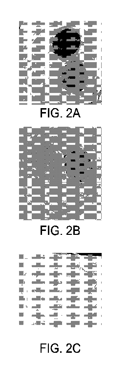
Figure 2 is an image of culture dishes showing the anti-fungal activity of
Rhodococcus
rhodochrous DAP 96253 is abolished in the presence of activated carbon.
Figure 3 shows SPME-GC-MS headspace analysis of uninduced (top) and induced
(bottom) Rhodococcus.
Figure 4 is a series of images showing Rhodococcus rhodochrous DAP 96253
controls
Pseudogymnoascus destructans spore germination. Experiment conducted at 15 C.
All images
captured at 200X magnification. Scale bar is 10um.
Figures 5A and 5B are images showing Explants inoculated with ¨100 P.
destructans
conidia each and incubated with induced Rhodococcus at 7 C for 40 days. No
exposed explants
developed any fungal colonization over 21 days. All controls were fully
colonized by day 14.
Figure 6 is a series of images showing non-growth Rhodococcus cell paste
exhibits
strong contact-independent inhibition of growth from conidia of P.
destructans. Images taken 21
DPI. Experiment conducted at 15 C.
Figure 7 is a series of images showing permanent inhibition of spore
germination 0
hours, 24 hours, 72 hours, or 7 days after exposure to Rhodococcus.
Figures 8A and 8B are images showing Rhodococcus has no contact or oral
toxicity to
adult Honey Bees.
Figure 9 is a plot showing mean body weight changes of bats exposed to
Rhodococcus
for 72 hours. WNS positive bats are separate from uninfected bats.
Figure 10 is a plot showing body mass of WNS positive bats treated with
Rhodococcus
for 48 hours (RRDAP48) and untreated control. Body weights of "0" indicate
mortality.
Figure 11 is a graph showing survival probability over time for infected M.
lucifugus
treated (RRDAP48) or not treated (NO RRDAP) with R. rhodochrous DAP96253.
Figure 12 is a gross pathology image of a wing from bat not treated with R.
rhodochrous
on Day 7.
Figure 13 is a gross pathology image of a wing from bat treated with R.
rhodochrous on
Day 20 showing reduced lesion and scar tissue formation.
Figure 14 is a graph showing cumlative survival of the treatment and control
groups,
segregated by infection severity, over the entire course of the trial.
3
CA 02951900 2016-12-09
WO 2015/191744 PCT/US2015/035146
DETAILED DESCRIPTION
As used herein, the singular forms "a", "an", "the", include plural referents
unless the
context clearly dictates otherwise.
Throughout the specification the word "comprising," or grammatical variations
thereof,
will be understood to imply the inclusion of a stated element, integer or
step, or group of
elements, integers or steps, but not the exclusion of any other element,
integer or step, or group
of elements, integers or steps.
The disclosed compositions, apparatuses, and methods arise from the surprising
finding
that one or more bacteria are capable of inhibiting or reducing fungal growth.
Therefore, method
are disclosed to reduce fungal growth in or on a subject by exposing the
subject to a
microorganism disclosed herein. This microorganism can be alive and
replicating, alive and non-
replicating, or dead, so long as the enzymatic activity in the cell is
maintained. In other
embodiments, the subject is exposed to one or more enzymes produced by the
bacteria that are
capable of inhibiting or reducing fungal growth. For example, in some
embodiments, the
enzymes are provided as an enzymatic extract from the disclosed microorganism.
When
enzymes or enzymatic extracts are used, cofactors can also be present, e.g.,
endogenous or
exogenous cofactors. In some cases, endogenous cofactors are substituted with
equivalent
cofactors. Regardless of the source, the cofactors can be provided in
catalytic amounts and can
also be regenerated as needed.
As used throughout, fungal growth includes all stages of the life cycle of a
fungus
including, but not limited to, spore germination, mycelium growth, and the
development and
formation of fruiting structures on the fungus.
Provided herein are methods and compositions for treating or preventing one or
more
fungal infections in a subject. The methods comprise exposing a subject to a
composition
comprising one or more bacteria, wherein the one or more bacteria are selected
from the group
consisting of genus Rhodococcus, genus Brevibacterium, genus Pseudonocardia,
genus
Nocardia, genus Pseudomonas and combinations thereof, and wherein the one or
more bacteria
are provided in a quantity sufficient to inhibit or reduce fungal growth in
the subject.
Optionally, the bacteria are induced to produce one or more enzymes. In some
embodiments,
the methods comprise exposing the subject to a composition comprising one or
more enzymes
selected from the group consisting of nitrile hydratases, amidases,
asparaginases, ACC
deaminases, cyanoalanine synthase-like enzymes, monooxygenases, dioxygenases,
cyanidases,
4
CA 02951900 2016-12-09
WO 2015/191744
PCT/US2015/035146
and combinations thereof, wherein the enzymes are provided in a quantity
sufficient to inhibit or
reduce fungal growth in the subject.
The methods and compositions are drawn to inhibiting or reducing fungal growth
in a
subject. Alternatively or additionally, the methods and compositions inhibit
or reduce toxin
development or release by a fungus.
The term "treatment" refers to the medical management of a subject with the
intent to
cure, ameliorate, stabilize, or prevent a disease, pathological condition, or
disorder. This term
includes active treatment, that is, treatment directed specifically toward the
improvement of a
disease, pathological condition, or disorder. In addition, this term includes
palliative treatment,
that is, treatment designed for the relief of symptoms rather than the curing
of the disease,
pathological condition, or disorder; preventative treatment, that is,
treatment directed to
minimizing or partially or completely inhibiting the development of the
associated disease,
pathological condition, or disorder; and supportive treatment, that is,
treatment employed to
supplement another specific therapy directed toward the improvement of the
associated disease,
pathological condition, or disorder. The term "treatment" is not limited to
treatment or
prescription by a medical professional, but also includes activities conducted
by any other
individual, including by the subject themselves.
The term "prevent" refers to a treatment administered before onset of a
disease or
condition that delays the onset of a disease or condition or reduces the
severity of the disease or
condition. Thus, if a treatment can treat a disease in a subject having
symptoms of the disease, it
can also prevent that disease in a subject who has yet to suffer some or all
of the symptoms.
As defined herein, "inhibiting or reducing fungal growth," and grammatical
variants
thereof, refers to any slowing, interruption, suppression, delay, or
inhibition of the fungal
growth. Inhibiting or reducing fungal growth can, for example, comprise
inhibiting or reducing
growth of resting fungal cells, which can include spore germination, mycelia
development,
and/or the formation of fruiting structures on the fungus (e.g.,
sporangia/sporophores).
Fungal growth can, for example, be produced by a fungus selected from the
group
consisting of mold, yeast, mildew, fungi that cause smut, fungi that cause
rust, fungi that cause
diseases of plants, and fungi that cause diseases of animals.
The term "subject" refers to any animal that is the target of administration
or treatment.
The subject can be a vertebrate, for example, a mammal. The term "mammal" is
known in the
art, and exemplary mammals include humans, primates, livestock animals
(including bovines,
porcines, etc.), companion animals (e.g., canines, felines, birds, horses,
etc.) and rodents (e.g.,
5
CA 02951900 2016-12-09
WO 2015/191744 PCT/US2015/035146
mice and rats). The subject can be an invertebrate animal, such as an insect
or other arthropod.
The subject can also be a human or veterinary patient. The term "patient"
refers to a subject
under the treatment of a clinician, e.g., physician.
In some cases, the subject is a bat. For example, the fungus can comprise
Pseudogymnoascus destructans. In these aspects, the composition can be
applied, for example,
to areas in or around a bat roost/ hibernacula. The composition can also be
provided in a bat lure
so the bat takes the catalyst back to its roost/ hibernacula. In some cases,
the composition is
provided as a vegetative probiotic. The composition can also be provided as a
non-vegetative
agent, e.g., as an ointment or spray. The composition can also be incorporated
into a fixed cell
matrix and placed on wing tags, arm bands, or collars. The composition can
also be incorporated
into a nesting structure/anchor upon which bats nest and/or congregates. In
some cases, the
treatment period is during late swarm or very early hibernation.
In some cases, the subject is a honey bee. For example, the fungus can be
selected from
the group consisting of Ascosphaera apis, Nosema apis, Aspergillus fumigatus,
Aspergillus
flavus, and Aspergillus niger. In these aspects, the composition can be
provided in a bait particle.
For example, the bait particle can be a microparticle configured to simulate a
pollen particle such
that a bee will pick up the particle and return it to the hive. In some
embodiments, the bat
particle is adhesive. The composition can also be provided in a wax, such as a
beeswax. In these
embodiments, the wax can be incorporated directly into a bee hive. The
composition can also be
provided as a powder, and optionally mixed with powdered sugar for application
as a dust to the
hive. The composition can also be incorporated into wax (bee wax, paraffin,
etc.) and used as a
base layer for hive frames. Hive frames are generally wooden structure that
bees build combs on.
Wax foundations are often included as a starting point for the bees, which
could be supplemented
with the disclosed composition. In some cases, the composition is added
beneath the hive (open
center concept), e.g., as a tray of cells. Without wishing to be bound by
theory, this can result in
VOCs that permeate the hive and are drawn into the hive by convection (hives
are warmer inside
compared to ambient temp.) The composition can also be provided as a probiotic
in
supplemental feed. For example, sugar solutions are typically provided by
keepers in winter to
supplement their diet, which can be supplemented with the disclosed
composition. The
composition can also be incorporated into a material at the entrance to the
hive "door mat"
whereby the bees come in contact with the material and bring it into the hive.
This type of
application takes advantage of the grooming habits of the bees to distribute
the material
throughout the colony.
6
CA 02951900 2016-12-09
WO 2015/191744
PCT/US2015/035146
In some cases, the subject is a snake or lizard. For example, the fungus can
be Snake
Fungal Disease or Yellow Fungus Syndrome. In these embodiments, the
composition can, for
example, be applied directly to the animal (probiotic), sprayed into burrows
(environmental
control), or delivered as a bait. Fungal infections in snakes are typically in
their facial area, so a
bait could be an effective delivery system for delivery of the disclosed
compositions to affected
areas.
In some cases, the subject is a human or veterinary patient. For cats and
dogs, for
example, delivery may be accomplished by an ointment, liquid, aerosol or
spray. In the
composition is administered in a manner that prevents the animal from licking
off the agent or
bother the site being treated. For example, the composition can be
administered in a manner that
quickly dries. In some embodiments, cleaning agents are used to "prep" and
clean the affected
area permitting better contact and dosing with the disclosed compositions. In
some cases, a mild
anesthetic is used to reduce irritation. This anesthetic can be administered
separately or in the
same composition with the disclosed bacteria, enzymes, or enzymatic extracts.
Fungi cause a wide variety of diseases in humans. While some fungi cause
infections
limited to the outermost layers of the skin and hair (superficial mycoses),
other fungi cause
cutaneous mycoses by penetrating to the keratinized layers of the skin, hair
and nails and
triggering pathologic changes in the host. Subcutaneous mycoses cause
infections in the dermis,
subcutaneous tissues, muscle and fascia and are often chronic. Systemic
mycoses originate
primarily in the lung and may cause secondary infections in other organ
systems in the body.
Patients with immune system deficiencies are often prone to opportunistic
mycoses.
Examples of Microsporum species include M. canis and M. gypseum. Microsporum
is
one of the several fungal genera that cause dermatophytosis. Dermatophytosis
is a general term
used to define the infection in hair, skin or nails due to any dermatophyte
species. Similar to
other dermatophytes, Microsporum has the ability to degrade keratin and thus
can reside on skin
and its appendages and remains noninvasive. Notably, Microsporum spp. mostly
infect the hair
and skin. Microsporum canis is the principal cause of ringworm in dogs and
cats and a
zoophilic fungal species causing sporadic dermatophytosis in humans,
especially tinea capitis in
children with cats and dogs.
Skin infection by a Trichophyton species occurs mainly on the back of the
neck, scalp or
beard. Symptoms of a Trichophyton species infection include inflamed scalp
lesions, inflamed
neck lesions, inflamed beard lesions, scarring, and permanent hair loss.
Examples of
Trichophyton species include T rubrum, T. tonsurans and T men tagrophytes.
7
CA 02951900 2016-12-09
WO 2015/191744
PCT/US2015/035146
Trichophyton tonsurans is an anthropophilic endothrix species of fungi that
causes
epidemic dermatophytosis in Europe, South America, and the U.S. It infects
some animals and
requires thiamine for growth. It is the most common cause of tinea capitis in
the U.S., forming
black dots where hair breaks off at the skin surface. Trichophyton rubrum is a
fungus that is the
most common cause of tinea pedis (athlete's foot), tinea cruris, and tinea
(ringworm).
Trichophyton rubum is the most common of the dermatophytes causing fingernail
fungus
infections. While most fungal skin infections are irritating and difficult to
treat, there are reports
of fungal infections resulting in death, such as Trichophyton mentagrophytes
skin infection migrated to the lymph nodes, testes, vertebrae and CNS.
The genus Epidermophyton contains two species; Epidermophyton floccosum and
Epidermophyton stockdaleae. E. stockdaleae is known to be nonpathogenic,
leaving E.
floccosum as the only species causing infections in humans. E. floccosum is
one of the common
causes of dermatophytosis in otherwise healthy individuals. It infects skin
(tinea corporis, tinea
cruris, tinea pedis) and nails (onychomycosis). The infection is restricted to
the nonliving
cornified layers of epidermis since the fungus lacks the ability to penetrate
the viable tissues of
the immunocompetent host. Disseminated infections due to any of the
dermatophytes are very
unlikely due to the restriction of the infection to keratinized tissues.
However, invasive E.
floccosum infection has been reported in an immunocompromised patient with
Behcet's
syndrome. As with all forms of dermatophytosis, Epidermophyton floccosum
infections are
communicable and usually transmitted by contact, particularly in common
showers and gym
facilities.
Examples of Candida species include C. albicans, C. parapsiliosis, and C.
krusei.
Patients with chronic mucocutaneous candidiasis may develop candida infection
of the nails.
Candida species may invade nails previously damaged by infection or trauma and
cause
infection in the periungual area and underneath the nailbed. The nailfold
becomes erythematous,
swollen and tender with an occasional discharge. The disease causes loss of
the cuticle, nail
dystrophy, and onycholysis with discoloration around the lateral nailfold. In
all forms of
onychomycosis, the nail becomes variously disfigured and distorted. A specific
example of
a fungal infection caused by the fungi and yeasts discussed above is
onychomycosis
(nail infection). Fungal infections affecting the nails or scalp are very
difficult to treat due
to fungal infection in follicle roots or under the nail itself
Onychomycosis is a chronic, persistent fungal, yeast, and/or mold infection of
the nail
bed which causes thickening and discoloration of the nail, sometimes
accompanied by pain and
8
CA 02951900 2016-12-09
WO 2015/191744 PCT/US2015/035146
disability. This fungal infection affects 25% of adults, and the incidence
rises with age, such that
the prevalence in adults over 50 years of age is 40%. According to a study
reported in Podiatry
Today, over 35 million people in the United States have onychomycosis, and up
to 50% of those
affected by the disease do not receive treatment.
In addition, Candida species, and Candida albicans in particular, play an
etiologic role in
the development of chronic paronychia, a common infection of the soft tissue
around the
fingernail or toenail, where bacteria may act as co-pathogens. Swollen,
erythematous and tender
nail folds without fluctuance are characteristic of chronic paronychia.
Eventually, the nail plates
become thickened and discolored, with pronounced transverse ridges and the
cuticles and nail
folds may separate from the nail plate, forming a space for the invasion of
various
microorganisms. Onychomycosis has long been one of the most difficult fungal
infections to
treat. The length of time it takes the nail to grow, the impenetrability of
the nail plate, and
location of the infection between the nail bed and plate are major factors
interfering with the
eradication of fungal agents affecting these tissues. Thus, eradication of
symptoms is very slow
and may take a whole year or even longer. Topical antifungals have low
efficacy because of their
antifungal spectrum may be limited to dermatophytes and because of restricted
penetration of the
antifungal agent across the nail. Systemic treatment with antifungal agents
has shown relapse
rates of 40% or higher, and have significant risks, including hepatic and/or
cardiac toxicity, and
adverse drug interactions. Thus, there is a significant need for alternative,
and more effective,
methods of treating fungal, yeast, and/or mold infections such as
onychomycosis.
Therefore, in some embodiments, the disclosed compositions are directly
applied to the
skin or nails of a subject, e.g., in the form of an ointment. In particular
cases, this can be used to
treat or prevent, for example, Geomyces pannorum.
The disclosed compositions can also be used in containers used to store items
that need to
be kept sterile for human use. For example, the disclosed compositions can be
incorporated into
contact lens storage containers, e.g., as a fixed cell application. In
particular cases, this can be
used to treat or prevent, for example, Fusarium oxysporum or Fusarium solani.
Systemic mycoses due to opportunistic pathogens are infections of patients
with immune
deficiencies who would otherwise not be infected. Examples of
immunocompromised conditions
include AIDS, alteration of normal flora by antibiotics, immunosuppressive
therapy, and
metastatic cancer. Examples of opportunistic mycoses include Candidiasis,
Cryptococcosis and
Aspergillosis.
9
CA 02951900 2016-12-09
WO 2015/191744 PCT/US2015/035146
In some embodiments, the disclosed compositions are used for environmental
control in a
home or health facility where opportunistic mycoses can occur. For example,
the disclosed
compositions can be imbedded in an air-filter. In particular cases, this can
be used to treat or
prevent, for example, Aspergillus fumigatus or Aspergillus flavus.
In some embodiments, the disclosed compositions are used for to treat or
prevent
infection of wounds and burns, which are susceptible to infection by
microorganisms, such as
bacteria and fungi. Microbial infection typically slows or prevents the
healing of a wound or
burn, and may lead to a localized or systemic infection of the wounded or
burned organism.
Accordingly, in some aspects a method is provided for inhibiting the growth of
microorganisms
in, or on, living tissue. These methods can include the step of contacting
living tissue that is
infected with microorganisms with a composition disclosed herein in amounts
sufficient to
inhibit growth of the microorganisms. In some embodiments, the living tissue
has been wounded
or burned.
As used herein, the term "wound" encompasses physical injuries to living
tissue and/or
interruption to the integrity of living tissue, such as cuts, tears,
abrasions, and lesions and
crushed tissue, as well as pimples, ulcers and hemorrhoids.
The term "wound dressing" refers to a material that is used to cover a wound.
Examples
of wound dressings include ointments, gels, salves, bandages and gauze.
A variety of living tissues of an animal body can be treated using the
disclosed
composition and methods. For example, the methods can be used to inhibit the
growth of
microorganisms on skin lesions, burns on the skin, or on wounds (such as cuts
or abrasions) of
the skin. The methods can also be used, for example, to inhibit the growth of
microorganisms
within a body cavity (e.g., abdomen) or a joint (e.g., a knee joint), on the
surface of an eye, or in
the mouth.
In some embodiments, the disclosed bacteria, enzyme, and/or the enzymatic
extract
isolated from the bacteria are applied to a wound dressing that is
administered to the wound or
site of infection. In some embodiments, the disclosed bacteria, enzyme, and/or
the enzymatic
extract isolated from the bacteria are introduced onto the surface of an eye,
or into the ear canal,
using a dropper. Ointments, creams or gels comprising bacteria, enzyme, and/or
the enzymatic
extract isolated from the bacteria, may, for example, be rubbed onto a surface
of a living
organism. Other examples of methods for contacting living tissue with a
composition comprising
bacteria, enzyme, and/or the enzymatic extract isolated from the bacteria
include flushing or
irrigating the living tissue with a solution containing bacteria, enzyme,
and/or the enzymatic
CA 02951900 2016-12-09
WO 2015/191744 PCT/US2015/035146
extract isolated from the bacteria; rubbing living tissue with a medical
dressing containing a
solution containing bacteria, enzyme, and/or the enzymatic extract isolated
from the bacteria;
spraying living tissue (e.g., by using a nebulizer) with a composition
containing bacteria,
enzyme, and/or the enzymatic extract isolated from the bacteria; introducing a
solution
containing bacteria, enzyme, and/or the enzymatic extract isolated from the
bacteria into a living
body using, for example, a tube, catheter, canula or endoscopic device;
introducing a
composition containing bacteria, enzyme, and/or the enzymatic extract isolated
from the bacteria
into an orifice of a living body using, for example, a suppository or tampon;
and contacting oral
tissue with a composition containing bacteria, enzyme, and/or the enzymatic
extract isolated
from the bacteria, for example by gargling or rinsing the oral cavity with a
composition
containing bacteria, enzyme, and/or the enzymatic extract isolated from the
bacteria.
Wound dressings suitable for use in the disclosed methods can be any material
that is
biologically acceptable and suitable for placing on a wound. In exemplary
embodiments,
the wound dressing may be a woven or non- woven fabric of synthetic or
nonsynthetic fibers, or
any combination thereof For example, the wound dressing can be gauze. The
gauze may be
absorbent and can be, for example, wetted with a composition containing
bacteria, enzyme,
and/or the enzymatic extract isolated from the bacteria. The dressing may also
comprise a
support, such as a polymer foam, a natural or man-made sponge, a gel or a
membrane that may
absorb or have disposed thereon, a composition containing bacteria, enzyme,
and/or the
enzymatic extract isolated from the bacteria. Again by way of example, the
support can be a
film, a natural or synthetic polymer, or a rigid or malleable material.
In some embodiments, the disclosed compositions and methods can also treat or
prevent
bacterial infections in a subject, e.g., in wounds or burns. Without wishing
to be bound by
theory, this effect of the disclosed compositions may be due to quorum
quenching mechanism.
In particular cases, this can be used to treat or prevent, for example,
Pseudomonas aeruginosa or
Burkholderia cepacia.
The disclosed compositions and methods can also be used to treat or prevent
infections in
domestic animals such as dogs, cats, and birds. In particular cases, this can
be used to treat or
prevent, for example, Microsporum canis (dogs, cats), Aspergillus flavus, A.
fumigatus (dogs,
birds), and Trichophyton verrucosum.
The disclosed compositions and methods can also be used to prevent toxicosis
from
aflatoxin exposure. Aflatoxin-producing members of Aspergillus are common and
widespread in
nature. They can colonize and contaminate grain before harvest or during
storage. Host crops,
11
CA 02951900 2016-12-09
WO 2015/191744 PCT/US2015/035146
which include maize, sorghum, and groundnuts, are particularly susceptible to
infection by
Aspergillus following prolonged exposure to a high-humidity environment, or
damage from
stressful conditions such as drought, a condition that lowers the barrier to
entry. The toxin also
may be found in the milk of animals that are fed contaminated feed. High-level
aflatoxin
exposure produces an acute hepatic necrosis, resulting later in cirrhosis, or
carcinoma of the
liver. Acute liver failure is made manifest by bleeding, edema, alteration in
digestion, changes to
the absorption and/or metabolism of nutrients, and mental changes and/or coma.
In certain embodiments, the methods and compositions for inhibiting or
reducing fungal
growth comprises exposing the subject to one or more bacteria selected from
the group
consisting of genus Rhodococcus, genus Brevibacterium, genus Pseudomonas,
genus Nocardia,
genus Pseudonocardia and combinations thereof The one or more bacteria can,
for example,
include Rhodococcus spp. The Rhodococcus spp can, for example, include
Rhodococcus
rhodochrous DAP 96253 strain, Rhodococcus rhodochrous DAP 96622 strain,
Rhodococcus
erythropolis, or combinations thereof Optionally, the compositions comprise
Rhodococcus
rhodochrous and Rhodococcus erythropolis. Exemplary organisms include, but are
not limited
to, Pseudomonas chloroaphis (ATCC 43051) (Gram-negative), Pseudomonas
chloroaphis
(ATCC 13985) (Gram-negative), Rhodococcus erythropolis (ATCC 47072) (Gram-
positive),
and Brevibacterium ketoglutamicum (ATCC 21533) (Gram-positive). Examples of
Nocardia
and Pseudonocardia species have been described in European Patent No. 0790310;
Collins and
Knowles J. Gen. Microbiol. 129:711-718 (1983); Harper Biochem. J. 165:309-319
(1977);
Harper Int. J. Biochem. 17:677-683 (1985); Linton and Knowles J Gen.
Microbiol. 132:1493-
1501 (1986); and Yamaki et al., J. Ferm. Bioeng. 83:474-477 (1997).
Although in some embodiments the one or more bacteria are selected from the
group
consisting of Rhodococcus spp., Brevibacterium ketoglutamicum, and Pseudomonas
chloroaphis,
any bacterium that inhibits or reduces fungal growth when exposed to an animal
can be used in
the present methods. For example, bacteria belonging to the genus Nocardia
[see Japanese
Patent Application No. 54-129190], Rhodococcus [see Japanese Patent
Application No. 2-470],
Rhizobium [see Japanese Patent Application No. 5-236977], Klebsiella [Japanese
Patent
Application No. 5-30982], Aeromonas [Japanese Patent Application No. 5-30983],
Agrobacterium [Japanese Patent Application No. 8-154691], Bacillus [Japanese
Patent
Application No. 8-187092], Pseudonocardia [Japanese Patent Application No. 8-
56684],
Burkholderia, Corynebacterium, and Pseudomonas are non-limiting examples of
bacteria that
can be used. Not all species within a given genus exhibit the same type of
enzyme activity
12
CA 02951900 2016-12-09
WO 2015/191744 PCT/US2015/035146
and/or production. Thus, it is possible to have a genus generally known to
include strains
capable of exhibiting a desired activity but have one or more strains that do
not naturally exhibit
the desired activity or one or more strains which do not exhibit the activity
when grown on the
same medium as the species which exhibit this activity. Thus, host
microorganisms can include
strains of bacteria that are not specifically known to have the desired
activity but are from a
genus known to have specific strains capable of producing the desired
activity. Such strains can
have transferred thereto one or more genes useful to cause the desired
activity. Non-limiting
examples of such strains include Rhodococcus equi and Rhodococcus globerulus
PWD1.
Further, specific examples of bacteria include, but are not limited to,
Nocardia sp.,
Rhodococcus sp., Rhodococcus rhodochrous, Klebsiella sp., Aeromonas sp.,
Citrobacter
freundii, Agrobacterium rhizogenes , Agrobacterium tumefaciens, Xanthobacter
"lavas, Erwinia
nigrifluens , Enterobacter sp., Streptomyces sp., Rhizobium sp., Rhizobium
loti, Rhizobium
legminosarum, Rhizobium merioti, Pantoea agglomerans, Klebsiella pneumoniae
subsp.
pneumoniae, Agrobacterium radiobacter, , Bacillus smith ii, Pseudonocardia
thermophila,
Pseudomonas chloroaphis, Rhodococcus erythropolis, Brevibacterium
ketoglutamicum, and
Pseudonocardia thermophila. Optionally, the microorganisms used can, for
example, comprise
Rhodococcus rhodochrous DAP 96253 and Rhodococcus rhodochrous DAP 96622, and
combinations thereof
As used herein, exposing a subject to one or more bacteria includes, for
example,
exposing the subject to intact bacterial cells, bacterial cell lysates, and/or
bacterial extracts that
possess enzymatic activity (i.e., "enzymatic extracts"). Methods for preparing
lysates and
enzymatic extracts from cells, including bacterial cells, are routine in the
art. Optionally, the one
or more bacteria or enzymatic extracts are fixed with glutaraldehyde and
crosslinked.
Optionally, the crosslinked, glutaraldehyde-fixed bacteria or extract is
formulated with a carrier
into a spray.
In certain embodiments, the methods and compositions for inhibiting or
reducing fungal
growth comprise exposing the subject to an enzyme. The enzyme can be selected
from the
group consisting of nitrile hydratase, amidase, asparaginase, ACC (1-
aminocyclopropane-1-
carboxylic acid) deaminase, cyanoalanine synthase-like enzyme, alkane
monooxygenase,
ammonium monooxygenase, methane monooxygenase, toluene dioxygenase, cyanidase,
and/or a
combination thereof The enzyme can be provided within a composition for
exposure to the
subject. The enzyme can also be a purified enzyme or can be provided as an
enzymatic extract
as described above. Optionally, the methods for inhibiting or reducing fungal
growth in a animal
13
CA 02951900 2016-12-09
WO 2015/191744 PCT/US2015/035146
comprise exposing the location to a composition comprising an enzyme, the
enzyme being
selected from one or more of nitrile hydratase, amidase, asparaginase, ACC
deaminase,
cyanoalanine synthase-like enzyme, alkane monooxygenase, ammonium
monooxygenase,
methane monooxygenase, toluene dioxygenase, and cyanidase. The one or more
bacteria,
enzymatic extract, or enzymes used in the methods may at times be more
generally referred to
herein as the "catalyst."
In the methods provided herein, the subject or its habitat is exposed to one
or more
bacteria, one or more enzymes, enzymatic extract isolated from or derived from
the one or more
bacteria, or any combination thereof, in a quantity sufficient to inhibit or
reduce fungal growth.
In some embodiments, the subject or its habitat is exposed to one or more
bacteria in
combination with one or more exogenous enzymes and/or enzymatic extracts.
"Exogenous"
refers to enzymes or enzymatic extracts that are isolated and/or purified ex
situ and is
distinguished from enzymes produced by bacteria in situ. This combined
exposure can take place
simultaneously and/or sequentially. For example, the subject or its habitat
can be exposed to
exogenous enzymes and/or enzymatic extracts 1 to 60 minutes, 1 to 24 hours, or
1 to 7 days after
exposure to the bacteria.
"Exposing" a subject or its habitat to one or more bacteria, one or more
enzymes, and/or
an enzymatic extract includes any method of presenting a bacterium, enzyme,
and/or extract to
the subject or its habitat. Optionally, the subject or its habitat is
indirectly exposed to the one or
more bacteria, one or more enzymes, and/or the enzymatic extract. Indirect
methods of exposure
include, for example, placing the one or more bacteria, one or more enzymes,
and/or enzymatic
extract in the general proximity of the subject or its habitat (i.e., indirect
exposure). Optionally,
the subject or its habitat is directly exposed to one or more bacteria, one or
more enzymes,
and/or the enzymatic extract, whereby the one or more bacteria, one or more
enzymes, and/or
enzymatic extract are in direct contact with the subject or its habitat.
Without wishing to be bound by theory, in some embodiments, indirect exposure
results
in volatile organic compound (VOC) release from the bacteria that contacts the
subject and
inhibits or reduces fungal growth.
In certain embodiments, exposure of the bacteria, enzyme, and/or the enzymatic
extract
isolated from the bacteria can occur, for example, by providing the bacteria,
enzyme, and/or
enzymatic extract in liquid form and spraying it onto or near the subject or
its habitat. The
bacteria, enzyme, and/or enzymatic extract can, for example, further comprise
a liquid carrier.
Liquid carriers can be selected from the group consisting of an aromatic
hydrocarbon, a
14
CA 02951900 2016-12-09
WO 2015/191744 PCT/US2015/035146
substituted naphthalene, a phthalic acid ester, an aliphatic hydrocarbon, an
alcohol, and a glycol.
Optionally, the liquid carrier can be a wax or similar type material coating,
which could be
applied to the plant as a liquid, but would be solid at ambient or lower
temperatures. Optionally,
the bacteria, enzyme and/or enzymatic extract are provided onto or near the
subject or its habitat
by a fog or spray. For example, the bacteria, enzyme or enzymatic extract can
be provided to a
habitat where the fungi is to be controlled.
In certain embodiments, exposure of the one or more bacteria, one or more
enzymes,
and/or the enzymatic extract isolated from the bacteria can occur, for
example, by providing the
bacteria, enzyme, and/or enzymatic extract in solid form and dusting it onto
or near the subject
or its habitat. The bacteria, enzyme, and/or enzymatic extract can, for
example, further comprise
a solid carrier. The solid carrier can be selected from the group consisting
of a dust, a wettable
powder, a water dispersible granule, and mineral fillers. Optionally, the
solid carrier is a mineral
filler. Mineral fillers can, for example, be selected from the group
consisting of a calcite, a
silica, a talc, a kaolin, a montmorillonite, and an attapulgite. Other solid
supports for use with
the bacteria, enzyme, and/or enzymatic extract are described herein.
The herein disclosed compositions, including pharmaceutical composition, may
be
administered in a number of ways depending on whether local or systemic
treatment is desired,
and on the area to be treated. For example, the disclosed compositions can be
administered
intravenously, intraperitoneally, intramuscularly, subcutaneously,
intracavity, or transdermally.
The compositions may be administered orally, parenterally (e.g.,
intravenously), by
intramuscular injection, by intraperitoneal injection, transdermally,
extracorporeally,
ophthalmically, vaginally, rectally, intranasally, topically or the like,
including topical intranasal
administration or administration by inhalant.
In certain embodiments, the one or more bacteria, one or more enzymes, and/or
enzymatic extract further comprise a coating, wherein the coating makes the
one or more
bacteria, one or more enzymes, and/or enzymatic extract water resistant. The
coating can be
selected from a hydrophobic fatty acid polyester coating or a wax. Optionally,
the hydrophobic
fatty acid polyester coating is a long chain fatty acid polyester derived from
sucrose, sorbitol,
sorbinose, glycerol, or raffinose.
Also provided herein are compositions for inhibiting or reducing fungal
growth. The
compositions can, for example, comprise one or more bacteria, one or more
enzymes, and/or one
or more enzymatic extracts capable of inhibiting or reducing fungal growth.
The compositions
can further comprise solid, liquid, aerosols, gels, and gelatinous carriers,
as disclosed above,
CA 02951900 2016-12-09
WO 2015/191744 PCT/US2015/035146
and/or media and media components for inducing and stabilizing the one or more
bacteria, one
or more enzymes, and/or enzymatic extracts, as disclosed below. Optionally,
the compositions
can be converted into pellet form for distribution or application to the
subject or its habitat.
Optionally, the one or more bacteria, one or more enzymes, and/or enzymatic
extract are
used in combination with other agents that inhibit or reduce fungal growth.
For example, the
provided methods can further comprise the step of exposing the subject or its
habitat to an agent
that inhibits or reduces fungal growth, e.g., a fungicide. Likewise, the
provided compositions
can further comprise an agent that inhibits or reduces fungal growth, e.g., a
fungicide. Agents
that inhibit or reduce fungal growth include, but are not limited to,
anthocyanins, organic acids,
such as, propionic acid and sorbic acid, aluminosilicates, clays, zeolites,
and calcium propanoate.
The provided composition(s) can also be used in combination with one or more
classes of
antibiotics (e.g., Aminoglycosides, Cephalosporins, Chloramphenicol,
Clindamycin,
Erythromycins, Fluoroquinolones, Macrolides, Azolides, Metronidazole,
Penicillins,
Tetracyclines, Trimethoprim-sulfamethoxazole, Vancomycin).
As defined herein, a "sufficient" quantity or effective amount of the
bacteria, enzyme,
and/or enzymatic extract will depend on a variety of factors, including, but
not limited to, the
particular bacteria, enzyme, and/or enzymatic extract utilized in the method,
the form in which
the bacteria is exposed to the location (e.g., as intact bacterial cells (dead
or alive), cell lysates,
enzymatic extracts, and/or enzymes as described above), the means by which the
bacteria,
enzyme, and/or enzymatic extract is exposed to the location, the length of
time of the exposure,
and the type and amount of fungal signal compounds that result in the
inhibition or reduction of
fungal growth. Optionally, the quantity of bacteria exposed to the location is
in the range of 1 to
250 mg of cell-dry weight or the equivalent thereof for enzymatic extracts and
enzymes. For
example, for 1 mg of dry weight of cells, typically there are 150-300 units of
nitrile hydratase,
10-25 units of amidase, 7-15 units of cyanidase, 7-20 units of ACC deaminase,
and 7-20 units of
cyanoalanine synthase-like enzyme. By way of other examples, the quantity of
bacteria exposed
to the location is in the range of 0.1 mg to 1 g, 0.1 to 400 mg, 1 to 200 mg,
1 to 80 mg, or 1 to 10
mg of cell-dry weight or the equivalent thereof for enzymatic extracts and
enzymes. By way of
example, the quantity of bacteria exposed to the subject or its habitat is,
for example, in the
range of 0.1 mg to 1 g per 9-10 kilos (kg) of plant or plant part. It would be
a matter of routine
experimentation for the skilled artisan to determine the "sufficient" quantity
of the one or more
bacteria, one or more enzymes, or enzymatic extract necessary to inhibit or
reduce fungal
growth. For example, if the bacteria, one or more enzymes, or enzymatic
extract necessary to
16
CA 02951900 2016-12-09
WO 2015/191744 PCT/US2015/035146
inhibit or reduce fungal growth are immobilized or stabilized, the quantity of
bacteria, one or
more enzymes, or enzymatic extract is adjusted to inhibit or reduce fungal
growth.
In certain embodiments, the one or more bacteria are "induced" to exhibit a
desired
characteristic (e.g., the expression of a desired level of activity of an
enzyme of the bacteria) by
exposure or treatment with a suitable inducing agent. Inducing agents include,
but are not
limited to urea, methyl carbamate, cobalt, asparagine, glutamine, and
combinations thereof
Optionally, the one or more bacteria are exposed to or treated with urea,
methyl carbamate,
methacrylamide, or acetamide. Optionally, the one or more bacteria are exposed
to or treated
with a mixture of inducing agents comprising urea or methyl carbamate and one
or more of
asparagine and cobalt. In some embodiments, the compositions and methods
optionally exclude
an inducing agent, such as cobalt.
The inducing agent, when used, can be added at any time during cultivation of
the
desired cells. For example, with respect to bacteria, the culture medium can
be supplemented
with an inducing agent prior to beginning cultivation of the bacteria.
Alternately, the bacteria
could be cultivated on a medium for a predetermined amount of time to grow the
bacteria and
the inducing agent could be added at one or more predetermined times to induce
the desired
enzymatic activity in the bacteria. Moreover, the inducing agent could be
added to the growth
medium (or to a separate mixture including the previously grown bacteria) to
induce the desired
activity in the bacteria after the growth of the bacteria is completed or
during a second growth or
maintenance phase.
While not intending to be limited to a particular mechanism, "inducing" the
bacteria may
result in the production or activation (or increased production or increased
activity) of one or
more of enzymes, such as nitrile hydratase, amidase, asparaginase, ACC
deaminase,
cyanoalanine synthase-like enzyme, alkane monooxygenase, ammonium
monooxygenase,
methane monooxygenase, toluene dioxygenase, and/or cyanidase, and the
induction of one or
more of these enzymes may play a role in inhibiting or reducing fungal growth.
"Nitrile
hydratases," "amidases," "asparaginases," "ACC deaminases," "cyanoalanine
synthase-like
enzymes," "AMO-type (alkane or ammonium) monooxygenases," "methane
monooxygenases,"
"toluene dioxygenases," and "cyanidases" comprise families of enzymes present
in cells from
various organisms, including but not limited to, bacteria, fungi, plants, and
animals. Such
enzymes are well known, and each class of enzyme possesses recognized
enzymatic activities.
The methods of inducing an enzymatic activity can be accomplished without the
requirement of introducing hazardous nitriles, such as acrylonitrile, into the
environment.
17
CA 02951900 2016-12-09
WO 2015/191744 PCT/US2015/035146
Previously, it was believed that induction of specific enzyme activity in
certain microorganisms
required the addition of chemical inducers. For example, in the induction of
nitrile hydratase
activity in Rhodococcus rhodochrous and Pseudomonas chloroaphis, it was
generally believed
to be necessary to supplement with hazardous chemicals, such as acetonitrile,
acrylonitrile,
acrylamide, and the like. However, enzymatic activity in nitrile hydratase
producing
microorganisms can be induced with the use of non-hazardous media additives,
such as amide
containing amino acids and derivates thereof, and optionally stabilized with
trehalose.
Optionally, asparagine, glutamine, or combinations thereof, can be used as
inducers. Methods of
inducing and stabilizing enzymatic activity in microorganisms are described in
U.S. Patent No.
7,531,343 and U.S. Patent No. 7,531,344, which are incorporated herein by
reference.
The disclosed methods of inducing enzymatic activity provide for the
production and
stability of a number of enzymes using modified media, immobilization, and
stabilization
techniques, as described herein. For example, enzymatic activity can be
induced and stabilized
through the use of media comprising amide-containing amino acids, or
derivatives thereof, and,
optionally stabilized by, trehalose. In some embodiments, the methods of
induction and
stabilization comprise culturing a nitrile hydratase producing microorganism
in a medium
comprising one or more amide containing amino acids or derivatives thereof,
and, optionally,
trehalose. Optionally, disclosed are methods for inducing nitrile-hydratase
using a medium
supplemented with amide containing amino acids or derivatives thereof, which
preferably
include asparagine, glutamine or a combination thereof Optionally, disclosed
are methods for
inducing nitrile-hydratase using a nutritionally complete medium supplemented
with only
asparagine. Optionally, disclosed are methods for inducing nitrile-hydratase
using a nutritionally
complete medium supplemented with only glutamine. Optionally, disclosed are
methods for
stabilizing nitrile-hydratase using a nutritionally complete medium
supplemented with only
trehalose. More particularly, the methods of induction and stabilization
comprise culturing the
microorganism in the medium and optionally collecting the cultured
microorganisms or enzymes
produced by the microorganisms.
Induction and stabilization of enzymes can be achieved without the use of
hazardous
nitriles. However, while the induction methods eliminate the need for
hazardous chemicals for
enzyme activity induction, the use of such further inducers is not excluded.
For example, one or
more nitriles could be used to assist in specific activity development. Media
supplemented with
succinonitrile and cobalt can be useful for induction of enzymes, including,
for example, nitrile
hydratase, amidase, asparaginase I, ACC deaminase, cyanoalanine synthase-like
enzyme, alkane
18
CA 02951900 2016-12-09
WO 2015/191744 PCT/US2015/035146
monooxygenase, ammonium monooxygenase, methane monooxygenase, toluene
dioxygenase,
and cyanidase. However, the use of nitriles is not necessary for induction of
enzyme activity.
While the use of nitriles and other hazardous chemicals is certainly not
preferred, optionally,
such use is possible.
Stabilization of enzyme activity can be achieved through immobilization
methods, such
as affixation, entrapment, and cross-linking, thereby, extending the time
during which enzyme
activity can be used. Thus, in some embodiments, induction methods and methods
of delaying a
chill injury response further comprise at least partially immobilizing the
microorganism.
Stabilization can be provided by immobilizing the enzymes, enzymatic extracts,
or
microorganisms producing the enzymes or enzymatic extracts. For example,
enzymes or
enzymatic extracts harvested from the microorganisms or the induced
microorganisms
themselves can be immobilized to a substrate as a means to stabilize the
induced activity.
Optionally, the nitrile hydratase producing microorganisms are at least
partially immobilized.
Optionally, the enzymes or microorganisms are at least partially entrapped in
or located on the
surface of a substrate. This allows for presentation of an immobilized
material with induced
activity (e.g., a catalyst) in such a manner as to facilitate reaction of the
catalyst with an intended
material and recovery of a desired product while simultaneously retaining the
catalyst in the
reaction medium and in a reactive mode. In certain embodiments, the
stabilization through
immobilization methods, such as affixation and entrapment, of the one or more
bacteria kills or
inactivates the one or more bacteria. Thus, optionally, the induced
microorganisms used in the
present methods are dead (killed) or inactivated, but are still capable of
exhibiting catalyst
activity.
Any substrate generally useful for affixation of enzymes, enzymatic extracts,
or
microorganisms can be used. Optionally, the substrate comprises alginate or
salts thereof
Alginate is a linear copolymer with homopolymeric blocks of (1-4)-linked 13-D-
mannuronate
(M) and its C-5 epimer a-L-guluronate (G) residues, respectively, covalently
linked together in
different sequences or blocks. The monomers can appear in homopolymeric blocks
of
consecutive G-residues (G-blocks), consecutive M-residues (M-blocks),
alternating M and G-
residues (MG-blocks), or randomly organized blocks. Optionally, calcium
alginate is used as the
substrate. The calcium alginate can, for example, be cross-linked, such as
with
polyethyleneimine, to form a hardened calcium alginate substrate. Further
description of such
immobilization techniques can be found in Bucke, "Cell Immobilization in
Calcium Alginate,"
Methods in Enzymology, vol. 135, Part B (ed. K. Mosbach) pp. 175-189 (1987),
which is
19
CA 02951900 2016-12-09
WO 2015/191744 PCT/US2015/035146
incorporated herein by reference. The stabilization effect of immobilization
using
polyethylenimine cross-linked calcium alginate is discussed in U.S. Patent No.
7,943,549, which
is hereby incorporated by reference in its entirety.
Optionally, the substrate comprises an amide-containing polymer. Any polymer
comprising one or more amide groups can be used. Optionally, the substrate
comprises a
polyacrylamide polymer.
Stabilization can further be achieved through cross-linking. For example
induced
microorganisms can be chemically cross-linked to form agglutinations of cells.
Optionally, the
induced microorganisms are fixed and cross-linked using glutaraldehyde. For
example,
microorganisms can be suspended in a mixture of de-ionized water and
glutaraldehyde followed
by addition of polyethylenimine until maximum flocculation is achieved. The
cross-linked
microorganisms (typically in the form of particles formed of a number of
cells) can be harvested
by simple filtration. Further description of such techniques is provided in
Lopez-Gallego, et al.,
J. Biotechnol. 119:70-75 (2005), which is incorporated herein by reference. In
certain
embodiments, the cross-linking kills or inactivates the microorganism. Thus,
optionally, the
induced microorganisms used in the present methods are dead (killed) or
inactivated, but are still
capable of exhibiting catalyst activity.
Optionally, the microorganisms, enzymes, and/or enzymatic extracts can be
encapsulated
rather than allowed to remain in the classic Brownian motion. Such
encapsulation facilitates
collection, retention, and reuse of the microorganisms and generally comprises
affixation of the
microorganisms to a substrate. Such affixation can also facilitate
stabilization of the
microorganisms, enzymes, and/or enzymatic extracts as described above, or may
be solely to
facilitate ease of handling of the induced microorganisms, enzymes, or
enzymatic extracts.
The microorganisms, enzymes, and/or enzymatic extracts can be immobilized by
any
method generally recognized for immobilization of microorganisms, enzymes,
and/or enzymatic
extracts such as sorption, electrostatic bonding, covalent bonding, and the
like. Generally, the
microorganisms, enzymes, and/or enzymatic extracts are immobilized or
entrapped on a solid
support which aids in the recovery of the microorganisms enzymes, or enzymatic
extracts from a
mixture or solution, such as a detoxification reaction mixture. Suitable solid
supports include,
but are not limited to, granular activated carbon, compost, wood or wood
products, (e.g., paper,
wood chips, wood nuggets, shredded pallets or trees), bran (e.g., wheat bran),
metal or metal
oxide particles (e.g., alumina, ruthenium, iron oxide), ion exchange resins,
DEAE cellulose,
DEAESEPHADEX polymer, waxes/coating materials (such as those used as a
coating for
CA 02951900 2016-12-09
WO 2015/191744 PCT/US2015/035146
fruits and vegetables and inanimate surfaces), ceramic beads, cross-linked
polyacrylamide beads,
cubes, pills, or other gel forms, alginate beads, K-carrageenan cubes, as well
as solid particles
that can be recovered from the aqueous solutions due to inherent magnetic
ability. The shape of
the catalyst is variable (in that the desired dynamic properties of the
particular entity are
integrated with volume/surface area relationships that influence catalyst
activity). Optionally,
the induced microorganism is immobilized in alginate beads that have been
cross-linked with
polyethyleneimine or is immobilized in a polyacrylamide-type polymer.
In some embodiments, the compositions and medium used in the induction and
stabilization methods further comprise one or more amide containing amino
acids or derivatives
thereof, and/or trehalose. The amide containing amino acids can, for example,
be selected from
the group consisting of asparagine, glutamine, derivatives thereof, or
combinations thereof For
example, the amide-containing amino acids may include natural forms of
asparagine, anhydrous
asparagine, asparagine monohydrate, or natural forms of glutamine, anhydrous
glutamine, and/or
glutamine monohydrate, each in the form of the L- isomer or D- isomer.
The concentration of the amide containing amino acids or derivatives thereof
in the
medium can vary depending upon the desired end result of the culture. For
example, a culture
may be carried out for the purpose of producing microorganisms having a
specific enzymatic
activity. Optionally, a culture may be carried out for the purpose of forming
and collecting a
specific enzyme from the cultured microorganisms. Optionally, a culture may be
carried out for
the purpose of forming and collecting a plurality of enzymes having the same
or different
activities and functions.
The amount of the amide containing amino acids, or derivatives thereof, added
to the
growth medium or mixture can generally be up to 10,000 parts per million (ppm)
(i.e., 1% by
weight) based on the overall weight of the medium or mixture. The induction
methods are
particularly beneficial, however, in that enzyme activity can be induced
through addition of even
lesser amounts. Optionally, the one or more amide containing amino acids are
present at a
concentration of at least 50 ppm. By way of other examples, the concentration
of the amide
containing amino acids or derivatives thereof is in the range of 50 ppm to
5,000 ppm, 100 ppm
to 3,000 ppm, 200 ppm to 2,000 ppm, 250 ppm to 1500 ppm, 500 ppm to 1250 ppm,
or 500 ppm
to 1000 ppm.
In some embodiments, the stabilization methods include the use of trehalose.
The
concentration of trehalose in the compositions or medium used in the induction
methods can be
at least 1 gram per liter (g/L). Optionally, the concentration of trehalose is
in the range of lg/L
21
CA 02951900 2016-12-09
WO 2015/191744 PCT/US2015/035146
to 50 g/L, or 1 g/L to 10 g/L. Optionally, the concentration of trehalose in
the medium is at least
4g/L.
The amide containing amino acids or derivatives thereof and/or trehalose are
added to a
nutritionally complete media. A suitable nutritionally complete medium
generally is a growth
medium that can supply a microorganism with the necessary nutrients required
for its growth,
which minimally includes a carbon and/or nitrogen source. One specific example
is the
commercially available R2A agar medium, which typically consists of agar,
yeast extract,
proteose peptone, casein hydrolysate, glucose, soluble starch, sodium
pyruvate, dipotassium
hydrogenphosphate, and magnesium sulfate. Another example of a nutritionally
complete liquid
medium is Yeast Extract Malt Extract Agar (YEMEA), which consists of glucose,
malt extract,
and yeast extract (but specifically excludes agar). Also, media of similar
composition, but of
vegetable origin can be used for the disclosed methods. Any nutritionally
complete medium
known in the art could be used for the disclosed methods, the above media
being described for
exemplary purposes only. Such nutritionally complete media can be included in
the
compositions described herein.
Optionally, the disclosed compositions and media can contain further
additives.
Typically, the other supplements or nutrients are those useful for assisting
in greater cell growth,
greater cell mass, or accelerated growth. For example, the compositions and
media can comprise
a carbohydrate source in addition to any carbohydrate source already present
in the nutritionally
complete medium.
As described above, most media typically contain some content of carbohydrate
(e.g.,
glucose); however, it can be useful to include an additional carbohydrate
source (e.g., maltose or
less refined sugars, such as dextrose equivalents that would be polymers of
dextrose, or
compositions such as molasses or sorghum, or any carbohydrate that supports
growth of the cell
and induction of the desired activity). The type of excess carbohydrate
provided can depend
upon the desired outcome of the culture. For example, the addition of
carbohydrates, such as
maltose or maltodextrin, has been found to provide improved induction of
asparaginase I.
Additionally, the addition of carbohydrates, such as maltose or maltodextrin,
potentially
improves stability of enzymatic activity (e.g., nitrile hydratase activity).
In some embodiments, the compositions and media further comprise cobalt.
Cobalt or a
salt thereof can be added to the mixture or media. For example, the addition
of cobalt (e.g.,
cobalt chloride) to the media can be particularly useful for increasing the
mass of the enzyme
produced by the cultured microorganisms. Cobalt or a salt thereof can, for
example, be added to
22
CA 02951900 2016-12-09
WO 2015/191744 PCT/US2015/035146
the culture medium such that the cobalt concentration is an amount up to 400
ppm. Cobalt can,
for example, be present at a concentration of 5 ppm to 400 ppm, 10 ppm to 100
ppm, 10 ppm to
80 ppm, or 10 ppm to 25 ppm.
In some embodiments, the compositions and media further comprise urea. Urea or
a salt
thereof can be added to the mixture or media. Urea or a salt thereof can, for
example, be added
to the culture medium such that the urea concentration is in an amount up to
10 g/L. Urea can,
for example, be present in a concentration of 5 g/L to 30 g/L, 5 g/L to 20
g/L, 5 g/L to 12 g/L, or
7 g/L to 10 g/L. Optionally, urea is present at a concentration of 7.5 g/L.
Optionally, both urea
and cobalt are added to the media.
The compositions and media may also include further components. For example,
other
suitable medium components may include commercial additives, such as
cottonseed protein,
maltose, maltodextrin, and other commercial carbohydrates. Optionally, the
medium further
comprises maltose or maltodextrin. Maltose or maltodextrin, for example, can
be added to the
culture medium such that the maltose or maltodextrin concentration is at least
1 g/L. Optionally,
the compositions and media are free of any nitrile containing compounds.
Nitrile compounds
were previously required in the culture medium to induce enzyme activity
toward two or more
nitrile compounds. The compositions described herein achieve this through the
use of
completely safe trehalose and/or amide containing amino acids or derivatives
thereof; therefore,
the medium can be free of any nitrile containing compounds.
"Enzymatic activity," as used herein, generally refers to the ability of an
enzyme to act as
a catalyst in a process, such as the conversion of one compound to another
compound. Likewise,
the desired activity referred to herein can include the activity of one or
more enzymes being
actively expressed by one or more microorganisms. In particular, nitrile
hydratase catalyzes the
hydrolysis of nitrile (or cyanohydrin) to the corresponding amide (or hydroxy
acid). Amidase
catalyzes the hydrolysis of an amide to the corresponding acid or hydroxy
acid. Similarly, an
asparaginase enzyme, such as asparaginase I, catalyzes the hydrolysis of
asparagine to aspartic
acid. ACC deaminase catalyzes the hydrolysis of 1-aminocyclopropane-1-
carboxylate to
ammonia and a-ketobutyrate. Beta-cyanoalanine synthase catalyzes the formation
of the non-
protein amino acid cyanoalanine from cysteine and cyanide. Cyanidase catalyzes
the hydrolysis
of cyanide to ammonia and formate. Monooxygenases such as for example, alkane,
alkene, or
ammonium monooxygenase and methane monooxygenase that can catalyze the
hydrolysis of
ethylene to ethylene epoxide. Toluene dioxygenase can, for example, oxidize
ethylene, and is
23
CA 02951900 2016-12-09
WO 2015/191744 PCT/US2015/035146
known as an AMO-like enzyme. Ethylene degradation activity results in the
degradation of
produced ethylene.
Activity can be referred to in terms of "units" per mass of enzyme or cells
(typically
based on the dry weight of the cells, e.g., units/mg cdw). A "unit" generally
refers to the ability
to convert a specific content of a compound to a different compound under a
defined set of
conditions as a function of time. Optionally, one "unit" of nitrile hydratase
activity refers to the
ability to convert 1 [Imo' of acrylonitrile to its corresponding amide per
minute, per milligram of
cells (dry weight) at a pH of 7.0 and a temperature of 30 C. Similarly, one
unit of amidase
activity refers to the ability to convert 1 [Imo' of acrylamide to its
corresponding acid per minute,
per milligram of cells (dry weight) at a pH of 7.0 and a temperature of 30 C.
Further, one unit of
asparaginase I activity refers to the ability to convert 1 [Imo' of asparagine
to its corresponding
acid per minute, per milligram of cells (dry weight) at a pH of 7.0 and a
temperature of 30 C.
Further, one unit of ACC deaminase activity refers to the ability to convert 1
[Imo' of 1-
aminocyclopropane- 1 -carboxylate to ammonia and a-ketobutyrate per minute,
per milligram of
cells (dry weight) at a pH of 7.0 and a temperature of 30 C. Further, one unit
of cyanoalanine
synthase-like enzyme activity refers to the ability to convert 1 [Imo' of
cysteine and cyanide to
cyanoalanine per minute, per milligram of cells (dry weight) at a pH of 7.0
and a temperature of
30 C. Further, one unit of cyanidase activity refers to the ability to convert
1 [Imo' of cyanide to
ammonia and formate per minute, per milligram of cells (dry weight) at a pH of
7.0 and a
temperature of 30 C. Further, one unit of alkane or ammonium monooxygenase
(AMO) or
methane monooxygenase activity refers to the ability to convert 1 [Imo' of
ethylene to ethylene
epoxide. Further, one unit of toluene dioxygenase refers to the ability to
convert 1 [Imo' of
ethylene to ethylene epoxide. Assays for measuring nitrile hydratase activity,
amidase activity,
asparaginase activity, ACC deaminase activity, cyanoalanine synthase-like
enzyme activity,
Monooxygenases such as for example, alkane, alkene, or ammonium monooxygenase
activity,
methane monooxygenase activity, EDTA ¨monooxygenase, or NTA-monooxygenase, and
cyanidase activity are known in the art and include, for example, the
detection of free ammonia.
See, e.g., Fawcett and Scott, J. Clin. Pathol. 13:156-9 (1960).
In some cases, any bacterial, fungal, plant, or animal cell capable of
producing or being
induced to produce nitrile hydratase, amidase, asparaginase, ACC deaminase
activity,
cyanoalanine synthase-like enzyme activity, alkane or ammonium monooxygenase
(AMO)
activity, methane monooxygenase activity, toluene dioxygenase activity, and
cyanidase activity,
or any combination thereof may be used herein. A nitrile hydratase, amidase,
asparaginase,
24
CA 02951900 2016-12-09
WO 2015/191744 PCT/US2015/035146
ACC deaminase, cyanoalanine synthase-like enzyme, alkane or ammonium
monooxygenase,
methane monooxygenase, toluene dioxygenase, and/or cyanidase may be produced
constitutively
in a cell from a particular organism (e.g., a bacterium, fungus, plant cell,
or animal cell) or,
alternatively, a cell may produce the desired enzyme or enzymes only following
"induction"
with a suitable inducing agent. "Constitutively" is intended to mean that at
least one enzyme
disclosed herein is continually produced or expressed in a particular cell
type. Other cell types,
however, may need to be "induced," as described above, to express nitrile
hydratase, amidase,
asparaginase, ACC deaminase, cyanoalanine synthase-like enzyme, alkane or
ammonium
monooxygenase, methane monooxygenase, toluene dioxygenase, and cyanidase at a
sufficient
quantity or enzymatic activity level to fungal growth. That is, an enzyme
disclosed herein may
only be produced (or produced at sufficient levels) following exposure to or
treatment with a
suitable inducing agent. Such inducing agents are known and outlined above.
For example, the
one or more bacteria are treated with an inducing agent such as urea, methyl
carbamate, cobalt,
asparagine, glutamine, or any mixture thereof, more particularly urea or
methyl carbamate
optionally in combination with asparagine or cobalt. Furthermore, as disclosed
in U.S. Patent
Nos. 7,531,343 and 7,531,344, which are incorporated by reference in their
entireties, entitled
"Induction and Stabilization of Enzymatic Activity in Microorganisms,"
asparaginase I activity
can be induced in Rhodococcus rhodochrous DAP 96622 (Gram-positive) or
Rhodococcus
rhodochrous DAP 96253 (Gram-positive), in medium supplemented with amide
containing
amino acids or derivatives thereof Other strains of Rhodococcus can also
preferentially be
induced to exhibit asparaginase I enzymatic activity utilizing amide
containing amino acids or
derivatives thereof
P. chloroaphis (ATCC Deposit No. 43051), which produces asparaginase I
activity in the
presence of asparagine and ACC deaminase, and B. kletoglutamicum (ATCC Deposit
No.
21533), a Gram-positive bacterium that has also been shown to produce
asparaginase activity,
are also used in the disclosed methods. Fungal cells, such as those from the
genus Fusarium,
plant cells, and animal cells, that express a nitrile hydratase, amidase,
and/or an asparaginase,
may also be used herein, either as whole cells or as a source from which to
isolate one or more of
the above enzymes.
The nucleotide and amino acid sequences for several nitrile hydratases,
amidases, and
asparaginases (e.g., type I asparaginases) from various organisms are
disclosed in publicly
available sequence databases. A non-limiting list of representative nitrile
hydratases and
aliphatic amidases known in the art is set forth in Tables 1 and 2 and in the
sequence listing. The
CA 02951900 2016-12-09
WO 2015/191744 PCT/US2015/035146
"protein score" referred to in Tables 1 and 2 provide an overview of
percentage confidence
intervals (% Confid. Interval) of the identification of the isolated proteins
based on mass
spectroscopy data.
Table 1: Amino Acid Sequence Information for Representative Nitrite Hydratases
Protein Score
Source organism Accession No. Sequence Identifier
(% Confid. Interval)
Rhodococcus sp. 806580 SEQ ID NO:1 100%
Nocardia sp. 27261874 SEQ ID NO:2 100%
Rhodococcus rhodochrous 49058 SEQ ID NO:3 100%
Uncultured bacterium (BD2); beta-
27657379 SEQ ID NO:4 100%
subunit of nitrite hydratase
Rhodococcus sp. 806581 SEQ ID NO:5 100%
Rhodococcus rhodochrous 581528 SEQ ID NO:6 100%
Uncultured bacterium (SP1);
7657369 SEQ ID NO:7 100%
alpha-subunit of nitrite hydratase
Table 2: Amino Acid Sequence Information for Representative Aliphatic Amidases
Sequence Protein
Score
Source organism Accession No.
Identifier (% Confid.
Interval)
Rhodococcus rhodochrous 62461692 SEQ ID NO:8 100%
Nocardia farcinica IFM 10152 54022723 SEQ ID NO:9 100%
Pseudomonas aeruginosa PA01 15598562 SEQ ID NO:10
98.3%
Helicobacter pylori J99 15611349 SEQ ID NO:11
99.6%
Helicobacter pylori 26695 2313392 SEQ ID NO:12
97.7%
Pseudomonas aeruginosa 150980 SEQ ID NO:13 94%
Optionally, host cells that have been genetically engineered to express a
nitrile hydratase,
amidase, asparaginase, ACC deaminase, cyanoalanine synthase-like enzyme,
alkane
monooxygenase, toluene dioxygenase, and/or cyanidase can be exposed to a
location for
inhibiting or reducing fungal growth or development of fungal growth.
Specifically, a
polynucleotide that encodes a nitrile hydratase, amidase, asparaginase, ACC
deaminase,
cyanoalanine synthase-like enzyme, alkane or ammonium monooxygenase, methane
monooxygenase, toluene dioxygenase, or cyanidase (or multiple polynucleotides
each of which
encodes a nitrile hydratase, amidase, asparaginase, ACC deaminase,
cyanoalanine synthase-like
enzyme, alkane or ammonium monooxygenase, methane monooxygenase, toluene
dioxygenase,
or cyanidase) may be introduced by standard molecular biology techniques into
a host cell to
produce a transgenic cell that expresses one or more of the enzymes. The use
of the terms
"polynucleotide," "polynucleotide construct," "nucleotide," or "nucleotide
construct" is not
intended to limit to polynucleotides or nucleotides comprising DNA. Those of
ordinary skill in
the art will recognize that polynucleotides and nucleotides can comprise
ribonucleotides and
26
CA 02951900 2016-12-09
WO 2015/191744 PCT/US2015/035146
combinations of ribonucleotides and deoxyribonucleotides. Such
deoxyribonucleotides and
ribonucleotides include both naturally occurring molecules and synthetic
analogues. The
polynucleotides described herein encompass all forms of sequences including,
but not limited to,
single-stranded forms, double-stranded forms, and the like.
Variants and fragments of polynucleotides that encode polypeptides that retain
the
desired enzymatic activity (i.e., nitrile hydratase, amidase, asparaginase,
ACC deaminase,
cyanoalanine synthase-like enzyme, alkane or ammonium monooxygenase, methane
monooxygenase, toluene dioxygenase, or cyanidase activity) may also be used
herein. By
"fragment" is intended a portion of the polynucleotide and hence also encodes
a portion of the
corresponding protein. Polynucleotides that are fragments of an enzyme
nucleotide sequence
generally comprise at least 10, 15, 20, 50, 75, 100, 150, 200, 250, 300, 350,
400, 450, 500, 550,
600, 650, 700, 800, 900, 1,000, 1,100, 1,200, 1,300, or 1,400 contiguous
nucleotides, or up to the
number of nucleotides present in a full-length enzyme polynucleotide sequence.
A
polynucleotide fragment will encode a polypeptide with a desired enzymatic
activity and will
generally encode at least 15, 25, 30, 50, 100, 150, 200, or 250 contiguous
amino acids, or up to
the total number of amino acids present in a full-length enzyme amino acid
sequence. "Variant"
is intended to mean substantially similar sequences. Generally, variants of a
particular enzyme
sequence will have at least 40%, 45%, 50%, 55%, 60%, 65%, 70%, 75%, 80%, 85%,
90%, 91%,
92%, 93%, 94%, 95%, 96%, 97%, 98%, 99% or more sequence identity to the
reference enzyme
sequence, as determined by standard sequence alignment programs. Variant
polynucleotides
described herein will encode polypeptides with the desired enzyme activity. By
way of example,
the relatedness between two polynucleotides or two polypeptides can be
described as identity.
The identity between two sequences can be determined using the Needleman-
Wunsch algorithm
(Needleman and Wunsch, 1970, J. MoL Biol. 48:443-453) as implemented in the
Needle
program of the EMBOSS package (EMBOSS: The European Molecular Biology Open
Software
Suite, Rice et al., 2000, Trends Genet. 16:276-7). The output of Needle
labeled "longest
identity" is used as the percent identity and is calculated as (Identical
Residues (i.e., nucleotides
or peptides) x 100)/(Length of Alignment - Total Number of Gaps in Alignment).
As used in the context of production of transgenic cells, the term
"introducing" is
intended to mean presenting to a host cell, particularly a microorganism such
as Escherichia
colt, with a polynucleotide that encodes a nitrile hydratase, amidase,
asparaginase, ACC
deaminase, cyanoalanine synthase-like enzyme, alkane or ammonium
monooxygenase, methane
monooxygenase, toluene dioxygenase, and/or cyanidase. Optionally, the
polynucleotide will be
27
CA 02951900 2016-12-09
WO 2015/191744 PCT/US2015/035146
presented in such a manner that the sequence gains access to the interior of a
host cell, including
its potential insertion into the genome of the host cell. The disclosed
methods do not depend on
a particular protocol for introducing a sequence into a host cell, only that
the polynucleotide
gains access to the interior of at least one host cell. Methods for
introducing polynucleotides
into host cells are well known, including, but not limited to, stable
transfection methods,
transient transfection methods, and virus-mediated methods. "Stable
transfection" is intended to
mean that the polynucleotide construct introduced into a host cell integrates
into the genome of
the host and is capable of being inherited by the progeny thereof "Transient
transfection" or
"transient expression" is intended to mean that a polynucleotide is introduced
into the host cell
but does not integrate into the host's genome.
Furthermore, the nitrile hydratase, amidase, asparaginase, ACC deaminase,
cyanoalanine
synthase-like enzyme, monooxygenases such as for example, alkane, alkene, or
ammonium
monooxygenase, methane monooxygenase, toluene dioxygenase, or cyanidase
nucleotide
sequence may be contained in, for example, a plasmid for introduction into the
host cell. Typical
plasmids of interest include vectors having defined cloning sites, origins of
replication, and
selectable markers. The plasmid may further include transcription and
translation initiation
sequences and transcription and translation terminators. Plasmids can also
include generic
expression cassettes containing at least one independent terminator sequence,
sequences
permitting replication of the cassette in eukaryotes, or prokaryotes, or both,
(e.g., shuttle vectors)
and selection markers for both prokaryotic and eukaryotic systems. Vectors are
suitable for
replication and integration in prokaryotes, eukaryotes, or optimally both. For
general
descriptions of cloning, packaging, and expression systems and methods, see
Giliman and Smith,
Gene 8:81-97 (1979); Roberts et al., Nature 328:731-734 (1987); Berger and
Kimmel, Guide to
Molecular Cloning Techniques, Methods in Enzymology, Vol. 152 (Academic Press,
Inc., San
Diego, California) (1989); Sambrook et al., Molecular Cloning: A Laboratory
Manual, Vols. 1-3
(2d ed; Cold Spring Harbor Laboratory Press, Plainview, New York) (1989); and
Ausubel et al.,
Current Protocols in Molecular Biology, Current Protocols (Greene Publishing
Associates, Inc.,
and John Wiley & Sons, Inc., New York; 1994 Supplement) (1994). Transgenic
host cells that
express one or more of the enzymes may be used in the disclosed methods as
whole cells or as a
biological source from which one or more enzymes can be isolated.
Apparatuses and carriers for inhibiting or reducing fungal growth and for
performing the
methods disclosed are further provided. In particular embodiments, an
apparatus or carrier for
inhibiting or reducing fungal growth comprising a catalyst that comprises one
or more bacteria
28
CA 02951900 2016-12-09
WO 2015/191744 PCT/US2015/035146
selected from the group consisting of Rhodococcus spp., Pseudomonas
chloroaphis,
Brevibacterium ketoglutamicum, and mixtures thereof is disclosed herein.
Rhodococcus
rhodochrous DAP 96253 strain, Rhodococcus rhodochrous DAP 96622 strain,
Rhodococcus
erythropolis, or mixtures thereof may be used in certain aspects. The one or
more bacteria of an
apparatus or carrier are provided in a quantity sufficient to inhibit or
reduce fungal growth as
defined herein above. In other aspects, the catalyst comprises one or more
enzymes (i.e., nitrile
hydratase, amidase, asparaginase, ACC deaminase, cyanoalanine synthase-like
enzyme, alkane
or ammonium monooxygenase, methane monooxygenase, toluene dioxygenase, and/or
cyanidase) in a quantity or at an enzymatic activity level sufficient to
inhibit or reduce fungal
growth. Sources of the desired enzymes for use as a catalyst in the
apparatuses or carriers
disclosed herein are also described in detail above. For example, the catalyst
may be used in the
form of whole cells that produce (or are induced or genetically modified to
produce) one or more
of the enzymes or may comprise the enzyme(s) themselves in an isolated,
purified, or semi-
purified form. A carrier for compositions for inhibiting or reducing fungal
growth can, for
example, be selected from the group consisting of paper, DEAE, cellulose,
waxes,
glutaraldehyde, and granular activated carbon.
Apparatuses for inhibiting or reducing fungal growth encompassed by the
present
disclosure may be provided in a variety of suitable formats and may be
appropriate for single use
or multiple uses (e.g., "re-chargeable"). In particular embodiments, the
catalyst is provided in an
immobilized format. Any process or matrix for immobilizing the catalyst may be
used so long
as the ability of the one or more bacteria (or enzymes) to inhibit or reduce
fungal growth is
retained. For example, the catalyst may be immobilized in a matrix comprising
alginate (e.g.,
calcium alginate), carrageenan, DEAE-cellulose, or polyacrylamide. Other such
matrices are
well known in the art and may be further cross-linked with any appropriate
cross-linking agent,
including but not limited to glutaraldehyde and/or polyethylenimine, to
increase the mechanical
strength of the catalyst matrix. In one aspect, the catalyst is immobilized in
a glutaraldehyde
cross-linked DEAE-cellulose matrix. The catalyst, particularly the catalyst in
an immobilized
form, may be further presented as a "catalyst module element." A catalyst
module element
comprises a catalyst, such as an immobilized catalyst, within an additional
structure that, for
example, reduces potential contact with the catalyst, facilitates replacement
of the catalyst, or
permits air flow across the catalyst.
In some embodiments, the matrix comprises alginate, or salts thereof Alginate
is a linear
copolymer with homopolymeric blocks of (1-4)-linked P-D-mannuronate (M) and
its C-5 epimer
29
CA 02951900 2016-12-09
WO 2015/191744 PCT/US2015/035146
a-L-guluronate (G) residues, respectively, covalently linked together in
different sequences or
blocks. The monomers can appear in homopolymeric blocks of consecutive G-
residues (G-
blocks), consecutive M-residues (M-blocks), alternating M and G-residues (MG-
blocks), or
randomly organized blocks. In one embodiment, calcium alginate is used as the
substrate, more
particularly calcium alginate that has been cross-linked, such as with
polyethylenimine, to form a
hardened calcium alginate substrate. Further description of such
immobilization techniques can
be found in Bucke (1987) "Cell Immobilization in Calcium Alginate" in Methods
in
Enzymology, Vol. 135(B) (Academic Press, Inc., San Diego, California; Mosbach,
ed.), which is
incorporated herein by reference. An exemplary method of immobilization using
polyethylenimine cross-linked calcium alginate is also described below in
Example 5. In
another embodiment, the matrix comprises an amide-containing polymer. Any
polymer
comprising one or more amide groups could be used. In one embodiment, the
substrate
comprises a polyacrylamide polymer.
Increased mechanical strength of an immobilized catalyst matrix can be
achieved through
cross-linking. For example, cells can be chemically cross-linked to form
agglutinations of cells.
In one embodiment, cells harvested are cross-linked using glutaraldehyde. For
example, cells
can be suspended in a mixture of de-ionized water and glutaraldehyde followed
by addition of
polyethylenimine (PEI) until maximum flocculation is achieved. The cross-
linked cells
(typically in the form of particles formed of a number of cells) can be
harvested by simple
filtration. Further description of such techniques is provided in Lopez-
Gallego et al. (2005) J.
Biotechnol. 119:70-75, which is hereby incorporated by reference in its
entirety.
In certain aspects, the immobilized catalyst or one or more catalyst module
elements are
placed in, placed on, or affixed to a "physical structure." The physical
structure includes but is
not limited to a film, sheet, coating layer, box, pouch, bag, or slotted
chamber capable of holding
one or more catalyst module elements. The physical structure may further
comprise more than
one individual structure, whereby all of the individual structures are
connected to a central
catalyst or catalyst module element.
The skilled artisan will further recognize that any of the methods,
apparatuses, physical
structures, compositions, or carriers disclosed herein can be combined with
other known
methods, apparatuses, physical structures, compositions, and carriers for
inhibiting or reducing
fungal growth. Moreover, as described above, increased ethylene production has
also been
observed during attack of plants or plant parts by pathogenic organisms.
Accordingly, the
CA 02951900 2016-12-09
WO 2015/191744 PCT/US2015/035146
methods and apparatuses disclosed herein may find further use in improving
plant response to
pathogens.
Disclosed are materials, compositions, and components that can be used for,
can be used
in conjunction with, can be used in preparation for, or are products of the
disclosed methods and
compositions. These and other materials are disclosed herein, and it is
understood that when
combinations, subsets, interactions, groups, etc. of these materials are
disclosed that while
specific reference of each various individual and collective combinations and
permutations of
these compounds may not be explicitly disclosed, each is specifically
contemplated and
described herein. For example, if a method is disclosed and discussed and a
number of
modifications that can be made to a number of molecules including the method
are discussed,
each and every combination and permutation of the method, and the
modifications that are
possible are specifically contemplated unless specifically indicated to the
contrary. Likewise,
any subset or combination of these is also specifically contemplated and
disclosed. This concept
applies to all aspects of this disclosure including, but not limited to, steps
in methods using the
disclosed compositions. Thus, if there are a variety of additional steps that
can be performed, it
is understood that each of these additional steps can be performed with any
specific method steps
or combination of method steps of the disclosed methods, and that each such
combination or
subset of combinations is specifically contemplated and should be considered
disclosed.
Publications cited herein and the material for which they are cited are hereby
specifically
incorporated by reference in their entireties.
EXAMPLES
Example 1: Bacterially-mediated contact-independent antagonism of fungal
diseases of
bats and honeybees.
One out of three bites of food come from a bee-pollinated plant, and nearly
1/3 of all
honey bee colonies in the U.S. have vanished. Chalkbrood is a fungal disease
caused by
Ascosphaera apis that infests the gut of honey bee larva. The fungus competes
with the larva for
food, ultimately causing it to starve. The fungus will then go on to consume
the rest of the larva's
body, causing it to appear white and chalky.
Stonebrood is a fungal disease caused by Aspergillus fumigatus, Aspergillus
flavus, and
Aspergillus niger. It causes mummification of the brood of a honey bee colony.
The fungi are
common soil inhabitants and are also pathogenic to other insects, birds, and
mammals. The
disease is difficult to identify in the early stages of infection. The spores
of the different species
31
CA 02951900 2016-12-09
WO 2015/191744 PCT/US2015/035146
have different colors and can also cause respiratory damage to humans and
other animals. When
a bee larva injests spores, the germination of these spores in the gut results
in rapid growth
growing rapidly to form a collar-like ring near the head. After death, the
larvae turn black and
become difficult to crush, hence the name stonebrood. Eventually, the fungus
erupts from the
integument of the larva and forms a false skin. In this stage, the larvae are
covered with powdery
fungal spores. Worker bees clean out the infected brood and the hive may
recover depending on
factors such as the strength of the colony, the level of infection, and
hygienic habits of the strain
of bees (there is variation in the trait among different subspecies/races).
White-nose syndrome (WNS) is a fungal disease associated with the deaths of at
least 5.7
million to 6.7 million North American bats. The condition, named for a
distinctive fungal growth
around the muzzles and on the wings of hibernating bats, was first identified
in a cave in
Schoharie County, New York, in February 2006. It has rapidly spread, and as of
2013, the
condition had been found in over 115 caves and mines ranging mostly throughout
the
Northeastern U.S. and as far south as Alabama and west to Missouri and into
four Canadian
provinces. It is believed that Pseudogymnoascus destructans (formerly Geomyces
destructans) is
the sole cause of the disease. No treatment or means of preventing
transmission is available, and
the mortality rate of some species has been observed at 95%. WNS and the
increased
development of wind-power facilities are threatening populations of
insectivorous bats in North
America. Bats are voracious predators of nocturnal insects, including many
crop and forest pests.
There is evidence that loss of bats in North America could lead to
agricultural losses estimated at
more than $3.7 billion/year (Boyles et al. Science 2011 332(6025):41-42).
Urgent efforts are
needed to educate the public and policy-makers about the ecological and
economic importance
of insectivorous bats and to provide practical conservation solutions.
Experiments were conducted to evaluate Rhodococcus rhodochrous DAP 96253 for
contact-independent antagonism of P. destructans and A. apis. Figures 5A and
5B show that
induced cells of Rhodococcus rhodochrous DAP 96253 is an effective biological
control agent
for P. destructans (Fig. 1A) and A. apis (Fig. 1B). This anti-fungal activity
is abolished in the
presence of activated carbon (Fig. 2). Figure 3 shows SPME-GC-MS headspace
analysis of
uninduced (top) and induced (bottom) Rhodococcus.
Induced cells of Rhodococcus rhodochrous DAP 96253 halts Pseudogymnoascus
destructans spore germination (Fig. 4) and prevents P. destructans fungal
colonization ex vivo in
explants (Fig. 5). Moroever, this effect is contact-independent (Fig. 6) and
permanent (Fig. 7).
32
CA 02951900 2016-12-09
WO 2015/191744 PCT/US2015/035146
As shown in Figure 8 and the Table 7, induced cells of Rhodococcus has no
contact or
oral toxicity to adult Honey Bees.
Table 7. Contact Toxicity in Honey Bees
Contact toxicity 0 24 48 72 96 120
Dimethoate control 0.00% 76.00% 86.00% 90.00%
92.00% 94.00%
Untreated 0.00%
0.00% 0.00% 2.00% 2.00% 2.00%
Plate exposed 0.00% 0.00% 0.00% 0.00% 0.00%
2.00%
Paste exposed 0.00% 0.00% 0.00% 0.00% 0.00%
0.00%
Likewise, healthy, torpid, and euthermic bats had no observable negative
reactions to
induced Rhodococcus. Neither exposed or control groups had any mortality in an
in vitro trial.
Therefore, induced cells of R. rhodochrous can be used to treat and prevent
fungal
infection in bees and bats. Commercial bees can be treated by the application
of the catalyst to
the colony through incorporation of the catalyst in the hive matrix or through
water, etc. The
inoculation of wild, native bees presents more of a problem. However, by
incorporating the
catalyst (live or dead) into a "partially-sticky" small particle, bees that
are attracted to a
bait/simulant will pick up the particles. Upon their return to the hive, the
bees can be groomed
by other bees in the colony, resulting in the inoculation of the colony. For
example, the partially
sticky material can be similar to the microbeads/nanobeads that are used in
"sticky-notes".
The catalyst can also be incorporated into wax/wax-like materials and
structures.
Induced cells of R. rhodochrous DAP 96253 have been incorporated into wax(es)
that can then
be applied to fruit (by spray, soak, dip, etc.) or applied to (or incorporated
into the structure)
packing boxes/containers. Such applications showed both delayed ripening and
mold inhibition.
Example waxes that can be used include petroleum, animal, insect, or vegetable
based waxes
and/or wax emulsions. Based upon this, the catalyst could be incorporated into
wax (such as
beeswax) to make "authentic" wax structures in the hive or alternatively into
other wax, waxes,
or wax compositions to present the catalyst to bees in the hive.
A number of the bats infected with P. destructans and exposed to induced cells
of R.
rhodochrous DAP 96253 exhibited hybernacular behavior typical of normal
hibernating bats,
whereas, infected bats in the control group exhibited behavior indicative of
the disease. The
infected control bats also showed dehydration typically seen in diseased bats.
Therefore it is
possible to use the induced cell-R. rhodochrous derived catalyst to treat bats
which carry the
disease. However, induced cells of prepared R. rhodochrous DAP 96253 do not
have to be in
contact with bats or with P. destructans in order for the catalyst to inhibit
or inhibit and kill the
33
CA 02951900 2016-12-09
WO 2015/191744 PCT/US2015/035146
spores or propagules of P. destructans. Catalyst containing induced cell-R.
rhodochrous can be
configured such that the catalyst can be applied to areas of caves where
hibernating bats reside,
or presented as a substratum where bats can congregate. Furthermore the
catalyst could be
incorporated into bats/lures such that the bats bring the catalyst into the
caves/hibernacula.
Example 2: In vivo evaluation of fungistatic volatile compounds from native
soil bacteria
Rhodococcus rhodochrous
Materials and Methods
Exposure Trial to Evaluate Toxicity (Toxicity Assessment): Collection,
Sampling and
Treatment Assignment
Hibernating adult male M. lucifugus (n=18) were collected from a privately
owned mine
in Marion County, Missouri on 26 January 2014. Each bat was swabbed to
determine if Pd was
present on their wing membrane or muzzles. After swabbing, each bat was placed
individually in
sterile cloth bags, and subsequently each bag was placed in an insulated
container lined with
damp towels to maintain high humidity and kept warm (25-30 C) to reduce
thermal stress while
in transit. Bats were transported from the collection site to University of
Missouri Columbia
(UMC) where they were transferred to a BSL2 room and evaluated for mass (WT,
g) (Acculab
Pocket Pro 150-B, Edgewood, NY) and forearm length (FA, mm) to assess body
condition index
(BCI; mass (g)/forearm length (mm)). Photos of each bat's wings were taken
with
illumination/transillumination of wing membranes using both natural light and
long-wavelength
UV light (360-385 nm) to check for fungal erosion fluorescence. Wing condition
scores (WS)
were also obtained.
The dehydration status (DS) of each bat was recorded using the following WNS
Dehydration Scoring index:
0: No evidence of dehydration ¨ skin immediately returns to normal position
after
tenting, eyes normal, mucous membranes pink and moist (0-5%)
1: Slight dehydration - wings slightly dry, slight delay in return of the skin
to normal
position, slight increase in CRT (6-8%).
2: Marginal dehydration ¨ wings dry but not with wrinkled appearance, eyes
slightly
sunken, delay in return of the skin to normal position (8-10%).
3: Dehydrated ¨ wings dry with slightly wrinkled appearance, eyes sunken,
obvious
delay in return of skin to normal (10-12%).
34
CA 02951900 2016-12-09
WO 2015/191744 PCT/US2015/035146
4: Serious dehydration - wings dry wrinkled appearance and rough in areas,
eyes sunken,
skin remains tented, signs of shock (>12%).
Each bat was uniquely marked with a lipped, numbered band (Porzana, Ltd, East
Sussex,
UK) and fitted with a custom-modified temperature sensitive data logger
(iButtons, Model
D51922L, Thermocron, Baulkam Hills, NSW, Australia) to obtain skin
temperatures (Tsk) every
30 minutes to evaluate time spent in torpor. Torpor was defined as Tsk 10 C or
more below its
highest temperature (Tmax). Data loggers were attached by trimming a small
patch of hair
(interscapular) where the device was attached using a non-toxic adhesive
(OstoBond, Montreal
Ostomy, Quebec, Canada).
Animals were transferred randomly into nylon-mesh cages (n=2, 37 x 37 x 75 cm
Apogee ReptariumTM, Dallas, TX, USA) with water and mealworms ad libitum and
placed
within a single hibernation chamber (Geneva Scientific Model I-36NL, Fontana,
WI, USA)
maintaining a constant temperature of 7 C and 95% relative humidity to
acclimate to captivity
and await results of swab analysis.
Due to concerns that Pd negative collection sites may no longer be available
in Missouri,
it was determined that individuals with very low levels of Pd would be allowed
in the toxicity
trials if they exhibited otherwise normal behavior. It was considered that low
levels of Pd would
represent bats at initial exposure to infection and with lowest potential for
clinical WNS effects.
Enclosures were equipped with infrared cameras (Nightowl, model CM1150H,
Walpole, MA,
USA) and video recorders (Apollo, DVR5, Nightowl, Walpole, MA, USA) to monitor
bat
behavior. Bats were visually inspected via camera up to 4 times daily and
activity was
automatically recorded if movement was sufficient to trigger camera.
Real-time PCR analysis for detection of Pd was conducted. All samples were run
in
duplicate and a sample was considered positive for Pd if either one of the
runs detected Pd at a
cycle threshold (CT) less than 40. Real-time PCR detected low levels of Pd in
7 of the 18
individuals (mean CT=38.7, range 37.4-39.9) and 2 also were lethargic during
handling and were
hanging singly during the acclimatization period. Since these 2 individuals
were potentially
exhibiting signs of clinical WNS, they were excluded from the toxicity
assessment and held for
subsequent treatment trials by placing them in a separate enclosure within the
control group (NO
RRDAP) chamber. A previous study found that disease transmission did not occur
between
infected and uninfected bats in the same chamber in separate enclosures with
no physical contact
between bats.
CA 02951900 2016-12-09
WO 2015/191744 PCT/US2015/035146
Chambers were opened for visual evaluation of bats, food and water
replacement, and
initiation of RRDAP exposure. The 5 individuals with low Pd values were
randomly assigned to
either the control group (n=3; NO RRDAP) or exposure group (n=2; RRDAP72)
followed by the
Pd negative bats (NO RRDAP n=5; RRDAP72 n=6). Blood samples were collected
from
randomly selected individuals (n=8; NO RRDAP n=4, RRDAP72 n=4) from the
interfemoral
vein to evaluate CBC and electrolytes for a concurrent study; fluid volume was
replaced by
equal quantity of lactated Ringers solution (LRS) to facilitate blood volume
recovery. Bats were
placed in mesh enclosures by treatment group and each enclosure was placed in
an
environmental chamber (n=2 chambers; Geneva Scientific Model I-36NL, Fontana,
WI, USA).
Each chamber was maintained at 10 C for 24 hours to allow bats to recover from
blood
sampling. Chambers were then lowered back to 7 C and exposure to RRDAP
initiated.
Behaviors of each group were evaluated by viewing all recordings and assigning
behaviors to categories (Table 8) and whether clusters were maintained during
non-active
periods. Individual ID could not always be discerned so times in activities
were calculated as
total times for each group divided by number of bats in group per day of
experiment and
cumulative over the 26 day period.
Table 8. Codes for Bat Behavior Classifications
CODE ACTIVITY DESCRIPTION
A Slight movement, bat hanging singly
B Slight movement, bat hanging in cluster
C Active movement, bat hanging singly -- grooming
D Active movement, bat hanging in cluster -- grooming
E Active movement, crawling or flying
F Active movement, drinking
G Active movement, in feed dish
Toxicity Assessment: RRDAP Exposure
Induced R. rhodochrous DAP 96253 cells were supplied in sealed petri dishes.
Dishes
were placed on the floor of the RRDAP exposure chamber at approximately lg ft-
3of total air
volume. The NO RRDAP control chamber door was opened and closed without
placing petri
plates to simulate any potential disturbance associated with placing the
RRDAP. Since 72 hours
was the maximum level exposure to induced cells of R. rhodochrous DAP 96253 to
be
evaluated in subsequent treatment trials, the RRDAP was allowed to be in this
chamber for 72
hours at which time the plates containing the induced cells of R. rhodochrous
DAP 96253 were
removed (RRDAP exposure group = RRDAP72). As with RRDAP placement in the
treatment
36
CA 02951900 2016-12-09
WO 2015/191744 PCT/US2015/035146
chamber, the door of the control chamber was also opened and closed to emulate
conditions of
the exposure group. Bats were visually inspected via camera at 4 hour
intervals for the first 24
hours to watch for visible behavioral response to the exposure (i.e. rubbing
of eyes or face or
difficulty breathing). Starting day 2 visual inspections were at 4 hour
intervals between 0700 and
1900 and cameras were checked each morning to determine if bats displayed
behavior indicating
distress during the night.
The two bats with potential symptoms of WNS were held untreated until Day 14;
at that
time all 18 bats were in good condition. Therefore, these 2 bats were treated
for 24 hours by
placing them in a small (13" x 13" x 13", Live Monarch Foundation, Boca Ratan,
FL) enclosure
within a 70 qt (2.33 cu ft) cooler (Coleman ¨ Model 6270, Wichita, KS, USA)
and maintained at
10 C and petri plates (100mm) with congruent lawns of R. rhodochrous DAP 96253
were placed
in the holding cooler at approximately lg ft-3 of total air volume. After
treatment they were
placed in the VOC treatment chamber in a separate enclosure from the RRDAP72
treatment
group.
Bats were maintained in separate chambers until day 26 when they were moved
into a
single chamber (Geneva Scientific Model AH-36VL, Fontana, WI, USA) but
maintained in
separate enclosures to allow use of 2 identical chambers for treatment trials.
Analyses of body
condition and survival are based on the 26 day period they were maintained in
separate
chambers. Bats were maintained at 7 C until Day 45 to determine if any
developed WNS
symptoms or visual fungus growth. At Day 45, chamber temperature was gradually
increased to
allow bats to transition to euthermic conditions in preparation for release.
They were swabbed to
determine Pd levels; those found negative for Pd were released at their site
of capture.
Toxicity Assessment: Analysis
All analyses were performed in (SAS 9.3, SAS Institute Inc., Allison 1995,
SAS 2010).
Differences in baseline health characteristics (WT, BCI, DS, WS) were tested
for statistical
significance using a mixed model approach to estimate treatment differences
among repeatedly
measured outcomes across the time range of the trial. The dependency in the
data due to multiple
observations per subject was accounted for using repeated measures in PROC
MIXED. Means
were compared (a=0.05) at Day 1 and at approximately 10 day intervals by
treatment and
treatment*Pd status. Correlation between predictor variables was evaluated in
PROC CORR.
Survival (OS) was estimated with non-parametric Kaplan-Meier models and
difference in
survival were tested with a log-rank test (PROC LIFETEST). Multivariate Cox
proportional
hazard models (CPHM) were fit to determine the effects of covariates (starting
weight (SWT),
37
CA 02951900 2016-12-09
WO 2015/191744
PCT/US2015/035146
BCI, WS, DS, CT) on survival using maximum partial likelihood as computed in
PROC PHREG.
Bat mortality was expressed as a failure event and surviving bats at each
interval were expressed
as censored data. In the models, stratification was allowed for according to
initial Pd status, in
order to control for any variation in the shape of the hazard function between
presence or
absence of detectible conidia at trial onset. Omitting stratification,
however, led to similar
results. WT and BCI were modeled separately due to inherent correlation,
(Pearson, r=0.90,
p=<.0001) and compared AIC of best model set.
Toxicity Assessment: Necropsy and Histopathology:
Bats not surviving the exposure trial (n=1) were evaluated for necropsy and
histopathology.
R. rhodochrous DAP 96253 (RRDAP) Treatment Trial: Collection, Sampling and
Treatment Assignment
Sampling followed the methods of the toxicity assessment except that
hibernating adult
M. lucifugus (n=16; males=11, females=5) were collected from a cave in
Franklin County,
Missouri where bats had previously tested positive for Pd in 2012 and 2013.
Each bat had visible
signs of fungus on face, ears and/or forearms when collected. Bats were
processed, transported
and housed following the methods outlined for the toxicity assessment except
that only water
was allowed ad libitum; food was offered at each opening of the chambers and
quantities
consumed by each individual recorded to be used as an analysis covariate.
Urine and feces were
collected opportunistically for concurrent studies. As with the toxicity
study, bats were allowed
to acclimate to captivity prior to initiating treatments.
The chambers were opened for visual evaluation and weighing of bats, water
replacement, feeding and initiation of treatment. Bats were randomly assigned
to either the
control group (n=8; NO RRDAP) or treatment group (n=8; RRDAP48). Females were
randomly
assigned first to ensure representation in both groups (RRDAP48 n=3; NO RRDAP
n=2), and
then males were randomly assigned (RRDAP48 n=5; NO RRDAP n=6). Blood samples
were
collected from the interfemoral vein to evaluate CBC and electrolytes for a
concurrent study as
in toxicity assessments.
RRDAP Treatment Trial: RRDAP Treatment
Non-growth cells of induced R. rhodochrous DAP96253 were supplied as cell
paste in
sealed container. Cell paste (27 gr, 1 g ft-3 chamber interior volume) was
measured by weight
into a sterile glass dish (Pyrex 8 x 10 units); the container of cell paste
was placed on the floor of
the RRDAP48 chamber. The door of the NO RRDAP chamber was sham opened without
38
CA 02951900 2016-12-09
WO 2015/191744 PCT/US2015/035146
placing fermentation paste to simulate any potential disturbance associated
with placing the
RRDAP. Bats in the treatment group were exposed for 24 hours; the paste was
removed, sealed
and refrigerated. As with RRDAP placement in the treatment chamber, the door
of the control
chamber was sham opened and closed to emulate conditions of the treatment
group when the
RRDAP was removed. Bats were visually inspected via camera at 4 hour intervals
for the first 24
hours to watch for visible behavioral response to the treatment. Starting day
2 through day 7,
visual inspections were at 4 hour intervals between 0700 and 1900 and cameras
were checked
each morning to determine if bats displayed behavior indicating distress
during the night.
On day 7 both chambers were opened to visually inspect, weigh and offer food
to all bats
and to replace water within chambers. An additional treatment was initiated
for 24 hours starting
on day 8 as no observable distress (no abnormalities of skin, mucous
membranes, or respiration)
were observed in either group. The 2 Pd positive bats from the toxicity
assessment were included
as a low Pd cohort (representing treatment at an early stage of infection)
bringing total number of
bats in the RRDAP48 treatment group to 10.
Bats were placed in mesh enclosures (n=2, 37 x 37 x 75 cm Apogee ReptarumTM,
Dallas,
TX, USA) by treatment group and each enclosure was placed in an environmental
chamber (n=2
chambers; Geneva Scientific Model I-36NL, Fontana, WI, USA). Each chamber was
maintained
at 10 C for 24 hours to allow bats to recover from blood sampling; chambers
were then lowered
to 7 C and treatment initiated.
Behaviors of each group were evaluated by viewing all recordings and assigning
behaviors to categories (Table 8) and whether clusters were maintained during
non-active
periods. As in the toxicity assessments, activities were calculated as total
times for each group
per activity divided by number of bats in group per day of experiment and
cumulative.
To confirm inhibition or eradication of viable mycelia within wing tissues of
surviving
bats and second season disease development without subsequent exposure to Pd,
survivors were
transferred from the hibernation chamber to a flight chamber (2m x 2m x 2.5m)
and received
care. These bats were placed into a hibernation chamber to monitor for
presence of Pd or
development of disease.
RRDAP Treatment Trial: Necropsy, Histopathology, and Wing Scoring
All bats that died or were euthanized during the treatment trial underwent
gross necropsy.
Immediately after necropsy, the entire bat was fixed in 10% neutral buffered
formalin. In order
to determine if the exposure could have attributed to the cause of death, all
internal organs
except the reproductive tract were trimmed and embedded in preparation for
hematoxylin and
39
CA 02951900 2016-12-09
WO 2015/191744 PCT/US2015/035146
eosin (H&E) stain. One entire wing was chosen at random and removed from each
bat. The wing
membrane was trimmed into multiple rectangular strips and the cut surfaces
were embedded for
sectioning and staining. Wing membranes were stained with periodic acid-Schiff
stain (PAS) in
addition to H&E to allow complete visualization of the fungus. H&E and PAS
stained wing
slides from each bat were scored blindly by a veterinary evaluator. Using a
light microscope,
slides were examined histologically for amount of Pd fungus and degree of
hyphae invasion of
the wings. Wing condition was scored from 0 (normal) to 4 (severe) using the
scale in Table 9.
Table 9. Histologic severity scoring (SS) of white-nose syndrome (WNS) using
wing membrane.
Histologic Score Description
Parameter
Amount of Pd 0 (normal) No evidence of conidia and fungus
Fungus 1 (minimal) Conidia present on wing surface, fungus present
in 1-5 hair follicles
without epithelial cell involvement, and/or 1-5 scattered colonies of
conidia or fungal cupping lesions throughout wing.
2 (mild) At least one fungal cupping lesion and/or fungal
hyphae present
throughout wing in most 20x focal fields.
3 (moderate) At least two fungal cupping lesions and/or fungal hyphae in most
20x fields.
4 (severe) Fungal cupping lesions and fungal hyphae in all
20x fields
Degree of Hyphae 0 (normal) No evidence of conidia and fungus
Invasion 1 (minimal) Fungus present on epidermis with no invasion
into dermis
2 (mild) Fungus present on epidermis and penetrating into
dermis
3 (moderate) Fungus present on epidermis and within dermis
4 (severe) Fungus penetrating entire cross section of wing
Inflammation 0 (normal) No inflammatory response present
1 (minimal) Few neutrophils throughout wing tissue, minimal edema may be
present
2 (mild) Few neutrophils throughout wing tissue, with at
least one focal area
of neutrophils around a fungal cupping lesion or an area of dense
fungal hyphae, mild edema may be present
3 (moderate) Many neutrophils throughout the wing tissue, with at least two
focal
areas of neutrophils around fungal cupping lesions, mild to
moderate edema may be present
4 (severe) Many neutrophils throughout the wing tissue,
multifocal areas of
neutrophils around fungal cupping lesions, moderate to severe
edema may be present
Necrosis 0 (normal) No necrosis present
1 (minimal) At least one area of necrosis present on wing and less than half
of
wing width affected
2 (mild) At least one area of necrosis present on wing
and more than half of
wing width affected
3 (moderate) At least one area of necrosis present on wing and the entire wing
width affected
4 (severe) Multifocal areas of necrosis present on wing and
the entire wing
width affected
CA 02951900 2016-12-09
WO 2015/191744 PCT/US2015/035146
Slides were prepared and stained by Idexx Bioresearch Histopathology Service
(Columbia, MO).
RRDAP Treatment Trial: Analysis
Analyses were performed as in toxicity assessment in SAS (SAS 9.3, SAS
Institute
Inc.). WT and BCI were modeled separately due to inherent correlation,
(Pearson, r=0.91,
p=<.0001). Multivariate Cox proportional hazard models (CPHM) were fitted
while accounting
for covariates that could affect outcome (SWT, WS, DS, CT, (food consumption
(FD)) using
Cox regression adapted for analysis of time-dependent covariates, ties were
handled using the
TIES =EXACT option in the PHREG procedure and likelihood ratio tests were
computed.
Survival and hazard functions were estimated with fixed (SEX, CT (categories),
TRT) and time-
dependent variables (WT, BCI, WS, DS, FD) using both the survival time
notation and the
counting process notation. For count notation, multiple records (with
consecutive start and end
times) for each individual were created to account for changes in the time-
dependent variables
when each mortality event occurred over the study period. Bat mortality was
expressed as a
failure event and surviving bats at each interval were expressed as censored
data. A stepwise
approach was used to sequentially remove non-significant variables if the
resultant model had no
significant change in the HR or AIC. Proportionality assumptions were checked
with
Schoenfelds's residuals and overall goodness of fit (GOF) evaluation. Models
were compared
using raw CT or relative initial Pd load (categories=low, medium or high CT)
in order to control
for any variation in the shape of the hazard function between amount of
detectible conidia at trial
onset. WT and BCI were modeled separately due to inherent correlation,
(Pearson, r=0.90,
p=<.0001) and AIC of best model was compared to evaluate predictive ability.
Analyses of
overall survival (OS) were analyzed with non-parametric Kaplan-Meier curves
and differences
assessed by log-rank test (PROC LIFETEST).
Results
16 healthy and 18 naturally Pd infected M. lucifugus were studied in vivo
under
controlled conditions to evaluate the toxicity and efficacy of R. rhodochrous
DAP 96253 for
treatment of WNS. No evidence of toxicity was found in either group and a
significant increase
in survival in the WNS affected group was observed.
Toxicity Trials
Data (WT and BCI) were normally distributed on Day 1. Shapiro-Wilks test of
Normal
Distribution for bat weight (g) for bats included in Toxicity Assessment and
Randomized
Control Treatment Trial of induced Rhodococcus rhodochrous DAP 96253.
41
CA 02951900 2016-12-09
WO 2015/191744 PCT/US2015/035146
Toxicity Assessment
WT: NO RRDAP-low W=0.98 p=0.78 n=3, NO RRDAP-none W=0.89 p=0.35 n=5;
RRDAP72-low W=1 p=1 n=2, RRDAP72-none W=0.84 p=0.13 n=6.
Treatment Trials
WT: NO RRDAP-high w=0.94 p=0.63 n=5; NO RRDAP-med w=0.83 p=0.19 n=3;
RRDAP48-high w=0.90 p=0.35 n=6; RRDAP48-med w=1 p=1 n=2; RRDAP48-low w=1 p=1
n=2.
Initial WT and BCI were generally lower for the RRDAP72 group but no
significant
differences (a=0.05) were found for mean WT or BCI at Day 1, 8, or 26 between
treatment
groups and treatment group*Pd status (Tables 10-13; Fig. 9). WS, DS, and CT
were not strongly
correlated (highest correlation, Pearson, r=0.38, WS:CT). One bat from the NO
RRDAP, Pd-
status=neg, group died on Day 15, resulting in a single failure event;
necropsy, results of culture,
PCR and histopathology were all negative for WNS. In summary, the NO RRDAP
group had 1
failure out of 8 (12.5%) and the RRDAP72 group had no failures out of 8
resulting in 93.5% of
the of the total group being censored. This high level of censored data
precluded assessment of
the contribution of covariates and Hazard Ratio estimate. Cumulative survival
at the end of the
toxicity assessment was 0.88 and 1.00 (NO RRDAP and RRDAP72, respectively). No
difference
in survival was found between either treatment (z2=1.39, df=1, p=0.24) or
treatment stratified by
Pd status (z2=1.58, df=1, p=0.21). AIC values comparing stratified and
unstratified forms of the
model indicated the stratified model had slightly better fit (AIC values, 4.80
and 5.54,
respectively) which may suggest Pd status had at least a minor influence on
weight and body
condition index.
Table 10. Mean body weight for bats included in toxicity assessment of induced
R. rhodochrous DAP96253
for inhibition of P. destructans (Pd) by treatment groups.
Day 1 Day 8 Day 26
Treatment Mean Mean Mean
N Wt sd F p Wt sd F p Wt sd F p
(g) (g) (g)
NO
RRDAP 8 8.56 1.10 0.92 0.35 8.38 1.25 0.67 0.42 8.27 1.44 0.19 0.67
(Control)
RRDAP72 8 8.08 1.01 7.96 1.01 8.01 1.02
Standard deviation (sd), F-test value, p-value, alpha=0.05
Table 11. Mean body weight for bats included in toxicity assessment of induced
R. rhodochrous
DAP96253 for inhibition of P. destructans (Pd) by treatment groups and Pd
status.
42
CA 02951900 2016-12-09
WO 2015/191744 PCT/US2015/035146
Day 1 Day 8 Day 26
Treatment Mean Mean Mean
N Wt sd F p Wt sd F p Wt sd F p
(g) (g) (g)
NO
RRDAP 5 8.22 1.26 2.12 0.59 8.04 1.48 2.21 0.61 7.72* 1.74 4.47 0.45
PT1=norr
NO
RRDAP 3 9.13 0.50 8.96 0.50 9.00 0.50
PD=pos
e72
6 8.25 1.01 1.96 0.84 8.15 0.92 1.67 0.81 8.25 0.85 3.28 0.72
PD=n
RRDAP72
PD=pos 2 7.60 0.99 7.40 0.30 7.30 1.20
Standard deviation (sd), F-test value, p-value, alpha=0.05; * n=4
Table 12. Mean body condition index for bats included in toxicity assessment
of induced R.
rhodochrous DAP96253 for inhibition of P. destructans (Pd) by treatment
groups.
Day 1 Day 8 Day 26
Treatment Mean Mean Mean
BCI
sd F p BCI sd F p BCI sd F p
NO
RRDAP 8 0.212 0.035 0.73 0.56 0.208 0.038 1.00 0.34 0.207 0.043 0.65
0.43
(Control)
RRDAP72 8 0.199 0.018 0.201 0.014 0.202 0.016
Standard deviation (sd), F-test value, p-value, alpha=0.05
Table 13. Mean body condition index for bats included in toxicity assessment
induced R. rhodochrous
DAP96253 for inhibition of P. destructans (Pd) by treatment groups and Pd
status.
Day 1 Day 8 Day 26
Treatment Mean Mean Mean
BCI
sd F p BCI sd F p BCI sd F p
NO
RRDAP 5 0.203 0.042 1.47 0.62 0.190 0.047 0.66 0.65 0.194* 0.056 0.72
0.62
PD=neg
NO
RRDAP 3 0.227 0.012 0.223 0.015 0.222 0.013
PD=pos
RRDAP72
PD=neg 6 0.203 0.018 0.90 0.95 0.204 0.014 0.24 0.92 0.207 0.016 1.92
0.87
RRDAP72
PD=pos 2 0.187 0.013 0.190 0.009 0.191 0.006
Standard deviation (sd), F-test value, p-value, alpha=0.05; * n=4.
Due to these bats being held for concurrent studies, they were not euthanized
to evaluate
histological differences in skin and organ tissues. However, after both groups
were placed in the
43
CA 02951900 2016-12-09
WO 2015/191744 PCT/US2015/035146
single chamber for holding until spring, 2 additional bats from each treatment
group died and
tissues from all major organs and wings were evaluated for potential
differences between the
control group and those exposed to induced cells of R. rhodochrous DAP 96253.
On gross necropsy, each bat was noted to have minimal visceral and
subcutaneous
adipose tissue. On histopathological examination, bats did not exhibit
evidence of fungal
invasion of wings; however, two bats had multifocal areas of Pd conidia
present along the
epidermis. None of the mortality observed in these bats appeared to be related
to exposure.
Treatment Trials
Data (WT and BCI) were normally distributed on Day 1. WS, DS and CT were not
strongly correlated (highest correlation, r=0.55, (WS:DS)). There were no
significant differences
(a=0.05) in mean WT, BCI, WS, or DS at Day 1, 10, 16, 23 between treatment
groups and
treatment group by CT (Tables 14-15, Fig. 10). Survival models were evaluated
with 2
approaches, the first included raw CT values as a covariate and the second
included categories of
low, medium or high CT values to stratify the data set. By either approach,
cumulative survival
was greater for the RRDAP48 treatment group than the NO RRDAP group (0.0 and
0.6; NO
RRDAP and RRDAP48, respectively) (Fig. 11). Tests of nonzero slope of the
scaled Schoenfeld
residuals on time indicated proportional hazard criteria were met. By Day 23,
no individual
survived from the NO RRDAP group so survival curves are compared for this
period even
though 6 bats from the RRDAP48 group are alive to date.
Table 14. Mean body weight for bats included in toxicity assessment of induced
R. rhodochrous
DAP96253 for inhibition of P. destructans (Pd) by treatment groups.
Day 1 Day 8 Day 26
Treatment Mean Mean Mean
N Wt sd F P Wt sd F P Wt sd F P
(g) (g) (g)
NO
RRDAP 8
8.56 1.10 0.92 0.35 8.38 1.25 0.67 0.42 8.27 1.44 0.19 0.67
(Control)
RRDAP72 8 8.08 1.01 7.96 1.01 8.01 1.02
Standard deviation (sd), F-test value, p-value, alpha=0.05
Table 15. Mean body weight for bats included in toxicity assessment of induced
R. rhodochrous
DAP96253 for inhibition of P. destructans (Pd) by treatment groups and Pd
status.
Day 1 Day 8 Day 26
Mean Mean Mean
Treatment N Wt sd F P Wt sd F P Wt sd F P
(g) (g) (g)
NO 5
8.22 1.26 2.12 0.59 8.04 1.48 2.21 0.61 7.72* 1.74 4.47 0.45
44
CA 02951900 2016-12-09
WO 2015/191744 PCT/US2015/035146
RRDAP
PD=neg
NO
RRDAP 3 9.13 0.50 8.96 0.50 9.00 0.50
PD=pos
RRDAP72
6 8.25 1.01 1.96 0.84 8.15 0.92 1.67 0.81 8.25 0.85 3.28 0.72
PD=neg
RRDAP72
2 7.60 0.99 7.40 0.30 7.30 1.20
PD=pos
Standard deviation (sd), F-test value, p-value, alpha=0.05; * n=4.
Table 16. Mean body condition index for bats included in toxicity assessment
of induced R.
rhodochrous DAP96253 for inhibition of P. destructans (Pd) by treatment
groups.
Day 1 Day 8 Day 26
Treatment Mean Mean Mean
BCI
sd F p BCI sd F p BCI sd F p
NO
RRDAP 8 0.212 0.035 0.73 0.56 0.208 0.038 1.00 0.34 0.207 0.043 0.65 0.43
(Control)
RRDAP72 8 0.199 0.018 0.201 0.014 0.202 0.016
Standard deviation (sd), F-test value, p-value, alpha=0.05
Table 17. Mean body condition index for bats included in toxicity assessment
induced R. rhodochrous
DAP96253 for inhibition of P. destructans (Pd) by treatment groups and Pd
status.
Day 1 Day 8 Day 26
Treatment Mean Mean Mean
BCI
sd F p BCI sd F p BCI sd F p
NO
RRDAP 5 0.203 0.042 1.47 0.62 0.190 0.047 0.66 0.65 0.194* 0.056 0.72 0.62
PD=neg
NO
RRDAP 3 0.227 0.012 0.223 0.015 0.222 0.013
PD=pos
RRDAP72
PD=neg 6 0.203 0.018 0.90 0.95 0.204 0.014 0.24 0.92 0.207 0.016 1.92 0.87
RRDAP72
PD=pos 2 0.187 0.013 0.190 0.009 0.191 0.006
Standard deviation (sd), F-test value, p-value, alpha=0.05; * n=4.
Table 18. Day 1 Mean Wt by Sex
Effect DF F Value Pr > F
SEX 1 0.56 0.47
SEX*CT 2 0.20 0.66
TRT*SEX 1 2.26 0.16
Table 19. Day 1 WT
Effect DF F Value Pr > F
CA 02951900 2016-12-09
WO 2015/191744 PCT/US2015/035146
TRT 1 0.18 0.68
CT 2 0.23 0.64
TRT*CT 1 2.98 0.12
Table 20. Day 1 BCI
Effect DF F Value Pr > F
TRT 1 0.08 0.79
CT 2 0.48 0.50
TRT*CT 1 0.02 0.89
Table 21. Day 1 Wingscore and Dehydration Score
Source DF F Value Pr > F
WINGSCORE 1 0.38 0.55
DEHYSCORE 1 0.84 0.37
WINGSCORE*DEHYSCORE 1 0.47 0.50
Table 22. Day 1 Mean WT by TRT*CT
TRT CT WT MEAN MEAN
Number
NO RRDAP high 7.28 1
NO RRDAP med 7.17 2
RRDAP48 high 7.47 3
RRDAP48 low 6.70 4
RRDAP48 med 7.60 5
Means for effect TRT*CT
HO: Mean(i)=Mean(j)
i/j 1 2 3 4 5
1 0.9996 0.9937 0.8881 0.9857
2 0.9996 0.9791 0.9593 0.9687
3 0.9937 0.9791 0.7339 0.9995
4 0.8881 0.9593 0.7339 0.7616
0.9857 0.9687 0.9995 0.7616
5 Gross necropsy (NO RRDAP n=8; RRDAP48 n=4) revealed minimal
subcutaneous and
visceral adipose tissue which is consistent with the reduced fat stores
observed in WNS affected
bats. On histopathological examination of bats, NO RRDAP bats had a mean score
of 4.00 0 sd
(n=4) for amount of Pd fungus present throughout the wing membrane and a mean
score of 3.25
0.96 for degree of hyphae fungal invasion. The RRDAP48 bats had a mean score
of 2.67 0.58
sd (n=3) for amount of fungus present throughout the wing membrane and a mean
score of 2.27
0.58 for degree of hyphae fungal invasion.
46
CA 02951900 2016-12-09
WO 2015/191744 PCT/US2015/035146
In order to elucidate whether R. rhodochrous DAP 96253 volatiles provide a
safe, non-
contact treatment option to increase survival of bats naturally infected with
Pd, in vivo trials
were conducted to assess potential toxicity to healthy M. lucifugus and
randomized control trials
(RCT) to assess efficacy of treatment in bats with WNS. Results from the
toxicity assessments
indicates that induced R. rhodochrous DAP 96253 had no effect on healthy bats.
No aberrant
behaviors indicating irritation to eyes or mucous membranes were observed
during or after
exposure to RRDAP. Overall survival did not differ between exposed bats and
those not exposed
in this toxicological assessment. Behavior observed in both groups were
consistent with normal
torpid behavior of bats held in hibernation chambers at low ambient
temperatures. Bats were
primarily torpid but did arouse between torpor bouts and exhibited species
typical behaviors
including grooming, flying, crawling, and drinking. Similar behaviors have
been reported during
infection trials for WNS-affected and WNS-unaffected bats.
While time observed in active behaviors differed somewhat between the 2
groups, the
differences in total time spent active and time drinking were not significant.
However, the NO
RRDAP group spent significantly more time in grooming and eating behavior than
did the
RRDAP72 group. Weight decline was similar among all bats until approximately
Day 8 post
exposure (Fig. 9). However, WT and BCI of the NO RRDAP group continued to
decline over
the length of the experiment while bats in the RRDAP72 group began increasing
in WT and BCI
following evaluations on Day 8. While changes in Pd loads were not analyzed in
this trial, it is
possible that the 3 individuals with low positive spore loads could have
transmitted conidia to
other individuals in this group and 1 or more of bats in this group could
potentially have been
exhibiting subclinical effects of Pd infection. Histopathological examination
of wing tissue of 2
individuals from this group revealed one small area of conidia on the
epidermal surface of the
wings; however, histologic evaluation did not reveal mycelial invasion of
tissues.
Results from the treatment trials show that exposure to induced R. rhodochrous
DAP96253 shared airspace reduced the mortality of naturally WNS infected M.
lucifugus and
improve survival (0% NO RRDAP, 60% RRDAP48) (Fig.11).
Gross pathology of the NO RRDAP treatment group indicated minimal visual
evidence
of infection without assistance of UV light or histology (Fig. 12). However,
all NO RRDAP bats
presented with progressive clinical physiological symptoms of disease
including lethargy,
dehydration, inability to thermoregulate to euthermic temperature when handled
for assessment,
pale mucus membranes, reduced ability to swallow and general malaise leading
to moribund
condition and death. These symptoms are consistent with any of the currently
hypothesized
47
CA 02951900 2016-12-09
WO 2015/191744 PCT/US2015/035146
modes of mortality including dehydration, starvation, electrolyte imbalance
and acid-base
imbalances. All individuals in this group died prior to development of visual
gross pathology of
wing lesions.
In RRDAP48 treated bats, disease progression of symptoms associated with
clinical
physiological disruption were less severe. However, starting approximately Day
10, wing tissues
began to visually suggest cellular contraction, dehydration and associated
self-adherence of
tissue consistent with descriptions of post emergence pathology even though
their torpor bouts
trended longer and arousal duration trended lower than the untreated group.
Wing lesions
associated with disease progression while RRDAP48 bats were maintained at 7 C
were
characteristically either stellate or linear areas of wing membrane that
resembled dense tissue
when back-lit (Fig. 13).
Wings appeared to lose elasticity and tensile strength, became dry and were
susceptible
to tearing, suggesting the initiation of an inflammatory response even while
bats were
maintaining torpid body temperatures and behavior. Additionally,
histopathological findings of
bats from this group included mild multi-focal accumulations of neutrophils
present in the vessel
lumen of the liver, lungs, heart and wing. Interestingly, the majority of the
neutrophils observed
in the wing, were intraluminal or perivascular, and were not surrounding the
fungal cupping
lesions. This provides additional evidence of a systemic inflammatory
response, even at lower
body temperatures.
Hibernation is characterized by intervals of torpor ranging in duration from a
few days to
weeks. Basal metabolic rate during torpor represents <5% of normal rates and
basal temperature
(Tb) is maintained within a few degrees above ambient temperatures (TO (Carey
et al. 2004).
Torpor is interrupted by periods of arousal with intense metabolic activity.
During hibernation,
many physiological functions are effectively halted while a few proceed at
greatly reduced rates.
While considerable information detailing various aspects of the morphological,
physiological,
and biochemical changes that are associated with hibernation is available for
some mammals, the
cellular and molecular bases are still poorly understood. Currently, very
little is known about the
immune system of bats; either during hibernation or euthermic periods. Recent
studies indicate
the innate immune cell function is altered in torpid animals, it remains
unknown whether innate
sensing pathways, cytokine production, cell recruitment or other aspects of
innate immune cell
function are affected.
The treated bats maintained torpor for longer intervals and displayed more
typical
clustering behavior than the untreated bats yet they initiated an immune
response within two
48
CA 02951900 2016-12-09
WO 2015/191744 PCT/US2015/035146
weeks of treatment. This suggests additional biochemical mechanisms may be
involved in the
interactions between bats and Pd. There may be two possible explanations for
this response: (a)
killing the fungal conidia on the body surfaces and disruption of mycelial
growth (lysis of fungal
cell walls) within their wing tissues created increased signaling to their
suppressed immune
system, or (b) some mechanism (perhaps pattern recognition receptors (PRR) and
pathogen
associated molecular patterns (PAMP)) utilized by Pd to evade recognition by
the bat's immune
system was disrupted allowing them to recognize the pathogen. Since the
objectives were to
assess survivorship, these hypotheses were not evaluated.
Recent evidence indicates Pd load remains very low on all species during
summer and
early autumn with significant increases coinciding with increased periods of
torpor and Pd loads
peaking at the end of hibernation. Induced R. rhodochrous have been shown to
be very
effective in killing conidia, and since the bats with low conidia loads at
experiment onset never
developed evidence of fungal invasion of tissue, the preventative qualities of
induced cells of R.
rhodochrous DAP 96253 are demonstrated. Furthermore, the efficacy of treating
WNS is further
established. Earliest trials with R. rhodochrous in this study were on bats
not clinically infected
with the pathogen to assess whether the treatment was safe for use with bats.
This resulted in
bats used in treatment trials having additional time for disease progression
and therefore, a more
advanced stage of disease (evidenced by fluorescing wing tissues) and
homeostasis disruptions
that were already initiated. However, even with clinical disease and its
related impacts at study
onset, a significant improvement in survival for bats in shared airspace with
R. rhodochrous
DAP 96253 was shown. The disclosed results suggest improved survival in bats
with lowest
initial conidial load or lower levels of tissue invasion (Fig. 14). Therefore,
treatment prior to
fungal wing invasion offers highest opportunity to increase survival.
The rapid spread of Pd through eastern hibernacula allows little time for bats
to adapt
physiologically or to find more favorable hibernation sites. WNS driven
declines towards
extinction will likely continue for highly affected bat species unless
populations can adapt fast
enough to counter the rate of decline; essentially it becomes a race between
demography and
adaptive response. Bats treated with R. rhodochrous volatiles suggest a gain
in advantage over
the pathogen, which may provide the critical time needed to survive during a
single hibernation
period. This may, in turn, provide bats additional time to develop resistance
or tolerance to Pd.
The results presented above serve as the first example of a treatment reducing
the
mortality of Pd infected bats in vivo and represents a major milestone in the
development of
tools to combat this wildlife epidemic. The successful implementation of an
integrated disease
49
CA 02951900 2016-12-09
WO 2015/191744 PCT/US2015/035146
management (IDM) system requires numerous and diverse methods of control,
targeting specific
features and phases of the disease cycle to obtain synergistic benefits. IDM
has been used with
tremendous success in the management of fungal pathogens in agriculture such
as toxigenic
Aspergillus flavus in peanuts as well as plant pathogenic fungi in hydroponic
systems, The
identification and validation of the antagonistic capacity of induced R.
rhodochrous DAP 96253
facilitates the development of similar approaches in the management of WNS in
bats. While
complete mitigation of the development of WNS in bats is unlikely due to the
vast geographic
distribution of Pd as well as its ability to survive in host-free
environments, a recent model
suggests that moderate increases in survivorship may have a long-term
beneficial impact to
susceptible species. Accordingly the increase in survivorship presented in
this manuscript
indicates the potential of this control agent to have a significant impact on
the survival of
imperiled species and prevent the regional extirpation predicted if WNS
remains unmitigated.
Unless defined otherwise, all technical and scientific terms used herein have
the same
meanings as commonly understood by one of skill in the art to which the
disclosed invention
belongs. Publications cited herein and the materials for which they are cited
are specifically
incorporated by reference.
Those skilled in the art will recognize, or be able to ascertain using no more
than routine
experimentation, many equivalents to the specific embodiments of the invention
described
herein. Such equivalents are intended to be encompassed by the following
claims.