Note: Descriptions are shown in the official language in which they were submitted.
CA 02953470 2016-12-22
WO 2016/001932 PCT/IL2015/050690
1 -
IMPROVED ATHERECTOMY DEVICE
Field of the Invention
The present invention relates to the field of rotational atherectomy devices
for
removing plaque and clots that have accumulated on a blood vessel wall. More
particularly, the invention relates to an atherectomy device that is capable
of
opening a chronic total occlusion.
Background of the Invention
Some rotational atherectomy devices for removing plaque and clots that have
accumulated on a blood vessel wall are known from the prior art. Relatively
soft, cholesterol-rich atheromatous material often hardens into a calcified
atherosclerotic plaque to restrict the flow of blood.
Several methods are currently available to form a channel through a blocked
blood vessel. Initially, a guidewire is used to probe a channel through the
blockage in the blood vessel in order to reach a downstream unblocked blood
vessel portion. After the guidewire has been advanced through the blockage, a
physician manually guides a catheter through an introducer sheath and over
the guidewire to the atheroma site. For example, an angioplasty balloon
catheter is passed over the guidewire and is inflated to dilate the blockage.
This method is known to succeed in soft or partial blockages of a blood
vessel,
through which the guidewire can be easily passed. It carries the risk,
however,
of causing tears in the arterial wall due to the diameter of the inflated
balloon.
Moreover, such methods do not remove the atheromatous material from the
vessel.
Other methods use catheter devices having a rotating or vibrating tip operated
by an external drive unit or power source, which is coupled to the tip by a
CA 02953470 2016-12-22
WO 2016/001932 PCT/IL2015/050690
- 2 -
flexible drive element, such as a cable, spring or shaft. Such devices such as
disclosed in US 6,818,002 are introduced into a blood vessel over a guidewire,
and the atheroma or blood clot material is shaved from the wall of the artery,
while risking perforation of the arterial wall, and may then be aspirated by
the
catheter out of the vessel in order to prevent distal emholization.
Such methods employing a guidewire to first probe a channel through the
blockage in the blood vessel and then introducing the rotating or vibrating
device over the guidewire to remove the atheroma are in less use because most
physicians can treat the occluded vessel with a balloon or stent if the
guidewire
has already passed the occluded segment.
WO 2013/056262 discloses a rotational atherectomy device having a handle, a
distal cutter assembly for cutting and capturing occlusive material, and a
catheter extending between the handle and the cutter assembly. A motor
housed within the handle causes a torque shaft connected to the cutter
assembly to rotate concentrically. The outer diameter of the cutter assembly
is
greater than or equal to that of the catheter to maximize the overall cutting
area. The reduced diameter of the catheter body reduces frictional contact
with
the vessel wall and also permits the injection of radiographic contrast
material
around the catheter body in the guide sheath.
This atherectomy device configuration has some deficiencies. Due to its
relatively large dimensions, the cutter assembly has difficulty in penetrating
the blockage when contacting the smooth and hardened plaque, and often
slides towards the vessel wall in response to the encountered resistance to
the
removal of the occlusive material. Even if the prior art atherectomy device
were successful somehow in initially penetrating the hardened plaque, it would
have difficulty in forming an opening in the plaque since the rotating cutter
assembly follows a concentric path with respect to the guidewire, and
therefore
CA 02953470 2016-12-22
WO 2016/001932 PCT/IL2015/050690
- 3 -
is able to cut only that occlusive material coinciding with, or adjoining, the
guidewire: if the occlusive material adjoining the guidewire is hardened,
however, it generally cannot be penetrated. Additionally, the physician is not
able to accurately visualize the location of the distal cutter assembly by
means
of the contrast material since the contrast material is injected between the
catheter and the introducer sheath at a distance from the distal end.
Copending International Publication WO 2014/106847 by the same Applicant
discloses an expandable atherectomy device, comprising a rotatably motor-
driven flexible hollow shaft that is slidable over a guidewire introducible
through a flexible catheter tube and is coaxial with the longitudinal axis of
the
guidewire, an expandable cutting unit connected to a distal end of the hollow
shaft, and an actuator which is operable to induce selective expansion of the
cutting unit. The hollow shaft comprises inner and outer tubular portions,
which may be embodied by inner and outer cables and which are
simultaneously rotatable while one of the inner and outer tubular portions
slides over the other in a direction substantially parallel to the
longitudinal
axis. The cutting unit is expandable in response to an actuated action which
causes two separated ends of the cutting unit to be brought closer together.
The cutting unit, when expanded, is eccentrically rotatable about the
longitudinal axis to cut and remove atheromous material from a blood vessel.
Although this atherectomy device is very effective in removing atheromous
material and in being selectively introducible to both large and small sized
blood vessels, several mechanical difficulties have been found:
1. As result of the compression force applied to the hollow shaft that
causes
the two ends of the cutting unit to be brought closer together, the coils of
the
outer cable tend to expand and to be separated from each other. The outer
diameter of the cable becomes enlarged, increasing the frictional force with a
CA 02953470 2016-12-22
WO 2016/001932 PCT/IL2015/050690
- 4 -
plastic tube into which removed atheromous material is drawn and shortening
the total length of the hollow shaft.
2. Although the proximally located actuator allows relative linear
movement and simultaneous rotation of the inner and outer tubular portions,
the hollow shaft is not provided with a mechanism at its distal end to assure
uniform rotation of the inner and the outer portions. Without such a
mechanism, the cutting unit is subject to failure.
3. The angle of the cutting unit made by the coils of the external cable
must be optimized to reduce force and fatigue at the bending points, and to
assure maximum of flexibility and strength of these coils in close and open
conditions. Moreover, if the external cable is made of memory metal (Nitinol)
and the internal cable is made of stainless steel, it is extremely difficult
to
weld the two metal compositions together. More importantly, welding of
Nitinol creates a local thermal effect that weakens the material and increase
the possibility to break at the weld zone.
4. The physician generally needs to inject contrast material after the
removal of atheroma from a blood vessel, to determine if the treated vessel is
now unoccluded and to verify that no damage was made to the blood vessel
wall. In order to do this, the atherectomy catheter must be removed, and
replaced by another catheter to be introduced to the atheroma site over the
guidewire. This requires time, especially if the atheroma is not fully removed
and an additional operation with the atherectomy catheter is required.
It is an object of the present invention to provide a rotatable atherectomy
device that is assured of penetrating hardened plaque within a blood vessel
without risking perforation of the blood vessel wall.
It is an additional object of the present invention to provide a rotatable
atherectomy device that is capable of cutting occlusive material which does
not
CA 02953470 2016-12-22
WO 2016/001932 PCT/IL2015/050690
- 5 -
adjoin the guidewire over which the catheter is introduced into the blood
vessel.
It is an additional object of the present invention to provide a rotatable
atherectomy device that is not dependent upon a guidewire that first probes a
channel through the blockage.
It is an additional object of the present invention to provide an atherectomy
device that facilitates the accurate visualization of its distal end by means
of
contrast material, and without requiring separate catheters for an atheroma
removing operation and for injection of contrast material.
It is yet an additional object of the present invention to provide, in one
embodiment, an atherectomy device having an expandable cutting unit and a
hollow shaft consisting of coaxial inner and outer cables arranged such that
the coils of the outer cable will remain in abutting relation with each other
and
in a substantially uniform shape and diameter.
Other objects and advantages of the invention will become apparent as the
description proceeds.
Summary of the Invention
The present invention provides an atherectomy device for opening a chronic
total occlusion within a blood vessel, comprising a rotatably motor-driven
flexible hollow shaft that is slidable over a guidewire and is coaxial with a
longitudinal axis of the guidewire; an annular distal end of the hollow
shaft having a diameter that is only slightly larger than the diameter of the
guidewire and configured with cutting surfaces for piercing hardened
atherosclerotic plaque of a chronic total occlusion that has accumulated
within
a blood vessel; a tubular sleeve proximally spaced from said distal end of the
CA 02953470 2016-12-22
WO 2016/001932 PCT/IL2015/050690
- 6 -
hollow shaft, and fitted over, and secured to a first peripheral surface of
the
hollow shaft, wherein a distal end of said sleeve has a slightly larger
diameter
than said distal end of the hollow shaft and is configured with cutting
surfaces
for enlarging an opening formed in the plaque; and an asymmetric cutting unit
extending between said sleeve and a second peripheral surface of the hollow
shaft which is proximally spaced from said first peripheral surface, to enable
eccentric rotation of the hollow shaft about the longitudinal axis for cutting
and removing additional occlusive material from the blood vessel to open the
chronic total occlusion.
In one aspect, the asymmetric cutting unit is a helical strand unit that
radially
protrudes from the sleeve and from the second peripheral surface of the hollow
shaft, said helical strand unit being wound about the hollow shaft in such a
way that only one diametrical end of the hollow shaft is surrounded by said
helical strand unit for a given axial length of the hollow shaft.
In one aspect, the helical strand unit has a polygonal cross section that is
configured with a first side adapted to atraumatically contact a wall of the
blood vessel and with a sharp edge, suitable for cutting the occlusive
material,
at a vertex of said first side and of an adjacent second side.
In one aspect, the asymmetric cutting unit is connected or secured to the
sleeve
and to a second peripheral surface of the hollow shaft.
In one aspect, the hollow shaft comprises coaxial inner and outer tube layers
to
each of which a corresponding end of the helical strand unit is secured or
attached, and an actuator which is operable to cause longitudinal displacement
of one of said inner and outer tube layers relative to the other and to induce
selective expansion of the helical strand unit.
CA 02953470 2016-12-22
WO 2016/001932 PCT/IL2015/050690
- 7 -
In one aspect, the actuator is configured with a proximal port in
communication with a lumen of the hollow shaft, through which contrast
material is injectable.
In one aspect, the outer tube layer is a cable of multi-coil construction.
In one aspect, the atherectomy device further comprises a coil expansion
limiter secured or attached to the outer tube layer, for increasing
compressive
strength of the outer tube layer. The coil expansion limiter may be a helical
mono-coil spring wound over the cable of the outer tube layer and having a
larger pitch than the pitch of the coils of the outer tube layer, to maintain
a
substantially uniform outer cable shape and diameter. A pitch ratio of the
outer cable to the mono-coil spring may range from 1.0-1.1 to 1.0-10.
In one aspect, the atherectomy device further comprises an additional
tubular sleeve fitted over, and secured to the outer cable at the second
peripheral surface of the hollow shaft, a first aperture being formed in said
additional sleeve by which a portion of the helical strand unit is fixated and
a
second aperture for receiving a corresponding end of the mono-coil spring
being
formed in said additional sleeve.
In one aspect, the sleeve is connected to the outer tube layer, for ensuring
coaxial and concurrent rotation of the inner and outer tube layers
substantially throughout the entire length of the hollow shaft.
In one aspect, the atherectomy device further comprises a metal tube
surrounding and engaged with the sleeve, wherein a distal end of the helical
strand unit is secured said metal tube and said sleeve. A distal end of the
metal tube may be formed with one or more cutting surfaces for further
enlarging the opening formed in the plaque.
CA 02953470 2016-12-22
WO 2016/001932 PCT/IL2015/050690
- 8 -
The present invention is also directed to a cutting unit for use in removing
occlusive material within a blood vessel, comprising a helical strand unit
wound about a shaft of an atherectomy device in such a way that only one
diametrical end of the shaft is surrounded by said helical strand unit for a
given axial length of the shaft to enable eccentric rotation of the shaft
about its
longitudinal axis for cutting and removing occlusive material from the blood
vessel, wherein said helical strand unit has a polygonal cross section that is
configured with a first side adapted to atraumatically contact a wall of the
blood vessel and with a sharp edge, suitable for cutting the occlusive
material,
at a vertex of said first side and of an adjacent second side.
Brief Description of the Drawings
In the drawings:
Fig. 1 is a perspective view of an atherectomy device, according to one
embodiment of the present invention, shown in a collapsed condition;
Fig. 2 is a perspective view of the atherectomy device of Fig. 1, shown in
an expanded condition;
Fig. 3 is a perspective, cross sectional view of a tubular sleeve used in
conjunction with the atherectomy device of Fig. 1;
Fig. 4 is a perspective view of a cutting unit fixation device;
Fig. 5 is an enlargement of the fixation device of Fig. 4;
Figs. 6 and 7 are a partial longitudinal cross section of the atherectomy
device of Fig. 1, showing a proximal portion thereof including an aspiration
system and an actuator in two different positions, respectively;
Fig. 8A is a perspective view of an outer tube layer and of a helical
cutting unit integrally formed therewith;
Fig. 8B is a cross sectional view of the cutting unit of Fig. 8A;
CA 02953470 2016-12-22
WO 2016/001932 PCT/IL2015/050690
- 9 -
- Fig. 9 is a perspective view of an outer cable arrangement for an
expandable atherectomy device, according to one embodiment of the present
invention;
Fig. 10 is a larger scale view of the outer cable arrangement of Fig. 9;
Fig. 11 is a schematic illustration of an occlusion that has formed within
a blood vessel; and
Fig. 12 is an exploded side view of distally located apparatus for
enabling concurrent rotation of the inner and outer tubular portions.
Detailed Description of Preferred Embodiments
The atherectomy device of the present invention comprises a motor-driven and
flexible hollow shaft that is slidable over a guidewire. While the distal end
of
prior art rotatable atherectomy devices for removing occlusive material from a
blood vessel has a relatively large and uniform diameter and tends to slip
towards the blood vessel wall following contact with hardened plaque, often
perforating the wall, the rotatable atherectomy device of the present
invention
has a distal end with a varying diameter that gradually increases from a small
diameter suitable to pierce the hardened plaque to an increased diameter
suitable to enlarge the pierced opening.
The unique configuration of the atherectomy device facilitates opening of a
chronic total occlusion (CTO) that is not penetrable by a guidewire, although
it
is also suitable for cutting and removing other occlusive material as well.
Prior
art atherectomy devices are incapable of reliably opening a CTO due to the
prolonged blockage that results in severely hardened plaque. Patients
suffering from a CTO have had to be treated heretofore with a peripheral
bypass or with a leg amputation. The minimally invasive approach (also
known as endovascular) using the atherectomy device of the present invention
therefore helps patients from avoid avoiding much pain and suffering.
CA 02953470 2016-12-22
WO 2016/001932 PCT/IL2015/050690
-
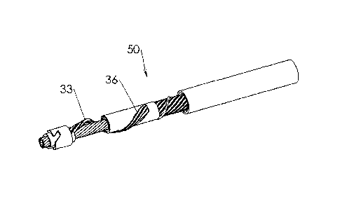
Fig. 1 illustrates a perspective view of a distal portion of a rotatable
atherectomy device, generally designated by numeral 50, according to one
embodiment of the present invention. Atherectomy device 50 comprises a
flexible rotatable hollow shaft 13 received within the interior of plastic
tube 17
into which removed occlusive material is drawn by means of an aspiration
system. Hollow shaft 13 comprises outer tubular portion 5 and inner tubular
portion 14 that are able to rotate simultaneously while sliding one over the
other in a direction parallel to the longitudinal axis of the shaft.
An expandable helical cutting unit 33 extends between tubular sleeves 18 and
22, which are fitted over, and secured such as by laser welding to, outer
tubular portion 5 and inner tubular portion 14, respectively. The use of a
helical cutting unit is advantageous in that it promotes good penetrability
into
hardened plaque, similarly to the operation of a screw or drill bit, but with
significant material savings. Helical cutting unit 33 is wound about hollow
shaft 13 by a sufficiently long pitch such that only one diametrical end of
the
hollow shaft is partially surrounded by helical cutting unit 33 for a given
axial
length of the hollow shaft. The long pitch is suitable for the fast rotational
speeds of hollow shaft 13 that are characteristic of occlusive material
removal
operations, e.g. 5000 rpm or greater. This asymmetrical configuration of
helical
cutting unit 33 results in the eccentric rotation of hollow shaft 13,
contributing
to clinical advantages, as will be described hereinafter.
An adjusting member 9 (Fig. 6) is provided for selectively expanding cutting
unit 33 away from the longitudinal axis of hollow shaft 13, typically by
controlled retraction of the inner tubular portion 14 by sliding movement
inside outer tubular portion 5. Retraction of the inner tubular portion brings
the ends of the flexible cutting unit together, thus causing the cutting unit
to
expand outwardly away from the longitudinal axis of the shaft and enlarging
CA 02953470 2016-12-22
WO 2016/001932 PCT/IL2015/050690
11 -
the area encompassed by the flexible cutting unit. The expanded cutting unit
facilitates disintegration and removal of the atheroma from the blood vessel
when rotating.
As shown in Fig. 6, guidewire 10 is received within the interior of inner
tubular portion 14. The proximal end of plastic tube 17 is connected to a
distal
portion of handle body 8, within which is housed a motor 11 for driving hollow
shaft 13. A physician uses handle body 8 to manipulate the atherectomy device
during the course of a material removal operation.
Inner tubular portion 14 distally protrudes from outer tubular portion 5. The
distal end of inner tubular portion 14, which has a diameter only slightly
greatly than the guidewire for guiding atherectomy device 50 through a blood
vessel, e.g. ranging from 0.1-0.6 mm, and which first encounters the occlusive
material, is formed with circumferentially extending cutting teeth 51, or any
other type of cutting surfaces, for example by a micro-laser cutting
technique.
Sleeve 22 slightly proximally spaced from distal cutting teeth 51 is fitted
over,
and secured to outer tubular portion 5. The distal end of sleeve 22 is formed
with circumferentially extending cutting teeth 52, or any other type of
cutting
surfaces, constituting means for increasing an opening formed in the hardened
plaque. Since sleeve 22 protrudes radially from outer tubular portion 5,
cutting
teeth 52 have a slightly larger diameter than cutting teeth 51, a difference
on
the order of only tenths of a millimeter.
As shown in Fig. 3, sleeve 22 is connected to the distal end of outer tubular
portion 5, to provide a reactive force when the expandable cutting unit
undergoes extreme resistance due to hard atheroma, for example when
attempting to drill a chronic total occlusion.
CA 02953470 2016-12-22
WO 2016/001932 PCT/IL2015/050690
- 12 -
A third means for penetrating and removing occlusive material is in the form
of sharpened metal tube 37, which may be fitted over locking device 22, and
therefore of a slightly larger diameter than sleeve 22, a difference on the
order
of only tenths of a millimeter.
As shown in Figs. 4 and 5, tube 37 is configured with one or more slots 41
that
extend proximally from the distal edge 43 of the tube. Each of the slots 41 is
formed obliquely with respect to distal edge 43, forming cutting surfaces 46
and 47 at the two lateral ends of slot 41, respectively, by which the
atherectomy device is able to penetrate a proximal surface of hard atheroma
material. Helical cutting unit 33 in the collapsed condition radially
protruding
a distance that defines a slightly larger diameter than tube 37, a difference
on
the order of only tenths of a millimeter, is then able to remove additional
atheroma material.
Fig. 2 illustrates atherectomy device 50 when cutting unit 33 is set to an
expanded condition, such that the defined diameter increases on the order of
several millimeters.
The four cutting means provided with atherectomy device 50 thus define
cutting surfaces of gradually increased size to allow the pierced opening to
be
suitably enlarged.
Fig. 6 illustrates a proximal portion of atherectomy device 50, which is
generally disposed externally to a patient's body.
Plastic tube 17 is connected to the distal tip of handle body 8 by
schematically
illustrated connection means 131, which may be a flexible shaft connector, a
regular Luer lock type connector, or any other suitable connector. Connection
means 131 may be detachable.
CA 02953470 2016-12-22
WO 2016/001932 PCT/IL2015/050690
- 13 -
The aspiration system 40 comprises a miniature vacuum pump 6 and a
collection bag 1, to which are drawn the disintegrated atheroma particles via
first aspiration line 2 extending from the annular space between the distal
narrowed tip of catheter body 8 and outer tubular portion 14 to vacuum pump
6, and second aspiration line 22 extending from vacuum pump 6 to collection
bag 1. Battery unit 4 having a switch 5 powers both vacuum pump 6 and
motor 11.
Motor 11 connected to battery unit 4 by wires 205 and 206 is housed within
chamber 23 of handle body 8 between distal seal 12 and intermediate seal 134,
which are fixed and through which tubular portions 5 and 14 of the hollow
coaxial shaft pass. Motor 11, which is sealed by fixed seals 12 and 134 and by
displaceable seal 135, is drivingly engaged with outer tubular portion 5. Heat
shrink material, e.g. adhesive material, may be advantageously used to engage
motor 11 with outer tubular portion 5 even though the latter may be made
from Nitinol.
A longitudinally displaceable adjusting member 9 for initiating selective
expansion of the flexible cutting unit is fitted within handle body 8. A seal
135
connected to the distal face of adjusting member 9 is sealingly engaged with
the inner wall of catheter body 8. The proximal end of inner tubular portion
14
is connected by adhesion or laser welding to rotating bearing 132, which is
seated in a complementary cavity formed in adjusting member 9 and in contact
with the proximal face of seal 135. This arrangement allows inner tubular
portion 14 to be longitudinally displaced together with adjusting member 9 in
or out of handle body 8 while simultaneously rotating.
Inner tubular layer 14 is longitudinally displaceable since it is connected to
rotating bearing 132. Distal displacement of adjusting member 9 reduces the
CA 02953470 2016-12-22
WO 2016/001932 PCT/IL2015/050690
- 14 -
spacing between sleeves 18 and 22 and causes the helical cutting unit to
expand, as shown in Fig. 2. Conversely when adjusting member 9 is proximally
displaced, sleeves 18 and 22 are caused to be separated, so that the expanded
cutting unit is forced to collapse, as shown in Fig. 1.
To limit the longitudinal displacement of adjusting member 9, outer tubular
portion 5 is formed with a long and narrow window 31 that may be positioned
within the confines of motor chamber 23. Within the interior of window 31 a
pin 7 welded or otherwise attached to inner tubular portion 14 is allowed to
change its position without interference while adjusting member 9 is being
longitudinal displaced. However, when pin 7 contacts one of the lower and
upper edges of window 31, as shown in Figs. 6 and 7, respectively, at a
corresponding extreme position of adjusting member 9 and of inner tubular
portion 14 connected thereto, additional longitudinal displacement is a same
direction is prevented. Nevertheless pin 7 enables outer tubular portion 5 and
inner tubular portion 14 to rotate in unison.
It will be appreciated that any other suitable adjusting member, actuator or
aspiration system is also within the scope of the invention.
Contrast material for accurately locating the position of the atherectomy
device distal tip, or verifying that a blood vessel is unoccluded and its wall
is
not damaged following an occlusive material removing operation, is injectable
through the lumen of inner tubular portion 14 via central opening 119. By
injecting the contrast material through the annular space of opening 119
surrounding guidewire 10, the same catheter may be advantageously used for
both an occlusive material removing operation and injection of the contrast
material, as opposed to prior art methods for which two separate catheters are
needed. Inner tubular portion 14 may be covered with thin-walled and liquid
impervious heat shrink material, thereby transforming the hollow shaft to a
CA 02953470 2016-12-22
WO 2016/001932 PCT/IL2015/050690
- 15 -
sealed tube through which the injected contrast material can reliably flow to
the occlusion site. Alternatively, a sealant such as parylene, polyamide,
polymeric material and sprayed plastic material can be applied to the inner
surface of a multi-coiled inner tubular portion. If so desired, the entire
inner
tubular portion 14 may be made of a liquid impervious, molded or extruded
plastic material.
Fig. 12 illustrates, in exploded view, distally located apparatus for enabling
concurrent rotation of the inner and outer tubular portions. Sleeves 22 and
188, which may be arcuate and may subtend an angle of approximately 160
degrees, are made from the same metallic material, and have identical inner
and outer diameters. Sleeve 22 is connected to inner tubular portion 14 and
covered by part 52. Sleeve 188 is connected to outer tubular portion 5 and
covered by sleeve 18
Since sleeves 22 and 188 have the same diameters and are connected to a
corresponding tubular portion, they are in slidable abutting relation with
each
other. This sliding action enables the inner and outer tubular portions to be
linearly displaced one within the other while being concurrently rotatable.
The linear displacement is dependent on the length of the sleeves,
corresponding to the change in length needed to expand and contract the
helical cutting unit.
In one embodiment, the outer tubular portion is a cable of multi-coil
construction arranged such that each coil is tightly wound about the
longitudinal axis of the hollow shaft, for example obliquely wound about the
longitudinal axis so as to abut with an adjacent coil.
CA 02953470 2016-12-22
WO 2016/001932 PCT/IL2015/050690
- 16 -
Spiral strands that suitable for the present invention may be made of
stainless
steel or Nitinol and include the ACTON series of cable tube type FLAT or STD
made by Asahi Intecc (Asahi Intecc Co. Ltd., Japan), or the HSS1z) series of
tubes made by Fort Wayne Metals (Fort Wayne, Indiana). The one or more
wires or strips that are formed together to define this closely-wound spiral
or
tube may all have the same diameter, or alternatively, some wires or strips
may have a larger diameter than others, thereby forming a coaxial flexible
hollow shaft with round or elliptical outer contour and a closely rounded
internal lumen. Alternatively, the strands or straps may be made of a plastic
material.
As shown in Fig. 8A, helical cutting unit 33 may be integrally formed with
outer cable 5 such that the two illustrated joined strands 48 and 49 of
helical
cutting unit 33 are longer than the other coils 21 of outer cable 5 and
distally
extend beyond the distal end 26 of outer cable 5. Helical cutting unit 33 may
comprise a single strand, or any other desired number of strands.
Nitinol having the characteristic of super elasticity, or Series 300 flexible
stainless steel is suitable for the integrally formed and expandable helical
cutting unit 33.
With reference to Figs. 1, 2, 4 and 5, the strands of integrally formed
helical
cutting unit 33 are radially separated from outer cable 5 by sleeve 18. One
portion of cutting unit 33 is fixated within a helical aperture 36 formed in
sleeve 18, and its distal end is fixated between metal tube 37 and sleeve 22.
The ability to mechanically fixate the strands of helical cutting unit 33
between sleeve 22 and tube 37 obviates the need of having to weld the Nitinol
strands of cutting unit 33 to sleeve 22 made of stainless steel. Nitinol and
stainless steel are dissimilar metals, so that if welded together,
intermetallics
are liable to form in a weld zone, resulting in brittle joints. Helical
aperture 36
CA 02953470 2016-12-22
WO 2016/001932 PCT/IL2015/050690
- 17 -
assures that a longitudinally central region of cutting unit 33 will be
radially
separated by a significantly large and desired distance from outer cable 5, in
order to cut the occlusive material by a corresponding diameter, and further
urges helical cutting unit 33 to expand at a specific angle and away from
outer
cable 5.
Alternatively, helical cutting unit 33 is formed separately from outer cable
5,
and its two ends are fixated by sleeve 18 and tube 37, respectively.
Figs. 9 and 10 illustrate an outer cable arrangement 10 for an expandable
atherectomy device. In order to maintain the adjacent tightly wound spiral
strands 3 defining outer cable 5 of the flexible and coaxial hollow shaft in
abutting relation with each other and in a uniform shape and diameter, a
helical mono-coil spring 15 is wound over outer cable 5. A tubular sleeve 18
is
fitted over, and secured such as by welding to, outer cable 5 at each end 12,
whether the distal end or proximal end, of mono-coil spring 15, for ensuring
suitable mono-coil tension. An aperture 19 for receiving a corresponding mono-
coil end 12 is formed in sleeve 18.
Mono-coil spring 15 extends throughout substantially the entire length of
outer
cable 5, as shown in Fig. 10.
As another means for maintaining the coil uniformity of outer cable 5, helical
mono-coil spring 15 has a larger pitch than the pitch of the coils of outer
cable
5. The pitch ratio of mono-coil spring 15 to outer cable 5 may range from 1.0-
1.1 to 1.0-10. Also, the inner diameter of mono-coil spring 15 is less than or
equal to the outer diameter of outer cable 5. Mono-coil spring 15 may be made
of any suitable biocompatible material, including stainless steel, memory
shape metal, and polymer. The cross section of mono-coil spring 15 may be
round, square or any other suitable shape.
CA 02953470 2016-12-22
WO 2016/001932 PCT/IL2015/050690
- 18 -
In addition to the aforementioned novel structural features, the screw shape
of
the coils of outer cable 5 or of mono-coil spring 15 help to convey blood and
disintegrated atheroma material along the longitudinal axis of the shaft as
result of the Archimedes screw effect.
Alternatively or in addition, the adjacent coils of outer cable 5 may be
maintained in abutting relation and their expansion may be prevented by
applying extra thin-walled heat shrink material, e.g. made of polyester.
Other means for limiting coil expansion include a plurality of longitudinally
spaced rings connected to outer cable 5, laser welding and adhesive
attachment.
In another embodiment, the helical cutting unit is fixed and inexpandable, and
the hollow shaft may be made of a single tubular portion which may be
integrak with the helical cutting unit. When the helical cutting unit is fixed
at
a relatively small and fixed radial separation from the hollow shaft, although
greater than the other cutting means, the atherectomy device of the present
invention is advantageously narrower than prior art atherectomy devices and
is therefore capable of removing occlusive material from narrow blood vessels,
for example those having a diameter of as small as 2 mm.
Prior to describing the unique operation of the atherectomy device, reference
is
first made to Fig. 11, which schematically illustrates the formation of an
occlusion 80 in a blood vessel 81. Occlusion 80 is formed as a result of
cardiovascular disease by which fat and cholesterol build up on the walls 84
of
blood vessels. As additional occlusive material including plaque forms on the
blood vessel walls 84, the lumen of blood vessel 81 becomes narrower and the
flowrate of blood therethrough becomes reduced. Eventually the occlusive
CA 02953470 2016-12-22
WO 2016/001932 PCT/IL2015/050690
- 19 -
material accumulates throughout the lumen, and the flow of blow across
occlusion 80 ceases.
Due to the pulsating nature of the blood flow 86 in the direction indicated by
the arrows that continuously applies a periodic force to the proximal surface
87
of occlusion 80, the proximal surface becomes convex. Also, proximal surface
87
becomes compressed to form hardened plaque and then a CTO, characterized
by complete interruption of blood flow. Distal surface 89 of occlusion 89
becomes convex since it is not exposed to pulsating blood flow 86.
The hardness of occlusion 80 is not uniform, containing regions of hard
plaques
that include calcium and scar tissue and soft plaques that include viscous
cholesterol material. The central concave region of proximal surface 87 is
always softer that the surrounding regions of proximal surface 87. Soft plaque
tends to allow the passage therethrough of a guidewire: however hard plaque
deflects a guidewire or a large sized cutting device during an attempt to
penetrate occlusion 80. When a prior art cutting device is deflected, the
cutting
surfaces many times contact and tear blood vessel wall 84.
The use of the atherectomy device of the present device, and particularly of
the
helical cutting unit, overcomes the aforementioned drawbacks and allows the
hard plaque of CTOs to be atraumatically opened and removed.
During the eccentric rotation of the hollow shaft within blood vessel 81, the
hollow shaft is caused to be laterally displaced, i.e. displaced in a
direction
radially spaced from the longitudinal axis of the blood vessel. Eventually the
helical cutting unit contacts blood vessel wall 84 as a result of the lateral
displacement, and the hollow shaft is consequently caused to be laterally
displaced in an opposite direction in response to the impact with blood vessel
wall 84. This alternating lateral movement is accompanied by longitudinal
CA 02953470 2016-12-22
WO 2016/001932 PCT/IL2015/050690
- 20 -
displacement initiated by manipulation of the atherectomy device by the
physician in order to remove the occlusive material. As a result of the
alternating lateral movement, the hollow shaft is substantially self-centered,
and the cutting surfaces of the hollow shaft's distal tip are able to pierce a
central relatively soft plaque region. The distal tip remains in engagement
with relatively soft plaque region after the plaque has been pierced. The
other
cutting surfaces are then able to enlarge the opening, as described
hereinabove.
It will be appreciated that the central relatively soft plaque region pierced
by
the distal tip during the lateral movement is not necessarily at the true
center
of the blood vessel.
The self-centering feature based on alternating lateral movement of the
atherectomy device is contingent on atraumatic contact between the helical
cutting unit and the blood vessel wall. The profile of helical cutting unit 33
shown in Fig. 8A is configured to ensure atraumatic contact between the
helical cutting unit and the blood vessel wall during the alternating lateral
movement. Each of strands 48 and 49 has a trapezoidal cross section 60 when
cut in a plane perpendicular to the longitudinal axis of outer cable 5.
As shown in Fig. 8B, a first side 63 of trapezoidal cross section 60, disposed
generally laterally outwardly, is adapted to atraumatically contact a wall of
the blood vessel during lateral movement of the atherectomy device. A vertex
64 at the intersection of first side 63 and second side 66 adjacent to first
side
63 is sufficiently sharp to cut the occlusive material. A vertex 69 at the
intersection of second side 66 and third side 67 adjacent to second side 66
and
opposite first side 63, however, is dull, being formed with a curved surface
having a sufficiently large radius to prevent plaque removal and tearing of
the
blood vessel wall.
CA 02953470 2016-12-22
WO 2016/001932 PCT/IL2015/050690
- 21 -
The Applicant has found that a ratio of the length of second side 66 to the
length of first side 63 ranging from 1 1.13 is sufficient to prevent vertex 64
from contacting and tearing the blood vessel wall during concurrent eccentric
rotation and alternating lateral movement of the hollow shaft. This range is
based on a combination of several parameters, including planarity of second
side 66, rotation speed of the hollow shaft, motor torque, pushability of the
hollow shaft and the relative change in diameter of all the cutting means.
The configuration of trapezoidal cross section 60 is particularly useful for
blood
vessels having an angular disposition of no greater than 70 degrees, to
prevent
tearing of the blood vessel wall during navigation through a tortuous blood
vessel, although the atherectomy device of the present invention is also
applicable to such blood vessels as well.
While prior art atherectomy devices have been unable to penetrate the distal
surface of an occlusion in a retrograde approach, and particularly of a CTO,
due to its convex formation as shown in Fig. 11, the atherectomy device of the
present invention can easily penetrate the distal surface and enlarge the
opening, similarly to the method described hereinabove.
While some embodiments of the invention have been described by way of
illustration, it will be apparent that the invention can be carried out with
many modifications, variations and adaptations, and with the use of numerous
equivalents or alternative solutions that are within the scope of persons
skilled
in the art, without exceeding the scope of the claims.