Note: Descriptions are shown in the official language in which they were submitted.
CA 02953564 2016-12-22
WO 2016/010810 PCT/US2015/039682
SYSTEMS, METHODS, AND DEVICES FOR CANNULA INSERTION
INCORPORATION BY REFERENCE TO ANY PRIORITY APPLICATIONS
[0001] The present application claims priority benefit of U.S. Patent App.
No.
14/793,980, filed on July 8, 2015, and U.S. Provisional Patent App. No.
62/025,385, filed
on July 16, 2014.
BACKGROUND
Field
[0002] The disclosure relates generally to the field of insertion systems,
and
more particularly to systems for cannula insertion.
Description of the Related Art
[0003] In ophthalmic surgery, and in other surgical contexts, a surgeon may
desire to create a port opening on the surface tissue of a patient and to
insert a cannula
through which the surgeon can perform surgical operations. Generally, such
port
openings are called cannulas in the ophthalmic surgery context.
SUMMARY
[0004] In some embodiments, a method of implanting a cannula in an eye of a
patient comprises, or alternatively consists essentially of, positioning a
cannula insertion
system at a first angle relative to a surface of the eye, distally advancing a
trocar-cannula
pair into the eye at the first angle, positioning the cannula insertion system
at a second
angle relative to the surface of the eye, and distally advancing the trocar-
cannula pair
further into the eye at the second angle. The trocar-cannula pair comprises a
needle
portion and a cannula.
[0005] The first angle may be less than 90 . The first angle may be 45 .
The
second angle may be 90 . Distally advancing the trocar-cannula pair into the
eye at the
first angle may comprise distally advancing a slide inserter. Distally
advancing the trocar-
cannula pair into the eye at the first angle may comprise pressing a flexible
trigger button.
Distally advancing the trocar-cannula pair further into the eye at the second
angle may
comprise pressing a flexible trigger button. Pressing a flexible trigger
button may
comprise allowing a spring to decompress. The method may further comprise
retracting
-1-
CA 02953564 2016-12-22
WO 2016/010810 PCT/US2015/039682
the needle portion of the trocar-cannula pair out of the eye. The method may
further
comprise positioning a second trocar-cannula pair for positioning in the eye
of the patient.
Positioning the second trocar-cannula pair may comprise rotating the second
trocar-
cannula pair. Rotating the second trocar-cannula pair may comprise rotating a
guiding
trail tube having a longitudinal axis aligned with a longitudinal axis of the
cannula
insertion system. Rotating the second trocar-cannula pair may comprise
rotating a
revolver chamber having a longitudinal axis radially offset from a
longitudinal axis of the
cannula insertion system. Positioning the second trocar-cannula pair may
comprise
horizontally advancing the second trocar-cannula pair transverse to a
longitudinal axis of
the cannula insertion system.
[0006] In some embodiments, a two-stage cannula insertion system comprises,
or alternatively consist essentially of, a housing, a control mechanism
slidably movable
relative to the housing, a spring, an activation mechanism coupled to the
spring, a shaft
coupled to the spring, and a trocar-cannula pair coupled to the control
mechanism. The
shaft is releasably engagable with the control mechanism. The control
mechanism is
configured to move independently of the shaft when the shaft is disengaged
with the
control mechanism. The trocar-cannula pair comprises a needle portion and a
cannula.
[0007] The control mechanism may be configured to advance the trocar-
cannula pair during a first stage. The activation mechanism may be configured
to advance
the trocar-cannula pair during a second stage. The system may further comprise
a
mechanical assembly housing a plurality of trocar-cannula pairs including the
trocar-
cannula pair. The mechanical assembly may be configured to disengage the
needle
portion from the control mechanism after the first stage. The mechanical
assembly may
be configured to couple a second trocar-cannula pair with the control
mechanism. The
mechanical assembly may comprise a rotatable guiding trail tube having a
longitudinal
axis aligned with a longitudinal axis of the cannula insertion system. The
mechanical
assembly may comprise a rotatable revolver chamber having a longitudinal axis
radially
offset from a longitudinal axis of the cannula insertion system. The
mechanical assembly
may comprise a cartridge configured to horizontally advance the second trocar-
cannula
pair transverse to a longitudinal axis of the cannula insertion system.
[0008] In some embodiments, a two-stage cannula insertion system for use in
ophthalmic surgery comprises, or alternatively consists essentially of, a
first stage
insertion mechanism and a second stage insertion mechanism. The first stage
insertion
-2-
CA 02953564 2016-12-22
WO 2016/010810 PCT/US2015/039682
mechanism is configured to insert a trocar-cannula pair partially into ocular
tissue at a
first angle. The trocar-cannula pair comprises a needle portion and a cannula.
The
second stage insertion mechanism is configured to insert the trocar-cannula
pair through
the ocular tissue at a second angle and to implant the cannula in the ocular
tissue. The
needle portion is removable while maintaining the cannula in the ocular
tissue.
[0009] The system may further comprise a mechanical assembly housing a
plurality of trocar-cannula pairs including the trocar-cannula pair. The first
stage
insertion mechanism may be configured to releasably engage at least one of the
plurality
of trocar-cannula pairs housed in the mechanical assembly. At least one of the
first stage
insertion mechanism and the second stage insertion mechanism may be configured
to be
automatically activated by a user engaging a button.
[0010] In some embodiments, a system comprises a two-stage inserter device
for semi-automatically inserting a cannula into body tissue during a surgical
operation, for
example, for ophthalmic surgery. In some embodiments, the system comprises a
housing.
The system can comprise a first stage assembly. In some embodiments, the first
stage
assembly comprises a first inner locking shaft with attached deformable first
cantilever
structure that the said first cantilever structure can be pressed and deformed
toward the
center of the shaft. In some embodiments, the first cantilever can recover to
its original
shape if no force exerts on it. In some embodiments, the system comprises a
first flexible
activation button that the inner extrusion structure can push inward and cause
the
deformable cantilever to bend and/or deform toward the center of the shaft. In
some
embodiments, the system comprises a first outer locking shell that has an
opening on the
shell that can latch the cantilever structure along the longitudinal direction
of the shell.
[0011] In some embodiments, the system comprises a first guide trail that
is
attached to the inside of the first outer locking shell, is able to guide the
longitudinal slide
motion of the inner locking shaft, and/or functions as a mechanical stop to
inhibit or
prevent the first cantilever from moving away from the latching position so
the latching
mechanism is secured. In some embodiments, the system comprises a first spring
that can
push the first inner locking shaft and first outer locking shell apart once
released along the
longitudinal direction of the spring. In some embodiments, the system
comprises a
second stage assembly. In some embodiments, the second stage assembly
comprises a
second inner locking shaft with attached deformable second cantilever
structure that the
second cantilever structure can be pressed and deformed toward the center of
the shaft.
-3-
CA 02953564 2016-12-22
WO 2016/010810 PCT/US2015/039682
[0012] In some embodiments, the second cantilever can recover to its
original
shape if no force exerts on it. In some embodiments, the system comprises a
second
flexible activation button that the inner extrusion structure can push inward
and cause the
second deformable cantilever to bend and/or deform toward the center of shaft.
In some
embodiments, the system comprises a second outer locking shell that has an
opening on
the shell that can latch the cantilever structure along the longitudinal
direction of the shell.
In some embodiments, the system comprises a second guide trail that is
attached to the
inside of the first outer locking shell. In some embodiments, the second guide
is able to
guide the longitudinal slide motion of the inner locking shaft, and can
function as a
mechanical stop to inhibit or prevent the first cantilever from moving away
from the
latching position so the latching mechanism is secured.
[0013] In some embodiments, the system comprises a second spring that can
push the first inner locking shaft and first outer locking shell apart once
released along the
longitudinal direction of the spring. In some embodiments, the system
comprises an off-
axis mechanical hollow interlock structure close to the end of the inner
locking shaft for
interlocking with a trocar or trocar agent at the shaft's distal end; has an
off-axis concave
hollow channel and a connected hollow bay structure that allows the external
interlock
structure to engage, move in and then stay in the bay securely. In some
embodiments, the
system comprises a trocar or a trocar agent that has a sideway extruded beam
functioning
as an off-axis mechanical interlock for interlocking the hollow interlock
structure on the
shaft.
[0014] In some embodiments, the system comprises a cannula that is loaded
on but separable from the trocar or trocar agent. In some embodiments, the
second spring
comprises a greater force constant than the first spring. In some embodiments,
the force
and momentum generated from the second spring once its energy is released are
greater
than those from the second spring. In the first stage, in some embodiments,
one end of
the first spring exerts force on part of the first inner locking shaft and the
other end of the
first spring exerts force on part of the first outer locking shell. In some
embodiments, the
first locking shaft is attached to the second outer shell.
[0015] In the second stage, In some embodiments, one end of the second
spring exerts force on part of the second inner locking shaft and the other
end of the
second spring exerts force on part of the second outer locking shell. In some
embodiments, the two springs can be replaced by mechanical structures or
mechanisms
-4-
CA 02953564 2016-12-22
WO 2016/010810 PCT/US2015/039682
that can store potential energy and release later upon activation. The
mechanisms for
storing potential energy or driving the shaft can comprise rubber band with
pulley,
pneumatic pump, electromagnetic transducer, linear motor, pneumatic piston, or
the like.
In some embodiments, the relative locations of the two stage assemblies are
stacked along
their longitudinal direction. In some embodiments, one stage assembly is
hollow in center
to accommodate/enclose the other stage assembly in a concentric fashion. In
some
embodiments, the one stage assembly can be moving parallel to the moving
direction of
the other stage assembly in an off-axis parallel motion direction fashion.
[0016] In some embodiments, the system comprises a two-stage inserter
device for semi-automatically inserting a cannula into a body during, for
example, an
ophthalmic surgical procedure. In some embodiments, the system comprises a
housing
and a first stage assembly. In some embodiments, the system comprises an h-
shape slider
inner shaft. The h-shape slider can comprise a right leg and left leg. In some
embodiments, the right leg (beam) of the "h" shape is flexible and deformable
when it is
manually pressed at the beam's side and can recover when not pressed.
[0017] In some embodiments, the left leg (beam) of the h-slider is non-
flexible. In some embodiments, the h-shape slider comprises a top beam. The
top beam
of the h-slider can be the main axial structure of the shaft and has protruded
blocks at both
sides (inward and outward from paper when reading "h") for guiding the sliding
motion in
trails in later front house structure, and can comprise an off-axis mechanical
hollow
interlock structure close to the distal end of the top beam of the h-slider
for interlocking
with a trocar or trocar agent. In some embodiments, the hollow interlock
structure has an
off-axis concave hollow channel and a connected hollow bay structure that
allows the
external interlock structure to engage, move in and then stay in the bay
securely.
[0018] In some embodiments, the system comprises a trocar or a trocar agent
that has a sideway extruded beam functioning as an off-axis mechanical
interlock for
interlocking the hollow interlock structure on the h-slider. In some
embodiments, the
system comprises a front house shell that has an elongate side opening
functioning as a
guide trail for the h-slider.
[0019] In some embodiments, one of the inner surface end of the elongated
side opening can stop and latch the terminal face of the non-flexible left
beam of the h-
slider from moving backward. In some embodiments, the system comprises two
elongate
grooves functioning as guide trails on both elongated inner side surfaces of
the opening,
-5-
CA 02953564 2016-12-22
WO 2016/010810 PCT/US2015/039682
to house the protruded blocks on top beam of the h-slider; has openings at
both distal ends
to allow the top beam (main shaft) of the h-slider to move through along the
longitudinal
direction of the shell; is mechanically connected and fixed with the later
inner locking
shaft.
[0020] In some embodiments, the system comprises a second stage assembly.
In some embodiments, the second stage assembly comprises an inner locking
shaft with
deformable cantilever structure. In some embodiments, the cantilever structure
can be
pressed and deformed toward the center of the shaft. In some embodiments, the
cantilever can recover to its original shape if no forces exerts on it; has a
ring extrusion on
the shaft functioning as a mechanical stopper against the later ring blocker
in the outer
locking shall, and can be mechanically connected and fixed with the front
house. In some
embodiments, the system comprises a flexible activation button that the inner
extrusion
structure can push inward and cause the deformable cantilever to bend and/or
deform
toward the center of shaft.
[0021] In some embodiments, the system comprises an outer locking shell
that
has an opening on the shell that can latch the cantilever structure along the
longitudinal
direction of the shell. In some embodiments, the system comprises a ring
blocker
structure protruded inward for stopping the longitudinal slide motion of the
inner locking
shaft. The ring blocker can function as a mechanical stop to inhibit or
prevent the inner
shaft from moving forward too far. In some embodiments, the system comprises a
spring
stopper at its distal end to allow the spring stay and exert force against
this shell. In some
embodiments, the system comprises a spring that can push the inner locking
shaft and first
outer locking shell apart once released along the longitudinal direction of
the spring. In
some embodiments, the system comprises a cannula that is loaded on but
separable from
the trocar or trocar agent.
[0022] In some embodiments, the system comprises a plurality of preloaded
trocar-cannula pairs with each pair having an individual slider inserter
surrounding the
handpiece. The system can comprise a plurality of trocars and/or trocar
agents. In some
embodiments, each trocar and/or trocar agent can load a cannula. In some
embodiments,
each trocar and/or trocar agent comprises a sideway extruded beam that can
function as an
off axis mechanical interlock for interlocking a hollow interlock structure in
an h-slide
inserter.
-6-
CA 02953564 2016-12-22
WO 2016/010810 PCT/US2015/039682
[0023] In some embodiments, the system comprises a revolver chamber that
comprises a plurality of trocar-cannula pairs. In some embodiments, the
revolver
chamber is configured to comprise only a single slide inserter. In some
embodiments, the
revolver chamber can be configured to comprise a plurality of slide inserters
for each
trocar-cannula pair. The revolver chamber can comprise a cylindrical housing
configured
to house a plurality of guiding trails. The revolver chamber can be configured
to rotate
with respect to a longitudinal axis of the revolver chamber. In some
embodiments, the
guiding trails comprises two grooves that can accommodate the side protrusion
blocks of
a trocar carrier that is configured to be loaded with a trocar-cannula pair.
In some
embodiments, the guiding trail can be configured to guide the sliding action
of the trocar
carrier in order to implant a trocar-cannula pair into a patient. In some
embodiments, the
system comprises a blocker structure at a center portion of the revolver
chamber. The
blocker structure can comprise a blocker disc structure that can latch to a
protruded
bottom block of the trocar carrier. In some embodiments, the blocker structure
can be
configured to inhibit or prevent trocar carriers that are not being used from
sliding
forward. In some embodiments, the system comprises a blocker disc structure
that can be
configured to latch onto the cannulas. In some embodiments, the blocker disc
structure
can inhibit or prevent the cannulas that are not in use from sliding forward.
[0024] In some embodiments, the system comprises a side cartridge
configured to house a plurality of trocar-cannula pairs. The side cartridge
comprises a
single slide inserter configured to be loaded with a trocar-cannula pair from
the side
cartridge. In some embodiments, the side cartridge can be configured to be
loaded with a
plurality of trocar-cannula pairs with each having a slide inserter. In some
embodiments,
the side cartridge comprises a rectangular internal chamber that can be
configured to
house a plurality of trocar-cannula pairs. In some embodiments, the trocar-
cannula pairs
are rectangular. In some embodiments, the side cartridge comprises an internal
pusher
block that can be configured to push trocar-cannula pairs sideways towards the
center of a
cylindrical housing of the cannula insertion system. In some embodiments, the
pusher
block can be configured to be pushed by compressing a spring that is
positioned between
the internal end wall of the side cartridge and the internal side wall of the
pusher block.
In some embodiments, the spring can be substituted with a variety of drive
mechanisms
including but not limited to a rubber band and pulley apparatus,
electromagnetic
transducer, pneumatic pump, linear motor, pneumatic piston, or the like. In
some
-7-
CA 02953564 2016-12-22
WO 2016/010810 PCT/US2015/039682
embodiments, the slide inserter as disclosed in any of the embodiments herein
can be
powered by any of the foregoing drive mechanisms such that the slide inserter
is
configured to automatically insert partially the trocar needle into the
sclera.
[0025] For purposes of this summary, certain aspects, advantages, and novel
features of the invention are described herein. It is to be understood that
not necessarily
all such advantages may be achieved in accordance with any particular
embodiment of the
invention. Thus, for example, those skilled in the art will recognize that the
invention
may be embodied or carried out in a manner that achieves one advantage or
group of
advantages as taught herein without necessarily achieving other advantages as
may be
taught or suggested herein.
BRIEF DESCRIPTION OF THE DRAWINGS
[0026] The foregoing and other possible features, aspects, and advantages
of
the embodiments of the invention are described in detail below with reference
to the
drawings of various embodiments, which are intended to illustrate and not to
limit the
embodiments of the invention. The drawings comprise the following figures in
which:
[0027] Figure 1 is a perspective view of an example cannula insertion
system.
[0028] Figures 2A-2C depict an example of operating an example cannula
insertion system.
[0029] Figures 3A and 3B depict an example of creating a wound using an
example cannula insertion system.
[0030] Figures 3C and 3D depict another example of creating a wound using
an example cannula insertion system.
[0031] Figures 4A-4C illustrate example operations of an example trocar
agent for coupling to a cannula insertion system.
[0032] Figures 5A-5C illustrate example operations of an example cannula
insertion system.
[0033] Figure 5D is an exploded view of the cannula insertion system of
Figures 5A-5C.
[0034] Figures 6A-6D illustrate example operations of example control
mechanisms for a cannula insertion system.
[0035] Figure 7 is an exploded view of example components of an example
cannula insertion system.
-8-
CA 02953564 2016-12-22
WO 2016/010810 PCT/US2015/039682
[0036] Figures 8A-8D illustrate example operations of an example cannula
insertion system.
[0037] Figure 9 is a perspective view of an example cannula insertion
system.
[0038] Figure 10 is a perspective view an example cannula insertion system.
[0039] Figure 11 is a perspective view an example cannula insertion system.
[0040] Figures 12A and 12B illustrate an example of inserting the example
cannula insertion system of Figure 11 in an eye.
[0041] Figures 13A-13C illustrate an example cannula insertion system at
different stages.
[0042] Figure 13D is an exploded view of example components of the cannula
insertion system of Figures 13A-13C.
[0043] Figures 14A and 14B illustrate example operation of the cannula
insertion system of Figures 13A-13C.
[0044] Figures 15A-15C illustrate an example cannula insertion system at
different stages.
[0045] Figure 15D is an exploded view of example components of the cannula
insertion system of Figures 15A-15C.
[0046] Figure 15E is a further exploded view of example components of the
cannula insertion system of Figures 15A-15C.
[0047] Figures 15F-15H illustrate an example cannula insertion system at
different stages.
[0048] Figures 151-15K further illustrate example operations of the cannula
insertion system of Figures 15F-15H.
[0049] Figure 16 juxtaposes the cannula insertion system of Figures 15A-15C
and the cannula insertion system of Figure 1.
[0050] Figures 17A-17D illustrate an example cannula insertion system.
[0051] Figure 17E is an exploded view of example components of the cannula
insertion system of Figures 17A-17D.
[0052] Figure 17F is a magnified view of an example component of the
cannula insertion system of Figures 17A-17D.
-9-
CA 02953564 2016-12-22
WO 2016/010810 PCT/US2015/039682
DETAILED DESCRIPTION
[0053] Although several embodiments, examples, and illustrations are
disclosed herein, it will be understood by those of ordinary skill in the art
that the
inventions described herein extends beyond the specifically disclosed
embodiments,
examples, and illustrations and includes other uses of the inventions and
obvious
modifications and equivalents thereof. Embodiments of the inventions are
described with
reference to the accompanying figures, wherein like numerals refer to like
elements
throughout. The terminology used in the description presented herein is not
intended to
be interpreted in any limited or restrictive manner simply because it is being
used in
conjunction with a detailed description of certain specific embodiments of the
inventions.
In addition, embodiments of the inventions can comprise combinations of
features, and no
single feature may be solely responsible for desirable attributes or essential
to practicing
the inventions herein described.
[0054] In ophthalmic surgery, and in other surgical operations, surgeons
often
insert cannulas into surgical openings, which are also known as sclerotomies
in the
ophthalmic surgical context. A cannula can comprise a cannula body and a
cannula tube.
The cannula body can be used to hold the cannula fixed in the surface tissue,
for example,
in the sclera. Generally, the cannula body is coupled to the cannula tube. The
cannula
tube can be a flexible tube that is inserted in the body to allow and guide
surgical
instruments into the interior body portion of a patient. In many instances,
the cannula is
inserted into the body manually by a surgeon using a handheld tool, which can
comprise a
needle and a handle portion. A cannula may be placed on the needle, and the
surgeon,
holding the handle portion, can insert the needle into the sclera and push the
needle into
the eye until the cannula is positioned to the sclerotomy. The surgeon is free
to maneuver
the handle portion in any manner the surgeon chooses. Use of such handheld
tools can
allow for great variation in the way cannulas are inserted into a sclera.
[0055] There is a need for the cannula insertion systems disclosed herein,
which can allow for a consistent procedure for creating a sclerotomy. In some
embodiments, the cannula insertion system can be configured to perform a large
part of
the task in creating a sclerotomy. For example, the cannula insertion system
can be
configured to insert a needle in the sclera with a consistent force. In some
embodiments,
the cannula insertion system can be configured to divide the cannula insertion
process into
a two-step process for the user (e.g., surgeon), which can improve procedure
performance
-10-
CA 02953564 2016-12-22
WO 2016/010810 PCT/US2015/039682
consistency. In some embodiments, the cannula insertion system is configured
to be a
low-cost device to manufacture and produce. The cannula insertion system can
be a
disposable device.
[0056] In some embodiments, the cannula insertion system is ergonomically
designed to fit in a user's hand. The cannula insertion system can be
configured to be
operated with a single hand, thereby freeing the other hand of the user to
perform other
surgical tasks. In some embodiments, the cannula insertion system can be semi-
automatic
or fully automatic such that a user need not exert force on the body tissue in
order to insert
a cannula. By reducing the amount of user force, the surgical procedure can be
less tiring
for the user. Semi-automatic and fully automatic systems can ensure that a
consistent
amount of force is being exerted on the body tissue, which can inhibit or
prevent injury
and unintended damage to the body tissue. Semi-automatic and fully automatic
systems
can reduce the duration for a user to insert a cannula.
[0057] In some embodiments, the cannula insertion system can be configured
with a two-stage insertion function, which may provide one or more of the
advantages
described herein. For example, the first stage of the insertion function can
comprise a
sliding step and the second stage of the insertion function can comprise an
automatic
mechanical releasing step. In some embodiments, the cannula insertion system
can
comprise a sliding portion to allow a user to manually slide the sliding
portion from a
proximal first position to a distal second position. By moving the sliding
portion from the
first position to the second position, the user can insert a needle portion
that is coupled to
the sliding portion into the sclera portion of an eye, for example at an angle
less than 90 .
In some embodiments, the needle portion is inserted only partially into the
sclera and not
entirely through the sclera during the first stage of the insertion function.
In some
embodiments, the user, during the first stage of the insertion function, can
position the
cannula insertion device at about a 45 angle relative to the surface of the
sclera when
manually sliding the sliding portion toward the sclera to insert the needle
portion into the
eye.
[0058] By angling the cannula insertion device at about a 45 angle, the
user
can create an oblique sclerotomy. The first stage may consistently create an
oblique
sclerotomy partially, at least partially, or fully thorough the sclera. After
creating the
oblique sclerotomy, the user can perform the second stage of the insertion
function.
During the second stage, the user can move the cannula insertion device from
the about
-11-
CA 02953564 2016-12-22
WO 2016/010810 PCT/US2015/039682
450 angle to about a 90 angle relative to the surface of the sclera. After
repositioning the
cannula insertion device in about a 90 angle, the user can activate/trigger
an automatic
activation mechanism configured to allow a force to be applied to the needle
portion in
order to fully insert the needle portion through the sclera and into the
vitreous. In
applying the force on the needle portion, a cannula that is positioned on the
needle portion
is forced through the sclerotomy and is positioned on the sclera.
[0059] In some embodiments, during the first stage, only the needle or
trocar
enters the eye such that the insertion force is the force on the needle to
create a portion of
the aperture. In some embodiments, during the second stage, along with
creating an
additional portion of the aperture, a cannula that is larger than the needle
is forced into the
aperture such that the insertion force in the second stage is greater than the
force on the
needle to create the additional portion of the aperture. If the second stage
is fully manual
(e.g., without assistance by a device such as a spring), the application of
manual force by
a user may primarily push the eye towards the back of the socket, which can
cause trauma
and/or create a leaky incision.
[0060] In some embodiments, the application of the force positions the
cannula
in the vitreous of the eye and the cannula body in the sclera. The creation of
the oblique
sclerotomy in the first stage allows for the creation of a shearing force or a
tension force
up by the sclera on the cannula body. The creation of these forces may
advantageously
allow the cannula body to be more securely held in the sclera.
[0061] The forces are created at least partially by the creation of the
oblique
sclerotomy. The creation of the oblique sclerotomy in the first stage is more
advantageous than the creation of a non-oblique sclerotomy that is generally
created when
the needle portion is inserted into the sclera initially at about a 90 angle
relative to the
surface of the sclera. The shearing forces or the tension forces created by a
non-oblique
sclerotomy are less than the shearing forces or tension forces created by an
oblique
sclerotomy. An oblique sclerotomy can better secure the cannula body than a
non-oblique
sclerotomy.
[0062] In some embodiments, the cannula insertion systems disclosed herein
may advantageously provide a two-stage inserter function. In the first stage,
the system
allows for partial insertion of the needle portion into the wound, and in the
second stage
the system allows for the needle portion to be forced through the entire depth
of the tissue.
This two-stage insertion action can allow for the creation of an oblique
sclerotomy, or
-12-
CA 02953564 2016-12-22
WO 2016/010810 PCT/US2015/039682
other surgical opening, that is better suited for securing a cannula body in
the sclera. The
oblique sclerotomy may be consistently created across many cannulization
procedures, for
example, because a user of the device does not have to precisely stop
insertion of a trocar
partially through the sclera, which is typically about 0.3 mm to about 1 mm
thick, but may
rely on mechanical assistance that inhibits or prevents further insertion
during a first
stage.
[0063] -- The creation of the oblique sclerotomy, or other surgical opening,
allows for the creation of additional shearing forces or tension forces that
are not present
in non-oblique sclerotomies or other non-oblique surgical openings. The
creation of an
oblique sclerotomy can be advantageous because the insertion of the needle
portion at
about a 45 angle creates a shelf-like wound in which the two shelf portions
can come
together and overlap each other after the needle portion and the cannula have
been
removed from the wound, allowing for self-sealing of the wound without
suturing. Lack
of suturing can reduce irritation of the eye and complications that might
result from such
irritation.
[0064] -- By reducing or eliminating suturing of the wound, patient healing
time
and patient discomfort can be reduced after the surgery. One-step insertion
systems
generally cannot create an oblique sclerotomy or other surgical opening
because the
needle portion is generally inserted into the sclera at about a 90 angle, and
such a process
does not create any shelf-like wound with two planes that can come together
and overlap
each other.
[0065] -- In some embodiments, a cannula insertion system comprises a
plurality
of trocar-cannula pairs for easy and fast insertion into an eye of a patient.
In some
embodiments, the cannula insertion system comprises a plurality of trocar-
cannula pairs
each having an individual slide inserter. In some embodiments, the cannula
insertion
system comprises a revolver chamber that can be configured to house a
plurality of trocar-
cannula pairs with only one slide inserter. In some embodiments, the cannula
insertion
system comprises a side cartridge chamber configured to house a plurality of
trocar-
cannula pairs that can be loaded onto a single slide inserter of the cannula
insertion
system. In some embodiments, the cannula insertion system comprises other
mechanical
assemblies for housing and/or loading a plurality of trocar-cannula pairs onto
the cannula
insertion system.
-13-
CA 02953564 2016-12-22
WO 2016/010810 PCT/US2015/039682
[0066] In some
embodiments, the cannula insertion system is configured to be
a cost effective device for reliably delivering trocar and cannula systems to
an eye of a
patient. In some embodiments, the operational procedure comprises two stages
of semi-
automatic insertion motions and one manual disengaging operation. In
some
embodiments, the cannula insertion system is configured to drive the trocar
into the sclera
of an eye of a patient to create a small incision in the sclerotomy. In some
embodiments,
the cannula insertion system is configured to deliver the cannula to the eye
of the patient
by guiding the cannula along the trocar in order to secure the cannula to the
sclerotomy
securely. The cannula insertion system can be configured to disengage the
trocar from the
cannula manually. In some embodiments, the cannula is configured to be left in
the
sclerotomy of the eye of the patient to create a port for instruments to enter
and exit the
internal chamber of the eye. In some embodiments, the cannula insertion system
is
configured to insert trocar-cannula pairs with a two step action that is
triggered by the
user. In some embodiments, the cannula insertion system is configured to drive
the trocar
and the cannula forward when the user releases the loaded spring or other
biasing
mechanism in the cannula insertion system.
[0067] It can be
advantageous to incorporate a plurality of trocar-cannula pairs
into a cannula insertion system to reduce the surgical duration for loading
trocar-cannula
pairs individually onto a cannula insertion system. In some embodiments, it is
advantageous for a plurality of trocar-cannula pairs to be preloaded onto a
cannula
insertion system to reduce or mitigate the risk of contamination due to
loading trocar-
cannulas individually. In some embodiments, the cannula insertion system is
configured
to occupy a smaller space and/or have a lower cost for storage and/or
packaging. In some
embodiments, the cannula insertion system is configured to create a sclerotomy
wound
consistently such that the size of the wound and the time for generating the
sclerotomy
can be about the same. In some embodiments, the cannula insertion system is
configured
to have a two stage action for insertion of the cannula-trocar to allow the
user to perform
consecutive incisions with two different angles. In some embodiments, the
system is
configured to create a first incision by introducing the trocar into a sclera
by utilizing a
45 angle with respect to the surface of the sclera. In some embodiments, the
system is
configured to create a second incision to deliver the cannula at a 90 angle
with respect to
the surface of the sclera. In some embodiments, the foregoing two-step
incision process
can allow the cannula to be better secured in the sclera than a one-step
incision process.
-14-
CA 02953564 2016-12-22
WO 2016/010810 PCT/US2015/039682
In some embodiments, the second incision action is much stronger than the
first incision
action so that the cannula can be successfully delivered along the trocar-
guided incision
pathway. In some embodiments, the second incision can overcome resistance of
the
sclera around the sclerotomy because the outer diameter of the cannula tube on
the trocar
needle is greater than the outer diameter of the trocar guide needle, allowing
use of greater
force. In some embodiments, the cannula insertion system is a low cost and/or
disposable
device. In some embodiments, the cannula insertion system is configured to
allow a user
to only use the index finger of the user to trigger the two stage incision
process.
[0068] Figure 1 is a perspective view of an example cannula insertion
system
102. In some embodiments, the cannula insertion system 102 comprises a
substantially
cylindrical device having a proximal end 105 and a distal end 104. At the
distal end 104,
the system can comprise a needle portion 112 configured to pierce tissue, for
example the
sclera of an eye. In some embodiments, the needle portion 112 comprises a 25
gauge
needle configured to puncture through body tissue. One of ordinary skill in
the art will
appreciate that other gauge needles can be utilized for needle portion 112,
for example 15
gauge, 20 gauge, 23 gauge, 27 gauge, 30 gauge, 35 gauge, or the like. In some
embodiments, the needle portion 112 is coupled to a sliding portion 110. The
sliding
portion 110 can be configured to slide within a hollow tubular portion of the
cannula
insertion system 102. In some embodiments, the sliding portion 110 is coupled
to a slider
control mechanism 108.
[0069] In some embodiments, the control mechanism 108 is configured to
slide along a groove or channel positioned on the outer surface of the distal
end 104 of the
cannula insertion system 102. By grasping the cannula insertion system 102 in
the palm
of a hand of a user, the device 102 can be grasped between the thumb and the
remaining
fingers of the user. For example, the device 102 can be grasped like a
flashlight. In some
embodiments, the user can utilize the index finger or thumb of the user to
slide the slider
control mechanism 108 from a first position toward the distal end 104 to a
second
position. By distally advancing the slider control mechanism 108 using the
index finger
or thumb of the user, the slider portion 110 and the needle portion 112 also
move forward
in a distal direction to allow the needle portion 112 to puncture body tissue,
such as the
sclera of an eye.
[0070] The device 102 can comprise an activation or actuation mechanism 114
that is configured to release a spring or biasing mechanism 106 that is housed
in the
-15-
CA 02953564 2016-12-22
WO 2016/010810 PCT/US2015/039682
hollow tubular portion 103 of the device 102. In some embodiments, the spring
106 is
coupled at the proximal end 105 of the device 102. At the distal end of the
spring 106,
the spring 106 is coupled to a shaft 404 (not shown) that is housed within the
hollow
tubular portion 103 of the device 102. In some embodiments, the shaft 404 is
coupled to
the sliding portion 110. When the user activates/triggers the activation
mechanism 114,
the load or force of a compressed spring 106 is released and a force is
applied by the
spring 106 on the shaft 404, causing the shaft 404 to slide forward in a
distal direction.
The sliding of the shaft 404 causes the sliding portion 110 and the needle
portion 112 to
move forward in a distal direction, allowing the needle portion 112 to further
pierce into
body tissue, such as the sclera. Figure 1 shows a ruler for an example scale,
but other
sizes of the device 102 are possible (e.g., based at least partially on gauge
of the needle
portion 112).
[0071] -- Figures 2A-2C depict an example of operating an example cannula
insertion system 102. Figure 2A shows the device 102 configured in a loaded
configuration. In the loaded configuration, the device 102 comprises a cannula
1008
loaded on the needle portion 112. In some embodiments, the cannula 1008
comprises a
cannula tube portion 1004 and/or a cannula body portion 1002. In some
embodiments, as
the needle portion 112 is pushed into the body tissue (e.g., by distal
advancement of the
sliding control mechanism 108), the cannula 1008 is also forced into the body
tissue such
that the cannula 1008 is secured and fixed in the body tissue by the cannula
body portion
1002. In the loaded configuration, the spring 106 is in a compressed state in
the hollow
tubular portion 103 of the device 102.
[0072] -- Figure 2B shows the device 102 after a first stage of a two-stage
operation. In the first stage, a user can utilize slide or push the slider
control mechanism
108 in a distal direction as indicated by the arrow 109 to move the slider
portion 110 and
the needle portion 112 forward in a distal direction. As shown in Figure 2B,
distal
movement of the slider control mechanism 108 causes the slider portion 110 to
move a
distance illustrated by a post-first stage length 204. The length 204 is not
substantially
longer than the length 202 shown in Figure 2A, which represents the visible
length of the
slider portion 110 prior to slider control mechanism 108 and the slider
portion 110 being
extended in a distal direction.
[0073] -- The length of translation of the slider control mechanism 108 may be
the same or substantially the same as the length of translation of the slider
portion 110, for
-16-
CA 02953564 2016-12-22
WO 2016/010810 PCT/US2015/039682
example by direct mechanical engagement. In some embodiments, gears or other
means
may be used to increase or decrease the length of translation of the slider
portion 110.
[0074] In some embodiments, the device 102 is configured to only extend the
slider portion 110 a sufficient distance to enable the needle portion 112 to
partially pierce,
and not extend all the way through, the body tissue, such as the sclera. In
the first stage,
the user of the device 102 can position the needle portion 112 at about a 45
angle relative
to the surface of the body tissue, such as the sclera. By positioning the
needle portion 112
at about a 45 angle (or in some instances, at angle between about 89 and
about 1 ,
between about 60 and about 30 , between about 50 and about 40 , and ranges
therebetween), the user can slide the slider control mechanism 108 forward in
a distal
direction to allow the needle portion 112 to pierce the body tissue at an
angle relative to
the surface of the tissue, creating a shelf-like wound. The device 102 may be
used for
form a one or two angle oblique sclerotomy, which may provide one or more of
the
advantages described herein.
[0075] In some embodiments, a kit or package including the device 102
comprises an angle guide to help a user determine that the first stage
insertion is at an
angle or within a range of angles. The guide may help to stabilize the device
102 during
advancement of the slider control mechanism 108 during the first stage.
[0076] Figure 2C shows the device 102 after a second stage of a two-stage
operation. A user can activate the second stage by using the index finger, for
example, to
activate or trigger the activation mechanism 114. In some embodiments, the
spring 106 is
maintained in a compressed state by the activation mechanism 114. By
activating the
activation mechanism 114, the compressed spring 106 that is housed in the
hollow tubular
portion 103 of the device 102 is released from the compressed state to an
uncompressed
state and applies a force that causes the sliding portion 110 and the needle
portion 112 to
advance further in the distal direction, as shown by the arrow 111. The
additional
movement forward in the distal direction causes the needle portion 112 to
pierce entirely
through the body tissue, such as the sclera.
[0077] By activating the activation mechanism 114, the slider portion 110
does
not change length 204. Activating the activation mechanism 114 causes the
distal portion
104 of the device 102 to extend from a length 206 (Figure 2B) to a larger
length 208
(Figure 2C). The length 208 is substantially larger than the length 204, which
can allow
the needle portion to be extended through the body tissue, such as the sclera.
In some
-17-
CA 02953564 2016-12-22
WO 2016/010810 PCT/US2015/039682
embodiments, the user can position the device 102 at about a 90 angle
relative to the
surface of the body tissue prior to activating the activation mechanism 114.
By moving
the device 102 from a first angle (e.g., about 45 ) to a second angle (e.g.,
about 90 ), the
cannula body portion 1002 can be securely positioned in the shelf-like wound
between the
angled edges.
[0078] Figures 3A and 3B depict an example of creating a wound using an
example cannula insertion system. The piercing of the body tissue at an angle
can create a
shelf-like wound in which the two planes 210, 212 of the wound appear similar
to a shelf.
Each plane 210, 212 comprises an angled edge 216, 218, respectively, of the
wound.
When the needle portion 112 of the cannula insertions system is removed from
the wound
214, the angled edges 216, 218 can come together and overlap each other to
form a self-
sealing wound 214 that may not require sutures for closing the wound. Figures
3A and
3B schematically illustrate an oblique wound that can be created when the
needle portion
112 pierces entirely through body tissue at an angle of about 45 relative to
the surface of
the body tissue. The angle may be steep enough that, in combination with the
thickness of
the tissue, a vertical line cannot pass through the wound 214 without
modifying at least
one of the edges 216, 218.
[0079] Figures 3C and 3D depict another example of creating a wound 236
using an example cannula insertion system. The wound 236 comprises an oblique
portion
224 and a vertical portion 234 that can be created when, during a first stage
of a cannula
insertion process, a user partially pierces the body tissue with the needle
portion 112 of a
cannula insertion system at an angle of about 45 relative to the surface of
the body tissue,
and, during a second stage of the cannula insertion process, the user pierces
entirely
through the tissue at an angle of about 90 relative to the surface of the
body tissue. The
piercing of the body tissue in such a manner creates two planes 220, 222 that
appear
similar to a shelf. Each plane 220, 222 comprises an angled edge 226, 228,
respectively,
of the wound 236 as well as a vertical edge 230, 232, respectively. When the
needle
portion 112 is removed from the wound 236, the angled edges 226, 228 ad the
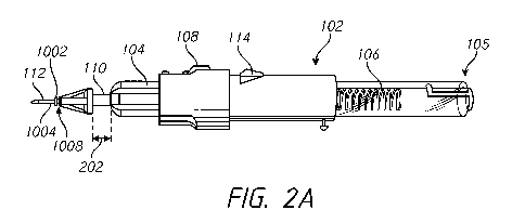
vertical
edges 230, 232 can come together and overlap each other to form a self-sealing
wound
236 that may not require sutures for closing the wound 236. The angle of the
first stage
may be steep enough that, in combination with the thickness of the tissue, a
vertical line
cannot pass through the wound 236 without modifying at least one of the edges
226, 228.
-18-
CA 02953564 2016-12-22
WO 2016/010810 PCT/US2015/039682
[0080] In some embodiments, the wound 214 in Figures 3A and 3B and/or the
wound 236 in Figures 3C and 3D can secure a cannula better than a wound with
only a
vertical portion that is created when a user pierces entirely through the body
tissue at an
angle of about 90 relative to the surface of the body tissue.
[0081] Figures 4A-4C illustrate example operations of an example trocar
agent for coupling to a cannula insertion system. In some embodiments, the
cannula
insertion system (e.g., the system 102) comprises a coupling mechanism 305 at
the distal
end of the slider portion 110. In some embodiments, the coupling mechanism 305
comprises a groove 306. In some embodiments, the coupling mechanism 305 is
configured to receive a trocar agent 302. By receiving a trocar agent 302, the
system can
be easily loaded with a needle portion 112. In some embodiments, the needle
portion 112
is already loaded with a cannula 1008 (Figures 2A-2C). Loading a cannula 1008
on the
needle portion 112 may advantageously allow the user to easily and quickly
load a trocar
agent 302 onto a cannula insertion system 102 by coupling the trocar agent 302
to the
coupling mechanism 305.
[0082] To couple the trocar agent 302 to the coupling mechanism 305, a user
can insert the trocar agent 302 into the coupling mechanism 305 until a
protrusion 304
abuts a distal portion of the slider portion 110, as shown by the arrow 308 in
Figure 4A.
To lock the trocar agent 302 into the coupling mechanism, the user can rotate
the trocar
agent 302 as shown by the arrow 310 in Figure 4B whereby the protrusion 304
travels
along the groove 306, for example until the protrusion 304 is locked into the
groove
portion 306 at a terminal end of the groove 306.
[0083] Figures 5A-5C illustrate example operations of an example cannula
insertion system 500. Figure 5D is an exploded view of the cannula insertion
system 500
of Figures 5A-5C. In some embodiments, the slider control mechanism 108 can be
implemented as a flexible h-slider with a front trocar agent locker. In some
embodiments,
the slider control mechanism 108 comprises a user interface portion 408
configured to
allow the index finger, for example, of the user to interface with the slider
control
mechanism 108. In some embodiments, the slider control mechanism 108 comprises
a
linear center portion 410 that is coupled to the user interface portion 408
and that is
configured to slide in a groove 412 on the distal end 411 of the hollow
tubular portion
103.
-19-
CA 02953564 2016-12-22
WO 2016/010810 PCT/US2015/039682
[0084] In some embodiments, the slider control mechanism 108 comprises a
slider portion 110 that is coupled to a linear center portion 410. In some
embodiments,
the user can interface with the user interface portion 408 to slide forward
the slider control
mechanism 108 to move the slider portion 110 forward, which pushes the needle
portion
112 coupled to the slider portion 110 into body tissue. In some embodiments,
the slider
control mechanism 108 can move independently of a housing 402 and/or a shaft
404 to
move or advance the needle portion 112 a distance 204. After moving the needle
portion
112 the distance 204, in some embodiments, the slider control mechanism 108 is
locked
with the shaft 404 such that when the shaft 404 is advanced in a distal
direction, the
needle portion 112 coupled to the slider control mechanism 108 can advance in
a distal
direction with the shaft 404.
[0085] In some embodiments, the shaft 404 is positioned in the hollow
tubular
portion 103 of the device. A proximal end 416 of the shaft 404 can interact
with the
spring 106, for example abutting or coupled to the spring with a shoulder,
lip, ring, or
flange 418. In some embodiments, the spring 106 is housed in the hollow
tubular portion
103 of the device 106. In some embodiments, the shaft 404 can be moved in a
proximal
direction to compress the spring 106. To lock the spring 106 in a compressed
configuration, the proximal end of the hollow tubular portion 103 can be
occluded (e.g.,
by a plug) and the activation mechanism 114 can be moved proximally to
compress the
spring 106 against the occlusion. The activation mechanism 114 is biased
radially
outwardly and, upon moving to at least the longitudinal positon of a
transverse surface
414 of the hollow tubular portion 103, can interface with (e.g., abut) the
surface 414 (e.g.,
moving from the orientation of Figure 5C to the orientation of Figure 5B). The
interfacing of activation mechanism 114 with the surface 414 inhibits or
prevents the
shaft 404 from advancing in a distal direction, thereby keeping the spring 106
in a
compressed state and inhibiting or preventing the spring 106 from forcing the
shaft 404 in
a distal direction. Figures 5A and 5B show the activation mechanism 114
abutting the
surface 414. As shown in Figure 5B, distal movement of the slider control
mechanism
108 causes the slider portion 110 to move a distance illustrated by a post-
first stage length
204. In some embodiments, the length 204 is between about 0.3 mm and about 1
mm,
between about 0.2 mm and about 0.5 mm, between about 0.1 mm and about 0.3 mm,
or
between about 0.1 mm and about 0.2 mm.
-20-
CA 02953564 2016-12-22
WO 2016/010810 PCT/US2015/039682
[0086] As shown in Figure 5C, in some embodiments, the user can press the
activation mechanism 114 radially inward to release the compressed spring 106.
By
depressing the activation mechanism 114 radially inward, the surface 414 no
longer
interfaces with the activation mechanism 114, which allows the shaft 404 to
advance in a
distal direction due to the decompression of the spring 106. As the spring 106
decompresses, the spring 106 exerts a force on the distal end of the shaft
404, which
causes the shaft 404 to advance distally in the hollow tubular structure 103.
By activating
the activation mechanism 114, the slider portion does not change length 204.
Activating
the activation mechanism 114 causes the distal portion of the device to extend
from a
length 206 (Figure 5B) to a larger length 208 (Figure 5C).
[0087] In some embodiments, the difference between the length 206 and the
length 208, which is the length that the needle portion 112 is further
extended, is between
about 0.3 mm and about 1 mm, between about 0.2 mm and about 0.8 mm, or between
about 0.1 mm and about 0.5 mm. In some embodiments, the sum of the length 204
and
the difference between the length 206 and the length 208 is between about 0.5
mm and
about 1.5 mm, which would be sufficient to traverse a sclera having a
thickness between
about 0.3 mm and about 1 mm at a 45 angle. In some embodiments, a ratio
between the
length 204 and the difference between the length 206 and the length 208 is
between about
1:1 and about 1:5, between about 1:1 and about 1:3, between about 1:1 and
about 1:4, or
between about 1:1 and about 1:2. Other lengths and ratios are also possible.
For
example, the length 204 may be configured to be more than half of the
thickness of the
sclera.
[0088] Figure 5D shows the hollow tubular portion 103, the spring 106, the
slider control mechanism 108, the trocar mechanism 302, the housing 402, and
the shaft
404 in exploded view. Other elements and/or modifications thereof may be used
instead
of and/or in combination with the illustrated elements.
[0089] Figures 6A-6D illustrate example operations of example control
mechanisms for a cannula insertion system 500. The system 500 may be used for
form a
one or two angle oblique sclerotomy, which may provide one or more of the
advantages
described herein. In some embodiments, the cannula insertion system 500
comprises a
slider control mechanism 108. The slider control mechanism 108 can comprise a
flexible
beam portion 501 which can be configured to flex depending upon a position
along a
guide trail or channel 505 of a hollow tubular portion 103. In some
embodiments, the
-21-
CA 02953564 2016-12-22
WO 2016/010810 PCT/US2015/039682
slider control mechanism 108 comprises a substantially nonflexible or rigid
beam portion
502 in the hollow tubular portion 103. In some embodiments, when the slider
control
mechanism 108 is in a proximal position, a needle portion coupled thereto is
not distally
extended.
[0090] In the proximal or loaded or first position shown in Figure 6A, the
flexible beam portion 501 is outwardly flexed due to interaction of the
nonflexible beam
portion 502 and a backward stopper portion 504 of a housing 402. The backward
stopper
portion 504 forces the slider control mechanism 108 inwardly towards the
radial center of
the device, causing the flexible beam portion 501 to flex upon interaction
with an outer
surface of the housing 402. In some embodiments, the flexible beam portion 501
comprises a protrusion 512 that interacts with the outer surface of the
housing 402. In
some embodiments, the user can slide the slider control mechanism 108 in a
distal
direction to a second position, allowing the nonflexible beam portion 502 to
slide distally
past the backward stopper protrusion 504, as shown in Figure 6B. With the
nonflexible
beam portion 502 not interacting with the backward stopper protrusion 504,
there is no
longer an inward force on the slider control mechanism 108, and the flexible
beam
portion 501 is released from the flexed state.
[0091] In some embodiments, as the nonflexible beam portion 502 slides
beyond the backward stopper protrusion 504, the slider control mechanism 108
is
configured to make a clicking sound (e.g., upon the protrusion 512 falling
into the trail
505), which may indicate to the user that the needle portion has advanced in
body tissue
such as the sclera. In some embodiments, the backward stopper protrusion 504
can be
configured to interface with a distal portion 508 of the nonflexible beam
portion 502 in
order to inhibit or prevent the slider control mechanism 108 from moving
backward in a
proximal direction during use. In some embodiments the backward stopper
protrusion
504 locks the slider control mechanism 108 with the shaft 404. In the locked
state, any
movement of the shaft 404 causes the slider mechanism and the needle portion
112 which
is coupled to the slider control mechanism 108 to move in the same distal
direction.
[0092] As illustrated in Figure 6C, the activation mechanism 114 can be
positioned in opening 510 of the tubular housing 103 of the device. In some
embodiments, the outer edge of the activation mechanism 114 is flush with the
outer
surface of the tubular housing 103, which may inhibit or prevent accidental
activating of
the activation mechanism 114. In some embodiments, the outer surface of the
activation
-22-
CA 02953564 2016-12-22
WO 2016/010810 PCT/US2015/039682
mechanism 114 can be positioned slightly inward of or below the outer surface
of the
tubular housing 103, which can inhibit or prevent accidental activating of the
activation
mechanism 114. By inwardly depressing the activation mechanism 114, the
deformed
cantilever portion of the activation mechanism 114 is allowed to slide within
the hollow
tubular portion of the device 102, as shown in Figure 6D.
[0093] Figure 7 is an exploded view of example components of an example
cannula insertion system 600. In some embodiments, the cannula insertion
system
comprises a first spring 602 and a second spring 604 that can be used to
implement a two-
stage cannula insertion process. Along the lines described above with respect
to the
spring 106 and illustrated in Figures 5B and 5C, for example, and described in
further
detail with respect to Figures 8A-8D, the first spring 602 can be configured
to advance the
needle portion 112 slightly forward in a distal direction to allow the needle
portion 112 to
partially pierce body tissue such as the sclera, and, the second spring 604
can be
configured to advance the needle portion 112 a longer distance in order to
advance the
needle portion 112 entirely through the body tissue. In some embodiments, the
cannula
insertion system comprises a first outer locking shell 606 comprising a first
surface 628
and a first guide trail 624, a second outer locking shell 608 comprising a
second surface
626 and a second guide trail 622, a first inner locking shaft 609 comprising
an activation
mechanism 610, a second inner locking shaft 607 comprising an activation
mechanism
608, a trocar 620 including the needle portion 112, a cannula 302, a first
sheath 630
comprising a first flexible activation button 612, and a second sheath 632
comprising a
second flexible activation button 614. In some embodiments, the first and
second springs,
outer locking shells, inner locking shafts, and sheaths may be identical to
each other or
include at least one identical feature (e.g., size, shape, material, element,
etc.).
[0094] Figures 8A-8D illustrate example operations of an example cannula
insertion system 600. The system 600 may be used for form a one or two angle
oblique
sclerotomy, which may provide one or more of the advantages described herein.
Figure
8A is a front view and Figure 8B is a side view of the cannula insertion
system in an
initial state. As illustrated in Figures 8A-8C, during a first stage, a user
can activate the
activation mechanism 610 by pressing the first flexible activation button 612
radially
outward of the activation mechanism 610 to push the activation mechanism 610
radially
inward and out of engagement with the surface 628, allowing the first inner
locking shaft
609 to move distally and the first compressed spring 602 to expand, thereby
forcing the
-23-
CA 02953564 2016-12-22
WO 2016/010810 PCT/US2015/039682
needle portion 112 to move in a distal direction by a first distance 640. As
illustrated in
Figures 8C and 8D, during a second stage, a user can activate the activation
mechanism
608 by pressing the first flexible activation button 614 radially outward of
the activation
mechanism 608 to push the activation mechanism 608 radially inward and out of
engagement with the surface 626, allowing the second inner locking shaft 607
to move
distally and the second compressed spring 604 to expand, thereby forcing the
needle
portion 112 to move in a distal direction by a second distance 642. The first
distance 640
may be the same or different than the second distance 642.
[0095] An advantage that may be provided by the system 600 is that the
advancement of the needle portion 112 is entirely automatic in that no distal
movement by
a user causes distal movement of the needle portion 112. A user need not exert
any force
on the needle portion 112 to pierce the body tissue. In comparison, in semi-
automatic
systems comprising a single spring, a user exerts some force on needle portion
112, for
example by distally advancing a slider control mechanism 108, for the needle
portion 112
pierce through body tissue during the first stage. In a completely automatic
system, a user
can advantageously exert a consistent force when inserting a cannula into body
tissue. A
user may expend less energy using a completely automatic system than a semi-
automatic
system. An automatic system can reduce the time for inserting a cannula into
body tissue.
[0096] Figure 9 is a perspective view of an example cannula insertion
system
800. In some embodiments, the cannula insertion system 800 is configured to
insert a
cannula into body tissue in one stage. In some embodiments, the cannula
insertion system
800 comprises a compressed spring 804 coupled to an activation mechanism 806
and
around an inner locking shaft 808, which is in an outer tube 802. An outer
protection
shell (e.g., around the spring 804) may be used. A user can activate the
activation
mechanism 806 to release the compressed spring 804 to exert force on the
needle portion
112. The force exerted on the needle portion 112 drives the needle portion 112
into the
body tissue.
[0097] Figure 10 is a perspective view an example cannula insertion system
900. In some embodiments, the cannula insertion system comprises a standard
trocar 910
coupled to a lancing device 904, which includes a cannula 904 and a needle
portion 908
(e.g., comprising a 25 gauge needle). In some embodiments, the cannula
insertion system
900 comprises a button 902 that allows the user to release a compressed
spring. By
-24-
CA 02953564 2016-12-22
WO 2016/010810 PCT/US2015/039682
releasing the compressed spring, a force is exerted on the trocar 910, which
drives the
needle portion 908 and the cannula 906 of the lancing device 904 into body
tissue.
[0098] Figure 11 is a perspective view an example cannula insertion system
1000. In some embodiments, the cannula insertion system 1000 comprises a
standard
trocar 1010 coupled to a slider portion 110, which includes a cannula 1008 and
a needle
portion 1006 (e.g., comprising a 25 gauge needle). In some embodiments, the
needle
portion 1006 is preloaded with the cannula 1008. In some embodiments, the
cannula
1008 comprises a cannula body 1002 and a cannula tube 1004. In some
embodiments, the
cannula system 1000 comprises a button configured to release a compressed
spring when
activated by a user. Releasing the compressed spring causes a force to be
exerted on the
needle portion 1006 and the cannula 1008 as at least the needle portion 1006
and the
cannula tube 1004 are inserted into body tissue.
[0099] Figures 12A and 12B illustrate an example of inserting the cannula
insertion system 1000 of Figure 11 in an eye. The eye was a live rabbit eye,
which is a
suitable substitute representation of a human eye. In Figure 12A, the slider
mechanism
110 and the proximal portion of the cannula body 1002 are visible, and the
needle portion
1006 and the cannula tube 1004 are in the eye. Figure 12A also shows an
optional
protection tube 1012, which may be attached to the trocar 1006 and around the
slider
portion 110, for example using the channel 1014 shown in Figure 11. Figure 12B
shows
the extended needle 1006 after removal from the eye.
[0100] Figures 13A-13C illustrate an example cannula insertion system 1200
at different stages. Figure 13D is an exploded view of example components of
the
cannula insertion system 1200 of Figures 13A-13C. As shown in Figures 13A and
13D,
for example, the cannula insertion system 1200 comprises a plurality of
preloaded trocar-
cannula pairs 1204, 1206, 1208. In some embodiments, the trocar-cannula pairs
1204,
1206, 1208 are coupled to a slide inserter 1210, 1212, 1214, respectively. In
some
embodiments, the cannula insertion system 1200 comprises a single slide
inserter
configured to be loaded with the trocar-cannula pairs 1204, 1206, 1208 when
each trocar-
cannula pair is about to be inserted in body tissue. In some embodiments, the
cannula
insertion system 1202 comprises two, three, four, five, or more preloaded
trocar-cannula
pairs each including an individual slide inserter. At least two and/or all of
the trocar-
cannula pairs may have a property (e.g., needle gauge, cannula gauge) that is
the same as
each other. At least two and/or all of the trocar-cannula pairs may have a
property (e.g.,
-25-
CA 02953564 2016-12-22
WO 2016/010810 PCT/US2015/039682
needle gauge, cannula gauge) that is different from each other. The slide
inserter 1210,
1212, 1214 may interlock with a cannula and needle portion as shown and
described with
respect to Figures 4A-4C, for example.
[0101] In some embodiments, the cannula insertion system 1200 comprises a
housing 1218 having an opening 1219. The opening 1219 comprises an edge or
surface
1217 configured to interface with a surface of a flexible trigger button 1220.
Engagement
between the flexible trigger button 1220 and the edge 1217 inhibits or
prevents the
interlocking shaft 1215 from being advanced forward due to the loaded,
compressed
spring 1222. In some embodiments, the cannula insertion system 1202 comprises
a cover
1216 configured to protect the trocar-cannula pairs 1204, 1206, 1208.
[0102] In some embodiments, a user of the cannula insertion system 1200
rotates a guiding trail tube 1302 (e.g., through a window or aperture in the
cover 1216) to
place a desired one of the trocar-cannula pairs 1204, 1206, 1208 in a position
for insertion
into body tissue. For example, the user can position a trocar-cannula pair
1208 such that
the slide inserter 1214 is aligned (e.g., circumferentially aligned) with the
flexible trigger
button 1220 (e.g., as illustrated in Figure 12A).
[0103] In some embodiments, the cannula insertion system 1200 is configured
to allow a user to longitudinally slide the slide inserter 1214 towards the
distal end 1201
to allow the user to introduce the trocar needle partly into the sclera of a
patient while
creating a sclerotomy, as shown in Figure 13B. In some embodiments, the slide
inserter
1214 is configured to automatically and partially insert the trocar needle
into the sclera by
a spring mechanism (e.g., as described with respect to Figures 7-8D), motor,
pneumatic
drive, or other mechanism or other combination thereof, thereby avoiding the
user sliding
the slide inserter 1214. The system 1200 may be used for form a one or two
angle oblique
sclerotomy, which may provide one or more of the advantages described herein.
[0104] As shown in Figures 13C, 14A, and 14B, in some embodiments, the
cannula insertion system 1200 is configured to allow the user to press and/or
deform the
flexible trigger button 1220 to allow the flexible trigger button 1220 to be
disengaged
with edge 1217 to release the compressed spring 1222. By disengaging the
trigger button
1220 from the edge 1217, the spring 1222 is allowed to decompress and apply a
force
onto shaft 1215. By releasing the compressed spring 1222, the shaft 1215 can
apply a
longitudinal force on an edge 1306 (Figure 13D) of the slide inserter 1214,
which is
aligned with an interface edge 1304 of the shaft 1215. By applying a
longitudinal force
-26-
CA 02953564 2016-12-22
WO 2016/010810 PCT/US2015/039682
on the slide inserter 1214, the trocar-cannula pair 1208 is forced further
into the sclera of
the patient. The trocar-cannula pair 1208 may be inserted through the sclera
and the
cannula may be implanted in the sclera.
[0105] During this two-stage insertion process, a user can first insert the
trocar
needle into the sclera at an oblique angle relative to the surface of an eye
of a patient. In a
second stage, the user can position the cannula insertion system 1200 at a
substantially 90
degree angle relative to the surface of the eye before pressing and/or
deforming the
flexible trigger button 1220 such that a trocar-cannula pair is further
inserted into the
sclera by a distance 1224 by the longitudinal force exerted by the
decompressing spring
1222.
[0106] To disengage the cannula insertion system 1200 from the inserted
cannula, the cannula insertion system 1200 can be configured to allow the user
to
longitudinally slide the slide inserter 1214 towards the proximal end 1203. By
moving
the slide inserter 1214 towards the proximal end 1203, the trocar needle is
withdrawn
from the eye of the patient and from the cannula that is now implanted in the
sclera of the
eye. In some embodiments, the cannula insertion system 1202 can be configured
to allow
the user to rotate the protective cover 1216 to allow the user to insert a
second trocar-
cannula pair (e.g., the trocar cannula pair 1204 or the trocar cannula pair
1206) into the
patient. To insert the second trocar-cannula pair into the patient, the user
can repeat the
foregoing process. The cannula insertion system 1200 can be used to insert
multiple
different cannulas into a patient.
[0107] In some embodiments, the cannula insertion system 1200 is configured
to allow a user to compress the spring 1222 by longitudinally sliding the
shaft 1215
toward the proximal end 1203 of the cannula insertion system 1200. The user
can grasp
the proximal end 1203 with one hand while using the other hand to
longitudinally slide
the shaft 1215 by grasping the cover 1216, which is coupled to the shaft 1215,
near the
distal end 1201 and moving the distal end 1201 towards the proximal end 1203
until the
cantilever of the flexible trigger button flexes outwardly through the opening
1219 and the
flexible trigger button 1220 engages the edge 1217.
[0108] Figures 15A-15C illustrate an example cannula insertion system 1500
at different stages. Figure 15D is an exploded view of example components of
the
cannula insertion system 1500 of Figures 15A-15C. Figure 15E is a further
exploded
view of example components of the cannula insertion system 1500 of Figures 15A-
15C.
-27-
CA 02953564 2016-12-22
WO 2016/010810 PCT/US2015/039682
[0109] As shown in Figures 15A and 15D, for example, the cannula insertion
system 1500 comprises a revolver chamber 1504 configured to house a plurality
of trocar-
cannula pairs 1204, 1206, 1208. In some embodiments, the cannula insertion
system
1500 includes a single or only one slide inserter 1502 configured to interact
with and
position any of the trocar-cannula pairs 1204, 1206, 1208, for example in
contrast to the
cannula insertion system 1200 in which each trocar-cannula pair was coupled to
respective a slide inserter. In some embodiments, the revolver chamber 1504
can be
configured to house a plurality of trocar-cannula pairs with each having a
slide inserter,
for example like the cannula insertion system 1200. In some embodiments, the
revolver
chamber 1504 can comprise two, three, four, five, or more trocar-cannula pairs
for
insertion into the eye of a patient. The revolver chamber 1504 may comprise
trocar guide
trails to longitudinally guide the trocar-cannula pairs upon application of
force by the
slide inserted 1502. At least two and/or all of the trocar-cannula pairs may
have a
property (e.g., needle gauge, cannula gauge) that is the same as each other.
At least two
and/or all of the trocar-cannula pairs may have a property (e.g., needle
gauge, cannula
gauge) that is different from each other.
[0110] In some embodiments, the cannula insertion system 1200 comprises a
housing 1218 having an opening 1219. The opening 1219 comprises an edge or
surface
1217 configured to interface with a surface of a flexible trigger button 1220.
Engagement
between the flexible trigger button 1220 and the edge 1217 inhibits or
prevents the
interlocking shaft 1215 from being advanced forward due to the loaded,
compressed
spring 1222.
[0111] In some embodiments, the revolver chamber 1504 is positioned or
centered lateral to a center longitudinal axis 1520 of the cannula insertion
system 1500.
The revolver chamber 1504 can be positioned such that the desired trocar-
cannula pair
1204, 1206, 1208 is placed substantially in the center longitudinal 1520 axis
of the
cannula insertion system 1500. In some embodiments, placement of the trocar-
cannula
pair in the center longitudinal axis 1520 allows the trocar-cannula pair to be
aligned with
the shaft 1215. The shaft 1215 is coupled to a guide rail 1508, which can
guide the slide
inserter 1502.
[0112] In some embodiments, the cannula insertion system 1500 is configured
to allow a user to rotate the revolver chamber 1504 in order to place a
selected trocar-
cannula pair 1204, 1206, 1208 in a position for insertion into a patient. For
example, the
-28-
CA 02953564 2016-12-22
WO 2016/010810 PCT/US2015/039682
revolver chamber 1504 can be rotated such that the trocar-cannula pair 1208 is
aligned
with the slide inserter 1502 (e.g., as illustrated in Figure 15A). In some
embodiments, the
cannula insertion system 1200 is configured to allow a user to longitudinally
slide the
slide inserter 1502 towards the distal end 1501 to allow the user to introduce
the trocar
needle partly into the sclera of a patient while creating a sclerotomy, as
shown in Figure
15B. Figure 15E also shows the general longitudinal position of the trocar-
cannula pairs
1204, 1206, 1208 after the trocar-cannula pair 1204 is moved due to movement
of the
slide inserter 1502. In some embodiments, the slide inserter 1502 is
configured to
automatically and partially insert the trocar needle into the sclera by a
spring mechanism
(e.g., as described with respect to Figures 7-8D), motor, pneumatic drive, or
other
mechanism or other combination thereof, thereby avoiding the user sliding the
slide
inserter 1502.
[0113] In some embodiments, the cannula insertion system 1500 is configured
to allow the user to press and/or deform the flexible trigger button 1220 to
allow the
flexible trigger button 1220 to be disengaged with edge 1217 to release the
compressed
spring 1222, as shown in Figure 15C. By disengaging the trigger button 1220
from the
edge 1217, the spring 1222 is allowed to decompress and apply a force onto
shaft 1215.
The force on shaft 1215 can be applied to an edge 1516 (Figure 15D) of the
slide inserter
1502, which is aligned with an interface edge 1304 of the shaft 1215. By
applying a
longitudinal force towards the distal end 1501, the trocar-cannula pair 1208
is further
driven into the sclera of the eye. The trocar-cannula pair 1208 may be
inserted through
the sclera and the cannula may be implanted in the sclera.
[0114] During this two-stage insertion process, a user can first insert the
trocar
needle into the sclera at an oblique angle relative to the surface of an eye
of a patient. In a
second stage, the user can position the cannula insertion system 1500 at a
substantially 90
degree angle relative to the surface of the eye before pressing and/or
deforming the
flexible trigger button 1220 such that a trocar-cannula pair is further
inserted into the
sclera by a distance 1524 by the longitudinal force exerted by the
decompressing spring
1222. The system 1500 may be used for form a one or two angle oblique
sclerotomy,
which may provide one or more of the advantages described herein.
[0115] In some embodiments, the cannula insertion system 1500 can be
disengaged from the cannula that has been implanted in an eye of a patient by
allowing
the user to longitudinally slide the slide inserter 1502 toward the proximal
end 1503. By
-29-
CA 02953564 2016-12-22
WO 2016/010810 PCT/US2015/039682
sliding the slide inserter 1502 in a proximal direction, the trocar needle can
be withdrawn
from the implanted cannula. To insert a second cannula in the patient, the
user to rotate
the revolver chamber 1504 to position a second trocar-cannula pair (e.g., the
trocar-
cannula pair 1204 or the trocar-cannula pair 1206) at a location such that the
second
trocar-cannula pair is aligned with the slide inserter 1502. The user can then
repeat the
foregoing process in order to implant the second trocar-cannula pair. The
cannula
insertion system 1500 can be used to insert multiple different cannulas into a
patient.
[0116] In some embodiments, the cannula insertion system 1500 is configured
to allow the spring 1222 to be compressed when a user grasps the proximal end
1503 with
one hand and longitudinally slides the shaft 1215 towards the proximal end
1503 by
grasping the revolver chamber 1504, which is coupled to the shaft 1215, near
the distal
end 1501 and moving the distal end 1501 towards the proximal end 1503 until
the
cantilever of the flexible trigger button 1220 flexes outwardly through the
opening 1219
and the flexible trigger button 1220 engages the edge 1217.
[0117] Figures 15F-15H illustrate an example cannula insertion system 1550
at different stages. Figures 15I-15K further illustrate example operations of
the cannula
insertion system 1550 of Figures 15F-15H. As shown in Figure 15D and 15F-15H,
in
some embodiments, the cannula insertion system 1550 optionally comprises one
or more
blockers 1510 for inhibiting or preventing the implantation of the non-aligned
trocar-
cannula pairs that are housed in the revolver chamber 1504. A cannula
insertion system
1550 without a blocker 1510 may function as described above for the cannula
insertion
system 1500. The cannula insertion system 1550 is an example of a system
comprising
four trocar-cannula pairs, for example compared to the three trocar-cannula
pairs of the
cannula insertion system 1500. The components and operation of the cannula
insertion
system 1550 may otherwise be the same or substantially the same as the cannula
insertion
system 1500.
[0118] In some embodiments, the blocker 1510 is configured to lock the
positions of the other trocar-cannula pairs such that the trocar-cannula pairs
that are not in
use are inhibited or prevented from sliding forward after the spring 1222 has
been
released from a compressed state. For example, the blocker 1510 may comprise a
partially annular flange 1511 configured to interact with (e.g., mechanically
impede
movement of) a proximal portion of one, some, or all of the non-aligned trocar-
cannula
pairs.
-30-
CA 02953564 2016-12-22
WO 2016/010810 PCT/US2015/039682
[0119] Referring to Figures 15F and 151, four trocar-cannula pairs 1204,
1206,
1208, 1209 are in the revolver chamber 1504 at an initial position. The trocar-
cannula
pair 1208 is aligned with the slide inserter 1502, which is radially outwardly
deformed in
a proximal position due to interaction of a flexible beam 1505 of the slide
inserter 1502
with a more rigid or less flexible beam of the shaft 1215.
[0120] Referring to Figures 15G and 15J, the slide inserter 1502 has been
longitudinally distally advanced. Advancement of the slide inserter 1502
distally
advances the trocar-cannula pair 1208, which is free of the partially arcuate
flange 1511 of
the blocker 1510, by a distance 1522 and distal to the revolver chamber 1504.
Advancement of the slide inserter 1502 does not distally advance the trocar-
cannula pairs
1204, 1206, 1209 because the slide inserted 1502 does not interact with the
trocar-cannula
pairs 1204, 1206, 1209 and/or because the flange 1511 of the blocker 1510
inhibits or
prevents advancement the trocar-cannula pairs 1204, 1206, 1209. After
advancement, the
slide inserter 1502 is no longer radially outwardly deformed since the
flexible beam 1505
of the slide inserter 1502 is not interacting with a more rigid or less
flexible beam of the
shaft 1215. The flexible beam 1505 may flex radially inwardly and lock against
a
backward stopper 1515 of the slider 1215 to lock the slide inserter 1502 and
trocar-
cannula pair 1208 in the position shown in Figures 15G and 15J.
[0121] Figure 15H shows the cannula insertion system 1550 after the
flexible
trigger button 1220 has been pressed or deformed radially inward to allow the
flexible
trigger button 1220 to disengage with edge 1217 to release the compressed
spring 1222.
By disengaging the trigger button 1220 from the edge 1217, the spring 1222 is
allowed to
decompress and apply a force onto shaft 1215. The force on shaft 1215 can be
applied to
an edge 1516 (Figure 15D) of the slide inserter 1502, which is aligned with an
interface
edge 1304 of the shaft 1215. By applying a longitudinal force towards the
distal end
1501, the trocar-cannula pair 1208 is driven a distance 1524 and further into
the sclera of
the eye. The system 1550 may be used for form a one or two angle oblique
sclerotomy,
which may provide one or more of the advantages described herein.
[0122] Figure 15K shows the cannula insertion system 1550 in a partially
retracted position, and for clarity omitting the revolver chamber 1504. The
user first pulls
and deforms the slide inserter 1502 radially outward so that the flexible beam
1505 can
slide proximally past the backward stopper, and the user second proximally
longitudinally
retracts the slide inserter 1502. Retraction of the slide inserter 1502 also
retracts the
-31-
CA 02953564 2016-12-22
WO 2016/010810 PCT/US2015/039682
needle portion of the trocar-cannula pair 1208, leaving the cannula implanted
in the
sclera. The slide inserter may comprise a pivot point, weakened portion,
and/or the like to
promote deformation when desired. The next trocar-cannula pair can be aligned
with the
slide inserter 1502 and disengaged from the blocker 1510, and the foregoing
process can
be repeated.
[0123] Figure 16 juxtaposes the cannula insertion system 1500 of Figures
15A-15C and the cannula insertion system of Figure 1. In some embodiments, the
cannula insertion system 1500 comprising a revolver chamber 1504 comprises
similar
dimensions and/or components as the example cannula insertion system 102,
which does
not comprise a revolver chamber, for example because the cannula insertion
system 102 is
configured to position a single cannula rather than a plurality of cannulas.
[0124] Figures 17A-17D illustrate an example cannula insertion system 2000.
Figure 17E is an exploded view of example components of the cannula insertion
2000
system of Figures 17A-17D. Figures 17A and 17B are side views and Figures 17C
and
17D are front views. Figure 17A shows the cannula insertion 2000 system in a
first
configuration, Figures 17B and 17C show the cannula insertion 2000 system in a
second
configuration, and Figure 17D shows the cannula insertion 2000 system in a
third
configuration. Figure 17F is a magnified view of an example component 2010 of
the
cannula insertion 2000 system of Figures 17A-17D.
[0125] The cannula insertion system 2000 comprises a side cartridge 2002
configured to house a plurality of trocar-cannula pairs 1204, 1206, 1208,
1209. In
contrast to the guiding trail tube 1302 and the revolver chamber 1504, for
example, which
can be rotated to position a trocar-cannula pair for insertion, the side
cartridge 2002 can
transversely advance a trocar-cannula pair into position for insertion. In
some
embodiments, the cannula insertion system 2000 comprises a single slide
inserter 1502
for implanting the plurality of trocar-cannula pairs housed in the side
cartridge 2002, for
example as described with respect to the cannula insertion systems 1500, 1550.
In some
embodiments, the cannula insertion system 2000 can comprise a plurality of
slide
inserters, one for implanting each of the plurality of trocar-cannula pairs
housed in the
side cartridge 2002, for example as described with respect to the cannula
insertion system
1200. The trocar-cannula pairs 1204, 1206, 1208, 1209 can comprise a
substantially
rectangular or square or other shape configuration for positioning in the side
cartridge
2002 and for loading the trocar-cannula pairs into the cannula insertion
system 2000 for
-32-
CA 02953564 2016-12-22
WO 2016/010810 PCT/US2015/039682
implantation into a patient. In some embodiments, the side cartridge 2002 is
configured
to house two, three, four, five, or more trocar-cannula pairs for insertion
into the eye of a
patient. At least two and/or all of the trocar-cannula pairs may have a
property (e.g.,
needle gauge, cannula gauge) that is the same as each other. At least two
and/or all of the
trocar-cannula pairs may have a property (e.g., needle gauge, cannula gauge)
that is
different from each other.
[0126] In some embodiments, the cannula insertion system 2000 comprises a
side cartridge 2002 that is proximate to the distal end 2001 of the cannula
insertion
system 2000. Referring to Figure 17F, in some embodiments, the side cartridge
2002
comprises a button or surface 2304 that is coupled to a spring 2302. The side
cartridge
2002 can be configured to allow the user to press the button 2304 in order to
apply a force
on spring 2302 such that a force is applied on the trocar-cannula pairs housed
in the side
cartridge 2002. The force on the trocar-cannula pairs is configured to
horizontally, or
transversely with respect to the longitudinal axis of the cannula insertion
system 2000,
slide at least one of the trocar-cannula pairs into a longitudinal center of
the cannula
insertion system 2000 to position that trocar-cannula pair to be implanted in
a patient.
[0127] In some embodiments, the cannula insertion system 2000 is configured
to allow a user to longitudinally advance the slide inserter 1502 towards the
distal end
2001. By longitudinally advancing the slide inserter 1502 in the distal
direction, the
needle portion of the trocar-cannula pair 1208 can be initially inserted into
the sclera of an
eye of a patient. In some embodiments, the cannula insertion system 2000 is
configured
to allow the user to press and/or deform the flexible trigger button 1220
radially inward to
disengage the flexible trigger button 1220 from the edge 1217. Disengagement
of the
flexible trigger button 1220 from the edge 1217 allows the spring 1222 to
decompress and
apply a force on the shaft 1215. The force applied on the shaft 1215 causes
the shaft 1215
to apply a force on the slide inserter 1502, which causes the trocar-cannula
pair 1208 to be
inserted further into the sclera of an eye of a patient. The trocar-cannula
pair 1208 may be
inserted through the sclera and the cannula may be implanted in the sclera.
[0128] The user can position the cannula insertion system 2000 at an angle
relative to the surface of the eye of the patient during the first stage
illustrated from Figure
17A to Figure 17B, which includes inserting the trocar needle into the sclera
using the
slide inserter 1216. The user can position the cannula insertion system 2000
at
substantially a 90 degree angle relative to the surface of the eye of the
patient during the
-33-
CA 02953564 2016-12-22
WO 2016/010810 PCT/US2015/039682
second stage illustrated from Figure 17C to Figure 17D, which includes
inserting the
trocar needle into the sclera by pressing and/or deforming the flexible
trigger button 1220.
The system 2000 may be used for form a one or two angle oblique sclerotomy,
which may
provide one or more of the advantages described herein.
[0129] To disengage the cannula insertion system 2000 from the implanted
cannula, the cannula insertion system 2000 can be configured to allow the user
to
longitudinally slide the slide inserter 1502 towards the proximal end 2003. By
retracting
the slide inserter 1502 proximally, the needle portion of the trocar-cannula
pair 1208 can
be withdrawn from the implanted cannula. In some embodiments, the cannula
insertion
system 2000 is configured to allow the user to press the button 2304 to allow
a second
trocar-cannula pair to be horizontally moved into the cannula insertion system
2000 such
that the second trocar-cannula pair can be placed in position for insertion
into the eye
using the foregoing process.
[0130] Conditional language, such as, among others, "can," "could,"
"might,"
or "may," unless specifically stated otherwise, or otherwise understood within
the context
as used, is generally intended to convey that certain embodiments include,
while other
embodiments do not include, certain features, elements, and/or steps. Thus,
such
conditional language is not generally intended to imply that features,
elements, and/or
steps are in any way required for one or more embodiments or that one or more
embodiments necessarily include, with or without user input or prompting,
whether these
features, elements and/or steps are included or are to be performed in any
particular
embodiment. The headings used herein are for the convenience of the reader
only and are
not meant to limit the scope of the inventions or claims.
[0131] While the methods and devices described herein may be susceptible to
various modifications and alternative forms, specific examples thereof have
been shown
in the drawings and are herein described in detail. It should be understood,
however, that
the invention is not to be limited to the particular forms or methods
disclosed, but, to the
contrary, the invention is to cover all modifications, equivalents, and
alternatives falling
within the spirit and scope of the various implementations described and the
appended
claims. Further, the disclosure herein of any particular feature, aspect,
method, property,
characteristic, quality, attribute, element, or the like in connection with an
implementation
or embodiment can be used in all other implementations or embodiments set
forth herein.
Any methods disclosed herein need not be performed in the order recited. The
methods
-34-
CA 02953564 2016-12-22
WO 2016/010810 PCT/US2015/039682
disclosed herein may include certain actions taken by a practitioner; however,
the
methods can also include any third-party instruction of those actions, either
expressly or
by implication. For example, actions such as "advancing a slide inserter"
include
"instructing advancement of a slide inserter." The ranges disclosed herein
also
encompass any and all overlap, sub-ranges, and combinations thereof. Language
such as
"up to," "at least," "greater than," "less than," "between," and the like
includes the
number recited. Numbers preceded by a term such as "about" or "approximately"
include
the recited numbers and should be interpreted based on the circumstances
(e.g., as
accurate as reasonably possible under the circumstances, for example 5%,
10%, 15%,
etc.). For example, "about 45 " includes "45 ." Phrases preceded by a term
such as
"substantially" include the recited phrase and should be interpreted based on
the
circumstances (e.g., as much as reasonably possible under the circumstances).
For
example, "substantially nonflexible" includes "nonflexible."
-35-