Note: Descriptions are shown in the official language in which they were submitted.
CA 02954633 2017-01-09
WO 2016/010787
PCT/US2015/039554
EUS GUIDED ACCESS DEVICE
Priority Claim
100011 The present application claims priority to U.S. Provisional Patent
Application Serial No.
62/024,747 filed July 15, 2014; the disclosure of which is incorporated
herewith by reference.
Background
[0002] The pancreas and biliary system together form an important part of the
digestive system.
The pancreas and liver produce digestive fluids (pancreatic juice and bile)
which help in the
process of digestion (i.e., the breakdown of foods into parts which can be
absorbed easily and
used by the body). These digestive fluids are passed through the pancreatic
duct and ducts of the
biliary system prior to exiting into the intestine. Blockage of any of these
ducts by, for example,
a cancer, gallstone or scarring, may result in the duct becoming backed up and
filled with fluid,
requiring drainage.
Summary
[0003] The present disclosure is directed to a system for endoscopic
ultrasound guided
drainage, comprising an access sheath extending longitudinally from a proximal
end to a distal
end and including an access lumen extending therethrough from the proximal end
to the distal
end, a stylet slidably received within the access lumen, the stylet extending
longitudinally from a
proximal end to distal end and including a channel extending therethrough, the
channel
configured to receive a fluid therethrough, and a dilating sheath extending
longitudinally from a
proximal end to a distal end and including a dilating lumen extending
therethrough, the dilating
lumen sized and shaped to slidably receive the access sheath.
[0004] In an embodiment, the access sheath may include a distal portion biased
toward a
1
CA 02954633 2017-01-09
WO 2016/010787
PCT/US2015/039554
curved configuration.
[0005] In an embodiment, the access sheath may be formed of a flexible
polymeric material
which permits the curved distal portion to be moved to a straightened
configuration when the
stylet is received therein.
[0006] In an embodiment, the curved configuration may be one of a pigtail
loop, a J-shape and
a shepherd's crook.
[0007] In an embodiment, the stylet may include a distal portion having a
diameter larger than
a remaining length of the stylet extending proximally from the distal portion.
[0008] In an embodiment, a portion of the channel extending through the distal
portion of the
stylet may be defined by an annular space extending about a longitudinal axis
of the stylet.
[0009] In an embodiment, the system may further comprise a handle assembly
coupled to a
proximal end of each of the stylet, access sheath and dilating sheath.
[0010] In an embodiment, the handle assembly may include an actuator for
moving the dilating
sheath longitudinally relative to the access sheath.
[0011] In an embodiment, the dilating sheath may include an electrode at a
distal end thereof
configured to cauterize tissue.
[0012] In an embodiment, a distal portion of the stylet may have a larger
diameter than a
remaining portion of the stylet extending proximally therefrom, the diameter
of the distal portion
of the stylet corresponding to a diameter of the access lumen to facilitate
puncturing of a target
tissue.
2
CA 02954633 2017-01-09
WO 2016/010787
PCT/US2015/039554
[0013] In an embodiment, the system may further comprise a handle assembly
coupled to a
proximal end of each of the stylet, access sheath and dilating sheath.
[0014] In an embodiment, the handle assembly may include an actuator for
moving the dilating
sheath longitudinally relative to the access sheath.
[0015] The present disclosure is also directed to a method for endoscopic
ultrasound guided
drainage, comprising inserting an access sheath and a stylet through a working
channel of an
endoscope into a target duct within a body, the stylet extending through a
lumen of the access
sheath such that a distal tip of the stylet extends distally past a distal end
of the access sheath so
that the distal tip punctures the target duct, injecting a contrast media
through a channel of the
stylet into the target duct to visually verify that the target duct is filled
with fluids, and advancing
a dilating sheath distally over the access sheath and into the target duct to
dilate the target duct.
Brief Description of the Drawings
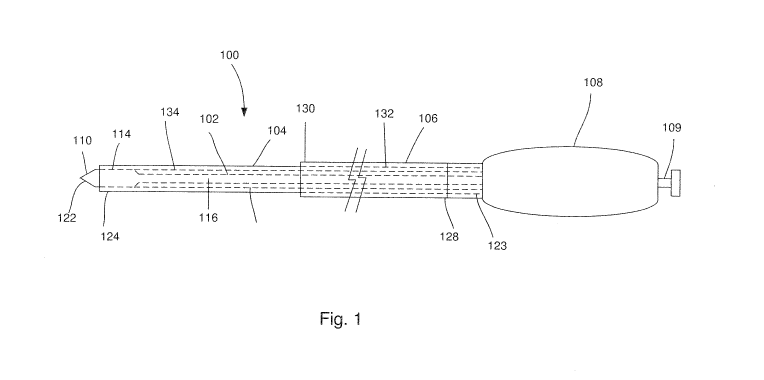
[0016] Fig. 1 shows a longitudinal cross-sectional view of a system according
to an exemplary
embodiment of the present disclosure;
Fig. 2 shows a longitudinal cross-sectional view of a distal portion of a
stylet of the
assembly of Fig. 1;
Fig. 3 shows a side view of an access sheath according to the system of Fig.
1;
Fig. 4 shows a side view of an access sheath according to another exemplary
embodiment of the present disclosure;
Fig. 5 shows a side view of an access sheath according to yet another
exemplary
3
CA 02954633 2017-01-09
WO 2016/010787
PCT/US2015/039554
embodiment of the present disclosure; and
Fig. 6 shows a perspective view of a handle assembly of the system of Fig. 1.
Detailed Description
[0017] The present disclosure may be further understood with reference to the
following
description and the appended drawings, wherein like elements are referred to
with the same
reference numerals. The present disclosure is directed to endoscopic medical
devices and, in
particular, relate to endoscopic ultrasound (EUS) guided drainage. Exemplary
embodiments
describe a EUS guided drainage systems comprising a stylet for injecting a
fluid into a fluid-
filled duct, an access sheath through which the stylet is inserted and a
dilating sheath for dilating
the fluid-filled duct to facilitate drainage. It will be understood by those
of skill in the art that the
system and method of the present disclosure may be used to drain, for example,
a bile duct, a
pancreatic duct, cysts, gallbladder, etc. It should be noted that the terms
"proximal" and "distal"
as used herein are intended to refer to a direction toward (proximal) and away
from (distal) a user
of the device.
[00181 As shown in Figs. 1 - 6, a system 100 according to an exemplary
embodiment of the
present disclosure comprises a stylet 102 for puncturing a fluid-filled tract
and injecting a fluid
(e.g., contrast media) thereinto and an access sheath 104 for providing access
into the fluid-filled
tract. The system 100 further comprises a dilating sheath 106 for dilating the
tract to facilitate
drainage. The system 100 is sized and shaped to be passed through a working
channel of an
endoscope to be visualized under ultrasound guidance. The system 100 may
further comprise a
handle assembly 108, which remains outside of a living body while the stylet
102 and the access
sheath 104 are inserted therein (e.g., along a body lumen accessed via a
naturally occurring body
orifice). The handle assembly 108 permits the stylet 102 to be removed
therefrom while the
access sheath 104 remains in the target duct. The handle assembly 108 also
includes an actuator
for advancing the dilating sheath 106 over the access sheath 104 and into the
target duct.
4
CA 02954633 2017-01-09
WO 2016/010787
PCT/US2015/039554
[0019] As shown in Fig. 2, a stylet 102 extends along a longitudinal axis from
a proximal end
109 to a distal end 110 and includes a channel 112 extending therethrough. The
stylet 102 may be
formed of a flexible material so that the stylet 102 may be passed along
tortuous paths, for
example, along a natural body lumen. In one exemplary embodiment, the stylet
102 may formed
of nitinol for both flexibility and superelasticity. It will be understood by
those of skill in the art,
however, that the stylet 102 may be formed of any of a variety of flexible
materials. The distal
end 110 includes a tapered distal tip 122 for puncturing the target duct. A
distal portion 114 of
the stylet 102 may have a larger diameter than a proximal portion 116 of the
stylet 102 extending
proximally therefrom. A length of the distal portion 114 of the stylet 102 may
range from
between 0.3 cm and 5.0 cm, and, in particular, may be about 1 cm. A proximal
portion 118 of
the channel 112 extends through the proximal portion 116 of the stylet 102
along the longitudinal
axis thereof while a distal portion 120 of the channel 112 extending through
the distal portion
114 of the stylet 102 is defined by an annular space extending about the
longitudinal axis of the
stylet 102. A fluid such as, for example, a contrast media, may be injected
into the target duct
via the channel 112 to verify that the target duct is filled with fluid (e.g.,
digestive fluid).
[0020] As shown in Fig. 1, the access sheath 104 extends longitudinally from a
proximal end
123 to a distal end 124 and includes a lumen 134 extending therethrough. The
lumen 134 is
sized and shaped to slidably receive the stylet 102 therein. In particular, an
inner diameter of the
lumen 134 in this embodiment substantially corresponds to an outer diameter of
the distal portion
114 of the stylet 102 so that when the stylet 102 is received therein, the
distal portion 114
completely fills the lumen 134 of the access sheath 104 to facilitate
puncturing of the target duct
when the access sheath 104, with the stylet 102 received therein, is inserted
into the target duct.
As shown in Fig. 3, the access sheath 104 may be biased to assume a desired a
curvature along a
distal portion 126 thereof. In one exemplary embodiment, the distal portion
126 of the access
sheath 104 is biased toward a pigtail configuration particularly suited for
stabilizing the access
sheath 104 in the target duct. In another exemplary embodiment, as shown in
Fig. 4, a distal
5
CA 02954633 2017-01-09
WO 2016/010787
PCT/US2015/039554
portion 126' of an access sheath 104' has a shepherd's crook configuration
(i.e., a curve which
directs the distal end of the sheath 104' back toward more proximal portions
thereorf) particularly
suited for directing a guidewire in a desired direction within the target
duct. In yet another
exemplary embodiment, as shown in Fig. 5, a distal portion 126" of an access
sheath 104" is
biased toward a J-shaped configuration (i.e., a curve in which the distal
portion 126" arcs away
from an axis of more proximal portions of the sheath 104"along an arc of 90
or less) for
directing a guidewire in another desired direction in the target duct.
[00211] The access sheath 104 may be formed of a polymeric material that is
sufficiently
flexible so that when the stylet 102 is received therein, the distal portion
126 of the access sheath
104 is straightened. Once the stylet 102 is removed therefrom, however, the
distal portion 126 of
the access sheath 104 is permitted to revert to its curved configuration. In
an exemplary
embodiment, the access sheath 104 is formed of braid reinforced polyamide. In
another
embodiment, the access sheath 104 is formed of multiple layers such as, for
example, PTFE,
braids, polyether block amide for kink resistance.
[0022] The dilating sheath 106 similarly extends longitudinally from a
proximal end 128 to a
distal end 130 and includes a lumen 132 extending therethrough. The lumen 132
is sized and
shaped to slidably receive the access sheath 104 therein so that the dilating
sheath 106 may be
advanced over the access sheath 104 to the target duct to dilate the
obstructed duct, thereby
facilitating drainage thereof. The dilating sheath 106 may be a cold dilator
such as, for example,
a sohendra type dilator and/or a balloon dilator. Alternatively, the dilating
sheath 106 may be a
hot dilator such as, for example, a cystome or needleknife, which includes
electrosurgical
capabilities. For example, the dilating sheath 106 may include an electrode
along the distal end
130 thereof for cauterizing tissue. In particular, the dilating sheath 106 may
be configured to
utilize electrosurgical dissection to facilitate dilation or to burn a lesion
as the dilating sheath 106
is inserted into the target duct. In embodiments in which the dilating sheath
106 includes an
electrode, the sheath 106 may include a second lumen (not shown) extending
therethrough for
6
CA 02954633 2017-01-09
WO 2016/010787
PCT/US2015/039554
carrying power to the electrode. The distal end 130 of the dilating sheath
106, however, may
have any of a variety of configurations facilitating insertion into the target
duct. In another
example, the distal end 130 may be tapered. Once the dilating sheath 106 is
advanced over the
access sheath 104 and inserted into the target duct, the dilating sheath 106
may be actuated to
dilate or expand the target duct. For example, the dilating sheath 106 may
have one or more
stepped diameters at discrete distances from the distal end or one or more
additional sheaths that
may be independently actuated to expand the path to the target duct.
[0023] As shown in Fig. 6, the handle assembly 108 includes a grip portion 136
extending from
a proximal end 138 to a distal end 140 and an extension portion 142 coupled to
the distal end 140
of the grip portion 136 and couplable to the proximal end 128 of the dilating
sheath 106. The
access sheath 104 may be received within and coupled to the grip portion 136
such that the
access sheath 104 extends through the lumen 132 of the dilating sheath 106.
The stylet 102
extends through the grip portion 136 and the extension portion 142 with the
proximal end of the
stylet 102 extending proximally of the proximal end 138 of the grip portion
and length of the
stylet 102 extending through the lumen 134 of the access sheath 104. Since the
proximal end
109 of the stylet 102 extends proximally from the grip portion 136, the stylet
102 may be
removed from the access sheath 104 by simply pulling the stylet 102 proximally
relative to the
handle assembly 108. The distal end 110 of the stylet 102 extends distally
past the distal end 124
of the access sheath 104 so that the tapered tip 122 may puncture the target
duct once the system
100 has been inserted into the body. The handle assembly 108 also includes an
actuator 144
which moves the dilating sheath 106 longitudinally relative to the access
sheath 104. In
particular, the actuator 144 may include a tab that is moved distally and
proximally with respect
to the grip portion 136 of the handle assembly 108 to advance and retract,
respectively, the
dilating sheath 106 over the access sheath 104.
[0024] According to a method using the system 100 according to an exemplary
embodiment of
the present disclosure, the system 100 is inserted through a working channel
of an endoscope via
7
CA 02954633 2017-01-09
WO 2016/010787
PCT/US2015/039554
ultrasound guidance to a target duct within the body. In an insertion
configuration, the access
sheath 104 may be fully housed within the dilating sheath 106 to protect the
endoscope through
which the system 100 is inserted from the sharp distal tip 122 of the stylet
102. Upon insertion
through the endoscope, the dilating sheath 106 may be retracted so that the
dilating sheath 106
does not extend over the portion of the access sheath 104 being inserted into
the target duct. At
this point, the distal end 110 of the stylet 102 extends distally past the
distal end 124 of the
access sheath 104. The distal tip 122 of the stylet 102 is then advanced
distally to penetrate the
target duct. Once the stylet 102 and the access sheath 104 have been inserted
into the target duct,
a contrast media (e.g., radiopaque dye) is inserted through the channel 112 of
the stylet 102 into
the target duct so that a user of the system 100 may visually verify that the
duct has been filled
with fluid and requires drainage. The stylet 102 may then be removed from the
access sheath
104 by drawing the stylet 102 proximally relative to the access sheath 104 so
that only the access
sheath 104 remains in the target duct. Upon removal of the stylet 102, the
distal portion 126 of
the access sheath 104 is freed to revert to the curved configuration to either
anchor the access
sheath 104 in the target duct or to direct a guidewire therethrough in a
desired direction. If the
access sheath 104 is not anchored in the target duct, a guidewire may be
inserted through the
lumen 134 of the access sheath 104 and into the target duct. A tip of the
guidewire is directed in
a direction corresponding to a curvature of the distal portion 126 of the
access sheath 104 to
contact an interior surface of the target duct to anchor the access sheath 104
thereto. The access
sheath 104 may be rotated by manipulating a portion of the handle assembly 108
to direct the
curved configuration in a desired direction.
[0025] Once the access sheath 104 has been anchored in the target duct, the
dilating sheath 106
is advanced over the access sheath 104 into the target duct. As described
above, the dilating
sheath 106 is advanced by moving the actuator 144 distally with respect to the
grip portion 136
of the handle assembly 108. The distal end 130 of the dilating sheath 106 is
configured to
facilitate insertion of the dilating sheath 106 into the target duct. In one
embodiment, an
electrode at the distal end 130 is activated to electrosurgically dissect
and/or cauterize a surface
8
CA 02954633 2017-01-09
WO 2016/010787
PCT/US2015/039554
tissue of the target duct to facilitate insertion therein. The dilating sheath
106 may be activated
to dilate the target duct, enlarging the duct beyond an obstruction thereof to
permit drainage of
the target duct. It will be understood by those of skill in the art that the
dilating sheath 106 may
dilate the target duct in any of a number of ways. In one example, the
dilating sheath 106 may
include an expansible balloon activated to expand the target duct. It will be
understood by those
of skill in the art that a user may also implement further treatment of the
blocked duct. In
particular, a stent may be inserted into the target into the target duct,
maintaining the duct in an
enlarged configuration to ensure continued drainage thereof
[0026] It will be apparent to those skilled in the art that various
modifications may be made in
the present disclosure, without departing from the scope of the disclosure.
Thus, it is intended
that the present disclosure cover the modifications and variations of his
disclosure provided that
they come within the scope of the appended claims and their equivalents.
9