Note: Descriptions are shown in the official language in which they were submitted.
CA 02955992 2017-01-20
WO 2016/012864 PCT/1B2015/001804
BIOMARKERS FOR ANDERSON-FABRY DISEASE
CROSS-REFERENCE
[0001] This application claims the benefit of U.S. Provisional Application
No. 62/028,225,
filed July 23, 2014, entitled "BIOMARKERS FOR ANDERSON-FABRY DISEASE," the
entire disclosure of which is hereby incorporated herein by reference for all
purposes.
REFERENCE TO A "SEQUENCE LISTING," A TABLE, OR A COMPUTER
PROGRAM LISTING APPENDIX SUBMITTED AS AN ASCII TEXT FILE
[0002] The Sequence Listing written in file 97513_951211.TXT, created on
July 22, 2015,
2,641 bytes, machine format IBM-PC, MS-Windows operating system, is hereby
incorporated
by reference in its entirety for all purposes.
BACKGROUND
[0003] Anderson-Fabry disease (AFD) is an X-linked lysosomal storage
disorder caused by
mutations in the GLA gene encoding the enzyme a-galactosidase A (a-GalA).1
Deficiencies in
a-GalA activity cause globotriaosylceramide (Gb3) to accumulate, and lead to
progressive
multisystem disease. Historical estimates of AFD prevalence were very low, but
these have
recently been recognized as underestimates in the context of multiple large-
scale metabolic and
genetic screening studies in Asia and Europe, wherein a high prevalence of
mutations
associated with late-onset or variant AFD phenotypes have been observed.2-5
Clinical
manifestations of AFD may be non-specific, and, due to its rarity, other
conditions are initially
suspected over AFD, such that a correct diagnosis may be delayed until after
irreversible end-
organ damage has occurred.1 Anderson-Fabry cardiomyopathy is the most common
cause of
death in AFD patients, followed by renal complications, which together
highlight the need for
improved diagnosis and treatment.6
[0004] Biomarker identification represents an expanding activity in AFD
research that have
the promise of addressing the present limitations to effective care that exist
in delayed
diagnoses.7 In addition to increasing diagnostic efficiency, biomarkers may
offer prognostic
information, or act as surrogates to monitor the effectiveness of a given
treatment.8' 9 Whole
blood, plasma and serum samples from peripheral veins offer a minimally-
invasive output that
reflects changes in various end-organs. In concert with techniques capable of
capturing low
abundance molecules, such as mass spectrometry, diagnostic algorithms may be
substantially
improved. Typically, the diagnosis of AFD is made based on a-GalA activity
levels in
peripheral blood or plasma; however, this method is unreliable in the case of
variant or late-
1
CA 02955992 2017-01-20
WO 2016/012864 PCT/1B2015/001804
onset cases, and frequently misses the AFD diagnosis in females.1 In order to
account for this,
females with suspected AFD must be genetically tested to confirm the presence
of a mutation
associated with AFD.10' 11 Multiple lines of evidence, however, show that
genetic testing is
itself hindered by ambiguities, which further underscores the need for
reliable, gender-specific
biomarkers to enhance the current diagnostic algorithm.12
[0005] The methods and compositions of the present invention help to
satisfy these and
other needs for such tests.
SUMMARY
[0006] Disclosed herein are compositions and methods for determining
Anderson-Fabry
Disease in a subject using biomarkers from a sample derived from the subject.
[0007] In a first aspect, disclosed herein is a method for diagnosing
Anderson-Fabry
Disease (AFD) in a male subject, comprising: obtaining a dataset associated
with a sample
obtained from the male subject, wherein the dataset comprises at least one
marker selected
from Table 2; analyzing the dataset to determine data for the markers, wherein
the data is
positively correlated or negatively correlated with a diagnosis of Anderson-
Fabry Disease in
the male subject.
[0008] In an embodiment, the dataset comprises data for at least two,
three, four, five, six,
seven, or eight markers. In another embodiment, the method further comprises
determining the
diagnosis of Anderson-Fabry Disease in the subject according to the relative
number of
positively correlated and negatively correlated marker expression level data
present in the
dataset.
[0009] In a second aspect, disclosed herein is a method for diagnosing
Anderson-Fabry
Disease (AFD) in a female subject, comprising: obtaining a dataset associated
with a sample
obtained from the female subject, wherein the dataset comprises at least one
marker selected
from Table 4; analyzing the dataset to determine data for the markers, wherein
the data is
positively correlated or negatively correlated with a diagnosis of Anderson-
Fabry Disease in
the female subject.
[0010] In an embodiment, the dataset comprises data for at least two,
three, four, five, six,
seven, eight or nine markers. In another embodiment, the method further
comprises
determining the diagnosis of Anderson-Fabry Disease in the subject according
to the relative
number of positively correlated and negatively correlated marker expression
level data present
in the dataset.
2
CA 02955992 2017-01-20
WO 2016/012864 PCT/1B2015/001804
[0011] In various embodiments of the above aspects, the sample obtained
from the subject
is a blood sample. In various embodiments of the above aspects, the data is
protein expression
data. In various embodiments of the above aspects, the protein expression data
is obtained
using mass spectrometry or other methods
[0012] In various embodiments of the above aspects, the method is
implemented using one
or more computers. In various embodiments of the above aspects, the dataset is
obtained
stored on a storage memory.
[0013] In various embodiments of the above aspects, obtaining the dataset
comprises
receiving the dataset directly or indirectly from a third party that has
processed the sample to
experimentally determine the dataset.
[0014] In various embodiments of the above aspects, the subject is a human
subject.
[0015] In various embodiments of the above aspects, the method further
comprises
assessing a clinical variable; and combining the assessment with the analysis
of the dataset to
diagnose Anderson-Fabry Disease (AFD) in the subject.
[0016] In a third aspect, disclosed herein is a method for predicting the
likelihood of
Anderson-Fabry Disease in a subject, comprising: obtaining a sample from a
male subject,
wherein the sample comprises at least one marker selected from Table 2, or
obtaining a sample
from a female subject, wherein the sample comprises at least one marker
selected from Table 4;
measuring proteins in the sample, wherein the dataset comprises protein
abundance data for the
markers; and analyzing the protein level data for the markers, wherein the
abundance of the
markers is positively correlated or negatively correlated with a diagnosis of
Anderson-Fabry
Disease in the subject.
[0017] In a fourth aspect, disclosed herein is a computer-implemented
method for
diagnosing Anderson-Fabry Disease in a subject, comprising: storing, in a
storage memory, a
dataset associated with a sample obtained from a male subject, wherein the
dataset comprises
data for at least one marker selected from Table 2, or storing, in a storage
memory, a dataset
associated with a sample obtained from a female subject, wherein the dataset
comprises data
for at least one marker selected from Table 4; and analyzing, by a computer
processor, the
dataset to determine the abundance of the markers, wherein the protein
abundance is positively
correlated or negatively correlated with a diagnosis of Anderson-Fabry Disease
in the subject.
[0018] In a fifth aspect, disclosed herein is a system for diagnosing
Anderson-Fabry
Disease in a subject, the system comprising: a storage memory for storing a
dataset associated
with a sample obtained from a male subject, wherein the dataset comprises data
for at least one
3
CA 02955992 2017-01-20
WO 2016/012864
PCT/1B2015/001804
marker selected from Table 2, or a storage memory for storing a dataset
associated with a
sample obtained from a female subject, wherein the dataset comprises data for
at least one
marker selected from Table 4; and a processor communicatively coupled to the
storage
memory for analyzing the dataset to determine the abundance of the markers,
wherein the
protein abundance are positively correlated or negatively correlated with a
diagnosis of
Anderson-Fabry Disease in the subject.
[0019] In a sixth aspect, disclosed herein is a computer-readable storage
medium storing
computer-executable program code, the program code comprising: program code
for storing a
dataset associated with a sample obtained from a male subject, wherein the
dataset comprises
data for at least one marker selected from Table 2, or a storage memory for
storing a dataset
associated with a sample obtained from a female subject, wherein the dataset
comprises data
for at least one marker selected from Table 4; and program code for analyzing
the dataset to
determine the abundance of the markers, wherein the levels of the markers are
positively
correlated or negatively correlated with a diagnosis of Anderson-Fabry Disease
in the subject.
[0020] In a seventh aspect, disclosed herein is a kit for use in diagnosing
Anderson-Fabry
Disease (AFD) in a subject, comprising: a set of reagents comprising a
plurality of reagents for
determining from a sample obtained from the subject data for at least one
marker selected from
Table 2 or 4; and instructions for using the plurality of reagents to
determine data from the
samples. In some embodiments, the data is expression level data from the
samples. In some
embodiments, the data is protein abundance data.
[0021] In various embodiments of the above, the analyzing step further
comprises applying
an interpretation function to the dataset for said markers to generate a
score, wherein said score
is indicative of the subject's Anderson-Fabry Disease (AFD) status.
[0022] In one embodiment, the interpretation function, if the subject is
male, is: score =
1.62+ 1.56 x A + 0.50 x B -0.15 x C - 0.26 x D - 0.36 x E - 0.49 x F - 0.67 x
G - 1.31 x H,
where A is Alpha 1 antichymotrypsin; B is Isoform 1 of Sex hormone-binding
globulin; C is
Hemoglobin alpha-2; D is 22 kDa protein; E is Peroxiredoxin 2; F is
Apolipoprotein E; G is
Afamin; and H is Beta Ala His dipeptidase, and where the score cut-off is
0.54.
[0023] In another embodiment, the interpretation function, if the subject
is female, is:
score =1¨
l_h e-2.05x(-0.49+0.72xa+0.30xb+ 0.25xc+0.14xd+0.13xe+0.11xf-0.03xg-0.24xh-
0.6xi)+0.142
4
CA 02955992 2017-01-20
WO 2016/012864 PCT/1B2015/001804
where a is Apolipoprotein E; b is Isoform 1 of Gelsolin; c is Kallistatin; d
is Peroxiredoxin 2; e
is Hemoglobin alpha-2; f is Paraoxonase PON 1; g is Protein Z-dependent
protease inhibitor; h
is Pigment epithelium-derived factor; and I is Actin, alpha cardiac muscle 1,
and where the
score cut-off is 0.51.
BRIEF DESCRIPTION OF THE DRAWINGS
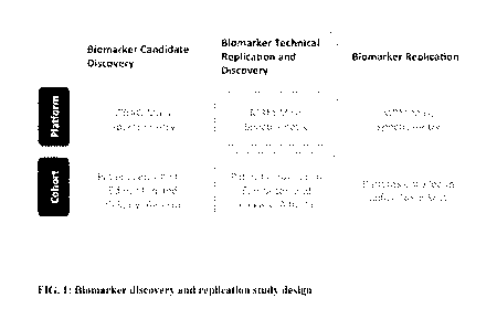
[0024] Figure 1. Biomarker discovery and replication study design.
[0025] Figures 2A-2D. Performance of the AFD biomarkers in the discovery
and
replication cohorts. FIG. 2A. Red dots indicate the biomarker score, based on
the 8-protein
biomarker panel, of all discovery Anderson-Fabry disease (AFD) patients on the
left and all
replication AFD patients on the right. The dark blue dots show the biomarker
score of the
healthy control (HC) individuals. The average biomarker score is shown with
red and dark blue
line for the AFD and HC subjects, respectively. The dotted line corresponds to
the biomarker
score cut-off of 0.54 for differentiating between AFD and HC subjects. FIG.
2B. The black
line shows the receiver operating characteristics (ROC) curve for the
discovery subjects while
the green lines corresponds to the replication subjects' ROC curve. AUC stands
for area under
the ROC curve. FIG. 2C. The biomarker score is shown for the male subjects
only and it
illustrates how well the AFD and HC subjects separate in the discovery and
replication cohorts.
FIG. 2D. The ROC curve for the male subjects with the black and green lines
corresponding to
the discovery and replication ROC curves, respectively.
[0026] Figures 3A-3B. Performance of the female-specific AFD biomarkers in
the
discovery and replication cohorts. FIG. 3A. Red dots indicate the biomarker
score, based on
the 9-protein female-specific biomarker panel for the discovery Anderson-Fabry
disease (AFD)
patients who have not received enzyme replacement therapy, on the left, and
female replication
AFD patients on the right. The dark blue dots show the biomarker score of the
healthy control
(HC) individuals. The average biomarker score is shown with red and dark blue
line for the
AFD and HC female subjects, respectively. The dotted line corresponds to the
biomarker score
cut-off of 0.51 for differentiating between FD and HC subjects. FIG. 3B. The
black line shows
the receiver operating characteristics (ROC) curve for the discovery subjects
while the green
lines corresponds to the replication subjects' ROC curve. AUC stands for area
under the ROC
curve.
CA 02955992 2017-01-20
WO 2016/012864 PCT/1B2015/001804
DETAILED DESCRIPTION
[0027] Anderson-Fabry disease (AFD) is an important X-linked metabolic
disease resulting
in progressive central nervous system, renal and cardiac diseases with a
gender-dependent
phenotype. Recent epidemiologic screening for AFD suggests a prevalence of
1:3000.
[0028] As disclosed in greater detail herein, we disclose a mass
spectrometry-based
proteomic screen for novel plasma biomarkers in a cohort of AFD patients in
comparison to
matched healthy controls, and a subsequent replication study in a separate
cohort of AFD
patients. We further identify gender-specific biomarkers panels, which may
lead to
improvements in diagnosing challenging cases, such as most AFD-affected
females, and
variant or late-onset phenotype males.
[0029] Specifically, we used an unbiased screening proteomic approach to
discover novel
plasma biomarker signatures in adult patients with AFD. In discovery and
validation cohorts,
we used a mass spectrometry iTRAQ proteomic approach followed by multiple
reaction
monitoring (MRM) assays, to identify biomarkers. Of the 38 protein groups
discovered by
iTRAQ, 18 already had existing MRM assays, and we identified an eight-
candidate biomarker
panel (a 22 kDa protein, afamin, alpha 1 antichyotrypsin, apolipoprotein E, 3-
Ala His
dipeptidase, hemoglobin a-2, isoform 1 of sex hormone-binding globulin and
peroxiredoxin 2)
which was very specific and sensitive for male AFD patients. In female AFD
patients, we
identified a nine-marker panel of proteins with only 3 proteins,
apolipoprotein E, hemoglobin
a-2 and peroxiredoxin 2, common to both genders, suggesting a gender-specific
alteration in
plasma biomarkers in patients with AFD.
[0030] Thus, disclosed herein are gender-specific plasma protein biomarker
panels that are
specific and sensitive for the AFD phenotype. The gender-specific panels offer
important
insight into potential differences in pathophysiology and prognosis between
males and females.
[0031] These and other features of the present teachings will become more
apparent from
the description herein. While the present teachings are described in
conjunction with various
embodiments, it is not intended that the present teachings be limited to such
embodiments. On
the contrary, the present teachings encompass various alternatives,
modifications, and
equivalents, as will be appreciated by those of skill in the art.
[0032] Most of the words used in this specification have the meaning that
would be
attributed to those words by one skilled in the art. Words specifically
defined in the
specification have the meaning provided in the context of the present
teachings as a whole, and
as are typically understood by those skilled in the art. In the event that a
conflict arises
6
CA 02955992 2017-01-20
WO 2016/012864 PCT/1B2015/001804
between an art-understood definition of a word or phrase and a definition of
the word or phrase
as specifically taught in this specification, the specification shall control.
[0033] It must be noted that, as used in the specification and the appended
claims, the
singular forms "a," "an," and "the" include plural referents unless the
context clearly dictates
otherwise.
[0034] Terms used in the claims and specification are defined as set forth
below unless
otherwise specified.
[0035] The term "status" of Anderson-Fabry disease (AFD) or "AFD status" as
used herein
refers to the status or extent of AFD in a subject. In some contexts, AFD
status may be
referred to as "significant", "non-significant", or "possible" AFD.
[0036] "Marker" or "markers" or "biomarker," "biomarkers," refers generally
to a
molecule (typically protein, carbohydrate, lipid, or nucleic acid) that is
expressed in cell or
tissue, which is useful for the diagnosis of AFD. A marker in the context of
the present
teachings encompasses, for example, without limitation, cytokines, chemokines,
growth
factors, proteins, peptides, nucleic acids, oligonucleotides, and metabolites,
together with their
related metabolites, mutations, variants, polymorphisms, modifications,
fragments, subunits,
degradation products, elements, and other analytes or sample-derived measures.
In the case of
a nucleic acid, a marker can include any allele, including wild-types alleles,
SNPs,
microsatellites, insertions, deletions, duplications, and translocations. A
marker can also
include a peptide encoded by a nucleic acid. Markers can also include mutated
proteins,
mutated nucleic acids, variations in copy numbers and/or transcript variants.
Markers also
encompass non-blood borne factors and non-analyte physiological markers of
health status,
and/or other factors or markers not measured from samples (e.g., biological
samples such as
bodily fluids), such as clinical parameters and traditional factors for
clinical assessments.
Markers can also include any indices that are calculated and/or created
mathematically.
Markers can also include combinations of any one or more of the foregoing
measurements,
including temporal trends and differences.
[0037] To "analyze" includes measurement and/or detection of data
associated with a
marker (such as, e.g., presence or absence of a protein, or nucleic acid
sequence, or constituent
expression levels) in the sample (or, e.g., by obtaining a dataset reporting
such measurements,
as described below). In some aspects, an analysis can include comparing the
measurement
and/or detection of at least one marker in samples from a subject pre- and
post-treatment or
7
CA 02955992 2017-01-20
WO 2016/012864 PCT/1B2015/001804
other control subject(s). The markers of the present teachings can be analyzed
by any of
various conventional methods known in the art.
[0038] A "subject" in the context of the present teachings is generally a
mammal. The
subject is generally a patient. The term "mammal" as used herein includes but
is not limited to
a human, non-human primate, dog, cat, mouse, rat, cow, horse, and pig. Mammals
other than
humans can be advantageously used as subjects that represent animal models of
heart
transplantion. A subject can be male or female.
[0039] A "sample" in the context of the present teachings refers to any
biological sample
that is isolated from a subject. A sample can include, without limitation, a
single cell or
multiple cells, fragments of cells, an aliquot of body fluid, whole blood,
platelets, serum,
plasma, red blood cells, white blood cells or leucocytes, endothelial cells,
tissue biopsies,
synovial fluid, lymphatic fluid, ascites fluid, and interstitial or
extracellular fluid. The term
"sample" also encompasses the fluid in spaces between cells, including
gingival crevicular
fluid, bone marrow, cerebrospinal fluid (CSF), saliva, mucous, sputum, semen,
sweat, urine, or
any other bodily fluids. "Blood sample" can refer to whole blood or any
fraction thereof,
including blood cells, red blood cells, white blood cells or leucocytes,
platelets, serum and
plasma. Samples can be obtained from a subject by means including but not
limited to
venipuncture, excretion, ejaculation, massage, biopsy, needle aspirate,
lavage, scraping,
surgical incision, or intervention or other means known in the art.
[0040] In particular aspects, the sample is a blood sample from the
subject.
[0041] A "dataset" is a set of data (e.g., numerical values) resulting from
evaluation of a
sample. The values of the dataset can be obtained, for example, by
experimentally obtaining
measures from a sample and constructing a dataset from these measurements; or
alternatively,
by obtaining a dataset from a service provider such as a laboratory, or from a
database or a
server on which the dataset has been stored. Similarly, the term "obtaining a
dataset associated
with a sample" encompasses obtaining a set of data determined from at least
one sample.
Obtaining a dataset encompasses obtaining a sample, and processing the sample
to
experimentally determine the data, e.g., via measuring, mass spectrometry,
antibody binding,
ELISA, PCR, microarray, one or more primers, or one or more probes. The phrase
also
encompasses receiving a set of data, e.g., from a third party that has
processed the sample to
experimentally determine the dataset. Additionally, the phrase encompasses
mining data from
at least one database or at least one publication or a combination of
databases and publications.
8
CA 02955992 2017-01-20
WO 2016/012864 PCT/1B2015/001804
[0042] "Measuring" or "measurement" in the context of the present teachings
refers to
determining the presence, absence, quantity, amount, or effective amount of a
marker or other
substance (e.g., protein or nucleic acid) in a clinical or subject-derived
sample, including the
presence, absence, or concentration levels of such markers or substances,
and/or evaluating the
values or categorization of a subject's clinical parameters.
[0043] The term "expression level data" refers to a value that represents a
direct, indirect,
or comparative measurement of the level of expression of a polypeptide or
polynucleotide (e.g.,
RNA or DNA). For example, "expression data" can refer to a value that
represents a direct,
indirect, or comparative measurement of the protein expression level of a
proteomic marker of
interest. In some embodiments, this measurement is performed by measuring
protein
concentration or protein level as described herein.
Markers and Clinical Factors
[0044] The quantity of one or more markers of the invention can be
indicated as a value. A
value can be one or more numerical values resulting from evaluation of a
sample under a
condition. The values can be obtained, for example, by experimentally
obtaining measures
from a sample by an assay performed in a laboratory, or alternatively,
obtaining a dataset from
a service provider such as a laboratory, or from a database or a server on
which the dataset has
been stored, e.g., on a storage memory.
[0045] In an embodiment, the quantity of one or more markers can be one or
more
numerical values associated with expression levels of one or more of the
markers of Tables 2
or 4 resulting from evaluation of a sample.
[0046] In an embodiment, a marker's associated value can be included in a
dataset
associated with a sample obtained from a subject. A dataset can include the
marker expression
value of two or more, three or more, four or more, five or more, six or more,
seven or more,
eight or more, or nine marker(s). For example, a dataset can include the
expression values for
one or more of the markers of Tables 2 or 4.
[0047] In an embodiment, a clinical factor can be included within a
dataset. A dataset can
include one or more, two or more, three or more, four or more, five or more,
six or more, seven
or more, eight or more, nine or more, ten or more, eleven or more, twelve or
more, thirteen or
more, fourteen or more, fifteen or more, sixteen or more, seventeen or more,
eighteen or more,
nineteen or more, twenty or more, twenty-one or more, twenty-two or more,
twenty-three or
more, twenty-four or more, twenty-five or more, twenty-six or more, twenty-
seven or more,
twenty-eight or more, twenty-nine or more, or thirty or more overlapping or
distinct clinical
9
CA 02955992 2017-01-20
WO 2016/012864 PCT/1B2015/001804
factor(s). A clinical factor can be, for example, the condition of a subject
in the presence of a
disease or in the absence of a disease, e.g., AFD. Alternatively, or in
addition, a clinical factor
can be the health status of a subject. Alternatively, or in addition, a
clinical factor can be age,
gender, clinical characteristics, organ function, functional status,
morphologic characteristics,
and quality of life assessments.
[0048] In another embodiment, the invention includes obtaining a sample
associated with a
subject, where the sample includes one or more markers. The sample can be
obtained by the
subject or by a third party, e.g., a medical professional. Examples of medical
professionals
include physicians, emergency medical technicians, nurses, first responders,
psychologists,
medical physics personnel, nurse practitioners, surgeons, dentists, and any
other obvious
medical professional as would be known to one skilled in the art. A sample can
include
peripheral blood cells, isolated leukocytes, or RNA extracted from peripheral
blood cells or
isolated leukocytes. The sample can be obtained from any bodily fluid, for
example, amniotic
fluid, aqueous humor, bile, lymph, breast milk, interstitial fluid, blood,
blood plasma, cerumen
(earwax), Cowper's fluid (pre-ejaculatory fluid), chyle, chyme, female
ejaculate, menses,
mucus, saliva, urine, vomit, tears, vaginal lubrication, sweat, serum, semen,
sebum, pus, pleural
fluid, cerebrospinal fluid, synovial fluid, intracellular fluid, and vitreous
humour. In an
example, the sample is obtained by a blood draw, where the medical
professional draws blood
from a subject, such as by a syringe. The bodily fluid can then be tested to
determine the value
of one or more markers using an assay. The value of the one or more markers
can then be
evaluated by the same party that performed the assay using the methods of the
invention or sent
to a third party for evaluation using the methods of the invention.
[0049] In some embodiments, one or more clinical factors in a subject can
be assessed. In
some embodiments, assessment of one or more clinical factors or variables in a
subject can be
combined with a marker analysis in the subject to diagnose AFD in a subject.
Assays
[0050] Techniques, methods, tools, algorithms, reagents and other necessary
aspects of
assays that may be employed to detect and/or quantify a particular marker or
set of markers are
varied. Of significance is not so much the particular method used to detect
the marker or set of
markers, but what markers to detect. As is reflected in the literature,
tremendous variation is
possible. Once the marker or set of markers to be detected or quantified is
identified, any of
several techniques may be well suited, with the provision of appropriate
reagents. One of skill
in the art, when provided with the set of markers to be identified, will be
capable of selecting
CA 02955992 2017-01-20
WO 2016/012864 PCT/1B2015/001804
the appropriate assay (for example, an ELISA, protein or antibody microarray
or similar
immunologic assay, or in some examples, use of an iTRAQ, iCAT, SELDI, or MRM-
MS
proteomic mass spectrometric based method, or a PCR based or a microarray
based assay for
nucleic acid markers) for performing the methods disclosed herein.
[0051] Proteins, protein complexes, or proteomic markers may be
specifically identified
and/or quantified by a variety of methods known in the art and may be used
alone or in
combination. Immunologic- or antibody-based techniques include enzyme-linked
immunosorbent assay (ELISA), radioimmunoassay (RIA), western blotting,
immunofluorescence, microarrays, some chromatographic techniques (i.e.
immunoaffinity
chromatography), flow cytometry, immunoprecipitation and the like. Such
methods are based
on the specificity of an antibody or antibodies for a particular epitope or
combination of
epitopes associated with the protein or protein complex of interest. Non-
immunologic methods
include those based on physical characteristics of the protein or protein
complex itself
Examples of such methods include electrophoresis, some chromatographic
techniques (e.g.
high performance liquid chromatography (HPLC), fast protein liquid
chromatography (FPLC),
affinity chromatography, ion exchange chromatography, size exclusion
chromatography and
the like), mass spectrometry, sequencing, protease digests, and the like. Such
methods are
based on the mass, charge, hydrophobicity or hydrophilicity, which is derived
from the amino
acid complement of the protein or protein complex, and the specific sequence
of the amino
acids. Exemplary methods include those described in, for example, PCT
Publication WO
2004/019000, WO 2000/00208, US 6670194. Immunologic and non-immunologic
methods
may be combined to identify or characterize a protein or protein complex.
Furthermore, there
are numerous methods for analyzing/detecting the products of each type of
reaction (for
example, fluorescence, luminescence, mass measurement, electrophoresis, etc.).
Furthermore,
reactions can occur in solution or on a solid support such as a glass slide, a
chip, a bead, or the
like.
[0052] Methods of producing antibodies for use in protein or antibody
arrays, or other
immunology based assays are known in the art. Once the marker or markers are
identified and
the amino acid sequence of the protein or polypeptide is identified, either by
querying of a
database or by having an appropriate sequence provided (for example, a
sequence listing as
provide herein), one of skill in the art will be able to use such information
to prepare one or
more appropriate antibodies and perform the selected assay.
11
CA 02955992 2017-01-20
WO 2016/012864 PCT/1B2015/001804
[0053] For preparation of monoclonal antibodies directed towards a
biomarker, any
technique that provides for the production of antibody molecules may be used.
Such techniques
include, but are not limited to, hybridomas or triomas (e.g. Kohler and
Milstein 1975, Nature
256:495-497; Gustafsson et al., 1991, Hum. Antibodies Hybridomas 2:26-32),
human B-cell
hybridoma or EBV hybridomas e.g. (Kozbor et al., 1983, Immunology Today
4:72;;Cole et al.,
1985, In: Monoclonal Antibodies and Cancer Therapy, Alan R. Liss, Inc., pp. 77-
96). Human,
or humanized antibodies may be used and can be obtained by using human
hybridomas (Cote
et al., 1983, Proc. Natl. Acad. Sci. USA 80:2026- 2030) or by transforming
human B cells with
EBV virus in vitro (Cole et al., 1985, In: Monoclonal Antibodies and Cancer
Therapy, Alan R.
Liss, Inc., pp. 77-96). Techniques developed for the production of "chimeric
antibodies"
(Morrison et al., 1984, Proc. Natl. Acad. Sci. USA 81:6851-6855; Neuberger et
al., 1984,
Nature 312:604-608; Takeda et al., 1985, Nature 314:452-454) by splicing a
sequence
encoding a mouse antibody molecule specific for a particular biomarker
together with a
sequence encoding a human antibody molecule of appropriate biological activity
may be used;
such antibodies are within the scope of this invention. Techniques described
for the production
of single chain antibodies (U.S. Patent 4,946,778) may be adapted to produce a
biomarker -
specific antibodies. An additional embodiment of the invention utilizes the
techniques
described for the construction of Fab expression libraries (Huse et al., 1989,
Science 246:1275-
1281) to allow rapid and easy identification of monoclonal Fab fragments with
the desired
specificity for a biomarker proteins. Non-human antibodies can be "humanized"
by known
methods (e.g., U.S. Patent No. 5,225,539).
[0054] Antibody fragments that contain an idiotype of a biomarker can be
generated by
techniques known in the art. For example, such fragments include, but are not
limited to, the
F(ab')2 fragment which can be produced by pepsin digestion of the antibody
molecule; the Fab'
fragment that can be generated by reducing the disulfide bridges of the
F(ab')2 fragment; the
Fab fragment that can be generated by treating the antibody molecular with
papain and a
reducing agent; and Fy fragments. Synthetic antibodies, e.g., antibodies
produced by chemical
synthesis, may also be useful in the present invention.
[0055] Standard reference works described herein and known to those skilled
in the
relevant art describe both immunologic and non-immunologic techniques, their
suitability for
particular sample types, antibodies, proteins or analyses. Standard reference
works setting
forth the general principles of immunology and assays employing immunologic
methods
known to those of skill in the art include, for example: Harlow and Lane,
Antibodies: A
12
CA 02955992 2017-01-20
WO 2016/012864 PCT/1B2015/001804
Laboratory Manual, 2d Ed., Cold Spring Harbor Laboratory Press, Cold Spring
Harbor, N. Y.
(1999); Harlow and Lane, Using Antibodies: A Laboratory Manual. Cold Spring
Harbor
Laboratory Press, New York; Coligan et al. eds. Current Protocols in
Immunology, John Wiley
& Sons, New York, NY (1992-2006); and Roitt et al., Immunology, 3d Ed., Mosby-
Year Book
Europe Limited, London (1993). Standard reference works setting forth the
general principles
of peptide synthesis technology and methods known to those of skill in the art
include, for
example: Chan et al., Fmoc Solid Phase Peptide Synthesis, Oxford University
Press, Oxford,
United Kingdom, 2005; Peptide and Protein Drug Analysis, ed. Reid, R., Marcel
Dekker, Inc.,
2000; Epitope Mapping, ed. Westwood et al., Oxford University Press, Oxford,
United
Kingdom, 2000; Sambrook et al., Molecular Cloning: A Laboratory Manual, 3rd
ed., Cold
Spring Harbor Press, Cold Spring Harbor, NY 2001; and Ausubel et al., Current
Protocols in
Molecular Biology, Greene Publishing Associates and John Wiley & Sons, NY,
1994).
[0056] A variety of methods for protein identification and quantitation are
currently
available, such as glycopeptide capture (Zhang et al., 2005. Mol Cell
Proteomics 4:144-155),
multidimensional protein identification technology (Mud-PIT) Washburn et al.,
2001 Nature
Biotechnology (19:242-247), and surface-enhanced laser desorption ionization
(SELDI-TOF)
(Hutches et al., 1993. Rapid Commun Mass Spec 7:576-580). In addition, several
isotope
labelling methods which allow quantification of multiple protein samples, such
as isobaric tags
for relative and absolute protein quantification (iTRAQ) (Ross et al., 2004
Mol Cell
Proteomics 3:1154-1169); isotope coded affinity tags (ICAT) (Gygi et al., 1999
Nature
Biotechnology 17:994-999), isotope coded protein labelling (ICPL) (Schmidt et
al., 2004.
Proteomics 5:4-15), and N-terminal isotope tagging (NIT) (Fedjaev et al., 2007
Rapid Commun
Mass Spectrom 21:2671-2679; Nam et al., 2005. J Chromatogr B Analyt Technol
Biomed Life
Sci. 826:91-107), provide a format suitable for high-throughput performance, a
trait
particularly useful in biomarker screening/identification studies.
[0057] A multiplexed iTRAQ methodology was employed for identification of
plasma
proteomic markers. iTRAQ was first described by Ross et al., 2004 (Mol Cell
Proteomics
3:1154-1169). While iTRAQ was one exemplary method used to detect the
peptides, other
methods described herein, for example immunological based methods such as
ELISA may also
be useful. Alternately, specific antibodies may be raised against the one or
more proteins,
isoforms, precursors, polypeptides, peptides, or portions or fragments
thereof, and the specific
antibody used to detect the presence of the one or more proteomic marker in
the sample.
Methods of selecting suitable peptides, immunizing animals (e.g. mice, rabbits
or the like) for
13
CA 02955992 2017-01-20
WO 2016/012864
PCT/1B2015/001804
the production of antisera and/or production and screening of hybridomas for
production of
monoclonal antibodies are known in the art, and described in the references
disclosed herein.
[0058] Another method used in the practice of the invention is MRM-MS
(multiple
reaction-monitoring mass spectrometry). MRM-MS based assays are known in the
art and
have been reviewed (Carr and Anderson, Clinical Chemistry, 54:11(2008)).
Interpretation Functions
[0059] In an embodiment, an interpretation function can be a function
produced by a
classification model. An interpretation function can also be produced by a
plurality of
classification models.
[0060] In an embodiment, an interpretation function derived from an elastic
net model can
take the form of (for males): score = 1.62 + 1.56 x A + 0.50 x B -0.15 x C -
0.26 x D -0.36 x E -
0.49 x F - 0.67 x G - 1.31 x H, where the variables and weights are as
indicated in the table below,
and the score cut-off is 0.54.
AFD Biomarkers
Protein ID Biomarker Protein Name Weight
A Alpha 1 antichymotrypsin 1.56
B Isoform 1 of
Sex hormone-binding globulin 0.50
C Hemoglobin alpha-2 -0.15
D 22 kDa protein
-0.26
E Peroxiredoxin 2
-0.36
F Apolipoprotein E -0.49
G Afamin -0.67
H Beta Ala His
dipeptidase -1.31
[0061] In an embodiment, an interpretation function derived from a support
vector machine
can take the form of (for females):
1
score = 1 __________________________________________________________
1 + e
0.25xc+0.14xd+0.13xe+0.11xf-0.03xg-0.24xh-0.6xi)+0.142
,
where the variables and weights are as indicated in the table below, and the
score cut-off is 0.51.
Female Specific Panel
Protein ID Biomarker Protein Name Weight
a Apolipoprotein E 0.72
b Isoform 1 of Gelsolin 0.30
c Kallistatin 0.25
d Peroxiredoxin 2 0.14
e Hemoglobin alpha-2 0.13
f Paraoxonase PON 1 0.11
14
CA 02955992 2017-01-20
WO 2016/012864
PCT/1B2015/001804
g Protein Z-dependent protease inhibitor -0.03
h Pigment epithelium-derived factor -0.24
i Actin, alpha cardiac muscle 1 -0.60
[0062] In an embodiment, a predictive model can include a partial least
squares model, an
elastic net model, a logistic regression model, a linear regression model, a
linear discriminant
analysis model, a ridge regression model, and a tree-based recursive
partitioning model. In an
embodiment, a predictive model can also include Support Vector Machines,
quadratic
discriminant analysis, or a LASSO regression model. See Elements of
Statistical Learning,
Springer 2003, Hastie, Tibshirani, Friedman; which is herein incorporated by
reference in its
entirety for all purposes. Classification model performance can be
characterized by an area
under the curve (AUC). In an embodiment, classification model performance is
characterized
by an AUC ranging from 0.68 to 0.70. In an embodiment, classification model
performance is
characterized by an AUC ranging from 0.70 to 0.79. In an embodiment,
classification model
performance is characterized by an AUC ranging from 0.80 to 0.89. In an
embodiment,
classification model performance is characterized by an AUC ranging from 0.90
to 0.99. In an
embodiment, classification model performance is characterized by an AUC of
0.70, 0.71, 0.72,
0.73, 0.74, 0.75, 0.76, 0.77, 0.78, 0.79, 0.80, 0.81, 0.82, 0.83, 0.84, 0.85,
0.86, 0.87, 0.88, 0.89,
0.90, 0.91, 0.92, 0.93, 0.94, 0.95, 0.96, 0.97, 0.98, 0.99, and 1Ø
Interpretation functions can
be developed using combinations of informative markers as shown in the
Examples below, or
using a single gene whose expression is highly correlated with Anderson-Fabry
Disease. In
certain embodiments, methods for classifying based on a single protein are
developed using
elastic net or support vector machine.
[0063] In one embodiment, an interpretation function can be built by
applying the formulas
listed above that aggregates the combined contribution of the selected
proteins and produces a
single number, called the score. The score will be compared to the cut-off in
order to
determine if the patient has Anderson-Fabry Disease.
Informative marker groups
[0064] In addition to the specific, exemplary markers identified in this
application by
name, accession number, or sequence, included within the scope of the
invention are all
operable variant sequences having at least 90% or at least 95% or at least 97%
or greater
identity to the exemplified sequences. The percentage of sequence identity may
be determined
using algorithms well known to those of ordinary skill in the art, including,
e.g., BLASTn, and
BLASTp, as described in Stephen F. Altschul et al., J. Mol. Biol. 215:403-410
(1990) and
CA 02955992 2017-01-20
WO 2016/012864 PCT/1B2015/001804
available at the National Center for Biotechnology Information website
maintained by the
National Institutes of Health. As described below, in accordance with an
embodiment of the
present invention, are all operable predictive models and methods for their
use in scoring and
optionally classifying samples that use a marker expression measurement that
is now known or
later discovered to be highly correlated with the expression of an exemplary
marker expression
value in addition to or in lieu of that exemplary marker expression value. For
the purposes of
the present invention, such highly correlated markers are contemplated either
to be within the
literal scope of the claimed inventions or alternatively encompassed as
equivalents to the
exemplary markers. Identification of markers having expression values that are
highly
correlated to those of the exemplary markers, and their use as a component of
a classification
model is well within the level of ordinary skill in the art.
Computer implementation
[0065] In one embodiment, a computer comprises at least one processor
coupled to a
chipset. Also coupled to the chipset are a memory, a storage device, a
keyboard, a graphics
adapter, a pointing device, and a network adapter. A display is coupled to the
graphics adapter.
In one embodiment, the functionality of the chipset is provided by a memory
controller hub
and an I/O controller hub. In another embodiment, the memory is coupled
directly to the
processor instead of the chipset.
[0066] The storage device is any device capable of holding data, like a
hard drive, compact
disk read-only memory (CD-ROM), DVD, or a solid-state memory device. The
memory holds
instructions and data used by the processor. The pointing device may be a
mouse, track ball, or
other type of pointing device, and is used in combination with the keyboard to
input data into
the computer system. The graphics adapter displays images and other
information on the
display. The network adapter couples the computer system to a local or wide
area network.
[0067] As is known in the art, a computer can have different and/or other
components than
those described previously. In addition, the computer can lack certain
components. Moreover,
the storage device can be local and/or remote from the computer (such as
embodied within a
storage area network (SAN)).
[0068] As is known in the art, the computer is adapted to execute computer
program
modules for providing functionality described herein. As used herein, the term
"module" refers
to computer program logic utilized to provide the specified functionality.
Thus, a module can
be implemented in hardware, firmware, and/or software. In one embodiment,
program
16
CA 02955992 2017-01-20
WO 2016/012864 PCT/1B2015/001804
modules are stored on the storage device, loaded into the memory, and executed
by the
processor.
[0069] The term percent "identity," in the context of two or more nucleic
acid or
polypeptide sequences, refer to two or more sequences or subsequences that
have a specified
percentage of nucleotides or amino acid residues that are the same, when
compared and aligned
for maximum correspondence, as measured using one of the sequence comparison
algorithms
described below (e.g., BLASTP and BLASTN or other algorithms available to
persons of skill)
or by visual inspection. Depending on the application, the percent "identity"
can exist over a
region of the sequence being compared, e.g., over a functional domain, or,
alternatively, exist
over the full length of the two sequences to be compared.
[0070] For sequence comparison, typically one sequence acts as a reference
sequence to
which test sequences are compared. When using a sequence comparison algorithm,
test and
reference sequences are input into a computer, subsequence coordinates are
designated, if
necessary, and sequence algorithm program parameters are designated. The
sequence
comparison algorithm then calculates the percent sequence identity for the
test sequence(s)
relative to the reference sequence, based on the designated program
parameters.
[0071] Optimal alignment of sequences for comparison can be conducted,
e.g., by the local
homology algorithm of Smith & Waterman, Adv. Appl. Math. 2:482 (1981), by the
homology
alignment algorithm of Needleman & Wunsch, J. Mol. Biol. 48:443 (1970), by the
search for
similarity method of Pearson & Lipman, Proc. Nat'l. Acad. Sci. USA 85:2444
(1988), by
computerized implementations of these algorithms (GAP, BESTFIT, FASTA, and
TFASTA in
the Wisconsin Genetics Software Package, Genetics Computer Group, 575 Science
Dr.,
Madison, Wis.), or by visual inspection (see generally Ausubel et al., infra).
[0072] One example of an algorithm that is suitable for determining percent
sequence
identity and sequence similarity is the BLAST algorithm, which is described in
Altschul et al.,
J. Mol. Biol. 215:403-410 (1990). Software for performing BLAST analyses is
publicly
available through the National Center for Biotechnology Information.
[0073] Embodiments of the entities described herein can include other
and/or different
modules than the ones described here. In addition, the functionality
attributed to the modules
can be performed by other or different modules in other embodiments. Moreover,
this
description occasionally omits the term "module" for purposes of clarity and
convenience.
17
CA 02955992 2017-01-20
WO 2016/012864 PCT/1B2015/001804
Kits
[0074] The invention provides kits for determining quantitative expression
data for one or
more markers selected from Tables 2 or 4 and instructions for using the data
to determine a
subject's AFD status. Optionally the kit may include packaging. The kit may be
used alone
for diagnosing a subject's AFD status, or it may be used in conjunction with
other methods for
determining clinical variables, or other assays that may be deemed
appropriate.
[0075] For example, the kit may comprise reagents for specific and
quantitative detection
of one or more than one proteomic markers selected from the markers found in
Tables 2 or 4,
along with instructions for the use of such reagents and methods for analyzing
the resulting
data. For example, the kit may comprise antibodies or fragments thereof,
specific for the
proteomic markers (primary antibodies), along with one or more secondary
antibodies that may
incorporate a detectable label; such antibodies may be used in an assay such
as an ELISA.
Alternately, the antibodies or fragments thereof may be fixed to a solid
surface, e.g. an
antibody array. The kit may be used alone for diagnosing a subject's AFD
status, or it may be
used in conjunction with other methods for determining clinical variables, or
other assays that
may be deemed appropriate. Instructions or other information useful to combine
the kit results
with those of other assays to provide a diagnosis of a subject's AFD status
may also be
provided.
EXAMPLES
[0076] Below are examples of specific embodiments of the invention. The
examples are
offered for illustrative purposes only, and are not intended to limit the
scope of the present
invention in any way. Efforts have been made to ensure accuracy with respect
to numbers used
(e.g., amounts, temperatures, etc.), but some experimental error and deviation
should, of
course, be allowed for.
[0077] The practice of embodiments of the invention will employ, unless
otherwise
indicated, conventional methods of protein chemistry, biochemistry,
recombinant DNA
techniques and pharmacology, within the skill of the art. Such techniques are
explained fully
in the literature. See, e.g., T.E. Creighton, Proteins: Structures and
Molecular Properties
(W.H. Freeman and Company, 1993); A.L. Lehninger, Biochemistry (Worth
Publishers, Inc.,
current addition); Sambrook et al., Molecular Cloning: A Laboratory Manual
(2nd Edition,
1989); Methods In Enzymology (S. Colowick and N. Kaplan eds., Academic Press,
Inc.);
Remington 's Pharmaceutical Sciences, 18th Edition (Easton, Pennsylvania: Mack
Publishing
18
CA 02955992 2017-01-20
WO 2016/012864
PCT/1B2015/001804
Company, 1990); Carey and Sundberg Advanced Organic Chemistry 3rd Ed. (Plenum
Press)
Vols A and B(1992).
[0078] The goal of our work discussed below is to identify biomarkers
useful for
determining AFD in a subject.
Example 1: General materials and methods and study cohorts.
Patient Cohorts
Discovery Cohort
[0079] All patients included in the study were enrolled from Metabolic
Clinics in
Edmonton and Calgary, Canada. Ethics approvals were obtained from the ethics
board at the
University of Alberta and University of Calgary.13' 14 Patients with AFD and
healthy control
(HC) individuals were approached by the study clinical coordinators, and those
who gave
informed consent were enrolled in the study. A total of 32 AFD and 14 HC
patients were
enrolled between 2010 and 2013 to make up the discovery cohort, which is
described in Table
1. Coronary artery disease (CAD) was defined as a history of MI/classic
unstable angina, or
pathological Q-waves (on ECG) or coronary angiogram showing >50% stenosis in
any major
epicardial coronaries. Cerebrovascular disease (CVD) was defined as a history
of TIA/Stroke
and/or brain MRI compatible with stroke/TIA or white matter changes consistent
with AFD.
Technical replication and recalibration was performed using the same patients
and samples
used for discovery but analyzed with a more clinically relevant platform,
multiple reaction
monitoring (MRM) mass spectrometry.
Replication Cohort
[0080] Replication was performed in AFD patients enrolled as part of the
Canadian Fabry
Disease Initiative (CFDI) in Halifax, Canada and HC subjects enrolled in
Vancouver, Canada.
Both studies were approved by Dalhousie University and the UBC Providence
Health Care
Research Ethics Board, respectively. The AFD and HC subjects were matched in
sex, age, and
other characteristics to the discovery cohort subjects, as shown in Table 1.
Sample Collection and Processing
[0081] Blood samples from the discovery cohort were collected in BDTM P100
tubes (BD,
Franklin Lakes, NJ). The replication cohort blood samples were collected in
EDTA tubes (BD,
Franklin Lake, NJ) and stored on ice until processing. For both cohorts, blood
was spun down
within 1 hr of collection and plasma was stored at -80 C until selected for
proteomic analysis.
Discovery Proteomics Platform
19
CA 02955992 2017-01-20
WO 2016/012864 PCT/1B2015/001804
[0082] An untargeted proteomic analysis with 8-plex isobaric tags for
relative and absolute
quantification (iTRAQ) was performed to identify biomarker of AFD. Analysis
was performed
in five phases: plasma depletion, trypsin digestion and iTRAQ labeling, high
pH reversed
phase fractionation, liquid chromatography (LC)-mass spectrometry (MS), and MS
data
analysis. The 14 most abundant plasma proteins were depleted using a custom-
made 5mL
avian immunoaffinity column (Genway Biotech, San Diego, CA, USA). Samples were
digested
with sequencing grade modified trypsin (Promega, Madison, WI, USA) and labeled
with
iTRAQ reagents 113, 114, 115, 116, 117, 118, 119, and 121 according to the
manufacturer's
protocol (Applied Biosystems, Foster City, CA, USA). Each iTRAQ set consisted
of seven
patient samples and one reference. The reference was randomly assigned to one
of the iTRAQ
labels. The study samples were randomized to the remaining seven iTRAQ labels
by balancing
groups between the six iTRAQ sets. High pH reversed phase fractionation was
performed with
an Agilent 1260 (Agilent, CA, USA) equipped with an XBridge C18 BEH300
(Waters, MA,
USA) 250mm X 4.6mm, Sum, 300A HPLC column. The peptide solution was separated
by on-
line reversed phase liquid chromatography using a Thermo Scientific EASY-
nanoLC II system
with a reversed-phase pre-column Magic C-18AQ (Michrom BioResources Inc,
Auburn, CA)
and an in-house prepared reversed-phase nano-analytical column packed with
Magic C-18AQ
(Michrom BioResources Inc, Auburn, CA), at a flow rate of 300 nl/min. The
chromatography
system was coupled on-line to an LTQ Orbitrap Velos mass spectrometer equipped
with a
Nanospray Flex source (Thermo Fisher Scientific, Bremen, Germany). All data
was analyzed
using Proteome Discoverer 1.3Ø339 (Thermo Scientific, part of Thermo Fisher
Scientific,
Bremen, Germany) and MASCOT v2.3 (Matrix Science, Boston, MA) software and
were
searched against the Uniprot, version 20121009, human database.
Replication Proteomics Platform
[0083] The discovery and replication cohorts' plasma samples were analyzed
using
Multiple Reaction Monitoring (MRM) mass spectrometry. For this study,
candidate biomarker
proteins, identified by iTRAQ in the discovery samples, with already existing
MRM assays
were measured by MRM. Additional peptides with existing MRM assay were also
quantitated
in the discovery and replication patient samples.
Statistical Analysis
[0084] The statistical analysis of the data was performed using R (www.r-
project.org) and
Bioconductor (www.bioconductor.org) as per our previously published
procedures.15 Briefly,
the FD biomarker discovery was performed in iTRAQ, technical replication and
recalibration
CA 02955992 2017-01-20
WO 2016/012864
PCT/1B2015/001804
was performed in the discovery patients in MRM, and replication was done in an
external
patient cohort in MRM (Figure 1). Protein groups detected by iTRAQ in less
than 75% of the
discovery cohort samples were eliminated and the data were log 2 transformed.
The missing
values were replaced with the k nearest neighbour algorithm. The quality of
the MRM data was
also evaluated and those peptides with median relative ratio <0.005, median
response <100,
and more than two standard of deviation being out of the 80-120 range were
eliminated from
further analyses. As in iTRAQ, peptides present in less than 75% of the
patients were
eliminated from analysis. At the next step, the levels of the peptides not
detected in a sample
were replaced with half of the minimum peptide level detected in the rest of
the patients.
Following this, the MRM data was log 2 transformed and standardized. For
proteins with
multiple peptides measured by MRM, the level of the protein was calculated
based on the
peptide with highest relative ratio in the majority of the samples analyzed.
Example 2: Clinical Characteristics of Patients with Anderson-Fabry Disease.
[0085] The
discovery cohort consisted of 32 patients with AFD recruited from Edmonton
and Calgary metabolic clinics, while our replication cohort was obtained from
the metabolic
clinic in Halifax, Canada (Table 1). Notably, the baseline characteristics and
medical therapy
were similar in both cohorts (Table 1). For the healthy control groups,
subjects with no history
of cardiovascular disease or risk-factors were selected to provide an age
range and gender
distribution similar to the AFD groups.
Table 1. Patient characteristics in the discovery and replication cohorts.
Discovery Cohort Replication Cohort
AFD Healthy Control AFD Healthy Control
N 32 14 32 16
Age (yr) 42 13 40.9 13 42.9 11.8 42.6 12.3
Gender (% Male) 50% 57% 50% 50%
eGFR (mL/min/1.73 m2) 96.3 10.1 83.7 32.2
LVH 50% 53%
ERT 59% 63%
CAD 0% 3%
21
CA 02955992 2017-01-20
WO 2016/012864 PCT/1B2015/001804
Diabetes Mellitus 0% 0%
CVD 13% 6%
ASA 81% 72%
Statin 84% 47%
ARB/ACE Inhibitor 97% 59%
Values represent mean SD; eGFR=estimated GFR using the MDRD equation; LVH=left
ventricular hypertrophy; ERT=enzyme replacement therapy; CAD=coronary artery
disease;
CVD=cerebrovascular disease; ASA=acetyl salicylic acid; ARB=AT1R blocker.
Example 3: iTRAQ Proteomic Fabry Disease Biomarker Discovery.
[0086] AFD samples were compared with HC by means of a moderated robust t-
test16
using limma Bioconductor package, developed for the analysis of ' omic' type
of data. The
proteins groups with p-value <0.05 were considered candidate biomarkers of
AFD. The area
under the receiver operating characteristics (AUC) curve was estimated based
on leave-one-out
cross-validation.
[0087] A total of 247 protein groups were detected in at least one sample.
Of these, 146
were present in at least 75% of the samples. There were 38 protein groups with
p-value<0.05
based on robust limma analysis. A candidate biomarker panel built with these
38 protein
groups had a 0.83 cross-validation AUC.
Example 4: Technical Replication and Recalibration of Proteomic AFD Biomarkers
in
MRM.
[0088] Replication of the AFD biomarkers was performed using the discovery
patients
analyzed by means of MRM. Since not all biomarkers had MRM assay available,
the
biomarker panel was recalibrated using a subset of the proteins with MRM data
that were also
statistically significant in the discovery MRM data between AFD and HC
samples. The
purpose of the recalibration was to recalculate the weights of the proteins
taking into account
that the panel contains fewer proteins (only those with MRM data and p-value
<0.05 in MRM).
[0089] Of the 38 protein groups discovered by iTRAQ, 18 had already
existing MRM
assay. Of these 8 had p-value<0.05 based on robust limma analysis (Table 2).
The biomarker
panel was recalibrated using the 8 proteins in the MRM data such that the
final model had the
most separation between the AFD patients and the HC subjects. Thus, this step
entailed
applying elastic net classification, like in iTRAQ discovery, on the 8
proteins. The cross-
22
CA 02955992 2017-01-20
WO 2016/012864 PCT/1B2015/001804
validation AUC of the 8-protein final biomarker panel was 0.84, as shown on
FIGS. 2A-2D. As
indicated in Table 3, the biomarker panel worked almost perfectly in male
patients, AUC=0.98,
and had the lowest performance in females, AUC=0.65. Thus, discovery analysis
was
performed to identify a biomarker panel for female AFD patients.
Table 2. The AFD biomarker panel proteins
iTRAQ MRM
Protein Fold Fold Direction
P-value P-value
Change Change (FD relative to HC)
22 kDa protein 0.02 1.32 0.01 1.45 down
Afamin 0.00 1.59 0.01 1.23 down
Alpha 1 antichymotrypsin 0.04 1.11 0.02 1.23 up
Apolipoprotein E 0.00 1.61 0.00 1.42 down
Beta Ala His dipeptidase 0.01 1.19 0.01 1.43 down
Hemoglobin alpha-2 0.03 1.61 0.02 1.79 down
Isoform 1 of Sex hormone-
0.03 1.13 0.04 1.60 up
binding globulin
Peroxiredoxin 2 0.00 1.56 0.00 1.55 down
Table 3. Performance characteristics of the AFD biomarker panel for all
samples and for males and females separately.
Performance
Cohort All Samples Females Males
Characteristic
AUC 0.84 0.65 0.98
Discovery Sensitivity 84% 75% 94%
Specificity 79% 50% 100%
AUC 0.83 0.76 0.91
Replication
Sensitivity 84% 75% 94%
23
CA 02955992 2017-01-20
WO 2016/012864 PCT/1B2015/001804
Specificity 63% 63% 63%
Example 5: MRM Proteomic Female-Specific AFD Biomarker Discovery.
[0090] Since the current diagnostic methods of AFD are not working very
well for female
patients, a separate discovery analysis was performed on the MRM data by
focusing on the
comparison of female FD patients who are not on enzyme replacement therapy
(ERT) and
female HCs. This analysis was similar to the biomarker discovery described for
iTRAQ but it
was performed in the MRM data of the discovery cohort.
[0091] A biomarker discovery was performed using the MRM data specifically
on female
AFD patients, which is the hardest group to diagnose using the current
clinically available
tests. A total of 306 peptides corresponding to 125 proteins were measured by
MRM. Of these,
137 peptides (71 proteins) passed quality control. A total of 70 proteins were
present in 75% of
the samples, which were analyzed with robust limma moderated t-test. The best
biomarker
panel consisted of 9 proteins, as listed in Table 4, and was built with
support vector machine
(SVM) classification method. The cross-validation AUC of this panel was 1.00
(FIGS. 3A-3B;
Table 5).
Table 4. The female-specific AFD biomarker panel proteins.
Direction
Peptide Fold
Protein P-value (AFD relative
(SEQ ID NOS:1-9) Change
to HC)
Actin, alpha cardiac muscle 1 SYELPDGQVITIGNER 0.03 1.43 Up
Apolipoprotein E AATVGSLAGQPLQER 0.04 1.27 Down
Hemoglobin alpha-2 TYFPHFDLSHGSAQVK 0.03 1.70
Up
Isoform 1 of Gelsolin EVQGFESATFLGYFK 0.01 1.35 Down
Kallistatin LGFTDLFSK 0.03 1.32 Down
Paraoxonase PON 1 SFNPNSPGK 0.05 1.51 Down
Peroxiredoxin 2 GLFIIDGK 0.07 1.76 Down
Pigment epithelium-derived
TVQAVLTVPK 0.04 1.25 Up
factor
24
CA 02955992 2017-01-20
WO 2016/012864 PCT/1B2015/001804
Protein Z-dependent protease
ETSNFGFSLLR 0.06 1.21 Up
inhibitor
Table 5. Performance characteristics of the female-specific AFD biomarker
panel.
Replication
Discovery Females
Females
AUC 1.00 0.82
Sensitivity 100% 88%
Specificity 100% 88%
Example 6: Replication of AFD Biomarkers in a Separate Cohort.
[0092] The final AFD biomarker panel built in MRM was tested in the 48
subject
recalibration and replication cohort (32 AFD and 16 HC). The female-specific
AFD biomarker
panel was also replicated in the female patients from the replication cohort
(16 AFD and 8
HC).
[0093] We used a replication cohort of patients with AFD from Halifax, Nova
Scotia. The
test AUC of the 8-protein final biomarker panel was 0.83, as shown on FIGS. 2A-
2D. As
indicated in Table 3, the biomarker panel still worked very well in male
patients, test
AUC=0.91, and had the lower performance in females, AUC=0.76.
Example 6: Replication of Female-Specific AFD Biomarkers in a Separate Cohort.
[0094] The 9-protein female-specific biomarker panel was tested in 16 AFD
and 8 HC
female subjects from the replication cohort by applying the panel and
associated weights as
identified in the discovery cohort. The replication AUC in this cohort of 24
subjects was 0.82
(FIGS. 3A-3B). When the cut-offset in the discovery cohort, to maximize
Youden's index,
was applied the sensitivity and specificity in the replication cohort were 88%
and 88%,
respectively (Table 5).
Discussion
[0095] In this study, we report the discovery and subsequent replication of
a novel set of
plasma protein markers for AFD. AFD is an important metabolic disorder with
deleterious
effects on many organ systems that culminates in end-organ failure, and
substantial morbidity
and mortality. On a global basis, AFD is now increasingly being recognized as
a small but
CA 02955992 2017-01-20
WO 2016/012864 PCT/1B2015/001804
significant contributor to cardiovascular morbidity.17-2 In particular,
variant and late-onset
phenotypes with primarily cardiovascular manifestations are being recognized
as an important
cause of cardiomyopathies.21' 22 Given that early identification and treatment
of AFD patients
with ERT can reduce progression of heart disease and renal dysfunction,
considerable research
has focused on improving the existing diagnostic algorithm.8' 23-26 In order
to generate a robust
biomarker panel, we used a proteomic discovery approach in a cohort of 32 AFD
patients in
comparison to 14 healthy control individuals, all from Edmonton and Calgary in
the province
of Alberta, Canada. We then replicated these results in a cohort of 32 AFD
patients from
Halifax, Canada in comparison to 16 healthy individuals from Vancouver,
Canada. The two
AFD cohorts were closely matched to their associated control groups in terms
of age and
gender, and the AFD cohorts were treated and managed concordantly with a
similar risk
profile. The emergence of a common biomarker panel in both cohorts suggests
that these
biomarkers reflect the presence of AFD regardless of optimum medical therapy.
[0096] Following discovery in the Alberta AFD cohort, we replicated the
results in the
Halifax AFD cohort to generate an eight-peptide biomarker panel that contained
markers that
had achieved a significance level of at least 0.05 and could reliably be
detected in both
proteomic platforms used. The identified peptides have diverse biological
roles, including
blood transport and composition, protease activity, and antioxidant effects.
All together these
reflect the complex multisystem involvement that is characteristic of AFD. In
males the eight-
peptide biomarker panel performed very well at separating AFD from controls
with an area
under the receiver operating characteristics curve of 0.98 in the discovery
cohort and 0.91 in
the replication cohort. Our eight-peptide panel for the whole AFD group was
not optimal for
female patients, which is likely driven by a gender-specific metabolic
response27 and in the
phenotypic manifestations28' 29 of AFD. We thus generated a nine-peptide panel
specific to
females, which may lead both to improved diagnostic catchment, and to better
prognostication
in female patients with AFD. Our female-specific panel contained more peptides
with roles in
protease activity and antioxidant effects, as well as cytoskeletal
composition, which was a
unique feature as compared to the whole AFD group. The female-specific panel
separated AFD
from controls with an AUC operating characteristics curve of 1.00 in the
discovery cohort and
0.81 in the replication cohort, and may provide an unprecedented ability to
detect AFD in
female heterozygotes. Presently, female heterozygotes represent the most
challenging AFD
patient group, because their symptoms may range from absent to severe, but
initially appear
mild. There is evidence that the majority of affected females do develop
clinically significant
26
CA 02955992 2017-01-20
WO 2016/012864 PCT/1B2015/001804
disease; however, their constellation of symptoms is frequently variable.16'
30,31 Alpha-
galactosidase A activity assays are not reliable in females, as the range in
affected individuals
ranges from very low to normal. Genetic testing is the present standard for
confirming AFD in
females. However, biomarker panels, such as the nine-peptide panel we have
identified, will be
helpful in the case of ambiguous mutations, or genetic lesions that confound
genetic analysis,
such as large scale deletions.12' 32
[0097] Our data indicate that differences between male hemizygotes and
female
heterozygotes are manifested in differences in pathophysiology in AFD. The
male and female
panels share three proteins: apolipoprotein E (ApoE), a constituent of
chylomicrons involved in
cholesterol shuttling; hemoglobin alpha-2 (Hba2), a constituent of normal
adult hemoglobin;
and peroxiredoxin 2 (Prx2), an abundant thiol protein in erythrocytes that
provides antioxidant
effects. ApoE and Prx2 are both decreased in male and female AFD patients,
which might
indicate a reduction in these patients' abilities to shuttle blood lipids, and
deal with oxidative
stress, respectively. Interestingly, Hba2 is decreased in males but increased
in females, which
may reflect the difference in anemia prevalence between male and female AFD
patients that is
consistent with the lower prevalence of severe renal complications in AFD
females.36' 33' 34 The
male biomarker panel contains afamin and isoform 1 of sex hormone-binding
globulin, general
and sex-hormone transport proteins, respectively, as well as alpha 1
antichyotrypsin and
carnosinase, a protease and protease inhibitor, respectively. The female
biomarker panel,
meanwhile, contains kallistatin and protein-Z dependent protease inhibitor,
which are both
protease inhibitors; however, cardiac-specific alpha actin and isoform 1 of
gelsolin, a
constituent of the cardiac cytoskeleton and an actin capping and severing
protein, respectively,
are also present. This suggests the integrity of the cardiac cytoskeleton is
modulated in females
with AFD in a more consistent manner than the males with AFD we studied.
[0098] Much of the effort to find urinary and plasma biomarkers in AFD has
been
metabolomic in nature and has focused largely on Gb3 and its metabolites,
including
globotriaosylsphingosine (lyso-Gb3).12' 35-44 Plasma lyso-Gb3 levels are
reduced in AFD
patients after initiation of ERT, while urinary lyso-Gb3 is correlated to some
indices of kidney
function.36-39 Recently, however, Mitobe et al. discovered a subset of
patients with late-onset
AFD due to the M296I mutation whose plasma lyso-Gb3 levels were not increased,
which
highlights the potential pitfalls of not expanding the diagnostic algorithm to
include new
biomarkers.45 With regards to two important characteristics of biomarkers,
correlating to
indices of disease severity and offering pathophysiological insight, metabolic
AFD biomarkers
27
CA 02955992 2017-01-20
WO 2016/012864 PCT/1B2015/001804
are insufficient. Indeed, Gb3 and its derivatives may not always reflect
disease severity,
particularly in variant cardiac and renal phenotypes.36' 39.
[0099] Proteomic analyses, meanwhile, offer a potential complement to
metabolomic
analyses, which, in concert, may generate a more complete picture of the
pathophysiology of
AFD.7' 46-49 In comparing our results to proteomic analysis in peripheral
blood mononuclear
cells (PBMCs), similar themes emerge, whereby cell signaling molecules are
altered, but there
is no direct overlap.49 Further, the AFD proteome in PBMCs implicates
inflammation, whereas
our data implicates oxidative stress, although, implying that these processes
are dysregulated in
tandem. Proteomic analysis may also reflect changes in serum proteins in
response to ERT in
pediatric AFD patients.7 Interestingly, when taking our data together with
published reports of
urinary proteomic changes in AFD, there are changes in mediators of protease
activity, cell
signaling molecules, and blood composition and lipid shuttling molecules, but
urinary
proteomes also implicate ECM remodeling through peptide fragments of
collagens, while our
data implicate cytoskeletal changes, at least in females.47' 48
References
1. Clarke JT. Narrative review: Fabry disease. Ann Intern Med. 2007;146:425-
433.
2. Spada M, Pagliardini S, Yasuda M, Tukel T, Thiagarajan G, Sakuraba H, et
al. High
incidence of later-onset fabry disease revealed by newborn screening. Am J Hum
Genet.
2006;79:31-40.
3. Lin HY, Chong KW, Hsu JH, Yu HC, Shih CC, Huang CH, et al. High
incidence of the
cardiac variant of fabry disease revealed by newborn screening in the taiwan
chinese
population. Circ Cardiovasc Genet. 2009;2:450-456.
4. Mechtler TP, Stary S, Metz TF, De Jesus VR, Greber-Platzer S, Pollak A,
et al.
Neonatal screening for lysosomal storage disorders: Feasibility and incidence
from a
nationwide study in austria. Lancet. 2012;379:335-341.
5. Inoue T, Hattori K, Ihara K, Ishii A, Nakamura K, Hirose S. Newborn
screening for
fabry disease in japan: Prevalence and genotypes of fabry disease in a pilot
study. J Hum
Genet. 2013;58:548-552.
6. Mehta A, Clarke JT, Giugliani R, Elliott P, Linhart A, Beck M, et al.
Natural course of
fabry disease: Changing pattern of causes of death in fos - fabry outcome
survey. J Med Genet.
2009;46:548-552.
28
CA 02955992 2017-01-20
WO 2016/012864 PCT/1B2015/001804
7. Moore DF, Krokhin OV, Beavis RC, Ries M, Robinson C, Goldin E, et al.
Proteomics
of specific treatment-related alterations in fabry disease: A strategy to
identify biological
abnormalities. Proc Nail Acad Sci USA. 2007;104:2873-2878.
8. Cox TM. Biomarkers in lysosomal storage diseases. In: Mehta A, Beck M,
Sunder-
Plassmann G, eds. Fabry disease: Perspectives from 5 years of fos. Oxford;
2006.
9. Klingler D, Hardt M. Targeting proteases in cardiovascular diseases by
mass
spectrometry-based proteomics. Circ Cardiovasc Genet. 2012;5:265.
10. Laney DA, Bennett RL, Clarke V, Fox A, Hopkin RJ, Johnson J, et al.
Fabry disease
practice guidelines: Recommendations of the national society of genetic
counselors. J Genet
Couns. 2013;22:555-564.
11. Havndrup 0, Christiansen M, Stoevring B, Jensen M, Hoffman-Bang J,
Andersen PS, et
al. Fabry disease mimicking hypertrophic cardiomyopathy: Genetic screening
needed for
establishing the diagnosis in women. Eur J Heart Fail. 2010;12:535-540.
12. Niemann M, Rolfs A, Stork S, Bijnens B, Breunig F, Beer M, et al. Gene
mutations
versus clinically relevant phenotypes: Lyso-gb3 defines fabry disease. Circ
Cardiovasc Genet.
2014;7:8-16.
13. Thompson RB, Chow K, Khan A, Chan A, Shanks M, Paterson I, et al. Ti
mapping
with cardiovascular mri is highly sensitive for fabry disease independent of
hypertrophy and
sex. Circ Cardiovasc Imaging. 2013;6:637-645.
14. Shanks M, Thompson RB, Paterson ID, Putko B, Khan A, Chan A, et al.
Systolic and
diastolic function assessment in fabry disease patients using speckle-tracking
imaging and
comparison with conventional echocardiographic measurements. J Am Soc
Echocardiogr.
2013;26:1407-1414.
15. Cohen Freue GV, Meredith A, Smith D, Bergman A, Sasaki M, Lam KK, et
al.
Computational biomarker pipeline from discovery to clinical implementation:
Plasma
proteomic biomarkers for cardiac transplantation. PLoS Comput Biol.
2013;9:e1002963.
16. Smyth GK. Linear models and empirical bayes methods for assessing
differential
expression in microarray experiments. Stat Appl Genet Mol Biol. 2004;3
:Article 3.
17. Nakao S, Takenaka T, Maeda M, Kodama C, Tanaka A, Tahara M, et al. An
atypical
variant of fabry's disease in men with left ventricular hypertrophy. N Engl J
Med.
1995;333:288-293.
29
CA 02955992 2017-01-20
WO 2016/012864 PCT/1B2015/001804
18. Sachdev B, Takenaka T, Teraguchi H, Tei C, Lee P, McKenna WJ, et al.
Prevalence of
anderson-fabry disease in male patients with late onset hypertrophic
cardiomyopathy.
Circulation. 2002;105 :1407-1411.
19. Chimenti C, Pieroni M, Morgante E, Antuzzi D, Russo A, Russo MA, et al.
Prevalence
of fabry disease in female patients with late-onset hypertrophic
cardiomyopathy. Circulation.
2004;110:1047-1053.
20. Elliott P, Baker R, Pasquale F, Quarta G, Ebrahim H, Mehta AB, et al.
Prevalence of
anderson-fabry disease in patients with hypertrophic cardiomyopathy: The
european anderson-
fabry disease survey. Heart. 2011;97:1957-1960.
21. Arbustini E, Narula N, Dec GW, Reddy KS, Greenberg B, Kushwaha S, et
al. The
moge(s) classification for a phenotype-genotype nomenclature of
cardiomyopathy: Endorsed
by the world heart federation. J Am Coll Cardiol. 2013;62:2046-2072.
22. Rao DA, Lakdawala NK, Miller AL, Loscalzo J. Clinical problem-solving.
In the thick
of it. N Engl J Med. 2013;368:1732-1738.
23. Weidemann F, Niemann M, Breunig F, Herrmann S, Beer M, Stork S, et al.
Long-term
effects of enzyme replacement therapy on fabry cardiomyopathy: Evidence for a
better
outcome with early treatment. Circulation. 2009;119:524-529.
24. Cuccurullo M, Beneduci A, Anand S, Mignani R, Cianciaruso B, Bachi A,
et al. Fabry
disease: Perspectives of urinary proteomics. J Nephrol. 2010;23 Suppl 16:S199-
212.
25. Aerts JM, Kallemeijn WW, Wegdam W, Joao Ferraz M, van Breemen MJ,
Dekker N, et
al. Biomarkers in the diagnosis of lysosomal storage disorders: Proteins,
lipids, and inhibodies.
J Inherit Metab Dis. 2011;34:605-619.
26. Weidemann F, Breunig F, Beer M, Sandstede J, Turschner 0, Voelker W, et
al.
Improvement of cardiac function during enzyme replacement therapy in patients
with fabry
disease: A prospective strain rate imaging study. Circulation. 2003;108:1299-
1301.
27. Durant B, Forth S, Sweetman L, Brignol N, Meng XL, Benjamin ER, et al.
Sex
differences of urinary and kidney globotriaosylceramide and lyso-
globotriaosylceramide in
fabry mice. J Lipid Res. 2011;52:1742-1746.
28. Weidemann F, Breunig F, Beer M, Sandstede J, Stork S, Voelker W, et al.
The variation
of morphological and functional cardiac manifestation in fabry disease:
Potential implications
for the time course of the disease. Eur Heart J. 2005;26:1221-1227.
CA 02955992 2017-01-20
WO 2016/012864 PCT/1B2015/001804
29. Niemann M, Herrmann S, Hu K, Breunig F, Strotmann J, Beer M, et al.
Differences in
fabry cardiomyopathy between female and male patients: Consequences for
diagnostic
assessment. JACC Cardiovasc Imaging. 2011;4:592-601.
30. MacDermot KD, Holmes A, Miners AH. Anderson-fabry disease: Clinical
manifestations and impact of disease in a cohort of 60 obligate carrier
females. J Med Genet.
2001;38:769-775.
31. Wang RY, Lelis A, Mirocha J, Wilcox WR. Heterozygous fabry women are
not just
carriers, but have a significant burden of disease and impaired quality of
life. Genet Med.
2007;9:34-45.
32. Feldt-Rasmussen U, Dobrovolny R, Nazarenko I, Ballegaard M, Hasholt L,
Rasmussen
AK, et al. Diagnostic dilemma: A young woman with fabry disease symptoms, no
family
history, and a "sequencing cryptic" alpha-galactosidase a large deletion. Mol
Genet Metab.
2011;104:314-318.
33. MacDermot KD, Holmes A, Miners AH. Anderson-fabry disease: Clinical
manifestations and impact of disease in a cohort of 98 hemizygous males. J Med
Genet.
2001;38:750-760.
34. Kleinert J, Dehout F, Schwarting A, de Lorenzo AG, Ricci R, Kampmann C,
et al.
Anemia is a new complication in fabry disease: Data from the fabry outcome
survey. Kidney
Int. 2005;67:1955-1960.
35. Aerts JM, Groener JE, Kuiper S, Donker-Koopman WE, Strijland A,
Ottenhoff R, et al.
Elevated globotriaosylsphingosine is a hallmark of fabry disease. Proc Natl
Acad Sci U S A.
2008;105:2812-2817.
36. Togawa T, Kodama T, Suzuki T, Sugawara K, Tsukimura T, Ohashi T, et al.
Plasma
globotriaosylsphingosine as a biomarker of fabry disease. Mol Genet Metab.
2010;100:257-
261.
37. Boutin M, Gagnon R, Lavoie P, Auray-Blais C. Lc-ms/ms analysis of
plasma lyso-gb3
in fabry disease. Clin Chim Acta. 2012;414:273-280.
38. van Breemen MJ, Rombach SM, Dekker N, Poorthuis BJ, Linthorst GE,
Zwinderman
AH, et al. Reduction of elevated plasma globotriaosylsphingosine in patients
with classic fabry
disease following enzyme replacement therapy. Biochim Biophys Acta.
2011;1812:70-76.
39. Auray-Blais C, Ntwari A, Clarke JT, Warnock DG, Oliveira JP, Young SP,
et al. How
well does urinary lyso-gb3 function as a biomarker in fabry disease? Clin Chim
Acta.
2010;411:1906-1914.
31
CA 02955992 2017-01-20
WO 2016/012864 PCT/1B2015/001804
40. Paschke E, Fauler G, Winkler H, Schlagenhauf A, Plecko B, Erwa W, et
al. Urinary
total globotriaosylceramide and isoforms to identify women with fabry disease:
A diagnostic
test study. Am J Kidney Dis. 2011;57:673-681.
41. Kruger R, Tholey A, Jakoby T, Vogelsberger R, Monnikes R, Rossmann H,
et al.
Quantification of the fabry marker lysogb3 in human plasma by tandem mass
spectrometry. J
Chromatogr B. 2012;883-884:128-135.
42. Auray-Blais C, Boutin M. Novel gb(3) isoforms detected in urine of
fabry disease
patients: A metabolomic study. Curr Med Chem. 2012;19:3241-3252.
43. Dupont FO, Gagnon R, Boutin M, Auray-Blais C. A metabolomic study
reveals novel
plasma lyso-gb3 analogs as fabry disease biomarkers. Curr Med Chem.
2013;20:280-288.
44. Gold H, Mirzaian M, Dekker N, Joao Ferraz M, Lugtenburg J, Codee JD, et
al.
Quantification of globotriaosylsphingosine in plasma and urine of fabry
patients by stable
isotope ultraperformance liquid chromatography-tandem mass spectrometry. Clin
Chem.
2013;59:547-556.
45. Mitobe S, Togawa T, Tsukimura T, Kodama T, Tanaka T, Doi K, et al.
Mutant alpha-
galactosidase a with m296i does not cause elevation of the plasma
globotriaosylsphingosine
level. Mol Genet Metab. 2012;107:623-626.
46. Vojtova L, Zima T, Tesar V, Michalova J, Prikryl P, Dostalova G, et al.
Study of
urinary proteomes in anderson-fabry disease. Ren Fail. 2010;32:1202-1209.
47. Kistler AD, Siwy J, Breunig F, Jeevaratnam P, Scherl A, Mullen W, et
al. A distinct
urinary biomarker pattern characteristic of female fabry patients that mirrors
response to
enzyme replacement therapy. Plos One. 2011;6:e20534.
48. Lepedda AJ, Fancellu L, Zinellu E, De Muro P, Nieddu G, Deiana GA, et
al. Urine
bikunin as a marker of renal impairment in fabry's disease. Biomed Res Int.
2013;2013:205948.
49. Cigna D, D'Anna C, Zizzo C, Francofonte D, Sorrentino I, Colomba P, et
al. Alteration
of proteomic profiles in pbmc isolated from patients with fabry disease:
Preliminary findings.
Mol Biosyst. 2013;9:1162-1168.
[00100] While the invention has been particularly shown and described with
reference to a
preferred embodiment and various alternate embodiments, it will be understood
by persons
skilled in the relevant art that various changes in form and details can be
made therein without
departing from the spirit and scope of the invention.
[00101] All references, issued patents and patent applications cited within
the body of the
instant specification are hereby incorporated by reference in their entirety,
for all purposes.
32