Note: Descriptions are shown in the official language in which they were submitted.
CA 02956712 2017-01-30
WO 2016/022654 PCT/US2015/043768
METHODS FOR THE ISOLATION OF EXTRACELLULAR VESICLES AND OTHER
BIOPARTICLES FROM URINE AND OTHER BIOFLUIDS
CROSS-REFERENCE TO RELATED APPLICATIONS
This application is related to U.S. Provisional Patent Application Serial No.
62/033,643,
entitled "Methods for the Isolation of Cell-Free Protein-Nucleic Acid
Complexes and
Microvesicle Bioparticles from Liquids", which was filed August 5, 2014, and
to U.S.
Provisional Patent Application Serial No. 62/033,644, entitled "Methods for
the Isolation of
Cell-Free Protein-Nucleic Acid Complexes, Biomarkers and Microvesicle
Bioparticles from
Urine", which was filed August 5, 2014. The entire contents of these patent
applications are
hereby incorporated by reference herein.
FIELD OF THE INVENTION
The invention relates to the field of cell biology, and in particular, to the
study of
circulating, cell-free, mernbrane-bound structures and protein-nucleic acid
complexes that are
produced and released by cells. The term "bioparticles" collectively describes
these and other
cell-free entities including proteins, non-vesicular lipids, DNA, RNA, and
certain small
molecules. The invention also relates to compositions and methods for the
isolation of
bioparticles produced by cells, which are useful, for example, in diagnostic,
prognostic, and
therapeutic applications.
BACKGROUND OF THE INVENTION
A diverse collection of protein-nucleic acid complexes and membrane-bound
structures
are released from mammalian cells during the course of their life and death
(Figure 1). Such
compositions are broadly termed "bioparticles". Exemplary protein-nucleic acid
complexes
include Ago2-microRNA complexes, which are known to exist as stable complexes
in cell-free
biofluids (Arroyo et al. Argonaute2 Complexes Carry a Population of
Circulating MicroRNAs
Independent of Vesicles in Human Plasma (2011) PNAS 108:5003-5008). Such
complexes are
released into the fluids of a subject (e.g., urine, blood, etc.) according to
the status of the cell
and/or upon degradation of the cell after death.
Membrane-bound structures (also known as extracellular vesicles or
microvesicles)
1
CA 02956712 2017-01-30
WO 2016/022654 PCT/US2015/043768
released from or otherwise derived from cells include exosomes, microvesicles,
apoptotic bodies,
and high density lipoprotein (HDL)-particles. (It is noted that the terms
"extracellular vesicles"
and "microvesicles" are used interchangeably herein to describe all cell-
derived membrane-
bound structures.)
The fitnction of extracellular vesicles is not clearly understood, although
they are
theorized to act as nano-shuttles for the transport and delivery of
information from one location
and/or cell type to distant locations and/or other cell types (Mathivanan and
Simpson,
"Exosomes: extracellular organelles important in intercellular communication,"
J. Proteomics
73(10):1907-1920 (2010)). Also, they are theorized to be involved in a wide
variety of
physiological processes, including cardiac disease, adaptive immune responses
to pathogens, and
in tumor biology. It is suggested that microvesicles may play roles in tumor
immune suppression,
metastasis, and tumor-stroma interactions. Microvesicles are thought to play a
role in immune
system cellular communication, for example, involving dendritic cells and B
cells (Raposo et at.,
J. Exp. Med. 183 (1996) 1161).
The ubiquitous presence of circulating microvesicles in body fluids, their
association with
a broad range of physiological processes, as well as their elevated levels in
human disease,
suggest that microvesicles can potentially serve as tools in molecular
medicine as measures of
physiological state, disease diagnostics, and possibly therapeutic targeting.
Although the study of microvesicles/exosomes had been greatly advanced with
the
development of analytical systems such as nanoparticle tracking analysis (NTA)
and fluorescent
nanoparticle tracking analysis (FNTA; see (i) Van der Pol et al., "Optical and
non-optical
methods for detection and characterization of microparticles and exosomes,"
Journal of
Thrombosis and Haemostasis (2010), doi: 10.1111/j.1538-7836.2010.04074.x; i
Dragovic et
at., "Sizing and phenotyping of cellular vesicles using Nanoparticle Tracking
Analysis,"
Nanomedicine: Nanotechnology, Biology and Medicine (2011),
doi:10.1016/j.nano.2011.04.003,
other technical challenges remain.
One of the significant technical challenges in current research in
microvesicles is the
problem of how to efficiently isolate the microvesicles from various sources.
Current
methodologies to isolate secreted microvesicles, including but not limited to
exosomes, are
constrained by technical limitations and other drawbacks. These known
methodologies are labor
intensive, time-consuming, costly, and can be unreliable for different fluids;
see Tauro et al.,
2
CA 02956712 2017-01-30
WO 2016/022654 PCT/US2015/043768
"Comparison of ultracentrifugation, density gradient separation, and
immunoaffinity capture
methods for isolating human colon cancer cell line LIM1863-derived exosomes,"
Methods
56(2):293-304 (print Feb 2012, Epub Jan 21, 2012),
doi:10.1016/j.ymeth.2012.01.002.
Historically, ultracentrifugation is the traditional method for microvesicle
isolation.
Generally, centrifugation is the process whereby a centrifugal force is
applied to a mixture,
whereby more-dense components of the mixture migrate away from the axis of the
centrifuge
relative to other less-dense components in the mixture. The force that is
applied to the mixture is
a function of the speed of the centrifuge rotor, and the radius of the spin.
In most applications,
the force of the spin will result in a precipitate (a pellet) to gather at the
bottom of the centrifuge
tube, where the remaining solution is properly called a "supernate" or
"supernatant." In other
similar applications, a density-based separation or "gradient centrifugation"
technique is used to
isolate a particular species from a mixture that contains components that are
both more dense and
less dense than the desired component (e.g., OptiPrepTm).
During the circular motion of a centrifuge rotor, the force that is applied is
the product of
the radius and the angular velocity of the spin, where the force is
traditionally expressed as
acceleration relative to "g," the standard acceleration due to gravity at the
Earth's surface. The
centrifugal force that is applied is termed the "relative centrifugal force"
(RCF), and is expressed
in multiples of "g" (or "x g").
The centrifugation procedures that have been used to isolate circulating
microvesicles can
incorporate as many as five centrifugation steps, with at least two of these
spins requiring
centrifugal forces in excess of 100,000 x g for several hours. Generally,
ultracentrifugation is
centrifugation conditions that produce forces in excess of 100,000 x g. These
ultracentrifugation
procedures are time consuming and labor intensive, and furthermore, are
constrained by the
requirement for expensive ultracentrifugation equipment. They can also be
unreliable for certain
fluids (see Figures 2 and 3).
Size exclusion chromatography can also be used to isolate microvesicles, for
example, by
using a SephadexTM 0200 column matrix. This approach is also time consuming
and the yields
are inconsistent. It also may be difficult or expensive to scale up to larger
quantities of biofluid.
Finally, these columns can be clogged by viscous biofluids.
Selective immunoaffinity capture (including immuno-precipitation) can also be
used to
isolate circulating microvesicles, for example, by using antibodies directed
against the epithelial
3
CA 02956712 2017-01-30
WO 2016/022654 PCT/US2015/043768
cell adhesion molecule, a type-i transmembrane cell-surface protein (also
known as EpCANI,
CD326, KSA., TROPI). The anti-EpCA1v1 antibodies can be coupled to magnetic
microbeads,
such as Dynabeads magnetic beads. This method has very low yields compared to
other
methods, and is costly due to the use of the immuno-reagents and magnetic
beads, and further,
these system reagents cannot be reused for subsequent isolations.
What is needed in the art are methods for the rapid and inexpensive isolation
of
extracel lular membrane particles, including microvesi.cles, exosomes, and
apoptotic bodies, as
well as any accompanying biomarkers, especially from biofluids such as urine.
It would also be
useful to have such a method that would isolate membrane-free protein-nucleic
acid particles,
cell-free messenger RNA, and cell-free DNA as well. Finally, for many
applications, it would be
desirable to obtain intact bioparticles for use in mechanistic, vaccine-
related, delivery-related and
therapeutic studies.
Such methods will ideally use common laboratory reagents and apparatus, and
will not
require high-speed centrifugation, such as ultracentrifugation. In addition,
methods that provide
higher yields than current methods are also needed, allowing for the isolation
of important
-biomarkers and/or therapeutic targets from a smaller volume of sample.
Furthermore, what is also needed in the art are methods for generating cell
culture media
that are free of endogenous bioparticles, or have reduced concentrations of
endogenous
bioparticles compared to traditional complete media.
SUMMARY OF THE INVENTION
The current invention is based, at least in part, upon discovery of a means
for isolating
bioparticles from liquid sample (e.g., biofluid) using several approaches,
including a crystal-
promoting and/or precipitation method and an apparent matrix-binding method
that is optionally
suitable for columns (without wishing to be bound by theory, the matrix-
binding method appears
to exploit pore sizes of certain materials to effect enrichment, such as the
pore sizes found in
porous beads, e.g., siliceous beads such as diatomaceous earth and perlite).
In certain aspects,
the invention provides methods for the rapid and inexpensive isolation of
bioparticles:
specifically, membrane-bound vesicles, cell-free protein-nucleic acid
complexes, cell-free
mRNA, and/or cell-free DNA can be isolated from almost any fluid. These
methods use
common laboratory equipment and reagents. They do not require high-speed
centrifugation,
4
CA 02956712 2017-01-30
WO 2016/022654 PCT/US2015/043768
such as ultracentrifugation. They do not require expensive membranes,
antibodies, antibody
fragments, beads, or sophisticated columns. Such methods produce a higher
yield of bioparticles
and known bioparticle markers than many other methods. The methods do not co-
purify
prohibitive amounts of PCR inhibitors that would complicate downstream nucleic
acid analysis.
In some embodiments, the methods allow for isolation of intact microvesicles,
enabling
mechanistic, delivery, vaccine-related, immunostimulation-related and
therapeutic downstream
studies.
The instant methods were primarily developed for bioparticle isolation from
urine but can
be used upon any biofluid, such as, but not limited to, blood plasma, blood
serum, cerebrospinal
fluid (CSF), saliva, synovial fluid, amniotic fluid, and cell culture media.
The methods of the
invention are even capable of isolating microvesicles from water (see below
Example). The
microvesicles isolated by the methods of the invention possess characteristics
of true
microvesicles, as assayed by protein markers, small_ RNAs, and Nanopartiele
tracking Analysis
(NTA). Also, analysis of the microRN.As isolated by the methods of the
invention suggests that
protein-nucleic acid complexes are also isolated.
:In certain embodiments, the invention provides methods for isolating released
bioparticles from whole urine samples, where those methods comprise i)
treating whole urine
samples with the reducing agent TCEP (tris(2-carboxyethyl)ph.osphine,
optional; TCEP protects
against the loss of microvesicles in the subsequent low speed spin), ii)
spinning the urine samples
in a low speed spin (typically 1000 x g for typically 5 minutes) to remove
cellular contamination
and debris (contained in the pellet), ill) applying the crystal and/or
precipitation inducing reagent
Monosodium Urate to the supernatant of the previous spin, iv) incubating the
mixture, typically
on ice or 4 degrees and typical ly for 15 minutes, v) centrifuging the mixture
to form a pellet and
a supernatant, most advantageously, in a low speed centrifugation, vi)
removing the supernatant
after the spin and, vii) recovering the pellet by resuspending in a
resuspension solution.
In certain other embodiments; the secreted bioparticles that are isolated are
exosomes. In
some embodiments, isolation of exosomes is confirmed by determining whether or
not the
isolated material is enriched for protein or nucleic acid makers that are
known to preferentially
segregate with exosomes. Confirmation can also be obtained by physical
analysis such as NTA
or electron microscopy where exosomes having an average diameter between about
40 mu and
about 150 nm is consistent with exosome isolation.
CA 02956712 2017-01-30
WO 2016/022654 PCT/US2015/043768
In certain embodiments, the secreted bioparticles are protein-nucleic acid
complexes such.
as AG02-miRNA particles. Evidence for these particles can be obtained by
assaying for specific
miRNAs known to take part in such complexes or by assaying for AGO2 protein.
In certain embodiments, the secreted bioparticles are cell-free rnRNA
particles. Evidence
for these particles can be obtained (and indeed was obtained) via assay for
specific mR.NA.s.
In some embodiments of the invention, another reducing agent other than TCEP
can be
used, such as DTT.
In some aspects of the invention, Uric Acid or other salts of Uric Acid (e.g.
Lithium,
Calcium, or Potassium Urate - see Figure 19) can be used instead of Monosodium
-Urate as the
crystal/precipitation-inducing agent.
The crystallization/precipitation-inducing agent can be prepared and
administered either
as a solid, slurry, or a liquid (Monosodium Urate, uric acid and other uric
acid salts can be
solubilized into basic buffers such as NaOH).
In certain embodiments, the invention provides methods for isolating released
bioparticles from whole urine samples, where those methods comprise i)
spinning the urine
samples in a low speed spin (typically 1000 x g for typically 5 minutes) to
remove cellular
contamination and debris (contained in the pellet), ii) applying porous beads
(e.g., siliceous
beads such as diatomaceous earth (DE) and/or perlite) to the cell-free urine
sample, or
alternatively applying the cell-free urine to column containing porous beads
(optionally, siliceous
beads, such as diatomaceous earth and perlite) iii) incubating the mixture,
typically at room
temperature and typically for 15 minutes, iv) centrifuging the mixture to form
a pellet and a
supernatant, most advantageously, in a low speed centrifugation, vi) removing
the supernatant
after the spin and, vii) recovering the pellet by resuspending the porous
beads in a resuspension
solution.
The invention is superior to ultracentrifugation methods because i) it does
not require an
expensive ultracentrifuge, ii) it is significantly faster, iii) it does not
lose as many microvesicles
in the first centrifugation step, and iv) as judged by some markers for urine
microvesicles and
extracellular miRNA, has a higher yield, especially in more dilute urine
samples (see Figures 4
and 5).
The invention is also superior to existing commercial and academic
precipitation methods
in that i) it does not lose as many microvesicles in the first centrifugation
step (see Figure 20), ii)
6
CA 02956712 2017-01-30
WO 2016/022654 PCT/US2015/043768
the incubation time is significantly shorter, iii) the crystal/precipitation-
inducing agent or the
porous beads are significantly less expensive than other precipitation-
inducing reagents, and iv)
as judged by some markers for urine microvesicles, has a higher yield,
especially in more dilute
samples (see Figures 4 and 5).
Certain embodiments of the invention are superior to existing precipitation,
column and
filtration methods, in that i) they do not lose as many microvesicles in the
first centrifugation
step, ii) do not require expensive column housing, column packing, or filters,
iii) can be
significantly faster than, iv) can be easily scaled up to large volumes of
biofluid and v) as judged
by some markers for urine microvesicles and extra-cellular miRNA, have a
higher yield.
Certain embodiments of the invention are superior to all other tested methods
known in
the art, in that the instant methods isolate from urine >50-fold more of the
well-known urine
microvesicle biomarker Aquaporin-2. Aquaporin-2 has been used as a general
biomarker for
urine microvesicles and also as a specific biomarker for various diseases and
drugs such as, but
not limited to, Nephrogenic Diabetes Insipidus, Hepatic Cirrhosis, Congestive
Heart Failure,
Lithium Nephrotoxicity, Vasopressin activity, and V2R Antagonist activity (see
Sasaki
Aquaporin 2: From its Discovery to Molecular Structure and Medical
Implications (2012)
Molecular Aspects of Medicine 33:535).
As certain embodiments of the invention are capable of isolating microvesicles
suspended even in saliva or unbuffered water alone, in some embodiments the
liquid sample can
be any biofluid including cell culture media; i.e., a culture media that has
been used to culture
cells. Other biofluids include, but are not limited to whole blood, blood
serum, blood plasma,
urine, saliva, sputum, breast milk, ascites fluid, synovial fluid, amniotic
fluid, semen,
cerebrospinal fluid, follicular fluid and tears.
In other aspects, the invention also provides methods for producing biotluids
or serum
that are depleted or partially depleted of endogenous microvesicles, or the
microvesicles are
below the limits of detection. These methods comprise i) spinning the biofluid
samples in a low
speed spin (typically 1000 x g for typically 5 minutes) to remove cellular
contamination and
debris (contained in the pellet), ii) applying the crystal/precipitation
inducing reagent
Monosodium Urate or porous beads (e.g., siliceous beads such as DE and/or
perlite) to the
supernatant of the previous spin, iii) incubating the mixture, iv)
centrifuging the mixture to form
a pellet and a supernatant, most advantageously, in a low speed
centrifugation, recovering the
7
CA 02956712 2017-01-30
WO 2016/022654 PCT/US2015/043768
supernatant after the spin, and (v) transferring the supernatant to a suitable
container, where the
s-upem.atant is the microvesicle-depleted
:In one aspect, the invention provides a method for isolating bioparticles
from a liquid
sample, the method involving: a) obtaining a liquid sample from a subject or
cell culture; b)
contacting the liquid sample with a crystal/precipitation-inducing agent under
conditions suitable
to allow for crystal formation and/or precipitation, thereby creating an
admixture; c) incubating
the admixture for a period of time sufficient to allow for crystal formation
and/or precipitation;
and d) separating the admixture to obtain a particle fraction containing
bioparticles, thereby
isolating bioparticles from the liquid sample.
In one embodiment, the crystal/precipitation-inducing agent is monosodium
'irate, uric
acid, a salt thereof and/or a combination thereof.
:In another embodiment, the admixture is present in an array of admixtures.
Optionally,
the array is a 96 well array.
In one embodiment, the admixture volume is less than about 1 mt. In another
embodiment, the step (d) of separating includes centrifugation. Optionally,
the centrifugation
creates a pellet that is subsequently resuspended in a solution.
:In one embodiment, the period of time of step (c) is at least 1 minute, at
least 5 minutes,
at least 10 minutes, 1-5 minutes, 5-10 minutes, 10-15 minutes, 15-30 minutes,
30 minutes or less,
15 minutes or less, 10 minutes or less, or 5 minutes or less.
In another embodiment, the isolated bioparticles include microvesicles.
Optionally, the
microvesicles include exosomes.
In one embodiment, the liquid sample includes a biofluid. In an additional
embodiment,
the liquid sample includes a fluid that is whole blood, blood serum, blood
plasma, urine, saliva,
sputum, breast milk, ascites fluid, synovial fluid, amniotic fluid, semen,
cerebrospinal fluid,
follicular fluid and/or tears.
:In another embodiment, the isolated microvesicles include a population of
microvesicles
possessing an average diameter of between about 40 nm and about 150 nm.
In one embodiment, the pellet is resuspended in a volume of solution that is
less than the
starting volume of the liquid sample.
8
CA 02956712 2017-01-30
WO 2016/022654 PCT/US2015/043768
In another embodiment, the resuspended pellet solution is enriched for at
least one
marker known to correlate with exosomes. Optionally, the at least one marker
is a protein
marker or a nucleic acid marker.
In one embodiment, the crystal/precipitation-inducing agent is monosodium
urate.
In another embodiment, the crystal/precipitation-inducing agent is uric acid.
In an additional embodiment, the crystal/precipitation-inducing agent is a
salt of uric
acid.
In certain embodiments, the centrifugation is a low-speed centrifugation.
Optionally, the
centrifugation is at about 2,000 x g.
Another aspect of the invention provides a method for isolating bioparticles
from a urine
sample, the method involving: a) obtaining a urine sample from a subject; b)
contacting the urine
sample with a whole urine prespin treatment solution, thereby creating a first
admixture; c)
separating the first admixture to create a pellet and a supernatant; d)
removing the pellet; e)
contacting the supernatant with a crystal/precipitation-inducing agent under
conditions suitable
to allow for crystal formation and/or precipitation, thereby creating a second
admixture; f)
incubating the second admixture for a period of time sufficient to allow for
crystal formation
and/or precipitation; g) separating the second admixture to obtain a particle
fraction containing
bioparticles, thereby isolating bioparticles from the urine sample.
In one embodiment, the second admixture volume is less than about I ml.
In certain embodiments, the whole urine prespin treatment solution includes a
reducing
agent and/or a buffer that lowers the pH of the sample below 6.
In one embodiment, the whole urine prespin treatment solution includes TCEP.
In another embodiment, either or both of the separating steps (c) and (g)
involve
centrifugation.
In one embodiment, the pellet of step (g) is resuspended in a volume of
solution that is
less than the starting volume of the liquid sample. in a related embodiment,
the resuspended
pellet solution of step (g) is enriched for at least one marker known to
correlate with exosomes.
In certain embodiments, either or both of the separating steps (c) and (g)
include a low-
speed centrifugation. Optionally, either or both of the separating steps (c)
and (g) involve
centrifugation at about 2,000 x g.
9
CA 02956712 2017-01-30
WO 2016/022654 PCT/US2015/043768
An additional aspect of the invention provides a method for reducing the
microvesicle
content of a liquid sample from a subject or cell culture, the method
involving: a) obtaining a
liquid sample from a subject or cell culture; b) contacting the liquid sample
with a
crystal/precipitation-inducing agent under conditions suitable to allow for
crystal formation
and/or precipitation, thereby creating an admixture; c) incubating the
admixture for a period of
time sufficient to allow for crystal foi illation and/or precipitation; d)
separating the admixture to
obtain a particle fraction and a liquid fraction and isolating the liquid
fraction, thereby reducing
the microvesicle content of a liquid sample from a subject or cell culture.
In one embodiment, the admixture volume is less than about 1 mt.
In certain embodiments, the liquid sample includes in vitro cell culture
serum.
In another embodiment, the liquid sample includes serum. Optionally, the serum
is
selected from the group consisting of a bovine serum, a horse serum, a human
serum, a rat
serum, a mouse serum, a rabbit serum, a sheep serum, a goat serum, a lamb
serum, a chicken
serum and a porcine serum. In a related embodiment, the serum is a fetal
bovine serum.
In some embodiments, the separating includes a low-speed centrifugation. In
one
embodiment, the separating includes centrifugation at about 2,000 x g.
Another aspect of the invention provides a method for isolating Aquaporin-2
(AQ-2)
from a urine sample, the method involving: a) obtaining a urine sample from a
subject; b)
contacting the urine sample with a crystal/precipitation-inducing agent under
conditions suitable
to allow for crystal formation andlor precipitation, thereby creating an
admixture; c) incubating
the admixture for a period of time sufficient to allow for crystal formation
and/or precipitation;
d) separating the admixture to obtain a particle fraction containing AQ-2,
thereby isolating AQ-2
from the urine sample
A further aspect of the invention provides a method for isolating secreted AQ-
2 from a
urine sample the method involving: a) obtaining a urine sample from a subject;
b) contacting the
urine sample with a whole urine prespin treatment solution, thereby creating a
first admixture; c)
separating the first admixture to create a pellet and a supernatant; d)
removing the pellet; e)
contacting the supernatant with a crystal/precipitation-inducing agent under
conditions suitable
to allow for crystal formation and/or precipitation, thereby creating a second
admixture; 0
incubating the second admixture for a period of time sufficient to allow for
crystal formation
CA 02956712 2017-01-30
WO 2016/022654 PCT/US2015/043768
and/or precipitation; g) separating the second admixture to obtain a particle
fraction containing
AQ-2, thereby isolating AQ-2 from the urine sample.
In certain embodiments, the second admixture is present in an array of second
admixtures. Optionally, the array is a 96 well array.
In one embodiment, the second admixture volume is less than about 1 ml.
In another aspect, the invention also provides a kit for isolating
bioparticles from a liquid
sample that includes a crystal/precipitation-inducing agent, and instructions
for its use. In one
embodiment, the liquid sample is a urine sample.
A further aspect of the invention provides a method for isolating bioparticles
from a urine
sample, the method involving: a) obtaining a urine sample from a subject; b)
contacting the urine
sample with a whole urine prespin treatment solution, thereby creating a first
admixture; c)
separating the first admixture to create a pellet and a supernatant; d)
removing the pellet; e)
contacting the supernatant with porous beads, thereby creating a second
admixture; f) incubating
the second admixture for a period of time sufficient to al low for porous bead-
bioparticle complex
formation; g) separating the second admixture to obtain a particle fraction
containing
bioparticles, thereby isolating bioparticles from the urine sample.
In certain embodiments, the whole urine prespin treatment solution includes a
reducing
agent and/or a buffer that lowers the pH of the sample below 6. Optionally,
the whole urine
prespin treatment solution includes TCEP.
In one embodiment, either or both of the separating steps (c) and (g) comprise
centrifugation.
In another embodiment, the separation step (g) includes an ultracentrifuge
spin at speeds
>75,000 x g.
In certain embodiments, the whole urine prespin treatment solution includes
CaCl2,
CaCO3 and/or Hydroxyapatite at a concentration >10 mM.
In another embodiment, the porous beads are porous siliceous beads, optionally
diatomaceous earth or perlite.
In certain embodiments, the pore size of the porous beads is about 0.1 to 10
microns,
optionally about 0.2 to 5 microns, optionally about 0.5 to 2 microns,
optionally about 1 micron.
In related embodiments, the separating steps (c) and (g) involve low speed
centriffigation
spins below 18,000 x g.
11
CA 02956712 2017-01-30
WO 2016/022654 PCT/US2015/043768
In one embodiment, the whole urine prespin treatment solution includes TCEP
immobilized on beads.
In another embodiment, the second admixture contains the supernatant resulting
from
separating step (c) with the TCEP immobilized beads removed.
In an additional aspect, the invention also provides a kit for isolating
bioparticles from a
urine sample that includes a whole urine prespin treatment solution and porous
beads, and
instructions for its use.
A further aspect of the invention provides a method for isolating bioparticles
from a
liquid sample, the method involving: a) obtaining a liquid sample from a
subject or cell culture;
b) contacting the liquid sample with a crystal/precipitation-inducing agent
under conditions
suitable to allow for crystal formation and/or precipitation, and porous
beads, thereby creating an
admixture; c) incubating the admixture for a period of time sufficient to
allow for crystal
formation and/or precipitation; and d) separating the admixture to obtain a
particle fraction
containing bioparticles, thereby isolating bioparticles from the liquid
sample.
An additional aspect of the invention provides a method for isolating
bioparticles from a
liquid sample, the method involving: a) obtaining a liquid sample from a
subject or cell culture;
b) contacting the liquid sample to a column containing porous beads and/or a
crystal/precipitation-inducing agent under conditions suitable to allow for
crystal formation
and/or precipitation; and c) eluting fractions from the column to obtain one
or more bioparticle-
enriched fractions, thereby isolating bioparticles from the liquid sample. (It
is contemplated that
either or both of (1) porous beads and (2) crystal/precipitation-inducing
agents as described
herein, alone or in combination, can also be employed effectively/with
advantage in column
formats. E.g., where a crystal/precipitation-inducing agent is applied to a
column, the
components (e.g., beads or other solid component particles of art-recognized
columns) of such a
column need not be porous; similarly, columns that include porous beads such
as those described
herein are contemplated as effective for bioparticle isolation, even in the
absence of
crystal/precipitation-inducing agents.)
BRIEF DESCRIPTION OF THE DRAWINGS
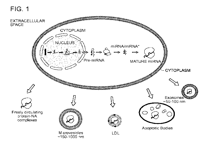
Figure I shows an exemplary range of biomarkers from cells such as miRNA
(depicted),
proteins, lipids, glycoproteins, DNA, mRNA, tRNA, etc., which can relatively
stably exist
12
CA 02956712 2017-01-30
WO 2016/022654 PCT/US2015/043768
outside of cells in various forms, including but not limited to: protein-
nucleic acid complexes,
exosomes, microvesicles, LDL particles, and apoptotic bodies.
Figure 2 shows a comparison of exemplary methods used and/or proposed for
isolation of
microvesicles. Notably, most such methods are optimized for isolation from
blood serum or
plasma of a subject.
Figure 3 shows a comparison of commercial kits available for microvesicle
isolation. Asterisks
indicate kits released in 2014. Boxed regions indicate potential drawbacks for
each kit.
Figure 4 shows that a Na Urate bioparticle isolation protocol of the invention
worked more
consistently than ultracentrifugation or any of three different commercial
kits. Unlike other
methods, Na urate isolated vesicle markers, even from dilute samples. Methods:
Two 15 ml
samples; 1 naturally concentrated (left panel) and 1 naturally dilute (right
panel) were split into 5
equal parts and were subjected to the Na Urate protocol (see Example 1),
ultracentrifugation, or
one of three commercial urine exosome isolation kits (from Life Technologies,
Exiqon, and
System Bio, respectively). Following each procedure, equal amounts of the
final pellet were
loaded onto SDS page and subjected to western blot analysis using antibodies
specific for
known microvesicle protein markers HSP70, Aquaporin 2, Rab5 and CD9. Only the
Na Urate
protocol isolated all four markers from both samples. As a control, vesicles
were also isolated by
ultracentrifugation (2000 x g 10 min spin, followed by a 17,000 x g 10 min
spin, followed by a
100,000 x g spin for 1 hour); vesicles were isolated using the following
commercial kits as per
their instructions: miCURY Exosome Isolation Kit (Exiqon, Woburn, MA),
ExoQui.k-TC, (SBI,
Mountain View, CA) and Total Exosome Isolation Reagent (Life Technologies,
Carlsbad, CA).
Figure 5 shows that Na Urate functioned even in very dilute samples. Methods:
A 12 ml first
void clean catch urine sample was split into four equal parts and subjected to
Na Urate ("Ymir"),
ultracentrifugation ("UC"), miCURY Exosome Isolation Kit ("Exiqon", Exiqon
Woburn, MA),
or ExoQuik-TC, ("SBI", SBI Mountain View, CA). The Na Urate prep was performed
as per
Example 1. Ultracentifugation was performed as per Figure 4. The commercial
kits were
performed as per manufacturer instructions. The resulting preps were subjected
to immunoblot
analysis with Mabs for vesicle markers Aquaporin 2, Rab5, and CD9. The full
strength preps are
shown in lane 1 of each panel. The same sample was also diluted 2x, 4x, and 8x
(lanes 2, 3, and
4, respectively, for each panel) with PBS before being subjected to the same
prep methods.
13
CA 02956712 2017-01-30
WO 2016/022654 PCT/US2015/043768
Figure 6 shows that the Na Urate protocol precipitated a subset of the total
extra-cellular protein
and thus could be considered a "purification". "A" corresponds to Amicon
preparation, while
"Ye" corresponds to the Na Urate protocol of certain aspects of the invention.
Method: A single
6 ml first void clean catch urine sample was split in two and either
concentrated with an
Amicon protein purification column (3000 MW cut-off) or subjected to the Na
Urate
bioparticle isolation protocol of certain aspects of the invention. Equal
amounts (by
volume) of each processed sample was loaded onto an SDS PAGE gel and subjected
to
Coomassie protein stain. Significantly less protein was seen in the
bioparticle isolation prep
(Y*), as compared to total protein from the Amicon column (A).
Figures 7A to 7C shows that the Na Urate process isolated high quality RNA,
especially
miRNA. Figure 7A shows a Bioanalyzer gel of small RNA isolated from a single
10 ml first
void clean catch by ultracentrifugation ((JC; half of the sample) and Ymir
Genomics' Na Urate
protocol (Y; half of the sample). Figure 7B shows a Bioanalyzer gel trace of
small RNA
isolated from a single first void clean catch by ultracentrifugation (UC) in
red and Ymir
Genomics' Na Urate protocol (Ymir) in green. Figure 7C shows relative amounts
of 3
miRNAs known to be found in human urine. Methods: RNA was isolated from
urinary vesicle
preps (UC and Y) with mirVANA Kit (LifeTechnologies). Small RNA quality and
concentration were determined using Agilent 2100 13ioanalyzer (Agilent
Technologies, Santa
Clara, CA) and Small RNA Kit (Agilent). RT- quantitative PCR. cDNA was
synthesized from
urinary vesicle RNA using the TaqMan Micro RNA RT Kit (LifeTechnologies)
according to the
manufacturer's instructions. qPCR was performed using TaqMan Gene Expression
Master Mix
(Life Technologies). Primers for hsa-mir-10b, hsa-mir-223, and hsa-mir-200c
were obtained
from Life Technologies.
Figure 8 shows that Na Urate purified complex RNA, including miRNAs. Indeed,
Na Urate
purified miRNA was more complex than Ultracentrifuge-purified miRNA. RNA from
identical
samples was isolated via Na Urate or Ultracentrifuge methods and analyzed for
microRNA
level(s) with Firefly miRNA Array Panel (Abeam Cambridge MA).
Figure 9 shows that the Na Urate protocol isolates RNA without PCR inhibitors.
One concern
for RNA purification from biofluids, especially urine, was that Enzymatic
inhibitors such as
Urea will be co-purified; however, a known amount of cel-mir-39 (a non-human
miRNA) was
14
CA 02956712 2017-01-30
WO 2016/022654 PCT/US2015/043768
spiked into a UC prep and a Na Urate prep. The amount of cel-mir-39 detected
was identical
between the two preps, demonstrating that Na Urate did not purify more PCR
inhibitors than
Ultracentrifuge.
Figure 10 shows transmission electron microscopy (TEM) images that demonstrate
that Na
Urate isolated whole exosomes, when used as described herein. A 4 ml sample of
first void
clean catch urine was split in two. Half was subjected to Na Urate
precipitation/crystallization
(Method Y, left panel; see example 1) and half was subjected to standard
ultracentrifugation.
Both methods yielded particles of similar sizes and shapes as judged by
transmission electron
microscopy. Vesicles were also isolated by ultracentrifugation as follows:
urine sample was
sequentially centrifuged for 10 min at 2,000xg and at 17,000xg for 10 min to
remove cells and
cellular debris then the resulting supernatant was centrifuged at 200,000xg
for 60min at 24C to
sediment exosomes.
Figure 11 shows that Na Urate isolated whole exosomes: the NanoSight
nanoparticle tracking
device measured the number and size of vesicles in a solution. Methods: A 1 ml
sample of first
void clean catch urine was split in two. Half was subjected to Na Urate
precipitation/crystallization (Method Y, right panel; see example 1) and half
was subjected to
standard ultracentrifugation. Both methods yielded particles of very similar
size and shape, as
judged by nanotracker particle sizing and counting. Vesicles were also
isolated by
ultracentrifugation as per Figure 4. Nanoparticle Tracking Analysis. Vesicles,
diluted in PBS,
were analyzed by nanoparticle tracking using the NanoSight NS300 system
(Malvern
Instruments, Malvern, UK) equipped with 405nm laser. Videos were collected and
analysed
using the NTA software (version 3.0 0060).
Figure 12 shows that the Na Urate protocol was scalable (protein). An
immunoblot of the
instant method (see example 1) from different amounts (indicated) of a single
first void clean
catch urine sample using Mabs specific for vesicle markers TSG101, Aquaporin
2, :Rab 5 and
CD9.
Figures 13A and 13B show that the Na Urate protocol was scalable (RNA). An qRT-
PCR
values for 3 miRNAs isolated from different amounts (indicated) of the same
first void clean
catch using the Na Urate protocol (example x). qRT-PCR traces used to
calculate Ct values
shown in Fig. 13A. The lower the Ct value the higher the concentration.
Methods: RNA
CA 02956712 2017-01-30
WO 2016/022654 PCT/US2015/043768
isolation and qRT-PCR as per Figure 12.
Figures 14A and 14B show that the Na Urate protocol could isolate extra-
cellular mRNA. qRT-
PCR values for GAPDH messenger RNA (mRNA) isolated from different first void
clean catch
urine samples from 3 donors using the Na Urate protocol (Method Y; example 1),
ultracentrifugation (UC) and the Norgen Urine Exosomal RNA Kit. qRT-PCR traces
used to
calculate Ct values shown in A. Methods: RNA isolation as per Figure 12,
except that an extra
15 minute DNAse step was added at the end.
Figure 15 shows that Na Urate isolated vesicles from UC-depleted urine
supernatant and even
from vesicles suspended in pure H20. The Na Urate ("Y") protocol isolated
vesicles that
ultracentrifugation ("UC") missed, whereas ultracentrifugation could not
isolate vesicles from
urine depleted of vesicles isolated by Na Urate. Furthermore, Na Urate was
even capable of
isolating a small amount of urine vesicles purified by ultracentrifugation and
resuspended in pure
H20, suggesting that Na Urate could isolate vesicles from any fluid. Methods:
4.5 mls of first
void clean catch urine was divided into three parts and subjected to either
just a control double
low speed spin (lane 1), the Na Urate protocol (example 1; lane 2) or
ultracentrifugation (as per
Figure 4; lane 3). The vesicle depleted supernatants were saved and subjected
to the reciprocal
methods Na Urate (lane 4) or ultracentrifugation (lane 5). Separately, a 1.5
ml first void clean
catch sample was subjected to ultracentrifugation. The vesicle pellet was
washed lx with PBS
then resuspended in H20. The H20 plus vesicles was subjected to Na Urate,
incubated, and spun
as per Figure 4 legend. Then analyzed by immunoblot with Mabs specific for
Aquaporin 2,
TSG101, and CD9.
Figure 16 shows that the Na Urate protocol isolated vesicle markers in saliva
as well as urine.
Methods: A first void clean catch urine sample was processed with Na Urate as
per Example 1.
A saliva sample was diluted 2x with PBS and then spun 2 x 1500g to remove
cells, cell debris,
and mucous. Na Urate was added to 5 mM (40 ul of .131M stock/ml of sample)
concentration
and incubated on ice for 20 minutes before being spun at 1000g for 5 minutes.
The resulting
pellet was resuspended in Laemmli buffer and run on PAGE along with the
results for the urine
prep. Irrummoblot analysis was performed with Mabs specific for vesicle
markers Rab5 and
CD9. UC = Ultracentrifugation protocol as per Figure 4 with 3 mls of Urine or
5 mls of Saliva
as indicated; Y1 = The instant method on 1 ml of Urine or Saliva as indicated;
Y3 = The instant
16
CA 02956712 2017-01-30
WO 2016/022654 PCT/US2015/043768
method on 3 mls of Urine or Saliva as indicated; D = 200 ul slurry of
Diatomaceous Earth as per
Example 1 on 3 mls of Urine or Saliva as indicated.
Figure 17 shows the 96-well plate protocol for the Na Urate protocol. The
protocol is one for
using the Na Urate Protocol for small volumes in a 96 well format, suitable
for automation
Figures 18A and 18B show 96-well plate data for the Na Urate protocol. The
efficiency of the
Na Urate protocol allowed for the isolation of measurable quantities for
biomarkers from small
volumes of sample. The simplicity of the Na Urate protocol allowed for the use
of 96 well plates
and semi-automation. In Figure 18A, an immunoblot using Mabs for vesicle
markers Aquaporin
2, Rab5, and CD9. A single first void clean catch urine sample was divided
into 6, 200 ul
portions and bioparticles were isolated using 96-well plate protocol (lanes 1-
5 which are
identical replicates towards precision data) and the standard test tube
protocol (lane 6 (tube
format)). RNA preps were made using the standard protocol (tube format) or 96-
well plate
protocol (96 Well Format) from multiple 200 ul aliquots from a single first
void clean catch urine
sample. The preps were subjected to qRT-PCR with Life Technologies miRNA
probes for mir-
200c. The 96-well plate format was identified as more efficient at isolating
mir-200c than the
standard tube format.
Figures 19A and 19B show that different Uric Acid salts work similarly in the
Urate-based EV
isolation protocol. Protocol for experiment (as per example 1). Western blot
analysis of vesicle
protein isolated from a single 12 ml first void clean catch urine sample
divided into 12 parts and
treated with different amounts (as labeled in ul) of different Urate salts as
labeled.
Figure 20 shows that TCEP added to the urine before the first spin reduced EV
loss. Adding
TCEP to sample before the first spin was easier than the current art, where
DTT is used to
recover EVs from the first pellet and leads to decreased pelleting of Tamm-
Horsefall Protein
(THP) and exosomes and increased final yield. TCEP was preferable to DTT for
this purpose
because it has a wider range of pH activity (urine has a pH range from 4-8).
Methods:
Immunoblot using a Mab specific for the urine vesicle marker Aqua-2G and
protein stain
showing THP of an experiment where extra-cellular vesicles were isolated using
multiple
centrifugal spins at indicated speeds either without (left panel) or with 16
mM (final
concentration) of TCEP added to the urine sample. Adding TCEP reduces the
amount of
pelleted THP and EVs and increases the yield from the final 200,000 x g
pellet. P2 = 2000 x g
17
CA 02956712 2017-01-30
WO 2016/022654 PCT/US2015/043768
spin for 10 minutes; P17 = 17,000 x g spin for 10 minutes; P200 = 200,000 x g
spin for 1 hour.
Figure 21 shows that Diatomaceous Earth (DE) isolated vesicle protein markers
from urine,
whereas control silica did not. A single 9 ml first void clean catch urine
sample was split in three
and either 1) subjected to 2 x 1500 g spin, 2) exposed to Diatomaceous Earth
protocol, or 3)
exposed to Silica particles as a control. The resulting preps were loaded onto
a SDS PAGE gel
transferred to Nitrocellulose and irnmunostained with antibodies specific for
vesicle markers
Aquaporin 2 and CD9. Protocol: 1 gram of Diatomaceous Earth or Control Silica
particles were
washed twice in PBS and then resuspended in 10 mls of PBS plus protease
inhibitors. After
vigorous vortexing, 150 ul of each slurry were pipetted into separate 3 ml
aliquots of a cell-free
urine sample in 15 ml polypropylene tubes. The tubes were rotated slowly for
30 minutes then
spun at 1500 g for 2 minutes. The urine supernatant was discarded and the
pellets were washed
2x with 3 mls of PBS. After the second wash the pellets were suspended in 100
ul of Laemmli
buffer, boiled for 3 minutes and 50 ul of each was loaded onto a SDS PAGE gel.
"Just Spin"
control used the same protocol except no DE was added ¨ showing that DE is
required.
Figure 22 shows that Diatomaceous Earth (DE) isolated saliva exosomes.
Immunoblot was
performed with Mabs specific for vesicle markers Rab5 and CD9. Lane 1;
bioparticle prep of 3
ml urine sample using DE (protocol as per Figure 21), Lane 2; blank, Lane 3; 2
x 1500 g spin of
mls of cell free saliva, Lane 4; 5 mls cell-free urine treated with silica
particles, Lane 5; 5 mls
cell-free urine treated with Diatomaceous Earth. Saliva Protocol: 7.5 mls of
saliva was diluted
with 7.5 mls of PBS. Then it was spun 2 x 2000g to remove cells, cell debris,
and mucous. The
resulting supernatant was split into 3, 5 ml aliquots. One aliquot (negative
control) was spun two
more times at 1500g. Either 150 ul of silica beads or Diatomaceous Earth
prepped as per figure
21 legend were added to the other two aliquots and then processed as per
figure 21.
Figure 23 shows that DE (optionally non-calcinated (N) and low permeable/small
pore size)
isolated EVs from urine. It was noted that the calcinated and larger pore
diameter DE Grades
worked the worst; C = calcinated; N = non-calcinated. Permeability was
measured in Darcies
(the higher the value, the more permeable). A single first void clean catch
urine sample was split
into 5 ml aliquots in 15 ml polypropylene tubes and exposed to 300 ul of a
slurry (1 g into 10 mls
of PBS) of different grades of Diatomaceous Earth acquired from several
sources (see below).
The mixture was incubated at RT for 20 minutes then the DE was removed from
the mixture by a
18
CA 02956712 2017-01-30
WO 2016/022654 PCT/US2015/043768
3 minute 1500 x g spin (supernatant poured off). The treated DE was washed 2x
by 5 mls of
PBS then suspended in 150 ul of Laemmli buffer. 50 ul of this was run on SDS
PAGE gel and
transferred to Nitrocellulose. The Nitrocellulose was probed with Mabs
specific for extra-
cellular vesicle markers CD9 and Aquaporin 2. Shown are signals from
glycosylated Aquaporin-
2 and CD9 as judged by MW and important properties (if known) of each grade of
DE. Grades
and sources of Diatomaceous Earth: W = Natural Food Grade DE from PermaGuard;
FP-4 =
Calcinated DE from Ep Minerals (Reno Nevada); FW-60 = Calcinated DE from Ep
Minerals
(Reno Nevada); FP-22 = Calcinated DE from Ep Minerals (Reno Nevada); FN-6 =
Natural DE
from Ep Minerals (Reno Nevada); Cel-S = Natural DE (Brand Name Celite-S) from
Sigma
Aldrich; AW-2 = Acid Washed DE from Ep Minerals (Reno Nevada).
Figure 24 shows that calcination and acid washing decreased DE's affinity for
exosomes. A
single first void clean catch urine sample was split into 5 ml aliquots in 15
ml polypropylene
tubes and exposed to 300 ul of a slurry (1 g into 10 mls of PBS) of different
grades of
Diatomaceous Earth acquired from several sources (see figure 23 Description).
The mixture was
incubated at RT for 20 minutes, then the DE was removed from the mixture by a
3 minute 1500
x g spin (supernatant poured oft). The treated DE was washed 2x by 5 mls of
PBS then
suspended in 150 ul of Laemmli buffer. 50 ul of this was run on SDS PAGE gel
and transferred
to Nitrocellulose. The Nitrocellulose was probed with Mabs specific for extra-
cellular vesicle
markers CD9 and Rab5. Shown are signals from :Rab5 and CD9 as judged by MW and
important properties (if known) of each grade of DE.
Figure 25 shows Perlite (Sil-Kleer) with smaller pore sizes/permeability can
also isolate Extra-
cellular Vesicles Si lKleer is the commercial name for a type of Perlite which
is volcanic glass
heated to expand and form pores. It contains less Si02 than DE. Methods: A
single first void
clean catch urine sample was split into 5 ml aliquots in 15 ml polypropylene
tubes and exposed
to 300 ul of a slurry (1 g into 10 mls of PBS) of different grades of
Diatomaceous Earth or Perlite
acquired from several sources (see below). The mixture was rocked slowly for
20 minutes then
the DE was removed from the mixture by a 3 minute 1500 x g spin (supernatant
poured off).
The treated DE was washed 2x by 5 mls of PBS then suspended in 150 ul of
Laernmli buffer. 50
ul of this was run on SDS PAGE gel and transferred to Nitrocellulose. The
Nitrocellulose was
probed with Mabs specific for extra-cellular vesicle markers CD9 and Aquaporin
2. Shown are
signals from glycosylated Aquaporin-2 and :Rab5 as judged by MW and important
properties (if
19
CA 02956712 2017-01-30
WO 2016/022654 PCT/US2015/043768
known) of each grade of DE. Grades and sources of Diatomaceous Earth: W =
Natural Food
Grade DE from PermaGuard; 17-S = #17-S grade Perlite(Sil-Kleer) from Silbrico
Corp
(Hodgkins, IL); 23-S = #23-S grade Perlite(Sil-Kleer) from Silbrico Corp
(Hodgkins, IL); 27-M=
#23-S grade Perlite(Sil-Kleer) from Silbrico Corp (Hodgkins, IL).
Figure 26 shows that Diatomaceous Earth (DE) purified complex RNA.
Diatomaceous Earth-
purified microRNA was more complex than Norgen kit: RNA from identical 30 ml
samples was
isolated via DE or Norgen kit and analyzed for microRNA level with Firefly
miRNA Array
Panel as per Figure 8.
Figure 27 shows that Diatomaceous Earth (DE) isolated exosomes from cell
culture media.
Jurkat Cells were grown for 24 hours in DMEM media plus 5% Fetal Bovine Serum.
Cells and
debris were spun out of 2 mls of the media for 10 minutes at 1500 x g. The
resulting cell free
media was split in two and subjected to a Diatomaceous Earth protocol (see
Figure 21) or an
Ultracentrifitgation protocol (see Figure 10). Furthermore, the bioparticle-
depleted supernatant
from the DE protocol was saved and subjected to the ultracentrifitgation
protocol. The pellets
from all three procedures were suspended in Laemmli buffer, and half of that
suspension was
loaded on an SDS PAGE gel, and was then transferred to Nitrocellulose and was
probed with a
monoclonal antibody (Mab) specific for vesicle marker Rab5. Lane 1;
Ultracentrifuge isolated
vesicles. Lane 2; DE isolated vesicles. Lane 3; DE treatment almost completely
depleted cell
culture media of vesicle-derived Rab5.
DETAILED DESCRIPTION OF THE INVENTION
The present invention provides compositions and methods for producing
preparations of
isolated secreted microvesicles, RNA, DNA and protein-nucleic acid complexes
(collectively
called "bioparticles") from a liquid sample. The invention also provides
methods for producing
biofluids and blood serum/plasma that has been at least partially depleted of
bioparticles. These
methods have a number of advantages over the state of the art, which will be
apparent from the
discussion herein.
In certain aspects, the instant invention provides methods for the isolation
of bioparticles
(including, e.g., microvesicles, exosomes, etc.) from a liquid sample (e.g., a
biofluid of a subject
or cell culture). Kits for performance of such isolation steps, and
instructions for their use, are
CA 02956712 2017-01-30
WO 2016/022654 PCT/US2015/043768
also provided.
I. Definitions
As used herein, the term "bioparticle" refers to cell-free, membraned
structures secreted
from mammalian cells such as but not limited to microvesicles, exosomes,
apoptotic bodies,
LDL-particles etc., plus cell-free, relatively stable, protein-nucleic
complexes secreted from
mammalian cells such as but not limited to microRNA-A002 complexes, plus cell-
free DNA
(cfDNA) and cell-free messenger RNA. Thus, certain exemplary bioparticles
include cell free
miRNA (depicted), proteins, lipids, glycoproteins, DNA, mRNA, tRNA, other
types of RNA,
etc., which can exist relatively stably outside of cells, in various forms,
including but not limited
to: protein-nucleic acid complexes, exosomes, microvesicles, LDL particles,
and apoptotic
bodies.
As used in this application, the term "cells" encompasses not only eukaryotic
cells, e.g.,
higher eukaryotic cells such as mammalian cells, as in human cells or mouse
cells, but also
prokaryotic cells, such as eubacteria cells and Archaea cells.
As used herein, the term "microvesicle" refers generally to any plasma
membrane bound
particle that may reside within the cell, or in the extracellular environment.
These structures are
not limited in any way with regard to in vivo localization (e.g.,
intracellular or extracellular), in a
body fluid, in a cell culture media, generated by in vitro cultured cells,
mechanism of origin or
size characteristics. In some embodiments, a microvesicle can range in size
with a lower size
limit of at least about 20 nanometers (nm) in diameter, or alternatively, 30
nm, or 40 nm, or 50
nm in diameter. In some embodiments, a microvesicle has an upper size limit of
not more than
about 1,000 tun (i.e., 1.0 micrometer, micron, or tom), or alternatively, not
more than about
1,500 nm, about 2,000 nm or about 2,500 nm. As used herein, the term "secreted
microvesicle" is
used synonymously with "circulating microvesicle (cMV)" or "extracellular
microvesicle (emV)"
or "extracellular vesicle (eV)" and refers to a subset of microvesicles that
are found in an
extracellular space under normal physiological conditions. As used herein, it
is not intended that
the term "circulating microvesicles" to be limited to microvesicles of any
particular size or size
range, or any particular production mechanism. For example, but not limited
to, a cMV of the
invention can be produced by (i) exocytosis from multivesicular bodies to
produce exosomes, (ii)
21
CA 02956712 2017-01-30
WO 2016/022654 PCT/US2015/043768
budding, fission and shedding of microvesicles directly from a cytoplasmic
membrane, and (iii)
membranous blebs caused by programmed cell death leading to the formation of
apoptotic
bodies. As used herein, the term "cMV" is not limited to microvesicles of any
particular size or
size range.
Although mechanistic theories for the endogenous production of circulating
microvesicles are found in the scientific literature, any knowledge of such
mechanisms is not
required to make or used the present invention. It is not intended that the
term "circulating
microvesicles" as used herein be limited in any way with regard to the
mechanism of their in
vivo production.
As used herein, the term "shedding microvesicle (SMV)" refers to a class of
microvesicles that are produced by cells using a mechanism of direct plasma
membrane budding,
fission and shedding to produce microvesicles that are released by a cell into
an extracellular
environment. As used herein, it is not intended that an SMV of the invention
be limited by any
particular size or size range.
As used herein, the term "exosome" refers to a subset of circulating
microvesicles that are
preformed microvesicles that are released from the cell following the exocytic
fusion of
intracellular multivesicular bodies with the plasma membrane, i.e., exosomes
have an endocytic
origin. As used herein, it is not intended that an exosome of the invention be
limited by any
particular size or size range.
As used herein, the term "crystal/precipitation-inducing agent" refers to an
agent capable
of promoting crystal formation and/or precipitation in a liquid sample.
Exemplary
"crystal/precipitation-inducing agents" of the invention include monosodium
urate, uric acid, a
salt thereof and a combination thereof.
As used herein, the term "apoptotic body" refers to a subset of circulating
microvesicles
that are produced as a result of apoptotic cell destruction. As used herein,
it is not intended that
an apoptotic body of the invention be limited by any particular size or size
range.
As used herein, the term "isolating," or "to isolate," refers to any
artificial (i.e., not
naturally occurring) process for treating a starting material, where the
process results in a more
useful form of a molecule or structure of interest that is in the starting
material. The "more useful
22
CA 02956712 2017-01-30
WO 2016/022654 PCT/US2015/043768
form" of the molecule or structure of interest can be characterized in a
variety of ways, no one of
which is limiting. For example, as used herein, the invention provides methods
for isolating
secreted microvesicles from conditioned cell culture media. Further, for
example, the process for
isolating can result in:
(i) the molecule of interest having a greater concentration in the isolated
form
compared to the starting material (e.g., concentrating),
(ii) the removal of any amount or any type of impurities from the starting
material
(e.g., purifying),
(iii) an increase in the ratio of the amount of molecule of interest to the
amount of any
undesired component in the starting material (e.g., enriching),
(iv) any artificial process for removing a molecule or structure of interest
from its natural
source or location;
(v) any artificial process for separating a molecule or structure of interest
from at least
one other component with which it is normally associated (e.g., purifying), or
(vi) any combination of (i), (ii), (iii), (iv) or (v).
Similarly, as used herein, the term "isolated" generally refers to the state
of the molecule
or structure of interest after the starting material has been subjected to a
method for isolating the
molecule of interest. That is to say, isolating a molecule of interest from a
starting material will
produce an isolated molecule. For example, the methods of the invention are
used to produce
preparations of isolated microvesicles. These preparations of microvesicles
have been isolated
from their natural source, for example, from urine, or from conditioned cell
culture media.
As used herein, the term "purifying" or "to purify" a molecule or structure of
interest
refers to a process for removing at least one impurity or contaminant from a
starting material.
For example, purifying a molecule of interest from a starting material refers
to a process for
removing at least one impurity from the starting material to produce a
relatively more pure form
of the molecule of interest.
As used herein, the term "substantially purified" refers to molecules or
structures of
interest that are removed from their natural environment or from a starting
material (i.e., they are
isolated) and where they are largely free from other components with which
they are naturally
associated or substantially free of other components that may render future
use or study sub-
23
CA 02956712 2017-01-30
WO 2016/022654 PCT/US2015/043768
optimal, difficult or impossible.
As used herein, the terms "purified" or "partially purified" refers to
molecules or
structures of interest that are removed from either (1) their natural
environment, or from (2) a
starting material (i.e., they are isolated), and where (a) at least one
impurity from the starting
material has been removed, or (b) at least one component with which the
molecule is naturally
associated has been removed. A "purified" or "partially purified" molecule may
still contain
additional components that may render future use or study of the molecule sub-
optimal, difficult
or impossible.
As used herein, the term "enriching" (and "enriched" and the like) refers to a
process
whereby a molecule of interest that is in a mixture has an increased ratio of
the amount of that
molecule to the amount of other undesired components in that mixture after the
enriching process
as compared to before the enriching process.
As used herein, the term "concentrating" refers to a process whereby a
molecule of
interest that is in a mixture that has been subjected to that process has a
greater concentration
after the process as compared to the concentration of the molecule in the
mixture before the
process.
As used herein, the term "depleted" refers to a mixture containing an
undesirable
component, where that undesirable component has been (i) completely removed
from the
mixture, (ii) sufficiently removed from the mixture to be undetectable, or
(iii) partially removed
from the mixture such that its concentration in the mixture is significantly
reduced. For example,
a blood serum that has been depleted of endogenous microvesicles may contain
no
microvesicles, or may contain no detectible microvesicles, or may contain a
reduced level of
microvesicles compared to the untreated serum.
As used herein, the expression "cell culture media" refers to any growth media
that can
support in vitro cell growth of a designated cell line. Such media can be
supplemented or non-
supplemented, for example, with 10% by volume, heat-inactivated fetal calf
serum.
As used herein, the expression "minimal defined cell culture media" or
"minimal media"
refers to any culture media where each component is defined by name and the
concentration of
each component is known. Minimal defined cell culture media generally does not
contain a
24
CA 02956712 2017-01-30
WO 2016/022654 PCT/US2015/043768
serum supplement. For example, Dulbecco's Modified Eagle's medium (DMEM) is a
defined
minimal cell culture media. Minimal defined cell culture media generally can
be used to culture
cells in vitro, but not for extended periods of time.
As used herein, the expression "complete cell culture media" refers to a
culture media
that comprises a defined minimal cell culture media, and in addition, also
comprises a complex
supplement that enhances the growth properties of the culture media. For
example, a blood
serum supplement is commonly added to a minimal media to produce a complete
cell culture
media. Fetal calf serum (FBS or FCS) is a common supplement (10% by volume)
that is added to
a minimal media to produce a complete culture media. Complete culture media
are used to
culture cells in vitro for indefinite (long) periods of time. [0075] As used
herein, the expression
"conditioned cell culture media" refers to any cell culture media (including
complete media or
minimal media) that has been exposed to live cells in culture. Conditioned
cell culture media
comprises not only the defined components of the minimal media and the serum
supplement, but
also contains additional components that the living cultured cells have
produced. In many cases,
conditioned cell culture media is a serum-free media.
Microvesicles
The term "microvesicles" (also known as microparticles) refers to a
heterogeneous in
vivo collection of membrane bound (i.e., encapsulated) biological structures.
These structures are
formed from lipid bilayer, which is the same lipid bilayer that comprises
eukaryotic cell
membranes. Microvesicles can reside within the cell, or in the extracellular
environment.
Microvesicle structures (intracellular and/or extracellular) are produced by
nearly all mammalian
cell types, as well as during in vitro cell culture.
The molecular composition of microvesicles is diverse, containing and/or
transporting a
variety of nucleic acids, proteins and lipids. Microvesicle molecular
composition is generally
reflective of the plasma membrane and antigenic content of the cell types,
tissues and organs
from which they originate. Mathivanan and Simpson, "Exosomes: extracellular
organel les
important in intercellular communication," J. Proteomics 73(10):1907-1920
(2010). Although
protein composition of the microvesicles varies, most of these structures are
enriched for various
soluble protein markers, including HSP70, Hsc70, CD63, CD9, CD81 and others.
Circulating
microvesicles have also been reported to contain nucleic acids, including
messenger RNAs,
CA 02956712 2017-01-30
WO 2016/022654 PCT/US2015/043768
DNAs, and relatively high levels of small RNAs and microRNAs.
Circulating microvesicles are associated with numerous cell functions,
including
intercellular (cell-to-cell) communication, removal of metabolic byproducts
and toxins
(including misfolded proteins, cytotoxic agents and metabolic waste),
angiogenesis, tissue
regeneration, endocytic recycling of the plasma membrane, selective removal of
plasma
membrane proteins and regulation of immune functions such as antigen
presentation. Some
microvesicles have been shown to transport messenger RNA (mRNA) and microRNA
(miRNA),
which is highly suggestive of microvesicles functioning as messengers that
allow one cell type to
regulate the activity of a distant cell type by acting as a shuttle that can
merge with the distant
cell and release its contents into that target recipient cell. This
microvesicle shuttle can utilize the
body fluids to travel to distant sites and control the activity of distant
target cells.
Circulating microvesicles (cMVs), or synonymously, extracellular microvesicles
(eMVs)
or extracellular vesicles (eVs), describe an eclectic group of microvesicles
that are released by
cells, and therefore, exist in extracellular spaces andlor reside in body
fluids. The mammalian
body fluids that are known or suspected to contain cMVs include, but are not
limited to, blood,
urine, saliva, breast milk, tears, sweat, ascites fluid and cerebrospinal
fluid. Secreted
microvesicles are also found in cell culture media that has been exposed to
cultured mammalian
cells.
With regard to defining and categorizing the cMV molecules that can be found
in body
fluids, there is lack of consensus as to the nomenclature and description of
the different types of
cMV particles. Some literature distinguishes at least three subcategories of
circulating
microvesicles, based on their mechanistic origin. The molecular/cellular
mechanisms that
produce microvesicles are theorized to include (i) exocytosis of intracellular
multivesicular
bodies, (ii) outward budding, fission and shedding of plasma membrane, and
(iii) byproducts of
apoptosis. The diverse collection of circulating microvesicle structures can
range in size from
about 20 nanometers (nm) to upwards of about 1,000 mn (i.e., 1.0 micrometer,
micron, or gnu)
in diameter.
The first recognized subgroup of cMVs are those produced by direct plasma
membrane
budding, fission and shedding. Some sources describe these shed microvesicles
as generally
large, namely with lower sizes limits of at least 100 nm or 200 nm, and with
an upper size limit
26
CA 02956712 2017-01-30
WO 2016/022654 PCT/US2015/043768
of about 1,000 nin in diameter. Some have proposed that these structures be
termed "ectosomes"
or "shedding microvesicles (SMVs)." Still other groups state that ectosome
particles may be as
small as 40 or 50 nm in diameter.
A second recognized subgroup of cMVs are exosomes, that is, the preformed
microvesicles that are released from the cell following the exocytic fusion of
intracellular
multivesicular bodies with the plasma membrane. These exosome structures are
generally
smaller than ectosomes, and have an upper size limit estimated to be about
100, 150 or 200 nm,
and a lower size limit of about 40 nm or 50 nm. However, various sources
differ in their size-
based definitions for exosomes, and this size distinction remains unresolved.
A third group of structures is the apoptotic blebs released by dying cells.
These
membrane structures have a less well-defined size range, and may be anywhere
from about 50
nm to about 5,000 nm in diameter.
A unified microvesicle nomenclature and classification system utilizing
broadly accepted
definitions has been elusive in the field. In the literature, microvesicles
have been alternatively
referred to as microparticles, nanoparticles, exosomes, ectosomes,
epididimosomes, argosomes,
exosome-like vesicles, promininosomes, prostasomes, dexosomes, texosomes,
archeosomes,
oncosomes, exosome-like vesicles, apoptotic blebs, extracellular vesicles and
shedding
microvesicles. In some publications, uses of these terms is conflicting or
overlapping. Simpson
and Mathivanan (2012), "Extracellular Microvesicles: The Need for
Internationally Recognized
Nomenclature and Stringent Purification Criteria". J Proteomics Bioinform (2).
doi:10.4172/jpb.10000e10. One source suggests that a preferred nomenclature
for circulating
microvesicle is based on the microvesicle's mechanism of origin. Namely, these
categories
would be (i) the ectosomes produced by membrane budding, (ii) the exosomes
produced by the
exocytosis to intracellular multivesicular bodies, and (iii) the membrane
blebs produced by the
process of apoptosis.
The release of exosomes was highlighted from different cell types in a variety
of
physiological contexts. Thus, it has been shown that B cells release exosomes
bearing molecules
of the major histocompatibility complex class II, which play a role in antigen
presentation
(Raposo et al., J. Exp. Med. 183 (1996) 1161). Similarly, it has been shown
that dendritic cells
produce exosomes (also referred dexosomes) with specific structural and
functional
27
CA 02956712 2017-01-30
WO 2016/022654 PCT/US2015/043768
characteristics, and playing a role in mediating the immune response,
including the stimulation
of cytotoxic T lymphocytes (Zitvogel et al., Nature Medicine 4 (1998) 594). It
has also been
shown that tumor cells secrete in a controlled manner, specific exosomes (also
designated
texosomes) bearing tumor antigens and are able to present these antigens or to
transmit them to
antigen-presenting cells. It is also known that mast cells accumulate
molecules in intracellular
vesicular compartments, which can be secreted in response to signals (Smith
and Weis,
Immunology Today 17 (1996) 60). In general, it seems that the cells emit
signals and
communicate with each other through membrane vesicles they release, which may
carry
antigenic patterns, MHC molecules, or any other signal (cytokine, growth
factor, etc.) which
have special structural and functional characteristics and are produced in
different physiological
situations.
II. Methods for Isolating Bioparticles
The present invention provides methods for the isolation of bioparticles from
liquid
samples. In certain embodiments, the liquid sample is urine. From urine as an
example, certain
methods of the invention comprise the following steps:
A) (Optional) Preparing a Whole Urine Prespin Treatment Solution (also called
Solution 1)
The purpose of the Whole Urine Prespin Treatment Solution is to reduce the
amount of
bioparticles lost in the first spin (prespin), which is typically performed to
reduce the amount of
cells and debris in the Whole Urine sample.
It is well known in the field that spinning urine at speeds above 17,000 x g
can lead to the
loss of microvesicles due to the trapping action of the protein THP (also
called uromodulin).
However, it was discovered that a large amount of microvesicles could also be
lost in the lower
speed spins (below even 3000 x g) that are typically used to remove cells and
debris (see Figure
20).
In certain embodiments, the Whole Urine Prespin Treatment Solution consists of
the
reducing agent TCEP. TCEP is preferred over DTT for this purpose, as it is
active in a broader
range of pH. In one embodiment, the concentration of the TCEP in the 10x
solution would be at
a concentration of 160mM. Other embodiments have the TCEP 10x concentration
being
28
CA 02956712 2017-01-30
WO 2016/022654 PCT/US2015/043768
between 80mM and 300mM. Other embodiments use other reducing agents such as
DTT at
similar concentrations.
In other embodiments, the Whole Urine Prespin Treatment Solution consists of
an acid
buffer plus reducing agent such that addition of the acid buffer-containing
Whole Urine Prespin
Treatment Solution reduces the pH of the Whole Urine below 6.
In other embodiments where the reducing agent is not used, the Whole Urine
Prespin
Treatment Solution consists of a basic buffer that increases the pH of the
Whole Urine to above 7
as it was discovered that without reducing agent present, less bioparticles
are lost if the pH of the
sample is above 7.
B) (Optional) Adding the Whole Urine Prespin Treatment Solution to the whole
urine sample.
In certain embodiments, a 11101 volume of a 10x Whole Urine Prespin Treatment
Solution is added to the whole urine sample to create a mixture. In other
embodiments any
combination of Prespin Treatment Solution and Urine Sample yielding a mixture
with a final
concentration of the TCEP or other reducing agent of from 5mM to 30 rnM and a
pH below 6 is
acceptable or, if no reducing agent is used, a pH above 7. No incubation is
necessary; the next
step can be taken immediately.
C) Centrifuging the Mixture
The mixture is subjected to a centrifugation. The centrifugation typically
forms a pellet
and a supernatant, although pelleted material may not be visible to the eye.
In contrast to the
prior art, this centrifugation does not require ultracentifugation, e.g., does
not require centrifugal
forces in excess of 100,000 x g. This centrifugation can be done at slower
speeds, for example, to
generate RCF values of not more than 30,000 x g, or not more than 20,000 x g,
or not more than
12,000 x g, or not more than 10,000 x g, or not more than 5,000 x g, or not
more than 2,000 x g,
or not more than 1,500 x g. In one embodiment, a centrifugation producing
1,000 x g is used.
The length of time for centrifugation is not limiting. In one embodiment, the
centrifugation is for
minutes. Alternatively, the centrifugation can proceed for one or more
minutes, two or more
minutes, three or more minutes, four or more minutes, six or more minutes,
seven or more
minutes, eight or more minutes, nine or more minutes, ten or more minutes,
fifteen or more
minutes, twenty or more minutes, etc.
29
CA 02956712 2017-01-30
WO 2016/022654 PCT/US2015/043768
D) Removing the Supernatant
Following the spin, the resulting supernatant is carefully removed so as not
to disturb the
pellet, and the pellet is discarded.
E) Combining the Urine Supernatant from D) with the
Crystallization/Precipitation Solution
(also called solution 2)
To initiate the crystallization/precipitation of bioparticles, Solution 2 (see
below) is added
to the Supernatant generated in step D) to create a mixture. In one
embodiment, a 1/10th volume
of a 10x concentration of Solution 2 is added to the supernatant, however, any
combination that
yields a .5x to 5x final concentration of Solution 2 in the mixture is
acceptable.
F) Incubating the Resulting Mixture
The resulting mixture is then incubated. The incubation can be with any degree
of
cooling, for example at 5 C, although such cooling is not always required. The
incubation times
can vary, and are not in any way limiting. For example, incubation can be
anywhere between 0
minutes to overnight (e.g., 16 hours). The incubation can be with or without
mixing, and the
mixing during the incubation period can be constant or intermittent. In
certain embodiments a
15-minute incubation on ice is performed.
G) Centrifuging the Mixture
The mixture from F) is subjected to a centrifugation. The centrifugation
typically forms a
pellet and a supernatant, although pelleted material may not be visible to the
eye. In contrast to
the prior art, this centrifugation does not require ultracentrifugation, e.g.,
does not require
centrifugal forces in excess of 100,000 x g. This centrifugation can be done
at slower speeds, for
example, to generate RCF values of not more than 30,000 x g, or not more than
20,000 x g, or
not more than 12,000 x g, or not more than 10,000 x g, or not more than 5,000
x g, or not more
than 2,000 x g, or not more than 1,500 x g. In the one embodiment, a
centrifugation producing
2,000 x g is performed. The length of time for centrifugation is not limiting.
In one embodiment,
the centrifugation is for 5 minutes.
H) Removing the Supernatant
CA 02956712 2017-01-30
WO 2016/022654 PCT/US2015/043768
Following the spin, the resulting supernatant is carefully removed so not to
disturb the
pellet, and this supernatant is discarded.
I) Resuspending the Pelleted Material
After removal of the supernatant, the pellet is resuspended in any desired
resuspension
solution and collected for further analysis. The resuspension solution can use
either water,
phosphate buffered saline (PBS), or any other suitable aqueous, such as any
isotonic solution. In
some embodiments, the resuspension solution is basic in nature, for example,
100 mM Tris pH 8.
The volume used for the resuspension is most typically the smallest possible
practical volume,
and is typically many times smaller than the volume of the original liquid
sample comprising the
secreted microvesicles. In some embodiments, the volume of the resuspension
solution is smaller
by at least one order of magnitude than the volume of the original liquid
sample.
111. Liquid Samples
The present invention provides methods for isolating circulating bioparticles
from liquid
samples. It is not intended that the nature of the liquid samples be in any
way limited, and can be
any liquid sample that contains bioparticles. Advantageously, very small
volumes of liquid
sample can be used, for example, as little as about 10 p.L, 50 pi, 0.1 mL, 0.2
mL, 0.3 mL, 0.4
mL, 0.5 mL, 1.0 mL, or 2.0 mL. or 3.0 mL. or 5.0 mL of starting sample can be
used.
In some embodiments, the liquid sample can be conditioned cell culture media
that has
been used to culture a cell line in vitro that has produced bioparticles, and
therefore, those
bioparticles are now contained in the conditioned media. The conditioned cell
culture media can
be a complete media (containing a serum supplement), or a serum-free culture
media.
In some embodiments where the conditioned cell culture media is a complete
media
comprising a serum supplement, the serum supplement that is used can be a
serum that has been
depleted of any endogenous circulating bioparticles prior to addition of the
supplement to the
defined minimal growth media. The present invention also provides methods for
producing such
bioparticle-depleted serum.
In some embodiments, the liquid sample is a biofluid (synonymous with body
fluid). The
body fluid that is used in the analysis is not particularly limited.
Bioparticles can be isolated from
31
CA 02956712 2017-01-30
WO 2016/022654 PCT/US2015/043768
any body fluid using the methods of the invention, even though a particular
body fluid is not
itemized herein, as it is intended that the present methods find use with any
and all body fluids.
For example, body fluids that can be analyzed by the methods of the invention
include, but are
not limited to, amniotic fluid, blood serum, blood plasma, breast milk,
cerebrospinal fluid,
lymph, mucus (including nasal drainage and phlegm), pericardial fluid,
peritoneal fluid, semen,
synoviat fluid, tears, urine, sweat, saliva, and ascites fluid.
IV. CRYSTALLIZATION/PRECIPITA.TION REAGENTS (Solution 2)
The present invention provides methods for the isolation of bioparticles from
liquid
samples, where the methods use a crystallization/precipitation solution
(Solution 2), combined
with the liquid sample, to initiate the bioparticles precipitation and
isolation. Certain
embodiments use Monosodium Urate in solid form, slurry form., or liquid form
(solubilized in a
basic solution such as NaOH). Another embodiment uses Uric Acid. Another
embodiment uses
some other salt of Uric acid. The amount used depends on the sample volume.
One embodiment
uses from 1 to 100 n11,1 Monosodium Urate. Optionally, a Monosodium -11rate or
other
crystallization/precipitation reagent at a concentration of 1, 2, 3, 4, 5, 10,
20, 30, 40, 50, 60, 70,
80,90 or 100 niVI can be added to a sample in an amount of 5 1.1.õ 10 1.tL, 20
itL, 30 1.1õ, 40
50 iaL, 60 iL, 704, 80 iaL, 90 i_tL, or 1004 or more, to promote a
crystallization/precipitation
event in the sample. It was discovered that Uric Acid and optionally
Monosodium [irate when
added to a liquid, optionally urine, crystallizes and/or induces a precipitate
that includes
bioparticles but excludes many proteins and salts contained in the liquid.
V. MATRIX REAGENTS (Solution 2)
The present invention provides methods and compositions for isolating
bioparticles that,
without wishing to be bound by theory, appear to exploit pore sizes of certain
materials to effect
bioparticle enrichment, such as the pore sizes found in porous beads, such as
siliceous beads or
particles, examples of which include diatomaceous earth (DE) and perlite. In
certain
embodiments, the porous beads (e.g., porous siliceous beads) are non-
calcinated, non-acid
washed, (i.e. natural grades) diatomaceous earth with average pore sizes
ranging from .1 to 10
microns and permeabilities less than 2 darcies. In some embodiments, the
porous beads (e.g.,
porous siliceous beads) are Perlite (i.e. treated volcanic glass) with pore
sizes from .1 to 10
32
CA 02956712 2017-01-30
WO 2016/022654 PCT/US2015/043768
microns and permeabilities less than 2 darcies. It is contemplated that matrix
reagents with
average pore sizes in the range of 0.01 micron to 50 microns, including in the
range of 0.01 to 1
micron, 0.5 to 40 microns, 0.5 to 50 microns, 1 to 20 microns, 1 to 10
microns, 2 to 5 microns,
and/or about 3, 4, and/or 5 microns are effective reagents for isolation of
microvesicles/bioparticles as described herein. Similarly, it is contemplated
that agents with
permeabilities of less than 5 darcies, less than 2 darcies, less than 1 darcy,
less than 0.5 darcies,
less than 0.3 darcies, or smaller can be effective reagents for the methods
and compositions of
the invention. Exemplary grades of DE are non-calcinated, non-acid washed,
"natural" forms
possessing pore sizes between .5 ¨ 2 microns in diameter and permeability
below .1 Darcies
(Figures 23-24). Certain exemplary Pertite grades have a permeability below 3
and pore sizes
below 10 microns (Figure 25).
EXAMPLES
The following examples are offered to illustrate; but not limit, the claimed
invention.
It is understood that various modifications of minor nature or substitutions
with
substantially similar reagents or components will be recognizable to persons
skilled in the art,
and these modifications or substitutions are intended to be included within
the spirit and purview
of this application and within the scope of the appended claims.
Cell-free membrane and/or pmtein-containing structures found in urine have
high value
as biomarkers for disease or disorder diagnosis, and even for approaches
involving screening of
urine for therapeutic targets (e.g., biomarker and/or targetable miRNAs). Cell-
free biomarkers
are preferred agents to work with because they are relatively easy to isolate,
are less variable in
content/consistency than whole cells and/or whole cell-containing fluids, and
can travel from the
tissue of their origin into easy to isolate biofluids, such as urine (Figure
1).
A number of methods have been implemented and/or proposed for isolating
microvesicles (MINTs) from urine; however, all have significant limitations
(Figures 2-3). In
particular, current methods other than those described herein ¨ both
commercial and non-
commercial ¨ possess one or more of the following drawbacks:
33
CA 02956712 2017-01-30
WO 2016/022654 PCT/US2015/043768
1. Certain methods require expensive equipment (e.g., ultracentrifuge
methods);
2. Certain methods require expensive kits (e.g, NEP, Qiagen, Exiqon);
3. Certain methods are difficult to scale up (e.g., Norgen (only a 1 ml
column),
ultrafiltration filters (e.g., Amicon) can clog
4. Many such methods are time consuming (e.g., ultracentrifuge methods, SBI,
Lifetech,
Exiqon);
5. Certain methods produce low yield, especially from certain fluids, such as
urine (e.gõ
NEP, SBI, Lifetech, Exiqon);
6. Certain methods use phenol (e.g., Lifetech).
An unmet need was thus identified for isolation of bioparticles (e.g.,
microvesicles,
exosomes, etc.) from urine, as well as other bodily fluids (e.g., saliva, as
well as blood, plasma,
etc.). The instant methods were newly identified to allow for rapid and
inexpensive isolation of
extracellular membrane particles, including microvesicles, exosomes and
apoptotic bodies. The
methods described herein were also observed to isolate membrane-free protein-
nucleic acid
particles as well. Finally, obtaining intact bioparticles is an advantage of
the current invention,
with such bioparticles used in mechanistic, vaccine- and delivery-related and
therapeutic studies.
One advantage of certain of the currently described methods is that they use
common
laboratory reagents and apparatuses, and do not require high-speed
centrifugation (e.g., use of an
ultracentrifuge). Thus, the current methods are designed to achieve a higher
yield than
previously available methods, also allowing for isolation of important
biomarkers and1or
therapeutic targets from a smaller volume of sample than could be obtained
using previously
described approaches.
Development of Novel Systems for Isolation of Circulating Bloparticles
Being unsatisfied with current methods for the isolation of circulating
bioparticles,
including exosomes, from urine and other biofluids, the following experiments
were initiated and
undertaken in an effort to develop new and improved methods for this purpose.
It was known that urine contains several constituents (chemicals) that can be
present at
saturating concentrations and thus can form crystals in vivo and in vitro and
also can be easily
precipitated from a urine sample in vitro. Since it was also known that
certain crystals can form
34
CA 02956712 2017-01-30
WO 2016/022654 PCT/US2015/043768
on and/or interact with epithelial cell membranes in the urinary tract, it was
hypothesized, since
microvesicles are membrane structures as well, that crystal growth on
microvesicles could be
induced artificially and then the microvesicle-crystal complex could be easily
centrifuged out of
solution as a method for microvesicle purification. Similarly, it was noticed
while working with
urine that the same group of endogenous chemicals present at near saturation
levels, often
spontaneously precipitated in vitro when exposed to lower temperatures and/or
artificial
concentration. it was hypothesized that artificially increasing the levels of
some of these
constituents could reliably induce a precipitate that would include
bioparticles. It was also
realized that the addition of these constituents of the urine approach to
other liquids containing
bioparticles (e.g., saliva, blood, plasma, etc.) would similarly
crystallize/precipitate bioparticles
(as was demonstrated in Example 3 below).
After an extensive trial and error process that examined different
constituents of urine, it
was discovered in certain embodiments that the addition of Monosodium Urate,
but also
optionally (additionally and/or alternatively) Uric Acid, or other salts of
Uric Acid could indeed
induce a crystallization/precipitation of urine and that the resulting
sediment of this included
biomarkers known to be present in microvesicles and cell-free protein-nucleic
acid complexes.
Example 1: A Newly Discovered Na Urate Protocol Isolated Microvesicles from
Urine
Quickly and Effectively
To exemplify certain methods of the invention, 3 m Is whole urine samples from
two
different donors (one sample was naturally concentrated and one sample was
naturally dilute)
were treated with 16 mM TCEP reducing agent as part of a Whole Urine Prespin
Treatment
Solution, which simultaneously reduced the pH to <6 and was believed to have
reduced the
matrix-forming properties of the abundant endogenous urine protein, THP. The
mixture was
immediately centrifuged at 1,000 x g for 5 minutes to remove cel Is and
debris. The supernatant
was gently removed and then 40 microliters per ml of sample of 131 mM
Monosodium urate (in
1 N NaOH) was added to create a mixture. This mixture was incubated for 15
minutes on ice
and then centrifuged for 5 minutes at 1,000 x g in a desktop microcentrifuge.
After
centrifugation, the supernatant was gently removed and the pellet was
resuspended in a small
volume of PBS buffer.
CA 02956712 2017-01-30
WO 2016/022654 PCT/US2015/043768
At the same time, using the same samples, bioparticles were isolated using the
gold
standard method of Ultracentrifugation using a published protocol (Fernandez-
Llama Tamm-
Horsfall Protein and Urinary Exosome Isolation (2010) Kidney Int. 77:736-742),
as well as with
three commercial precipitation kits (SBI, Life Technologies, and Exiqon),
following their
protocols. The instant method took 25 minutes, as compared to 2.5 hours for
ultracentrifuge, 14
hours for SBI, 2 hours for Exiqon and 3 hours for Life Technologies.. The
instant method
required no special equipment, while the Ultracentrifuge method requires a
¨$35,000
ultracentrifuge and rotor. The commercial methods all required an expenditure
of between ¨$2
to ¨$10, while the instant method required approximately 1 penny worth of
Monosodium urate.
Protein biomarkers for microvesicles were assayed for all of the above
preparations. As
shown in Figure 4 (which presents the results of multiple western blots using
antibodies specific
for four protein biomarkers), all 5 methods were able to isolate HSP70, Rab5,
and CD9 from the
more concentrated sample (left panel); however, the instant method isolated
significantly more
of the urine-specific vesicle marker Aquaporin-2G than the other methods. As
for the more
dilute sample, only the instant method isolated all four microvesicle markers
(right panel). The
commercial kits from Exiqon and Systems Bio were unable to isolate significant
amounts of any
of the biomarkers from the naturally dilute sample.
To ascertain if the instant method was consistently superior to other methods
for more
dilute urine samples, the instant method, UC, and commercial kits obtained
from Exiqon and SBI
were applied to 3 mls of a single concentrated sample, or to the same sample
diluted with PBS
2x, 4x, or 8x. As shown in Figure 5, only the instant method (second panel
from the left) was
able to isolate the biomarkers Aquaporin 2, Rab5 and CD9 from the 4x diluted
sample. In
contrast, the two commercial methods were unable to isolate any significant
biomarkers from the
2x diluted samples. Given the wide range of concentrations of urine samples
and given that
some diseases or conditions such as alcoholism, diabetes, and kidney disease
can cause a
substantial dilution or concentration of urine, the instant Na Urate methods'
ability to isolate
extra-cellular vesicles from a wide range of urine concentrations provided a
substantial
advantage over any and all art-recognized methods examined. By any criteria;
cost, time, or
consistency of yield, the instant method was superior to all of these methods
for isolating protein
biomarkers associated with microvesicles.
36
CA 02956712 2017-01-30
WO 2016/022654 PCT/US2015/043768
To determine if the instant method was indeed purifying bioparticles from
urine, that is, if
there was less protein in the instant method prep than in the starting sample,
a urine sample was
split in two with half of the sample concentrated by an Amicon protein
purification column
(Ultra-15; ultrace1-3K) and the instant method applied to the other half.
Equivalent amounts of
the resulting preps were loaded onto a SDS PAGE gel and the protein on the gel
was stained with
Coomassie stain. Figure 6 shows that there was substantially less total
protein in instant method
prep (Y*) than in the Amicon prep. Thus, the instant method concentrated
protein markers for
microvesicles while removing other proteins from urine. Because the Na Urate
method of the
invention precipitated a subset of the total extra-cellular protein, the
method was identified as a
true purification method.
To assess the quality of biomarkers isolated by the newly-identified Na Urate
process,
isolated RNA from such preparations was examined, particularly miRNA. As shown
in Figures
7A to 7C, in which the Na Urate process of the invention (labeled "Y" in
Figure 7A) was
compared to an ultracentrifuge (UC) process for the isolation of RNA from 10
ml of urine, high
quality RNAs of all types were obtained. The instant method specifically
produced an amount of
RNA equivalent to that produced by the ultracentrifuge method, as judged by
RNA Bioanalyzer
(Figures 7A and 7B).. As shown in Figure 7C, the instant method isolated from
8-24X more of 3
miRNAs than ultracentrifugation (UC), as assessed by quantitative RT-PCR. To
determine if
this was true for microRNAs in general, 69 respective microRNA levels were
assayed via Firefly
microRNA array.in samples obtained via UC or the instant method Figure 8 shows
that a similar
pattern of detected microRNAs was seen in both preps; however, the instant
method yielded a
significantly stronger signal for the majority of microRNAs. The fact that the
instant method
isolated similar amounts of total RNA but much more miRNA suggested that the
instant method
was isolating cell-free miRNA-protein complexes, as well as miRNAs contained
in extracellular
vesicles.
It was important for downstream analysis that a given method did not co-purify
PCR
inhibitors with the RNA. To test for this, isolated bioparticles prepped by
the instant method or
by ultracentrifuge were spiked with the non-endogenous microRNA cel-mir-39
from c. elegans,
and then PCR was performed using probes specific for this microRNA. lithe
instant method
introduced PCR inhibitors to a greater extent than the gold standard
ultracentrifuge method, then
a lower amount of cel-mir-39 would have been detected for the instant method
as compared to
37
CA 02956712 2017-01-30
WO 2016/022654 PCT/US2015/043768
ultracentrifuge. Since an identical amount of cel-mir-39 was detected in both
methods, Figure 9
shows that in the instant method, isolated RNA did not contain PCR inhibitors,
as compared to
ultracentrifuge preparations.
It was highly desirable to isolate whole microparticles, rather than just RNA
or protein
from microparticles. Whole microparticles can be used for functional
experiments directed
towards therapeutic discovery (De Toro et al. "Emerging roles of Exosomes in
Normal and
Pathological Conditions: New Insights for Diagnosis and Therapeutic
Applications. (2015)
Front. Immunol. 6:203). They also can be used as a delivery agent for
therapeutic and research
payloads (Iran et al. "Exosomes as Nanocarriers for Immunotherapy of Cancer
and
Inflammatory Diseases. (2015) Clin Immunol. PMID: 25842185). To determine if
the instant
method isolated whole microparticles, Transmission Electron Microscope and a
Nanosight
nanoparticle tracking analysis (NTA) device were employed. The Nanosight
device used lasers
to visualize and track the Brownian motion of individual particles (Dragovic
et al., "Sizing and
phenotyping of cellular vesicles using Nanoparticle Tracking Analysis,"
Nanomedicine:
Nanotechnology, Biology and Medicine (2011), doi:10.1016/j.nano.2011.04.003).
This allowed
for obtainment of precise size and concentration data for the isolated
microparticles.
If the instant method degraded or altered the microparticles significantly,
then the size
and/or shape of the vesicles would have been predicted to appear different
when compared to UC
isolated vesicles by Transmission Electron Microscopy. Figure 10 shows that
that was not the
case, as the populations of vesicles obtained using each method were
essentially
indistinguishable for size and shape. Similarly, the NTA traces from the
different preparations
would have been expected to show fewer particles and/or differently sized
particles, were there a
significant difference in the quality of the respective preparations. As shown
in Figure 11, the
instant method isolated a similar number of particles as the ultracentrifuge
method. Furthermore,
the size distribution of those particles obtained using the Na Urate method
was nearly identical in
comparing between the two methods. These results for the instant method
strongly suggested that
the instant method isolated whole exosome and microvesicle particles that
closely approximated
the gold standard isolation method of ultracentrifuge.
One of the drawbacks of isolating bioparticles using Ultracentrifugation,
binding
columns, and/or sieving columns is that there are substantial labor and
expense costs when
38
CA 02956712 2017-01-30
WO 2016/022654 PCT/US2015/043768
scaling up to larger volumes (i.e. many more ultracentrifugation runs and many
more expensive
columns are required for larger volumes, as each ultracentrifugation tube and
column could only
hold a small amount of sample). Therefore, it was of interest to determine if
the instant method
linearly scaled to larger volumes of urine for both protein and RNA
bioparticle markers. Figure
12 shows that scalability was an attribute of protein markers, and Figure 13
shows this was true
for microRNA markers.
As shown in Figures 14A and 14B, the instant method isolated extra-cellular
messenger
RNA (rnRNA) as well as or better than Ultracentrifugation or a commercial kit
specific for RNA
(obtained from Norgen).
Example 2: The Na Urate Process More Completely Depleted Urine of Bioparticles
than
the Ultracentrifuge Method
The fact that the instant Na Urate method isolated significantly more of
several protein
and microRNA markers for bioparticles, and also of particles as judged by NTA
and TEM (see
Example 1 above), strongly suggested that the instant method could isolate the
same bioparticles
which the heretofore gold standard method of ultracentrifugation could. This
was important, as
there was also value in depleting biofluids such as urine, blood serum/plasma,
and tissue culture
serum of bioparticles. To determine if the instant method more completely
depleted urine of
bioparticles than the ultracentrifuge method, the instant method and the
ultracentrifuge method
were applied to 1.5 mls of urine from the same sample. Subsequently, the
respective final
supernatants for each method represented bioparticle-depleted urine. These
depleted urine
samples were then applied to the alternate method (i.e. the instant method was
applied to the
ultracentrifuge supernatant and the ultracentrifuge method was applied to the
instant method
supernatant). Figure 15 shows that, as in Example 1 above, the instant method
final pellet
yielded significantly more AQ2 and CD9 than the ultracentrifuge method did
(lane 2 vs lane 3,
respectively). Strikingly, application of the instant method to the
ultracentrifuge method's
depleted supernatant yielded a significant amount of AQ-2 and CD9 exosomal
markers (lane 4),
suggesting that the instant method isolated a significant amount of exosomes
that the
ultracentrifugation method missed. On the other hand, the ultracentrifugation
method was
incapable of isolating any detectable exosomal markers from the instant
method's final
supernatant (lane 5). These results demonstrated that the instant method was
superior for
39
CA 02956712 2017-01-30
WO 2016/022654 PCT/US2015/043768
generating urine depleted of exosomes.
Example 3: The Na Urate Process Isolated Bloparticles/Microvesicles From Non-
Urine
Biofluids
To determine if the instant method could isolate bioparticles from liquid
other than urine,
bioparticles were initially isolated from 1.5 ml of urine using
ultracentrifuge. These bioparticles
were then added to pure water, and the instant method was applied. This was
considered to be an
ideal test for the hypothesis that the instant method could isolate
bioparticles from other fluids,
because water contains no salt, has a neutral pH, and also has no other
constituents of urine. As
Figure 15 shows, the instant method was capable of isolating a small amount of
TSG101 and a
significant amount of CD9 exosomal markers even from water (lane 6). Although
little Aqua-2
was recovered, this was likely due to the small amount of Aqua-2 isolated by
ultracentrifuge in
the first place (lane 3), which meant that little Aqua-2 was introduced into
the water at the outset.
This demonstrated the ability of the instant method to isolate bioparticles
from liquids other than
urine. To demonstrate this ability of the instant method in a natural
biofluid, the instant method
was also applied to lml and 3mls of saliva. As shown in Figure 16, the instant
method (Y1 and
Y3) was capable of isolating significant and dose-dependent amounts of
extracellular vesicle
markers Rab5 and CD9 from saliva.
Example 4: The Efficiency of the Na Urate Purification Methods of the
Invention Enabled
Use of 96 Well Format Plates for High-Throughput Bioparticle Isolation
Given the ability of the methods of the instant invention to isolate
significant amounts of
extracellular vesicle markers from 1 ml and lower amounts of urine sample (See
Figures 12 and
13A-13B) it became feasible, for the first time, to devise a bioparticle
isolation protocol suitable
for a 96-well plate format (1 ml and lower sample volumes). This was
significant, as the ability
to use 96-well plates allows for the automation of the method in a high-
throughput manner, as
there are many existing automation tools available for 96-well plates. Figure
17 describes a 96-
well plate protocol using TCEP and Sodium Urate and Figure 18 shows that this
protocol
successfully isolated significant quantities of extracellular vesicle protein
markers (Fig. 17A) and
microRNA mir-200c (Fig. 17B) from only 200 ul of sample. Surprisingly, this
format was
significantly superior to the more standard tube format for isolating mir-200c
(32 PCR Cts
CA 02956712 2017-01-30
WO 2016/022654 PCT/US2015/043768
compared to 34 PCR Cts).
Example 5: Alternative Urate/Uric Acid Compositions were Identified as
Effective for
Bioparticle Isolation from Urine and Other Biofluids
While the above experiments were primarily performed using sodium urate (Na
Urate) to
promote biomarker/microvesicle isolation from urine, a range of uric acid
salts also capable of
isolating such biomarkers/microvesicles was also identified. As shown in
Figures 19A and 19B,
uric acid salts other than sodium (of Na Urate), specifically, Calcium,
Lithium and Potassium,
were also assayed for the ability to isolate bioparticles/microvesicles. Thus,
many additional
salts of the compounds of the invention were also identified as active in the
methods of the
invention.
Example 6: Diatomaceous Earth Isolated Vesicle Protein Markers from Urine,
while
Control Silica did not
Following extensive searches for conditions that could exploit the association
of large,
matrix-forming proteins such as THP with exosomes for bioparticle
purification, it was initially
discovered that addition of the robust reducing agent TCEP within a pre-spin
centrifugation of
urine samples could allow for improved removal of whole cells in initial
clearing spins from
urine, while retaining exosomes within the supernatant of such spins (Figure
20). It was
additionally discovered that following such a pre-clearing spin performed with
one goal of
preventing formation of bioparticle-protein aggregates during such an initial
clearing spin,
bioparticle aggregation could then be promoted and used in favor of
purification of bioparticles
in a subsequent spin, via use of an agent capable of promoting bioparticle
aggregation (see
Example 1 and Figures 4-16). It was newly discovered that Diatomaceous Earth
and certain other
siliceous particles were surprisingly effective at promoting bioparticle
association and
aggregation, with both speed and at low cost, and with remarkably good yields
from multiple
biofluids (urine and saliva) of a widely representative number of bioparticle
markers (Figures 21,
22,). Indeed, the newly discovered methods of the invention accomplished
yields of a
remarkably broad RNA profile from urine or saliva (with speed and at
exceedingly low cost, see
Figures 21-23), as compared to prior art methods (e.g., Norgen). It was also
observed that
calcination and acid washing could decrease DE's affinity for exosomes (Figure
24).
41
CA 02956712 2017-01-30
WO 2016/022654 PCT/US2015/043768
DE is characterized by a nanometer to micrometer-range pore sizes. To examine
if non-
DE porous materials (i.e., Perlite, which is volcanic glass heated to expand
and form pores) were
also capable of isolating biomarkers/microvesicles, such agents were examined
within the
methods of the invention. As shown in Figure 25, Perlite (Sil-Kleer), which
possesses slightly
larger pore sizes/permeability than DE, could also isolate extracel lular
vesicles. The pore size of
the Perlite inversely correlated with its ability to isolate extracellular
vesicle markers.
The products of DE-directed bioparticle/microvesicle isolations were also
examined for
the integrity of RNA (i.e., miRNAs) within such preparations. As shown in
Figure 26, DE
purified highly complex populations of RNA (e.g., miRNAs), as compared to
Norgen kit
isolations. As shown in 'Figure 27, DE-directed bioparticle/microvesicle
isolation approaches of
the invention also were highly functional in isolating (as well as depleting)
exosomal biomarkers
from cell culture media.
Thus, a high speed, low cost and highly efficient method of isolating
bioparticles from.
multiple biofluids was identified, representing a dramatic improvement over
methods previously
described in the art.
All patents and publications mentioned in the specification are indicative of
the levels of
skill of those skilled in the art to which the invention pertains. All
references cited in this
disclosure are incorporated by reference to the same extent as if each
reference had been
incorporated by reference in its entirety individually.
One skilled in the art would readily appreciate that the present invention is
well adapted
to carry out the objects and obtain the ends and advantages mentioned, as well
as those inherent
therein. The methods and compositions described herein as presently
representative of preferred
embodiments are exemplary and are not intended as limitations on the scope of
the invention.
Changes therein and other uses will occur to those skilled in the art, which
are encompassed
within the spirit of the invention, are defined by the scope of the claims.
It will be readily apparent to one skilled in the art that varying
substitutions and
modifications can be made to the invention disclosed herein without departing
from the scope
and spirit of the invention. Thus, such additional embodiments are within the
scope of the
present invention and the following claims.
42
CA 02956712 2017-01-30
WO 2016/022654 PCT/US2015/043768
The invention illustratively described herein suitably can be practiced in the
absence of
any element or elements, limitation or limitations that are not specifically
disclosed herein. Thus,
for example, in each instance herein any of the terms "comprising",
"consisting essentially of',
and "consisting of' may be replaced with either of the other two terms. The
terms and
expressions which have been employed are used as terms of description and not
of limitation,
and there is no intention that in the use of such terms and expressions of
excluding any
equivalents of the features shown and described or portions thereof, but it is
recognized that
various modifications are possible within the scope of the invention claimed.
Thus, it should be
understood that although the present invention has been specifically disclosed
by preferred
embodiments, optional features, modification and variation of the concepts
herein disclosed may
be resorted to by those skilled in the art, and that such modifications and
variations are
considered to be within the scope of this invention as defined by the
description and the
appended claims.
In addition, where features or aspects of the invention are described in terms
of Markush
groups or other grouping of alternatives, those skilled in the art will
recognize that the invention
is also thereby described in terms of any individual member or subgroup of
members of the
Markush group or other group.
The use of the terms "a" and "an" and "the" and similar referents in the
context of
describing the invention (especially in the context of the following claims)
are to be construed to
cover both the singular and the plural, unless otherwise indicated herein or
clearly contradicted
by context. The terms "comprising," "having," "including," and "containing"
are to be construed
as open-ended terms (i.e., meaning "including, but not limited to,") unless
otherwise noted.
Recitation of ranges of values herein are merely intended to serve as a
shorthand method of
referring individually to each separate value falling within the range, unless
otherwise indicated
herein, and each separate value is incorporated into the specification as if
it were individually
recited herein. All methods described herein can be performed in any suitable
order unless
otherwise indicated herein or otherwise clearly contradicted by context. The
use of any and all
examples, or exemplary language (e.g., "such as") provided herein, is intended
merely to better
illuminate the invention and does not pose a limitation on the scope of the
invention unless
otherwise claimed. No language in the specification should be construed as
indicating any non-
claimed element as essential to the practice of the invention.
43
CA 02956712 2017-01-30
WO 2016/022654 PCT/US2015/043768
Embodiments of this invention are described herein, including the best mode
known to
the inventors for carrying out the invention. Variations of those embodiments
may become
apparent to those of ordinary skill in the art upon reading the foregoing
description.
The inventors expect skilled artisans to employ such variations as
appropriate, and the
inventors intend for the invention to be practiced otherwise than as
specifically described herein.
Accordingly, this invention includes all modifications and equivalents of the
subject matter
recited in the claims appended hereto as permitted by applicable law.
Moreover, any
combination of the above-described elements in all possible variations thereof
is encompassed by
the invention unless otherwise indicated herein or otherwise clearly
contradicted by context.
Those skilled in the art will recognize, or be able to ascertain using no more
than routine
experimentation, many equivalents to the specific embodiments of the invention
described
herein. Such equivalents are intended to be encompassed by the following
claims.
44