Note: Descriptions are shown in the official language in which they were submitted.
BONE PLATE SYSTEM AND METHOD
CLAIM OF PRIORITY
[0001] This application claims the benefit of U.S. Patent
Application Serial
No. 14/471,565, filed on August 28, 2014, the benefit of priority of which is
claimed hereby.
BACKGROUND
[0002] This section provides background information related to
the present
disclosure which is not necessarily prior art.
[0003] This disclosure relates generally to surgical devices and
procedures,
and more particularly, to orthopaedic surgical devices and procedures for the
internal fixation of fractured bones.
[0004] Bone plate systems for the internal fixation of fractured
bones of
patients are typically provided by manufacturers in non-sterile, reusable
trays to the
surgical care facilities. These trays may include a number of bone plates of
various
types, sizes and shapes for various patient anatomies and surgical
indications. The
trays also may include a number of reusable instruments and a large number of
bone
plate fasteners of numerous sizes and types, many more than what would
normally
be required for any given patient and surgical indication. Prior to the
surgical
procedure, the surgical care facility personnel must be sure that a complete
tray of
these components is assembled and sterilized, such as by steam autoclave.
Often a
manufacturer's sales representative may be present at the facility to assist
in
assembling the tray of necessary components in preparation for the surgical
procedure. After the procedure is completed, the tray of unused components may
be
sterilized and stored for a later surgical procedure. Any components missing
from
the tray generally are replenished and the entire tray is sterilized again
prior to a
surgical procedure for another patient.
[0005] The overall cost of providing to the surgeon many more
components
in sterile condition than would normally be required for a particular patient
with a
specific surgical indication can be significant. This overall cost may include
costs
1
CA 2959138 2020-01-06
CA 02959138 2017-02-23
WO 2016/033311
PCT/US2015/047157
related to the necessary inventory of bone plate system components, repeated
sterilization of the components, the need to make high quality, durable
instruments
for repeated use, the assistance of manufacturers' representatives, and other
factors.
Such factors may also impact the availability of such bone plate systems to
trauma
surgeons practicing in certain areas of the world.
SUMMARY
[0006] This section provides a general summary of the disclosure, and
is not
a comprehensive disclosure of its full scope or all of its features.
[0007] In one aspect, the present disclosure provides a bone plate system
for
the internal fixation of a fractured bone of a patient. The system includes a
bone
plate including a head portion and a shaft portion having a longitudinal axis.
The
head portion may be wider than the shaft portion. The bone plate further
includes a
plurality of threaded, locking bone fastener apertures defined in the shaft
portion,
and a plurality of non-threaded, non-locking bone fastener apertures defined
in the
shaft portion and aligned on the longitudinal axis. Each of the plurality of
non-
threaded apertures may be paired together with at least one of the plurality
of the
threaded apertures to form a plurality of discrete aperture clusters, wherein
a first
aperture cluster is positioned about a proximal end of the shaft portion, and
a second
aperture cluster is positioned about a distal end of the shaft portion. The
system may
include a plurality of bone fasteners, each bone fastener comprising a shaft
and a
head. Each head may be dimensioned and configured to threadedly engage the
threaded, locking bone fastener apertures to provide a fixed angle locking
construct.
Each head may also be dimensioned and configured to directly engage the non-
threaded, non-locking bone fastener apertures to provide a polyaxial
compressive
construct.
[0008] In another aspect, the present disclosure provides a method for
implanting a bone plate system for the internal fixation of a fractured bone
of a
patient. The method includes providing a bone plate including a head portion
and a
shaft portion. The shaft portion has a longitudinal axis and comprises a
plurality of
discrete aperture clusters, each aperture cluster including a non-threaded,
non-
2
CA 02959138 2017-02-23
WO 2016/033311
PCT/US2015/047157
locking bone fastener aperture configured to provide a polyaxial non-locking
compressive construct and at least one threaded, locking bone fastener
aperture. The
method includes aligning the bone plate with the bone. Once aligned, a first
bone
fastener may be inserted into the bone through at least one of the non-
threaded bone
fastener apertures in a direction normal to the longitudinal axis, dynamically
compressing the bone along the longitudinal axis of the shaft portion of the
bone
plate. A second bone fastener may be inserted into the bone through at least
one of
the threaded, locking bone fastener apertures in a direction oblique to the
longitudinal axis and securing the bone plate to the bone.
[0009] In yet another aspect, the method includes providing a bone plate
including a head portion and a shaft portion, the shaft portion having a
longitudinal
axis and comprising a pair of discrete aperture clusters and a
unidirectionally
ramped aperture aligned with the longitudinal axis and disposed between the
pair of
aperture clusters. Each aperture cluster may include a non-threaded, non-
locking
bone fastener aperture configured to provide a polyaxial compressive construct
and
at least one threaded, locking bone fastener aperture. Once the bone plate is
aligned
with the bone, the method includes shaping the bone plate with the bone. The
shaping may include inserting a first bone fastener into the bone through at
least one
of the unidirectionally ramped aperture and non-threaded bone fastener
apertures in
a direction normal to the longitudinal axis, and dynamically compressing the
bone
along the longitudinal axis of the shaft portion of the bone plate. The bone
plate is
then secured to the bone, which may include inserting a second bone fastener
into
the bone through at least one of the threaded, locking bone fastener apertures
in a
direction oblique to the longitudinal axis and locking the bone plate to the
bone.
[0010] Further areas of applicability will become apparent from the
description provided herein. The description and specific examples in this
summary
are intended for purposes of illustration only and are not intended to limit
the scope
of the present disclosure.
3
CA 02959138 2017-02-23
WO 2016/033311
PCT/US2015/047157
DRAWINGS
[0011] The drawings described herein are for illustrative purposes
only of
selected embodiments and not all possible implementations, and are not
intended to
limit the scope of the present disclosure.
[0012] FIG. 1 is a top perspective view of a single-use, orthopaedic
surgery
kit (or more simply, a single-use kit), showing a first embodiment of a
container
sealed inside of an outer package;
[0013] FIG. 2 is a top perspective view of an alternative embodiment
of a
container, which includes a bottom tray and a top lid, shown with the lid
closed;
[0014] FIG. 3 is a top perspective view of the container of FIG. 2, shown
with the lid opened;
[0015] FIG. 4 is a top perspective view of a single-use, distal volar
radius
(or DVR) kit;
[0016] FIG. 5 is a top perspective, detailed view of part of the DVR
kit of
FIG. 4, showing a first fastener positioned in a fastener length gage;
[0017] FIG. 6 is a top perspective, detailed view of another part of
the DVR
kit of FIG. 4, showing a drill guide as it is removed from a driver and placed
into a
reservoir of the container;
[0018] FIG. 7 is a top perspective, detailed view of another part of
the DVR
kit of FIG. 4, showing a retaining clip holding a DVR assembly in the
container;
[0019] FIG. 8 is a top perspective view of a single-use, DVR long kit;
[0020] FIG. 9 is a top perspective view of a single-use, fibula kit;
[0021] FIG. 10 is a top perspective view of a single-use, dorsal nail
plate (or
DNP) kit;
[0022] FIG. 11 is a top perspective view of a single-use, flexible fragment
fixation (or F3) kit;
[0023] FIG. 12 is a top perspective view of a single-use, proximal
radius kit;
[0024] FIG. 13 is a top perspective view of a navicular kit;
[0025] FIG. 14 is a perspective view of a first drive instrument,
which has a
first drive tip;
4
CA 02959138 2017-02-23
WO 2016/033311
PCT/US2015/047157
[0026] FIG. 15 is a perspective view of a depth gage for measuring the
length of a hole extending through an aperture of a bone plate and a coaxially
drilled
hole in the bone, the depth gage shown in an extended position; and
[0027] FIG. 16 is a perspective view of the depth gage of FIG. 15,
shown in
a retracted position;
[0028] FIG. 17 is a perspective view of a pair of wire drills;
[0029] FIG. 18 is a perspective, detail view of the distal ends of the
wire
drills shown in FIG. 17;
[0030] FIG. 19 is a perspective view of a first fastener, according to
a
second embodiment;
[0031] FIG. 20 is a perspective view of a first fastener, according to
a first
embodiment;
[0032] FIG. 21 is a perspective view of a second fastener;
[0033] FIG. 22 is a detail view of a first tapered, threaded head of
the first
.. fastener shown in FIG. 20;
[0034] FIG. 23 is a detail view of a crest portion of a thread of the
first
tapered, threaded head of FIG. 22;
[0035] FIG. 24 is a perspective, detail view of a double socket of the
second
fastener shown in FIG. 21;
[0036] FIG. 25 is a perspective, detail view of the first drive tip shown
in
FIG. 14;
[0037] FIG. 26 is a perspective, detail view of a second drive tip;
[0038] FIG. 27 is a perspective view of a first drill guide;
[0039] FIG. 28 is a perspective view of a second drill guide;
[0040] FIG. 29 is a cross-sectional view of a non-locking aperture;
[0041] FIG. 30 is a cross-sectional view of a bone plate showing three
possible trajectories of the first fastener of FIG. 20 inserted into the non-
locking
aperture of FIG. 29;
[0042] FIG. 31 is a cross-sectional view of a locking aperture;
[0043] FIG. 32 is a cross-sectional view of a bone plate with the first
fastener of FIG. 20 inserted at a fixed angle into the locking aperture of
FIG. 31;
5
CA 02959138 2017-02-23
WO 2016/033311
PCT/US2015/047157
[0044] FIG. 33 is a top view of a unidirectionally ramped aperture;
[0045] FIG. 34 is a cross-sectional view of the unidirectionally
ramped
aperture of FIG. 33;
[0046] FIG. 35 is a cross-sectional view of the unidirectionally
ramped
aperture of FIG. 34 with the first fastener of FIG. 20 partially inserted
therein;
[0047] FIG. 36 is a cross-sectional view of the unidirectionally
ramped
aperture of FIG. 35 with the first fastener of FIG. 20 fully inserted therein;
[0048] FIG. 37 is a top view of a bidirectionally ramped non-locking
aperture;
[0049] FIG. 38 is a cross-sectional view of the bidirectionally ramped non-
locking aperture of FIG. 37;
[0050] FIG. 39 is a top view of a unidirectionally ramped slot
positioned
along a longitudinal axis of a plate;
[0051] FIG. 40 is a cross-sectional view taken through the
longitudinal axis
of the plate of FIG. 39, showing a unidirectionally ramped slot;
[0052] FIG. 41 is a cross-sectional view taken through line 41-41 of
FIG.
39, showing the unidirectionally ramped slot of FIG. 39;
[0053] FIG. 42 is a top view of the first fastener partially inserted
into the
unidirectionally ramped slot of the bone plate;
[0054] FIG. 43 is a top view of the first fastener fully inserted into the
unidirectionally ramped slot;
[0055] FIG. 44 is a cross-sectional view, taken through the
longitudinal axis,
of the first fastener partially inserted into the unidirectionally ramped slot
as shown
in FIG. 42;
[0056] FIG. 45 is a cross-sectional view, taken through the longitudinal
axis,
of the first fastener fully inserted into the unidirectionally ramped slot as
shown in
FIG. 43;
[0057] FIG. 46 is a top view of a bidirectionally ramped slot in a
bone plate;
[0058] FIG. 47 is a cross-sectional view, taken through the
longitudinal axis,
of the bidirectionally ramped slot of FIG. 46;
[0059] FIG. 48 is an end view of a first DVR assembly;
6
CA 02959138 2017-02-23
WO 2016/033311
PCT/US2015/047157
[0060] FIG. 49 is a top view of the first DVR assembly of FIG. 48;
[0061] FIG. 50 is a perspective view of the first DVR assembly of FIG.
48;
[0062] FIG. 51 is an end view of a second DVR assembly;
[0063] FIG. 52 is a top view of the second DVR assembly of FIG. 51;
[0064] FIG. 53 is a perspective view of the second DVR assembly of FIG.
51;
[0065] FIG. 54 is an end view of a third DVR assembly;
[0066] FIG. 55 is a top view of the third DVR assembly of FIG. 54;
[0067] FIG. 56 is a perspective view of the third DVR assembly of FIG.
54;
[0068] FIG. 57 is perspective view of the fourth DVR assembly; and
[0069] FIG. 58 is a perspective view of the fifth DVR assembly.
[0070] Corresponding reference numerals indicate corresponding parts
throughout the several views of the drawings.
DETAILED DESCRIPTION
[0071] Example embodiments will now be described more fully with
reference to the accompanying drawings.
[0072] Example embodiments are provided so that this disclosure will
be
thorough, and will fully convey the scope to those who are skilled in the art.
Numerous specific details are set forth such as examples of specific
components,
devices, and methods, to provide a thorough understanding of embodiments of
the
present disclosure. It will be apparent to those skilled in the art that
specific details
need not be employed, that example embodiments may be embodied in many
different forms and that neither should be construed to limit the scope of the
disclosure. In some example embodiments, well-known processes, well-known
device structures, and well-known technologies are not described in detail.
[0073] The terminology used herein is for the purpose of describing
particular example embodiments only and is not intended to be limiting. As
used
herein, the singular forms "a," "an," and "the" may be intended to include the
plural
forms as well, unless the context clearly indicates otherwise. The terms
"comprises,"
"comprising," "including," and "having," are inclusive and therefore specify
the
7
CA 02959138 2017-02-23
WO 2016/033311
PCT/US2015/047157
presence of stated features, integers, steps, operations, elements, and/or
components,
but do not preclude the presence or addition of one or more other features,
integers,
steps, operations, elements, components, and/or groups thereof The method
steps,
processes, and operations described herein are not to be construed as
necessarily
.. requiring their performance in the particular order discussed or
illustrated, unless
specifically identified as an order of performance. It is also to be
understood that
additional or alternative steps may be employed.
[0074] When an element or layer is referred to as being "on," "engaged
to,"
"connected to," or "coupled to" another element or layer, it may be directly
on,
engaged, connected or coupled to the other element or layer, or intervening
elements
or layers may be present. In contrast, when an element is referred to as being
"directly on," "directly engaged to," "directly connected to," or "directly
coupled to"
another element or layer, there may be no intervening elements or layers
present.
Other words used to describe the relationship between elements should be
interpreted in a like fashion (e.g., "between" versus "directly between,"
"adjacent"
versus "directly adjacent," etc.). As used herein, the term "and/or" includes
any and
all combinations of one or more of the associated listed items.
[0075] Although the terms first, second, third, etc. may be used
herein to
describe various elements, components, regions, layers and/or sections, these
elements, components, regions, layers and/or sections should not be limited by
these
terms. These terms may be only used to distinguish one element, component,
region,
layer or section from another region, layer or section. Terms such as "first,"
"second," and other numerical terms when used herein do not imply a sequence
or
order unless clearly indicated by the context. Thus, a first element,
component,
region, layer or section discussed below could be termed a second element,
component, region, layer or section without departing from the teachings of
the
example embodiments.
[0076] Spatially relative terms, such as "inner," "outer," "beneath,"
"below,"
"lower," "above," "upper," and the like, may be used herein for ease of
description
.. to describe one element or feature's relationship to another element(s) or
feature(s)
as illustrated in the figures. Spatially relative terms may be intended to
encompass
8
CA 02959138 2017-02-23
WO 2016/033311
PCT/US2015/047157
different orientations of the device in use or operation in addition to the
orientation
depicted in the figures. For example, if the device in the figures is turned
over,
elements described as "below" or "beneath" other elements or features would
then
be oriented "above" the other elements or features. Thus, the example term
"below"
can encompass both an orientation of above and below. The device may be
otherwise oriented (rotated 90 degrees or at other orientations) and the
spatially
relative descriptors used herein interpreted accordingly.
100771 Throughout the following description, the term "user" may refer
to
the surgeon or other users of the single-use kit, including surgical
assistants,
technicians, and so on. Also, the term "single-use", as used herein, is
interchangeable with the terms "disposable" or "disposable, single-
indication",
meaning that the kit, including all the components contained therein, is
intended for
use for only one surgical patient. After completion of the surgical procedure,
the
components that are not implanted into the patient may be discarded using
conventional methods. However, for some embodiments, it is also possible that
the
single-use kit or a portion of it can be resterilized for use in a surgical
procedure for
another patient.
[0078] Each single-use kit is designed for transport from the
manufacturer to
the surgical care facility, storage, and then finally, sterile presentation to
the surgeon
for use during the surgical procedure. Using the appropriate single-use kit
for a
particular surgical procedure may reduce the need for the surgical care
facility to
maintain a large inventory of individual components that must be combined into
a
surgical tray and sterilized prior to that procedure. Furthermore, using the
appropriate single-use kit may reduce the need for special assistance from the
representatives of the component manufacturers, and assures the surgeon that
the
components are always new and in sterile condition. In addition, since the
single-use
kit may be designated to have a single product code, expensing the cost of the
kit to
the patient and/or the patient's health care provider may be simplified and
result in
reduced overhead costs for the surgical procedure.
[0079] We envision that occasionally the surgeon may select a single-use
kit
containing components for a particular type of bone fracture procedure, and
then
9
CA 02959138 2017-02-23
WO 2016/033311
PCT/US2015/047157
determine during the surgical procedure that the single-use kit is not
appropriate for
that patient. In case a single-use kit is contaminated during (or prior to) a
surgical
procedure for a patient and then is not used for that patient, it is possible
to steam
autoclave the kit with the components contained therein, such that the kit may
be
used in a surgical procedure for a different patient.
[0080] One advantage of these single-use kit embodiments is the
commonality of components that is possible due to the reduction of the number
of
bone plate fastener types required, as compared to currently available systems
for
similar surgical procedures. By minimizing the variety of required fastener
types for
attaching a plurality of different types of bone plates, an economy is
realized in the
instrumentation required to perform the various surgical procedures. As a
consequence, the size and cost of the single-use kit is minimized. We envision
that
this may increase availability of such kits to surgical care centers
throughout the
world, so that more trauma patients may be treated using the latest implants,
instruments and techniques. In addition, we envision that the overall surgical
procedure may be simplified, potentially resulting in reduced surgical
procedure
duration and improved clinical outcome for the patient.
[0081] Referring now to the figures, FIG. 1 is a top perspective view
of a
single-use kit 4, which includes a container 8 according to a first
embodiment, a
plurality of components (not visible) contained therein, and an outer package
2.
(Each of the plurality of components will be later described in detail for
each of the
single-use kit embodiments disclosed herein.) Outer package 2 physically
protects
container 8 and the components contained therein, and may also serve to seal
and to
maintain the sterility of container 8 and the components contained therein
until
accessed prior to or during the surgical procedure.
[0082] Outer package 2 may be formed from materials and by methods
that
are well known in the art for the sterile packaging of medical devices. Outer
package 2 includes a pan 5 that is sized and shaped to hold container 8. Pan 5
has a
peripheral lip 3 and may be formed from a plastic material suitable for
maintaining
sterility. Outer package 2 may include a removably attachable, sealing
membrane 6
that is adhered to peripheral lip 3 of pan 5 prior to sterilization by gamma
radiation
CA 02959138 2017-02-23
WO 2016/033311
PCT/US2015/047157
or other sterilization methods known in the art. The user peels sealing
membrane 6
from pan 5 to access container 8. Sealing membrane 6 may be formed from a
suitable, transparent plastic material so that a graphic 20 displayed on
container 8 is
visible prior to opening outer package 2. Graphic 20 may provide information
pertaining to, for example, the manufacturer, the distributor, the surgical
indications,
the product code(s), the components contained therein, the overall physical
characteristics (i.e., size and weight), the relevant patents, warnings,
directions for
opening, and so on. Alternatively, sealing membrane 6 may be formed from a
suitable, solidly colored or translucent plastic material, and may include a
graphic
that is similar or complementary to graphic 20
[0083] It should be understood that other embodiments of single-use
kit 4
may not include outer package 2 at all or that outer package 2 may provide
only
non-sterile protection for container 8 and the components contained therein.
For
example, container 8 and the components contained therein may be first removed
from outer package 2 in a non-sterile condition and then sterilized at the
surgical
care facility prior to the surgical procedure.
[0084] FIG. 2 is a top perspective view of a container 10 in a closed
configuration according to a second embodiment. FIG. 3 is a top perspective
view
of container 10 in an open configuration. Container 10 includes a bottom tray
22
and a top lid 12, each of which is formed, such as by injection molding, from
any
one of a number of polymers, including, for example, polysulfone,
polyetherimide
and polypropylene. Either one or both of tray 22 and lid 12 may be formed from
a
transparent polymer to allow viewing of the components contained therein
without
opening container 10.
[0085] Lid 12 may be formed from a polymer that is colored to indicate the
surgical indication of the kit or to provide some other type of information to
the
user. For example, a red color may indicate that the kit is to be used for the
right
side of the patient's anatomy, a lime color may indicate that the kit is to be
used for
the left side, and a white color may indicate that the kit is to be used for
either side.
[0086] As shown in FIGS. 2 and 3, tray 22 has a rectangular shape defined
by four tray sides 24, although other shapes may be desirable depending on
11
CA 02959138 2017-02-23
WO 2016/033311
PCT/US2015/047157
functional, economic, aesthetic or other reasons. Tray 22 includes a tray
bottom 26
that includes a plurality of openings 28 to facility steam access and drainage
during
sterilization. Lid 12 has a top portion 16 and a peripheral lip 14 that is
sized and
shaped to fit closely around tray 22. Top portion 16 of lid 12 includes a
plurality of
openings 18 to facilitate steam sterilization and drainage of container 10 and
the
components contained therein. Graphic 20 may be integrally molded into top
portion 16 of lid 12 and/or selectively highlighted with a suitable ink or
paint, such
as is well known in the art.
[0087] A pair of spaced apart, lid hinge elements 32 is integrally
formed on
lid 12 for attachment to a pair of spaced apart, tray hinge elements 33
integrally
formed on tray 22. Lid 12 may be removably attachable to tray 22 to facilitate
access to the components in tray 22 during the surgical procedure, while
conserving
available space on the surgical stand.
[0088] Similarly, a pair of spaced apart, lid latch elements 30,
positioned on
the opposite side from lid hinge elements 32 of container 10, is integrally
formed on
lid 12 for attachment to a pair of spaced apart, tray latch elements 31 formed
on tray
22. As is well known in the art, many types of lid hinge elements 32, lid
latch
elements 30, tray hinge elements 33 and tray latch elements 31 are possible.
[0089] As shown in FIG. 4, tray 22 is compartmentalized by several,
integrally formed partitions 34 extending from the inside of tray bottom 26
and the
inside of tray sides 24. Tray 22 also includes a shelf 36 that serves as an
easily
accessible, "screw caddy" for holding a plurality of implantable fasteners,
including,
for example, a plurality of first fasteners 401 and a plurality of second
fasteners 451
(as shown, for example, in FIG. 8). Shelf 36 and partitions 34 are configured
to
organize and retain the various combinations of all the required components
for at
least each of the embodiments, as will be described, of the single-use kit
embodiments shown herein.
[0090] Container 10, due to its versatility in design, may be used to
contain
many different combinations of components, depending on the surgical
indication,
for at least each of the single-use kit embodiments described herein. FIGS. 4,
8, 9,
10, 11, 12 and 13 show a few of the possible, single-use kit embodiments and
may
12
CA 02959138 2017-02-23
WO 2016/033311
PCT/US2015/047157
be referenced in combination. Each of these single-use kits is configured for
a
surgical procedure for the internal fixation of a particular bone of the arm,
leg, hand
or foot. FIG. 4 shows a distal volar radius kit 100 (or DVR kit 100) for a
fracture of
the distal radius of the forearm; FIG. 8 shows a single use, DVR Long kit,
also for a
fracture of the distal radius of the forearm; FIG. 9 shows a single-use,
fibula kit for a
fracture of the fibula of the lower leg; FIG. 10 shows a single-use, dorsal
nail plate
kit 210 (or DNP kit 210) for a fracture of the distal radius of the forearm;
FIG. 11
shows a single-use, flexible fracture fixation kit 220 (or F3 kit 220) for a
fracture of
a small bone, such as of the hand or foot; FIG. 12 shows a single-use,
proximal
radius kit 240 for a fracture of the proximal radius bone at the elbow; FIG.
13 shows
a single-use, navicular kit 230 for a fracture of the navicular bone of the
foot.
[0091] As shown in FIG. 4 and also in the detailed view of FIG. 5,
shelf 36
includes a plurality of first receptacles 38 and a plurality of second
receptacles 40.
Each of first receptacles 38 is sized and shaped to loosely retain first
fastener 401,
which has a nominal size, for example, of 2.7 mm, and each of second
receptacles
40 is sized and shaped to loosely retain second fastener 451, which has a
larger
nominal size, for example, of 3.5 mm. First receptacles 38 is integrally
formed in lid
12, wherein each pair of first receptacles 38 is connected by a bridge slot 39
to
facilitate injection molding. Lid 12 includes a plurality of ribs 50
integrally formed
into top portion 16, such that when lid 12 is in a closed position as shown in
FIG. 2,
each of ribs 50 abuts the exposed end of one of first fasteners 401 and second
fasteners 451, such that all of the fasteners are securely retained in
container 10.
[0092] First fastener receptacles 38 and second fastener receptacles
40 may
be arranged such that plurality of first fasteners 401 and plurality of second
fasteners 451 may be arranged in tray 22 in a plurality of groupings of eight
or
fewer fasteners. Each grouping corresponds to a particular one of a number of
distinct fastener lengths. As shown in the present example, first receptacles
38 and
second receptacles 40 are arranged in groupings, such that each grouping may
contain up to eight fasteners (four of first fasteners 401 and four of second
fastener
451) of the same shaft length. The number of receptacles within each groupings
may
13
CA 02959138 2017-02-23
WO 2016/033311
PCT/US2015/047157
vary in other embodiments. For example, each grouping may include two, four or
six receptacles for containing fasteners.
[0093] When lid 12 is opened, the user may easily grasp the exposed
end of
each fastener and remove it from shelf 36. Alternately, the user may pick each
fastener from shelf 36 using the drive instrument to be described. Since each
single-
use kit may include only the number of first fasteners 401 and second
fasteners 451
required for the particular surgical indication, with a few extra, a number of
first
receptacles 38 and second receptacles 40 may be empty.
[0094] Also as shown in FIGS. 4 and 5, shelf 36 includes a plurality
of
fastener length gages 42, each of which is configured and labeled by a size
label 44
for measuring incremental lengths of first fasteners 401 and second fasteners
451.
The incremental lengths may range, for example, between 8 mm and 24 mm by 2
mm increments. Each of length gages 42 has a seat 62, a channel 64 and a stop
66.
As shown in FIG. 5, when first fastener 401 of a particular, incremental
length is
mated into the appropriate one of gages 42, first fastener 401 fully fills
that
particular length gage without extending beyond seat 62. Using length gages
42, the
surgeon may quickly confirm the length of a selected fastener prior to
implantation
of the fastener into the patient, thus ensuring that the fastener is of
sufficient length
to properly engage bone, but not so long as to protrude too far from the bone
and
into soft tissue.
[0095] Still referring to FIG. 4, DVR kit 100 includes container 10
and a
plurality of components that include a first DVR assembly 102, a plurality of
first
fasteners 401, and a first drive instrument 70. The surgeon may use first
driver
instrument 70 to transmit a torque and drive each of first fasteners 401 into
bone.
The plurality of components of DVR kit 100 includes a depth gage 380 and at
least
one drill wire 370. Each of the single-use kit embodiments disclosed herein
contains
a vertically stacked, plurality of wire drills 370 that are retained between
one of tray
sides 24 and one of partitions 34 of tray 22, such that the tips of wire
drills 370 are
shielded from the other components and the user's hands.
[0096] As shown in FIG. 4, DVR kit 100 includes twenty-four of first
fasteners 401, although the quantity may vary. An appropriate quantity of
first
14
CA 02959138 2017-02-23
WO 2016/033311
PCT/1JS2015/047157
fasteners 401 of various lengths may be based on historical data for similar,
distal
volar radius fracture procedures, with a few more fasteners provide to allow
for
special circumstances (dropped or damaged fasteners, unusual fractures, etc.).
[0097] First DVR assembly 102 includes a first DVR bone plate 104
preassembled with a plurality of first drill guides 330. The surgeon may use
such
preassembled drill guides to guide a wire drill when drilling holes into the
fractured
bone, so that the drilled holes are properly aligned with the apertures of the
bone
plate, and while also protecting the internal threads of the apertures. Such
preassembled drill guides may also be useful for reshaping the bone plate
using
special bending instruments that fit over the drill guides. After drilling
each hole,
the surgeon may insert a drive end 80 of drive instrument 70 into drill guide
330 and
remove drill guide 130 from plate 104.
[0098] As shown in FIG. 6, each of a reservoir 46 and a stripping slot
47 is
integrally formed into container 10. The surgeon may use stripping slot 47 to
remove drill guide 330 from drive instrument 70, such that drill guide 330
falls into
reservoir 46. In the embodiment shown, the user may collect a plurality of
drill
guides in a easily visible, linear arrangement, such that the user can quickly
account
for the number of drill guides 330 that have been removed from plate 104. An
instructive label 48 (the letters "FG" stand for Fast GuideTM) aids the "first-
time"
user in understanding where to discard drill guides 330.
[0099] FIG. 7 is a top perspective, detailed view of part of DVR kit
100,
showing a retaining clip 52 holding DVR assembly 100 between partitions 34 of
container 10. First slots 54 and second slots 56 are formed into container 10,
and
clip 52 is configured, such that clip 52 may removably lock into one of first
slots 54
and its opposing one of second slots 56. Clip 52 straddles over and retains
DVR
assembly 100, and is easily removable so that DVR assembly 100 may be lifted
out
of container 10. When container 10 is in the closed configuration, lid 12
abuts and
holds clip 52 in place. In this embodiment of container 10, three pairs of
slots 54
and 56 are provided to allow placement of clip 52 in three different
positions. This
allows container 10 to have the versatility to contain different types of bone
plate
assemblies
CA 02959138 2017-02-23
WO 2016/033311
PCT/US2015/047157
[0100] FIG. 8 is a top perspective view of the single-use, DVR long
490 kit,
which includes container 10 and a plurality of components, including a DVR
long
plate assembly 492, a plurality of first fasteners 401, a plurality of second
fasteners
451, a plurality of wire drills 370, a second driver 90 and a depth gage 380.
DVR
long plate assembly 492 includes a plurality of first drill guides 330
preassembled to
a DVR long plate 494. DVR long plate 494 includes a plurality of apertures of
different types to be described, such that the user may attach DVR long plate
494 to
the fractured bone using at least a portion of each of the plurality of first
fasteners
401 and the plurality of second fasteners 451. (All bone plate apertures
described
herein should be understood to be fastener apertures, as opposed to K-wire
apertures, suturing apertures, etc.)
[0101] FIG. 9 is a top perspective view of the single-use, fibula kit
200,
which includes container 10 and a plurality of components, including a fibula
plate
assembly 202, plurality of first fasteners 401, plurality of second fasteners
451,
plurality of wire drills 370, second driver 90 and depth gage 380. Fibula
plate
assembly 202 includes a plurality of second drill guides 340 preassembled to a
fibula plate 204. Fibula plate 202 includes a plurality of apertures of
different types
to be described, such that the user may attach fibula plate 204 to the
fractured bone
using at least a portion of each of the plurality of first fasteners 401 and
the plurality
of second fasteners 451.
[0102] FIG. 10 is a top perspective view of the single-use, dorsal
nail plate
210 (or DNP) kit, which includes container 10 and a plurality of components,
including a DNP plate assembly 212, a plurality of first fasteners 401, a
plurality of
wire drills 370, first driver 70 and depth gage 380. Nail plate assembly 212
includes
a DNP plate 494 removably attached to a DNP handle 216. DNP plate 494 includes
a plurality of apertures to be described, such that the user may attach DNP
plate 494
to the fractured bone using at least a portion of each of the plurality of
first fasteners
401.
[0103] FIG. 11 is a top perspective view of the single-use, flexible
fragment
fixation (or F3) kit 220, which includes container 10 and a plurality of
components,
including a F3 plate assembly 222, plurality of first fasteners 401, plurality
of wire
16
CA 02959138 2017-02-23
WO 2016/033311
PCT/US2015/047157
drills 370, first driver 70 and depth gage 380. F3 plate assembly 222 includes
a F3
plate 224 having a plurality of apertures to be described, such that the user
may
attach F3 plate 224 to the fractured bone using at least a portion of each of
the
plurality of first fasteners 401. F3 kit 220 is particularly suitable for the
internal
fixation of a fractured, distal ulna bone, and therefore may also be referred
to as a
distal ulna kit.
[0104] FIG. 12 is a top perspective view of the single-use, proximal
radius
kit 240, which includes container 10 and a plurality of components, including
a
proximal radius plate assembly 242, a plurality of first fasteners 401, a
plurality of
wire drills 370, first driver 70 and depth gage 380. Proximal radius plate
assembly
242 includes a proximal radius plate 244 having a plurality of apertures to be
described, such that the user may attach proximal radius plate 244 to the
fractured
bone using at least a portion of each of the plurality of first fasteners 401.
[0105] FIG. 13 is a top perspective view of the navicular kit 230,
which
includes container 10 and a plurality of components, including a navicular
plate
assembly 230, a plurality of second fasteners 451, a plurality of wire drills
370,
second driver 90 and depth gage 380. Navicular plate assembly 230 includes a
plurality of second drill guides 340 preassembled with a navicular plate 236
having
a plurality of apertures to be described, such that the user may attach
navicular plate
244 to the fractured bone using at least a portion of each of the plurality of
first
fasteners 401. Navicular plate assembly 232 is removably retained upon a board
insert 234 that, in turn, is retained within container 10 by a pair of T-rails
68
integrally formed into container 10.
[0106] FIG. 14 is a perspective view of first drive instrument 70,
which has
a handle 72 connected by an attachment 76 to a shaft 74 that defines a
longitudinal
axis 81. At the distal end of shaft 74 is a first drive end 80 that includes a
first drive
tip 84. Handle 72 may be formed from a rigid, high strength polymer. Shaft 74
may
be formed from a stainless steel and has a proximal end (not visible in the
figures)
that is configured to be retainably insert-molded into handle 72 at attachment
76. As
will be further described in conjunction with FIG. 25, first drive tip 84 is
configured
for use with first fastener 401.
17
CA 02959138 2017-02-23
WO 2016/033311
PCT/US2015/047157
[0107] FIG. 15 is a perspective view of depth gage 380 for measuring
the
length of a hole extending through an aperture of a bone plate and a coaxially
drilled
hole in the bone, shown in an extended position. FIG. 16 is a perspective view
of
depth gage 380, shown in a retracted position. Depth gage 380 includes a body
component 382 that frictionally fits over a slide component 384 having a
distal and
proximal end defining a longitudinal axis 390 therebetween. Body component 382
is
adjustably movable along slide component 384 along longitudinal axis 390. Each
of
body component 382 and slide component 384 may be injection molded from a
rigid polymer. A feeler wire 386, which may be formed from stainless steel, is
attached to body component 382 and extends distally along longitudinal axis
390
from the distal end of slide component 384. Feeler wire 386 includes a hook
tip 387.
A ring 388 is connected to the proximal end of slide component 384 and is
sized and
shaped for a thumb of the user. A spool portion 389 of body component 382 is
sized
and shaped for placement between, for example, the fore and middle fingers of
the
user. The slide component 384 includes indicia 385 that corresponds to the
length of
feeler wire 386 extending distally from the distal end of slide component 384.
The
position of body component 382 is adjustable between a first stop 381 and a
second
stop 383 on slide component 384. The frictional fit between slide component
384
and body component 382 is sufficient to maintain this position when the user
releases depth gage 380.
[0108] The user may measure the length of the aperture through the
plate
and bone, and thereby determine the length of the fastener needed, by first
fully
extending feeler wire 386, then catching hook tip 387 on the edge of the
aperture on
the far side of the bone. Then the user adjusts spool portion 389 and ring 388
towards each other until the distal end of slide component 384 abuts the top
surface
of the bone plate. The user may read the indicia 385 that aligns with the
proximal
end of body component 382 and corresponds to the length of the aperture. Depth
gage 42 may also be used through a drill guide preassembled to the plate, in
which
case, the user may read the indicia 385 that aligns with the proximal end of
second
stop 383.
18
CA 02959138 2017-02-23
WO 2016/033311
PCT/US2015/047157
[0109] FIG. 17 is a perspective view of wire drill 370 and an
alternate wire
drill 360. FIG. 18 is a perspective, detail view of the distal ends of wire
drill 370 and
alternate wire drill 360. Each of wire drill 370 and alternate wire drill 360
may be
formed from a stainless steel and are well known in the art for drilling holes
in bone,
for the provisional fixation of fractured bones, for provisional attachment of
bone
plates to bone, and for other uses. Wire drill 370 has a proximal end 372, a
distal
end 373, a longitudinal axis 371 extending therebetween, and a fluted tip 376.
Wire
drill 360 has a proximal end 362, a distal end 364, a longitudinal axis 361
extending
therebetween, and a spade tip 366. Each of the single-use kits disclosed
herein may
contain at least one of alternate wire drill 360 and/or at least one of wire
drill 370. In
general, wire drill 370 may be used to prepare a pilot hole in bone for one of
first
fastener 401 and second fastener 451, so the diameter of wire drill 370 is
appropriately sized accordingly. One version of wire drill 360 may be what is
known in the art as a K-wire, which is primarily used for provisional
fixation, and
may be provided, for example, with a diameter of about 1.6 mm. The surgeon may
insert the K-wire through any one of the aforementioned apertures of the bone
plates
described herein, but it is well-known in the art that these plates may also
include
one or more smaller holes specifically for receiving K-wires and/or for
attaching
sutures thereto
[0110] FIGS. 19, 20 and 21 are perspective views of the same scale showing
the fasteners that may be included in the single-use kits described herein.
FIG. 20 is
a perspective view of first fastener 401. FIG. 19 is a perspective view of an
alternate
embodiment of first fastener 401, also referred to as a first fastener peg
431. FIG. 21
is a perspective view of second fastener 451. Each of first fastener 401,
first fastener
peg 431 and second fastener 451 may be formed from a metal such as a titanium
alloy such as Ti6A14V that is anodized for anti-galling and abrasion
resistance. First
fastener 401 and first fastener peg 431 may have a nominal size of 2.7 mm and
second fastener 451 may have a nominal size of 3.5 mm.
[0111] First fastener 401 has a head 404 and a shaft 408 that defines
a
longitudinal axis 420. Shaft 408 may be provided in a number of incremental
lengths, ranging from 8 mm to 24 mm by increments of 2 mm, for example. Head
19
CA 02959138 2017-02-23
WO 2016/033311
PCT/US2015/047157
404 includes a plurality of external threads 405 and a drive socket 412 that
has an
approximately square configuration. Shaft 408 has a plurality of threads 409
and a
tip 418 and is configured for self-tapping into a properly sized, drilled hole
in the
bone.
[0112] FIG. 22 is a detail view of head 404 of first fastener 401 shown in
FIG. 20. Threads 405 are conically tapered and define a taper angle 406 of
about 12
degrees, centered on longitudinal axis 420. Threads 405 may have a double-lead
type of thread as shown in FIG. 22. Head 404 is adapted for locking at a fixed
angle
into a tapered, threaded (locking) aperture of a bone plate, as is known in
the art.
But as will be described herein, head 404 is also adapted for use in
particular non-
threaded (non-locking) apertures. This is primarily due to the inclusion on
threads
405 of crest portions 402 that have a crest width 414 (FIG. 23
[0113] Crest width 414, as shown in FIG. 22, is 0.141 millimeters, but
may
be approximately in the range of 0.120 to 0.160 millimeters. A pitch distance
between adjacent crest portions 402, as shown in FIG. 22, is 0.559, but may be
approximately in the range of 0.500 to 0.600 millimeters. Therefore, a ratio
of crest
width 414 to pitch distance, as shown in FIG. 22, is 0.252, but may be
approximately in the range of 0.200 to 0.320. Since crest portions 414 are
relatively
wide as compared to tapered, threaded heads of some currently available bone
fasteners, first fastener 401 may be driven with high compressive force into a
smooth (non-threaded)aperture in various trajectories, such that threads 405
are not
"rolled over" or otherwise damaged during insertion. Without widened, crest
portion
414 on threads 405, it may be more likely that head 404 would "pull through"
the
aperture of the plate under high compressive load.
[0114] First fastener peg 431, shown in FIG. 19, includes a head 434 having
a drive socket 442 and may be identical to head 404 of first fastener 401.
First
fastener peg 431 also includes a smooth shaft 438, a rounded tip 448 and a
longitudinal axis 450. Like shaft 408 of first fastener 401, shaft 438 may be
provided in a number of incremental lengths, ranging from 8 mm to 24 mm by
increments of 2 mm, for example. First fastener peg 431 is primarily used in
locking
(threaded) apertures.
CA 02959138 2017-02-23
WO 2016/033311
PCT/US2015/047157
[0115] As shown in FIG. 21, second fastener 451 includes a head 454, a
shaft 458 and a longitudinal axis 470. Head 454 includes tapered threads 455
that
may be a triple-lead type thread and include crest portions 456 that provide
the same
advantages as described for threads 405 of first fastener 401. As shown in
FIG. 21,
tapered threads 455 may have similar dimensions for crest width and pitch
distance,
as compared to first fastener 401 of FIG. 22. Shaft 458 has threads 459 and a
tip 468
that may be conventionally designed for self-tapping into a drilled hole in
the bone.
Second fastener 451 may also be provided, for example, in the single-use kits
described herein with lengths in the range of approximately 8 mm to 24 mm in 2
mm increments.
[0116] FIG. 24 is a perspective, detail view of head 454 and a portion
of
shaft 458 of second fastener 451 shown in FIG. 21. Head 454 includes a double-
socket 462 that is sized and shaped for optimal use with second drive
instrument 90
(to be described next for FIG. 26), although it may also be used with first
drive
instrument 70. Double-socket 462 includes a distal recess 463 that is adjacent
and
coaxial to a proximal recess 464 on longitudinal axis 470. Each of proximal
recess
464 and distal recess 463 may have an approximately square configuration with
each pair of opposing sides on proximal recess 464 parallel to a corresponding
pair
of opposing sides on distal recess 463. The height of distal recess 463 may be
greater than the height of proximal recess 464. Proximal recess 464 is wider
than
distal recess 463, thereby forming a ledge 465 and fitting easily within the
tapered
shape of head 454 without weakening threads 455.
[0117] FIG. 25 is a perspective view of first drive end 80 of first
drive
instrument 70. FIG. 26 is a perspective view of second drive end 92 of second
drive
instrument 90. Each of first drive instrument 70 and second drive instrument
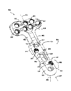
90,
also referred to as torque drivers, may be use to drive each of first fastener
401
(including first fastener peg 431) and the larger, second fastener 451.
However,
second drive instrument 90 may be used to transmit more torque to second
fastener
451 than what is possible using first drive instrument 70.
[0118] As shown in FIG. 25, drive end 80 includes a conical portion 88 that
transitions distally, in the direction of axis 81, shaft 74 to a square taper
portion 86,
21
CA 02959138 2017-02-23
WO 2016/033311
PCT/US2015/047157
which in turn transitions to a square drive tip 84. When drive tip 84 is fully
inserted
into either one of drive socket 412 of first fastener 401 or double-socket 462
of
second fastener 451, a portion of square taper 86 wedges into the non-tapered
sidewalls of either drive socket 412 or double-socket 462, respectively. This
feature
causes either one of first fastener 401 or second fastener 451 to "stick" to
the drive
end of either one of first drive instrument 70 or second drive instrument 451,
to
facilitate removal of each fastener from container 10 and to position the
fastener
into one of the apertures of the bone plate and partially into the drilled
hole in the
bone prior to transmitting torque to drive the fastener into the bone.
[0119] As shown in FIG. 26, drive end 92 includes a first tapered square
portion 95 that transitions distally, in the direction of axis 91, shaft 94 to
a first
square drive portion 96. A second tapered portion 97 extends distally along
axis 91
to a second square drive portion 98 that is smaller than first square drive
portion 96.
When drive tip 92 is fully inserted into either one of drive socket 412 of
first
fastener 401 or double-socket 462 of second fastener 451, a portion of second
square taper portion 97 wedges into the non-tapered sidewalls of either of
drive
socket 412 or double-socket 462, respectively. When drive end 92 is fully
inserted
into double-socket 462 of second fastener 451, at least one of first tapered
portion
95 or second tapered portion 97 stick into double-socket 462. This also serves
to aid
the surgeon in the pick-up and placement of the fasteners. For obvious
reasons,
when a limited number of fasteners are readily available, it is highly
desirable to
avoid dropping fasteners into the wound site of the patient or onto a non-
sterile
surface in the operating room.
[0120] FIG. 27 is a perspective view of a first drill guide 330 for
use with
one of the appropriately sized, wire drills 370, to drill a hole into bone for
receiving
first fastener 401. A plurality of first drill guides 330 may be preassembled
with
bone plates as previously shown in FIGS. 4, 8, 11 and 12. First drill guide
330
includes a body 336 having a distal end 332, a proximal end 334, and a bore
338
sized and shaped to guide the appropriately sized wire drill 370 and defining
a
longitudinal axis 331. Distal end 332 includes threads 333 for removable
attachment
to a threaded aperture of a bone plate. Proximal end 334 includes four
indentations
22
CA 02959138 2017-02-23
WO 2016/033311
PCT/US2015/047157
339 spaced evenly apart on the periphery of bore 338 for receiving square
drive tip
84 of first drive instrument 70 and second drive portion 98 of second drive
instrument 90.
[0121] FIG. 28 is a perspective view of a second drill guide 340 for
use with
one of the appropriately sized, wire drills 370, to drill a hole into bone for
receiving
second fastener 451. A plurality of second drill guides 340 may be
preassembled
with bone plates as previously shown in FIGS. 9 and 13. Second drill guide 340
includes a body 346 having a distal end 342, a proximal end 344, and a bore
348
sized and shaped to guide the appropriately sized wire drill 370 and defining
a
longitudinal axis 341. Distal end 342 includes threads 343 for removable
attachment
to a threaded aperture of a bone plate. Proximal end 344 includes four
indentations
349 spaced evenly apart on the periphery of bore 348 for receiving square
drive tip
84 of first drive instrument 70 and second drive portion 98 of second drive
instrument 90.
[0122] FIG. 29 is a cross-sectional view of a non-locking aperture 270 that
may be sized to receive first fastener 401. (The term "aperture", as used
herein, is
interchangeable with the term "hole".) Similarly, although not shown in detail
views
in the figures, non-locking aperture 270 may also be sized to receive second
fastener
451. FIG. 30 is a cross-sectional view of a portion of a bone plate 271 (for
no
particular surgical indication, but shown for description purposes), showing
three
possible trajectories of first fastener 401 of FIG. 20 inserted into non-
locking
aperture 270 of FIG. 29. Non-locking aperture 270 extends between a top
surface
286 and a bottom surface 284 of a plate 271 and defines an axis 272. Non-
locking
aperture 270 has a conical upper portion 274 and tapers from top surface 286
towards the middle of plate 271. A conical lower portion 278 is coaxial with
conical
upper portion 274 and tapers from bottom surface 284 towards the middle of
plate
271 to form a waist 282 with conical upper portion 274. The position and
orientation of waist 282 relative to top surface 286 may vary, but as shown in
FIG.
29, is deep enough to receive head 404 of first fastener 401, such that head
404 is
not proud to top surface 286. As shown in FIG. 30, first fastener 401 may be
inserted through plate 271 in any desired trajectory within a range defined by
a
23
CA 02959138 2017-02-23
WO 2016/033311
PCT/US2015/047157
conical angle 422, wherein axes 401', 401" and 401" define three possible
trajectories of first fastener 401 within that range. This multidirectional
ability
allows the surgeon to form a polyaxial non-locking compressive construct.
[0123] FIG. 31 is a cross-sectional view of a locking aperture 250,
which is
very similar to other locking apertures of bone plates that are well-known in
the art.
FIG. 32 is a cross-sectional view of bone plate 271 with first fastener 401 of
FIG. 20
inserted at a fixed angle into locking aperture 250. Similarly, although not
shown in
detailed views in the figures, locking aperture 250 may be sized to receive
second
fastener 451. Locking aperture 250 includes a tapered, threaded bore 254 for
receiving head 404 of first fastener 401. Bore 254 extends between top surface
286
and bottom surface 284 of plate 271 and defines an axis 252. As shown in FIG.
32,
when first fastener 401 is fully inserted into plate 271, axis 420 of first
fastener 401
is coaxial with axis 252 of locking aperture 250. This arrangement allows the
surgeon to form a fixed-angle locking construct.
[0124] FIG. 33 is a top view of a unidirectionally ramped (or UR) aperture
290. FIG. 34 is a cross-sectional view of UR aperture 290. FIG. 35 is a cross-
sectional view of UR aperture 290 with first fastener 401 partially inserted
therein.
FIG. 36 is a cross-sectional view of UR aperture 290 with first fastener 401
fully
inserted therein. UR aperture 290 may also be sized, although not shown in
detail
views in the figures, to receive second fastener 451. UR aperture 290 is a non-
locking type of aperture for compressively attaching the bone plate against
the bone.
The surgeon may also use UR aperture 290 to aid in reduction of the bone
fragments, i.e., the compression of bone fragments along the longitudinal axis
of the
bone plate, often referred to in the art as dynamic compression. As shown in
FIGS.
35 and 36, proper insertion of first fastener 401 into UR aperture 290 causes
plate
271 to shift in a direction depending on the orientation of UR aperture 290.
As
shown in FIGS. 33 and 34, UR aperture 290 has an upper conical portion 294
intersecting with a coaxially opposing, lower conical portion 298 to form a
waist
282 about an axis 292, in an arrangement similar to non-locking aperture 270
of
FIG. 29. UR aperture 290 further includes a circular bore portion 306 defining
an
axis 307 that is parallel and offset from axis 308. Circular bore portion 306
is sized
24
CA 02959138 2017-02-23
WO 2016/033311
PCT/US2015/047157
to receive shaft 408 of first fastener 401, but is too small to receive head
404. The
surgeon may drill a hole into bone that is approximately coaxial with axis 307
and
then insert first fastener 401 as shown in FIGS. 35 and 36, such that head 404
tends
to seat into upper conical portion 294, and "ramp" in a translation direction
along
plate axis 272. The translation distance possible is determined by an offset
distance
308 between axis 292 and 307.
[0125] FIG. 37 is a top view and FIG. 38 is a cross-sectional view of
a
bidirectionally ramped (BR) aperture 501, which is similar to UR aperture 290
of
FIG. 34. BR aperture 501 may be sized to receive first fastener 401 or second
fastener 451. The surgeon may use BR aperture 501 to compressively attach bone
plate 271 against the bone, and also to dynamically compress the bone
fragments
along the longitudinal axis of plate 271 in either of opposing directions.
This
bidirectional feature allows the surgeon to reduce fragments on either side of
BR
aperture 501. BR aperture 501 includes an upper conical portion 504 defining
an
axis 502, a coaxial, lower conical portion 508, a waist 512, a first circular
bore
portion 516 defining an axis 517, and an opposing second circular bore portion
518
defining an axis 519. The surgeon may use BR aperture 501 with first fastener
401
to translate plate 271 an offset distance 521 in a first direction along axis
272 of
plate 271, or an offset distance 522 in a second, opposing direction along
axis 272.
[0126] FIG. 39 is a top view of a unidirectionally ramped (UR) slot 310
positioned along axis 272 of plate 271. FIG. 40 is a cross-sectional view of
UR slot
310, taken through axis 272; FIG. 41 is a cross-sectional view of UR slot 310,
taken
through line 41-41 of FIG. 39; FIG. 42 is a top view of first fastener 401
partially
inserted into UR slot 310; FIG. 43 is a top view of first fastener 401 fully
inserted
into the UR slot 310; FIG. 44 is a cross-sectional view, taken through axis
272, of
first fastener 401 partially inserted into UR slot 310; FIG. 45 is a cross-
sectional
view, taken through axis 272, of first fastener 401 fully inserted into the UR
slot
310. UR slot 310 is more elongated than UR aperture 290, and also may be used
to
dynamically compress bone fragments as first fastener 401 is inserted into
bone.
The use of slotted apertures similar to UR slot 310 in bone plates is well-
known in
the art for reducing bone fragments as the surgeon attaches the plate to the
bone. UR
CA 02959138 2017-02-23
WO 2016/033311
PCT/US2015/047157
slot 310 has an elongated, tapered portion 314 that defines a slot axis 315
and tapers
from top surface 286 to bottom surface 284 of plate 271. A circular bore
portion 312
is formed into one end of tapered portion 314 and is sized to receive shaft
408 of
first fastener 401, but not head 404. As the surgeon inserts first fastener
401 into
bone as shown in FIGS. 44 and 45, head 404 tends to seat into tapered portion
310
and move plate 271 in a direction along axis 272 a distance 317 (FIG. 45).
[0127] FIGS. 46 and 47 shown a bidirectionally ramped (BR) slot 320
that is
similar to UR slot 310, except the surgeon may use BR slot 320 to dynamically
compress bone fragments in either of opposing directions along axis 292 of
plate
271. BR slot 320 includes an elongated, tapered portion 325 that tapers from
top
surface 286 to bottom surface 284 of plate 271. A first circular bore portion
322 and
a second circular bore portion 325 are formed into opposing ends of tapered
portion
325.
[0128] Each of UR slot 310 and BR slot 320 may be sized to receive
either
first fastener 401 or second fastener 451.
[0129] FIG. 48 is an end view, FIG. 49 is a top view, and FIG. 50 is a
perspective view of a first DVR assembly 102 that was earlier described for
FIG. 4.
First DVR assembly 102 includes a first DVR plate 104 that has a head 106, a
neck
108 and a shaft 110 that extends along a longitudinal axis 111.
[0130] Head 106 includes a plurality of locking apertures 250, each of
which
is assembled with a first drill guide 330. Each of locking apertures 250 of
head 106
defines a desired, fixed trajectory, such that insertion of first fastener 401
into each
locking aperture 250 of head 106 provides subchondral support of the
articulation
surface of the wrist joint of the distal radius.
[0131] Shaft 110 includes a plurality of locking apertures 250, a plurality
of
non-locking apertures 270, and one UR slot 310, wherein the respective axis of
each
aperture is generally directed inwardly towards the center of the underlying
bone.
Each of locking apertures 250 is assembled with one of first drill guides 330.
Each
of locking apertures 250, non-locking apertures 270, and UR slot 310 is sized
for
receiving first fastener 401. Each locking aperture 250 of shaft 110 is paired
closely
together with one of the non-locking apertures 270 to form four, spaced-apart,
26
CA 02959138 2017-02-23
WO 2016/033311
PCT/US2015/047157
groupings or clusters, including a first grouping 120, a second grouping 130,
a third
grouping 140 and a fourth grouping 146, and corresponding to a first region
121, a
second region 131, a third region 141, and a fourth region 147 on shaft 110.
First
grouping 120 opposes second grouping 130 about longitudinal axis 111 of plate
104, such that aperture axes 123 and 125 of first grouping 120 cross-over
aperture
axes 133 and 135 of second grouping 130. Similarly, third grouping 140 opposes
fourth grouping 146.
[0132] During the surgical procedure, the surgeon may insert one of
first
fasteners 401 into each of regions 121, 131, 141, and 147. The surgeon may
choose
whether to select one of locking apertures 250 or one of non-locking apertures
270
for each region. In general, surgeons may choose to use locking apertures 250
if the
underlying bone is not in condition to provide optimal engagement with the
threads
of shaft 110 of first fastener 401.
[0133] It should be appreciated that first DVR assembly 102 may be
attached to the distal radius of a patient using only one type of bone
fastener, i.e., a
plurality of first fasteners 401 of varying lengths. In many current bone
plate
systems for fixation of the distal radius, a number of different types of
fasteners are
required. By using only one type, it is possible to reduce the number of
instruments
required in DVR kit 100, thereby reducing the size of container 10 (FIG. 4)
and
potentially lowering the overall cost of DVR kit 100. Using only one type of
fastener also may help surgeons, especially those who are not greatly
experienced
doing the procedure, to perform the surgical procedure more quickly and
without
using the fasteners inappropriately.
[0134] FIG. 51 is an end view, FIG. 52 is a top view, and FIG. 53 is a
perspective view of a second DVR assembly 152, which includes a second DVR
plate 154 assembled with a plurality of first drill guides 330, and a
plurality of
second drill guides 340. Second DVR plate has a head 156, a neck 158 and a
shaft
160 that extends along a longitudinal axis 161.
[0135] Head 156 includes a plurality of locking apertures 250, each of
which
is assembled with one of first drill guides 330 and is sized for receiving one
of first
fasteners 401. Each of locking apertures 250 of head 156 defines a desired,
fixed
27
CA 02959138 2017-02-23
WO 2016/033311
PCT/US2015/047157
trajectory, such that insertion of first fastener 401 into each locking
aperture 250 of
head 156 provides subchondral support of the articulation surface of the wrist
joint
of the distal radius.
[0136] Shaft 154 includes two of locking apertures 250, each of which
is
assembled with one of second drill guides 340 and is sized to receive one of
second
fasteners 451. Each of locking apertures 250 in shaft 154 is paired closely
together
with one of UR apertures 290, each of which is sized to receive one of second
fasteners 451, to form a first grouping 170 that is spaced apart from a second
grouping 180 along axis 161. First grouping 170 corresponds to a first region
171
and second grouping 180 corresponds to a second region 181 of plate 154. As
for
first DVR assembly 102, the axes 173, 175, 183 and 185 of the apertures of
shaft
160 of second DVR assembly 152 are generally directed towards the center of
the
bone. Shaft 154 also includes BR slot 320 positioned approximately midway
along
axis 161.
[0137] Second DVR assembly 152 requires two types of fasteners, i.e., first
fasteners 401 and second fasteners 451 of varying lengths. However, we
envision
that using two of second fasteners 451 in shaft 160 precludes the need to use
four of
first fasteners 401 in shaft 110 of first DVR assembly 102. This facilitates a
quicker
surgical procedure and eliminates the cost of the additional two fasteners.
[0138] Another feature of second DVR assembly 152 is the enhanced ability
to draw the fractured bone fragments together axially as the fasteners are
inserted.
That is because, the dynamic compression that is achievable using UR apertures
290, if done in proper sequence, may be additive to the dynamic compression
that is
achievable using UR slot 310.
[0139] FIG. 54 is an end view, FIG. 55 is a top view, and FIG. 56 is a
perspective view of a third DVR assembly 552, which includes a third DVR plate
554 assembled with a plurality of first drill guides 330, and a plurality of
second
drill guides 340. Third DVR plate 554 has a head 556, a neck 558 and a shaft
560
that extends along a longitudinal axis 561.
[0140] Head 556 includes a plurality of locking apertures 250, each of
which
is assembled with one of first drill guides 330 and is sized for receiving one
of first
28
CA 02959138 2017-02-23
WO 2016/033311
PCT/US2015/047157
fasteners 401. Each of locking apertures 250 of head 556 defines a desired,
fixed
trajectory, such that insertion of first fastener 401 into each locking
aperture 250 of
head 556 provides subchondral support of the articulation surface of the wrist
joint
of the distal radius.
[0141] Shaft 554 includes two of locking apertures 250, each of which is
assembled with one of second drill guides 340 and is sized to receive one of
second
fasteners 451. Each of locking apertures 250 in shaft 554 is paired closely
together
with one of BR apertures 320, each of which is sized to receive one of second
fasteners 451, to form a first grouping 570 that is spaced apart from a second
grouping 580 along axis 561. First grouping 570 corresponds to a first region
571
and second grouping 180 corresponds to a second region 581 of plate 554. As
for
the previously described DVR assemblies 102 and 152, the axes of the apertures
of
shaft 560 of third DVR assembly 552 are generally directed towards the center
of
the bone. Shaft 554 also includes BR slot 320 positioned approximately midway
along axis 561.
[0142] Third DVR assembly 552 requires two types of fasteners, i.e.,
first
fasteners 401 and second fasteners 451 of varying lengths. However, as for
second
DVR assembly 152, we envision that using two of second fasteners 551 in shaft
560
precludes the need to use four of first fasteners 401 in shaft 110 of first
DVR
assembly 102. This facilitates a quicker surgical procedure and eliminates the
cost
of the additional two fasteners.
[0143] Again as with second DVR assembly 152, third DVR assembly 552
has the enhanced ability to draw the fractured bone fragments together axially
as the
fasteners are inserted since the dynamic compression that is achievable using
BR
apertures 501, if done in proper sequence, may be additive to the dynamic
compression that is achievable using BR slot 320. However, third DVR assembly
552 has the additional ability to provide dynamic compression in either
direction
along axis 561 of plate 554.
[0144] FIG. 57 is a perspective view of a fourth DVR assembly 652,
including a fourth DVR plate 654 that may be assembled with a plurality of
first
drill guides 330, and a plurality of second drill guides 340 (not shown but as
29
CA 02959138 2017-02-23
WO 2016/033311
PCT/US2015/047157
discussed above with regard to the other DVR assemblies 102, 152, 552 shown in
FIGS. 49-56). Fourth DVR plate 654 has a head 656, a neck 658, and a shaft 660
that extends along a longitudinal axis 661 of the DVR assembly 652. The head
656
may be wider than the shaft 660.
[0145] The head 656 may similarly include a plurality of spaced apart
locking apertures 250, each of which may be preassembled with one of the first
drill
guides 330 (not shown) and sized for receiving one of the first fasteners 401.
Each
of the locking apertures 250 of the head 656 may define a desired, fixed
trajectory,
such that insertion of first fastener 401 into each locking aperture 250 of
head 656
provides subchondral support of the articulation surface of the wrist joint of
the
distal radius.
[0146] As shown, the shaft 660 may also include a plurality of spaced-
apart
locking apertures 250, each of which may be assembled or preassembled with one
of the second drill guides 340 (not shown) and is sized to receive one of the
second
fasteners 451. Each of the locking apertures 250 in the shaft 660 may be
paired
closely together with a non-locking aperture 601, each of which may be sized
to
receive one of the second fasteners 451. As shown, the shaft 660 may include a
first
grouping, or first aperture cluster 670, spaced apart from a second aperture
cluster
680 generally along the longitudinal axis 661. The first aperture cluster 670
corresponds to a proximal region 671 of the shaft 660, and the second aperture
cluster 680 corresponds to a distal region 681 of the shaft 660. In various
aspects,
the locking apertures 250 of the aperture clusters 670, 680 may generally be
disposed on laterally opposed sides of the longitudinal axis 661.
[0147] The non-locking apertures 601 may be aligned on the
longitudinal
axis 661, and the locking apertures 250 may be offset from the longitudinal
axis
661. Similar to the previously described DVR assemblies 102, 152, and 552,
each
locking aperture 250 of the shaft 660 may define an opening having an axis 664
that
is generally inwardly directed towards a center of the fractured bone, which
is
oblique to the longitudinal axis 661. For example, in certain embodiments, the
size
of the implant head 656 may influence aspects of the design, including the
angles of
the longitudinal axis 664 of the locking aperture 250 with respect to the axis
665
CA 02959138 2017-02-23
WO 2016/033311
PCT/US2015/047157
normal or perpendicular to the plate shaft 660. By way of example, the angle
between longitudinal axis 664 and normal axis 665 may be from about 10 degrees
to
about 30 degrees, depending in part on the size of the bone, such that a screw
trajectory travels through a center region of the bone. In various
embodiments, at
.. least two respective threaded apertures 250 of the first and second
aperture clusters
670, 680 define openings having axes that may be divergent from each other.
The
non-locking apertures 601 generally define an opening having an axis 665
normal to
the longitudinal axis 661. As shown, the shaft 660 may also include a UR or BR
aperture or slot 620 positioned approximately midway or centered along the
.. longitudinal axis 661 and between the aperture clusters 670, 680. The bone
plates
654 may also have one or more shaped openings 688 extending between the top
and
bottom surfaces of the bone plate 654, which may be configured to guide a K-
wire
passed therethrough.
[0148] Two types of fasteners, i.e., first fasteners 401 and second
fasteners
451 of varying lengths may be used with the fourth DVR assembly 652. Again as
with second and third DVR assemblies 152, 552, the fourth DVR assembly 652
also
has the enhanced ability to draw the fractured bone fragments together axially
as the
fasteners are inserted since the dynamic compression that is achievable using
UR or
BR apertures 601, if done in proper sequence, may be additive to the dynamic
compression that is achievable using BR slot 620. Thus, the fourth DVR
assembly
652 has the additional ability to provide dynamic compression in either
direction
along the longitudinal axis 661 of the plate 654.
[0149] FIG. 58 is a perspective view of a fifth DVR assembly 752. The
fifth
DVR assembly 752 is similar in shape, design, and function to the fourth DVR
assembly 652, and corresponding references numbers from FIG. 57 are used to
indicate corresponding features in FIG. 58. Generally, the configuration of
the bone
plate 754 of FIG. 58 includes additional locking apertures 250 on both the
head 756
and shaft 760 portions. For example, the aperture clusters 770 and 780 include
two
threaded, locking bone fastener apertures 250. As shown, two of the apertures
250a
are on a first side of the longitudinal axis 661 and adjacent the BR slot 620,
laterally
opposed to two apertures 250b on a second side of the longitudinal axis 661,
31
CA 02959138 2017-02-23
WO 2016/033311
PCT/US2015/047157
adjacent the non-locking apertures 601. Considering this design, in various
aspects a
physician may be able to install and conform the assembly 752 to the natural
variations in an anatomy by installing non-locking screws first. During the
installation of the non-locking screws, a physician may become more aware of
the
bone quality and further decide whether locking screws may be needed. For
example, if a non-locking screw spins or otherwise loses its grip or is
stripped, a
locking screw may be inserted into aperture 250a and/or 250b as necessary.
[0150] The methods for implanting the bone plate system of FIGS. 57
and
58 are similar to the methods for implanting the DVR assemblies of the first
through
third assemblies 102, 152, 552. A bone plate is provided and aligned with the
fractured bone. The methods may include shaping the bone plate, including
inserting a first bone fastener into the bone through at least one of the
unidirectionally ramped aperture and non-threaded bone fastener apertures in a
direction normal to the longitudinal axis and dynamically compressing the bone
along the longitudinal axis of the shaft portion of the bone plate. Once
compressed
and/or shaped, the method may include securing the bone plate, including
inserting
a second bone fastener into the bone through at least one of the threaded,
locking
bone fastener apertures in a direction oblique to the longitudinal axis and
locking
the bone plate to the bone.
[0151] We have shown and described various embodiments and examples.
However, a person having ordinary skill in the art may modify the methods and
devices described herein without departing from the overall concept. For
instance,
the specific materials, dimensions and the scale of drawings should be
understood to
be non-limiting examples. Accordingly, we do not intend the scope of the
following
claims to be understood as limited to the details of structure, materials or
acts shown
and described in the specification and drawings.
[0152] The foregoing description of the embodiments has been provided
for
purposes of illustration and description. It is not intended to be exhaustive
or to limit
the disclosure. Individual elements or features of a particular embodiment are
generally not limited to that particular embodiment, but, where applicable,
are
interchangeable and can be used in a selected embodiment, even if not
specifically
32
CA 02959138 2017-02-23
WO 2016/033311
PCT/1JS2015/047157
shown or described. The same may also be varied in many ways. Such variations
are
not to be regarded as a departure from the disclosure, and all such
modifications are
intended to be included within the scope of the disclosure.
33