Note: Descriptions are shown in the official language in which they were submitted.
SERINE PROTEASES AS BIOMARKERS FOR OVARIAN CANCER
CROSS-REFERENCE TO RELATED APPLICATION
[001] This application claims the benefit of priority to U.S. Provisional
Application No.
62/043,290 (filed August 28, 2014), entitled -Kallikrein Family Proteases KLK6
and KLK7 as
Biomarkers for Ovarian Cancer,".
FIELD OF THE INVENTION
[002] The described invention generally relates to ovarian cancer.
BACKGROUND
Ovarian Cancer
[003] Ovarian cancer ranks as the fifth most common cancer in women and has
the highest
mortality rate among gynecologic malignancies (Suh KS, Park SW, Castro A,
Patel H, Blake
P, Liang M, et al. Ovarian cancer biomarkers for molecular biosensors and
translational
medicine. Expert Rev Mol Diagn 2010;10:1069-83; Landen CN Jr, Birrer MJ, Sood
AK. Early
events in the pathogenesis of epithelial ovarian cancer. J Clin Oncol
2008;26:995-1005).
Although the 5-year survival rate of ovarian cancer is around 90% when
detected in early stages
(I/II), nearly 80% of the new cases are diagnosed in advanced stages (III/IV)
because of the
asymptomatic nature of the disease at stage I and early stage II.
Unfortunately, the 5-year
survival rate of advanced ovarian cancer is as low as 11% (Altekruse SF,
Kosary CL, Krapcho
M, Neyman N, Aminou R, Waldron W, et al. SEER Cancer Statistics Review, 1975-
2007,
National Cancer Institute. Bethesda, MD). Therefore, there is a need to find
reliable biomarkers
for early detection of ovarian cancer.
Types of Ovarian Cancer
[004] Ovarian cancer is not a single disease but consists of more than 30
types and subtypes
of malignancies, each with its own histopathologic appearance and biologic
behavior.
Generally, ovarian cancers are grouped into 3 major categories: (1) epithelial
tumors (tumors
arising from cells that line or cover the ovaries); (2) germ cell tumors
(tumors that originate
from cells that are
1
Date Recue/Date Received 2020-08-28
CA 02960012 2017-02-28
WO 2016/033464 PCMJS2015/047434
destined to form eggs within the ovaries; and (3) sex cord-stromal cell tumors
(tumors that begin
in connective cells that hold the ovaries together and produce female
hormones). The most
common ovarian cancers are epithelial tumors, which account for about 90% of
all ovarian
cancers. Ovarian epithelial tumors are divided into subtypes which include
serous, papillary
serous, endometrioid, mucinous and clear cell tumors.
Common Epithelial Tumors
Serous
[005] The serous subtype of ovarian carcinoma accounts for approximately 60-
80% of
ovarian cancer cases and exhibits the most aggressive histology (Levanon K. et
al., J. Clinical
Oncology, November 10, 2008 Vol. 26 No. 32 5284-5293). Fewer than 25% of
serous ovarian
cancer cases are detected at an early stage (stages I and II), which reflects
grimly on survival
figures (Seidman J.D. et al., Int. J. Gynecol. Pathol. 23:41-44, 2004). High-
grade serous
carcinoma involves the surface of the ovary, often bilaterally, and the
peritoneal membranes,
with rapid onset of carcinomatosis, a fact that restricts the surgical options
to debulking only
(Levanon K. et al.. J. Clinical Oncology, November 10, 2008 Vol. 26 No. 32
5284-5293).
Despite the introduction of taxanes to therapeutic protocols and the prolonged
survival with
intraperitoneal chemotherapy administration, there has been little progress in
improving cure
rates, a parameter that is still solely dependent on the disease stage at the
time of presentation
(Levanon K. et al., J. Clinical Oncology, November 10, 2008 Vol. 26 No. 32
5284-5293).
Papillary Serous
[006] Papillary serous carcinoma of the ovary is one of the most common and
lethal
malignant tumors (Tong G-X et al., Modern Pathology (2007) 20, 856-863).
Papillary serous
histology accounts for 75% of ovarian cancers and its histological pattern
simulates the lining of
the fallopian tube (Jelovac D. and Armstrong D. K., CA: A Cancer Journal for
Clinicians, Vol.
61, Issue 3, pp. 183-203, May/June 2011). Most cases of papillary serous
ovarian cancer are
diagnosed at advanced stages, when the tumors have already metastasized (Kim
J. et al., PNAS,
March 6, 2012, Vol. 109, No. 10, pp. 3921-3926). Despite the steady
improvement of surgery
and chemotherapy, greater than 90% of women with advanced ovarian cancers die
after relapse
(Bukowski R. M. et al. (2007) Semin. Oncol. 34(Suppl 2):S1-S15). Early
detection of these
2
CA 02960012 2017-02-28
WO 2016/033464 PCMJS2015/047434
high-grade serous carcinomas is thus key to reducing ovarian cancer deaths
(Bast R. C. Jr. et al.
(2009) Nat. Rev. Cancer 9:415-428).
Endometrioid
[007] Ovarian endometrioid carcinomas account for only 10% of ovarian
carcinomas
(McConechy M. K. et al., Modern Pathology (2007) 27, 128-134). The majority of
ovarian
endometrioid carcinomas are low-grade carcinomas with good prognosis (Chen S.
et al., Modern
Pathology (2005) 18:903-911).
Mucinous
[008] The mucinous cell type accounts for approximately 10% of all primary
epithelial
ovarian carcinomas (Chan J. K. et al., Gynecol. Oncol. 2008; 109:370-376).
Most mucinous
epithelial ovarian carcinomas are diagnosed early (International Federation of
Gynecology and
Obstetrics (FIGO) stages and confined to one ovary. In stage I mucinous
epithelial
ovarian carcinomas. the 5-year disease-free survival rate is about 90%, which
is slightly better
than the 76% observed for patients with serous epithelial ovarian carcinomas
(Vergote I. et al.,
Lancet 2001; 357:176-182). Less frequently, primary mucinous epithelial
ovarian carcinoma is
associated with peritoneal carcinomatosis and/or extraperitoneal metastases
(FIGO stages JIB¨
IV). Unlike FIGO stage I tumors, advanced mucinous epithelial ovarian
carcinomas reportedly
have poorer prognoses than serous epithelial ovarian carcinomas (Omura G. A.
et al., J. Clin.
Oncol. 1991; 9:1138-1150; Teramukai S. et al., J. Clin. Oncol. 2007; 25:3302-
3306).
Clear Cell
[009] Ovarian clear cell adenocarcinomas account for <5% of all ovarian
malignancies and
3.7-12.1% of all epithelial ovarian carcinomas (Tan D. S. P. and Kaye S., J.
Clin. Pathol. April
2007; 60(4): 355-360). Compared to other epithelial ovarian cancer (EOC)
subtypes, when at an
advanced stage, they are associated with a poorer prognosis and are relatively
resistant to
conventional platinum-based chemotherapy (Sugiyama T. et al., Cancer. 2000
June 1;
88(11):2584-9). By contrast, early-stage clear cell ovarian cancer carries a
relatively good
prognosis (Tan D. S. P. and Kaye S., J. Clin. Pathol. April 2007; 60(4): 355-
360). Hence, early
3
CA 02960012 2017-02-28
WO 2016/033464 PCMJS2015/047434
detection is the key to improve prognosis and reduce deaths associated with
this type of ovarian
cancer.
Ovarian Cancer Staging
[0010] The process used to determine whether ovarian cancer has spread
within the ovaries
or to other parts of the body (i.e., metastasized) is called staging. It is
important to determine the
stage of ovarian cancer because the stage will determine the type of treatment
plan selected to
combat the disease. The results of tests used to diagnose ovarian cancer are
often also used to
stage the disease. Such tests include ultrasound, computerized tomography (CT)
scan, positron
emission tomography (PET) scan, magnetic resonance imaging (MRI), X-ray and
biopsy.
Ovarian cancer staging guidelines have been developed by the International
Federation of
Gynecologists and Obstetricians (FIGO). The FIGO staging system for ovarian
cancer is shown
in Table 1.
Table I. FIGO Ovarian Cancer Staging
Stage Area of Involvement
IA Tumor limited to 1 ovary, capsule intact, no tumor on surface,
negative washings
TB Tumor involves both ovaries; otherwise like IA
IC1 Surgical spill
IC2 Capsule rupture before surgery or tumor on ovarian surface
IC3 Malignant cells in the ascites or peritoneal washings
IIA Extension and/or implant on uterus and /or Fallopian tubes
JIB Extension to other pelvic intraperitoneal tissues
IIIA1 Positive retroperitoneal lymph nodes only
IIIA 1 (i) ¨ Metastasis < 10 mm
IIIA 1(H) ¨ Metastasis > 10 mm
II1A2 Microscopic, extrapelvic (above the brim) peritoneal involvement +
positive
retroperitoneal lymph nodes
IIIB Macroscopic, extrapelvic, peritoneal metastasis < 2 cm + positive
retroperitoneal
lymph nodes; includes extension to capsule of liver/spleen
4
CA 02960012 2017-02-28
WO 2016/033464 PCMJS2015/047434
IIIC Macroscopic, extrapelvic, peritoneal metastasis > 2 cm + positive
retroperitoneal
lymph nodes; includes extension to capsule of liver/spleen
IVA Pleural effusion with positive cytology
IVB Hepatic and/or splenic parenchymal metastasis, metastasis to extra-
abdominal organs
(including inguinal lymph nodes and lymph nodes outside of the abdominal
cavity)
Ovarian Cancer Grading
[0011] In addition to staging, an ovarian tumor can also be described by
grade (G). Grading
determines how similar ovarian cancer tissue is to normal tissue. Tumor grade
is determined by
microscopic examination of cancer tissue; with healthy cells appearing as well-
differentiated.
That is, the more differentiated the ovarian tumor, the better the prognosis.
The ovarian cancer
grading system is shown in Table 2.
Table 2. Ovarian Cancer Grading
Grade Description
GX Grade cannot be evaluated
GB Tissue considered borderline cancerous; low malignant potential
G1 Tissue is well-differentiated (healthy cells)
G2 Tissue is moderately differentiated (more abnormal than health cells)
G3 to G4 Tissue is poorly differentiated or undifferentiated (all or most
cells appear abnormal)
[0012] Serous ovarian cancer is not graded in this way and only considers a
low-grade and a
high-grade classification. Low-grade serous carcinomas exhibit low-grade
nuclei with
infrequent mitotic figures. They evolve from adenofibromas or borderline
tumors, have frequent
mutations of the KRAS, BRAF, or ERBB2 genes, and lack TP53 mutations (Type I
pathway).
Low-grade tumors are indolent and have better outcome than high-grade tumors.
In contrast,
high-grade serous carcinomas have high-grade nuclei and numerous mitotic
figures (See, yang
R. et al.. Adv. Anat. Pathol. Sep 2009; 16(5):267-282).
CA 02960012 2017-02-28
WO 2016/033464 PCMJS2015/047434
Treatment for Ovarian Cancer
Early Stage (FIGO Stage I-II) Ovarian Cancer
[0013] Due to the lack of effective screening programs, ovarian cancer is
diagnosed at an
early stage only in about 25% of cases (Kim A. et al.. Journal of Experimental
& Clinical Cancer
Research 2012, 31:14). In most of these cases, surgery is able to cure the
disease, and the five-
year survival rate for early-stage (stage I or II) ovarian cancer is around
90% (Hennessy BT, et
al., Lancet 2009, 374: 1371-82). Adjuvant chemotherapy for early stage ovarian
cancer is still
controversial, but some studies have shown its benefit under confined
conditions. According to
these studies, patients with IA or IB FIGO stage, non-clear-cell histology,
well-differentiated
(G1) tumors, and an "optimal" surgery (i.e., performed according to
international guidelines,
with pelvic and retroperitoneal assessment), appear not to benefit from
chemotherapy (Trimbos
TB et al., I Natl Cancer Inst 2003, 95:105-112). Thus, it is commonly believed
that, at least in
these cases, chemotherapy can probably be avoided and patients can be advised
to undergo
clinical and instrumental follow-up. In all the other (early stage) patients,
(adjuvant)
chemotherapy is indicated (Hennessy BT, et al., Lancet 2009, 374: 1371-82).
Advanced (FIGO Stage III-IV) Ovarian Cancer
[0014] The standard treatment for patients with advanced ovarian cancer is
maximal surgical
cytoreduction (i.e., total abdominal hysterectomy, bilateral salpingo-
oophorectomy, pelvic and
para-aortic lymphadenectomy and omentectomy) followed by systemic platinum-
based
chemotherapy (e.g., cisplatin followed by carboplatin-based combinations,
cisplatin with
paclitaxel, cisplatin with cyclophosphamide. cisplatin with doxorubicin,
etc.). The expected 5-
year survival for these patients is 10-30% (Hennessey BT et al., Lancet 2009,
374:1371-82). The
concept of primary debulking surgery is to diminish the residual tumor burden
to a point at
which adjuvant therapy will be optimally effective. The percentage of patients
with advanced
ovarian cancer who can optimally undergo cytoreductive surgery seems to range
from 17%-87%
(Ramirez I et al., Cancer Control 2011; 18(1): 22-30). This percentage can
largely depend on the
experience of the surgeon.
6
CA 02960012 2017-02-28
WO 2016/033464 PCMJS2015/047434
Novel Treatment Strategies for Ovarian Cancer
[0015] The larger expectation for improved prognosis in ovarian cancer is
related to the use
of new biological agents. A deeper knowledge of ovarian cancer biology has led
to the
identification of multiple molecular targets, such as growth factor receptors,
signal transduction
pathways, cell cycle regulators, and angiogenic mechanisms (Kim A et al.,
Journal of
Experimental & Clinical Cancer Research 2012, 31:14).
Bevacizumab
[0016] Bevacizumab is a 149-kDa recombinant humanized monoclonal IgG1
directed
against vascular endothelial growth factor (VEGF). It has been FDA-approved
for the treatment
of metastatic colorectal, breast, and non-small cell lung cancer and shows
promise in the
treatment of ovarian cancer. Several phase II studies have shown that
bevacizumab is active in
recurrent ovarian cancer (Ellis LM. Hiclin DJ, Nat Rev Cancer 2008, 8:579-591;
Raspollini MR
et al., Int J Surg Pathol 2005, 13:135-142).
[0017] VEGF expression is higher in ovarian cancer tumors than in normal
ovarian tissue or
benign ovarian tumors, and increasing VEGF expression in either cytosolic
fractions derived
from ovarian cancer tumors or serum VEGF levels in preoperative serum is
considered to be
associated with advanced stage and poor prognosis (Kim A et al., Journal of
Experimental &
Clinical Cancer Research 2012, 31:14).
[0018] In order to inhibit the VEGF pathway, there are two primary
strategies: (1) inhibition
of the VEGF ligand with antibodies or soluble receptors and (2) inhibition of
the VEGF receptor
(VEGI-R) with tyrosine kinase inhibitors (TKIs), or receptor antibodies. Of
the VEGF targeting
therapies, the one most employed has been inhibition of the VEGF ligand with
bevacizumab
(Avastin ).
[0019] Two phase III trials (G0G218, ICON 7) have recently evaluated the
role of
bevacizumab in first-line chemotherapy as an adjunct to carboplatin and
paclitaxel. Bevacizumab
plus chemotherapy (carboplatin-paclitaxel) and bevacizumab maintenance was
demonstrated to
prolong progression-free survival (PFS) by about 4 months (10.3 months versus
14.1 months)
7
CA 02960012 2017-02-28
WO 2016/033464 PCMJS2015/047434
compared to carboplatin-paclitaxel alone (Burger RA et al., N Engl J Med 2011,
365:2473-83;
Perren TJ et al., N Engl J Med 2011, 365:2484-96). A third trial (OCEANS
trial) showed that
the addition of bevacizumab prolonged PFS in platinum-sensitive recurrent
ovarian carcinoma
cases (Aghajanian C et al., J Clin Oncol 2011, 29).
VEGF Receptor Inhibitors
[0020] Oral inhibitors of the VEGF receptor (VEGFR) tyrosine kinase have
been shown to
have activity in patients with recurrent ovarian cancer, resulting in tumor
responses and
stabilization of disease, delaying tumor progression (Friedlander M et al.,
Gynecol Oncol 2010;
119:32-37; Ledermann JA et al., J Clin Oncol 2011; 29:3798-3804; Matulonis UA
et al., J Clin
Oncol 2009; 27:5601-5606; Biagi JJ et al., Ann Oncol 2011; 22:335-340; Matei D
et al., J Clin
Oncol 2011; 29:69-75). Two agents are now in first-line studies. Pazopanib is
an angiogenic
inhibitor with broad spectrum activity against all three VEGF receptors,
platelet derived growth
factor receptor (PDGFR) and c-Kit that has been approved for use in first-line
advanced renal
cancer. Over 900 patients including a sub-set in Asia have been recruited to a
maintenance study,
in which patients receive pazopanib 800 mg daily or placebo until progression,
or up to 2 years
(aGO OVaR-16; trial NCT 00866697; 01227928). The primary end-point is PFS.
[0021] The second trial is with nintedanib (BIBF1120), a potent inhibitor
of VEGFR/PDGFR
and fibroblast growth factor receptor. In this trial, nintedanib or placebo is
given with a standard
regimen of carboplatin and paclitaxel after surgery and continued as
maintenance therapy for up
to 2 years (aGO OVaR-12 trial NCT01015118). This trial also has PFS as its
primary end-point.
[0022] Targeting the angiopoietin axis is another strategy to develop anti-
angiogenic therapy.
aMG 386, a peptibody inhibiting the interaction of angiopoietin-1 and -2 to
the Tie2 receptor,
has been evaluated in combination with weekly paclitaxel in recurrent ovarian
cancer [Karlan,
B.Y. et al., J. Clin. Oncol. December 2011; doi: 10.12005C0.2010.34.31781. The
results of a
phase II trial have been promising and have led to further exploration within
the TRINOVa-3
trial of aMG 386/placebo plus carboplatin/paclitaxel in first-line ovarian
cancer (NCT01493505).
8
CA 02960012 2017-02-28
WO 2016/033464 PCMJS2015/047434
Epidermal Growth Factor Receptor (EGFR) Inhibitors
[0023] The epidermal growth factor receptor (EGFR) is overexpressed in up
to 70% of
ovarian cancer patients (Kohler M et al.. Eur J Cancer 1992; 28a:1432-1437).
However,
responses to EGFR inhibitors in recurrent ovarian cancer are infrequent, and,
as with lung
cancer, are dependent on the presence of a mutation in the catalytic domain of
the EGFR
(Schilder RJ et al, Clin Cancer Res 2005; 11:5539-5584). Erlotinib is a highly
potent oral
inhibitor of the tyrosine kinase region of the EGFR, and this has been studied
in a trial in which
patients with high-risk stage I and stage II¨IV epithelial ovarian cancer who
had completed
platinum-based chemotherapy were randomly assigned to erlotinib maintenance
therapy or
observation following chemotherapy.
Insulin Growth Factor (IGFR) Inhibitors
[0024] Insulin growth factor (IGF 1) is involved in the inhibition of
apoptosis, tumor
progression and metastases. aMG 479 is a monoclonal antibody that is a potent
inhibitor of the
IGF 1 receptor and OSI-906 is an oral dual kinase inhibitor of IGFR1 and the
insulin receptor.
The latter is in clinical trials in recurrent ovarian cancer. A randomized
phase II study of aMG
479 added to first-line chemotherapy in patients with optimally debulked
ovarian cancer is
ongoing (NCT00718523).
Poly (ADP-ribose) Polymerase (PARP) Inhibitors
[0025] The poly (ADP-ribose) polymerases (PARPs) are a large family of
multifunctional
enzymes (Rouleau M et al., Nat Rev Cancer 2010, 10:293-301). PARP-1, the most
abundant
isoform plays a key role in the repair of DNA single-strand breaks through the
repair of base
excisions. The inhibition of PARPs leads to the accumulation of DNA single-
strand breaks,
which causes DNA double-strand breaks at replication forks. These double-
strand breaks are
repaired in normal cells mainly by the error-free homologous recombination
double-stranded
DNA repair pathway, in which essential components are the tumor-suppressor
proteins BRCA1
and BRCA2. In the absence of either BRCA1 or BRCA2, these lesions are not
repaired, which
results in cell cycle arrest and cell death (Itamochi H, World J Biol Chem
2010, 1:209-220).
9
CA 02960012 2017-02-28
WO 2016/033464 PCMJS2015/047434
[0026] Fong et al. (J Clin Oncol 2010, 28:2512-2519) administered the PARP
inhibitor
olaparib to platinum refractory patients. Olaparib had a favorable safety
profile and a high
response rate, in particular in patients with BRCA mutation. In patients with
platinum-resistant
and platinum-refractory disease, the response rate was 41.7% and 15.4%.
respectively (Fong PC
et al., J Clin Oncol 2010, 28:2512-2519). Olaparib (AZD2281) was tested in
BRCA-mutated
patients with ovarian, primary peritoneal, and fallopian tube cancer. In the
study, 20 patients
(40%) responded to the therapy. Currently, randomized trials of olaparib and
other PARP
inhibitors in patients with ovarian cancer are underway.
Biomarkers for Ovarian Cancer
[0027] Identification of early detection biomarkers for ovarian carcinoma
remains a
challenge due to a wide range of morphological, clinical, and genetic
variations found in ovarian
cancer progression (Bast RC Jr, Hennessy B, Mills GB. The biology of ovarian
cancer: new
opportunities for translation. Nat Rev Cancer 2009;6:415-28). Currently
available biomarkers
lack specificity and sensitivity required for routine clinical use (Gubbels
JA, Claussen N. Kapur
AK, Connor JP, Patankar MS. The detection, treatment, and biology of
epithelial ovarian cancer.
J Ovarian Res 2010; 3:8). The only clinically validated biomarker used for
early detection,
disease monitoring, and assessing relapse or response to treatment is CA125;
however, it has low
specificity as a single marker and is not generally recommended for early
detection (Karam AK,
Karlan BY. Ovarian cancer: the duplicity of CA125 measurement. Nat Rev Clin
Oncol 2010;
7:335-9). Although its serum expression is elevated above normal in early
stage (23%) and late
stage (80%) disease, it lacks specificity and sensitivity for detection of
ovarian cancer (Bast RC
Jr, Urban N, Shidhar V, Smith D, Zhang Z, Skates S, et al. Early detection of
ovarian cancer:
promise and reality. Cancer Treat Res 2002;107:61-97). The overexpression of
CA125 is also
frequently observed in benign conditions (e.g., endometriosis) and thus lacks
accurate diagnostic
value for early stage disease (Tuxen MK, Soletormos G, Dombernowsky P. Serum
tumor marker
CA-125 for monitoring ovarian cancer during follow-up. Scand J Clin Lab Invest
2002; 62:177-
188). As an early detection biomarker of ovarian cancer, recent reports
suggest that human
epididymis protein 4 (HE4) provides greater sensitivity and specificity than
CA125 (Anderson
GL, McIntosh M, Wu L, Barnett M, Goodman G, Thorpe G, et al. Assessing lead
time of
selected ovarian cancer biomarkers: a nested case-control study. J Natl Cancer
Inst 2010; 102:26-
CA 02960012 2017-02-28
WO 2016/033464 PCMJS2015/047434
38; Hellstrom I, Hellstrom KR. SMRP and HE4 as biomarkers for ovarian
carcinoma when used
alone and in combination with CA125 and/or each other. Adv Exp Med Biol
2008;622:15-21),
and an assay that detects a combination of HE4, CA125, carcinoembryonic
antigen (CEA), and
vascular cell adhesion molecule 1 (VCAM-1) expression in serum has
significantly better
sensitivity to detect early stage ovarian cancer over benign tumors
(Yurkovetsky Z, Skates S,
Lomakin A, Nolen B, Pulsipher T, Modugno F, et al. Development of a
multimarker assay for
early detection of ovarian cancer. J Clin Oncol 2010;28:2159-66; Nolen B,
Velikokhatnaya
L, Marrangoni A, De Geest K, Lomakin A, Bast RC, et al. Serum biomarker panels
for the
discrimination of benign from malignant cases in patients with an adnexal
mass. Gynecol
Oncol. 2010;117:440-5). In addition, the Food and Drug Administration (FDA)
has approved an
OVA1 test consisting of a panel of five biomarkers: transthyretin,
apolipoprotein A-1, beta2-
microglobulin, transferrin, and CA125 (Zhang Z, Chan DW. The road from
discovery to clinical
diagnostics: lessons learned from the first FDA-cleared in vitro diagnostic
multivariate index
assay of proteomic biomarkers. Cancer Epidemiol Biomarkers Prey 2010;19:2995-
9). This
suggests that use of a combination of multiple markers may generate
synergistic advantages over
single marker diagnostics. Although the multimarker OVA-1 test demonstrates a
much higher
detection sensitivity than a test for CA-125 alone, it is most efficient in
detection of advanced-
stage ovarian cancer and was FDA-cleared for pre-surgical evaluation of women
already
possessing an ovarian mass.
Kallikrein Family of Serine Proteases
[0028] A number of human kallikrein (KLK) family members are associated
with human
cancers and exhibit differential expression in many types of advanced cancers,
including
gastrointestinal, head and neck, lung, ovarian, and brain (Donach M, Yu Y,
Artioli G, Banna G,
Feng W, Bast RC Jr, Combined use of biomarkers for detection of ovarian cancer
in high-risk
women. Tumour Biol 2010;31:209-15; McIntosh MW, Liu Y, Drescher C, Urban N,
Diamandis
EP. Validation and characterization of human kallikrein 11 as a serum marker
for diagnosis of
ovarian carcinoma. Clin Cancer Res 2007;13:4422-8; Shan SJ, Scorilas A,
Katsaros D, Rigault
de la Longrais I, Massobrio M, et al. Unfavorable prognostic value of human
kallikrein 7
quantified by ELISA in ovarian cancer cytosols. Clin Chem 2006; 52:1879-86),
however no
previous reports to date have focused on detection of early stage ovarian
cancer, specifically by
11
CA 02960012 2017-02-28
WO 2016/033464 PCMJS2015/047434
measuring levels of KLK6 and/or KLK7. In a study by El Sherbini et al., 40% of
stage I/II
patients (n=10-15) presented above-normal levels of KLK6 while 83.3% of stage
III/Iy patients
(N=12) were KLK6 positive (El-Sherbini et al. Diagnostic value of serum
kallikrein-related
peptidases 6 and 10 versus CA125 in ovarian cancer, Int. J. Gyn. Cancer 2011,
21(4): 625-632).
Although this study demonstrated that KLK6 has a better sensitivity than CA-
125 as an early
detection biomarker, it included serum tests only (El-Sherbini et al.
Diagnostic value of serum
kallikrein-related peptidases 6 and 10 versus CA125 in ovarian cancer, Int. J.
Gyn. Cancer 2011,
21(4): 625-632).
[0029] The human KLK family comprises 15 homologous secreted trypsin- or
chymotrypsin-
like senile proteases, encoded by tightly clustered genes found in the
chromosome 19q13.4
region (S otiropoul ou G, Pampalaki s G. Kallikrein-related peptidases:
bridges between immune
functions and extracellular matrix degradation. Biol Chem 2010; 391:321-31).
Kallikrein
transcription is regulated by many stimulatory and inhibitory factors,
including steroid hormones
(Lawrence MG, Lai J, Clements JA. Kallikreins on Steroids: Structure,
Function, and Hormonal
Regulation of Prostate-Specific Antigen and the Extended Kallikrein Locus.
Endocr Rev
2010;31:407-46). The KLKs are co-expressed in the epithelia of several organs
and mediate a
range of physiological functions, including skin desquamation and body fluid
homeostasis
(Emami N, Diamandis EP. New insights into the functional mechanisms and
clinical
applications of the kallikrein-related peptidase family. Mol Oncol 2007; 1:269-
87). A number of
studies have found that KLK genes/proteins are aberrantly expressed in
multiple human cancers,
and their overexpression in late stage tumors is often associated with
unfavorable patient
prognosis (Mavridis K, Scorilas A. Prognostic value and biological role of the
kallikrein-related
peptidases in human malignancies. Future Oncol 2010;6:269-85; Nathalie HY,
Chris P. Serge G,
Catherine C, Benjamin B, Claire B, et al. High kallikrein-related peptidase 6
in non-small cell
lung cancer cells: an indicator of tumour proliferation and poor prognosis. J
Cell Mol Med
2009;13:4014-22; Nagahara H, Mimori K, Utsunomiya T, Barnard GF, Ohira M,
Hirakawa K, et
al. Clinicopathologic and biological significance of kallikrein 6
overexpression in human gastric
cancer. Clin Cancer Res 2005; 11:6800-6). Expression is associated with cancer
cell growth,
angiogenesis, invasion, and metastasis by proteolytic processing of signaling
proteins and
extracellular matrix components (Paliouras M, Diamandis EP. The kallikrein
world: an update
12
CA 02960012 2017-02-28
WO 2016/033464 PCMJS2015/047434
on the human tissue kallikreins. Biol Chem 2006; 387:643-52). Similarly,
elevated KLK
expression has been reported in late stage ovarian cancer (Karp PD, Paley S,
Krieger CJ, Zhang
P. An evidence ontology for use in pathway/genome databases. Pac Symp
Biocomput 2004:190-
201); however, its expression or role in the early stages of disease has not
been extensively
studied.
Kallikrein 6 and Kallikrein 7
[0030] The kallikrein 6 (KLK6) protein is normally expressed as a proenzyme
in multiple
adult tissues. KLK6 protein is activated by cleavage by other proteases and
then secreted into
biological fluids (Blaber Si, Yoon H, Scarisbrick IA, Juliano MA, Blaber M.
The autolytic
regulation of human kallikrein-related peptidase 6. Biochemistry 2007;46:5209-
17;
Oikonomopoulou K, Batruch IH, Smith CR, Soosaipillai A, Diamandis EP,
Hollenberg MD.
Functional proteomics of kallikrein-related peptidases in ovarian cancer
ascites fluid. Biol Chem
2010; 391:381-90). Mature KLK6 degrades basic constituents of the
extracellular matrix and
basement membrane in tissues (Borgotio CA, Diamandis EP. The emerging roles of
human
tissue kallikreins in cancer. Nat Rev Cancer 2004;4:876-90). In advanced
ovarian cancers,
overexpression of this protease, among others, has been associated with
shorter disease-free
survival and overall survival (Prezas P, Arlt MJ, Viktorov P, Soosaipillai A,
Holzscheiter L,
Schmitt M, et al. Overexpression of the human tissue kallikrein genes KLK4, 5,
6, and 7
increases the malignant phenotype of ovarian cancer cells. Biol Chem 2006;
387:807-11). In
addition, overexpression of KLK6 in ovarian cancer cell lines leads to
transformation to a
malignant cell phenotype (Prezas P, Arlt MJ, HViktorov P, Soosaipillai A,
Holzscheiter L,
Schmitt M, et al. Overexpression of the human tissue kallikrein genes KLK4, 5.
6, and 7
increases the malignant phenotype of ovarian cancer cells. Biol Chem
2006;387:807-11). The
combination of KLK6 and KLK13 overexpression has been associated with tumor
recurrence
(White NM, Mathews M, Yousef GM. Prizada A, Popadiuk C, Dore JJ. KLK6 and
KLK13
predict tumor recurrence in epithelial ovarian carcinoma. Br J Cancer
2009;101:1107-13).
[0031] Similarly, kallikrein 7 (KLK7) (also known as stratum corneum
chymotryptic
enzyme) is overexpressed in human cancers and is secreted into bodily fluids
(Kyriakopoulou
LG, Yousef GM, Scorilas A, Katsaros D, Massobrio M, Fracchioli S, et al.
Prognostic value of
13
CA 02960012 2017-02-28
WO 2016/033464 PCMJS2015/047434
quantitatively assessed KLK7 expression in ovarian cancer. Clin Biochem 2003;
36:135-43;
Shaw JL, Diamandis EP. Distribution of 15 human kallikreins in tissues and
biological fluids.
Clin Chem 2007; 53:1423-32). Overexpression of KLK7 in ovarian carcinoma cells
results in
the formation of multicellular aggregates and promotes chemoresistance (Dong
Y. Tan OL,
Loessner D, Stephens C, Walpole C, Boyle GM, et al. Kallikrein-related
peptidase 7 promotes
multicellular aggregation via the alpha(5)beta(1) integrin pathway and
paclitaxel
chemoresistance in serous epithelial ovarian carcinoma. Cancer Res 2010;
70:2624-33).
[0032] In relation to the clinicopathology of tumor progression,
maintaining overexpression
of both KLK6 and KLK7 has strong implications in tumor metastasis. KLK6 and
KLK7
enzymatically target several major extracellular proteins, such as
fibronectin, laminin, other
structural proteins related to myelin basic protein, gelatin and casein
(Sotiropoulou G,
Pampalakis G, Diamandis EP. Functional roles of human kallikrein-related
peptidases. J Biol
Chem 2009; 284:32989-94). It has been hypothesized that the known activity of
KLK7 in the
desquamation of comified layers in normal skin may be similar to its role in
metastasis (Eissa A,
Diamandis EP. Human tissue kallikreins as promiscuous modulators of
homeostatic skin barrier
functions. Biol Chem 2008; 389:669-80).
Prostasin (PRSS8)
[0033] Prostasin (PRSS8), a trypsin-like proteinase (40 kDa), is a glycosyl-
phosphatidyl-
inositol (GPI)-anchored extracellular serine protease that is localized on
chromosome 16p11.2.
Prostasin was first isolated from seminal fluid and is normally produced by
the prostate gland
(Yu, J.X., L. Chao, and J. Chao, Prostasin is a novel human serine proteinase
from seminal fluid.
Purification, tissue distribution, and localization in prostate gland. J Biol
Chem, 1994. 269(29):
p. 18843-8). Its expression was demonstrated in epithelial cells and the ducts
of the prostate (Yu
1994), and it is also present in low levels on the apical surface of
epithelial tissues such as lung,
kidney, liver, bronchi, colon and salivary glands, indicating that it may have
roles in multiple
biological processes (Costa, F.P., et al., Prostasin, a potential tumor marker
in ovarian cancer--a
pilot study. Clinics (Sao Paulo), 2009. 64(7): p. 641-4). PRSS8 is present in
multiple tissues that
absorb sodium (Planes, C., et al., ENaC-mediated alveolar fluid clearance and
lung fluid balance
depend on the channel-activating protease 1. EMBO Mol Med, 2010. 2(1): p. 26-
37). It acts as a
14
CA 02960012 2017-02-28
WO 2016/033464 PCMJS2015/047434
proteolytic activator of the epithelial sodium channel in vitro, and thus is
thought to play a major
role in regulating sodium balance (Vallet, V., et al., An epithelial serine
protease activates the
amiloride-sensitive sodium channel. Nature, 1997. 389(6651): p. 607-10;
Vuagniaux, G., et al.,
Synergistic activation of ENaC by three membrane-bound channel-activating
serine proteases
(mCAP1, mCAP2, and mCAP3) and serum- and glucocorticoid-regulated kinase
(Sgkl) in
Xenopus Oocytes. J Gen Physiol, 2002. 120(2): p. 191-201; Vuagniaux, G., et
al., Activation of
the amiloride-sensitive epithelial sodium channel by the serine protease mCAP1
expressed in a
mouse cortical collecting duct cell line. J Am Soc Nephrol, 2000. 11(5): p.
828-34). PRSS8 is
over-expressed in many cancer types such as urinary bladder, uterus, prostate
and ovarian,
compared to its level in the corresponding normal tissue (Mitsui, S., et al.,
A novel serine
protease highly expressed in the pancreas is expressed in various kinds of
cancer cells. FEBS J,
2005. 272(19): p. 4911-23; Ovaere, P., et al., The emerging roles of serine
protease cascades in
the epidermis. Trends Biochem Sci, 2009. 34(9): p. 453-63; Selzer-Plon, J., et
al., Expression of
prostasin and its inhibitors during colorectal cancer carcinogenesis. BMC
Cancer, 2009. 9: p.
201). However, its activation of epithelial sodium channels suppresses the in
vitro invasiveness
of both prostate and breast cancer (Yu, J.X., L. Chao, and J. Chao, Prostasin
is a novel human
serine proteinase from seminal fluid. Purification, tissue distribution, and
localization in prostate
gland. J Biol Chem, 1994. 269(29): p. 18843-8; Yu, J.X., et al., Structure and
chromosomal
localization of the human prostasin (PRSS8) gene. Genomics, 1996. 32(3): p.
334-40; Verghese,
G.M., M.F. Gutknecht, and G.H. Caughey, Prostasin regulates epithelial
monolayer function:
cell-specific Gpldl-mediated secretion and functional role for GPI anchor. Am
J Physiol Cell
Physiol, 2006. 291(6): p. C1258-70). It was found that in bladder cancer, loss
of prostasin is
associated with EMT ¨ epithelial to mesenchymal transition ¨ a process during
which epithelial
cells are converted to migratory and invasive cells (Chen et al, BMC cancer
2009, 9: 377).
However, the role of prostasin in ovarian cancer is not understood (Dorn J. et
al., Crit Rev Clin
Lab Sci. 2014 Apr, 51(2): 63-84).
[0034] It has been shown that prostasin down-regulates epidermal growth
factor receptor
(EGFR) protein expression and epidermal growth factor (EGF)-induced
phosphorylation of
Erk1/2 in PC-3 prostate cancer cells, and it has been suggested that PRSS8
also cleaves the
extracellular domain of the epithelial EGFR. The cleaved receptor remains
continuously
CA 02960012 2017-02-28
WO 2016/033464 PCMJS2015/047434
phosphorylated and can potentially trigger metastasis. Moreover, levels of
PRSS8 in ovarian
carcinoma cell lines and in the serum of ovarian cancer patients have been
shown to be elevated
(Costa, F.P., et al., Prostasin. a potential tumor marker in ovarian cancer--a
pilot study. Clinics
(Sao Paulo), 2009. 64(7): p. 641-4).
[0035] Interest in using prostasin as a potential biomarker for ovarian
cancer was generated
by several studies that demonstrated that prostasin is up-regulated in ovarian
cancer tissues. In a
study by Mok et al., serum prostasin was measured by microarray technology in
64 ovarian
cancer patients and in 137 normal individuals. The serum prostasin mean level
of detection was
13.7 p.g/m1 in all ovarian cancer patients compared to 7.5 g/ml in all
control subjects. As a
result, sensitivity and specificity of prostasin as a biomarker was calculated
as high as 92% and
94%, respectively. Moreover, post-operation levels of PRSS8 in the majority of
the patients
posted a significant decline, indicating that PRSS8 may be potentially used
not only as a
diagnostic but also as a prognostic biomarker (Mok, S.C., et al., Prostasin. a
potential serum
marker for ovarian cancer: identification through microarray technology. J
Natl Cancer Inst,
2001. 93(19): p. 1458-64). Similarly, in a study by Costa et. al., levels of
PRSS8 mRNA were
evaluated in 12 ovarian cancer patients by RT-PCR and immune-staining relative
to the levels of
PRSS8 expression in normal prostate tissues. Costa et al. demonstrated that
PRSS8 levels were
120 to 410¨fold higher in ovarian cancer patients compared to normal controls
(Costa, F.P., et
al., Prostasin, a potential tumor marker in ovarian cancer--a pilot study.
Clinics (Sao Paulo),
2009. 64(7): p. 641-4). In another study, PRSS8 levels in ovarian cancer cell
lines were shown to
be linked to regulation by zinc-finger protein 217 (ZNF217), a protein
commonly over-expressed
during cancer progression that promotes tumor-cell survival. By using
Affymetrix Gene Chip
analysis in the ovarian cancer cell line HO-8910, silencing of the ZNF217 gene
was observed,
which resulted in nearly an 8-fold down-regulation of 164 genes compared to
non-silenced
control cells, including PRSS8 and WAP four-disulfide core domain 2 (WFDC2),
also known as
human epididymis protein 4 (HE4), which is currently used as an early
detection biomarker for
ovarian cancer (Sun, G., et al., Microarray analysis of gene expression in the
ovarian cancer cell
line HO-8910 with silencing of the ZNF217 gene. Mol Med Rep, 2009. 2(5): p.
851-5). Results
from these studies placed prostasin on the list of potential biomarkers for
early detection of
ovarian cancer (Rein et al., Journal of Oncology 2011, Article ID 475983, 17
pages).
16
CA 02960012 2017-02-28
WO 2016/033464 PCMJS2015/047434
[0036] Although these studies seemed to demonstrate that PRSS8 is
upregulated in early-
stage ovarian cancer (Costa, F.P., et al., Prostasin, a potential tumor marker
in ovarian cancer--a
pilot study. Clinics (Sao Paulo); Mok, S.C., et al., Prostasin, a potential
serum marker for ovarian
cancer: identification through microarray technology. J Natl Cancer Inst,
2001. 93(19): p. 1458-
64), interest in this protein as a potential biomarker has waned, primarily
due to studies which
showed opposing results. For example, Chien et al. observed that prostasin was
not among 61
genes that were overexpressed in stage I high grade carcinoma (Chien J. et
al., Gynecologic
Oncology 2009 Jul; 114(1): 3-11). In another study, over 40,000 genes were
analyzed in genome
arrays of epithelial tissues obtained from ovarian cancer patients in
different grades, stages and
subtypes of the disease, and compared to an analysis of normal ovarian tissue.
PRSS8 was not
among the genes that were expressed at least 3-fold higher in ovarian cancer
tissues vs. normal
tissues, nor was it among the genes that were part of a combination of genes
which could detect
all cancer cases (Lu K.H. et al, Clin Cancer Res. May 15, 2004 10; 3291).
[0037] There is a need for biomarkers useful in the early detection of
ovarian carcinoma.
The described invention provides three such biomarkers. Specifically, mRNA and
protein levels
of KLK6, KLK7 and PRSS8 are significantly elevated both in ovarian cancer
tissue and in
serum in early stage ovarian cancer. Because KLK6, KLK7 and PRSS8 mRNA and
protein can
be detected in serum, subjects with elevated levels of expression of these
markers in serum are
appropriate candidates for also obtaining and analyzing a tissue sample for
measuring the level
of expression of KLK6, KLK7 and PRSS8. When both tissue and serum are positive
for these
markers, an early diagnosis and early treatment is possible.
SUMMARY OF THE INVENTION
[0038] The described invention provides methods for detecting, diagnosing
and treating early
stage (I/II) ovarian cancer.
[0039] According to one aspect, the described invention provides a method
for detecting,
diagnosing and treating early stage (I/II) ovarian cancer in a subject
comprising: (a) obtaining a
serum sample from the subject and obtaining a normal serum control sample; (b)
isolating from
the sample obtained in (a) total RNA comprising a serine protease mRNA.
wherein the serine
17
CA 02960012 2017-02-28
WO 2016/033464 PCMJS2015/047434
protease is at least 2 selected from the group consisting of kallikrein 6
(KLK6), kallikrein 7
(KLK7), and PRSS8; (c) transforming the isolated total RNA of (b) into cDNA
comprising
serine protease cDNA; (d) amplifying the cDNA of (c); (e) measuring a level of
amplified serine
protease cDNA in (d) as a measure of expression of amplified serine protease
mRNA; (f)
comparing the level of expression of the amplified serine protease mRNA in (e)
expressed by the
subject with the level of expression of the amplified serine protease mRNA in
(e) expressed by
the normal serum control sample, wherein an increased level of expression of
the serine protease
mRNA expressed by the subject compared to the level of expression of the
serine protease
mRNA expressed by the normal serum control sample is indicative of possible
early stage
ovarian cancer in the subject; (g) when (f) is indicative of early stage
(I/11) ovarian cancer in the
subject, obtaining an ovarian tissue sample from the subject; (h) isolating
from the ovarian tissue
sample obtained in (g) total RNA comprising serine protease mRNA; (i)
transforming the
isolated total RNA of (h) into cDNA comprising serine protease cDNA; (j)
amplifying the cDNA
of (i): (k) measuring a level of amplified serine protease cDNA in (j) as a
measure of expression
of amplified serine protease mRNA; (1) comparing the level of expression of
the amplified serine
protease mRNA in (k) expressed by the subject with the level of expression of
the amplified
serine protease mRNA in (k) expressed by a normal ovarian tissue control
sample, wherein an
increased level of expression of the serine protease mRNA expressed by the
subject compared to
the level of expression of the serine protease mRNA expressed by the normal
ovarian tissue
control sample is indicative of possible early stage ovarian cancer in the
subject; (m) when both
(f) and (1) are indicative of early stage ovarian cancer, diagnosing early
stage (1/II) ovarian
cancer in the subject; and (n) treating the subject with a treatment regimen
effective to treat the
early stage (1/II) ovarian cancer.
[00401 According to another aspect, the described invention provides a
method for detecting,
diagnosing and treating early stage (I/II) ovarian cancer in a subject
comprising: (a) obtaining a
serum sample from the subject and a normal serum sample as a control; (b)
detecting serine
protease protein in the samples from (a) by reacting an anti-serine protease
antibody with the
patient serum sample and the normal serum control sample, wherein the serine
protease is at least
2 selected from the group consisting of kallikrein 6 (KLK6), kallikrein 7
(KLK7), and PRSS8;
(c) quantifying an amount of serine protease protein bound by the anti-serine
protease antibody
18
CA 02960012 2017-02-28
WO 2016/033464 PCMJS2015/047434
in (b); (d) comparing the amount of serine protease protein in (c) bound by
antibody in the
subject serum sample with the amount of the serine protease protein bound by
antibody in the
normal serum control sample, wherein an increased amount of the serine
protease protein bound
in the subject sample compared to the amount of the serine protease protein
bound in the normal
serum control sample is indicative of early stage (I/II) ovarian cancer in the
subject; (e) when (d)
is indicative of ovarian cancer in the subject, obtaining an ovarian tissue
sample from the subject
and a normal ovarian tissue sample as a control; (f) detecting serine protease
protein in the
samples from (e) by reacting an anti-serine protease antibody with the subject
ovarian tissue
sample and the normal ovarian tissue sample; (g) quantifying an amount of
serine protease
protein bound by the anti-serine protease antibody in (f); (h) comparing the
amount of serine
protease protein bound in the subject ovarian tissue sample with the amount of
the serine
protease protein bound in the normal ovarian tissue control sample, wherein an
increased amount
of the serine protease protein bound in the subject ovarian tissue sample
compared to the amount
of the serine protease protein bound in the normal ovarian tissue control
sample is indicative of
early stage (I/II) ovarian cancer in the subject; (i) when both (d) and (h)
are indicative of early
stage ovarian cancer, diagnosing early stage (I/II) ovarian cancer in the
subject; and (j) treating
the subject with a treatment regimen effective to treat ovarian cancer.
[0041] According to one embodiment, the ovarian tissue sample is
epithelial.
[0042] According to another embodiment, the normal serum control sample is
a pooled
normal serum sample.
[0043] According to one embodiment, the amplifying is performed by Reverse-
Transcriptase-Polymerase Chain Reaction (RT-PCR).
[0044] According to one embodiment, the detecting is performed by Western
blot or
immunohistochemistry.
[0045] According to one embodiment, the ovarian cancer is selected from the
group
consisting of serous, papillary serous, metastatic, borderline, mucinous and
clear cell.
19
[0046]
According to one embodiment, the ovarian cancer is a grade 1 ovarian cancer
characterized by: (i) well-differentiated tissue; or (ii) low grade nuclei
with infrequent mitotic
figures. According to another embodiment, the ovarian cancer is a stage I
ovarian cancer
characterized by: (i) a tumor limited to one ovary, capsule intact, no tumor
on ovarian surface
and negative washings (Stage IA); (ii) a tumor involving both ovaries, capsule
intact, no tumor
on ovarian surface and negative washings (Stage TB); (iii) surgical spill
(Stage Id); (iv)
capsule rupture before surgery or tumor on ovarian surface (Stage IC2); or (v)
malignant cells
in ascites or in peritoneal washings (Stage IC3). According to another
embodiment, the ovarian
cancer is a stage II ovarian cancer characterized by: (i) extension and/or
implant of a tumor on
uterus and/or Fallopian tubes (Stage IIA); or (ii) extension of a tumor to
other pelvic
intraperitoneal tissues (Stage IIB).
[0047]
According to one embodiment, the increased level of expression of the serine
protease mRNA expressed by the subject compared to the level of expression of
the serine
protease mRNA expressed by the normal ovarian tissue control sample is
indicative of an
expansion of tumor epithelial compai ________________________________ intent
cells. According to another embodiment, the
increased level of serine protease protein expressed by the subject compared
to the level of
expression of the serine protease protein expressed by the normal ovarian
tissue control sample
is indicative of an expansion of tumor epithelial compartment cells.
[0047a] According to one embodiment there is provided a method for detecting
and diagnosing
early stage (I/II) ovarian cancer in a subject comprising: (a) isolating from
a serum sample
obtained from the subject and a normal serum sample total RNA comprising mRNA
encoding
at least 2 serine proteases selected from the group consisting of kallikrein 6
(KLK6), kallikrein
7 (KLK7), and PRSS8; (b) transforming the isolated total RNA of (a) into cDNA
comprising
serine protease cDNA; (c) amplifying the cDNA of (b); (d) measuring a level of
amplified
serine protease cDNA comprising at least 2 serine proteases selected from the
group consisting
of kallikrein 6 (KLK6), kallikrein 7 (KLK7), and PRSS8 in (c) as a measure of
expression of
amplified serine protease mRNA; (e) comparing the level of expression of the
amplified serine
protease mRNA in (d) expressed by the subject with the level of expression of
the amplified
serine protease mRNA in (d) expressed by the normal serum control sample,
wherein an
increased level of expression of the serine protease mRNA expressed by the
subject compared
to the level of expression of the serine protease mRNA expressed by the normal
serum control
sample is indicative of possible early stage ovarian cancer in the subject (0
when (e) is
Date Recue/Date Received 2020-08-28
indicative of early stage (I/II) ovarian cancer in the subject, isolating
total RNA comprising
serine protease mRNA from an ovarian tissue sample previously obtained from
said subject;
(g) transforming the isolated total RNA of (f) into cDNA comprising serine
protease cDNA;
(h) amplifying the cDNA of (g); (i) measuring a level of amplified serine
protease cDNA in (h)
as a measure of expression of amplified serine protease mRNA; (j) comparing
the level of
expression of the amplified serine protease mRNA in (i) expressed by the
subject with the level
of expression of the amplified serine protease mRNA in (i) expressed by a
normal ovarian
tissue control sample, wherein an increased level of expression of serine
protease mRNA
expressed by the subject compared to the level of expression of the serine
protease mRNA
expressed by the normal ovarian tissue control sample is indicative of
possible early stage
ovarian cancer in the subject; (k) when both (e) and (j) are indicative of
early stage ovarian
cancer, diagnosing early stage (I/II) ovarian cancer in the subject; and (1)
designating the
subject diagnosed in step (k) as suitable for receiving a treatment regimen
effective to treat the
early stage (I/II) ovarian cancer in said subject.
[0047131 According to another embodiment there is provided a method for
detecting, and
diagnosing early stage (I/II) ovarian cancer in a subject comprising: (a)
detecting serine
protease protein in a serum sample from the subject and a normal serum sample
as a control by
reacting an anti-serine protease antibody with the subject serum sample and
the normal serum
control sample, wherein the serine protease comprises at least 2 serine
proteases selected from
the group consisting of kallikrein 6 (KLK6), kallikrein 7 (KLK7), and PRSS8;
(b) quantifying
an amount of serine protease protein bound by the anti-serine protease
antibody in (a); (c)
comparing the amount of serine protease protein in (b) bound by antibody in
the subject serum
sample with the amount of the serine protease protein bound by antibody in the
normal serum
control sample, wherein an increased amount of the serine protease protein
comprising at least
2 serine protease proteins selected from the group consisting of kallikrein 6
(KLK6), kallikrein
7 (KLK7), and PRSS8, bound in the subject sample compared to the amount of the
serine
protease protein bound in the normal serum control sample is indicative of
early stage (I/II)
ovarian cancer in the subject; (d) when (c) is indicative of ovarian cancer in
the subject,
detecting serine protease protein in an ovarian tissue sample previously
obtained from said
subject, and in a normal ovarian tissue as a control, by reacting an anti-
serine protease antibody
with the subject ovarian tissue sample and the normal ovarian tissue sample;
(e) quantifying an
amount of serine protease protein bound by the anti-serine protease antibody
in (d); (f)
comparing the amount of serine protease protein bound in the subject ovarian
tissue sample
20a
Date Recue/Date Received 2020-08-28
with the amount of the serine protease protein bound in the normal ovarian
tissue control
sample, wherein an increased amount of the serine protease protein bound in
the subject ovarian
tissue sample compared to the amount of the serine protease protein bound in
the normal
ovarian tissue control sample is indicative of early stage (I/II) ovarian
cancer in the subject; (g)
when both (c) and (0 are indicative of early stage ovarian cancer, diagnosing
early stage (I/II)
ovarian cancer in the subject; and (h) designating the subject diagnosed in
step (g) as suitable
for receiving a treatment regimen effective to treat the early stage (I/II)
ovarian cancer in said
subject
BRIEF DESCRIPTION OF THE DRAWINGS
[0048] For a
more complete understanding of the present disclosure, reference is made to
the following detailed description of exemplary embodiments considered in
conjunction with
the accompanying drawings.
[0049] Figure 1 shows overexpression of KLK6 and KLK7 genes in ovarian cancer
cell lines.
(A) Established ovarian cancer cell lines representing different age, stage,
and subtypes were
selected and tested for expression of known ovarian cancer genes (CA125, HE4,
and CEA) by
end-point PCR relative to normal ovarian epithelial cell lines. Amplified DNAs
were
qualitatively compared following electrophoretic separation as ethidium
bromide-stained
bands on agarose gels. (B) Gene expression of KLK6 and KLK7 in ovarian cancer
cell lines
(solid
20b
Date Recue/Date Received 2020-08-28
CA 02960012 2017-02-28
WO 2016/033464 PCMJS2015/047434
black bars) and normal ovary cell lines (N) was analyzed by qRT-PCR and
normalized against a
"primary-like" normal ovarian cell line (10SE523; solid gray bar). IOSE523
begins to senesce
after twenty passages while FIOSE118 is immortal. The mean fold change
represents triplicate
measurements, and the standard error bars are shown.
[0050] Figure 2 shows differential expression of KLK6 and KLK7 genes is
significantly
selective for ovarian cancer versus 18 other human cancer types. Over 390
cDNAs derived from
19 different cancer tissues and corresponding normal tissues were assayed by
RT-qPCR to
quantitatively measure KLK6 (A) and KLK7 (B) gene expression. The fold change
represents
the level of gene expression in cancer normalized against the corresponding
normal tissue. The
mean number of samples used in the assay was 15 for cancer and five for
corresponding normal
tissues. The statistical significance of differential KLK6 and KLK7 expression
in ovarian cancer
(solid black bar) over other cancer types (solid gray bars) was determined to
be p < 0.005 by
one-way ANOVA (SigmaStat). Abbreviations for cancer types are Ov = ovarian, AG
= Adrenal
Gland, Br = Breast, CV = Cervix, Co = Colon, En = Endometrium of Uterus, Kd =
Kidney, Es =
Esophagus, Lv = Liver. Lu = Lung, LN = Lymph Node, LT = Lymphoid Tissue, Pn =
Pancreas,
Pr = Prostate, St = Stomach, Te = Testis, TG = Thyroid Gland, UB = Urinary
Bladder, and Ut =
Uterus.
[0051] Figure 3 shows expression of KLK6 and KLK7 genes is subtype-specific
(A) and
increases progressively in advanced stages (B) and higher grades (C) of
ovarian tumors. Gene
expression levels quantitatively measured by qRT-PCR in cDNAs derived from
ovarian tumors
versus normal ovarian tissue. The level of fold change is represented by the
height of each
histogram, and error bars are indicated. Abbreviations for ovarian cancer
subtypes in (A) are PS
= Papillary Serous, S = Serous, E = Endometrioid, M = Mucinous, C = Clear
Cell, MC =
Metastatic Carcinoma, B = Borderline and MT = Mixed Type. Abbreviations used
in (B) are Gl,
G2, and G3, which represent progressive tumor grades; GB is the grade
corresponding to
borderline subtypes. In (C), the four progressive tumors stages are denoted as
I. II, III, and IV.
The number of ovarian tumor samples analyzed is given in parentheses.
21
CA 02960012 2017-02-28
WO 2016/033464 PCMJS2015/047434
[0052] Figure 4 shows KLK6 and KLK7 genes and proteins are overexpressed in
epithelia
but absent in stroma of ovarian tumors. (A) To detect mRNA in tissues, serous
ovarian tumors
(S) and normal tissues (N) were hybridized in situ with KLK6 and KLK7
oligonucleotide probes
followed by visualization of red chromogen staining by brightfield microscopy
under 10 x
magnifications. (B) To detect KLK6 and KLK7 protein expressions in tissues,
subtypes of
ovarian tumors (S = Serous, PS = Papillary Serous, E = Endometrioid) and
normal ovary tissues
(N) that are either in whole mount sections or tissue arrays were analyzed
with IHC. DAB
staining was visualized by brightfield microscopy. (C) Detection of protein
levels of KLK6 and
KLK7 in serous carcinomas by immunohistochemistry, observed under 40 x
magnification in
order to visualize the nuclear stain.
[0053] Figure 5 shows levels of KLK6 and KLK7 proteins in serum samples
from early
stage serous carcinomas and in human ovarian cancer cell lines. (A) Gene
expression of KLK6
(solid black bar) and KLK7 (solid gray bar) in serous and papillary serous
carcinomas from each
tumor stage (I, II, III. and IV) was analyzed by qRT-PCR, and the mean fold
changes were
calculated by normalizing against expression in normal epithelial ovary
tissues (N). The numbers
of samples used in the analyses are denoted in parentheses, and standard error
bars are indicated.
(B) Specific protein expression analyzed in immunoblots of pooled normal (N)
donor sera (n =
11) and various subtypes of ovarian cancer patients from multiple stages (I,
II, III, and IV).
Albumin and IgG were depleted from the serum samples using ProteoPrep Blue
Albumin and
IgG depletion kit (Sigma-Aldrich) prior to electrophoresis (SDS-PAGE).
Expression of KLK
protein in each sample was normalized versus the internal transferrin signal.
The histograms in
graphs adjacent to the immunoblots display the fold changes in KLK protein
expression levels,
normalized against pooled normal (N) serum and transferrin, as measured by
quantitative
densitometry. An asterisk (*) indicates serous and papillary serous
carcinomas.
[0054] Figure 6 shows CA125 and HE4 ELISA results for serum samples of
early stage
serous carcinoma patients. (A) Production of CA125 protein as measured by
ELISA. (B)
Production of HE4 protein as measured by ELISA. CA125 and HE4 protein levels
were
calculated as averages of triplicates. The line across the bottom of (A) and
(B) indicate the low
threshold for CA125 (A) or HE4 (B) upregulation. An asterisk (*) indicates
that a particular
22
CA 02960012 2017-02-28
WO 2016/033464 PCMJS2015/047434
patient demonstrated no upregulation of both CA125 and HE4. NRML = normal
ovarian
control.
[0055] Figure 7 shows KLK6 and KLK7 protein expression in samples obtained
from
normal, benign and ovarian cancer (OVC) individuals. (A) KLK6 and KLK7
staining was
measured in OVC (n=38), benign (n=44) and normal individuals (n=42) by
immunohistochemistry (IHC). In KLK6, benign levels were significantly lower
compared to
normal and OVC (P<0.05). In KLK7, OVC levels were significantly higher
compared to the
benign group (p<0.05). (B) Serum levels of KLK7 were measured in cancer
(n=17), benign and
normal (n=19 in each group). P<0.05 between normal and OVC groups. Horizontal
lines indicate
mean values.
[0056] Figure 8 shows expression of PRSS8 on tissue of normal ovary,
ovarian cancer and
other types of cancer (A), on normal and ovarian cancer cell lines (B) and as
detected by in situ
hybridization (ISH) (C ¨ top) and immunohistochemistry (IHC) (C ¨ bottom) in
normal and
ovarian cancer tissues (magnification of X 20). In panel A, over 390 cDNAs
derived from 19
different cancer tissues and corresponding normal tissues were assayed by RT-
qPCR to
quantitatively measure PRSS8 gene expression. The fold change represents the
level of gene
expression in cancer normalized against the corresponding normal tissue. The
mean number of
samples used in the assay was 18 for cancer and 1 for normal tissue. In panel
B, gene expression
of PRSS8 in ovarian cancer cell lines (solid black bars) and normal ovary cell
lines (N) was
analyzed by qRT-PCR and normalized against a "primary-like" normal ovarian
cell line
IOSE523. IOSE523 begins to senesce after twenty passages while FIOSE118 is
immortal. The
mean fold change represents triplicate measurements. In panel C, top section,
mRNA from
tissues of ovarian tumors in normal individuals and ovarian cancer patients
were hybridized in
situ with PRSS8 oligo-nucleotide probes followed by visualization of red
chromogen staining by
bright-field microscopy under 20X magnifications. In panel C, bottom section,
to detect PRSS8
protein expression in normal and cancer patients, tissues that were either in
whole mount section
or tissue arrays were analyzed with IHC. 3, 3'-Diaminobenzidine (DAB) staining
was visualized
by bright-field microscopy. Detection of protein levels of PRSS8 by
immunohistochemistry was
observed under 20X magnifications in order to visualize the nuclear stain.
23
CA 02960012 2017-02-28
WO 2016/033464 PCMJS2015/047434
[0057] Figure 9 shows PRSS8 expression in early-stage ovarian cancer (OVC).
PRSS8 gene
expression levels were measured in tumor tissues of ovarian cancer patients in
different stages of
the disease and were plotted as individual fold increase (A) and by average
and mean (B).
Variable number of serum samples, stage 1(8 samples), 11 (13), III (15), 4
(13) were stained and
analyzed by western blotting (C). Average levels of PRSS8 in tumor tissues of
ovarian cancer
patients are presented as a function of different grades of the disease (D).
[0058] Figure 10 shows PRSS8 expression in ovarian cancer (OVC) classified
in different
groups. (A), thick black lines represent median level of expression in each
group. (*) represents
significant difference (p<0.05). PSR = papillary serous; SR = serous; ENDM =
endometrioid;
BRDLINE = borderline; CLCR = clear cell. Detailed analysis of the expression
levels in each
group is provided in tabular form (B). Expression of PRSS8 in early stage
patients (stages I and
II) is presented within groups of 5 different OVC subtypes (C). Results are
presented as fold
increase over expression in normal individuals.
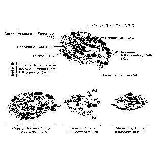
[0059] Figure 11 shows the cells of the tumor microenvironment (Cell 144,
March 4, 2011,
646-674).
[0060] Figure 12 shows tissue arrays of OVC, benign and normal cases
stained for PRSS8
by immunochemistry (magnification 40X).
DETAILED DESCRIPTION OF THE INVENTION
[0061] The described invention can be better understood from the following
description of
exemplary embodiments, taken in conjunction with the accompanying figures and
drawings. It
should be apparent to those skilled in the art that the described embodiments
of the described
invention provided herein are merely exemplary and illustrative and not
limiting.
Definitions
[0062] Various terms used throughout this specification shall have the
definitions set out
herein.
24
CA 02960012 2017-02-28
WO 2016/033464 PCMJS2015/047434
[0063] The term "biomarker" (or "biosignature") as used herein refers to a
peptide, a protein,
a nucleic acid, an antibody, a gene, a metabolite, or any other substance used
as an indicator of a
biologic state. It is a characteristic that is measured objectively and
evaluated as a cellular or
molecular indicator of normal biologic processes, pathogenic processes, or
pharmacologic
responses to a therapeutic intervention. The term "cancer biomarker" (or
"cancer biosignature")
as used herein refers to a peptide, a protein, a nucleic acid, an antibody, a
gene, a metabolite, or
any other substance used to detect the predisposition for, or the presence of,
primary or
metastatic cancer in a subject. According to the described invention,
biomarkers useful in the
detection of ovarian cancer include, but are not limited to. KLK6, KLK7 and
PRSS8.
[0064] The phrases "borderline tumor", -borderline cancer", -borderline
ovarian tumor" and
"borderline ovarian cancer" are used interchangeably herein to refer to a
group of tumors that, in
contrast to typical ovarian carcinomas, do not invade the ovarian stroma and
therefore are
considered noninvasive. Borderline tumors have a superior prognosis when
compared with other
ovarian carcinomas stage for stage.
[0065] The term "cDNA" refers to DNA synthesized from a mature mRNA
template. cDNA
most often is synthesized from mature mRNA using the enzyme reverse
transcriptase. The
enzyme operates on a single strand of mRNA, generating its complementary DNA
based on the
pairing of RNA base pairs (A, U, G, C) to their DNA complements (T, A, C, G).
There are
several methods known for generating cDNA to obtain, for example, eukaryotic
cDNA whose
introns have been spliced. Generally, these methods incorporate the following
steps: a) a
eukaryotic cell transcribes the DNA (from genes) into RNA (pre-mRNA); b) the
same cell
processes the pre-mRNA strands by splicing out introns, and adding a poly-A
tail and 5' Methyl-
Guanine cap; c) this mixture of mature mRNA strands is extracted from the
cell; d) a poly-T
oligonucleotide primer is hybridized onto the poly-A tail of the mature mRNA
template (reverse
transcriptase requires this double-stranded segment as a primer to start its
operation); e) reverse
transcriptase is added, along with deoxynucleotide triphosphates (A, T, G, C);
f) the reverse
transcriptase scans the mature mRNA and synthesizes a sequence of DNA that
complements the
mRNA template. This strand of DNA is complementary DNA (cDNA).
CA 02960012 2017-02-28
WO 2016/033464 PCMJS2015/047434
[0066] The term "cell" is used herein to refer to the structural and
functional unit of living
organisms and is the smallest unit of an organism classified as living.
[0067] The term "condition" as used herein, refers to a variety of health
states and is meant
to include disorders or diseases caused by injury or any underlying mechanism
or disorder.
[0068] The term "disease" or "disorder" as used herein refers to an
impairment of health or a
condition of abnormal functioning.
[0069] The term -gene" as used herein refers to a region of DNA that
controls a discrete
hereditary characteristic, usually corresponding to a single protein or RNA.
This definition
includes the entire functional unit, encompassing coding DNA sequences,
noncoding regulatory
DNA sequences and introns.
[0070] The tenin "hybridization" refers to the process of combining
complementary, single-
stranded nucleic acids into a single molecule. Nucleotides will bind to their
complement under
normal conditions, so two perfectly complementary strands will bind (or
'anneal') to each other
readily. However, due to the different molecular geometries of the
nucleotides, a single
inconsistency between the two strands will make binding between them more
energetically
unfavorable. Measuring the effects of base incompatibility by quantifying the
rate at which two
strands anneal can provide information as to the similarity in base sequence
between the two
strands being annealed. The term "specifically hybridizes" as used herein
refers to the process
whereby a nucleic acid distinctively or definitively forming base pairs with
complementary
regions of at least one strand of DNA that was not originally paired to the
nucleic acid. A
nucleic acid that selectively hybridizes undergoes hybridization, under
stringent hybridization
conditions, of the nucleic acid sequence to a specified nucleic acid target
sequence to a
detectably greater degree (e.2., at least 2-fold over background) than its
hybridization to non-
target nucleic acid sequences and to the substantial exclusion of non-target
nucleic acids.
Selectively hybridizing sequences typically have about at least 80% sequence
identity, at least
90% sequence identity, or at least 100% sequence identity (i.e.,
complementary) with each other.
26
CA 02960012 2017-02-28
WO 2016/033464 PCMJS2015/047434
[0071] The term "isolate" and its various grammatical forms as used herein
refers to placing,
setting apart, or obtaining a protein, molecule, substance, nucleic acid,
peptide, cell or particle, in
a form essentially free from contaminants or other materials with which it is
commonly
associated, separate from its natural environment.
[0072] The term "kallikrein" as used herein refers to a member of the Si
family (clan SA) of
trypsin-like serine proteases. Kallikrein proteins are encoded by 15
structurally similar, steroid
hormone regulated genes (KLK) that co-localize to chromosome 19q13.4.
Kallikrein proteins
are implicated in a wide range of physiologic functions such as blood pressure
regulation,
electrolyte balance, tissue remolding, pro-hormone processing, neural
plasticity and skin
desquamation.
[0073] The terms "kallikrein 6", "kallikrein-6", and "KLK6" are used
interchangeably herein
to refer to a member of the kallikrein family of serine proteases. The KLK6
gene comprises
11,043 nucleotides that encode a protein 244 amino acids in length. The KLK6
protein acts upon
amyloid precursor protein, myelin basic protein, gelatin, casein,
extracellular matrix proteins
(e.g., fibronectin, laminin, vitronectin and collagen); degrades a-synuclein
and prevents its
polymerization; and regulates axon outgrowth following spinal cord injury.
[0074] The terms "kallikrein 7", "kallikrein-7", and "KLK7" used
interchangeably herein
refer to a member of the kallikrein family of serine proteases. The KLK7 gene
comprises 7,627
nucleotides that encode a protein 253 amino acids in length. The KLK7 protein
catalyzes the
degradation of intercellular cohesive structures in the cornified layer of the
skin and is implicated
in the activation of precursors to inflammatory cytokines.
[0075] The terms "metastasis" or "metastases" as used herein refer to tumor
growth or
deposit that has spread via lymph or blood to an area of the body remote from
the primary tumor.
[0076] The term "metastasize" as used herein refers to the spread of cancer
from one part of
the body to another. A tumor formed by cells that have spread is called a
"metastatic tumor" or
"metastasis." The plural form of "metastasis" is -metastases."
27
CA 02960012 2017-02-28
WO 2016/033464 PCMJS2015/047434
[0077] The term "nucleic acid" as used herein refers to a
deoxyribonucleotide or
ribonucleotide polymer in either single- or double-stranded form, and unless
otherwise limited,
encompasses known analogues having the essential nature of natural nucleotides
in that they
hybridize to single-stranded nucleic acids in a manner similar to naturally
occurring nucleotides
(e.g., peptide nucleic acids).
[0078] The term "nucleotide" as used herein refers to a chemical compound
that consists of a
heterocyclic base, a sugar, and one or more phosphate groups. In the most
common nucleotides,
the base is a derivative of purine or pyrimidine, and the sugar is the pentose
deoxyribose or
ribose. Nucleotides are the monomers of nucleic acids, with three or more
bonding together in
order to form a nucleic acid. Nucleotides are the structural units of RNA,
DNA, and several
cofactors, including, but not limited to, CoA, FAD, DMN, NAD, and NADP.
Purines include
adenine (A), and guanine (C); pyrimidines include cytosine (C), thymine (T),
and uracil (U).
[0079] The term "peptide" is used herein to refer to two or more amino
acids joined by a
peptide bond.
[0080] The term "polynucleotide" refers to a deoxyribopolynucleotide,
ribopolynucleotide,
or analogs thereof that have the essential nature of a natural ribonucleotide
in that they hybridize,
under stringent hybridization conditions, to substantially the same nucleotide
sequence as
naturally occurring nucleotides and/or allow translation into the same amino
acid(s) as the
naturally occurring nucleotide(s). A polynucleotide may be full-length or a
subsequence of a
native or heterologous structural or regulatory gene. Unless otherwise
indicated, the term
includes reference to the specified sequence as well as the complementary
sequence thereof.
Thus, DNAs or RNAs with backbones modified for stability or for other reasons
are
"polynucleotides" as that term is intended herein. Moreover, DNAs or RNAs
comprising
unusual bases, such as inosine, or modified bases, such as tritylated bases,
to name just two
examples, are polynucleotides as the term is used herein. It will be
appreciated that a great
variety of modifications have been made to DNA and RNA that serve many useful
purposes
known to those of skill in the art. The term polynucleotide as it is employed
herein embraces
such chemically, enzymatically or metabolically modified forms of
polynucleotides, as well as
28
CA 02960012 2017-02-28
WO 2016/033464 PCMJS2015/047434
the chemical forms of DNA and RNA characteristic of viruses and cells,
including among other
things, simple and complex cells.
[0081] The terms "polypeptide", "peptide" and "protein" also apply to amino
acid polymers
in which one or more amino acid residue is an artificial chemical analogue of
a corresponding
naturally occurring amino acid, as well as to naturally occurring amino acid
polymers. The
essential nature of such analogues of naturally occurring amino acids is that,
when incorporated
into a protein that protein is specifically reactive to antibodies elicited to
the same protein but
consisting entirely of naturally occurring amino acids. The terms
"polypeptide", "peptide" and
"protein" also are inclusive of modifications including, but not limited to,
glycosylation, lipid
attachment, sulfation, gamma-carboxylation of glutamic acid residues,
hydroxylation and ADP-
ribosylation. It will be appreciated, as is well known and as noted above,
that polypeptides may
not be entirely linear. For instance, polypeptides may be branched as a result
of ubiquitination,
and they may be circular, with or without branching, generally as a result of
posttranslational
events, including natural processing event and events brought about by human
manipulation
which do not occur naturally. Circular, branched and branched circular
polypeptides may be
synthesized by non-translation natural process and by entirely synthetic
methods, as well.
[0082] The following terms are used herein to describe the sequence
relationships between
two or more nucleic acids or polynucleotides: (a) "reference sequence", (b)
"comparison
window", (c) "sequence identity", (d) "percentage of sequence identity", and
(e) "substantial
identity."
[0083] (a) The term "reference sequence" refers to a sequence used as a
basis for sequence
comparison. A reference sequence may be a subset or the entirety of a
specified sequence; for
example, as a segment of a full-length cDNA or gene sequence, or the complete
cDNA or gene
sequence.
[0084] (b) The term "comparison window" refers to a contiguous and
specified segment of
a polynucleotide sequence, wherein the polynucleotide sequence may be compared
to a reference
sequence and wherein the portion of the polynucleotide sequence in the
comparison window may
29
CA 02960012 2017-02-28
WO 2016/033464 PCMJS2015/047434
comprise additions or deletions (i.e., gaps) compared to the reference
sequence (which does not
comprise additions or deletions) for optimal alignment of the two sequences.
Generally, the
comparison window is at least 20 contiguous nucleotides in length, and
optionally can be at least
30 contiguous nucleotides in length, at least 40 contiguous nucleotides in
length, at least 50
contiguous nucleotides in length, at least 100 contiguous nucleotides in
length, or longer. Those
of skill in the art understand that to avoid a high similarity to a reference
sequence due to
inclusion of gaps in the polynucleotide sequence, a gap penalty typically is
introduced and is
subtracted from the number of matches.
[0085] Methods of alignment of sequences for comparison are well-known in
the art.
Optimal alignment of sequences for comparison may be conducted by the local
homology
algorithm of Smith and Waterman, Adv. And. Math. 2:482 (1981); by the homology
alignment
algorithm of Needleman and Wunsch, .I. Mol. Biol. 48:443 (1970); by the search
for similarity
method of Pearson and Lipman, Proc. Natl. Acad. Sci. 85:2444 (1988); by
computerized
implementations of these algorithms, including, but not limited to: CLUSTAL in
the PC/Gene
program by Intelligenetics, Mountain View, Calif.; GAP, BESTFIT, BLAST, FASTA,
and
TFASTA in the Wisconsin Genetics Software Package, Genetics Computer Group
(GCG), 575
Science Dr., Madison, Wis., USA; the CLUSTAL program is well described by
Higgins and
Sharp, Gene 73:237-244 (1988); Higgins and Sharp, CABIOS 5:151-153 (1989);
Corpet, et al.,
Nucleic Acids Research 16:10881-90 (1988); Huang, et al., Computer
Applications in the
Biosciences, 8:155-65 (1992), and Pearson, et al., Methods in Molecular
Biology, 24:307-331
(1994). The BLAST family of programs, which can be used for database
similarity searches,
includes: BLASTN for nucleotide query sequences against nucleotide database
sequences;
BLASTX for nucleotide query sequences against protein database sequences;
BLASTP for
protein query sequences against protein database sequences; TBLASTN for
protein query
sequences against nucleotide database sequences; and TBLASTX for nucleotide
query sequences
against nucleotide database sequences. See, Current Protocols in Molecular
Biology, Chapter
19, Ausubel. et al., Eds., Greene Publishing and Wiley-Interscience, New York
(1995).
[0086] Unless otherwise stated, sequence identity/similarity values
provided herein refer to
the value obtained using the BLAST 2.0 suite of programs using default
parameters. Altschul et
CA 02960012 2017-02-28
WO 2016/033464 PCMJS2015/047434
al., Nucleic Acids Res. 25:3389-3402 (1997). Software for performing BLAST
analyses is
publicly available, e.g., through the National Center for Biotechnology-
Information. This
algorithm involves first identifying high scoring sequence pairs (HSPs) by
identifying short
words of length W in the query sequence, which either match or satisfy some
positive-valued
threshold score T when aligned with a word of the same length in a database
sequence. T is
referred to as the neighborhood word score threshold (Altschul et al.. supra).
These initial
neighborhood word hits act as seeds for initiating searches to find longer
HSPs containing them.
The word hits then are extended in both directions along each sequence for as
far as the
cumulative alignment score can be increased. Cumulative scores are calculated
using, for
nucleotide sequences, the parameters M (reward score for a pair of matching
residues; always>0)
and N (penalty score for mismatching residues; always<0). For amino acid
sequences, a scoring
matrix is used to calculate the cumulative score. Extension of the word hits
in each direction are
halted when: the cumulative alignment score falls off by the quantity X from
its maximum
achieved value; the cumulative score goes to zero or below, due to the
accumulation of one or
more negative-scoring residue alignments; or the end of either sequence is
reached. The BLAST
algorithm parameters W, T, and X determine the sensitivity and speed of the
alignment. The
BLASTN program (for nucleotide sequences) uses as defaults a word length (W)
of 11, an
expectation (E) of 10, a cutoff of 100, M=5, N=-4, and a comparison of both
strands. For amino
acid sequences, the BLASTP program uses as defaults a word length (W) of 3, an
expectation
(E) of 10, and the BLOSUM62 scoring matrix (see Henikoff & Henikoff (1989)
Proc. Natl.
Acad. Sci. USA 89:10915).
[0087] In addition to calculating percent sequence identity, the BLAST
algorithm also
performs a statistical analysis of the similarity between two sequences (see,
e.g., Karlin &
Altschul, Proc. Natl. Acad. Sci. USA 90:5873-5787 (1993)). One measure of
similarity provided
by the BLAST algorithm is the smallest sum probability (P(N)), which provides
an indication of
the probability by which a match between two nucleotide or amino acid
sequences would occur
by chance. BLAST searches assume that proteins may be modeled as random
sequences.
However, many real proteins comprise regions of nonrandom sequences which may
be
homopolymeric tracts, short-period repeats, or regions enriched in one or more
amino acids.
Such low-complexity regions may be aligned between unrelated proteins even
though other
31
CA 02960012 2017-02-28
WO 2016/033464 PCMJS2015/047434
regions of the protein are entirely dissimilar. A number of low-complexity
filter programs may
be employed to reduce such low-complexity alignments. For example, the SEG
(Wooten and
Federhen, Comput. Chem., 17:149-163 (1993)) and XNU (Claverie and States,
Comput. Chem.,
17:191-201 (1993)) low-complexity filters may be employed alone or in
combination.
[0088] (c) The term "sequence identity" or "identity" in the context of two
nucleic acid or
polypeptide sequences is used herein to refer to the residues in the two
sequences that are the
same when aligned for maximum correspondence over a specified comparison
window. When
percentage of sequence identity is used in reference to proteins it is
recognized that residue
positions that are not identical often differ by conservative amino acid
substitutions, i.e., where
amino acid residues are substituted for other amino acid residues with similar
chemical
properties (e.g. charge or hydrophobicity) and therefore do not change the
functional properties
of the molecule. Where sequences differ in conservative substitutions, the
percent sequence
identity may be adjusted upwards to correct for the conservative nature of the
substitution.
Sequences that differ by such conservative substitutions are said to have
"sequence similarity" or
"similarity." Means for making this adjustment are well-known to those of
skill in the art.
Typically this involves scoring a conservative substitution as a partial
rather than a full
mismatch, thereby increasing the percentage sequence identity. Thus, for
example, where an
identical amino acid is given a score of 1 and a non-conservative substitution
is given a score of
zero, a conservative substitution is given a score between zero and 1. The
scoring of
conservative substitutions is calculated, e.g., according to the algorithm of
Meyers and Miller,
Computer Applic. Biol. Sci., 4:11-17 (1988) e.g., as implemented in the
program PC/GENE
(Intelligenetics, Mountain View, Calif., USA).
[0089] (d) The term "percentage of sequence identity" is used herein mean
the value
determined by comparing two optimally aligned sequences over a comparison
window, wherein
the portion of the polynucleotide sequence in the comparison window may
comprise additions or
deletions (i.e., gaps) as compared to the reference sequence (which does not
comprise additions
or deletions) for optimal alignment of the two sequences. The percentage is
calculated by
determining the number of positions at which the identical nucleic acid base
or amino acid
residue occurs in both sequences to yield the number of matched positions,
dividing the number
32
CA 02960012 2017-02-28
WO 2016/033464 PCMJS2015/047434
of matched positions by the total number of positions in the window of
comparison, and
multiplying the result by 100 to yield the percentage of sequence identity.
[0090] (e) The term "substantial identity" of polynucleotide sequences
means that a
polynucleotide comprises a sequence that has at least 70% sequence identity,
at least 80%
sequence identity, at least 90% sequence identity and at least 95% sequence
identity, compared
to a reference sequence using one of the alignment programs described using
standard
parameters. One of skill will recognize that these values may be adjusted
appropriately to
determine corresponding identity of proteins encoded by two nucleotide
sequences by taking into
account codon degeneracy, amino acid similarity, reading frame positioning and
the like.
Substantial identity of amino acid sequences for these purposes normally means
sequence
identity of at least 60%, or at least 70%, at least 80%, at least 90%, or at
least 95%. Another
indication that nucleotide sequences are substantially identical is if two
molecules hybridize to
each other under stringent conditions. However, nucleic acids that do not
hybridize to each other
under stringent conditions are still substantially identical if the
polypeptides that they encode are
substantially identical. This may occur, e.g., when a copy of a nucleic acid
is created using the
maximum codon degeneracy permitted by the genetic code. One indication that
two nucleic acid
sequences are substantially identical is that the polypeptide that the first
nucleic acid encodes is
immunologically cross reactive with the polypeptide encoded by the second
nucleic acid.
[0091] The terms "substantial identity" in the context of a peptide
indicates that a peptide
comprises a sequence with at least 70% sequence identity to a reference
sequence, at least 80%,
at least 85%, at least 90% or 95% sequence identity to the reference sequence
over a specified
comparison window. Optionally, optimal alignment is conducted using the
homology alignment
algorithm of Needleman and Wunsch, J. Mol. Biol. 48:443 (1970). An indication
that two
peptide sequences are substantially identical is that one peptide is
immunologically reactive with
antibodies raised against the second peptide. Thus, a peptide is substantially
identical to a
second peptide, for example, where the two peptides differ only by a
conservative substitution.
Peptides which are "substantially similar" share sequences as noted above
except that residue
positions that are not identical may differ by conservative amino acid
changes.
33
CA 02960012 2017-02-28
WO 2016/033464 PCMJS2015/047434
[0092] The
terms "poor clinical outcome", "poor outcome" or "PO" are used interchangeably
herein to refer to chemo-naIve primary refractory or early relapsing and chemo-
exposed,
multiple relapse within four (4) years or shortened survival/death two (2) to
three (3) years after
diagnosis.
[0093] The
term "primer" refers to a nucleic acid which, when hybridized to a strand of
DNA, is capable of initiating the synthesis of an extension product in the
presence of a suitable
polymerization agent. The primer is sufficiently long to uniquely hybridize to
a specific region of
the DNA strand. A primer also may be used on RNA, for example, to synthesize
the first strand
of cDNA.
[0094] The
term "progression" as used herein refers to the course of a disease, such as
ovarian cancer, as it becomes worse or spreads in the body.
[0095] The
term "progression free survival" or "PFS" as used herein refers to a length of
time during and after the treatment of a disease, such as cancer, that a
patient lives with the
disease but it does not get worse. In a clinical trial, measuring the
progression free survival is
one way to determine how well a new treatment works.
[0096] The
term "protein" is used herein to refer to a large complex molecule or
polypeptide
composed of amino acids. The sequence of the amino acids in the protein is
determined by the
sequence of the bases in the nucleic acid sequence that encodes it.
[0097] The
terms -PRSS8", -Prostasin", -Protease, Serine, 8", -Serine Protease 8",
-Channel-activating Protease 1" and "CAP1" are used interchangeably herein to
refer to a
member of the trypsin family of serine proteases. The PRSS8 gene comprises
4,398 nucleotides
that encode a protein 343 amino acids in length. The PRSS8 protein possesses a
trypsin-like
cleavage specificity with a preference for poly-basic substrates. The
proprotein is cleaved to
produce a light chain and a heavy chain which are associated by a disulfide
bond. The PRSS8
protein stimulates epithelial sodium channel (ENaC) activity via activating
cleavage of the
34
CA 02960012 2017-02-28
WO 2016/033464 PCMJS2015/047434
gamma subunits. PRSS8 shares sequence homology at the amino acid level with
acrosin, plasma
kallikrein, hepsin, testisin and gamma tryptase.
[0098] The term "quality of life" as used herein refers to the overall
enjoyment of life,
including aspects of an individual's sense of well-being and ability to carry
out various activities.
[0099] The terms "recurrence" or "relapse" are used interchangeably herein
to refer to the
return of a cancer after a first-line treatment and after a period of time
during which the cancer
cannot be detected.
[00100] The term "refractory" as used herein refers to cancer that does not
respond to
treatment. The cancer may be resistant at the beginning of treatment or it may
become resistant
during treatment. The term "primary refractory" as used herein refers to the
progression of
disease during induction treatment or a partial or transient response (e.g.
less than 60 days) to
induction therapy. The term "induction therapy" as used herein refers to the
first treatment given
for a disease which is often part of a standard set of treatments, for
example, surgery followed by
chemotherapy and radiation. Induction therapy is often accepted as the best
treatment option.
Induction therapy is also known as "first-line therapy," "primary therapy" and
"primary
treatment."
[00101] The term "relapse-free survival (RFS)" as used herein refers to the
length of time
after primary treatment for a cancer during which the patient survives without
any signs or
symptoms of that cancer. It is also called disease-free survival (DFS).
[00102] The term "relative" as used herein refers to something having, or
standing in, some
significant association to something else. The term "relative frequency" as
used herein refers to
the rate of occurrence of something having or standing in some significant
association to the rate
of occurrence of something else. For example, two cell types, X cells and Y
cells occupy a given
location. There are 5 X cells and 5 Y cells in that location. The relative
frequency of cell type X
is 5/10: the relative frequency of cell type Y is 5/10 in that location.
Following processing, there
are 5 X cells, but only 1 Y cell in that location. The relative frequency of
cell type X following
CA 02960012 2017-02-28
WO 2016/033464 PCT/1JS2015/047434
processing is 5/6, and the relative frequency of cell type Y following
processing is 1/6 in that
location.
[00103] The term "risk factor" as used herein refers to anything that raises
the chances of a
person developing a disease.
[00104] The terms "subject" and "patient" are used interchangeably herein to
refer to animal
species of mammalian origin that may benefit from the administration of a drug
composition or
method of the described invention. Examples of subjects include humans, and
other animals
such as horses, pigs, cattle, dogs, cats, rabbits, mice, rats and aquatic
mammals.
[00105] The term "syndrome" as used herein, refers to a pattern of symptoms
indicative of
some disease or condition.
[00106] Tissue Compartments. In multicellular organisms, cells that are
specialized to
perform common functions are usually organized into cooperative assemblies
embedded in a
complex network of secreted extracellular macromolecules, the extracellular
matrix (ECM), to
form specialized tissue compartments. Individual cells in such tissue
compartments are in
contact with ECM macromolecules. The ECM helps hold the cells and compartments
together
and provides an organized lattice or scaffold within which cells can migrate
and interact with one
another. In many cases, cells in a compartment can be held in place by direct
cell-cell adhesions.
In vertebrates, such compartments may be of four major types, a connective
tissue (CT)
compartment, an epithelial tissue (ET) compartment, a muscle tissue (MT)
compartment and a
nervous tissue (NT) compartment, which are derived from three embryonic germ
layers:
ectoderm, mesoderm and endoderm. The NT and portions of the ET compartments
are
differentiated from the ectoderm; the CT, MT and certain portions of the ET
compartments are
derived from the mesoderm; and further portions of the ET compartment are
derived from the
endoderm.
[00107] The term "tumor epithelial compartment cells" as used herein refers to
tumor cells
arising from cells in the epithelial compartment that form the epithelium. The
epithelium is a
36
CA 02960012 2017-02-28
WO 2016/033464 PCMJS2015/047434
layer of cells that covers the internal and external organs of the body, that
lines vessels, body
cavities, glands and organs, that forms the epidermis of the skin and the
surface layer of mucus
and serous membranes. Epithelial cells rest on a basement membrane and lie in
close proximity
with little intercellular material between them. Epithelial cells are devoid
of blood vessels and
may be simple (consisting of a single layer) or stratified (consisting of
several layers). Cells
comprising the epithelium may be flat (squamous), cube-shaped (cuboidal) or
cylindrical
(columnar). Modified forms of epithelium include, but are not limited to,
ciliated (hair-like
processes on the surface), pseudostratified (appears stratified because cells
are arranged with
their nuclei at different levels), glandular (composed of secreting cells) and
neuroepithelium
(composed of sensory cells). The epithelium may include goblet cells which
secrete mucus.
Squamous epithelium is classified as endothelium, which lines the blood
vessels and heart, and
mesothelium. which lines the serous cavities. Functions of the epithelium
include, but are not
limited to, protection, absorption, secretion, movement of substances through
ducts, production
of germ cells and reception of stimuli.
[00108] The tumor microenvironment. Tumors increasingly have been recognized
as
organs whose complexity approaches and may even exceed that of normal healthy
tissues.
Hanahan, D. and Weinberg, R.A., Hallmarks of Cancer: The nexi generation, Cell
144: 646-74
(2011). The biology of a tumor can be understood by studying the individual
specialized cells
within it (Fig. 11, upper), as well as the tumor microenvironment that they
construct during the
course of multistep tumorigenesis (Fig. 11, lower). Id.
[00109] As shown in Fig. 11, upper, an assemblage of distinct cell types
constitutes most solid
tumors. Both the parenchyma (functional tissue component) and stroma
(connective tissue and
supporting component) of tumors contain distinct cell types and subtypes that
collectively enable
tumor growth and progression. Id. Cancer cells initiate tumors and drive tumor
progression
forward. Traditionally, cancer cells within tumors have been portrayed as
reasonably
homogenous cell populations until relatively late in the course of tumor
progression, when
hyperproliferation combined with increased genetic instability spawn distinct
clonal
subpopulations. Reflecting such clonal heterogeneity, many human tumors are
histopathologically diverse, containing regions demarcated by various degrees
of differentiation,
37
CA 02960012 2017-02-28
WO 2016/033464 PCMJS2015/047434
proliferation, vascularity, inflammation and/or invasiveness. Cancer stem
cells within tumors
impose a further dimension of intratumor heterogeneity. Id. The significance
of CSCs as a
distinct phenotypic subclass of neoplastic cells remains a matter of debate.
Id.
[00110] Much of the cellular heterogeneity within tumors is found in their
stromal
compartments. Tumor-associated stromal cells may be supplied to growing tumors
by
proliferation of preexisting stromal cells, by differentiation in situ of
local stem/progenitor cells
originating in the neighboring normal tissue, or via recruitment of bone
marrow-derived
stem/progenitor cells. A key source of tumor-associated stromal cells is the
bone marrow. (Id.
Citing Bergfeld and DeClerck 2010; Fang and Salven (2011), Giaccia & Schipani
(2010);
Patenaude et al (2010); Lamagna & Bergers (2006)).
[00111] In many cases, fibroblasts constitute the preponderant cell
population of the tumor
stroma. The temi "cancer associated fibroblast) includes at least two distinct
cell types: (1) cells
with similarities to the fibroblasts that create the structureal foundation
supporting most normal
epithelial tissues; and (2) myofibroblasts, identifiable by their expression
of a-smooth muscle
actin (SMA).
[00112] Endothelial cells forming the tumor-associated vasculature are
prominent among the
stromal constituents. Id. The role of endothelial cells forming lymphatic
vessels is however
poorly understood. Id. Pericytes represent a specialized mesenchymal cell type
related to
smooth muscle cells with finger-like projections that wrap around the
endothelial tubing of blood
vessels. Pharmacological inhibition of signaling through the PDGF receptor
expressed by tumor
pericytes and bone marrow-derived pericyte progenitors results in reduced
pericyte coverage of
tumor vessels, which in turn destabilizes vascular integrity and function.
(Id. Citing Pietras and
Ostman (2010); Raza et al (2010); Gaengel et al (2009)).
[00113] Infiltrating cells of the immune system are also constituents of
tumors. Immune
inflammatory cells present in tumors can include both tumor-promoting and
tumor-killing
subclasses. Id.
38
CA 02960012 2017-02-28
WO 2016/033464 PCMJS2015/047434
[00114] Infiltrating cells of the immune system are also constituents of
tumors. Immune
inflammatory cells present in tumors can include both tumor-promoting and
tumor-killing
subclasses. Id.
[00115] As shown in Fig. 11 (lower), the multiple stromal cell types create a
succession of
tumor microenvironments that change as tumors invade normal tissue and
thereafter seed and
colonize distant tissues. The abundance, histologic organization, and
phenotypic characteristics
of the stromal cell types, and of the extracellular matrix (hatched
background), evolve during
progression, thereby enabling primary, invasive, and then metastatic growth.
The surrounding
normal cells of the primary and metastatic sites, shown schematically, likely
also affect the
character of the various neoplastic microenvironments. Id.
[00116] According to one embodiment, the described invention provides a method
for
detecting, diagnosing and treating early stage (I/II) ovarian cancer in a
subject comprising: (a)
obtaining a serum sample from the subject and obtaining a normal serum control
sample; (b)
isolating from the sample obtained in (a) total RNA comprising a serine
protease mRNA; (c)
transforming the isolated total RNA of (b) into cDNA comprising serine
protease cDNA; (d)
amplifying the cDNA of (c); (e) measuring a level of amplified senile protease
cDNA in (d) as a
measure of expression of amplified serine protease mRNA; (f) comparing the
level of expression
of the amplified serine protease mRNA in (e) expressed by the subject with the
level of
expression of the amplified serine protease mRNA in (e) expressed by the
normal serum control
sample, wherein an increased level of expression of the serine protease mRNA
expressed by the
subject compared to the level of expression of the serine protease mRNA
expressed by the
normal serum control sample is indicative of possible early stage ovarian
cancer in the subject;
(g) when (f) is indicative of early stage (I/II) ovarian cancer in the
subject, obtaining an ovarian
tissue sample from subject; (h) isolating from the ovarian tissue sample
obtained in (g) total
RNA comprising serine protease mRNA; (i) transforming the isolated total RNA
of (h) into
cDNA comprising serine protease cDNA; (j) amplifying the cDNA of (i); (k)
measuring a level
of amplified serine protease cDNA in (j) as a measure of expression of
amplified serine protease
mRNA; (1) comparing the level of expression of the amplified serine protease
mRNA in (k)
expressed by the subject with the level of expression of the amplified serine
protease mRNA in
39
CA 02960012 2017-02-28
WO 2016/033464 PCMJS2015/047434
(k) expressed by a normal ovarian tissue control sample, wherein an increased
level of
expression of the serine protease mRNA expressed by the subject compared to
the level of
expression of the serine protease mRNA expressed by the normal ovarian tissue
control sample
is indicative of possible early stage ovarian cancer in the subject; (m) when
both (f) and (1) are
indicative of early stage ovarian cancer, diagnosing early stage (I/II)
ovarian cancer in the
subject; and (n) treating the subject with a treatment regimen effective to
treat the early stage
(I/II) ovarian cancer.
[00 1 17] According to another embodiment, the described invention provides a
method for
detecting, diagnosing and treating early stage (1/II) ovarian cancer in a
subject comprising: (a)
obtaining a serum sample from the subject and a normal serum sample as a
control; (b) detecting
a serine protease protein in the samples from (a) by reacting an anti-serine
protease antibody
with the patient serum sample and the normal serum control sample; (c)
quantifying an amount
of serine protease protein bound by the anti-serine protease antibody in (b);
(d) comparing the
amount of serine protease protein in (c) bound by antibody in the subject
serum sample with the
amount of the serine protease protein bound by antibody in the normal serum
control sample,
wherein an increased amount of the serine protease protein bound in the
subject sample
compared to the amount of the serine protease protein bound in the normal
serum control sample
is indicative of early stage (I/II) ovarian cancer in the subject; (e) when
(d) is indicative of
ovarian cancer in the subject, obtaining an ovarian tissue sample from the
subject and a normal
ovarian tissue sample as a control; (f) detecting serine protease protein in
the samples from (e) by
reacting an anti-serine protease antibody with the subject ovarian tissue
sample and the normal
ovarian tissue sample; (g) quantifying an amount of serine protease protein
bound by the anti-
serine protease antibody in (f); (h) comparing the amount of serine protease
protein bound in the
subject ovarian tissue sample with the amount of the serine protease protein
bound in the normal
ovarian tissue control sample, wherein an increased amount of the serine
protease protein bound
in the subject ovarian tissue sample compared to the amount of the serine
protease protein bound
in the normal ovarian tissue control sample is indicative of early stage
(I/II) ovarian cancer in
the subject; (i) when both (d) and (h) are indicative of early stage ovarian
cancer, diagnosing
early stage (I/II) ovarian cancer in the subject; and (j) treating the subject
with a treatment
regimen effective to treat ovarian cancer.
CA 02960012 2017-02-28
WO 2016/033464 PCT/US2015/047434
[00118] According to one embodiment, the described invention utilizes serine
proteases.
Categories of serine proteases include, but are not limited to, trypsin-like,
chymotrypsin-like and
elastase-like. Non-limiting examples of trypsin-like serine proteases include
trypsin, kallikrein
(e.g.. KLK6 and KLK7), PRSS8 (prostasin), granzyme K, hepatocyte growth factor
activator and
the like. Examples of chymotrypsin-like serine proteases include, but are not
limited to,
chymotrypsin, chymotrypsin-like protease-1 (CTRL-1), chymotrypsin-like neutral
leukocyte
protease and the like. Non-limiting examples of elastase-like serine proteases
include neutrophil
elastase (NE), proteinase 3 (PR3). azurocidin (AZU) and the like.
[00119] According to one embodiment, the serine protease is at least two (2)
selected from the
group consisting of kallikrein 6 (KLK6), kallikrein 7 (KLK7), and PRSS8
[00120] According to one embodiment, the described invention utilizes a sample
obtained
from a subject. The sample can include, but is not limited to, a tissue
sample, a blood sample, a
serum sample, a urine sample, a saliva sample and the like. Methods of
obtaining a sample from
a subject are well known in the art. Such methods include, but are not limited
to, biopsy, such as
for example, a core biopsy or a fine needle biopsy. The sample may be a fresh,
a frozen or a
fixed, wax-embedded sample. Non-limiting examples of fixed, wax-embedded
samples include
formalin-fixed, paraffin-embedded samples.
[00121] According to one embodiment, the described invention utilizes isolated
nucleic acids.
Nucleic acids include, for example, DNA, RNA and mRNA. Nucleic acids can be
isolated, for
example, from tissues, cells, blood, serum, plasma, urine, saliva, semen and
the like. Protocols
and reagents for isolating nucleic acids are known. Non-limiting examples of
reagents used for
nucleic acid isolation include guanidine thiocyanate, guanidine hydrochloride
and guanidinium
thiocyanate-phenol-chloroform; the proprietary formulation of this reagent is
known as Trizol .
[00122] According to one embodiment, the described invention utilizes methods
employing
amplification of nucleic acids. According to one such embodiment,
amplification of nucleic
acids is accomplished by Polymerase Chain Reaction (PCR). Non-limiting
examples of PCR
include conventional PCR, real-time PCR, quantitative PCR, quantitative real-
time PCR,
41
CA 02960012 2017-02-28
WO 2016/033464 PCMJS2015/047434
multiplex PCR, conventional reverse-transcriptase PCR (RT-PCR), real-time RT-
PCR,
quantitative RT-PCR, quantitative real-time RT-PCR, multiplex RT-PCR and the
like.
[00123] Primers for PCR amplification of target sequences (e.g., mRNA
sequences of KLK6
and KLK7 genes) can be designed based on the sequence of the target sequence,
in accordance
with standard procedures. Primers function to anneal and amplify a unique
target sequence and
as generators of a signal for detection and monitoring of an amplification
reaction. According to
some embodiments, the primers are unlabeled (such as in conventional PCR),
while in other
embodiments, the primers are labeled, such as with a fluorescent moiety.
Labeled primers can be
of any type, including those that are typically used in quantitative RT-PCR
reactions, such as
Scorpions, Molecular Beacons, and the like.
[00124] Probes may be provided in addition to primers. Probes that can be used
for detection
of amplification of the unique genomic sequences (e.g., TaqMan probes) can be
designed to
hybridize to a sequence between the two amplification primers, preferably
within 5-15 bases of
one of the primer binding sites. Typically, probes are present in reaction
mixtures in conjunction
with primers or sets of primers for a particular amplification reaction, for
example, an
amplification of a unique target sequence. However, probes may be provided as
separate
components. which are separate from the primer(s) or other components of a
reaction mixture.
[00125] The primers and probes are designed to have a typical size for primers
and probes for
use in PCR reactions. In general, the primers are relatively short (about 10-
30 bases in length)
oligonucleotides, while the probes (e.g., TaqMan probes) may be from about 15-
35 bases in
length. The primers and probes are designed through a process that includes
identification of
unique sequences on a target nucleic acid, designing short oligonucleotides to
amplify or detect
those sequences, and synthesizing the oligonucleotides. Several
characteristics may be taken into
consideration when designing the primers and probe: e.g., the probe melting
temperature should
be higher than the primer melting temperatures, and the distance between the
3'-end of one
primer and the 5'-end of the probe may be greater than 8 nucleotides. One of
skill in the art may
select among such considerations and characteristics to provide suitable
primers and probes.
42
CA 02960012 2017-02-28
WO 2016/033464 PCMJS2015/047434
Protocols for synthesis of oligonucleotides are known to those skilled in the
art. Any suitable
protocol may be used in synthesizing the primers and probes of the invention.
[00126] Quantitative real-time RT-PCR is an accurate, precise, high throughput
assay. Real-
time PCR automates the process of quantitating reaction products for each
sample in every cycle.
According to some embodiments, real-time PCR systems rely upon the detection
and
quantitation of a fluorescent reporter, the signal of which increases in
direct proportion to the
amount of PCR product in a reaction.
[00127] According to some such embodiments, for example, the reporter is the
double-
stranded DNA-specific dye SYBR Green (Molecular Probes), which binds double-
stranded
DNA, and upon excitation emits light. Thus, as a PCR product accumulates,
fluorescence
increases. The SYBR Green (Molecular Probes, Eugene, Oregon) system is one
way to detect
and quantitate PCR products in real time. The SYBR Green dye binds, in a
sequence non-
specific manner, to double-stranded nucleic acids. It thus can be used for
detection and
quantitation of double-stranded products produced from single-stranded
templates (e.g., mRNA).
Other detectable probes and primers, such as amplifluor probes, and DNAzymes,
may be
optimized to be used for quantitative detection of amplification products.
[00128] Alternatives to SYBR Green include, but are not limited to, TaqMan
(Applied
Biosystems, Foster City, Calif.) and molecular beacons, both of which are
hybridization probes
relying on fluorescence resonance energy transfer (FRET) for quantitation.
TaqMan Probes are
oligonucleotides that contain a fluorescent dye, typically on the 5' base, and
a quenching dye,
typically located on the 3' base. More specifically, for TaqMan probes, when
the probe is intact,
the quencher quenches the signal produced by the fluorescent label. However,
upon binding of
the probe to the target sequence and subsequent digestion of the probe by the
5'-3 exonuclease
activity of a polymerase, such as Taq polymerase, the fluorescent moiety is
released from the
quencher moiety, and a detectable signal, which is proportional to the amount
of target nucleic
acid being produced, is produced and can be monitored. According to one
embodiment, Taq
polymerase is used in qRT-PCR due to its 5'-3' exonuclease activity, and it
changes the
fluorescence of the probes and allows amplification of CDR1 mRNA. TaqMan
probes rely on
43
CA 02960012 2017-02-28
WO 2016/033464 PCMJS2015/047434
degradation by a polymerase to generate a detectable signal, while Scorpions
and Molecular
Beacons rely on opening of a hairpin structure to provide a detectable signal.
Like TaqMan
probes, Scorpion probes contain both a fluorescent moiety and quenching
moiety on a single
probe. However, unlike TaqMan probes, Scorpions are not degraded during the
amplification
reaction. Rather, they are designed as primers for amplification reactions.
Scorpion primers are
designed to form hairpin structures in solution, which causes the fluorescent
moiety and the
quenching moiety to be in close proximity. Binding of the primers to target
nucleic acids unfolds
the hairpin structure and moves the quenching moiety a sufficient distance
away from the
fluorescent moiety that detectable fluorescence is emitted.
[00129] Molecular beacons also contain fluorescent and quenching dyes, but
FRET only
occurs when the quenching dye is directly adjacent to the fluorescent dye.
Molecular beacons are
designed to adopt a hairpin structure while free in solution, bringing the
fluorescent dye and
quencher in close proximity. When a molecular beacon hybridizes to a target,
the fluorescent dye
and quencher are separated, FRET does not occur, and the fluorescent dye emits
light upon
irradiation.
[00130] Multiplexing of PCR reactions is common. Multiplexing allows an
investigator to
assay two or more different gene targets in a single reaction through the use
of multiple probes or
primers, each specific for its own target and each comprising a fluorescent
moiety that emits at a
unique wavelength (as compared to the other probes). Multiplexing is possible
with TaqMan
probes, Molecular Beacons, and Scorpions. Due to its non-specific binding
nature, SYBR
Green may not be amenable to multiplexing.
[00131] Generally, a quantitative RT-PCR reaction is performed by one of two
methods:
comparison to a standard curve or comparison of Ct values. In the first of
these methods, a
standard curve of amplification products of a particular mRNA is made based on
amplification of
a series of different, known amounts of a pre-selected nucleic acid.
Amplification results of
reactions performed on a target nucleic acid are then compared to the standard
curve to obtain a
quantity, and that quantity can be extrapolated to an amount of the target in
the original sample.
While it is preferred to use an mRNA as the source for the standard curve, the
stability of mRNA
44
CA 02960012 2017-02-28
WO 2016/033464 PCMJS2015/047434
is known to affect the validity of such standard curves, and overcoming or
minimizing this
problem has proved to be difficult. To avoid the problems associated with
using mRNA as a
source for the standard curve, researchers have used DNA for generation of
standard curves.
While use of DNA overcomes the problems associated with use of mRNA, the mere
fact that it
avoids the problems creates yet another problem, i.e., because DNA templates
are relatively
stable, and because amplification of DNA does not require a first-strand
synthesis step (which
can be inefficient and variable across samples or preparations), the standard
curves produced
from DNA sources often do not correlate accurately to the amount of mRNA in a
test sample.
[00132] In the Ct comparison method for quantitating PCR products, expression
of a
housekeeping gene (such as (3-actin) is used as a standard against which
amplification of a target
nucleic acid (e.g., KLK6 and KLK7) is compared. Often, in this method, a
comparison of
expression of the target nucleic acid under two different conditions is
performed to determine
changes in expression patterns. This method avoids the problems associated
with instability of
RNA or use of DNA as a control that is seen when using the classical standard
curve method.
[00133] Controls are amplified in the same PCR reaction mixture as the target
sequence in an
effort to quantitate PCR products and determine amounts of target nucleic
acids in a sample.
These controls are often transcripts of housekeeping genes. Such housekeeping
genes include,
but are not limited to, I3-actin and GAPDH. The control is added to the
reaction mix and co-
amplified with the target nucleic acid. Fluorescent probes specific for both
are included in the
mixture, and two amplification curves are obtained. The relative expression of
the target nucleic
acid with respect to the control is then determined. Using this technique,
multiple, different
samples can be compared for expression of a target gene (e.g., KLK6 and KLK7),
with reference
back to the same control. Although adding a control to amplification reactions
can be a useful
alternative to other methods of quantitating expression levels, and can be a
useful method for
normalizing PCR reactions across samples, it does not allow one to determine
absolute amounts
of materials present in the amplification reaction mixture or in the original
sample. Rather, the
results are qualitative or semi-quantitative, giving an idea only of the
amount of one nucleic acid
(e.g., the target) in comparison to another (e.g., the control).
CA 02960012 2017-02-28
WO 2016/033464 PCMJS2015/047434
[00134] According to one embodiment, the described invention provides methods
that detect a
protein in a sample obtained from a subject. Such methods of protein detection
are well known
in the art and include, but are not limited to, Western blot,
immunohistochemistry, enzyme-
linked immunosorbant assay (ELISA), radioimmunoassay and the like.
[00135] Methods that detect proteins may employ antibodies. Antibodies are
serum proteins
the molecules of which possess small areas of their surface that are
complementary to small
chemical groupings on their targets. These complementary regions (referred to
as the antibody
combining sites or antigen binding sites) of which there are at least two per
antibody molecule,
and in some types of antibody molecules ten, eight, or in some species as many
as 12, may react
with their corresponding complementary region on the antigen (the antigenic
determinant or
epitope) to link several molecules of multivalent antigen together to form a
lattice.
[00136] The basic structural unit of a whole antibody molecule consists of
four polypeptide
chains, two identical light (L) chains (each containing about 220 amino acids)
and two identical
heavy (H) chains (each usually containing about 440 amino acids). The two
heavy chains and
two light chains are held together by a combination of noncovalent and
covalent (disulfide)
bonds. The molecule is composed of two identical halves, each with an
identical antigen-binding
site composed of the N-terminal region of a light chain and the N-terminal
region of a heavy
chain. Both light and heavy chains usually cooperate to form the antigen
binding surface.
[00137] Human antibodies show two kinds of light chains, lc and X; individual
molecules of
immunoglobulin generally are only one or the other. In normal serum, 60% of
the molecules
have been found to have lc determinants and 30 percent X. Many other species
have been found
to show two kinds of light chains, but their proportions vary. For example, in
the mouse and rat,
chains comprise but a few percent of the total; in the dog and cat, lc chains
are very low; the
horse does not appear to have any lc chain; rabbits may have 5 to 40% k,
depending on strain and
b-locus allotype; and chicken light chains are more homologous to X than K.
[00138] In mammals, there are five classes of antibodies, IgA, IgD, IgE, IgG,
and IgM, each
with its own class of heavy chain ¨ a (for IgA), 6 (for IgD), z (for IgE), y
(for IgG) and la (for
46
CA 02960012 2017-02-28
WO 2016/033464 PCMJS2015/047434
IgM). In addition, there are four subclasses of IgG immunoglobulins (IgGl,
IgG2, IgG3, IgG4)
having yl, y2, y3, and y4 heavy chains respectively. In its secreted form, IgM
is a pentamer
composed of five four-chain units, giving it a total of 10 antigen binding
sites. Each pentamer
contains one copy of a J chain, which is covalently inserted between two
adjacent tail regions.
[00139] All five immunoglobulin classes differ from other serum proteins in
that they show a
broad range of electrophoretic mobility and are not homogeneous. This
heterogeneity ¨ that
individual IgG molecules, for example, differ from one another in net charge ¨
is an intrinsic
property of the immuno globulins.
[00140] An antigenic determinant or epitope is an antigenic site on a
molecule. Sequential
antigenic determinants/epitopes essentially are linear chains. In ordered
structures, such as
helical polymers or proteins, the antigenic determinants/epitopes essentially
would be limited
regions or patches in or on the surface of the structure involving amino acid
side chains from
different portions of the molecule which could come close to one another.
These are
conformational determinants.
[00141] The principle of complementarity, which often is compared to the
fitting of a key in a
lock, involves relatively weak binding forces (hydrophobic and hydrogen bonds,
van der Waals
forces, and ionic interactions), which are able to act effectively only when
the two reacting
molecules can approach very closely to each other and indeed so closely that
the projecting
constituent atoms or groups of atoms of one molecule can fit into
complementary depressions or
recesses in the other. Antigen-antibody interactions show a high degree of
specificity, which is
manifest at many levels. Brought down to the molecular level, specificity
means that the
combining sites of antibodies to an antigen have a complementarity not at all
similar to the
antigenic determinants of an unrelated antigen. Whenever antigenic
determinants of two
different antigens have some structural similarity, some degree of fitting of
one determinant into
the combining site of some antibodies to the other may occur, and that this
phenomenon gives
rise to cross-reactions. Cross reactions are of major importance in
understanding the
complementarity or specificity of antigen-antibody reactions. Immunological
specificity or
47
CA 02960012 2017-02-28
WO 2016/033464 PCMJS2015/047434
complementarity makes possible the detection of small amounts of
impurities/contaminations
among antigens.
[00142] The term "antibodies" can include, for example, polyclonal antibodies,
monoclonal
antibodies, antibody fragments and the like.
[00143] For example, monoclonal antibodies (mAbs) can be generated by fusing
mouse
spleen cells from an immunized donor with a mouse myeloma cell line to yield
established
mouse hybridoma clones that grow in selective media. A hybridoma cell is an
immortalized
hybrid cell resulting from the in vitro fusion of an antibody-secreting B cell
with a myeloma cell.
In vitro immunization, which refers to primary activation of antigen-specific
B cells in culture, is
another well-established means of producing mouse monoclonal antibodies.
[00144] For example, diverse libraries of immunoglobulin heavy (VH) and light
(Vic and VA,)
chain variable genes from peripheral blood lymphocytes also can be amplified
by polymerase
chain reaction (PCR) amplification. Genes encoding single polypeptide chains
in which the
heavy and light chain variable domains are linked by a polypeptide spacer
(single chain Fv or
scFv) can be made by randomly combining heavy and light chain V-genes using
PCR. A
combinatorial library then can be cloned for display on the surface of
filamentous bacteriophage
by fusion to a minor coat protein at the tip of the phage.
[00145] For example, the technique of guided selection is based on human
immunoglobulin V
gene shuffling with rodent immunoglobulin V genes. The method entails (i)
shuffling a
repertoire of human k light chains with the heavy chain variable region (VH)
domain of a mouse
monoclonal antibody reactive with an antigen of interest; (ii) selecting half-
human Fabs on that
antigen (iii) using the selected X light chain genes as "docking domains" for
a library of human
heavy chains in a second shuffle to isolate clone Fab fragments having human
light chain genes;
(v) transfecting mouse myeloma cells by electroporation with mammalian cell
expression
vectors containing the genes; and (vi) expressing the V genes of the Fab
reactive with the antigen
as a complete IgG I, X antibody molecule in the mouse myeloma.
48
CA 02960012 2017-02-28
WO 2016/033464 PCMJS2015/047434
[00146] According to one embodiment, the ovarian cancer is selected from the
group
consisting of serous, papillary serous, metastatic, borderline, mucinous and
clear cell.
[00147] According to one embodiment, the described invention provides the
detection of low-
grade (e.g., Gl) ovarian cancer. Methods for grading of tumors are well-known.
For example,
tumor grade can be determined by microscopic examination of cancer tissue. Low-
grade ovarian
cancer can be characterized by well-differentiated tissue, low-grade nuclei
with infrequent
mitotic figures and the like.
[00148] According to one embodiment, the described invention provides the
detection of stage
I (e.g., IA, IB, IC1, IC2 and IC3)) ovarian cancer. Staging of tumors is well-
known in the art.
For example, tumor stage can be determined by tests including, but not limited
to, ultrasound,
computerized tomography (CT) scan, positron emission tomography (PET) scan,
magnetic
resonance imaging (MRI), X-ray and biopsy. Stage I ovarian cancer can be
characterized by a
tumor limited to one ovary, capsule intact, no tumor on ovarian surface and
negative washing; a
tumor involving both ovaries, capsule intact, no tumor on ovarian surface and
negative washings;
surgical spill; capsule rupture before surgery or tumor on ovarian surface;
malignant cells in
ascites or peritoneal washings and the like.
[00149] According to one embodiment, an increased level of expression of two
or more of
KLK6 mRNA, KLK7 mRNA, and PRSS8 mRNA expressed in ovarian tissue of a subject
compared to the level of expression of two or more of KLK6 mRNA, KLK7 mRNA,
and PRSS8
mRNA expressed by a normal ovarian tissue control sample is indicative of an
expansion of
tumor epithelial compartment cells.
[00150] According to one embodiment, an increased level of expression of two
or more of
KLK6 protein, KLK7 protein, and PRSS8 protein expressed in ovarian tissue of a
subject
compared to the level of expression of two or more of KLK6 protein, KLK7
protein, and PRSS8
protein expressed by a normal ovarian tissue control sample is indicative of
an expansion of
tumor epithelial compartment cells.
49
[00151] Where a range of values is provided, it is understood that each
intervening value, to
the tenth of the unit of the lower limit unless the context clearly dictates
otherwise, between
the upper and lower limit of that range and any other stated or intervening
value in that stated
range is encompassed within the invention. The upper and lower limits of these
smaller ranges
which may independently be included in the smaller ranges is also encompassed
within the
invention, subject to any specifically excluded limit in the stated range.
Where the stated range
includes one or both of the limits, ranges excluding either both of those
included limits are also
included in the invention.
[00152] Unless defined otherwise, all technical and scientific terms used
herein have the
same meaning as commonly understood by one of ordinary skill in the art to
which this
invention belongs. Although any methods and materials similar or equivalent to
those
described herein can also be used in the practice or testing of the present
invention, exemplary
methods and materials have been described.
[00153] It must be noted that as used herein and in the appended claims, the
singular forms
"a", "and", and "the" include plural references unless the context clearly
dictates otherwise.
[00154] The publications discussed herein are provided solely for their
disclosure prior to
the filing date of the present application. Nothing herein is to be construed
as an admission
that the present invention is not entitled to antedate such publication by
virtue of prior
invention. Further, the dates of publication provided may be different from
the actual
publication dates which may need to be independently confirmed.
EXAMPLES
[00155] The
following examples are put forth so as to provide those of ordinary skill in
the art with a complete disclosure and description of how to make and use the
present invention,
and are not intended to limit the scope of what the inventors regard as their
invention nor are
they
Date Recue/Date Received 2020-08-28
CA 02960012 2017-02-28
WO 2016/033464 PCMJS2015/047434
intended to represent that the experiments below are all or the only
experiments performed.
Efforts have been made to ensure accuracy with respect to numbers used (e.g.
amounts,
temperature, etc.) but some experimental errors and deviations should be
accounted for. Unless
indicated otherwise, parts are parts by weight, molecular weight is weight
average molecular
weight, temperature is in degrees Centigrade, and pressure is at or near
atmospheric.
MATERIALS AND METHODS
Bioinformatics
[00156] The Biomax BioXMTm Knowledge Management System (Biomax Informatics AG,
Munich. Germany) was used to mine and generate a rank list of candidate
ovarian cancer genes
from the 6,955 manually curated cancer genes and 2,200 biomarker genes of the
National Cancer
Institute (NCI) and the Cancer Gene Index (CGI). The Biomax BioLT TM Tool
(NLP) was used to
mine 18 million Medline abstracts (94 million sentences) and 24,000 Hugo genes
to find and
validate genes associated with cancer terms, gene-disease relationships, and
gene-
compound/treatment relationships for each of the 6.955 cancer genes. The NCI
Thesaurus Role
Codes and Karp' s Evidence Codes (Karp, S. et al.. Pacific Symposium on
Biocomputing 9: 190-
201(2004)) were used to annotate over 1.3 million related sentences. The
search for potential
biomarkers was performed by initiating queries on BioXM with a combination of
search terms,
including ovarian, cancer, biomarker, overexpression, and upregulation or
downregulation.
Cell Lines and Cell Culture
[00157] Ovarian cancer cell lines T0V21G, TOV112D, OV-90, CA0V3, SKOV3, PA-1,
5W626, and ES-2 were purchased from the American Type Culture Collection
(ATCC,
Manassas, VA) and cultured in the media suggested by the distributor. The SKOV-
1, IGROV-1,
HEY, OV-2008, A2780, UCI-101, and UCI-107 cells were obtained from Drs. Howell
(University of California, San Diego) and Carpenter (University of California,
Irvine), and all
were cultured in RPMI 1640 medium (Invitrogen, Carlsbad, CA). The D0V13 cell
line was
obtained from Dr. Bast (MD Anderson Cancer Center) and cultured in DMEM. The
C500882
cells were obtained from Dr. Karlan (University of California, Los Angeles)
and cultured in
McCoy's 5A medium. Cell line 2774 was obtained from Dr. Wolf (MD Anderson
Cancer
Center) and cultured in EMEM. The BG-1 cell line was obtained from Dr. Korach
(NIEHS,
51
CA 02960012 2017-02-28
WO 2016/033464 PCMJS2015/047434
National Institutes of Health) and cultured in a 1:1 mixture of DMEM and F12
media without
phenol red. The normal ovarian epithelial cell lines FHIOSE118 and IOSE523
were obtained
from Dr. Cheng (Moffitt Cancer Center) and Dr. Nelly Auersperg (University of
British
Columbia), respectively, and cultured in a 1:1 mixture of MCDB105 and Medium
199. All
culture media were supplemented with 5-15% v/v fetal bovine serum (FBS;
Hyclone, Logan,
UT) and penicillin/streptomycin solution (Invitrogen).
RT-PCR and Statistical Analysis
[00158] Total RNA was extracted from cells using Trizol (Invitrogen), and cDNA
was
generated with the SuperScript III RTS First-Strand cDNA Synthesis Kit
(lnvitrogen) as
described by the manufacturer. All amplification primers were synthesized for
use with the
ABI7900 RT-PCR device (Applied Biosystems, Foster City, CA) as recommended by
Applied
Biosystems, and they were demonstrated to produce a single PCR band of the
expected size by
electrophoresis through agarose gels of end-point PCR from cDNA template
generated from
normal ovarian cell lines. Typically, the primers were 20mers with melting
temperatures (Tm) of
58 C. For qPCR, 43 ng of cDNA. 10 pmole primers and SYBR Green PCR Master Mix
(Applied Biosystems) was used in a 20 ul total volume. All qPCR assays used
MicroAmp Fast
Optical 96-Well Reaction Plates with Barcode (Applied Biosystems) in the
standard mode (first
denaturation at 95 C for 10 minutes, and then 40 cycles at 95 C for 15 seconds
followed by
60 C for 1 minute). The qPCR data were normalized against internal GAPDH or 13-
actin cDNA
and then analyzed by software provided with the ABI7900. Poor quality
specimens that produced
no meaningful values after 40 cycles of qPCR were not included in the data
processing steps.
The TissueScan Cancer Survey Panel was used for screening 22 different human
cancer types
(over 380 biospecimens) and the Ovarian Cancer Panel I-IV was used for
determining the
expression level of genes at various stages, grades and subtypes of ovarian
cancer (over 190
biospecimens) (both from OriGene, Rockville, MD) as described by the
manufacturer. Tissue
Scan Survey Panels were purchased in a 96-well format with lyophilized cDNA
from various
patients with different cancer types. Each well of the plate contained 2-3 ng
of cDNA and the
plate was divided to scan two genes. The reaction mix was transferred to a
'Fast Plate', which is
compatible with the ABI 7900 HT RT-PCR machine. After dividing each plate into
two 'Fast-
Plate', each reaction consisted of approximately 1-1.5 ng of cDNA. The
conditions used are as
52
CA 02960012 2017-02-28
WO 2016/033464 PCMJS2015/047434
follow: 1st denaturation at 95 C for 10 minutes followed by 40 cycles of 95 C
for 15 seconds and
60 C for 1 minute. The data from Tissue Scan panels was normalized using beta-
Actin. All
cancer tissues in these panels contain an average of 75% cancer cells and 25%
surrounding
stroma. Ovarian tissue samples were also obtained from tissue banks at the MD
Anderson
Cancer Center (MDACC) and Thomas Jefferson University (TJU), both IRB-
approved. All qRT-
PCR assays were done in duplicate, and experiments were repeated a minimum of
two times. For
statistical analysis, SigmaPlot 12 (SysStat Software, Chicago, IL) or JMP4
(SAS Institute)
software was used to determine p values of differences in expression between
ovarian and other
cancer types versus corresponding normal tissues. A t-test or analysis of
variance (ANOVA) was
used to calculate differences between means of sample groups versus normal
controls and derive
the corresponding p values.
Immunohistochemistry
[00159] Whole-mount paraffin-embedded tissues and tissue arrays (US Biomax,
Rockville,
MD; Proteogenex, Culver City, CA; and the Tissue Bank of Thomas Jefferson
University, IRB-
approved) were subjected to histochemical staining as described by the
manufacturers of
antibodies. Tissue sections were de-paraffinized using Histochoice clearing
agent (Amresco,
Solon, OH) for 5 minutes followed by hydration steps with 100%, 90%, 70% and
50% ethanol
for 5 minutes each. After equilibrating with PBS for 5 minutes, the tissues
were incubated with
high pH solution (Amresco) at 95 C for 20 minutes to retrieve antigens.
Sections were cooled
and washed with PBS for 5 minutes, endogenous peroxidases were blocked by
incubating in
30% H202 for 15 minutes, sections were marked with hydrophobic PAP pen (Vector
Labs,
Burlingame, CA), blocked for 3 hours with 5% BSA in PBS/0.1% Tween-20, and
then incubated
in primary antibodies overnight at 4 C. The sections were washed twice in
PBS/0.1% Tween-20
for 10 minutes each and then once in PBS for 10 minutes. The tissues were
incubated with
appropriate secondary antibody (Jackson ImmunoResearch Laboratories) for 2
hours followed by
the same washing steps described above. Diaminobenzidine (DAB) kit (Vector
Labs) was used
to visualize the antigen; color development was interrupted by washing with
distilled water for 5
minutes. Hematoxylin (Amresco) was used as the counterstain to visualize the
nuclei. The
sections were dehydrated by using ethanol solutions in the sequence of 50%,
70%, 90%, 100%
for 5 minutes each and 5 minutes in Histochoice clearing agent.
Diaminobenzidine (DAB)
53
CA 02960012 2017-02-28
WO 2016/033464 PCMJS2015/047434
staining was visualized by brightfield microscopy. After mounting the tissues
(Permount, Vector
Labs), the slides were photographed with an Axio Imager Microscope (Carl
Zeiss, Thornwood,
NY), and images were taken at 20x and 40x magnification.
In situ Hybridization
[00160] Ovarian tissue sections were deparaffinized, processed for high-pH
antigen retrieval,
deproteinated by proteinase K treatment (10 [tg/m1) (Roche) in a 37 C water
bath for 20 minutes
and then treated with 0.2% w/v glycine for 30 seconds to inactivate the
enzyme. After fixing the
sections with 4% w/v paraformaldehyde (PFA; Electron Microscopy Sciences, Fort
Washington,
PA) for 10 minutes, the sections were blocked with hybridization buffer (50%
v/v formamide, 5x
saline-sodium citrate (SSC), 9.2 mM citric acid. 50 1..tg/m1 heparin, 500
Rg/m1 yeast RNA, and
0.1% v/v Tween-20) for 2 hours at 54 C. The sections were incubated overnight
in a humidified
chamber with digoxigenin (DIG)-labeled probes (20 nM, Exiqon, Woburn, MA). The
sequence
of the KLK6 probe was 5'-DIG-GACCAAGTCCTCACTCATCAC-3' (SEQ ID NO: 1), and for
the KLK7 probe was 5'-DIG-AAAGTACACAGAAGGAAGGAGA-3' (SEQ ID NO: 2). The
sequence of the PRSS8 probe was 5'-DIGN- GCAGTAAAACTCCTGACTCTCA (SEQ ID NO:
3). The sections were washed for 30 minutes three times with hybridization
washing solution
(50% v/v formamide containing 2x SSC) at 54 C and then with washing solution
(0.1% v/v
Tween-20 in phosphate-buffered saline, PBS) for 5 minutes five times at room
temperature.
After blocking for 3 hours at room temperature with 5% w/v bovine serum
albumin (BSA), the
sections were treated with mouse anti-DIG antibody (SC-57583. Santa Cruz
Biotechnology,
Santa Cruz, CA,) at a dilution of 1:1000 in 5% BSA overnight. The sections
were washed in PBS
containing 0.1% v/v Tween-20 for 5 minutes four times, and then once in PBS
for 5 minutes. To
visualize bound probe, the Envision G/2 System/AP Rabbit/Mouse Permanent Red
kit (Dako,
Carpinteria. CA) was used as described by the manufacturer. The stained
tissues were further
processed and photographed as described above for immunohistochemistry.
Immunoblot Analysis
[00161] Serum samples from ovarian cancer patients (n = 44) and normal female
donors (n =
10) were purchased from Proteogenex (Costa Mesa, CA) and Bioserve (Beltsville,
MD); these
samples represent diverse tumor stages, age groups, and races. The abundant
serum proteins
54
CA 02960012 2017-02-28
WO 2016/033464 PCMJS2015/047434
were depleted with ProteoPrep Blue Albumin and IgG Depletion Kit (both from
Sigma-Aldrich,
St. Louis, MO) as described by the manufacturer prior to separation of samples
by
electrophoresis through sodium dodecyl sulfate-containing polyacrylamide gels
(SDS-PAGE).
The columns were equilibrated using the equilibration buffer provided with the
kit. Serum
sample (65 [1.1) was added and incubated at room temperature for 10 minutes to
the equilibrated
column containing medium bed, and the column was then centrifuged for 60
seconds at 12,000
rpm and the eluate was reapplied to the medium bed, incubated for 10 minutes
and spun as
before, followed by washing the column with equilibration buffer (100 [11),
and then pooling
with the depleted serum. Bradford Assay (Bio-Rad Laboratory, Hercules, CA) was
used to
measure the protein concentration. Twenty micrograms (20 iug) of proteins from
eluates in
loading buffer (0.5 M Tris-HC1, 0.15 M NaC1, 1% IGEPAL, mini complete (Roche
Applied
Science, Indianapolis, IN) containing 10 [ig/m1 leupeptin, 10 tig/m1
aprotinin, 1 mM p-
methylsulfonyffluoride (PMSF), 1 mM NaV03, 0.05 M NaF, and 1 mM EGTA) were
resolved
by SDS-PAGE in 12.5% w/v gels, transferred to nitrocellulose membranes (Bio-
Rad Laboratory)
and then further examined by Western blot analysis. The membranes were probed
with primary
anti-KLK6 antibody, (H60) primary anti-KLK7 (1407) antibody and primary anti-
PRSS8
antibody (Santa Cruz Biotechnology) and then with appropriate horseradish
peroxidase (HRP)-
conjugated secondary antibodies (Jackson ImmunoResearch Laboratories, West
Grove, PA). The
reactive proteins were visualized by chemiluminescence with SuperSignal West
Dura substrate
(Thermo Fisher Scientific, Rockford, IL).
Enzyme-Linked Immuno-Sorbent Assay (ELISA)
[00162] ELISA kits (R&D Systems, Minneapolis, MN) for measuring production of
CA-125
and HE4 from corresponding ovarian cancer patients' serum (Proteogenix, Culver
City, CA)
were used for this study and the manufacturer's protocol was followed.
Briefly, all reagents,
standard dilutions and samples were prepared as directed in the product
insert. Next, excess
microplate strips from the plate frame were removed and returned to the foil
pouch containing
desiccant and resealed. 100 [iL of Assay Diluent was added to each well. Next,
100 [IL of
Standard, control, or sample was added to each well, the wells were covered
with a plate sealer,
and the wells were incubated at room temperature for 2 hours on a horizontal
orbital microplate
shaker. After incubation, the wells were aspirated and washed 4 times
according to the product
CA 02960012 2017-02-28
WO 2016/033464 PCMJS2015/047434
insert. Next, 200 uL of Conjugate was added to each well, the wells were
covered with a new
plate sealer, and the wells were incubated for 2 hours at room temperature on
a horizontal orbital
microplate shaker. Following incubation, the wells were aspirated and washed 4
times according
to the product insert. 200 [IL of Substrate Solution was added to each well
and incubated for 30
minutes at room temperature on the benchtop protected from light. After
incubation, 50 uL of
Stop Solution was added to each well. Each well was read at 450 nm (with
wavelength
correction set to 540 nm or 570 nm) within 30 minutes.
[00163] Recombinant human CA-125 (R&D Systems, Minneapolis, MN) or HE4
(Novoprotein, Summit, NJ) were used as positive controls and were further
diluted as standards.
Nineteen (19) serum samples of early stage patients (stage I, 7 patients; and
stage II, 12 patients)
were evaluated for this purpose. CA-125 and HE4 proteins were compared in two
stage III and
two stage IV patients. Levels of these proteins also were measured in serum of
three normal
individuals. Serum was diluted (1:4) before measurements and results were
calculated as
averages of triplicates of each serum sample. The colorimetric results were
read at 495 nm on a
BioTek Synergy HT reader. Gene5 software was used to read and analyze the
results.
Example 1: Pre-screening of Potential Ovarian Cancer Biomarkers using
Bioinformatics
[00164] To pre-screen human genes with high potential for use as early
detection biomarkers,
the BioXM bioinformatics platform was used with query strings including
ovarian, biomarker,
upregulation, downregulation, and overexpression, to mine and generate a rank
list of candidate
ovarian cancer genes from the 6,955 manually curated cancer genes of the
National Cancer
Institute (NCI) Cancer Gene Index (CGI). This cancer gene database was
originally derived
from clinicopathology-based projects (e.g., tumor staging) and is generally
accepted to be a
source of clinically relevant biomarkers for diagnostic use, especially for
early cancer detection.
The output data set contained a qualified list of 125 genes that represent
diverse processes,
including apoptosis, proliferation, invasion, metabolism, and angiogenesis.
Genes were
characterized based on signaling pathways. From the 125 genes. 33 genes were
either over- or
under-expressed. These 33 genes were validated using a library consisting of
19 ovarian cancer
cell lines and normalized against IOSE523. Out of the 33 genes, thirteen (13)
genes showed
robust differential expression in the majority of the 22 ovarian cancer cell
lines versus normal
56
CA 02960012 2017-02-28
WO 2016/033464 PCMJS2015/047434
ovarian cell lines examined (p<0.05). These 13 genes were selected to be
screened against the
Tissue Scan Panels containing cDNA from 18 different cancer types. Of the 13
genes screened,
3 genes (KLK6. KLK7 and PRSS8) were over expressed only in ovarian cancer.
[00165] Table 3. Bioinformatic-based data mining method for the identification
of potential
ovarian cancer biomarkers.
Potential biomarkers for ovarian cancer
ATP7B Ca125 CLEC3 KLK6 TOP2 ARID4 CEA ID2 IGFB P2 INHA
A
PDGFA HE4 DUSP1 BSG CLDN REEP5 MW IGP2B LGALS3 CDC2
3 P1 BP 5C
BRCA2 CA72-4 IL13R STAT3 CLDN CCT3 AFP IQGA MSLN NME 1
A2 4 P1
DNAJC B ARD1 PLK1 RAET1E COPS CD47 Prolactin RHOC 5T14 AKT2
15 5
KLK14 BCL2 VIL2 TITF1 CSF1 ETV4 MEC 1 RNAS Amh ANGP
E2 T2
AMH
KLK9 IGFII APOD TFF1 EFNB MACE AMH SYCP CDC25A XIS T
1 A4 1
WFDC BAG1 CD247 SPINK1 KLK1 SCGB 2 WT1 TRIM CSE1R KLK1
2 1 Al 25 0
ERCC I BAG3 CDC25 PRSS8 KLK1 SIX5 OGP PH GADD4 KLK1
3 5A 5
KLK8 B AG 4 DAB2 CCNE1 MVP ZNE21 CDX2 CYP2 KLK5
7 A
RBL2 0 steopo HMGA CEACA PARP EYA2 SMRP PTK2 JET KLK7
ntin 1 M6 1
SKP2 Maspin HOXB El S1 VEG ELF 1 Bc1-xL TACC MLAN A SOD1
7 FC 3
IGFBP MSN BCHE EPHA2 ASNS MUC5 TNERSF
AC 1B
57
CA 02960012 2017-02-28
WO 2016/033464 PCMJS2015/047434
Example 2: Pre-screening of Potential Ovarian Cancer Biomarkers using a
Library of
Ovarian Cancer Cell Lines
[00166] Expression pre-screening in ovarian cancer cell lines is a
practical solution for
obtaining broad expression profiles while sparing invaluable patient samples.
In this study, a
library of 19 ovarian cancer and two normal ovarian cell lines was used for
mRNA expression
screening. The phenotype of normal ovarian cell lines was a mixture of
epithelial and
fibroblastic (data not shown). Initially, all 117 genes were tested against
seven ovarian cancer
cell lines representing different grades and subtypes and against two normal
ovarian cell lines by
qRT-PCR.
[00167] As seen in Figure 1A, mRNAs of the clinically established ovarian
cancer biomarkers
CA125 (cancer antigen 125). HE4 (human epididymis protein 4). and CEA
(carcinoembryonic
antigen) were overexpressed in all these lines. From the first stage of
screening, 30 candidate
genes were selected that were differentially expressed in cancer versus
control cell lines: APOD
(apolipoprotein D), BCHE (butyrylcholinesterase), BCL-2 (B-cell lymphoma 2),
CA125, CDX2
(caudal type homeobox 2), CLDN3 (claudin 3), CLDN4 (claudin 4), CSF1(colony
stimulating
factor 1), DAB2 (mitogen-responsive phosphoprotein, homolog 2), DUSP1 (dual
specificity
phosphatase 1), ETS1 (v-ets avian erythroblastosis virus E26 oncogene homolog
1), IGFBP5
(insulin-like growth factor binding protein 5), IL13RA2 (interleukin 13
receptor, alpha 2), JUP
(junction plakoglobin), KLK5 (kallikrein 5), KLK6 (kallikrein 6), KLK7
(kallikrein 7), KLK8
(kallikrein 8), KLK13 (kallikrein 13), MAGEA4 (melanoma antigen family A, 4),
MAS PIN
(mammary serpin), MIF (macrophage migration inhibitory factor), MLANA (melan-
A), MSLN
(mesothelin), P11 (S100 calcium binding protein A10), PRSS8 (protease serine
8), ST14
(suppression of tumorigenicity 14), TNFRSF1B (tumor necrosis factor receptor
superfamily
member 1B), VEGFC (vascular endothelial growth factor C) and WFDC2 (HE4: Human
Epididymis Protein 4). Of these, 12 genes were consistently either up- or
downregulated more
than 10-fold in more than 70% of all ovarian cancer cell lines that represent
different grades
derived from mostly late-stage ovarian cancer: BCL2, CDX2, KLK7, KLK6, P11 and
PRSS8
genes were upregulated, and IGFBP5, DUSP1, DAB2, VEGFC, IL13RA2, and APOD
genes
were downregulated. Among these 12 genes, KLK6 and KLK7 were consistently
upregulated
58
CA 02960012 2017-02-28
WO 2016/033464 PCMJS2015/047434
(>10-fold) in the majority of the ovarian cancer cell lines originally created
for mRNA
expression screening (Figure 1B).
Example 3: Elevated Expression of KLK6 and KLK7 mRNA in Ovarian Cancer
Specimens
[00168] In this study, expression of the selected 12 genes was analyzed by qRT-
PCR as a final
screening step, measured in normal and cancer samples from 394 individuals and
representing 18
different tumor types, apart from ovarian cancer.
[00169] The analysis indicated that the mean differential mRNA expression
between ovarian
tumor versus normal ovarian tissues was over 500-fold for KLK6 (p <0.001) and
over 3000-fold
for KLK7 (p <0.001). The normal control was a mixture of epithelial and stoma]
components,
representing the true normal ovary. In addition, the differential
overexpression of both mRNAs
was highly specific to ovarian cancer relative to "cancer versus corresponding
normal tissues" of
other cancer types (p < 0.001 at 95% confidence level, CI = 20 with 30% of
total population)
(Figure 2). The difference between cancer versus corresponding normal tissue
was greatest in
ovarian cancer compared with other major cancer types. The expression of KLK6
and KLK7
was downregulated in breast (87-fold and 75-fold, respectively) and kidney
cancers (68-fold and
234-fold, respectively) relative to corresponding normal tissues.
[00170] Without being bound by theory, the higher expression of KLK6 and KLK7
genes in
ovarian carcinomas versus normal ovarian tissues (largely stroma elements; >
100-fold
difference) suggests that basal expression may be tightly regulated in the
epithelium by the
presence of hormones and other factors in normal ovaries.
Example 4: Expression of KLK6 and KLK7 mRNAs in Subtypes, Grades and Stages of
Ovarian Cancer
[00171] In this study, the gene expression patterns of KLK6 and KLK7 in
ovarian cancers
were analyzed by qRT-PCR in 192 cDNA samples derived from normal (n = 27) and
ovarian
cancer (n = 135 and 142 for KLK6 and KLK7, respectively) tissues representing
eight major
59
CA 02960012 2017-02-28
WO 2016/033464 PCMJS2015/047434
subtypes of epithelial origin, including papillary serous, serous,
endometrioid, mucinous, clear
cell, metastatic carcinoma, and borderline cases.
[00172] First, subtype-dependent expression of KLKs was evaluated. Both KLK6
and KLK7
mRNAs were significantly overexpressed (p<0.005) in papillary serous, serous,
metastatic
carcinoma, borderline carcinomas and mixed-type carcinomas versus normal ovary
tissues
(Figure 3A). Both genes were also overexpressed (p<0.01), albeit to a lesser
extent, in mucinous
and clear cell subtypes, especially for KLK6. This overexpression signature
was found in
subtypes that occur in more than 90% of ovarian cancers. Thus, KLK6 and KLK7
are potential
candidates for early detection markers.
[00173] Next, grade-dependent expression was evaluated and it was found that
KLK6 and
KLK7 transcripts were overexpressed 84-and 212-fold, respectively, in low-
grade (G1) tumors
versus normal controls (p < 0.001). Expression of both KLK 6 and KLK 7
increased >3-fold
from lower (G1) to higher (G3) tumor grades or to borderline (GB) tumors
(Figure 3B).
[00174] For tumor staging, both KLK6 and KLK7 mRNA levels were elevated 76-
fold and
331-fold, respectively, in stage I versus the normal controls (p < 0.001)
(Figure 3C). The
expression of KLK6 and KLK7 was not statistically significant (p > 0.05)
between subsequent
grades and stages. The elevated mRNA levels were maintained in advanced tumor
grades and
stages.
[00175] Together, these data suggest that KLK6 and KLK7 can be useful as
biomarkers for
detection of low-grade and early stage ovarian cancers.
Example 5: Overexpression and Specificity of KLK6 and KLK7 in Tumor Epithelia
[00176] In this study, the overexpression and localization of KLK6 and KLK7
were verified
by histologic analysis of 512 samples of normal ovary and ovarian tumors and
by hybridization
CA 02960012 2017-02-28
WO 2016/033464 PCMJS2015/047434
of customized oligonucleotide probes for each gene in situ on either whole
mount or tissue
arrays.
[00177] Transcripts of KLK6 and KLK7 were expressed at a basal level in
normal ovaries.
The expression of these two genes increased significantly in tumors versus
controls, and their
expression was limited exclusively to the epithelium compartment of all
ovarian tumors analyzed
(Figure 4A). In all cases analyzed, neighboring tumor stroma, regardless of
subtype, was
negative for expression. Moderate differences in staining intensities were
observed between low
versus high grade, and early versus late stages, of the ovarian tumors (data
not shown).
[00178] Immunohistochemical analysis of KLK6 and KLK7 on the same set of
tissues
demonstrated that protein expression patterns of KLK6 and KLK7 were identical
to those seen
by in situ hybridization (Figure 4B). Both proteins were expressed exclusively
in tumor
epithelium of serous, endometroid and papillary serous cancers, whereas the
neighboring stroma
is minimally positive in all subtypes of ovarian tumors tested (Figure 4B).
Location of KLK7
protein is predominantly cytoplasmic in ovarian cancer cells, whereas that of
KLK6 is both
cytoplasmic and nuclear (Figure 4C).
[00179] Immunohistologic analysis of normal ovarian tissues demonstrated that
KLK6 and
KLK7 proteins were exclusively located in the epithelium surface of normal
ovarian tissues but
not in the neighboring stroma. These results demonstrated that the increase in
KLK6 and KLK7
mRNA and protein expression was directly associated with an expansion of tumor
cells in the
tumor epithelial compartment.
[00180] Without being bound by theory, the increase in KLK6 and KLK7 mRNA and
protein
expression may be related to the secretion of cytokines, growth factors,
steroids and the
expression of hormone receptors on the ovarian surface epithelium in
epithelial ovarian cancers.
It is understood that kallikreins are more enzymatically active and expressed
at higher levels
around the ovulation period when the ovaries are stimulated by gonadotrophin
(Holland AM,
Findlay JK, Clements JA. Kallikrein gene expression in the gonadotrophin-
stimulated rat ovary.
J Endocrinol 2001;170:243-50). As such, steroid hormone-related signaling has
also been found
61
CA 02960012 2017-02-28
WO 2016/033464 PCMJS2015/047434
in other cancer types (Graham JD, Mote PA, Salagame U, Balleine RL, Huschtscha
LI, Clarke
CL. Hormone-responsive model of primary human breast epithelium. J Mammary
Gland Biol
Neoplasia 2009;14:367-79; Langner C, Ratschek M, Rehak P, Schips L, Zigeuner
R. Steroid
hormone receptor expression in renal cell carcinoma: an immunohistochemical
analysis of 182
tumors. J Urol 2004;171:611-4; Kumar R, Gururaj AE, Vadlamudi RK, Rayala SK.
The clinical
relevance of steroid hormone receptor corepressors. Clin Cancer Res
2005;11:2822-31). The up-
regulation of KLK6 and KLK7 in ovarian cancer thus may involve additional co-
factors or
unique properties of hormonal surge.
Example 6: Levels of KLK6 and KLK7 mRNA are Elevated in Biopsy Samples from
Early
Stage Papillary Serous and Serous Ovarian Cancer Patients
[00181] In this study, elevated expression of KLK6 and KLK7 at early stages of
serous and
papillary serous ovarian carcinomas, which comprise the most frequently
diagnosed ovarian
tumors, was found from analysis of a total of 59 early stage ovarian cancer
samples obtained
from Thomas Jefferson University and MD Anderson Cancer Center tissue
archives.
[00182] KLK6 and KLK7 mRNA expression was elevated in all tumor stages of
serous and
papillary serous carcinomas versus normal ovarian epithelial tissues (Figure
5A). Transcripts
were overexpressed from nearly 200-fold to over 300-fold in stage I or II
carcinomas versus
normal ovary tissues (p <0.001 for both KLK6 and KLK7), indicating their
utility as biomarkers
for early detection of serous and papillary serous subtypes in biopsy samples.
In these subtypes
of ovarian tumors, the expression of KLK7, but not KLK6, continues to increase
in stage III and
stage IV.
Example 7: Levels of KLK6 and KLK7 Proteins are Elevated in Sera from Early
Stage
Papillary Serous and Serous Ovarian Cancer Patients
[00183] KLK family members are secreted proteins (Henkhaus RS, Roy UK, Cavallo-
Medved
D. Sloane BF, Gemer EW. Ignatenko NA. Caveolin- 1-mediated expression and
secretion of
kallikrein 6 in colon cancer cells. Neoplasia 2008;10:140-8). In this study,
the protein levels of
KLK6 and KLK7 in sera obtained from ovarian cancer patients was further
investigated by
immunoblot analysis.
62
CA 02960012 2017-02-28
WO 2016/033464 PCMJS2015/047434
[00184] Serum samples were pre-cleared to deplete the most abundant serum
proteins. KLK6
and KLK7 did not bind to serum proteins that were bound to the pre-clearing
column (data not
shown). Both KLK6 and KLK7 protein expression was significantly elevated in
serum samples
from stage I serous and papillary serous ovarian cancer versus pooled normal
serum (Figure 5B).
Other subtypes showed mixed expression levels in the serum samples from early
stage tumors.
The mean fold increase in serous and two subtypes in stage I versus pooled
normal serum was
determined by densitometry to be 22-fold for KLK6 and 6.7-fold for KLK7
(p<0.01, Figure 5B),
suggesting that both KLK6 and KLK7 can be used as early detection biomarkers
in serum
samples.
[00185] As opposed to KLK6 and KLK7 levels in ovarian cancer tissue, KLK6 and
KLK7
levels in serum, when measured by immunoblotting, reach a peak at stage I and
then decrease in
stages III and IV. The mechanisms underlying the decreased KLK6 and KLK7
protein levels
detected in stage III and stage IV ovarian cancer are not understood. Without
being bound by
theory, possible explanations include blockade of secretion pathways or loss
of epitopes by
enhanced proteolysis of KLK6 and KLK7.
Example 8: KLK6 and KLK7 Can Complement Established Ovarian Cancer Biomarkers
HE4 and CA125 for Early Detection of Ovarian Cancer
[00186] The most common measurements that clinicians use to detect ovarian
cancer are of
CA125 levels, in addition to pelvic examination, ascites and consideration of
family history.
Due to the limitations of CA125 for early detection of ovarian cancer, the
addition of HE4
(considered to be an early detection biomarker) to CA125 may improve overall
sensitivity but
may not be sufficient for detecting different subtypes of ovarian cancer. In
this study, ELISA
assays were used to detect CA125 and HE4 in early stage ovarian cancer
patients.
[00187] ELISA assays to detect CA125 and HE4 in early stage ovarian cancer
patients
demonstrated that CA125 was detected above normal levels in only 7 out of 19
early stage
patients; and in 3 out of 4 advanced stage (stages IIIC and IV) patients
(Figure 6). Highest levels
of CA125 and HE4 overall were measured in stage IV patients. HE4 was found in
11 out of 19
early stage patients and in 3 out of 4 advanced stage patients. Highest levels
were detected in one
63
CA 02960012 2017-02-28
WO 2016/033464 PCMJS2015/047434
stage HA patient, in three stage II patients, as well as in one stage IV
patient. These results
indicate that sensitivity (or true positive rate) was only 0.61 for CA125 and
0.7 for HE4 in early
stage patients (Stages I and II) suggesting that CA125 and HE4 do not
sufficiently complement
each other for use as early detection ovarian cancer biomarkers.
[00188] Based on the results, a group of seven (7) "false-negative" patients
was defined for
both CA125 and HE4. This group included four (4) patients in stage I, two (2)
patients in stage II
and one (1) patient in stage III. When the same patient population was
analyzed for expression of
KLK6 and KLK7 by immunoblotting, as demonstrated in Figure 5B, both proteins
were
significantly upregulated, particularly in Stage I serous and papillary serous
ovarian carcinoma.
[00189] The preliminary serum ELISA data indicated that levels of KLK7 protein
in ovarian
cancer patients were significantly higher compared to KLK7 protein levels in
normal individuals
(p<0.05). There was no significant difference in serum KLK7 protein levels
between benign and
normal individuals (P>0.05) (Figure 7). Similarly, Shan et al. reported that
tissue KLK6
concentrations were significantly elevated in ovarian cancer group (N=259)
compared to its
levels in benign (N=49) and normal (N=34) groups (P<0.001). No significant
difference in
KLK6 concentrations were detected between the benign and the normal group
(Shan S J et al.,
Transcriptional upregulation of human tissue kallikrein 6 in ovarian cancer:
clinical and
mechanistic aspects. Br. J. Cancer, 2007. 96(2): p. 362-72).
Example 9: Over-expression of PRSS8 is Highly Sepcific to Ovarian Cancer
[00190] In this study, PCR, in situ hybridization and immunohistochemical
analysis was used
to determine expression of PRSS8 on tissue of normal ovary, ovarian cancer and
other cancer
types, as well as on normal and ovarian cancer cell lines.
[00191] All of the selected 13 genes (Example 1) were subjected to a final
screening step by
using normal and cancer samples that represent 18 tumor types and over 390
individuals (Tissue
Scan from Origene, data not shown). From this screening step, PRSS8 gene was
chosen based
64
CA 02960012 2017-02-28
WO 2016/033464 PCMJS2015/047434
on the specificity and the level of up-regulation in ovarian cancer when
compared with other
cancer types. The differential gene expression of PRSS8 between ovarian cancer
and normal
ovarian tissues was over 100 fold (Figure 8A). Among the non-ovarian cancers
tested, urinary
bladder cancer was the only cancer over-expressing PRSS8, although to a much
lesser degree
compared to ovarian cancer (Figure 8A). PRSS8 is down-regulated on tumor cells
of pancreatic
cancer (X180) and, to a lesser extent, on those of stomach cancer (Figure 8A).
[00192] Figure 8B shows PCR analysis of PRSS8 expression on 2 normal and 18
ovarian
cancer cell lines. PCR analysis revealed that 10 of the 18 ovarian cancer cell
lines tested over-
expressed PRSS8 protein (i.e., prostasin), while the PRSS8 protein was
negatively expressed on
normal cell lines (FHIOSE118 and IOSE523). The cancer cell lines SW626 and
CA0V3
demonstrated increased expression of PRSS8 nearly 300 fold and 100 fold,
respectively (Figure
8B).
[00193] In order to observe the level of both PRSS8 gene and PRSS8 protein
expression,
staining of PRSS8 in ovarian cancer tissue as compared to normal ovarian
tissue was performed,
using in situ hybridization and immunohistochemistry. For the former
technique, a customized
probe was hybridized in situ with over 500 normal and ovarian tumor tissues in
a tissue array
format. PRSS8 transcript was expressed at a basal level in normal ovaries, but
the expression
level of the gene increased significantly in the epithelium compartment of
tumors (Figure 8C, top
panels). In all cases, neighboring tumor stroma compartment was not stained.
Without being
bound by theory, this data suggests exclusive expression of PRSS8 in the tumor
epithelia. The
difference in gene expression for PRSS8 between mucinous versus serous and
borderline
subtypes was consistent at the tissue level. The immunohistochemical staining
of PRSS8 proteins
was identical to the in situ staining pattern (Figure 8C, bottom panels). The
PRSS8 protein
stained positive for cytoplasm of tumor epithelia, while the staining was
absent in the nucleus.
Without being limited by theory, this data suggests that gene over-expression
translates to high
level of protein production. The neighboring stroma that is adjacent to tumor
epithelia were
minimally stained for both proteases. This signature of basal level staining
coincided with the in
situ data.
CA 02960012 2017-02-28
WO 2016/033464 PCMJS2015/047434
[00194] These data indicate that PRSS8 is specific to ovarian cancer tissues
and is expressed
on many ovarian cancer cell lines as compared to normal tissues and cells.
Example 10: PRSS8 is Over-expressed on Tissues from Patients with Early
Stages/Grades
of Ovarian Cancer and Remains Over-expressed Throughout the Stages of
the Disease
[00195] Since our goal was to explore the potential of PRSS8 as an early
detection biomarker
for ovarian cancer, this study measured the expression of PRSS8 protein in
different stages of
malignancy.
[00196] Ovarian cancer (OVC) stage indicates how far the tumors have spread
beyond the
ovaries and is determined by procedures such as biopsies and cytological
analyses. Briefly, in
stage I OVC, the tumor is confined to one or both ovaries. In stage II, the
tumor is localized
within the pelvic organs such as uterus and fallopian tubes and has not yet
spread to the
abdominal organs. Stage III indicates that the tumor has reached abdominal
organs or the
lymphatic system. In stage IV, the tumor has reached distal organs such as
lung, liver, brain etc.
Each stage can be further divided into three categories (A, B and C). In this
study, patient data
was grouped into 7 stage categories: Stage I-IA (n=25), IB-IC (n=18), IA-B-C
(n=18), III-IIIA
(n=19), IIIB (n=23), IIIC (n=45) and IV (n=11).
[00197] RT-PCR analysis of patients' tumor cells was conducted and expression
of PRSS8 (as
fold increase over expression in normal individuals) is shown in Figure 9.
Figure 9A shows
individual PCR results as a function of patients in different groups of cancer
stage (stage I to
stage IV). Due to differences in the number of patients (n) in each group,
unpaired t-test was
performed. No significant differences between the groups of patients in
different stages was
observed (P>0.05). However, nearly all patients exhibited increased levels of
PRSS8 as
compared to normal individuals. Figure 9B shows the median and the mean of
PRSS8 expression
within and between different cancer stages/groups of patients. The bar graph
demonstrates that
PRSS8 can be used as a marker for early detection of ovarian cancer, as these
measures were
notably higher in the early stage groups (e.g., stages I and II).
66
CA 02960012 2017-02-28
WO 2016/033464 PCMJS2015/047434
[00198] Western Blot staining of individual sera from OVC patients in various
stages of the
disease corresponded to the data obtained from RT-PCR analysis of OVC patient
tissues. Figure
9C shows that PRSS8 protein expression was observed in the majority of stage I
and II patient
sera. Similar to the PCR data, weaker expression was observed in stage III and
IV patients
although all bands were visible. These expression-by-stage results indicate
that PRSS8 is
expressed throughout the stages of OVC, with higher expression observed in the
early stages
(e.g., stages I and II).
[00199] Ovarian cancer (OVC) is distinguished not only by stages but also by
grades. While
OVC stage indicates how far the tumor has metastasized, OVC grade indicates
the abnormality
of the tumor cells (as observed through a microscope). That is, OVC grade is
indicative of the
aggressiveness of the tumor cells. Briefly, ovarian cancers are graded from
grade -1 to grade 3:
grade 1 indicates that the cells appear close to normal and grade 3 is defined
by very abnormal-
looking cells (i.e., highly aggressive). Grade 2 is assigned to OVC cells that
are observed to be
between the grade 1 and grade 3. In addition, grade GB indicates a boarder-
line tumor that is an
epithelial type of OVC with a low malignancy potential.
[00200] Figure 9D shows that PRSS8 expression is up-regulated in patients of
all OVC
grades. Scores were particularly high in grades I and in GB. These results
indicate that PRSS8
can be used as a biomarker for detection of early stage/grade ovarian cancer.
It should be noted
that the number of patients in each group significantly varies, pertaining to
the nature of this
malignancy. Ovarian cancer is normally detected in the late stages of the
disease, thus resulting
in a high mortality rate for OVC patients.
[00201] In a separate analysis, it was determined whether the level of PRSS8
gene expression
increased with age of patients across the various stages of OVC. No
correlation between PRSS8
gene expression levels across all stages, within each stage or between ages
was identified (data
not shown). This analysis indicates that PRSS8 can be used for early detection
of OVC
regardless of the patient's age.
67
CA 02960012 2017-02-28
WO 2016/033464 PCMJS2015/047434
Example 11: PRSS8 is Over-expressed in All Main Subtypes of Ovarian Cancer
(OVC)
and Demonstrates Significant Over-expression in Serous OVC Patient Tissue
[00202] It is thought that most ovarian cancer subtypes, such as serous,
papillary-serous,
mucinous, clear cell and endometrioid, have a different history of initiation
and development.
However, most OVC biomarker studies and treatment protocols are not subtype-
specific.
[00203] In this study, levels of PRSS8 were measured in patients with
different subtypes of
OVC.
[00204] The results of this study indicate that PRSS8 expression level in
serous patients is
significantly higher (P<0.05) compared to papillary-serous patients.
Expression of PRSS8 was
not significantly different in endometrioid patients as compared to any of the
above groups. Due
to the lower number of patients in the clear cell and borderline patients (8
and II patients,
respectively), results in these groups were presented but not statistically
analyzed.
[00205] Expression of PRSS8 in patients with different types of OVC by stage
of the disease
was further analyzed (Figure 10B). Notably, levels of PRSS8 protein were up-
regulated in all
types of stage II OVC and remained up-regulated throughout the stages. Due to
the high standard
deviation between the patients in each group, median values were lower than
average values and
further statistical analysis was not possible due to the low number of
patients in each category. In
endometrioid OVC type, more than 50% of the patients (20/37) were detected in
stage I, while in
serous and papillary serous OVC, the majority of the patients were detected in
stage III (28/40
and 37/53, respectively). Without being bound by theory, this may be due to
the different nature
of metastasis of these OVC subtypes. While endometrioid and clear cell tend to
stay confined to
the ovaries, serous and papillary serous subtypes aggressively metastasize to
other organs.
Because serous and papillary serous subtypes present no early specific
symptoms, they are often
detected at a later stage.
[00206] These data indicate that KLK6, KLK7 and PRSS8 are selective biomarkers
for early
detection of the most common types of ovarian cancer and may complement CA125
and HE4 as
early detection tumor biomarkers.
68
CA 02960012 2017-02-28
WO 2016/033464 PCMJS2015/047434
[00207] Example 12. . Localization of PRSS8 protein in ovarian tissues of OVC,
benign and
normal individuals and in non-OVC cancer tissues.
[00208] Tissue arrays of OVC, benign and normal cases were stained for PRSS8
by
immunohistochemistry (magnification 40X). Tissue arrays were generated from
normal ovary
(Al), serous adenocarcinoma with control (A2), endometriod adenocarcinoma with
control (A3),
serous cystadenoma with control (A4), benign ovary tissue (B1), serous
adenocarcinoma various
tissue arrays (Cl), papillary serous adenocarcinoma various tissue arrays
(C2), mucinous
adenocarcinoma various arransy (C3), endometriod adenocarcinoma (C4), clear
cell various
arrays (C5), borderline carcinoma (C6), transitional cell carcinoma (C7),
cancer (non-OVC) (D1
and D2).
[00209] Immunostaining of all tissue arrays used in this study were given
scores (0-3)
according to levels of staining, where a score of (0) means negative staining,
(1) means weak
positive staining, (2) means positive staining, and (3) means strong positive
staining. Bar plots
of PRSS8 immunostaining score by OVC stage (El) and by OVA grade (E2).
n=number of
stained arrays in each group.
[00210] PRSS8 does not appear in ovary tissue sections of normal individuals
(Fig. 12A1b)
compared to the same sections stained with a negative control (Fig. 12A1a).
PRSS8 was
abundant in tissue sections derived from OVC patients in early stage (Fig.
12A2b and 12A3b) as
well as in late stage (Fig. 12A4b) as compared to their corresponding negative
control staining
(Fig. 12A2a, A3a and A4a, respectively). Next tissue sections of several cases
of benign
conditions such as theca call tumors and chocolate and simple cysts (n=46
overall)) were stained.
It was found that PRSS8 appears in tissues derived from benign patients, who
had theca cell
tumors (Fig. 12B1a and 12B1b) and chocolate cyst (Fig. 12B1c), although
patterns of staining
were noticeably different compared to those observed in OVC patients. Tissue
sections of
different subtypes of OVC patients in early stages of the malignancy (Fig.
12C) were stained and
presented three cases for each subtype (a, b and c). Although the staining
patterns were
sometimes different among the different OVC subtypes, it is noticeable that
PRSS8 is
upregulated in these tissues from an early stage of the disease. Sections were
generated from
69
CA 02960012 2017-02-28
WO 2016/033464 PCMJS2015/047434
serous (Fig. 12C1), papillary serous (Fig. 12C2), mucinous (Fig. 12C3),
endometriod (Fig.
12C4) and clear cell (Fig. 12C5) carcinoma. Representative staining of tissue
sections derived
from borderline (Fig. 12C6) and transitional (Fig. 12C7) OVC also are shown.
In addition, when
tissue sections were generated from adjacent cancerous tissues (non-OVC) such
as in cancer of
the omentum (Fig. 12D1) and mixed mullerian tumors (Fig. 12D2), nearly no
PRSS8 staining
was observed. Finally, dot plots of scoring tissue sections derived from
normal, benign, and
OVC by PRSS8 staining across different stages and grades were presented. Fig.
12E1 shows
that PRSS8 was present in tissue sections derived from OVC patients as well as
from benign
condition patients (theca tumors and cysts) but not in ovarian tissues of
normal individuals.
Staining patterns of OVC tissues were different as compared to those derived
from benign
patients. PRSS8 protein was significantly more abundant in tissues derived
from Stage 1 OVC
patients compared to patients in other stages and in benign conditions
(P<0.01). When similar
analysis was performed by OVC grade, tissue sections from grade 2 patients
contained more
PRSS8 as compared to other OVC grades (P<0.05). These results indicate that
PRSS8 is
upregulated in OVC tissues from early stage disease and is not present in
ovary tissue of normal
individuals. The immunohistochemistry staining also showed that PRSS8 is
present in tissue
sections derived from patients with benign conditions.
[00211] While the present invention has been described with reference to the
specific
embodiments thereof it should be understood by those skilled in the art that
various changes may
be made and equivalents may be substituted without departing from the true
spirit and scope of
the invention. In addition, many modifications may be made to adopt a
particular situation,
material, composition of matter, process, process step or steps, to the
objective spirit and scope
of the present invention. All such modifications are intended to be within the
scope of the claims
appended hereto.