Note: Descriptions are shown in the official language in which they were submitted.
CA 02960612 2017-03-08
'
DEVICE FOR FIXING BIOLOGICAL SOFT TISSUE, AND METHOD FOR
PRODUCING SAME
1. Field of the Invention
[0001]
The present invention relates to a device for fixing
biological soft tissue using a magnesium-based alloy material.
2. Description of the Related Art
[0002]
Materials that are stable in vivo such as titanium materials
have been used, for example, in surgical vascular clips, as
prior-art devices for fixing biological soft tissue. Devices
using titanium materials not only become unnecessary after
suturing and healing the incision tissue, but cause problems such
as metal artifacts (a phenomenon whereby artificial noise appears
on the captured image when a highly dense, highly absorbent
material such as a metal that has high X-ray absorption is
present in the measurement target) during magnetic resonance
imaging (MRI) and X-ray computed tomography (CT), interfere with
prognostication, and the like because they remain semipermanently
in the body.
[0003]
On the other hand, magnesium, which is an essential
biological element, is drawing attention as a structural material
because high specific strength is obtained from a light weight.
It also has excellent biocompatibility and is biodegradable, and
1
CA 02960612 2017-03-08
is therefore expected to find application as a material for
devices for fixing biological soft tissue. Pure magnesium,
however, has low ductility, presenting a concern about rupturing
of the device when biological soft tissue is fixed.
In recent studies as well, several magnesium-based alloy
materials have been developed as materials for devices that are
degraded in vivo. However, these materials are inadequate in
terms of their deformability for use as a device for fixing
biological soft tissue such as a surgical clip, staple, or the
like.
[0004]
For example, an Mg alloy material of Mg-Zn-RE having a long-
period stacking structure with Zn and a rare earth element (RE:
one or more of Gd, Tb, and Tm) contained in Mg is known as a
conventionally known magnesium-based alloy material (see Patent
Document 1). The problem, however, is that rare earth elements
are expensive as a material and have inadequate deformability for
use as a device for fixing biological soft tissue.
[0005]
A ternary Mg alloy material of Mg-Ca-Zn that is inexpensive
as it does not use rare earth elements and comprises elements
that pose no problem of biotoxicity is also known as a
conventionally known magnesium-based alloy material (see Patent
Document 2). Nonetheless, there is concern about the rapid
degradation rate in vivo because the amount of elements added is
2
CA 02960612 2017-03-08
==
large. The magnesium-based alloy material disclosed in Patent
Document 2 aims to increase the strength of magnesium, does not
place importance on deformability, and a periodic structure which
is a unique reinforced structure is not formed unless the average
grain size is 1 gm or less.
[0006]
Here, the characteristics of ternary Mg alloy materials of
Mg-Ca-Zn having a crystal grain structure with an average grain
size of 0.3-2 gm (Mg alloy materials serving as comparative
examples) are described with reference to FIG. 24 and FIG. 25.
FIG. 24 shows a characteristics graph of the compressive true
stress-true strain relationship for materials subjected only to
hot extrusion that conducts hot extrusion at 250 C and not to
annealing. The compressive true stress-true strain relationship
corresponds to the compressive deformation. Four types of Mg
alloy materials served as comparative examples, and the Ca and Zn
content levels (atom%) of the Mg alloy materials are noted in the
graphs of FIG. 24(1). The Mg alloy materials of Comparative
Examples 1-4 could be confirmed to have low deformability because
all of the alloys ruptured at a true strain of 0.15 or less. FIG.
24(2) shows an image observed by transmission electron microscope
of the Mg alloy material of Comparative Example 4. The crystal
grain size of the Mg alloy of Comparative Example 4 can be
confirmed to be 1 gm or less based on FIG. 24(2).
[0007]
3
CA 029 612 2017-03-08
FIG. 25 shows a characteristics graph of the compressive
true stress-true strain relationship of a material subjected only
to a hot extrusion process that conducts hot extrusion at 300 C
and not to annealing. The compressive true stress-true strain
relationship corresponds to the compressive deformation. The Ca
and Zn content levels (atom%) of the Mg alloy materials are the
same as in the graph of FIG. 24(1). The Mg alloy materials of
Comparative Examples 5-8 could be confirmed to have low
deformability because all of the alloys ruptured at a true strain
of 0.15 or less.
[0008]
In addition, depending on the magnesium-based alloy
material, magnesium is not the main component as the added
concentration of alloy elements increases, and the problem is
that the toxicity of ions or compounds generated by elution of
added elements appears. In view of this, materials are known that
ensure function as a magnesium-based biodegradable metal material
that select only a low-biotoxicity element as one metallic
element of a second component added to Mg, do not raise the
concentration of the element added as the second component any
more than necessary, and do not include precipitates and
intermetallic compounds (see Patent Document 3). In the
magnesium-based alloy materials of Patent Document 3, the
toxicity of the elemental compound to the body depends on the
concentration (amount) in the body, and the lower the amount of
4
CA 029 612 2317-03-
'.
A
the element added, the lower the possibility of toxicity
appearing. Therefore, the highest concentration of the content of
the second component is set at about 1/3 of the solid-solubility
limit concentration in magnesium for any remaining elements,
except for elements with obvious biotoxicity.
[0009]
Then, since the addition of Ca, Yb, Gd, In, and the like
having a large metal bond radius lowers the steady-state
degradation rate more than Au, Ag, Al, Zn, and the like having a
small metal bond radius, the corrosion resistance of the alloy
material is controlled by the type and amount of the second
element added in magnesium-based alloy materials.
However, when the second component added to Mg is Zn or Ca,
which are essential biological elements, the content thereof need
not be set at about 1/3 of the solid-solubility limit
concentration in magnesium. In addition, nothing is mentioned
about ternary Mg alloy materials of Mg-Ca-Zn in Patent Document
3.
PRIOR ART DOCUMENTS
PATENT DOCUMENTS
[0010]Patent Document 1: Japanese Unexamined Patent
Publication No. 2009-221579
Patent Document 2: International Publication Pamphlet WO
2013/069638
Patent Document 3: Japanese Patent No. 5333886
CA 02960612 2017-03-08
'..
SUMMARY OF THE INVENTION
PROBLEMS TO BE SOLVED BY THE INVENTION
[0011]
As described above, several magnesium-based alloy materials
have been developed as materials for devices that are degraded in
vivo. The problem, however, is that the deformability is
inadequate for use as a device for fixing biological soft tissue
such as a surgical clip, staple, or the like.
[0012]
In view of this situation, an object of the present
invention is to provide a device for fixing biological soft
tissue, the device comprising a magnesium-based alloy material,
wherein the device is endowed with strength and deformability for
being used as a device for fastening biological soft tissue
(organs, blood vessels, etc.) that has been cut or separated due
to an incision or the like during a surgical procedure, and is
completely degraded in vivo and excreted after suturing the soft
tissue or healing the incision tissue.
MEANS FOR SOLVING THE ABOVEMENTIONED PROBLEMS
[0013]
As a result of in-depth studies of addition contents
(amounts) of zinc and calcium, which are essential biological
elements, added to magnesium and methods for preparing magnesium-
based alloys, the present inventors obtained findings indicating
that a device comprising a ternary Mg alloy material of Mg-Ca-Zn
6
CA 02960612 2017-03-08
of a specific composition is useful as a device for fixing
biological soft tissue.
[0014]
Specifically, the device for fixing biological soft tissue
of the present invention is a device comprising a ternary Mg
alloy material of Mg-Ca-Zn; the Mg alloy material contains Ca and
Zn within the solid-solubility limit with respect to Mg, the
remainder comprises Mg and unavoidable impurities, the Zn content
is 0.5 atom% or less, the Ca and Zn content levels are such that
Ca:Zn=1:x (where x is 1 to 3) by atomic ratio, and the crystal
grain structure is equiaxed and has an average crystal grain size
of 20-250 m.
Such a configuration provides strength and deformability as
a device for fixing biological soft tissue, as well as being
completely degraded in vivo after suturing the soft tissue or
after healing the incision tissue.
[0015]
Here, when the Zn content becomes greater than 0.5 atom%,
the in vivo degradation rate increases, and large amounts of gas
are generated in association with degradation after implantation
in the body. This is known to be a cause of delayed tissue
recovery. The Zn content is therefore controlled to 0.5 atom% or
less. In addition, when the Zn content becomes less than a Ca and
Zn content of Ca:Zn=1:1 by atomic ratio, the problem is that the
necessary ductility is not obtained. On the other hand, when the
7
CA 02960612 2017-03-08
,
Zn content becomes greater than Ca:Zn=1:3, the problem is the
rapid degradation rate exhibited.
[0016]
The device for fixing biological soft tissue of the present
invention comprises an equiaxed crystal grain structure having an
average crystal grain size of 20-250 gm, and not only the
strength but also the deformability can be improved by conducting
annealing. Furthermore, the average crystal grain size is
measured by the linear intercept method from an image of the
crystal grain structure.
[0017]
In addition, the device for fixing biological soft tissue of
the present invention more preferably is a device comprising a
ternary Mg alloy material of Mg-Ca-Zn; the Mg alloy material
contains Ca and Zn within the solid-solubility limit with respect
to Mg, the remainder consists of Mg and unavoidable impurities,
the Zn content is from 0.2 atom% to 0.4 atom%, the Ca and Zn
content levels are such that Ca:Zn=1:x (where x is 2 to 3), and
the crystal grain structure is equiaxed and has an average
crystal grain size of 20-250 gm.
The in vivo degradation rate is most preferably such that
the tissue is joined and held for the period of 2-8 weeks it
takes biological soft tissue to unite and, the device then
degrades completely within about one year. To achieve this, the
Zn content should be from 0.2 atom% to 0.4 atom%, and the
8
CA 02960612 2017-03-08
=
relationship Ca:Zn=1:x (where x is 2 to 3).
The device for fixing biological soft tissue of the present
invention comprises an equiaxed crystal grain structure having an
average crystal grain size of 20-250 gm, and not only the
strength but also the deformability can be improved by conducting
annealing. The average crystal grain size may be measured by the
linear intercept method from an image of the crystal grain
structure.
[0018]
Since high bending formability is required, the device for
fixing biological soft tissue of the present invention should
comprise a material in which crystal grain boundaries having
crystal misorientation of 15 or more or crystal subgrain
boundaries having crystal misorientation of from 3 to less than
15 have been formed, these being boundaries for dividing the
crystal grain structure during deformation. A crystal grain
boundary having crystal misorientation of 15 or more is an
interface called a high-angle grain boundary, and the crystal
grain structure is obviously divided during deformation.
Alternatively, the crystal grain structure is divided during
deformation even if the crystal misorientation is less than 15
as long as there is a crystal subgrain boundary. Furthermore, the
reason the lower limit value of the crystal misorientation of the
crystal subgrain boundary is set at 3 is because the lower limit
value is defined as the limit value of crystal misorientation
9
CA 02960612 2017-03-08
that can be confirmed by observation of the structure, and it was
set at the minimum value (=3 ) that can be observed by operating
an electron beam in combination with a scanning electron
microscope (SEM) and using the EBSD (electron back scatter
diffraction) patterns that make it possible to measure the
microcrystal orientation and crystal system.
Control should also be exerted by heat treatment so that an
equiaxed crystal grain structure having an average crystal grain
size of 20-250 Rm after annealing is confirmed within the crystal
grains of the Mg alloy material. This is related to preventing
fracture due to stress concertation and makes it possible to
raise the bending formability at normal temperature. Refining the
crystal structure also has the advantage of increasing the
strength after forming.
[0019]
The device for fixing biological soft tissue of the present
invention features a biodegradation residual ratio of 50-92% four
weeks after implantation and an amount of gas generated in
association with degradation of not more than twice the volume of
the space formed during bioimplantation.
The device for fixing biological soft tissue of the present
invention also features that the biodegradation rate can be
controlled using the Ca and Zn content levels as parameters.
[0020]
A method for producing the device for fixing biological soft
CA 02960612 2017-03-08
w
tissue described above will be explained next.
The method for producing a device for fixing biological soft
tissue is a method for producing a device comprising a ternary Mg
alloy material of Mg-Ca-Zn that carries out the following steps
1)-7) in order.
1) A step for preparing an Mg alloy material by adding Ca
and Zn to Mg within the solid-solubility limit so that the
content of Zn relative to Mg is 0.5 atom% or less and the Ca and
Zn content levels establish the relationship Ca:Zn=1:x (where x
is 1 to 3) by atomic ratio
2) An ingot production step for producing an ingot by
melting and casting the Mg alloy material
3) A homogenization heat treatment step for homogenization
heat treatment of the ingot
4) A hot extrusion step for conducting hot extrusion at
least once in a temperature range of 250-450 C
5) An annealing step for conducting annealing in a
temperature range of 350-450 C
6) A forming step for forming into the desired device shape
7) A surface removal step for removing impurities including
oxides on the device surface.
[0021]
Here, the annealing step of 5) above may expose the ingot to
high temperature for several tens of seconds immediately after
extrusion by raising the hot extrusion temperature and slowing
11
CA 02960612 2017-03-08
the hot extrusion rate in the hot extrusion step.
In the annealing step of 5) above, preferably, annealing is
carried out for from one to eight hours at a temperature close to
400 C when the Zn content relative to the Mg content is from 0.2
atom% to 0.4 atom% and the Ca and Zn content levels establish the
relationship Ca:Zn=1:x (where x is 2 to 3) by atomic ratio in the
Mg alloy material.
[0022]
Carrying out hot extrusion in a temperature range of 250-
450 C makes it possible to form an equiaxed crystal grain
structure having a grain size from the submicron order to about
m.
In addition, conducting annealing in a temperature range of
350-450 C makes it possible to form an equiaxed crystal grain
structure having a crystal grain size of 20-250 m after
annealing.
Annealing is a heat treatment that removes internal
distortion due to work hardening, grows the crystal grain
structure, and improves deformability, and is conducted to obtain
adequate strength and ductility for use as a clip. For example,
the material is allowed to stand in air and cooled after heating
to a temperature of 400 C and holding for a certain length of
time of about one to eight hours. The crystal grain size is
measured by the linear intercept method from an image of the
crystal grain structure; however, other known measurement methods
12
CA 02960612 2017-03-08
may be used.
[0023]
In addition, a first hot extrusion step for carrying out hot
extrusion in a temperature range of 250-400 C and a second hot
extrusion step for carrying out hot extrusion at a temperature
higher than the temperature in the first hot extrusion step and
in a temperature range of 350-450 C may be conducted instead of
the hot extrusion step for carrying out hot extrusion in a
temperature range of 250-450 C and the annealing step for
conducting annealing in a temperature range of 350-450 C. This is
because the second hot extrusion step conducted at a higher
temperature obtains the same effects as annealing.
[0024]
Furthermore, a multi-stage hot extrusion step is also
permissible rather than two steps comprising a first hot
extrusion step and a second hot extrusion step. In this case,
processing is conducted at a higher temperature in the hot
extrusion step of the final stage than the temperature of the hot
extrusion steps of previous stages.
The biodegradation rate can be controlled using the Ca and
Zn content levels as parameters in the method for producing a
device for fixing biological soft tissue of the present
invention.
EFFECT OF THE INVENTION
[0025]
13
CA 029612 2317-133
The device for fixing biological soft tissue of the present
invention is guaranteed to be safe even after degradation in the
body because is it composed only of magnesium as the main
component and calcium and zinc, which are essential biological
elements, as added elements. It also has the strength and
deformability to fix biological soft tissue, and also has the
effect of making it possible to properly control the degradation
rate.
BRIEF DESCRIPTION OF THE DRAWINGS
[0026]
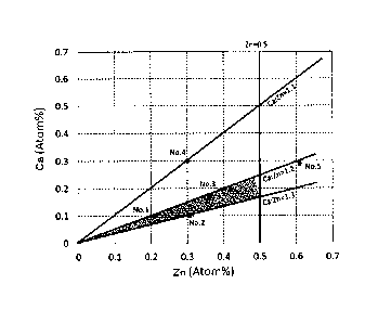
FIG. 1 shows a graph of the Ca and Zn content levels of
ternary Mg alloy materials of Mg-Ca-Zn;
FIG. 2 is a production flow chart of a device for fixing
biological soft tissue;
FIG. 3 is a strain distribution map of a produced clip;
FIG. 4 shows a graph (1) of the true stress-true strain
relations of annealed clips;
FIG. 5 shows a graph (2) of the true stress-true strain
relations of annealed clips;
FIG. 6 shows a graph (3) of the true stress-true strain
relations of annealed clips;
FIG. 7 shows the crystal orientation analysis results of
annealed clips;
FIG. 8 shows a graph of the biodegradability of annealed
clips;
14
CA 02960612 2017-03-08
,
FIG. 9 shows an X-ray CT sectional image (1) after
implanting an annealed clip in vivo;
FIG. 10 shows an X-ray CT sectional image (2) after
implanting an annealed clip in vivo;
FIG. 11 shows an X-ray CT sectional image after implanting
a titanium device (Comparative Example 1) in vivo;
FIG. 12 shows an X-ray CT sectional image after implanting a
device having a high Zn content (Comparative Example 2) in vivo;
FIG. 13 shows a crystal grain structure micrograph;
FIG. 14 shows a graph of the implantation time and the
volume residual ratio (Example 3);
FIG. 15 shows reconstructed images of X-ray CT sectional
images (Example 3);
FIG. 16 shows graphs of the measurement of the Mg ion
concentration, etc. in the blood (Example 3);
FIG. 17 shows the results of observation of the surrounding
cells and tissues (Example 3);
FIG. 18 shows the crystal orientation analysis results by
EBSD (Example 4);
FIG. 19 is reconstructed images 1 of X-ray CT sectional
images of a rat (Example 4);
FIG. 20 is reconstructed images 2 of X-ray CT sectional
images of a rat (Example 4);
FIG. 21 shows a graph (4) of the true stress-true strain
relations of annealed clips;
CA 029 612 2017-03-08 22 shows a graph (5) of the true stress-true strain
relations of annealed clips;
FIG. 23 shows a graph (6) of the true stress-true strain
relations of annealed clips;
FIG. 24 is an explanatory drawing (1) of a conventional fine
crystal grain material;
FIG. 25 is an explanatory drawing (2) of a conventional fine
crystal grain material;
FIG. 26 shows a graph of the true stress-true strain
relationship of pure magnesium used in finite element calculation
of clips of the embodiment.
DESCRIPTION OF THE PREFERRED EMBODIMENTS
[0027]
Examples of embodiments of the present invention are
explained in detail below with reference to the accompanying
drawings. Furthermore, the scope of the present invention is not
limited to the following examples and illustrated examples;
numerous modifications and variations are possible.
EXAMPLE 1
[0028]
FIG. 1 is a graph of the Ca and Zn content levels of ternary
Mg alloy materials of Mg-Ca-Zn. The results obtained by
evaluating the utility of the five samples (Mg alloy materials
No. 1-No. 5) shown in FIG. 1 as devices for fixing biological
soft tissue are explained below. The five samples (Mg alloy
16
CA 02960612 2017-03-08
materials No. 1-No. 5) were as shown in Table 1 below.
[0029]
[Table 1]
Ca:Zn
Mg Ca Zn Fe Si Ni
No.
(content Comments
(atom%) (atom) (atom) (atom%) (atom) (atom-s)
ratio)
1:2
1 99.69 0.10 0.21 0.002 0.003 <0.001 (1
10) Example A
:2.
1:3
2 99.59 0.10 0.31 0.001 <0.001
<0.001 (1310) Example B
:.
1:2
3 99.48 0.16 0.36 0.001 <0.001
<0.001 (1:2.25) Example C
:
4 99.39 0.31 0.30 0.003 0.004 <0.001 (1
Example D
10.197)
99.10 0.29 0.61 0.002 <0.001 <0.001 1:2 Comparative
(1:2.10) Example
[0030]
The production of the five samples (Mg alloy materials No.
1-No. 5) and the method for producing devices for fixing
biological soft tissue using these Mg alloy materials will be
explained with reference to FIG. 2.
First, an Mg alloy material is prepared by adding the Ca and
Zn content levels relative to Mg in the amounts shown in Nos. 1-5
in Table 1 above by atomic ratio (S01: Mg alloy material
preparation step). Then, the Mg alloy material is melted and cast
to produce an ingot (S02: ingot production step).
[0031]
Next, the ingot is subjected to homogenization heat
treatment (S03: homogenization heat treatment step). Hot
extrusion is then carried out in a temperature range of 300 C
(SO4: hot extrusion step), and the crystal grain structure of the
interior is refined by plastic working. Annealing is conducted
17
CA 02960612 2017-03-08
thereafter in a temperature range of 400 C (S05: annealing step).
A homogeneous material can be obtained by holding for a long
period of time after carrying out hot extrusion (SO4).
The material is then formed into the desired clip shape
(S06: forming step), and the impurities including oxides on the
clip surface are removed (S07: surface removal step).
[0032]
Finite element analysis of the strain distribution
associated with fixation of the clip was conducted on clips made
from the mesh models produced.
FIG. 3 is an equivalent plastic strain distribution diagram
of a clip 10. The strain distribution diagram shown in FIG. 3
shows the results obtained by using finite element analysis based
on the material data of pure magnesium (average crystal grain
size: 47 m). A graph of the true stress-true strain relationship
of pure magnesium used in the finite element calculation of clip
is shown in FIG. 26. The dotted line in the graph of FIG. 26
is a plot assuming that the material reaches a constant value
without rupturing even after stress has reached the maximum
value. The left drawing in FIG. 3 shows a V-shaped clip (mesh
model before deformation, open state before pinching); the right
drawing shows the clip in a closed state. The locations labeled
11-15 in FIG. 3 each show a part with different shading on the
image of the clip. The folded part 11 of the clip in a closed
state is the part under the greatest strain, and the strain
18
CA 029 612 2017-03-08 in the order 12, 13, 14. The shaded part labeled 15 is
a part under virtually no strain. Calculation showed the maximum
relative plastic strain to be 0.357. This value of 0.357 changes
depending on the clip material and shape, but is not changed by
the size of the clip. Finite element analysis was conducted on
the material parameters of pure magnesium and mesh model shapes
of clips set and produced in the examples. When deformed into the
shape in the right drawing of FIG. 3, the limit strain required
for deformation was determined to be 0.357 using the maximum
relative plastic strain value in the clip model. In other words,
a value of 0.357 was set as one target indicator. Therefore, if
the material used in the example is changed, the maximum strain
value, that is, the limit value that serves as a target
indicator, also changes since the strain distribution during
deformation changes as well. Given that the clip shape and size
are not limited, the maximum strain value of a clip of the mesh
model shape used in the example serves as a benchmark in the
present invention.
[0033]
A material that does not break at a strain of 0.357 or more
must be used in a clip of the mesh model shape used in the
example. The tissue can be fixed without the clip rupturing at
part 11 which is under the greatest strain in the clip produced.
As will be described below, experimental results were obtained
showing that the magnesium alloy produced in this example is a
19
CA 02960612 2017-03-08
,
material that does not rupture at a true strain of 0.357 due to
compression. It is thereby understood that biological soft tissue
can be fixed using a clip made of a ternary Mg alloy material of
Mg-Ca-Zn shown in the embodiment.
[0034]
FIG. 4 relates to Mg alloy material No. 1 (Example A) and
shows a graph of the true stress-true strain relations of clips
annealed for one hour or eight hours at temperatures of 350 C,
400 C, and 450 C. In the graph of FIG. 4, the horizontal axis is
the true strain, and the vertical axis is the true stress. It is
understood from the graph of FIG. 4 that a clip made from Mg
alloy material No. 1 (Example A) does not rupture even when
strain of 0.357 or more arises, except under conditions of one
hour at 350 C and eight hours at 450 C. In other words,
coarsening of the crystal grains is inadequate in heat treatment
for one hour, and heat treatment for eight hours is necessary
when the annealing temperature is low, such as 350 C. In
addition, heat treatment for one hour is adequate and a crystal
structure that clears strain of the required value of 0.357 or
more can be obtained when the annealing temperature is high, such
as 450 C. In contrast to this, the crystal structure coarsens
more than is necessary in heat treatment for eight hours, and
strain of the required value of 0.357 or more therefore cannot be
cleared. This suggested the existence of an optimum annealing
temperature range and holding time range.
CA 029 612 2017-03-08
FIG. 5 relates to Mg alloy material No. 5 (Comparative
Example) and shows a graph of the true stress-true strain
relations of clips annealed for one hour or eight hours at
temperatures of 350 C, 400 C, and 450 C. It is understood from
the graph of FIG. 5 that a clip made from Mg alloy material No. 5
(Comparative Example) lacks reproducibility of data for clearing
strain of the required value of 0.357 or more.
[0036]
FIG. 6 shows a graph of the true stress-true strain
relations of clips subjected to four types of annealing (one
hour, two hours, four hours, or eight hours) at 400 C in clips
made from Mg alloy material No. 1 (Example A). Based on the graph
of FIG. 6, the true stress-true strain relations sometimes
improve and sometimes decline when annealing is conducted for
eight hours in a clip made from Mg alloy material No. 1 (Example
A). It is believed that the pinning effect of the solute atoms on
the crystal grain boundaries declines and the crystal structure
tends to coarsen partially when the annealing time is eight hours
because the concentrations of the major solute atoms, which are
calcium and zinc, are low in a material of Mg alloy material No.
1. This suggests that there is a possibility that the required
value will not be satisfied when the annealing time is long when
the solute atom concentration is low. This in turn suggested the
existence of an optimum holding time range for annealing.
21
CA 02960612 2017-03-08
[0037]
The crystal grain structure of a material that does not
rupture even when strain of 0.357 or more arises will be
explained here. FIGS. 13(1)-(3), respectively, relate to Mg alloy
material No. 1 (Example A) and show crystal grain structure
micrographs of clips annealed for eight hours at a temperature of
350 C, two hours at 400 C, and one hour at 450 C. Clips annealed
for eight hours at 350 C, two hours at 400 C, and one hour at
450 C do not rupture even when strain of 0.357 or more arises, as
shown in FIG. 4 and FIG. 6 (see FIG. 4 for eight hours at 350 C,
FIG. 6 for two hours at 400 C, and FIG. 4 for one hour at 450 C)
The crystal grain structure micrographs of FIGS. 13(1)-(3) make
it possible to confirm that the crystal grain size of the
annealed clips is about 20 pm for small grains and about 250 jtm
for large grains.
[0038]
FIG. 21 shows a graph of the true stress-true strain
relations of clips subjected to four types of annealing (one
hour, two hours, four hours, or eight hours) at 400 C in clips
comprising Mg alloy material No. 2 (Example B). The graph of FIG.
21 confirmed that the true stress-true strain relations sometimes
improve and sometimes decline when annealed for four hours or
eight hours in clips made from Mg alloy material No. 2 (Example
B), but that the true strain characteristics improve when
annealed for one hour or two hours. This suggested the existence
22
CA 02960612 2017-03-08
of an optimum holding time range in annealing in clips comprising
Mg alloy material No. 2 (Example B).
[0039]
FIG. 22 shows a graph of the true stress-true strain
relations of clips subjected to four types of annealing (one
hour, two hours, four hours, or eight hours) at 400 C in clips
comprising Mg alloy material No. 3 (Example C). The graph of FIG.
22 confirmed that the true stress-true strain relations sometimes
improve and sometimes decline when annealed for four hours and
eight hours in clips comprising Mg alloy material No. 3 (Example
C), but the true stress-true strain relations improve when
annealed for one hour or two hours. This suggested the existence
of an optimum holding time range for annealing in clips
comprising Mg alloy material No. 3 (Example C).
[0040]
FIG. 23 shows a graph of the true stress-true strain
relations of clips subjected to five types of annealing (one
hour, two hours, three hours, four hour, or eight hours) at 400 C
in clips comprising Mg alloy material No. 4 (Example D). The
graph of FIG. 23 confirmed that the true stress-true strain
relations improve when annealed for three hours in clips
comprising Mg alloy material No. 4 (Example D). In addition, the
true stress-true strain relations were confirmed to sometimes
improve and sometimes decline when annealed for four hours.
However, those annealed for one hour, two hours, and eight hours
23
CA 02960612 2017-03-08
were confirmed to lack reproducibility of data on clearing strain
of the required value of 0.357 or more. This suggested the
existence of an optimum holding time range for annealing in clips
made of Mg alloy material No. 4 (Example D).
[0041]
The results obtained by operating an electron beam in
combination with a scanning electron microscope (SEM), conducting
crystal orientation analysis using EBSD which can measure the
crystal orientation and crystal system, and elucidating the
plastic deformation behavior will be explained next.
FIGS. 7(1) and (2) show the results of crystal orientation
analysis of annealed cylindrical test pieces. FIG. 7(1) shows the
crystal grain structure inside the recovered compressed test
piece when the load was removed after compressing an Mg alloy
material (No. 1: Example A) to a true strain of 0.123. Figure
7(2) shows the crystal grain structure inside the recovered
compressed test piece when the load was removed after compressing
a cylindrical test piece made of an Mg alloy material (No. 1:
Example A) to a true strain of 0.193. The "nominal stress (on)-
nominal strain (En) relationship (curve)" was determined from the
"load-displacement relationship (curve)" obtained by compression
testing of the cylindrical test pieces under the respective
conditions, and the strain value of the crystal grain structure
was calculated by the "true stress (ot=on(1-En))-true strain (Et¨
ln(1-En)) relationship (curve)." Here, the nominal stress is the
24
CA 02960612 2017-03-08
load divided by the initial cross-sectional area, and the nominal
strain is the (initial height of the test piece-height after
deformation) divided by the initial height of the test piece.
Boundaries having misorientation of several degrees are
confirmed every several microns inside the crystal grains of the
Mg alloy material in the compressed specimen corresponding to the
closed state of the clip shown in FIG. 7(2), that is, during
deformation. It is thereby understood that the strain accumulated
in association with deformation is dynamically recovered due to
the formation of subgrains, the formation of cracks (microscopic
cracks) due to stress concentration is avoided by the occurrence
of "dynamic recovery," and this contributes to improving the
ductility.
[0042]
FIG. 8 shows a graph of the biodegradability of annealed
clips. These are the results of in vitro tests conducted by
immersion for a certain period of time in a solution simulating
body fluid (E-MEM: 10% FBS, CO2 concentration: 5%, 37 C)
The left side of the graph of FIG. 8 relates to Mg alloy
materials Nos. 1-No. 3 and shows the volume residual ratio of the
clip when an environment similar to that within the body was
constructed, and the clip produced was left in the static
environment for four weeks. The right side of the graph relates
to Mg alloy materials No. 1 and No. 2 and shows the volume
residual ratio of the clip when an fluid circulating environment
CA 029 612 2317-03-
=
-
similar to that within the body was constructed, and the clip
produced was allowed to stand for four weeks in the slowly
refluxed solution described above, that is, allowed to stand for
four weeks in a circulating environment. Here, the volume
residual ratio is taken to be the ratio determined as a result of
dividing the residual volume of the magnesium alloy calculated
from CT observation images by the volume before immersion.
[0043]
It is understood from the graph of FIG. 8 that the volume
residual ratios of clips after four weeks in a static environment
are all 90% or greater, the residual ratios of the clips after
four weeks in a circulating environment are all 85% or greater,
and the biodegradation rates are appropriate as a device for
fixing biological soft tissue. In addition, since the volume
residual ratio increased in the order Mg alloy material No. 1
(Example A), No. 2 (Example B), No. 3 (Example C) according to
the in vitro test method immersed for a certain period of time in
a solution simulating body fluid described above, the
biodegradation rate is understood to lengthen in this order. It
is also understood from this that the biodegradation rate can be
adjusted by the Ca and Zn concentrations.
As described above, devices using Mg alloy materials No. 1-
No. 3 were clarified to be useful as devices for fixing
biological soft tissue.
EXAMPLE 2
26
CA 029 612 2017-03-08
The biodegradability and safety of the device for fixing
biological soft tissue produced were confirmed in Example 2. An
explanation follows.
FIG. 9 and FIG. 10 show X-ray CT sectional images of a U-
shaped device for fixing biological soft tissue produced by the
same method as in Example 1, that is, by annealing, after
implantation in the body of a mouse.
The device for fixing biological tissue was confirmed based
on the X-ray CT sectional images to maintain its U-shape 7, 14,
and 28 days after implantation.
FIG. 9(1) is an image of seven days after implantation in a
mouse. FIG. 9(2) is an image of 14 days after implantation in a
mouse. In both cases, the changes in the volume of the space from
immediately after implantation are very slight. The amount of gas
generated is therefore understood to be minute, and no rapid gas
generation is found.
[0045]
FIG. 10(1) is a reconstructed image of an X-ray CT sectional
image of immediately after implantation in a mouse. FIG. 10(2) is
a reconstructed image of an X-ray CT sectional image of 28 days
after implantation in a mouse. Although a decrease in volume due
to uniform degradation is found after 28 days, the reconstructed
device is understood to maintain its U-shape. It is thereby
understood that the device maintains its fastening performance
27
CA 02960612 2017-03-08
during this period without any parts missing. Furthermore, it was
confirmed that no lesions were seen based on X-ray CT or visual
observation of the surrounding tissues at the time of extraction.
[0046]
The biodegradability of a device made of titanium
(Comparative Example 1) and a device having a high Zn content
(Comparative Example 2) will be described here as comparative
examples.
FIG. 11 shows an X-ray CT sectional image of after
implantation of a titanium device (Comparative Example 1) in
vivo. FIG. 12 shows an X-ray CT sectional image after
implantation of a device having a Zn content of 6 atom%
(Comparative Example 2) in vivo.
The titanium device (Comparative Example 1) is understood to
maintain its shape without degrading even 28 days after
implantation in a mouse (see FIG. 11). Although not shown in the
drawing, the titanium device (Comparative Example 1) has a large
artifact effect on the X-ray CT sectional image and can be said
to make observation of the biological tissue difficult.
[0047]
On the other hand, the Mg alloy material including a large
amount (6 atom%) of zinc generated a large amount of gas
(hydrogen) in association with biodegradation after seven days
due to its rapid biodegradation rate. FIG. 12(1) shows an X-ray
CT sectional image of seven days after implantation in a mouse.
28
CA 029 612 2017-03-08 FIG. 12(1), there is a black spatial region showing a
trace of
a gas pool remaining after the device has disappeared, and there
is a clear part on the edges of the spatial region; it was
possible to confirm a metal structure, which is the remaining
clip, and bone.
[0048]
In addition, the metal structure degraded completely after
14 days, changing into compounds such as calcium phosphate,
magnesium phosphate, magnesium carbonate, and calcium carbonate,
and corrosion products of the device remained in the body of the
mouse. FIG. 12(2) shows an X-ray CT sectional image of 14 days
after implantation in a mouse. Corrosion products are difficult
to discern when buried in soft tissue because their contrast is
lower than that of bone, but parts of corrosion products (calcium
phosphate, magnesium phosphate, magnesium carbonate, calcium
carbonate) can be confirmed in FIG. 12(2).
[0049]
In comparison to the changes over time in the materials of
the above two Comparative Examples 1 and 2, the device for fixing
biological soft tissue of the present invention is understood to
have performance that makes it possible to avoid delayed tissue
recovery associated with the generation of a large amount of gas,
to have fastening and holding performance for a suitable length
of time within the body, and to be minimally harmful to the body.
EXAMPLE 3
29
CA 02960612 2017-03-08
[0050]
<Subcutaneous implantation test in the abdomen of mice>
The results of a study that implanted clips produced by the
same method as in Example 2 (referred to hereinafter as clips of
the present example) subcutaneously to the abdomen of mice will
be explained first. Titanium clips (Comparative Example 1) and
clips having a Zn content of 6 atom% (Comparative Example 2) were
also tested by subcutaneous implantation to the abdomen of mice
in the same way as comparative examples.
Although no growth of the space due to gas generation could
be observed upon external observation with the clips of the
embodiment or the titanium clip (Comparative Example 1) one week
after implantation, growth of a large space was seen with the
clip of Comparative Example 2 having a high Zn content. The clip
of Comparative Example 2 having a high Zn content appeared to
have generated a large amount of gas (hydrogen) in association
with biodegradation after one week due to its rapid
biodegradation rate.
[0051]
FIG. 14 shows a graph of the implantation time and the
volume residual ratio of clips of the embodiment. The graph plots
the mean values of three mice used in the test. As shown in FIG.
14, the clips of the embodiment decreased in volume over time in
the body of the mouse, and reached 70% one month (four weeks)
after implantation and 50% three months (12 weeks) after
CA 02960612 2017-03-08
implantation.
FIGS. 15(a)-(e), respectively, show reconstructed images of
X-ray CT sectional images of clips of the embodiment one week,
two weeks, three weeks, four weeks, and 12 weeks after
implantation in mice. FIG. 15 confirmed that clips of the
embodiment retain the shape of the clip at the time of
implantation after 12 weeks.
[0052]
Next shown are the results obtained by measuring the Mg ion
concentration and the like in the body 12 weeks after
implantation. The measurement targets are shown below in Table 2.
The serum test data were also analyzed statistically.
Furthermore, in statistical analysis, the data were assumed to
have a normal distribution, the variance was judged by F-test,
and those of equal variance were subsequently analyzed using
Student's t-test and those of unequal variance were analyzed by
Welch's t-test. The significance level was set at p<0.05 in all
analyses.
[0053]
[Table 2]
Measurement target Explanation
Mg (mg/dL) Blood magnesium level
Creatinine (numerical value rises as renal function
CRE (mg/dL)
declines)
Aspartate aminotransferase (numerical value rises in
AST (IU/L) association with disorders of the liver and
myocardium)
ALT (IU/L) Alanine aminotransferase (numerical value rises due to
destruction of cells in the liver)
Alkaline phosphatase (numerical value rises due to
ALP (IU/L) abnormalities of the hepatobiliary system, obstruction
and stenosis of the biliary tract)
31
CA 02960612 2017-03-08
[0054]
FIG. 16 shows graphs of the measurement results of the blood
Mg ion concentration and the like in the body up to 12 weeks
after implantation. Each of the graphs of FIGS. 16(1)-(5) shows
the numerical values of Mg, CRE, AST, ALP, and ALT after one
week, two weeks, three weeks, four weeks, and 12 weeks for the
clips produced (embodiment), titanium clips (Comparative Example
1), and clips having high Zn (Comparative Example 2),
respectively. In the bar graphs, data from after elapse of the
predetermined length of time are arranged in three bars from left
to right in the order Comparative Example 1, Comparative Example
2, embodiment. The far right bar of each graph shows the
numerical values after four weeks taking normal mice not
subjected to laparoscopy or implantation as a control.
Furthermore, the data in the graphs are the mean of the data from
three mice.
The fact that the results obtained by measuring the blood Mg
concentration up to 12 weeks after implantation did not reveal a
significant increase in concentration makes it possible to
confirm that the eluted ion is excreted outside the body.
[0055]
FIG. 17 shows the results of observation of the surrounding
cells and tissues two weeks after implantation. FIGS. 17(1)-(3)
show the results obtained by staining the cells and tissues
32
CA 02960612 2017-03-08
surrounding the implanted clip produced (embodiment), titanium
clip (Comparative Example 1), and clip having a high Zn content
(Comparative Example 2) by hematoxylin-eosin stain (HE stain) and
=
SR stain by Sirius red (images on the left are HE stain; images
on the right are SR stain).
There was no inflammatory response, the surrounding cells
and tissues were normal, and the clip of the embodiment was
confirmed to be biologically safe based on cell observation of
the cells and tissues surrounding the implanted clip produced
(embodiment) and the cells and tissues surrounding the implanted
titanium clip (Comparative Example 1). On the other hand, fibrous
morphology was not seen, intercellular substrate (cell walls) was
destroyed, nuclei were not formed in cells, and the tissue
appeared necrotic in observation of the cells and tissues
surrounding the implanted clip having high Zn (Comparative
Example 2).
EXAMPLE 4
[0056]
<Vascular anastomosis test using rats>
Example 4 confirmed the biodegradability and safety of clips
produced, unlike the clip production methods of Examples 2 and 3,
by raising the hot extrusion temperature and slowing the hot
extrusion rate in the hot extrusion step to expose the ingot to a
high-temperature state for several tens of seconds immediately
after extrusion and conducting annealing immediately after the
33
CA 02960612 2017-03-08
hot extrusion step. An explanation follows.
[0057]
The clips of Example 4 have the Zn and Ca contents of the Mg
alloy material of No. 1 in Table 1 described above in Example 1.
Specifically, 0.1 atom% of Ca and 0.21 atom% of Zn were added
relative to 99.69 atom% of Mg, an ingot was produced by melting
and casting, and the ingot was subjected to homogenization heat
treatment. After heat treatment, the ingot was subjected to stage
one hot extrusion at 350 C, and the ingot 90 mm in diameter was
processed to a diameter of 22 mm. The diameter was brought to 20
mm by cutting the 22 ram diameter, the ingot was subjected to
stage two hot extrusion at 410 C, and processed into a V-shaped
cross-section. Annealing by exposure to 400-410 C was conducted
immediately after the stage two hot extrusion. Impurities
including oxides were subsequently removed from the clip surface.
[0058]
FIG. 18 shows the results obtained by operating an electron
beam in combination with a scanning electron microscope (SEM) and
conducting crystal orientation analysis of the clips produced
using EBSD, which can measure the microcrystal orientation and
crystal system. The crystal orientation analysis results shown in
FIG. 18 confirmed the crystal structure of the clips produced to
be an equiaxed crystal grain structure. In addition, the average
crystal grain size of the crystal structure of the clips produced
measured using the intercept method was 28.8 ( m) near the valley
34
CA 02960612 2017-03-08
of the V-shape of the clip and 31.5 (Km) near the top of the V-
shape.
The clips produced were confirmed to have an equiaxed
crystal grain structure having an average crystal grain size of
approximately 30 (Km). These clips have excellent deformability
in a closed V-shape because, as explained in FIG. 7, boundaries
having misorientation of several degrees appear every several
microns within the crystal grains (subgrains are formed), the
strain accumulated in association with deformed state is
dynamically recovered, and the formation of cracks due to stress
concentration is avoided (relaxation of stress concentration).
[0059]
Next, the results obtained by anastomosing a blood vessel
connected to part of the rat liver and the bile duct using the
clips produced will be explained. The abdomen of the rat was cut
open, a blood vessel connected to part of the liver and the bile
duct were placed together and anastomosed by closing the V-shaped
clip. The liver was subsequently resected.
FIG. 19 shows reconstructed images of X-ray CT sectional
images of the chest of the rat one week and four weeks (one
month) after resection. In FIG. 19, (1) shows the reconstructed
images one week after resection and (2) shows those four weeks
(one month) after resection. In FIGS. 19(1) and (2), (a) was
anastomosed by a clip of the embodiment and (b) was anastomosed
by a clip of Comparative Example 1.
CA 02960612 2017-03-08
[0060]
As shown in FIG. 19, it can be inferred that the expected
fastening performance of the clip was maintained since the rats
survived even four weeks after liver resection, that is, after
severing the blood vessel and bile duct, and X-ray CT did not
find generation of a large amount of gas or opening of the clip.
The clip is also expected to finally be degraded and
excreted after maintaining its fastening performance for a
certain length of time as degradation advances uniformly within
the body of the rat. This confirmed the possibility of realizing
a safe biodegradable clip.
[0061]
FIG. 20 shows X-ray CT sectional images of a rat. In FIG.
20, (1) is an X-ray CT sectional image from one week after
resection and (2) is from four weeks (one month) after resection.
FIGS. 20(1) and (2) both show anastomosis by a clip of the
embodiment. The clip of the embodiment is understood to be less
likely to create metal artifacts during X-ray CT imaging than
when a conventional titanium clip is used, and to make it
possible to observe the biological tissue clearly without image
correction.
INDUSTRIAL APPLICABILITY
[0062]
The device for fixing biological soft tissue of the present
invention is useful in surgical clips, staple, and the like
36
CA 02960612 2017-03-08
because it can keep tissues joined for the 2-8 weeks it takes
biological soft tissue to suture and is excreted after being
completely degraded in about one year.
KEY
[0063]
10: Clip
37