Note: Descriptions are shown in the official language in which they were submitted.
CA 02960860 2017-03-09
WO 2016/044799 PCT/US2015/051072
INJECTABLE MICROPARTICLES FOR HYPER-LOCALIZED RELEASE OF
THERAPEUTIC AGENTS
CROSS REFERENCE TO RELATED APPLICATION
This application claims the benefit under 35 U.S.C. 119(e) to
U.S. Provisional Application No. 62/052,959, filed September 19, 2014, which
application is hereby incorporated by reference in its entirety.
BACKGROUND
Technical Field
This disclosure relates to an injectable sustained release
composition comprising drug-loaded microparticles and a method of delivering
the same.
Description of the Related Art
Drug-loaded microparticles have been used for sustained release
of therapeutic agents. However, truly localized release is difficult to
achieve
and burst release remains as one of the main factors to cause undesirable
systemic side effects. Accordingly, there is a medical need to not only extend
the local duration of action of the therapeutic agent, but to effectively
reduce the
systemic side effects associated with that administration.
BRIEF SUMMARY
Described herein are pharmaceutical compositions, injectable
dosage forms and method of using the same for treating or managing pain,
infection, malignancy, in a body compartment, such as a joint space, an
epidural space, a vitreous body of an eye, a surgically created space, an
intracranial space or a space adjacent to an implant surgical site, or a solid
tumor.
The present disclosure provides a membrane based, diffusion-
driven release mechanism with drug particle sizing large enough to allow high
drug loading, but small enough to be injected.
1
CA 02960860 2017-03-09
WO 2016/044799 PCT/US2015/051072
As provided herein, a "drug" or "therapeutic agent" is coated with
a semi-permeable polymeric shell and injected into a body compartment.
Water then diffuses through the polymer and dissolves the drug core (D)
creating a saturated solution inside the membrane (C) and essentially sink
conditions outside the particle (c). This concentration gradient drives a
constant (zero order) release of drug from the drug particle as long as there
is
some drug core remaining to maintain a saturated solution. The period of
release can be tuned by altering the permeability of the polymer coating.
The present disclosure further relates to using local anesthetics
(amides) for the purpose above focusing on subarachnoid block (primarily in
palliative cancer pain); extradural blockade (palliative care); and nerve
plexus
blockade (i.e. Brachial Plexus) for analgesia, anesthesia, limb and digit
grafting
to improve blood flow, vascular procedures for same.
The present disclosure further relates to injection of therapeutic
agents (e.g., CNS modulators focused on GABA receptors) locally in the area
of nerve damage, including intracranial injection and possibly also
subcutaneous injection.
The present disclosure further relates to injection for systemic
delivery (subcutaneously) and for local application (bonded to &/or applied
with)
with implants (pacemakers, defibrillators, orthopedic implants, artificial
hearts)
of one or more antibiotics, and near or at a surgical site.
The present disclosure further relates to local delivery of powerful
chemotherapeutic agents and hormones given for the treatment of malignancy.
BRIEF DESCRIPTION OF THE SEVERAL VIEWS OF THE DRAWINGS
The following figures set forth embodiments in which like
reference numerals denote like parts. Embodiments are illustrated by way of
example and not by way of limitation in all of the accompanying figures
wherein:
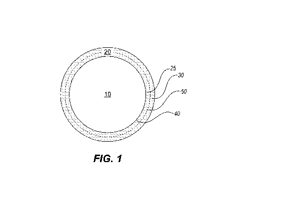
Figure 1 shows schematically a microparticle of core/shell
morphology.
2
CA 02960860 2017-03-09
WO 2016/044799 PCT/US2015/051072
Figure 2 shows the in vitro release profiles of fluticasone
propionate as uncoated powder, uncoated crystal and coated crystal.
Figure 3A shows the release profiles of fluticasone propionate
microparticles having undergone heat-treatment at various temperatures.
Figure 3B shows the release half-lives of fluticasone propionate
microparticles having undergone heat-treatment at various temperatures.
Figure 4A and 4B show the particle size distribution of the
fluticasone propionate microparticles as compared to particle size
distribution of
triamcinolone hexacetonide (TA) (Kenalog TM).
Figure 5 is a graph showing the relative amounts of fluticasone
propionate and PVA in microparticles by iHNMR analysis.
Figure 6 is a graph showing dissolution profiles of triamcinolone
hexacetonide (TA) as compared to sustained release (SR) formulations of
fluticasone propionate (FP) according to embodiments of this disclosure.
Figure 7 is a graph showing plasma fluticasone (FP) levels,
synovial fluid FP levels after injection of 20 mg formulation into knee joint
of
sheep as compared to intra-articular pharmacokinetics of triamcinolone
hexacetonide (40mg) from human subjects.
Figures 8A, 8B and 80 demonstrate the results of a histological
examination of the injected joints of sheep showing no abnormalities.
Figure 9 shows the local concentrations in tissue and synovial
fluid of knee joints of dogs for a period of 60 days following a single
injection of
a low dose fluticasone propionate. The plasma concentrations were too low to
detect.
Figure 10 shows the local concentrations in tissue and synovial
fluid of knee joints of dogs, as well as the plasma concentrations, for a
period of
60 days following a single injection of a high dose fluticasone propionate.
Figure 11 shows the plasma concentrations of fluticasone
propionate following injections to the knee joints of sheep as compared to
those
of dogs. The microparticles for each injection had undergone different heat-
treatments prior to being formulated into injectable compositions.
3
CA 02960860 2017-03-09
WO 2016/044799 PCT/US2015/051072
Figure 12 shows the plasma concentrations of fluticasone
propionate in the knee joints of dogs over a period of 45 hours following a
single injection. The pharmacokinetic (PK) curve indicates a lack of initial
burst.
DETAILED DESCRIPTION
Described herein are pharmaceutical compositions, injectable
dosage forms and method of using the same for treating or managing local
inflammation, pain (including post-surgery pain), infection, malignancy, in a
body compartment, such as a joint space, an epidural space, a vitreous body of
an eye, a surgically created space, an intracranial space or a space adjacent
to
an implant, a surgical site or a solid tumor.
The pharmaceutical composition includes a plurality of
microparticles in core/shell morphology. In particular, the microparticle
includes
a crystalline drug core of a therapeutic agent and a polymeric shell
encapsulating the crystalline drug core. As discussed in further detail
herein,
the injectable microparticles are characterized with high drug-loading, narrow
size distribution and a sustained release profile of pseudo zero-order release
over a certain period within a body compartment, as defined herein, or
subcutaneously. The release periods depend on the affliction or the
corresponding therapeutic agent. Therapeutic agents for pain management
may be released during a period of 2-12 months, whereas antibiotics may be
released during a period of 3-7 days.
The sustained release delivery mechanism is based on
dissolution. While not wishing to be bound by any specific mechanism of
action, it has been found that when the drug particles coated with semi-
permeable polymeric shells are injected into a body compartment, water from
the body compartment diffuses through the polymeric shell and partially
dissolves the crystal drug core. As a result, a saturated solution of the drug
is
formed inside the polymeric shell. Since there are essentially sink conditions
in
the fluid (e.g., synovia when the body compartment is a joint) in which the
microparticles are injected and reside, a concentration gradient is created
which
continuously drives the drug out of the microparticles and into the
surrounding
4
CA 02960860 2017-03-09
WO 2016/044799 PCT/US2015/051072
fluid. As long as there is some drug core remaining to maintain a saturated
solution within the polymeric shell, a constant (i.e., zero order or pseudo-
zero
order) release of the drug from the coated microparticles is obtained.
Also disclosed herein is a method for reducing or managing pain,
e.g. due to surgical pain, chronic pain or neuropathic pain, by administering
an
injectable dosage form to a body compartment. Advantageously, the release is
highly localized within the local tissue or fluid medium of the body
compartment
to ensure a long-acting local therapeutic level, while maintaining a low or
undetectable systemic level of the therapeutic agent.
Definitions
The articles "a" and "an" are used herein to refer to one or to more
than one (i.e., to at least one) of the grammatical object of the article. By
way
of example, "an element" means one element or more than one element.
The term "plurality" means "two or more", unless expressly
specified otherwise. For example, "plurality" may simply refer to a
multiplicity of
microparticles (two or more) or an entire population of microparticles in a
given
composition or dosage form, e.g., for purpose of calculating the size
distribution
of the microparticles.
As used herein, unless specifically indicated otherwise, the word
"or" means "either/or," but is not limited to "either/or." Instead, "or" may
also
mean "and/or."
As used herein, the term "therapeutic agent" and "drug" are used
interchangeable and refer to any agent that can produce a therapeutic effect
or
benefit. When used with respect to a therapeutic agent or a drug (e.g., a
chemotherapy agent), the terms "sustained release" or "extended release" are
used interchangeably. Sustained release refers to continuously releasing the
therapeutic agent over an extended period of time after administration of a
single dose, thus providing a prolonged therapeutic effect throughout the
release period.
"Sustained release" is in contrast to a bolus type administration in
which the entire amount of the active agent/substance is made biologically
5
CA 02960860 2017-03-09
WO 2016/044799 PCT/US2015/051072
available at one time. Nevertheless, "sustained release" may include an
initial
faster release followed by a longer, extended period of slower release. As
discussed in further detail below, the construction of the microparticles
makes it
possible to minimize the initial faster release (e.g., a burst release) and
prolong
the extended release period to achieve a profile of near constant release that
is
irrespective of the drug concentration (i.e., a zero-order or pseudo zero-
order
release).
Not all non-zero release is within the meaning of "sustained
release." Rather, "sustained release" should provide at least a minimum
therapeutically effective amount (as defined herein) of the therapeutic agent
during the release period. It should be understood that the minimum
therapeutically effective amount of the therapeutic agent depends on the
severity of afflictions to be addressed.
"Sustained release period" refers to the entire period of release
during which a local concentration of the therapeutic agent is maintained at
or
above a minimum therapeutically effective amount. The desired sustained-
release period can, of course, vary with the disease or condition being
treated,
the nature of the therapeutic agent, and the condition of the particular
patient to
be treated. Thus, the desired sustained-release period can be determined by
the attending physician.
"Local concentration" refers to the concentration of the drug within
a body compartment (as defined herein), including the concentration in the
tissue or fluid of the body compartment.
"Plasma concentration" refers to the concentration of the drug in
the plasma or serum. The injectable microparticles are capable of hyper-
localized release during a prolonged period while maintaining a low plasma
concentration, e.g., sufficiently low to minimize undesirable systemic side
effects during the sustained release period.
Within the scope of the present disclosure, sustained release of
the therapeutic agent is achieved due to the unique structure of the
microparticles, which are in core/shell morphology. In particular, a
crystalline
6
CA 02960860 2017-03-09
WO 2016/044799 PCT/US2015/051072
drug core of a therapeutic agent is encapsulated by a polymeric shell
composed of one or more polymeric coatings, each permeable to the
therapeutic agent. In a preferred embodiment, all layers comprise the same
polymer. In other embodiments, two to four layers of the polymer are coated on
the therapeutic agent, with each layer incrementally slowing the release of
the
active ingredient and collectively providing the desired sustained release.
Furthermore, sustained release of the therapeutic agent is achieved by
tailoring
this delivery platform to the aqueous or sink environment of the body
compartment.
As used herein, a "patient," or "subject," to be treated by the
methods according to various embodiments may mean either a human or a
non-human animal, such as primates, mammals, and vertebrates.
The phrase "therapeutically effective amount" refers to an amount
of a therapeutic agent that, when delivered to a body compartment in the form
of the coated microparticles as defined herein, produces a degree of reduced
symptoms in the body compartment in a patient (at a reasonable benefit/risk
ratio applicable to any medical treatment). The effective amount of the
therapeutic agent may vary depending on such factors as the type and severity
of arthritis being treated, its advancement, the degree of pain to which
patient is
subject, the particular microparticle being administered, the active agent
and/or
the size/age/gender of the subject. One of ordinary skill in the art may
empirically determine the effective amount of a particular therapeutic agent
according to known methods in the art. Unless specified otherwise,
"therapeutically effective amount" refers to the amount of the therapeutic
agent
localized within the body compartment.
"Minimum therapeutically effective amount" is the least amount of
the therapeutic agent that is capable of producing a therapeutic effect (e.g.,
pain reduction or anti-inflammation).
"EC50" is the concentration of the therapeutic agent that provides
50% of the maximal effect, e.g., in reducing pain.
7
CA 02960860 2017-03-09
WO 2016/044799 PCT/US2015/051072
"Unit dosage form" refers to physically discrete units (e.g., loaded
syringe cylinders) suitable as unitary dosages for human subjects, each unit
containing a predetermined quantity of the therapeutic agent in association
with
a pharmaceutical acceptable vehicle. The quantity of the therapeutic agent is
calculated to produce the desired therapeutic effect for a desired period of
time.
The term "treating" is art-recognized and includes treating the
disease or condition by ameliorating at least one symptom of the particular
disease or condition, even if the underlying pathophysiology is not affected.
"Body compartment" refers to a space or cavity within the body of
a vertebrate (including human) that is accessible by injection. Typically, the
body compartment is at least semi-enclosed or fully enclosed by hard or soft
tissue (e.g., bones, membranes, ligamentous structure) that defines the space.
Soft tissue is typically present and may have various degrees of
vascularization. The body compartment typically contains a fluid, such as the
synovial fluid in the joints, spinal fluid in the epidural and the vitreous
humour in
the vitreous body of the eye. The fluid may or may not communicate with the
outside of the body compartment. More specifically, the body compartment
may be naturally occurring anatomical space such as a synovial joint, an
epidural space or a vitreous body of an eye. In addition, the body compartment
may also be a surgically created space (e.g., a pocket for inserting an
implanted device, soft tissue implant such as breast implant, and the like) or
any space near the implant that can be accessed through injection. The body
compartment may also be a space near a tumor, in particular, a solid tumor.
The body compartment may also be an intracranial space. The body
compartment may also be a site near or at a surgical site.
The term "synovial joint" refers to a moveable articulation of two
or more bones. The articulation is defined by a synovial cavity, which
contains
a volume of synovial fluid, is lined with a synovial membrane, and is
surrounded
by a fibrous capsule. The opposing bone surfaces are each covered with a
layer of cartilage. The cartilage and synovial fluid reduce friction between
the
articulating bone surfaces and enable smooth movements. Synovial joints can
8
CA 02960860 2017-03-09
WO 2016/044799 PCT/US2015/051072
be further distinguished by their shape, which controls the movements they
allow. For example, hinge joints act like the hinge on a door, allowing
flexion
and extension in just one plane. An example is the elbow between the
humerus and the ulna. Ball and socket joints, such as the hip, allow movement
in several planes simultaneously. Condyloid (or ellipsoid) joints, such as the
knee, permit motion in more than one plane in some positions but not others.
For example, no rotation is possible in the extended knee, but some rotation
is
possible when the knee is flexed. Pivot joints, such as the elbow (between the
radius and the ulna), allow one bone to rotate around another. Saddle joints,
such as at the thumb (between the metacarpal and carpal) are so named
because of their saddle shape, and allow movement in a variety of directions.
Finally, gliding joints, such as in the carpals of the wrist, allow a wide
variety of
movement, but not much distance.
Synovial joints include, but are not limited to, shoulder
(glenohumeral and acromioclavicular), elbow (ulno-humeral, radio-capitellar
and proximal radioulnar), forearm (radioulnar, radiocarpal, ulnocarpal), wrist
(distal radioulnar, radio-carpal, ulno-carpal, mid carpal), hand (carpo-
metacarpal, metocarpophalangeal, interphalangeal), spine (intervertebral),
hip,
knee, ankle (tibiotalar, tibiofibular), and foot (talocalcaneal,
talonavicular,
intertarsal, tarso-metatarsal, metatarsal-phalangeal, interphalangeal).
"Intra-ocular" and "intravitreous" are used herein interchangeably
to mean within the vitreous humour of the eye.
As used herein, the term "microparticle" means a particle having
mean dimension less than 1 mm. Although the microparticles are substantially
spherical in some embodiments, the microparticles can be any solid geometric
shape which is not inconsistent with the principles of the disclosure,
including,
without limitation, needles, ellipsoids, cylinders, polyhedrons and irregular
shapes.
Microparticles are coated drug particles, which may be crystalline,
polycrystalline, or amorphous. As used herein, a microparticle has a
"core/shell" morphology, shown schematically in Figure 1, in which the drug
9
CA 02960860 2017-03-09
WO 2016/044799 PCT/US2015/051072
core (10) is encapsulated by a polymeric shell (20), the polymeric shell may
include one or more thin coatings of the same or different polymers (two
coatings, 25 and 30, are shown). Importantly, the polymeric shell (20) is
formed
of polymer coatings that are not miscible with the drug core, thus, the
interface
(40) between the drug core and the polymeric shell is sharp with minimal
amounts of drug or polymer (e.g., less than 5%, or less than 1% or less than
0.5% of the total weight of either the drug or polymer shall be mixed). If the
drug core contains a highly hydrophobic drug, the polymeric shell preferably
includes at least one hydrophilic polymer. Conversely, if the drug core
contains
a highly hydrophilic drug, the polymeric shell preferably includes at least
one
hydrophobic polymer. Although the polymeric shell may be ultimately
degraded, it should maintain its structural integrity throughout the sustained
release period, thus retaining an environment for the dissolving drug core to
form a saturated solution.
As used herein, the terms "core particle," and "drug core"
interchangeably refer to a pre-formed particle that could be a single crystal
or
multiple crystals, or amorphous particle of the drug. The drug core is
encapsulated by a polymeric shell. The core particle can further comprise
other
compounds, including, without limitation, binders, buffers, antioxidants,
excipients, and additional active pharmaceutical ingredients. The core
particle
can be a single large crystal, a multiplicity of crystals, or mixtures of the
above.
In a preferred embodiment, the drug core is substantially pure drug (i.e., at
least
90%, or at least 95% or at least 98% of the entire weight of the drug core is
the
drug). In a preferred embodiment, the drug core is 100% crystalline drug.
As used herein, "polymeric shell" includes one or more polymeric
coatings. "Polymeric coating" means a thin layer of linear, branched or cross-
linked macromolecules that has a continuous surface surrounding the
crystalline drug core. Referring to Figure 1, the polymeric coatings (25 and
30)
are sequentially and concentrically coated on the drug core (20). Although the
drug core (20) and the immediate adjacent polymeric coating (25) should be
immiscible, the polymeric coatings (25 and 30) themselves may be in intimate
CA 02960860 2017-03-09
WO 2016/044799 PCT/US2015/051072
contact with each other, allowing for certain degrees of miscibility at the
interface (50) between adjacent coatings in order to form a polymeric shell
(20)
of a cohesive structure that affords structural integrity during the sustained
release period. The polymeric shell must substantially surround or envelope
the core particles.
"Coating solution" refers to a solution of pre-formed polymers
(e.g., commercially available polymers) and is suitable for coating the drug
core
according to known methods of the art, e.g. fluidized bed coating.
As used herein, the term "permeable" means allowing the
passage of molecules of the therapeutic agent by diffusion but not by fluid
flow.
As used herein, the term "semi-permeable" means permeable to
some molecules but not to others. As used herein, semi-permeable polymeric
shell are permeable to at least water and the therapeutic agent within the
coated microparticles of the disclosure.
"Dissolution half-life" is an in vitro measurement of the dissolution
characteristics of the microparticles. Specifically, the dissolution half-life
is the
amount of time that is taken for half of the original loading of the drug in
the
microparticles to dissolve and release into a dissolution medium under a
specific set of dissolution conditions. Although carried out in vitro, the
dissolution half-life is nevertheless an art-recognized factor to consider in
predicting in vivo release characteristics and can represent an accelerated
model of the sustained release behavior in vivo. In particular, dissolution
half-
life provides a qualitative tool for predicting in vivo behaviors by comparing
the
dissolutions half-lives of various formulations. For instance, formulations
that
exhibit a longer dissolution half-life in vitro are expected to exhibit a
longer
sustained release period in vivo. Unless specified otherwise, the dissolution
system used for measuring dissolution half-life the microparticles is USP Type
II
(paddle).
"Dissolution profile" is a graphic representation of the percentage
dissolution as measured by time. Besides providing quantitatively the
dissolution amount as a function of time, the curvature of the profile
qualitatively
11
CA 02960860 2017-03-09
WO 2016/044799
PCT/US2015/051072
shows the extent of the initial burst. For example, a sharp rise in the
curvature
indicates a faster initial release (burst) when compared with a gentler rise.
"Vehicle" refers to a non-toxic carrier, adjuvant, or solvent into
which the microparticles are suspended. The vehicle does not alter or destroy
the pharmacological activity of the therapeutic agent with which it is
formulated.
Pharmaceutically acceptable carriers or vehicles that may be used in the
compositions include, but are not limited to, water, physiological saline,
hyaluronic acid, and the like. As used herein, the term "biocompatible" means
characterized by not causing a toxic, injurious or immunological response when
brought into contact with living tissue, particularly human or other mammalian
tissue.
As used herein, the term "biodegradable" means capable of
partially or completely dissolving or decomposing in living tissue,
particularly
human or other mammalian tissue. Biodegradable compounds can be
degraded by any mechanism, including, without limitation, hydrolysis,
catalysis
and enzymatic action.
As used herein with respect to polymeric coatings, the term
"substantially degraded" means degraded to the degree that approximately
50% of the chemical bonds resulting from polymerization of the polymer-
forming solution to form the polymeric coating have been broken.
As used herein with respect to the polymeric shell of the
disclosure, the term "structural integrity" means retaining a continuous
surface
which is semi-permeable and permits diffusion, but does not include any
discontinuities which permit fluid flow.
As used herein, the term "external environment" means the local
area or region of tissue surrounding the coated microparticles of the
disclosure
after direct injection into the body compartment.
As used herein, the term "saturated" means containing the
maximum concentration of a solute (e.g., an active pharmaceutical ingredient)
that can be dissolved at a given temperature.
12
CA 02960860 2017-03-09
WO 2016/044799 PCT/US2015/051072
As used herein, the term "substantially insoluble" means having a
solubility of less than 1 part solute per 1000 parts solvent by weight.
As used herein, the term "hydrophobic" means having lower
affinity for an aqueous solvent than an organic solvent.
As used herein, the term "hydrophilic" means having lower affinity
for an organic solvent than an aqueous solvent.
As used herein, term "pseudo-zero-order kinetics" means
sustained-release of the therapeutic agent which exhibits kinetics which is
zero-
order (i.e., independent of concentration) or between zero-order and first-
order
(i.e., proportional to concentration) kinetics over the sustained-release
period,
where the concentration is based on the total amount of the active
pharmaceutical ingredient contained within the coated microparticles. In some
embodiments, the release of the active pharmaceutical ingredient exhibits
kinetics which more closely approximate zero-order than first-order kinetics.
As used herein, the recitation of a numerical range for a variable
is intended to convey that the disclosure may be practiced with the variable
equal to any of the values within that range. Thus, for a variable which is
inherently discrete, the variable can be equal to any integer value within the
numerical range, including the end-points of the range. Similarly, for a
variable
which is inherently continuous, the variable can be equal to any real value
within the numerical range, including the end-points of the range. As an
example, and without limitation, a variable which is described as having
values
between 0 and 2 can take the values 0, 1 or 2 if the variable is inherently
discrete, and can take the values 0.0, 0.1, 0.01, 0.001, or any other real
values
0 and if the variable is inherently continuous.
Microparticles
The microparticles of the core/shell morphology described herein
are constructed to exhibit a sustained release profile uniquely suited for
highly
localized, extended delivery of a therapeutic agent within a body compartment.
In particular, the microparticle includes (1) a drug core of more than 70% by
weight of the microparticle, wherein the drug core includes one or more
13
CA 02960860 2017-03-09
WO 2016/044799 PCT/US2015/051072
therapeutic agent; and (2) a polymeric shell encapsulating the drug core,
whereby the polymeric shell is in contact but immiscible with the crystalline
drug
core.
The in vivo sustained release profile is correlatable to the in vitro
dissolution characteristics of the microparticles, which in turn are
determined
by, among others, the solubility of the drug core, the permeability, the level
of
crosslinking and the rate of degradation of the polymeric shell.
Drug Core
The drug core may comprise one or more therapeutic agents in
any one of the following classes. In preferred embodiments, the drug core is
pure drug, as defined herein.
i. Local Anesthetics
In some embodiments, the therapeutic agent may be one or more
local anesthetics (amides) for subarachnoid block (primarily in palliative
Cancer
pain); extradural blockade (palliative care); and nerve plexus blockade (i.e.
Brachial Plexus), or for analgesia, anesthesia, limb and digit grafting to
improve
blood flow, vascular procedures for same.
Specifically, the therapeutic agent may be Lidocaine, Bupivicaine
and Ropivicaine. Other amine-containing "caine" type drugs include, for
example, centbucridine, tetracaine, Novocaine (procaine), ambucaine,
amolanone, amylcaine, benoxinate, betoxycaine, carticaine, chloroprocaine,
cocaethylene, cyclomethycaine, butethamine, butoxycaine, carticaine,
dibucaine, dimethisoquin, dimethocaine, diperodon, dyclonine, ecogonidine,
ecognine, euprocin, fenalcomine, formocaine, hexylcaine, hydroxyteteracaine,
leucinocaine, levoxadrol, metabutoxycaine, myrtecaine, butamben, bupivicaine,
mepivacaine, beta-adrenoceptor antagonists, opioid analgesics, butanilicaine,
ethyl aminobenzoate, fomocine, hydroxyprocaine, isobutyl p-aminobenzoate,
naepaine, octacaine, orthocaine, oxethazaine, parenthoxycaine, phenacine,
piperocaine, polidocanol, pramoxine, prilocalne, propanocaine, proparacaine,
propipocaine, pseudococaine, pyrrocaine, salicyl alcohol, parethyoxycaine,
14
CA 02960860 2017-03-09
WO 2016/044799 PCT/US2015/051072
piridocaine, risocaine, tolycaine, trimecaine, tetracaine, anticonvulsants,
antihistamines, articaine, cocaine, procaine, amethocaine, chloroprocaine,
marcaine, chloroprocaine, etidocaine, prilocaine, lignocaine, benzocaine,
zolamine, ropivacaine, dibucaine, as pharmaceutically acceptable salt thereof,
or mixtures thereof.
ii. Central Nerve System (CNS) Agents
CNS medication may be administered locally in the area of nerve
damage and possibly also subcutaneously. Suitable CNS agents are CNS
modulators focused on GABA receptors. In specific embodiments, the CNS
agents may be Gabapentin, PreGabalin (Lyrica), Topiramate (Topamax),
Valproic Acid (Valproate) or Oxcarbazepine. The CNS drugs may also be a
neurotransmitter, such as dopamine, a dopamine agonist or a dopamine
precursor (e.g., L-3,4-dihydroxyphenylalanine).
iii. Antibiotics
Antibiotics may be administered systemically (subcutaneously) or
locally, for example, bonded to &/or applied with implants such as Pacemakers,
Defibrillators, Orthopedic implants, artificial hearts and the like.
Specific antibiotics may be: beta-lactam antibiotics such as
cephalosporins, including, first generation cephalosprins such as Cafazolin,
Cephalexin; second generation cephalosprins such as Cefuroxime, Cefoxitin,
Cefprozil, and third generation cephalosprins such as Cefixime, Ceftazidime,
Ceftriaxone and Cefotaxime.
Additional examples of antibiotics include the Penicillin class and
combinations including the same, such as Piperacillin and Tazobactam.
iv. Chemotherapeutic or Anti-tumor Agents
In some embodiments, the present disclosure provides local
delivery of powerful chemotherapeutic agents and hormones given for the
treatment of malignancy. The hyper-localized delivery of drug into capsule
maximizes effect and minimizes any side effects.
CA 02960860 2017-03-09
WO 2016/044799 PCT/US2015/051072
Any existing therapies for the malignancy can be formulated into
the drug/shell structures for localized release. The types of tumors and
locales
for malignancies include, for example, prostate cancer medications (e.g., anti-
androgen therapy and chemotherapeutics); brain tumor medications (e.g.
steroids and chemotherapeutics particularly for discrete tumours in the brain
whether benign or malignant); ovarian cancer medications; spinal tumor
medications; and osteosarcoma medication.
The crystalline drug core may also be for example a corticosteroid
drug, which is shown to exhibit pseudo-zero order localized release with
minimal systemic concentration. The preparation, release behaviors and
characteristics are described in PCT/US2014/031502, which application is
incorporated herein in its entirety.
As the preferred system is for formulating therapeutic agent, and
as this is a "dissolution based delivery system," therapeutic agents of
relative
low solubility are preferred.
In general, the crystalline form of a given therapeutic agent has
even lower solubility than the amorphous form of the same drug, resulting in a
longer dissolution half-life and less initial burst. Accordingly, the drug
core may
be a single large crystal or an aggregation of multiple small crystals.
Crystalline
drug core coated with a polymeric shell further extends the period of
dissolution
and further minimizes any initial burst.
The therapeutic agents are used in amounts that are
therapeutically effective, which varies widely depending largely on the
particular
agent being used. The amount of agent incorporated into the composition also
depends upon the desired release profile, the concentration of the agent
required for a biological effect, and the length of time that the biologically
active
substance has to be released for treatment.
There is no critical upper limit on the amount of therapeutic agent
incorporated except for that of an acceptable solution or dispersion viscosity
to
maintain the physical characteristics desired for the composition. The lower
limit of the agent incorporated into the polymer system is dependent upon the
16
CA 02960860 2017-03-09
WO 2016/044799 PCT/US2015/051072
activity of the therapeutic agent and the length of time needed for treatment.
Thus, the amount of the therapeutic agent should not be so small that it fails
to
produce the desired physiological effect, nor so large that it is released in
an
uncontrollable manner.
A key advantage of the injectable microparticles lies in the much
higher drug loading than previously known drug-loaded microparticles. In other
words, each microparticle has a comparatively and significantly smaller
fraction
as the polymeric shell, and a comparatively and significantly greater fraction
as
the drug core.
Moreover, the drug core is substantially pure drug as the drug
core is prepared from recrystallized drug in the form of either a single large
crystal or an aggregate of smaller crystals. Thus, "substantially pure" means
at
least 90%, or at least 95% or at least 98%, or 100% of the entire weight of
the
drug core is the drug in a crystalline form.
Thus, in various embodiments, in each microparticle, 70-97% of
the total weight of microparticle is the therapeutic agent and 3-30% is
polymer.
In one embodiment, the drug core is more than 70% of the total weight of the
microparticle and less than 30% of the total weight of the microparticle is
the
polymeric shell. In other embodiments, the drug core is more than 75%, more
than 80%, more than 85%, more than 90% or more than 95% of the total weight
of the microparticle, with the remainder of the microparticle being the
polymeric
shell.
Polymeric Shell
The polymeric shell comprises one or more concentrically or
consecutively coated polymeric coatings of the same or different polymers.
Standard biocompatible and biodegradable polymeric coatings known in the art
can be employed to the extent that they meet the requirements described
above with respect to retaining permeability and/or structural integrity
during the
desired sustained-release period. While the sustained release period is
enhanced within the scope of the disclosure via higher drug loading and the
beneficial and unexpected interaction of the body compartment and the
17
CA 02960860 2017-03-09
WO 2016/044799 PCT/US2015/051072
dissolution-based delivery system described herein, there are additional
factors
at play supporting the superior efficacy of the method herein including, but
not
limited to:
= the degree of solubility of the therapeutic agent
= the size of the core particle and/or the amount of the
therapeutic agent initially present in the core particle
= the presence of other compounds within the core particle that
affect the rate of release of the therapeutic agent
= the permeability of the polymeric coating(s) to the therapeutic
agent
= the rate of degradation of the polymeric coating(s), as well as
other factors.
As is known in the art, both the permeability and biodegradability
of polymeric coatings can be affected by the choice of polymeric material
(e.g.,
degree of hydrophobicity or hydrophilicity relative to the therapeutic agent;
degree of lability of bonds under physiological conditions), degree of cross-
linking and thickness. For co-polymers, the ratio of the different monomers
also
can be varied to affect permeability and biodegradability.
In preferred embodiments, suitable biocompatible and
biodegradable polymers include polyvinyl alcohol (PVA), poly(p-xylylene)
polymers (trademarked as Parylene ), poly(lactic acid) (PLA), poly(glycolic
acid) (PGA), poly(lactic-co-glycolic acid) (PLGA), poly(c-caprolactone) (PCL),
poly(valerolactone) (PVL), poly(c-decalactone) (PDL), poly(1,4-dioxane-2,3-
dione), poly(1,3-dioxane-2-one), poly(para-dioxanone) (PDS),
poly(hydroxybutyric acid) (PHB), poly(hydroxyvaleric acid) (PHV), ethylene
vinyl
acetate (EVA) and poly(8-malic acid) (PM LA).
In order to affect permeability and release rates, the polymeric
coatings can optionally be covalently or ionically cross-linked. For example,
monomers can be chosen which include chemical groups which are capable of
forming additional bonds between monomers, or separate cross-linking agents
can be included in the polymer-forming solutions in addition to the monomers.
18
CA 02960860 2017-03-09
WO 2016/044799
PCT/US2015/051072
In some embodiments, the cross-linking groups are thermally activated,
whereas in other embodiments they are photoactivated, including
photoactivation by visible or ultraviolet radiation. Cross-linking groups
include,
without limitation, unsaturated groups such as vinyl, allyl, cinnamate,
acrylate,
diacrylate, oligoacrylate, methacrylate, dimethacrylate, and
oligomethoacrylate
groups. As many therapeutic agents are hydrophobic, and because it is
desirable to reduce or avoid dissolution of the drug core into the polymeric
shell
in order to maintain a sharp interface between the core and shell, the
polymeric
shell should include a hydrophilic polymer, particularly in the coating that
is
most proximate to the crystalline core. Examples of hydrophilic polymeric
coatings include, without limitation, poly(vinyl alcohol) (PVA), poly(ethylene
glycol) (PEG), poly(ethylene oxide), poly(vinylpyrrolidone),
poly(ethyloxazoline),
or polysaccharides or carbohydrates such as alkylcelluloses,
hydroxyalkylcelluloses, hyaluronic acid, dextran, heparan sulfate, chondroitin
sulfate, heparin, or alginate, or proteins such as gelatin, collagen, albumin,
ovalbumin, or polyamino acids.
Additional examples of suitable polymers can be prepared from
monomers selected from the following group: sugar phosphates, alkylcellulose,
hydroxyalkylcelluloses, lactic acid, glycolic acid, (3-propiolactone, 13.-
butyrolactone, y-butyrolactone, pivalolactone, a-hydroxy butyric acid, a-
hydroxyethyl butyric acid, a-hydroxy isovaleric acid, a-hydroxy-13-methyl
valeric
acid, a-hydroxy caproic acid, a-hydroxy isocaproic acid, a-hydroxy heptanic
acid, a-hydroxy octanic acid, a-hydroxy decanoic acid, a-hydroxy myristic
acid,
a-hydroxy stearic acid, a-hydroxy lignoceric acid and 13-phenol lactic acid.
Because the drug core is comprised of at least 70% by weight of
the microparticles, the overall sizes of the microparticles are largely
determined
by the size of the drug core. Typically, the polymeric shell has a thickness
of
about less than 25%, less than 20%, less than 12%, or less than 5% or less
than 3% of the total diameter of the microparticle. Likewise, the weight of
the
microparticle is also predominately the weight of the crystalline core,
resulting in
19
CA 02960860 2017-03-09
WO 2016/044799 PCT/US2015/051072
a high drug loading. In preferred embodiments, the microparticle comprises 90-
98% w/w of crystalline drug core and 2-10% w/w of polymeric shell.
In various embodiments, the microparticles have a mean diameter
of between 50 pm and 800 pm, or a mean diameter of between 60 pm and 250
pm, or a mean diameter of between 80 pm and 150 pm.
In a preferred embodiment, the mean diameter is 150 pm with a
standard deviation of less than 50% of the mean diameter. In another preferred
embodiment, the mean diameter is 75 pm with a standard deviation of less than
50% of the mean diameter.
Methods of Forming Microparticles
Methods of forming polymeric coatings on particles are well
known in the art. For example, standard techniques include solvent
evaporation/extraction techniques, in-water drying techniques (see, e.g., U.S.
Pat. No. 4,994,281), organic phase separation techniques (see, e.g., U.S. Pat.
No. 5,639,480), spray-drying techniques (see, e.g., U.S. Pat. No. 5,651,990),
air suspension techniques, and dip coating techniques.
In a most preferred form, the method of forming microparticles as
described in U.S. Patent Publication 2007/003619, which is fully incorporated
herein by reference. The crystalline drug core is coated with one or more
layers of polymeric coatings, which together form the polymeric shell. For
example, in one aspect, a PVA polymeric coating can be applied using a dip
coating technique. In brief, a 1`)/0 coating solution of PVA in water can be
formed by dissolving excess PVA in water at 60 C. for 2 h (see, e.g., Byron
and Dalby (1987), J. Pharm. Sci. 76(1):65-67). Alternatively, a higher
concentration PVA solution (e.g., 3-4%) can be prepared in a reflux with
heating
to approximately 90-100 C. After cooling, the microparticles can be added to
the PVA solution and agitated by, for example, swirling or stirring. The
microparticles are then removed from the solution by, for example, filtration
on
filter paper with a mesh size appropriate to the microparticles. Optionally,
vacuum-filtration can be employed to assist in drying. Untreated, PVA
polymeric coatings or films are readily permeable to water and hydrophilic
CA 02960860 2017-03-09
WO 2016/044799 PCT/US2015/051072
drugs. Heating of PVA, however, causes an increase in crystallinity and
decrease of permeability of up to 500-fold with increasing temperatures in the
range of 100-250 C. for periods of 0-160 hours (Byron and Dalby (1987),
supra). Thus, in some embodiments, PVA polymeric coatings can be heated to
temperatures between 100 C. and 250 C., between 125 C. and 175 C., or
between 155 C. and 170 C. for periods between 1 sec. and 160 hours,
between 1 min. and 10 hours, or between 5 minutes and 2 hours. Most
preferably, heating is to 220 C for one hour, or 90% or more, depending on the
degree of permeation needed. Optionally, the coating process can be repeated
several times to build-up a thicker polymeric coating. Most preferably, 2-5
coatings are applied to achieve a 5% thickness of coating.
In one embodiment, the microparticles undergo a precision heat
treatment step at a temperature within the range of 210-230 C for at least one
hour. It is unexpectedly discovered that the level of crosslin king, and hence
permeability, can be precision controlled by heating the microparticles within
this temperature range. More preferably, the heat treatment step is carried
out
at 220 C for one hour. As discussed in further detail below in connection with
the dissolution characteristics and Example 6, heat-treated microparticles at
a
particular temperature range (210-230 C) surprisingly attain a level of
crosslinking and permeability that are capable of significantly enhancing the
dissolution half-life.
In vitro Dissolution Characteristics
The structure of the microparticles makes it possible for a highly
localized delivery system based on dissolution. Accordingly, in vitro
dissolution
characteristics, such as dissolution half-life are correlatable to the
sustained
release period in vivo.
It is important to recognize that dissolutions models are designed
to give an accelerated dissolution as compared to in vivo release. An IVIVC
that mirrored the actual in vivo dissolution could take months to complete.
Nevertheless, an accelerated USP type II standard dissolution is useful to
21
CA 02960860 2017-03-09
WO 2016/044799 PCT/US2015/051072
provide a qualitative comparison among various formulations and to offer a
predicator for the in vivo release behaviors.
PCT/US2014/031502 demonstrates methods for quantifying in
vitro dissolution characteristics in the context of a corticosteroid drug,
which
methods may also be extended to quantifying the dissolution characteristics of
the microparticles described herein.
Figure 2 shows the effect of the microparticle structures on
dissolution rates. More specifically, Figure 2 shows the in vitro release
profiles
of uncoated fluticasone propionate powder (amorphous or very small crystals),
uncoated fluticasone propionate crystals and coated fluticasone propionate
crystals. The dissolution profiles clearly show a trend of longer dissolution
half-
life and less initial burst in the crystalline drug as compared to amorphous
drug.
The trend is more pronounced for the coated crystalline drug compared to the
uncoated crystalline drug. Additional details of the dissolution conditions
are
described in the Example sections.
The process of forming the microparticles also has a profound
impact on the dissolution characteristics. In particular, a precision heat-
treatment within a narrow temperature range (e.g., 210-230 C) unexpectedly
provides a significantly enhanced dissolution half-life when compared to those
of microparticles having undergone heat treatment at temperatures outside of
this range. In a dissolution test using United States Pharmacopoeia Type II
apparatus, wherein the dissolution conditions are 3 milligrams of
microparticles
in 200 milliliters of dissolution medium of 70% methanol and 30% of water at
C, the dissolution profiles of microparticles that have undergone heat
25 treatments at 160 C, 190 C, 220 C and 250 C are shown in Figure 3A.
Microparticles heat-treated at 220 C have the slowest and gentlest initial
release, as compared to those of microparticles treated at temperature above
or below 220 C. Figure 3B shows the dissolution half-lives of the
microparticles
of Figure 3A. As shown, microparticles heat-treated at 220 C have a
significantly longer dissolution half-life (12-20 hours) than those of the
other
microparticles (all less than 8 hours).
22
CA 02960860 2017-03-09
WO 2016/044799 PCT/US2015/051072
The result indicates that precision thermal processing (i.e.,
heating within a narrow range of temperature for a specific period of time)
afford
certain structural characteristics (including, e.g., degrees of crosslinking,
crystallinity, porosity and/or permeability) that are most effective in
enhancing
the dissolution half-life, and by extension, the sustained release period.
In vivo Release Characteristics
PCT/US2014/031502 demonstrates that corticosteroid
microparticles are capable of highly localized sustained releasing of the
corticosteroid drug within a body compartment (e.g., an intra-articular space)
for
2- 12 months after a single injection, or more typically, for 2-9 months, or
for 3-6
months after a single injection. The results are discussed in more detail in
Examples 10-13.
Even as the local concentrations exceed the EC50 of
corticosteroid, the plasma concentration of the corticosteroid drug
unexpectedly
remains much lower than the local concentrations at any given time during the
sustained release period and can be below quantifiable limit after 7 days. The
low plasma concentration minimizes any clinically significant HPA axis
suppression.
Moreover, the corticosteroid microparticles do not exhibit any
significant initial burst (locally or systemically), unlike known drug-loaded
microparticles.
The methods described in PCT/US2014/031502 for quantifying in
vivo release characteristics in the context of the corticosteroid drug may
also be
extended to quantifying the dissolution characteristics of the microparticles
described herein.
The in vivo release characteristics confirm the release mechanism
of pseudo-zero order of the drug-loaded microparticles described herein, by
which mechanism a therapeutic agent is released at a nearly constant rate so
long as a saturated solution can be maintained within the polymeric shell
(e.g.,
for more than 60 days or for more than 90 days, or for more than 180 days),
irrespective of the original drug loading. See also Examples 10-13.
23
CA 02960860 2017-03-09
WO 2016/044799 PCT/US2015/051072
Further, the in vivo release behaviors are correlatable to the in
vitro dissolution behaviors. In particular, microparticles that have undergone
heat-treatments at different temperatures (220 C vs. 130 C) exhibited in vivo
release behaviors that are consistent with their in vitro dissolutions. See
also,
Examples 8 and 11.
Pharmaceutical Composition
One embodiment provides a pharmaceutical composition
comprising: a plurality of microparticles, the microparticle including 1) a
crystalline drug core of more than 70% by weight of the microparticle, wherein
the crystalline drug core includes one or more crystals of a therapeutic
agent;
and (2) a polymeric shell encapsulating the crystalline drug core, wherein the
polymeric shell is in contact but immiscible with the crystalline drug core,
wherein said composition when dissolution tested using United States
Pharmacopoeia Type II apparatus exhibits a dissolution half-life of 12-20
hours,
wherein the dissolution conditions are 3 milligrams of microparticles in 200
milliliters of dissolution medium of 70% methanol and 30% of water at 25 C.
In a preferred embodiment, the crystalline drug core comprises a
therapeutic agent such as an anesthetic agent, a central nerve system agent.
an antibiotic, or a chemotherapeutic agent.
In certain embodiments, the microparticles have undergone a
heat-treatment step within a temperature range of 210-230 C.
In various embodiments, the mean diameters of the microparticles
are in the range between 50 pm and 800 pm, or in the range between 60 pm
and 250 pm, or in the range between 80 pm and 150 pm.
In further embodiments, the crystalline drug core is more than
75%, more than 80%, more than 85%, more than 90% or more than 95% of the
total weight of the microparticle, with the remainder of the microparticles
being
the polymeric shell.
In various embodiments, at least 90%, at least 95%, at least 98%,
or 100% of the entire weight of the drug core is the drug in a crystalline
form.
24
CA 02960860 2017-03-09
WO 2016/044799 PCT/US2015/051072
In preferred embodiments, the diameters of the microparticles in a
given pharmaceutical composition may be tailored or selected to suit a
particular route of administration. Thus, one embodiment provides an
injectable
composition, in which more than 90% of the microparticles have diameters in
the range of 100-300 pm, which are particularly suitable for an epidural
injection. Another embodiment provides an injectable composition comprising
microparticles in which more than 90% of the microparticles have diameters in
the range of 50-100 pm, which are particularly suitable for intra-articular or
intra-ocular injection.
Because the dissolution rate of the crystalline drug is related to
the size of the crystals, i.e., the smaller the crystals, the higher the
initial burst
rate (see Figure 2), it is preferred that the population of microparticles in
a
pharmaceutical composition has a narrow size distribution. Thus, in one
embodiment, the plurality of microparticles in the pharmaceutical composition
have a mean diameter in the range of 50 pm to 300 pm and a standard
deviation of less than 50% of the mean diameter.
In a preferred embodiment, the mean diameter is 150 pm with a
standard deviation of less than 50% of the mean diameter (e.g., for epidural
injections). In another preferred embodiment, the mean diameter is 75 pm with
a standard deviation of less than 50% of the mean diameter (e.g., for intra-
articular or intra-ocular injections).
In a further embodiment, the pharmaceutical composition further
comprises a pharmaceutically acceptable vehicle, in which the plurality of
microparticles is suspended. It is preferred that the microparticles of
therapeutic agent are mixed with the vehicle immediately prior to injection,
so
there is no time for the therapeutic agent to dissolve into the vehicle and
there
is no or substantially no initial burst of drug prior to injection.
Unit Dosage Form
A unit dosage form is a pharmaceutical composition (including all
the embodiments as described above) having a predetermined quantity of the
drug-loaded microparticles which, after a single injection, provides sustained
CA 02960860 2017-03-09
WO 2016/044799 PCT/US2015/051072
release of the therapeutic agent for a specified period. The quantity of the
microparticles in a unit dosage will depend upon several factors including the
routes of administration (intra-articular, intra-epidural, or intra-ocular),
the body
weight and the age of the patient, the severity of pain or infection, or the
risk of
potential side effects considering the general health status of the person to
be
treated.
Advantageously, because the drug-loaded microparticles
described herein are capable of near zero-order release with little initial
burst,
the initial loading the drug in the unit dosage form can be rationally
designed
according to the desired sustained release period.
Thus, one embodiment provides an injectable unit dosage form of
a therapeutic agent for injecting into a body compartment, the injectable unit
dosage form comprising: a plurality of microparticles, the microparticle
including
(1) a crystalline drug core of more than 70% by weight of the microparticle;
and
(2) a polymeric shell encapsulating the crystalline drug core, wherein the
crystalline drug core includes one or more therapeutic agent, and the
polymeric
shell is in contact but immiscible with the crystalline drug core, wherein the
injectable dosage form is capable of sustained-release of the therapeutic
agent
for a period of 2-20 months while maintaining a minimum therapeutically
effective concentration of the therapeutic agent within the body compartment.
In a further embodiment, the sustained release period is 2-9
months.
In a further embodiment, the sustained release period is 3-6
months.
In other embodiment, the plasma concentration of the therapeutic
agent is below quantifiable level after 7 days.
In various embodiments, the unit dosage form comprises 0.5-500
mg of therapeutic agent. In other embodiments, the unit dosage form
comprises 3-500mg of therapeutic agent.
In various embodiments, the unit dosage form further comprises a
pharmaceutically acceptable vehicle. Preferably, the vehicle is combined with
26
CA 02960860 2017-03-09
WO 2016/044799
PCT/US2015/051072
the drug-loaded microparticles immediately before injection to avoid
dissolution
of the drug into the vehicle. Advantageously, because of the lack of initial
burst,
any dissolution of the drug into the vehicle during normal handling time in
preparation for an injection is insignificant. In contrast, many known drug-
loaded sustained release formulations are capable of saturating the vehicle
during handling time due to an initial burst.
Methods of Using and Routes of Administration
The pharmaceutical compositions and dosage forms described
herein are particularly suited to be injected into a body compartment for
highly
localized, sustained release of therapeutic agent. The body compartment
typically contains soft tissue and/or fluid within an enclosure or semi-
enclosure.
The injection is directed to the soft tissue or the fluid, into which the drug-
loaded
microparticles are released. When needed, the injection can be guided by an
imaging system such as an ultrasonic or X-ray device.
In one embodiment, the injection is administered intra-articularly
for sustained-release of a therapeutic agent in the synovium or synovial
fluid.
In another embodiment, the injection is administered into an
epidural space for sustained-release of a therapeutic agent.
In a further embodiment, the injection is administered intra-
ocularly, or intra-vitreously for sustained-release of a therapeutic agent in
the
vitreous humour.
In a further embodiment, the injection is administered to a
surgically created pocket or a natural space near an implant for sustained-
release of a therapeutic agent therein for reducing pain (e.g., anesthetics),
infection (antibiotics) or solid tumor (chemotherapeutic agents).
In other embodiments, the pharmaceutical compositions and
dosage forms may be suitable for systemic administration for sustained release
of a therapeutic agent, in particular, a chemotherapeutic agent.
As an alternative to injection, the drug-loaded microparticles may
also be first affixed to an implant such as pacemakers, defibrillators,
orthopedic
27
CA 02960860 2017-03-09
WO 2016/044799 PCT/US2015/051072
implants, artificial hearts prior to implantation for reducing infection or
surgical
adhesion.
The drug-loaded microparticles may also be combined with mesh,
film or membrane (e.g., a surgical mesh) by coating, adhesion or soaking. The
mesh, film or membrane incorporating the microparticles may be placed in a
body compartment or surgical site. This route of administration is
particularly
suited for antibiotics-loaded microparticles.
Diseases that May be Treated Using the Formulations of this Disclosure
Various embodiments provide long-acting treatments or therapies
for reducing pain or infection, CNS disorders or treating cancer/tumors.
Thus, one embodiment provides a method of managing pain in a
body compartment of a patient in need thereof, comprising injecting to the
body
compartment a therapeutically effective amount of pharmaceutical composition
having a plurality of microparticles, the microparticle including 1) a
crystalline
drug core of more than 70% (preferably more than 90%) by weight of the
microparticle, wherein the crystalline drug core includes an anesthetic agent;
and (2) a polymeric shell encapsulating the crystalline drug core, wherein the
polymeric shell is in contact but immiscible with the crystalline drug core.
In various embodiments, the microparticles have undergone a
heat-treatment step within a temperature range of 210-230 C.
In various embodiments, the mean diameters of the microparticles
are in the range between 50 pm and 800 pm, or in the range between 60 pm
and 250 pm, or in the range between 80 pm and 150 pm.
In preferred embodiments, the diameters of the microparticles in a
given pharmaceutical composition may be tailored or selected to suit a
particular route of administration. Thus, one embodiment provides an
injectable
composition, in which more than 90% of the microparticles have diameters in
the range of 100-300 pm, which are particularly suitable for an epidural
injection. Another embodiment provides an injectable composition comprising
microparticles in which more than 90% of the microparticles have diameters in
the range of 50-100 pm.
28
CA 02960860 2017-03-09
WO 2016/044799 PCT/US2015/051072
In further embodiments, the crystalline drug core is comprised of
more than 75%, more than 80%, more than 85%, more than 90% or more than
95% of the total weight of the microparticle, while the remainder being the
polymeric shell.
In various embodiments, at least 90%, at least 95%, at least 98%,
or 100% of the entire weight of the drug core is the drug in a crystalline
form.
In certain embodiments, said composition when dissolution tested
using United States Pharmacopoeia Type II apparatus exhibits a dissolution
half-life of 12-20 hours, wherein the dissolution conditions are 3 milligrams
of
microparticles in 200 milliliters of dissolution medium of 70% methanol and
30%
of water at 25 C.
In other embodiments, said composition when dissolution tested
using United States Pharmacopoeia Type II apparatus exhibits a dissolution
half-life of 12-20 hours, wherein the dissolution conditions are 3 milligrams
of
microparticles in 200 milliliters of dissolution medium of 70% methanol and
30%
of water at 25 C.
Another embodiment provides a method of treating central nerve
system disorder a patient in need thereof, comprising injecting to the patient
a
unit dosage form having a plurality of microparticles, the microparticle
including
(1) a crystalline drug core of more than 70% (preferably more than 90%) by
weight of the microparticle; and (2) a polymeric shell encapsulating the
crystalline drug core, wherein the crystalline drug core includes a central
nerve
system (CNS) drug, and the polymeric shell is in contact but immiscible with
the
crystalline drug core.
In further embodiments, the injectable dosage form is capable of
sustained-release of the CNS drug for a period of 2-12 months while
maintaining a minimum therapeutically effective concentration of the CNS drug
within the body compartment.
A further embodiment provides a method of treating infection in a
body compartment of a patient in need thereof, comprising injecting to the
body
compartment a single injection of a unit dosage form having a plurality of
29
CA 02960860 2017-03-09
WO 2016/044799
PCT/US2015/051072
microparticles, the microparticle including (1) a crystalline drug core of
more
than 70% (preferably more than 90%) by weight of the microparticle; and (2) a
polymeric shell encapsulating the crystalline drug core, wherein the
crystalline
drug core includes an antibiotic agent, and the polymeric shell is in contact
but
immiscible with the crystalline drug core.
In further embodiments, the injectable dosage form is capable of
sustained-release of the antibiotic agent, for a period of 1-7 days while
maintaining a minimum therapeutically effective concentration of the
antibiotic
agent within the body compartment.
A further embodiment provides a method of treating cancer or
solid tumor in a patient in need thereof, comprising injecting (e.g. to a body
compartment or systemically) a unit dosage form having a plurality of
microparticles, the microparticle including (1) a crystalline drug core of
more
than 70% (preferably more than 90%) by weight of the microparticle; and (2) a
polymeric shell encapsulating the crystalline drug core, wherein the
crystalline
drug core includes a chemotherapeutic agent, and the polymeric shell is in
contact but immiscible with the crystalline drug core.
In further embodiments, the injectable dosage form is capable of
sustained-release of the chemotherapeutic agent for a period of 2-12 months
while maintaining a minimum therapeutically effective concentration of the
chemotherapeutic agent within the body compartment or systemically.
Additional specific embodiments include:
= said microparticles have a mean diameter of between 50 pm
and 800 pm.
= said microparticles have a mean diameter of between 60 pm
and 250 pm.
= said microparticles have a mean diameter of between 80 pm
and 150 pm.
= sustained release refers to at least three months.
CA 02960860 2017-03-09
WO 2016/044799 PCT/US2015/051072
= wherein said pharmaceutical preparation for sustained release
comprises large particles of substantially pure therapeutic agent coated with
at
least one biocompatible or bio-erodible polymer.
= which reduces or eliminates an initial drug burst.
= the polymer comprises at least one of polylactic acid, polyvinyl
alcohol and ParyleneTM
= the disease progression is slowed or halted due to the
maintenance of the constant low level of drug in the body compartment.
= the particles of drug are mixed with the vehicle immediately
prior to injection, so there is no time for the drug to dissolve into the
vehicle and
there is no or substantially no initial burst of drug.
= the present method has fewer systemic side effects than other
therapies
= diffusion of said therapeutic agent across said first polymeric
coating exhibits pseudo-zero-order kinetics during said sustained-release
period.
= said first polymeric coating is not degraded until AFTER a
sustained release period (which is a point of differentiation as compared to
other sustained release formulations)
= said first polymeric coating maintains structural integrity during
said sustained-release period.
= said microparticles have a maximum dimension between 50
pm and 250 pm.
= said microparticles have a maximum dimension between 50
pm and 150 pm.
= said therapeutic agent is hydrophobic and said first coating
solution is hydrophilic.
= The polymeric shell comprises one or more polymeric coatings
that are the same or different and may comprise a polymer or co-polymer
including at least one monomer selected from the group consisting of sugar
phosphates, alkylcellulose, hydroxyalkylcelluloses, lactic acid, glycolic
acid, 13-
31
CA 02960860 2017-03-09
WO 2016/044799 PCT/US2015/051072
propiolactone, 6-butyrolactone, y-butyrolactone, pivalolactone, a-hydroxy
butyric acid, a-hydroxyethyl butyric acid, a-hydroxy isovaleric acid, a-
hydroxy-6-
methyl valeric acid, a-hydroxy caproic acid, a-hydroxy isocaproic acid, a-
hydroxy heptanic acid, a-hydroxy octanic acid, a-hydroxy decanoic acid, a-
hydroxy myristic acid, a-hydroxy stearic acid, a-hydroxy lignoceric acid, 6-
phenol lactic acid, ethylene vinyl acetate, and vinyl alcohol.
= the polymeric coating is applied to said core particles by an air
suspension technique.
= said polymeric coating is applied to said core particles by a dip
coating technique.
These and other changes can be made to the present systems,
methods and articles in light of the above description. In general, in the
following claims, the terms used should not be construed to limit the
disclosure
to the specific embodiments disclosed in the specification and the claims, but
should be construed to include all possible embodiments along with the full
scope of equivalents to which such claims are entitled. Accordingly, the
disclosure is not limited by the disclosure, but instead its scope is to be
determined entirely by the following claims.
EXAMPLES:
EXAMPLE 1
GENERAL PROCEDURE FOR PREPARING CRYSTALLINE DRUG CORE
To fluticasone propionate (FP) powder (1 g), methanol (100 mL) is
added and the suspension heated with stirring until a clear solution is
obtained.
The flask is left at room temperature over-night resulting in the formation of
needle-shaped crystals. The crystals are collected using a Buchner funnel and
thoroughly oven-dried at 40-50 C for 2 h. The dry FP particles are added to an
80-170 pm mesh sieve along with a monolayer of glass beads. A 30-60 pm
mesh sieve is added below the sieve containing the FP particles and beads,
followed by shaking for 3-4 min. The 80-170 pm mesh sieve is replaced with a
clean 80-170 pm mesh sieve, a 2000 pm mesh sieve added to the top
32
CA 02960860 2017-03-09
WO 2016/044799 PCT/US2015/051072
(optional), and the sieve stack attached to a Buchner funnel. The content of
the
80-170 pm mesh sieve containing the FP particles and beads is gently poured
into the 2000 pm mesh sieve to collect the glass beads and washed with
deionized water (DI-H20) under suction. The 2000 pm mesh sieve is removed
and the content of the 80-150 pm mesh sieve washed with DI-H20 under
suction. A total of 200-300 mL of DI-H20 typically is used. Alternatively, the
content of the sieves may be washed with TWEEN-80 (0.1% w/v) before
washing with water, or the glass beads are replaced by gentle grinding using a
glass rod in a 212 pm mesh sieve. The content of the 80-170 pm and 30-60
pm mesh sieves is separately dried at 40 C and the dry material combined for
polymer coating.
EXAMPLE 2
SIZE DISTRIBUTION OF CRYSTALLINE DRUG CORE
1 gram of fluticasone propionate (FP) powder (CAS 80474-14-2)
was dissolved in 100mL of ACS-grade methanol over a hot plate. The final
solution was clear. This solution was cooled and allowed to rest for 24 h at
room temperature. The resulting crystals were filtered, sieved and collected
below 180 pm screens (-180 pm), cleaned with 0.1% TWEEN-80 aqueous
solution, and washed twice with distilled water and dried at 40 C for 3h. 940
mg of fluticasone propionate crystals (94% yield) were obtained using this
procedure. Figure 4A and 4B show the mean particle sizes obtained and size
distributions.
Figure 4A is a graph representing the particle size distribution of
fluticasone propionate monodisperse distribution with mean particle size of
ca.
110 pM, and the standard deviation is ca. 41 pM. Particles of these sizes can
be injected easily through 23g needle (internal diameter 320 pM)
As a comparison, Figure 4B is a graph representing the particle
size distribution of Traimcinolone Acetonide (KenalogTm). The mean particle
size is ca. 20 pM. There is a relatively wide distribution with a second peak
at
ca. 1 pM. The standard deviation is about 13 pM. These small particles
33
CA 02960860 2017-03-09
WO 2016/044799 PCT/US2015/051072
contribute to the burst effect seen with this type of formulation common in
the
prior art. See also Figure 6.
EXAMPLE 3
GENERAL PROCEDURE FOR COATING CRYSTALLINE DRUG CORE
The dry FP crystals prepared according Example 1 are coated
with polyvinyl alcohol (PVA, 2% w/v in 25% v/v isopropyl alcohol in DI-H20) in
a
model VFC-LAB Micro benchtop fluidized bed coater system (Vector
Corporation) using the following range of parameters:
airflow, 50-60 L min-1;
nozzle air, 5.0-25 psi;
pump speed, 10-35 rpm;
inlet temperature, 99 C;
exhaust temperature, 35-40 C;
spray on/off cycle: 0.1/0.3 min.
The PVA content is periodically measured by quantitative 1H
nuclear magnetic resonance (NMR) spectroscopy by comparing the relative
signal intensities of the FP and PVA resonances in the drug product to
corresponding signals from calibration standards (See Example 3). A target
final PVA concentration in the drug product is in the range of 0.1-20% w/w, or
preferably 2-10% w/w. Coating of the particles is continued until the desired
amount of PVA has been achieved. The coated particles are then dried in an
oven at 40 C for 1 h. The dry, coated particles are sieved in a sieve stack
defined by 150 pm mesh and 53 pm mesh sieves.
EXAMPLE 4
NMR ANALYSIS FOR DETERMINING DRUG CONTENT IN MICROPARTICLES
NMR analysis was used to determine the amounts of the drug
core and the polymeric shell in microparticles by calibrating with samples of
known quantity of the pure drug.
34
CA 02960860 2017-03-09
WO 2016/044799 PCT/US2015/051072
The NMR system includes a Bruker Spectrospin 300 MHz
magnet, Bruker B-ACS 120 autosampler, Bruker Avance II 300 console, and a
Bruker BBO 300 MHz Si 5mm with Z gradient probe. A calibration curve was
prepared using five samples of known fluticasone propionate, and PVA
concentrations made in NMR grade d6-DMSO. Proton (1H) NMR was run on
two samples: the first containing only pure fluticasone propionate and the
second containing PVA-coated fluticasone. Each sample was loaded manually
and spun at 20 Hz inside the magnet. The probe was tuned and matched for
proton (1H) NMR. The magnet was shimmed manually with the first sample in
the magnet. Each sample was integrated for 1.5 hours with 1024 scans.
Fluticasone peaks were integrated from 5.5 to 6.35 ppm, and the PVA peak
was integrated from 4.15 to 4.7 ppm (see Figure 5). Using this method, the
finished coated fluticasone particles were determined to contain 2.1% PVA
total
weight of coated particles. Assuming spherical particle shape and mean
particle diameter of 100 pm, this represents a coating thickness of ca. 7 pm.
EXAMPLE 5
IN VITRO DISSOLUTION ANALYSIS
To each vessel (1000 mL capacity) of a USP Type II dissolution
system is added the dissolution medium and 3 mg of PVA-coated FP particles.
The dissolution medium typically consists of 5-90% v/v of an alcohol-water
mixture, where the alcohol can be methanol, ethanol, and isopropanol. The
volume of dissolution medium used is in the 50-750 mL range. The
temperature of the dissolution medium is maintained either at room temperature
or at a temperature in the 5-45 C range. Aliquots are removed from the
dissolution medium at regular, predetermined time points and the samples are
stored for subsequent analysis, such as with UV-visible absorption
spectroscopy or high performance liquid chromatography.
A specific set of dissolution conditions is as follows:
drug for dissolution: 3 mg PVA-coated FP particles;
CA 02960860 2017-03-09
WO 2016/044799 PCT/US2015/051072
dissolution medium: 200 ml of 70% v/v ethanol and 30% v/v
water;
dissolution temperature: 25 C.
EXAMPLE 6
THERMAL PROCESSING AND EFFECTS ON DISSOLUTIONS
The coated microparticles prepared according to Example 2 were
thermal processed, i.e., heat treated for a specific period of time.
Specifically,
the interior of a borosilicate Petri dish was lined with aluminum foil and a
monolayer of PVA-coated FP particles was spread. The dish was covered with
perforated aluminum foil. An oven was pre-heated to the desired set-point and
the samples were heat-treated for a pre-determined amount of time. The
temperature set-point were 160 C, 190 C, 220 C and 250 C.
Figure 3A shows the dissolution profiles of microparticles having
undergone heat treatments at the above temperatures. The dissolution
conditions are as follows: 3 mg of PVA-coated FP microparticles were dissolved
in a dissolution medium of 200 ml of 70% v/v ethanol and 30% v/v water at
C. The resulting concentration-time data are analyzed (e.g., one phase
decay model) to afford the dissolution half-life (shown in Figure 3B).
As shown in Figure 3A, microparticles heat-treated at 220 C have
20 the slowest and gentlest initial release, as compared to those of
microparticles
treated at temperature above or below 220 C.
Figure 3B shows that the dissolution half-lives of the
microparticles of Figure 3A. As shown, microparticles heat-treated at 220 C
have a significant longer dissolution half-life (12-20 hours) that those of
the
25 other microparticles (all less than 8 hours).
EXAMPLE 7
SUSTAINED RELEASE (SR) FORMULATIONS FOR ANIMAL STUDY (SHEEP)
Dry FP crystals were prepared according to Example 1 and were
coated with polyvinyl alcohol (PVA, 2% w/v in 25% v/v isopropyl alcohol in DI-
36
CA 02960860 2017-03-09
WO 2016/044799 PCT/US2015/051072
H20) in a model VFC-LAB Micro bench top fluidized bed coater system (Vector
Corporation) using the following range of parameters: air flow, 50-60 L/min;
nozzle air, 23 psi; pump speed, 15 rpm; inlet temperature, 99 C; exhaust
temperature, 35-40 C; spray on/off cycle: 0.1/0.3 min.
The resulting microparticles were then heat-treated at 130 C for 3
hours.
The microparticles have mean diameters in the range of 60-150
pm. The PVA content of the resulting microparticles was 2.4% as analyzed by
NMR analysis according to the method described in Example 4.
EXAMPLE 8
SUSTAINED RELEASE (SR) FORMULATIONS FOR ANIMAL STUDY (DOG)
Dry FP crystals were prepared according to the above procedures
and were coated with polyvinyl alcohol (PVA, 2% w/v in 25% v/v isopropyl
alcohol in DI-H20) in a model VFC-LAB Micro benchtop fluidized bed coater
system (Vector Corporation) using the following range of parameters: air flow,
50-60 L/min; nozzle air, 8.0 psi; pump speed, 25 rpm; inlet temperature, 99 C;
exhaust temperature, 35-40 C; spray on/off cycle: 0.1/0.3 min.
The resulting microparticles were then heat-treated at 220 C for
1.5 hours.
The microparticles have mean diameters in the range of 60-150
pm. The PVA content of the resulting microparticles was 4.6% as analyzed by
NMR analysis according to the method described in Example 4.
Figure 6 shows the dissolutions profiles of the microparticles
prepared by Example 8 compared to the microparticles prepared by Example 7.
In addition, Figure 6 further shows the dissolution profiles of another
corticosteroid (triamcinolone acetonide) and fluticasone propionate powder
(uncoated, non-crystalline or very small, less than lOpm crystals). Both
coated
FP microparticles (Examples 7 and 8) exhibit much longer dissolution half-
lives
and less initial bursts than the FP powder and triamcinolone acetonide. In
addition, microparticles that have been heat-treated at 220 C are shown to
37
CA 02960860 2017-03-09
WO 2016/044799 PCT/US2015/051072
have even longer dissolution half-life than microparticles similarly prepared
but
heat-treated at 130 C (Example 7).
The dissolution conditions were as follows:
drug for dissolution: 3 mg PVA-coated FP particles
dissolution medium: 200 ml of 70% v/v ethanol and 30% v/v
water;
dissolution temperature: 25 C.
EXAMPLE 9
FORMULATION OF SUSPENSION / INJECTABILITY.
Optimized suspension formulations of coated particles were
obtained using an iterative process, whereby different suspension solutions at
varying concentrations were assessed for their ability to keep coated
particles
in suspension. The most homogeneously distributed formulations were then
injected through needle sizes ranging from 18 to 25 gauge. Particle transfer
efficiency was measured by HPLC. A 1`)/0 CMC solution provided the maximum
suspension and a 23 gauge needle provided adequate injection efficiency.
Sterility. Polymer-coated fluticasone particles were steam-
sterilized (122 C, 16 psi, 30 min) in amber vials. The sterilization process
did
not affect the chemical composition of the formulation according to 1H NMR
spectroscopy and HPLC analysis. See Figure 5. In vitro studies in 500 mL
USP Type II systems confirmed that the sterile material had the same
fluticasone release profile as the same material prior to autoclaving.
EXAMPLE 10
IN wvo PHARMACOKINETIC (PK) STUDIES (SHEEP)
In a non-GLP exploratory study, the local toxicity and drug
concentration levels were evaluated for 3 months in sheep (n=4) after a single
intra-articular injection into the left stifle joint using a 23G needle of a
tuberculin
syringe. The injectable dosage form was 0.5mL of 20 mg extended release
fluticasone propionate (EP-104) prepared according to Example 7.
38
CA 02960860 2017-03-09
WO 2016/044799 PCT/US2015/051072
Clinical observations were performed throughout the study, and
histopathology was performed at the end of the study to evaluate local
toxicity.
To evaluate fluticasone propionate concentration levels in treated knees,
synovial fluid samples were collected at designated time points. Blood was
collected throughout the study to determine plasma concentration levels.
Plasma fluticasone levels were measured by HPLC-MS. Mistry N, et al.
Characterisation of impurities in bulk drug batches of fluticasone propionate
using directly coupled HPLC-NMR spectroscopy and HPLC-MS. Journal of
Pharmaceutical and Biomedical Analysis 16(4):697-705, 1997. Mortality,
morbidity, and body weights were also evaluated.
There were no changes during clinical observations, and no
histopathologic changes occurred in any of the knees after 3 months. There
was no mortality or morbidity, and sheep gained weight throughout the study.
Fluticasone propionate concentrations were detected in synovial
fluid at 3 months (n=4; 11,51, 9.39, 13.22, and 18,89 ngimL). Plasma
concentration levels were less and declined at a greater rate than those of
synovial fluid Fluticasone propionate concentrations in plasma were below
quantifiable limits (BQL) at 0 or below 0.3 ng/mL beginning at Day 70. Plasma
and synovial fluid concentrations throughout the study are provided in Figure
7.
Of note is an absence of burst and sustained local concentrations
achieved for the duration of the experiment. The reported EC50 for fluticasone
propionate is 7 -30 pg/ml. Mollmann H, et al. Pharmacokinetic and
pharmacodynamic evaluation of fluticasone propionate after inhaled
administration, European journal of clinical pharmacology Feb; 53(6):459-67,
1998. Significantly, after 90 days, the local concentration of FP in the
synovial
fluid remained considerable amount (n=4; 11.51, 9.39, 13.22, and 18.89 ng/mL)
and above the EC50 level, while the plasma concentration was no longer
detectable (the plasma concentration became BQL at day 70).
As a comparison, the release of triamcinolone hexacetonide
(40mg) from human subjects is also plotted in Figure 7. Derendorf H, et al.
Pharmacokinetics and pharmacodynamics of glucocorticoid suspensions after
39
CA 02960860 2017-03-09
WO 2016/044799 PCT/US2015/051072
intra-articular administration. Clinical Pharmacology and Therapeutics Mar;
39(3):313-7 (1986). As shown, triamcinolone hexacetonide release shows a
significant initial burst followed by rapid decline. The duration of release
is
significant shorter than that of the coated FP microparticles described
herein,
despite having a much higher initial dose.
The shape of the PK curve of the corticosteroid microparticles is
substantially different from that of the triamcinolone hexacetonide. The slow
rise and near constant release over a period of 60 days confirms the release
mechanism of pseudo-zero order, by which the corticosteroid drug is released
at a nearly constant rate so long as a saturated solution can be maintained
within the polymeric shell (e.g., for 60 days), irrespective of the original
drug
loading.
The animals were euthanized on day 90 and the joints excised
and sent for histology. There were no safety or toxicity issues noted on
clinical
examination. Histological examination of the injected joints showed no
abnormalities (Figures 8A, 8B, and 80).
EXAMPLE 11
IN v/vo PHARMACOKINETIC (PK) STUDIES (DOGS)
Extended release fluticasone propionate formulation (EP-104IAR)
was prepared according to Example 8. The in vivo release characteristics were
evaluated in the knee of Beagle dogs (n=32) during a 60-day study. Two
groups of 16 male and female dogs were evaluated. Group 1 (n=8 males and 8
females) were administered a target dose of 0.6 mg EP-104IAR by intra-
articular injection (the low dose group). Group 2 was administered a target
dose of 12 mg EP-104IAR by intra-articular injection (the high dose group).
Synovial fluid and plasma were collected at 7, 29, 46, and 60 days
after injection, and cartilage tissue drug concentrations and microscopic
changes were also evaluated at these time points. Mortality checks, clinical
observations, and body weight measurements were performed. Blood was
collected for plasma bioanalysis from all surviving animals at pre-dose, and
on
CA 02960860 2017-03-09
WO 2016/044799
PCT/US2015/051072
Days 3, 5, and 7; and twice weekly thereafter until necropsy (including the
day
of necropsy). Two animals/sex from each group were euthanized on Day 7, 29,
46 or 60. Prior to necropsy, synovial fluid was collected for bioanalysis.
Results:
In the low dose group, there were no measurable concentrations
of free fluticasone propionate in plasma at any of the sampling time points,
indicating the drug remained in the joint. See Figure 9.
In the high dose group, measurable but low plasma
concentrations occurred on Day 3 after injection and ranged from 0.2 to
0.5 ng/mL. On the other hand, local concentrations of the drug in the synovial
fluid and tissue were significantly higher throughout the entire period of the
study. See Figure 10.
The highest concentrations of fluticasone propionate in synovial
fluid generally occurred on Day 7 in both dose groups and ranged from 3 to
25 ng/mL in the low dose group (Figure 9) and 179 to 855 ng/mL in the high
dose group (Figure 10). In the low dose group, measurable fluticasone
propionate concentrations in synovial fluid were detected at Day 60, but
concentrations were below the limit of quantification (1.0 ng/mL) at this
collection time point. Fluticasone propionate concentrations in synovial fluid
of
high dose animals at Day 60 were 97 to 209 ng/mL.
EXAMPLE 12
COMPARATIVE RESULTS ¨ SHEEP VS. DOG STUDIES
Figure 6 demonstrates the impact on dissolution characteristics by
a thermal processing step during the microparticle formation. In particular,
microparticles that have undergone a precision thermal processing step (220 C
for 1.5 hours) exhibited a significantly longer dissolution half-life than
that of
microparticles that have undergone a thermal processing step at a much lower
temperature (130 C for 3 hours). The result indicates that the precision
thermal
processing step at 220 C has caused certain structural changes in the
polymeric shell that in turn altered its permeation characteristics.
41
CA 02960860 2017-03-09
WO 2016/044799 PCT/US2015/051072
Microparticles that have undergone different thermal processing
steps were used in the sheep study (heat-treated at 130 C) and dog study
(heat-treated at 220 C) and their in vivo sustained release behaviors were
discussed in Examples 9 and 10, respectively.
Figure 11 shows the plasma concentrations measured in the
sheep study as compared to those in the dog study. As shown, the plasma
concentrations in the sheep study exhibited much higher concentrations after 3
days, when compared to those in the dog study, despite the fact that the sheep
received a substantially lower dose (0.25mg/kg) than the dogs (1.2mg/kg).
Moreover, the plasma concentrations in the dogs were largely constant before
they became undetectable. In contrast, the plasma concentrations in the sheep
exhibited more variations over the release period. The results indicate that
the
thermal processing step during the microparticle formation had a significantly
impact on the release behaviors in vivo, much like it did on the dissolution
behaviors in vitro (See Example 8).
EXAMPLE 13
LACK OF INITIAL BURST
Fluticasone propionate microparticles were prepared according to
Example 8. Microparticles having mean diameters in the range of 50-100 pm
were used to study the plasma pharmacokinetic (PK) in the first two days
following injection. Two groups of dogs (n=3 per group) were injected with a
2mg dose (low dose) and a 60mg dose (high dose), respectively.
Most sustained release formulations are expected to exhibit an
initial burst or a peak in the plasma within the first 48 hours following
dosing.
Unexpectedly, however, the FP sustained release formation according to an
embodiment of this disclosure shows no initial burst. Figure 12 shows a
complete absence of initial burst or peak in the first 2 days in the high dose
group and all samples were below limit of quantification (albeit detectable).
In
the low dose group only a single sample was detectable, but was below
quantification. Accordingly, the sustained release formulations described
42
CA 02960860 2017-03-09
WO 2016/044799 PCT/US2015/051072
herein are capable of highly localized delivery of a corticosteroid (e.g.,
fluticasone propionate) while keeping the systemic corticosteroid below the
level that may result in any clinically significant HPA axis suppression.
Significantly, the complete absence of an initial burst in even the high dose
group indicates that the in vivo release is following a zero-order or pseudo-
zero
order pattern.
All of the above U.S. patents, U.S. patent application publications,
U.S. patent applications, foreign patents, foreign patent applications and non-
patent publications referred to in this specification and/or listed in the
Application Data Sheet, are incorporated herein by reference, in their
entirety.
43