Note: Descriptions are shown in the official language in which they were submitted.
CA 02961062 2017-03-10
WO 2016/064580
PCT/US2015/054426
INTERNALLY ILLUMINATED SURGICAL PROBE
FIELD OF THE INVENTION
100011 The present disclosure relates to apparatuses and methods for
ophthalmic
medical procedures, and more particularly, to apparatuses and methods
including
vitreous fluid illumination.
BACKGROUND
100021 Many microsurgical procedures require precision cutting and/or
removal
of various body tissues. For example, certain ophthalmic surgical procedures
require
the cutting and/or removal of the vitreous humor, a transparent jelly-like
material that
fills the posterior segment of the eye. The vitreous humor, or vitreous, is
composed of
numerous microscopic fibrils that are often attached to the retina. Therefore,
cutting
and removal of the vitreous must be done with great care to avoid traction on
the
retina, the separation of the retina from the choroid, a retinal tear, or, in
the worst
case, cutting and removal of the retina itself. Delicate operations such as
mobile
tissue management (e.g., cutting and removal of vitreous near a detached
portion of
the retina or a retinal tear), vitreous base dissection, and cutting and
removal of
membranes are particularly difficult.
100031 The use of microsurgical cutting probes in posterior segment
ophthalmic
surgery is well known. Such vitrectomy probes are typically inserted via an
incision
in the sclera near the pars plana. The surgeon may also insert other
microsurgical
instruments such as a fiber optic illuminator, an infusion cannula, or an
aspiration
probe during the posterior segment surgery. The surgeon performs the procedure
while viewing the eye under a microscope.
[00041 Standard vitrectomy probes typically include a hollow needle with
a port
on the end to pull in vitreous fibrils. An inner member, placed within the
hollow
needle, moves back and forth to open and close the port. This operates to cut
any
fibrils that enter the port while it is open.
100051 Typically, the surgeon uses a separate fiber optic illuminator
probe when
performing a vitrectomy. The illuminator probe is used to provide oblique
CA 02961062 2017-03-10
WO 2016/064580
PCT/US2015/054426
illumination of the vitreous for distinction from other fluids within the eye.
Without
such illumination, such other fluids may be indistinguishable from the
vitreous fibrils.
Manipulation of both the vitrectomy probe and the illuminator probe can be
difficult.
Additionally, use of the illuminator probe may require an additional incision
in the
patient's eye for insertion of the illuminator probe. There is a need for
continued
improvement in the use and operability of vitrectomy probes. The probes
discussed
herein are arranged to address one or more of the deficiencies in the prior
art.
SUMMARY
[0006] This disclosure relates generally to, and encompasses, apparatuses
and
methods for removing fluid from the eye, and more specifically to ophthalmic
surgical systems and methods of using the systems to remove fluid from the
eye.
[0007] According to some embodiments, a surgical probe (e.g., an
ophthalmic
surgical probe for treating an eye of a patient) includes a body, a cutting
element
extending distally from the body including a sleeve member comprising a port
at an
end, and an inner member disposed within the sleeve member, the inner member
being movable (e.g., axially) with respect to the sleeve member to open and
close the
port. The probe further includes an illumination element disposed within the
sleeve
member, the illumination element configured to project light out of the port.
[0008] According to some embodiments, a surgical system (e.g., an
ophthalmic
surgical system) includes a probe having a body and a cutting element
extending
distally from the body, the cutting element including a sleeve member
comprising a
port at an end, an inner member disposed within the sleeve member, and an
actuating
element configured to move the inner member (e.g., axially) with respect to
the sleeve
member to open and close the port. The probe further includes an illumination
element disposed within the sleeve member, the illumination element configured
to
project light out of the port. The system further includes a console
comprising a light
source in optical communication with the illumination element of the probe.
[0009] According to some embodiments, a method for using a surgical probe
(e.g., a vitrectomy probe) includes reciprocally actuating an inner member of
a cutting
element with respect to a sleeve member of the cutting element, the inner
member
being positioned within the sleeve member, the sleeve member extending
distally
2
CA 02961062 2017-03-10
WO 2016/064580
PCT/US2015/054426
from a body of a probe, the sleeve member comprising a port positioned such
that
actuating of the inner member opens and closes the port, and projecting light
from an
illumination element to a region outside the port, the illumination element
being
disposed within the sleeve member.
100101 It is to be understood that both the foregoing general description
and the
following detailed description are exemplary and explanatory in nature and are
intended to provide an understanding of the present disclosure without
limiting the
scope of the present disclosure. In that regard, additional aspects, features,
and
advantages of the present disclosure will be apparent to one skilled in the
art from the
following detailed description.
BRIEF DESCRIPTION OF THE DRAWINGS
100111 The accompanying drawings illustrate embodiments of the devices
and
methods disclosed herein and together with the description, serve to explain
the
principles of the present disclosure.
[0012] Fig. 1 is a diagram showing an illustrative surgical probe system
according
to one example incorporating the principles described herein.
[0013] Fig. 2 is a diagram showing an illustrative longitudinal cross-
sectional
view of a portion of surgical probe with internal illumination according to
one
example incorporating the principles described herein.
100141 Figs. 3A and 3B are diagrams showing illustrative longitudinal
cross-
sectional views of a surgical probe with an illumination element secured to an
inner
member according to one example incorporating the principles described herein.
[0015] Figs. 4A and 4B are diagrams showing illustrative longitudinal
cross-
sectional views of a surgical probe with an illumination element secured to a
sleeve
member according to one example incorporating the principles described herein.
[0016] Figs. 5A and 5B are diagrams showing axial cross-sectional views
of
exemplary surgical probes with one or more illumination elements secured to
the
inner member according to examples incorporating the principles described
herein.
3
CA 02961062 2017-03-10
WO 2016/064580
PCT/US2015/054426
[0017] Figs. 6A and 6B are diagrams showing axial cross-sectional views
of
exemplary surgical probes with one or more illumination elements secured to
the
sleeve member according to examples incorporating the principles described
herein.
100.181 Fig. 7 is a diagram showing a surgical system. with an internally
illuminated surgical probe performing a surgical procedure on a patient
according to
one example incorporating the principles described herein.
[0019] Fig. 8 is a flowchart showing an illustrative method for treating
a patient
with an internally illuminated surgical probe according to one example
incorporating
the principles described herein.
DETAILED DESCRIPTION
[0020] For the purposes of promoting an understanding of the principles
of the
present disclosure, reference will now be made to the embodiments illustrated
in the
drawings, and specific language will be used to describe the same. It will
nevertheless be understood that no limitation of the scope of the disclosure
is
intended. Any alterations and further modifications to the described devices,
instruments, methods, and any further application of the principles of the
present
disclosure are fully contemplated as would normally occur to one skilled in
the art to
which the disclosure relates. In particular, it is fully contemplated that the
features,
components, and/or steps described with respect to one embodiment may be
combined with the features, components, and/or steps described with respect to
other
embodiments of the present disclosure. For simplicity, in some instances the
same
reference numbers are used throughout the drawings to refer to the same or
like parts.
100211 The present disclosure relates to apparatuses, systems, and
methods for
removing tissue and/or fluid from a body (e.g., removing ocular tissue and/or
fluid
from the eye). The various figures show embodiments of exemplary surgical
probes
(e.g., ophthalmic surgical probes) and methods of using the devices to remove
tissue
and/or fluid from a patient. Embodiments described herein incorporate an
illumination element that may operate to illuminate vitreous and provide
visual
enhancement to a surgeon performing a procedure. While several embodiments are
presented herein for removing vitreous from a patient's eye, one of ordinary
skill in
4
CA 02961062 2017-03-10
WO 2016/064580
PCT/US2015/054426
the all, would understand that similar embodiments could be used to remove
tissue
and/or fluid from other locations in the body without departing from the
general intent
or teachings of the present disclosure.
100221 Fig. 1 is a diagram showing an illustrative vitrectomy surgical
system 100.
According to the present example, the vitrectomy surgical system 100 includes
a base
housing 102 and an associated display screen 104 showing data relating to
system
operation and performance during a vitrectomy surgical procedure. In this
exemplary
embodiment, the vitrectomy surgical system 100 includes a mobile console that
may
be used by a health care provider to perform a vitrectomy surgical procedure.
The
vitrectomy surgical system 100 includes a vitrectomy probe 112 and is
configured to
be used during an ophthalmic surgical procedure, such as, for example, a
vitrectomy
surgical procedure. The base housing 102 may be configured to process,
receive, and
store data and provide signals to the vitrectomy probe and/or the display 104.
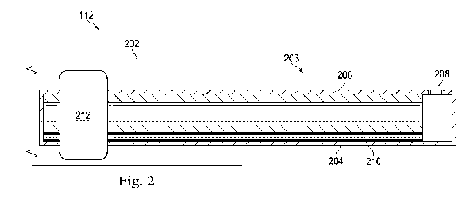
[0023] Fig. 2 is a stylized diagram showing a portion of the illustrative
vitrectomy
probe 112 with internal illumination. Fig. 2 shows a longitudinal cross-
sectional view
of the vitrectomy probe 112. According to the present example, the vitrectomy
probe
112 includes a body 202, which is shown in part. The body 202 supports a
cutting
element 203 that includes a sleeve member 204, an inner member 206, and an
illumination element 210.
[0024] The body 202 may be made from a variety of materials commonly used
to
form such tools. For example, the body 202 may be made of; for example, a
lightweight aluminum or plastic. The exterior portion of the body 202 may be
ergonomically designed for comfortable grasping by a surgeon or operator of
the
vitrectomy probe 112. The inner portion of the body 202 is designed to support
the
cutting element 203 and other features that may be included with the probe
112.
[0025) The cutting element 203 includes the inner member 206 and the
sleeve
member 204. The sleeve member 204 is a hollow needle designed to enter a
patient's
eye. The sleeve member 204 includes a port 208 at the distal end. The port 208
is
disposed along the side of the distal end as illustrated. The port 208 may be
a square,
rectangular, circular, elliptical, or other shaped opening. The opening is
designed to
allow vitreous fibrils from the patient's eye to enter. Movement (e.g., axial,
CA 02961062 2017-03-10
WO 2016/064580
PCT/US2015/054426
rotational, etc.) of the inner member 206 within the sleeve member 204
operates to
open and close the port 208, thereby cutting any vitreous fibrils that enter
the port 208
while it is open.
100261 The inner member 206 of the cutting element 203 is a hollow tube
that
operates as the cutter portion of the vitrectomy probe 112. Thus, the end of
the inner
member 206 is sufficiently sharp to cut vitreous fibrils. The inner member 206
may
be made from a variety of materials, including for example, stainless steel
and others.
In some cases, the inner member 206 may include multiple members attached
together. For example, the distal end of the inner member 206 may be a cutter
member made of a different material than the proximal end. The proximal end of
the
inner member 206 may be connected to an actuating element 212 that moves the
inner
member 206 with respect to the sleeve member 204.
100271 The illumination element 210 is arranged to provide oblique
illumination
of vitreous fibrils outside the cutting element 203 from within the cutting
element
203. Thus, a vitrectomy procedure can be performed without the use of a
separate
illuminator probe. Alternatively, the internally illuminated probe may be used
to
supplement light from a separate illuminator probe. The illumination element
210 is
arranged to project light from within the cutting element 203 through the port
208.
Thus, the illumination element 210 can be positioned such that light is
projected from
the port 208 in an advantageous manner.
[0028] In some embodiments, the illumination element 210 is an optical
fiber
having an illuminating portion. The optical fiber may extend from a console,
such as
console 102, to the distal end of the probe 112. The optical fiber may be
designed to
propagate a sufficient amount of light so as to adequately illuminate the
vitreous
fibrils during a vitrectomy procedure. As will be described in further detail
below, the
illumination element 210 may be secured to either the inner member 206 or the
sleeve
member 204.
100291 Figs. 3A-3B are diagrams showing an illustrative longitudinal
cross-
sectional view of a vitrectomy probe 112 with an illumination element 302
secured to
the inner member 206. Thus, the illumination element 302 moves with the inner
member 206. According to the present example, the illumination element 302
6
CA 02961062 2017-03-10
WO 2016/064580
PCT/US2015/054426
includes an illuminating portion 303 and is secured to the inner member 206 on
a side
that is opposite of the port 208. The illuminating portion 303 is therefore
arranged to
direct light across the open end of the inner member 206 towards the port 208.
Thus,
light 306 from the illuminating portion 303 of the illumination element 302
can be
more efficiently projected out of the port 208. Fig. 3A illustrates the inner
member
206 in a position that is proximal of the port 208. Fig. 3B illustrates the
inner member
206 in a position that is distal of the port 208.
100301 The illumination element 302 may be secured to the inner member
206 in
a variety of ways. For example, the inner member 206 may include a channel
(not
shown) in which the illumination element 302 is placed. Particularly, in the
example
where the illumination element 302 is a fiber optic cable, the fiber optic
cable may run
through the channel of the inner member 206 until it terminates at the distal
end of the
inner member 206. The illumination element 302 may also be bonded to the
surface
of the inner member 206. A surface of the illumination element 302 may include
a
material that is selected to have less friction with the inner surface of the
sleeve
member 204. Thus, as the inner member 206 moves with respect to the sleeve
member, the illumination element 302 is less prone to damage.
100311 In one example, a notch 304 is formed within the inner member 206
at the
distal end of the inner member 206. The notch 304 is positioned between the
illumination element 304 and the port 208. The notch 304 may be designed to
expose
the illuminating portion 303 which is shown as a side of an optical fiber of
the
illumination element 302. Thus, light 306 may be emitted in the direction of
and
projected out of the port 208.
100321 The illumination element 302 may be designed to project light in a
manner
that is convenient for an operator of the probe 112. For example, during a
vitrectomy
procedure, it is preferable to have light projected in an oblique manner with
respect to
the position of the operator. If light is projected back towards the operator,
it can
cause a glare that makes it difficult for the operator to view the illuminated
vitreous
fibrils. Moreover, during the vitrectomy procedure, the operator of the probe
112 may
rotate the cutting element 203 in various positions to effectively remove all
the
vitreous fibrils. Thus, the illumination element 302 can be designed to
project light
306 out of the port 208 and angled towards the distal end 308 of the cutting
element
7
CA 02961062 2017-03-10
WO 2016/064580
PCT/US2015/054426
203. Here, the light projected out of the port is angled obliquely and in a
distal
direction. The side of the inner member 206 may prevent light from being
emitted in
a direction directly transverse to the axial direction. This allows the
operator to use
the probe 112 at a variety of angular positions without having light projected
back at
the operator.
100331 When the distal end of the inner member 206 is positioned proximal
of the
port 208, as illustrated in Fig. 3A, vitreous fibrils are able to enter the
port 208.
Additionally, the light 306 from the illumination element 302 exits the port
208 to
provide oblique illumination of vitreous fibrils outside of the port 208. As
described
above, the inner member 206 reciprocally actuates axially with respect to the
sleeve
member 204 to open and close the port 208. As the port 208 is closing, any
vitreous
fibers within the port are severed. The severed fibrils can then be aspirated
through
the hollow inner member 206.
[0034] When the distal end of the inner member 206 is positioned distal
of the
port 208, as illustrated in Fig. 3B, the illumination element 302 no longer
projects
light 306 out of the port 208. While the light source may be maintained, and
the light
still be projected out of the illumination element, the inner member 206 is
positioned
such that the light is not directed out of the port 208. The inner member 206
may
move with respect to the sleeve member 204 at a rate within the range of 5,000
to
10,000 cycles per minute, for example. Because the inner member 206 moves at
such
a rate, the light 306 is interrupted at a frequency that is too high to be
detected by the
human eye. Thus, the operator of the probe is provided with a seemingly steady
light
source with which to view the vitreous fibrils.
100351 Figs. 4A-4B are diagrams showing an illustrative longitudinal
cross-
sectional view of a vitrectomy probe with an illumination element 402 secured
to the
sleeve member 204. Thus, movement of the inner member 206 does not cause
movement of the illumination element 402. According to the present example,
the
illumination element 402 is secured to the sleeve member 204 on a side that is
opposite of the port 208. Thus, light 306 from the illumination element 302
can more
efficiently be projected out of the port 208. Fig. 4A illustrates the inner
member 206
in a position that is proximal of the port 208. Fig. 4B illustrates the inner
member 206
in a position that is distal of the port 208.
CA 02961062 2017-03-10
WO 2016/064580
PCT/US2015/054426
100361 The illumination element 402 may be secured to the sleeve member
204 in
a variety of ways. For example, the sleeve member 204 may include a channel
(not
shown) in which the illumination element 402 is placed. Particularly, in the
example
where the illumination element 402 is a fiber optic cable, the fiber optic
cable may run
through the channel of the sleeve member 204 until it terminates near the port
208.
The illumination element 402 may also be bonded to the surface of the sleeve
member
204.
100371 In one example, the illumination element 402 may be a fiber optic
cable
with a core 403 and a cladding 404. The cladding 404 has a lower refractive
index
than the core 404, thus allowing the fiber optic cable to act as an optical
waveguide.
The cladding may have a removed portion 406 on a side of the cable near the
distal
end forming an illuminating portion 405. Thus, light 306 is projected from the
illuminating portion 405 substantially perpendicular to the axis of the cable,
allowing
it to be projected out of the port 208.
[0038] Like illumination element 302, illumination element 402 may be
designed
to project light in a manner that is convenient for an operator of the probe
112.
Specifically, the illumination element 402 can be designed to project light
out of the
port 208 and angled towards the distal end 308 of the cutting element 203. In
Fig. 4,
it accomplishes this by arranging the illumination portion 405 at a location
proximal
of the port 208. Thus, the operator is able to use the probe 112 at a variety
of angular
positions without having light projected back at the operator.
100391 When the distal end of the inner member 206 is positioned proximal
of the
port 208, as illustrated in Fig. 4A, the light 306 from the illumination
element 402
exits the port 208 to provide oblique illumination of vitreous fibrils outside
of the port
208. As described above, the inner member 206 reciprocally actuates axially
with
respect to the sleeve member 204 to open and close the port 208. As the port
208 is
closing, any vitreous fibers within the port are severed. The severed fibrils
can then
be aspirated through the hollow inner member 206.
[0040] When the distal end of the inner member 206 is positioned distal
of the
port 208, as illustrated in Fig. 4B, the illumination element 402 is blocked
by the inner
member 206 and no longer projects light 306 out of the port 208. But, as
described
9
CA 02961062 2017-03-10
WO 2016/06-1580
PCT/US2015/054426
above, the inner member 206 may move with respect to the sleeve member 204 at
a
rate within the range of 5,000 to 10,000 cycles per minute, for example.
Because the
inner member 206 moves at such a rate, the light 306 is interrupted at a
frequency that
is too high to be detected by the human eye. Thus, the operator of the probe
is
provided with a seemingly steady light source with which to view the vitreous
fibrils.
[0041] Figs. 5A-5B are diagrams showing axial cross-sectional views of
vitrectomy probes with one or more illumination elements secured to the inner
member 206. Fig. 5A illustrates an axial cross-sectional view of a vitrectomy
probe
with a single illumination element 302 secured to the inner member 206 and
placed at
an opposite side from the port 208. This may be the same probe shown in Figs.
3A
and 3B. Thus, the light 306 can be efficiently projected out of the port 208.
It is
contemplated that the single illumination element 302 can be placed at other
locations
along the circumference of the inner member 206 such that light can be
directed out
of the port 208.
[0042] Fig. 5B illustrates an axial cross-sectional view of a vitrectomy
probe with
multiple illumination elements 302, 502, 504 secured to the inner member 206.
Use
of multiple illumination elements positioned around the circumference of the
inner
member 206 may result in a wider illumination profile 510. In some
embodiments,
even more illumination elements may be included. Each of the illumination
elements
302, 502, 504 may be secured to the inner member 206 in a manner as described
above in the text accompanying Figs. 3A-3B. Additionally, the inner member 206
may include a notch (e.g., 304, Figs. 3A-3B) for each of the illumination
elements
302, 502, 504 that allows light from the respective illumination element to be
directed
out of the port 208.
[0043] Figs. 6A-6B are diagrams showing axial cross-sectional views of
vitrectomy probes with one or more illumination elements secured to the sleeve
member 204. Fig. 6A illustrates an axial cross-sectional view of a vitrectomy
probe
with a single illumination element 402 secured to the sleeve member 204 and
placed
at a side that is opposite from the port 208. Thus, the light 306 can be
efficiently
projected out of the port 208. This probe may be the same probe shown in Figs.
4A
and 4B. It is contemplated that the single illumination element 402 can be
placed at
other locations along the circumference of the sleeve member 204.
CA 02961062 2017-03-10
WO 2016/064580
PCT/US2015/054426
[0044] Fig. 6B illustrates an axial cross-sectional view of a vitrectomy
probe with
multiple illumination elements 402, 602, 604 secured to the sleeve member 204.
Use
of multiple illumination elements positioned around the circumference of the
sleeve
member 204 may result in a wider illumination profile 610. In some
embodiments,
even more illumination elements may be included. Each of the illumination
elements
402, 602, 604 may be secured to the sleeve member 204 in a manner as described
above in the text accompanying Figs. 4A-4B. Additionally, each of the
illumination
elements 402, 602, 604 may have a corresponding portion of the cladding
removed
(e.g., 406, Figs. 4A-4B) so that light from the respective illumination
elements is
appropriately directed out of the port 208.
[0045] Fig. 7 is a diagram showing an ophthalmic surgical system with an
internally illuminated vitrectomy probe. According to the present example, the
system 700 includes a console 702 and a hand piece 706. The console 702
includes a
control system 704 and a light source 710. The hand piece 706 may be the same
probe 112 discussed above, or may be another probe used by an operator or
surgeon
to treat a condition of the eye. In this example, the distal portion is
inserted into the
eye of a patient 708.
100461 The console 702 includes all the necessary components to drive and
work
with the hand piece 706. Additional components and features of the console
would be
apparent to one of ordinary skill in the art. The control system 704 within
the console
702 provides the desired signals to the hand piece 706 to cause the inner
member to
move with respect to the sleeve member and cut vitreous fibrils.
100471 The light source 710 may provide light with sufficient luminosity
so that
when projected out of the illumination element of the hand piece 706, vitreous
fibrils
are sufficiently visible to the operator of the hand piece 706. The light may
also have
a selected color temperature so as to best illuminate the vitreous fibrils.
[0048] Fig. 8 is a flowchart showing an illustrative method 800 for
treating a
patient with an internally illuminated vitrectomy probe. According to the
present
example, the method 800 includes creating an incision in an eye of a patient
at 802.
At 804, the method 800 includes inserting a vitrectomy probe into the eye of
the
patient.
CA 02961062 2017-03-10
WO 2016/064580
PCT/US2015/054426
100491 According to some examples, the probe includes an internal
illumination
element as described above. The probe also includes a cutting element having a
hollow sleeve member extending distally from. the body and an inner member
within
the hollow sleeve member.
100501 At 806, the method 800 includes reciprocally actuating the inner
member
of the cutting element. For example, an actuating element secured to the inner
member may move the inner member in the distal or proximal direction within
the
sleeve member. The movement opens or closes the port formed in the distal
portion
of the hollow sleeve member.
100511 At 808, the method 800 includes projecting light from an
illumination
element. Element 808 may be performed simultaneously with 806. Specifically,
light
may be projected form the illumination element while the inner member moves
axially with respect to the sleeve member. The light may be projected out of
the port
and angled to the distal end of the cutting element so as to provide oblique
illumination of vitreous fibrils.
100521 Other embodiments of the surgical systems having illuminated
probes
include illumination elements disposed along the inner member or the sleeve
member
on the same side as the port, and may be arranged with an illuminating portion
that
emits light in an oblique distal direction. Some of these embodiments emit
light in a
beam that includes light emission at a right angle relative to the axial
direction. Other
embodiments are also contemplated.
100531 Persons of ordinary skill in the art will appreciate that the
embodiments
encompassed by the present disclosure are not limited to the particular
exemplary
embodiments described above. in that regard, although illustrative embodiments
have
been shown and described, a wide range of modification, change, and
substitution is
contemplated in the foregoing disclosure. It is understood that such
variations may be
made to the foregoing without departing from the scope of the present
disclosure.
Accordingly, it is appropriate that the appended claims be construed broadly
and in a
manner consistent with the present disclosure.
12