Note: Descriptions are shown in the official language in which they were submitted.
81803891
NEOADJUVANT USE OF ANTIBODY-DRUG CONJUGATES
Inventor: David M. Goldenberg
ASSIGNEE: IMMUNOMEDICS, INC.
Related Applications
[01] This
application claims the benefit under 35 U.S.C. 119(e) of provisional U.S.
Patent
Application Serial No. 62/060,858, filed October 7, 2014.
[02]
FIELD OF THE INVENTION
[03] The present invention relates to use of immunoconjugates in neoadjuvant
therapy.
Preferably, the immunoconjugates comprise an antibody moiety and a drug moiety
selected
from the camptothecin or anthracycline groups of drugs. More preferably, the
antibody
moiety binds to a tumor-associated antigen (TAA). Most preferably, the
camptothecin is SN-
38 or the anthracycline is a prodrug form of 2-pyrrolinodoxorubicin (referred
to herein as
P2PDox). The antibody and drug moieties may be linked via an intracellularly
cleavable
linkage that increases therapeutic efficacy. Preferably, the linker joining
the antibody moiety
and the drug moiety is CL2A, as described below. In particular embodiments,
the
immunoconjugates may be administered at specific dosages and/or schedules of
administration that provide for optimal efficacy and minimal toxicity,
allowing effective
treatment of cancers that are resistant to standard anti-cancer therapies,
such as triple negative
breast cancer (TNBC), metastatic colon cancer, metastatic non-small-cell lung
cancer
(NSCLC), metastatic pancreatic cancer, metastatic renal cell carcinoma,
metastatic gastric
cancer, metastatic prostate cancer, or metastatic small-cell lung cancer. A
preferred
embodiment relates to neoadjuvant use in TNBC. The neoadjuvant immunoconjugate
is
administered prior to standard anti-cancer therapies, such as surgery,
radiation therapy,
chemotherapy, or immunotherapy.
-1-
Date Recue/Date Received 2022-03-16
CA 02961774 2017-03-17
WO 2016/057398
PCT/US2015/054011
BACKGROUND OF THE INVENTION
[04] Neoadjuvant agents are administered to a patient prior to treatment
with a primary
therapy, such as surgery or radiation therapy (see, e.g., Wikipedia ¨
Neoadjuvant therapy).
The object of neoadjuvant cancer therapy is to reduce the size or extent of
the patient's
tumor(s) before the primary therapy, preferably improving the likelihood of
successful
outcome and/or decreasing the adverse effects of more extensive treatment that
would be
required in the absence of neoadjuvant therapy (Id.). Neoadjuvant treatment
may also target
micrometasteses that may be unaffected by the primary therapy (Id.). Recently,
neoadjuvant
therapy has been gaining a role as a means to test novel chemotherapies more
expeditiously,
because responses to neoadjuvant therapy can be assessed rapidly in a
relatively small
number of patients, and can be predictive of longer-term outcome (Rastagi et
al., 2008, J Clin
Oncol 26:778-85; Bardia & Baselga, 2013, Clin Cancer Res 19:6360-70). Indeed,
evidence
from neoadjuvant studies indicates that determination of pathologic complete
response (pCR)
at surgery (i.e., no residual disease in the breast and axilla) is predictive
of long-term clinical
response, even after two cycles of neoadjuvant chemotherapy (Rastagi et al.,
2008, J Clin
Oncol 26:778-85; von Mickwitz et al., 2012, J Clin Oncol 30:1796-1804; Huober
et al., 2010,
Breast Cancer Res Treat 124:133-40).
[05] The history of neoadjuvant treatment in cancer is extensive. Much of the
earlier work
in this field related to use of neoadjuvant chemotherapy prior to surgerical
excision or
radiation therapy. Ervin et al. (1984, Arch Otolaryngol 110:241-5) reported
neoadjuvant
chemotherapy of advanced head and neck cancer with cisplatin, bleomycin and
methotrexate,
before surgery plus radiotherapy or high-dose radiotherapy alone. Although
some
improvement in outcome was seen, particularly where neoadjuvant therapy
resulted in
substantial tumor reduction, relapse of disease was common (Ervin et al,
1984). Neoadjuvant
chemotherapy and/or radiation therapy has also been reported in osteogenic
sarcoma (Rosen
& Nirenberg, 1985, Prog Clin Biol Res 201:39-51), breast cancer (Ragaz et al.,
1985, Prog
Clin Biol Res 201:77-87), esophageal cancer (Kelsen et al., 1986, Semin Surg
Oncol 2:170-
6), anal and rectal cancer (Smith et al., 1986, Am J Surg 151:577-80), lung
cancer (Cox et al.,
1986, Cancer Treat Rep 70:1219-20) and many other forms of cancer. While
improved
outcome is often reported with neoadjuvant therapy, the degree to which
neoadjuvant
chemotherapy and/or radiation therapy improves long-term patient survival in
cancer has
generally not yet been confirmed by prospective studies (see, e.g., Bittoni et
al., 2014,
Gastroenterol Res Pract 2014:183852; Doval et al., 2013, J Indian Med Assoc
111:629-31).
-2-
CA 02961774 2017-03-17
WO 2016/057398
PCT/US2015/054011
[06] Recently, neoadjuvant use of antibodies or antibody-drug conjugates
(ADCs) has
been attempted in breast cancer. Pertuzumab (anti-HER2) has been investigated
and received
FDA approval in combination with trastuzumab and docetaxel in neoadjuvant
treatment of
HER2-positive metastatic breast cancer (Sabatier & Goncalves, 2014, Bull
Cancer 101:765-
71; Esserman & DeMichele, 2014, Clin Cancer Res 20:3632-36). Ado-trastuzumab
emtansine (T-DM1), comprising an anti-HER2 antibody conjugated to the potent
microtubule
inhibitor emtansine, has been approved for use in HER2-positive metastatic
breast cancer for
patients who have failed previous therapy and is being investigated for
neoadjuvant use
(Corrigan et al., 2014, Ann Pharmacother [Epub ahead of print, July 31,
2014]).
[07] While these results are promising, anti-HER2 antibodies are of little
use in, for
example, triple-negative breast cancer (TNBC), which lacks expression of
estrogen receptors,
progesterone receptors and HER2 (e.g., Gogia et al., 2014, Indian J Cancer
51:163-6). TNBC
accounts for about 10 to 20% of breast cancers and is more aggressive and
lethal than other
forms of this disease, with virtually all women with metastatic TNBC
ultimately dying of the
disease, despite systemic therapy. A need exists in the field for more
effective forms of
immunoconjugate-based neoadjuvant cancer therapy, particularly for forms of
cancer that are
resistant to standard anti-cancer treatments, such as TNBC.
SUMMARY OF THE INVENTION
[08] The present invention makes use of antibody conjugates of drugs, such as
camptothecins (e.g., SN-38) or anthracyclines (e.g., P2PDOX), that have
nanomolar toxicities
in vitro, compared to the sub-nanomolar to picomolar toxicities of ultratoxic
chemotherapeutic agents like calicheamicin, maytansinoids or MMAE. Use of
drugs that are
not ultratoxic allows the use of antibody-drug linkers that do not require
cell internalization
for the release of free drugs, but rather allow some extracellular release of
drug. With the
CL2A linker described below, 50% of the conjugated drug is released in 24 hr,
thereby
augmenting the bioavailability of the drug by liberating it both
extracellularly and
intracellularly. In addition, the use of relatively non-toxic drugs allows the
administration of
higher dosages of ADCs, leading to better therapeutic effects.
[09] The present invention resolves an unfulfilled need in the art by
providing neoadjuvant
methods and compositions for preparing and administering ADCs, such as
camptothecin-
antibody or anthracycline-antibody immunoconjugates. Preferably, the
camptothecin is SN-
38 or the anthracycline is P2PDOX. The disclosed methods and compositions are
of use for
the neoadjuvant treatment of cancers which are refractory or less responsive
to other forms of
therapy. Refractory cancers may include, but are not limited to, triple-
negative breast cancer,
-3-
CA 02961774 2017-03-17
WO 2016/057398
PCT/US2015/054011
metastatic colon cancer, metastatic non-small-cell lung cancer (NSCLC),
metastatic
pancreatic cancer, metastatic renal cell carcinoma, metastatic gastric cancer,
metastatic
prostate cancer, or metastatic small-cell lung cancer.
[010] The antibody can be of various isotypes, preferably human IgGl, IgG2,
IgG3 or IgG4,
more preferably comprising human TgG1 hinge and constant region sequences. The
antibody
or fragment thereof can be a chimeric, humanized, or fully human antibody or
antigen-
binding fragment thereof, such as half-IgG4 antibodies, as described by van
der Neut
Kolfschoten et al. (Science 2007; 317:1554-1557), or single domain antibodies
(e.g.,
nanobodies) as commercially available (e.g., ABLYNX , Ghent, Belgium). More
preferably, the antibody or fragment thereof may be designed or selected to
comprise human
constant region sequences that belong to specific allotypes, which may result
in reduced
immunogenicity when the immunoconjugate is administered to a human subject.
Preferred
allotypes for administration include a non-Glml allotype (nG1m1), such as
G1m3, G1m3,1,
G1m3,2 or G1m3,1,2. More preferably, the allotype is selected from the group
consisting of
the nGlml, G1m3, nG1m1,2 and Km3 allotypes.
[011] For neoadjuvant treatment of cancer, many antigens expressed by or
otherwise
associated with tumor cells are known in the art, including but not limited
to, carbonic
anhydrase IX, alpha-fetoprotein (AFP), a-actinin-4, A3, antigen specific for
A33 antibody,
ART-4, B7, Ba 733, BAGE, BrE3-antigen, CA125, CAMEL, CAP-1, CASP-8/m, CCL19,
CCL21, CD1, CD la, CD2, CD3, CD4, CD5, CD8, CD11A, CD14, CD15, CD16, CD18,
CD19, CD20, CD21, CD22, CD23, CD25, CD29, CD30, CD32b, CD33, CD37, CD38,
CD40, CD4OL, CD44, CD45, CD46, CD52, CD54, CD55, CD59, CD64, CD66a-e, CD67,
CD70, CD7OL, CD74, CD79a, CD80, CD83, CD95, CD126, CD132, CD133, CD138,
CD147, CD154, CDC27, CDK-4/m, CDKN2A, CTLA-4, CXCR4, CXCR7, CXCL12, HTF-
la, colon-specific antigen-p (CSAp), CEACAM5, CEACAM6, c-Met, DAM, EGFR,
EGFRvIII, EGP-1 (TROP-2), EGP-2, ELF2-M, Ep-CAM, fibroblast growth factor
(FGF),
Flt-1, Flt-3, folate receptor, G250 antigen, GAGE, gp100, GRO-[3, HLA-DR,
HM1.24,
human chorionic gonadotropin (HCG) and its subunits, HER2/neu, histone H2B,
histone H3,
histone H4, HMGB-1, hypoxia inducible factor (HIF-1), HSP70-2M, HST-2, Ia, IGF-
1R,
IFN-y, IFN-a, IFN-13, IFN-X, IL-4R, IL-6R, IL-13R, IL-15R, IL-17R, IL-18R, IL-
2, IL-6, IL-
8, IL-12, IL-15, IL-17, IL-18, IL-23, IL-25, insulin-like growth factor-1 (IGF-
1), KC4-
antigen, KS-1-antigen, KS1-4, Le-Y, LDR/FUT, macrophage migration inhibitory
factor
(MIF), MAGE, MAGE-3, MART-1, MART-2, NY-ESO-1, TRAG-3, mCRP, MCP-1, MIP-
1A, MIP-1B, MIF, MUC1, MUC2, MUC3, MUC4, MUC5ac, MUC13, MUC16, MUM-1/2,
-4-
81803891
MUM-3, NCA66, NCA95, NCA90, PAM4 antigen, pancreatic cancer mucin, PD-1, PD-
L1,
PD-1 receptor, placental growth factor, p53, PLAGL2, prostatic acid
phosphatase, PSA,
PRAME, PSMA, P1GF, ILGF, ILGF-1R, IL-6, IL-25, RS5, RANTES, T101, SAGE, S100,
survivin, survivin-2B, TAC, TAG-72, tenascin, TRAIL receptors, TNF-a, Tn
antigen,
Thomson-Friedenreich antigens, tumor necrosis antigens, VEGFR, ED-B
fibronectin, WT-1,
17-1A-antigen, complement factors C3, C3a, C313, C5a, C5, an angiogenesis
marker, 13c1-2,
bc1-6, Kras, an oncogene marker and an oncogene product (see, e.g., Sensi et
al., Clin Cancer
Res 2006, 12:5023-32; Parmiani et al., J Immunol 2007, 178:1975-79; Novellino
et al.
Cancer Immunol Immunother 2005, 54:187-207). Preferably, the antibody binds to
AFP,
CEACAM5, CEACAM6, CSAp, EGP-1 (TROP-2), AFP, MUC5ac, PAM4 antigen, CD74,
CD19, CD20, CD22 or HLA-DR.
[012] Exemplary antibodies that may be utilized include, but are not limited
to, hR1 (anti-
IGF-1R, U.S. Patent Application Serial No. 12/722,645, filed 3/12/10), hPAM4
(anti-
MUC5ac, U.S. Patent No. 7,282,567), hA20 (anti-CD20, U.S. Patent No.
7,151,164), hA19
(anti-CD19, U.S. Patent No. 7,109,304), hIMMU31 (anti-AFP, U.S. Patent No.
7,300,655),
hLL1 (anti-CD74, U.S. Patent No. 7,312,318), hLL2 (anti-CD22, U.S. Patent No.
5,789,554),
hRFB4 (anti-CD22, U.S. Prov. Pat. Appl. Serial No. 61/944,295, filed 2/25/14),
hMu-9 (anti-
CSAp, U.S. Patent No. 7,387,772), hL243 (anti-HLA-DR, U.S. Patent No.
7,612,180), hMN-
14 (anti-CEACAM5, U.S. Patent No. 6,676,924), hMN-15 (anti-CEACAM6, U.S.
Patent No.
8,287,865), hRS7 (anti-TROP-2, U.S. Patent No. 7,238,785), hMN-3 (anti-
CEACAM6, U.S.
Patent No. 7,541,440), Ab124 and Ab125 (anti-CXCR4, U.S. Patent No.
7,138,496). More
preferably, the antibody is IMMU-31 (anti-AFP), hRS7 (anti-TROP-2), hMN-14
(anti-
CEACAM5), hMN-3 (anti-CEACAM6), hMN-15 (anti-CEACAM6), hLL1 (anti-CD74),
hLL2 (anti-CD22), hL243 or IMMU-114 (anti-HLA-DR), hA19 (anti-CD19) or hA20
(anti-
CD20). As used herein, the terms epratuzumab and hLL2 are interchangeable, as
are the
terms veltuzumab and hA20, and hL243g4P, hL243gamma4P and IMMU-114.
[013] Alternative antibodies of use include, but are not limited to, abciximab
(anti-
glycoprotein IIb/IIIa), alemtuzumab (anti-CD52), bevacizumab (anti-VEGF),
cetuximab
(anti-EGFR), gemtuzumab (anti-CD33), ibritumomab (anti-CD20), pan itumumab
(anti-
EGFR), rituximab (anti-CD20), tositumomab (anti-CD20), trastuzumab (anti-
ErbB2),
lambrolizumab (anti-PD-1 receptor), nivolumab (anti-PD-1 receptor), ipilimumab
(anti-
CTLA-4), abagovomab (anti-CA-125), adecatumumab (anti-EpCAM), atlizumab (anti-
IL-6
receptor), benralizumab (anti-CD125), obinutuzumab (GA101, anti-CD20), CC49
(anti-
-5-
Date Recue/Date Received 2022-03-16
CA 02961774 2017-03-17
WO 2016/057398
PCT/US2015/054011
TAG-72), AB-PG1-XG1-026 (anti-PSMA, U.S. Patent Application 11/983,372,
deposited as
ATCC PTA-4405 and PTA-4406), D2/B (anti-PSMA, WO 2009/130575), tocilizumab
(anti-
IL-6 receptor), basiliximab (anti-CD25), daclizumab (anti-CD25), efalizumab
(anti-CD11a),
GA101 (anti-CD20; Glycart Roche), muromonab-CD3 (anti-CD3 receptor),
natalizumab
(anti-a4 integrin), omalizumab (anti-TgE); anti-TNF-a antibodies such as
CDP571 (Ofei et
al., 2011, Diabetes 45:881-85), MTNFA1, M2TNFAI, M3TNFA1, M3TNFAB1, M302B,
M303 (Thermo Scientific, Rockford, IL), infliximab (Centocor, Malvern, PA),
certolizumab
pegol (UCB, Brussels, Belgium), anti-CD4OL (UCB, Brussels, Belgium),
adalimumab
(Abbott, Abbott Park, IL), and belimumab (Human Genome Sciences). Recently,
humanized
antibodies against human histones H2B, H3 and H4 have been disclosed (U.S.
Patent
Application Serial No. 14/180,646) that may be utilized in the disclosed
methods and
compositions.
[014] Preferably, the antibody moiety links to at least one drug moiety, more
preferably 1 to
about 5 drug moieties, alternatively about 6 to 12 drug moieties. In various
embodiments, the
antibody moiety may be attached to 4 or 6 drug moieties, or to 5 or less drug
moieties. The
number of drug moieties per antibody moiety may be 1,2, 3,4, 5, 6, 7, or more.
[015] An exemplary camptothecin is CPT-11. Extensive clinical data are
available
concerning CPT-11's pharmacology and its in vivo conversion to the active SN-
38 (Iyer and
Ratain, Cancer Chemother Pharmacol. 42:S31-43 (1998); Mathijssen et al., Clin
Cancer Res.
7:2182-2194 (2002); Rivory, Ann NY Acad Sci. 922:205-215, 2000)). The active
form SN-38
is about 2 to 3 orders of magnitude more potent than CPT-11. In specific
preferred
embodiments, the immunoconjugate may be an hMN-14-SN-38, hMN-3-SN-38, hMN-15-
SN-38, hIMMU-31-SN-38, hRS7-SN-38, hR1-SN-38, hA20-SN-38, hPAM4-SN-38, hL243-
SN-38, hLL1-SN-38, hRFB4-SN-38, hMu-9-SN-38 or liLL2-SN-38 conjugate. More
preferably, a CL2A linker is used to conjugate the SN-38 to the antibody
moiety.
[016] An exemplary anthracycline is a prodrug form of 2-pyrrolinodoxorubicin
(P2PDox),
such as N-(4,4-diacetoxybutyl)doxorubicin, disclosed in U.S. Patent
Application Serial No.
14/175,089. Surprisingly, P2PDox has been found to be tightly bound to
conjugated antibody,
due to the formation of cross-links with antibody peptide chains. The cross-
linking assists in
minimizing toxicity, for example cardiotoxicity, that would result from
release of free drug in
circulation. Preferably, the P2PDox is attached to interchain disulfide thiol
groups while in
the prodrug form. The prodrug protection is rapidly removed in vivo soon after
injection and
the resulting 2-PDox portion of the conjugate cross-links the peptide chains
of the antibody,
forming intramolecular cross-linking within the antibody molecule. This both
stabilizes the
-6-
CA 02961774 2017-03-17
WO 2016/057398
PCT/US2015/054011
ADC and prevents cross-linking to other molecules in circulation. In specific
preferred
embodiments, the immunoconjugate may be an hMN-14-P2PDox, hMN-3-P2PDox, hMN-
15-P2PDox, hIMMU-31-P2PDox, hRS7-P2PDox, hR1-P2PDox, hA20-P2PDox, hPAM4-
P2PDox, hL243-P2PDox, hLL1-P2PDox, hRFB4-P2PDox, hMu-9-P2PDox or hLL2-
P2PDox conjugate.
[017] Various embodiments may concern use of the subject methods and
compositions to
treat a cancer, including but not limited to non-Hodgkin's lymphomas, B-cell
acute and
chronic lymphoid leukemias, Burkitt lymphoma, Hodgkin's lymphoma, acute large
B-cell
lymphoma, hairy cell leukemia, acute myeloid leukemia, chronic myeloid
leukemia, acute
lymphocytic leukemia, chronic lymphocytic leukemia, T-cell lymphomas and
leukemias,
multiple myeloma, Waldenstrom's macroglobulinemia, carcinomas, melanomas,
sarcomas,
gliomas, bone, and skin cancers. The carcinomas may include carcinomas of the
oral cavity,
esophagus, gastrointestinal tract, pulmonary tract, lung, stomach, colon,
breast, ovary,
prostate, uterus, endometrium, cervix, urinary bladder, pancreas, bone, brain,
connective
tissue, liver, gall bladder, urinary bladder, kidney, skin, central nervous
system and testes.
[018] In certain embodiments, the drug conjugates may be used as neoadjuvants
prior to
treatment with surgery, radiation therapy, chemotherapy, immunotherapy with
naked
antibodies, radioimmunotherapy, immunomodulators, and the like. These
neoadjuvant
therapies can allow lower doses of each therapeutic to be given, thus reducing
certain severe
side effects, or improving the efficacy of other treatments such as surgery.
[019] Preferred optimal dosing of immunoconjugates may include a dosage of
between 3
mg/kg and 18 mg/kg, preferably given either weekly, twice weekly, every other
week or
every third week. The optimal dosing schedule may include treatment cycles of
two
consecutive weeks of therapy followed by one, two, three or four weeks of
rest, or alternating
weeks of therapy and rest, or one week of therapy followed by two, three or
four weeks of
rest, or three weeks of therapy followed by one, two, three or four weeks of
rest, or four
weeks of therapy followed by one, two, three or four weeks of rest, or five
weeks of therapy
followed by one, two, three, four or five weeks of rest, or administration
once every two
weeks, once every three weeks or once a month. Treatment may be extended for
any number
of cycles, preferably at least 2, at least 4, at least 6, at least 8, at least
10, at least 12, at least
14, or at least 16 cycles. The dosage may be up to 24 mg/kg. Exemplary dosages
of use may
include 1 mg/kg, 2 mg/kg, 3 mg/kg, 4 mg/kg, 5 mg/kg, 6 mg/kg, 7 mg/kg, 8
mg/kg, 9 mg/kg,
mg/kg, 11 mg/kg, 12 mg/kg, 13 mg/kg, 14 mg/kg, 15 mg/kg, 16 mg/kg, 17 mg/kg,
18
mg/kg, 19 mg/kg, 20 mg/kg, 22 mg/kg and 24 mg/kg. Preferred dosages are 4, 6,
8, 9, 10, 12,
-7-
CA 02961774 2017-03-17
WO 2016/057398
PCT/US2015/054011
14, 16 or 18 mg/kg. The person of ordinary skill will realize that a variety
of factors, such as
age, general health, specific organ function or weight, as well as effects of
prior therapy on
specific organ systems (e.g., bone marrow) may be considered in selecting an
optimal dosage
of immunoconjugate, and that the dosage and/or frequency of administration may
be
increased or decreased during the course of therapy. The dosage may be
repeated as needed,
with evidence of tumor shrinkage observed after as few as 4 to 8 doses. The
optimized
dosages and schedules of administration for neoadjuvant use disclosed herein
show
unexpected superior efficacy and reduced toxicity in human subjects, which
could not have
been predicted from animal model studies. Surprisingly, the superior efficacy
allows
treatment of tumors that were previously found to be resistant to one or more
standard anti-
cancer therapies.
[020] A surprising result with the instant claimed compositions and methods is
the
unexpected tolerability of high doses of antibody-drug conjugate, even with
repeated
infusions, with only relatively low-grade toxicities of nausea and vomiting
observed, or
manageable neutropenia. A further surprising result is the lack of
accumulation of the
antibody-drug conjugate, unlike other products that have conjugated
chemotherapeutic drugs
to albumin, PEG or other carriers. The lack of accumulation is associated with
improved
tolerability and lack of serious toxicity even after repeated or increased
dosing. These
surprising results allow optimization of dosage and delivery schedule, with
unexpectedly high
efficacies and low toxicities. The claimed methods provide for shrinkage of
solid tumors, in
individuals with previously resistant cancers, of 15% or more, preferably 20%
or more,
preferably 30% or more, more preferably 40% or more in size (as measured by
longest
diameter). The person of ordinary skill will realize that tumor size may be
measured by a
variety of different techniques, such as total tumor volume, maximal tumor
size in any
dimension or a combination of size measurements in several dimensions. This
may be with
standard radiological procedures, such as computed tomography,
ultrasonography, and/or
positron-emission tomography. The means of measuring size is less important
than observing
a trend of decreasing tumor size with immunoconjugate treatment, preferably
resulting in
elimination of the tumor.
[021] While the immunoconjugate may be administered as a periodic bolus
injection, in
alternative embodiments the immunoconjugate may be administered by continuous
infusion
of antibody-drug conjugates. In order to increase the Cmax and extend the PK
of the
immunoconjugate in the blood, a continuous infusion may be administered for
example by
indwelling catheter. Such devices are known in the art, such as HICKMAN ,
BROVIAC
-8-
81803891
or PORT-A-CATHO catheters (see, e.g., Skolnik et al., Ther Drug Monit 32:741-
48, 2010)
and any such known indwelling catheter may be used. A variety of continuous
infusion pumps
are also known in the art and any such known infusion pump may be used. The
dosage range
for continuous infusion may be between 0.1 and 3.0 mg/kg per day. More
preferably, these
immunoconjugates can be administered by intravenous infusions over relatively
short periods
of 2 to 5 hours, more preferably 2-3 hours.
[022] In
particularly preferred embodiments, the immunoconjugates and dosing schedules
may be efficacious in patients resistant to standard therapies. For example,
an hMN-14-SN-38
immunoconjugate may be administered to a patient who has not responded to
prior therapy
with irinotecan, the parent agent of SN-38. Surprisingly, the irinotecan-
resistant patient may
show a partial or even a complete response to hMN-14-SN-38. The ability of the
immunoconjugate to specifically target the tumor tissue may overcome tumor
resistance by
improved targeting and enhanced delivery of the therapeutic agent.
Alternatively, an anti-
CEACAM5 immunoconjugate, such as hMN-14, may be co-administered with an anti-
CEACAM6 immunoconjugate, such as hMN-3 or hMN-15. Other antibody-SN-38 or
antibody-P2PDox immunoconjugates may show similar improved efficacy and/or
decreased
toxicity, compared to alternative standard therapeutic treatments, and
combinations of
different immunoconjugates, or ADCs in combination with an antibody conjugated
to a
radionuclide, toxin or other drug, may provide even more improved efficacy
and/or reduced
toxicity. A specific preferred subject may be a metastatic colon cancer
patient, a triple-
negative breast cancer patient, a HER+, ER+, progesterone+ breast cancer
patient, a
metastatic non-small-cell lung cancer (NSCLC) patient, a metastatic pancreatic
cancer patient,
a metastatic renal cell carcinoma patient, a metastatic gastric cancer
patient, a metastatic
prostate cancer patient, or a metastatic small-cell lung cancer patient.
[022a] The present invention as claimed relates to of an antibody-drug
conjugate (ADC) for
neoadjuvant treatment of cancer prior to a standard anti-cancer therapy,
wherein the antibody-
drug conjugate (ADC) comprises an antibody moiety conjugated to a drug, and
the antibody
moiety is an anti-Trop-2 hRS7 antibody and the drug is SN 38; and the standard
anti-cancer
therapy is selected from the group consisting of surgery and radiation
therapy.
- 9 -
Date Recue/Date Received 2022-03-16
81803891
BRIEF DESCRIPTION OF THE FIGURES
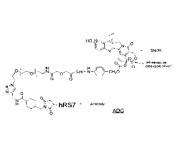
[023] FIG. 1. Exemplary antibody-drug conjugate, showing the hRS7 anti-TROP-
2
antibody conjugated via an intracellularly cleavable CL2A linker to the SN-38
camptothecin
drug.
[024] FIG. 2A. Structure of doxorubicin. "Me" is a methyl group.
[025] FIG. 2B. Structure of 2-pyrrolinodoxorubicin,(2-PDox). "Me" is a
methyl group.
[026] FIG. 2C. Structure of a prodrug form of 2-
pyrrolinodoxorubicin,(P2PDox). "Me" is a
methyl group and "Ac" is an acetyl group.
[027] FIG. 2D. Structure of a maleimide-activated form of P2PDox, for
antibody coupling.
"Me" is a methyl group and "Ac" is an acetyl group.
- 9a -
Date Recue/Date Received 2022-03-16
CA 02961774 2017-03-17
WO 2016/057398
PCT/US2015/054011
[028] FIG. 3A. In vivo efficacy of IMMU-132 in Calu-3 human NSCLC xenografts.
[029] FIG. 3B. In vivo efficacy of IMMU-132 in COLO 205 human colon cancer
xenografts.
[030] FIG. 3C. In vivo efficacy of IMMU-132 in Capan-1 human pancreatic cancer
xenografts.
[031] FIG. 3D. In vivo efficacy of 1MMU-132 in BxPC-3 human pancreatic canccr
xenografts.
[032] FIG. 3E. In vivo efficacy of IMMU-132 in SK-MES-1 human squamous cell
lung
cancer xenografts.
[033] FIG. 3F. In vivo efficacy of IMMU-132 in NCI-N87 human gastric cancer
xenografts.
[034] FIG. 4A. In vivo efficacy of IMMU-132 in MDA-MB-468 human TNBC
xenografts.
[035] FIG. 4B. In vivo efficacy of IMMU-132 in MDA-MB-468 human TNBC
xenografts.
Tumor bearing mice were initially treated with control (non-targeting) ADC.
After allowing
the tumors to grow, the mice were administered the indicated dosages of IMMU-
132, starting
on day 78. Even after allowing the tumors to grow to large size, the IMMU-132
was effective
to induce tumor regression.
[036] FIG. 4C. In vivo efficacy of IMMU-132 in MDA-MB-231 human TNBC
xenografts.
[037] FIG. 5A. Best response for 14 TNBC patients enrolled in TMMU-132-01
trial.
[038] FIG. 5B. Time to progression for 14 TNBC patients enrolled in IMMU-132-
01 trial.
[039] FIG. 6A. IMMU-132 peak serum concentrations of IgG and ADC, as a
function of
dose level.
[040] FIG. 6B. IMMU-132 peak serum concentrations of IgG and ADC, as a
function of
dose level when normalized to patient weight.
[041] FIG. 7A. Pharmacokinetics of 1MMU-132 total IgG vs. total 1MMU-132.
[042] FIG. 7B. Pharmacokinetics of IMMU-132 total SN-38 vs. free SN-38.
[043] FIG. 7C. Clearance of IMMU-132 based on ELISA or on SN-38 concentration
in the
serum.
[044] FIG. 8A. Different IMMU-132 dosing regimens in NCI-N87 human gastric
carcinoma xenografts.
[045] FIG. 8B. Different IMMU-132 dosing regimens in BxPC-3 human pancreatic
adenocarcinoma xenografts.
[046] FIG. 9A. IMMU-132 mediated apoptotic signaling in NCI-N87 human gastric
carcinoma exposed to 1 ii_tM of free SN-38 or the equivalent amount of IMMU-
132.
-10-
CA 02961774 2017-03-17
WO 2016/057398
PCT/US2015/054011
[047] FIG. 9B. IMMU-132 mediated PARP cleavage in SK-BR-3 and MDA-MB 486
breast carcinoma cells.
[048] FIG.10. Pharmacotoxicology studies of IMMU-132 vs. irinotecan in a
pancreatic
cancer xenograft model.
[049] FIG. 11. Neoadjuvant treatment regimen for paclitaxel +/- TM-MU-132 in
TNBC.
[050] FIG. 12. Therapy in nude mice bearing s.c. human tumor xenografts using
2.25
mg/kg protein dose (0.064 mg/kg of drug dose) of MAb-P2PDox conjugates twice
weekly x
2 weeks in nude mice with Capan-1 human pancreatic adenocarcinoma xenografts
(n = 5).
[051] FIG. 13A. MTD study of hLL1-P2PDox conjugates with multiple injections.
Mice
administered hLL1-P2PDox (q4dx4) at 25 jig i.v. per dose.
[052] FIG. 13B. MTD study of hLL1-P2PDox conjugates with multiple injections.
Mice
administered hLL1-P2PDox (q4dx4) at 50 lig i.v. per dose.
[053] FIG. 13C. MTD study of hLL1-P2PDox conjugates with multiple injections.
Mice
administered hLL1-P2PDox (q4dx4) at 100 lug i.v. per dose.
[054] FIG. 13D. MTD study of hLL1-P2PDox conjugates with multiple injections.
Mice
administered hLL1-P2PDox (q4dx4) at 150 lag i.v. per dose.
[055] FIG. 14A. In vivo efficacy of P2PDox conjugates in nude mice with NCI-
N87 human
gastric cancer xenografts. Mice were administered a saline control.
[056] FIG. 14B. In vivo efficacy of P2PDox conjugates in nude mice with NCI-
N87 human
gastric cancer xenografts. Mice were administered 45 jig of hA20-P2PDox as
indicated by
arrows.
[057] FIG. 14C. In vivo efficacy of P2PDox conjugates in nude mice with NCI-
N87 human
gastric cancer xenografts. Mice were administered 45 jig of hMN-15-P2PDox as
indicated
by arrows.
[058] FIG. 14D. In vivo efficacy of P2PDox conjugates in nude mice with NCI-
N87 human
gastric cancer xenografts. Mice were administered 45 jig of hRS7-P2PDox as
indicated by
arrows.
[059] FIG. 14E. In vivo efficacy of P2PDox conjugates in nude mice with NCI-
N87 human
gastric cancer xenografts. Mice were administered 45 jig of hLL1-P2PDox as
indicated by
arrows.
[060] FIG. 14F. In vivo efficacy of P2PDox conjugates in nude mice with NCI-
N87 human
gastric cancer xenografts. Mice were administered 45 jig of h-14-P2PDox as
indicated
by arrows.
-11-
CA 02961774 2017-03-17
WO 2016/057398
PCT/US2015/054011
[061] FIG. 15. Effect of different dosing schedules of hRS7-P2PDox on survival
in nude
mice with NCI-N87 human gastric carcinoma xenografts.
[062] FIG. 16. Effect of different single doses of hRS7-P2PDox on growth of
human
gastric carcinoma xenografts.
[063] FIG. 17. Effect of different single doses of hRS7-P2PDox on survival of
mice
bearing human gastric carcinoma xenografts.
[064] FIG. 18. ADCC of various hRS7-ADCs vs. hRS7 IgG.
DETAILED DESCRIPTION OF THE INVENTION
Definitions
[065] In the description that follows, a number of terms are used and the
following
definitions are provided to facilitate understanding of the claimed subject
matter. Terms that
are not expressly defined herein are used in accordance with their plain and
ordinary
meanings.
[066] Unless otherwise specified, a or an means "one or more."
[067] The term about is used herein to mean plus or minus ten percent (10%) of
a value.
For example, "about 100" refers to any number between 90 and 110.
[068] An antibody, as used herein, refers to a full-length (i.e., naturally
occurring or formed
by normal immunoglobulin gene fragment recombinatorial processes)
immunoglobulin
molecule (e.g., an IgG antibody) or an antigen-binding portion of an
immunoglobulin
molecule, such as an antibody fragment. An antibody or antibody fragment may
be
conjugated or otherwise derivatized within the scope of the claimed subject
matter. Such
antibodies include but are not limited to IgGI, IgG2, IgG3, IgG4 (and IgG4
subforms), as
well as IgA isotypes.
[069] An antibody fragment is a portion of an antibody such as F(abp2, F(ab)2,
Fab', Fab,
Fv, scFv (single chain Fv), single domain antibodies (DABs or VHHs) and the
like, including
the half-molecules of IgG4 cited above (van der Neut Kolfschoten et al.
(Science 2007;
317(14 Sept):1554-1557). A commercially available form of single domain
antibody,
referred to as a nanobody (ABLYNX , Ghent, Belgium), is discussed in further
detail below.
Regardless of structure, an antibody fragment of use binds with the same
antigen that is
recognized by the intact antibody. The term "antibody fragment" also includes
synthetic or
genetically engineered proteins that act like an antibody by binding to a
specific antigen to
form a complex. For example, antibody fragments include isolated fragments
consisting of
-12-
CA 02961774 2017-03-17
WO 2016/057398
PCT/US2015/054011
the variable regions, such as the "Fv" fragments consisting of the variable
regions of the
heavy and light chains, recombinant single chain polypeptide molecules in
which light and
heavy variable regions are connected by a peptide linker ("scFv proteins"),
and minimal
recognition units consisting of the amino acid residues that mimic the
hypervariable region,
such as CDRs. The Fv fragments may be constructed in different ways to yield
multivalent
and/or multispecific binding forms. In the case of multivalent, they have more
than one
binding site against the specific epitope, whereas with multispecific forms,
more than one
epitope (either of the same antigen or against one antigen and a different
antigen) is bound.
[070] A naked antibody is generally an entire (full-length) antibody that is
not conjugated to
a therapeutic agent. This is so because the Fc portion of the antibody
molecule provides
effector or immunological functions, such as complement fixation and ADCC
(antibody-
dependent cell cytotoxicity), which set mechanisms into action that may result
in cell lysis.
However, the Fe portion may not be required for therapeutic function of the
antibody, but
rather other mechanisms, such as apoptosis, anti-angiogenesis, anti-metastatic
activity, anti-
adhesion activity, such as inhibition of heterotypic or homotypic adhesion,
and interference in
signaling pathways, may come into play and interfere with disease progression.
Naked
antibodies include both polyclonal and monoclonal antibodies, and fragments
thereof, that
include murine antibodies, as well as certain recombinant antibodies, such as
chimeric,
humanized or human antibodies and fragments thereof. As used herein, "naked"
is
synonymous with "unconjugated," and means not linked or conjugated to a
therapeutic agent.
[071] A chimeric antibody is a recombinant protein that contains the variable
domains of
both the heavy and light antibody chains, including the complementarity
determining regions
(CDRs) of an antibody derived from one species, preferably a rodent antibody,
more
preferably a murine antibody, while the constant domains of the antibody
molecule are
derived from those of a human antibody. For veterinary applications, the
constant domains of
the chimeric antibody may be derived from that of other species, such as a
primate, cat or
dog.
[072] A humanized antibody is a recombinant protein in which the CDRs from an
antibody
from one species; e.g., a murine antibody, are transferred from the heavy and
light variable
chains of the murine antibody into human heavy and light variable domains
(framework
regions). The constant domains of the antibody molecule are derived from those
of a human
antibody. In some cases, specific residues of the framework region of the
humanized
antibody, particularly those that are touching or close to the CDR sequences,
may be
-13-
81803891
modified, for example replaced with the corresponding residues from the
original murine,
rodent, subhuman primate, or other antibody.
[073] A human antibody is an antibody obtained, for example, from transgenic
mice that
have been "engineered" to produce human antibodies in response to antigenic
challenge. In
this technique, elements of the human heavy and light chain loci are
introduced into strains of
mice derived from embryonic stem cell lines that contain targeted disruptions
of the
endogenous heavy chain and light chain loci. The transgenic mice can
synthesize human
antibodies specific for various antigens, and the mice can be used to produce
human
antibody-secreting hybridomas. Methods for obtaining human antibodies from
transgenic
mice are described by Green et al., Nature Genet. 7:13 (1994), Lonberg et al.,
Nature
368:856 (1994), and Taylor et al., Int. Immun. 6:579 (1994). A fully human
antibody also
can be constructed by genetic or chromosomal transfection methods, as well as
phage display
technology, all of which are known in the art. See for example, McCafferty et
al., Nature
348:552-553 (1990) for the production of human antibodies and fragments
thereof in vitro,
from immunoglobulin variable domain gene repertoires from unimmunized donors.
In this
technique, antibody variable domain genes are cloned in-frame into either a
major or minor
coat protein gene of a filamentous bacteriophage, and displayed as functional
antibody
fragments on the surface of the phage particle. Because the filamentous
particle contains a
single-stranded DNA copy of the phage genome, selections based on the
functional properties
of the antibody also result in selection of the gene encoding the antibody
exhibiting those
properties. In this way, the phage mimics some of the properties of the B
cell. Phage display
can be performed in a variety of formats, for their review, see e.g. Johnson
and Chiswell,
Current Opinion in Structural Biology 3:5564-571 (1993). Human antibodies may
also be
generated by in vitro activated B cells. See U.S. Patent Nos. 5,567,610 and
5,229,275.
[074] A therapeutic agent is a molecule or atom that is administered
separately,
concurrently or sequentially with a binding moiety, e.g., an antibody or
antibody fragment,
and is useful in the treatment of a disease. Examples of therapeutic agents
include, but are
not limited to, antibodies, antibody fragments, conjugates, drugs, cytotoxic
agents,
proapoptotie agents, toxins, nucleases (including DNAses and RNAses),
hormones,
immunomodulators, chelators, boron compounds, photoactive agents or dyes,
radioisotopes
or radionuclides, oligonucleotides, interference RNA, peptides, anti-
angiogenic agents,
chemotherapeutic agents, cyokines, chemokines, prodrugs, enzymes, binding
proteins or
peptides or combinations thereof
-14-
Date Recue/Date Received 2022-03-16
CA 02961774 2017-03-17
WO 2016/057398
PCT/US2015/054011
[075] An immunoconjugate is an antibody, antibody fragment or other antibody
moiety
conjugated to a therapeutic agent. As used herein, the terms "conjugate" and
"immunoconjugate" are used interchangeably.
[076] As used herein, the term antibody fusion protein is a recombinantly-
produced antigen-
binding molecule in which one or more natural antibodies, single-chain
antibodies or
antibody fragments are linked to another moiety, such as a protein or peptide,
a toxin, a
cytokine, a hormone, etc. In certain preferred embodiments, the fusion protein
may comprise
two or more of the same or different antibodies, antibody fragments or single-
chain
antibodies fused together, which may bind to the same epitope, different
epitopes on the same
antigen, or different antigens.
[077] An immunomodulator is a therapeutic agent that when present, alters,
suppresses or
stimulates the body's immune system. Typically, an immunomodulator of use
stimulates
immune cells to proliferate or become activated in an immune response cascade,
such as
macrophages, dendritic cells, B-cells, and/or T-cells. An example of an
immunomodulator as
described herein is a cytokine, which is a soluble small protein of
approximately 5-20 kDa
that is released by one cell population (e.g., primed T-lymphocytes) on
contact with specific
antigens, and which acts as an intercellular mediator between cells. As the
skilled artisan will
understand, examples of cytokines include lymphokines, monokines,
interleukins, and several
related signaling molecules, such as tumor necrosis factor (TNF) and
interferons.
Chemokines are a subset of cytokines. Certain interleukins and interferons are
examples of
cytokines that stimulate T cell or other immune cell proliferation.
[078] CPT is an abbreviation for camptothecin. As used in the present
application, CPT
represents camptothecin itself or an analog or derivative of camptothecin. The
structures of
camptothecin and some of its analogs, with the numbering indicated and the
rings labeled
with letters A-E, are given in formula 1 in Chart 1 below.
[079] Chart 1
CPT: RI = R2 = R3 = H
R., 2 10-Hydroxy-CPT: RI = OH; R2 = R3 = H
7
RI
s*:
C N 0
CPT-11: R1 = ; R2 = ethyl; g = H
N 0
E 0
0 SN-38: R1 = OH; R2 = ethyl; Ps = H
OH
( 1 ) Topotecan: I = OH; R2 = H; R3 = CH2-N(CH3)2
-15-
CA 02961774 2017-03-17
WO 2016/057398
PCT/US2015/054011
Camptothecin Conjugates
[080] Non-limiting methods and compositions for preparing immunoconjugates
comprising
a camptothecin therapeutic agent attached to an antibody or antigen-binding
antibody
fragment are described below. In preferred embodiments, the solubility of the
drug is
enhanced by placing a defined polyethyleneglycol (PEG) moiety (i.e., a PEG
containing a
defined number of monomeric units) between the drug and the antibody, wherein
the defined
PEG is a low molecular weight PEG, preferably containing 1-30 monomeric units,
more
preferably containing 1-12 monomeric units.
[081] Preferably, a first linker connects the drug at one end and may
terminate with an
acetylene or an azide group at the other end. This first linker may comprise a
defined PEG
moiety with an azide or acetylene group at one end and a different reactive
group, such as
carboxylic acid or hydroxyl group, at the other end. Said bifunctional defined
PEG may be
attached to the amine group of an amino alcohol, and the hydroxyl group of the
latter may be
attached to the hydroxyl group on the drug in the form of a carbonate.
Alternatively, the non-
azide(or acetylene) moiety of said defined bifunctional PEG may be attached to
the N-
terminus of an L-amino acid or a polypeptide, with the C-terminus attached to
the amino
group of amino alcohol, and the hydroxy group of the latter may be attached to
the hydroxyl
group of the drug in the form of carbonate or carbamate, respectively.
[082] A second linker, comprising an antibody-coupling group and a reactive
group
complementary to the azide (or acetylene) group of the first linker, namely
acetylene (or
azide), may react with the drug-(first linker) conjugate via acetylene-azide
cycloaddition
reaction to furnish a final bifunctional drug product that is useful for
conjugating to disease-
targeting antibodies. The antibody-coupling group is preferably either a thiol
or a thiol-
reactive group.
[083] In the acetylenc-azide 'click chemistry' coupling, a copper (+1)-
catalyzed
cycloaddition reaction occurs between an acetylene moiety and an azide moiety
(Kolb HC
and Shatpless KB, Drug Discov Today 2003; 8: 1128-37), although alternative
forms of click
chemistry are known and may be used. The reaction uses a mixture of cuprous
bromide and
triphenylphosphine to enable highly efficient coupling in non-polar organic
solvents, such as
dichloromethane. The advantage of click chemistry is that it is
chemoselective, and
complements other well-known conjugation chemistries such as the thiol-
maleimide reaction.
In the following discussion, where a conjugate comprises an antibody or
antibody fragment,
another type of binding moiety, such as a targeting peptide, may be
substituted.
[084] Methods for selective regeneration of the 10-hydroxyl group in the
presence of the C-
-16-
CA 02961774 2017-03-17
WO 2016/057398
PCT/US2015/054011
20 carbonate in preparations of drug-linker precursor involving CPT analogs
such as SN-38
are provided below. Other protecting groups for reactive hydroxyl groups in
drugs such as the
phenolic hydroxyl in SN-38, for example t-butyldimethylsilyl or t-
butyldiphenylsilyl, may
also be used, and these may be deprotected by tetrabutylammonium fluoride
prior to linking
of the derivatized drug to an antibody-coupling moiety. The 10-hydroxyl group
of CPT
analogs is alternatively protected as an ester or carbonate, other than
'130C', such that the
bifunctional CPT is conjugated to an antibody without prior deprotection of
this protecting
group. The protecting group may be readily deprotected under physiological pH
conditions
after the bioconjugate is administered.
[085] An exemplary embodiment of an ADC is shown in FIG. 1, which illustrates
the
structure of hRS7 (anti-TROP-2) conjugated via the intracellularly cleavable
CL2A linker to
the SN-38 camptothecin.
[086] In various embodiments, the conjugates of antibodies and drugs may be
purified by
tangential flow filtration (TFF) method using a 50,000 Da molecular weight cut-
off
membrane using 25 to 30 diafiltration volumes of the conjugate formulation
buffer for
purifying hundreds of grams of the conjugates. This method obviates a need to
employ
expensive and cumbersome chromatographic purifications on size-exclusion and
hydrophobic
chromatography columns.
[087] In other embodiment, the conjugates are formulated in Good's biological
buffers at a
pH of 6 to 7.0, and lyophilized for storage. Preferably, the Good's buffer is
selected from the
group consisting of 2-(N-moipholino)ethanesulfonic acid (MES), 3-(N-
morpholino)propanesulfonic acid (MOPS), 4-(2-hydroxyethyl)piperazine-1-
ethanesulfonic
acid (HEPES), and 1,4-piperazinediethanesulfonic acid (PIPES), in the pH range
of 6-7,
preferably in the pH range of 6.5 to 7, and at a buffer concentration of 10-
100 mM, preferably
25 mM. The most preferred formulation buffer is 25 mM MES, pH 6.5.
[088] In further embodiments, the purified conjugates are combined with
excipients such as
trehalose and polysorbate 80, lyophilized, and stored as lyophilates in the
temperature range
of -20 C to 8 C.
Anthracycline Conjugates
[089] FIG. 2 shows an exemplary anthracycline, pro-2-pyrrolinodoxorubicin
(P2PDox), of
use for conjugation to form ADCs. The parent compound, 2-pyrrolinodoxorubicin,
was
described first in 1996 by Schally's group, who later used it for conjugating
to a number of
receptor-targeted peptides for preclinical explorations (Nagy et al., 1996a,
Proc Natl Acad Sci
-17-
CA 02961774 2017-03-17
WO 2016/057398
PCT/US2015/054011
U S A 93:7269-73; Nagy et al., 1996b, Proc Natl Acad Sci U S A 93:2464-9; Nagy
etal.,
1997, Proc Nat! Acad Sci U S A 94:652-6; Nagy et al., 1998, Proc Nat! Acad Sci
U S A
95:1794-9). This is a derivative of doxorubicin, with the daunosamine nitrogen
incorporated
into a 5-membered enamine, making it a highly potent alkylating agent, with
cytotoxicity
500-1000 times that of doxorubicin. The drug's ultratoxicity necessitates
special handling in
isolators, for safety.
[090] A prodrug form of 2-pyrrolinodoxorubicin was investigated by another
group, who
disclosed a derivative of doxorubicin, namely N-(4,4-
diacetoxybutyl)doxorubicin (Farquhar
etal., 1998, J Med Chem 41:965-72; U.S. Patent Nos. 5,196,522; 6,433,150),
which is
convertible to 2-pyn-olinodoxorubicin in vivo. This derivative was prepared by
reductive
alkylati on of doxorubicin with 4,4-diacetoxybutyraldehyde. However, this
prodrug was not
attached to an antibody or other targeting molecule using an acid-labile
group, such as
hydrazone, as the cleavable linker, at the thiols of disulfide-reduced
antibodies.
[091] Various of the Examples below use P2PDox as the drug in the ADC for
neoadjuvant
use. There are several advantages to this: (i) handling only the prodrug,
thereby mitigating
safety concerns; (ii) the raw material doxorubicin (Dox) is available in
quantity in the cGMP
grade; and (iii) the chemistry of converting Dox to activated P2PDox (P2PDox)
involves only
a few synthetic steps. FIG. 2A-2D shows the structures of Dox, 2-PDox, P2PDox
(P2PDox),
and activated P2PDox. For coupling to IgG, we activated P2PDox with SMCC-
hydrazide, a
procedure that introduces acid-labile hydrazone as well as the maleimide
group, the latter for
conjugation to thiols of mildly reduced antibody.
[092] The choice of 2-pyrrolinodoxorubicin as one of the drugs for ADC
neoadjuvant use,
particularly its prodrug form for conjugation to MAbs, enables rapid
immunoconjugate
development, as the raw material doxorubicin is readily available in the cGMP
grade. The
ketone on P2PDox provides a handle to incorporate acid-labile hydrazone and
antibody-
binding maleimide groups in a single step. The derivatization of the amino
group of the
doxorubicin in the 2-PDox version should overcome multi-drug resistance
associated with
doxorubicin, based on literature precedents ( Farquhar etal., 1998, 41:965-72;
Guillemard &
Uri, 2004, Oncogene 23:3613-21). The design provides an option to add
hydrophilic groups
into the linker or N-alkyl portion, if desired, for radiolabeling purpose
and/or further
modulating administrable dose, without affecting the active 2-
pyrrolinodoxorubicin structure
that is generated in vivo.
[093] Most of the ADCs currently being examined by others incorporate tubulin-
acting,
-18-
CA 02961774 2017-03-17
WO 2016/057398
PCT/US2015/054011
ultratoxic, maytansinoids and auristatins, which are cell-cycle-phase-
specific. Anecdotally,
except for trastuzumab-DM1, these ADCs appear to exhibit a relatively narrow
therapeutic
index clinically in solid cancers. A DNA-alkylating agent, such as 2-PDox, is
cell-cycle-
phase-nonspecific. The proposed ADC, based on a drug component that acts by a
different
mechanism of cell-killing, an internalizing antibody that shows greater cancer
specificity than
many others, such as EpCAM MAbs, and the chemistry of linking, offers a
departure from
other ultratoxic ADCs, and provides an improved therapeutic index. As shown
below,
preclinical studies conducted to date in aggressive xenograft models of
pancreatic, breast, and
gastric cancers show the hRS7-P2PDox conjugate to be very active at low and
safe doses,
leading to complete regressions. Studies in mice bearing human hematological
and solid
tumors treated with a variety of antibodies targeting such tumors and
conjugated with
P2PDox also show excellent tumor control (retardation or regression of growth,
as compared
to control groups), even at infrequent doses, with dose-limiting toxicities
due mostly to
neutropenia, which is controlled by adjusting the doses to be lower than the
maximal
tolerated dose (MTD), usually a dose that results in 5% or less mortality. The
studies support
neoadjuvant use of antibody-P2PDox conjugates.
General Antibody Techniques
[094] Techniques for preparing monoclonal antibodies against virtually any
target antigen
are well known in the art. See, for example, Kohler and Milstein, IVature 256:
495 (1975),
and Coligan et al. (eds.), CURRENT PROTOCOLS IN IMMUNOLOGY, VOL. 1, pages
2.5.1-2.6.7 (John Wiley & Sons 1991). Briefly, monoclonal antibodies can be
obtained by
injecting mice with a composition comprising an antigen, removing the spleen
to obtain B-
lymphocytes, fusing the B-lymphocytes with myeloma cells to produce
hybridomas, cloning
the hybridomas, selecting positive clones which produce antibodies to the
antigen, culturing
the clones that produce antibodies to the antigen, and isolating the
antibodies from the
hybridoma cultures.
[095] Various techniques, such as production of chimeric or humanized
antibodies, may involve
procedures of antibody cloning and construction. The antigen-binding Vie
(variable light chain)
and VH (variable heavy chain) sequences for an antibody of interest may be
obtained by a
variety of molecular cloning procedures, such as RT-PCR, 5'-RACE, and cDNA
library
screening. The V genes of an antibody from a cell that expresses a murine
antibody can be
cloned by PCR amplification and sequenced. To confirm their authenticity, the
cloned VL and
VH genes can be expressed in cell culture as a chimeric Ab as described by
Orlandi et al., (Proc.
Natl. Acad. Sci., USA, 86: 3833 (1989)). Based on the V gene sequences, a
humanized
-19-
81803891
antibody can then be designed and constructed as described by Leung et al.
(VIol. Immunol., 32:
1413 (1995)).
[096] cDNA can be prepared from any known hybridoma line or transfected cell
line producing
a murine antibody by general molecular cloning techniques (Sambrook et al.,
Molecular
Cloning, A laboratory manual, 2nd Ed (1989)). The Vic sequence for the
antibody may be
amplified using the primers VK1BACK and VK1FOR (Orlandi et al., 1989) or the
extended
primer set described by Leung et al. (Bio Techniques, 15: 286 (1993)). The VH
sequences can be
amplified using the primer pair VH1BACK/VH1FOR (Orlandi et al., 1989) or the
primers
annealing to the constant region of murine IgG described by Leung et al.
(Hybridoma, 13:469
(1994)). Humanized V genes can be constructed by a combination of long
oligonucleotide
template syntheses and PCR amplification as described by Leung et al. (Mol.
Immunol., 32:
1413 (1995)).
[097] PCR products for Vic can be subcloned into a staging vector, such as a
pBR327-based
staging vector, VKpBR, that contains an Ig promoter, a signal peptide sequence
and convenient
restriction sites. PCR products for VH can be subcloned into a similar staging
vector, such as the
pBluescript-based VHpBS. Expression cassettes containing the Vic and VH
sequences together
with the promoter and signal peptide sequences can be excised from VKpBR and
VHpBS and
ligated into appropriate expression vectors, such as pKh and pG lg,
respectively (Leung et al.,
Hybridoma, 13:469 (1994)). The expression vectors can be co-transfected into
an appropriate
cell and supernatant fluids monitored for production of a chimeric, humanized
or human
antibody. Alternatively, the Vic and VH expression cassettes can be excised
and subcloned into a
single expression vector, such as pdHL2, as described by Gillies et al.
Immunol. Methods
125:191 (1989) and also shown in Losman et al., Cancer, 80:2660 (1997)).
[098] In an alternative embodiment, expression vectors may be transfected into
host cells that
have been pre-adapted for tran sfecti on, growth and expression in senim-free
medium.
Exemplary cell lines that may be used include the Sp/EEE, Sp/ESF and Sp/ESF-X
cell lines
(see, e.g., U.S. Patent Nos. 7,531,327; 7,537,930 and 7,608,425). These
exemplary cell
lines are based on the Sp2/0 myeloma cell line, transfected with a mutant Bcl-
EEE gene,
exposed to methotrexate to amplify transfected gene sequences and pre-adapted
to
serum-free cell line for protein expression.
Chimeric and Humanized Antibodies
[099] A chimeric antibody is a recombinant protein in which the variable
regions of a
human antibody have been replaced by the variable regions of, for example, a
mouse
-20-
Date Recue/Date Received 2022-03-16
81803891
antibody, including the complementarity-determining regions (CDRs) of the
mouse antibody.
Chimeric antibodies exhibit decreased immunogcnicity and increased stability
when
administered to a subject. Methods for constructing chimeric antibodies are
well known in
the art (e.g., Leung et al., 1994, Hybridoma 13:469).
[0100] A chimeric monoclonal antibody may be humanized by transferring the
mouse CDRs
from the heavy and light variable chains of the mouse immunoglobulin into the
corresponding variable domains of a human antibody. The mouse framework
regions (FR) in
the chimeric monoclonal antibody are also replaced with human FR sequences. To
preserve
the stability and antigen specificity of the humanized monoclonal, one or more
human FR
residues may be replaced by the mouse counterpart residues. Humanized
monoclonal
antibodies may be used for therapeutic treatment of subjects. Techniques for
production of
humanized monoclonal antibodies are well known in the art. (See, e.g., Jones
et al., 1986,
Nature, 321:522; Riechmann et al., Nature, 1988, 332:323; Verhoeyen et al.,
1988, Science,
239:1534; Carter et al., 1992, Proc. Nat'l Acad. Sci. USA, 89:4285; Sandhu,
Crit. Rev.
Biotech., 1992, 12:437; Tempest et al., 1991, Biotechnology 9:266; Singer et
al., J. Immun.,
1993, 150:2844.)
[0101] Other embodiments may concern non-human primate antibodies. General
techniques
for raising therapeutically useful antibodies in baboons may be found, for
example, in
Goldenberg et al., WO 91/11465 (1991), and in Losman et al., Int. J. Cancer
46: 310 (1990).
In another embodiment, an antibody may be a human monoclonal antibody. Such
antibodies
may be obtained from transgenic mice that have been engineered to produce
specific human
antibodies in response to antigenic challenge, as discussed below.
Human Antibodies
[0102] Methods for producing fully human antibodies using either combinatorial
approaches
or transgenic animals transformed with human immunoglobulin loci are known in
the art
(e.g., Mancini et al., 2004, New Microbiol. 27:315-28; Conrad and Scheller,
2005, Comb.
Chem. High Throughput Screen. 8:117-26; Brekke and Loset, 2003, Curr. Opin.
Phamacol.
3:544-50). Such fully human antibodies are expected to exhibit even fewer side
effects than
chimeric or humanized antibodies and to function in vivo as essentially
endogenous human
antibodies. In certain embodiments, the claimed methods and procedures may
utilize
human antibodies produced by such techniques.
[0103] In one alternative, the phage display technique may be used to generate
human
antibodies (e.g., Dantas-Barbosa et al., 2005, Genet. Mol. Res. 4:126-40).
Human antibodies
may be generated from normal humans or from humans
-21-
Date Recue/Date Received 2022-03-16
81803891
that exhibit a particular disease state, such as cancer (Dantas-Barbosa et
al., 2005). The
advantage to constructing human antibodies from a diseased individual is that
the circulating
antibody repertoire may be biased towards antibodies against disease-
associated antigens.
[0104] In one non-limiting example of this methodology, Dantas-Barbosa et al.
(2005)
constructed a phage display library of human Fab antibody fragments from
osteosarcoma
patients. Generally, total RNA was obtained from circulating blood lymphocytes
(Id.).
Recombinant Fab were cloned from the la, 7 and lc chain antibody repertoires
and inserted
into a phage display library (Id.) RNAs were converted to cDNAs and used to
make Fab
cDNA libraries using specific primers against the heavy and light chain
immunoglobulin
sequences (Marks et al., 1991, J. Mol. Biol. 222:581-97).
Library construction was performed according to Andris-Widhopf et al. (2000,
In: Phage
Display Laboratory Manual, Barbas et al. (eds), 14 edition, Cold Spring Harbor
Laboratory
Press, Cold Spring Harbor, NY pp. 9.1 to 9.22). The final Fab fragments were
digested
with restriction endonucleases and inserted into the bacteriophage genome to
make the
phage display library. Such libraries may be screened by standard phage
display methods.
The skilled artisan will realize that this technique is exemplary only and any
known method
for making and screening human antibodies or antibody fragments by phage
display may
be utilized.
[0105] In another alternative, transgenic animals that have been genetically
engineered to
produce human antibodies may be used to generate antibodies against
essentially any
immunogenic target, using standard immunization protocols as discussed above.
Methods for
obtaining human antibodies from transgenic mice are described by Green et al.,
Nature
Genet. 7:13 (1994), Lonberg et al., Nature 368:856 (1994), and Taylor et al.,
Int. Immun.
6:579 (1994). A non-limiting example of such a system is the XenoMouse (e.g.,
Green et
al., 1999, J. Immunol. Methods 231:11-23) from Abgenix (Fremont, CA). In the
XenoMouse0
and similar animals, the mouse antibody genes have been inactivated and
replaced by functional
human antibody genes, while the remainder of the mouse immune system remains
intact.
[0106] The XenoMouset was transformed with germline-configured YACs (yeast
artificial
chromosomes) that contained portions of the human IgH and Ig kappa loci,
including the
majority of the variable region sequences, along accessory genes and
regulatory sequences.
The human variable region repertoire may be used to generate antibody
producing B cells,
which may be processed into hybridomas by known techniques. A XenoMouse0
immunized
with a target antigen will produce human antibodies by the normal immune
response, which
Date Recue/Date Received 2022-03-16
CA 02961774 2017-03-17
WO 2016/057398
PCT/US2015/054011
may be harvested and/or produced by standard techniques discussed above. A
variety of
strains of XenoMousek are available, each of which is capable of producing a
different class
of antibody. Trans genically produced human antibodies have been shown to have
therapeutic
potential, while retaining the pharmacokinetic properties of normal human
antibodies (Green
et al., 1999). The skilled artisan will realize that the claimed compositions
and methods are
not limited to use of the XenoMouseg system but may utilize any transgenic
animal that has
been genetically engineered to produce human antibodies.
Production of Antibody Fragments
[0107] Antibody fragments may be obtained, for example, by pepsin or papain
digestion of
whole antibodies by conventional methods. For example, antibody fragments may
be
produced by enzymatic cleavage of antibodies with pepsin to provide a 5S
fragment denoted
F(ab'),. This fragment may be further cleaved using a thiol reducing agent
and, optionally, a
blocking group for the sulfhydryl groups resulting from cleavage of disulfide
linkages, to
produce 3.5S Fab' monovalent fragments. Alternatively, an enzymatic cleavage
using pepsin
produces two monovalent Fab fragments and an Fe fragment. Exemplary methods
for
producing antibody fragments are disclosed in U.S. Pat. No. 4,036,945; U.S.
Pat. No.
4,331,647; Nisonoff et al., 1960, Arch Biochem Biophys, 89:230; Porter, 1959,
Biochem. J.,
73:119; Edelman et al., 1967, METHODS IN ENZYMOLOGY, page 422 (Academic
Press),
and Coligan et al. (eds.), 1991, CURRENT PROTOCOLS IN IMMUNOLOGY, (John Wiley
& Sons).
[0108] Other methods of cleaving antibodies, such as separation of heavy
chains to form
monovalent light-heavy chain fragments, further cleavage of fragments or other
enzymatic,
chemical or genetic techniques also may be used, so long as the fragments bind
to the antigen
that is recognized by the intact antibody. For example, Fv fragments comprise
an association
of VH and VL chains. This association can be noncovalent, as described in
Inbar et al., 1972,
Proc. Nat'l. Acad. Sci. USA, 69:2659. Alternatively, the variable chains may
be linked by an
intermolecular disulfide bond or cross-linked by chemicals such as
glutaraldehyde. See
Sandhu, 1992, Crit. Rev. Biotech., 12:437.
[0109] Preferably, the Fv fragments comprise VH and VL chains connected by a
peptide
linker. These single-chain antigen binding proteins (scFv) are prepared by
constructing a
structural gene comprising DNA sequences encoding the VH and VL domains,
connected by
an oligonucleotides linker sequence. The structural gene is inserted into an
expression vector
that is subsequently introduced into a host cell, such as E. coli. The
recombinant host cells
synthesize a single polypeptide chain with a linker peptide bridging the two V
domains.
-23-
81803891
Methods for producing scFvs are well-known in the art. See Whitlow et al.,
1991, Methods:
A Companion to Methods in Enzymology 2:97; Bird et al., 1988, Science,
242:423; U.S. Pat.
No. 4,946,778; Pack et al., 1993, Bio/Technology, 11:1271, and Sandhu, 1992,
Crit. Rev.
Biotech., 12:437.
[0110] Another form of an antibody fragment is a single-domain antibody (dAb),
sometimes
referred to as a single chain antibody. Techniques for producing single-domain
antibodies
are well known in the art (see, e.g., Cossins et al., Protein Expression and
Purification, 2007,
51:253-59; Shuntao et al., Molec Immunol 06, 43:1912-19; Tanha et al., J.
Biol. Chem. 2001,
276:24774-780). Other types of antibody fragments may comprise one or more
complementarity-determining regions (CDRs). CDR peptides ("minimal recognition
units")
can be obtained by constructing genes encoding the CDR of an antibody of
interest. Such
genes are prepared, for example, by using the polymerase chain reaction to
synthesize the
variable region from RNA of antibody-producing cells. See Larrick et al.,
1991, Methods: A
Companion to Methods in Enzymology 2:106; Ritter et al. (eds.), 1995,
MONOCLONAL
ANTIBODIES: PRODUCTION, ENGINEERING AND CLINICAL APPLICATION, pages
166-179 (Cambridge University Press); Birch et al., (eds.), 1995, MONOCLONAL
ANTIBODIES: PRINCIPLES AND APPLICATIONS, pages 137-185 (Wiley-Liss, Inc.)
Antibody Variations
[0111] In certain embodiments, the sequences of antibodies, such as the Fc
portions of
antibodies, may be varied to optimize the physiological characteristics of the
conjugates, such
as the half-life in serum. Methods of substituting amino acid sequences in
proteins are
widely known in the art, such as by site-directed mutagenesis (e.g. Sambrook
et al., Molecular
Cloning, A laboratory manual, 2"d Ed, 1989). In preferred embodiments, the
variation may
involve the addition or removal of one or more glycosylation sites in the Fe
sequence (e.g.,
U.S. Patent No. 6,254,868). In other preferred embodiments, specific amino
acid substitutions in the Fe sequence may be made (e.g., Hornick et al., 2000,
J Nucl
Med 41:355-62; Hinton et al., 2006, J Immunol 176:346-56; Petkova et al. 2006,
Int
Immunol 18:1759-69; U.S. Patent No. 7,217,797).
Antibody Allotyp es
[0112] Immunogenicity of therapeutic antibodies is associated with increased
risk of infusion
reactions and decreased duration of therapeutic response (Baert et al., 2003,
N Engl J Med
348:602-08). The extent to which therapeutic antibodies induce an immune
response in the host
may be determined in part by the allotype of the antibody (Stickler et al.,
2011, Genes and
-74-
Date Recue/Date Received 2022-03-16
CA 02961774 2017-03-17
WO 2016/057398
PCT/US2015/054011
Immunity 12:213-21). Antibody allotype is related to amino acid sequence
variations at specific
locations in the constant region sequences of the antibody. The allotypes of
IgG antibodies
containing a heavy chain 7-type constant region are designated as Gm allotypes
(1976, J
Immunol 117:1056-59).
[0113] For the common TgG1 human antibodies, the most prevalent allotype is
Glml (Stickler
etal., 2011, Genes and Immunity 12:213-21). However, the G1m3 allotype also
occurs
frequently in Caucasians (Stickler et al., 2011). It has been reported that
Glml antibodies
contain allotypic sequences that tend to induce an immune response when
administered to non-
Glml (nG1m1) recipients, such as G1m3 patients (Stickler et al., 2011). Non-
Glml allotype
antibodies are not as immunogenic when administered to Glml patients (Stickler
et al., 2011).
[0114] The human Glml allotype comprises the amino acids aspartic acid at
Kabat position
356 and leucine at Kabat position 358 in the CH3 sequence of the heavy chain
IgGl. The
nGlml allotype comprises the amino acids glutamic acid at Kabat position 356
and methionine
at Kabat position 358. Both Glml and nGlml allotypes comprise a glutamic acid
residue at
Kabat position 357 and the allotypes are sometimes referred to as DEL and EEM
allotypes. A
non-limiting example of the heavy chain constant region sequences for Glml and
nGlml
allotype antibodies is shown below for the exemplary antibodies rituximab (SEQ
ID NO:1) and
veltuzumab (SEQ ID NO:2).
Rituximab heavy chain variable region sequence (SEQ ID NO:1)
ASTKGPSVFPLAPSSKSTSGGTAALGCLVKDYFPEPVTVSWNSGALTSGVHTFP
AVLQSSGLYSLSSVVTVPSSSLGTQTYICNVNHKPSNTKVDKKAEPKSCDKTH
TCPPCPAPELLGGPSVFLFPPKPKDILMISRTPEVTCVVVDVSHEDPEVKFNWY
VDGVEVHNAKTKPREEQYNSTYRVVSVLTVLHQDWLNGKEYKCKVSNKALP
APTEKTISKAKGQPREPQVYTLPPSRDELTKNQVSLTCLVKGFYPSDIAVEWES
NGQPENN YKTTPP VLDSDGSFFLY SKLTVDKSRWQQGN VF SC S VMHEALHNH
YTQKSLSLSPGK
Veltuzumab heavy chain variable region (SEQ ID NO:2)
ASTKGPSVFPLAPSSKSTSGGTAALGCLVKDYFPEPVTVSWNSGALTSGVHTFP
AVLQSSGLYSLSSVVTVPSSSLGTQTYICNVNIIKPSNTKVDKRVEPKSCDKTH
TCPPCPAPELLGGPSVFLFPPKPKDILMISRTPEVICVVVDVSHEDPEVKFNWY
VDGVEVHNAKTKPREEQYNSTYRVVSVLTVLHQDWLNGKEYKCKVSNKALP
APIEKTISKAKGQPREPQVYTLPPSREEMTKNQVSLTCLVKGFYPSDIAVEWES
NGQPENNYKTTPPVLDSDGSFFLYSKLTVDKSRWQQGNVFSCSVMHEALHNH
YTQKSLSLSPGK
-25-
CA 02961774 2017-03-17
WO 2016/057398
PCT/US2015/054011
[0115] Jefferis and Lefranc (2009, mAbs 1:1-7) reviewed sequence variations
characteristic of
IgG allotypes and their effect on immunogenicity. They reported that the Glm3
allotype is
characterized by an arginine residue at Kabat position 214, compared to a
lysine residue at Kabat
214 in the G1m17 allotype. The nG1m1,2 allotype was characterized by glutamic
acid at Kabat
position 356, metliionine at Kabat position 358 and alanine at Kabat position
431. The Glm1,2
allotypc was characterized by aspartic acid at Kabat position 356, leucine at
Kabat position 358
and glycine at Kabat position 431. In addition to heavy chain constant region
sequence variants,
Jefferis and Lefranc (2009) reported allotypic variants in the kappa light
chain constant region,
with the Km1 allotype characterized by valine at Kabat position 153 and
leucine at Kabat
position 191, the Km1,2 allotype by alanine at Kabat position 153 and leucine
at Kabat position
191, and the Km3 allotypoe characterized by alanine at Kabat position 153 and
valine at Kabat
position 191.
[0116] With regard to therapeutic antibodies, veltuzumab and rituximab are,
respectively,
humanized and chimeric IgG1 antibodies against CD20, of use for therapy of a
wide variety of
hematological malignancies. Table 1 compares the allotype sequences of
rituximab vs.
veltuzumab. As shown in Table 1, rituximab (G1m17,1) is a DEL allotype IgGI,
with an
additional sequence variation at Kabat position 214 (heavy chain CH1) of
lysine in rituximab vs.
arginine in veltuzumab. It has been reported that veltuzumab is less
immunogenic in subjects
than rituximab (see, e.g., Morchhauser et al., 2009, J Clin Oncol 27:3346-53;
Goldenberg et al.,
2009, Blood 113:1062-70; Robak & Robak, 2011, BioDrugs 25:13-25), an effect
that has been
attributed to the difference between humanized and chimeric antibodies.
However, the
difference in allotypes between the EEM and DEL allotypes likely also accounts
for the lower
immunogenicity of veltuzumab.
Table 1. Allotypes of Rituximab vs. Veltuzumab
Heavy chain position and associated allotypes
Complete allotype 214 (allotype) 356/358 (allotype) 431
(allotype)
Rituximab G1m17,1 K 17 D/L 1 A
Veltuzumab G1m3 R 3 E/M A
[0117] In order to reduce the immunogenicity of therapeutic antibodies in
individuals of nGlml
genotype, it is desirable to select the allotype of the antibody to correspond
to the G1m3
allotypc, characterized by arginine at Kabat 214, and the nG1m1,2 null-
allotypc, characterized
by glutamic acid at Kabat position 356, methionine at Kabat position 358 and
alanine at Kabat
position 431. Surprisingly, it was found that repeated subcutaneous
administration of Glm3
antibodies over a long period of time did not result in a significant immune
response. In
alternative embodiments, the human TgG4 heavy chain in common with the Glm3
allotype has
-26-
81803891
arginine at Kabat 214, glutamic acid at Kabat 356, methionine at Kabat 359 and
alanine at Kabat
431. Since immunogenicity appears to relate at least in part to the residues
at those locations,
use of the human IgG4 heavy chain constant region sequence for therapeutic
antibodies is also a
preferred embodiment. Combinations of Glm3 IgG1 antibodies with IgG4
antibodies may also
be of use for therapeutic administration.
Known Antibodies
[0118] In various embodiments, the claimed methods and compositions may
utilize any of a
variety of antibodies known in the art. Antibodies of use may be commercially
obtained from
a number of known sources. For example, a variety of antibody secreting
hybridoma lines
are available from the American Type Culture Collection (ATCC, Manassas, VA).
A large
number of antibodies against various disease targets, including but not
limited to tumor-
associated antigens, have been deposited at the ATCC and/or have published
variable region
sequences and are available for use in the claimed methods and compositions.
See, e.g., U.S.
Patent Nos. 7,312,318; 7,282,567; 7,151,164; 7,074,403; 7,060,802; 7,056,509;
7,049,060;
7,045,132; 7,041,803; 7,041,802; 7,041,293; 7,038,018; 7,037,498; 7,012,133;
7,001,598;
6,998,468; 6,994,976; 6,994,852; 6,989,241; 6,974,863; 6,965,018; 6,964,854;
6,962,981;
6,962,813; 6,956,107; 6,951,924; 6,949,244; 6,946,129; 6,943,020; 6,939,547;
6,921,645;
6,921,645; 6,921,533; 6,919,433; 6,919,078; 6,916,475; 6,905,681; 6,899,879;
6,893,625;
6,887,468; 6,887,466; 6,884,594; 6,881,405; 6,878,812; 6,875,580; 6,872,568;
6,867,006;
6,864,062; 6,861,511; 6,861,227; 6,861,226; 6,838,282; 6,835,549; 6,835,370;
6,824,780;
6,824,778; 6,812,206; 6,793,924; 6,783,758; 6,770,450; 6,767,711; 6,764,688;
6,764,681;
6,764,679; 6,743,898; 6,733,981; 6,730,307; 6,720,155; 6,716,966; 6,709,653;
6,693,176;
6,692,908; 6,689,607; 6,689,362; 6,689,355; 6,682,737; 6,682,736; 6,682,734;
6,673,344;
6,653,104; 6,652,852; 6,635,482; 6,630,144; 6,610,833; 6,610,294; 6,605,441;
6,605,279;
6,596,852; 6,592,868; 6,576,745; 6,572;856; 6,566,076; 6,562,618; 6,545,130;
6,544,749;
6,534,058; 6,528,625; 6,528,269; 6,521,227; 6,518,404; 6,511,665; 6,491,915;
6,488,930;
6,482,598; 6,482,408; 6,479,247; 6,468,531; 6,468,529; 6,465,173; 6,461,823;
6,458,356;
6,455,044; 6,455,040, 6,451,310; 6,444,206; 6,441,143; 6,432,404; 6,432,402;
6,419,928;
6,413,726; 6,406,694; 6,403,770; 6,403,091; 6,395,276; 6,395,274; 6,387,350;
6,383,759;
6,383,484; 6,376,654; 6,372,215; 6,359,126; 6,355,481; 6,355,444; 6,355,245;
6,355,244;
6,346,246; 6,344,198; 6,340,571; 6,340,459; 6,331,175; 6,306,393; 6,254,868;
6,187,287;
6,183,744; 6,129,914; 6,120,767; 6,096,289; 6,077,499; 5,922,302; 5,874,540;
5,814,440;
5,798,229; 5,789,554; 5,776,456; 5,736,119; 5,716,595; 5,677,136; 5,587,459;
5,443,953;
5,525,338. These
-27-
Date Recue/Date Received 2022-03-16
81803891
are exemplary only and a wide variety of other antibodies and their hybridomas
are known in
the art. The skilled artisan will realize that antibody sequences or antibody-
secreting
hybridomas against almost any disease-associated antigen may be obtained by a
simple
search of the ATCC, NCBI and/or USPTO databases for antibodies against a
selected
disease-associated target of interest. The antigen binding domains of the
cloned antibodies
may be amplified, excised, ligated into an expression vector, transfected into
an adapted host
cell and used for protein production, using standard techniques well known in
the art (see,
e.g., U.S. Patent Nos. 7,531,327; 7,537,930; 7,608,425 and 7,785,880).
10119] Particular antibodies that may be of use within the scope of the
claimed methods and
compositions include, but arc not limited to, LL1 (anti-CD74), LL2 or RFB4
(anti-CD22),
veltuzumab (hA20, anti-CD20), rituxumab (anti-CD20), obinutuzumab (GA101, anti-
CD20),
lambrolizumab (anti-PD-1 receptor), nivolumab (anti-PD-1 receptor), ipilimumab
(anti-
CTLA-4), RS7 (anti-epithelial glycoprotein-1 (EGP-1, also known as TROP-2)),
PAM4 or
KC4 (both anti-mucin), MN-14 (anti-carcinoembryonic antigen (CEA, also known
as CD66e
or CEACAM5), MN-15 or MN-3 (anti-CEACAM6), Mu-9 (anti-colon-specific antigen-
p),
Immu 31 (an anti-alpha-fetoprotein), R1 (anti-IGF-1R), Al9 (anti-CD19), TAG-72
(e.g.,
CC49), Tn, J591 or HuJ591 (anti-PSMA (prostate-specific membrane antigen)), AB-
PG1-
XG1-026 (anti-PSMA dimer), D2/B (anti-PSMA), G250 (an anti-carbonic anhydrasc
IX
MAb), L243 (anti-HLA-DR) alemtuzumab (anti-CD52), bevacizumab (anti-VEGF),
cetuximab (anti-EGFR), gemtuzumab (anti-CD33), ibritumomab tiuxetan (anti-
CD20);
panitumumab (anti-EGFR); tositumomab (anti-CD20); PAM4 (aka clivatuzumab, anti-
mucin) and trastuzumab (anti-ErbB2). Such antibodies are known in the art
(e.g., U.S. Patent
Nos. 5,686,072; 5,874,540; 6,107,090; 6,183,744; 6,306,393; 6,653,104;
6,730.300;
6,899,864; 6,926,893; 6,962,702; 7,074,403; 7,230,084; 7,238,785; 7,238,786;
7,256,004;
7,282,567; 7,300,655; 7,312,318; 7,585,491; 7,612,180; 7,642,239; and U.S.
Patent
Application Publ. No. 20050271671; 20060193865; 20060210475; 20070087001.)
Specific known antibodies of use include liPAM4 (U.S. Patent No. 7,282,567),
hA20
(U.S. Patent No. 7,151,164), hAl9 (U.S. Patent No. 7,109,304), hIMMU-31 (U.S.
Patent No.
7,300,655), hLL1 (U.S. Patent No. 7,312,318, ), hLL2 (U.S. Patent No.
5,789,554), hMu-9
(U.S. Patent No. 7,387,772), hL243 (U.S. Patent No. 7,612,180), hMN-14 (U.S.
Patent No.
6,676,924), hMN-15 (U.S. Patent No. 8,287,865), hR1 (U.S. Patent Application
12/772,645),
hRS7 (U.S. Patent No. 7,238,785), hMN-3 (U.S. Patent No. 7,541,440), AB-PG1-
XG1-026
(U.S. Patent Application 11/983,372,
-28-
Date Recue/Date Received 2022-03-16
81803891
deposited as ATCC PTA-4405 and PTA-4406) and D2/B (WO 20091130575) .
[0120] Other useful antigens that may be targeted using the described
conjugates include
carbonic anhydrase IX, alpha-fetoprotein (AFP), a-actinin-4, A3, antigen
specific for A33
antibody, ART-4, B7, Ba 733, BAGE, BrE3-antigen, CA125, CAMEL, CAP-1, CASP-
8/m,
CCL19, CCL21, CD1, CD1a, CD2, CD3, CD4, CD5, CD8, CD11A, CD14, CD15, CD16,
CD18, CD19, CD20, CD21, CD22, CD23, CD25, CD29, CD30, CD32b, CD33, CD37,
CD38, CD40, CD4OL, CD44, CD45, CD46, CD52, CD54, CD55, CD59, CD64, CD66a-e,
CT)67, CD70, CD7OTõ CD74, CD79a, CD80, CD83, CD95, CD126, CD132, CD133, CD138,
CD147, CD154, CDC27, CDK-4/m, CDKN2A, CTLA-4, CXCR4, CXCR7, CXCL12, HIP-
la, colon-specific antigen-p (CSAp), CEACAM5, CEACAM6, c-Met, DAM, EGFR,
EGFRvIII, EGP-1 (TROP-2), EGP-2, ELF2-M, Ep-CAM, fibroblast growth factor
(FGF),
Flt-1, Flt-3, folate receptor, G250 antigen, GAGE, gp100, GRO-fl, HLA-DR,
HM1.24,
human chorionic gonadotropin (HCG) and its subunits, HER2/neu, HMGB-1, hypoxia
inducible factor (H1F-1), HSP70-2M, HST-2, la, IGF-1R,IFN-7,1FN-a, IFN-13,114N-
2u, IL-
4R, IL-6R, IL-13R, IL-15R, IL-17R, IL-18R, IL-2, IL-6, IL-8, IL-12, IL-15, IL-
17, IL-18, IL-
23, IL-25, insulin-like growth factor-1 (IGF-1), KC4-antigen, KS-1-antigen,
KS1-4, Le-Y,
LDR/FUT, macrophage migration inhibitory factor (MIF), MAGE, MAGE-3, MART-1,
MART-2, NY-ESO-1, TRAG-3, mCRP, MCP-1, MIP-1A, MIP-1B, MIF, MUC1, MUC2,
MUC3, MUC4, MUC5ac, MUC13, M1JC16, M[JM-1/2, MUM-3, NCA66, NCA95, NCA90,
PAM4 antigen, pancreatic cancer mucin, PD-1, PD-L1, PD-1 receptor, placental
growth
factor, p53, PLAGL2, prostatic acid phosphatase, PSA, PRAME, PSMA, P1GF, ILGF,
ILGF-
1R, IL-6, IL-25, RS5, RANTES, T101, SAGE, S100, survivin, survivin-2B, TAC,
TAG-72,
tenascin, TRAIL receptors, TNF-a, Tn antigen, Thomson-Friedenreich antigens,
tumor
necrosis antigens, VEGFR, ED-B fibronectin, WT-1, 17-1A-antigen, complement
factors C3,
C3a, C3b, C5a, C5, an angiogenesis marker, bc1-2, bc1-6, Kras, an oncogene
marker and an
oncogene product (see, e.g., Sensi et al., Clin Cancer Res 2006, 12:5023-32;
Parmiani et al., J
Immunol 2007, 178:1975-79; Novellino et al. Cancer Immunol Immunother 2005,
54:187-
207).
[0121] A comprehensive analysis of suitable antigen (Cluster Designation, or
CD) targets on
hematopoietic malignant cells, as shown by flow cytometry and which can be a
guide to
selecting suitable antibodies for drug-conjugated immunotherapy, is Craig and
Foon, Blood
prepublished online January 15, 2008; DOL 10.1182/blood-2007-11-120535.
-29-
Date Recue/Date Received 2022-03-16
CA 02961774 2017-03-17
WO 2016/057398
PCT/US2015/054011
[0122] The CD66 antigens consist of five different glycoproteins with similar
structures,
CD66a-e, encoded by the carcinoembryonic antigen (CEA) gene family members,
BCG,
CGM6, NCA, CGM1 and CEA, respectively. These CD66 antigens (e.g., CEACAM6) are
expressed mainly in granulocytes, normal epithelial cells of the digestive
tract and tumor cells
of various tissues. Also included as suitable targets for cancers are cancer
testis antigens,
such as NY-ESO-1 (Theurillat ct al., Int. J. Cancer 2007; 120(11):2411-7), as
well as CD79a
in myeloid leukemia (Kozlov et al., Cancer Genet. Cytogenet. 2005; 163(1):62-
7) and also B-
cell diseases, and CD79b for non-Hodgkin's lymphoma (Poison et al., Blood
110(2):616-
623). A number of the aforementioned antigens are disclosed in U.S.
Provisional Application
Serial No. 60/426,379, entitled "Use of Multi-specific, Non-covalent Complexes
for Targeted
Delivery of Therapeutics," filed November 15, 2002. Cancer stem cells, which
are ascribed
to be more therapy-resistant precursor malignant cell populations (Hill and
Perris, J. Natl.
Cancer Inst. 2007; 99:1435-40), have antigens that can be targeted in certain
cancer types,
such as CD133 in prostate cancer (Maitland et al., Ernst Schering Found.
Sympos. Proc.
2006; 5:155-79), non-small-cell lung cancer (Donnenberg et al., J. Control
Release 2007;
122(3):385-91), and glioblastoma (Beier et al., Cancer Res. 2007; 67(9):4010-
5), and CD44
in colorectal cancer (Dalerba er al., Proc. Natl. Acad. Sci. USA 2007;
104(24)10158-63),
pancreatic cancer (Li et al., Cancer Res. 2007; 67(3):1030-7), and in head and
neck squamous
cell carcinoma (Prince et al., Proc. Natl. Acad. Sci. USA 2007; 104(3)973-8).
Another useful
target for breast cancer therapy is the LW-1 antigen described by Taylor et
al. (Biochem. J.
2003; 375:51-9).
[0123] For multiple myeloma therapy, suitable targeting antibodies have been
described
against, for example, CD38 and CD138 (Stevenson, Mol Med 2006; 12(11-12):345-
346;
Tassone et al., Blood 2004; 104(12):3688-96), CD74 (Stein et al., ibid.), CSI
(Tai et al.,
Blood 2008; 112(4):1329-37, and CD40 (Tai et al., 2005; Cancer Res.
65(13):5898-5906).
[0124] Macrophage migration inhibitory factor (MIF) is an important regulator
of innate and
adaptive immunity and apoptosis. It has been reported that CD74 is the
endogenous receptor
for MIF (Leng et al., 2003, J Exp Med 197:1467-76). The therapeutic effect of
antagonistic
anti-CD74 antibodies on MIF-mediated intracellular pathways may be of use for
treatment of
a broad range of disease states, such as cancers of the bladder, prostate,
breast, lung, colon
and chronic lymphocytic leukemia (e.g., Meyer-Siegler et al., 2004, BMC Cancer
12:34;
Shachar & Haran, 2011, Leuk Lymphoma 52:1446-54). Milatuzumab (hLL1) is an
exemplary
anti-CD74 antibody of therapeutic use for treatment of MIF-mediated diseases.
-30-
CA 02961774 2017-03-17
WO 2016/057398
PCT/US2015/054011
[0125] Anti-INF-a antibodies are known in the art and may be of use to treat
cancer. Known
antibodies against INF-a include the human antibody CDP571 (Ofei et al., 2011,
Diabetes
45:881-85); murine antibodies MTNFAI, M2TNFAI, M3INFAI, M3INFABI, M302B and
M303 (Thermo Scientific, Rockford, IL); infliximab (Centocor, Malvern, PA);
certolizumab
pegol (UCB, Brussels, Belgium); and adalimumab (Abbott, Abbott Park, TL).
These and
many other known anti-INF-a antibodies may be used in the claimed methods and
compositions.
[0126] Checkpoint inhibitor antibodies have been used primarily in cancer
therapy. Immune
checkpoints refer to inhibitory pathways in the immune system that are
responsible for
maintaining self-tolerance and modulating the degree of immune system response
to
minimize peripheral tissue damage. However, tumor cells can also activate
immune system
checkpoints to decrease the effectiveness of immune response against tumor
tissues.
Exemplary checkpoint inhibitor antibodies against cytotoxic I-lymphocyte
antigen 4
(CTLA4, also known as CD152), programmed cell death protein 1 (PD1, also known
as
CD279) and programmed cell death 1 ligand 1 (PD-L1, also known as CD274), may
be used
in combination with one or more other agents to enhance the effectiveness of
immune
response against cancer cells or tissues. Exemplary anti-PD1 antibodies
include
lambrolizumab (MK-3475, MERCK), nivolumab (BMS-936558, BRISTOL-MYERS
SQUIBB), AMP-224 (MERCK), and pidilizumab (CT-011, CURETECH LTD.). Anti-PD1
antibodies are commercially available, for example from ABCAM (AB137132),
BIOLEGEND4) (EH12.2H7, RMP1-14) and AFFYMETRIX EBIOSCIENCE (J105, J116,
MIH4). Exemplary anti-PD-L1 antibodies include MDX-1105 (MEDAREX), MEDI4736
(MEDIMMUNE) MPDL3280A (GENENTECH) and BMS-936559 (BRISTOL-MYERS
SQUIBB). Anti-PD-Ll antibodies are also commercially available, for example
from
AFFYMETR1X EBIOSC1ENCE (M1H1). Exemplary anti-CTLA4 antibodies include
ipilimumab (Bristol-Myers Squibb) and tremelimumab (PFIZER). Anti-PD1
antibodies are
commercially available, for example from ABCAMO (AB134090), SINO BIOLOGICAL
INC. (11159-H03H, 11159-H08H), and THERMO SCIENTIFIC PIERCE (PA5-29572, PAS-
23967, PAS-26465, MA1-12205, MA1-35914). Ipilimumab has recently received FDA
approval for treatment of metastatic melanoma (Wada et al., 2013, J Transl Med
11:89).
[0127] In another preferred embodiment, antibodies are used that internalize
rapidly and are
then re-expressed, processed and presented on cell surfaces, enabling
continual uptake and
accretion of circulating conjugate by the cell. An example of a most-prefen-ed
antibody/antigen pair is LL1, an anti-CD74 MAb (invariant chain, class II-
specific
-31-
81803891
chaperone, Ii) (see, e.g., U.S. Patent Nos. 6,653,104; 7,312,318). The CD74
antigen
is highly expressed on B-cell lymphomas (including multiple mycloma) and
leukemias, certain T-cell lymphomas, melanomas, colonic, lung, and renal
cancers,
glioblastomas, and certain other cancers (Ong et al., Immunology 98:296-302
(1999)).
A review of the use of CD74 antibodies in cancer is contained in Stein et al.,
Clin
Cancer Res. 2007 Sep 15;13(18 Pt 2):5556s-5563s,.
[0128] The diseases that are preferably treated with anti-CD74 antibodies
include, but are not
limited to, non-Hodgkin's lymphoma, Hodgkin's disease, melanoma, lung, renal,
colonic
cancers, glioblastome multiforme, histiocytomas, myeloid leukemias, and
multiple mycloma.
Continual expression of the CD74 antigen for short periods of time on the
surface of target
cells, followed by internalization of the antigen, and re-expression of the
antigen, enables the
targeting LL1 antibody to be internalized along with any chemotherapeutic
moiety it carries.
This allows a high, and therapeutic, concentration of LL1-chemotherapeutic
drug conjugate
to be accumulated inside such cells. Internalized LL1-chemotherapeutic drug
conjugates are
cycled through lysosomes and endosomes, and the chemotherapeutic moiety is
released in an
active form within the target cells.
[0129] The antibodies discussed above and other known antibodies against tumor-
associated
antigens may be used as immunoconjugates, in the practice of the claimed
methods and
compositions.
Bispecific and Multispecific Antibodies
[0130] Bispecific antibodies are useful in a number of biomedical
applications. For instance,
a bispecific antibody with binding sites for a tumor cell surface antigen and
for a T-cell
surface receptor can direct the lysis of specific tumor cells by T cells.
Bispecific antibodies
recognizing gliomas and the CD3 epitope on T cells have been successfully used
in treating
brain tumors in human patients (Nitta, et al. Lancet. 1990; 355:368-371). In
certain
embodiments, the techniques and compositions for therapeutic agent conjugation
disclosed
herein may be used with bispecific or multispecific antibodies as the antibody
moieties.
[0131] Numerous methods to produce bispecific or multispecific antibodies are
known, as
disclosed, for example, in U.S. Patent No. 7,405,320. Bispecific antibodies
can be produced
by the quadroma method, which involves the fusion of two different hybridomas,
each
producing a monoclonal antibody recognizing a different antigenic site
(Milstein and Cuello,
Nature, 1983; 305:537-540).
-32-
Date Recue/Date Received 2022-03-16
81803891
[0132] Another method for producing bispecific antibodies uses
heterobifunctional cross-
linkers to chemically tether two different monoclonal antibodies (Staerz, et
al. Nature. 1985;
314:628-631; Perez, et al. Nature. 1985; 316:354-356). Bispecific antibodies
can also be
produced by reduction of each of two parental monoclonal antibodies to the
respective half
molecules, which are then mixed and allowed to reoxidize to obtain the hybrid
structure
(Staerz and Bevan. Proc Natl Acad Sci U S A. 1986; 83:1453-1457). Another
alternative
involves chemically cross-linking two or three separately purified Fab'
fragments using
appropriate linkers. (See, e.g., European Patent Application 0453082).
[0133] Other methods include improving the efficiency of generating hybrid
hybridomas by
gene transfer of distinct selectable markers via retrovirus-derived shuttle
vectors into
respective parental hybridomas, which are fused subsequently (DeMonte, et al.
Proc Natl
Acad Sci U S A. 1990, 87:2941-2945); or transfection of a hybridoma cell line
with
expression plasmids containing the heavy and light chain genes of a different
antibody.
[0134] Cognate Vii and VL domains can be joined with a peptide linker of
appropriate
composition and length (usually consisting of more than 12 amino acid
residues) to form a
single-chain Fv (scFv) with binding activity. Methods of manufacturing scFvs
are disclosed
in U.S. Pat. No. 4,946,778 and U.S. Pat. No. 5,132,405. Reduction of the
peptide linker
length to less than 12 amino acid residues prevents pairing of VH and VL
domains on the
same chain and forces pairing of VH and VL domains with complementary domains
on other
chains, resulting in the formation of functional multimers. Polypeptide chains
of V0 and VL
domains that are joined with linkers between 3 and 12 amino acid residues form
predominantly
dimers (termed diabodies). With linkers between 0 and 2 amino acid residues,
trimers
(termed triabody) and tetramers (termed tetrabody) are favored, but the exact
patterns of
oligomerization appear to depend on the composition as well as the orientation
of V-domains
(VH-linker-VL or VL- linker-VH), in addition to the linker length.
[0135] These techniques for producing multispecific or bispecific antibodies
exhibit various
difficulties in terms of low yield, necessity for purification, low stability
or the labor-
intensiveness of the technique. More recently, bispecific constructs known as
"DOCK-AND-
LOCKTM" (DNLTM) have been used to produce combinations of virtually any
desired
antibodies, antibody fragments and other effector molecules (see, e.g., U.S.
Patent Nos.
7,550,143; 7,521,056; 7,534,866; 7,527,787 and USSN 11/925,408). The technique
utilizes complementary
-33-
Date Recue/Date Received 2022-03-16
81803891
protein binding domains, referred to as anchoring domains (AD) and
dimerization and
docking domains (DDD), which bind to each other and allow the assembly of
complex
structures, ranging from dimers, trimers, tetramers, quintamers and hexamers.
These form
stable complexes in high yield without requirement for extensive purification.
The technique
allows the assembly of monospecific, bispecific or multispecific antibodies.
Any of the
techniques known in the art for making bispecific or multispecific antibodies
may be utilized
in the practice of the presently claimed methods.
[0136] Combinations of use, such as are preferred for cancer therapies,
include CD20 +
CD22 antibodies, CD74 + CD20 antibodies, CD74 + CD22 antibodies, CEACAM5 (CEA)
+
CEACAM6 (NCA) antibodies, insulin-like growth factor (ILGF) + CEACAM5
antibodies,
EGP-1 (e.g., RS-7) + ILGF antibodies, CEACAM5 + EGFR antibodies. Such
antibodies
need not only be used in combination, but can be combined as fusion proteins
of various
forms, such as IgG, Fab, scFv, and the like, as described in U.S. Patent Nos.
6,083,477;
6,183,744 and 6,962,702 and U.S. Patent Application Publication Nos.
20030124058;
20030219433; 20040001825; 20040202666; 20040219156; 20040219203; 20040235065;
20050002945; 20050014207; 20050025709; 20050079184; 20050169926; 20050175582;
20050249738; 20060014245 and 20060034759.
Pre-Targeting
[0137] Bispecific or multispecific antibodies may also be utilized in pre-
targeting techniques.
Pre-targeting is a multistep process originally developed to resolve the slow
blood clearance
of directly targeting antibodies, which contributes to undesirable toxicity to
normal tissues
such as bone marrow. With pre-targeting, a radionuclide or other therapeutic
agent is
attached to a small delivery molecule (targetable construct) that is cleared
within minutes
from the blood. A pre-targeting bispecific or multispecific antibody, which
has binding sites
for the targetable construct as well as a target antigen, is administered
first, free antibody is
allowed to clear from circulation and then the targetable construct is
administered.
[0138] Pre-targeting methods are disclosed, for example, in Goodwin et al.,
U.S. Pat. No.
4,863,713; Goodwin et al., J. Nucl. Med. 29:226, 1988; Hnatowich et al., J.
Nucl. Med.
28:1294, 1987; Oehr et al., J. Nucl. Med. 29:728, 1988; Klibanov et al., J.
Nucl. Med.
29:1951, 1988; Sinitsyn et al., J. Nucl. Med. 30:66, 1989; Kalofonos et al.,
J. Nucl. Med.
31:1791, 1990; Schechter et al., Int. J. Cancer 48:167, 1991; Paganelli et
al., Cancer Res.
51:5960, 1991; Paganelli et al., Nucl. Med. Commun. 12:211, 1991; U.S. Pat.
No. 5,256,395;
Stickney et al., Cancer Res. 51:6650, 1991; Yuan et al., Cancer Res. 51:3119,
1991; U.S. Pat.
-34-
Date Recue/Date Received 2022-03-16
81803891
Nos. 6,077,499; 7,011,812; 7,300,644; 7,074,405; 6,962,702; 7,387,772;
7,052,872;
7,138,103; 6,090,381; 6,472,511; 6,962,702; and 6,962,702.
[0139] A pre-targeting method of treating or diagnosing a disease or disorder
in a subject
may be provided by: (1) administering to the subject a bispecific antibody or
antibody
fragment; (2) optionally administering to the subject a clearing composition,
and allowing the
composition to clear the antibody from circulation; and (3) administering to
the subject the
targetable construct, containing one or more chelated or chemically bound
therapeutic or
diagnostic agents, such as SN-38 or P2PDox. A pre-targeting technique may be
use as a step
in neoadjuvant therapy.
Targetable Constructs
[0140] In certain embodiments, targetable construct peptides labeled with one
or more
therapeutic or diagnostic agents for use in pre-targeting may be selected to
bind to a
bispecific antibody with one or more binding sites for a targetable construct
peptide and one
or more binding sites for a target antigen associated with a disease or
condition. Bispecific
antibodies may be used in a pretargeting technique wherein the antibody may be
administered
first to a subject. Sufficient time may be allowed for the bispecific antibody
to bind to a
target antigen and for unbound antibody to clear from circulation. Then a
targetable
construct, such as a labeled peptide, may be administered to the subject and
allowed to bind
to the bispecific antibody and localize at the diseased cell or tissue.
[0141] Such targetable constructs can be of diverse structure and are selected
not only for the
availability of an antibody or fragment that binds with high affinity to the
targetable
construct, but also for rapid in vivo clearance when used within the pre-
targeting method and
bispecific antibodies (bsAb) or multispecific antibodies. Hydrophobic agents
are best at
eliciting strong immune responses, whereas hydrophilic agents are preferred
for rapid in vivo
clearance. Thus, a balance between hydrophobic and hydrophilic character is
established.
This may be accomplished, in part, by using hydrophilic chelating agents to
offset the
inherent hydrophobicity of many organic moieties. Also, sub-units of the
targetable construct
may be chosen which have opposite solution properties, for example, peptides,
which contain
amino acids, some of which are hydrophobic and some of which are hydrophilic.
[0142] Peptides having as few as two amino acid residues, preferably two to
ten residues,
may be used and may also be coupled to other moieties, such as chelating
agents. The linker
should be a low molecular weight conjugate, preferably having a molecular
weight of less
than 50,000 daltons, and advantageously less than about 20,000 daltons, 10,000
daltons or
-35-
Date Recue/Date Received 2022-03-16
81803891
5,000 daltons. More usually, the targetable construct peptide will have four
or more residues
and one or more haptens for binding, e.g., to a bispccific antibody. Exemplary
haptcns may
include In-DTPA (indium-diethylene triamine pentaacetic acid) or HSG
(histamine succinyl
glycine). The targetable construct may also comprise one or more chelating
moieties, such as
DOTA (1,4,7,10-tetraazacyclododecane1,4,7,10-tetraacetic acid), NOTA (1,4,7-
triaza-
cyclononane-1,4,7-triacetic acid), TETA (p-bromoacetamido-benzyl-
tetraethylaminetetraacetic acid), NETA ([2-(4,7-
biscarboxymethyl[1,4,7]triazacyclononan-1-
yl-ethyl]-2-carbonylmethyl-amino]acetic acid) or other known chelating
moieties. Chelating
moieties may be used, for example, to bind to a therapeutic and or diagnostic
radionuclide,
paramagnetic ion or contrast agent.
[0143] The targetable construct may also comprise unnatural amino acids, e.g.,
D-amino
acids, in the backbone structure to increase the stability of the peptide in
vivo. In alternative
embodiments, other backbone structures such as those constructed from non-
natural amino
acids or peptoids may be used.
[0144] The peptides used as targetable constructs are conveniently synthesized
on an
automated peptide synthesizer using a solid-phase support and standard
techniques of
repetitive orthogonal deprotection and coupling. Free amino groups in the
peptide, that are to
be used later for conjugation of chelating moieties or other agents, are
advantageously
blocked with standard protecting groups such as a Boc group, while N-terminal
residues may
be acetylated to increase serum stability. Such protecting groups are well
known to the skilled
artisan. See Greene and Wuts Protective Groups in Organic Synthesis, 1999
(John Wiley and
Sons, N.Y.). When the peptides are prepared for later use within the
bispecific antibody
system, they are advantageously cleaved from the resins to generate the
corresponding C-
terminal amides, in order to inhibit in vivo carboxypeptidase activity.
[0145] Where pretargeting with bispecific antibodies is used, the antibody
will contain a first
binding site for an antigen produced by or associated with a target tissue and
a second
binding site for a hapten on the targetable construct. Exemplary haptens
include, but are not
limited to, HSG and In-DTPA. Antibodies raised to the HSG hapten are known
(e.g. 679
antibody) and can be easily incorporated into the appropriate bispecific
antibody (see, e.g.,
U.S. Patent Nos. 6,962,702; 7,138,103 and 7,300,644). However, other haptens
and antibodies
that bind to them are known in the art and may be used, such as In-DTPA and
the 734
antibody (e.g., U.S. Patent No.7,534,431).
-36-
Date Recue/Date Received 2022-03-16
81803891
DOCK-AND-LOCKTm (DNLTM)
[0146] In preferred embodiments, a bivalent or multivalent antibody is formed
as a DOCK-
AND-LOCKTM (DNLTM) complex (see, e.g., U.S. Patent Nos. 7,521,056; 7,527,787;
7,534,866; 7,550,143 and 7,666,400.) Generally, the technique takes advantage
of the specific and high- affinity binding interactions that occur between a
dimerization
and docking domain (DDD) sequence of the regulatory (R) subunits of cAMP-
dependent
protein kinase (PKA) and an anchor domain (AD) sequence derived from any of a
variety
of AKAP proteins (Baillie et al., FEBS Letters. 2005; 579: 3264. Wong and
Scott, Nat. Rev.
Mol. Cell Biol. 2004; 5: 959). The DDD and AD peptides may be attached to any
protein,
peptide or other molecule. Because the DDD sequences spontaneously dimerize
and
bind to the AD sequence, the technique allows the formation of complexes
between any
selected molecules that may be attached to DDD or AD sequences.
[0147] Although the standard DNLTM complex comprises a trimer with two DDD-
linked
molecules attached to one AD-linked molecule, variations in complex structure
allow the
formation of dimers, trimers, tetramers, pentamers, hexamers and other
multimers. In some
embodiments, the DNLTm complex may comprise two or more antibodies, antibody
fragments or fusion proteins which bind to the same antigenic determinant or
to two or more
different antigens. The DNLTM complex may also comprise one or more other
effectors, such
as proteins, peptides, immunomodulators, cytokines, interleukins, interferons,
binding
proteins, peptide ligands, carrier proteins, toxins, ribonucleases such as
onconase, inhibitory
oligonucleotides such as siRNA, antigens or xenoantigens, polymers such as
PEG, enzymes,
therapeutic agents, hormones, cytotoxic agents, anti-angiogenic agents, pro-
apoptotic agents
or any other molecule or aggregate.
[0148] PKA, which plays a central role in one of the best studied signal
transduction
pathways triggered by the binding of the second messenger cAMP to the R
subunits, was first
isolated from rabbit skeletal muscle in 1968 (Walsh et al., J. Biol. Chem.
1968;243:3763).
The structure of the holoenzyme consists of two catalytic subunits held in an
inactive form by
the R subunits (Taylor, J. Biol. Chem. 1989;264:8443). Isozymes of PKA are
found with two
types of R subunits (RI and RE), and each type has a and 13 isoforms (Scott,
Pharmacol.
Ther. 1991;50:123). Thus, the four isoforms of PKA regulatory subunits are
RIa, RII3, Rna
and RII13. The R subunits have been isolated only as stable dimers and the
dimerization
domain has been shown to consist of the first 44 amino-terminal residues of
RIIa (Newlon et
-37 -
Date Recue/Date Received 2022-03-16
CA 02961774 2017-03-17
WO 2016/057398
PCT/US2015/054011
al., Nat. Struct. Biol. 1999; 6:222). As discussed below, similar portions of
the amino acid
sequences of other regulatory subunits are involved in dimerization and
docking, each located
near the N-terminal end of the regulatory subunit. Binding of cAMP to the R
subunits leads
to the release of active catalytic subunits for a broad spectrum of
serineithreonine kinase
activities, which are oriented toward selected substrates through the
compartmentalization of
PKA via its docking with AKAPs (Scott et al., J. Biol. Chem. 1990;265;21561)
[0149] Since the first AKAP, microtubule-associated protein-2, was
characterized in 1984
(Lohmann et al., Proc. Natl. Acad. Sci USA. 1984; 81:6723), more than 50 AKAPs
that
localize to various sub-cellular sites, including plasma membrane, actin
cytoskeleton,
nucleus, mitochondria, and endoplasmic reticulum, have been identified with
diverse
structures in species ranging from yeast to humans (Wong and Scott, Nat. Rev.
Mol. Cell
Biol. 2004;5:959). The AD of AKAPs for PKA is an amphipathic helix of 14-18
residues
(Carr et al., J. Biol. Chem. 1991;266:14188). The amino acid sequences of the
AD are quite
varied among individual AKAPs, with the binding affinities reported for RII
dimers ranging
from 2 to 90 nM (Alto et al., Proc. Natl. Acad. Sci. USA. 2003;100:4445).
AKAPs will only
bind to dimeric R subunits. For human RIIct, the AD binds to a hydrophobic
surface formed
by the 23 amino-terminal residues (Colledge and Scott, Trends Cell Biol. 1999;
6:216). Thus,
the dimerization domain and AKAP binding domain of human RIla arc both located
within
the same N-terminal 44 amino acid sequence (Newlon et al., Nat. Struct. Biol.
1999;6:222;
Nelvlon et al., EMBO J. 2001;20:1651), which is termed the DDD herein.
[0150] We have developed a platform technology to utilize the DDD of human PKA
regulatory subunits and the AD of AKAP as an excellent pair of linker modules
for docking
any two entities, referred to hereafter as A and B, into a noncovalent
complex, which could
be further locked into a DNLTM complex through the introduction of cysteine
residues into
both the DDD and AD at strategic positions to facilitate the formation of
disulfide bonds.
The general methodology of the approach is as follows. Entity A is constructed
by linking a
DDD sequence to a precursor of A, resulting in a first component hereafter
referred to as a.
Because the DDD sequence would effect the spontaneous formation of a dimer, A
would thus
be composed of a2. Entity B is constructed by linking an AD sequence to a
precursor of B,
resulting in a second component hereafter referred to as b. The dimeric motif
of DDD
contained in a2 will create a docking site for binding to the AD sequence
contained in b, thus
facilitating a ready association of a2 and b to form a binary, trimeric
complex composed of
a2b. This binding event is made irreversible with a subsequent reaction to
covalently secure
-38-
CA 02961774 2017-03-17
WO 2016/057398
PCT/US2015/054011
the two entities via disulfide bridges, which occurs very efficiently based on
the principle of
effective local concentration because the initial binding interactions should
bring the reactive
thiol groups placed onto both the DDD and AD into proximity (Chmura et al.,
Proc. Natl.
Acad. Sci. USA. 2001;98:8480) to ligate site-specifically. Using various
combinations of
linkers, adaptor modules and precursors, a wide variety of DNLTM constructs of
different
stoichiometry may be produced and used (see, e.g., U.S. Nos. 7,550,143;
7,521,056;
7,534,866; 7,527,787 and 7,666,400.)
[0151] By attaching the DDD and AD away from the functional groups of the two
precursors, such site-specific ligations are also expected to preserve the
original activities of
the two precursors. This approach is modular in nature and potentially can be
applied to link,
site-specifically and covalently, a wide range of substances, including
peptides, proteins,
antibodies, antibody fragments, and other effector moieties with a wide range
of activities.
Utilizing the fusion protein method of constructing AD and DDD conjugated
effectors
described in the Examples below, virtually any protein or peptide may be
incorporated into a
DNLTm construct. However, the technique is not limiting and other methods of
conjugation
may be utilized.
[0152] A variety of methods are known for making fusion proteins, including
nucleic acid
synthesis, hybridization and/or amplification to produce a synthetic double-
stranded nucleic
acid encoding a fusion protein of interest. Such double-stranded nucleic acids
may be
inserted into expression vectors for fusion protein production by standard
molecular biology
techniques (see, e.g. Sambrook et al., Molecular Cloning, A laboratory manual,
2nd Ed, 1989).
In such preferred embodiments, the AD and/or DDD moiety may be attached to
either the N-
terminal or C-terminal end of an effector protein or peptide. However, the
skilled artisan will
realize that the site of attachment of an AD or DDD moiety to an effector
moiety may vary,
depending on the chemical nature of the effector moiety and the part(s) of the
effector moiety
involved in its physiological activity. Site-specific attachment of a variety
of effector moieties
may be performed using techniques known in the art, such as the use of
bivalent cross-linking
reagents and/or other chemical conjugation techniques.
Alternative MU TM Structures
[0153] In certain alternative embodiments, DNLTM constructs may be formed
using
alternatively constructed antibodies or antibody fragments, in which an AD
moiety may be
attached at the C-terminal end of the kappa light chain (Ck), instead of the C-
terminal end of
the Fc on the heavy chain. The alternatively formed DNLTm constructs may be
prepared as
disclosed in Provisional U.S. Patent Application Serial Nos. 61/654,310, filed
June 1, 2012,
-39-
81803891
61/662,086, filed June 20, 2012, 61/673,553, filed July 19, 2012, and
61/682,531, filed
August 13, 2012 .The light chain conjugated DNLTM constructs exhibit enhanced
Fc-effector function activity in vitro and improved pharmacokinetics,
stability and
anti-lymphoma activity in vivo (Rossi et al., 2013, Bioconjug Chem 24:63-71).
[0154] Ck-conjugated DNLTM constructs may be prepared as disclosed in
Provisional U.S.
Patent Application Serial Nos. 61/654,310, 61/662,086, 61/673,553, and
61/682,531. Briefly,
Ck-AD2-IgG, was generated by recombinant engineering, whereby the AD2 peptide
was
fused to the C-terminal end of the kappa light chain. Because the natural C-
terminus of CK is
a cysteine residue, which forms a disulfide bridge to CH 1, a 16-amino acid
residue -hinge"
linker was used to space the AD2 from the CK-VH1 disulfide bridge. The
mammalian
expression vectors for Ck-AD2-IgG-veltuzumab and Ck-AD2-IgG-epratuzumab were
constructed using the pdHL2 vector, which was used previously for expression
of the
homologous CH3-AD2-IgG modules. A 2208-bp nucleotide sequence was synthesized
comprising the pdIIL2 vector sequence ranging from the Bain HI restriction
site within the
VK/CK intron to the Xho I restriction site 3' of the Ck intron, with the
insertion of the coding
sequence for the hinge linker and AD2, in frame at the 3'end of the coding
sequence for CK.
This synthetic sequence was inserted into the IgG-pdHL2 expression vectors for
veltuzumab
and epratuzumab via Bain HI and Xho I restriction sites. Generation of
production clones
with SpESFX-10 were performed as described for the CH3-AD2-TgG modules. Ck-AD2-
TgG-
veltuzumab and Ck-AD2-IgG-epratuzumab were produced by stably-transfected
production
clones in batch roller bottle culture, and purified from the supernatant fluid
in a single step
using MabSelect (GE Healthcare) Protein A affinity chromatography.
Pltage Display
[0155] Certain embodiments of the claimed compositions and/or methods may
concern
binding peptides and/or peptide mimetics of various target molecules, cells or
tissues.
Binding peptides may be identified by any method known in the art, including
but not
limiting to the phage display technique. Various methods of phage display and
techniques
for producing diverse populations of peptides are well known in the art. For
example, U.S.
Pat. Nos. 5,223,409; 5,622,699 and 6,068,829 disclose methods for preparing a
phage library.
The phage display technique involves genetically manipulating bacteriophage so
that small
peptides can be expressed on their surface (Smith and Scott, 1985, Science
228:1315-1317;
Smith and Scott, 1993, Meth. Enzymol. 21:228-257). In addition to peptides,
larger protein
-40-
Date Recue/Date Received 2022-03-16
CA 02961774 2017-03-17
WO 2016/057398
PCT/US2015/054011
domains such as single-chain antibodies may also be displayed on the surface
of phage
particles (Arap etal., 1998, Science 279:377-380).
[0156] Targeting amino acid sequences selective for a given organ, tissue,
cell type or target
molecule may be isolated by panning (Pasqualini and Ruoslahti, 1996, Nature
380:364-366;
Pasqualini, 1999, The Quart. J. Nucl. Med. 43:159-162). In brief, a library of
phage
containing putative targeting peptides is administered to an intact organism
or to isolated
organs, tissues, cell types or target molecules and samples containing bound
phage are
collected. Phage that bind to a target may be eluted from a target organ,
tissue, cell type or
target molecule and then amplified by growing them in host bacteria.
[0157] In certain embodiments, the phage may be propagated in host bacteria
between rounds
of panning. Rather than being lysed by the phage, the bacteria may instead
secrete multiple
copies of phage that display a particular insert. If desired, the amplified
phage may be
exposed to the target organs, tissues, cell types or target molecule again and
collected for
additional rounds of panning. Multiple rounds of panning may be performed
until a
population of selective or specific binders is obtained. The amino acid
sequence of the
peptides may be determined by sequencing the DNA corresponding to the
targeting peptide
insert in the phage genome. The identified targeting peptide may then be
produced as a
synthetic peptide by standard protein chemistry techniques (Arap et al., 1998,
Smith etal.,
1985).
[0158] In some embodiments, a subtraction protocol may be used to further
reduce
background phage binding. The purpose of subtraction is to remove phage from
the library
that bind to targets other than the target of interest. In alternative
embodiments, the phage
library may be prescreened against a control cell, tissue or organ. For
example, tumor-
binding peptides may be identified after prescreening a library against a
control normal cell
line. After subtraction the library may be screened against the molecule,
cell, tissue or organ
of interest. Other methods of subtraction protocols are known and may be used
in the
practice of the claimed methods, for example as disclosed in U.S Patent Nos.
5,840,841,
5,705,610, 5,670,312 and 5,492,807.
Nanobodies
[0159] Nanobodies are single-domain antibodies of about 12-15 kDa in size
(about 110
amino acids in length). Nanobodies can selectively bind to target antigens,
like full-size
antibodies, and have similar affinities for antigens. However, because of
their much smaller
size, they may be capable of better penetration into solid tumors. The smaller
size also
contributes to the stability of the nanobody, which is more resistant to pH
and temperature
-41-
81803891
extremes than full size antibodies (Van Der Linden et al., 1999, Biochim
Biophys Act
1431:37-46). Single-domain antibodies were originally developed following the
discovery
that camelids (camels, alpacas, llamas) possess fully functional antibodies
without light
chains (e.g., Hamsen et al., 2007, Appl Microbiol Biotechnol 77:13-22). The
heavy-chain
antibodies consist of a single variable domain (VHH) and two constant domains
(CH2 and
CH3). Like antibodies, nanobodies may be developed and used as multivalent
and/or
bispecific constructs. Humanized forms of nanobodies are in commercial
development that
are targeted to a variety of target antigens, such as IL-6R, vWF, TNF, RSV,
RANKL, IL-17A
& F and IgE (e.g., ABLYNX , Ghent, Belgium), with potential clinical use in
cancer (e.g.,
Saerens et al., 2008, Curr Opin Pharmacol 8:600-8; Muyldermans, 2013, Ann Rev
Biochem
82:775-97).
[0160] The plasma half-life of nanobodies is shorter than that of full-size
antibodies, with
elimination primarily by the renal route. Because they lack an Fe region, they
do not exhibit
complement dependent cytotoxicity.
[0161] Nanobodies may be produced by immunization of camels, llamas, alpacas
or sharks
with target antigen, following by isolation of mRNA, cloning into libraries
and screening for
antigen binding. Nanobody sequences may be humanized by standard techniques
(e.g., Jones
et al., 1986, Nature 321: 522, Riechmann et al., 1988, Nature 332: 323,
Verhoeyen et al.,
1988, Science 239: 1534, Carter et al., 1992, Proc. Nat'l Acad. Sci. USA 89:
4285, Sandhu,
1992, Crit. Rev. Biotech. 12: 437, Singer et al., 1993, J. Immun. 150: 2844).
Humanization is
relatively straight-forward because of the high homology between camelid and
human FR
sequences.
[0162] In various embodiments, the subject immunoconjugates may comprise
nanobodies for
targeted delivery of conjugated drug to cells, tissues or organs. Nanobodies
of use are
disclosed, for example, in U.S. Patent Nos. 7,807,162; 7,939,277; 8,188,223;
8,217,140;
8,372,398; 8,557,965; 8,623,361 and 8,629,244.)
Conjugation Protocols
[0163] The preferred conjugation protocol is based on a thiol-maleimide, a
thiol-
vinylsulfone, a thiol-bromoacetamide, or a thiol-iodoacetamide reaction that
are facile at
neutral or acidic pH. This obviates the need for higher pH conditions for
conjugations as, for
instance, would be necessitated when using active esters. Further details of
exemplary
conjugation protocols are described below in the Examples section.
rTherapeutic Treatment
-42-
Date Recue/Date Received 2022-03-16
81803891
[0164] In another aspect, the invention relates to a method of treating a
subject, comprising
administering a therapeutically effective amount of a therapeutic conjugate as
described
herein to a subject. Diseases that may be treated with the therapeutic
conjugates described
herein include, but are not limited to B-cell malignancies (e.g., non-
Hodgkin's lymphoma,
mantle cell lymphoma, multiple myeloma, Hodgkin's lymphoma, diffuse large B
cell
lymphoma, Burkitt lymphoma, follicular lymphoma, acute lymphoeytic leukemia,
chronic
lymphocytic leukemia, hairy cell leukemia) using, for example an anti-CD22
antibody such
as the hLL2 MAb (epratuzumab, see U.S. Patent No. 6,183,744), against another
CD22
epitope (hRFB4) or antibodies against other B cell antigens, such as CD19,
CD20, CD21,
CD22, CD23, CD37, CD40, CD4OL, CD52, CD74, CD80 or HLA-DR. Other diseases
include, but are not limited to, adenocareinomas of endodermally-derived
digestive system
epithelia, cancers such as breast cancer and non-small cell lung cancer, and
other carcinomas,
sarcomas, glial tumors, myeloid leukemias, etc. In particular, antibodies
against an antigen,
e.g., an oncofetal antigen, produced by or associated with a malignant solid
tumor or
hematopoietic neoplasm, e.g., a gastrointestinal, stomach, colon, esophageal,
liver, lung,
breast, pancreatic, liver, prostate, ovarian, testicular, brain, bone or
lymphatic tumor, a
sarcoma or a melanoma, are advantageously used. Such therapeutics can be given
once or
repeatedly, depending on the disease state and tolerability of the conjugate,
and can also be
used optionally in combination with other therapeutic modalities, such as
surgery, external
radiation, radioimmunotherapy, immunotherapy, chemotherapy, antisense therapy,
interference RNA therapy, gene therapy, and the like. Each combination will be
adapted to
the tumor type, stage, patient condition and prior therapy, and other factors
considered by the
managing physician.
[0165] As used herein, the term "subject" refers to any animal (i.e.,
vertebrates and
invertebrates) including, but not limited to mammals, including humans. It is
not intended
that the term be limited to a particular age or sex. Thus, adult and newborn
subjects, as well
as fetuses, whether male or female, are encompassed by the term. Doses given
herein are for
humans, but can be adjusted to the size of other mammals, as well as children,
in accordance
with weight or square meter size.
[0166] In a preferred embodiment, therapeutic conjugates comprising an anti-
EGP-1 (anti-
TROP-2) antibody such as the hRS7 MAb can be used to treat carcinomas such as
carcinomas of the esophagus, pancreas, lung, stomach, colon and rectum,
urinary bladder,
breast, ovary, uterus, kidney and prostate, as disclosed in U.S. Patent No.
7,238,785;
7,517,964 and 8,084,583.
-43-
Date Recue/Date Received 2022-03-16
81803891
An hRS7 antibody is a humanized antibody that comprises light chain
complementarily-
determining region (CDR) sequences CDR1 (KASQDVSIAVA, SEQ ID NO:3); CDR2
(SASYRYT, SEQ ID NO:4); and CDR3 (QQHYITPLT, SEQ ID NO:5) and heavy chain
CDR sequences CDR1 (NYGMN, SEQ ID NO:6); CDR2 (WINTYTGEPTYTDDFKG, SEQ
ID NO:7) and CDR3 (GGFGSSYWYFDV, SEQ ID NO:8).
[0167] In another preferred embodiment, therapeutic conjugates comprising an
anti-
CEACAM5 antibody (e.g., hMN-14, labretuzumab) and/or an anti-CEACAM6 antibody
(e.g., hMN-3 or hMN-15) may be used to treat any of a variety of cancers that
express
CEACAM5 and/or CEACA1VI6, as disclosed in U.S. Patent Nos. 8,287,865;
7,951,369;
5,874,540; 6,676,924 and 8,267,865. Solid tumors that may be treated using
anti-CEACAM5, anti-CEACAM6, or a combination of the two include but are not
limited to
breast, lung, pancreatic, esophageal, medullary thyroid, ovarian, colon,
rectum, urinary
bladder, mouth and stomach cancers. A majority of carcinomas, including
gastrointestinal,
respiratory, genitourinary and breast cancers express CEACAM5 and may be
treated with the
subject immunoconjugates. An hMN-14 antibody is a humanized antibody that
comprises light
chain variable region CDR sequences CDR1 (KASQDVGTSVA; SEQ ID NO:9), CDR2
(WTSTRHT; SEQ ID NO:10), and CDR3 (QQYSLYRS; SEQ ID NO:11), and the heavy
chain
variable region CDR sequences CDR1 (TYWMS; SEQ ID NO:12), CDR2
(EIHPDSSTINYAPSLKD;
SEQ ID NO:13) and CDR3 (LYFGFPWFAY; SEQ ID NO:14).
[0168] An hMN-3 antibody is a humanized antibody that comprises light chain
variable
region CDR sequences CDR1 (RSSQSIVHSNGNTYLE, SEQ ID NO:15), CDR2
(KVSNRFS, SEQ ID NO:16) and CDR3 (FQGSHVPPT, SEQ ID NO:17) and the heavy
chain CDR sequences CDR1 (NYGMN, SEQ ID NO:18), CDR2
(WINTYTGEPTYADDFKG, SEQ ID NO:19) and CDR3 (KGWMDFNSSLDY, SEQ ID
NO :20).
[0169] An hMN-15 antibody is a humanized antibody that comprises light chain
variable
region CDR sequences SASSRVSYIH (SEQ ID NO:21); GTSTLAS (SEQ ID NO:22); and
QQWSYNPPT (SEQ ID NO:23); and heavy chain variable region CDR sequences DYYMS
(SEQ ID NO:24); FIANKANGHTTDYSPSVKG (SEQ ID NO:25); and DMGIRWNFDV
(SEQ ID NO:26).
[0170] In another preferred embodiment, therapeutic conjugates comprising an
anti-CD74
antibody (e.g., hLL1, milatuzumab, disclosed in -ES. Patent Nos. 7,074,403;
7,312,318;
7,772,373; 7,919,087 and 7,931,903)
-44-
Date Recue/Date Received 2022-03-16
81803891
may be used to treat any of a variety of cancers that express CD74, including
but
not limited to renal, lung, intestinal, stomach, breast, prostate or ovarian
cancer, as well as
several hematological cancers, such as multiple myeloma, chronic lymphocytic
leukemia,
acute lymphoblastic leukemia, non-Hodgkin lymphoma, and Hodgkin lymphoma. An
hLL1
antibody is a humanized antibody comprising the light chain CDR sequences CDR1
(RSSQSLVHRNGNTYLH; SEQ ID NO:27), CDR2 (TVSNRFS; SEQ ID NO:28), and
CDR3 (SQSSHVPPT; SEQ ID NO:29) and the heavy chain variable region CDR
sequences
CDR1 (NYGVN; SEQ ID NO:30), CDR2 (WINPNTGEPTFDDDFKG; SEQ ID NO:31), and
CDR3 (SRGKNEAWFAY; SEQ ID NO:32).
[0171] In another preferred embodiment, therapeutic conjugates comprising an
anti-CD22
antibody (e.g., hLL2, epratuzumab, disclosed in U.S. Patent Nos. 5,789,554;
6,183,744;
6,187,287; 6,306,393; 7,074,403 and 7,641,901, or the chimeric or humanized
RFB4 antibody)
may be used to treat any of a variety of cancers that express CD22, including
but not limited
to indolent forms of B- cell lymphomas, aggressive forms of B-cell lymphomas,
chronic
lymphatic leukemias, acute lymphatic leukemias, non-Hodgkin's lymphoma,
Hodgkin's
lymphoma, Burkitt lymphoma, follicular lymphoma or diffuse B-cell lymphoma.
An hLL2 antibody is a humanized antibody comprising light chain CDR sequences
CDR1
(KSSQSVLYSANHKYLA, SEQ ID NO:33), CDR2 (WASTRES, SEQ ID NO:34), and
CDR3 (HQYLSSWTF, SEQ ID NO:35) and the heavy chain CDR sequences CDR1
(SYWLH, SEQ ID NO:36), CDR2 (YINPRNDYTEYNQNFKD, SEQ ID NO:37), and
CDR3 (RDITTFY, SEQ ID NO:38)
[0172] In a preferred embodiment, therapeutic conjugates comprising anti-CSAp
antibodies,
such as the hMu-9 MAb, can be used to treat colorectal, as well as pancreatic
and ovarian
cancers as disclosed in U.S. Patent Nos. 6,962,702; 7,387,772; 7,414,121;
7,553,953;
7,641,891 and 7,670,804. An hMu-9 antibody is a humanized antibody comprising
light chain CDR sequences CDR1 (RSSQSIVHSNGNTYLE, SEQ ID NO:39), CDR2
(KVSNRFS, SEQ ID NO:40), and CDR3 (FQGSRVPYT, SEQ ID NO:41), and heavy chain
variable CDR sequences CDR1 (EYVIT, SEQ ID NO:42), CDR2 (EIYPGSGSTSYNEKFK,
SEQ ID NO:43), and CDR3 (EDL, SEQ ID NO:44).
[0173] Therapeutic conjugates comprising the hPAM4 MAb can be used to treat
pancreatic
cancer or other solid tumors, as disclosed in I J. S. Patent Nos. 7,238,786
and 7,282,567.
An hPAM4 antibody is a humanized antibody comprising light chain variable
region
CDR sequencs CDR1
-45-
Date Recue/Date Received 2022-03-16
81803891
(SASSSVSSSYLY, SEQ ID NO:45); CDR2 (STSNLAS, SEQ ID NO:46); and CDR3
(HQWNRYPYT, SEQ ID NO:47); and heavy chain CDR sequences CDR1 (SYVLH, SEQ
ID NO:48); CDR2 (YINPYNDGTQYNEKFKG, SEQ ID NO:49) and CDR3
(GFGGSYGFAY, SEQ ID NO:50).
[0174] In another preferred embodiment, therapeutic conjugates comprising an
anti-AFP
MAb, such as IMMU31, can be used to treat hepatocellular carcinoma, germ cell
tumors, and
other AFP-producing tumors using humanized, chimeric and human antibody forms,
as
disclosed in U.S. Patent No. 7,300,655. An IMMU31 antibody is a humanized
antibody
comprising the heavy chain CDR sequences CDR1 (SYVIH, SEQ ID NO:51), CDR2
(YIHPYNGGTKYNEKFKG, SEQ ID NO:52) and CDR3 (SGGGDPFAY, SEQ ID NO:53)
and the light chain CDR1 (KASQDINKYIG, SEQ ID NO:54), CDR2 (YTSALLP,
SEQ ID NO:55) and CDR3 (LQYDDLWT, SEQ ID NO:56).
[0175] In another preferred embodiment, therapeutic conjugates comprising an
anti-HLA-DR
MAb, such as hL243, can be used to treat lymphoma, leukemia, cancers of the
skin,
esophagus, stomach, colon, rectum, pancreas, lung, breast, ovary, bladder,
endometrium,
cervix, testes, kidney, liver, melanoma or other HLA-DR-producing tumors, as
disclosed in
U.S. Patent No. 7,612,180. An hL243 antibody is a humanized antibody
comprising the
heavy chain CDR sequences CDR1 (NYGMN, SEQ ID NO:57), CDR2
(WINTYTREPTYADDFKG, SEQ ID NO:58), and CDR3 (DITAVVPTGFDY, SEQ ID NO:59)
and light chain CDR sequences CDR1 (RASENIYSNLA, SEQ ID NO:60), CDR2 (AASNLAD,
SEQ ID NO:61), and CDR3 (QHFWTTPWA, SEQ ID NO:62).
[0176] In another preferred embodiment, therapeutic conjugates comprising an
anti-CD20
MAb, such as veltuzumab (hA20), 1F5, obinutuzumab (GA101), or rituximab, can
be used
to treat lymphoma, leukemia, Burkitt lymphoma, non-Hodgkin's lymphoma,
follicular
lymphoma, small lymphocytic lymphoma, diffuse B-cell lymphoma, marginal zone
lymphoma, chronic lymphocytic leukemia, acute lymphocytic leukemia, as
disclosed in U.S.
Patent Nos. 7,435,803 or 8,287,864. An hA20 (veltuzumab) antibody is a
humanized
antibody comprising the light chain CDR sequences CDRL1 (RASSSVSYIH, SEQ ID
NO:63), CDRL2 (ATSNLAS, SEQ ID NO:64) and CDRL3 (QQWTSNPPT, SEQ ID NO:65)
and heavy chain CDR sequences CDRH1 (SYNMH, SEQ ID NO:66), CDRH2
(AIYPGNGDTSYNQKFKG, SEQ ID NO:67) and CDRH3 (STYYGGDWYFDV, SEQ ID NO:68).
-46-
Date Recue/Date Received 2022-03-16
81803891
[0177] In another preferred embodiment, therapeutic conjugates comprising an
anti-CD19
MAb, such as hA19, can be used to treat B-cell related lymphomas and
leukemias, such as
non-Hodgkin's lymphoma, chronic lymphocytic leukemia or acute lymphoblastic
leukemia,
as disclosed in U.S. Patent Nos. 7,109,304, 7,462,352, 7,902,338, 8,147,831
and 8,337,840.
An hA19 antibody is a humanized antibody comprising the light chain CDR
sequences CDR1
KASQSVDYDGDSYLN (SEQ ID NO: 69); CDR2 DASNLVS (SEQ ID NO:70); and CDR3
QQSTEDPWT (SEQ TD NO: 71) and the heavy chain CDR sequences CDR1 SYWMN (SEQ
ID NO: 72); CDR2 QIWPGDGDTNYNGKFKG (SEQ ID NO:73) and CDR3
RETTTVGRYYYAMDY (SEQ ID NO:74).
[0178] Therapeutic conjugates comprising anti-tenascin antibodies can be used
to treat
hematopoietic and solid tumors, and conjugates comprising antibodies to
tenascin can be
used to treat solid tumors, preferably brain cancers like glioblastomas.
[0179] Preferably, the antibodies that are used in the treatment of human
disease are human
or humanized (CDR-grafted) versions of antibodies; although murine and
chimeric versions
of antibodies can be used. Same species IgG molecules as delivery agents are
mostly
preferred to minimize immune responses. This is particularly important when
considering
repeat treatments. For humans, a human or humanized IgG antibody is less
likely to generate
an anti-IgG immune response from patients. Antibodies such as hLL1 and hLL2
rapidly
internalize after binding to internalizing antigen on target cells, which
means that the
chemotherapeutic drug being carried is rapidly internalized into cells as
well. However,
antibodies that have slower rates of internalization can also be used to
effect selective
therapy.
[0180] The person of ordinary skill will realize that the subject
immunoconjugates,
comprising a camptothecin or anthracycline conjugated to an antibody or
antibody fragment,
may be used alone or in combination with one or more other therapeutic agents,
such as a
second antibody, second antibody fragment, second immunoconjugate,
radionuclide, toxin,
drug, chemotherapeutic agent, radiation therapy, chemokine, cytokine,
immunomodulator,
enzyme, hormone, oligonucleotide, RNAi or siRNA. Such additional therapeutic
agents may
be administered separately, in combination with, or attached to the subject
antibody-drug
immunoconjugates.
[0181] In certain embodiments, a therapeutic agent used in combination with
the
immunoconjugates of this invention may comprise one or more isotopes.
Radioactive
isotopes useful for treating diseased tissue include, but are not limited to-
111111, 177Lu, 212Bi,
-47-
Date Recue/Date Received 2022-03-16
CA 02961774 2017-03-17
WO 2016/057398
PCT/US2015/054011
213- -
1BAt, 62Cu, 67Cu, "Y, 1251, "1I, 32P, "P, "Sc,
i, 2 l 111Ag, 67Ga,
142pr, 153sm, 161Tb,
166Dy, 166110, 186Re, "Re, 189Re, 212pb, 223Ra, 225 c,
A 59Fe, 75Se, 77As, "Sr, 991V10,
105Rh, 109pd, 143pr, 14.9pm, t69Er, 1941r, 198Au, 199A11, 227Th and 211Pb. The
therapeutic
radionuclide preferably has a decay-energy in the range of 20 to 6,000 keV,
preferably in the
ranges 60 to 200 keV for an Auger emitter, 100-2,500 keV for a beta emitter,
and 4,000-
6,000 keV for an alpha emitter. Maximum decay energies of useful beta-particle-
emitting
nuclides are preferably 20-5,000 keV, more preferably 100-4,000 keV, and most
preferably
500-2,500 keV. Also preferred are radionuclides that substantially decay with
Auger-emitting
particles. For example, Co-58, Ga-67, Br-80m, Tc-99m, Rh-103m, Pt-109, In-111,
Sb-119, I-
125, Ho-161, Os-189m and Ir-192. Decay energies of useful beta-particle-
emitting nuclides
are preferably <1,000 keV, more preferably <100 keV, and most preferably <70
keV. Also
preferred are radionuclides that substantially decay with generation of alpha-
particles. Such
radionuclides include, but are not limited to: Dy-152, At-211, Bi-212, Ra-223,
Rn-219, Po-
215, Bi-211, Ac-225, Fr-221, At-217, Bi-213, Th-227 and Fm-255. Decay energies
of useful
alpha-particle-emitting radionuclides are preferably 2,000-10,000 keV, more
preferably
3,000-8,000 keV, and most preferably 4,000-7,000 keV. Additional potential
radioisotopes
of use include "C, 13N, 0, 75Br, 198Au, 224Ac, 126-,
133j, "Br, 113m111, 95Ru, 97Ru, 1 3Ru,
I nig, 121.Te, 122mTe, 125mTe, 65Tm, 1677,m, 16sTm, 197pt, 109pd,
105Rh,
105Ru,
142pr, 143pr, 161Tb, 166Bu, 'Au,
57Co, 58CO, 'Cr,5 59Fe, 75Se,
201T1, 225m, 76Br, 169yb,
and the like.
[0182] Radionuclides and other metals may be delivered, for example, using
chelating groups
attached to an antibody or conjugate. Macrocyclic chelates such as NOTA, DOTA,
and
TETA are of use with a variety of metals and radiometals, most particularly
with
radionuclides of gallium, yttrium and copper, respectively. Such metal-chelate
complexes
can be made very stable by tailoring the ring size to the metal of interest.
Other ring-type
chelates, such as macrocyclic polyethers for complexing 223Ra, may be used.
[0183] Therapeutic agents of use in combination with the immunoconjugates
described
herein also include, for example, chemotherapeutic drugs such as vinca
alkaloids,
anthracyclines, epidophyllotoxins, taxanes, antimetabolites, tyrosine kinase
inhibitors,
alkylating agents, antibiotics, Cox-2 inhibitors, antimitotics, antiangiogenic
and proapoptotic
agents, particularly doxorubicin, methotrexate, taxol, other camptothecins,
and others from
these and other classes of anticancer agents, and the like. Other cancer
chemotherapeutic
drugs include nitrogen mustards, alkyl sulfonates, nitrosoureas, triazenes,
folic acid analogs,
pyrimidine analogs, purine analogs, platinum coordination complexes, hormones,
and the
-48-
CA 02961774 2017-03-17
WO 2016/057398
PCT/US2015/054011
like. Suitable chemotherapeutic agents are described in REMINGTON'S
PHARMACEUTICAL SCIENCES, 19th Ed. (Mack Publishing Co. 1995), and in
GOODMAN AND GILMAN'S THE PHARMACOLOGICAL BASIS OF
THERAPEUTICS, 7th Ed. (MacMillan Publishing Co. 1985), as well as revised
editions of
these publications. Other suitable chemotherapeutic agents, such as
experimental drugs, are
known to those of skill in the art.
[0184] Exemplary drugs of use include, but are not limited to, 5-fluorouracil,
afatinib,
aplidin, azaribine, anastrozole, anthracyclines, axitinib, AVL-101, AVL-291,
bendamustine,
bleomycin, bortezomib, bosutinib, bryostatin-1, busulfan, calicheamycin,
camptothecin,
carboplatin, 10-hydroxycamptothecin, carmustine, celebrex, chlorambucil,
cisplatin (CDDP),
Cox-2 inhibitors, irinotecan (CPT-11), SN-38, carboplatin, cladribine,
camptothecans,
crizotinib, cyclophosphamide, cytarabine, dacarbazine, dasatinib, dinaciclib,
docetaxel,
dactinomycin, daunorubicin, doxorubicin, 2-pyrrolinodoxorubicine (2P-DOX),
cyano-
morpholino doxorubicin, doxorubicin glucuronide, epirubicin glucuronide,
erlotinib,
estramustine, epidophyllotoxin, erlotinib, entinostat, estrogen receptor
binding agents,
etoposide (VP16), etoposide glucuronide, etoposide phosphate, exemestane,
fingolimod,
floxuridine (FUdR), 3',5'-0-dioleoyl-FudR (FUdR-d0), fludarabine, flutamide,
farnesyl-
protein transferase inhibitors, flavopiridol, fostamatinib, ganetespib, GDC-
0834, GS-1101,
getitinib, gemcitabine, hydroxyurea, ibrutinib, idarubicin, idelalisib,
ifosfamide, imatinib, L-
asparaginase, lapatinib, lenolidamide, leucovorin, LFM-A13, lomustine,
mechlorethamine,
melphalan, mercaptopurine, 6-mercaptopurine, methotrexate, mitoxantrone,
mithramycin,
mitomycin, mitotane, navelbine, neratinib, nilotinib, nitrosurea, olaparib,
plicomycin,
procarbazine, paclitaxel, PCI-32765, pentostatin, PSI-341, raloxifene,
semustine, sorafenib,
streptozocin, SU11248, sunitinib, tamoxifen, temazolomide (an aqueous form of
DTTC),
transplatinum, thalidomide, thioguanine, thiotepa, teniposide, topotecan,
uracil mustard,
vatalanib, vinorelbine, vinblastine, vincristine, vinca alkaloids and ZD1839.
Such agents may
be part of the conjugates described herein or may alternatively be
administered in
combination with the described conjugates, either prior to, simultaneously
with or after the
conjugate. Alternatively, one or more therapeutic naked antibodies as are
known in the art
may be used in combination with the described conjugates. Exemplary
therapeutic naked
antibodies are described above.
[0185] Therapeutic agents that may be used in concert with the
immunoconjugates also may
comprise toxins conjugated to targeting moieties. Toxins that may be used in
this regard
include ricin, abrin, ribonuclease (RNase), DNase I, Staphylococcal
enterotoxin-A, pokeweed
-49-
CA 02961774 2017-03-17
WO 2016/057398
PCT/US2015/054011
antiviral protein, gelonin, diphtheria toxin, Pseudomonas exotoxin, and
Pseudomonas
endotoxin. (See, e.g., Pastan. et al., Cell (1986), 47:641, and Sharkey and
Goldenberg, CA
Cancer J Clin. 2006 Jul-Aug;56(4):226-43.) Additional toxins suitable for use
herein are
known to those of skill in the art and are disclosed in U.S. 6,077,499.
[0186] Yet another class of therapeutic agent may comprise one or more
immunomodulators.
lmmunomodulators of use may be selected from a cytokine, a stem cell growth
factor, a
lymphotoxin, an hematopoietic factor, a colony stimulating factor (CSF), an
interferon (IFN),
erythropoietin, thrombopoietin and a combination thereof. Specifically useful
are
lymphotoxins such as tumor necrosis factor (TNF), hematopoietic factors, such
as interleukin
(IL), colony stimulating factor, such as granulocyte-colony stimulating factor
(G-CSF) or
granulocyte macrophage-colony stimulating factor (GM-CSF), interferon, such as
interferons-a, -13, -y or -X, and stem cell growth factor, such as that
designated "Si factor".
Included among the cytokines are growth hormones such as human growth hormone,
N-
methionyl human growth hormone, and bovine growth hormone; parathyroid
hormone;
thyroxine; insulin; proinsulin; relaxin; prorelaxin; glycoprotein hormones
such as follicle
stimulating hormone (FSH), thyroid stimulating hormone (TSH), and luteinizing
hormone
(LH); hepatic growth factor; prostaglandin, fibroblast growth factor;
prolactin; placental
lactogen, OB protein; tumor necrosis factor-a and - B; mullerian-inhibiting
substance; mouse
gonadotropin-associated peptide; inhibin; activin; vascular endothelial growth
factor;
integrin; thrombopoietin (TP0); nerve growth factors such as NGF-B; platelet-
growth factor;
transforming growth factors (TGFs) such as TGF- a and TGF- B; insulin-like
growth factor-I
and -II; erythropoietin (EPO); osteoinductive factors; interferons such as
interferon-a, 43, -y
and -X; colony stimulating factors (CSFs) such as macrophage-CSF (M-CSF);
interleukins
(ILs) such as IL-1, IL-la, IL-2, IL-3, IL-4, IL-5, IL-6, IL-7, IL-8, IL-9, IL-
10, IL-11, IL-12;
IL-13, IL-14, IL-15, IL-16, IL-17, IL-18, IL-21, IL-25, LIF, kit-ligand or FLT-
3, angiostatin,
thrombospondin, endostatin, tumor necrosis factor and lymphotoxin (LT). As
used herein,
the term cytokine includes proteins from natural sources or from recombinant
cell culture and
biologically active equivalents of the native sequence cytokines.
[0187] Chemokines of use include RANTES, MCAF, MIP1-alpha, MIP1-Beta and IP-
10.
Formulation and Administration
101881 Suitable routes of administration of the conjugates include, without
limitation, oral,
parenteral, subcutaneous, rectal, transmucosal, intestinal administration,
intramuscular,
intramedullary, intrathecal, direct intraventricular, intravenous,
intravitreal, intraperitoneal,
-50-
CA 02961774 2017-03-17
WO 2016/057398
PCT/US2015/054011
intranasal, or intraocular injections. The preferred routes of administration
are parenteral.
Alternatively, one may administer the compound in a local rather than systemic
manner, for
example, via injection of the compound directly into a solid tumor.
[0189] Immunoconjugates can be formulated according to known methods to
prepare
pharmaceutically useful compositions, whereby the immunoconjugate is combined
in a
mixture with a pharmaceutically suitable excipient. Sterile phosphate-buffered
saline is one
example of a pharmaceutically suitable excipient. Other suitable excipients
are well-known
to those in the art. See, for example, Ansel et al., PHARMACEUTICAL DOSAGE
FORMS
AND DRUG DELIVERY SYSTEMS, 5th Edition (Lea & Febiger 1990), and Gennaro
(ed.),
REMINGTON'S PHARMACEUTICAL SCIENCES, 18th Edition (Mack Publishing
Company 1990), and revised editions thereof.
[0190] In a preferred embodiment, the immunoconjugate is formulated in Good's
biological
buffer (pH 6-7), using a buffer selected from the group consisting of N-(2-
acetamido)-2-
aminoethanesulfonic acid (ACES); N-(2-acetamido)iminodiacetic acid (ADA); N,N-
bis(2-
hydroxyethyl)-2-aminoethanesulfonic acid (BES); 4-(2-hydroxyethyl)piperazine-1-
ethanesulfonic acid (HEPES); 2-(N-morpholino)ethanesulfonic acid (MES); 3-(N-
morpholino)propanesulfonic acid (MOPS); 3-(N-morpholiny1)-2-
hydroxypropanesulfonic
acid (MOPS0); and piperazine-N,N'-bis(2-ethanesulfonic acid) [Pipes]. More
preferred
buffers are MES or MOPS, preferably in the concentration range of 20 to 100
mM, more
preferably about 25 mM. Most preferred is 25 mM MES, pH 6.5. The formulation
may
further comprise 25 mM trehalose and 0.01% v/v polysorbate 80 as excipients,
with the final
buffer concentration modified to 22.25 mM as a result of added excipients. The
preferred
method of storage is as a lyophilized formulation of the conjugates, stored in
the temperature
range of -20 C to 2 C, with the most preferred storage at 2 C to 8 C.
[0191] The immunoconjugate can be formulated for intravenous administration
via, for
example, bolus injection, slow infusion or continuous infusion. Preferably,
the antibody of
the present invention is infused over a period of less than about 4 hours, and
more preferably,
over a period of less than about 3 hours. For example, the first 25-50 mg
could be infused
within 30 minutes, preferably even 15 min, and the remainder infused over the
next 2-3 hrs.
Formulations for injection can be presented in unit dosage form, e.g., in
ampoules or in multi-
dose containers, with an added preservative. The compositions can take such
forms as
suspensions, solutions or emulsions in oily or aqueous vehicles, and can
contain formulatory
agents such as suspending, stabilizing and/or dispersing agents.
Alternatively, the active
-51-
CA 02961774 2017-03-17
WO 2016/057398
PCT/US2015/054011
ingredient can be in powder form for constitution with a suitable vehicle,
e.g., sterile
pyrogen-free water, before use.
[0192] Additional pharmaceutical methods may be employed to control the
duration of action
of the therapeutic conjugate. Control release preparations can be prepared
through the use of
polymers to complex or adsorb the immunoconjugate. For example, biocompatible
polymers
include matrices of poly(ethylene-co-vinyl acetate) and matrices of a
polyanhydride
copolymer of a stearic acid dimer and sebacic acid. Sherwood et al.,
Bio/Technology 10:
1446 (1992). The rate of release of an immunoconjugate from such a matrix
depends upon
the molecular weight of the immunoconjugate, the amount of immunoconjugate
within the
matrix, and the size of dispersed particles. Saltzman et al., Biophys. .1 55:
163 (1989);
Sherwood et al., supra. Other solid dosage forms are described in Ansel et
al.,
PHARMACEUTICAL DOSAGE FORMS AND DRUG DELIVERY SYSTEMS, 5th
Edition (Lea & Febiger 1990), and Gennaro (ed.), REMINGTON'S PHARMACEUTICAL
SCIENCES, 18th Edition (Mack Publishing Company 1990), and revised editions
thereof.
[0193] Generally, the dosage of an administered immunoconjugate for humans
will vary
depending upon such factors as the patient's age, weight, height, sex, general
medical
condition and previous medical history. It may be desirable to provide the
recipient with a
dosage of immunoconjugate that is in the range of from about 1 mg/kg to 24
mg/kg as a
single intravenous infusion, although a lower or higher dosage also may be
administered as
circumstances dictate. A dosage of 1-20 mg/kg for a 70 kg patient, for
example, is 70-1,400
mg, or 41-824 mg/m2 for a 1.7-m patient. The dosage may be repeated as needed,
for
example, once per week for 4-10 weeks, once per week for 8 weeks, or once per
week for 4
weeks. It may also be given less frequently, such as every other week for
several months, or
monthly or quarterly for many months, as needed in a maintenance therapy.
Preferred
dosages may include, but are not limited to, 1 mg/kg, 2 mg/kg, 3 mg/kg, 4
mg/kg, 5 mg/kg, 6
mg/kg, 7 mg/kg, 8 mg/kg, 9 mg/kg, 10 mg/kg, 11 mg/kg, 12 mg/kg, 13 mg/kg, 14
mg/kg, 15
mg/kg, 16 mg/kg, 17 mg/kg, 18 mg/kg, 19 mg/kg, 20 mg/kg, 22 mg/kg and 24
mg/kg. Any
amount in the range of 1 to 24 mg/kg may be used. The dosage is preferably
administered
multiple times, once or twice a week. A minimum dosage schedule of 4 weeks,
more
preferably 8 weeks, more preferably 16 weeks or longer may be used. The
schedule of
administration may comprise administration once or twice a week, on a cycle
selected from
the group consisting of: (i) weekly; (ii) every other week; (iii) one week of
therapy followed
by two, three or four weeks off; (iv) two weeks of therapy followed by one,
two, three or four
weeks off; (v) three weeks of therapy followed by one, two, three, four or
five week off; (vi)
-52-
CA 02961774 2017-03-17
WO 2016/057398
PCT/US2015/054011
four weeks of therapy followed by one, two, three, four or five week off;
(vii) five weeks of
therapy followed by one, two, three, four or five week off; and (viii)
monthly. The cycle may
be repeated 4, 6, 8, 10, 12, 16 or 20 times or more.
[0194] Alternatively, an immunoconjugate may be administered as one dosage
every 2 or 3
weeks, repeated for a total of at least 3 dosages. Or, twice per week for 4-6
weeks. If the
dosage is lowered to approximately 200-300 mg/m2 (340 mg per dosage for a 1.7-
m patient,
or 4.9 mg/kg for a 70 kg patient), it may be administered once or even twice
weekly for 4 to
weeks. Alternatively, the dosage schedule may be decreased, namely every 2 or
3 weeks
for 2-3 months. It has been determined, however, that even higher doses, such
as 12 mg/kg
once weekly or once every 2-3 weeks can be administered by slow i.v. infusion,
for repeated
dosing cycles. The dosing schedule can optionally be repeated at other
intervals and dosage
may be given through various parenteral routes, with appropriate adjustment of
the dose and
schedule.
[0195] In preferred embodiments, the immunoconjugates are of use for therapy
of cancer.
Examples of cancers include, but are not limited to, carcinoma, lymphoma,
glioblastoma,
melanoma, sarcoma, and leukemia, myeloma, or lymphoid malignancies. More
particular
examples of such cancers are noted below and include: squamous cell cancer
(e.g., epithelial
squamous cell cancer), Ewing sarcoma, Wilms tumor, astrocytomas, lung cancer
including
small-cell lung cancer, non-small cell lung cancer, adenocarcinoma of the lung
and squamous
carcinoma of the lung, cancer of the peritoneum, gastric or stomach cancer
including
gastrointestinal cancer, pancreatic cancer, glioblastoma multiforme, cervical
cancer, ovarian
cancer, liver cancer, bladder cancer, hepatoma, hepatocellular carcinoma,
neuroendocrine
tumors, medullary thyroid cancer, differentiated thyroid carcinoma, breast
cancer, ovarian
cancer, colon cancer, rectal cancer, endometrial cancer or uterine carcinoma,
salivary gland
carcinoma, kidney or renal cancer, prostate cancer, vulvar cancer, anal
carcinoma, penile
carcinoma, as well as head-and-neck cancer. The term "cancer" includes primary
malignant
cells or tumors (e.g., those whose cells have not migrated to sites in the
subject's body other
than the site of the original malignancy or tumor) and secondary malignant
cells or tumors
(e.g., those arising from metastasis, the migration of malignant cells or
tumor cells to
secondary sites that are different from the site of the original tumor).
[0196] Other examples of cancers or malignancies include, but are not limited
to: Acute
Childhood Lymphoblastic Leukemia, Acute Lymphoblastic Leukemia, Acute
Lymphocytic
Leukemia, Acute Myeloid Leukemia, Adrenocortical Carcinoma, Adult (Primary)
Hepatocellular Cancer, Adult (Primary) Liver Cancer, Adult Acute Lymphocytic
Leukemia,
-53-
CA 02961774 2017-03-17
WO 2016/057398
PCT/US2015/054011
Adult Acute Myeloid Leukemia, Adult Hodgkin's Lymphoma, Adult Lymphocytic
Leukemia, Adult Non-Hodgkin's Lymphoma, Adult Primary Liver Cancer, Adult Soft
Tissue
Sarcoma, AIDS-Related Lymphoma, AIDS-Related Malignancies, Anal Cancer,
Astrocytoma, Bile Duct Cancer, Bladder Cancer, Bone Cancer, Brain Stem Glioma,
Brain
Tumors, Breast Cancer, Cancer of the Renal Pelvis and Ureter, Central Nervous
System
(Primary) Lymphoma, Central Nervous System Lymphoma, Cerebellar Astrocytoma,
Cerebral Astrocytoma, Cervical Cancer, Childhood (Primary) Hepatocellular
Cancer,
Childhood (Primary) Liver Cancer, Childhood Acute Lymphoblastic Leukemia,
Childhood
Acute Myeloid Leukemia, Childhood Brain Stem Glioma, Childhood Cerebellar
Astrocytoma, Childhood Cerebral Astrocytoma, Childhood Extracranial Germ Cell
Tumors,
Childhood Hodgkin's Disease, Childhood Hodgkin's Lymphoma, Childhood
Hypothalamic
and Visual Pathway Glioma, Childhood Lymphoblastic Leukemia, Childhood
Medulloblastoma, Childhood Non-Hodgkin's Lymphoma, Childhood Pineal and
Supratentorial Primitive Neuroectodermal Tumors, Childhood Primary Liver
Cancer,
Childhood Rhabdomyosarcoma, Childhood Soft Tissue Sarcoma, Childhood Visual
Pathway
and Hypothalamic Glioma, Chronic Lymphocytic Leukemia, Chronic Myelogenous
Leukemia, Colon Cancer, Cutaneous T-Cell Lymphoma, Endocrine Pancreas Islet
Cell
Carcinoma, Endometrial Cancer, Ependymoma, Epithelial Cancer, Esophageal
Cancer,
Ewing's Sarcoma and Related Tumors, Exocrine Pancreatic Cancer, Extracranial
Germ Cell
Tumor, Extragonadal Germ Cell Tumor, Extrahepatic Bile Duct Cancer, Eye
Cancer, Female
Breast Cancer, Gaucher's Disease, Gallbladder Cancer, Gastric Cancer,
Gastrointestinal
Carcinoid Tumor, Gastrointestinal Tumors, Germ Cell Tumors, Gestational
Trophoblastic
Tumor, Hairy Cell Leukemia, Head and Neck Cancer, Hepatocellular Cancer,
Hodgkin's
Lymphoma, Hypergammaglobulinemia, Hypopharyngeal Cancer, -Intestinal Cancers,
lntraocular Melanoma, Islet Cell Carcinoma, Islet Cell Pancreatic Cancer,
Kaposi's Sarcoma,
Kidney Cancer, Laryngeal Cancer, Lip and Oral Cavity Cancer, Liver Cancer,
Lung Cancer,
Lymphoproliferative Disorders, Macroglobulinemia, Male Breast Cancer,
Malignant
Mesothelioma, Malignant Thymoma, Medulloblastoma, Melanoma, Mesothelioma,
Metastatic Occult Primary Squamous Neck Cancer, Metastatic Primary Squamous
Neck
Cancer, Metastatic Squamous Neck Cancer, Multiple Myeloma, Multiple
Myeloma/Plasma
Cell Neoplasm, Myelodysplastic Syndrome, Myelogenous Leukemia, Myeloid
Leukemia,
Myeloproliferative Disorders, Nasal Cavity and Paranasal Sinus Cancer,
Nasopharyngeal
Cancer, Neuroblastoma, Non-Hodgkin's Lymphoma, Nonmelanoma Skin Cancer, Non-
Small
Cell Lung Cancer, Occult Primary Metastatic Squamous Neck Cancer,
Oropharyngeal
-54-
CA 02961774 2017-03-17
WO 2016/057398
PCT/US2015/054011
Cancer, Osteo-/Malignant Fibrous Sarcoma, Osteosarcoma/Malignant Fibrous
Histiocytoma,
Osteosarcoma/Malignant Fibrous Histiocytoma of Bone, Ovarian Epithelial
Cancer, Ovarian
Germ Cell Tumor, Ovarian Low Malignant Potential Tumor, Pancreatic Cancer,
Paraproteinemias, Polycythemia vera, Parathyroid Cancer, Penile Cancer,
Pheocliromocytoma, Pituitary Tumor, Primary Central Nervous System Lymphoma,
Primary
Liver Cancer, Prostate Cancer, Rectal Cancer, Renal Cell Cancer, Renal Pelvis
and Ureter
Cancer, Retinoblastoma, Rhabdomyosarcoma, Salivary Gland Cancer, Sarcoidosis
Sarcomas,
Sezary Syndrome, Skin Cancer, Small Cell Lung Cancer, Small Intestine Cancer,
Soft Tissue
Sarcoma, Squamous Neck Cancer, Stomach Cancer, Supratentorial Primitive
Neuroectodermal and Pineal Tumors, T-Cell Lymphoma, Testicular Cancer,
Thymoma,
Thyroid Cancer, Transitional Cell Cancer of the Renal Pelvis and Ureter,
Transitional Renal
Pelvis and Ureter Cancer, Trophoblastic Tumors, Ureter and Renal Pelvis Cell
Cancer,
Urethral Cancer, Uterine Cancer, Uterine Sarcoma, Vaginal Cancer, Visual
Pathway and
Hypothalamic Glioma, Vulvar Cancer, Waldenstrom's macroglobulinemia, Wilms'
tumor,
and any other hyperproliferative disease, besides neoplasia, located in an
organ system listed
above.
[0197] The methods and compositions described and claimed herein may be used
to treat
malignant or premalignant conditions and to prevent progression to a
neoplastic or malignant
state, including but not limited to those disorders described above. Such uses
are indicated in
conditions known or suspected of preceding progression to neoplasia or cancer,
in particular,
where non-neoplastic cell growth consisting of hyperplasia, metaplasia, or
most particularly,
dysplasia has occurred (for review of such abnormal growth conditions, see
Robbins and
Angell, Basic Pathology, 2d Ed., W. B. Saunders Co., Philadelphia, pp. 68-79
(1976)).
[0198] Dysplasia is frequently a forerunner of cancer, and is found mainly in
the epithelia. It
is the most disorderly form of non-neoplastic cell growth, involving a loss in
individual cell
uniformity and in the architectural orientation of cells. Dysplasia
characteristically occurs
where there exists chronic irritation or inflammation. Dysplastic disorders
which can be
treated include, but are not limited to, anhidrotic ectodermal dysplasia,
anterofacial dysplasia,
asphyxiating thoracic dysplasia, atriodigital dysplasia, bronchopulmonary
dysplasia, cerebral
dysplasia, cervical dysplasia, chondroectodermal dysplasia, cleidocranial
dysplasia,
congenital ectodermal dysplasia, craniodiaphysial dysplasia, craniocarpotarsal
dysplasia,
craniometaphysial dysplasia, dentin dysplasia, diaphysial dysplasia,
ectodermal dysplasia,
enamel dysplasia, encephalo-ophthalmic dysplasia, dysplasia epiphysialis
hemimelia,
dysplasia epiphysialis multiplex, dysplasia epiphysialis punctata, epithelial
dysplasia,
-55-
CA 02961774 2017-03-17
WO 2016/057398
PCT/US2015/054011
faciodigitogenital dysplasia, familial fibrous dysplasia of jaws, familial
white folded
dysplasia, fibromuscular dysplasia, fibrous dysplasia of bone, florid osseous
dysplasia,
hereditary renal-retinal dysplasia, hidrotic ectodermal dysplasia,
hypohidrotic ectodermal
dysplasia, lymphopenic thymic dysplasia, mammary dysplasia, mandibulofacial
dysplasia,
metaphysial dysplasia, Mondini dysplasia, monostotic fibrous dysplasia,
mucoepithelial
dysplasia, multiple cpiphysial dysplasia, oculoauriculovertebral dysplasia,
oculodentodigital
dysplasia, oculovertebral dysplasia, odontogenic dysplasia,
opthalmomandibulomelic
dysplasia, periapical cemental dysplasia, polyostotic fibrous dysplasia,
pseudoachondroplastic spondyloepiphysial dysplasia, retinal dysplasia, septo-
optic dysplasia,
spondyloepiphysial dysplasia, and ventriculoradial dysplasia.
[0199] Additional pre-neoplastic disorders which can be treated include, but
are not limited
to, benign dysproliferative disorders (e.g., benign tumors, fibrocystic
conditions, tissue
hypertrophy, intestinal polyps or adenomas, and esophageal dysplasia),
leukoplakia,
keratoses, Bowen's disease, Farmer's Skin, solar cheilitis, and solar
keratosis.
[0200] In preferred embodiments, the method of the invention is used to
inhibit growth,
progression, and/or metastasis of cancers, in particular those listed above.
[0201] Additional hyperproliferative diseases, disorders, and/or conditions
include, but are
not limited to, progression, and/or metastases of malignancies and related
disorders such as
leukemia (including acute leukemias; e.g., acute lymphocytic leukemia, acute
myelocytic
leukemia [including myeloblastic, promyelocytic, myelomonocytic, monocytic,
and
erythroleukemia]) and chronic leukemias (e.g., chronic myelocytic
[granulocytic] leukemia
and chronic lymphocytic leukemia), polycythemia vera, lymphomas (e.g.,
Hodgkin's disease
and non-Hodgkin's disease), multiple myeloma, Waldenstrom's macroglobulinemia,
heavy
chain disease, and solid tumors including, but not limited to, sarcomas and
carcinomas such
as fibrosarcoma, myxosarcoma, liposarcoma, chondrosarcoma, osteogenic sarcoma,
chordoma, angiosarcoma, endotheliosarcoma, lymphangiosarcoma,
lymphangioendotheliosarcoma, synovioma, mesothelioma, Ewing's tumor,
leiomyosarcoma,
rhabdomyosarcoma, colon carcinoma, pancreatic cancer, breast cancer, ovarian
cancer,
prostate cancer, squamous cell carcinoma, basal cell carcinoma,
adenocarcinoma, sweat gland
carcinoma, sebaceous gland carcinoma, papillary carcinoma, papillary
adenocarcinomas,
cystadenocarcinoma, medullary carcinoma, bronchogenic carcinoma, renal cell
carcinoma,
hepatoma, bile duct carcinoma, choriocarcinoma, seminoma, embryonal carcinoma,
Wilm's
tumor, cervical cancer, testicular tumor, lung carcinoma, small cell lung
carcinoma, bladder
carcinoma, epithelial carcinoma, glioma, astrocytoma, medulloblastoma,
craniopharyngioma,
-56-
CA 02961774 2017-03-17
WO 2016/057398
PCT/US2015/054011
ependymoma, pinealoma, emangioblastoma, acoustic neuroma, oligodendroglioma,
meningioma, melanoma, neuroblastoma, and retinoblastoma.
Triple Negative Breast Cancer (TNBC)
[0202] In specific embodiments, the present invention relates to use of ADC
immunoconjugates for neoadjuvant treatment of triple negative breast cancer.
TNBC, as the
term suggests, refers to breast cancers that are devoid of the expression of
estrogen and
progesterone receptors (ER, PR), and also HER2 (ERBB2). This type comprises
about 15-
20% of all invasive breast cancers and is highly malignant. TNBC patients show
higher rates
of distant recurrence and death compared to other breast cancers (Millikan et
al., Breast
Cancer Res Treat 2008; 109:123-139), with the median survival of those with
metastatic
disease being only 13 months (Kassam et al., Clin Breast Cancer 2009; 9:29-
33). In terms of
prevalence, TNBC is more frequent in younger patients, in BRCAI mutation
carriers, and in
specific ethnic groups, such as African-Americans and Hispanic women (Bauer et
al., Cancer
2007, 109:1721-1728; Sorlie et al., Proc Nall Acad Sci USA 2003, 100: 8418-
8423; 26-28;
Foulkes et al., N Engl J Med 2010, 363: 1938-1948). This suggests that a
germline genetic
background plays a role in the transcription and differentiation of TNBC.
Histologically,
most TNBC tumors are invasive ductal carcinomas, and are higher histologic
grade, larger
tumor size, and most often are lymph node positive at diagnosis (Dent et al.,
Clin Cancer Res
2007, 13:4429-4434). Prognosis is related to metastatic behavior, and the
different subtypes
show different patterns of metastasis. Breast cancer metastasizes commonly to
the bone, but
basal-like disease is dominated by metastasis to brain, lung, and distant
lymph nodes
(Kennecke et al., J Clin Oncol 2010, 28: 3271-3277; Sihto et al., Breast
Cancer Res 2011,
13:R87). The non-basal forms show a similar pattern of metastasis, but with
more frequent
metastasis to liver.
[0203] Lehmann et al. (Lehmann et al., J Clin Invest 2011, 121(7):2750-2767)
proposed six
specific molecular subtypes for TNBC: two basal-like, BL1 and BL2, which are
the most
prevalent and received this designation because of being similar to the basal-
like intrinsic
subtype, and expression of genes expressed by such cells, such as cytokeratins
5, 6 or 17; a
third immunomodulatory subtype; mesenchymal (M) and mesenchymal stem-like
(MSL)
subtypes; and a sixth being luminal androgen receptor (LAR).
[0204] The basal-like tumors show high proliferation and frequent mutations of
TP53, and
are associated with BRCA1 mutation status. Such tumors are thought to have
deregulated
DNA repair by deficiency in homologous recombination with BRCA inactivated in
BRCA
mutation carriers (Rehman et al., Nat Rev Clin Oncol 2010, 7:718-724). BL1
sublines have
-57-
CA 02961774 2017-03-17
WO 2016/057398
PCT/US2015/054011
been found to be preferentially responsive to cisplatin, which causes DNA
damage through
the formation of guanine cross-linkages. Several clinical trials have
evaluated and continue to
evaluate the role of platinum agents in the basal-like TNBCs (Ademuyiwa et
al., J Onc
2013;2013:219869). The LAR type may be responsive to anti-androgen receptor
treatment,
which is under clinical evaluation.
[0205] Treatment of TNBC, similar to typical breast cancer, involves surgery,
radiotherapy,
and chemotherapy, since no targeted therapies are currently available. The
most active
chemotherapeutics are anthracyclines and taxanes. A meta-analysis of seven
neoadjuvant
clinical trials showed a pathologic complete response (absence of invasive
breast cancer in
the breast and lymph nodes) was achieved in 36% of patients undergoing therapy
with
anthracyclines and taxanes (von Minckwitz et al. J Clin Oncol 2012, 30(15):
1796-1804). A
summary of various trials of neoadjuvant therapy in TNBC has indicated that
such common
cancer drugs as doxorubin, cyclophosphamide, doxorubicin, taxane, epirubicin,
docetaxel,
paclitaxel, bevacizumab, gemcitabine, methotrexate, 5-fluorouracil,
capcitabine, vincristine,
cisplatin, and carboplatin, given in various combinations, resulted in pCR
rates of 28-52% in
trials involving more than 100 patients (Engebraaten et al. Am J Pathol 2013
Oct;183(4):1064-1074). Although platinum-containing regimens are active in
breast cancer,
TNBC results with such regimens have not been equally superior (Engebraaten et
al. Am J
Pathol 2013 Oct;183(4):1064-1074). The addition of carboplatin to a regimen of
epirubicin,
cyclophosphamide, and docataxel did not show an advantage of adding
carboplatin in the
neoadjuvant setting (Alba et al. Breast Cancer Res Treat 2012 Nov;136(2):487-
493).
[0206] In general, patients with TNBC have a higher response to chemotherapy
than those
with ER-positive, HER2-negative tumors, but paradoxically they have a poorer
outcome
(Carey et al., Clin Cancer Res. 2007 Apr 15;13(8):2329-2334). The rate of pCR
in TNBC call
be 20% or higher (Engebraaten et al. Am J Pathol 2013 Oct;183(4):1064-1074),
and those
responding have a good long-term outcome, but the majority of TNBC patients do
not
achieve pCR, and thus progress more aggressively than patients with ER'
disease (Carey et
al., Clin Cancer Res. 2007 Apr 15;13(8):2329-2334; Liedtke et al., J Clin
Oncol. 2008 Mar
10;26(8):1275-1281). For TNBC patients who do not achieve a pCR, the
recurrence rate is
between 40 and 50% over a period of five years (von Minckwitz et al. J Clin
Oncol 2012,
30(15): 1796-1804; Liedtke et al., J Clin Oncol. 2008 Mar 10;26(8):1275-1281),
confirming
the poor outcome despite a likely higher chemosensitivity of TNBC patients.
This
emphasizes the critical unmet need for novel therapeutics to achieve a higher
rate of pCR in
TNBC.
-58-
CA 02961774 2017-03-17
WO 2016/057398
PCT/US2015/054011
[0207] Biologic therapeutic agents also are being evaluated in patients with
TNBC, such as
PARP inhibitors and vascular endothelial growth factor (VEGF) inhibitors
(Schneider et al., J
Clin Oncol 2005;23(8):1782-1790; von Minckwitz et al., N Eng J Med 2012;
366(4): 299-
309). Indeed, because TNBC has clinical similarities to BRCA-associated breast
cancer,
PARP inhibitors are attractive candidates for the therapy of TNBC, but as yet
this has not
been substantiated. However, 1MMU-132, which has a similar mechanism of action
as a
topoisomerase I inhibitor (Cardillo et al., Clin Cancer Res 2011; 17:3157-
3169), has shown
clinical regressions as a monotherapy in patients with metastatic TNBC, as
well as in TNBC
cell lines (Cardillo et al., Clin Cancer Res 2011; 17:3157-3169 and Examples
below).
IMMU-132 in TNBC Neoadjuvant Therapy
[0208] Certain embodiments concern the neoadjuvant use in TNBC of IMMU-132, an
ADC
comprising multiple copies of the SN-38 camptothecin type drug attached to the
anti-TROP-2
hRS7 antibody. SN-38 is the active metabolite of CPT-11 (irinotecan), which is
not a
standard drug in breast cancer, but may offer a unique therapeutic benefit in
TNBC. It has
been postulated that TNBC is similar in its phenotype to cancers expressing
the BR CA]
mutation, and therefore may be responsive to agents that are effective in such
BR CA]
patients, such as platinum and alkylating agents, as well as topoisomerase I
inhibitors (such
as camptothecins like topotecan and irinotecan), due to deficiencies in the
DNA repair
mechanisms associated with BRCA. These act by inducing breaks in double-
stranded DNA,
affecting DNA repair and causing cell death (Hastak et al., Cancer Res
2010;70:7970-7980).
A similar mechanism of action, inhibiting poly (adenosine diphosphate ribose)
polymerase
(PARP), which also plays a critical role in DNA repair, like BRCA, but PARP
inhibitors
cause damage to one strand of DNA, which cannot be repaired by homologous
recombination
due to BRCA mutation and PARP inhibition.
[0209] We have found that the immunoconjugatc 1MMU-132, comprising SN-38, a
topoisomerase I inhibitor, conjugated to a humanized monoclonal antibody (mAb)
that
selectively targets a protein (TROP-2) enhanced in expression in TNBC (and
other epithelial
cancers), has a similar effect of breaking DNA strands via PARP inhibition
(Cardillo et al.,
Clin Cancer Res 2011; 17:3157-3169). Therefore, this immunoconjugate may
replace
platinum and other DNA-damaging drugs (e.g., cisplatin, carboplatin), which
have high
toxicities, in a combination therapy of TNBC. Current results with IMMU-132 in
patients
with metastatic solid cancers, including metastatic TNBC, indicate that it is
better tolerated
than the parental irinotecan drug because of its apparent high therapeutic
index, with more
manageable toxicities than when irinotecan is given. Although irinotecan and
topoisomerase I
-59-
CA 02961774 2017-03-17
WO 2016/057398
PCT/US2015/054011
inhibitors are not known to be active in breast cancers, including TNBC,
current trials in
TNBC with IMMU-132 suggest that it is effective in metastatic TNBC (Starodub
et al., J
Clin Oncol 32:5s, 2014 (suppl; abstr 3032)), possibly because a higher ratio
of this DNA-
damaging drug is targeted to the tumor sites by selective binding of the
carrier antibody to the
TROP-2 protein on 'TNBC cells, than when irinotecan is given (based on
preclinical findings
discussed in the Examples below). Indeed, our prcclinical studies have
confirmed activity of
IMMU-132 in TNBC xenograft models. Other attributes of IM1V1IU-132 are that it
involves an
internalizing antibody (RS7), thus selectively incorporating the toxic drug,
SN-38, into the
targeted cancer cells. Further, TROP-2, the target of IMMU-132, is a calcium
signal-
transducing protein expressed by many epithelial cancers in higher amounts
than normal
cells, and has been shown in some cancers to be a prognostic indicator for
increased
malignancy. The current experience with IMMU-132 in both a variety of advanced
solid
cancers, as well as TNBC, confirms the preclinical human cancer xenograft
study results,
described below and published in part (Cardillo et al., Clin Cancer Res 2011;
17:3157-3169).
[0210] TROP-2, the target antigen of the IMMU-132 immunoconjugate, is a 36-kDa
cell-
surface glycoprotein expressed on a variety of human carcinomas including
lung, breast,
colorectal, pancreas, prostate and ovarian (see, e.g., Lipinski et al. Proc
Nat! Acad Sci USA
1981;78:5147-5150; Stein et al. Cancer Res 1990;50:1330-1336; Alberti et al.
H.vbridoma
1992; 11:539-545; Wang et al. iVlül Cancer Ther 2008;7:280-285; Cubas et al.
Biochim
Biophys Acta 2009; 1796:309-314). It has been shown to function as a calcium
signal
transducer (Ripani et al. Int J Cancer 1998; 76:671-676) and linked to cell
migration and
anchorage-independent growth (Hastak et al., Cancer Res 2010;70:7970-7980).
Importantly,
high expression of TROP-2 is associated with more aggressive disease and a
poor prognosis,
making it an ideal target for cancer therapy (Shimada et al. Cancer Sci 2005;
96:668-675;
Wang et al. Mol Cancer Ther 2008;7:280-285; Cubas et al. Biochim Biophys Acta
2009;
1796:309-314).
[0211] SN-38 cannot be used as such due to its insolubility in aqueous media.
The
bioconversion of irinotecan to the active drug, SN-38, is very inefficient,
and is also patient-
variable. In order to make SN-38 directly available to target sites, an
antibody-drug conjugate
(ADC) of SN-38 was prepared with an anti-TROP-2 (hRS7) antibody. A number of
linkers
were examined, and one, 'CL2A', was chosen as the optimally performing linker
(Cardillo et
al., Clin Cancer Res 2011; 17:3157-3169; Moon et al. J Med Chem 2008.51,6916-
6926;
Govindan et al. Clin Cancer Res 2009;15:6052-6061; Govindan et al. +fol.
Cancer Ther 2013
Jun;12(6):968-978). The conjugate of the SN-38 derivative, CL2A-SN-38,
indicating the
-60-
CA 02961774 2017-03-17
WO 2016/057398
PCT/US2015/054011
conjugation site and the drug cleavage site is shown in FIG. 1. The
conjugation of CL2A-
SN-38 to mildly reduced hRS7 had been described (Moon et al. J Med Chem 2008.
51, 6916-
6926; Govindan et al. Clin Cancer Res 2009;15:6052-6061). Antigen-binding for
TROP-2'
cell lines was preserved in the ADC, which also exhibited single-digit
nanomolar potency
similar to that for free drug, in a number of TROP-2 cell lines in vitro. The
average drug-
antibody substitution ratio (DAR) was determined to be 7.6 by mass spectral
measurements,
and the same was corroborated by absorbance measurements. The level of
unconjugated free
drug was <5%, within the specification limit for clinical grade preparations.
[0212] Large-scale preparations of the ADC have been carried out on 200-gram
scale of the
antibody, under cGMP conditions and QA oversight of both the CL2A-SN-38
component and
the ADC. Eight such preparations (8 x 200 grams) of the conjugate had been
prepared to-
date, with product stored at 2-8 C as lyophilized preparations in 100-mg
aliquots. The
stability of the preparation to storage has been documented for 3 years now.
[0213] In human serum, at 37 C in vitro, hRS7-CL2A-SN-38 has been shown to
release
50% of free drug in ¨24 h. The CL2A linker leads to much higher
bioavailability of free drug
compared to the conjugate with a different, highly stable, linker. This is
because the
liberation of free drug is not contingent upon internalization of the antibody
conjugate and
subsequent cellular processing, which also makes it attractive in situations
where the antigen
density on the tumor surface is low.
Pathologic Complete Response (pCR) as a Surrogate Endpoint in TNBC
[0214] pCR has become an accepted endpoint for neoadjuvant therapy trials in
patients with
TNBC (Bardia & Baselga, Clin Cancer Res 2013 Dec 1;19(23):6360-6370; Ademuyiwa
et
al., J Onc 2013;2013:219869; Food and Drug Administration. Draft Guidance for
Industry:
Pathologic Complete Response in Neoadjuvant Treatment of High-Risk Early-Stage
Breast
Cancer: Use as an Endpoint to Support Accelerated Approval. May 2012). pCR is
defined in
the FDA Guidance document "as the absence of any residual invasive cancer on
hematoxylin
and eosin evaluation of the resected breast specimen and all sampled
ipsilateral lymph nodes
following completion of neoadjuvant systemic therapy (i.e., ypTO ypN0 in the
current AJCC
staging system)" (Food and Drug Administration. Draft Guidance for Industry:
Pathologic
Complete Response in Neoadjuvant Treatment of High-Risk Early-Stage Breast
Cancer: Use
as an Endpoint to Support Accelerated Approval. May 2012) (italics emphasis
added).
Although this allows drugs to be tested quickly and in relatively smaller
numbers of patients
than in the setting of metastatic disease, with assessment of efficacy at
surgery possibly
-61-
CA 02961774 2017-03-17
WO 2016/057398
PCT/US2015/054011
leading to accelerated (provisional) drug approval based on surgical results
within about 6
months or earlier after onset of the neoadjuvant therapy, follow-up to
determine event-free
survival (EFS) and overall survival (OS) rates are still needed to
substantiate clinical benefit
leading to final drug approval. This pathway for accelerated approval means
that the drug is
made available to general use much earlier, increasing its inclusion in a
variety of clinical
studies and settings, which can offer very rapid benefit to women facing
surgery for TNBC.
If the toxicity profile of IMMU-132 is truly better than the platinum and PARP
drugs that
address a similar mechanism of action, then addition of this novel therapeutic
may invoke a
paradigm change in the neoadjuvant (and perhaps also adjuvant and metastatic
settings) of
TNBC therapy. Also, since the target of IMMU-132, TROP-2, is also expressed in
high
prevalence in non-triple-negative (TN) tumors, this agent may also show unique
activity in
these other breast cancer types. Promising results in the therapy of
metastatic and early
TNBC would encourage studying this agent also in patients with non-TN cancers.
[0215] Among neoadjuvant trials for TNBC reviewed in one meta-analysis, only 3
trials
reported long-term disease-free survival and OS, and these were excellent for
patients who
received platinum-based neoadjuvant therapy (Petrelli et al. Breast Cancer Res
Treat 2014;
144:223-232). Patients with TNBC who achieved a pCR in breast and axilla
(ypTONO) had a
better event-free survival than patients with residual disease. In yet another
analysis of 12
international trials involving 11,955 patients, pCR in TNBC proved to be a
surrogate
endpoint for improved EFS and OS (Cortazar et al. Lancet 2014 Jul
12;384(9938):164-172).
In terms of EFS, such as at 3 years, pCR patients showed a 90% rate compared
to about 60%
for non-pCR TNBC patients (Cortazar et al. Lancet 2014 Jul 12;384(9938):164-
172).
[0216] In conclusion, neoadjuvant chemotherapy has become a standard of care
for a
responsive subgroup of patients with TNBC, despite the fact that no specific
therapy regimen
has been established as preferred; only use of taxanes and anthracyclincs seem
to be standard
modalities. Adding a new targeted therapeutic that shows good activity in
metastatic TNBC
patients who have failed prior systemic treatments may be best and most
rapidly evaluated in
a neoadjuvant setting, where it is added to conventional combination therapy
and compared
to the same therapy without this candidate therapeutic. The testing of novel
agents in
comparator trials may help establish a standard therapy, and also may permit
the expansion of
the responsive types to other subgroups and to increase the pCR response rate
beyond the
levels being achieved currently at high levels of toxicity to the patients. It
is generally agreed
that anthracyclines combined with taxanes and cyclophosphamide provide a high
rate of pCR
in TNBC (von Minckwitz & Fontanella, Breast 2013 Aug;22 Suppl 2:S149-S151),
this comes
-62-
CA 02961774 2017-03-17
WO 2016/057398
PCT/US2015/054011
with a high toxicity. The addition of platinum agents, such as cisplatin and
carboplatin, are
also under investigation. The studies below assess the addition of IMMU-132,
as a
topoisomerase I inhibitor, to the TAC regimen, because carboplatin comes with
increased
toxicity when combined with TAC. This approach should result in more breast
conservation,
more tolerable treatments, and an increased cure rate.
Kits
[0217] Various embodiments may concern kits containing components suitable for
treating a
patient. Exemplary kits may contain at least one conjugated antibody or other
targeting
moiety as described herein. If the composition containing components for
administration is
not formulated for delivery via the alimentary canal, such as by oral
delivery, a device
capable of delivering the kit components through some other route may be
included. One
type of device, for applications such as parenteral delivery, is a syringe
that is used to inject
the composition into the body of a subject. Inhalation devices may also be
used.
[0218] The kit components may be packaged together or separated into two or
more
containers. In some embodiments, the containers may be vials that contain
sterile,
lyophilized formulations of a composition that are suitable for
reconstitution. A kit may also
contain one or more buffers suitable for reconstitution and/or dilution of
other reagents. Other
containers that may be used include, but are not limited to, a pouch, tray,
box, tube, or the
like. Kit components may be packaged and maintained sterilely within the
containers.
Another component that can be included is instructions to a person using a kit
for its use.
EXAMPLES
[0219] Various embodiments of the present invention are illustrated by the
following
examples, without limiting the scope thereof
Example 1. Preparation of CL2A-SN-38 Immunoconjugates
[0220] In a preferred reaction scheme for synthesis of CL2A-SN-38, EEDQ
(0.382g) was
added to a mixture of commercially available Fmoc-Lys(MMT)-OH (0.943g) and p-
aminobenzyl alcohol (0.190g) in methylene choloride (10 mL) at room
temperature and
stirred for 4 h. Extractive work up followed by flash chromatograph yielded
1.051 g of
material as white foam. Electrospray mass spectrum showed peaks at m/e 745.8
(M+H) and
nv'e 780.3 (M+CI), consistent with structure. The Lys(MMT)-PABOH intermediate
(0.93 g)
was dissolved in diethylamine (10 mL) and stirred for 2 h. After solvent
removal, the residue
was washed in hexane to obtain 0.6 g of the intermediate as colorless
precipitate (91.6% pure
-63-
CA 02961774 2017-03-17
WO 2016/057398
PCT/US2015/054011
by HPLC). HPLC ret. time : 2.06 min. Electrospray mass spectrum showed peaks
at m/e
523.8 (M+H), m/e 546.2 (M+Na) and m/e 522.5 (M-H).
[0221] This crude intermediate (0.565g) was coupled with commercially
available 042-
azidoethyl)-0'-(N-diglycoly1-2-aminoethyl)heptaethyleneglycol (`PEG-N3',
0.627g) using
EEDQ in methylene chloride (10 mL). Solvent removal and flash chromatography
yielded
0.99 g of azido-PEG-Lys(MMT)-PABOH intermediate (in light yellow oil; 87%
yield).
Electrospray mass spectrum showed peaks at m/e 1061.3 (M+H), m/e 1082.7 (M+Na)
and
nv'e 1058.8(M-H), consistent with structure. The intermediate 3 (0.92 g) was
reacted with 10-
0-TBDMS-SN-38-20-0-chloroformate in methylene chloride (10 mL) for 10 min
under
argon. The mixture was purified by flash chromatography to obtain 0.944g of
azido-PEG-
Lys(MMT)-PAB-0-SN-38-TBDMS (yield = 68%) as light yellow oil. To this
intermediate
(0.94 g) in methylene chloride (10 mL) was added a mixture of TBAF (1M in THF,
0.885
mL) and acetic acid (0.085 mL) in methylene chloride (3 mL), then stirred for
10 min. The
mixture was diluted with methylene chloride (100 mL) and washed with 0.25 M
sodium
citrate and brine. The solvent removal yielded 0.835g of azido-PEG-Lys(MMT)-
PAB-0-SN-
38 as a yellow oily product (99% purity). Electrospray mass spectrum showed
peaks at mie
1478 (M+H), m/e 1500.6 (M+Na), m/e 1476.5 (M-H), mie 1590.5 (M+TFA),
consistent with
structure.
[0222] This azido-derivatized SN-38 intermediate (0.803g) was reacted with 4-
(N-
maleimidomethyl)-N-(2-propynyl)cyclohexane-1-carboxamide ("MCC-yne"; 0.233 g)
in
methylene chloride (10 mL) in the presence of CuBr ( 0.0083 g,), DIEA (0.01
mL) and
triphenylphosphine ( 0.015 g), for 18 h. Extractive work up, including washing
with and
0.1M EDTA ( 10 mL), and flash chromatography yielded 0.891 g of MCC-PEG-
Lys(MMT)-
PAB-O-SN-38 intermediate (yield = 93%) as yellow foam. Electrospray mass
spectrum
showed peaks at m/c 1753.3 (M+H), m/e 1751.6 (M-H), 1864.5 (M+TFA), consistent
with
structure. Finally, deprotection with a mixture of dichloroacetic acid ( 0.3
mL) and anisole
(0.03 mL) in methylene chloride (3 mL), followed by precipitation with ether
yielded 0.18 g
(97% yield) of CL2A-SN-38 final product as light yellow powder. Electrospray
mass
spectrum showed peaks at m/e 1480.7 (M+H), 1478.5 (M-H), consistent with
structure.
Conjugation to antibodies
[0223] The anti-CEACAM5 humanized MAb, hMN-14, the anti-CD22 humanized MAb,
hLL2, the anti-CD20 humanized MAb, hA20, the anti-EGP-1 humanized MAb, hRS7,
and
anti-mucin humanized MAb, hPAM4, were conjugated to CL2A-SN-38 to prepare
immunoconjugates. Each antibody was mildly reduced with Tris (2-carboxyethyl)
phosphine
-64-
CA 02961774 2017-03-17
WO 2016/057398
PCT/US2015/054011
(TCEP) in phosphate buffer at pH in the range of 7-7.4, the pH was adjusted to
6.5, and
reacted with ¨ 10-fold molar excess of CL2A-SN-38 using DMSO at 5-10 % as
co-
solvent, and incubating for 20 min at ambient temperature. Any excess thiol
was capped with
N-ethylmaleimide used as an aqueous solution at a 10-fold molar excess with
respect to
antibody.
[0224] The conjugate was purified by tangential flow filtration (TFF), using
20-30
diafiltration volumes of the final formulation buffer, 25 mM MES, pH 6.5. This
method
avoided cumbersome sequential purification on size-exclusion and hydrophobic
columns,
thereby enabling hundreds of grams of conjugates to be purified in a facile
manncr. The
product was assayed for SN-38 by absorbance at 366 nm and correlating with
standard
values. The protein concentration was deduced from absorbance at 280 nm,
corrected for
spillover of SN-38 absorbance at this wavelength. From these, the SN-38/MAb
substitution
ratios (DAR) were determined. The purified conjugates were stored as
lyophilized
formulations in glass vials, capped under vacuum and stored in a ¨20 C
freezer. Drug-
antibody rations (DAR) were typically in the 5-to-7 range (i.e., 5 to 7 drug
moieties per
antibody moiety).
[0225] The ADCs described above were purified and buffer-exchanged with 2-(N-
morpholino)ethanesulfonic acid (MES), pH 6.5, and further formulated with
trehalose (25
mM final concentration) and polysorbate 80 (0.01% v/v final concentration),
with the final
buffer concentration becoming 22.25 mM as a result of excipient addition. The
formulated
conjugates were lyophilized and stored in sealed vials, with storage at 2 C ¨
8 C. The
lyophilized immunoconjugates were stable under the storage conditions and
maintained their
physiological activities.
Example 2. Pre-Clinical Studies In Various Solid Tumors Treated With IMMU-
132
[0226] In Vitro Characterization - A TROP-2-positive human prostate carcinoma
cell line,
PC-3, was used as a target to assess possible changes in antigen binding by
IMMU-132 in
comparison to unconjugated hRS7 IgG. As measured on three separate occasions,
there was
no significant difference between the binding of IMMU-132 and unconjugated
hRS7 IgG
(KD-value, 0.658 + 0.140 nM vs. 0.564 0.055 nM, respectively).
[0227] Human neonatal receptor (FcRn) binding was determined by surface
plasmon
resonance (BIACore) analysis using a low density FcRn biosensor chip. Three
binding runs
using three separate sets of five dilutions for each test agent demonstrated
that conjugation of
-65-
CA 02961774 2017-03-17
WO 2016/057398 PCT/US2015/054011
SN-38 to hRS7 IgG did not significantly affect its binding affinity for FeRn
(Kr-values 92.4
5.7 nM and 191.9 + 47.6 nM, respectively; P = 0.07).
[0228] TROP-2 is expressed in a wide range of human solid tumor cell lines,
including
TNBC cell lines (e.g., MDA-MB-231 and MDA-MB-268). Expression levels vary
between
tumor types and within types (Table 2). For example, there is approximately 10-
fold more
TROP-2 expressed by MDA-MB-468 compared to MDA-MB-231, but there is no
difference
when compared to the HER2-' SK-BR-3 tumor line. All these tumor types are
sensitive to the
cytotoxic effects IMMU-132. Concentrations needed to cause 50% growth
inhibition (IC50-
values) are in the low nanomolar range. In these assays, free SN-38 tends to
be more
cytotoxic than the ADC, but this is most likely due to the ready availability
of the drug in the
free form versus the time it takes for the IMMMU-132 to bind to, and be taken
up by the cell.
There does not appear to be a correlation between TROP-2 expression levels and
sensitivity
to SN-38. BxPC-3 has the highest expression level of TROP-2, but there is a 4-
fold
difference in IC50-values between the free SN-38 and IMMU-132, whereas COLO
205
expressed approximately 8-fold less TROP-2 in comparison but exhibits only a 2-
fold
difference in IC50-values.
TabIe kb fttfX
and I MM II-132 in Variou. Solid Cancer Tumor Lines.
TROP-2 Tumor Free 2IMMU- 3ADC/SN-
Cell Line Type Expression SN-38 132 38 activity
157,376
Capan-1 Pancreatic 36,976 6 9 1.5
493,773 +
BxPC-3 Pancreatic 97,779 1 4 4
128,201 +
Calu-3 NSCLC 50,708 7 20 2.9
SK- Squamous
MES-1 Cell Lung 29,488 5,824 9 23 2.6
246,857
NCI-N87 Gastric 64,651 4 4 1
COLO
205 Colon 58,179 6,909 1 2 2
SK-OV-4 Ovarian n. d. 18 18 1
PC-3 Prostate n.d. 2 4 2
-66-
CA 02961774 2017-03-17
WO 2016/057398
PCT/US2015/054011
MDA- 301,603+
MB-468 TNBC 29,470 2 4 2
MDA-
MB-231 TNBC 32,380 5,460 6 19 3.2
HER2' 328,281
SK-BR-3 Breast 47,996 2 3 1.5
'Number of surface TROP-2 molecules per cell; 21050-value is shown as
SN-38 equivalents of IMMU-132; 3Fold-difference in IC50-values between
free SN-38 and ADC. n.d. Not Done (TROP-2 expression confirmed by
FACS analysis, but number of copies per cell have not been determined).
[0229] In Vivo Efficacy in Various Solid Tumors - Initial efficacy studies of
IMMU-132 were
performed in multiple solid tumor xenograft models (Cardillo et al., Clin
Cancer Res 2011;
17:3157-3169). These included pancreatic, lung, colon, and gastric cancers
(FIG. 3; doses
are given in SN-38 equivalents). In all 6 solid tumors examined, IMMU-132
significantly
inhibited tumor growth when compared to saline control (P<0.043, area under
the curve
AUC). In 5 of 6, the specific IMMU-132 therapy provided a significant anti-
tumor response
when compared to a non-tumor targeting control ADC (P<0.05, AUC).
Additionally,
IMMU-132 provided a significantly greater anti-tumor effect when compared to
mice
administered the equivalent of 10-fold more SN-38 in the form of irinotecan
(FIG. 3A, C, &
D). Only when the MTD of irinotecan was administered to the animals was parity
with the
ADC achieved in these experiments (FIG. 3B & E). However, the total SN-38
equivalents
contained in the irinotecan regimen given to mice totaled ¨ 2,400 Mg, which
was 37.5-fold
greater than the SN-38 equivalents administered with IMMU-132 treatment
regimen (64 pig).
Importantly, mice convert irinotecan to SN-38 more efficiently, by as much as
5- to 10-fold
higher than humans (Morton et al. Cancer Res 2000; 60(15):4206-4210; Furman et
al. J Clin
Oncol 1999; 17(6):1815-1824; Zamboni et al. Clin Cancer Res 1998; 4(2):455-
462). Thus,
even with this great irinotecan advantage in the mice, IMMU-132 provided an
equivalent
anti-tumor effect.
Example 3. hi Vivo Studies in TNBC Treated With IMMU-132
[0230] IMMU-132 was assessed in mice bearing MDA-MB-468 TNBC tumors (FIG. 4A;
doses are given in SN-38 equivalents). IMMU-132 (0.2 mg/kg) caused significant
tumor
regressions when compared to saline, irinotecan (6 mg/kg), or control anti-
CD20 ADC,
hA20-CL2A-SN-38 (P<0.0012, AUC). Even when the dose was lowered to 0.12 mg/kg,
IMMU-132 significantly reduced tumor volume in the mice (P<0.0017, AUC).
Importantly,
-67-
CA 02961774 2017-03-17
WO 2016/057398
PCT/US2015/054011
the total amount of SN-38 equivalents given at this low amount of IMMU-132 was
only 9.6
pg, whereas the 6 mg/kg administered to the mice represented a 62.5-fold
advantage (600 pg
cumulative dose). However, at the time the first mouse in the irinotecan group
was lost due
to disease progression, tumor volumes (TV) in the mice treated with IMMU-132
(0.12
mg/kg) were significantly smaller than in those mice treated with the
irinotecan (TV=0.17
0.12 cm3 vs. 0.53 0.29 cm3, respectively; P=0.0094, two-tailed t-test). As
was found in the
other solid tumor models, specific targeting of a small amount of SN-38 to the
tumor with
IMMU-132 was much more effective than a much larger dose of untargeted drug.
[0231] On therapy day 56 (Day 78 post-transplant), tumors in mice in the low
dose hA20-
CL2A-SN-38 (anti-CD20) control group (0.12 mg/kg) progressed to a point that
they were no
different than saline control mice (TV=0.74 0.41 cm3 vs. 0.63 0.37 cm3,
respectively).
At that time-point, it was decided to determine if these tumors would respond
to the IMMU-
132 treatment, despite their progression to a considerably larger size (FIG.
4B). All of the
mice demonstrated a positive response with the tumors significantly smaller
five weeks later
than when therapy with IMMU-132 began on day 78 (TV.14 0.14 cm3 vs. 0.74
0.41
cm3, respectively; P=0.0031, two-tailed t-test), even tumors that were greater
than 1.0 cm3 at
the time IMMU-132 treatment began, regressed more than 88% (1.32 cm3 regressed
to 0.06
cm3 and 1.08 cm3 regressed to 0.13 cm3). In contrast to the effects observed
in MDA-MB-
468, mice bearing MDA-MB-231 TNBC tumors did not respond to IMMU-132 treatment
(FIG. 4C), which may be related to the very rapid proliferation of this tumor
and/or its very
low expression of TROP-2.
Example 4. Human Clinical Trials With IMMU-132
[0232] IMMU-132 has completed a dose-finding Phase I trial in 25 patients with
diverse
metastatic solid cancers, who relapsed after their last therapy and had a
median of 3 prior
therapies. Twenty-three of the 25 were assessable for RECIST1.1 by computed
tomography
(CT), and the findings were as follows. (1) A dose schedule of treatment on
days 1 and 8 of a
21-day cycle, giving 8-10 mg/kg per dose, was tolerable and effective. (2)
Repeated cycles
could be administered, even up to 10 months, with occasional dose delays
and/or dose
reductions, but minimal need of G-CSF hematopoietic support. (3) Despite
repeated courses
of therapy, no anti-drug or anti-antibody antibodies were detected by a
sensitive ELISA test.
(4) The major toxicities were neutropenia, diarrhea, and alopecia, consistent
with those of
irinotecan, the parental compound of SN-38 when given alone, but more
manageable with
IMMU-132. (5) IMMU-132 showed antitumor activity in human subjects in vivo by
-68-
CA 02961774 2017-03-17
WO 2016/057398
PCT/US2015/054011
achieving stable disease in 13 patients, partial response in 3 (TNBC, small-
cell lung cancer,
and colorectal cancer), and progressive disease in 7, as best response. The
longest time-to-
progression was almost 57 weeks, in a patient with metastatic hormone-
refractive prostate
cancer in the Phase I study.
[0233] These results are being confirmed in a Phase II trial, which has
already enrolled over
100 patients with diverse metastatic solid cancers at either the 8 or 10 mg/kg
doses given on
days 1 and 8 of a 21-day cycle, repeated as frequently as the patient's
condition and tolerance
permits. The side effects were no different than experience in the Phase I
experience, but
efficacy has been quite encouraging, especially in metastatic TNBC patients.
[0234] At present, among 14 CT-evaluable TNBC patients, 4 have experienced a
partial
response, 6 have stabilization of disease (one showing a 27% shrinkage by
RECIST1.1), and
4 having progressive disease as best response (FIG. 5A-B). Enrollment of TNBC
in this trial
continues, with the expectation that 20 patients will be assessed for safety
and response.
These TNBC patients had 1-9 prior therapies, relapsing to the last one prior
to being enrolled.
Noteworthy, patients 12-14, showing PRs, had 2-6 prior therapies prior to
being enrolled.
[0235] The first patient responding in the Phase I portion showed an
impressive response
between skin involvement before and after therapy (not shown). Other patients
with partial
responses in the Phase II portion include small-cell lung cancer, non-small-
cell lung cancer,
esophageal cancer, and urinary bladder cancer.
[0236] These results confirm that IMMU-132 has a better therapeutic index than
irinotecan,
since it is active at less toxic doses, indicating that the higher doses of SN-
38 selectively
targeted by the anti-TROP-2 antibody provides sufficient selective DNA-damage
by this
topoisomerase I inhibitor to achieve responses in more cancer types than are
known to
respond to this class of agents. Surprising is the high response rate observed
in patients with
metastatic TNBC after they had failed repeated prior therapies, since it is
well-known that
responses diminish with each successive therapy. Evaluation by
immunohistochemistry has
shown that almost all of the archived tumor specimens in this trial expressed
the target
antigen, TROP-2. In a separate study of tumor microarrays of TNBC and non-
TNBC, we
have confirmed that over 90% express TROP-2, of which over 65% have intense 2+
and 3+
staining. Hence, we conclude that TNBC, and probably also non-TNBC, are
sensitive
neoplasms to this SN-38-conjugated antibody targeting TROP-2 in these cancers,
thus
representing the first tumor-targeted therapy for TNBC.
Dose-limiting toxicities and potential concerns when combined with other
agents
-69-
CA 02961774 2017-03-17
WO 2016/057398
PCT/US2015/054011
[0237] The tolerable neutropenia (no evidence of neuropathies or other side
effects that are
prominent for adriamycins, taxanes, and platinum agents) suggests that IMMU-
132 can be
combined with one or more of these agents, especially in treatment-naïve
patients in a
neoadjuvant setting. Given the extended use of paclitaxel followed or preceded
by
doxorubicin + cyclophosphamide, we reasoned that TM-MU-132 should be given in
a
combination with paclitaxel for twelve weeks, where the typical cycle of 1MMU-
132 is 8
mg/kg given on days 1 and 8 every 21 days. After completing the paclitaxel
IMMU-132
12-week treatment regimen, doxorubicin and cyclophosphamide are administered
for 4
courses, every other week, as per standard protocols being used (examples
below). The
control arm just has therapy with paclitaxel, doxorubicin + cyclophosphamide,
as per the
investigational arm where IMMU-132 is an add-on. In order to limit neutropenia
and avoid
febrile neutropenia, dose reduction of IMMU-132 in its regimen is allowed
according to the
grade and duration of neutropenia experienced by the combination. As in
current experience,
paclitaxel may also need reduction, and the option of hematopoietic support
with G-CSF
(e.g., Neupogen) is left to the discretion of the managing physician for the
first therapy
course. However, we require prophylactic G-CSF therapy after beginning of the
second arm
of doxorubicin + cyclophosphamide (based on recommendation of co-
investigators).
[0238] Based on our experience with IMMU-132 in over 125 metastatic cancer
patients
treated to-date with multiple courses of therapy, and where the patients had
many prior
cytotoxic therapies, the following IMMU-132 dose and schedule are used.
Although patients
with several prior therapies have tolerated multiple doses of IMMU-132 at 8,
10, and even 12
mg/kg, we are using 8 mg/kg for this combination since this dose level rarely
results in >
grade 2 neutropenia, even after multiple doses, and is therapeutically active
at this level.
While we expect neutropenic effects to occur with paclitaxel, the 8.0 mg/kg
dose level of
1MMU-132 mitigates more severe outcomes in this combination.
Example 5. Pharmacokinetics and Immunogenicity of Patients with Diverse
Advanced Cancers Receiving IMMU-132.
[0239] In the Phase I trial discussed above, serum samples were collected
within 30 min of
the end of the infusion (most infusions lasted 2 to 3.5 h), representing
"peak" levels", and
then right before the next dose, representing "trough" levels, for each
treatment. Two ELISA
assays were used to measure peak and trough serum samples. One assay uses
plates coated
with an anti-SN-38 antibody (developed by Immunomedics) to capture the product
by
binding SN-38 attached to the intact conjugate (ADC). The bound product is
then identified
as the antibody of interest using a specific anti-idiotype antibody (i.e.,
anti-hRS7 IgG). Thus,
-70-
CA 02961774 2017-03-17
WO 2016/057398 PCT/US2015/054011
this assay measures the intact conjugate. The other assay is configured to
detect hRS7 IgG in
the serum. FIG. 6A shows the peak levels of the IgG and ADC in the 4 dose
levels. When
the peak levels are normalized to the patient's weight (i.e., ilg/mLikg), a
trend for increasing
concentrations as the dose increased is seen (FIG. 6B).
[0240] Analysis of the concentration of the 2 products in the serum of a
representative patient
over multiple doses showed peak levels remained at similar levels, adjusting
lower when the
dose was reduced (not shown). No ADC was present in any of the trough samples,
but low
concentrations of the IgG could be detected within 7-14 days of the previous
dose (not
shown). This finding is consistent with in vitro stability data indicating 50%
of the SN-38 is
released from the IgG in ¨1 day (Cardillo et al., Clin Cancer Res 2011;
17:3157-3169).
[0241] Select peak and trough serum samples from patients enrolled in the
Phase I portion of
the trial were subjected to SN-38 determinations by extracting serum, with
analysis by
reversed-phase HPLC. The analysis was performed in 2 parts, one that detected
unbound
SN-38 in the extracted serum (Free), while the other process included an acid-
hydrolysis step
prior to extraction that released SN-38 bound to the IgG (Total), which would
then be
measured as part of the total SN-38 (i.e., bound + Free SN-38). In 5 patients
with
determinations of unbound SN-38, its level was <3% of the total amount of SN-
38 found in
the serum (Table 3). Thus, >97% of the SN-38 in the serum was bound to the IgG
within 30
minutes of the end of the infusion.
Table 3. Serum concentrations (ng/mL) of SN-38, unbound (free) and total
(after acid
hydrolysis). Samples were taken 0.5 h after the first dose in select patients.
mg/kg
Patient # Total Free
10 7355 179
12 mg/kg
3230 97
17 3081 65
18 6675 118
22 6138 183
[0242] In select patients enrolled in the expansion portion of the trial,
additional PK samples
were collected at 1, 2, and 3 days after the first and second dose to examine
the clearance rate
of the IgG, conjugate, and SN-38 more carefully. FIG. 7 shows a representative
patient who
-71-
CA 02961774 2017-03-17
WO 2016/057398
PCT/US2015/054011
received 8 mg/kg, with additional samples collected 1 and 2 days after each of
the first 2
treatments. FIG. 7A plots the clearance of the IgG and IMMU-132 for the first
2 doses, and
the peak/trough samples that were collected for the next cycle (days 21 and
28). These data
confirm the intact conjugate clears more quickly than the IgG, but again, this
occurs because
the SN-38 is being released. By day 7, all of the SN-38 would have been
released and
therefore no IMMU-132 was detected, while there was still a small amount of
the lgG
present. FIG. 7B provides the corresponding concentrations of SN-38, TOTAL and
FREE.
Again, the amount of FREE SN-38 was only 1.8 to 6.1% of the total SN-38 in the
samples.
By day 7, only a trace amount of SN-38 remains in the serum. FIG. 7C plots the
clearance of
IMMU-132 based on the ELISA method of detection or on the SN-38 concentration
in the
serum. Both sets of data overlap, providing evidence that the ELISA is a
reasonable surrogate
determination for SN-38 clearance, which reflects the fact that most of the SN-
38 in the
serum is bound to the IgG.
[0243] IMMU-132 lacks immunogenicity, even after repeated injections over many
months.
ELISA assays to detect antibody responses to the hRS7 IgG or SN-38 are
performed prior to
the start of the study and throughout the treatments every 4 to 6 weeks. Only
one patient
tested to date had a positive baseline antibody to hRS7 IgG (i.e., >50 ng/mL).
However,
there have been no anti-hRS7 IgG nor anti-SN-38 antibody responses detected
over the
course of treatment in any patient to date, and this is in patients who have
received as many
as 30 injections.
Example 6. Clinical Dosing Schemes
[0244] Clinically, IMMU-132 is being administered at 8 mg/kg (i.e., ¨0.16
ps/kg SN-38
equivalents). A human dose of 8 mg/kg translates to a mouse dose of 98.4 mg/kg
or
approximately 2 mg to a 20-g mouse. This dose was fractionated in one of three
different
dosing schemes, with one group of animals receiving two 1MMU-132 doses of 1 mg
(days 1
and 15), representing an every-other-week dosing regimen, one group was given
four doses
of 500 ug (days 1, 8, 22, and 29), representing a once weekly for 2 weeks in a
21-day
treatment cycle, and a final group given eight doses of 250 ug (days 1, 4, 8,
11, 22, 25, 29,
and 32), representing a twice weekly for 2 weeks in a 21-day treatment cycle.
These dosing
schemes were tested in human gastric carcinoma and pancreatic adenocarcinoma
xenograft
models (FIG. 8).
[0245] In animals bearing NCI-N87 human gastric carcinoma xenografts (FIG.
8A), all three
(2 x 1 mg, 4 x 0.5 mg, and 8 x 0.25 mg) dosages provided a significant anti-
tumor effect
-72-
CA 02961774 2017-03-17
WO 2016/057398
PCT/US2015/054011
when compared to untreated control animals (P<0.0001; AUC). While there were
no
significant differences in survival, both the 4 x 0.5 mg and 8 x 0.25 mg
groups resulted in
88% and 100% positive response rates versus only 22% for the 2 x 1 mg group.
Additionally, on day 49 (the day the first mouse in the 2 x lmg group reached
>1.0 cm3
tumor volume), tumors were significantly smaller in the 4 x 0.5 mg group (mean
TV=0.637
0.274 cm3 vs. 0.341 + 0.255 cm3, respectively; P=0.0259). In all, 3 of 8 mice
were still alive
and still demonstrating a positive response in the 4 x 0.5 mg group when the
study ended on
day 98 versus 0 of 9 in the 2 x 1 mg group.
[0246] Likewise, in the BxPC-3 human pancreatic adenocarcinoma xenograft model
(FIG.
8B), all three (2 x 1 mg, 4 x 0.5 mg, and 8 x 0.25 mg) dosages significantly
inhibited tumor
growth when compared to untreated control animals (P<0.0009; AUC). Both the 4
x 0.5 mg
and 8 x 0.25 mg groups resulted in significantly smaller tumors on therapy day
32 when
compared to the 2 x 1 mg treatment group (day when first mice euthanized for
disease
progression; P<0.0093). Overall, mice treated with 4 x 0.5 mg IMMU-132
demonstrated a
significant anti-tumor effect when compared to the 2 x lmg treatment group
(P=0.0357). In
terms of survival there was no difference between the 2 x 1 mg group and the 4
x 0.5 mg
group (median survival = 35 and 46 days, respectively). However, mice treated
with 8 x 0.25
mg did demonstrate a superior survival benefit when compared to the other two
treatment
groups (MST=53 days; P<0.0349; log-rank). All three had greater than 70%
positive
response rate and there was no significant difference in time to tumor
progression. Overall,
these data suggest that fractionating the dose provides better growth control
than giving a
large bolus dose every other week. This is supportive of the weekly dosing
being
implemented clinically.
Example 7. Tolerability
[0247] Both mice and cynomolgus monkeys were utilized to assess toxicokinetics
of 1MMU-
132 (Cardillo et al., Clin Cancer Res 2011; 17:3157-3169) In Swiss-Webster
mice, two
doses of IMMU-132 at SN-38 equivalents of 4, 8, or 12 mg/kg (250, 500, or 750
mg/kg
protein dose) were administered three days apart. No overt signs of toxicity
were observed in
the animals as indicated by no loss in body weight (results on file). No
hematopoietic
toxicities occurred and serum chemistries only revealed elevated aspartate
transaminase
(AST) and alanine transaminase (ALT) levels seven days after the second
injection.
However, these enzymes returned to normal and there was no evidence of hepatic
lesions
upon histopathologic examination.
-73-
CA 02961774 2017-03-17
WO 2016/057398
PCT/US2015/054011
[0248] Since IMMU-132 does not cross-react with the TROP-2 expressed by mice,
a second
study was performed in a cynomolgus monkey model, whose TROP-2 is recognized
by
IMMU-132. In this experiment, one group of six monkeys (3 male and 3 female)
received
0.96 mg/kg SN-38 equivalents (60 mg/kg protein dose) IMMU-132 while a second
group of
six received 1.92 mg/kg (120 mg/kg protein dose). Doses were administered
three days
apart. All monkeys tolerated the two 0.96 mg/kg 1MMU-132 doses with only
transient
decreases in blood counts, all of which remained in the normal range. Weight
loss was less
the 8%, which returned to baseline within 15 days post-injection.
Histopathology showed
only minimal to moderate microscopic changes to hematopoietic and
gastrointestinal organs
as well as female reproductive organs, eight days after the second injection.
All these were
returning to normal by day 32, the end of the recovery period. In contrast,
the two 1.92
mg/kg doses (120 mg/kg protein dose) proved to be toxic to the monkeys. One
animal died
as a result of gastrointestinal complications and severe bone marrow
suppression. The
remaining animals in this group likewise demonstrated similar organ toxicities
as those
observed in the monkeys that received the 0.96 mg/kg doses but to a much more
severe level.
These end-organ toxicities were consistent with irinotecan, but most
importantly there was no
evidence of TROP-2-targeted toxicity in the many different normal tissues
expressing TROP-
2. This study suggested the maximum tolerated dose for two injections of IMMU-
132 was
between 0.96 and 1.92 mg/kg, which the human equivalent dose would be between
20 and 40
mg/kg (protein doses).
Example 8. Mechanism of Action
[0249] Generally, cancer cells can undergo apoptosis via one of two main
pathways referred
to as either the extrinsic or intrinsic apoptotic pathways (Fulda & Debatin,
Oncogene 2006;
25(34):4798-4811). The extrinsic pathway is characterized by engagement of
cell surface
death receptors (e.g., TNF family receptors) leading to activation of caspase-
8, which leads to
activation of down-stream caspases such as caspase-3, and ultimately to
cleavage of poly-
ADP-ribose polymerase (PARP), DNA fragmentation and cell death. Conversely,
the
intrinsic pathway can be triggered either by direct DNA damage (e.g. ionizing
radiation) or
by other cellular stresses (e.g., cell cycle arrest) that lead to cytochrome c
release from the
mitochondria into the cytoplasm. Cytochrome c then forms a complex with
apoptotic
protease activating factor-1 (APAF-1), which acts as a platform to activate
caspase-9, that
then begins the caspase activation cascade including caspase-3 and -7, PARP
cleavage, and
cell death.
-74-
CA 02961774 2017-03-17
WO 2016/057398
PCT/US2015/054011
[0250] SN-38 is a known topoisomerase-I inhibitor that induces significant
damage to a cell's
DNA. It mediates the up-regulation of early pro-apoptotic proteins, p53 and
p21wAfliciPl,
resulting in caspase activation and PARP cleavage (Cusack et al. Cancer Res
2001; 61:3535-
3540; Liu et al. Cancer Lett 2009; 274:47-53; Lagadec et al. Br J Cancer 2008;
98:335-344;
Whitacre et al. Clin Cancer Res 1999; 5:665-672). Expression of p21
WAF1/Ciplis associated
with G1 arrest of the cell cycle and is thus a hallmark of the intrinsic
pathway (Sherr &
Roberts, Genes Dev 1995; 9:1149-1163). We previously demonstrated that IMMU-
132
likewise could mediate the up-regulation of early pro-apoptosis signaling
events (p53 and
WAF1/Cipl =
p21 ), resulting PARP cleavage in NSCLC (Calu-3) and pancreatic (BxPC-3)
cell
lines (Cardillo et al., Clin Cancer Res 2011; 17:3157-3169).
[0251] In order to better define the apoptotic pathway utilized by IMMU-132,
the NCI-N87
human gastric carcinoma cell line was exposed to 1 IAM of free SN-38 or the
equivalent
amount of IMMU-132 (FIG. 9A). Both free SN-38 and IMMU-132 mediate the up-
regulation of p21WAF1/Upl, though it is not until 48 h that the amount of up-
regulation is the
same between cells exposed to free SN-38 versus IMMU-132. This may be due to
the delay
in uptake and release of SN-38 from the ADC compared to the ready availability
when just
the free drug is added to the cultured cells. Given the rapid clearance of
irinotecan relative to
IMMU-132 in vivo, one would expect the advantage of SN-38 accumulation in the
tumors to
be with IMMU-132, as is demonstrated below in the pharmaco-toxicological
studies.
[0252] In terms of caspase activation, both the free SN-38 and IMMU-132
demonstrate
cleavage of caspase-9 and -7 within 48 h of exposure. This result is
consistent with a
previous report in a HT-29 colonic cell line in which 10 nM caused cleavage of
caspase 9
after a 72 h exposure (Lagadec et al. Br J Cancer 2008; 98:335-344). However,
in that same
report, they did not see caspase-3 cleavage, whereas we do show cleavage. It
could be that
this is an early event that is not detected at 72 h or the amount of SN-38
they used (10 nM)
was not high enough to show this compared to the 1 p..M we used in our assays.
Finally, both
free SN-38 and IMMU-132 mediated PARP cleavage. This first becomes evident at
24 ii
with increased cleavage at 48 h. Taken together, these data confirm that the
SN-38 contained
in IMMU-132 has the same activity as free SN-38 and that the intrinsic
apoptotic pathway is
being induced by this ADC.
[0253] The microtubule-inhibitor, paclitaxel, mediates the induction of
p21wAFliciP1 in human
breast carcinomas (Blagosklonny et al. Cancer Res 1995; 55: 4623-4626). This
induction
can be independent of p53 status (Li et al. Cancer Res 1996; 56: 5055-5062).
In addition,
-75-
CA 02961774 2017-03-17
WO 2016/057398
PCT/US2015/054011
paclitaxel has been shown to activate caspase-2 in human breast cancer cells
which in turn
begins the caspase-activation cascade and PARP cleavage (Jelinek et al. Cancer
Cell Int
2013; 13: 42). As shown (FIG. 9B), IMMU-132 likewise was shown to mediate PARP
cleavage in two different human breast cell lines, including the TNBC MDA-MB-
468. Near
complete cleavage was evident within 48 h of exposure to 1 jiM free SN-38 or
the equivalent
amount of IMMU-132. Additionally, p21wAF17c1P1 was up-regulated in MDA-MB-468,
which
contains mutantp53. Ongoing experiments will expand on determining which
caspases are
activated in breast cancer cells by IMMU-132 and what, if any, overlap may
exist with
paclitaxel.
[0254] A best-case scenario when combining two chemotherapeutics would be the
achievement of synergy. To do this the two agents should either work by two
independent
pathways leading to the same end or to work in concert to amplify a single
pathway. An
example of this in TNBC was shown when methylseleninic acid was combined with
paclitaxel (Qi et al. PLoS ONE 2012; 7: e31539). Both agents were capable of
activating
caspase-3 and induce PARP cleavage alone, but when combined, they increased
the degree of
this activation with the result of synergistically inhibiting cell growth.
Like methylseleninic
acid, 1MMU-132 makes uses many of these same apoptotic pathways and therefore
might
also work in synergy with paclitaxel to amplify these signals.
[0255] Combinations of paclitaxel with 5-FU, gemcitabine, and carboplatin have
been shown
to be antagonistic if not administered in the correct sequence (Qi et al. PLoS
ONE 2012; 7:
e31539; Johnson et al. Clin Cancer Res 1997; 3: 1739-1745; Johnson et al. Clin
Cancer Res
1999; 5:2559-2565; Sui et al. Cancer Biol Ther 2006; 5: 1015-1021; Xiong et
al. Cancer
Biol Ther 2007; 6: 1067-1073). In the case of gemcitabine and carboplatin, the
administration of paclitaxel prior to these other agents was necessary to
prevent antagonism
(Sui et al. Cancer Biol. Ther 2006; 5: 1015-1021; Xiong et al. Cancer Biol.
Ther 2007; 6:
1067-1073). However, for 5-FU, the sequencing did not change this antagonistic
outcome
(Johnson et al. Clin Cancer Res 1999; 5: 2559-2565). A key finding was that 5-
FU blocked
paclitaxel induction of p21wAl-1icTi. Interestingly, when SN-38 was combined
with 5-FU,
antagonism was observed if 5-FU was added before SN-38 or if they were added
simultaneously. Only when cells were exposed to SN-38 first, could 5-FU be
added to
achieve synergy (Torigoe et al. Anticancer Res 2009; 29: 2083-2090). Given
that IMMU-
132, like paclitaxel, mediates the up-regulation of p21 WAF 1 /Cipl, uses
similar apoptotic
pathways, and both had similar antagonistic problems with 5-FU, it is possible
the two agents
-76-
CA 02961774 2017-03-17
WO 2016/057398
PCT/US2015/054011
would work in synergy. Ongoing experiments are examining the possible benefits
of
combining IMMU-132 therapy with paclitaxel. These include in vitro apoptosis
signaling
events and in vivo models of TNBC, in which mice are being treated with the
combination of
IMMU-132 and paclitaxel.
Example 9. Pharmacotoxicology Studies of IMMU-132 vs. Irinotecan in a
Pancreatic Cancer Xenograft Model
[0256] The clearance and uptake of SN-38 in tissues were examined in nude mice
bearing
Capan-1 pancreatic cancer xenografts (-0.06-0.27 g) injected IV with 40 mg/kg
of irinotecan
(773 lug; total SN-38 equivalents = 448 lig) or IMMU-132 (1.0 mg with a DAR=
7.6, SN-38
equivalents = 20 lug). The irinotecan dose is the MTD in mice and is
equivalent in humans to
3.25 mg/kg or ¨126 mg/m2. The IMMU-132 dose is well below the MTD in mice, and
represents a human equivalent dose of ¨4 mg/kg IMMU-132 (80 i.tg/kg SN-38
equivalents).
Groups of animals (n = 3) were necropsied at 5 intervals; for irinotecan: 5
min, 1, 2, 6, and
24 h and for IMMU-132: 1, 6, 24, 48, 72 h. Serum taken from animals were
diluted 1:2 in
water and then extracted with equal parts of in methanol:ethylene glyco1:1M
ZnSO4. In this
media, SN-38, SN-38G (glucuronidate), and irinotecan is taken into the organic
phase, while
proteins are precipitated. Thus, any SN-38 bound to the IgG would be
precipitated and go
undetected.
[0257] In order to detect the SN-38 bound to the IgG, the serum sample first
had to be acid
hydrolyzed with 6M HC1 and then neutralized before adding the extraction
media. This
process releases all IgG-bound SN-38, which would be isolated in the organic
phase of the
extraction media, along with any unbound (i.e., free) SN-38 that was in the
sample. The
acid-hydrolyzed extracted samples are said to measure the TOTAL amount of SN-
38 in the
sample, while samples that are only extracted measure the unbound or free SN-
38 in the
sample. The amount of IgG-bound SN-38 can be derived by subtracting the free
from the
TOTAL. Serum and tissue homogenates from IMMU-132-treated animals are
therefore
process in 2 ways, non-hydrolyzed/extracted to isolate unbound SN-38 and acid-
hydrolyzed/extracted samples giving the TOTAL SN-38. The tumor, liver, and
small
intestinal contents were first homogenized in water (10 parts water to 1 part
tissue). All
samples prior to extraction are spiked with an internal standard, 10-hydroxyl-
camptothecin
(10-0H CT), which aids in quantitation and monitoring recovery. A small
portion (e.g., 5 to
50 lit) of the organic phase of the extract is applied to an analytical C-18
HPLC column that
was pre-calibrated with standards from each of the products of interest. The
AUC for each
product peak (i.e., SN-38, SN-38G, irinotecan, and 10-0H CT) is determined. A
ratio of the
-77-
CA 02961774 2017-03-17
WO 2016/057398
PCT/US2015/054011
product peak to the internal standard peak is calculated and plotted against a
standard curve
ranging from 10 to 10,000 ng/mL for each agent. Log-transformed data are fit
using linear
regression. The sensitivity of the assay for the serum samples was 20 ng/mL,
since samples
were diluted 2:1 for analysis, while the sensitivity for tissue samples was
110 ng/mL, since
the homogenate was diluted 11:1 (1 part tissue to 10 parts water).
[0258] In the irinotecan-treated animals, SN-38 could only be detected in the
tumors at 5
min, 1, and 2 h), yet irinotecan and SN-38G were detected in these same
samples and in the
6-h sample (FIG. 10); none of these products was detected in the 24-h sample.
Despite
having high levels of irinotecan in the tumors, concentrations of SN-38 or SN-
38G were very
small. For example, at 2 h, with an average of 101.6 58.6 gig of irinotecan
in the tumors,
there were only 1.6 1.0 (1.6%) and 1.3 1.2 lug/g (1.3%) of SN-38 and SN-
38G in the
tumors, respectively. Thus, while there was a sizeable amount of irinotecan
taken into the
Capan-1 tumors very quickly, the irinotecan within the tumor was very poorly
converted to
SN-38.
[0259] For animals receiving IMMU-132, there were no detectable levels of
unbound SN-38
or SN-38G in the tumor; however, high levels of SN-38 [TOTAL] were detected.
Since there
was no unbound SN-38 or SN-38G detected, this means all of the SN-38 in the
tumor of the
IMMU-132-treated animals was bound to the IgG, illustrating that the product
integrity is
retained, with SN-38 being delivered to the tumor exclusively by the binding
of the conjugate
to the tumor. It is important to keep in mind that the mole equivalents of SN-
38 injected with
IMMU-132 is only a fraction of the mole equivalents of SN-38 contained in the
irinotecan
injected animals (e.g., 448 ug SN-38, or if based on a 25% conversion, ¨115 ug
vs 20 ug for
IMMU-132).
[0260] Comparing the AUC for SN-38 delivered to the tumors for irinotecan (3.9
!Lig/rh) to
that of the SN-38 [TOTAL] in the tumors for 1MMU-132 (474 ug/rh ), one finds
1MMU-
132 has the ability to deliver nearly 120-fold more SN-38 to the Capan-1 tumor
than
irinotecan, even though as in this study, animals were given 22-fold less SN-
38 equivalents
with IMMU-132 than irinotecan (448 vs 20 lug).
[0261] A similar analysis was performed in animals bearing Capan-1 tumors that
were given
an irrelevant, non-targeting antibody conjugate. Tumor/blood concentrations of
the IMMU-
132 were 4-fold higher than the ratios found in the animals given the non-
targeting SN-38
antibody conjugate, confirming the benefits of using a conjugate that binds
specifically to the
tumor.
-78-
CA 02961774 2017-03-17
WO 2016/057398
PCT/US2015/054011
[0262] Analysis of liver and feces: Unbound SN-38 levels in the liver of
animals given
IMMU-132 were very low, averaging 130 + 58 ng/g at 1 h, with no detectable
levels found at
later time. Even SN-38[TOTAL] concentrations were relatively low, averaging
1023 202,
909 186, and 203 29 ng/g at 1, 6, and 24 h, respectively, with no
detectable levels found
in the liver at 48 and 72 h. This is in good agreement with relatively low
concentrations of
unbound SN-38 in the feces isolated from the small intestine (442 103 ng and
912 373 ng
in the entire contents at 1 and 6 h). SN-38 concentrations in the TOTAL-
processed samples
(Bound + Unbound) were only slightly higher than in the FREE-processed samples
(Unbound), confirming that the intact conjugate does not traverse the
hepatobiliary
elimination pathway into the intestine.
[0263] In the irinotecan-treated animals, SN-38 concentrations were much
higher, peaking in
the liver at 5 min (3,511 476 ng/g), decreasing to 1296 505 ng/g at 1 h
(at this time, it is
nearly 10-fold higher than Unbound SN-38 in animals given IMMU-132), and
finally
measuring 638 ng/g at 6 h before becoming undetectable at 24 h. Surprisingly,
SN-38 could
be detected in the small intestinal contents even as early as 5 min (799 550
ng/g), and
maintaining a level of'-10,000 ng/g over 6 h before becoming undetectable at
24 h. This
illustrates how quickly irinotecan is converted to SN-38 and SN-38G. As
mentioned earlier,
blood clearance data had shown more than 98% of the product had been
eliminated from the
blood within just 5 min.
Example 10. Anti-CD74-CL2A-SN-38 Conjugates for Treatment of CD74+
Human Cancers
[0264] CD74 is an attractive target for antibody-drug conjugates (ADC),
because it
internalizes and recycles after antibody binding. CD74 mostly is associated
with
hematological cancers, but is expressed also in solid cancers. Therefore, the
utility of ADCs
prepared with the humanized anti-CD74 antibody, milatuzumab, for the therapy
CD74-
expressing solid tumors was examined. Milatuzumab-doxorubicin and two
milatuzumab-SN-
38 conjugates were prepared with cleavable linkers (CL2A and CL2E), differing
in their
stability in serum and how they release SN-38 in the lysosome. CD74 expression
was
determined by flow cytometry and immunohistology. In vitro cytotoxicity and in
vivo
therapeutic studies were performed in the human cancer cell lines A-375
(melanoma), HuH-7
and Hep-G2 (hepatoma), Capan-1 (pancreatic), and NCI-N87 (gastric), and Raji
Burkitt
lymphoma. The milatuzumab-SN-38 ADC was compared to SN-38 ADCs prepared with
anti-
TROP-2 and anti-CEACAM6 antibodies in xenografts expressing their target
antigens.
-79-
CA 02961774 2017-03-17
WO 2016/057398
PCT/US2015/054011
[0265] Milatuzumab-doxorubicin was most effective in the lymphoma model, while
in A-375
and Capan-1, only the milatuzumab-CL2A-SN-38 showed a therapeutic benefit.
Despite
much lower surface expression of CD74 than TROP-2 or CEACAM6, milatuzumab-CL2A-
SN-38 had similar efficacy in Capan-1 as anti-TROP-2-CL2A-SN-38, but in NCI-
N87, the
anti-CEACAM6 and anti-TROP-2 conjugates were superior. Studies in 2 hepatoma
cell lines
at a single dose level showed significant benefit over saline-treated animals,
but not against
an irrelevant IgG conjugate. CD74 is a suitable target for ADCs in some solid
tumor
xenografts, with efficacy largely influenced by uniformity of CD74 expression,
and with
CL2A-linked SN-38 conjugates providing the best therapeutic responses.
Materials and Methods
[0266] Human tumor cell lines. Raji Burkitt lymphoma, A-375 (melanoma), Capan-
1
(pancreatic adenocarcinoma), NCI-N87 (gastric carcinoma), Hep-G2 hepatoma and
MC/CAR
myeloma cell lines were purchased from American Tissue Culture Collection
(Manassas,
VA). HuH-7 hepatoma cell line was purchased from Japan Health Science Research
Resources Bank (Osaka, Japan). All cell lines were cultured in a humidified
CO2 incubator
(5%) at 37 C in recommended media containing 10% to 20% fetal-calf serum and
supplements. Cells were passaged <50 times and checked regularly for
mycoplasma.
[0267] Antibodies and conjugation methods. Milatuzumab (anti-CD74 MAb),
epratuzumab
(anti-CD22), veltuzumab (anti-CD20),labetuzumab (anti-CEACAM5),1fMN15 (anti-
CEACAM6), and hRS7 (anti-TROP-2) are humanized IgGI monoclonal antibodies.
CL2A
and CL2E linkers and their SN-38 derivatives were prepared and conjugated to
antibodies as
described in the Examples above. The milatuzumab-doxorubicin conjugates were
prepared
as previously described (Griffiths et al., 2003, Clin Cancer Res 9:6567-71).
All conjugates
were prepared by disulfide reduction of the TgG, followed by reaction with the
corresponding
maleimide derivatives of these linkers. Spectrophotometric analyses estimated
the drug:IgG
molar substitution ratio was 5-7 (1.0 mg of the protein contains ¨16 mg of SN-
38 or 25 mg of
doxorubicin equivalent).
[0268] In vitro cell binding and cytotoxicity. Assays to compare cell binding
of the
unconjugated and conjugated milatuzumab to antigen-positive cells and
cytotoxicity testing
used the MTS dye reduction method (Promega, Madison, WI).
[0269] Flow cytometry and immunohistology. Flow cytometry was performed in a
manner
that provided an assessment of only membrane-bound or membrane and cytoplasmic
antigen.
Immunohistology was performed on formalin-fixed, paraffin-embedded sections of
-80-
CA 02961774 2017-03-17
WO 2016/057398
PCT/US2015/054011
subcutaneous tumor xenografts, staining without antigen retrieval methods,
using antibodies
at 10 j.tg/mL that were revealed with an anti-human IgG conjugate.
[0270] In vivo studies. Female nude mice (4-8 weeks old) or female SCID mice
(7 weeks
old) were purchased from Taconic (Germantown, NY) and used after a 1-week
quarantine.
All agents, including saline controls, were administered intraperitoneally
twice-weekly for 4
weeks. Specific doses are given in Results. Toxicity was assessed by weekly
weight
measurements. For the Raji Burkitt lymphoma model, SCID mice were injected
intravenously with 2.5x10⁶Raji cells in 0.1 mL media. Five days later, animals
received a
single intravenous injection (0.1 mL) of the conjugate or saline (N =
10/group). Mice were
observed daily for signs of distress and paralysis, and were euthanized when
either hind-limb
paralysis developed, >15% loss of initial weight, or if otherwise moribund
(surrogate survival
endpoints).
[0271] Subcutaneous tumors were measure by caliper in two dimensions, and the
tumor
volume (TV) calculated as L xw2/2, where L is the longest diameter and w is
the shortest.
Measurements were made at least once weekly, with animals terminated when
tumors grew
to 1.0 cm3 (i.e., surrogate survival end-point). The A-375 melanoma cell line
(6 x 10⁶ cells in
0.2 mL) was implanted in nude mice and therapy was initiated when tumors
averaged 0.23
0.06 cm3 (N = 8/group). Capan-1 was implanted subcutaneously in nude mice
using a
combination of tumor suspension from serially-passaged tumors (0.3 mL of a 15%
w/v tumor
suspension) combined with 8x10⁶ cells from tissue culture. Treatments were
initiated when
TV averaged 0.27 0.05 cm3 (N = 10/group). NCI-N87 gastric tumor xenografts
were
initiated by injecting 0.2 mL of a 1:1 (v/v) mixture of matrigel and lx i07
cells from terminal
culture subcutaneously. Therapy was started when the TV averaged 0.249 0.045
cm3 (N =
7/group). The same procedure was followed for developing the Hep-G2 and HuH-7
hepatoma xenografis in nude mice. Therapy was started when Hep-G2 averaged
0.364
0.062 cm3 (N = 5/group) and HuH-7 averaged 0.298 0.055 cm3 (N = 5/group).
[0272] Efficacy is expressed in Kaplan-Meier survival curves, using the
surrogate end-points
mentioned above for determining the median survival times. Analysis was
performed by a
log-rank (Mantel-Cox) test using Prism GraphPad software (LaJolla, CA), with
significance
at P <0.05.
Results
[0273] CD74 expression in human tumor cell lines and xenografts. Six cell
lines derived
from 4 different solid tumor types were identified as CD74-positive based
primarily on the
-81-
CA 02961774 2017-03-17
WO 2016/057398 PCT/US2015/054011
analysis of permeabilized cells (Table 4), since the MFI of membrane-only CD74
in the solid
tumor cell lines very often was <2-fold higher than the background MFI (except
A-375
melanoma cell line). Surface CD74 expression in Raji was >5-fold higher than
the solid
tumor cell lines, but total CD74 in permeabilized Raji cells was similar to
most of the solid
tumor cell lines.
Table 4. CD74 expression by flow cytometry expressed as mean fluorescent
intensity (MFI) of milatuzumab-positive gated cells.
Surface Surface and cytoplasmic
hLL1 MFI Ratio hLL1 MFI Ratio
Cell line (bkgd)a hLL1:bkgd (bkgd)b hLL1:bkgd
Panc CA Capan-1 22 (12) 1.8 248 (5) 49.6
Hs746T 17 (8) 2.1 144 (5) 28.8
Gastric
NCI-N87 5 (4) 1.3 220 (6) 36.7
Melanoma A-375 16 (3) 5.3 185 (6) 30.8
Hep-G2 9 (6) 1.5 156 (5) 31.2
Hepatoma
HuH-7 8(5) 1.6 114(4) 28.5
Lymphoma Raji 59 (3) 19.6 143 (5) 28.6
ND, not done
'Background MFI of cells incubated with GAH-FITC only.
[0274] Immunohistology showed Raji subcutaneous xenografts had a largely
uniform and
intense staining, with prominent cell surface labeling (not shown). The Hep-G2
hepatoma
cell line had the most uniform uptake of the solid tumors, with moderately
strong, but
predominantly cytoplasmic, staining (not shown), followed by the A-375
melanoma cell line
that had somewhat less uniform staining with more intense, yet mostly
cytoplasmic,
expression (not shown). The Capan-1 pancreatic (not shown) and NCI-N87 (not
shown)
gastric carcinoma cell lines had moderate (Capan-1) to intense (NCI-N87) CD74
staining, but
it was not uniformly distributed. The HuH-7 hepatoma cell line (not shown) had
the least
uniform and the weakest staining.
[0275] Immunoreactivity of the conjugates. Ka values for unconjugated
milatuzumab,
milatuzumab-CL2A-SN-38 and milatuzumab-CL2E-SN-38 conjugates were not
significantly
different, averaging 0.77 nM, 0.59 nM, and 0.80 nM, respectively. Kd values
for the
unconjugated and doxorubicin-conjugated milatuzumab measured in the MC/CAR
multiple
myeloma cell line were 0.5 0.02 nM and 0.8 0.2 nM, respectively (Sapra et
al., 2008, Clin
Cancer Res 14:1888-96).
-82-
CA 02961774 2017-03-17
WO 2016/057398
PCT/US2015/054011
[0276] In vitro drug release and serum stabilities of conjugates. The release
mechanisms of
SN-38 from the mercaptoethanol-capped CL2A and CL2E linkers were determined in
an
environment partially simulating lysosomal conditions, namely, low pH (pH
5.0), and in the
presence or absence of cathepsin B. The CL2E-SN-38 substrate was inert at pH 5
in the
absence of the enzyme (not shown), but in the presence of cathepsin B,
cleavage at the Pile-
Lys site proceeded quickly, with a half-life of 34 min (not shown). The
formation of active
SN-38 requires intramolecular cyclization of the carbamate bond at the 10th
position of SN-
38, which occurred more slowly, with a half-life of 10.7 h (not shown).
[0277] As expected, cathepsin B had no effect on the release of active SN-38
in the CL2A
linker. However, CL2A has a cleavable benzyl carbonate bond, releasing active
SN-38 at a
rate similar to the CL2E linker at pH 5.0, with a half-life of 10.2 h (not
shown). The
milatuzumab-doxorubicin conjugate, which has a pH-sensitive acylhydrazone
bond, had a
half-life of 7 to 8 hat pH 5.0 (not shown).
[0278] While all of these linkers release the drug at relatively similar rates
under
lysosomally-relevant conditions, they have very different stabilities in
serum. Milatuzumab-
CL2A-SN-38 released 50% of free SN-38 in 21.55 0.17 h (not shown),
consistent with
other CL2A-SN-38 conjugates. The CL2E-SN-38 conjugate, however, was highly
inert, with
a half-life extrapolated to ¨2100 h. The milatuzumab-doxorubicin conjugate
released 50% of
the doxorubicin in 98 h, which was similar to 2 other antibody-doxorubicin
conjugates (not
shown).
[0279] Cytotoxicity. A significant issue related to the evaluation of these
conjugates was the
relative potency of free doxorubicin and SN-38 in hematopoietic and solid
tumor cell lines.
Our group previously reported that SN-38 was active in several B-cell lymphoma
and acute
leukemia cell lines, with potencies ranging from 0.13 to 2.28 nM (Sharkey et
al., 2011, Mo/
Cancer Ther 11:224-34). SN-38 potency in 4 of the solid tumor cell lines that
were later used
for in vivo therapy studies ranged from 2.0 to 6 nM (not shown). Doxorubicin
had a mixed
response, with 3-4 nM potency in the Raji lymphoma and the A-375 melanoma cell
lines, but
it was nearly 10 times less potent against Capan-1, NCI-N87, and Hep G2 cell
lines. Other
studies comparing the potency of SN-38 to doxorubicin found: LS174T colon
cancer, 18 vs.
18 (nM potency of SN-38 vs. doxorubicin, respectively); MDA-MB-231 breast
cancer, 2 vs.
2 nM; SK-OV-4 ovarian cancer, 18 vs. 90 nM; Calu-3 lung adenocarcinoma, 32 vs.
582 nM;
Capan-2 pancreatic cancer, 37 vs. 221 nM; and NCI-H466 small cell lung cancer,
0.1 vs. 2
nM. Thus, SN-38 was 5-to 20-fold more potent than doxorubicin in 4 of these 6
cell lines,
with similar potency in LS174T and MDA-MB-231. Collectively, these data
indicate that
-83-
CA 02961774 2017-03-17
WO 2016/057398
PCT/US2015/054011
doxorubicin is less effective against solid tumors than SN-38, while SN-38
appears to be
equally effective in solid and hematopoietic tumors.
[0280] As expected, the 3 conjugate forms were often some order of magnitude
less potent
than the free drug in vitro, since both drugs are expected to be transported
readily into the
cells, while drug conjugates require antibody binding to transport drug inside
the cell (not
shown). The CL2A-linked SN-38 conjugate is an exception, since more than 90%
of the SN-
38 is released from the conjugate into the media over the 4-day assay period
(Cardillo et al.,
2011, Clin Cancer Res 17:3157-69; Sharkey et al., 2011, Mol Cancer Ther 11:224-
34). Thus,
even if the conjugate was internalized rapidly, it would be difficult to
discern differences
between the free drug and the CL2A-linked drug.
[0281] The stable CL2E-linked SN-38 performed comparatively well in the Raji
cell line,
compared to free SN-38, but it had substantially (7- to 16-fold) lower potency
in the 4 solid
tumor cell lines, suggesting the relatively low surface expression of CD74 may
be playing a
role in minimizing drug transport in these solid tumors. The milatuzumab-
doxorubicin
conjugate had substantial differences in its potency when compared to the free
doxorubicin in
all cell lines, which was of similar magnitude as the CL2E-SN-38 conjugates to
free SN-38 in
the solid tumor cell lines.
[0282] In the 6 additional cell lines mentioned above, the milatuzumab-CL2A-SN-
38
conjugate was 9- to 60-times more potent than the milatuzumab-doxorubicin
conjugate (not
shown), but again, this result was influenced largely by the fact that the
CL2A-linked
conjugate releases most of its SN-38 into the media over the 4-day incubation
period,
whereas the doxorubicin conjugate would at most release 50% of its drug over
this same
time. The CL2E-linked milatuzumab was not examined in these other cell lines.
[0283] In vivo therapy of human tumor xenografts. Previous in vivo studies
with the
milatuzumab-doxorubicin or SN-38 conjugates prepared with various antibodies
had
indicated they were efficacious at doses far lower than their maximum
tolerated dose
(Griffiths et al., 2003, Clin Cancer Res 9:6567-71; Sapra et al., 2005, Clin
Cancer Res
11:5257-64; Govindan et al., 2009, Clin Cancer Res 15:6052-61; Cardillo et
al., 2011, Clin
Cancer Res 17:3157-69; Sharkey et al., 2011, Mol Cancer Ther 11:224-34), and
thus in vivo
testing focused on comparing similar, but fixed, amounts of each conjugate at
levels that were
well-tolerated.
[0284] Initial studies first examined the doxorubicin and SN-38 conjugates in
a disseminated
Raji model of lymphoma in order to gauge how the milatuzumab-doxorubicin
conjugate
compared to the 2 SN-38 conjugates (not shown). All specific conjugates were
significantly
-84-
CA 02961774 2017-03-17
WO 2016/057398
PCT/US2015/054011
better than non-targeting labetuzumab-SN-38 conjugate or saline-treated
animals, which had
a median survival of only 20 days (P <0.0001). Despite in vitro studies
indicating as much as
an 8-fold advantage for the SN-38 conjugates in Raji, the best survival was
seen with the
milatuzumab-doxorubicin conjugates, where all animals given a single 17.5
mg/kg (350 mg)
dose and 7/10 animals given 2.0 mg/kg (40 lug) were alive at the conclusion of
the study (day
112) (e.g., 17.5 mg/kg dose milatuzumab-doxorubicin vs. milatuzumab-CL2A-SN-
38, P =
0.0012). Survival was significantly lower for the more stable CL2E-SN-38
conjugates (P<
0.0001 and P = 0.0197, 17.5 and 2.0 mg/kg doses for the CL2A vs. CL2E,
respectively), even
though in vitro studies suggested that both conjugates would release active SN-
38 at similar
rates when internalized.
[0285] Five solid tumor cell lines were examined, starting with the A-375
melanoma cell
line, since it had the best in vitro response to both doxorubicin and SN-38. A-
375 xenografts
grew rapidly, with saline-treated control animals having a median survival of
only 10.5 days
(not shown). A 12.5 mg/kg (0.25 mg per animal) twice-weekly dose of the
milatuzumab-
CL2A-SN-38 conjugate extended survival to 28 days (P = 0.0006), which was
significantly
better than the control epratuzumab-CL2A-SN-38 conjugate having a median
survival of 17.5
days (P = 0.0089), with the latter not being significantly different from the
saline-treated
animals (P = 0.1967). The milatuzumab-CL2A conjugate provided significantly
longer
survival than the milatuzumab-CL2E-SN-38 conjugate (P = 0 .0014), which had
the same
median survival of 14 days as its control epratuzumab-CL2E-SN-38 conjugate.
Despite
giving a 2-fold higher dose of the milatuzumab-doxorubicin than the SN-38
conjugates, the
median survival was no better than the saline-treated animals (10.5 days).
[0286] As with the A-375 melanoma model, in Capan-1, only the CL2A-linked SN-
38
conjugate was effective, with a median survival of 35 days, significantly
different from
untreated animals (P <0.036) (not shown), even at a lower dose (5 mg/kg;100
lug per animal)
(P<0.02). Neither the milatuzumab-CL2E nor the non-targeting epratuzumab-CL2A-
SN-38
conjugates, or a 2-fold higher dose of the milatuzumab-doxorubicin conjugate,
provided any
survival advantage (P = 0.44 vs. saline). It is noteworthy that in the same
study with animals
given the same dose of the internalizing anti-TROP-2 CL2A-SN-38 conjugate
(hRS7-SN-38;
IMMU-132), the median survival was equal to milatuzumab-CL2A-SN-38 (not
shown). The
hRS7-CL2A-SN-38 conjugate had been identified previously as an ADC of interest
for
treating a variety of solid tumors (Cardillo et al., 2011, Clin Cancer Res
17:3157-69). The
MFI for surface-binding hRS7 on Capan-1 was 237 (not shown), compared to 22
for
milatuzumab (see Table 4). Thus, despite having a substantially lower surface
antigen
-85-
CA 02961774 2017-03-17
WO 2016/057398
PCT/US2015/054011
expression, the milatuzumab-CL2A-SN-38 conjugate performed as well as the hRS7-
CL2A-
SN-38 conjugate in this model.
[0287] With the milatuzumab-doxorubicin conjugate having inferior therapeutic
results in 2
of the solid tumor xenografts, the focus shifted to compare the milatuzumab-SN-
38
conjugates to SN-38 conjugates prepared with other humanized antibodies
against TROP-2
(hRS7) or CEACAM6 (hMN-15), which are more highly expressed on the surface of
many
solid tumors (Blumenthal et al., 2007, BMC Cancer 7:2; Stein et al., 1993, Int
J Cancer
55:938-46). Three additional xenograft models were examined.
[0288] In the gastric tumor model, NCI-N87, animals given 17.5 mg/kg/dose (350
lug) of
milatuzumab-CL2A-SN-38 provided some improvement in survival, but it failed to
meet
statistical significance compared to the saline-treated animals (31 vs. 14
days; P = 0.0760) or
to the non-binding veltuzumab anti-CD2O-CL2A-SN39 conjugate (21 days; P =
0.3128) (not
shown). However, the hRS7- and hMN-15-CL2A conjugates significantly improved
the
median survival to 66 and 63 days, respectively (P = 0.0001). The MFI for
surface-expressed
TROP-2 and CEACAM6 were 795 and 1123, respectively, much higher than CD74 that
was
just 5 (see Table 4). Immunohistology showed a relatively intense cytoplasmic
expression of
CD74 in the xenograft of this cell line, but importantly it was scattered,
appearing only in
defined pockets within the tumor (not shown). CEACAM6 and TROP-2 were more
uniformly expressed than CD74 (not shown), with CEACAM6 being more intensely
present
both cytoplasmically and on the membrane, and TROP-2 primarily found on the
membrane.
Thus, the improved survival with the anti-CEACAM6 and anti-TROP-2 conjugates
most
likely reflects both higher antigen density and more uniform expression in NCI-
N87.
[0289] In the Hep-G2 hepatoma cell line (not shown), immunohistology showed a
very
uniform expression with moderate cytoplasmic staining of CD74, and flow
cytometry
indicated a relatively low surface expression (MEI= 9). The MF1 with hMN-15
was 175 and
immunohistology showed a fairly uniform membrane and cytoplasmic expression of
CEACAM6, with isolated pockets of very intense membrane staining (not shown).
A study
in animals bearing Hep-G2 xenografts found the milatuzumab-CL2A-SN-38 extended
survival to 45 days compared to 21 days in the saline-treated group (P =
0.0048), while the
hMN-15-CL2A-SN-38 conjugate improved survival to 35 days. There was a trend
favoring
the milatuzumab conjugate over hMN-15-CL2A-SN-38, but it did not achieve
statistical
significance (46 vs. 35 days; P = 0.0802). However, the non-binding veltuzumab-
CL2A-SN-
38 conjugate provided a similar survival advantage as the milatuzumab
conjugate. We
previously observed therapeutic results with non-binding conjugates could be
similar to the
-86-
CA 02961774 2017-03-17
WO 2016/057398
PCT/US2015/054011
specific CL2A-linked conjugate, particularly at higher protein doses, but
titration of the
specific and control conjugates usually revealed selectively. Thus, neither of
the specific
conjugates provided a selective therapeutic advantage at these doses in this
cell line.
[0290] Another study using the HuH-7 hepatoma cell line (not shown), which had
similar
surface expression, but slightly lower cytoplasmic levels as Hep-G2 (see Table
4), found the
hMN-15-SN-38 conjugate providing a longer (35 vs.18 days), albeit not
significantly
different, survival advantage than the milatuzumab-CL2A conjugate (P =
0.2944). While
both the hMN-15 and milatuzumab conjugates were significantly better than the
saline-
treated animals (P = 0.008 and 0.009, respectively), again, neither conjugate
was significantly
different from the non-targeted veltuzumab-SN-38 conjugate at this dose level
(P = 0.4602
and 0.9033, respectively). CEACAM6 surface expression was relatively low in
this cell line
(MFI = 81), and immunohistology showed that both CD74 (not shown) and CEACAM6
(not
shown) were very faint and highly scattered.
[0291] These studies show that neoadjuvant use of SN-38 antibody-drug
conjugates is not
limited to anti-TROP2 (hRS7) antibody, but may also utilize antibodies against
different
antigenic targets, such as CD74.
Example 11. Use of hRS7-SN-38 (EVEVIU-132) to treat therapy-refractive
metastatic colonic cancer (mCRC)
[0292] The patient was a 62-year-old woman with mCRC who originally presented
with
metastatic disease in January 2012. She had laparoscopic ileal transverse
colectomy as the
first therapy a couple of weeks after diagnosis, and then received 4 cycles of
FOLFOX
(leucovorin, 5-fluorouracil, oxaliplatin) chemotherapy in a neoadjuvant
setting prior to right
hepatectomy in March 2012 for removal of metastatic lesions in the right lobe
of the liver.
This was followed by an adjuvant FOLFOX regimen that resumed in June, 2012,
for a total
of 12 cycles of FOLFOX. In August, oxaliplatin was dropped from the regimen
due to
worsening neurotoxicity. Her last cycle of 5-FU was on 09/25/12.
[0293] CT done in Jan 2013 showed metastases to liver. She was then assessed
as a good
candidate for enrollment to IMMU-132 (hRS7-CL2A-SN-38) investigational study.
Comorbidities in her medical history include asthma, diabetes mellitus,
hypertension,
hypercholesteremia, heart murmur, hiatal hernia, hypothyroidism, carpel tunnel
syndrome,
glaucoma, depression, restless leg syndrome, and neuropathy. Her surgical
history includes
tubo-ligation (1975), thyroidectomy (1983), cholescystectomy (2001), carpel
tunnel release
(2008), and glaucoma surgery.
-87-
CA 02961774 2017-03-17
WO 2016/057398
PCT/US2015/054011
[0294] At the time of entry into this trial, her target lesion was a 3.1-cm
tumor in the left lobe
of the liver. Non-target lesions included several hypo-attenuated masses in
the liver. Her
baseline CEA was 781 ng/m.
[0295] After the patient signed the informed consent, IMMU-132 was given on a
once-
weekly schedule by infusion for 2 consecutive weeks, then a rest of one week,
this
constituting a treatment cycle. These cycles were repeated as tolerated. The
first infusion of
IMMU-132 (8mg/kg) was started on Feb 15, 2013, and completed without notable
events.
She experienced nausea (Grade 2) and fatigue (Grade 2) during the course of
the first cycle
and has been continuing the treatment since then without major adverse events.
She reported
alopecia and constipation in March 2013. The first response assessment done
(after 6 doses)
on 04/08/2013 showed a shrinkage of target lesion by 29% by computed
tomography (CT).
Her CEA level decreased to 230 ng/m1 on March 25, 2013. In the second response
assessment (after 10 doses) on May 23, 2013, the target lesion shrank by 39%,
thus
constituting a partial response by RECIST criteria. She has been continuing
treatment as of
06/14/13, receiving 6 cycles constituting 12 doses of hRS7-CL2A-SN-38 (IMMU-
132) at 8
mg/kg. Her overall health and clinical symptoms improved considerably since
starting this
investigational treatment.
Example 12. Use of hRS7-SN-38 (EVEN/U-132) to treat therapy-refractive
metastatic breast cancer
[0296] The patient was a 57-year-old woman with stage IV, triple-negative,
breast cancer
(ER/PR negative, HER-neu negative), originally diagnosed in 2005. She
underwent a
lumpectomy of her left breast in 2005, followed by Dose-Dense ACT in adjuvant
setting in
September 2005. She then received radiation therapy, which was completed in
November.
Local recurrence of the disease was identified when the patient palpated a
lump in the
contralateral (right) breast in early 2012, and was then treated with CMF
(cyclophosphamide,
methotrexate, 5-fluorouracil) chemotherapy. Her disease recurred in the same
year, with
metastatic lesions in the skin of the chest wall. She then received a
carboplatin + TAXOLO
chemotherapy regimen, during which thrombocytopenia resulted. Her disease
progressed and
she was started on weekly doxorubicin, which was continued for 6 doses. The
skin disease
also was progressing. An FDG-PET scan on 09/26/12 showed progression of
disease on the
chest wall and enlarged, solid, axillary nodes. The patient was given
oxycodone for pain
control.
[0297] She was given IXEMPRA from October 2012 until February 2013 (every 2
weeks
for 4 months), when the chest wall lesion opened up and bled. She was then put
on
-88-
CA 02961774 2017-03-17
WO 2016/057398
PCT/US2015/054011
XELODA , which was not tolerated well due to neuropathy in her hands and feet,
as well as
constipation. The skin lesions were progressive and then she was enrolled in
the IMMU-132
trial after giving informed consent. The patient also had a medical history of
hyperthyroidism
and visual disturbances, with high risk of CNS disease (however, brain MRI was
negative for
CNS disease). At the time of enrollment to this trial, her cutaneous lesions
(target) in the right
breast measured 4.4 cm and 2.0 cm in the largest diameter. She had another non-
target lesion
in the right breast and one enlarged lymph node each in the right and left
axilla.
[0298] The first IMMU-132 infusion (12 mg/kg) was started on March 12, 2013,
which was
tolerated well. Her second infusion was delayed due to Grade 3 absolute
neutrophil count
(ANC) reduction (0.9) on the scheduled day of infusion, one week later. After
a week delay
and after receiving NEULASTA , her second IMMU-132 was administered, with a
25%
dose reduction at 9 mg/kg. Thereafter she has been receiving IMMU-132 on
schedule as per
protocol, once weekly for 2 weeks, then one week off. Her first response
assessment on May
17, 2013, after 3 therapy cycles, showed a 43% decrease in the sum of the long
diameter of
the target lesions, constituting a partial response by RECIST criteria. She is
continuing
treatment at the 9 mg/kg dose level. Her overall health and clinical symptoms
improved
considerably since she started treatment with IMMU-132.
Example 13. Neoadjuvant Use of IMMU-132 to treat TNBC
[0299] The study is open to 18 years and older females with operable TNBC,
Stages II and
III. Eligible patients are enrolled and randomized by computer to one of two
therapy arms
that are identical except for one arm having IMMU-132 added to paclitaxel in
the first course
of combination therapy, as shown in the following treatment regimen (FIG. 11).
Dose-
reduction and dose-delay rules are established for IMMU-132 if toxicity
exceeds that
predicted from prior trials, so that paclitaxel is kept at the prescribed dose
if possible. Use of
myeloid growth factors, such as G-CSF, arc at the discretion of the treating
physician for this
first combination course in the investigational arm, but when patients receive
the combination
of doxorubicin and cyclophosphamide as the final chemotherapy course prior to
surgery,
prophylactic administration of G-CSF is mandated in order to reduce the risk
of febrile
neutropenia.
Inclusion Criteria
1. Written informed consent by women with diagnosed invasive breast cancer who
are >18 years old.
-89-
CA 02961774 2017-03-17
WO 2016/057398
PCT/US2015/054011
2. Histologically-confirmed invasive breast cancer by core needle or
incisional (not
excisional) biopsy. Clinical stage T2-4 NO-2 or T1 NI-2 by physical exam or
radiological studies.
3. Documented Breast Cancer Gene (BRCA) germline mutation testing.
4. Estrogen receptor (ER)-negative, progesterone-receptor-negative, and human
epidermal growth factor (HER2)-negative (triple-negative) breast cancer.
5. ECOG performance status 0-1.
6. Normal cardiac function confirmed by ECG and cardiac ultrasound (LVEF or
shortening fraction) within 1 months of registration.
7. Adequate bone marrow function as reflected by CBC and Hgb levels.
8. Not of childbrearing potential OR a negative serum pregnancy test prior to
randomization.
Exclusion Criteria
1. Previous systemic or loco-regional anticancer therapy, including
investigational
agents with therapeutic intent for current breast cancer.
2. Previous treatment with any of the agents included in this trial.
3. Metastatic breast cancer
4. Concurrent treatment with an ovarian replacement therapy or with hormonal
agents or any estrogen receptor modulator at the time of randomization.
5. A history of seizure within 12 months of study entry.
6. Pre-existing neuropathy from any cause in excess of Grade 1.
7. Pre-existing abnormally low values of leukocytes (neutrophils),
platelets,
erythrocytes, or hemoglobin.
8. History within 5 years of a cancer other than cervical cancer in situ or
basal cell
carcinoma of the skin.
9. Medical conditions that indicate intolerant to neoadjuvant therapy
(uncontrolled
pulmonary disease, diabetes mellitus, severe infection, active peptic ulcer,
coagulation disorder, connective tissue disease, myelosuppressive disease).
10. Inadequate liver or renal function.
11. Contraindication for using dexamethasone or high-dose corticosteroids.
12. History of congestive heart failure or other serious cardiac conditions,
including
untrolled hypertension.
13. Pregnancy or breast-feeding.
-90-
CA 02961774 2017-03-17
WO 2016/057398
PCT/US2015/054011
14. Treatment with any investigational drug within 30 days before commencing
this
study treatment.
[0283] Safety and Tolerability - Safety and tolerability are assessed from
adverse events,
standard safety laboratories (CBC, differential and platelet counts, serum
chemistries and
urinalysis), physical examination, vital signs, and EKG. Additional cardiac
monitoring as
required by standard of care for doxorubicin therapy is performed. Adverse
events are
classified by the MedDRA system, and all adverse events and abnormal
laboratories are
classified for severity using NCI CTCAEv4.0 toxicity grades. Pharmacokinetics
(PK) for
IMMU-132 and also for paclitaxel in the investigational group are evaluated in
a
representative group of patients. Results are characterized by standard PK
parameters,
including peak and trough values, area-under-the-curve (AUC), Cmax, and T1/2.
Immunogenicity of IMMU-132 is assessed by ELISA test measuring anti-R57 and
anti-SN-
38 titers in the blood of the patients.
[0284] Statistical Plan and Data Analysis - We aim to achieve an increase in
the pCR rate
with the addition of IMMU-132 of at least 20 percentage points (57% increase)
to be
clinically meaningful. For power calculations, we assume a pCR rate of 35% in
the control
arm without IMMU-132 and 55% in the arm with IMMU-132. With 1:1 randomization,
and
standard statistics comparing proportions, a minimum of 94 patients in each
arm will provide
80% power to detect with 95% confidence whether this advantage exists between
the two
arms. Thus, the minimum sample size required for this endpoint would be
approximately 200
patients.
[0285] In addition, patients are followed for recurrence and survival for up
to 5 years. A
10%-point improvement in event-free survival (EFS) or overall survival (OS)
with the
addition of TMMU-132 is accepted as providing a clinically significant
benefit. Several
studies of TNBC show 3 year EFS rates with standard neoadjuvant chemotherapy
of 60-80%.
For calculations, we assume 70% in the control arm, since this is determined
from the large
meta-analysis of 1157 TNBC patients (43), and therefore 80% in the arm with
IMMU-132.
Assuming EFS is approximately an exponentially decaying function, the median
EFS is 5.8
years in the control arm and 9.4 years in the arm with IMMU-132, with a 0.62
hazard ratio
(HR) between treatment arms. Assuming accrual over no sooner than 3 years with
analysis no
sooner than after 4 years of follow-up, about 200 patients in each arm will
provide 85%
power to detect with 95% confidence whether this EFS difference exists between
the two
arms. Thus, using EFS to demonstrate a clinically significant benefit, the
required sample
-91-
CA 02961774 2017-03-17
WO 2016/057398
PCT/US2015/054011
size required for this endpoint is approximately 400 patients, but with an
overage of 10% to
provide for drop-outs, we estimate that 440 patients are enrolled for this
trial.
[0286] Most studies of neoadjuvant therapy in TNBC consider recurrence
endpoints, not
survival. While this study has not been powered for OS, an MD Anderson study
(Liedtke et
al., J Clin Oncol. 2008 Mar 10;26(8):1275-1281) reported that OS rates at 5
years were close
to their PFS rates at 3 years. This suggests that sufficient events would have
occurred after
approximately 2 additional years of follow-up to have similar power to
demonstrate the same
10%-point improvement measure of clinically significant benefit based on OS.
Example 14. Use of hRS7-SN-38 (IMMU-132) to treat refractory, metastatic,
non-small cell lung cancer
[0300] A 60-year-old man is diagnosed with non-small cell lung cancer. The
patient then
presents with a left mediastinal mass measuring 6.5 x 4 cm and pleural
effusion. After
signing informed consent, the patient is given IMMU-132 at a dose of 18 mg/kg
every other
week. During the first two injections, brief periods of neutropenia and
diarrhea are
experienced, with 4 bowel movements within 4 hours, but these resolve or
respond to
symptomatic medications within 2 days. After a total of 6 infusions of IMMU-
132, CT
evaluation of the index lesion shows a 22% reduction, just below a partial
response but
definite tumor shrinkage. The patient continues with this therapy for another
two months,
when a partial response of 45% tumor shrinkage of the sum of the diameters of
the index
lesion is noted by CT, thus constituting a partial response by RECIST
criteria.
[0301] The patient is then given chemotherapy regimens of carboplatin,
bevacizumab for 6
months and shows a response, and then after progressing, receives further
courses of
chemotherapy with carboplatin, etoposide, TAXOTERE , and gemcitabine. The
mediastinal
tumor is eliminated with the neoadjuvant use of ADC combined whith
chemotherapy.
Example 15. Treatment of Metastatic Pancreatic Cancer with Anti-MUC5ac-
CL2A-SN-38 Immunoconjugate
[0302] This 44-year-old patient has a history of metastatic pancreatic
carcinoma, with
pancreas ductal adenocarcinoma in the pancreas head, and showing metastases to
left and
right lobes of the liver, the former measuring 3 x 4 cm and the latter
measuring 2 x 3 cm. The
patient is given a course of gemcitabine but shows no objective response. Four
weeks later,
he is given hPAM4-CL2A-SN-38 i.v. at a dose of 8 mg/kg twice-weekly for 2
weeks, with
one week off, and then repeated for another 2 cycles. CT studies are done one
week later and
elimination of the metastases and a reduction in the primary tumor mass of 32%
(partial
-92-
CA 02961774 2017-03-17
WO 2016/057398
PCT/US2015/054011
response), alongside a drop in his blood CA19-9 titer from 220 at baseline to
75 at the time of
radiological evaluation. The patient shows only grade 1 nausea and vomiting
after each
treatment with the antibody-drug conjugate, and a grade 2 neutropenia at the
end of the last
treatment cycle, which resolves 4 weeks later. The elimination of metastatic
lesions and size
reduction of the primary tumor allows surgical removal of the previously
inoperable tumor
mass. Six months after surgical treatment, the patient shows no signs of
recurrence of the
pancreatic carcinoma.
Example 16. Production and Use of Pro-2-Pyrrolinodoxorubicin (P2PDox)
[0303] Synthesis - Structures of intermediates in the synthetic pathway of
P2PDox, as well as
a maleimide derivative of P2PDox suitable for conjugation to antibodies or
other proteins or
sulfhydryl-containing peptides, are disclosed herein. A general scheme for
producing an
exemplary P2PDox is shown in Scheme 1 below. We have performed 1-g scale
reactions to
generate > 1 g of 4,4-diacetoxybutyraldehyde in an yield of 40%. To avoid
using sodium
cyanoborohydride that can potentially contaminate products with cyanide, the
reducing agent
was changed to sodium triacetoxyborohydride in reductive alkylation. On an
exploratory
scale, >80% conversion of doxorubicin to P2PDox was recorded. This was
increased to 2-g
scale to generate > lg of P2PDox. (Scheme 1). The 4,4-diacetoxybutyraldehyde
was prepared
by a modification of the reported method (Nagy et al., 1998, Proc Natl Acad
Sci U S A
95:1794-9), which was necessary to avoid a hazardous ozonolysis step.
Diacetoxylation of
commercially available 4-pentene-1-al with acetic anhydride and indium
chloride catalysis,
followed by oxidative cleavage of olefin by ruthenium chloride and sodium
periodate
combination (Yang & Zhang, 2001, 66:4814-8) furnished the 4,4-
diacetoxybutyraldehyde,
which was reductively coupled to doxorubicin to obtain P2PDox.
[0304] The following steps were involved: (i) To a mixture of acetic anhydride
(7.45 mL)
and indium chloride (0.56 g) in dichloromethane (20 mL) was added 5.05 g of 4-
penten- 1-al.
After 10 to 30 min, the reaction mixture was treated with 25% aqueous sodium
acetate (20
mL), and the organic layer was washed with brine and dried. Solvent removal
gave 15.3 g of
the liquid prouct, which was taken to the next step; (ii) 3.5 mM ruthenium
chloride stock
solution in water (69.4 mL) was added to the solution of the step (i) product
in
dichloromethane in 6:1 acetonitrile-water (350 mL). Sodium periodate (29.7 g)
was added in
portions. After completion of reaction, as judged by TLC analysis, the
reaction mixture was
treated with 30 mL of saturated sodium thiosulfate, filtered through a pad of
celite, and
acetonitrile was evaporated off. The remaining aqueous layer was extracted
with ethyl
-93-
CA 02961774 2017-03-17
WO 2016/057398
PCT/US2015/054011
acetate, washed with 25% sodium acetate, water, and brine, and dried. The
crude material
was purified by chromatography on silica gel using ethyl acetate-hexane
mixture for elution.
The pure product was used for reductive alkylation of doxorubicin in the next
step; (iii) 1.5
grams of doxorubicin hydrochloride was dissolved in 1,1,1,3,3,3,-
hexafluoroisopropanol (195
mL) and diisopropylethylamine (2.7 mL), and reacted with 3.4 g (7-fold molar
excess) of the
aldehyde from step (ii) and 0.66 g of sodium triacetoxyborohydride. The
reaction was
complete in 10 mm, and the product was purified on silica gel using methylene
chloride-
isopropanol mixtures for elution, resulting in 0.96 g of pure product.
Electrospray mass
spectrum showed the mass at miz 716.2570 (M+H) consistent with the structure
of the
product. The structure was also confirmed by proton and C-13 NMR spectra. (iv)
P2PDox
from step iii was converted to MCC hydrazone using SMCC hydrazide as follows:
To 0.6 g
of P2PDox dissolved in 75 mL of anhydrous methanol, and treated with 0.34 g of
SMCC
hydrazide, calculated to be 1.8-fold excess based on the spectrophotometric
quantification of
the amount of P2PDox used. The percent conversion was judged to be 88% by
HPLC. LC-
MS analysis the showed the product peak at m/z of 949.3734 (M+H), consistent
with the
calculated mass (m/z) of 949.3713 (M+H). The material, after solvent removal,
was used as
such for conjugation since underivatized starting material did not conjugate
and was removed
during conjugate purification process.
Scheme-1
H2C=CHCH2CH2CHO Ac20,
InCI3).. H2C=CHCH2CH2CH(OAc)2
RuC13/Na104
I
doxorubicin
Pro-2-PDox .4 _________ CHO-CH2CH2CH(OAc)2
(P2PDox) NaBH(OAc)3; (CF3)2CHOH; DIEA
('C in Fig 1)
SMCC hydrazide, Me0H
Activated P2PDox
('D' in Fig 1)
[0305] Small-scale conjugate preparation - Conjugate preparation followed a
general
methodology of mildly reducing interchain disulfides of TgG with TCEP in PBS,
followed by
-94-
CA 02961774 2017-03-17
WO 2016/057398
PCT/US2015/054011
coupling to a 10-fold excess of activated P2PDox. The conjugates were purified
on
centrifuged size exclusion chromatography (SEC) on SEPHADEX equilibrated in
25 mM
3-(N-morpholino)propanesulfonic acid (MOPS), pH 6.8, followed by passage over
a
hydrophobic column. The products were formulated with trehalose and
polysorbate 80, and
lyophilized. The conjugated product, with a substitution in the range of 4-7
drug/TgG, eluted
as a single peak by size-exclusion HPLC, and contained typically <1% of
unconjugated free
drug by reversed-phase HPLC.
[0306] Scaled-up conjugate preparation - Conjugate of humanized anti-TROP-2
antibody,
hRS7, was prepared, on 5-g and 10-g scale, by TCEP reduction of an antibody,
followed by
in situ conjugation using a 12-fold excess of activated P2PDox, with DMSO as
co-solvent
(5% v/v). The product was purified by tangential flow filtration using 25 mM
MOPS buffer,
pH 6.8, with 20-diafiltration volumes for purification. The product was
formulated with 25
mM trehalose and 0.01% TWEEN 80, aliquotted in 20-mg or 100-mg lots, and
lyophilized.
Representative Conjugates
Conjugate Lot P2PDo HPLC
Protein x/IgG Aggr. Free
recover drug
1 hIMMU-31- 1122-138 75.0% 7.39 1.9% 0.26%
P2PDox
2 hA20-P2PDox 1122-135 85.7% 6.79 <2% <0.1%
3 h111-P2PDox 1122-145 88.6% 7.10 2.8% 0.2%
4 hRS7-P2PDox 1122-142 80.1% 7.17 1.8% 0.12%
hMN15- 1122-180 74.9% 6.87 1.1% 0.46%
P2PDox
6 hMN-14- 1122-183 80.2% 6.78 2.1% 0.53%
P2PDox
[0307] Conjugates have also been prepared for hPAM4-P2PDox, hLL2-P2PDox and
RFB4-
P2PDox, with similar protein recovery and purity (not shown).
Example 17. In Vitro Preclinical Studies With P2PDox
[0308] In vitro cell-binding studies ¨ Retention of antibody binding was
confirmed by cell
binding assays comparing binding of the conjugate to unconjugated antibody
(Chari, 2008,
Ace Chem Res 41:98-107). The potency of the conjugate was tested in a 4-day
MTS assay
-95-
CA 02961774 2017-03-17
WO 2016/057398
PCT/US2015/054011
using appropriate target cells. The hRS7-P2PDox conjugate exhibited IC50
values of
0.35-1.09 nM in gastric (NCI-N87), pancreatic (Capan-1), and breast (MDA-MB-
468)
human cancer cell lines, with free drug exhibiting 0.02-0.07 nM potency in the
same cell
lines.
[0309] Serum stability ¨ Serum stability of prototypical P2PDox conjugate,
hRS7-P2PDox,
was determined by incubating in human serum at a concentration of 0.2 mg/mL at
37 C. The
incubate was analyzed by HPLC using butyl hydrophobic interaction
chromatography (HIC)
column in which there was good retention time separation between the peak due
to free drug
and that due to conjugate or higher molecular weight species. This analysis
showed that there
was no release of free drug from the conjugate, suggesting high serum
stability of the
conjugate. When the same experiment was repeated with hRS7-doxorubicin
conjugate,
containing the same cleavable linker as hRS7-P2PDox, and where the free drug
was
independently verified to be released with a half-life of 96 h, clear
formation of free drug
peak, namely doxorubicin peak, was seen on HIC HPLC.
[0310] Surprisingly, it was determined that the P2PDox conjugate was held
tightly to the
antibody because it cross-linked the peptide chains of the antibody together.
The cross-
linking stabilizes the attachment of the drug to the antibody so that the drug
is only released
intracellularly after the antibody is metabolized. The cross-linking assists
in minimizing
toxicity, for example cardiotoxicity, that would result from release of free
drug in circulation.
Previous use of 2-PDox peptide conjugates failed because the drug cross-linked
the peptide to
other proteins or peptides in vivo. With the present conjugates, the P2PDox is
attached to
interchain disulfide thiol groups while in the prodrug form. The prodrug
protection is rapidly
removed in vivo soon after injection and the resulting 2-PDox portion of the
conjugate cross-
links the peptide chains of the antibody, forming intramolecular cross-linking
within the
antibody molecule. This both stabilizes the ADC and prevents cross-linking to
other
molecules in circulation.
Example 18: In Vivo Preclinical Studies With P2PDox
[0311] General - Tumor size was determined by caliper measurements of length
(L) and
width (W) with tumor volume calculated as (LxW2)/2. Tumors were measured and
mice
weighed twice a week. Mice were euthanized if their tumors reached >1 cm' in
size, lost
greater than 15% of their starting body weight, or otherwise became moribund.
Statistical
analysis for the tumor growth data was based on area under the curve (AUC) and
survival
time. Profiles of individual tumor growth were obtained through linear curve
modeling. An f-
-96-
CA 02961774 2017-03-17
WO 2016/057398
PCT/US2015/054011
test was employed to determine equality of variance between groups prior to
statistical
analysis of growth curves. A two-tailed t-test was used to assess statistical
significance
between all the various treatment groups and non-specific controls. For the
saline control
analysis a one-tailed t-test was used to assess significance. Survival studies
were analyzed
using Kaplan-Meier plots (log-rank analysis), using the Prism GraphPad
Software (v4.03)
software package (Advanced Graphics Software, Inc.; Encinitas, CA). All doses
in preclinical
experiments are expressed in antibody amounts. In terms of drug, 100 mg of
antibody (5
mg/kg) in a 20-g mouse, for example, carries 1.4 ug-2.8 tg (0.14-0.17 mg/kg)
of P2PDox
equivalent dose when using an ADC with 3-6 drugs/IgG.
[0312] A single i.v. dose of > 300 ug [¨ 10 ug of P2PDox] of the conjugate was
lethal, but 4
doses of 45 ug given in 2 weeks were tolerated by all animals. Using this
dosing regimen, we
examined the therapeutic effect of hRS7-P2PDox in 2 human tumor xenograft
models,
Capan-1 (pancreatic cancer) and NCI-N87 (gastric cancer). Therapy began 7 days
after tumor
transplantation in nude mice. In the established, 7-day-old, Capan-1 model,
100% of
established tumors quickly regressed, with no evidence of re-growth (FIG. 12).
This result
was reproduced in a repeat experiment (not shown). Similar findings were made
in the
established NCI-N87 model (not shown), where a 2nd course of therapy,
administered after
day 70, was safely tolerated and led to further regressions of residual tumor
(not shown). The
internalizing hRS7-SN-38 conjugate, targeting Trop-2, provided better
therapeutic responses
than a conjugate of a poorly internalizing anti-CEACAM5 antibody, hMN-14 (FIG.
2). A
non-targeted anti-CD20 ADC, hA20-P2PDox, was ineffective, indicating selective
therapeutic efficacy (FIG. 2). Data from a breast cancer xenograft (MDA-MB-
468) and a
second pancreatic cancer xenograft (not shown) reiterate the same trend of the
conjugate's
specific and significant antitumor effects.
[0313] PK and toxicity of hRS7-P2PDox with substitutions of 6.8 or 3.7
drug/IgG ¨
Antibody-drug conjugates (ADCs) carrying as much as 8 ultratoxic drugs/MAb are
known to
clear faster than unmodified MAID and to increase off-target toxicity, a
finding that has led to
the current trends to use drug substitutions of < 4 (Hamblett et al., 2004,
Clin Cancer Res
10:7063-70). Conjugates were prepared and evaluated with mean drug/MAb
substitution
ratios (MSRs) of ¨6:1 and ¨3:1. Groups of normal mice (n = 5) were
administered, i.v., single
doses of unmodified hRS7 or hRS7-P2PDox with drug substitution of 6.8 or 3.7
(same
protein dose), and serum samples were collected at 30 min, 4 h, 24 h, 72 h,
and 168 h post-
injection. These were analyzed by ELISA for antibody concentration. There were
no
-97-
CA 02961774 2017-03-17
WO 2016/057398 PCT/US2015/054011
significant differences in serum concentrations at various times, indicating
that these cleared
similarly. The PK parameters (Cmax, AUC, etc.) were similar. Conjugates with
either higher
or lower drug substitution had similar tolerability in nude mice, when the
administered at the
same dose of conjugated drug.
[0314] Therapeutic Efficacy at Minimum Effective Dose (MED) ¨ Anti-TROP-2
antibody
conjugate, hRS7-P2PDox, was evaluated in nude mice bearing NCI-N87 human
gastric
cancer xenografts by administering a single bolus protein dose of 9 mg/kg,
6.75 mg/kg, 4.5
mg/kg, 2.25 mg/kg, or 1 mg/kg. The therapy was started when the mean tumor
volume
(mTV) was 0.256 cm3. On day 21, mTV in the saline control group (non-treatment
group)
was 0.801 0.181 cm3 which was significantly larger than that in mice treated
with 9, 6.75,
4.5, or 2.25 mg/kg dose with mTV of 0.211 0.042 cm3, 0.239 0Ø054 cm3,
0.264 0.087
cm3, and 0.567 0.179 cm3, respectively (P<0.0047, one tailed t-test). From
these, the
minimum effective dose was judged to be 2.25 mg/kg, while 9 mg/kg represented
MTD.
Example 19. MTD of Antibody-P2PDox
[0315] An MTD study comparing 2-PDox and P2PDox conjugates of prototype
antibody,
hLL1, in mice demonstrated that the P2PDox conjugate was much more potent (not
shown).
The MTD of a single i.v. injection was between 100 and 300 lug. The MTD of
multiple
injections, at a schedule of every four days for a total of four injections
(q4dx4) was then
determined, using protein doses between 25 g to 150 lug per injection. At
these doses, a
cumulative dose of between 100 and 600 lug was given to the animals. Table 5
below
summarizes the various groups.
Table 5. Dosage and Schedule for MTD of antibody-P2PDox
12 Female Athymic Nude Mice
Group N Treatment Total Amount
1 3 2514..,,. q4dx,r1 100 pg
2 3 50 pg i.v. q4dx4 200 pg
3 3 100 pg i.v. q4dx4 400 pg
4 3 150 pgi.v. q4dx4 600 pg
[0316] Graphs showing weight loss are shown in FIG. 13A-D. Only those mice
treated with
25 jig P2PDox-ADC continue to show no signs of toxicity. This is a cumulative
dose of 100
jig which was also the dose tolerated when administered as a single injection
(not shown).
Therefore, the MTD for multiple injections of a P2PDox-ADC in mice is 25 jug
q4dx4 from
-98-
CA 02961774 2017-03-17
WO 2016/057398
PCT/US2015/054011
this experiment. A careful analysis of data and repetition of the experiment
established the
MID for fractionated dosing to be 45 g of protein dose of the conjugate,
administered every
4 days for 2 weeks (45 14, q4dx4 schedule).
Example 20. Additional Studies With P2PDox Conjugates
[0283] No significant difference in binding of the antibody moiety to NCI-N87
gastric
carcinoma cells was observed between unconjugated hRS7 and P2PDox-hRS7
conjugated to
6 molecules of P2PDox per antibody (not shown). The lack of effect of
conjugation on
antibody binding to target antigen was confirmed for P2PDox-hMN-15 (anti-
CEACAM6),
P2PDox-hLL2 (anti-CD22) and P2PDox-hMN-24 (anti-CEACAM5) conjugates. It is
concluded that conjugation of P2PDox to antibodies does not affect antibody-
antigen binding
activity.
[0284] The cytotoxicity of P2PDox-mAb conjugates to target cells was examined.
hRS7-
P2PDox and hMN-15-P2PDox were cytotoxic to MDA-MB-468, AG S, NCI-N87 and
Capan-1 solid tumor cell lines (not shown). hMN-14-P2PDox was cytotoxic to
Capan-1,
BxPC-3 and AsPC-1 human pancreatic tumor lines and AGS, NCI-N87 and LS1471
human
gastric and colonic tumor lines (not shown). hLL2-P2PDOx was cytotoxic to
Daudi, Raji,
Ramos and JVM-3 hematopoietic tumor lines (not shown). IC50 values for the
conjugates
were in the nanomolar concentration range (not shown).
[0285] Further in vivo efficacy studies were performed in nude mice implanted
with NC-
N87 human gastric cancer xenografts (FIG. 14A-F). One treatment cycle with 4 x
45 tig of
hRS7-P2PDox rapidly regressed all tumors (FIG. 14D). A second treatment cycle
was
initiated about 2 months after the end of the first cycle, resulting in
complete regression of all
but one of the hRS7-P2PDox treated animals. The hA20, hLL1 and hMN-14
conjugates had
little effect on tumor progression (FIG. 14A, 14B, 14E and 14F).
Administration of
P2PDox-hMN-15 resulted in a delayed regression of gastric cancer, which was
less effective
than the hRS7 conjugate.
[0286] The effect of varying dosage schedule on anti-tumor efficacy was
examined (FIG.
15). The experiment began 9 days after tumor implantation when mean tumor
volume for all
groups was 0.383 cm', and ended on day 93 (84 days after initiation of
therapy). In this
study, a single dose of 180 g, two weekly doses of 90 jig, and q4dx4 of 45
jig all resulted in
significantly enhanced survival (FIG. 15). For the saline control, 0 of 9 mice
survived (not
shown). For mice receiving 45 g q4dx4 of hRS7-P2PDox, 8 of 9 mice were alive
at day 94
(not shown). For mice receiving 90 g weekly x 2 of hRS7-P2PDox, 9 of 9 mice
were alive
-99-
CA 02961774 2017-03-17
WO 2016/057398
PCT/US2015/054011
at day 94 (not shown). For mice receiving a single dose of 180 i.tg of hRS7-
P2PDox,78 of 9
mice were alive at day 94 (not shown).
[0287] At the same dosage schedule, the control hA20 conjugate had no effect
on survival
(FIG. 15). A toxicity study showed that the three dosage schedules of hRS7-
P2PDox
resulted in similarly low levels of toxicity (not shown).
[0288] The hRS7-P2PDox conjugate was also effective in Capan-1 pancreatic
cancer (not
shown) and was more effective at inhibiting tumor growth than a hRS7-SN-38
conjugate (not
shown). The hPAM4-P2PDox conjugate was also more effective at inhibiting
growth of
Capan-1 human pancreatic cancer than an hPAM4-SN-38 conjugate (not shown). At
63 days
after Capan-1 tumor injection (with therapy starting at 1 days post-
innoculation), 0 of 10
mice were alive in the saline control, 10 of 10 mice were alive in mice
treated twice weekly x
2 weeks with 45 i.tg of hPAM4-P2PDox, 2 of 10 mice were alive in mice treated
twice
weekly x 2 weeks with 45 j.tg of hA20-P2PDox, 0 of 10 mice were alive in mice
treated twice
weekly x 4 weeks with 250 lug of hPAM4-SN-38, and 0 of 10 mice were alive in
mice treated
twice weekly x 4 weeks with 25014 of h2O-SN-38.
[0289] hRS7-P2PDox was substantially more effective than hRS7-SN-38 at
inhibiting
growth of PxPC-3 pancreatic cancer (not shown) and was slightly more effective
than hRS7-
SN-38 at inhibiting growth of MDA-MB-468 breast cancer (not shown).
[0290] The effect of different single doses of hRS7-P2PDox on growth of NCT-
N87 gastric
carcinoma xenografts is shown in FIG. 16. Using a single dose, the maximum
effect on
tumor growth was observed at 90 i.tg or higher (FIG. 16). A single dose of 45
jig was the
minimum required to see a significant survival benefit compared to saline
control (FIG. 17).
[0291] The ADCC activity of various hRS7-ADC conjugates was determined in
comparison
to hRS7 TgG (FIG. 18). PBMCs were purified from blood purchased from the Blood
Center
of New Jersey. A Trop-2-positive human pancreatic adenocarcinoma cell line
(BxPC-3) was
used as the target cell line with an effector to target ratio of 100:1. ADCC
mediated by hRS7
IgG was compared to hRS7-Pro-2-PDox, hRS7-CL2A-SN-38, and the reduced and
capped
hRS7-NEM. All were used at 33.3 nM.
[0292] Results are shown in FIG. 18. Overall activity was low, but
significant. There was
8.5% specific lysis for the hRS7 IgG which was not significantly different
from hRS7-Pro-2-
PDox. Both were significantly better than hLL2 control and hRS7-NEM and hRS7-
SN-38
(P<0.02, two-tailed t-test). There was no difference between hRS7-NEM and hRS7-
SN-38.
Example 21. Treatment of Triple Negative Breast Cancer With P2PDox-hRS7
ADC
-100-
81803891
[0283] P2PDox-hRS7 ADC is prepared as described in the Examples above.
Patients with
triple-negative breast cancer who had failed at least two standard therapies
receive 3 cycles of
70 mg P2PDox-hRS7 injected i.v. every 3 weeks. Objective responses are
observed at this
dose level of P2PDox- hRS7, with an average decrease in tumor volume of 35%,
after two
cycles of therapy. All serum samples evaluated for human anti-hRS7 antibody
(HAHA) are
negative. Reduction of tumor mass is followed by surgical removal of the
tumor.
[0317] From the foregoing description, one skilled in the art can easily
ascertain the essential
characteristics of this invention, and without departing from the spirit and
scope thereof, can
make various changes and modifications of the invention to adapt it to various
usage and
conditions without undue experimentation.
-101-
Date Recue/Date Received 2022-03-16
<210> 1
<211> 330
<212> PRT
<213> Artificial Sequence
<220>
<223> Description of Artificial Sequence: Synthetic
polypeptide
<400> 1
Ala Ser Thr Lys Gly Pro Ser Val Phe Pro Leu Ala Pro Ser Ser Lys
1 5 10 15
Ser Thr Ser Gly Gly Thr Ala Ala Lcu Gly Cys Leu Val Lys Asp Tyr
20 25 30
Phe Pro Glu Pro Val Thr Val Ser Trp Asn Ser Gly Ala Len The Ser
35 40 45
Gly Val His Thr Phe Pro Ala Val Leo Gin Ser Ser Gly Leu Tyr Ser
50 55 60
Leu Ser Ser Val Val Thr Val Pro Ser Ser Ser Leu Gly Thr Gin The
65 70 75 80
Tyr Ile Cys Asn Val Asn His Lys Pro Ser Asn Thr Lys Val Asp Lys
85 90 95
Lys Ala Glu Pro Lys Ser Cys Asp Lys Thr His Thr Cys Pro Pro Cys
100 105 110
Pro Ala Pro Glu TPU Leu Gly Gly Pro Ser Val Phe Leu Phe Pro Pro
115 120 125
Lys Prc Lys Asp Thr Leu Met Ile Ser Arg Thr Pro Glu Vol Thr Cys
130 135 140
Vol Val Val Asp Val Her His Glu Asp Pro Glu Val Lys Phe Asn Trp
145 150 155 160
Tyr Val Asp Gly Val Glu Val His Asn Ala Lys Thr Lys Pro Arg Glu
165 170 175
Glu Gin Tyr Asn Ser Thr Tyr Arg Val Val Ser Val Leu Thr Val Leu
180 185 190
His Gin Asp Trp Leu Asn Gly Lys Glu Tyr Lys Cys Lys Val Ser Asn
195 200 205
Lys Ala Leu Pro Ala Pro Ile Glu Lys Thr Ile Ser Lys Ala Lys Gly
210 215 220
Gin Pro Arg Glu Pro Gin Val Tyr Thr Leu Pro Pro Ser Arg Asp Glu
225 230 235 240
Leu Thr Lys Asn Gin Val Ser Leu Thr Cys Leu Val Lys Gly Phe Tyr
245 250 255
Pro Ser Asp Ile Ala Val Glu Trp Glu Ser Asn Gly Gin Pro Glu Asn
260 265 270
Asn Tyr Lys Thr Thr Pro Pro Val Leu Asp Ser Asp Gly Ser. Phe Phe
275 280 285
Leu Tyr Ser Lys Leu Thr Val Asp Lys Ser Arg Trp Gin Gin Gly Asn
290 295 300
Val Phe Ser Cys Ser Val Met His Glu Ala Leu His Asn His Tyr Thr
305 310 315 320
Gin Lys Ser Leu Ser Leu Ser Pro Gly Lys
325 330
<210> 2
<211> 330
101a
CA 2961774 2017-06-08
<212> PRT
<213> Artificial Sequence
<220>
<223> Description of Artificial Sequence: Synthetic
polypeptide
<400> 2
Ala Ser Thr Lys Gly Pro Ser Val Phe Pro Leu Ala Pro Ser Ser Lys
1 5 10 15
Ser Thr Ser Gly Gly Thr Ala Ala Leu Gly Cys Leu Val Lys Asp Tyr
20 25 30
Phe Pro Glu Pro Val Thr Val Ser Trp Asn Ser Gly Ala Leu Thr Ser
35 40 45
Gly Vol His Thr Phe Pro Ala Val Leu Gln Ser Ser Gly Leu Tyr Ser
50 55 60
Leu Ser Ser Vol Vol Thr Val Pro Ser Ser Ser Leu Gly Thr Gin Thr
65 70 75 80
Tyr Ile Cys Asn Val Asn His Lys Pro Ser Asn Thr Lys Val Asp Lys
85 90 95
Arg Val Glu Pro Lys Ser Cys Asp Lys Thr His Thr Cys Pro Pro Cys
100 105 110
Pro Ala Pro Glu Leu Leu Gly Gly Pro Ser Vol Phe Leu Phe Pro Pro
115 120 125
Lys Pro Lys Asp Thr Leu Met Ile Ser Arg Thr Pro Glu Val Thr Cys
130 135 140
Vol Val Val Asp Vol Ser His Glu Asp Pro Glu Vol Lys Phe Asn Trp
145 150 155 160
Tyr Vol Asp Gly Val Glu Val His Asn Ala Lys Thr Lys Pro Arg Glu
165 170 175
Glu Gin Tyr Asn Ser Thr Tyr Arg Val Vol Ser Val Leu Thr Val Leu
180 185 190
His Gin Asp Trp Leu Asn Sly Lys Glu Tyr Lys Cys Lys Val Ser Asn
195 200 205
Lys Ala Leu Pro Ala Pro Ile Glu Lys Thr Ile Ser Lys Ala Lys Gly
210 215 220
Gin Pro Ara Glu Pro Gin Val Tyr Thr Leu Pro Pro Ser Arg Glu Glu
225 230 235 240
Met Thr Lys Asn Gin Val Her Leu Thr Cys Leu Vol Lys Gly Phe Tyr
245 250 255
Pro Ser Asp Ile Ala Val Glu Trp Glu Ser Asn Sly Gin Pro Glu Asn
260 265 270
Asn Tyr Lys Thr Thr Pro Pro Val Leu Asp Ser Asp Gly Ser Phe Phe
275 280 285
Leu Tyr Ser Lys Leu Thr Val Asp Lys Ser Ary Trp Gin Gin Gly Asn
290 295 300
Val Phe Ser Cys Ser Vol Met His Glu Ala Leu His Asn His Tyr Thr
305 310 315 320
Gin Lys Ser Lou Ser Leu Ser Pro Gly Lys
325 330
<210> 3
<211> 11
<212> PRT
<213> Artificial Sequence
101b
CA 2961774 2017-06-08
<220>
<223> Description of Artificial Sequence: Synthetic
peptide
<400> 3
Lys Ala Ser Gln Asp Val Ser Ile Ala Val Ala
1 5 10
<210> 4
<211> 7
<212> PRT
<213> Artificial Sequence
<220>
<223> Description of Artificial Sequence: Synthetic
peptide
<400> 4
Ser Ala Ser Tyr Arg Tyr Thr
1 5
<210> 5
<211> 9
<212> PRT
<213> Artificial Sequence
<220>
<223> Description of Artificial Sequence: Synthetic
peptide
<400>
Gin Gin His Tyr Ile Thr Pro Leu Thr
1 5
<210> 6
<211> 5
<212> PRT
<213> Artificial Sequence
<220>
<223> Description of Artificial Sequence: Synthetic
peptide
<400> 6
Asn Tyr Gly MeL Asn
1 5
<210> 7
<211> 17
<212> PRT
<213> Artificial Sequence
101C
CA 2961774 2017-06-08
<220>
<223> Description of Artificial Sequence: Synthetic
peptide
<400> 7
Trp Ile Asn Thr Tyr Thr Gly Glu Pre Thr Tyr Thr Asp Asp Phe Lys
1 5 1C 15
Gly
<210> 8
<211> 12
<212> PRT
<213> Artificial Sequence
<220>
<223> Description of Artificial Sequence: Synthetic
peptide
<400> 8
Sly Gly Phe Gly Ser Ser Tyr Trp Tyr Phc Asp Vol
1 5 10
<210> 9
<211> 11
<212> PRT
<213> Artificial Sequence
<220>
<223> Description of Artificial Sequence: Synthetic
peptide
<400> 9
Lys Ala Ser Gin Asp Val Gly Thr Ser Val Ala
1 5 10
<210> 10
<211> 7
<212> PRT
<213> Artificial Sequence
<220>
<223> Description of Artificial Sequence: Synthetic
peptide
<400> 10
Trp Thr Ser Thr Arg His Thr
1 5
<210> 11
<211> 8
<212> PRT
<213> Artificial Sequence
101d
CA 2961774 2017-06-08
<220>
<223> Description of Artificial Sequence: Synthetic
peptide
<400> 11
Gin Gin Tyr Ser Leu Tyr Ary Ser
1 5
<210> 12
<211> 5
<212> PRT
<213> Artificial Sequence
<220>
<223> Description of Artificial Sequence: Synthetic
peptide
<400> 12
Thr Tyr Trp Met Ser
1 5
<210> 13
<211> 17
<212> PRT
<213> Artificial Sequence
<220>
<223> Description of Artificial Sequence: Synthetic
peptide
<400> 13
Glu Ile His Pro Asp Ser Ser Thr Ile Asn Tyr Ala Pro Ser LE-u Lys
1 5 10 15
Asp
<210> 14
<211> 10
<212> PRT
<213> Artificial Sequence
<220>
<223> Description of Artificial Sequence: Synthetic
peptide
<400> 14
Leu Tyr Phe Gly Phe Pro Trp Phe Ala Tyr
1 5 le
<210> 15
<211> 16
<212> PPT
<213> Artificial Sequence
101e
CA 2961774 2017-06-08
<220>
<223> Description of Artificial Sequence: Synthetic
peptide
<400> 15
Arg Ser Ser Gin Ser Ile Val His Ser Asn Gly Asn Thr Tyr Len Glu
1 5 10 15
<210> 16
<211> 7
<212> PRT
<213> Artificial Sequence
<220>
<223> Description of Artificial Sequence: Synthetic
peptide
<400> 16
Lys Val Ser Asn Arg Phe Ser
1 5
<210> 17
<211> 9
<212> PRT
<213> Artificial Sequence
<220>
<223> Description of Artificial Sequence: Synthetic
peptide
<400> 17
Phe Gin Gly Ser His Val Pro Pro Thr
1 5
<210> 18
<211> 5
<212> PRT
<213> Artificial sequence
<220>
<223> Description of Artificial Sequence: Synthetic
peptide
<400> 18
Asn Tyr Gly Met An
1 5
<210> 19
<211> 17
<212> PRT
<213> Artificial Sequence
101f
=
CA 2961774 2017-06-08
<220>
<223> Description of Artificial Sequence: Synthetic
peptide
<400> 19
Trp Ile Asn Thr Tyr Thr Sly Gin Pro Thr Tyr Ala Asp Asp Phe Lys
1 5 10 15
Sly
<210> 20
<211> 12
<212> PRT
<213> Artificial Sequence
<220>
<223> Description of Artificial Sequence: Synthetic
peptide
<400> 20
Lys Gly Trp Met Asp Phe Asn Ser Ser Leu Asp Tyr
1 5 10
<210> 21
<211> 10
<212> PRT
<213> Artificial Sequence
<220>
<223> Description of Artificial Sequence: Synthetic
peptide
<400> 21
Ser Ala Ser Ser Arg Val Ser Tyr Ile His
1 5 10
<210> 22
<211> 7
<212> PRT
<213> Artificial Sequence
<220>
<223> Description of Artificial Sequence: Synthetic
peptide
<400> 22
Gly Thr Ser Thr Len Ala Ser
1 5
<210> 23
<211> 9
<212> PRT
<213> Artificial Sequence
101g
CA 2961774 2017-06-08
<220>
<223> Description of Artificial Sequence: Synthetic
peptide
<400> 23
Gin Gin Trp Ser Tyr Asn Pro Pro Thr
1 5
<210> 24
<211> 5
<212> PRT
<213> Artificial Sequence
<220>
<223> Description of Artificial Sequence: Synthetic
peptide
<400> 24
Asp Tyr Tyr Met Ser
1 5
<210> 25
<211> 19
<212> PRT
<213> Artificial Sequence
<220>
<223> Description of Artificial Sequence: Synthetic
peptide
<400> 25
Phe Ile Ala Asn Lys Ala Asn Ply His Thr Thr Asp Tyr Ser Pro Ser
1 5 10 13
Val Lys Gly
<210> 26
<211> 10
<212> PRT
<213> Artificial Sequence
<220>
<223> Description of Artificial Sequence: Synthetic
peptide
<400> 26
Asp Met Sly Ile Arg Trp Asn Phe Asp Val
1 5 10
<210> 27
<211> 16
<212> PRT
<213> Artificial Sequence
101h
CA 2961774 2017-06-08
<220>
<223> Description of Artificial Sequence: Synthetic
peptide
<400> 27
Arg Ser Ser Gin Ser Leu Val His Arg Asn Gly Asn Thr Tyr Leu His
1 5 10 15
<210> 28
<211> 7
<212> PRT
<213> Artificial Sequence
<220>
<223> Description of Artificial Sequence: Synthetic
peptide
<400> 28
Thr Val Ser Asn Arg Phe Ser
1 5
<210> 29
<211> 9
<212> PRT
<213> Artificial Sequence
<220>
<223> Description of Artificial Sequence: Synthetic
peptide
<400> 29
Ser Gin Ser Ser His Val Pro Pro Thr
1 5
<210> 30
<211> 5
<212> PRT
<213> Artificial Sequence
<220>
<223> Description of Artificial Sequence: Synthefic
peptide
<400> 3C
Asn Tyr Gly Val Asn
1 5
<210> 31
<211> 17
<212> PRT
<213> Artificial Sequence
lOu
CA 2961774 2017-06-08
<220>
<223> Description of Artificial Sequence: Synthetic
peptide
<400> 31
Trp Tie Asn Pro Asn Thr Gly Glu Pro Thr Phe Asp Asp Asp Phe Lys
1 5 10 15
Gly
<210> 32
<211> 11
<212> PRT
<213> Artificial Sequence
<220>
<223> Description of Artificial Sequence: Synthetic
peptide
<400> 32
Ser Arg Gly Lys Asn Glu Ala Trp Phe Ala Tyr
1 5 10
<210> 33
<211> 16
<212> PRT
<213> Artificial Sequence
<220>
<223> Description of Artificial Sequence: Synthetic
peptide
<400> 33
Lys Ser Ser Gln Ser Val Leu Tyr Ser Ala ASU His Lys Tyr Leu Ala
1 5 10 15
<210> 34
<211> 7
<212> PPT
<213> Artificial Sequence
<220>
<223> Description of Artificial Sequence: Synthetic
peptide
<400> 34
Trp Ala Ser Thr Arg Glu Ser
<210> 35
<211> 9
<212> PRT
<213> Artificial Sequence
101j
CA 2961774 2017-06-08
<220>
<223> Description of Artificial Sequence: Synthetic
peptide
<400> 35
His Gin Tyr Leo Ser Ser Trp Thr the
1 5
<210> 36
<211> 5
<212> PRT
<213> Artificial Sequence
<220>
<223> Description of Artificial Sequence: Synthetic
peptide
<400> 36
Ser Tyr Trp Leo His
1 5
<210> 37
<211> 17
<212> PRT
<213> Artificial Sequence
<220>
<223> Description of Artificial Sequence: Synthetic
peptide
<400> 37
Tyr Ile Asn Pro Ary Asn Asp Tyr Thr Glu Tyr Asn Gin Asn Phe Lys
10 15
Asp
<210> 38
<211> 7
<212> PRI'
<213> Artificial Sequence
<220>
<223> Description of Artificial Sequence: Synthetic
peptide
<400> 38
Arg Asp Ile Thr Thr Phe Tyr
1 5
<210> 39
<211> 16
<212> PRT
<213> Artificial Sequence
101k
CA 2961774 2017-06-08
<220>
<223> Description of Artificial Sequence: SynLhetic
peptide
<400> 39
Arg Ser Ser Gin Ser Ile Val His Ser Asn Giy Asn Thr Tyr Leu Glu
1 5 10 15
<210> 40
<211> 7
<212> PAT
<213> Artificial Sequence
<220>
<223> Description of Artificial Sequence: Synthetic
pepLide
<400> 40
Lys Val Ser Aan Arg Phe Ser
1 5
<210> 41
<211> 9
<212> PRT
<213> Artificial Sequence
<220>
<223> Description of Artificial Sequence: Synthetic
peptide
<400> 41
Phe Gin Gly Ser Arg Val Pro Tyr Thr
<210> 42
<211> 5
<212> PAT
<213> Artificial Sequence
<220>
<223> Description of Artificial Sequence: Synthetic
peptide
<400> 42
Glu Tyr Val Ile Thr
1 5
<210> 43
<211> 16
<212> PAT
<213> Artificial Sequence
1011
CA 2961774 2017-06-08
<220>
<223> Description of Artificial Sequence: Synthetic
peptide
<400> 43
Glu Ile Tyr Pro Gly Ser Sly Ser Thr Ser Tyr Asn Glu Lys Phe Lys
1 5 10 15
<210> 44
<211> 3
<212> PAT
<213> Artificial Sequence
<220>
<223> Description of Artificial Sequence: Synthetic
peptide
<400> 44
Glu Asp Lea
1
<210> 45
<211> 12
<212> PAT
<213> Artificial Sequence
<220>
<223> Description of Artificial Sequence: SyntheLic
peptide
<400> 45
Ser Ala Ser Ser Ser Val Ser Ser Ser Tyr Leu Tyr
1 5 10
<210> 46
<211> 7
<212> PAT
<213> Artificial Sequence
<220>
<223> Description of Artificial Sequence: Synthetic
peptide
<400> 46
Ser Thr Ser Asn Leu Ala Ser
1 5
<210> 47
<211> 9
<212> PAT
<213> Artificial Sequence
101M
CA 2961774 2017-06-08
<220>
<223> Description of Artificial Sequence: Synthetic
peptide
<400> 47
His Gin Trp Asn Arg Tyr Pro Tyr Thr
1 5
<210> 48
<211> 5
<212> PRT
<213> Artificial Sequence
<220>
<223> Description of Artificial Sequence: SyntheLic
peptide
<400> 48
Ser Tyr Val Leu His
1 5
<210> 49
<211> 17
<212> PRT
<213> Artificial Sequence
<220>
<223> Description of Artificial Sequence: Synthetic
peptide
<400> 49
Tyr Ile Asn Pro Tyr Asn Asp Gly Thr Gin Tyr Asn Glu Lys PEe Lys
1 5 10 15
Gly
<210> 50
<211> 10
<212> PRT
<213> Artificial Sequence
<220>
<223> Description of Artificial Sequence: Synthetic
peptide
<400> 50
Gly Phe Gly Gly Ser Tyr Gly Phe Ala Tyr
1 5 10
<210> 51
<211> 5
<212> PRT
<213> Artificial Sequence
101n
CA 2961774 2017-06-08
<220>
<223> Description of Artificial Sequence: Synthetic
peptide
<400> 51
Ser Tyr Vol Ile His
1 5
<210> 52
<211> 17
<212> PRT
<213> Artificial Sequence
<220>
<223> Description of Artificial Sequence: SyntheLic
peptide
<400> 52
Tyr Ile His Pro Tyr Asn Gly Gly Thr Lys Tyr Asn Glu Lys Phe Lys
1 5 10 15
Sly
<210> 53
<211> 9
<212> PRT
<213> Artificial Sequence
<220>
<223> Description of Artificial Sequence: Synthetic
peptide
<400> 53
Ser Gly Gly Gly Asp Pro Phe Ala Tyr
1 5
<210> 54
<211> 11
<212> PRT
<213> Artificial Sequence
<220>
<223> Description of Artificial Sequence: Synthetic
peptide
<400> 54
Lys Ala Ser Gin Asp Ile Asn Lys Tyr Ile Gly
1 5 10
<210> 55
<211> 7
<212> PRT
<213> Artificial Sequence
1010
CA 2961774 2017-06-08
<220>
<223> Description of Artificial Sequence: Synthetic
peptide
<400> 55
Tyr Thr Ser Ala fen -'eu Pro
1 5
<210> 56
<211> 8
<212> PAT
<213> Artificial Sequence
<220>
<223> Description of Artificial Sequence: Synthetic
peptide
<400> 56
Leu Gin Tyr Asp Asp Leu Trp Thr
1 5
<210> 57
<211> 5
<212> PAT
<213> Artificial Sequence
<220>
<223> Description of Artificial Sequence: Synthetic
peptide
<400> 57
Asn Tyr Ply Net Asn
1 5
<210> 58
<211> 17
<212> PAT
<213> Artificial Sequence
<220>
<223> Description of Artificial Sequence: Synthetic
peptide
<400> 58
Trp Ile Asn Thr Tyr Thr Arg Glu Pro Thr Tyr Ala Asp Asp Phe Lys
1 5 10 15
Gly
<210> 59
<211> 12
<212> PAT
<213> Artificial Sequence
101p
CA 2961774 2017-06-08
<220>
<223> Description of Artificial Sequence: Synthetic
peptide
<400> 59
Asp Ile Thr Ala Val Val Pro Thr Gly Phe Asp Tyr
1 5 10
<210> 60
<211> 11
<212> PRT
<213> Artificial Sequence
<220>
<223> Description of Artificial Sequence: Synthetic
peptide
<400> 60
Arg Ala Ser Glu Asn Ile Tyr Ser Asn Leu Ala
1 5 10
<210> 61
<211> 7
<212> PRT
<213> Artificial Sequence
<220>
<223> Description of Artificial Sequence: Synthetic
peptide
<400> 61
Ala Ala Ser Asn Leu Ala Asp
1 5
<210> 62
<211> 9
<212> PRT
<213> Artificial Sequence
<220>
<223> Description of Artificial Sequence: Synthetic
peptide
<400> 62
Gin His Phe Trp Thr Thr Pro Trp Ala
1 5
<210> 63
<211> 10
<212> PRT
<213> Artificial Sequence
101q
CA 2961774 2017-06-08
<220>
<223> Description of Artificial Sequence: Synthetic
peptide
<400> 63
Arg Ala Ser Ser Ser Val Ser Tyr Ile His
1 5 10
<210> 64
<211> 7
<212> PRT
<213> Artificial Sequence
<220>
<223> Description of Artificial Sequence: Synthetic
peptide
<400> 64
Ala Thr Ser Asn Leu Ala Ser
1 5
<210> 63
<211> 9
<212> PRT
<213> Artificial Sequence
<220>
<223> Description of Artificial Sequence: Synthetic
peptide
<400> 65
Gin G111 Trp Thr Ser Asn Pro Pro Thr
1 5
<210> 66
<211> 5
<212> PRT
<213> Artificial Sequence
<220>
<223> Description of Artificial Sequence: Synthetic
peptide
<400> 66
Ser Tyr Asn Met His
1 5
<210> 67
<211> 17
<212> PRT
<213> Artificial Sequence
101r
CA 2961774 2017-06-08
<220>
<223> Description of Artificial Sequence: Synthetic
peptide
<400> 67
Ale Ile Tyr Pro Gly Asn Gly Asp Thr Ser Tyr Asn Gin Lys Phe Lys
1 5 10 15
Gly
<210> 68
<211> 12
<212> PRT
<213> Artificial Sequence
<220>
<223> Description of Artificial Sequence: Synthetic
peptide
<400> 68
Ser Thr Tyr Tyr Gly Gly Asp Trp Tyr Phe Asp Val
1 5 1C
<210> 69
<211> 15
<212> PRT
<213> Artificial Sequence
<220>
<223> Description of Artificial Sequence: Synthetic
peptide
<400> 69
Lys Ala Ser Gin Ser Val Asp Tyr Asp Gly Asp Ser Tyr Leu Asn
1 5 10 15
<210> 70
<211> 7
<212> PRT
<213> Artificial Sequence
<220>
<223> Description of Artificial Sequence: Synthetic
peptide
<400> 70
Asp Ala Ser Asn Leu Va Ser
1 5
<210> 71
<211> 9
<212> PAT
<213> Artificial Sequence
101S
CA 2961774 2017-06-08
<220>
<223> Description of Artificial Sequence: Synthetic
peptide
<400> 71
Gin Gin Ser Thr Glu Asp Pro Trp Thr
1 5
<210> 72
<211> 5
<212> PRT
<213> Artificial Sequence
<220>
<223> Description of Artificial Sequence: Synthetic
peptide
<400> 72
Ser Tyr Trp Met Asn
1 5
<210> 73
<211> 17
<212> PRT
<213> Artificial Sequence
<220>
<223> Description of Artificial Sequence: Synthetic
peptide
<400> 73
Gin lie Trp Pro Gly Asp Gly Asp Thr Asn Tyr Asn Gly Lys Phe Lys
1 5 10 15
Gly
<210> 74
<211> 15
<212> PRT
<213> Artificial Sequence
<220>
<223> Description of Artificial Sequence: Synthetic
peptide
<400> 74
Arg Glu Thr Thr Thr Val Gly Arg Tyr Tyr Tyr Ala Met Asp Tyr
1 5 10 15
1011
CA 2961774 2017-06-08