Note: Descriptions are shown in the official language in which they were submitted.
CA 02962444 2017-03-23
WO 2016/054591 PCT/US2015/053853
CARDIOSPHERE-DERIVED CELLS AND EXOSOMES SECRETED BY SUCH
CELLS IN THE TREATMENT OF MUSCULAR DYSTROPHY
STATEMENT REGARDING FEDERALLY-SPONSORED RESEARCH
This invention was made with government support under RO1 HL083109 awarded by
the National Institutes of Health. The government has certain rights in the
invention.
FIELD OF THE INVENTION
This invention relates to the use of cells and their extracts, specifically
cellular
exosomes, for therapeutic use, including treatment of heart disease.
BACKGROUND
Duchenne muscular dystrophy (DMD) afflicts ¨20,000 boys and young men in the
USA. The central cause is a genetic abnormality in the dystrophin complex,
with secondary
damage to skeletal muscle and heart tissue. Although virtually all patients
are treated with
corticosteroids, no treatment has been proven effective. Heart failure (HF)
secondary to
DMD afflicts virtually all DMD patients aged >15 years, and is often the cause
of death.
DMD-associated HF aggressively progresses from the initial insult (a genetic
abnormality in
the dystrophin complex), to asymptomatic abnormalities in cardiac structure
and function
(stage B), to overt symptomatic HF (stage C), to advanced HF (stage D) and
death.
Progression of HF is associated with high risk of hospitalization and intense
overall health
care resource utilization. Modes of death during the course of DMD-associated
HF include
sudden cardiac death (which increases as HF worsens), or progressive HF
culminating in
circulatory collapse. Moreover, much of the disability in the later years of
DMD is due to HF
rather than to skeletal muscle disease. Thus, DMD HF represents an important,
neglected
target for innovative therapy.
A highly promising avenue of therapy for cardiac-related diseases and
conditions
includes cardiosphere-derived cells (CDCs) that are capable of stimulating
regeneration,
an gi o gen esi s, and functional improvement in in farcted myocardium.
Previous or ongoing
trials involving CDCs target adult patients in stage B; wherein heart function
is depressed,
but symptoms of HF have yet to appear. In DMD-associated HF, therapeutic
approaches may
be most dramatic for stages C and D. These later stages of disease are
associated with high
mortality (>20% per year) despite optimal medical therapy, which have also
never been
shown to actually slow the progression of disease in DMD patients. Because of
exclusionary
1
CA 02962444 2017-03-23
WO 2016/054591 PCMJS2015/053853
comorbidities, heart transplantation is not an option for DMD patients. These
patients are
also not candidates for mechanical circulatory support devices. In summary, no
treatment
modality currently available addresses the underlying pathophysiology of DMD-
associated
HF, which is a loss of functional heart muscle and conversion of living heart
muscle to scar.
Interestingly, growing evidence suggests that the positive therapeutic
benefits of
CDCs occur through indirect mechanisms, with most of the newly regenerated
myocardium
and vasculature being of endogenous origin. Perhaps due to the fact that CDCs
are rich
biological factories that secrete many growth factors and cytokines, the
beneficial therapeutic
effects of CDCs manage to persist long after the injected cells have been
cleared. Of critical
interest is understanding whether these positive factors may exist in cellular
exosomes
produced by CDCs, the lipid bilayer nanovesicles secreted by cells when
multivesicular
endosomes fuse with the plasma membrane. Confirming a role for secreted
exosomes in
these processes has yet to be considered, and understanding these processes
governing CDC-
initiated regeneration may open new avenues therapeutic approaches. Because no
existing
therapy can reverse the progression of DMD HF, CDC-derived exosomes may
effectively
address a major unmet medical need, by recruiting various synergistic
mechanisms of benefit
that have been observed in animal models of HF. This includes the ability to
attract
endogenous stem cells to sites of myocardial injury, promote differentiation
into heart muscle
and vessels, and potentially reversing the pathophysiology of HF. The
potential benefits of
an exosome-based approach as an alternative to cell therapy is particularly
compelling given
the unavailability of conventional therapy to late stage patients. The
possibility exists that
CDC-derived exosomes may fundamentally alter the natural history of the
disease.
Described herein are compositions and techniques related to generation and
therapeutic application of CDC-derived exosomes. These biological molecules
contain a
unique milieu of biological factors, including cytokines, growth factors,
transcription factors,
nucleic acids including non-coding nucleic acids such as microRNAs that serve
to initiate and
promote many of the therapeutic effects of CDCs. The Inventors' work
demonstrates that
exosomes and their constituent microRNAs favorably modulate apoptosis,
inflammation and
fibrosis in the injured heart, leading to functional recovery and increase
tissue viability. Thus,
CDC-derived exosomes represent a novel "cell-free" therapeutic candidate for
tissue repair.
SUMMARY OF THE INVENTION
Described herein is a method of treatment, including selecting a subject in
need of
treatment for heart failure secondary to a chronic degenerative muscular
disease and
2
CA 02962444 2017-03-23
WO 2016/054591 PCMJS2015/053853
administering a composition including a plurality of exosomes to the subject,
wherein the
plurality of the exosomes are isolated from cardiosphere-derived cells (CDCs)
grown in
serum-free media, include exosomes with a diameter of about 90 nm to about 200
rim and are
CD81+, CD63+, or both, and further wherein administration of the composition
treats the
subject. In other embodiments, the chronic degenerative muscular disease is
Duchenne
muscular dystrophy. In other embodiments, administering a composition includes
about 1 to
about 100 mg exosome protein in a single dose. In other embodiments, a single
dose is
administered multiple times to the subject. In other embodiments,
administering a
composition includes injection. In other embodiments, the injection includes
percutaneous
injection. In other embodiments, the injection is directly into heart muscle.
In other
embodiments, administering a composition includes myocardial infusion. In
other
embodiments, myocardial infusion is intra-arterial or intravenous. In other
embodiments,
treatment of the subject results in decreased fibrosis, decreased
inflammation, increased
mitochondrial function and/or increased cardiomyogenesis. In other
embodiments, decreased
fibrosis includes a reduction in collagen accumulation. In other embodiments,
collagen
includes collagen I and/or collagen III. In other embodiments, decreased
inflammation
includes an increase in cytoplasmic nuclear factor (erythroid-derived 2)-like
2 (Nrf2),
reduction in fatty acid peroxidation end products, reduced numbers of
inflammatory cells,
and/or upregulated expression of antioxidants. In other embodiments,
antioxidants include
heme oxygenase-1 (H0-1), catalase, superoxide dismutase-2 (SOD-2), and
glutamate-cystein
ligase catalytic (GCLC) subunit. In other embodiments, inflammatory cells
include CD68+
macrophages and CD3+ T-cells. In other embodiments, increased mitochondrial
function
includes increased mitochondrial ultrastructure and/or increased mitochondrial
biogenesis. In
other embodiments, increased mitochondrial function includes increased nuclear
PPAR-y co-
activator-1 (PGC-1) expression. In other embodiments, the exosomes include one
or more
microRNAs selected from the group consisting of: microRNAs miR-146a, miR148a,
miR22,
miR-24, miR-210, miR-150, miR-140-3p, miR-19a, miR-27b, miR-19b, miR-27a, miR-
376c,
miR-128, miR-320a, miR-143, miR-21, miR-130a, miR-9, miR-185, and miR-23a.
Further described herein is method of treatment, including selecting a subject
in need
of treatment for heart failure secondary to a chronic muscular disease and
administering a
composition including cardiosphere-derived cells (CDCs), wherein
administration of the
composition treats the subject. In other embodiments, the chronic muscular
disease is
Duchenne muscular dystrophy. In other embodiments, administering a composition
includes
about 1 x 10⁵ to about 1 x 10⁸ or more CDCs in a single dose. In other
embodiments,
3
CA 2962444
administering a composition includes myocardial infusion. In other
embodiments, myocardial
infusion is intracoronary. In other embodiments, myocardial infusion is intra-
arterial or
intravenous. In other embodiments, treatment of the subject results in
decreased fibrosis,
decreased inflammation, increased mitochondrial function and/or increased
cardiomyogenesis.
In other embodiments, decreased fibrosis includes a reduction in collagen
accumulation. In
other embodiments, collagen includes collagen I and/or collagen III. In other
embodiments,
decreased inflammation includes an increase in cytoplasmic nuclear factor
(erythroid-derived
2)-like 2 (Nrf2), reduction in fatty acid peroxidation end products, reduced
numbers of
inflammatory cells, and/or upregulated expression of antioxidants. In other
embodiments,
antioxidants include heme oxygenase-1 (H0-1), catalase, superoxide dismutase-2
(SOD-2), and
glutamate-cystein ligase catalytic (GCLC) subunit. In other embodiments,
inflammatory cells
include CD68+ macrophages and CD3+ T-cells. In other embodiments, increased
mitochondrial function includes increased mitochondrial ultrastructure and/or
increased
mitochondrial biogenesis. In other embodiments, increased mitochondrial
function includes
increased nuclear PPAR-y co-activator-1 (PGC-1) expression.
The present invention also includes a composition for use to treat heart
failure
secondary to a chronic degenerative muscular disease in a subject; the
composition comprising a
plurality of exosomes, wherein the plurality of the exosomes are isolated from
cardiosphere-derived
cells (CDCs) grown in serum-free media, comprise exosomes with a diameter of
about 90 nm to
about 200 nm and are CD81+, CD63+, or both. The present invention also
includes a composition
for use to treat heart failure secondary to a chronic muscular disease in a
subject; the
composition comprising cardiosphere-derived cells (CDCs).
The present invention also includes a composition for the treatment of heart
failure
secondary to a chronic degenerative muscular disease in a subject; the
composition comprising:
a plurality of exosomes isolated from cardiosphere-derived cells (CDCs); the
plurality of
exosomes comprising exosomes with a diameter of about 90 nm to about 200 nm;
and the
plurality of exosomes comprising exosomes that are CD81+, CD63+, or both. The
present
invention also includes a composition for the treatment of heart failure
secondary to a chronic
muscular disease in a subject; the composition comprising cardiosphere-derived
cells (CDCs).
4
Date Recue/Date Received 2020-10-05
CA 2962444
The present invention also includes a use of a composition for the manufacture
of a
medicament for treating heart failure secondary to a chronic degenerative
muscular disease in a
subject, the composition comprising: a plurality of exosomes, wherein the
plurality of the
exosomes are isolated from cardiosphere-derived cells (CDCs). The present
invention also
includes a use of a composition for treating heart failure secondary to a
chronic degenerative
muscular disease in a subject, the composition comprising: a plurality of
exosomes, wherein the
plurality of the exosomes are isolated from cardiosphere-derived cells (CDCs).
The present
invention also includes a use of a composition for the manufacture of a
medicament for treating
heart failure secondary to muscular dystrophy in a subject, the composition
comprising:
cardiosphere-derived cells (CDCs). The present invention also includes a use
of a composition
for treating heart failure secondary to muscular dystrophy in a subject, the
composition
comprising: cardiosphere-derived cells (CDCs).
Various embodiments of the claimed invention relate to a composition for the
treatment
of heart failure secondary to Duchenne muscular dystrophy in a subject; the
composition
comprising a plurality of exosomes, wherein the plurality of the exosomes are
isolated from
cardiosphere-derived cells (CDCs) grown in serum-free media, comprise exosomes
with a diameter
of about 90 nm to about 200 nm and are CD81+, CD63+, or both.
Various embodiments of the claimed invention relate to a composition for the
treatment
of heart failure secondary to Duchenne muscular dystrophy in a subject; the
composition
comprising cardiosphere-derived cells (CDCs).
Various embodiments of the claimed invention relate to a composition for the
treatment
of heart failure secondary to Duchenne muscular dystrophy in a subject; the
composition
comprising: a plurality of exosomes isolated from cardiosphere-derived cells
(CDCs); the
plurality of exosomes comprising exosomes with a diameter of about 90 nm to
about 200 nm;
and the plurality of exosomes comprising exosomes that are CD81+, CD63+, or
both.
Various embodiments of the claimed invention relate to a composition for the
treatment
of heart failure secondary to Duchenne muscular dystrophy in a subject; the
composition
comprising cardiosphere-derived cells (CDCs).
Various embodiments of the claimed invention relate to use of a composition
for the
manufacture of a medicament for treating heart failure secondary to Duchenne
muscular
4a
Date Recue/Date Received 2022-03-04
CA 2962444
dystrophy in a subject, the composition comprising: a plurality of exosomes,
wherein the
plurality of the exosomes are isolated from cardiosphere-derived cells (CDCs).
Various embodiments of the claimed invention relate to use of a composition
for
treating heart failure secondary to Duchenne muscular dystrophy in a subject,
the composition
comprising:
a plurality of exosomes, wherein the plurality of the exosomes are isolated
from cardiosphere-
derived cells (CDCs).
Various embodiments of the claimed invention relate to use of a composition
for the
manufacture of a medicament for treating heart failure secondary to Duchenne
muscular
dystrophy in a subject, the composition comprising:
cardiosphere-derived cells (CDCs).
BRIEF DESCRIPTION OF FIGURES
Figure 1. Characterization of Cardiosphere-Derived Cells Exosomes. (A) RNA
content measured in exosome pellets derived from cardiosphere-derived cells
(CDCs) and normal
human dermal fibroblasts (NHDF) compared to conditioned media from both
samples. (B)
Exosomal RNA is protected from RNase degradation by the lipid bilayer membrane
of exosomes.
Exosome pellets were treated with RNase A in the presence or absence of triton
to assess
protection from RNase-mediated degradation. All samples were treated with
proteinase K to
dissociate complexes which might otherwise shield RNA (n = 4 technical
replicates). (C) CDC
and NHDF exosomes express ubiquitous exosome markers as revealed by mass
spectrometry.
(D) Exosome quantification from CDC- and NHDF-conditioned media based on
expression of
the conserved CD63 marker (n = 3 technical repeats). (E) Exosomes isolated
from CDCs
Visualized by transmission electron microscopy. Three populations (by size)
are illustrated. (F)
Size distribution of exosomes derived from CDCs, measured from transmission
electron
microscopic images; n = 100 exosomes counted. CDC exosomes enhance
angiogenesis and
promote neonatal rat cardiomyocyte (NRCM) survival and proliferation in vitro.
4b
Date Recue/Date Received 2022-03-04
CA 02962444 2017-03-23
WO 2016/054591 PCMJS2015/053853
Figure 2. CDC Exosomes Produce Structural and Functional Benefits in Mouse
Hearts after MI. (A) In the acute model, SCID Beige mice underwent MI and
hearts were
injected with CDC exosomes, NHDF exosomes, or vehicle (control). Animals (n =
8 animals
per group) were echoed at days 1, 15, and 30 and were then sacrificed for
histological
analysis. CDC exosomes increase left ventricular ejection fraction (LVEF).
(B¨E) Structural
benefits of CDC exosomes. Representative Masson's trichrome-stained sections
of hearts
from each of the three groups (B) and pooled morphometric analysis (C¨E; n = 3
hearts per
group) reveal decreased scar and increased viable mass in hearts injected with
CDC
exosomes. (F) In the chronic model, 3-month-old SCID Beige mice (n = 6 animals
per group)
underwent MI. Three weeks later, animals were injected intramyocardially with
CDC
exosomes or control. Functional measurements were taken 24 hr before injection
(day 21)
and 3 weeks later (day 42), after which animals were sacrificed for
histological analysis. (G¨
J) As in the acute MI model, CDC exosomes produce functional and structural
benefits in
mouse hearts (n = 4 hearts per group) in a model of chronic MI. *p < 0.05, **p
< 0.01, and
***p <0.001 using one-way ANOVA with Tukey's post hoc test and two-tailed
Student's t
test. Data are represented as mean and SEM. See also Figures 8 and 9.
Figure 3. Exosome Inhibition Attenuates CDC-Mediated Benefits. (A) GW4869
inhibited exosome production in CDCs in a dose-dependent manner (n = 3
technical
replicates). (B) GW4869 does not affect CDC viability as shown by calcein
assay of CDCs
treated with 6W4869 or its solvent DMSO (n = 4 technical replicates). (C and
D) Neonatal
rat cardiomyocytes (NRCMs) were cultured on chamber slides and treated with
media
conditioned by CDCs exposed to either GW4869 or DMSO. NRCMs were then treated
with
culture media and after 5 days, slides were stained for Ki67 and TUNEL to
assess
proliferation and apoptosis (n = 4 technical replicates per group). (E) Pooled
data for left
ventricular ejection fraction (n = 8 animals per group). (F¨I) Representative
Masson's
trichrome-stained heart sections from two groups (F) and pooled morphometric
analysis (G¨
I; n = 4 hearts per group) reveal impairment of CDC-mediated benefit as
evident in pooled
data for scar mass, viable mass, and infarct wall thickness (IWT) in hearts
injected with
GW869-treated CDCs. *p < 0.05 and **p < 0.01 using Student's t test. Data are
represented
as mean and SEM. See also Figure 10.
Figure 4. miR-146a Is Highly Enriched in CDC Exosomes and Confers
Therapeutic Benefit In Vitro and In Vivo. (A) Fold changes of microRNA
abundance
in CDC exosomes compared to NHDF exosomes (n = 4 independent experiments).
Total
RNA(including microRNAs) was isolated from CDC exosomes and NHDF exosomes. qRT-
5
CA 02962444 2017-03-23
WO 2016/054591 PCMJS2015/053853
PCR was performed on an microRNA array. (B) Venn diagram showing the variable
microRNA profile between CDC and NHDF exosomes. Font size reflects the
magnitude of
differential expression of each microRNA. (C) Infarcted mouse hearts treated
with CDC-
derived exosomes have elevated levels of miR-146a compared to NHDF exosome-
treated
hearts(n = 2 animals per group, three technical replicates per group). (D) miR-
146a protects
stressed neonatal rat cardiomyocytes. Cardiomyocytes were pretreated with 80
nM miR-146a
mimic or mimic control then exposed to 100 mM hydrogen peroxide for 2.5 hr in
serum-free
media (n = 4 technical replicates; neonatal rat cardiomyocytes under study
were derived from
to 30 rat pups from three different mothers). (E) Microarray data showing fold
differences
in mRNA abundance between miR-146a and mimic-control treated cardiomyocytes.
miR-
15 146a5uppre5ses Irakl and Traf6 in stressed neonatal rat cardiomyocytes.
(F) miR-146a-
deficient animals have severely impaired cardiac function and structure
following acute MI.
Pooled data for left ventricular ejection fraction (n = 8 animals per group).
(G¨J)
Representative Masson's trichrome-stained sections of hearts from three groups
(G) and
pooled morphometric analysis (H¨J; n = 4 hearts per group) reveal impairment
of CDC-
20 mediated benefit as evident in pooled data for scar mass, viable mass, and
infarct wall
thickness (1VVT) in hearts injected with GW869-treated CDCs. *p < 0.05, 1p <
0.05; **p <
0.01, and { { p < 0.01 using Student's t test (*KO versus WT; {KO versus KO-
R). Data are
represented as mean and SEM. See also Figures 11 and 12.
Figure 5. miR-146a Improves Systolic Function in Acute and Chronic Mouse
Models of MI. (A) miR-146a knockdown in CDC exosomes diminishes their capacity
to
protect stressed NRVMs in vitro (n = three technical replicates; neonatal rat
cardiomyocytes
were derived from 20 to 30 rat pups from three different mothers). CDCs were
transfected
with either a miR-146a inhibitor or a hairpin control with a sequence based on
Caenorhabditis
elegans microRNAs (HP-CTRL). (B¨F) Acute MI protocol data. Time course of left
ventricular ejection fraction (n = 6 animals per group; B). Representative
Masson's
trichrome-stained sections of hearts from each of the two groups (C) and
pooled
morphometric analysis (n = 4 hearts per group) reveal decreased scar mass,
increased viable
mass, and increased infarct wall thickness in animals treated with miR-146a
compared to
microRNA control (D¨F). (G¨L) miR-146a reproduces some of the structural and
functional
benefits seen in CDC-exosome-treated hearts in a mouse model of chronic MI
(miR-146a
mimic or mimic control injected on day 21 after MI; n = 6 animals per group).
Three weeks
later (day 42), animals treated with miR-146a showed comparable cardiac
function to control
(G) but adverse remodeling was significantly attenuated (H). Scar mass was
also similar (I).
6
CA 02962444 2017-03-23
WO 2016/054591 PCMJS2015/053853
Viability and infarct wall thickness were significant structural benefits (J
and K), but scar
mass was not reduced (I). Analysis was done using Student's t test; *p < 0.05,
**p < 0.01,
and ***p < 0.001. Data are represented as mean and SEM. See also Figures 12
and 13.
Figure 6. miR-146a Targets Genes Involved in MI Pathology. (A and B)
Downregulation of known miR-146a targets in chronic MI mouse hearts 7 days
after
injection of miR-146a or mimic control. (A) Western blot for IRAK, TRAF6,
SMAD4,
NOX4, and MPO (a marker of neutrophil infiltration). Each well is loaded with
protein lysate
pooled from two hearts per group, so that the blot represents pooled samples
of two animals
each with n = 4 technical replicates. (B) Densitometric analysis of blot in
(A) normalized to
glyceraldehyde 3-phosphate dehydrogenase (GAPDH). (C) Schematic of the
Inventors'
working hypothesis. CDCs promote functional and structural benefits in the
injured
myocardium in a primarily paracrine manner. CDCs secrete exosomes that contain
microRNAs that mediate benefits in the injured myocardium. These microRNAs
target
transcripts in the various compartments of the myocardium, which ultimately
leads to
increased cardiac function, increases in viable tissue, and decrease of scar
after MI.
Figure 7. Isolation of Exosomes from CDCs. (A) Graphical representation of
exosome isolation and purification for exosomes. (B) Cell viability (calcein)
and cell death
(Ethidium homodimer-1) assay performed on CDCs over the 15 day serum-free
conditioning
period. (C) Representative images of CDCs before and after serum-free
conditioning.
Figure 8. CDC Exosomes Reduce Inflammation In A Mouse Model Of Acute MI.
(A) Representative protein arrays for 40 pro-inflammatory markers. (B)
Quantification of
inflammatory proteins in mouse hearts treated with CDC-exosomes, NHDF-
exosomes, or
control. Data comes from three mouse hearts per group. Analysis was done using
one-way
ANOVA (95% CI) (n=3 hearts per group). Data represented as mean and standard
error of
the mean.
Figure 9. CDC-Exosomes Produce Structural And Functional Benefits In Mouse
Hearts After MI. CDC-exosomes stimulate functional improvement and attenuate
adverse
remodeling and cardiac hypertrophy in a mouse model of chronic MI. Animals
treated with
CDC-exosomes showed significant functional improvement compared to control as
shown by
fractional area change (A), end systolic volume (B) and end diastolic volume
(C) (A-C, n=6
animals per group). Animals treated with CDCexosomes also showed structural
improvements as noted as seen in percent of the circumference of tissue
sections that are scar
(D), decreased cardiomyocyte hypertrophy (E) as measured by staining with
wheat germ
agglutinin and DAPI (F) and increased angiogenesis in the infarct zone (G).
Less
7
CA 02962444 2017-03-23
WO 2016/054591 PCMJS2015/053853
cardiomyocyte death was observed in the border zone of CDC-exosome-treated
animals
compared to control. (H, I) (D-I n=4 hearts per group) *P<0.05, **P<0.01,
***P<0.001.
using Student's t test, all scale bars represent 50 [.tm. Data represented as
mean and standard
error of the mean.
Figure 10. Inhibition Of Exosome Secretion In CDCs Diminishes The Protective
Effects Of CDCs In Vitro. Neonatal rat ventricular myocytes were stressed with
50 1..tM
H202 for 15 minutes followed by trans-well treatment with CDCs pre-treated
with 5 [tM of
Spiroepoxide, 20 [tM of GW4869, or vehicle (DMSO). (A) Cell death was measured
using
TUNEL staining (red), Phalloidin (green), and DAPI (blue). (B) Pooled data of
the four
groups represented as proportion of TUNEL positive cardiomyocyte nuclei of
total cells
counted (n=3 technical replicates from neonatal rat cardiomyocytes derived
from 20-30 rat
pups from 3 different mothers) (B). *P<0.05, **P<0.01, ***P<0.001 using
Student's t test,
all scale bars represent 50 m. Data represented as mean and standard error of
the mean.
Figure 11. Heat Map Of Mir PCR Array Identifies Mir-146a As The Most
Differentially Expressed microRNA. Heat map showing fold regulation
differential
abundance data for transcripts between CDC exosomes and NHDF exosomes overlaid
onto
the PCR Array plate layout.
Figure 12. miR-146a Protects Stressed Neonatal Rat Cardiomyocytes. (A)
Cardiomyocytes were pre-treated with 80 nM miR-146a mimic or mimic-control
then
exposed to 5 mM cobalt chloride for 2 hours (n=4 technical replicates per
group of neonatal
rat cardiomyocytes derived from 20-30 rat pups from 3 different mothers) (B,
C) CDC
exosomes derived from CDCs transfected with mir-146a hairpin inhibitor.
Exosomes were
derived from conditioned media and mir146a knockdown confirmed by qPCR in
exosomes.
(C) Decreased levels of mir-146a in NRVMs treated with 146a-free exosomes
compared to
control (n=3 technical replicates per group of neonatal rat cardiomyocytes
derived from 20-
30 rat pups from 3 different mothers). Pathway analysis derived from
transcriptome data
showing affected pathways and (B) Pathway depiction showing MYC activation as
a putative
hub, based on microarray data analysis.
Figure 13. miR-146a Reproduces Some But Not All The Effects Of CDC-
Exosomes. miR-146a attenuates adverse remodeling and cardiac hypertrophy in a
mouse
model of chronic MI. (A,C) Animals treated with CDC-exosomes showed no
significant
functional improvement compared to control as shown by fractional area change
(A) , end
systolic volume (B) and end diastolic volume (C) (AC, n= 6 animals per group).
Structural
improvements however were noted as seen in percent of the circumference of
tissue sections
8
CA 02962444 2017-03-23
WO 2016/054591 PCMJS2015/053853
that are scar (D), and decreased cardiomyocyte hypertrophy (E) as measured by
staining with
wheat germ agglutinin and DAPI. No differences in angiogenesis were observed
between the
two groups (G). Less cardiomyocyte death was observed in the border zone of
mir 146a-
treated animals compared to control. (H, I) (D-I, n=4 hearts per group)
*P<0.05, **P<0.01,
***P<0.001 using Student's t test, all scale bars represent 50 [tm. Data
represented as mean
and standard error of the mean.
Figure 14. CDC Treatment Heightened Activity Of Nrf2 Antioxidative Pathway
And Increased Expression Of Nrf2 Downstream Gene Products. (A) Representative
immunohistochemical images depicting Nrf2 in the mdx mouse hearts three weeks
after
treatment with vehicle (Mdx+Vehicle) or CDC (Mdx+CDC). Age-matched wild type
mice
(CTL) served as control. (B) and (C): Representative western blots and pool
data
demonstrating cytoplasmic and nuclear Nrf2 content (B) and the protein
abundance of Nrf2
downstream-gene products (C): HO-1, modulatory (GCLM) and catalytic (GCLC)
subunits
of glutamate-cysteine ligasc, SOD-1, catalase and SOD-2 in the mdx mouse
hearts 3 weeks
after treatment with vehicle or CDC. The experimental mice were recruited at
10 months of
age. Marked increase in phosphorylated Nrf2 (Nrf2-ps40) in the cytoplasm was
accompanied
with augmented nuclear Nrf2 content and increased expression of Nrf2
downstream gene
products in the CDC-treated mdx mice (B,C). Data are means SEM; n = 7 in
each group.
1-13 <0.05 vs. Mdx+Vehicle and control (CTL; wild type); Scale bars: 5 lam.
Figure 15. CDC Treatment Markedly Restored Mitochondrial Structure And
Content And, Enhanced Expression Of Respiratory Chain Subunits In The Heart
Tissue Of Mdx Mice. (A): Representative images of transmission electron
microscopy of
cardiomyocyte mitochondria in mdx mice 3 weeks after treatment with vehicle
(Mdx+Vehicle) or CDC (Mdx+CDC). Elongated mitochondria with altered
(rounded/tubular)
crista were predominant in the cardiomyocyte of mdx mice at 10 months of age.
CDC
treatment significantly restored cardiomyocyte mitochondrial size and crista
structure
(lamellar crista). (B): Representative western blots and pool data depicting
nuclear Nrfl
protein content and protein abundance of cytoplasmic and nuclear mitochondrial
transcription
factor A (mtTFA) in the heart tissue of vehicle/CDC-treated Mdx mice and age-
matched
wild-type mice (CTL) 3 weeks after treatment. (C): Bar graph demonstrating
mitochondrial
DNA copy numbers per cell in the heart tissue of experimental animals 3 weeks
after
treatment. (D): Representative western blots and pool data showing protein
content of
mitochondrial respiratory chain subunits in the heart tissue of Mdx mice 3
weeks after
treatment with vehicle (Mdx+vehicle) and CDC (Mdx+CDC). Concomitant
upregulation of
9
CA 02962444 2017-03-23
WO 2016/054591 PCMJS2015/053853
Nrfl and mtTFA were associated with increased mitochondrial DNA copy numbers
and
accompanied with restored expression of mitochondrial respiratory chain
subunits. PC*:
positive control. Data are means SEM; n = 7 in each group. TP < 0.05 vs.
Mdx+Vehicle and
control (CTL; wild type); if P < 0.001 vs. Mdx+CDC and control (CTL; wild
type).
Figure 16. Ultrastructeral Degenerative Alterations In The Heart Of 10-Month-
Old Mdx Mice Diminished Markedly 3 Weeks After Treatment With CDC. (A):
Representative images of transmission electron microscopy of cardiomyocytes
illustrating
intracellular accumulation of amorphous proteins, extensive sarcomeric
disruption and
irregularities (Z streaming) and disorganized altered interfibrillar
mitochondria in the 10-
month-old Mdx mice. CDC markedly decreased cardiomyocyte degenerative
alterations 3
weeks after intramyocardial injection. (B): Bar graphs depicting average
length of
mitochondria and total number and percentage of rounded crista in mitochondria
in wild type
control mice (CTL) and in vehicle- (Mdx+vehicle) and CDC-treated Mdx
mice(Mdx+CDC)
3 weeks after treatment. Data arc means SEM;TP < 0.005 vs. Mdx+CDC and
control (CTL;
wild type); if P < 0.005 vs. Mdx+vehicle and control (CTL; wild type).
Figure 17. CDC Treatment Reduced Cardiac Collagen Content And Fibrosis.
Representative Masson trichrome images (A) and western blots and pooled data
(B)
representing fibrosis and collagen content in the mdx mouse hearts 3 weeks
after treatment
with vehicle (Mdx+Vehicle) or CDC (Mdx+CDC). Age-matched wild type mice (CTL)
served as control. Collagen band size: 90-150 kDa. Data are means SEM; n = 7
in each
group. 1-P < 0.01 vs. Mdx¨CDC and control (CTL; wild type). Scale bars: lmm
Figure 18. CDC Treatment Increased Cardionnyocyte Cycling And Proliferation
And Augmented Number Of C-Kit Positive Cells Differentiating Into Cardiac
Lineage
(C-Kit+Nkx2.5+). Representative immunohistochemical images and pooled data
((A)-(C);
CTL [wild type], vehicle and CDC-treated Mdx mouse hearts stained for Ki67
(A), aurora B
(B), c-kit and Nkx2.5 (C)) from Mdx mice treated at 10 months of age. Arrows
point to
Ki67+ (A) and aurora B+ (B) cardiomyocytes and the cells positive for both c-
kit and Nkx2.5
(C). Fractions of cycling (Ki67+) and proliferating (Aurora B+) cardiomyocytes
are
expressed as the number of Ki67+ and aurora B+ cardiomyocytes divided by the
total number
of cardiomyocytes per high-power field (HPF), respectively (Pooled data (A),
(B)). The
portion of c-kit+Nkx2.5+ cells was calculated as the number of c-kit+Nkx2.5+
cells divided
by the total number of cardiomyocytes per HPF (Pooled data (C)). WGA
(Wheatgerm
agglutinin) was applied for staining and delineation of cell membrane. Data
are means
CA 02962444 2017-03-23
WO 2016/054591 PCMJS2015/053853
SEM; n =7 in each group. 1-P < 0.01 vs. Mdx+Vehicle and control (CTL; wild
type); Scale
bars: 10
Figure 19. Functional Benefits After Cardiosphere-Derived Cell (CDC)
Transplantation. Pooled data for left ventricular ejection fraction (EF) and
LV end-diastolic
(LV EDV) and end-systolic (LV ESV) volumes show that CDC transplantation
resulted in a
sustained improvement of EF, LV EDV and LV ESV for 3 months in Mdx mice that
received
CDC at 10 months of age. Data are means SEM; n=5 (control wild type) and
n=12
(Mdx+vehicle, Mdx+CDC). * P<0.05 vs Gq+CDC; *** P<0.001 vs Gq+CDC.
Figure 20. Enhanced Maximal Exercise Capacity With CDC Treatment. Age-
matched wild type mice (CTL) and 10-month-old Mdx mice treated with vehicle
(Mdx+vehicle) or CDC (Mdx+CDC) were subjected to weekly high intensity
exercise
(stepwise increase in average speed every two minutes until exhaustion),
starting 3 weeks
after CDC/vehicle treatment. Sustained improvement of exercise capacity was
observed in
CDC-treated mdx mice relative to vehicle-treated mice. Data are means + SEM;
n=6 (control
wild type) and n=11 (Mdx+vehicle, Mdx+CDC). * P<0.05 vs Gq+Vehicle.
Figure 21. Functional Benefits After Transplantation Of Human CDC-Derived
Exosomes. Pooled data for left ventricular ejection fraction (EF) and LV end-
diastolic (LV
EDV) and end-systolic (LV ESV) volumes show that exosome transplantation
resulted in
improvement of EF, LV EDV and LV ESV three weeks after intramyocardial
injection in 10-
month-old Mdx mice. Data are means + SEM; n=11 in each group. * P<0.05 vs
Gq+CDC.
Figure. 22. CDC-Derived Exosomes Reduce Cardiac Collagen Content And
Fibrosis. Representative western blots and pooled data depicting collagen I
and III protein
content in the mdx mouse hearts 3 weeks after treatment with vehicle
(Mdx+Vehicle) or
exosomes (Mdx+X0). Age-matched wild type mice (CTL) served as control.
Collagen band
size: 90-150 kDa. Data are means SEM; n = 7 in each group. 1-P < 0.01 vs.
Mdx+XO and
control (CTL; wild type).
Figure 23. Function, survival and antioxidant pathways improved by CDC
transplantation in mdx mice. A: Ejection fraction (EF) in CDC-injected mdx
mice
(Mdx+CDC) and vehicle-injected mdx mice (Mdx+Vehicle) in response to
injections at
baseline (10 mos of age) and 3 months later (n=12 each). B: Exercise capacity
in mice
subjected to weekly high-intensity treadmill exercise, starting 3 weeks after
CDC or vehicle
administration (CTL: n=7; Mdx+Vehicle & Mdx+CDC: n=11 each). C: Kaplan-Meier
analysis of survival in the same animals as B shows lower survival in vehicle-
treated mdx
mice than in CDC-treated mdx mice or wild-type controls (p<0.001, log rank
test); the latter
11
CA 02962444 2017-03-23
WO 2016/054591 PCMJS2015/053853
two groups, however, were statistically comparable. D: Immunohistochemical
images of Nrf2
in mdx mouse hearts 3 weeks after administration of vehicle or CDCs. Age-
matched wild-
type mice (CTL) served as control. Scale bars: 10 lam. E: Western blots and
pooled data for
protein abundance of phospho-Akt(Akt-pT308), cytoplasmic phospho-Nrf2
(Nrf2_ps4o),
nuclear Nrf2 and downstream gene products: heme oxygenase-1 (H0-1), catalase,
superoxide
dismutase2 (SOD-2), and catalytic subunit of glutamate-cysteine ligase (GCLC)
in mdx
mouse hearts 3 weeks after administration of vehicle or CDCs (n=4-6). F:
Pooled data and
representative western blot of myocardial malondialdehyde protein adducts 3
weeks after
injections as indicated, showing attenuation of oxidative stress in Mdx+CDC.
Pooled data are
means SEM. *P<0.05 vs. Mdx+CDC, #P<0.005 vs. Mdx+CDC; tP<0.05 vs.
Mdx+Vehicle and CTL (WT, wild type mice); ,tP<0.002 vs. Mdx+CDC and CTL (WT,
wild
type mice).
Figure 24. Mitochondrial dysfunction and inflammation attenuated by CDC
transplantation in mdx mouse hearts. A: Transmission electron microscopy (TEM)
images
from mdx mouse hearts 3 weeks after administration of vehicle (Mdx+Vehicle) or
CDC
(Mdx+CDC). Age-matched wild-type mice (CTL) served as control. B: Numbers of
mitochondria from TEM images. C: Mitochondrial DNA copy numbers (per nuclear
genome)
in the heart tissue 3 weeks after treatment. D: Representative western blots
and pooled data
for mitochondrial respiratory chain subunits in heart tissue from CTL and mdx
mice 3 wks
after treatment (n=4-6 per group). E: Oxygen consumption rate (OCR) of
mitochondria
isolated from the hearts of CTL and CDC- or vehicle-treated mdx mice 3 weeks
after
treatment (CTL: n=3; Mdx+Vehicle & Mdx+CDC: n=8 each). Substrates (pyruvate,
malate
and ADP), a selective uncoupler (FCCP) and blockers (Oligomycin [Olig.];
Antimycin &
Rotenone [Anti. & Rot.]) of oxidative phosphorylation were applied when
indicated. F:
Western blots and pooled data depicting protein abundance of mitochondrial
PINK1 and
nuclear PPARy co-activator-1 (PGC-1) 3 days and 3 weeks after CDC
administration in mdx
mouse hearts (n=4-6). G: Immunohistochemical images of hearts stained for
inflammatory
cell markers CD68, CD20 and CD3. H: Western blots, pooled data and bar graph
(lower
right) representing average number of indicated inflammatory cells in mdx
mouse hearts. In
CDC-treated mice, accumulation of CD68 macrophages (upper row) and CD3 T cells
(lower row) was reduced in association with inhibition of NF-KB pathway. Data
are means
SEM. tP<0.05 vs. Mdx+Vehicle and CTL (WT, wild type mice); ,tP<0.003 vs.
Mdx+CDC
and CTL (WT, wild type mice); *P<0.05 vs. Mdx+CDC. Scale bars: 51um(A); 10 ,um
(G).
12
CA 02962444 2017-03-23
WO 2016/054591 PCMJS2015/053853
Figure 25. CDC exosomes reproduce benefits of CDCs in mdx mice. A: Sustained
functional benefit for at least 3 months with each of two sequential CDC
exosome injections
in mdx mice (n=11). B&C: Diminished cardiac collagen content (B) and enhanced
cardiomyogenesis (C) 3 weeks after CDC exosome injection. Western blots and
pooled data
for cardiac collagen IA and IIIA (B), and immunohistochemical images and
pooled data (C:
CTL [wild type], vehicle and CDCexosome-treated [Mdx+XO] mdx mouse hearts
stained for
Ki67[C1] and Aurora B [C2]; n=4-6 per group). Arrows point to Ki67 (Cl) and
Aurora B+
(C2) cardiomyocytes. WGA (Wheat germ agglutinin) was applied for staining and
delineation of cell membrane. Data are means SEM; *P<0.05 vs. Mdx+XO,
tP<0.02 vs.
Mdx+Vehicle and CTL (WT, wild type mice); ,tP<0.01 vs. Mdx+XO and CTL (WT,
wild type
mice), scale bar:101um.
Figure 26. CDC exosomes in human Duchenne cardiomyocytes and miR-148 in
mdx mice. A: Calcium transients from normal and Duchenne human iPS-derived
cardiomyocytes measured during 1Hz burst pacing. Duchenne cardiomyocytes
primed with
vehicle (DMD CM) or CDC exosomes 1 week before assessment (DMD CM+X0). Bar
graphs of calcium transient: time to peak and alternans (variation in beat-to-
beat calcium
transient amplitude). B: Oxygen consumption rate (OCR) in human Duchenne
cardiomyocytes primed with CDC exosomes [DMD CM (CDC-X0')] or exosomes from
normal human dermal fibroblasts [NHDF, as control; DMD CM (NHDF-X0)] 1 week
before OCR measurement. Normal (Normal CM) and non-treated Duchenne
cardiomyocytes
.. (DMD CM) were studied in parallel. See Fig. 2 legend for abbreviations. C:
Differential
expression of microRNAs in CDC exosomes isolated from hypoxic conditioned
media (2%
02) compared to CDC exosomes isolated from normoxic conditioned media (n=2),
including
only miRs with >20 sequence hits. D: Injection of miR-148 mimic
intramyocardially partially
restored cardiac function in mdx mouse hearts 3 weeks after treatment. E:
Western blots and
pooled data for nuclear p65 (left) and phosphorylated Akt (right) in mdx mouse
hearts 3
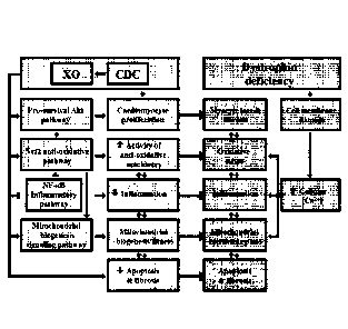
weeks after miR-148 treatment. F: Schematic of pathophysiological mechanisms
operative in
Duchenne cardiomyopathy and the cellular mechanisms recruited by CDCs and
their
exosomes (XO). All data are means SEM except for the box plot (means SD).
Figure 27. LV end-diastolic (LV EDV) and end-systolic (LV ESV) volumes after
cardiospherederived cell (CDC), CDC exosome (CDC-XO) and miR-148
administrations. CDC and CDC-XO transplantations resulted in a sustained
improvement of
LV EDV and LV ESV for 3 months after both first and second (3 months interval)
injections
in mdx mice, relative to placebo. Three weeks after miR148 injection, LV EDV
and LV ESV
13
CA 02962444 2017-03-23
WO 2016/054591 PCMJS2015/053853
were partially improved. Data are means SEM; n = 12 in each group. #P<O. 05
vs
Mdx+Vehicle.
Figure 28. Percentage engraftment of CDCs 1, 2 and 3 weeks after
transplantation. Percentage engraftment of CDCs at 1 week was ¨8% and <1% at 2
weeks.
By 3 weeks, no surviving CDCs could be detected. n = 3 at each time point.
Figure 29. Cardiomyogenesis and diminished fibrosis with CDC treatment in
mdx mice. Enhanced cardiomyogenesis (A&B) and diminished cardiac fibrosis (C)
and
collagen content (D) 3 weeks after CDC injection in mdx mice. Representative
immunohistochemical images and pooled data (A&B: CTL [wild type], vehicle and
CDC-
treated [Mdx+CDC] mdx mouse hearts stained for Ki67 [A] and Aurora B [B]; n=4-
6 per
group). Arrows point to Ki67 (A) and aurora B (B) cardiomyocytes.
Representative Masson
trichrome images (C) and western blots and pooled data (D) depicting cardiac
collagen IA
and IIIA. Data are means -= SEM; 7`13<0.05 vs. Mdx+Vehicle and CTL (control);
#P<0.05 vs.
Mdx+CDC and CTL (control). Scale bars: 101um (A).
Figure 30. Fold changes of microRNAs in CDC exosomes isolated from hypoxic
conditioned media. Under hypoxic conditions (2% 02) compared to CDC exosomes
isolated from normoxic conditioned media; fold change >10 and <20 were
included.
NEBNext Small RNA Library Prep kit (New England BioLabs, Ipswich, MA) was used
for
miRNA-seq library preparation of extracted small RNAs from the exosomes. RNAs
were
extracted from exosomes using miRNeasy Serum/Plasma Kit (QIAGEN, Germantown,
MD).
Figure 31. Isolated exosomes by ultracentrifugation were analyzed by
nanoparticle tracking. Using the NanoSight NS300 system (NanoSight Ltd, UK),
videos
were collected and analyzed using NTAsoftware (version 2.3), with the minimal
expected
particle size, minimum track length, and blur setting, all set to automatic.
Camera shutter
speed was fixed at 30.01 ms and camera gain was set to 500. Camera sensitivity
and detection
threshold were set close to maximum (15 or 16) and minimum (3 or 4),
respectively, to reveal
small particles. Ambient temperature was recorded manually, ranging from 24 to
27 C. For
each sample, five videos of 60 seconds duration were recorded, with a 10-
second delay
between recordings, generating five replicate histograms that were averaged.
Representative
five replicate histograms depicting size/concentration. Standard error of the
mean
concentration, calculated from 5 replicates, is shown in red in right graph.
14
CA 2962444
DETAILED DESCRIPTION
Unless defined otherwise, technical and scientific terms used herein have the
same meaning
as commonly understood by one of ordinary skill in the art to which this
invention belongs. Allen
et al., Remington: The Science and Practice of Pharmacy 22n1 ed,
Pharmaceutical Press
(September 15, 2012); Homyak et al., Introduction to Nanoscience and
Nanotechnology, CRC
Press (2008); Singleton and Sainsbury, Dictionary of Microbiology and
Molecular Biology 3' ed.,
revised ed, J. Wiley & Sons (New York, NY 2006); Smith, March's Advanced
Organic Chemistry
Reactions, Mechanisms and Structure 7th ed, J. Wiley & Sons (New York, NY
2013); Singleton,
Dictionary of DNA and Genome Technology 3' ed, Wiley-Blackwell (November 28,
2012); and
Green and Sambrook, Molecular Cloning: A Laboratory Manual 4th ed, Cold Spring
Harbor
Laboratory Press (Cold Spring Harbor, NY 2012), provide one skilled in the art
with a general
guide to many of the terms used in the present application. For references on
how to prepare
antibodies, see Greenfield, Antibodies A Laboratory Manual 2' ed., Cold Spring
Harbor Press
(Cold Spring Harbor NY, 2013); Kohler and Milstein, Derivation of specific
antibody-producing
tissue culture and tumor lines by cell fusion, Eur. J. Immunol. 1976 Jul,
6(7):511-9; Queen and
Selick, Humanized immunoglobulins, U. S. Patent No. 5,585,089 (1996 Dec); and
Riechmann et al.,
Reshaping human antibodies for therapy, Nature 1988 Mar 24, 332(6162):323-7.
One skilled in the art will recognize many methods and materials similar or
equivalent to
those described herein, which could be used in the practice of the present
invention. Indeed, the
present invention is in no way limited to the methods and materials described.
For purposes of the
present invention, the following terms are defined below.
As used in the description herein and throughout the claims that follow, the
meaning of "a,"
"an," and "the" includes plural reference unless the context clearly dictates
otherwise. Also, as
used in the description herein, the meaning of "in" includes "in" and "on"
unless the context clearly
dictates otherwise.
Duchenne muscular dystrophy, a crippling genetic disease leading to premature
death,
affects the heart as well as skeletal muscle. Indeed, cardiomyopathy is the
leading cause of death in
Duchenne patients. There are no approved treatments for the cardiomyopathy,
and novel Duchenne-
specific experimental approaches such as exon skipping do not benefit the
heart. Here the Inventors
demonstrate that cardiosphere-derived cells (CDCs), in advanced clinical
testing for therapeutic
regeneration after myocardial infarction, reverse the key
Date Recue/Date Received 2020-10-05
CA 02962444 2017-03-23
WO 2016/054591 PCMJS2015/053853
pathophys io logic al hallmarks of .. Duchenne .. cardiornyopathy ..
(oxidative
stress, inflammation, fibrosis and mitochondrial dysfunction) in mdx mice.
Exosomes
secreted by human CDCs reproduce the benefits of CDCs in mdx mice, and reverse
abnormalities of calcium cycling and mitochondrial respiration in human
Duchenne
cardiomyocytes.
Absence of dystrophin in Duchenne muscular dystrophy (DMD) leads to membrane
fragility and secondary damage to muscle (both skeletal and cardiac). Early
disability is due
predominantly to the skeletal myopathy, but heart failure is the most common
cause of death.
No currently available treatment modality addresses the underlying
pathophysiology of
DMD-associated heart failure, a loss of functional heart muscle and conversion
of living heart
muscle to scar. Cardiosphere-derived cells (CDCs) may represent a viable
therapeutic option.
Healthy heart muscle regrew and scar decreased in the first-in-human CADUCEUS
trial of
CDCs in myocardial infarction; these findings are now being further tested in
a randomized,
placebocontrolled multicenter clinical trial of allogeneic CDCs. Preclinical
studies show that
CDCs are not only regenerative, but also anti-inflammatory and anti-fibrotic;
they work
indirectly via the secretion of exosomes laden with noncoding RNA including
microRNAs
(miRs). In a murine model of myocardial infarction, CDC-exosomes mimic the
functional
and structural benefits of CDCs, while blockade of exosome biosynthesis
renders CDCs
ineffective. Given the clinical data and the mechanism of action, the
Inventors reasoned that
CDCs might be useful in treating Duchenne cardiomyopathy. The goal is not to
replace
dystrophin, but rather to offset the pathophysiological consequences of
dystrophin deletion,
by recruiting regeneration, reversing fibrosis and targeting inflammation.
Exosomes, secreted lipid vesicles containing a rich milieu of biological
factors,
provide powerful paracrine signals by which stem cells potentiate their
biological effects to
neighboring cells, including diseased or injured cells. Through the
encapsulation and transfer
of proteins, bio-active lipid and nucleic acid "cargo", there is increasing
recognition that
these natural delivery devices are capable of inducing significant phenotypic
and functional
changes in recipient cells that lead to activation of regenerative programs.
The role of such
indirect mechanisms to in stem cell initiated regeneration is strongly
suggested by growing
evidence that after stem cell administration and clearance from delivery sites
in tissue and
organs, regeneration processes nevertheless persist and arise from endogenous
tissues.
The "paracrine hypothesis" of stem cell regenerative activity has created a
paradigm
shift by which clinical applications based on exosomes secreted by the stem
cells may prove
16
CA 02962444 2017-03-23
WO 2016/054591 PCMJS2015/053853
superior, or provide distinct advantages, when compared to transplant and
delivery of stem
cells themselves. Stem cell-derived exosomes have been identified and isolated
from
supernatants of several cell types with demonstrated therapeutic potential,
including
mesenchymal stromal (MSC), (bone marrow stem cells) mononuclear (MNC) cells,
immune
cells (dendritic and CD34+), human neural stem cells (hNSCs), among others. In
the context
of heart disease, human cardiosphere derived cells (CDCs) are known to improve
myocardium and vasculature. Stem cell-derived exosomes, including those
produced by
CDCs, may provide a potent and rich source for developing "cell-free"
therapies.
In addition, exosome-based, "cell-free" therapies, in contrast to cell
therapy, provide
distinct advantages in regenerative medicine. As non-viable entities, with
reduced or non-
existent immunogenic or tumorigenic potential, these features significantly
obviate safety
issues. For example, stem cell-derived exosomes are less immunogenic than
parental cells, as
a result of a lower content of membrane-bound proteins, including MHC complex
molecules.
Replacing the administration of live cells with their secreted exosomes,
mitigates many of the
safety concerns and limitations associated with the transplantation of viable
replicating cells.
In addition, exosome encapsulation of bioactive components in lipid vesicles
allows
protection of contents from degradation in vivo, thereby potentially negating
obstacles often
associated with delivery of soluble molecules such as cytokines, growth
factors, transcription
factors and RNAs. This comparative ease of administration may ultimately
allow for
repeated and sustained delivery to patients, thereby maximizing the potential
for regeneration
and repair of diseased and/or dysfunctional tissue.
Also, exosome production under defined conditions allows for easier
manufacture and
scale-up opportunity. Manufacture of exosomes is akin to conventional
biopharmacological
product manufacture, allowing for standardization in production and quality
control for
dosage and biological activity testing. Further, the durability of exosomes in
culture allows
for the acquisition of large quantities of exosomes through their collection
from a culture
medium in which the exosomes are secreted over periods of time.
While it is now well-established that exosomes are involved in intercellular
communication between different cell types, much remains to be discovered in
regard to the
mechanisms of their production within parental cells of origin and effects on
target recipient
cells. Exosomes have been reported to be involved in numerous cellular, tissue
and
physiological processes, including immune modulating processes, angiogenesis,
migration of
endothelial cells in connection with tumor growth, or reducing damage in
ischemia
17
CA 02962444 2017-03-23
WO 2016/054591 PCMJS2015/053853
reperfusion injury. Of critical scientific interest in establishing whether
exosomes secreted
by cells, such as cardiosphere-derived cells (CDCs), are capable of
reproducing the
therapeutic benefits of their parental cells, or possibly, are indispensable
in effectuating such
therapeutic benefits.
General Features of Exosomes. Secreted by a wide range of cell types, exosomes
are
lipid bilayer vesicles that are enriched in a variety of biological factors,
including cytokines,
growth factors, transcription factors, lipids, and coding and non-coding
nucleic acids.
Exosomes are found in blood, urine, amniotic fluid, interstitial and
extracellular spaces.
These exocytosed vesicles of endosomal origin can range in size between 30-200
nm,
including sizes of 40-100 rim, and possess a cup-shaped morphology, as
revealed by electron
microscopy. Their initial formation begins with inward budding of the cell
membrane to form
endosomes, which is followed by invagination of the limiting membrane of late
endosomes to
form multivesicular bodies (MVB). Fusion of the MVB with the plasma membrane
results in
the release of the internal vesicles to the extracellular space, through the
formation of vesicles
thereafter known as exosomes.
As described, the "cargo" contents of exosomes reflect their parental cellular
origin,
as containing distinct subsets of biological factors in connection with their
parent cellular
origin, including the cell regulatory state of the parental cells when formed.
The rich
biological milieu of different proteins, including cytokines and growth
factors, lipids, coding
and noncoding RNA molecules, within exosomes are all necessarily derived from
their
parental cells. In addition to containing a rich array of cytosolic
derivatives, exosomes
further express the extracellular domain of membrane-bound receptors at the
surface of the
membrane.
The described encapsulation and formation processes necessarily create
heterogeneity
in exosome compositions based on parental cellular origin and regulatory state
at time of
formation. Nevertheless, generic budding formation and release mechanisms
establish a
common set of features as a consequence of their origin, such as endosome-
associated
proteins (e.g., Rab GTPase, SNAREs, Annexins, and flotillin), proteins that
are known to
cluster into microdomains at the plasma membrane or at endosomes (four
transmembrane
domain tetraspanins, e.g., CD63, CD81, CD82, CD53, and CD37), lipid raft
associated
proteins (e.g., glycosylphosphatidylinositol- anchored proteins and
flotillin), cholesterol,
sphingomyelin, and hexosylceramides, as examples.
18
CA 02962444 2017-03-23
WO 2016/054591 PCMJS2015/053853
In addition to these core components reflecting their vesicle origin, a
critical property
of exosomes is a demonstrated capability to contain both mRNA and microRNA
associated
with signaling processes, with both cargo mRNA being capable to translation in
recipient
cells, or microRNA functionally degrading target mRNA in recipient cells.
Other noncoding
RNAs, capable for influencing gene expression, may also be present in
exosomes. While the
processes governing the selective incorporation of mRNA or microRNA
populations into
exosomes is not entirely understood, it is clear that RNA molecules are
selectively, not
randomly incorporated into exosomes, as revealed by studies reporting
enrichment of
exosome cargo RNAs when compared to the RNA profiles of the originating cells.
Given the
growing understanding of how such RNA molecules play a role in disease
pathogenesis and
regenerative processes, the presence of RNA molecules in exosomes and apparent
potency in
affecting target recipient cells suggests critical features that can be
deployed in therapeutic
approaches.
Importantly, the natural bilayer membrane encapsulation of exosomes provides a
protected and controlled internal microenvironment that allows cargo contents
to persist or
migrate in the bloodstream or within tissues without degradation. Their
release into the
extracellular environment allows for interaction with recipient cells via
adhesion to the cell
surface mediated by lipid-ligand receptor interactions, internalization via
endocytic uptake, or
by direct fusion of the vesicles and cell membrane. These processes lead to
the release of
exosome cargo content into the target cell.
The net result of exosome-cell interactions is modulation of genetic pathways
in the
target recipient cell, as induced through any of several different mechanisms
including
antigen presentation, the transfer of transcription factors, cytokines, growth
factors, nucleic
acid such as mRNA and microRNAs. In the stem cell context, embryonic stem cell
(ESC)-
derived exosomes have been demonstrated to shuttle/transfer mRNA and proteins
to
hematopoietic progenitors. Other studies have shown that adult stem cell-
derived exosomes
also shuttle selected patterns of mRNA, microRNA and pre-microRNA associated
with
several cellular functions involved in the control of transcription,
proliferation and cell
immune regulation.
Isolation and Preparation of Exosomes. Exosome isolation relies on exploiting
their generic biochemical and biophysical features for separation and
analysis. For example,
differential ultracentrifugation has become a leading technique wherein
secreted exosomes
are isolated from the supernatants of cultured cells. This approach allows for
separation of
19
CA 02962444 2017-03-23
WO 2016/054591 PCMJS2015/053853
exosomes from nonmembranous particles, by exploiting their relatively low
buoyant density.
Size exclusion allows for their separation from biochemically similar, but
biophysically
different microvesicles, which possess larger diameters of up to 1,000 nm.
Differences in
flotation velocity further allows for separation of differentially sized
exosomes. In general,
exosome sizes will possess a diameter ranging from 30-200 nm, including sizes
of 40-100
nm. Further purification may rely on specific properties of the particular
exosomes of
interest. This includes, for example, use of immunoadsorption with a protein
of interest to
select specific vesicles with exoplasmic or outward orientations.
Among current methods (differential centrifugation, discontinuous density
gradients,
immunoaffinity, ultrafiltration and high performance liquid chromatography
(HPLC),
differential ultracentrifugation is the most commonly used for exosome
isolation. This
technique utilizes increasing centrifugal force from 2000xg to 10,000xg to
separate the
medium- and larger-sized particles and cell debris from the exosome pellet at
100,000xg.
Centrifugation alone allows for significant separation/collection of exosomes
from a
conditioned medium, although it is insufficient to remove various protein
aggregates, genetic
materials, particulates from media and cell debris that are common
contaminants. Enhanced
specificity of exosome purification may deploy sequential centrifugation in
combination with
ultrafiltration, or equilibrium density gradient centrifugation in a sucrose
density gradient, to
provide for the greater purity of the exosome preparation (flotation density
1.1-1.2g/m1) or
application of a discrete sugar cushion in preparation.
Importantly, ultrafiltration can be used to purify exosomes without
compromising
their biological activity. Membranes with different pore sizes - such as 100
kDa molecular
weight cut-off (MWCO) and gel filtration to eliminate smaller particles - have
been used to
avoid the use of a nonneutral pH or non-physiological salt concentration.
Currently available
tangential flow filtration (TFF) systems are scalable (to >10,000L), allowing
one to not only
purify, but concentrate the exosome fractions, and such approaches are less
time consuming
than differential centrifugation. HPLC can also be used to purify exosomes to
homogeneouslysized particles and preserve their biological activity as the
preparation is
maintained at a physiological pH and salt concentration.
Other chemical methods have exploit differential solubility of exosomes for
precipitation techniques, addition to volume-excluding polymers (e.g.,
polyethylene glycols
(PEGs)), possibly combined additional rounds of centrifugation or filtration.
For example, a
precipitation reagent, ExoQuick0, can be added to conditioned cell media to
quickly and
rapidly precipitate a population of exosomes, although re-suspension of
pellets prepared via
CA 02962444 2017-03-23
WO 2016/054591 PCMJS2015/053853
this technique may be difficult. Flow field-flow fractionation (F1FFF) is an
elution-based
technique that is used to separate and characterize macromolecules (e.g.,
proteins) and nano-
to micro-sized particles (e.g., organelles and cells) and which has been
successfully applied to
fractionate exosomes from culture media.
Beyond these techniques relying on general biochemical and biophysical
features,
focused techniques may be applied to isolated specific exosomes of interest.
This includes
relying on antibody immunoaffinity to recognizing certain exosome-associated
antigens. As
described, exosomes further express the extracellular domain of membrane-bound
receptors
at the surface of the membrane. This presents a ripe opportunity for isolating
and segregating
exosomes in connections with their parental cellular origin, based on a shared
antigenic
profile. Conjugation to magnetic beads, chromatography matrices, plates or
microfluidic
devices allows isolating of specific exosome populations of interest as may be
related to their
production from a parent cell of interest or associated cellular regulatory
state. Other
affinity-capture methods use lectins which bind to specific saccharide
residues on the
exosome surface.
Exosome-Based Therapies. A chief goal of developing exosome-based therapy is
the creation of "cell-free" therapies, wherein the benefits of cellular
therapeutics can be
provided with reduced risks or in scenarios in which cell therapy would be
unavailable. For
example, Duchenne muscular dystrophy (DMD) associated heart failure (HF),
particularly at
later stages, presents significant exclusionary comorbiditi es, wherein cell,
tissue, heart or
mechanical transplantation may not be an option for late stages C and D. As
described, the
therapeutic benefits of cell-based therapies such as cardiosphere-derived
cells (CDCs) appear
to occur through indirect mechanisms involving regenerated myocardium and
vasculature
arising from endogenous origin. Cellular exosomes produced by CDCs may allow
for
production and delivery of growth factors, transcription factors, cytokines
and nucleic acids
for new therapeutic approaches in a manner that not only ameliorates
progression of the
disease, but repairs and regenerates disease and/or dysfunctional tissue. In
this regard, CDC-
derived exosomes may effectively address a major unmet medical need, by
recruiting
synergistic mechanisms to attract endogenous stem cells to sites of myocardial
injury,
promote differentiation into heart muscle and vessels, thereby reversing the
pathophysiology
of HF.
More specifically, DMD is an X-linked recessive disorder characterized by
myopathy
(cell membrane damage in muscle fiber) as exemplified by a variety of
pathological features.
21
CA 02962444 2017-03-23
WO 2016/054591 PCMJS2015/053853
this includes skeletal muscle weakness starting 3-5 years from onset,
progressive weakness,
wheelchair dependency at approximately 13 years from onset. Importantly,
cardiomyopathy
is observed to take hold in 1/3 of patients from less than 13 years from
onset, increasing to
1/2 of patients less than 18 years from onset, and in all patients after 18
years. Dilated
cardiomyopathy includes left ventricle posterobasal fibrosis; conduction
abnormalities are
mainly intra-atrial: SVT with abnormal AV nodal conduction. Patients may
further suffer
from smooth muscle myopathy including vascular dysfunction, further including
GI and
urinary tract systems involvement.
Common prognosis is death from respiratory
insufficiency or cardiomyopathy. Underlying these clinical features is
dystrophin gene
mutation (deletion) wherein loss of dystrophin results in cellular membrane
damage and
leakage of extracellular Ca2 into the cell. Elevated intracellular levels
ultimately result in
increased oxidative and/or nitrosative stress and inflammation, and activation
of calpain. The
combination of these effects results in muscle proteolysis and apoptosis,
leading to the
degradative features described above.
Based on this pathophysiology of DMD patients, including an environment of
increased oxidative and/or nitrosative stress, elevated inflammation, pro-
apoptotic and
remodeling states, therapeutic approaches involving CDCs may provide
significant benefits
in reversing the course of the disease. CDCs have been demonstrated as
promoting anti-
oxidative, anti-inflammatory, anti-apoptotic, anti-remodeling effects, in
addition to enhancing
regenerative capacity. In this regard, it is suggested that CDC administration
is beneficial in
retarding/reversing DMD, and exosome populations derived from CDCs may allow
for these
benefits to be delivered, while avoiding obstacles associated with cell-based
therapy.
In particular, stem cell-derived exosomes are likely to be less immunogenic
than
parental cells. The possibility of replacing the administration of live cells
with secreted
exosomes, mitigates many of the safety concerns and limitations associated
with the
transplantation of viable cells. In addition, exosome encapsulation of
bioactive components
in lipid vesicles allows protection of contents from degradation in vivo,
thereby potentially
negating obstacles often associated with delivery of soluble molecules such as
cytokines,
growth factors, transcription factors and RNAs, while potentially allowing for
increased
concentrations to be provided. Particularly for chronic conditions, such as
DMD, repeated
and sustained delivery to patients may maximize the potential for regeneration
and repair of
diseased and/or dysfunctional tissue, in a manner that would be difficult or
unsafe with a cell-
based therapy.
Fully realizing these benefits requires an improved understanding of
whether exosomes secreted by cells such as CDCs, are alone capable of
reproducing
22
CA 02962444 2017-03-23
WO 2016/054591 PCMJS2015/053853
therapeutic benefits of their parental cells, or possibly indispensable in
these processes.
Confirming the role of exosomes in such processes will allow their application
in new
therapeutic approaches, including "cell-free" use in subjects for which
cellular transplant or
administration is unavailable (e.g., late stage heart disease), as
pharmacological, device-based
intervention or surgery may not be prudent treatment modalities for such
subject. There is a
great need in the art for identifying means by which to deliver the benefits
of stem cell
regeneration, without resorting to mechanisms involving administration or
transplant of the
cell themselves.
Described herein are compositions and methods and compositions providing
significant benefits in the repair or regeneration of damaged or diseased
tissues via "cell-free"
methods involving exosomes. Specifically, human cardiosphere-derived cells
(CDC)-derived
exosomes are demonstrated as effective in reducing scar size and regenerating
viable
myocardium. Such results confirm that the major benefits of CDC cell therapy
are mediated
by exosomes, including specific microRNAs identified by the Inventors as
enriched in CDCs.
Described herein is a method of treatment, including selecting a subject in
need of
treatment for heart failure secondary to a chronic degenerative muscular
disease and
administering a composition including a plurality of exosomes to the subject,
wherein the
plurality of the exosomes are isolated from cardiosphere-derived cells (CDCs)
grown in
serum-free media, include exosomes with a diameter of about 90 nm to about 200
nm and are
CD81+, CD63+, or both, and further wherein administration of the composition
treats the
subject. In other embodiments, the chronic degenerative muscular disease is
Duchenne
muscular dystrophy. In other embodiments, administering a composition includes
about 1
to about 100 mg exosome protein in a single dose. In other embodiments, a
single dose is
administered multiple times to the subject. In other embodiments,
administering a
composition includes injection. In other embodiments, the injection includes
percutaneous
injection. In other embodiments, the injection is directly into heart muscle.
In other
embodiments, administering a composition includes myocardial infusion. In
other
embodiments, myocardial infusion is intra-arterial or intravenous. In other
embodiments,
treatment of the subject results in decreased fibrosis, decreased
inflammation, increased
mitochondrial function and/or increased cardiomyogenesis. In other
embodiments, decreased
fibrosis includes a reduction in collagen accumulation. In other embodiments,
collagen
includes collagen I and/or collagen III. In other embodiments, decreased
inflammation
23
CA 02962444 2017-03-23
WO 2016/054591 PCMJS2015/053853
includes an increase in cytoplasmic nuclear factor (erythroid-derived 2)-like
2 (Nrf2),
reduction in fatty acid peroxidation end products, reduced numbers of
inflammatory cells,
and/or upregulated expression of antioxidants. In other embodiments,
antioxidants include
heme oxygenase-1 (H0-1), catalase, superoxide dismutase-2 (SOD-2), and
glutamate-cystein
ligase catalytic (GCLC) subunit. In other embodiments, inflammatory cells
include CD68+
macrophages and CD3+ T-cells. In other embodiments, increased mitochondrial
function
includes increased mitochondrial ultrastructure and/or increased mitochondrial
biogenesis. In
other embodiments, increased mitochondrial function includes increased nuclear
PPAR-y co-
activator-1 (PGC-1) expression. In other embodiments, the exosomes include one
or more
microRNAs selected from the group consisting of: microRNAs miR-146a, miR148a,
miR22,
miR-24, miR-210, miR-150, miR-140-3p, miR-19a, miR-27b, miR-19b, miR-27a, miR-
376c,
miR-128, miR-320a, miR-143, miR-21, miR-130a, miR-9, miR-185, and miR-23a.
Further described herein is method of treatment, including selecting a subject
in need
of treatment for heart failure secondary to a chronic muscular disease and
administering a
composition including cardiosphere-derived cells (CDCs), wherein
administration of the
composition treats the subject. In other embodiments, the chronic muscular
disease is
Duchenne muscular dystrophy. In other embodiments, administering a composition
includes
about 1 x 10⁵ to about 1 x 10⁸ or more CDCs in a single dose. In another
example, the
number of administered CDCs includes intracoronary 25 million CDCs per
coronary artery
(i.e., 75 million CDCs total) as another baseline for exosome dosage quantity.
In various
embodiments, the numbers of CDCs includes 1 x 10⁵, 1 x 10⁶, 1 x 10⁷, 1 x 10⁸,
1 x 10⁹ CDCs
in a single dose as another baseline for exosome dosage quantity. In certain
instances, this
may be prorated to body weight (range 100,000-1M CDCs/kg body weight total CDC
dose).
In other embodiments, administering a composition includes myocardial
infusion. In other
embodiments, myocardial infusion is intracoronary. In other embodiments,
myocardial
infusion is intra-arterial or intravenous. In other embodiments, treatment of
the subject
results in decreased fibrosis, decreased inflammation, increased mitochondrial
function
and/or increased cardiomyogenesis. In other embodiments, decreased fibrosis
includes a
reduction in collagen accumulation. In other embodiments, collagen includes
collagen I
and/or collagen III. In other embodiments, decreased inflammation includes an
increase in
cytoplasmic nuclear factor (erythroid-derived 2)-like 2 (Nrf2), reduction in
fatty acid
peroxidation end products, reduced numbers of inflammatory cells, and/or
upregulated
expression of antioxidants. In other embodiments, antioxidants include heme
oxygenase-1
24
CA 2962444
(H0-1), catalase, superoxide dismutase-2 (SOD-2), and glutamate-cystein ligase
catalytic (GCLC)
subunit. In other embodiments, inflammatory cells include CD68+ macrophages
and CD3+ T-cells.
In other embodiments, increased mitochondrial function includes increased
mitochondrial
ultrastructure and/or increased mitochondrial biogenesis. In other
embodiments, increased
mitochondrial function includes increased nuclear PPAR-y co-activator-1 (PGC-
1) expression.
.. Further examples are found in U.S. App. No. 11/666,685, 12/622,143, and
12/622,106.
Described herein is a composition including a plurality of exosomes. In
certain
embodiments, the plurality of exosomes are generated by a method including
providing a
population of cells, and isolating a plurality of exosomes from the population
of cells.
In various embodiments, the cells are stem cells, progenitors and/or
precursors. In other
embodiments, the stem cells, progenitors and/or precursors are cardiosphere-
derived cells (CDCs). In
other embodiments, the stem cells, progenitors and/or precursors are
pluripotent stem cells (pSCs),
such as embryonic stem cells (ESCs) and induced pluripotent stem cells (iPSCs)
derived from any
one of various somatic sources in the body such as fibroblasts, blood and
hematopoietic stem cells
(hSCs), immune cells, bone and bone marrow, neural tissue, among others. In
other embodiments,
the stem cells, progenitors and/or precursors include hSCs, mesenchymal stem
cells (MSCs), or
endothelial precursor cells (EPCs). In various embodiments, the cells are stem
cells, progenitors
and/or precursors derived from human biopsy tissue. In various embodiments,
the cells are stem
cells, progenitors and/or precursors are a primary culture. In various
embodiments, the cells are stem
.. cells, progenitors and/or precursors which may constitute a cell line
capable of serial passaging.
In various embodiments, the plurality of exosomes is isolated from the
supernatants of the
population of cells. This includes, for example, exosomes secreted into media
as conditioned by a
population of cells in culture, further including cell lines capable of serial
passaging. In certain
embodiments, the cells are cultured in serum-free media. In certain
embodiments, the cells in culture
are grown to 10, 20, 30, 40, 50, 60, 70, 80, 90, or 90% or more confluency
when exosomes are isolated.
In certain embodiments, the population of cells has been genetically
manipulated. This includes, for
example, knockout (KO) or transgenic (TG) cell lines, wherein an endogenous
gene has been removed
and/or an exogenous introduced in a stable, persistent manner. This further
includes transient
knockdown of one or more genes and associated coding and non-coding
transcripts within the
Date Recue/Date Received 2020-10-05
CA 02962444 2017-03-23
WO 2016/054591 PCMJS2015/053853
population of cells, via any number of methods known in the art, such as
introduction of
dsRNA, siRNA, microRNA, etc. This further includes transient expression of one
or more
genes and associated coding and non-coding transcripts within the population
of cells, via any
number of methods known in the art, such as introduction of a vector, plasmid,
artificial
plasmid, replicative and/or non-relicative virus, etc. In other embodiments,
the population of
cells has been altered by exposure to environmental conditions (e.g.,
hypoxia), small
molecule addition, presence/absence of exogenous factors (e.g., growth
factors, cytokines) at
the time, or substantially contemporaneous with, isolating the plurality of
exosomes in a
manner altering the regulatory state of the cell. For example, one may add a
differentiation
agent to a population of stem cells, progenitors and/or precursors in order to
promote partial
or full differentiation of the cell, and thereafter derive a plurality of
exosomes. In various
embodiments, altering the regulatory state of the cell changes composition of
one or more
exosomes in the plurality of exosomes.
In various embodiments, the plurality of exosomes include one or more exosomes
that
are about 10 nm to about 250 nm in diameter, including those about 10 nm to
about 15 nm,
about 15 nm to about 20 nm, about 20 nm to about 25 nm, about 25 nm to about
30 nm, about
nm to about 35 nm, about 35 nm to about 40 nm, about 40 nm to about 50 nm,
about 50
nm to about 60 nm3 about 60 nm to about 70 nm, about 70 nm to about 80 nm,
about 80 nm
to about 90 nm, about 90 nm to about 95 nm, about 95 nm to about 100 nm, about
100 nm to
about 105 nm, about 105 nm to about 110 nm, about 110 nm to about 115 nm,
about 115 nm
25 to
about 120 nm, about 120 nm to about 125 nm, about 125 nm to about 130 nm,
about 130
nm to about 135 nm, about 135 nm to about 140 nm, about 140 nm to about 145
nm, about
145 nm to about 150 nm, about 150 to about 200 nm, about 200 nm to about 250
nm, about
250 nm or more.
In various embodiments, the plurality of exosomes includes one or more
exosomes
30
expressing a biomarker. In certain embodiments, the biomarkers are
tetraspanins. In other
embodiments, the tetraspanins are one or more selected from the group
including CD63,
CD81, CD82, CD53, and CD37. In other embodiments, the exosomes express one or
more
lipid raft associated protiens (e.g., glycosylphosphatidylinositol-anchored
proteins and
flotillin), cholesterol, sphingomyelin, and/or hexosylceramides.
In several embodiments, the plurality of exosomes includes one or more
exosomes
containing a biological protein. In various embodiments, the biological
protein includes
transcription factors, cytokines, growth factors, and similar proteins capable
of modulating
signaling pathways in a target cell. In various embodiments, the biological
protein is capable
26
CA 02962444 2017-03-23
WO 2016/054591 PCMJS2015/053853
of facilitating regeneration and/or improved function of a tissue. In various
embodiments, the
biological protein is capable of modulating pathways related to Irakl, Traf6,
toll-like receptor
(TLR) signaling pathway, NOX-4, SMAD-4, and/or TGF-13. In other embodiments,
the
biological protein related to exosome formation and packaging of cytosolic
proteins such as
Hsp70, Hsp90,14-3-3 epsilon, PI(M2, GW182 and AG02.
In other embodiments, the plurality of exosomes includes one or more exosomes
containing a signaling lipid. This includes ceramide and derivatives. In other
embodiments,
the plurality of exosomes includes one or more exosomes containing a coding
and/or non-
coding nucleic acid.
In several embodiments, the plurality of exosomes includes one or more
exosomes
containing microRNAs. In various embodiments, these microRNAs can include miR-
146a,
miR22, miR-24, miR-210, miR-150, miR-140-3p, miR-19a, miR-27b, miR-19b, miR-
27a,
miR-376c, miR-128, miR-320a, miR-143, miR-21, miR-130a, miR-9, miR-185, and/or
miR-
23a. In several embodiments, the plurality of exosomes include one or more
exosomes
enriched in at least one of miR-146a, miR22, miR-24, miR-210, miR-150, miR-140-
3p, miR-
19a, miR-27b, miR-19b, miR-27a, miR-376c, miR-128, miR-320a, miR-143, miR-21,
miR-
130a, miR-9, miR-185, and/or miR-23a. In several embodiments, the plurality of
exosomes
includes one or more exosomes enriched in at least one of miR-146a, miR-22,
miR-24.
Enrichment can be measured by, for example, comparing the amount of one or
more of the
described microRNAs when derived from cells providing salutary benefit in a
therapeutic
setting (e.g., cardiosphere-derived cells (CDCs) compared to cells that do not
provide such a
salutary benefit (e.g., fibroblasts). Enrichment may also be measured in
absolute or relative
quantities, such as when compared to a standardized dilution series.
In other embodiments, the plurality of exosomes can include one or more
exosomes
containing microRNAs. This includes various microRNAs known in the art, such
as miR-
23a, miR-23b, miR-24, miR-26a, miR27-a, miR-30c, let-7e, mir- 19b, miR-125b,
mir-27b,
let-7a, miR-19a, let-7c, miR-140-3p, miR-125a-5p, miR-132, miR-150, miR-155,
mir-210,
let-7b, miR-24, miR-423-5p, miR-22, let-7f, and/or miR-146a.
In other embodiments, the plurality of exosomes can include one or more
exosomes
containing microRNAs. This includes various microRNAs known in the art, such
as miR-17,
miR-21 , miR-92, miR92a, miR-29, miR-29a, miR-29b, miR-29c, miR-34, mi-R34a,
miR-
150, miR-451, miR-145, miR-143, miR-144, miR-193a-3p, miR-133a, miR-155, miR-
181a,
miR-214, miR-199b, miR-199a, miR-210, miR-126, miR-378, miR-363 and miR-30b,
and
miR-499. Other microRNAs known in the art include miR-92, miR-17, miR-21, miR-
92,
27
CA 02962444 2017-03-23
WO 2016/054591 PCMJS2015/053853
miR92a, miR-29, miR- 29a, miR-29b, miR-29c, miR-34, mi-R34a, miR-150, miR-451,
miR-
145, miR-143, miR- 144, miR-193a-3p, miR-133a, miR-155, miR-181a, miR-214, miR-
199b,
miR-199a, miR- 126, miR-378, miR-363 and miR-30b, and/or miR-499.
In several embodiments, isolating a plurality of exosomes from the population
of cells
includes centrifugation of the cells and/or media conditioned by the cells. In
several
embodiments, ultracentrifugation is used. In several embodiments, isolating a
plurality of
exosomes from the population of cells is via size-exclusion filtration. In
other embodiments,
isolating a plurality of exosomes from the population of cells includes use of
discontinuous
density gradients, immunoaffinity, ultrafiltration and/or high performance
liquid
chromatography (HPLC).
In certain embodiments, differential ultracentrifugation includes using
centrifugal
force from 1000-2000xg, 2000-3000xg, 3000-4000xg, 4000-5000xg, 5000xg-6000xg,
6000-
7000xg, 7000-8000xg, 8000-9000xg, 9000-10,000xg, to 10,000xg or more to
separate larger-
sized particles from a plurality of exosome derived from the cells. In certain
embodiments,
differential ultracentrifugation includes using centrifugal force from 10,000-
20,000xg,
20,000-30,000xg, 30,000-40,000xg, 40,000-50,000xg, 50,000xg-60,000xg, 60,000-
70,000xg,
70,000-80,000xg, 80,000-90,000xg, 90,000-100,000xg, to 10,000xg or more to
separate
larger-sized particles from a plurality of exosome derived from the cells.
In other embodiments, isolating a plurality of exosomes from the population of
cells
includes use of filtration or ultrafiltration. In certain embodiments, a size
exclusion
membrane with different pore sizes is used. For example, a size exclusion
membrane can
include use of a filter with a pore size of 0.1-0.5 p.M, 0.5-1.0 iitM, 1-2.5
pM, 2.5-5 04, 5 or
more M. In certain embodiments, the pore size is about 0.2 iuM. In certain
embodiments,
filtration or ultrafiltration includes size exclusion ranging from 100-500
daltons (Da), 500-1
kDa, 1-2 kDa, 2-5 kDa, 5-10 kDa, 10-25 kDa, 25-50 kDa, 50-100 kDa, 100-250
kDa, 250-
500 kDa, 500 or more kDa. In certain embodiments, the size exclusion is for
about 2-5 kDa.
In certain embodiments, the size exclusion is for about 3 kDa. In other
embodiments,
filtration or ultrafiltration includes size exclusion includes use of hollow
fiber membranes
capable of isolating particles ranging from 100-500 daltons (Da), 500-1 kDa, 1-
2 kDa, 2-5
kDa, 5-10 kDa, 10-25 kDa, 25-50 kDa, 50-100 kDa, 100-250 kDa, 250-500 kDa, 500
or more
kDa. In certain embodiments, the size exclusion is for about 2-5 kDa. In
certain
embodiments, the size exclusion is for about 3 kDa. In other embodiments, a
molecular
weight cut-off (MWCO) gel filtration capable of isolating particles ranging
from 100-500
daltons (Da), 500-1 kDa, 1-2 kDa, 2-5 kDa, 5-10 kDa, 10-25 kDa, 25-50 kDa, 50-
100 kDa,
28
CA 02962444 2017-03-23
WO 2016/054591 PCMJS2015/053853
100-250 kDa, 250-500 kDa, 500 or more kDa. In certain embodiments, the size
exclusion is
for about 2-5 kDa. In certain embodiments, the size exclusion is for about 3
kDa. In various
embodiments, such systems are used in combination with variable fluid flow
systems.
In other embodiments, isolating a plurality of exosomes from the population of
cells
includes use of tangential flow filtration (TFF) systems are used purify
and/or concentrate the
exosome fractions. In other embodiments, isolating a plurality of exosomes
from the
population of cells includes use of (HPLC) can also be used to purify exosomes
to
homogeneously sized particles. In various embodiments, density gradients as
used, such as
centrifugation in a sucrose density gradient or application of a discrete
sugar cushion in
preparation.
In other embodiments, isolating a plurality of exosomes from the population of
cells
includes use of a precipitation reagent. For example, a precipitation reagent,
ExoQuick , can
be added to conditioned cell media to quickly and rapidly precipitate a
population of
exosomes. In other embodiments, isolating a plurality of exosomes from the
population of
cells includes use of volume-excluding polymers (e.g., polyethylene glycols
(PEGs)) are
used. In another embodiment, isolating a plurality of exosomes from the
population of cells
includes use of flow field-flow fractionation (F1FFF), an elution-based
technique.
In certain embodiments, isolating a plurality of exosomes from the population
of cells
includes use of one or more capture agents to isolate one or more exosomes
possessing
specific biomarkers or containing particular biological molecules. In one
embodiment, one or
more capture agents include at least one antibody. For example, antibody
immunoaffinity
recognizing exosome-associated antigens is used to capture specific exosomes.
In other
embodiments, the at least one antibody are conjugated to a fixed surface, such
as magnetic
beads, chromatography matrices, plates or microfluidic devices, thereby
allowing isolation of
the specific exosome populations of interest. In other embodiments, isolating
a plurality of
exosomes from the population of cells includes use of one or more capture
agents that is not
an antibody. This includes, for example, use of a "bait" molecule presenting
an antigenic
feature complementary to a corresponding molecule of interest on the exosome
surface, such
as a receptor or other coupling molecule. In one embodiment, the non-antibody
capture agent
is a lectin capable of binding to polysaccharide residues on the exosome
surface.
In various embodiments, the CDCs are mammalian. In other embodiments, the CDCs
are human. As disclosed above, in some embodiments, synthetic exosomes are
generated,
which can be isolated by similar mechanisms as those above. In various
embodiments, the
29
CA 2962444
composition that is a plurality of exosomes is a pharmaceutical composition
further including a
pharmaceutically acceptable carrier.
In various embodiments, the plurality of exosomes range in size from 30 to 300
nm. In
various embodiments, the plurality of exosomes range in size from 40 to 100
nm. In certain
embodiments, the plurality of exosomes is cardiosphere-derived cell (CDC)
exosomes. In certain
embodiments, the plurality of exosomes includes exosomes that are CD63+. In
various
embodiments, the exosomes include microRNAs miR-146a, miR22, miR-24, miR-210,
miR-150,
miR-140-3p, miR-19a, miR-27b, miR-19b, miR-27a, miR-376c, miR-128, miR-320a,
miR-143,
miR-21, miR-130a, miR-9, miR-185, and/or miR-23a. In other embodiments, the
exosomes are 2-5
kDa, such as 3 kDa. Other examples or embodiments relating to the composition
and techniques
involving exosomes are presented, in PCT Pub. No. WO 2014/028,493.
Described herein is a method for treatment including, selecting a subject in
need of treatment,
administering a composition including a plurality of exosomes to the
individual, wherein the
administration of the composition treats the subject. In certain embodiments,
the subject is in need to
treatment for a disease and/or condition involving tissue damage or
dysfunction. In other embodiments,
.. the disease and/or condition involving tissue damage or dysfunction is
heart disease. In other
embodiments, the plurality of exosomes includes exosomes including one or more
microRNAs.
In certain embodiments, the plurality of exosomes are generated by a method
including
providing a population of cells, and isolating a plurality of exosomes from
the population of cells.
In various embodiments, the cells are stem cells, progenitors and/or
precursors. In other
embodiments, the stem cells, progenitors and/or precursors are cardiosphere-
derived cells (CDCs).
In other embodiments, the stem cells, progenitors and/or precursors are
pluripotent stem cells
(pSCs), such as embryonic stem cells (ESCs) and induced pluripotent stem cells
(iPSCs) derived
from any one of various somatic sources in the body such as fibroblasts, blood
and hematopoietic
stem cells (hSCs), immune cells, bone and bone marrow, neural tissue, among
others. In other
embodiments, the stem cells, progenitors and/or precursors includes hSCs,
mesenchymal stem cells
(MSCs), or endothelial precursor cells (EPCs). In various embodiments, the
cells are stem cells,
progenitors and/or precursors derived from human biopsy tissue. In various
embodiments, the cells
are stem cells, progenitors and/or precursors are a primary culture. In
various embodiments, the
cells are stem cells, progenitors and/or precursors are a cell line capable of
serial passaging. In
certain embodiments, the exosomes are synthetic.
Date Recue/Date Received 2020-10-05
CA 02962444 2017-03-23
WO 2016/054591 PCMJS2015/053853
In various embodiments, the plurality of exosomes is derived from cardiosphere-
derived cells (CDCs). In other embodiments, the plurality of exosomes includes
exosomes
including one or more biological molecules. In other embodiments, the
plurality of exosomes
includes exosomes enriched for one or more biological molecules when derived
from CDCs
compared to exosome derived from non-CDC sources. In various embodiments, the
one or
more biological molecules are proteins, growth factors, cytokines,
transcription factors and/or
morphogenic factors. In other embodiments, the plurality of exosomes includes
exosomes
enriched for one or more biological molecules includes microRNAs, further
including
microRNAs that are enriched when derived from CDCs compared to exosome derived
from
non-CDC sources. In various embodiments, these microRNAs can include miR-146a,
miR22, miR-24, miR-210, miR-150, miR-140-3p, miR-19a, miR-27b, miR-19b, miR-
27a,
miR-376c, miR-128, miR-320a, miR-143, miR-21, miR-130a, miR-9, miR-185, and/or
miR-
23a. In several embodiments, the plurality of exosomes includes one or more
exosomes
enriched in at least one of miR-146a, miR-22, miR-24.
In various embodiments, the CDCs arc mammalian. In other embodiments, the CDCs
are human. In certain embodiments, the exosomes arc synthetic. In certain
embodiments, the
synthetic exosomes possess substantially similar content (e.g., microRNAs,
biological
molecules) as exosomes derived from CDCs.
In various embodiments, administration of the plurality of exosomes alters
gene
expression in the damaged or dysfunctional tissue, improves viability of the
damaged tissue,
and/or enhances regeneration or production of new tissue in the individual. In
various
embodiments, the quantities of exosomes that are administered to achieved
these effects
range from 1 x 10⁶ to 1 x 10⁷, 1 x 10⁷ to 1 x 10⁸, 1 x 10⁸ to 1 x 10⁹, 1 x 10⁹
to 1 x 10¹⁰, 1 x
1010 to 1 x 10¹¹, 1 x 10¹¹ to 1 x 10¹², 1 x 10¹² or more. In other
embodiments, the numbers of
exosomes is relative to the number of cells used in a clinically relevant dose
for a cell-therapy
method. For example, it has been demonstrated that 3mL / 3 x 10⁵ CDCs, is
capable of
providing therapeutic benefit in intracoronary administration, and therefore,
a plurality of
exosomes as derived from that number of cells in a clinically relevant dose
for a cell-therapy
method. In various embodiments, administration can be in repeated doses. In
another
example, the number of administered CDCs includes intracoronary 25 million
CDCs per
coronary artery (i.e., 75 million CDCs total) as another baseline for exosome
dosage quantity.
In various embodiments, the numbers of CDCs includes 1 x 10, 1 x 10⁶, 1 x 10⁷,
1 x 10⁸, 1 x
109 CDCs in a single dose as another baseline for exosome dosage quantity. In
certain
instances, this may be prorated to body weight (range 100,000-1M CDCs/kg body
weight
31
CA 02962444 2017-03-23
WO 2016/054591 PCMJS2015/053853
total CDC dose). In various embodiments, exosome quantity may be defined by
protein
quantity, such as dosages including 1-10, 10-25, 25-50, 50-75, 75-100, or 100
or more mg
exosome protein.
Defining an effective dose range, dosing regimen and route of administration,
may be
guided by studies using fluorescently labeled exosomes, and measuring target
tissue
retention, which can be >10X, >50X, or >100X background, as measured 5, 10,
15, 30, or 30
or more min as a screening criterion. In certain embodiments, >100X background
measured
at 30 mins is a baseline measurement for a low and high dose that is then
assess for safety
and bioactivity (e.g., using MRI endpoints: scar size, global and regional
function). In
various embodiments, single doses are compared to two, three, four, four or
more
sequentially-applied doses. In
various embodiments, the repeated or sequentially-applied
doses are provided for treatment of an acute disease and/or condition. In
various
embodiments, the repeated or sequentially-applied doses are provided for
treatment of a
chronic disease and/or condition.
In various embodiments, administration of exosomes to the subject occurs
through
any of known techniques in the art. In some embodiments, this includes
percutaneous
delivery. In other embodiments, myocardial infusion is used, for example, the
use of
intracoronary catheters. In
various embodiments, delivery can be intra-arterial or
intravenous. Additional delivery sites include any one or more compartments of
the heart,
such as arterial, venous, and/or ventricular locations. In certain
embodiments, administration
can include delivery to a tissue or organ site that is different from the site
or diseased and/or
dysfunctional tissue. In certain embodiments, the delivery is via inhalation
or oral
administration.
In various embodiments, administration of the plurality of exosomes alters
gene
expression in the damaged or dysfunctional tissue, improves viability of the
damaged tissue,
and/or enhances regeneration or production of new tissue in the individual. In
various
embodiments, administration of the exosomes results in functional improvement
in the tissue.
In several embodiments, the damaged or dysfunctional tissue includes cardiac
tissue.
For example, in certain embodiments in which cardiac tissue is damaged or
dysfunctional, functional improvement may include increased cardiac output,
contractility,
ventricular function and/or reduction in arrhythmia (among other functional
improvements).
For other tissues, improved function may be realized as well, such as enhanced
cognition in
response to treatment of neural damage, improved blood-oxygen transfer in
response to
32
CA 02962444 2017-03-23
WO 2016/054591 PCMJS2015/053853
treatment of lung damage, improved immune function in response to treatment of
damaged
immunological-related tissues.
In various embodiments, administration of the plurality of exosomes alters
gene
expression in the damaged or dysfunctional tissue, improves viability of the
damaged tissue,
and/or enhances regeneration or production of new tissue in the individual. In
various
embodiments, administration of the exosomes results in functional improvement
in the tissue.
In several embodiments, the damaged or dysfunctional tissue includes skeletal
muscle tissue.
For example, in certain embodiments in which skeletal muscle tissue is damaged
or
dysfunctional, functional improvement may include increased contractile
strength, improved
ability to walk (for example, and increase in the six-minute walk test
results), improved
ability to stand from a seated position, improved ability to sit from a
recumbent or supine
position, or improved manual dexterity such as pointing and/or clicking a
mouse.
In various embodiments, the damaged or dysfunctional tissue is in need of
repair,
regeneration, or improved function due to an acute event. Acute events
include, but are not
limited to, trauma such as laceration, crush or impact injury, shock, loss of
blood or oxygen
flow, infection, chemical or heat exposure, poison or venom exposure, drug
overuse or
overexposure, and the like. In other embodiments, the damaged tissue is
cardiac tissue and
the acute event includes a myocardial infarction. In some embodiments,
administration of the
exosomes results in an increase in cardiac wall thickness in the area
subjected to the
infarction.
In other embodiments, tissue is also subject to damage due to chronic disease,
such as
for example congestive heart failure, including as conditions secondary to
diseases such as
Duchenne muscular dystrophy, ischemic heart disease, hypertension, valvular
heart disease,
dilated cardiomyopathy, infection, diabetes, and the like. In various
embodiments, the
administration can be in repeated doses, such as two, three, four, four or
more sequentially-
applied doses. In various embodiments, the repeated or sequentially-applied
doses are
provided for treatment of an acute disease and/or condition. In various
embodiments, the
repeated or sequentially-applied doses are provided for treatment of a chronic
disease and/or
condition.
Other sources of damage also include, but are not limited to, injury, age-
related
degeneration, cancer, and infection. In several embodiments, the regenerative
cells are from
the same tissue type as is in need of repair or regeneration. In several other
embodiments, the
regenerative cells are from a tissue type other than the tissue in need of
repair or regeneration.
33
CA 02962444 2017-03-23
WO 2016/054591 PCMJS2015/053853
In certain embodiments, the method of treatment includes, selecting a subject
in need
of treatment for a heart related disease and/or condition, administering a
composition
including a plurality of exosomes to the individual, wherein the
administration of the
composition treats thesubject. In various embodiments, the heart related
disease and/or
condition includes heart failure, further including Duchenne muscular
dystrophy related heart
failure. In various embodiments, the plurality of exosomes range in size from
30 to 300 nm.
In various embodiments, the plurality of exosomes range in size from 40 to 100
nm. In
certain embodiments, the plurality of exosomes are cardiosphere-derived cell
(CDC)
exosomes. In certain embodiments, the plurality of exosomes include exosomes
that are
CD63+. In various embodiments, the exosomes include microRNAs miR-146a, miR22,
miR-24, miR-210, miR-150, miR-140-3p, miR-19 a, miR-27b, miR-19b, miR-27 a,
miR-376c,
miR-128, miR-320a, miR-143, miR-21, miR-130a, miR-9, miR-185, and/or miR-23a.
In
other embodiments, the exosomes are 2-5 kDa, such as 3 kDa. In other
embodiments,
administering a composition includes a dosage of 1 x 10⁸, 1 x 10⁸ to 1 x 10⁹,
1 x 10⁹ to 1 x
1010, 1 x 10¹⁰ to 1 x 10¹¹, 1 x 10¹¹ to 1 x 10¹², 1 X 10¹² or more exosomes.
For example, it has
been demonstrated that 3mL / 3 x 10⁵ CDCs, is capable of providing therapeutic
benefit in
intracoronary administration, and therefore, a plurality of exosomes as
derived from that
number of cells in a clinically relevant dose for a cell-therapy method. In
various
embodiments, administration can be in repeated doses. In another example, the
number of
administered CDCs includes intracoronary 25 million CDCs per coronary artery
(i.e., 75
million CDCs total) as another baseline for exosome dosage quantity. In
various
embodiments, the numbers of CDCs includes 1 x 10⁵, 1 x 10⁶, 1 x 10⁷, 1 x 10⁸,
1 x 10⁹ CDCs
in a single dose. In certain instances, this may be prorated to body weight
(range 100,000-
1M CDCs/kg body weight total CDC dose). In various embodiments, exosome
quantity may
be defined by protein quantity, such as dosages including 1-10, 10-25, 25-50,
50-75, 75-100,
or 100 ore more mg exosome protein. In various embodiments, administering a
composition
includes multiple dosages of the exosomes. In various embodiments, the
repeated or
sequentially-applied doses are provided for treatment of an acute disease
and/or condition. In
various embodiments, the repeated or sequentially-applied doses are provided
for treatment
of a chronic disease and/or condition. In other embodiments, administering a
composition
includes percutaneous injection. In other embodiments, administering a
composition includes
myocardial infusion. In other embodiments, administering a composition
includes use of a
intracoronary catheter. In other embodiments, administration a composition
includes intra-
arterial or intravenous delivery.
34
CA 02962444 2017-03-23
WO 2016/054591 PCMJS2015/053853
Further described herein is a method of improving cardiac performance in a
subject
including, selecting a subject, administering a composition including a
plurality of exosomes
to the individual, wherein the administration of the composition improves
cardiac
performance in the subject. In other embodiments, improving cardiac
performance can be
demonstrated, by for example, improvements in baseline ejection volume. In
other
embodiments, improving cardiac performance relates to increases in viable
tissue, reduction
in scar mass, improvements in wall thickness, regenerative remodeling of
injury sites,
enhanced angiogenesis, improvements in cardiomyogenic effects, reduction in
apoptosis,
and/or decrease in levels of pro-inflammatory cytokines.
In certain embodiments, the method of improving cardiac performance includes,
selecting a subject in need of treatment for a heart related disease and/or
condition,
administering a composition including a plurality of exosomes to the
individual, wherein the
administration of the composition treats thesubject. In various embodiments,
the heart related
disease and/or condition includes heart failure, further including Duchenne
muscular
dystrophy related heart failure. In various embodiments, the plurality of
exosomes range in
size from 30 to 300 nm. In various embodiments, the plurality of exosomes
range in size
from 40 to 100 nm. In certain embodiments, the plurality of exosomes are
cardiosphere-
derived cell (CDC) exosomes. In certain embodiments, the plurality of exosomes
include
exosomes that are CD63+. In various embodiments, the exosomes include
microRNAs
miR-146a, miR22, miR-24, miR-210, miR-150, miR-140-3p, miR-19a, miR-27b, miR-
19b,
miR-27a, miR-376c, miR-128, miR-320a, miR-143, miR-21, miR-130a, miR-9, miR-
185,
and/or miR-23a. In other embodiments, the exosomes are 2-5 kDa, such as 3 kDa.
In other
embodiments, administering a composition includes a dosage of 1 x 10⁸, 1 x 10⁸
to 1 x 10⁹, 1
x 109 to 1 x 10¹⁰, 1 x 10¹⁰ to 1 x 10¹¹, 1 x 10¹¹ to 1 x 10¹², 1 x 10¹² or
more exosomes. For
example, it has been demonstrated that 3m1L / 3 x 10⁵ CDCs, is capable of
providing
therapeutic benefit in intracoronary administration, and therefore, a
plurality of exosomes as
derived from that number of cells in a clinically relevant dose for a cell-
therapy method. In
various embodiments, administration can be in repeated doses. In another
example, the
number of administered CDCs includes intracoronary 25 million CDCs per
coronary artery
(i.e., 75 million CDCs total) as another baseline for exosome dosage quantity.
In various
embodiments, the numbers of CDCs includes 1 x 10⁵, 1 x 10⁶, 1 x 10⁷, 1 x 10⁸,
1 x 10⁹ CDCs
in a single dose. In certain instances, this may be prorated to body weight
(range 100,000-
1M CDCs/kg body weight total CDC dose). In various embodiments, exosome
quantity may
be defined by protein quantity, such as dosages including 1-10, 10-25, 25-50,
50-75, 75-100,
CA 2962444
or 100 or more mg exosome protein. In various embodiments, administering a
composition includes
multiple dosages of the exosomes. In various embodiments, the repeated or
sequentially-applied
doses are provided for treatment of an acute disease and/or condition. In
various embodiments, the
repeated or sequentially-applied doses are provided for treatment of a chronic
disease and/or
condition. In other embodiments, administering a composition includes
percutaneous injection. In
other embodiments, administering a composition includes myocardial infusion.
In other
embodiments, administering a composition includes use of a intracoronary
catheter. In other
embodiments, administration a composition includes intra-arterial or
intravenous delivery.
Herein the Inventors demonstrate that cardiosphere-derived cells (CDCs), in
advanced
clinical testing for therapeutic regeneration after myocardial infarction,
reverse the key
pathophysiological
hallmarks of Duchenne cardiomyopathy (oxidative stress, inflammation,
fibrosis and mitochondrial dysfunction) in mdx mice. Exosomes secreted by
human CDCs
reproduce the benefits of CDCs in mdx mice, and reverse abnormalities of
calcium cycling and
mitochondrial respiration in human Duchenne cardiomyocytes. Both CDCs and
their exosomes
improve heart function in mdx mice; a single injection of CDCs suffices to
increase maximal
exercise capacity and improve survival. Delivery of a microRNA enriched in CDC
exosomes,
miR-148a, mimics key effects of CDCs and CDC exosomes. Thus, CDCs ameliorate
Duchenne
cardiomyopathy via exosome-mediated transfer of signaling molecules including
miR-148a. The
present findings motivate clinical testing of CDCs in patients with Duchenne
cardiomyopathy.
Example]
CDC Culture
Endomyocardial biopsies from the right ventricular aspect of the
interventricular septum are
obtained from healthy hearts of deceased tissue donors. Cardiosphere-derived
cells were derived as
described previously. See Makkar et al., (2012). "Intracoronary cardiosphere-
derived cells for heart
regeneration after myocardial infarction (CADUCEUS): a prospective, randomized
phase 1 trial."
Lancet 379, 895-904 (2012).
In brief, heart biopsies are minced into small fragments and briefly digested
with
collagenase. Explants were then cultured on 20 mg/ml fibronectin-coated
dishes. Stromal-like flat
cells and phase-bright round cells grow out spontaneously from tissue
fragments and
36
Date Recue/Date Received 2020-10-05
CA 02962444 2017-03-23
WO 2016/054591 PCMJS2015/053853
reach confluence by 2-3 weeks. These cells are harvested using 0.25% trypsin
and cultured
in suspension on 20 mg/ml poly d-lysine to form self-aggregating
cardiospheres.
cardiosphere-derived cells (CDCs) are obtained by seeding cardiospheres onto
fibronectin-
coated dishes and passaged. All cultures are maintained at 5% CO2 at 37 C,
using IMDM
basic medium supplemented with 20% FBS, 1% penicillin/streptomycin, and 0.1 ml
2-
mere apto ethanol.
Example 2
Media Conditioning and Exosome Purification
Exosomes are harvested from CDCs at passage 4. One can also isolate exosomes
from normal human dermal fibroblasts (NHDF), cells that have been previously
utilized as
controls providing no salutary benefit, as a control.
CDCs and NHDFs are conditioned in serum-free media for 15 days at 100%
confluence. Aspirated media is then centrifuged at 3,000xg for 15 min to
remove cellular
debris. Exosomes were then isolated using Exoquick Exosome Precipitation
Solution.
Exosome pellets are resuspended in the appropriate media and used for assays.
Expression of the conserved exosome marker CD63 is verified using ELISA. RNA
content of
exosome pellets can also be quantified using a Nanodrop spectrophotometer. For
generation
of miR-146a-deficient exosomes, CDC are transfected in suspension with
miRIDIAN miR-
146a hairpin inhibitor or a miRIDIAN hairpin control and seeded on to
fibronectin-coated
flasks. Exosomes are isolated from serum-free conditioned media (48 hr
conditioning).
Example 3
Exosomal RNA Degradation
Exosomal RNA degradation is performed by suspending exosome pellets in 2 ml of
PBS. To one sample, 100 ml of Triton X-100 (Sigma Aldrich) is added to achieve
5% triton
concentration. Exosomes are treated with 0.4 mg/ml RNase A treatment for 10
min at 37 C.
Samples are further treated with 0.1 mg/ml Proteinase K for 20 min at 37 C.
RNA is purified
from samples using an microRNA isolation kit. RNA levels are measured using
Nanodrop.
37
CA 02962444 2017-03-23
WO 2016/054591 PCMJS2015/053853
Example 4
Mass Spectrometty Analysis on Exosome Pellets
Proteins are prepared for digestion using the filter-assisted sample
preparation (FASP)
method. Concentrations re measured using a Qubitfluorometer. Trypsin is added
at a 1:40
enzyme-to-substrate ratio and the sample incubated overnight on a heat block
at 37 C. The
device was centrifuged and the filtrate collected. Digested peptides are
desalted using C18
stop-and-go extraction (STAGE) tips. Peptides are fractionated by strong anion
exchange
STAGE tip chromatography. Peptides re eluted from the C18 STAGE tip and dried.
Each
fraction is analyzed with liquid chromatography-tandem mass spectrometry.
Samples are
loaded to a 2 cm x 100 mm I.D. trap column. The analytical column is 13 cm x
75 mm I.D.
fused silica with a pulled tip emitter. The mass spectrometer is programmed to
acquire, by
data-dependent acquisition, tandem mass spectra from the top 15 ions in the
full scan from
400 to 1,400 m/z. Mass spectrometer RAW data files re converted to MGF
format using
msconvert. MGF files re searched using X!Hunter against the latest spectral
library available
on the GPM at the time. MGF files arc also searched using X! !Tandem using
both the native
and k-score scoring algorithms and by OMSSA. Proteins re required to have one
or more
unique peptides with peptide E-value scores of 0.01 or less from X!!Tandem,
0.01 or less
from OMSSA, 0.001 or less and theta values of 0.5 or greater from X!Hunter
searches, and
protein E-value scores of 0.0001 or less from X!!Tandem and X!Hunter.
Example 5
Alyocyte Isolation, Angiogenesis Assay
For studies establishing the effects of exosome application, a variety of cell
types can
be used. For example, neonatal rat cardiomyoctes (NRCMs) can be isolated from
1- to 2-
day-old Sprague Dawley rat pups and cultured in monolayers. Another useful
source
includes human vein umbilical vein endothelial cells plated on growth factor-
deprived
Matrigel (BD Biosciences) to assay angiogenesis.
Cells are then incubated with 7x10⁸ and 4.0x10s CDC exosomes or NHDF exosomes,
respectively. Difference in doses reflects the different exosome output from
cells during
conditioning. Cells were allowed to produce exosomes under similar conditions
such that the
relative doses might be representative of the relative exosome production in
vivo. Four hours
later, tube formation was measured.
38
CA 02962444 2017-03-23
WO 2016/054591 PCMJS2015/053853
Example 6
In Vitro Cardiomyocyte Assay Exosome Treatment
The Inventors plated 1.5x10⁴ NRCMs in fibronectin-coated eight-chamber slides.
After 5 days, media is replaced with new fresh media containing 3.5x10⁸ or
2x10⁸ CDC or
NHDF exosomes, respectively. Cells are then fixed with 4% paraformaldehyde for
30 min at
4 C. Chambers are washed three times with cold PBS then blocked and
permeabilized with
Dako/0.1% Saponin (Invitrogen) for 1 hr at 37 C. Cells are incubated
(overnight, 4 C) with
rabbit anti-Ki-67 (1:100) primary antibody and mouse anti-a-sarcomericactinin
(Abeam).
Cells are then washed three times with PBS and incubated with goat antimouse
(Cy5) and
goat antirabbit (FITC) in TUNEL stain solution for 1 hr at 37 C. Slides are
then washed
three times in PBS, stained with 1:8,000 40,6-diamidino-2-phenylindole stain
solution, and
mounted using Prolong antifade solution(Invitrogen). Slides were imaged using
confocal
microscopy.
Example 7
Carcliomyocyte Stress Assay
One injury model can include use of NRCMs plated in a monolayer on fibronectin-
coated 12-well plates and treated with either 40 nM of miR-146a or mimic for
24 hr. Media
is then changed and cells were washed three times with PBS. Cells are then
stressed using
hydrogen peroxide (100 mM H202 in serum free media for 2 hr) or cobalt
chloride
(5mMCoC12 in serum-free media for 2 hr). Viability is measured by washing
cells with PBS
and treating with 20 mM Calcein PBS solution for 20 min at 37 C in dark
conditions.
Fluorescence is read using a Soft Max Pro 5 Plate Reader (Molecular Devices).
Data per
well is the average of nine consecutive measurements.
A second model includes plating cardiomyocytes on 25 mm precoated glass
coverslips (Fischer Scientific) in six-well plates. Cells are stressed using
50 mM H202 for
15min followed incubation with transwell membrane inserts containing CDCs or
incubation
with CDC exosomes for 4 hr. Cells are then washed, fixed with
paraformaldehyde, and
stained for analysis as explained above.
Example 8
Exosome Inhibition in CDCs
CDCs are grown to confluence in T175 flasks. For in vitro studies, CDCs were
conditioned for 15 days in 20 mM GW4869 (Sigma Aldrich), serum-free media, or
serum-
39
CA 02962444 2017-03-23
WO 2016/054591 PCMJS2015/053853
free media containing an equivalent volume of DMSO. For in vitro transwell
insert assays of
cardiomyocyte stress, one can treat CDCs with 20 mM GW4869 (Sigma Aldrich), or
5 mM
Spiroepoxide (Santa Cruz Biotechnology) for 12 hr. CDCs are washed three times
in PBS
and supplanted with serum-free media. Inserts containing treated CDCs are
added into six-
well plates containing cardiomyocytes. For in vivo studies, CDCs are treated
with 20 mM
GW4869 or an equivalent volume of DMSO for 12 hr. Prior to injection, CDC
flasks are
washed twice with PBS, trypsinized, and counted; i05 CDCs were injected per
animal.
Example 9
Acute and Chronic Myocardial Infarction Model
Three-month-old male severe combined immunodeficient (SCID)- beige mice are
anesthetized with isoflurane. Following surgical preparation, a 2 cm vertical
incision is
introduced in the midclavicular line for a lateral thoracotomy. The left
anterior descending
was ligated using 7-0 silk. Animals are injected with exosomes, microRNAs,
CDCs, or
media control at two pen-infarct sites with a volume of 40 ml per injection.
For the chronic model of MI, animals are infarcted as described above without
any
treatment administration. Three weeks later, the animals are given the
treatment in the same
manner as above. For exosome treatments, pellets are resuspended in Iscove's
Modified
Dulbecco's (IMDM) basal media. Animals are injected with 2.8x10⁹ and 1.56x10⁹
of CDC
and NHDF exosomes, respectively. microRNA-treated animals are injected with 80
ng of
miR-146a or microRNA mimic control. In brief, miRIDIAN miR-146a or miRIDIAN
negative control is vortexed in Dharmafect (Thermo Scientific) transfection
reagent and
IMDM basal media and incubated for 10 min at room temperature to allow
complexes to
form. microRNA complexes are resuspended in IMDM for injection. For CDC
treatments,
animals are injected with 105 CDCs as described.
Echocardiography SCID beige mice are evaluated via echocardiography 24 hr
(baseline), 14 days, and 4 weeks after surgery using Vevo 770 Imaging System
(Visual
Sonics). After induction of light general anesthesia, hearts are 3D imaged in
the long axis
view at the level of maximum left ventricular diameter. Left ventricular
ejection fraction can
be measured with Visual Sonics version 1.3.8 software from 2D views of LV end-
diastolic
and LV end-systolic area. Each animal/time point is measured multiple times
and the
average used for statistical analysis.
CA 02962444 2017-03-23
WO 2016/054591 PCMJS2015/053853
Example 10
Histology
Animals are sacrificed 4 weeks after MI. Hearts are harvested and a transverse
cut is
made slightly above the MI suture. The apical portion is then imbedded in
optimum cutting
temperature solution in a base mold/embedding ring block. Blocks are
immediately frozen
by submersion in cold 2-methylbutane. Hearts are sectioned at a thickness of 5
mM.
Example 11
Masson 's Trichrome Staining
Two slides containing a total of four sections per heart are stained using
Masson's
trichrome stain. In brief, sections are treated overnight in Bouin's solution.
Slides are then
rinsed for 10 min under running water and stained with Weigert's hematoxylin
for 5 min.
Thereafter, slides are then rinsed and stained with scarlet-acid fuchsin for 5
min and rinsed
again. Slides are then further stained with phosphotungstic/phosphomolybdic,
aniline blue,
and 2% acetic acid for 5 min each. Slides were then rinsed, dried, and mounted
using DPX
mounting media.
Example 12
Morphometry
Morphometric analysis of heart sections was performed using Image J software.
Briefly, 2D images of stained sections are split into blue, red, and green
channels (only the
blue was used). Infarct size can be established by measuring area and
intensity of blue in each
section to calculate infarct size. Percent viable and infarct mass were
calculated by averaging
percent infarct across four sections analyzed per heart. Infarct and viable
masses were
calculated as the product of the infarct or viable tissue, the height of the
average mouse heart
(3 mm) and the specific gravity of heart tissue (1.05 g/m1). Infarct wall
thickness is
calculated by measuring the thinnest area of the infarct. In a chronic model
of MI where
significant hypertrophy and adverse remodeling took place, one can adjust the
viable mass of
each heart based on the derived mass of cardiomyocytes in the tissue.
Myocyte mass is obtained by measuring the cross-sectional area of
perpendicularly
sectioned cardiomyocytes (defined as round cells with red cytoplasm and a
visible nucleus in
the center). The Inventors measured at least 25 myocytes per heart. Myocyte
volume is
quantified using the simplifying assumption of a cylindrical shape; mass was
derived by
multiplying volumes by the specific gravity of a cardiomyocyte (1.15 mg/ml).
The viable
mass of each mouse heart was divided by the mass of the cardiomyocytes in that
heart.
41
CA 02962444 2017-03-23
WO 2016/054591 PCMJS2015/053853
Example 13
CDC Exosomes Enhance Angiogenesis and Promote Cardiomyocyte Survival and
Proliferation
Exosomes are isolated from serum-free media conditioned over 15 days by
cultured
human CDCs (or normal human dermal fibroblasts (NHDFs) as a therapeutically
inert
control) (Figure 7 available online). By the end of the conditioning period,
most of the CDCs
remained alive despite the absence of regular media changes (Figures 7B and
7C). Purified
exosome pellets were enriched in RNA (Figure 1A). The Inventors confirmed that
the RNA
resides within exosomes by exposing the pellet to RNasc A in the presence of
5% triton
(Figure 1B), with proteinase K added to dissociate protein complexes that may
shield RNA.
Mass spectrometry confirmed the presence of conserved exosomal biogenesis
proteins
(Figure 1C) including CD63, which the Inventors used to quantify exosome yield
(Figure
1D). Transmission electron microscopy revealed most exosomes to be 30-90 nm in
diameter, although smaller and larger particles were also present (Figures lE
and 1F),
consistent with reports of exosomes derived from vascular cells. In vitro
assays revealed
major effects of CDC exosomes on angiogenesis, cardiomyocyte proliferation,
and apoptosis.
CDC exosomes, but not NHDF exosomes, promoted tube formation in human
umbilical cord endothelial cells, indicative of enhanced angiogenesis (Figure
1G). CDC-
exosome-treated neonatal cardiomyocytes proliferated more than those exposed
to NHDF
exosomes or media only, as evidenced by higher proportions of Ki67-positive
nuclei (Figure
1H). In
addition, CDC-exosome-treated cardiomyocytes exhibited fewer terminal
deoxynucleotidyltransferase nick end labeling (TUNEL)-positive nuclei (Figure
1I). Thus,
CDC exosomes stimulate angiogenesis, promote cardiomyocyte proliferation, and
decrease
programmed cell death. These effects reproduce those of the parent CDCs.
Example 14
CDC Exosomes Improve Cardiac Function, Impart Structural Benefits, and
Increase Viable
Hass after MI
It is known that CDCs stimulate functional improvement and regeneration in the
infarcted myocardium in both animals and humans, but of central important to
the present
technology is whether exosomes derived from CDCs can reproduce or are
indispensable to
these processes. To assess therapeutic efficacy in an established preclinical
model, the
Inventors induced acute MI in immunodeficient mice then injected CDC exosomes,
NHDF
exosomes, or serum-free media into the MI border zone.
42
CA 02962444 2017-03-23
WO 2016/054591 PCMJS2015/053853
At 15 and 30 days after injection, global heart function was greater in
animals injected
with CDC exosomes compared with NHDF exosomes or media controls (Figure 2A).
At the
histological level, CDC-exosome-treated hearts exhibited decreased scar mass,
increased
viable mass, and increased infarcted wall thickness compared to NHDF exosome
and media
controls (Figures 2B-2E). Proinflammatory cytokine levels were also lower in
CDC-
exosome-treated hearts (Figure 8). In all these respects, CDC exosomes mimic
the known
benefits of CDCs themselves.
The acute MI model, while used extensively to assess bioactivity, cannot
distinguish
cardioprotective effects from genuine regeneration. To make this distinction,
the Inventors
performed another set of experiments in which the Inventors injected exosomes
21 days after
MI, when myocardial scar is well established. Three weeks later, hearts
injected with CDC
exosomes showed multiple structural and functional benefits: improved ejection
fraction
(Figure 2F; also improved fractional area change, Figure 9A), lower scar mass
(representative
images in Figure 2G and pooled data in Figure 2H), higher viable mass (Figure
21), and
thicker infarcted walls (Figure 2J). Moreover, hearts treated with CDC
exosomes exhibited
less chamber dilation (Figures 9B and 9C), smaller infarct circumference
(Figure 9D), and
diminished compensatory myocyte hypertrophy (Figures 9E and 9F) relative to
the grossly
distorted control hearts. The density of microvessels was increased (Figures
2K and 9G) and
apoptotic cardiomyocyte nuclei were less frequent (Figures 9H and 91) in CDC-
exosome-
treated hearts. The net growth of new myocardium in the setting of established
scar fulfills
the central criterion for therapeutic regeneration; the improvement in
function and the
attenuation of adverse remodeling attest to the physiological significance of
the tissue
changes. The Inventors conclude that CDC exosomes indeed mediate genuine
cardiac
regeneration, while favoring angiogenesis and tissue preservation.
Example 15
Inhibition of Exosome Secretion Attenuates CDC Benefit
If exosomes mediate the therapeutic effects of CDC transplantation, then
inhibition of
exosome secretion would logically be expected to block the benefits. To test
this concept, the
Inventors treated CDCs with GW4869, a reversible inhibitor of neutral
sphingomyelinase that
prevents exosome release. Exposure to GW4869 blocked exosome production in a
dose-
dependent manner (Figure 3A), with complete suppression at 20 mM (a dose
without
apparent short-term cytotoxicity; e.g., no impairment of proliferation; Figure
3B).
43
CA 02962444 2017-03-23
WO 2016/054591 PCMJS2015/053853
Suppression of exosome release abrogated the indirect benefits of CDCs in
vitro
because media conditioned by GW4869-treated CDCs did not enhance cardiomyocyte
proliferation or attenuate apoptosis (Figures 3C and 3D).
Spiroepoxide, a specific
irreversible inhibitor of neutral sphingomyelinase, mimicked the antiapoptotic
effects of
GW4869 on stressed cardiomyocytes (Figures 10A and 10B). In vivo, CDCs
pretreated with
GW4869 exerted no functional (Figure 3E) or structural (Figures 3F-3I)
benefits in acute MI,
in contrast to vehicle-only (DMSO) controls that conferred all the expected
therapeutic
effects of CDCs. Thus, exosome secretion by CDCs is required for CDCmediated
benefits in
vitro and in vivo.
Example 16
CDC Exosomes Are Enriched in miR-146a, which Plays an Important Role in MI
Pathology
To investigate the basis of the therapeutic benefit of CDC exosomes, the
Inventors
compared their microRNA repertoire to that of NHDF exosomes using a PCR
microarray of
the 88 best-defined microRNAs. The microRNA content of the two cell types
differed
dramatically. Forty-three microRNAs were differentially present in the two
groups; among
these, miR-146a was the most highly enriched in CDC exosomes (262-fold higher
than in
NHDF exosomes; Figures 4A, 4B, and 11). Furthermore, miR-146a tissue levels
were
increased in post-MI hearts from animals injected with CDC exosomes relative
to those
injected with NHDF exosomes (Figure 4C), rendering plausible the idea that CDC
exosomes
might act via miR-146a transfer. Exposure of neonatal rat cardiomyocytes to a
miR-146a
mimic increased cardiomyocyte viability and protected against oxidant stress
(Figures 4D and
12A). Whole-transcriptome microarrays revealed downregulation of Irakl and
Traf6, two
signaling mediators of the TLR-NFkB axis that are known targets of miR-146a
(Figure 4E).
Ingenuity pathway analysis pointed to changes in pathways involved in cell
survival, cell
cycling, cellular organization, and morphology, all of which are relevant to
ischemic injury
(Figure 11D) and share links to the basal transcription factor Myc (Figure
11E). To probe the
biological role of miR-146a in myocardial injury, the Inventors induced acute
MI in miR-
146a knockout (146a KO) mice and compared them with wild-type mice of the same
strain
(WT), as well as 146a KO mice "rescued" by injection of a miR-146a mimic at
the time of
MI (146a KO-R).
After MI, the 146a KO mice showed deeply impaired heart function and adverse
remodeling compared to WTor 146a KO-R (Figures 4F and 4G). Histological
analysis
revealed significant increases in scar mass (Figure 4H) and decreases in
infarct wall thickness
44
CA 02962444 2017-03-23
WO 2016/054591 PCMJS2015/053853
in the 146a KO, but not in WT or 146a KO-R (Figure 4J). Viable mass was
greatest in the
146a KO-R group (Figure 41), perhaps indicating a supraphysiological effect of
the injected
miR-146a mimic. These findings point to a critical role of miR-146a in MI and
give reason
to suspect that miR-146a may mediate some of the therapeutic benefits of CDC
exosomes.
Importantly, the Inventors further established that miR-146a leads to thicker
infarct
.. wall thickness and increased viable tissue in a mouse model of myocardial
infarct. To
investigate the contribution of miR-146a to the greater exosome effect, the
Inventors
developed miR-146a-deficient exosomes by transfecting CDCs with a miR-146a
hairpin
inhibitor (or a control hairpin) followed by media conditioning and exosome
isolation.
Successful knockdown of miR-146a was confirmed by qPCR on resultant exosomes
and on
NRVMs exposed to either control or miR-146adep1eted exosomes (Figures 12B and
12C).
The antiapoptotic effect of CDC exosomes was evident by comparing TUNEL
positivity in
untreated NRVMs (left column, Figure 5A) to that in NRVMs treated with control
CDC
exosomes (right column, Figure 5A). Exosomes deficient in miR-146a conferred
less
protection from oxidant stress (middle column, Figure 5A) than did control CDC
exosomes,
.. but still significantly suppressed apoptosis. These data hint that miR-146a
underlies some, but
not all, of the beneficial effect of CDC exosomes. To further probe this
question in vivo, the
Inventors implemented the same MI models as in Figure 2 but injecting either a
miR-146a
mimic or a microRNA mimic control. Mice injected with miR-146a mimic during
acute MI
exhibited improved pump function (Figure 5B), decreased scar mass, and
increased viable
.. heart tissue (Figures 5C-5F). In the chronic MI model, where regeneration
can be studied
more rigorously, animals treated with miR-146a showed only minor,
statistically insignificant
functional improvement (Figures 5G and 13A). Furthermore, histological
analysis showed no
difference in scar mass (Figures 5H and 51). However, hearts treated with miR-
146a mimic
did show increased viable tissue, thicker infarcted walls (Figures 51 and 5K),
and less adverse
remodeling than controls (Figures 13B-13D). Evaluation of angiogenesis showed
no
significant differences between the two groups (Figures 5L and 13G). However,
lower
frequencies of cardiomyocyte apoptosis were observed in the miR-146a-injected
hearts
(Figures 13H and 131), consistent with the in vitro data (Figure 5A). Thus, in
the chronic MI
model, miR-146a reproduces the cardiomyogenic and antiapoptotic effects, but
not the
remaining functional and structural benefits, of CDC exosomes (cf. Figures 2F-
2J and 9A-
9C). Exogenous miR-146a is known to suppress ischemia/reperfusion injury via
targeting of
Irak-1 and Traf6, both involved in the toll-like receptor (TLR) signaling
pathway. TLR
CA 02962444 2017-03-23
WO 2016/054591 PCMJS2015/053853
signaling underlying innate immunity plays a major role in the pathology of
sterile
inflammation, including MI.
The CDC-exosome-mediated reductions of proinflammatory cytokines (Figure 8)
and
suppression of Irakl and Traf6 by miR-146a (which is augmented in hearts
injected with
CDC exosomes; Figure 4C) are consistent with blunted TLR signaling. In
addition, miR-
146a suppresses NOX-4, which has been shown to impart oxidative stress and to
potentiate
cardiac injury, and SMAD4, a member of the transforming growth factor b (TGF-
b)
profibrotic pathway. To confirm that these targets are indeed downregulated,
the Inventors
performed western blots on chronic MI hearts 7 days after treatment with miR-
146a. Indeed,
all of the aforementioned targets were silenced in miR-146a-treated hearts
compared to
mimic control (Figures 6A and 6B). The Inventors also found lower levels of
myeloperoxidase, a surrogate of neutrophil infiltration.
Example 17
Differences in baseline ejection fraction between different mouse strains
The Inventors observed a noticeably high baseline ejection fraction for these
animals.
It was surmised that this difference is due to the different background strain
of mice used in
the knockouts (C57BL6). In all other experiments in the manuscript, the strain
of mice used
is SCID-Beige. SCID-Beige mice lack mature B and T cells as well as Natural
Killer (NK)
cells. This fundamental difference in immune competence likely accounts for
the contrast in
the baseline measurement as they respond to injury differently. In most of the
experiments in
this manuscript the Inventors chose the SCID-Beige mouse since they are
permissive to
human cells (which are the source of the CDC and the exosomes). However an
appropriate
control for the 146a KO mouse was a wild type from the same background strain
which the
BL6 background. This has been previously documented. Strain has previously
been shown to
be a significant determinant of wound healing after myocardial infarction.
Example 18
MiR-146a effect on immune infiltration
Attenuating the inflammatory immune response is not necessarily abrogating it
altogether. Innate immune cells including macrophages have been shown to play
pro
regenerative roles. Furthermore unpublished data from the Inventors' lab show
that
macrophage trafficking is not affected by CDC treatment, but macrophages
treated with
46
CA 02962444 2017-03-23
WO 2016/054591 PCMJS2015/053853
CDCs do switch from an M1 (proirtflammatory) to an anti-inflammatory and pro-
healing
phenotype M2.
Example 19
Discussion
Cardiosphere-derived cells have been shown to induce therapeutic regeneration
of the
infarcted human heart. In a form of injury traditionally thought to be
irreversible, CDCs led
to shrinkage of scar and growth of new, functional myocardium. Similar effects
have been
corroborated in animal models. Here, the Inventors show that exosomes
reproduce CDC-
induced therapeutic regeneration, and that inhibition of exosome production
undermines the
benefits of CDCs. Exosomes contain microRNAs, which have the ability to alter
cell
behavior through paracrine mechanisms (Figure 6B). Among these, the Inventors
have
identified miR-146a as being particularly enriched in CDC exosomes. When
administered
alone, miR-146a reproduces some, but not all, of the salient benefits of CDCs
and of CDC
exosomes (Figure 3). Likewise, miR-146a-depleted exosomes were still able to
suppress
cardiomyocyte apoptosis (Figure 5A), albeit more weakly than when miR-146a is
present.
Treating hearts with miR-146a in a chronic model of MI (after the scar is
permanent) does
reproduce the increase in viable mass that is the signature of therapeutic
regeneration, but
fails to mimic two key beneficial effects of CDC exosomes: decreased scar mass
and
improved global function. The increase in viable myocardium does not suffice
to increase
function, possibly because of inadequate angiogenesis elicited by miR-146a.
The Inventors
conclude that miR-146a plays an important part in mediating the effects of CDC
exosomes,
but alone does not suffice to confer comprehensive therapeutic benefit. Other
microRNAs in
the repertoire may exert synonymous or perhaps synergistic effects with miR-
146a. For
instance, miR-22 (another microRNA highly enriched in CDC exosomes) has been
shown to
be critical for adaptive responses to cardiac stress. Likewise, miR-24 (also
identified in CDC
exosomes) modulates cardiac fibrosis by targeting furin, a member of the
profibrotic TGF-b
signaling pathway; overexpression of miR-24 in a model of MI decreased
myocardial scar
formation. The possible roles of these microRNAs as mediators of CDC exosome
benefits,
alone or in combination with miR-146a, remain to be studied. Whereas
dissection of the
active principles within CDC exosomes is worthwhile, deconstruction of the
nanovesicles
may be counterproductive from a therapeutic perspective. CDC exosomes are
naturally cell
permeant, and their lipid bilayer coat protects their payloads from
degradation as particles
47
CA 02962444 2017-03-23
WO 2016/054591 PCMJS2015/053853
shuttle from cell to cell, so that the intact particles themselves may be well
suited for disease
applications.
Injection of CDC-derived exosomes into the injured heart mimics the structural
and
functional benefits of CDC transplantation; conversely, inhibition of exosome
secretion by
CDC s abrogates the therapeutic benefits of transplanted CDCs. Not all
exosomes are
salutary: Injection of exosomes from dermal fibroblasts, control cells which
are
therapeutically inert, had no benefit. DC -exosomes decreased acute
cardiomyocyte death
and inflammatory cytokine release, while attenuating left ventricular (LV)
remodeling and
fibrosis in the injured heart. MicroRNA arrays reveal several "signature
microRNAs" that are
highly up-regulated in CDC-exosomes. In contrast, mass spectrometry indicates
that the
protein composition of CDC-exosomes is conventional and comparable to that of
fibroblast
exosomes.
This work implicates exosomes, and the microRNAs they contain, as crucial
mediators of CDC-induced cardiac regeneration. CDCs exert diverse but
coordinated effects:
they recruit endogenous progenitor cells and coax surviving heart cells to
proliferate; on the
other hand, injected CDCs suppress maladaptive LV remodeling, apoptosis,
inflammation,
and tissue fibrosis after MI. While it is possible that CDCs secrete a medley
of individual
growth factors and cytokines that collectively produce diverse benefits, the
involvement of
master-regulator microRNAs within exosomes would help tie together the various
effects
without postulating complex mixtures of numerous secreted protein factors.
Moreover,
microRNAs are known to confer long-lasting benefits and fundamental
alterations of the
injured microenvironment helping to rationalize the sustained benefits of CDCs
despite their
evanescent survival in the tissue. CDC exosomes contain rich signaling
information
conferred by a cell type that is the first shown to be capable of producing
regeneration in a
setting of "permanent" injury, and confer the same benefits as CDCs without
transplantation
of living cells. For all these reasons, CDC exosomes merit further development
as cell-free
therapeutic candidates.
Based on the results described herein, CDC-exosomes are demonstrated as
capable of
treating heart-related conditions, such as treat heart failure (HF) associated
with Duchenne
muscular dystrophy (DMD). Exosomes secreted by cells are capable of
reproducing
therapeutic benefits of their parental cells and based on the described
knockdown studies,
appear to be indispensable in effectuating such therapeutic benefits.
Importantly, these
results have further identified that within their rich biological cargo of
various proteins and
RNA, microRNAs play a central role in activating regenerative processes,
suggesting
48
CA 02962444 2017-03-23
WO 2016/054591 PCMJS2015/053853
compelling applications in clinical therapeutics. Exosomes have significant
advantages over
traditional cell-based therapies including manufacturing advantages, relative
ease of
definition and characterization, lack of tumorgenicity and immunogenicity, and
possibility of
administration in therapeutic scenarios for which cell, tissue, organ or
mechanical transplant
is not available. Thus, CDC-exosomes represent a significant advance in
biologic therapy.
Example 20
Statistical analysis
All results are presented as mean SEM; results for alternans are presented as
mean
SD. Normality and equality of variances of data sets were first tested using
Kolmogorov-
Smirnov and Levene's tests, respectively. If both were confirmed, t-test or
analysis of
variance followed by Bonferroni 's post hoc test were used for determination
of statistical
significance; if either normality or equality of variances was not assured,
nonparametric tests
(Wilcoxon test or Kruskal-Wallis test followed by Dunn's post-test) were
applied (SPSS II,
SPSS Inc., Chicago, Illinois). No preliminary data were available for a power
analysis.
Experiments were planned with a sample size of 4 animals per group as an
initial pilot
project. Results from the pilot project allowed us to power subsequent
studies. The study
followed preclinical reporting standards, as described.
Example 21
Echocardiography
Echocardiographic studies were performed two days before (Baseline) and 3
weeks, 2
and 3 months after first CDC/CDC exosome (CDC-XO) injection and 3 weeks, 2 and
3
months after second CDC/CDC-XO injection using the Vevo 770 Imaging System
(VisualSonics, Toronto, Canada). The same imaging system was used to perform
echocardiographic studies at baseline (2 day before) and 3 weeks after miR148a
mimic
injection. After induction of light general anesthesia, the heart was imaged
at the level of the
greatest LV diameter. LV ejection fraction (LVEF) was measured with
VisualSonics version
1.3.8 software from 2-dimensional long-axis views. Changes in left ventricular
(LV) end
diastolic and systolic volumes: First and second CDC or CDC-XO transplantation
resulted in
.................................. a sustained improvement of LV end-diastolic
(LV EDV) and end-systolic (LV ESV) volumes
in indx mice, relative to placebo, for at least 6 months. Delivery of miR-148a
partially
improved LV EDV and LV ESV (Fig. 27).
49
CA 02962444 2017-03-23
WO 2016/054591 PCMJS2015/053853
Example 22
Treadmill Exercise Testing
Exercise capacity was assessed weekly with Exer-3/6 open treadmill (Columbus
Instruments, Columbus, OH), beginning 3 weeks after CDC/vehicle injection
(exercise
capacity measured in a subset of mdx mice 1 week pre-operatively was
equivalent to that
measured 3 weeks post-operatively in the Mdx+Vehicle group; data not shown).
After an
acclimation period (10 m/min for 20 min) stepwise increases in average speed
(1 m/min)
were applied every two minutes during treadmill exercise until the mouse
became exhausted
(spending >10 seconds on shocker; continuous nudging was used during treadmill
to help
mice stay on the track). Subsequently, the mouse was returned to the cage and
the total
distance recorded. After 3 months of weekly exercise, CDC/vehicle nzdx mice
along with
wild-type age-matched mice were followed for assessment of mortality. The
treadmill
protocol conformed to guidelines from the American Physiological Society'.
Example 23
Expansion of CD Cs
Mouse CDCs were expanded from wild-type strain-matched mouse hearts
(C57BL/10ScSnJ wild type mouse heart) as described2. Briefly, ventricular
tissues were
minced into ¨1 mm explants, partially digested enzymatically and plated on
adherent
(flbronectin-coated) culture dishes. These explants spontaneously yield
outgrowth cells
(explant-derived cells) which were harvested once confluent and plated in
suspension culture
(105 cells/mL on poly-D-lysine¨coated dishes) to enable self-assembly of three-
dimensional
cardiospheres. Subsequent replating of cardiospheres on adherent culture
dishes yielded
CDCs which were used in all experiments at passage one.
Example 24
Assessment of CDC Engraftment by Real-Time Polymemse Chain Reaction
Quantitative polymerase chain reaction (PCR) was performed 1, 2 and 3 weeks
after
CDC injection to assess cell engraftment. Male CDCs were injected to enable
detection of the
SRY gene located on the Y chromosome as a marker of engraftment using the
TaqMan assay
(Applied Biosystems, Foster City, CA). The whole mouse heart was harvested,
weighed, and
homogenized. A standard curve was generated with multiple dilutions of genomic
DNA
isolated from the injected CDCs. All samples were spiked with equal amounts of
genomic
DNA from non-injected mouse hearts as a control. For each reaction, 50 ng of
genomic DNA
CA 02962444 2017-03-23
WO 2016/054591 PCMJS2015/053853
was used. Real-time PCR was performed in triplicate. Engraftment was
quantified from the
standard curve. Percentage engraftment of CDCs at 1 week was ¨8% and <1% at 2
weeks. By
3 weeks, no surviving CDCs could be detected (Fig. 28).
Example 25
Cardiomyocyte Proliferation and Cardiac Collagen Content after CDC Injection
Paraffin-embedded sections from apical, middle and basal parts of each heart
were
used for Masson's trichrome staining and immunostaining with antibodies
against Ki67 and
aurora B. Myocardial abundance of collagen I Al and collagen III Al was
measured by
Western blot analysis (Fig. 29).
Example 26
Exosomes
Exosomes were isolated from serum-free media conditioned overnight (24 hr) by
cultured human CDCs (CDC-XO) [or normal human dermal fibroblasts (NHDF) as a
control]
in hypoxia (2% 02; default condition) or normoxia (20% 02, solely for studies
comparing
RNA content of exosomes). Ultracentrifugation (100,000g for 1 hr) was used to
isolate
exosomes from conditioned media after sequential centrifugations at 300g
(10min) and
10,000g (30min) and filtration with 0.22 micron filters. Isolated exosomes
underwent RNA
extraction and subsequently RNA sequencing (Fig. 30) or were re-suspended in
PBS (for in
vivo and in vitro experiments) and the ratio of exosome to protein was
measured using Micro
BCA Protein Assay Kit (Life technologies, Grand Island, NY) and Nanosight
particle counter
(Fig. 31), respectively. Preliminary dose-response studies identified 2x 1 07
and
1 x10⁹exosomes/n protein from hypoxic CDCs as effective doses for in vitro and
in vivo
experiments, respectively. Similar concentrations of exosomes were used for
the experiments
in which NHDF exosomes were applied. Preliminary pilot in vivo experiments
were
performed using exosomes isolated by ultracentrifugation or Exoquick kit (SBI,
Mountain
View, CA) as described, yielding similar results with the two isolation
methods.
Example 27
CDC, CDC-exosome and miR-148 Injections
To optimize the process of CDC transplantation, preliminary dose-response
experiments were performed, which identified lx l0 cells in first injection
and 1x10⁴ cells in
second injection (3 months after first injection) as effective doses,
consistent with prior dose-
51
CA 02962444 2017-03-23
WO 2016/054591 PCMJS2015/053853
ranging experiments in ischemic and nonischemic mouse models. A total of lx
i05 cells/401aL
phosphate-buffered saline (PBS; first injection) or 1x10⁴ ce1ls/401.i PBS
(second injection)
or PBS alone were injected into left ventricular (LV) myocardium divided
equally among 4
points as described. The LV was visually divided into three zones: basal,
middle, and apical,
with one injection in the basal, two in the middle and one in the apical zone.
10-month-old
CDC/mdx and vehicle/mdx mice were injected with CDCs (Mdx+CDC, n=12) or
vehicle
[placebo: Mdx+Vehicle (PBS), n=12] twice (3 months interval), respectively.
Injections were
during open-chest thoracotomy via a 28 1/2 gauge-needle. All surgical
procedures were carried
out while the animals were under general anesthesia (Dexmedetomidine
(0.5mg/kg)/Ketamine (75mg/kg); IP; once before surgery). Similar protocols
were used for
injection of CDC-exosomes and miR-148 into myocardium. A miR-148a mimic (hsa-
miR-
148a-3p, 2tig; Sigma-Aldrich, St. Louis, MO) was mixed with RNAiMAX
transfection
reagent (life technologies. Grand Island, NY) for 30 min at room temperature
at a total
volume of 40 and injected into 4 points per heart as described above.
Example 28
Histology
Mice were sacrificed 3 weeks (CTL: n=4; Mdx+Vehicle: n=6;
Mdx+CDC/Mdx+CDC-X0: n=6 each) or 3 months (CTL: n=4; Mdx+Vehicle: n=6;
Mdx+CDC/Mdx+CDC-X0: n=6) after first CDC/CDC-XO injections and 3 weeks after
miR-
148 injection (n=6). Paraffin-embedded sections from apical, middle and basal
parts of each
heart were used for histology. Masson's trichrome staining (HT15 Trichrome
Stain [Masson]
Kit; Sigma-Aldrich, St. Louis, MO) was performed for evaluation of fibrosis. T
cells, B cells
and macrophages were assessed by immunostaining with antibodies against mouse
CD3,
CD20 and CD68, respectively, and the average number of cells in each heart was
calculated
from counting cells in 10 fields (20x magnification) from each of 10 sections
selected
randomly from the apical (3 sections; 50tim interval), middle (4sections;
50j.im interval) and
basal (3 sections; 501.tm interval) regions of each heart. Actively-cycling
and proliferating
(Ki67+ & Aurora B+) cardiomyocytes were counted in the same manner, and the
cycling and
proliferating fractions were expressed as the number of Ki67+ and Aurora B+
cardiomyocytes
divided by the total number of cardiomyocytes per high-power field (HPF),
respectively, as
described. Measurements were averaged for each heart. Immunofluorescence
staining: Heat-
induced epitope retrieval in low pH buffer (DAKO, Carpinteria, CA) was
followed by 2
52
CA 02962444 2017-03-23
WO 2016/054591 PCMJS2015/053853
hours permeabilization/blocking with Protein Block Solution (DAKO,
Carpinteria, CA)
contained 1% saponin (Sigma, St. Louis, MO; Protein Block Solution contained
3% saponin
was applied for immunofluoreseence staining of Ki67). Subsequently, primary
antibodies in
Protein Block Solution were applied overnight in 4 C for immunofluorescence
staining of 5-
Jim sections from apical, middle and basal parts of each heart. After 3x wash
with PBS, each
10 minutes, Alexa Fluor secondary antibodies (Life Technologies, Grand Island,
NY) were
used for detection. Images were taken by a Leica TCS SP5 X confocal microscopy
system.
Immunofluorescence staining was conducted using antibodies against mouse Ki-67
(SP6;
1:50; Thermo Fisher Scientific, Fremont, CA), WGA (Wheat germ agglutinin;
1:200; Life
Technologies, Grand Island, NY), Nrf2 (C20; 1:50; Santa Cruz Biotechnology,
Santa Cruz,
CA), aurora B (1:250; BD Biosciences, San Jose, CA). Immunoperoxidase
staining:
Immunohistochemical detection of CD3, CD20 and CD68 was performed on 5-ium
sections
using prediluted rabbit monoclonal antibodies from Ventana Medical System
(Tuscon, AZ;
CD68) and Cell Marque (Rocklin, CA; CD3, CD20). Staining was conducted on the
Leica
Bond-Max Vcntana automated slide stainer (Chicago, IL) using onboard heat-
induced
epitope retrieval method in high pH ER2 buffer (Leica Biosystems, Buffalo
Grove, IL). The
staining was visualized using the Dako Envision rabbit detection System and
Dako DAB
(Carpinteria, CA). The slides were subsequently counterstained with mayer's
hematoxylin for
1 minute and coverslipped. Electron microscopy: Apical (1 cube), middle (3
cubes from right,
middle and left subparts) and basal (3 cubes from right, middle and left
subparts) parts of
posterior wall from each heart (CTL: n=3; Mdx+Vehicle: n=3; Mdx+CDC: n=3) were
fixed
by immersion of 1mm2 cubes in 2% glutaraldehyde, postfixed in osmium, and
embedded in
epon. Sections were cut at silver thickness, stained with uranyl acetate and
lead citrate, and
viewed with JEOL 1010 equipped with AMT digital camera system.
Example 29
Western Blots
Western blot analysis was performed to compare myocardial abundance of target
proteins contributing to Nrf2 signaling [Nrf2, phospho-Nrf2 (Nrf2-ps40.
) and Nrf2
downstream gene products: heme oxygenase-1 (H0-1), catalasc, superoxide
dismutasc-2
(SOD-2), and catalytic subunit of glutamate-cysteine ligase (GCLC)], Nrf2
phosphorylation
[phospho-Akt(Akt-p308)], oxidative phosphorylation [CI (NDUFB8 subunit), CII
(SDHB
subunit), CIV (MTC01 subunit), CIII (UQCRC2 subunit) and CV (ATPSA subunit)],
mitochondrial biogenesis (PGC-1), mitophagy (PINK1), inflammation (NF-KB and
MCP-1)
53
CA 02962444 2017-03-23
WO 2016/054591 PCT/1JS2015/053853
and fibrosis (Collagen IA1 and collagen IIIA1). Myocardial density of
malondialdehyde
protein adducts, a marker of oxidative stress, was also measured by Western
blotting (WB).
Samples from apical, middle and basal parts of each heart (each 1 mm-thick
transverse
section) were mixed and homogenized, and nuclear and cytoplasmic fractions
were extracted
per manufacturer's instructions (CelLytic NuCLEAR Extraction Kit, Sigma-
Aldrich, St.
Louis, MO). Mitochondria were extracted from fresh whole hearts (CTL: n=3;
Mdx+Vehicle:
n=8; Mdx+CDC: n=8) as described in respirometry section. Cytoplasmic, nuclear
and
mitochondrial extracts for WB analysis were stored at ¨80C . The protein
concentrations in
extracts were determined by the Micro BCA Protein Assay Kit (Life
technologies, Grand
Island, NY). Target proteins in the cytoplasmic, nuclear and mitochondrial
fractions were
measured by Western blot analysis using the following antibodies: antibodies
against mouse
Nrf2, HO-1, catalase, SOD-2, GCLC, collagen IA1, and collagen IIIAL and PGC-1
were
purchased from Santa Cruz Biotechnology (Santa Cruz, CA), phospho-Nrf2 (Nrf2-
ps40;
Biorbyt, San Francisco, CA), respiratory chain subunits (Total OXPHOS Rodent
WB
Antibody Cocktail antibody), malondialdehyde, citrate synthase and TBP (Abeam,
Cambridge, MA), Akt and Akt-pT3" , p-11(13-a, (Cell Signaling Technology,
Denver,
CO), PINK1, MCP-1 and NF-KB p65 (Sigma-Aldrich, St. Louis, MO) antibodies were
purchased from the cited sources. Antibodies to TBP (TATA binding protein) and
citrate
synthase were used for measurements of the housekeeping proteins for nuclear
(TBP),
cytosolic and mitochondrial (citrate synthase) target proteins. Western blot
methods: Briefly,
aliquots containing 20 lug proteins were fractionated on 8, 10 and 4-12% Bis-
Tris gel (Life
technologies, Grand Island, NY) at 120 V for 2 h and transferred to a PVDF
membrane (Life
technologies, Grand Island, NY). The membrane was incubated for 1 h in
blocking buffer (lx
TBS, 0.05% Tween-20 and 5% nonfat milk) and then overnight in the same buffer
containing
the given antibodies at optimal dilutions listed in Table 1.
Table 1. Antibodies and the optimal dilutions applied in Western blot
analyses.
Antibody p-Akt Akt Malondialdehyde NF-KB p65 Nr12
HO-1 -- p-Nrf2
Dilution 1:500 1:1000 1:1000 1:1000 1:500
1:500 1:250
Antibody TBP Collagen I Collagen
III Citrate syn. p-KB-a IKB-a PGC-1
Dilution 1:2000 1:500 1:500 1:2000 1:500
1:1000 1:500
Antibody Catalase SOD-2 GCLC MCP OXPHOS PINK1
54
CA 02962444 2017-03-23
WO 2016/054591 PCMJS2015/053853
Dilution 1:500 1:500 1:500 1:100 1:500 2
g/m1
The membrane was washed 3 times for 5 min in 1 TBS, 0.05% Tween-20 before a 2-
h incubation in a buffer (lx TBS, 0.05% Tween-20 and 3% nonfat milk)
containing
horseradish peroxidase-linked anti-rabbit IgG, anti-mouse IgG (Cell Signaling
Technology,
Denver, CO) and anti-goat IgG (Sigma-Aldrich, St. Louis, MO) at 1:1000-3000
dilution. The
membrane was washed 3 times for 5 min in 1 x TBS, 0.05% Tween-20 and developed
by
autoluminography using the ECL chemiluminescent agents (Super Signal West Pico
Chemiluminescent Substrate; Life Technologies, Grand Island, NY). Citrate
synthase and
TBP were used as housekeeping proteins against which expressions of the
proteins of interest
were normalized. Phosphorylated Akt, Nrf2 and Ii(B-ct were normalized to total
Akt, Nrf2
and NB-u. Western blot analyses of collagen I and collagen III were conducted
under non-
reducing, non-denaturing condition.
Example 30
A4itochondrial DNA
Extracted DNAs (Q1Aamp DNA Mini Kit, QIAGEN, Germantown, MD) from whole
heart tissue were used to measure mitochondrial to nuclear DNA ratio using PCR
format per
manufacturer's instructions (NovaQUANTrm Mouse Mitochondrial to Nuclear Ratio
kit,
EMD Millipore, Billerica, MA).
Example 31
Respirometry
Mice were sacrificed via cervical dislocation after isofluorane anesthesia.
Hearts were
immediately excised, rinsed in PBS and homogenized via polytron in lmL ice
cold HES
buffer (250mM sucrose, 1mM EDTA, 10mM HEPES, pH 7.4). Lysates were spun down
at
1000g for 5min at 4 C to remove unbroken cells and large debris. Supernatant
was then spun
down at 7000g for 10min at 4 C to separate mitochondria-enriched fraction from
crude
cytosol. Pellet was resuspended in lmL HES buffer (A subportion in lysis
buffer for WB).
Protein quantification was performed and adjustment with HES buffer to obtain
sample
containing 10jig protein in 50 L buffer which was loaded into a 24-well
Seahorse cell culture
plate, which was spun down at 2000g for 20min at 4 C to allow mitochondria
adherence to
the plate surface. 4500_, MAS buffer (70mM sucrose, 220mM mannitol, 5m1M
KH2PO4,
5mM MgCl2, 1mM EGTA, 0.2% fatty acid-free BSA, pH 7.4) was then added prior to
CA 02962444 2017-03-23
WO 2016/054591 PCMJS2015/053853
.. Seahorse XF24 mitochondria stress test. 5mM/5mM pyruvate/malate and 0.25mM
ADP was
used to stimulate mitochondrial oxidative phosphorylation followed by liLiM
oligomycin,
FCCP, and a mixture of 1 M antimycin, 500nM rotenone. Citrate synthase
activity was
measured in sample lysates to normalize for actual amount of mitochondria
loaded for test.
Seahorse respirometry on normal and human Duchenne iPs cell derived
cardiomyocytes was
performed using SeahorseTM XF96 Extracellular Flux analyzer as described.
Example 32
Intracellular Ca2+ Recordings
iPS- derived cardiomyocytes were loaded for 30 min with 5 IAM of the
fluorogenic
calcium-sensitive dye, Cal-520 (AAT Bioquest, Sunnyvale, CA) and paced via
field
stimulation at a frequency of 1 Hz using an Ion-Optix Myopacer (IonOptix Corp)
delivering
0.2 ms square voltage pulses with an amplitude of 20 V via two platinum wires
placed on
each side of the chamber base (-1 cm separation). The Inventors used the xyt
mode (2D) of a
Leica TCS-SP5-II (Leica Microsystems Inc.; Wetzlar, Germany) to image
intracellular Ca2'.
Cal 520 was excited with a 488 nm laser and its emission (>505 nm) was
collected with a
10X objective (Leica: N PLAN 10x/0.25) at scan speeds ranging from 36 to 7 ms
per frame
depending on the field size. The fluorescence intensity (F) proportional to
Ca2 concentration
was normalized to baseline fluorescence, FO (F/FO). Time to peak and Ca2'
transient
amplitude (F/F0) were analyzed with the software Clampfit (ver. 10.2,
Molecular Devices,
Inc.). Beat-to-beat altemans in each group calculated over the 5-10 sec
interval of pacing at 1
Hz. The amplitude of each transient from each cell (n=10 cells in each group)
was measured
during pacing and mean and standard deviation were calculated and compared
among groups.
Example 33
Function, Survival and Antioxidant Pathways Improved by CDC Transplantation in
mdx
it
Intramyocardial injection of first and second (lower) doses of CDCs in mdx
mice
produced a sustained improvement of left ventricular function (as manifested
by ejection
fraction [EFI) and volumes, relative to placebo, for at least 6 months (Fig.
23A and 27). The
CDC-induced improvement in EF persisted beyond the point at which no surviving
CDCs
were detectable in mdx hearts (3 weeks after CDC delivery; Fig. 28). In
addition to improving
EF, CDC injection enhanced ambulatory function (Fig. 23B). Age-matched wild-
type mice
(CTL) and 10-month-old mdx mice (distinct from the mdx mice studied for
evaluation of
56
CA 02962444 2017-03-23
WO 2016/054591 PCMJS2015/053853
cardiac function) were subjected to weekly high-intensity treadmill exercise,
starting 3 weeks
after CDC or vehicle administration. CDC-treated mdx mice showed a substantial
increase in
maximal exercise capacity, relative to vehicle-treated mdx mice, over the 3
mos that it was
measured; survival also differed in the two groups (Fig. 23C). By ¨23 mos of
age all vehicle-
treated mdx mice had died, whereas >50% of CDC-treated mdx mice remained alive
(Fig.
23C). Injection of CDCs led to activation of the Nrf2 anti-oxidant pathway and
upregulation
of downstream gene products (Fig. 23E). Concomitantly, oxidative stress was
attenuated
(Fig. 23F). Nrf2 is normally sequestered in the cytoplasm via binding to its
repressor
molecule, Keapl. Oxidative stress (as well as Nrf2 phosphorylation by protein
kinases such
as Akt) causes dissociation of the Nrf2-Keap1 complex, which culminates in
nuclear
translocation of Nrf2 and transcriptional activation of antioxidant enzymes.
In mdx hearts,
levels of phosphorylated Akt and cytoplasmic and nuclear Nrf2 were high (as
expected in
response to oxidative stress); CDC treatment further increased their protein
levels (Fig. 23E).
As a consequence, downstream effectors heme oxygenase-1 (H0-1), catalase,
superoxide
dismutase-2 (SOD-2), and the catalytic subunit of glutamate-cysteine ligase
(GCLC) were
upregulated (Fig. 23E), leading to a profound reduction of malondialdehyde
adducts (fatty
acid peroxidation end products; Fig. 23F) in CDC-treated mdx heart.
Histological analysis
revealed extensive fibrosis in a typical vehicle-treated mdx heart, but much
less in a CDC-
treated mdx heart (comparable to an age-matched wild-type [WT] control).
Likewise,
Western blot analysis showed that CDC treatment largely reversed the
accumulation of
collagens I and III in mdx heart tissue 3 weeks after treatment (Fig. 29).
Example 34
Mitochondrial DysfUnction and Inflammation Attenuated by CDC Transplantation
in mdx
House Hearts
Mitochondrial structure and function are abnormal in muscular dystrophy-
associated
heart failure. In mdx hearts, mitochondrial integrity improved 3 weeks after
CDC injection:
CDCs restored mitochondrial ultrastructure (Fig. 24A), increased mitochondrial
DNA copy
numbers (but not mitochondrial number; Fig. 24B & C), augmented levels of
respiratory
chain subunits (Fig. 24D) and normalized the deficient respiratory capacity of
isolated mdx
mitochondria (Fig. 24E).
Key regulators of mitochondrial biogenesis and mitophagy, PGC-1 (nuclear PPARy
co-activatorl ) and PINK1, respectively, were upregulated 3 days and
downregulated 3 weeks
57
CA 02962444 2017-03-23
WO 2016/054591 PCMJS2015/053853
after CDC treatment (Fig. 24F), consistent with an initial turnover of damaged
mitochondria
followed by repopulation with stable competent mitochondria. Of note, the
improved
mitochondrial integrity and decreased mitochondrial turnover observed 3 weeks
after CDC
treatment in mdx mouse hearts were associated with upregulation of antioxidant
enzymes and
reductions of oxidative stress and inflammation (Fig. 24G & H). NFKB, the
master regulator
of pro-inflammatory cytokines and chemokines, was activated in vehicle mdx
hearts:
Increases in phosphorylated IKB and nuclear p65 contents were accompanied by
marked
upregulation of MCP1 (monocyte chemoattractant proteinl) and accumulation of
CD68
macrophages and CD3 T cells. CDC treatment reversed activation of NEKB and
decreased
the number of inflammatory cells in mdx hearts 3 weeks after CDC injection
(Fig. 24G & H).
The Inventors also probed the effects of CDCs on cardiomyogenesis. Vehicle-
treated in&
hearts exhibited a several-fold increase in the numbers of cycling (Ki67-) and
proliferating
(aurora 13') cardiomyocytes (Fig. 29), presumably as a compensation for
ongoing
cardiomyocyte loss. CDCs are known to increase endogenous cardiomyogenesis in
ischemic
and non-ischemic models. Likewise, CDC treatment promoted cardiomyogenesis in
the mdx
heart, as evidenced by a marked increase in Ki67 and aurora B' cardiomyocytes
(Fig. 29).
Example 35
CDC-secreted Exosonzes Reproduce Benefits of CDCs in mdx Mice
Exosomes secreted by CDCs (CDC-exosomes) mimic the functional and structural
benefits of CDCs in a murine model of myocardial infarction. In the mdx mouse
model of
DMD, likewise, functional, anti-fibrotic, and cardiomyogenic benefits of CDCs
are
reproduced by administration of exosomes isolated from media conditioned by
hypoxic
CDCs. Intramyocardial injection of two repeat doses of human CDC-exosomes
(separated by
3 months) led to sustained improvement in EF in mdx mice, relative to vehicle-
treated mice
(Fig. 25A & Fig. 27). Meanwhile, the amounts of collagen I and III decreased
in CDC-
exosome-injected mdx hearts (Fig. 25B), along with marked increases in the
numbers of
cycling (Ki67+, Fig. 25C1) and proliferating (aurora B+, Fig. 25C2)
cardiomyocytes.
Example 36
CDC-exosomes in Human Duchenne Carcliomyocytes and miR-148a in mdx Mice
Duchenne human iPS-derived cardiomyocytes (DMD CMs) exhibit a number of
phenotypic deficits also seen in mdx mouse hearts. Decreased oxygen
consumption rate
(OCR), reminiscent of that observed in mdx heart mitochondria (Fig. 24E), and
abnormal
58
CA 02962444 2017-03-23
WO 2016/054591 PCMJS2015/053853
calcium cycling are among the reported deficits21. Priming DMD CMs with CDC-
exosomes
one week earlier normalized OCR, but priming with exosomes from normal human
dermal
fibroblasts (NHDFexosomes) had no effect. Beat-to-beat calcium transient
alternans during
1Hz burst pacing, a measure of arrhythmogenicity, was likewise suppressed by
priming DMD
CMs with CDCexosomes (Fig. 26A & B). Comparison of microRNA (miR) content of
CDC-
exosomes isolated from hypoxic versus normoxic CDCs revealed differences in
miR
expression (Fig. 26C), with notable augmentation of miR-148a in hypoxia. Given
that the
Inventors' CDC-exosomes were grown under hypoxia, the Inventors tested the
effects of
miR-148a administration. Three weeks after intramyocardial injection of miR-
148a mimic,
EF fraction was partially restored, and NFKB was suppressed, by miR-148a, but
phospho-Akt
level decreased (Fig. 26D & E). The phospho-Akt changes are directionally
opposite to those
seen with CDC injection (Fig. 23E), indicating that miR-148a mimics some, but
not all, of
the effects of CDCs and CDC-exosomes.
Table 2. 95% Confidence interval of difference
*pAkt(CDC) *pNrt2 *Nuclear *H0-1
Nrf2
1 (0.20 , 0.43) 1(0.46 ,0.62) 1(0.14 ,0.20) 1(0.04 ,0.23)
2(0.11 ,0.31) 2(0.42 ,0.56) 2(0.06 ,0.13) 2(0.02 ,0.23)
MDA p-hd3 p65 MCP-1
1 (1.37 , 1.50) 1(0.43 ,0.52) 1(0.32 ,0.36) 1(0.28 ,0.41)
3 (1.08 , 1.21) 3 (0.28 , 0.37) 3 (0.28 , 0.32) 3
(0.27 , 0.36)
CIII CV Collagen Collagen
I(CDC) III(CDC)
1 (-1.27 , -1.07) 1 (-0.82 ,-0.63) 1 (0.10 , 0.19)
1(0.05 ,0.23)
3 (-0.42 ,-0.29) 3 (-0.57 ,-0.39) 3 (0.07 , 0.14)
3(0.05 ,0.11)
PINK1(Wk3) *PGC-1(D3) PGC-1(Wk3) Collagen I(X0)
1(0.22 ,0.26) 1(0.36 ,0.46) 1(0.13 ,0.26) 1(0.38 ,0.71)
3 (0.05 ,0.16) 2(0.13 ,0.22) 3 (0.11 ,0.17) 3 (0.36, 0.69)
CD3 *AuroraB(CDC) *Ki67(CDC) *AuroraB(X0)
1 (0.79 , 1.73) 1(0.001 ,0.01) 1(0.001 ,0.01) 1 (0.001, 0.004)
3 (0.04 , 1.08) 2 (0.0001, 0.004) 2 (0.0001 , 0.01) 2
(0.0002, 0.004)
59
CA 02962444 2017-03-23
WO 2016/054591 PCMJS2015/053853
*Catalase *S0D-2 *GCLC
1 (0.04 , 0.21) 1(0.16 ,0.25) 1(0.09 ,0.13)
2 (0.07 , 0.11) 2 (0.10 , 0.14) 2 (0.08 , 0.15)
CI CII CIV
1 (-1.69, -1.56) 1 (-1.85 ,-1.72) 1 (-1.14 ,-0.97)
3 (-0.79 , -0.70) 3 (-0.70, - 3 (-0.51 , -0.41)
0.54)
P65(miR-148) pAkt(miR- *PINK1(D3)
148)
1(0.37 ,0.46) 1 (0.096 , 0.15) 1(0.25 ,0.34)
3(0.08 ,0.53) 3 (0.054 , 0.15) 2(0.05 ,0.19)
Collagen
III(X0) Alternans CD68
1(0.36 , 0.59) 1 (-0.024 , 0.83) 1(0.18 ,0.67)
3 (0.25, 0.35) 3 (-0.59 , 0.37)
3 (0.01 , 0.56)
*Ki67(X0)
1(0.001,0.01)
2 (0.0001, 0.01)
1: Wild type 2: Mdx+Vehiele 3: Mdx+CDC or Mdx+CDC-exosome or Mdx+miR148
* 95% confidence interval (CI) for the difference between 3 vs 1; 3 vs 2. The
rest denotes
95% CI for the difference between 2 vs 1, 2 vs 3.
Example 37
Discussion
Although heart disease may not be apparent in DMD patients for a decade or
more
after the diagnosis of skeletal myopathy, cardiomyopathy progresses rapidly
once it becomes
evident. Serial cardiac magnetic resonance imaging studies have revealed that
fibrosis, while
often initially restricted to just one segment of the heart, spreads quickly
and inexorably
thereafter23. The result is impairment of global heart function and early
death. There is no
effective treatment to reverse, prevent, or slow the progression of DMD
cardiomyopathy.
Recognizing that CDCs exert regenerative effects that may be salutary in DMD,
the Inventors
tested the effects of CDC injection early in the course of DMD cardiomyopathy.
The
Inventors discovered that CDCs attenuate fibrosis and inflammation in the mdx
heart, while
CA 02962444 2017-03-23
WO 2016/054591 PCMJS2015/053853
.. improving pump function, increasing exercise capacity and enhancing
survival. The salient
benefits of CDCs were reproduced by CDC-exosomes. The Inventors' findings
support the
hypothesis that CDCs act by secreting exosomes laden with genetic signals,
including (but
not limited to) miR-148a. These exosomes are taken up by the surrounding
myocardium,
where they antagonize multiple pathophysiological pathways that underlie DMD
cardiomyopathy. The constellation of effects is synergistic: oxidative stress,
inflammation
and fibrosis are blunted, while cardiomyogenesis and mitochondrial function
are augmented.
The results are notable in that CDCs and their exosomes not only forestall
progression, but
actually reverse the central functional deficits of DMD cardiomyopathy. Major
improvements
in mortality and exercise capacity occur without targeting dystrophin,
providing proof of
concept that the root genetic cause need not be corrected in order for DMD
therapies to be
highly effective. Given that CDCs are already in advanced clinical testing,
the Inventors'
results support the initiation of clinical trials of CDCs in patients with DMD
cardiomyopathy.
Indeed, based upon the present findings, the HOPE-Duchenne trial will soon
investigate the
safety and tolerability of allogeneic CDCs administered by multi-vessel
intracoronary
infusion in subjects with heart failure secondary to DMD.
The various methods and techniques described above provide a number of ways to
carry out the invention. Of course, it is to be understood that not
necessarily all objectives or
advantages described may be achieved in accordance with any particular
embodiment
described herein. Thus, for example, those skilled in the art will recognize
that the methods
can be performed in a manner that achieves or optimizes one advantage or group
of
advantages as taught herein without necessarily achieving other objectives or
advantages as
may be taught or suggested herein. A variety of advantageous and
disadvantageous
alternatives are mentioned herein. It is to be understood that some preferred
embodiments
specifically include one, another, or several advantageous features, while
others specifically
.. exclude one, another, or several disadvantageous features, while still
others specifically
mitigate a present disadvantageous feature by inclusion of one, another, or
several
advantageous features.
Furthermore, the skilled artisan will recognize the applicability of various
features
from different embodiments. Similarly, the various elements, features and
steps discussed
above, as well as other known equivalents for each such element, feature or
step, can be
mixed and matched by one of ordinary skill in this art to perform methods in
accordance with
61
CA 02962444 2017-03-23
WO 2016/054591 PCMJS2015/053853
principles described herein. Among the various elements, features, and steps
some will be
specifically included and others specifically excluded in diverse embodiments.
Although the invention has been disclosed in the context of certain
embodiments and
examples, it will be understood by those skilled in the art that the
embodiments of the
invention extend beyond the specifically disclosed embodiments to other
alternative
embodiments and/or uses and modifications and equivalents thereof.
Many variations and alternative elements have been disclosed in embodiments of
the
present invention. Still further variations and alternate elements will be
apparent to one of
skill in the art. Among these variations, without limitation, are sources of
cardiosphere
derived cells, the use of alternative sources such as cells derived directly
from heart biopsies
(explant-derived cells), or from self-assembling clusters of heart-derived
cells
(cardiospheres), exosomes produced by such cells, method of isolating,
characterizing or
altering exosomes produced by such cells, and the particular use of the
products created
through the teachings of the invention. Various embodiments of the invention
can
specifically include or exclude any of these variations or elements.
In some embodiments, the numbers expressing quantities of ingredients,
properties
such as concentration, reaction conditions, and so forth, used to describe and
claim certain
embodiments of the invention are to be understood as being modified in some
instances by
the term "about." Accordingly, in some embodiments, the numerical parameters
set forth in
the written description and attached claims are approximations that can vary
depending upon
the desired properties sought to be obtained by a particular embodiment. In
some
embodiments, the numerical parameters should be construed in light of the
number of
reported significant digits and by applying ordinary rounding techniques.
Notwithstanding
that the numerical ranges and parameters setting forth the broad scope of some
embodiments
of the invention are approximations, the numerical values set forth in the
specific examples
are reported as precisely as practicable. The numerical values presented in
some
embodiments of the invention may contain certain errors necessarily resulting
from the
standard deviation found in their respective testing measurements.
In some embodiments, the terms "a" and "an" and "the" and similar references
used
in the context of describing a particular embodiment of the invention
(especially in the
context of certain of the following claims) can be construed to cover both the
singular and the
plural. The recitation of ranges of values herein is merely intended to serve
as a shorthand
method of referring individually to each separate value falling within the
range. Unless
otherwise indicated herein, each individual value is incorporated into the
specification as if it
62
CA 2962444
were individually recited herein. All methods described herein can be
performed in any suitable
order unless otherwise indicated herein or otherwise clearly contradicted by
context. The use of
any and all examples, or exemplary language (e.g. "such as") provided with
respect to certain
embodiments herein is intended merely to better illuminate the invention and
does not pose a
limitation on the scope of the invention otherwise claimed. No language in the
specification should
be construed as indicating any non-claimed element essential to the practice
of the invention.
Groupings of alternative elements or embodiments of the invention disclosed
herein are not
to be construed as limitations. Each group member can be referred to and
claimed individually or
in any combination with other members of the group or other elements found
herein. One or more
members of a group can be included in, or deleted from, a group for reasons of
convenience and/or
patentability. When any such inclusion or deletion occurs, the specification
is herein deemed to
contain the group as modified thus fulfilling the written description of all
Markush groups used in
the appended claims.
Preferred embodiments of this invention are described herein, including the
best mode
known to the inventors for carrying out the invention. Variations on those
preferred embodiments
will become apparent to those of ordinary skill in the art upon reading the
foregoing description. It
is contemplated that skilled artisans can employ such variations as
appropriate, and the invention
can be practiced otherwise than specifically described herein. Accordingly,
many embodiments of
this invention include all modifications and equivalents of the subject matter
recited in the claims
appended hereto as permitted by applicable law. Moreover, any combination of
the above-
described elements in all possible variations thereof is encompassed by the
invention unless
otherwise indicated herein or otherwise clearly contradicted by context.
In closing, it is to be understood that the embodiments of the invention
disclosed herein are
illustrative of the principles of the present invention. Other modifications
that can be employed can
be within the scope of the invention. Thus, by way of example, but not of
limitation, alternative
configurations of the present invention can be utilized in accordance with the
teachings herein.
Accordingly, embodiments of the present invention are not limited to that
precisely as shown and
described.
63
Date Recue/Date Received 2020-10-05