Note: Descriptions are shown in the official language in which they were submitted.
METHOD AND DEVICE FOR SHEATHLESS
TRANSRADIAL CATHETERIZATION
[001]
FIELD OF THE PRESENT DISCLOSURE
[002] This disclosure relates to medical devices for percutaneous
endovascular
procedures and, more particularly, to techniques for transradial
catheterization using
radial artery access.
BACKGROUND
[003] A growing number of interventional procedures may be performed
percutaneously by using one or more catheters to access treatment areas in the
patient's
vasculature or other regions. Although many procedures typically gain access
through
the femoral artery, certain access related complications are associated with
this entry
point. For example, major bleeding complication, retroperitoneal bleeding,
blood
.. transfusion, pseudoaneursym, difficult to achieve hemostasis following the
completion
of the procedure, prolonged period of immobilization, are more likely to
happen with
transfemoral approach. Larger the entry hole in the femoral artery, more
likely are the
above-mentioned complications. Correspondingly, it may be desirable to
catheterize
other vessels to reduce or avoid such complications or catheterize the femoral
artery
with a smaller diameter entry hole.
[004] One suitable technique for catheterization is to gain access
through the radial
artery located in the patient's wrist. Transradial catheterization offers a
number of
benefits compared to the femoral approach, including a reduction in bleeding
complications and more rapid ambulation. However, certain challenges are
associated
catheterization of this small size vessel. For example, spasm, pain and/or
discomfort
may occur. Radial artery catheterization may also lead to iatrogenic radial
artery
occlusion. Still further, radial catheterization limits the overall diameter
of the guide
Date Recue/Date Received 2022-03-09
CA 02962930 2017-03-28
WO 2016/053993 PCT/US2015/052866
catheter being used may be limited to 6 French size in most patients,
precluding the
ability to perform some of the more complex coronary, peripheral endovascular
and
structural cardiac intervention procedures. The important predictors of radial
artery
spasm during transradial catheterization include a smaller size body mass
index, smaller
radial artery, and larger "sheath diameter to radial artery diameter index."
As will be
appreciated, spasm may lead to pain, irritation and inflammation, reducing the
success
rate of transradial catheterization. Likewise, the most important predictors
of radial
artery occlusion after transradial catheterization include the sex of the
patient, as
females typically exhibit relatively smaller vessel diameters, and the use of
a 6 French
(or larger) sheath. Therefore, all of these challenges result from the
relatively smaller
diameter of the radial artery and the corresponding increased potential for
stretching,
expanding or irritating the artery by inserting a device having an outer
diameter larger
than the inner diameter of the radial artery.
[005] These challenges are exacerbated when a sheath is employed in the
catheterization procedure. Since the guide catheter is delivered through the
sheath, it
necessarily must have a greater outside diameter. The outer diameter of a
sheath is on
average 0.60 millimeter larger than the corresponding size catheter. To
address this
situation, attempts have been made to develop sheathless systems. Some
approaches
nevertheless still require a radial sheath and thus are not true sheathless
systems.
Currently available sheathless systems are expensive and increase costs by
requiring use
of a new system with each guide catheter exchange. Currently available
sheathless
systems also require specific configurations of the guide catheter being used
with the
system, and correspondingly limit the choice of catheter size and shape,
potentially
preventing the operator from using a preferred guide catheter shape or design.
[006] Accordingly there is a need for a device and method for transradial
catheterization that allows the use of an increased diameter guide catheter by
avoiding
the necessity of deploying the guide catheter through a sheath. Further, it
would be
desirable to facilitate the exchange of guide catheters while providing
sheathless access.
Still further, it would be desirable to allow the use of any guide catheter of
choice, such
as of any size, shape and/or manufacturer. As will be described in the
following
materials, this disclosure satisfies these and other needs.
2
CA 02962930 2017-03-28
WO 2016/053993 PCT/US2015/052866
SUMMARY
[007] The present disclosure is directed to a dilator for gaining access to
a vessel of
a patient, comprising an elongated body with proximal and distal ends, a
distal portion
with a maximal outer diameter that extends from the distal end to a guidewire
exit port,
a proximal portion with a reduced profile that extends from the guidewire exit
port to
the proximal end and a lumen that extends between the distal end and the
guidewire exit
port. The reduced profile proximal portion may have a smaller cross sectional
area than
the maximal diameter distal portion. For example, the reduced profile proximal
portion
may have a cross sectional area at least 40% smaller than the maximal diameter
distal
.. portion. The cross sectional area may further be at least 50% smaller than
the maximal
diameter distal portion.
[008] In one aspect, the reduced profile proximal portion may have a semi-
cylindrical configuration and the maximal diameter distal portion may have a
cylindrical configuration.
[009] The disclosure also includes a kit for gaining access to a vessel of
a patient.
The kit may include a first dilator having an elongated body with proximal and
distal
ends, a distal portion with a maximal outer diameter that extends from the
distal end to a
guidewire exit port, a proximal portion with a reduced profile that extends
from the
guidewire exit port to the proximal end and a lumen that extends between the
distal end
and the guidewire exit port and a second dilator having an elongated body with
proximal and distal ends, a distal portion with a maximal outer diameter that
extends
from the distal end to a guidewire exit port, a proximal portion with a
reduced profile
that extends from the guidewire exit port to the proximal end and a lumen that
extends
between the distal end and the guidewire exit port.
[0010] ln one aspect, the lumen of the first dilator may have a smaller
inner
diameter than the lumen of the second dilator. The maximal outer diameter
distal
portion of the first dilator may have an outer diameter the same as the
maximal outer
diameter distal portion of the second dilator. Alternatively, the maximal
outer diameter
distal portion of the first dilator may have an outer diameter smaller than
the maximal
.. outer diameter distal portion of the second dilator.
3
CA 02962930 2017-03-28
WO 2016/053993 PCT/US2015/052866
[0011] The disclosure also includes a method for gaining access to a
vessel of a
patient. The method may involve providing a first dilator having an elongated
body
with proximal and distal ends, a distal portion with a maximal outer diameter
that
extends from the distal end to a guidewire exit port, a proximal portion with
a reduced
profile that extends from the guidewire exit port to the proximal end and a
lumen that
extends between the distal end and the guidewire exit port, positioning a
first guidewire
within the vessel of the patient, advancing the first dilator over the first
guidewire into
the vessel without being inserted through a sheath, advancing a first guide
catheter over
the first dilator and removing the first dilator. The first dilator may be
preloaded into
the first guide catheter before advancing the first dilator.
[0012] In one aspect, the method may also involve introducing a second
guidewire
through the first guide catheter and positioning a distal end of the second
guidewire at a
desired region of within the patient, wherein the second guidewire has a
larger diameter
than the first guidewire. At least a portion of a procedure may be performed
with the
first guide catheter.
[0013] In one aspect, the method may also involve withdrawing the first
guide
catheter, providing a second dilator having an elongated body with proximal
and distal
ends, a distal portion with a maximal outer diameter that extends from the
distal end to a
guidewire exit port, a proximal portion with a reduced profile that extends
from the
guidewire exit port to the proximal end and a lumen that extends between the
distal end
and the guidewire exit port, advancing the second dilator over the second
guidewire into
the vessel without being inserted through a sheath, advancing a second guide
catheter
over the second dilator and removing the second dilator. The second dilator
may be
preloaded into the second guide catheter before advancing the first dilator.
[0014] In one aspect, the method may also involve performing at least a
portion of
the procedure with the second guide catheter.
[0015] In one aspect, the vessel may be a radial artery.
BRIEF DESCRIPTION OF THE DRAWINGS
[0016] Further features and advantages will become apparent from the
following
and more particular description of the preferred embodiments of the
disclosure, as
4
CA 02962930 2017-03-28
WO 2016/053993 PCT/US2015/052866
illustrated in the accompanying drawings, and in which like referenced
characters
generally refer to the same parts or elements throughout the views, and in
which:
[0017] FIG. 1 is an elevational view of a first dilator to be advanced
over a first
guidewire for sheathless access of a vessel, according to one embodiment.
[0018] FIG. 2 is an elevational view of a second dilator to be advanced
over a
second guidewire for sheathless access of a vessel, according to one
embodiment.
[0019] FIG. 3 is a schematic view showing achieving radial access with a
needle,
according to one embodiment.
[0020] FIG. 4 is a schematic view showing insertion of a first guidewire
into a
radial artery, according to one embodiment.
[0021] FIG. 5 is a schematic view showing removal of the needle,
according to one
embodiment.
[0022] FIG. 6 is a schematic view illustrating preloading a first guide
catheter over
the first dilator and advancing the assembly into the radial artery, according
to one
embodiment.
[0023] FIG. 7 is a schematic view showing removal of the first guidewire,
according
to one embodiment.
[0024] FIG. 8 is a schematic view showing a first guide catheter advanced
over the
first dilator up to the distal marker, according to one embodiment.
[0025] FIG. 9 is a schematic view illustrating the removal of the first
dilator,
according to one embodiment.
[0026] FIG. 10 is a schematic view showing a second guidewire inserted
and the
first guide catheter advanced over the second guidewire, according to one
embodiment.
[0027] FIG. 11 is a schematic view illustrating the first guide catheter
being
removed for exchange, according to one embodiment.
5
CA 02962930 2017-03-28
WO 2016/053993 PCT/US2015/052866
[0028] FIG. 12 is a schematic view illustrating a second guide catheter
being loaded
over the second dilator, according to one embodiment.
[0029] FIG. 13 is a schematic view illustration the second guide catheter
and second
dilator being inserted into a radial artery, according to one embodiment.
DETAILED DESCRIPTION
[0030] At the outset, it is to be understood that this disclosure is not
limited to
particularly exemplified materials, architectures, routines, methods or
structures as such
may vary. Thus, although a number of such options, similar or equivalent to
those
described herein, can be used in the practice or embodiments of this
disclosure, the
preferred materials and methods are described herein.
[0031] It is also to be understood that the terminology used herein is
for the purpose
of describing particular embodiments of this disclosure only and is not
intended to be
limiting.
[0032] The detailed description set forth below in connection with the
appended
drawings is intended as a description of exemplary embodiments of the present
disclosure and is not intended to represent the only exemplary embodiments in
which
the present disclosure can be practiced. The term "exemplary" used throughout
this
description means "serving as an example, instance, or illustration," and
should not
necessarily be construed as preferred or advantageous over other exemplary
embodiments. The detailed description includes specific details for the
purpose of
providing a thorough understanding of the exemplary embodiments of the
specification.
It will be apparent to those skilled in the art that the exemplary embodiments
of the
specification may be practiced without these specific details. In some
instances, well
known structures and devices are shown in block diagram form in order to avoid
obscuring the novelty of the exemplary embodiments presented herein.
[0033] For purposes of convenience and clarity only, directional terms,
such as top,
bottom, left, right, up, down, over, above, below, beneath, rear, back, and
front, may be
used with respect to the accompanying drawings. These and similar directional
terms
should not be construed to limit the scope of the disclosure in any manner.
6
CA 02962930 2017-03-28
WO 2016/053993 PCT/US2015/052866
[0034] Unless defined otherwise, all technical and scientific terms used
herein have
the same meaning as commonly understood by one having ordinary skill in the
art to
which the disclosure pertains.
[0035] Finally, as used in this specification and the appended claims,
the singular
forms "a, "an" and "the" include plural referents unless the content clearly
dictates
otherwise.
[0036] As noted above, transradial catheterization offers significant
benefits over
femoral approaches due to the potential for reduced complications. By
employing the
techniques of this disclosure, the use of a sheath may be avoided when
introducing a
guide catheter into a vessel of the patient, such as the radial artery. Since
a sheath is not
required, a correspondingly larger diameter guide catheter may be employed.
For a
majority of coronary interventions, a 6 French guide catheter is required.
While the
median radial artery diameter ranges from 1.9 mm to 2.5 mm in different ethnic
populations, conventional use of a 2.5 mm access (6 French sheath) is more
likely to
lead to above-mentioned problems.
[0037] While sheaths were originally designed for femoral artery access,
differences
in anatomy and physiology of the arteries such as radial artery and pedal
artery may
preclude the need to employ a sheath for access. Thus, sheathless access in to
an artery
may be used to carry out every catheterization task and may allow use of a
guide
catheter having an outer diameter that is significantly smaller than that
which would be
required when using a respective sheath size. For example, entry with a guide
catheter
instead of a sheath may be accomplished with a smaller overall diameter for a
respective
French size, such as 0.5 mm or smaller. In turn, this reduces the potential
for stretching
or expanding the artery and likewise may reduce irritation, inflammation, pain
and/or
the chance of iatrogenic artery occlusion. As such, these techniques may be
employed
to reduce the size of entry in any catheterization procedure, including those
for
transradial, transbrachial, transfemoral and transpedal access as well as
others.
[0038] The techniques of this disclosure permit transradial access, for
example,
while avoiding the need of using a sheath completely and is therefore a true
sheathless
access that may be used for diagnostic as well as all kinds of coronary
intervention
procedures and peripheral procedures. Notably, these techniques work in every
patient
7
CA 02962930 2017-03-28
WO 2016/053993
PCT/US2015/052866
with a smaller size puncture (hole) for the respective catheter size required.
Most
diagnostic and many intervention procedures may be performed by 1.67 mm (5
French)
guide catheter with sheathless access, and, if required, the same access may
be
expanded to a larger size, such as a 2.00 mm (6 French) or a 2.32 mm (7
French) guide
catheter. Even for such increased sizes, the use of a correspondingly larger
sheath is
avoided to reduce radial access size in every procedure and thereby reduce or
even
eliminate the limitations of radial access, such as spasm, pain, injury,
radial occlusion,
and the inability to perform complex interventions. Embodiments of the present
disclosure may solve all the above-mentioned problems related to transradial
catheterization.
[0039] To help
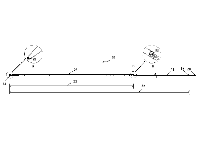
illustrate aspects of this disclosure, an exemplary embodiment of a
radial access dilator is shown schematically in FIG. 1 in an elevational view.
As shown,
first radial access dilator 10 is an elongated member having a tapered distal
end 12, with
an outer diameter that increases to a maximal outer diameter in distal portion
14. The
maximal outer diameter may be selected to closely conform to the inner
diameter of the
guide catheter to be used. The diameter may be constant throughout distal
portion 14
until first radial access dilator 10 transitions to a reduced profile at
guidewire exit port
16. Proximal portion 18 of radial access dilator 10 extends from guidewire
exit port 16
to proximal end 20. Guidewire lumen 22 extends from distal end 12, as shown in
the
perspective view of inset detail A, to guidewire exit port 16, as shown in the
perspective
view of inset detail B. Guidewire lumen 22 may exhibit a constant inner
diameter along
its length and may be sized to closely conform to the outer diameter of a
first guidewire
to be used with first radial access dilator 10. Inset detail B also shows the
transition of
first radial access dilator 10 from its maximal outer diameter to the reduced
profile. In
this embodiment, distal portion 14 has a cylindrical configuration
corresponding to the
maximal outer diameter while proximal portion 18 has a semi-cylindrical
configuration.
However, other shapes and configurations may be employed as desired. As will
be
appreciated, the reduced profile of proximal portion 18 has a smaller cross
sectional
area of material as compared to the maximal outer diameter of distal portion
14. In one
aspect, the smaller cross sectional area may represent a reduction in the
range of
approximately 40-60%, such as approximately 50%. Since the reduced profile
proximal
portion 18 represents a significant proportion of the overall length of radial
access
8
CA 02962930 2017-03-28
WO 2016/053993 PCT/US2015/052866
dilator 10, it may create less friction with a guide catheter, facilitating
advancement of
the guide catheter over radial access dilator 10.
[0040] As will be described in further detail below, exchange of guide
catheters
may be accomplished through the use of a second radial access dilator 30,
which is
depicted in FIG. 2. The overall design of radial access dilator 30 is similar
to radial
access dilator 10, so many of the elements are described with the same
reference
numbers. For example, second radial access dilator 30 is also an elongated
member
having a tapered distal end 12, with a maximal outer diameter in distal
portion 14.
Notably, the maximal outer diameter may be the same for both radial access
dilator 10
and radial access dilator 30, so that guide catheter having the same inner
diameter may
readily be exchanged. Second radial access dilator 30 also transitions to a
reduced
profile at guidewire exit port 16, with proximal portion 18 extending to
proximal end
20. Significantly, guidewire lumen 32, which extends from distal end 12, as
shown in
the perspective view of inset detail A, to guidewire exit port 16, as shown in
the
perspective view of inset detail B, may exhibit a constant inner diameter
along its length
and may be sized to closely conform to the outer diameter of a second
guidewire, such
that the second guidewire has a greater diameter than the first guidewire.
Also similar
to radial access dilator 10, distal portion 14 of radial access dilator 30 has
a cylindrical
configuration corresponding to the maximal outer diameter while proximal
portion 18
has a semi-cylindrical configuration, but other configurations may be
employed, so that
the reduced profile of proximal portion 18 has a smaller cross sectional area
of material
as compared to the maximal outer diameter of distal portion 14. With radial
access
dilator 30, the smaller cross sectional area may also represent a reduction in
the range of
approximately 40-60%, such as approximately 50%.
[0041] Both radial access dilator 10 and radial access dilator 30 may be
formed
from any suitable polymeric material having the desired characteristics. In
some
embodiments, nylon (polyamide), urethane, polypropylene, as well as polyamide
co-
polymers such as, for example, polyether block amides (PEBAXO), or others may
be
employed. Further, the relative dimensions of radial access dilator 10 and
radial access
dilator 30 may be selected as desired. In one embodiment, both radial access
dilator 10
and radial access dilator 30 may have an overall length of approximately 130
cm, so as
to extend approximately 10-20 cm from the proximal end of the guide catheter
when
9
CA 02962930 2017-03-28
WO 2016/053993 PCT/US2015/052866
preloaded for introduction into the vessel. With this configuration, the
proximal ends of
both the guide catheter and the dilator may be manipulated during introduction
and
advancement. Accordingly, a suitable range for the length of either or both
radial
access dilator 10 and radial access dilator 30 is approximately 130 to 140 cm
in some
embodiments and may be other lengths as desired depending on the procedure
and/or
the artery being accessed. The maximal outer diameter of radial access dilator
10 and
radial access dilator 30 may correspond to the inner diameter of the guide
catheter(s)
being used in the procedure. For example, for a 6 French guide catheter, the
maximal
outer diameter may be approximately 1.80 mm, with corresponding adjustment for
other
sizes. The inner diameter of lumens 22 and 32 may also be selected based on
the
diameters of the first and second guidewires being used during the exchange
procedure.
In one embodiment, lumen 22 of radial access dilator 10 may have a diameter
corresponding to a 0.021" (0.58 mm) guidewire and lumen 32 of radial access
dilator 30
may have a diameter corresponding to a 0.035" (0.88 mm) guidewire, although
different
guidewires having other diameters may be employed as desired.
[0042] The distance to guidewire exit port 16 may be tailored to the
desired
application and, in some embodiments, may be the same for both radial access
dilator
10 and radial access dilator 30. For example, the distance from distal end 12
to
guidewire exit port for radial access dilator 10, as indicated by D1 in FIG.
1, may be
approximately 30 cm and the distance from distal end 12 to guidewire exit port
for
radial access dilator 30, as indicated by D1 in FIG. 2, may also be
approximately 30 cm.
Depending on the procedure and/or the vessel being accessed, these distances
may be
adjusted as desired. An exemplary range for either or both radial access
dilator 10 and
radial access dilator 30 for D1 is 15 cm to 35 cm. It will be appreciated that
either or
both radial access dilator 10 and radial access dilator 30 may be advanced
only a
relatively short distance into the patient's vasculature relative to the
location where the
procedure is to be performed, which in some embodiments may be in the range of
20 to
40 cm. For example, guidewire exit port 16 may remain outside the patient's
body. As
such, guidewire exit port 16 may be located relatively closer to distal end 12
than
proximal end 20. A relatively smooth, atraumatic transition between the
maximal outer
diameter portion 14 and the outer diameter of the guide catheter is formed due
to the
close conformance of the outer diameter of the dilator and the inner diameter
of the
guide catheter, facilitating the advancement of the guide catheter over the
dilator. Once
CA 02962930 2017-03-28
WO 2016/053993 PCT/US2015/052866
the guide catheter has been suitably advanced, such as so that its distal end
is adjacent
the junction between tapered distal end 12 and maximal outer diameter portion
14, the
dilator may be removed. In one aspect, this may correspond to proximity
between
marker 34 or any other suitable indicator and any suitable reference point
relative to the
proximal end of the guide catheter. Tapered distal end 12 may be about four cm
in
length to provide smooth transition or dilation of the skin, subcutaneous
tissue and
artery wall. If desired, some or all of both radial access dilator 10 and
radial access
dilator 30 may have a hydrophilic coating to facilitate introduction and
advancement
through the patient's vasculature as well as to reduce friction when a guide
catheter is
advanced over the dilator. In one aspect, tapered distal end 12 and proximal
portion 14
of either or both radial access dilator 10 and radial access dilator 30 may
have a
hydrophilic coating.
[0043] Further, either of both radial access dilator 10 and radial access
dilator 30
may feature one or markers 34 at a suitable distance from distal end 12 as
indicated by
D2 in FIG. 1 for radial access dilator 10 and as indicated by D2 in FIG. 2 for
radial
access dilator 30. Markers 34 may be positioned at distances D2 of either or
both of 93
cm and 103 cm, or at other suitable locations as desired.
[0044] One suitable technique for employing one or both radial access
dilator 10
and radial access dilator 30 for transradial catheterization, including
exchange of guide
catheters if desired is schematically represented in FIGs. 3-13. In the
following
materials, the technique is described in the context of specific guide
catheter and
guidewire sizes while providing transradial access, but one of ordinary skill
in the art
will recognize that it may be extended to cover use of other sizes of guide
catheters and
guidewires and may be used for access to other vessel in a patient.
[0045] Beginning with FIG. 3, transradial access is achieved by palpation
or
ultrasound guidance per the operator's choice. The radial artery (RA) is
punctured
using a 21-gauge needle 40. An anterior or posterior puncture can be performed
by
either using a bare needle or intra-cath venous access needle, respectively.
In either
case, once the pulsatile blood flow is seen, a 0.021 inch guidewire 42 is
inserted in the
radial artery as indicated by FIG. 4. Guidewire 42 may be approximately 40 cm
in
some embodiments, although different lengths and diameters may be used as
desired..
Needle 40 is removed while securing guidewire 42 in the radial artery lumen
and
11
CA 02962930 2017-03-28
WO 2016/053993 PCT/US2015/052866
hemostasis is achieved as shown in FIG. 5. As noted radial access dilator 10
has an
outer diameter similar to that of the inner diameter of the guide catheter
intended to use
(e.g., for a 6 French guide, the maximal outer diameter of radial access
dilator 10 is 1.80
mm), and is about 130 cm long.
[0046] The guide catheter 44 of operator's choice will be pre-loaded on
radial
access dilator 10, which may then be advanced over guidewire 42 in to the
radial artery
as shown in FIG. 6. After advancing the first 30 cm of radial access dilator
10,
guidewire 42 will exit from guidewire exit port 16. Guidewire 42 may then be
removed
as shown in FIG. 7 and guide catheter 44 may then be advanced into the radial
artery
.. over radial access dilator 10 as shown in FIG. 8. A suitable distal marker,
such as
marker 34, may help indicate the relative position of radial access dilator 10
within the
vasculature. A tapered, hydrophilic guide catheter 44 may be used to
facilitate entry
into the radial artery. As will be appreciated, due to the similarity in size
between the
inner diameter of guide catheter 44 and the maximal outer diameter of radial
access
.. dilator 10, a smooth transition is achieved. After advancing the guide
catheter for
approximately 25-30 cm, radial access dilator 10 may be removed as shown in
FIG. 9.
Next, a 0.035 inch guidewire 46 may be inserted through guide catheter 44,
similar to
current standard transradial catheterization, and advanced to an area
corresponding to
the procedure being performed. Guidewire 46 may have a J configuration as
shown and
.. may have a length of approximately 260 cm, although different lengths and
diameters
may be used as desired. Guide catheter 44 is advanced over guidewire 46 in to
the
ascending aorta, descending aorta or other location, again depending on the
procedure
being performed and used to perform intended procedure.
[0047] lf the operator needs to use a different shape or larger size
guide catheter,
guidewire 46 may be maintained in its advanced location, or if it has been
removed, it
may be advanced again through guide catheter 44. Guide catheter 44 may then be
removed as shown in FIG. 11 and hemostasis is achieved by applying gentle
pressure on
the wrist. Radial access dilator 30 is preloaded into new guide catheter 48
and threaded
over guidewire 46 in place. As noted, radial access dilator 30 has lumen 32
that
.. corresponds to guidewire 46 and may have the same maximal outer diameter as
radial
access dilator 10 if guide catheter 48 is of the same size, or may have a
correspondingly
greater maximal outer diameter to closely conform to the inner diameter of
guide
12
CA 02962930 2017-03-28
WO 2016/053993 PCT/US2015/052866
catheter 48 if larger. Radial access dilator 30 and guide catheter 48 may then
be
advanced into the radial artery and to the treatment area over guide wire 46
as shown in
FIG. 12. Radial access dilator 30 may then be removed as shown in FIG. 13, to
allow
guide catheter 48 to be used to continue the procedure. After completing the
procedure
the guide catheter is removed and a radial hemostatic band can be applied
similar to
current practice of patent hemostasis.
[0048] As will be appreciated from the above description, access to the
radial artery
using either or both radial access dilator 10 and radial access dilator 30 may
be
accomplished with at least a 0.5 mm smaller hole and a smaller intrusion into
the radial
artery as compared to conventional entry with a sheath. For example, a 6
French sheath
will lead to 2.61 mm puncture in radial artery while using radial access
dilator 10, the
radial artery puncture and the maximal diameter of a device to be inserted in
the artery
may be reduced to approximately 2.00 mm. In this manner, most or all patients
will be
able to tolerate the use of a 6 French guide catheter, with considerably less
trauma.
Further, a 7 French guide catheter (outer diameter 2.3 mm) may be used with a
greater
proportion of patients, so that more complex coronary procedures and
peripheral
procedures may be performed. In other procedures, a 5 French guide catheter
may be
employed. Regardless of the size of the guide catheter, the requirement of a
smaller
hole and avoidance/reduction of expansion and/or irritation of the radial
artery as
compared to access with a sheath will reduce or eliminate, spasm, pain,
inflammation
and occlusion, and allow a successful transradial catheterization. As noted,
the
techniques of the this disclosure allow the operator to use any guide catheter
of any
shape or size, by any manufacturer, and allows the operator to exchange guide
catheters
as many times as required without additional cost (equipment). Since the
techniques
provide a true sheathless access and do not require any radial sheath,
substantial cost
savings may be realized in addition to the reduction in invasiveness of the
procedure.
[0049] The preceding description has been presented with reference to
presently
disclosed embodiments of the invention. Workers skilled in the art and
technology to
which this invention pertains will appreciate that alterations and changes in
the
described structure may be practiced without meaningfully departing from the
principal,
spirit and scope of this invention. As understood by one of ordinary skill in
the art, the
drawings are not necessarily to scale. Accordingly, the foregoing description
should not
13
CA 02962930 2017-03-28
WO 2016/053993 PCT/US2015/052866
be read as pertaining only to the precise structures described and illustrated
in the
accompanying drawings, but rather should be read consistent with and as
support to the
following claims which are to have their fullest and fair scope.
14