Note: Descriptions are shown in the official language in which they were submitted.
DEMANDE OU BREVET VOLUMINEUX
LA PRESENTE PARTIE DE CETTE DEMANDE OU CE BREVET COMPREND
PLUS D'UN TOME.
CECI EST LE TOME 1 DE 2
CONTENANT LES PAGES 1 A 163
NOTE : Pour les tomes additionels, veuillez contacter le Bureau canadien des
brevets
JUMBO APPLICATIONS/PATENTS
THIS SECTION OF THE APPLICATION/PATENT CONTAINS MORE THAN ONE
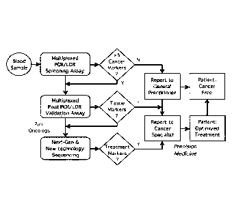
VOLUME
THIS IS VOLUME 1 OF 2
CONTAINING PAGES 1 TO 163
NOTE: For additional volumes, please contact the Canadian Patent Office
NOM DU FICHIER / FILE NAME:
NOTE POUR LE TOME / VOLUME NOTE:
- 1 -
METHOD FOR IDENTIFICATION AND RELATIVE QUANTIFICATION OF
NUCLEIC ACID SEQUENCE EXPRESSION, SPLICE VARIANT, TRANSLOCATION,
COPY NUMBER, OR METHYLATION CHANGES USING COMBINED NUCLEASE,
LIGATION, AND POLYMERASE REACTIONS WITH CARRYOVER PREVENTION
[0001]
FIELD OF THE INVENTION
[0002] The present invention relates to methods for identifying and
quantifying nucleic
acid sequence, expression, splice variant, translocation, copy number, and/or
methylation
changes using combined nuclease, ligation, and polymerase reactions with
carryover prevention.
BACKGROUND OF THE INVENTION
[0003] Blood carries oxygen, nutrients, and physiological signals to
every cell in the
body, while simultaneously providing immunity and protection against outside
pathogens. Yet
the same ability of blood to spread sustenance also allows for dissemination
of disease, be it
cancer cells metastasizing to the liver, Ebola virus ravaging the capillaries,
Streptococcus
pyo genes liquefying flesh, or HIV eluding detection within the very CD4 cells
that aim to
eliminate infections.
100041 The universal propensity of pathogens and cancers alike to
spread via the blood
also creates an opportunity for identification and early detection ¨ allowing
physicians to better
treat and manage patient care. The evolution of AIDS treatments went hand-in-
hand with
improvements in nucleic acid diagnostics, from initial reverse-transcription
PCR assays to
protect the nations' blood supply, to sequencing drug-resistant variants, to
RT-PCR
quantification of viral load to determine treatment efficacy over time. To
date, those infected
have not been cured, but sophisticated diagnostic tools have guided treatment,
epidemiological,
and political decisions to stem this global epidemic.
[0005] Cancer is the leading cause of death in developed countries and the
second
leading cause of death in developing countries. Cancer has now become the
biggest cause of
mortality worldwide, with an estimated 8.2 million deaths from cancer in 2012.
Cancer cases
worldwide are forecast to rise by 75% and reach close to 25 million over the
next two decades. A
Date Recue/Date Received 2022-01-17
CA 02963687 2017-04-04
WO 2016/057832 PCT/US2015/054759
- 2 -
recent report by the world health organization concludes: "(The) Global battle
against cancer
won't be won with treatment alone. Effective prevention measures (are)
urgently needed to
prevent (a) cancer crisis". Detection of early cancer in the blood is the best
means of effective
prevention. It will save lives by enabling earlier and better treatment, as
well as reduce the cost
of cancer care.
[0006] Plasma or serum from a cancer patient contains nucleic acids
released from
cancers cells undergoing abnormal physiological processes. These nucleic acids
have already
demonstrated diagnostic utility (Diaz and Bardelli, J Clin Oncol 32: 579-
586(2014);
Bettegowda etal., Sci Trans! Med 6: 224 (2014); Newman etal., Nat Med 20: 548-
554 (2014);
.. Thierry et al., Nat Med 20: 430-435 (2014)). A further source of nucleic
acids is within
circulating tumor cells (CTCs), although early stage and a significant
fraction of localized tumors
send out very few to no CTC's per ml. Normal plasma or serum contains nucleic
acids released
from normal cells undergoing normal physiological processes (i.e. exosome
secretion,
apoptosis). There may be additional release of nucleic acids under conditions
of stress,
.. inflammation, infection, or injury.
100071 The challenge to develop reliable diagnostic and screening
tests is to distinguish
those markers emanating from the tumor that are indicative of disease (e.g.,
early cancer) vs.
presence of the same markers emanating from normal tissue (which would lead to
a false-
positive signal). There is also a need to balance the number of markers
examined and the cost of
the test, with the specificity and sensitivity of the assay. Comprehensive
molecular profiling
(mRNA, methylation, copy number, miRNA, mutations) of thousands of tumors by
The Cancer
Genome Atlas Consortium (TCGA), has revealed that colorectal tumors are as
different from
each other as they are from breast, prostrate, or other epithelial cancers
(TCGA "Comprehensive
Molecular Characterization of Human Colon and Rectal Cancer Nature 487: 330-
337 (2014)).
.. Further, those few markers they share in common (e.g., KRAS mutations,) are
also present in
multiple cancer types, hindering the ability to pinpoint the tissue of origin.
For early cancer
detection, the nucleic acid assay should serve primarily as a screening tool,
requiring the
availability of secondary diagnostic follow-up (e.g., colonoscopy for
colorectal cancer).
[0008] Compounding the biological problem is the need to reliably
quantify mutation,
.. promoter methylation, or DNA or RNA copy number from either a very small
number of initial
cells (i.e. from CTCs), or when the cancer signal is from cell-free DNA
(cfDNA) in the blood
and diluted by an excess of nucleic acid arising from normal cells, or
inadvertently released from
normal blood cells during sample processing (Mateo et al., Genome Biol 15: 448
(2014)).
CA 02963687 2017-04-04
WO 2016/057832 PCTMS2015/054759
- 3 -
[0009] Likewise, an analogous problem of identifying rare target is
encountered when
using nucleic-acid-based techniques to detect infectious diseases directly in
the blood. Briefly,
either the pathogen may be present at 1 or less colony forming units (cfu)/ml,
and/or there are
many potential pathogens and sequence variations responsible for virulence or
drug resistance.
While these issues are exemplified with cancer, it is recognized that the
solutions are equally
applicable to infectious diseases.
A continuum of diagnostic needs require a continuum of diagnostic tests.
[0010] The majority of current molecular diagnostics efforts in cancer have
centered on:
(i) prognostic and predictive genomics, e.g., identifying inherited mutations
in cancer
predisposition genes, such as BrCA1, BrCA2, (Ford etal. Am J Hum Genet 62: 676-
689 (1998) )
(ii) individualized treatment, e.g., mutations in the EGFR gene guiding
personalized medicine
(Sequist and Lynch, Ann Rev Med, 59: 429-442 (2008), and (iii) recurrence
monitoring, e.g.,
detecting emerging KRAS mutations in patients developing resistance to drug
treatments (Hiley
et al., Genome Biol 15: 453 (2014); Amado et aL, J Clin Oncol 26: 1626-
1634(2008)). Yet,
this misses major opportunities in the cancer molecular diagnostics continuum:
(i) more frequent
screening of those with a family history, (ii) screening for detection of
early disease, and (iii)
monitoring treatment efficacy. To address these three unmet needs, a new
metric for blood-
based detection termed "cancer marker load", analogous to viral load is herein
proposed.
[0011] DNA sequencing provides the ultimate ability to distinguish all
nucleic acid
changes associated with disease. However, the process still requires multiple
up-front sample
and template preparation, and is not always cost-effective. DNA microarrays
can provide
substantial information about multiple sequence variants, such as SNPs or
different RNA
expression levels, and are less costly then sequencing; however, they are less
suited for obtaining
highly quantitative results, nor for detecting low abundance mutations. On the
other end of the
spectrum is the TaqManTm reaction, which provides real-time quantification of
a known gene,
but is less suitable for distinguishing multiple sequence variants or low
abundance mutations.
[0012] It is critical to match each unmet diagnostic need with the
appropriate diagnostic
test ¨ one that combines the divergent goals of achieving both high
sensitivity (i.e., low false-
negatives) and high specificity (i.e., low false-positives) at a low cost. For
example, direct
sequencing of EGFR exons from a tumor biopsy to determine treatment for non-
small cell lung
cancer (NSCLC) is significantly more accurate and cost effective than
designing TaqManTm
probes for the over 180 known mutations whose drug response is already
catalogued (Jia etal.
CA 02963687 2017-04-04
WO 2016/057832 PCT/US2015/054759
- 4 -
Genome Res 23: 1434-1445 (2013)). The most sensitive technique for detecting
point mutations,
BEAMing (Dressman et al., Proc Natl Acad Sci USA 100: 8817-8822 (2003)), rely
on prior
knowledge of which mutations to look for, and thus are best suited for
monitoring for disease
recurrence, rather than for early detection. Likewise, to monitor blood levels
of Bcr-Abl
translocations when treating CML patients with Gleevec (Jabbour et al., Cancer
112: 2112-2118
(2008)), a simple quantitative reverse-transcription PCR assay is far
preferable to sequencing the
entire genomic DNA in 1 ml of blood (9 million cells x 3 GB = 27 million Gb of
raw data).
[0013] Sequencing 2.1 Gb each of cell-free DNA (cfDNA) isolated from
NSCLC
patients was used to provide 10,000-fold coverage on 125 kb of targeted DNA
(Kandoth etal.
Nature 502: 333-339(2013)). This approach correctly identified mutations
present in matched
tumors, although only 50% of stage 1 tumors were covered. The approach has
promise for
NSCLC, where samples average 5 to 20 mutations / Mb, however would not be cost
effective for
other cancers such as breast and ovarian, that average less than 1 to 2
mutations per Mb. Current
up-front ligation, amplification, and/or capture steps required for highly
accurate targeted deep
sequencing are still more complex than multiplexed PCR-TaqManTm or PCR-LDR
assays.
[0014] A comprehensive data analysis of over 600 colorectal cancer
samples that takes
into account tumor heterogeneity, tumor clusters, and biological /technical
false-positives
ranging from 3% to 1 0 % per individual marker showed that the optimal early
detection screen
for colorectal cancer would require at least 5 to 6 positive markers out of 24
markers tested
(Bacolod et al,. Cancer Res 69:723-727 (2009); Tsafrir etal. Cancer Res 66:
2129-2137 (2006),
Weinstein et al., Nat Genet 45: 1113-1120(2013); Navin N.E. Genome Biol 15:
452 (2014);
Hiley et al., Genome Biol 15: 453 (2014)); Esserman et al. Lancet Oncol 15:
e234-242 (2014)).
Further, marker distribution is biased into different tumor clades, e.g., some
tumors are heavily
methylated, while others are barely methylated, and indistinguishable from age-
related
methylation of adjacent tissue. Consequently, a multidimensional approach
using combinations
of 3-5 sets of mutation, methylation, miRNA, mRNA, copy-variation, alternative
splicing, or
translocation markers is needed to obtain sufficient coverage of all different
tumor clades.
Analogous to non-invasive prenatal screening for trisomy, based on sequencing
or performing
ligation detection on random fragments of cfDNA (Benn et al., Ultrasound
Obstet GynecoL
42(1):15-33 (2013); Chiu et al., Proc Natl Acad Sci USA 105: 20458 ¨ 20463
(2008); Juneau
et al., Fetal Diagn Ther. 36(4) (2014)), the actual markers scored in a cancer
screen are
secondary to accurate quantification of those positive markers in the plasma.
CA 02963687 2017-04-04
WO 2016/057832 PCT/US2015/054759
- 5 -
Technical Challenges of Cancer Diagnostic Test Development.
[0015] Diagnostic tests that aim to fmd very rare or low-abundance
mutant sequences
face potential false-positive signal arising from: (i) polymerase error in
replicating wild-type
target, (ii) DNA sequencing error, (iii) mis-ligation on wild-type target,
(iii) target independent
PCR product, and (iv) carryover contamination of PCR products arising from a
previous positive
sample. The profound clinical implications of a positive test result when
screening for cancer
demand that such a test use all means possible to virtually eliminate false-
positives.
[0016] Central to the concept of nucleic acid detection is the
selective amplification or
purification of the desired cancer-specific markers away from the same or
closely similar
markers from normal cells. These approaches include: (i) multiple primer
binding regions for
orthogonal amplification and detection, (ii) affinity selection of CTC's or
exosomes, and (iii)
spatial dilution of the sample.
[0017] The success of PCR-LDR, which uses 4 primer-binding regions to
assure
sensitivity and specificity, has previously been demonstrated. Desired regions
are amplified
using pairs or even tandem pairs of PCR primers, followed by orthogonal nested
LDR primer
pairs for detection. One advantage of using PCR-LDR is the ability to perform
proportional PCR
amplification of multiple fragments to enrich for low copy targets, and then
use quantitative
LDR to directly identify cancer-specific mutations. Biofire/bioMerieux has
developed a similar
technology termed "film array"; wherein initial multiplexed PCR reaction
products are
redistributed into individual wells, and then nested real-time PCR performed
with SYBR Green
Dye detection.
[0018] Affinity purification of CTC's using antibody or aptamer
capture has been
demonstrated (Adams et al., J Am Chem Soc 130: 8633-8641 (2008); Dharmasiri et
al.,
Electrophoresis 30: 3289-3300 (2009); Soper etal. Biosens Bioelectron 21: 1932-
1942 (2006)).
Peptide affinity capture of exosomes has been reported in the literature.
Enrichment of these
tumor-specific fractions from the blood enables copy number quantification, as
well as
simplifying screening and verification assays.
[0019] The last approach, spatial dilution of the sample, is employed
in digital PCR as
well as its close cousin known as BEAMing (Vogelstein and Kinzler, Proc Natl
Acad Sc! USA.
96(16):9236-41 (1999); Dressman et al., Proc Nat! Acad Sci USA 100: 8817-8822
(2003)). The
rational for digital PCR is to overcome the limit of enzymatic discrimination
when the sample
comprises very few target molecules containing a known mutation in a 1,000 to
10,000-fold
excess of wild-type DNA. By diluting input DNA into 20,000 or more droplets or
beads to
CA 02963687 2017-04-04
WO 2016/057832 PCMJS2015/054759
- 6 -
distribute less than one molecule of target per droplet, the DNA may be
amplified via PCR, and
then detected via probe hybridization or TaqManTm reaction, giving in essence
a 0/1 digital
score. The approach is currently the most sensitive for finding point
mutations in plasma, but it
does require prior knowledge of the mutations being scored, as well as a
separate digital dilution
for each mutation, which would deplete the entire sample to score just a few
mutations.
Real-time PCR & Mierofluidie instrumentation
[0020] A number of PCR assays / microfabricated devices have been
designed for rapid
detection of pathogens and disease-associated translocations and mutations.
Each
assay/hardware combination has particular strengths, but when combined with
the real world
problem of multidimensional and multiplexed markers required for cancer
detection, the
flexibility of PCR-LDR with microfluidics provides certain advantages.
[0021] Instrumentation, assay design, and microfluidic architecture
need to be seamlessly
integrated. Some PCR instrumentation use real-time fluorescence or end-point
fluorescence to
quantify initial template molecules by cycling chambers, wells, or droplets
through different
temperatures. Yet other instrumentation comprises addressable microfluidic
plates for real-time
PCR detection. However the high cost of both the instruments and consumables
has limited the
widespread use of these machines for clinical applications.
[00221 In a different architecture, termed continuous-flow PCR, the
reaction mix moves
through channels that are neatly arranged in a radiator pattern, and flow over
heating elements
that are at fixed temperatures. This architecture allows the entire
amplification reaction to be
completed in a few minutes, and is ideal for capillary separation and readout.
For ligase
detection reactions, the readout may be achieved by taking advantage of LDR-
FRET or
electronic detection. In LDR-FRET, one primer has a donor, the other has an
acceptor group,
and after ligation they form a hairpin. This allows for counting single
ligation events to obtain
highly quantitative readouts of input DNA copy number. Alternatively, by
appending gold-
nanoparticles on each primer, the ligation product will contain two nano-
particles, and these may
be distinguished using electronic readout.
[00231 In considering various degrees of automation, the approach
described herein is
guided by the principles of "modularity" and "scalability". Firstly, the
process should be
separated into modular steps that may initially be optimized on separate
instruments. For
example, the device may be comprised of a first module for purification of DNA
from plasma
cfDNA as well as RNA from exosomes, a second module for multiplexed reverse
transcription
CA 02963687 2017-04-04
WO 2016/057832 PCT/US2015/054759
- 7 -
and/or limited amplification of various targets, and a third module for
generating and detecting
ligation products. Such a modular architecture allows for swapping in improved
modules that
keep pace with technological developments. For the modularity approach to
work, it is critical
that products from one module can be moved seamlessly into the next module,
without leakage
and without worry of crossover contamination.
[0024] Secondly, the modular design should be amenable to scalable
manufacture in high
volumes at low cost. The manufacturing costs and how primers / reagents /
samples are
deposited into the device must be taken into consideration.
[0025] The present invention is directed at overcoming these and other
deficiencies in the
art.
SUMMARY OF THE INVENTION
100261 A first aspect of the present invention is directed to a method
for identifying, in a
sample, one or more nucleic acid molecules containing a target nucleotide
sequence differing
from nucleotide sequences in other nucleic acid molecules in the sample, or
other samples, by
one or more nucleotides, one or more copy numbers, one or more transcript
sequences, and/or
one or more methylated residues. This method involves providing a sample
potentially
containing one or more nucleic acid molecules containing the target nucleotide
sequence
differing from the nucleotide sequences in other nucleic acid molecules by one
or more
nucleotides, one or more copy numbers, one or more transcript sequences,
and/or one or more
methylated residues, and contacting the sample with one or more enzymes
capable of digesting
deoxyuracil (dU) containing nucleic acid molecules present in the sample. One
or more primary
oligonucleotide primer sets are provided, each primary oligonucleotide primer
set comprising (a)
a first primary oligonucleotide primer that comprises a nucleotide sequence
that is
complementary to a sequence adjacent to the target nucleotide sequence, and
(b) a second
primary oligonucleotide primer that comprises a nucleotide sequence that is
complementary to a
portion of an extension product formed from the first primary oligonucleotide
primer. The
contacted sample is blended with the one or more primary oligonucleotide
primer sets, a
deoxynucleotide mix including dUTP, and a DNA polymerase to form a polymerase
chain
reaction mixture, and the polymerase chain reaction mixture is subjected to
one or more
polymerase chain reaction cycles comprising a denaturation treatment, a
hybridization treatment,
and an extension treatment, thereby forming primary extension products
comprising the target
CA 02963687 2017-04-04
WO 2016/057832 PCTMS2015/054759
- 8 -
nucleotide sequence or a complement thereof. The method further involves
blending the primary
extension products with a ligase and one or more oligonucleotide probe sets to
form a ligation
reaction mixture. Each oligonucleotide probe set comprises (a) a first
oligonucleotide probe
having a target nucleotide sequence-specific portion, and (b) a second
oligonucleotide probe
having a target nucleotide sequence-specific portion, and wherein the first
and second
oligonucleotide probes of a probe set are configured to hybridize, in a base
specific manner, on a
complementary target nucleotide sequence of a primary extension product. The
first and second
oligonucleotide probes of the one or more oligonucleotide probe sets are
ligated together to form
ligated product sequences in the ligation reaction mixture, and the ligated
product sequences in
the sample are detected and distinguished to identify the presence of one or
more nucleic acid
molecules containing target nucleotide sequences differing from nucleotide
sequences in other
nucleic acid molecules in the sample by one or more nucleotides, one or more
copy numbers, one
or more transcript sequences, and/or one or more methylated residues.
[0027] Another aspect of the present invention is directed to a method
for identifying, in
a sample, one or more nucleic acid molecules containing a target nucleotide
sequence differing
from nucleotide sequences in other nucleic acid molecules in the sample, or
other samples, by
one or more nucleotides, one or more copy numbers, one or more transcript
sequences, and/or
one or more methylated residues. This method involves providing a sample
containing one or
more nucleic acid molecules potentially containing the target nucleotide
sequence differing from
the nucleotide sequences in other nucleic acid molecules by one or more
nucleotides, one or
more copy numbers, one or more transcript sequences, and/or one or more
methylated residues.
The method further involves providing one or more enzymes capable of digesting
deoxyuracil
(dU) containing nucleic acid molecules present in the sample, and providing
one or more
primary oligonucleotide primer sets, each primary oligonucleotide primer set
comprising (a) a
first primary oligonucleotide primer that comprises a nucleotide sequence that
is complementary
to a sequence adjacent to the target nucleotide sequence and (b) a second
primary
oligonucleotide primer that comprises a nucleotide sequence that is
complementary to a portion
of an extension product formed from the first primary oligonucleotide primer.
The sample is
blended with the one or more primary oligonucleotide primer sets, the one or
more enzymes
capable of digesting deoxyuracil (dU) containing nucleic acid molecules in the
sample, a
deoxynucleotide mix including dUTP, and a DNA polymerase to form a polymerase
chain
reaction mixture. The polymerase chain reaction mixture is subjected to
conditions suitable for
digesting deoxyuracil (dU) containing nucleic acid molecules present in the
polymerase chain
CA 02963687 2017-04-04
WO 2016/057832 PCT11JS2015/054759
- 9 -
reaction mixture, and for one or more polymerase chain reaction cycles
comprising a
denaturation treatment, a hybridization treatment, and an extension treatment,
thereby forming
primary extension products comprising the target nucleotide sequence or a
complement thereof.
The method further involves blending the primary extension products with a
ligase and one or
more oligonucleotide probe sets to form a ligation reaction mixture, wherein
each
oligonucleotide probe set comprises (a) a first oligonucleotide probe having a
5' primer-specific
portion and a 3' target nucleotide sequence-specific portion, and (b) a second
oligonucleotide
probe having a 5' target nucleotide sequence-specific portion and a 3' primer-
specific portion.
The first and second oligonucleotide probes of a probe set are configured to
hybridize, in a base
specific manner, on a complementary target nucleotide sequence of a primary
extension product.
The ligation reaction mixture is subjected to one or more ligation reaction
cycles whereby the
first and second oligonucleotide probes of the one or more oligonucleotide
probe sets are ligated
together to form ligated product sequences in the ligation reaction mixture
where each ligated
product sequence comprises the 5' primer-specific portion, the target-specific
portions, and the
__ 3' primer-specific portion. The method further involves providing one or
more secondary
oligonucleotide primer sets, each secondary oligonucleotide primer set
comprising (a) a first
secondary oligonucleotide primer comprising the same nucleotide sequence as
the 5' primer-
specific portion of the ligated product sequence and (b) a second secondary
oligonucleotide
primer comprising a nucleotide sequence that is complementary to the 3' primer-
specific portion
of the ligated product sequence, and blending the ligated product sequences,
the one or more
secondary oligonucleotide primer sets, the one or more enzymes capable of
digesting
deoxyuracil (dU) containing nucleic acid molecules, a deoxynucleotide mix
including dUTP, and
a DNA polymerase to form a second polymerase chain reaction mixture. The
second polymerase
chain reaction mixture is subjected to conditions suitable for digesting
deoxyuracil (dU)
containing nucleic acid molecules present in the second polymerase chain
reaction mixture, and
one or more polymerase chain reaction cycles comprising a denaturation
treatment, a
hybridization treatment, and an extension treatment thereby forming secondary
extension
products. The secondary extension products are detected and distinguished in
the sample to
identify the presence of one or more nucleic acid molecules containing target
nucleotide
sequences differing from nucleotide sequences in other nucleic acid molecules
in the sample by
one or more nucleotides, one or more copy numbers, one or more transcript
sequences, and/or
one or more methylated residues.
CA 02963687 2017-04-04
WO 2016/057832 PCT/1JS2015/054759
- 10 -
[0028] Another aspect of the present invention is directed to a method
for identifying, in
a sample, one or more nucleic acid molecules containing a target nucleotide
sequence differing
from nucleotide sequences in other nucleic acid molecules in the sample, or
other samples, by
one or more nucleotides, one or more copy numbers, one or more transcript
sequences, and/or
one or more methylated residues. This method involves providing a sample
containing one or
more nucleic acid molecules potentially containing the target nucleotide
sequence differing from
the nucleotide sequences in other nucleic acid molecules by one or more
nucleotides, one or
more copy numbers, one or more transcript sequences, and/or one or more
methylated residues;
providing one or more enzymes capable of digesting deoxyuracil (dU) containing
nucleic acid
molecules present in the sample; and providing one or more primary
oligonucleotide primer sets,
each primary oligonucleotide primer set comprising (a) a first primary
oligonucleotide primer
that comprises a nucleotide sequence that is complementary to a sequence
adjacent to the target
nucleotide sequence and (b) a second primary oligonucleotide primer that
comprises a nucleotide
sequence that is complementary to a portion of an extension product formed
from the first
primary oligonucleotide primer. The method further involves blending the
sample, the one or
more primary oligonucleotide primer sets, the one or more enzymes capable of
digesting
deoxyuracil (dU) containing nucleic acid molecules in the sample, a
deoxynucleotide mix
including dUTP, and a DNA polymerase to form a polymerase chain reaction
mixture. The
polymerase chain reaction mixture is subjected to conditions suitable for
digesting deoxyuracil
(dU) containing nucleic acid molecules present in the polymerase chain
reaction mixture, and for
one or more polymerase chain reaction cycles comprising a denaturation
treatment, a
hybridization treatment, and an extension treatment, thereby forming primary
extension products
comprising the target nucleotide sequence or a complement thereof. The primary
extension
products are blended with a ligase and one or more oligonucleotide probe sets
to form a ligation
reaction mixture, wherein each oligonucleotide probe set comprises (a) a first
oligonucleotide
probe having a 5' portion and a 3' target nucleotide sequence-specific
portion, and (b) a second
oligonucleotide probe having a 5' target nucleotide sequence-specific portion
and a 3' portion,
where the 5' portion of the first oligonucleotide probe of the probe set is
complementary to a
portion of the 3' portion of the second oligonucleotide probe, where one probe
of the probe set
comprises a detectable signal generating moiety, and where the first and
second oligonucleotide
probes of a probe set are configured to hybridize, in a base specific manner,
on a complementary
target nucleotide sequence of a primary extension product. The method further
involves
subjecting the ligation reaction mixture to one or more ligation reaction
cycles whereby the first
CA 02963687 2017-04-04
WO 2016/057832 PCT/US2015/054759
- 11 -
and second oligonucleotide probes of the one or more oligonucleotide probe
sets are ligated
together to form ligated product sequences in the ligation reaction mixture
where each ligated
product sequence comprises the 5' portion, the target-specific portions, the
3' portion, and the
detectable signal generating moiety. The 5' portion of the ligated product
sequence is hybridized
to its complementary 3' portion and signal from the detectable signal
generating moiety that is
produced upon said hybridizing is detected. The ligated product sequences are
distinguished in
the sample based on said detecting to identify the presence of one or more
nucleic acid
molecules containing target nucleotide sequences differing from nucleotide
sequences in other
nucleic acid molecules in the sample by one or more nucleotides, one or more
copy numbers, one
or more transcript sequences, and/or one or more methylated residues.
100291 Another aspect of the present invention is directed to a method
for identifying, in
a sample, one or more nucleic acid molecules containing a target nucleotide
sequence differing
from nucleotide sequences in other nucleic acid molecules in the sample, or
other samples, by
one or more methylated residue. This method involves providing a sample
potentially containing
one or more nucleic acid molecules comprising the target nucleotide sequence
differing from the
nucleotide sequences in other nucleic acid molecules by one or more methylated
residues and
contacting the sample with one or more enzymes capable of digesting
deoxyuracil (dU)
containing nucleic acid molecules present in the sample. The method further
involves contacting
the sample with one or more methylation sensitive enzymes to form a
restriction enzyme reaction
mixture, wherein the one or more methylation sensitive enzyme cleaves nucleic
acid molecules
in the sample that contain one or more =methylated residues within at least
one methylation
sensitive enzyme recognition sequence. One or more primary oligonucleotide
primer sets are
provided, each primary oligonucleotide primer set comprising (a) first primary
oligonucleotide
primer comprising a nucleotide sequence that is complementary to a region of
the target
nucleotide sequence that is upstream of the one or more methylated residues
and (b) a second
primary oligonucleotide primer comprising a nucleotide sequence that is the
same as a region of
the target nucleotide sequence that is downstream of the one or more
methylated residues. The
restriction enzyme reaction mixture is blended with the one or more primary
oligonucleotide
primer sets, a deoxynucleotide mix including dUTP, and a DNA polymerase to
form a primary
polymerase chain reaction mixture. The method further involves subjecting the
primary
polymerase chain reaction mixture to one or more polymerase chain reaction
cycles comprising a
denaturation treatment, a hybridization treatment, and an extension treatment,
thereby forming
primary extension products comprising the target nucleotide sequence or a
complement thereof.
CA 02963687 2017-04-04
WO 2016/057832 PCT/US2015/054759
- 12 -
One or more secondary oligonucleotide primer sets are provided, each secondary
oligonucleotide
primer set comprising first and second nested oligonucleotide primers capable
of hybridizing to
the primary extension products The primary extension products are blended with
the one or more
secondary oligonucleotide primer sets, a deoxynucleotide mix including dUTP,
and a DNA
polymerase to form a secondary polymerase chain reaction mixture, and the
secondary
polymerase chain reaction mixture is subjected to one or more polymerase chain
reaction cycles
comprising a denaturation treatment, a hybridization treatment, and an
extension treatment
thereby forming secondary extension products. The secondary extension products
in the sample
are detected and distinguished to identify the presence of one or more nucleic
acid molecules
containing target nucleotide sequences differing from nucleotide sequences in
other nucleic acid
molecules in the sample by one or more methylated residues.
[0030] Another aspect of the present invention is directed to a method
for identifying in a
sample, one or more target ribonucleic acid molecules differing in sequence
from other
ribonucleic acid molecules in the sample due to alternative splicing,
alternative transcript,
alternative start site, alternative coding sequence, alternative non-coding
sequence, exon
insertion, exon deletion, intron insertion, translocation, mutation, or other
rearrangement at the
genome level. This method involves providing a sample containing one or more
target
ribonucleic acid molecules potentially containing a sequence differing from
other ribonucleic
acid molecules, and contacting the sample with one or more enzymes capable of
digesting dU
containing nucleic acid molecules potentially present in the sample. One or
more
oligonucleotide primers are provided, each primer being complementary to the
one or more
target ribonucleic acid molecule. The contacted sample is blended with the one
or more
oligonucleotide primers, and a reverse-transcriptase to form a reverse-
transcription mixture, and
complementary deoxyribonucleic acid (cDNA) molecules are generated in the
reverse
.. transcription mixture. Each cDNA molecule comprises a nucleotide sequence
that is
complementary to the target ribonucleic acid molecule sequence and contains
dU. The method
further involves providing one or more oligonucleotide primer sets, each
primer set comprising
(a) a first oligonucleotide primer comprising a nucleotide sequence that is
complementary to a
portion of a cDNA nucleotide sequence adjacent to the target ribonucleic acid
molecule sequence
complement of the cDNA, and (b) a second oligonucleotide primer comprising a
nucleotide
sequence that is complementary to a portion of an extension product formed
from the first
oligonucleotide primer. The reverse transcription mixture containing the cDNA
molecules is
blended with the one or more oligonucleotide primer sets, and a polymerase to
form a
CA 02963687 2017-04-04
WO 2016/057832 PCT/US2015/054759
- 13 -
polymerase reaction mixture, and the polymerase chain reaction mixture is
subjected to one or
more polymerase chain reaction cycles comprising a denaturation treatment, a
hybridization
treatment, and an extension treatment thereby forming one or more different
primary extension
products. The method further involves providing one or more oligonucleotide
probe sets. Each
probe set comprises (a) a first oligonucleotide probe having a target sequence-
specific portion,
and (b) a second oligonucleotide probe having a target sequence-specific
portion, wherein the
first and second oligonucleotide probes of a probe set are configured to
hybridize, in a base
specific manner, on a complementary portion of a primary extension product
corresponding to
the target ribonucleic acid molecule sequence. The primary extension products
are contacted
with a ligase and the one or more oligonucleotide probe sets to form a
ligation reaction mixture
and the first and second probes of the one or more oligonucleotide probe sets
are ligated together
to form ligated product sequences in the ligase reaction mixture. The ligated
product sequences
in the sample are detected and distinguished thereby identifying the presence
of one or more
target ribonucleic acid molecules differing in sequence from other ribonucleic
acid molecules in
the sample due to alternative splicing, alternative transcript, alternative
start site, alternative
coding sequence, alternative non-coding sequence, exon insertion, exon
deletion, intron
insertion, translocation, mutations, or other rearrangement at the genome
level.
[00311 Another aspect of the present invention is directed to a method
for identifying in a
sample, one or more target ribonucleic acid molecules differing in sequence
from other
ribonucleic acid molecules in the sample due to alternative splicing,
alternative transcript,
alternative start site, alternative coding sequence, alternative non-coding
sequence, exon
insertion, exon deletion, intron insertion, translocation, mutation, or other
rearrangement at the
genome level. This method involves providing a sample containing one or more
target
ribonucleic acid molecules potentially differing in sequence from other
ribonucleic acid
molecules, and contacting the sample with one or more enzymes capable of
digesting dU
containing nucleic acid molecules potentially present in the sample. One or
more
oligonucleotide primers is provided, each primer being complementary to the
one or more target
ribonucleic acid molecules, and the contacted sample is blended with the one
or more
oligonucleotide primers, a deoxynucleotide mix including dUTP, and a reverse-
transcriptase to
form a reverse-transcription mixture. Complementary deoxyribonucleic acid
(cDNA) molecules
are generated in the reverse transcription mixture, each cDNA molecule
comprising a nucleotide
sequence that is complementary to the target ribonucleic acid molecule and
contains dU. The
method further involves providing one or more oligonucleotide primer sets,
each primer set
CA 02963687 2017-04-04
WO 2016/057832 PCT11JS2015/054759
- 14 -
comprising (a) a first oligonucleotide primer comprising a nucleotide sequence
that is
complementary to a portion of a cDNA nucleotide sequence adjacent to the
target ribonucleic
acid molecule sequence complement of the cDNA, and (b) a second
oligonucleotide primer
comprising a nucleotide sequence that is complementary to a portion of an
extension product
formed from the first oligonucleotide primer. The reverse transcription
mixture containing the
cDNA molecules is blended with the one or more oligonucleotide primer sets, a
deoxynucleotide
mix including dUTP, and a polymerase to form a polymerase reaction mixture,
and the
polymerase chain reaction mixture is subjected to one or more polymerase chain
reaction cycles
comprising a denaturation treatment, a hybridization treatment, and an
extension treatment
thereby forming one or more different primary extension products. The method
further involves
providing one or more oligonucleotide probe sets, each probe set comprising
(a) a first
oligonucleotide probe having a 5' primer-specific portion and a 3' target
sequence-specific
portion, and (b) a second oligonucleotide probe having a 5' target sequence-
specific portion and
a 3' primer-specific portion, where the first and second oligonucleotide
probes of a probe set are
configured to hybridize, in a base specific manner, on complementary portions
of a primary
extension product corresponding to the target ribonucleic acid molecule
sequence. The primary
extension products are contacted with a ligase and the one or more
oligonucleotide probe sets to
form a ligation reaction mixture, and the ligation reaction mixture is
subjected to one or more
ligation reaction cycles whereby the first and second probes of the one or
more oligonucleotide
probe sets are ligated together to form ligated product sequences in the
ligase reaction mixture,
where each ligated product sequence comprises the 5' primer-specific portion,
the target-specific
portions, and the 3' primer-specific portion. The method further involves
providing one or more
secondary oligonucleotide primer sets, each secondary oligonucleotide primer
set comprising (a)
a first secondary oligonucleotide primer comprising the same nucleotide
sequence as the 5'
primer-specific portion of the ligated product sequence and (b) a second
secondary
oligonucleotide primer comprising a nucleotide sequence that is complementary
to the 3' primer-
specific portion of the ligated product sequence, and blending the ligated
product sequences, the
one or more secondary oligonucleotide primer sets with one or more enzymes
capable of
digesting deoxyuracil (dU) containing nucleic acid molecules, a
deoxynucleotide mix including
dUTP, and a DNA polymerase to form a second polymerase chain reaction mixture.
The second
polymerase chain reaction mixture is subjected to conditions suitable for
digesting deoxyuracil
(dU) containing nucleic acid molecules present in the second polymerase chain
reaction mixture,
and one or more polymerase chain reaction cycles comprising a denaturation
treatment, a
CA 02963687 20170404
WO 2016/057832 PCT/US2015/054759
- 15 -
hybridization treatment, and an extension treatment thereby forming secondary
extension
products. The secondary extension products in the sample are detected and
distinguished thereby
identifying the presence of one or more ribonucleic acid molecules differing
in sequence from
other ribonucleic acid molecules in the sample due to alternative splicing,
alternative transcript,
alternative start site, alternative coding sequence, alternative non-coding
sequence, exon
insertion, exon deletion, intron insertion, translocation, mutation, or other
rearrangement at the
genome level.
[0032] Another aspect of the present invention is directed to a method
for identifying in a
sample, one or more target ribonucleic acid molecules differing in sequence
from other
ribonucleic acid molecules in the sample due to alternative splicing,
alternative transcript,
alternative start site, alternative coding sequence, alternative non-coding
sequence, exon
insertion, exon deletion, intron insertion, translocation, mutation, or other
rearrangement at the
genome level. This method involves providing a sample containing one or more
target
ribonucleic acid molecules potentially differing in sequence from other
ribonucleic acid
molecules, and contacting the sample with one or more enzymes capable of
digesting dU
containing nucleic acid molecules potentially present in the sample. The
method further involves
providing one or more oligonucleotide primers, each primer being complementary
to the one or
more target ribonucleic acid molecules, and blending the contacted sample, the
one or more
oligonucleotide primers, a deoxynucleotide mix including dUTP, and a reverse-
transcriptase to
form a reverse-transcription mixture. Complementary deoxyribonucleic acid
(cDNA) molecules
are generated in the reverse transcription mixture, each cDNA molecule
comprising a nucleotide
sequence that is complementary to the target ribonucleic acid molecule and
contains dU. The
method further involves providing one or more oligonucleotide primer sets,
each primer set
comprising (a) a first oligonucleotide primer comprising a nucleotide sequence
that is
complementary to a portion of a cDNA nucleotide sequence adjacent to the
target ribonucleic
acid molecule sequence complement of the cDNA, and (b) a second
oligonucleotide primer
comprising a nucleotide sequence that is complementary to a portion of an
extension product
formed from the first oligonucleotide primer. The reverse transcription
mixture containing the
cDNA molecules is blended with the one or more oligonucleotide primer sets, a
deoxynucleotide
mix including dUTP, and a polymerase to form a polymerase reaction mixture,
and the
polymerase chain reaction mixture is subjected to one or more polymerase chain
reaction cycles
comprising a denaturation treatment, a hybridization treatment, and an
extension treatment
thereby forming one or more different primary extension products. The method
further involves
CA 02963687 2017-04-04
WO 2016/057832 PCT11JS2015/054759
- 16 -
providing one or more oligonucleotide probe sets, each probe set comprising
(a) a first
oligonucleotide probe having a 5' portion and a 3' target nucleotide sequence-
specific portion,
and (b) a second oligonucleotide probe having a 5' target nucleotide sequence-
specific portion
and a 3' portion, where the 5' portion of the first oligonucleotide probe of
the probe set is
complementary to a portion of the 3' portion of the second oligonucleotide
probe, where one
probe of the probe set comprises a detectable signal generating moiety, and
where the first and
second oligonucleotide probes of a probe set are configured to hybridize, in a
base specific
manner, on complementary portions of a primary extension product corresponding
to the target
ribonucleic acid molecule sequence. The primary extension products are
contacted with a ligase
and the one or more oligonucleotide probe sets to fount a ligation reaction
mixture, and the
ligation reaction mixture is subjected to one or more ligation reaction cycles
whereby the first
and second probes of the one or more oligonucleotide probe sets are ligated
together to form
ligated product sequences in the ligase reaction mixture, where each ligated
product sequence
comprises the 5' portion, the target-specific portions, the 3' portion, and
the detectable signal
generating moiety. The 5' portion of the ligated product sequence is
hybridized to its
complementary 3' portion, and the signal from the detectable signal generating
moiety that is
produced upon said hybridizing is detected. The ligated product sequences in
the sample are
detected based on said detecting to identify the presence of one or more
ribonucleic acid
molecules differing in sequence from other ribonucleic acid molecules in the
sample due to
alternative splicing, alternative transcript, alternative start site,
alternative coding sequence,
alternative non-coding sequence, exon insertion, exon deletion, intron
insertion, translocation,
mutation, or other rearrangement at the genome level.
[0033] Another aspect of the present invention is directed to a method
for identifying, in
a sample, one or more target micro-ribonucleic acid (miRNA) molecules
differing in sequence
from other miRNA molecules in the sample by one or more bases. This method
involves
providing a sample containing one or more target miRNA molecules potentially
differing in
sequence from other miRNA molecules in the sample by one or more bases, and
contacting the
sample with one or more enzymes capable of digesting dU containing nucleic
acid molecules
potentially present in the sample. One or more oligonucleotide primer sets are
provided, each
primer set comprising (a) a first oligonucleotide primer having a 5' stem-loop
portion, a blocking
group, an internal primer-specific portion within the loop region, and a 3'
nucleotide sequence
portion that is complementary to a 3' portion of the target miRNA molecule
sequence, (b) a
second oligonucleotide primer having a 3' nucleotide sequence portion that is
complementary to
CA 02963687 2017-04-04
WO 2016/057832 PCT/US2015/054759
- 17 -
a complement of the 5' end of the target miRNA molecule sequence, and a 5'
primer-specific
portion, (c) a third oligonucleotide primer comprising a nucleotide sequence
that is the same as
the internal primer-specific portion of the first oligonucleotide primer, and
(d) a fourth
oligonucleotide primer comprising a nucleotide sequence that is the same as
the 5' primer-
specific portion of the second oligonucleotide primer. The contacted sample is
blended with the
one or more first oligonucleotide primers of a primer set, a deoxynucleotide
mix including
dUTP, and a reverse transcriptase to form a reverse transcription reaction
mixture. The first
oligonucleotide primer hybridizes to the target miRNA molecule sequence, if
present in the
sample, and the reverse transcriptase extends the 3' end of the hybridized
first oligonucleotide
primer to generate an extended first oligonucleotide primer comprising the
complement of the
target miRNA molecule sequence. The method further involves blending the
reverse
transcription reaction mixture with the second, third, and fourth
oligonucleotide primers of the
primer set to form a polymerase reaction mixture under conditions effective
for the one or more
second oligonucleotide primers of a primer set to hybridize to the region of
the extended first
oligonucleotide primer comprising the complement of the target miRNA molecule
sequence and
extend to generate a primary extension product comprising the 5' primer-
specific portion, a
nucleotide sequence corresponding to the target miRNA molecule sequence, and
the complement
of the internal primer-specific portion. The polymerase chain reaction mixture
is subjected to
one or more polymerase chain reaction cycles comprising a denaturation
treatment, a
hybridization treatment, and an extension treatment thereby forming a
plurality of primary
extension products. The method further involves blending the plurality of
primary extension
products with a ligase and one or more oligonucleotide probe sets to form a
ligation reaction
mixture. Each oligonucleotide probe set comprises (a) a first oligonucleotide
probe having a
target sequence-specific portion, and (b) a second oligonucleotide probe
having a target
sequence-specific portion and a portion complementary to a primary extension
product, wherein
the first and second oligonucleotide probes of a probe set are configured to
hybridize, in a base
specific manner on complementary portions of a primary extension product
corresponding to the
target miRNA molecule sequence. The first and second oligonucleotide probes of
the one or
more oligonucleotide probe sets are ligated together to form ligated product
sequences in the
ligation reaction mixture, and the ligated product sequences in the sample are
detected and
distinguished thereby identifying one or more target miRNA molecules differing
in sequence
from other miRNA molecules in the sample by one or more bases.
CA 02963687 2017-04-04
WO 2016/057832 PCT/US2015/054759
-18-
100341 Another aspect of the present invention is directed to a method
for identifying, in
a sample, one or more target micro-ribonucleic acid (miRNA) molecules
differing in sequence
from other miRNA molecules in the sample by one or more bases. This method
involves
providing a sample containing one or more target miRNA molecules potentially
differing in
sequence from other miRNA molecules in the sample by one or more bases, and
contacting the
sample with one or more enzymes capable of digesting dU containing nucleic
acid molecules
potentially present in the sample. The method further involves providing one
or more
oligonucleotide primer sets, each primer set comprising (a) a first
oligonucleotide primer having
a 5' stem-loop portion, a blocking group, an internal primer-specific portion
within the loop
region, and a 3' nucleotide sequence portion that is complementary to a 3'
portion of the target
miRNA molecule sequence, (b) a second oligonucleotide primer having a 3'
nucleotide sequence
portion that is complementary to a complement of the 5' end of the target
miRNA molecule
sequence, and a 5' primer-specific portion, (c) a third oligonucleotide primer
comprising a
nucleotide sequence that is the same as the internal primer-specific portion
of the first
.. oligonucleotide primer, and (d) a fourth oligonucleotide primer comprising
a nucleotide
sequence that is the same as the 5' primer-specific portion of the second
oligonucleotide primer.
The contacted sample is blended with the one or more first oligonucleotide
primers of a primer
set, a deoxynucleotide mix including dUTP, and a reverse transcriptase to form
a reverse
transcription reaction mixture where the first oligonucleotide primer
hybridizes to the target
.. miRNA molecule sequence, if present in the sample, and the reverse
transcriptase extends the 3'
end of the hybridized first oligonucleotide primer to generate an extended
first oligonucleotide
primer comprising the complement of the target miRNA molecule sequence. The
reverse
transcription reaction mixture is blended with the second, third, and fourth
oligonucleotide
primers of the primer set to form a polymerase reaction mixture under
conditions effective for
the one or more second oligonucleotide primers of a primer set to hybridize to
the region of the
extended first oligonucleotide primer comprising the complement of the target
miRNA molecule
sequence and extend to generate a primary extension product comprising the 5'
primer-specific
portion, a nucleotide sequence corresponding to the target miRNA molecule
sequence, and the
complement of the internal primer-specific portion. The polymerase chain
reaction mixture is
subjected to one or more polymerase chain reaction cycles comprising a
denaturation treatment,
a hybridization treatment, and an extension treatment thereby forming a
plurality of primary
extension products. The plurality of primary extension products are blended
with a ligase and
one or more oligonucleotide probe sets to form a ligation reaction mixture,
where each
CA 02963687 2017-01-09
WO 2016/057832 PCT/US2015/054759
- 19 -
oligonucleotide probe set comprises (a) a first oligonucleotide probe having a
5' primer-specific
portion and a 3' targetsequence-specific portion, and (b) a second
oligonucleotide probe having a
5' target sequence-specific portion, a portion complementary to a primary
extension product, and
a 3' primer-specific portion, and where the first and second oligonucleotide
probes of a probe set
are configured to hybridize, in a base specific manner, on complementary
portions of a primary
extension product corresponding to the target miRNA molecule sequence. The
ligation reaction
mixture is subjected to one or more ligation reaction cycles whereby the first
and second
oligonucleotide probes of the one or more oligonucleotide probe sets are
ligated together to form
ligated product sequences in the ligation reaction mixture wherein each
ligated product sequence
comprises the 5' primer-specific portion, the target-specific portions, and
the 3' primer-specific
portion. The method further involves providing one or more secondary
oligonucleotide primer
sets, each secondary oligonucleotide primer set comprising (a) a first
secondary oligonucleotide
primer comprising the same nucleotide sequence as the 5' primer-specific
portion of the ligated
product sequence and (b) a second secondary oligonucleotide primer comprising
a nucleotide
sequence that is complementary to the 3' primer-specific pardon of the ligated
product sequence,
and blending the ligated product sequences, the one or more secondary
oligonucleotide primer
sets, with one or more enzymes capable of digesting deoxyuracil (dU)
containing nucleic acid
molecules, a deoxynucleotide mix including dUTP, and a DNA polymerase to form
a second
polymerase chain reaction mixture. The second polymerase chain reaction
mixture is subjected
to conditions suitable for digesting deoxyuracil (dU) containing nucleic acid
molecules present in
the second polymerase chain reaction mixture, and one or more polymerase chain
reaction cycles
comprising a denaturation treatment, a hybridization treatment, and an
extension treatment
thereby forming secondary extension product. The secondary extension products
in the sample
are detected and distinguished thereby identifying one or more target miRNA
molecules
differing in sequence from other miRNA molecules in the sample by one or more
bases.
100351 Another aspect of the present invention is directed to a method
for identifying, in
a sample, one or more target micro-ribonucleic acid (miRNA) molecules
differing in sequence
from other miRNA molecules in the sample by one or more bases. This method
involves
providing a sample containing one or more target miRNA molecules potentially
differing in
sequence from other miRNA molecules in the sample by one or more bases, and
contacting the
sample with one or more enzymes capable of digesting dU containing nucleic
acid molecules
potentially present in the sample. The method further involves providing one
or more
oligonucleotide primer sets, each primer set comprising (a) a first
oligonucleotide primer having
CA 02963687 2017-04-04
WO 2016/057832 PCT/US2015/054759
- 20 -
a 5' stem-loop portion, a blocking group, an internal primer-specific portion
within the loop
region, and a 3' nucleotide sequence portion that is complementary to a 3'
portion of the target
miRNA molecule sequence, (b) a second oligonucleotide primer having a 3'
nucleotide sequence
portion that is complementary to a complement of the 5' end of the target
miRNA molecule
sequence, and a 5' primer-specific portion, (c) a third oligonucleotide primer
comprising a
nucleotide sequence that is the same as the internal primer-specific portion
of the first
oligonucleotide primer, and (d) a fourth oligonucleotide primer comprising a
nucleotide
sequence that is the same as the 5' primer-specific portion of the second
oligonucleotide primer.
The contacted sample is blended with the one or more first oligonucleotide
primers of a primer
set, a deoxynucleotide mix including dUTP, and a reverse transcriptase to form
a reverse
transcription reaction mixture wherein the first oligonucleotide primer
hybridizes to the target
miRNA molecule sequence, if present in the sample, and the reverse
transcriptase extends the 3'
end of the hybridized first oligonucleotide primer to generate an extended
first oligonucleotide
primer comprising the complement of the target miRNA molecule sequence. The
reverse
transcription reaction mixture is blended with the second, third, and fourth
oligonucleotide
primers of the primer set to form a polymerase reaction mixture under
conditions effective for
the one or more second oligonucleotide primers of a primer set to hybridize to
the region of the
extended first oligonucleotide primer comprising the complement of the target
miRNA molecule
sequence and extend to generate a primary extension product comprising the 5'
primer-specific
portion, a nucleotide sequence corresponding to the target miRNA molecule
sequence, and the
complement of the internal primer-specific portion. The method further
involves subjecting the
polymerase chain reaction mixture to one or more polymerase chain reaction
cycles comprising a
denaturation treatment, a hybridization treatment, and an extension treatment
thereby forming a
plurality of primary extension products. The plurality of primary extension
products are blended
with a ligase and one or more oligonucleotide probe sets to form a ligation
reaction mixture,
wherein each oligonucleotide probe set comprises (a) a first oligonucleotide
probe having a 5'
portion and a 3' target nucleotide sequence-specific portion, and (b) a second
oligonucleotide
probe having a 5' target nucleotide sequence-specific portion and a 3'
portion, where the 5'
portion of the first oligonucleotide probe of the probe set is complementary
to a portion of the 3'
portion of the second oligonucleotide probe, where one probe of the probe set
comprises a
detectable signal generating moiety, and where the first and second
oligonucleotide probes of a
probe set are configured to hybridize, in a base specific manner, on
complementary portions of a
primary extension product corresponding to the target miRNA molecule sequence.
The ligation
CA 02963687 2017-04-04
WO 2016/057832 PCT/1JS2015/054759
- 21 -
reaction mixture is subjected to one or more ligation reaction cycles whereby
the first and second
oligonucleotide probes of the one or more oligonucleotide probe sets are
ligated together to form
ligated product sequences in the ligation reaction mixture wherein each
ligated product sequence
comprises the 5' portion, the target-specific portions, the 3' portion, and
the detectable signal
.. generating moiety. The 5' portion of the ligated product sequence is
hybridized to its
complementary 3' portion, and signal from the detectable signal generating
moiety that is
produced upon said hybridizing is detected. The ligated product sequences in
the sample are
distinguished based on said detecting to identify the presence one or more
target miRNA
molecules differing in sequence from other miRNA molecules in the sample by
one or more
bases.
[0036] Another aspect of the present invention is directed to a method
for identifying, in
a sample, one or more target micro-ribonucleic acid (miRNA) molecules
differing in sequence
from other miRNA molecules in the sample by one or more bases. This method
involves
providing a sample containing one or more target miRNA molecules potentially
differing in
.. sequence from other miRNA molecules by one or more base differences, and
contacting the
sample with one or more enzymes capable of digesting dU containing nucleic
acid molecules
potentially present in the sample. The contacted sample is blended with a
ligase and a first
oligonucleotide probe comprising a 5' phosphate, a 5' stem-loop portion, an
internal primer-
specific portion within the loop region, a blocking group, and a 3' nucleotide
sequence that is
complementary to a 3' portion of the target miRNA molecule sequence to form a
ligation
reaction. The method further involves ligating the target miRNA molecule
sequence at its 3' end
to the 5' phosphate of the first oligonucleotide probe to generate a chimeric
nucleic acid
molecule comprising the target miRNA molecule sequence, if present in the
sample, appended to
the first oligonucleotide probe. One or more oligonucleotide primer sets are
provided, each
primer set comprising (a) a first oligonucleotide primer comprising a 3'
nucleotide sequence that
is complementary to a complement of the 5' end of the target miRNA molecule
sequence, and a
5' primer-specific portion. (b) a second oligonucleotide primer comprising a
nucleotide sequence
that is complementary to the internal primer-specific portion of the first
oligonucleotide probe,
and (c) a third oligonucleotide primer comprising a nucleotide sequence that
is the same as the 5'
primer-specific portion of the first oligonucleotide primer. The chimeric
nucleic acid molecule is
blended with the one or more second oligonucleotide primers, a deoxynucleotide
mix including
dUTP, and a reverse transcriptase to form a reverse transcription reaction
mixture, wherein the
one or more second oligonucleotide primers of a primer set hybridizes to the
internal primer
CA 02963687 2017-04-04
WO 2016/057832 PCT/US2015/05.1759
- 22 -
specific portion of the chimeric nucleic acid molecule, and extends at its 3'
end to generate a
complement of the chimeric nucleic acid molecule, if present in the sample.
The method further
involves blending the reverse transcription reaction mixture with the first
and third
oligonucleotide primers of a primer set to form a polymerase reaction mixture,
and subjecting the
.. polymerase chain reaction mixture to one or more polymerase chain reaction
cycles comprising a
denaturation treatment, a hybridization treatment, and an extension treatment
thereby forming
primary extension products. The primary extension products comprise the 5'
primer-specific
portion, a nucleotide sequence corresponding to the target miRNA molecule
sequence, and the
complement of the internal primer-specific portion. The primary extension
products are blended
with a ligase and one or more oligonucleotide probe sets to form a ligation
reaction mixture.
Each oligonucleotide probe set comprises (a) a first oligonucleotide probe
having a target
sequence-specific portion, and (b) a second oligonucleotide probe having a
target sequence-
specific portion and a portion complementary to a primary extension product,
wherein the first
and second oligonucleotide probes of a probe set are configured to hybridize,
in a base specific
manner, on complementary portions of a primary extension product corresponding
to the target
miRNA molecule sequence. The first and second oligonucleotide probes of the
one or more
oligonucleotide probe sets are ligated together to form ligated product
sequences in the ligation
reaction mixture, and the ligated product sequences in the sample are detected
and distinguished
thereby identifying one or more target miRNA molecules differing in sequence
from other
miRNA molecules in the sample by one or more bases.
[0037] Another aspect of the present invention method for identifying,
in a sample, one
or more target micro-ribonucleic acid (miRNA) molecules differing in sequence
from other
miRNA molecules in the sample by one or more bases. This method involves
providing a
sample containing one or more miRNA molecules potentially differing in
sequence from other
miRNA molecules by one or more base differences, and contacting the sample
with one or more
enzymes capable of digesting dU containing nucleic acid molecules potentially
present in the
sample. The contacted sample is blended with a ligase and a first
oligonucleotide probe
comprising a 5' phosphate, a 5' stem-loop portion, an internal primer-specific
portion within the
loop region, a blocking group, and a 3' nucleotide sequence that is
complementary to a 3' portion
of the target miRNA molecule sequence to form a ligation reaction, and the
target miRNA
molecule sequence at its 3'end is ligated to the 5' phosphate of the first
oligonucleotide probe to
generate a chimeric nucleic acid molecule comprising the target miRNA molecule
sequence, if
present in the sample, appended to the first oligonucleotide probe. The method
further involves
CA 02963687 2017-04-04
WO 2016/057832 PCT11JS2015/054759
- 23 -
providing one or more oligonucleotide primer sets, each primer set comprising
(a) a first
oligonucleotide primer comprising a 3' nucleotide sequence that is
complementary to a
complement of the 5' end of the target miRNA molecule sequence, and a 5'
primer-specific
portion, (b) a second oligonucleotide primer comprising a nucleotide sequence
that is
complementary to the internal primer-specific portion of the first
oligonucleotide probe, and (c) a
third oligonucleotide primer comprising a nucleotide sequence that is the same
as the 5' primer-
specific portion of the first oligonucleotide primer. The chimeric nucleic
acid molecule is
blended with the one or more second oligonucleotide primers, a deoxynucleotide
mix including
dUTP, and a reverse transcriptase to form a reverse transcription reaction
mixture, where the one
or more second oligonucleotide primers of a primer set hybridizes to the
internal primer specific
portion of the chimeric nucleic acid molecule and extends at its 3' end to
generate a complement
of the chimeric nucleic acid molecule, if present in the sample. The reverse
transcription
reaction mixture is blended with the first and third oligonucleotide primers
of a primer set to
form a polymerase reaction mixture, arid the polymerase chain reaction mixture
is subjected to
one or more polymerase chain reaction cycles comprising a denaturation
treatment, a
hybridization treatment, and an extension treatment thereby folining primary
extension products
comprising the 5' primer-specific portion, a nucleotide sequence corresponding
to the target
miRNA molecule sequence, and the complement of the internal primer-specific
portion. The
primary extension products are blended with a ligase and one or more
oligonucleotide probe sets
to form a ligation reaction mixture, wherein each oligonucleotide probe set
comprises (a) a first
oligonucleotide probe having a 5' primer-specific portion and a 3' target
sequence-specific
portion, and (h) a second oligonucleotide probe having a 5' target sequence-
specific portion, a
portion complementary to a primary extension product, and a 3' primer-specific
portion, wherein
the first and second oligonucleotide probes of a probe set are configured to
hybridize, in a base
specific manner, on complementary portions of a primary extension product
corresponding to the
target miRNA molecule sequence. The ligation reaction mixture is subjected to
one or more
ligation reaction cycles whereby the first and second oligonucleotide probes
of the one or more
oligonucleotide probe sets are ligated together to form ligated product
sequences in the ligation
reaction mixture, wherein each ligated product sequence comprises the 5'
primer-specific
portion, the target-specific portions, and the 3' primer-specific portion. The
method further
involves providing one or more secondary oligonucleotide primer sets, each
secondary
oligonucleotide primer set comprising (a) a first secondary oligonucleotide
primer comprising
the same nucleotide sequence as the 5' primer-specific portion of the ligated
product sequence
CA 02963687 2017-04-04
WO 2016/057832 PCT11JS2015/054759
- 24 -
and (b) a second secondary oligonucleotide primer comprising a nucleotide
sequence that is
complementary to the 3' primer-specific portion of the ligated product
sequence. The ligated
product sequences are blended with the one or more secondary oligonucleotide
primer sets, one
or more enzymes capable of digesting deoxyuracil (dU) containing nucleic acid
molecules. a
deoxynucleotide mix including dUTP, and a DNA polymerase to form a second
polymerase
chain reaction mixture. The second polymerase chain reaction mixture is
subjected to conditions
suitable for digesting deoxyuracil (dU) containing nucleic acid molecules
present in the second
polymerase chain reaction mixture, and one or more polymerase chain reaction
cycles
comprising a denaturation treatment, a hybridization treatment, and an
extension treatment
thereby forming secondary extension products. The secondary extension products
in the sample
are detected and distinguished thereby identifying one or more target miRNA
molecules
differing in sequence from other miRNA molecules in the sample by one or more
bases.
[00381 Another aspect of the present invention is directed to a method
for identifying, in
a sample, one or more target micro-ribonucleic acid (miRNA) molecules
differing in sequence
from other miRNA molecules in the sample by one or more bases. This method
involves
providing a sample containing one or more miRNA molecules potentially
differing in sequence
from other miRNA molecules by one or more base differences, and contacting the
sample with
one or more enzymes capable of digesting dU containing nucleic acid molecules
potentially
present in the sample. The contacted sample is blended with a ligase and a
first oligonucleotide
probe comprising a 5' phosphate, a 5' stem-loop portion, an internal primer-
specific portion
within the loop region, a blocking group, and a 3' nucleotide sequence that is
complementary to
a 3' portion of the target miRNA molecule sequence to form a ligation
reaction. The target
miRNA molecule sequence is ligated at its 3'end to the 5' phosphate of the
first oligonucleotide
probe to generate a chimeric nucleic acid molecule comprising the target miRNA
molecule
sequence, if present in the sample, appended to the first oligonucleotide
probe. The method
further involves providing one or more oligonucleotide primer sets, each
primer set comprising
(a) a first oligonucleotide primer comprising a 3' nucleotide sequence that is
complementary to a
complement of the 5' end of the target miRNA molecule sequence, and a 5'
primer-specific
portion, (b) a second oligonucleotide primer comprising a nucleotide sequence
that is
complementary to the internal primer-specific portion of the first
oligonucleotide probe, and (c) a
third oligonucleotide primer comprising a nucleotide sequence that is the same
as the 5' primer-
specific portion of the first oligonucleotide primer. The chimeric nucleic
acid molecule is
blended with the one or more second oligonucleotide primers, a deoxynucleotide
mix including
CA 02963687 2017-04-04
WO 2016/057832 PCT11JS2015/054759
- 25 -
dUTP, and a reverse transcriptase to form a reverse transcription reaction
mixture, wherein the
one or more second oligonucleotide primers of a primer set hybridizes to the
internal primer
specific portion of the chimeric nucleic acid molecule and extends at its 3'
end to generate a
complement of the chimeric nucleic acid molecule, if present in the sample.
The reverse
transcription reaction mixture is blended with the first and third
oligonucleotide primers of a
primer set to form a polymerase reaction mixture, and the polymerase chain
reaction mixture is
subjected to one or more polymerase chain reaction cycles comprising a
denaturation treatment,
a hybridization treatment, and an extension treatment thereby forming primary
extension
products comprising the 5' primer-specific portion, a nucleotide sequence
corresponding to the
target miRNA molecule sequence, and the complement of the internal primer-
specific portion.
The primary extension products are blended with a ligase and one or more
oligonucleotide probe
sets to foi __ in a ligation reaction mixture, wherein each oligonucleotide
probe set comprises (a) a
first oligonucleotide probe having a 5' portion and a 3' target nucleotide
sequence-specific
portion, and (b) a second oligonucleotide probe having a 5' target nucleotide
sequence-specific
portion and a 3' portion, where the 5' portion of the first oligonucleotide
probe of the probe set is
complementary to a portion of the 3' portion of the second oligonucleotide
probe, where one
probe of the probe set comprises a detectable signal generating moiety, and
where the first and
second oligonucleotide probes of a probe set are configured to hybridize, in a
base specific
manner, on complementary portions of a primary extension product corresponding
to the target
miRNA molecule sequence. The ligation reaction mixture is subjected to one or
more ligation
reaction cycles whereby the first and second oligonucleotide probes of the one
or more
oligonucleotide probe sets are ligated together to form ligated product
sequences in the ligation
reaction mixture wherein each ligated product sequence comprises the 5'
portion, the target-
specific portions, the 3' portion, and the detectable signal generating
moiety. The the 5' portion
of the ligated product sequence is hybridized to its complementary 3' portion,
and signal from
the detectable signal generating moiety that is produced upon said hybridizing
is detected. The
ligated product sequences are distinguished in the sample based on said
detecting to identify the
presence one or more target miRNA molecules differing in sequence from other
miRNA
molecules in the sample by one or more bases.
[0039] Another aspect of the present invention is directed to a device for
simultaneously
adding liquids to two or more wells in a row and/or column of a microtiter
plate. The device has
opposed top and bottom surfaces with the top surface having openings leading
into the wells and
the bottom surface defining closed ends of the wells. The device comprises a
first layer defined
CA 02963687 2017-04-04
WO 2016/057832 PCT11JS2015/054759
- 26 -
by first and second boundaries with metering chambers extending between the
first and second
boundaries of said first layer and in fluid communication with one another.
The first layer is
configured to be fitted, in an operative position, proximate to the microtiter
plate with the first
boundary of the first layer being closest to the top surface of the microtiter
plate and each of the
metering chambers being in fluid communication with an individual well in a
row and/or column
of the microtiter plate. The first layer further comprises a filling chamber
in fluid
communication with one or more of the metering chambers. The device comprises
a second
layer defined by first and second boundaries with a filling port extending
between the first and
second boundaries of the second layer. The second layer is configured to be
fitted, in an
operative position, on the first layer with the first boundary of the second
layer adjacent to the
second boundary of the first layer and the filling port being aligned with the
filling chamber.
When the first layer, second layer, and microtiter plate are positioned with
respect to one another
in their operative positions, liquid entering the device through the filling
port will pass through
the input chamber, the metering chambers, and into two or more wells in a row
and/or column of
the microtiter plate.
[0040] Another aspect of the present invention is directed to a method
of adding liquids
to two or more wells in a row and/or column of a microtiter plate having
opposed top and bottom
surfaces with the top surface having openings leading into the wells and the
bottom surface
defining closed ends of the wells. This method involves providing a device
comprising a first
layer having first and second boundaries with metering chambers extending
between the first and
second boundaries of the first layer and in fluid communication with one
another. The first layer
of the device is configured to be fitted, in an operative position, proximate
to the microtiter plate
with the first boundaries of the first layer being closest to the top surface
of the microtiter plate
and one of the metering chambers being in fluid communication with an
individual well in a row
and/or column of the microtiter plate. The first layer further comprising a
filling chamber in
fluid communication with one or more of said metering chambers. The device
comprises a
second layer having first and second boundaries with a filling port extending
between the first
and second boundaries of the second layer. The second layer is configured to
be fitted, in an
operative position, on the first layer with the first boundary of the second
layer adjacent to the
second boundary of the first layer and the filling port being aligned with the
charge chamber.
When the first layer, second layer, and microtiter plate are positioned with
respect to one another
in their operative positions, liquid entering the device through the filling
port will pass through
the filling chamber, the metering chambers, and into two or more wells in a
row and/or column
CA 02963687 2017-04-04
WO 2016/057832 PCT/1JS2015/054759
-27 -
of said microtiter plate. The method further involves filling the device with
liquid, and
discharging liquid in the device into two or more wells in a row and/or column
of said microtiter
plate.
[0041] The present invention describes a number of approaches for
detecting mutations,
expression, splice variant, translocation, copy number, and/or methylation
changes in target
nucleic acid molecules using nuclease, ligase and polymerase reactions. The
present invention
solves the problems of carry over prevention, as well as allowing for spatial
multiplexing to
provide relative quantification, similar to digital PCR. Such technology may
be utilized for non-
invasive early detection of cancer, non-invasive prognosis of cancer, and
monitoring for cancer
recurrence from plasma or serum samples.
BRIEF DESCRIPTION OF THE DRAWINGS
[00421 Figure 1 illustrates a conditional logic tree for an early
detection pan cancer test
based on analysis of a patient's blood sample.
[0043] Figure 2 illustrates a work flow for analysis of DNA, mRNA, and
miRNA from
plasma cfDNA, exosomes, and CTC.
[0044] Figure 3 illustrates the generic analysis of DNA, mRNA or miRNA
by
distribution of one sample, which has initially been diluted and distributed
across 24 tubes for
multiplexed PCR or reverse-transcriptase PCR followed by LDR with tagged
probes, into 24 x
16 rows of a microtiter plate.
[0045] Figure 4 illustrates the addition of 16 different tag primer
sets across the 24
columns of a microtiter plate from Figure 3.
[0046] Figure 5 illustrates a hypothetic signal pattern generated from
the real-time
detection of each well on a microtiter plate from Figure 4.
100471 Figure 6 illustrates the workflow for analysis of DNA from plasma
cfDNA or
CTC.
[0048] Figure 7 illustrates the analysis of DNA, by distribution of
one sample, which has
initially been diluted and distributed across 24 tubes for multiplexed PCR or
reverse-
transcriptase PCR followed by LDR with tagged probes, into 24 x 16 rows of a
microtiter plate.
[0049] Figure 8 illustrates the addition of 16 different tag primer sets
across the 24
columns of a microtiter plate from Figure 7.
CA 02963687 2017-04-04
WO 2016/057832 PCT/US2015/054759
-28 -
[0050] Figure 9 illustrates a hypothetic signal pattern generated from
the real-time
detection of each well on a microtiter plate from Figure 8.
[0051] Figure 10 illustrates a work flow for analysis of mRNA or miRNA
from plasma
exosomes and illustrates a work flow for analysis of DNA, mRNA, and miRNA from
plasma
cfDNA, exosomes and CTC.
[0052] Figure 11 illustrates the analysis of mRNA or miRNA, by
distribution of one
sample, which has initially been serially diluted into 24 tubes for
multiplexed reverse-
transcriptase PCR followed by LDR with tagged probes, into 24 x 16 rows of a
microtiter plate.
[0053] Figure 12 illustrates the addition of 16 different tag primer
sets across the 24
columns of a microtiter plate from Figure 11.
[0054] Figure 13 illustrates a hypothetic signal pattern generated
from the real-time
detection of each well on a microtiter plate from Figure 12.
[0055] Figure 14 illustrates the workflow for the generic analysis of
DNA, mRNA or
miRNA by distribution of one sample, which has initially been diluted across
24 tubes for
multiplexed PCR or reverse-transcriptase PCR followed by LDR with tagged
probes, for
streptavidin mediated capture in 24 x 16 rows of a microtiter plate.
[0056] Figure 15 illustrates the generic analysis of DNA, mRNA or
miRNA by
distribution of 24 samples, which have initially been diluted and distributed
across 24 tubes for
multiplexed PCR or reverse-transcriptase PCR followed by LDR with tagged
probes, into 24 x
16 rows of a microtiter plate.
[0057] Figure 16 illustrates the streptavidin mediated capture of the
biotinylated PCR or
RT-PCR amplicons in the wells of a microtiter plate from Figure 15.
[0058] Figure 17 illustrates the addition of 16 different tag primer
sets across the 24
columns of a microtiter plate from Figure 16.
[0059] Figure 18 illustrates a hypothetic signal pattern generated from the
real-time
detection of LDR-FRET in each well on a microtiter plate from Figure 17.
[0060] Figure 19 illustrates the workflow for the analysis of DNA by
distribution of one
sample, which has initially been diluted across 24 tubes for multiplexed PCR
followed by LDR
with tagged probes, for streptavidin mediated capture in 24 x 16 rows of a
microtiter plate.
[0061] Figure 20 illustrates the analysis of DNA by distribution of 24
samples, which
have initially been diluted and distributed across 24 tubes for multiplexed
PCR followed by LDR
with tagged probes, into 24 x 16 rows of a microtiter plate.
CA 02963687 2017-04-04
WO 2016/057832 PCT11JS2015/054759
- 29 -
[00621 Figure 21 illustrates the streptavidin mediated capture of the
biotinylated PCR or
RT-PCR amplicons in the wells of a microtiter plate from Figure 20.
[0063] Figure 22 illustrates the addition of 16 different tag primer
sets across the 24
columns of a microtiter plate from Figure 21.
[0064] Figure 23 illustrates a hypothetic signal pattern generated from the
real-time
detection of LDR-FRET in each well on a microtiter plate from Figure 22.
[0065] Figure 24 illustrates the workflow for the analysis of mRNA &
miRNA by
distribution of one sample, which has initially been diluted across 24 tubes
for multiplexed PCR
followed by LDR with tagged probes, for streptavidin mediated capture in 24 x
16 rows of a
microtiter plate.
[0066] Figure 25 illustrates the analysis of mRNA or miRNA, by
distribution of one
sample. which has initially been serially diluted into 24 tubes for
multiplexed reverse-
transcriptase PCR followed by LDR with tagged probes, into 24 x 16 rows of a
microtiter plate.
[0067] Figure 26 illustrates the streptavidin mediated capture of RT-
PCR amplicons in
the wells of a microtiter plate from Figure 25.
[0068] Figure 27 illustrates the addition of 16 different tag primer
sets across the 24
columns of a microtiter plate from Figure 26.
[0069] Figure 28 illustrates a hypothetic signal pattern generated
from the real-time
detection of LDR-FRET in each well on a microtiter plate from Figure 27.
[0070] Figure 29 illustrates a simulation of a Poisson distribution of 6 to
48 molecules
distributed across 24 wells. At top is a tabular form of number of starting
molecules versus
molecules per well. At bottom is a histogram of # molecules (x) versus # of
wells (y).
[0071] Figure 30 illustrates a simulation of a Poisson distribution of
12 to 96 molecules
distributed across 24 wells. At top is a tabular form of number of starting
molecules versus
molecules per well. At bottom is a histogram of # molecules (x) versus # of
wells (y).
[0072] Figure 31 illustrates a simulation of a Poisson distribution of
12 to 96 molecules
distributed across 48 wells. At top is a tabular form of number of starting
molecules versus
molecules per well. At bottom is a histogram of # molecules (x) versus 4 of
wells (y).
[0073] Figure 32 illustrates a simulation of a Poisson distribution of
24 to 192 molecules
distributed across 48 wells. At top is a tabular form of number of starting
molecules versus
molecules per well. At bottom is a histogram of # molecules (x) versus # of
wells (y).
CA 02963687 2017-04-04
WO 2016/057832 PCT/US2015/054759
-30-
100741 Figure 33 illustrates a simulation of a Poisson distribution of
1 to 8 molecules
distributed across 8 wells. At top is a tabular form of number of starting
molecules versus
molecules per well. At bottom is a histogram of # molecules (x) versus # of
wells (y).
[0075] Figure 34 illustrates a simulation of a Poisson distribution of
2 to 16 molecules
distributed across 8 wells. At top is a tabular form of number of starting
molecules versus
molecules per well. At bottom is a histogram of # molecules (x) versus # of
wells (y).
[0076] Figure 35 illustrates a simulation of a Poisson distribution of
4 to 32 molecules
distributed across 8 wells. At top is a tabular form of number of starting
molecules versus
molecules per well. At bottom is a histogram of # molecules (x) versus # of
wells (y).
[0077] Figure 36 illustrates a simulation of a Poisson distribution of 8 to
64 molecules
distributed across 8 wells. At top is a tabular form of number of starting
molecules versus
molecules per well. At bottom is a histogram of # molecules (x) versus # of
wells (y).
[0078] Figure 37 illustrates a simulation of a Poisson distribution of
16 to 128 molecules
distributed across 8 wells. At top is a tabular form of number of starting
molecules versus
molecules per well. At bottom is a histogram of # molecules (x) versus # of
wells (y).
[0079] Figure 38 illustrates PCR-LDR-qPCR carryover prevention
reaction with Taqman
detection to identify or relatively quantify target(s) and/or mutations..
[0080] Figure 39 illustrates PCR-qLDR carryover prevention reaction
with FRET
detection to identify or relatively quantify target(s) and/or mutations.
[0081] Figure 40 illustrates PCR-LDR-qPCR carryover prevention reaction
with Taqman
detection to identify or relatively quantify target(s) and/or mutations.
[0082] Figure 41 illustrates PCR-LDR-qPCR carryover prevention
reaction with 'ragman
detection to identify or relatively quantify target(s) and/or mutations.
[0083] Figure 42 illustrates PCR-qLDR carryover prevention reaction
with FRET
detection to identify or relatively quantify target(s) and/or mutations.
[0084] Figure 43 illustrates PCR-qLDR carryover prevention reaction
with FRET
detection to identify or relatively quantify target(s) and/or mutations.
[0085] Figure 44 illustrates PCR-LDR-qPCR carryover prevention
reaction with UniTaq
detection to identify or relatively quantify target(s) and/or mutations.
[0086] Figure 45 illustrates PCR-LDR-qPCR carryover prevention reaction
with Taqman
detection to identify or relatively quantify target methylation.
100871 Figure 46 illustrates PCR-LDR-qPCR carryover prevention
reaction with UniTaq
detection to identify or relatively quantify target methylation.
CA 02963687 2017-04-04
WO 2016/057832 PCT11JS2015/054759
-31-
100881 Figure 47 illustrates PCR-qLDR carryover prevention reaction
with FRET
detection to identify or relatively quantify target methylation.
[0089] Figure 48 illustrates Nuclease-Ligation-PCR-qPCR carryover
prevention reaction
with Taqman detection to identify or relatively quantify target methylation.
[0090] Figure 49 illustrates Nuclease-Ligation-PCR-qPCR carryover
prevention reaction
with UniTaq detection to identify or relatively quantify target methylation.
100911 Figure 50 illustrates PCR-LDR-qPCR carryover prevention
reaction with Taqman
detection to identify or relatively quantify target methylation.
[0092] Figure 51 illustrates PCR-LDR-qPCR carryover prevention
reaction with UniTaq
detection to identify or relatively quantify target methylation.
[0093] Figure 52 illustrates PCR-qLDR carryover prevention reaction
with FRET
detection to identify or relatively quantify target methylation.
[0094] Figure 53 illustrates PCR-qPCR carryover prevention reaction
with Taqman
detection to identify or relatively quantify target methylation.
[0095] Figure 54 illustrates an overview of PCR-LDR-qPCR carryover
prevention
reaction to identify or relatively quantify translocations at the mRNA level.
[0096] Figure 55 illustrates RT-PCR-LDR-qPCR carryover prevention
reaction with
Taqman detection to identify or relatively quantify translocations at the mRNA
level.
[0097] Figure 56 illustrates RT-PCR-LDR-qPCR carryover prevention
reaction with
UniTaq detection to identify or relatively quantify translocations at the mRNA
level.
[0098] Figure 57 illustrates RT-PCR-qLDR carryover prevention reaction
with FRET
detection to identify or relatively quantify translocations at the mRNA level.
[0099] Figure 58 illustrates an overview of RT-PCR-LDR-qPCR carryover
prevention
reaction to identify or relatively quantify alternative splicing.
[0100] Figure 59 illustrates RT-PCR-LDR-qPCR carryover prevention reaction
with
Taqman detection to identify or relatively quantify wild-type and
alternatively spliced
transcripts.
[0101] Figure 60 illustrates RT-PCR-LDR-qPCR carryover prevention
reaction with
UniTaq detection to identify or relatively quantify wild-type and
alternatively spliced transcripts.
[0102] Figure 61 illustrates RT-PCR-qLDR carryover prevention reaction with
FRET
detection to identify or relatively quantify wild-type and alternatively
spliced transcripts.
[0103] Figure 62 illustrates RT-PCR-LDR-qPCR carryover prevention
reaction with
Taqman detection to identify or relatively quantify low-level alternatively
spliced transcripts.
CA 02963687 2017-04-04
WO 2016/057832 PCT/US2015/054759
- 32 -
[0104] Figure 63 illustrates RT-PCR-LDR-qPCR carryover prevention
reaction with
UniTaq detection to identify or relatively quantify low-level alternatively
spliced transcripts.
101051 Figure 64 illustrates RT-PCR-qLDR carryover prevention reaction
with FRET
detection to identify or relatively quantify low-level alternatively spliced
transcripts.
[01061 Figure 65 illustrates an overview of RT-PCR-LDR-qPCR carryover
prevention
reaction to identify or relatively quantify alternative splicing.
[0107] Figure 66 illustrates RT-PCR-LDR-qPCR carryover prevention
reaction with
Taqman detection to identify or relatively quantify wild-type and alternative
transcript start site.
101081 Figure 67 illustrates RT-PCR-LDR-qPCR carryover prevention
reaction with
UniTaq detection to identify or relatively quantify wild-type and alternative
transcript start site.
[01091 Figure 68 illustrates RT-PCR-qLDR carryover prevention reaction
with FRET
detection to identify or relatively quantify wild-type and alternative
transcript start site.
[01101 Figure 69 illustrates RT-PCR-LDR-qPCR carryover prevention
reaction with
Taqman detection to identify or relatively quantify low level of alternative
transcript start site.
[0111] Figure 70 illustrates RT-PCR-LDR-qPCR carryover prevention reaction
with
UniTaq detection to identify or relatively quantify low level of alternative
transcript start site.
[01121 Figure 71 illustrates RT-PCR-qLDR carryover prevention reaction
with FRET
detection to identify or relatively quantify low level of alternative
transcript start site.
[01131 Figure 72 illustrates an overview of RT-PCR-LDR-qPCR carryover
prevention
.. reaction to identify or relatively quantify exon deletion.
[0114] Figure 73 illustrates RT-PCR-LDR-qPCR carryover prevention
reaction with
Taqman detection to identify or relatively quantify wild-type and
alternatively spliced (exon
deletion) transcript.
[0115] Figure 74 illustrates RT-PCR-LDR-qPCR carryover prevention
reaction with
UniTaq detection to identify or relatively quantify wild-type and
alternatively spliced (exon
deletion) transcript.
[0116] Figure 75 illustrates RT-PCR-qLDR carryover prevention reaction
with FRET
detection to identify or relatively quantify wild-type and alternatively
spliced (exon deletion)
transcript.
[01171 Figure 76 illustrates RT-PCR-LDR-qPCR carryover prevention reaction
with
Taqman detection to identify or relatively quantify low-level alternatively
spliced (exon
deletion) transcript.
CA 02963687 2017-04-04
WO 2016/057832
PCT/1152015/05.1759
- 33 -
[0118] Figure 77 illustrates RT-PCR-LDR-qPCR carryover prevention
reaction with
UniTaq detection to identify or relatively quantify low-level alternatively
spliced (exon deletion)
transcript.
[0119] Figure 78 illustrates RT-PCR-qLDR carryover prevention reaction
with FRET
detection to identify or relatively quantify low-level alternatively spliced
(exon deletion)
transcript.
[01201 Figure 79 illustrates an overview of RT-PCR-LDR-qPCR carryover
prevention
reaction to identify or relatively quantify alternative splicing with intron
insertion.
[0121] Figure 80 illustrates RT-PCR-LDR-qPCR carryover prevention
reaction with
Taqman detection to identify or relatively quantify wild-type and
alternatively spliced (intron
insertion) transcript.
[0122] Figure 81 illustrates RT-PCR-LDR-qPCR carryover prevention
reaction with
UniTaq detection to identify or relatively quantify wild-type and
alternatively spliced (intron
insertion) transcript.
[0123] Figure 82 illustrates RT-PCR-qLDR carryover prevention reaction with
FRET
detection to identify or relatively quantify wild-type and alternatively
spliced (intron insertion)
transcript.
[0124] Figure 83 illustrates RT-PCR-LDR-qPCR carryover prevention
reaction with
Taqman detection to identify or relatively quantify low-level alternatively
spliced (intron
insertion) transcript.
[0125] Figure 84 illustrates RT-PCR-LDR-qPCR carryover prevention
reaction with
UniTaq detection to identify or relatively quantify low-level alternatively
spliced (intron
insertion) transcript.
[0126] Figure 85 illustrates RT-PCR-qLDR carryover prevention reaction
with FRET
detection to identify or relatively quantify low-level alternatively spliced
(intron insertion)
transcript.
[0127] Figure 86 illustrates PCR-LDR-qPCR carryover prevention
reaction with Taqman
detection to enumerate DNA copy number.
[0128] Figure 87 illustrates PCR-LDR-qPCR carryover prevention
reaction with UniTaq
detection to enumerate DNA copy number.
[0129] Figure 88 illustrates PCR-qLDR carryover prevention reaction
with FRET
detection to enumerate DNA copy number.
CA 02963687 2017-04-04
WO 2016/057832 PCT/US2015/054759
- 34 -
[0130] Figure 89 illustrates RT-PCR-LDR-qPCR carryover prevention
reaction with
Taqman detection to enumerate RNA copy number.
[0131] Figure 90 illustrates RT-PCR-LDR-qPCR carryover prevention
reaction with
UniTaq detection to enumerate RNA copy number.
[0132] Figure 91 illustrates RT-PCR-qLDR carryover prevention reaction with
FRET
detection to enumerate RNA copy number.
[0133] Figure 92 illustrates RT-PCR-LDR-qPCR carryover prevention
reaction with
Taqman detection to identify or relatively quantify miRNA.
[0134] Figure 93 illustrates RT-PCR-LDR-qPCR carryover prevention
reaction with
UniTaq detection to identify or relatively quantify miRNA.
[0135] Figure 94 illustrates RT-PCR-qLDR carryover prevention reaction
with FRET
detection to identify or relatively quantify miRNA.
[0136] Figure 95 illustrates Ligation-RT-PCR-LDR-qPCR carryover
prevention reaction
with Taqman detection to identify or relatively quantify miRNA.
[0137] Figure 96 illustrates Ligation-RT-PCR-LDR-qPCR carryover prevention
reaction
with UniTaq detection to identify or relatively quantify miRNA.
[0138] Figure 97 illustrates Ligation-RT-PCR-qLDR carryover prevention
reaction with
FRET detection to identify or relatively quantify miRNA.
[0139] Figure 98 illustrates Ligation-RT-PCR-LDR-qPCR carryover
prevention reaction
with Taqman detection to identify or relatively quantify miRNA.
[0140] Figure 99 illustrates Ligation-RT-PCR-LDR-qPCR carryover
prevention reaction
with UniTaq detection to identify or relatively quantify miRNA.
[0141] Figure 100 illustrates Ligation-RT-PCR-qLDR carryover
prevention reaction with
FRET detection to identify or relatively quantify miRNA.
[0142] Figure 101 illustrates the dimensions of one version of a
commercially available
384 well microtiter plate.
[0143] Figure 102 illustrates the dimensions of another version of a
commercially
available 384 well microtiter plate.
[0144] Figure 103 shows a top and side view of a typical 384 well
microtiter plate
configuration.
[0145] Figure 104 shows a perspective view of a typical 384 well
microtiter plate
configuration.
CA 02963687 2017-04-04
WO 2016/057832 PCT/US2015/054759
- 35 -
[0146] Figure 105 illustrates a top and side view of the intermediate
layer of a sample
dispersion device positioned above several wells of a microtiter plate.
101471 Figure 106 illustrates an exploded perspective view of the
intermediate layer of a
sample dispersion device positioned above several wells of a microtiter plate.
101481 Figure 107 illustrates a top and side view of the first and
intermediate layers of a
sample dispersion device positioned above several wells of a microtiter plate.
[0149] Figure 108 illustrates an exploded perspective view of the
first and intermediate
layers of a sample dispersion device positioned above several wells of a
microtiter plate.
[0150] Figure 109 illustrates a top and side view of the third, first,
and intermediate
layers of a sample dispersion device positioned above several wells of a
microtiter plate.
[0151] Figure 110 illustrates an exploded perspective view of the
third, first, and
intermediate layers of a sample dispersion device positioned above several
wells of a microtiter
plate.
[0152] Figure 111 illustrates a top and side view of the second,
third, first, and
intermediate layers of a sample dispersion device positioned above several
wells of a microtiter
plate.
[0153] Figure 112 illustrates an exploded perspective view of the
second, third, first, and
intermediate layers of a sample dispersion device positioned above several
wells of a microtiter
plate.
[0154] Figure 113 illustrates a top and side view of the first and
intermediate layers of a
sample dispersion device that uses an alternative filling port to pressure
fill the channels and
metering chambers of each row of a microtiter plate.
[0155] Figure 114 illustrates an exploded perspective view of the
first and intermediate
layers of a sample dispersion device that uses an alternative filling port to
pressure fill the
channels and metering chambers of each row of a microtiter plate.
[0156] Figure 115 illustrates a top and side view of the third, first,
and intermediate
layers of a sample dispersion device that uses an alternative filling port to
pressure fill the
channels and metering chambers of each row of a microtiter plate.
[0157] Figure 116 illustrates an exploded perspective view of the
third, first, and
intermediate layers of a sample dispersion device that uses an alternative
filling port to pressure
fill the channels and metering chambers of each row of a microtiter plate.
CA 02963687 2017-04-04
WO 2016/057832 PCP1JS2015/054759
- 36 -
[0158] Figure 117 illustrates a top and side view of the second,
third, first, and
intermediate layers of a sample dispersion device that uses an alternative
filling port to pressure
fill the channels and metering chambers of each row of a microtiter plate.
[0159] Figure 118 illustrates an exploded perspective view of the
second, third, first, and
intermediate layers of a sample dispersion device that uses an alternative
filling port to pressure
fill the channels and metering chambers of each row of a microtiter plate.
[0160] Figure 119 illustrates a top and side view of the intermediate
layer on the exit side
of a sample dispersion device that independently addresses each row of a
microtiter plate.
[0161] Figure 120 illustrates an exploded perspective view of the
intennediate layer on
the exit side of a sample dispersion device that independently addresses each
row of a microtiter
plate.
[0162] Figure 121 illustrates a top and side view of the first and
intermediate layers on
the exit side of a sample dispersion device that independently addresses each
row of a microtiter
plate.
[0163] Figure 122 illustrates an exploded perspective view of the first and
intermediate
layers on the exit side of a sample dispersion device that independently
addresses each row of a
microtiter plate.
[0164] Figure 123 illustrates a top and side view of the third, first,
and intermediate
layers on the exit side of a sample dispersion device that independently
addresses each row of a
microtiter plate.
[0165] Figure 124 illustrates an exploded perspective view of the
third, first, and
intermediate layers on the exit side of a sample dispersion device that
independently addresses
each row of a microtiter plate.
[0166] Figure 125 illustrates a top and side view of the second,
third, first, and
intermediate layers on the exit side of a sample dispersion device that
independently addresses
each row of a microtiter plate.
[0167] Figure 126 illustrates an exploded perspective view of the
second, third, first, and
intermediate layers on the exit side of a sample dispersion device that
independently addresses
each row of a microtiter plate.
[0168] Figure 127 illustrates a top and side view of the intermediate layer
of a sample
dispersion device that independently addresses each row from both sides of a
microtiter plate.
CA 02963687 2017-04-04
WO 2016/057832 PCT/US2015/054759
- 37 -
[0169] Figure 128 illustrates an exploded perspective view of the
intermediate layer a
sample dispersion device that independently addresses each row from both sides
of a microtiter
plate.
[0170] Figure 129 illustrates a top and side view of the first and
intermediate layers of a
sample dispersion device that independently addresses each row from both sides
of a microtiter
plate.
[0171] Figure 130 illustrates an exploded perspective view of the
first and intermediate
layers of a sample dispersion device that independently addresses each row
from both sides of a
microtiter plate.
[0172] Figure 131 illustrates a top and side view of the third, first, and
intermediate
layers of a sample dispersion device that independently addresses each row
from both sides of a
microtiter plate.
[0173] Figure 132 illustrates an exploded perspective view of the
third, first, and
intermediate layers of a sample dispersion device that independently addresses
each row from
both sides of a microtiter plate.
[0174] Figure 133 illustrates a top and side view of the second,
third, first, and
intermediate layers of a sample dispersion device that independently addresses
each row from
both sides of a microtiter plate.
[0175] Figure 134 illustrates an exploded perspective view of the
second, third, first, and
intermediate layers of a sample dispersion device that independently addresses
each row from
both sides of a microtiter plate.
[0176] Figure 135 illustrates a top and side view of the intermediate
layer of a sample
dispersion device that independently addresses each row and each column of a
microtiter plate.
[0177] Figure 136 illustrates an exploded perspective view of the
intermediate layer of a
sample dispersion device that independently addresses each row and each column
of a microtiter
plate.
[0178] Figure 137 illustrates a top and side view of a first region of
the first layer and the
intermediate layer of a sample dispersion device that independently addresses
each row and each
column of a microtiter plate.
[0179] Figure 138 illustrates an exploded perspective view of the first
region of the first
layer and the intermediate layer of a sample dispersion device that
independently addresses each
row and each column of a microtiter plate.
CA 02963687 2017-04-04
WO 2016/057832
PCMJS2015/054759
- 38 -
[0180] Figure 139 illustrates a top and side view of the first and
second regions of the
first layer and the intermediate layer of a sample dispersion device that
independently addresses
each row and each column of a microtiter plate.
[0181] Figure 140 illustrates an exploded perspective view of the
first and second regions
of the first layer and the intermediate layer of a sample dispersion device
that independently
addresses each row and each column of a microtiter plate.
[0182] Figure 141 illustrates a top and side view of the third layer,
first layer (with first
and second regions), and intermediate layer of a sample dispersion device that
independently
addresses each row and each column of a microtiter plate.
[0183] Figure 142 illustrates an exploded perspective view of the third
layer, first layer
(with first and second regions), and intermediate layer of a sample dispersion
device that
independently addresses each row and each column of a microtiter plate.
[0184] Figure 143 illustrates a top and side view of the second layer,
third layer, first
layer (with first and second regions), and intermediate layer of a sample
dispersion device that
independently addresses each row and each column of a microtiter plate.
[0185] Figure 144 illustrates an exploded perspective view of the
second layer, third
layer, first layer (with first and second regions), and intermediate layer of
a sample dispersion
device that independently addresses each row and each column of a microtiter
plate.
[0186] Figure 145 illustrates a PCR-LDR-qPCR carryover prevention
reaction with
Taqman readout to identify or relatively quantify low-level mutations.
[0187] Figure 146 illustrates a PCR-LDR-qPCR carryover prevention
reaction with
UniTaq readout to identify or relatively quantify low-level mutations.
[0188] Figure 147 illustrates a PCR-qLDR carryover prevention reaction
with FRET
readout to identify or relatively quantify low-level mutations.
[0189] Figure 148 illustrates a PCR-LDR-qPCR carryover prevention reaction
with
Taqman readout to identify or relatively quantify low-level mutations.
[0190] Figure 149 illustrates a PCR-LDR-qPCR carryover prevention
reaction with
UniTaq readout to identify or relatively quantify low-level mutations.
[0191]
Figure 150 illustrates a PCR-qLDR carryover prevention reaction with FRET
readout to identify or relatively quantify low-level mutations.
[0192]
Figure 151 illustrates a PCR-LDR-qPCR carryover prevention reaction with
Taqman readout to identify or relatively quantify low-level target
methylation.
CA 02963687 2017-04-04
WO 2016/057832
PCT/1152015/05.1759
-39-
101931 Figure 152 illustrates a PCR-LDR-qPCR carryover prevention
reaction with
UniTaq readout to identify or relatively quantify low-level target
methylation.
101941 Figure 153 illustrates a PCR-qLDR carryover prevention reaction
with FRET
readout to identify or relatively quantify low-level target methylation.
101951 Figure 154 illustrates a Loop-PCR-LDR-qPCR carryover prevention
reaction with
Taqman readout to identify or relatively quantify low-level target(s) and/or
mutations.
101961 Figure 155 illustrates a Loop-PCR-qLDR carryover prevention
reaction with
FRET readout to identify or relatively quantify low-level mutations.
101971 Figure 156 illustrates a Loop-PCR-LDR-qPCR carryover prevention
reaction with
Taqman readout to identify or relatively quantify low-level target
methylation.
[0198] Figure 157 illustrates a Loop-PCR-qLDR carryover prevention
reaction with
FRET readout to identify or relatively quantify low-level target methylation.
[0199] Figure 158 illustrates a PCR-qPCR carryover prevention reaction
with Taqman
readout to identify or relatively quantify low-level target methylation.
[0200] Figure 159 illustrates the real-time PCR amplification plots
obtained in the PCR-
LDR-qPCR experiments to detect the BRAF V600E mutation in an excess of wild-
type DNA.
[0201] Figure 160 illustrates the real-time PCR amplification plots
obtained in the pixel
PCR-LDR-qPCR experiments to enumerate single molecules of the BRAF V600E
mutation.
[0202] Figure 161 illustrates the real-time PCR amplification plots
obtained in the pixel
PCR-LDR-qPCR experiments to enumerate single molecules of the BRAF V600E
mutation in
the presence of an excess of wild-type DNA from plasma.
[0203] Figure 162 illustrates the real-time PCR amplification plots
obtained in the PCR-
LDR-qPCR experiments to detect the TP53 R248Q mutation in the presence of an
excess of
wild-type DNA.
[0204] Figure 163 illustrates the real-time PCR amplification plots
obtained in the PCR-
LDR-qPCR experiments to detect the TP53 R248Q mutation in the presence of an
excess of
wild-type DNA.
[0205] Figure 164 illustrates the real-time PCR amplification plots
obtained in the pixel
PCR-LDR-qPCR experiments to enumerate single molecules of the TP53 R248Q
mutation in the
presence of an excess of wild-type DNA.
[0206] Figure 165 illustrates the real-time PCR amplification plots
obtained in the pixel
PCR-LDR-qPCR experiments to enumerate single molecules of the TP53 R248Q
mutation in the
presence of an excess of wild-type DNA from plasma.
CA 02963687 2017-04-04
WO 2016/057832 PCT11JS2015/054759
- 40 -
[0207] Figure 166 illustrates the real-time PCR amplification plots
obtained in the PCR-
LDR-qPCR experiments to detect the KRAS G12C mutation in the presence of an
excess of
wild-type DNA.
[0208] Figure 167 illustrates the real-time PCR amplification plots
obtained in the PCR-
LDR-qPCR experiments to detect the KRAS G12S mutation in the presence of an
excess of
wild-type DNA.
[0209] Figure 168 illustrates the real-time PCR amplification plots
obtained in the pixel
PCR-LDR-qPCR experiments to enumerate single molecules of the KRAS Gl2C
mutation in the
presence of an excess of wild-type DNA from plasma.
[02101 Figure 169 illustrates the real-time PCR amplification plots
obtained in the PCR-
LDR-qPCR experiments to detect the KRAS G12D mutation in the presence of an
excess of
wild-type DNA.
[0211] Figure 170 illustrates the real-time PCR amplification plots
obtained in the PCR-
LDR-qPCR experiments to detect the KRAS G12A mutation in the presence of an
excess of
wild-type DNA.
[02121 Figure 171 illustrates the real-time PCR amplification plots
obtained in the PCR-
LDR-qPCR experiments to detect the KRAS G12V mutation in the presence of an
excess of
wild-type DNA.
[0213] Figure 172 illustrates the real-time PCR amplification plots
obtained in the pixel
PCR-LDR-qPCR experiments to enumerate single molecules of the KRAS G12V
mutation in the
presence of an excess of wild-type DNA in plasma.
[0214] Figure 173 illustrates the real-time PCR amplification plots
obtained in the PCR-
LDR-qPCR experiments to detect the presence or absence of methylation of the
Vimentin gene.
[0215] Figure 174 illustrates the real-time PCR amplification plots
obtained in the pixel
PCR-LDR-qPCR experiments to enumerate single molecules of methylated DNA in
the presence
of an excess of unmethylated DNA (hgDNA).
[0216] Figure 175 illustrates the real-time PCR amplification plots
obtained in the
experiment to detect methylation of the VIM_S3 top strand, using the Taqman
probe version
"A."
[0217] Figure 176 illustrates the real-time PCR amplification plots
obtained in the
experiment to detect methylation of the VIM S3 bottom strand, using the Taqman
probe version
"A."
CA 02963687 2017-04-04
WO 2016/057832 PCT/US2015/054759
- 41 -
[0218] Figure 177 illustrates the real-time PCR amplification plots
obtained in the
experiment to detect methylation of the VIM S3 top strand, using the Taqman
probe version "B."
[0219] Figure 178 illustrates the real-time PCR amplification plots
obtained in the
experiment to detect methylation of the VIM S3 bottom strand, using the Taqman
probe version
DETAILED DESCRIPTION OF THE INVENTION
A Universal Design for Early Detection of Disease Using "Disease Marker Load"
[0220] The most cost-effective early disease detection test may
combine an initial
multiplexed coupled amplification and ligation assay to determine "disease
load". For cancer
detection this would achieve > 95% sensitivity for all cancers (pan-oncology),
at > 97%
specificity. A flow chart for a cancer tumor load assay is illustrated in
Figure 1. An initial
multiplexed PCR/LDR screening assay scoring for mutation, methylation, miRNA,
mRNA,
alternative splicing, and translocations identifies those samples with > 5 of
24-48 markers
positive. Presumptive positive samples are then assayed using "pixel" PCR/LDR
with additional
tissue-specific markers to validate the initial result, and identify tissue of
origin. The physician
may then order targeted sequencing to further guide treatment decisions for
the patient.
[0221] The present invention is directed to a universal diagnostic
approach that seeks to
combine the best features of digital polymerase chain reaction (PCR), ligation
detection reaction
(LDR), and quantitative detection of multiple disease markers, e.g., cancer
markers. The family
of assay architecture and devices comprises three modules for PCR-LDR
quantification of low-
abundance disease markers from blood. Each module may be optimized
independently, and may
be manual, semi-automated, or fully automated. The design enables integrating
the modules
together, such that any module may be optimized independently to bring
improved performance
to the entire assay.
[0222] The first family of assay designs is based on an initial multiplexed
PCR or RT-
PCR amplification followed by multiplexed LDR using LDR probes having unique
sequence
tags containing primer-specific portions. The products are distributed and
mixed with tag primer
sets with target-directed TaqManTm probes, or alternatively with UniTaq primer
sets, and the
input target nucleic acids quantified using real-time PCR
[0223] The first module takes an input sample of blood, and separates
plasma from red
blood cells (RBCs) and white blood cells (WBCs). It separates plasma again to
remove any
residual cells. In addition, cell fractions containing all WBCs and
circulating tumor cells (CTCs)
CA 02963687 2017-04-04
WO 2016/057832 PCI11LS2015/05.1759
- 42 -
and some RBCs are separated. Exosomes are separated or affinity captured from
plasma. The
module purifies (i) RNA from the WBC and CTC fraction, (ii) miRNA and RNA from
exosomes, and (iii) cell free DNA (cfDNA) from plasma.
[0224] The second module enables distribution of the above components
into 24 or 48
chambers or wells for spatial multiplexing with proportional multiplexed PCR
or RT-PCR
amplification of targeted gene, promoter, miRNA, or niRNA regions. These
include: (i) specific
splice-variant or gene-fusion mRNAs in the CTC containing WBC fraction, (ii)
specific miRNAs
from exosomes, (iii) specific mRNAs from exosomes, (iv) specific cancer gene
DNA regions
from cfDNA, and (v) specific (methylated) promoter regions from cfDNA.
[0225] The third module enables spatial distribution of the above products
into wells of a
microtiter plate, e.g., in a 24 x 16 or 48 x 32 configuration. This module
enables detection and
enumeration of LDR products using real-time PCR to provide quantitative
results for each
disease marker.
[0226] The first and second modules may be configured to process
multiple samples
simultaneously for the screening assay mode, where the LDR products containing
sequence tags
provide relative quantitative results using real-time PCR readout (see Figure
2). In this
configuration, DNA and RNA isolated from various blood fractions from 24
individual samples
are subjected to multiplexed PCR-LDR and RT-PCR-LDR, then distributed down a
column, e.g.,
16 wells in the microtiter plate as illustrated in Figure 3. Tag primer sets
with target-directed
TaqManTm probes, or alternatively with UniTaq primer sets are added across the
rows as shown
in Figure 4, allowing for real-time PCR (Figure 5). In this illustration,
samples #2 & #15 have
strong signal at >5 positions, so are considered potentially positive (pending
additional
verification as described more below), while sample #8 with 4 weak signals
should also get
additional workup.
[0227] The two modules may also be configured to process a single sample,
with spatial
multiplexing enabling "pixel" PCR/LDR, where the LDR products enable
enumeration of the
original target molecules. This is analogous to digital PCR, but at a higher
level of multiplexing
(see Figure 6). In this configuration the DNA and RNA from a single sample are
distributed into
24 chambers prior to multiplex PCR-LDR as shown in Figure 7. In this
embodiment, some
chambers have one or no target molecules. After multiplexing, the LDR products
are distributed
down a column, e.g., 16 wells in the microtiter plate (Figure 7). Tag primer
sets with target-
directed TaqManTm probes, or alternatively UniTaq primer sets, are added
across the rows (see
Figure 8), allowing for real-time PCR (see Figure 9). The results are
interpreted based on
CA 02963687 2017-04-04
WO 2016/057832 PCT/US2015/054759
-43 -
Poisson distribution of Ct value as representing an integral multiple of a
single molecule in the
original mix, i.e., 0, 1, 2 etc. Figures 29 and 30 show a Poisson distribution
of 6 to 48 and 12 to
96 molecules in 24 wells, respectively, and Figures 31 and 32 show a Poisson
distribution of 12
to 96 and 24 to 192 molecules in 48 wells, respectively. Figure 9, row A shows
(# addresses: #
initial target molecule) of (17:0; 6:1; 1:2), which corresponds to 8
molecules. Figure 9, row K
shows (7:0; 10:1; 5:2; 2:4), which corresponds to about 30 molecules.
[0228] A different form of dilution and distribution may be used to
enumerate molecules
over a wider range, as illustrated for miRNA or mRNA quantification in Figures
10¨ 13. Here
the initial sample is distributed into 8 chambers, diluted 10-fold and
distributed into another 8
chambers, etc. (see Figure 11). The samples are subjected to multiplexed RT-
PCR and LDR,
and the LDR products are individually distributed down a column (Figure 11).
Tag primer sets
with target-directed TaqManTm probes, or alternatively UniTaq primer sets, are
added across the
rows (see Figure 12), allowing for real-time PCR and detection (see Figure
13). For the example
of 24 chambers, this can quantify across 3 orders of magnitude, but across 48
chambers it can
cover 6 orders of magnitude differences. Figures 33-37 show Poisson
distributions of 1 to 128
molecules in 8 wells. In the example illustrated in Figure 13, row G, the
first two dilutions give
higher signal than the last 8 wells (1:0; 3:1; 3:2; 1:4), which corresponds to
about 14-16 x 100 x
1.25 = 1,750 to 2,000 molecules. In contrast, row N of Figure 13 gave signal
only in the first 8
well distribution (4:0; 3:1; 1:2), which corresponds to about 5-6 x 1.25 = 6
to 8 molecules.
[0229] The second family of assay designs is based on an initial
multiplexed PCR or RT-
PCR amplification followed by distribution and capture of PCR amplified
targets on the wells of
a microtiter plate. A single cycle of LDR enables capture of LDR products on
the correct targets
on the solid support, while mis-ligations are washed away. The LDR products
are quantified,
either through LDR-FRET, real-time PCR, or other reporter systems.
[0230] The first module takes an input sample of blood, and separates CTCs
if present,
separates plasma from blood cells, and exosomes from plasma, and then purifies
(i) DNA and
RNA from CTCs if present, (ii) miRNA and RNA from exosomes, and (iii) cfDNA
from plasma.
[0231] The second module enables distribution of the above components
into 24 or 48
chambers or wells for spatial multiplexing with proportional multiplexed PCR
or RT-PCR
amplification of targeted gene, promoter, miRNA, or mRNA regions. These
include: (i) specific
chromosomal regions for copy number enumeration from CTC's, (ii) specific
splice-variant or
gene-fusion mRNAs from CTC's, (iii) specific miRNAs from exosomes, (iv)
specific mRNAs
CA 02963687 2017-04-04
WO 2016/057832 PCT11JS2015/054759
- 44 -
from exosomes, (v) specific cancer gene DNA regions from cfDNA, and (vi)
specific
(methylated) promoter regions from cfDNA.
[0232] The third module enables spatial distribution of the above
products down a
column, e.g., 16 wells in the microtiter plate, followed by capture of the
amplified target on the
solid support, e.g., in a 24 x 16 or 48 x 32 configuration. This module
enables capture of LDR
products on the solid support, followed by detection and enumeration of LDR
products to
provide quantitative results for each marker.
[0233] The first and second modules may be configured to process
multiple samples
simultaneously for the screening assay mode, wherein the LDR products provide
relative
quantitative results, analogous to real-time PCR readout (see schematic
overview of Figure 14).
In this configuration, DNA and RNA isolated from various blood fractions from
24 individual
samples are subjected to multiplexed PCR and RT-PCR, then distributed down a
column, e.g., 16
wells in the microtiter plate, and captured on the solid support (see Figures
15 and 16). LDR
probes are added across the rows, and ligation on correct target captures the
product while
unreacted primer is washed away (Figure 17). The LDR-FRET results illustrated
in Figure 18
are shown in their respective wells, although in this module, the products may
also be selectively
denatured and enumerated using capillary electrophoresis to provide
quantitative results. In this
illustration (Figure 18), samples #2 & 15 have strong signal at >5 positions,
so are presumptive
positive, while sample 8 with 4 weak signals should also get additional workup
[0234] The two modules may also be configured to process a single sample,
with spatial
multiplexing enabling "pixel" PCR/LDR, wherein the LDR products enable
enumeration of the
original target molecules, analogous to digital PCR, but at a higher level of
multiplexing (Figure
19). In this configuration the DNA and RNA from a single sample are
distributed into 24
chambers prior to multiplex PCR (Figure 20), such that some chambers have one
or no target
molecules. The amplicons are distributed down a column, e.g., 16 wells in the
microtiter plate,
and captured on the solid support (Figures 20 and 21). LDR probe addition and
LDR product
detection is as above (Figures 22 and 23). The results are interpreted based
on Poisson
distribution of LDR value as representing an integral multiple of a single
molecule in the original
mix, i.e., 0, 1, 2 etc. Figures 29 and 30 show the Poisson distribution of 6-
48 and 12-96
molecules in 24 wells, respectively, and Figures 31 and 32 show the Poisson
distribution of 12-
96 and 24-192 molecules in 48 wells, respectively. Figure 23, row A shows (#
addresses: #
initial target molecule) of (17:0; 6:1; 1:2), which corresponds to 8
molecules. Figure 23, row K
shows (7:0; 10:1; 5:2; 2: 4), which corresponds to about 30 molecules.
CA 02963687 2017-04-04
WO 2016/057832 PCT11JS2015/054759
- 45 -
[0235] A different form of dilution and distribution may be used to
enumerate molecules
over a wider range, as illustrated for miRNA or mRNA quantification in Figures
24 ¨ 28. Here
the initial sample is distributed into 8 chambers, diluted 10-fold and
distributed into another 8
chambers, etc. (Figure 25). The samples are subjected to multiplex RT-PCR and
distributed
down a column. The RT-PCR products are captured on a solid support (Figure 26)
and LDR
primer sets are added across the rows (see Figure 27). For the example of 24
chambers, this can
quantify across 3 orders of magnitude, but across 48 chambers it can cover 6
orders of magnitude
differences (see Poisson distributions of Figures 33-37). In the example
illustrated in Figure 28,
row G, the first two dilutions give higher signal than the last 8 wells (1:0;
3:1; 3:2; 1:4), which
corresponds to about 14-16 x 100 x 1.25 = 1,750 to 2,000 molecules. In
contrast, Figure 28, row
N gave signal only in the first 8 well distribution (4:0; 3:1; 1:2), which
corresponds to about 5-6
x 1.25 = 6 to 8 molecules.
False-Positives and Carryover Protection
[0236] There is a technical challenge of distinguishing true signal
generated from the
desired disease-specific nucleic acid differences vs. false signal generated
from normal nucleic
acids present in the sample vs. false signal generated in the absence of the
disease-specific
nucleic acid differences (i.e. somatic mutations).
[0237] A number of solutions to these challenges are presented below,
but they share
some common themes.
[0238] The first theme is multiplexing. PCR works best when primer
concentration is
relatively high, from 50nM to 500nM, limiting multiplexing. Further, the more
PCR primer
pairs added, the chances of amplifying incorrect products or creating primer-
dimers increase
exponentially. In contrast, for LDR probes. low concentrations on the order of
4nM to 20nM are
used, and probe-dimers are limited by the requirement for adjacent
hybridization on the target to
.. allow for a ligation event. Use of low concentrations of gene-specific PCR
primers or LDR
probes containing universal primer sequence "tails" allows for subsequent
addition of higher
concentrations of universal primers to achieve proportional amplification of
the initial PCR or
LDR products. Another way to avoid or minimize false PCR amplicons or primer
dimers is to
use PCR primers containing a few extra bases and a blocking group, which is
liberated to form a
free 3'0H by cleavage with a nuclease only when hybridized to the target,
e.g., a ribonucleotide
base as the blocking group and RNase H2 as the cleaving nuclease.
CA 02963687 2017-04-04
WO 2016/057832 PCT/IJS2015/054759
-46-
102391 The second theme is fluctuations in signal due to low input
target nucleic acids.
Often, the target nucleic acid originated from a few cells, either captured as
CTCs, or from tumor
cells that underwent apoptosis and released their DNA as small fragments (140¨
160 bp) in the
serum. Under such conditions, it is preferable to perform some level of
proportional
amplification to avoid missing the signal altogether or reporting inaccurate
copy number due to
fluctuations when distributing small numbers of starting molecules into
individual wells (for
real-time, or droplet PCR quantification). As long as these initial universal
amplifications are
kept at a reasonable level (approximately 12 to 20 cycles), the risk of
carryover contamination
during opening of the tube and distributing amplicons for subsequent
detection/quantification
(using real-time, or droplet PCR) is minimized.
[0240] The third theme is target-independent signal, also known as "No
Template
Control" (NTC). This arises from either polymerase or ligase reactions that
occur in the absence
of the correct target. Some of this signal may be minimized by judicious
primer design. For
ligation reactions, the 5'4 3' nuclease activity of polymerase may be used to
liberate the 5'
.. phosphate of the downstream ligation primer (only when hybridized to the
target), so it is
suitable for ligation. Further specificity for distinguishing presence of a
low-level mutation may
be achieved by: (i) using upstream LDR probes containing a mismatch in the 2'
or 3"I position
from the 3'0H, (ii) using LDR probes to wild-type sequence that (optionally)
ligate but do not
undergo additional amplification, and (iii) using upstream LDR probes
containing a few extra
bases and a blocking group, which is liberated to form a free 3'0H by cleavage
with a nuclease
only when hybridized to the complementary target (e.g., RNase H2 and a
ribonucleotide base).
102411 The fourth theme is either suppressed (reduced) amplification
or incorrect (false)
amplification due to unused primers in the reaction. One approach to eliminate
such unused
primers is to capture genomic or target or amplified target DNA on a solid
support, allow
ligation probes to hybridize and ligate, and then remove probes or products
that are not
hybridized. Alternative solutions include pre-amplification, followed by
subsequent nested LDR
and/or PCR steps, such that there is a second level of selection in the
process.
102421 The fifth theme is carryover prevention. Carryover signal may
be eliminated by
standard uracil incorporation during the universal amplification step, and
using UDG (and
optionally AP endonuclease) in the pre-amplification workup procedure.
Incorporation of
carryover prevention is central to the methods of the present invention as
described in more
detail below. The initial PCR amplification is performed using incorporation
of uracil. The
LDR reaction is performed with LDR probes lacking uracil. Thus, when the LDR
products are
CA 02963687 2017-04-04
WO 2016/057832 PCT/1JS2015/054759
- 47 -
subjected to real-time PCR quantification, addition of UDG destroys the
initial PCR products,
but not the LDR products. Further, since LDR is a linear process and the tag
primers use
sequences absent from the human genome. accidental carryover of LDR products
back to the
original PCR will not cause template-independent amplification. Additional
schemes to provide
.. carryover prevention with methylated targets include use of restriction
endonucleases before
amplification, or after bisulfite treatment if using the latter approach as
described infra.
Methods of Identifying Disease Markers
[0243] A first aspect of the present invention is directed to a method
for identifying, in a
sample, one or more nucleic acid molecules containing a target nucleotide
sequence differing
.. from nucleotide sequences in other nucleic acid molecules in the sample, or
other samples, by
one or more nucleotides, one or more copy numbers, one or more transcript
sequences, and/or
one or more methylated residues. This method involves providing a sample
potentially
containing one or more nucleic acid molecules containing the target nucleotide
sequence
differing from the nucleotide sequences in other nucleic acid molecules by one
or more
nucleotides, one or more copy numbers, one or more transcript sequences,
and/or one or more
methylated residues, and contacting the sample with one or more enzymes
capable of digesting
deoxyuracil (dU) containing nucleic acid molecules present in the sample. One
or more primary
oligonucleotide primer sets are provided, each primary oligonucleotide primer
set comprising (a)
a first primary oligonucleotide primer that comprises a nucleotide sequence
that is
complementary to a sequence adjacent to the target nucleotide sequence, and
(b) a second
primary oligonucleotide primer that comprises a nucleotide sequence that is
complementary to a
portion of an extension product formed from the first primary oligonucleotide
primer. The
contacted sample is blended with the one or more primary oligonucleotide
primer sets, a
deoxynucleotide mix including dUTP, and a DNA polymerase to form a polymerase
chain
reaction mixture, and the polymerase chain reaction mixture is subjected to
one or more
polymerase chain reaction cycles comprising a denaturation treatment, a
hybridization treatment,
and an extension treatment, thereby forming primary extension products
comprising the target
nucleotide sequence or a complement thereof. The method further involves
blending the primary
extension products with a ligase and one or more oligonucleotide probe sets to
form a ligation
reaction mixture. Each oligonucleotide probe set comprises (a) a first
oligonucleotide probe
having a target nucleotide sequence-specific portion, and (b) a second
oligonucleotide probe
having a target nucleotide sequence-specific portion, wherein the first and
second
CA 02963687 2017-04-04
WO 2016/057832
PCT11JS2015/054759
- 48 -
oligonucleotide probes of a probe set are configured to hybridize, in a base
specific manner,
adjacent to one another on a complementary target nucleotide sequence of a
primary extension
product with a junction between them. The first and second oligonucleotide
probes of the one or
more oligonucleotide probe sets are ligated together to foi ____________ in
ligated product sequences in the
ligation reaction mixture, and the ligated product sequences in the sample are
detected and
distinguished to identify the presence of one or more nucleic acid molecules
containing target
nucleotide sequences differing from nucleotide sequences in other nucleic acid
molecules in the
sample by one or more nucleotides, one or more copy numbers, one or more
transcript
sequences, and/or one or more methylated residues.
[0244] Figures 38-44 illustrate various embodiments of this aspect of the
present
invention.
102451 Figure 38 (steps A¨F) illustrates an exemplary PCR-LDR-qPCR
carryover
prevention reaction to detect mutations from genomic or cfDNA. This method
starts by isolating
genomic DNA or cell free DNA (cfDNA) as shown in step A. As shown in Figure 38
(step B),
the DNA sample is treated with an enzyme capable of digesting deoxyuracil (dU)
containing
nucleic acid molecules that may be present in the sample. Suitable enzymes
include, without
limitation, E. coil uracil DNA glycosylase (UDG), Antarctic Theiniolabile UDG,
or Human
single-strand-selective monofunctional uracil-DNA Glycosylase (hSMUG1). The
sample is then
subject to an amplification reaction. e.g., a polymerase chain reaction (PCR)
to amplify mutation
containing regions of interest. The amplification reaction is carried out
using locus specific
primers and a deoxynucleotide mix that includes dUTP. In one embodiment,
limited cycle
amplification (12-20 cycles) is performed to maintain relative ratios of
different amplicons being
produced. In another embodiment, the regions of interest are amplified using
20-40 cycles. The
amplified products contain dU as shown in Figure 38, step C, which allows for
subsequent
treatment with UDG or a similar enzyme for carryover prevention.
102461 As shown in Figure 38 step D, target-specific oligonucleotide
probes are
hybridized to the amplified products and ligase (filled circle) covalently
seals the two
oligonucleotides together when hybridized to their complementary sequence. The
upstream
oligonucleotide probe contains a 5' primer-specific portion (Ai) and the
downstream
oligonucleotide probe contains a 3' primer-specific portion (Ci') that permits
subsequent
amplification of the ligation product. Following ligation, the ligation
products are aliquot into
separate wells containing one or more tag-specific primer pairs, each pair
comprising of matched
primers Ai and Ci, treated with UDG or similar enzyme to remove dU containing
amplification
- 49 -
products or contaminants, PCR amplified, and detected. As shown in Figures 38,
steps E & F,
detection of the ligation product can be carried out using traditional
TaqManTm detection assay
(see U.S. Patent No. 6,270,967 to Whitcombe et al., and U.S. Patent No.
7,601,821 to Anderson
et al.). For detection using
TaqManTm an oligonucleotide probe spanning the ligation junction is used in
conjunction with
primers suitable for hybridization on the primer-specific portions of the
ligation products for
amplification and detection. The TaqManTm probe contains a fluorescent
reporter group on one
end (Fl) and a quencher molecule (Q) on the other end that are in close enough
proximity to
each other in the intact probe that the quencher molecule quenches
fluorescence of the reporter
group. During amplification, the TaqManTm probe and upstream primer hybridize
to their
complementary regions of the ligation product. The 5'4 3' nuclease activity of
the polymerase
extends the hybridized primer and liberates the fluorescent group of the
TaqManTm probe to
generate a detectable signal (Figure 38, step F). Use of dUTP during the
amplification reaction
generates products containing dU, which can subsequently be destroyed using
UDG for
carryover prevention.
[0247] Figure 39 illustrates an exemplary PCR-qLDR carryover
prevention reaction to
detect mutations from genomic or cfDNA. This method starts by isolating
genomic DNA or cell
free DNA (cfDNA) as shown in step A. As shown in Figure 39, step B, the DNA
sample is
treated with a deoxyuracil (dU) digesting enzyme, such as UDG, to digest dU
containing nucleic
acid molecules that may be present in the sample, and then subject to an
amplification reaction,
e.g., a polymerase chain reaction (PCR) to amplify mutation containing regions
of interest. The
amplification reaction is carried out using locus specific primers and a
deoxynucleotide mix that
includes dUTP. In one embodiment, limited cycle amplification (12-20 cycles)
is performed to
maintain relative ratios of different amplicons being produced. In another
embodiment, the
regions of interest are amplified using 20-40 cycles. In this embodiment, the
locus specific
primers also contain 5' primer regions, e.g., universal primer regions, which
enables a
subsequent universal PCR amplification using biotin labeled primers to append
a 5' biotin to the
amplification products containing the region of interest (Figure 39, step B).
[0248] As shown in Figure 39, step C, the amplification products
incorporate dU,
allowing for carryover prevention, and are captured on a solid support via the
appended 5' biotin
moiety. The mutation of interest is detected using mutation specific ligation
probes as illustrated
in Figure 39, step D. In this embodiment, the first ligation probe contains a
3' target specific
region and a 5' tail sequence with a donor or acceptor moiety and the second
ligation probe in a
Date Recue/Date Received 2022-01-17
CA 02963687 2017-04-04
WO 2016/057832 PCT/US2015/054759
- 50 -
probe set contains a 5' target specific region and 3' tail sequence with an
acceptor or donor
moiety, respectively. The 5' and 3' tail sequences of the ligation probes in a
probe set are
complementary to each other and the acceptor and donor groups are capable of
generating a
detectable signal via Forster resonance energy transfer (FRET) when brought in
close proximity
to each other. Following ligation, unligated oligonucleotide probes are washed
away, and the
ligation product is denatured from the immobilized amplification products.
Upon denaturation
(Figure 39, step E), the complementary 5' and 3'tail sequences of the ligation
products hybridize
to each other bringing the donor and acceptor groups in close proximity to
each other to generate
a detectable FRET signal.
102491 The ligation products formed in accordance with this aspect of the
present
invention can be distinguished using an alternative to FRET detection. For
example, the
upstream probe may contain a fluorescent reporter group on the 5' end followed
by the tail
sequence portion, a quenching group (e.g., ZEN), and the target-specific
portion as shown in
Figure 39, step F. In the single-stranded form, the fluorescent group is
quenched by the Zen
group. Upon ligation of the upstream and downstream ligation probes and
denaturation of the
resulting the ligation product, the complementary 5' and 3' tail portions of
the ligation product
hybridize to form a short double stranded portion. In this formation the
reporter group is no
longer quenched by the quenching group and a detectable signal is produced.
102501 Figure 39 illustrates a biotinylated universal primer-
streptavidin coated surface
approach for capturing extension products on a solid support. Such capture may
occur prior to or
subsequent to the ligation step. Other approaches for linking the product to
the solid support
include covalently attaching a portion of or the majority of the universal
primer to the solid
support prior to PCR amplification.
[0251] In addition to capture of polymerase extension products using
biotin-streptavidin,
the primers can be designed to include a capture sequence on the 5' end, a
polymerase extension
blocking group, and a universal or target-specific portion on the 3' end.
After amplification, the
5' capture sequence portion of the products will be single stranded, and if it
is a long and/or GC
rich sequence, it may be captured on a complementary sequence under conditions
which allow
for denaturation of the non-captured strand, or removal by cleavage, e.g.,
lambda exonuclease
cleavage. The capture step may be enhanced by use of PNA, LNA, or other
nucleotide
analogues within the primer, capture probe sequence or both.
[0252] In another embodiment, the primer may be covalently attached to
the solid surface
using Dibenzocyclooctyl (DBCO) for copper-free click chemistry (to an azide);
5-Octadiynyl dU
-51 -
for click chemistry (to an azide); Amino Modifier C6 dT (for peptide linkage);
or Azide, for
click chemistry to an alkene or DBCO.
[0253] Figure 40 illustrates another exemplary PCR-LDR-qPCR carryover
prevention
reaction to detect mutations. Genomic or cfDNA is isolated (Figure 40, step
A), and the isolated
DNA sample is treated with UDG to digest dU containing nucleic acid molecules
that may be
present in the sample. In this embodiment, initial amplification is carried
out using locus
specific PCR primers that contain a cleavable blocking group at their 3' ends.
The blocking
group prevents non-target specific polymerase extension and amplification. As
shown in Figure
40, step B, a suitable blocking group is an RNA base (r) that is cleaved by
RNase-H (star
symbol) only upon hybridization of the primer to its complementary sequence
(see e.g., Dobosy
et. al. "RNase H-Dependent PCR (rhPCR): Improved Specificity and Single
Nucleotide
Polymorphism Detection Using Blocked Cleavable Primers," BMC Biotechnology
11(80): 1011
(2011) ). Cleavage of the RNA base
liberates a 3'0H suitable for extension by polymerase,
102541 Following cleavage of the primer blocking groups, the region of
interest is
amplified and the PCR product contains dU allowing for carryover prevention
(Figure 40, step
C). Target-specific oligonucleotide probes containing primer tags (Ai and Ci')
are then
hybridized to the amplified products in a base specific manner, and ligase
(filled circle)
covalently seals the two oligonucleotides together when hybridized to their
complementary
sequence (Figure 40, step D). The ligation products are detected using pairs
of matched primers
Ai and Ci, and TaqManim probes that span the ligation junction as described in
Figure 38 (see
Figure 40, steps E-F).
102551 Figure 41 illustrates another exemplary PCR-LDR-qPCR carryover
prevention
reaction to detect mutations. Genomic or cfDNA is isolated (Figure 41, step
A), and the isolated
DNA sample is treated with UDG to digest dU containing nucleic acid molecules
that may be
present in the sample (Figure 41, step B). The region of interest is amplified
using locus specific
primers and a deoxynucleotide mix that includes dUTP. In one embodiment,
limited cycle
amplification (12-20 cycles) is perfot tiled to maintain relative ratios of
different amplicons being
produced. In another embodiment, the regions of interest are amplified using
20-40 cycles. The
.. amplified products contain dU as shown in Figure 41, step C, which allows
for subsequent
treatment with UDG or a similar enzyme for carryover prevention.
102561 As shown in Figure 41 step D, target-specific oligonucleotide
probes are
hybridized to the amplified products and ligase (filled circle) covalently
seals the MO
Date Recue/Date Received 2022-01-17
CA 02963687 2017-04-04
WO 2016/057832 PCT/US2015/054759
- 52 -
oligonucleotides together when hybridized to their complementary sequence. In
this
embodiment, the upstream oligonucleotide probe having a sequence specific for
detecting the
mutation of interest further contains a 5' primer-specific portion (Ai) to
facilitate subsequent
detection of the ligation product, while the upstream oligonucleotide probe
having a sequence
specific for detecting the wildtype (non-mutated) nucleic acid sequence does
not contain a 5'
primer-specific portion. The downstream oligonucleotide probe, having a
sequence common to
both mutant and wildtype sequences contains a 3' primer-specific portion (Ci')
that, together
with the 5' primer specific portion (Al) of the upstream probe having a
sequence specific for
detecting the mutation, permit subsequent amplification and detection of only
mutant ligation
products. As illustrated in step D of this Figure, another layer of
specificity can be incorporated
into the method by including a 3' cleavable blocking group (Blk 3', e.g. C3
spacer), and an RNA
base (r), in the upstream ligation probe. Upon target-specific hybridization,
RNase H (star
symbol) removes the RNA base to generate a ligation competent 3'0H group
(Figure 41, step
D). Following ligation, the ligation products can be detected using pairs of
matched primers Ai
and Ci, and TaqManTm probes that span the ligation junction as described in
Figure 38 (see
Figure 41, steps E-G), or using other suitable means known in the art.
102571 Figure 42 illustrates another exemplary PCR-qLDR carryover
prevention reaction
to detect mutations. Genomic or cfDNA is isolated (Figure 42, step A), and the
isolated DNA
sample is treated with UDG to digest dU containing nucleic acid molecules that
may be present
in the sample (Figure 42, step B). The region of interest is amplified using
locus specific primers
and a deoxynucleotide mix that includes dUTP. In this embodiment, the locus
specific primers
also contain 5' primer regions, e.g., universal primer regions. Such sequences
enable a
subsequent universal PCR amplification using biotin labeled primers to append
a 5' biotin to the
amplification products containing the region of interest (Figure 42, step B).
The biotinylated
.. PCR products are immobilized to a solid support and the mutation of
interest is detected using
mutation specific ligation probes as illustrated in Figure 42, step D. In this
embodiment, the
ligation probes of a ligation pair capable of detecting the mutant nucleic
acid sequence (but not
the wild-type sequence) contain complementary tail sequences and an acceptor
or donor group,
respectively, capable of generating a detectable signal via FRET when brought
in close
proximity to each other as described supra for Figure 39. As illustrated in
step D of this Figure,
another layer of specificity can be incorporated into the method by including
a 3' cleavable
blocking group, (e.g. C3-spacer), and an RNA base (r), in the upstream
ligation probe. Upon
target-specific hybridization, RNase H (star symbol) removes the RNA base to
generate a
CA 02963687 2017-04-04
WO 2016/057832 PCT11JS2015/054759
- 53 -
ligation competent 3'0H group (Figure 42, step D). Following ligation (Figure
42, step E), the
complementary 5' and 3' tail ends of the ligation products hybridize to each
other bringing their
respective donor and acceptor moieties in close proximity to each other to
generate a detectable
FRET signal (Figure 42, step F).
[0258] Figure 43 illustrates another exemplary PCR-qLDR carryover
prevention reaction
to detect mutations. Genomic or cfDNA is isolated (Figure 43, step A), and the
isolated DNA
sample is treated with UDG to digest dU containing nucleic acid molecules that
may be present
in the sample (Figure 43, step B). The region of interest is amplified using
locus specific primers
and a deoxynucleotide mix that includes dUTP. In this embodiment, the locus
specific primers
also contain 5' primer regions, e.g, universal primer regions. These regions
enable a subsequent
universal PCR amplification using biotin labeled primers to append a 5' biotin
to the
amplification products containing the region of interest (Figure 43, step B).
The biotinylated
PCR products are immobilized to a solid support and the mutation of interest
is detected using
mutation specific ligation probes as illustrated in Figure 43, step D. In this
embodiment, the
oligonucleotide probes of a probe set are designed such that the 3'-most base
of the first
oligonucleotide probe is overlapped by the immediately flanking 5'-most base
of the second
oligonucleotide probe that is complementary to the target nucleic acid
molecule as shown in
Figure 43. step D. The overlapping nucleotide is referred to as a "flap". When
the overlapping
flap nucleotide of the second oligonucleotide probe is complementary to the
target nucleic acid
molecule sequence and the same sequence as the terminating 3' nucleotide of
the first
oligonucleotide probe, the phosphodiester bond immediately upstream of the
flap nucleotide of
the second oligonucleotide probe is discriminatingly cleaved by an enzyme
having flap
endonuclease (FEN) or 5' nuclease activity (e.g. the 5'-3' exonuclease of Taq
polymerase). That
specific FEN activity produces a ligation competent 5' phosphate end on the
second
oligonucleotide probe that is precisely positioned alongside the adjacent 3'
OH of the first
oligonucleotide probe. As a consequence of (a) target specific annealing by
oligonucleotide
probes adjacent to each other, (b) selective generation of 5' phosphates only
when the cleaved
flap nucleotide matches the template, and (c) addition of a ligase that
discriminates against non-
Watson-Crick pairing for the 3'-base of the first oligonucleotide probe, very
high target detection
specificity and sensitivity is achieved. In accordance with this embodiment,
the oligonucleotide
probes for ligation also contain complementary tail sequences and an acceptor
or donor group,
respectively, capable of generating a detectable signal via FRET when brought
in close
proximity to each other as described supra for Figure 39. Following ligation
(Figure 43, step E),
- 54 -
the complementary 5' and 3' tail ends of the ligation products hybridize to
each other bringing
their respective donor and acceptor moieties in close proximity to each other
to generate a
detectable FRET signal (Figure 43, step F).
[0259] Figure 44 illustrates another PCR-LDR-qPCR carryover prevention
reaction to
detect mutations. Genomic or cfDNA is isolated (Figure 44, step A), and the
isolated DNA
sample is treated with UDG to digest dU containing nucleic acid molecules that
may be present
in the sample (Figure 44, step B). The region of interest is amplified using
locus specific primers
and a deoxynucleotide mix that includes dUTP. In this embodiment, the ligation
probes are
designed to contain UniTaq primer and tag sequences to facilitate detections.
The UniTaq
system is fully described in U.S. Patent Application Publication No.
2011/0212846 to Spier.
The UniTaq system involves the use of
three unique "tag" sequences, where at least one of the unique tag sequences
(Ai) is present in
the first oligonucleotide probe, and the second and third unique tag portions
(Bi' and Ci') are in
the second oligonucleotide probe sequence as shown in Figure 44, step D. Upon
ligation of
oligonucleotide probes in a probe set, the resulting ligation product will
contain the Ai
sequence¨target specific sequences¨Bi' sequence¨Ci' sequence. The essence of
the UniTaq
approach is that both oligonucleotide probes of a ligation probe set need to
be correct in order to
get a positive signal, which allows for highly multiplexed nucleic acid
detection. For example,
and as described herein, this is achieved by requiring hybridization of two
parts, i.e., two of the
tags, to each other.
[02601 Prior to detecting the ligation product, the sample is treated
with UDG to destroy
original target amplicons allowing only authentic ligation products to be
detected. For detection,
the ligation product containing Ai (a first primer-specific portion), Bi' (a
UniTaq detection
portion), and Ci' (a second primer-specific portion) is primed on both strands
using a first
oligonucleotide primer having the same nucleotide sequence as Ai, and a second
oligonucleotide
primer that is complementary to Ci' (i.e., Ci). The first oligonucleotide
primer also includes a
UniTaq detection probe (Bi) that has a detectable label Fl on one end and a
quencher molecule
(Q) on the other end (Fl-Bi-Q-Ai). Optionally positioned proximal to the
quencher is a
polymerase-blocking unit, e.g., HEG, THF, Sp-18, ZEN, or any other blocker
known in the art
that is sufficient to stop polymerase extension. PCR amplification results in
the foifflation of
double stranded products as shown in Figure 44, step F). In this example, a
polymerase-blocking
unit prevents a polymerase from copying the 5 portion (Bi) of the first
universal primer, such
that the bottom strand of product cannot form a hairpin when it becomes single-
stranded.
Date Recue/Date Received 2022-01-17
CA 02963687 2017-04-04
WO 2016/057832 PCT11JS2015/054759
- 55 -
Formation of such a hairpin would result in the 3' end of the stem annealing
to the amplicon such
that polymerase extension of this 3' end would terminate the PCR reaction.
[0261] The double stranded PCR products are denatured, and when the
temperature is
subsequently decreased, the upper strand of product forms a hairpin having a
stem between the 5'
portion (Bi) of the first oligonucleotide primer and portion Bi'at the
opposite end of the strand
(Figure 44, step G). Also during this step, the second oligonucleotide primer
anneals to the 5'-
primer specific portion (Ci') of the hairpinned product. Upon extension of the
second universal
primer in step G, 5' nuclease activity of the polymerase cleaves the
detectable label D1 or the
quencher molecule from the 5' end of the amplicon, thereby increasing the
distance between the
___________________ label and the quencher and pei miffing detection of the
label.
102621 Figure 145 illustrates another exemplary PCR-LDR-qPCR carryover
prevention
reaction to detect low-level mutations. Genomic or cfDNA is isolated (Figure
145, step A), and
the isolated DNA sample is treated with UDC to digest dU containing nucleic
acid molecules
that may be present in the sample (Figure 145, step B). The region of interest
is selectively
amplified using mutation-selective upstream primers, locus-specific downstream
primers, and a
deoxynucleotide mix that includes dUTP. As illustrated in this Figure, another
layer of
selectivity can be incorporated into the method by including a 3' cleavable
blocking group (Blk
3', e.g. C3 spacer), and a mutation-specific RNA base (mr), in the upstream
mutation-specific
primer. Upon target-specific hybridization, RNase H (star symbol) removes the
RNA base to
liberate a 3'0H group suitable for polymerase extension (Figure 145, step B).
RNaseH will
preferentially cleave the RNA base when it is perfectly matched to mutant DNA,
but will be less
likely to cleave the RNA base when hybridized to wild-type DNA. Once the
cleavage reaction
has occurred, the polymerase faithfully extends the liberated 3'0H and copies
the mutant or
wild-type base of the target. Thus, in contrast to allele-specific PCR, the
PCR primer does not
propagate a primer-derived mutation. Instead, by copying the base through
repeated cycles of
hybridization, cleavage, elongation, and denaturation, this PCR selectively
amplifies mutant
target over wild-type target during each cycle of amplification. Optional
primers with wild-type
sequence lack the RNA base and remain blocked, thus further reducing
amplification of wild-
type sequence. Optionally aliquot sample into 24, 48, or 96 wells prior to
PCR. The amplified
products contain dU as shown in Figure 145, step C, which allows for
subsequent treatment with
UDG or a similar enzyme for carryover prevention.
102631 As shown in Figure 145 step D, target-specific oligonucleotide
probes are
hybridized to the amplified products and ligase (filled circle) covalently
seals the two
CA 02963687 2017-04-04
WO 2016/057832 PCT/US2015/054759
- 56 -
oligonucleotides together when hybridized to their complementary sequence. In
this
embodiment, the upstream oligonucleotide probe having a sequence specific for
detecting the
mutation of interest further contains a 5' primer-specific portion (Ai) to
facilitate subsequent
detection of the ligation product, while the upstream oligonucleotide probe
having a sequence
specific for detecting the wild-type (non-mutated) nucleic acid sequence does
not contain a 5'
primer-specific portion. The downstream oligonucleotide probe, having a
sequence common to
both mutant and wild-type sequences contains a 3' primer-specific portion
(Ci') that, together
with the 5' primer specific portion (Al) of the upstream probe having a
sequence specific for
detecting the mutation, permit subsequent amplification and detection of only
mutant ligation
products. As illustrated in step D of this Figure, another layer of
specificity can be incorporated
into the method by including a 3' cleavable blocking group (Blk 3', e.g. C3
spacer), and an RNA
base (r), in the upstream ligation probe. Upon target-specific hybridization,
RNase H (star
symbol) removes the RNA base to generate a ligation competent 3'0H group
(Figure 145, step
D). Following ligation, the ligation products can be detected using pairs of
matched primers Ai
and Ci, and TaqManTm probes that span the ligation junction as described in
Figure 38 (see
Figure 145, steps E-G), or using other suitable means known in the art.
102641 Figure 146 illustrates another exemplary PCR-LDR-qPCR carryover
prevention
reaction to detect low-level mutations. Genomic or cfDNA is isolated (Figure
146, step A), and
the isolated DNA sample is treated with UDG to digest dU containing nucleic
acid molecules
that may be present in the sample (Figure 146, step B). The region of interest
is selectively
amplified using mutation-selective upstream primers, locus-specific downstream
primers, and a
deoxynucleotide mix that includes dUTP. As illustrated in this Figure, another
layer of
selectivity can be incorporated into the method by including a 3' cleavable
blocking group (Blk
3', e.g. C3 spacer), and a mutation-specific RNA base (mr), in the upstream
mutation-specific
primer. Upon target-specific hybridization, RNase H (star symbol) removes the
RNA base to
liberate a 3'0H group suitable for polymerase extension (Figure 146, step B).
RNaseH will
preferentially cleave the RNA base when it is perfectly matched to mutant DNA,
but will be less
likely to cleave the RNA base when hybridized to wild-type DNA. Once the
cleavage reaction
has occurred, the polymerase faithfully extends the liberated 3'0H and copies
the mutant or
wild-type base of the target. Thus, in contrast to allele-specific PCR, the
PCR primer does not
propagate a primer-derived mutation. Instead, by copying the base through
repeated cycles of
hybridization, cleavage, elongation, and denaturation, this PCR selectively
amplifies mutant
target over wild-type target during each cycle of amplification. Optional
primers with wild-type
CA 02963687 2017-04-04
WO 2016/057832 PCTMS2015/054759
- 57 -
sequence lack the RNA base and remain blocked, thus further reducing
amplification of wild-
type sequence. Optionally aliquot sample into 12, 24, 48, or 96 wells prior to
PCR. The
amplified products contain dU as shown in Figure 146, step C, which allows for
subsequent
treatment with UDG or a similar enzyme for carryover prevention.
[02651 As shown in Figure 146 step D, target-specific oligonucleotide
probes are
hybridized to the amplified products and ligase (filled circle) covalently
seals the two
oligonucleotides together when hybridized to their complementary sequence. In
this
embodiment, the upstream oligonucleotide probe having a sequence specific for
detecting the
mutation of interest further contains a 5' primer-specific portion (Ai) to
facilitate subsequent
detection of the ligation product, while the upstream oligonucleotide probe
having a sequence
specific for detecting the wild-type (non-mutated) nucleic acid sequence does
not contain a 5'
primer-specific portion. The downstream oligonucleotide probe, having a
sequence common to
both mutant and wild-type sequences contains a 3' primer-specific portion (Bi'-
Ci') that,
together with the 5' primer specific portion (Al) of the upstream probe having
a sequence
specific for detecting the mutation, permit subsequent amplification and
detection of only mutant
ligation products. As illustrated in step D of this Figure, another layer of
specificity can be
incorporated into the method by including a 3' cleavable blocking group (Blk
3', e.g. C3 spacer),
and an RNA base (r), in the upstream ligation probe. Upon target-specific
hybridization, RNase
H (star symbol) removes the RNA base to generate a ligation competent 3'0H
group (Figure
146, step D). Following ligation, the ligation products are amplified using
UniTaq-specific
primers (i.e., F 1-Bi-Q-Ai, Ci) and detected as described supra for Figure 44
(see Figure 146,
steps E-H), or using other suitable means known in the art.
102661 Figure 147 illustrates another exemplary PCR-qLDR carryover
prevention
reaction to detect low-level mutations. Genomic or cfDNA is isolated (Figure
147, step A), and
the isolated DNA sample is treated with UDG to digest dU containing nucleic
acid molecules
that may be present in the sample (Figure 147, step B). The region of interest
is selectively
amplified using mutation-selective upstream primers, locus-specific downstream
primers, and a
deoxynucleotide mix that includes dUTP. As illustrated in step B of this
Figure, another layer of
selectivity can be incorporated into the method by including a 3' cleavable
blocking group (Blk
3', e.g. C3 spacer), and a mutation-specific RNA base (mr), in the upstream
mutation-specific
primer. Upon target-specific hybridization, RNase H (star symbol) removes the
RNA base to
liberate a 3'0H group suitable for polymerase extension (Figure 147, step B).
RNaseH will
preferentially cleave the RNA base when it is perfectly matched to mutant DNA,
but will be less
CA 02963687 2017-04-04
WO 2016/057832 PCT11JS2015/054759
- 58 -
likely to cleave the RNA base when hybridized to wild-type DNA. Once the
cleavage reaction
has occurred, the polymerase faithfully extends the liberated 3'0H and copies
the mutant or
wild-type base of the target. Thus, in contrast to allele-specific PCR, the
PCR primer does not
propagate a primer-derived mutation. Instead, by copying the base through
repeated cycles of
hybridization, cleavage, elongation, and denaturation. this PCR selectively
amplifies mutant
target over wild-type target during each cycle of amplification. Optional
primers with wild-type
sequence lack the RNA base and remain blocked, thus further reducing
amplification of wild-
type sequence. In this embodiment, the downstream locus-specific primers also
contain 5'
primer regions, e.g., universal primer regions, that enables universal PCR
amplification using
biotin labeled primers to append a 5' biotin to the amplification products
containing the region of
interest (Figure 146, step B). Optionally aliquot sample into 12, 24, 48, or
96 wells prior to PCR.
The biotinylated PCR products are immobilized to a solid support and the
mutation of interest is
detected using mutation specific ligation probes as illustrated in Figure 147,
step D. In this
embodiment, the ligation probes of a ligation pair capable of detecting the
mutant nucleic acid
sequence (but not the wild-type sequence) contain complementary tail sequences
and an acceptor
or donor group, respectively, capable of generating a detectable signal via
FRET when brought
in close proximity to each other as described supra for Figure 39. As
illustrated in this Figure,
another layer of specificity can be incorporated into the method by including
a 3' cleavable
blocking group, (e.g. C3-spacer), and an RNA base (r), in the upstream
ligation probe. Upon
target-specific hybridization, RNase H (star symbol) removes the RNA base to
generate a
ligation competent 3'0H group (Figure 147, step D). Following ligation (Figure
147, step E),
the complementary 5' and 3' tail ends of the ligation products hybridize to
each other bringing
their respective donor and acceptor moieties in close proximity to each other
to generate a
detectable FRET signal (Figure 147, step F).
[0267] Figure 148 illustrates another exemplary PCR-LDR-qPCR carryover
prevention
reaction to detect low-level mutations. Genomic or cfDNA is isolated (Figure
148, step A), and
the isolated DNA sample is treated with UDG to digest dU containing nucleic
acid molecules
that may be present in the sample (Figure 148, step B). The region of interest
is selectively
amplified using locus-specific upstream primers, locus-specific downstream
primers, a blocking
LNA or PNA probe comprising wild-type sequence, and a deoxynucleotide mix that
includes
dUTP. In this embodiment, another layer of selectivity can be incorporated
into the method by
including a 3' cleavable blocking group (Blk 3', e.g. C3 spacer), and an RNA
base (r), in the
upstream primer. Upon target-specific hybridization, RNase H (star symbol)
removes the RNA
CA 02963687 2017-04-04
WO 2016/057832 PCP1JS2015/054759
- 59 -
base to liberate a 3'0H group which is a few bases upstream of the mutation,
and suitable for
polymerase extension (Figure 148, step B). A blocking LNA or PNA probe
comprising wild-type
sequence that partially overlaps with the upstream PCR primer will
preferentially compete in
binding to wild-type sequence over the upstream primer, but not as much to
mutant DNA, and
thus suppresses amplification of wild-type DNA during each round of PCR.
Optionally aliquot
sample into 12, 24, 48, or 96 wells prior to PCR. The amplified products
contain dU as shown
in Figure 148. step C, which allows for subsequent treatment with UDG or a
similar enzyme for
carryover prevention.
[9268] As shown in Figure 148 step D, target-specific oligonucleotide
probes are
hybridized to the amplified products and ligase (filled circle) covalently
seals the two
oligonucleotides together when hybridized to their complementary sequence. In
this
embodiment, the upstream oligonucleotide probe having a sequence specific for
detecting the
mutation of interest further contains a 5' primer-specific portion (Ai) to
facilitate subsequent
detection of the ligation product. Once again, the presence of blocking LNA or
PNA probe
comprising wild-type sequence suppresses ligation to wild-type target sequence
if present after
the enrichment of mutant sequence during the PCR amplification step. The
downstream
oligonucleotide probe, having a sequence common to both mutant and wild-type
sequences
contains a 3' primer-specific portion (Ci') that, together with the 5' primer
specific portion (Ai)
of the upstream probe having a sequence specific for detecting the mutation,
permit subsequent
amplification and detection of only mutant ligation products. As illustrated
in step D of this
Figure, another layer of specificity can be incorporated into the method by
including a 3'
cleavable blocking group (Blk 3', e.g. C3 spacer), and an RNA base (r), in the
upstream ligation
probe. Upon target-specific hybridization, RNase H (star symbol) removes the
RNA base to
generate a ligation competent 3'0H group (Figure 148, step D). Following
ligation, the ligation
products can be detected using pairs of matched primers Ai and Ci, and
TaqManTm probes that
span the ligation junction as described supra for Figure 38 (see Figure 148,
steps E-G), or using
other suitable means known in the art.
[02691 Figure 149 illustrates another exemplary PCR-LDR-qPCR carryover
prevention
reaction to detect low-level mutations. Genomic or cfDNA is isolated (Figure
149, step A), and
the isolated DNA sample is treated with UDG to digest clU containing nucleic
acid molecules
that may be present in the sample (Figure 149, step B). Upstream locus-
specific primers are
designed a few bases upstream of the mutation, and include a 3' cleavable
blocking group (Blk
3', e.g. C3 spacer), and an RNA base (r). Upon target-specific hybridization,
RNase H (star
CA 02963687 2017-04-04
WO 2016/057832 PCT/US2015/054759
- 60 -
symbol) removes the RNA base to liberate a 3'0H that is suitable for
polymerase extension
(Figure 149, step B). A blocking LNA or PNA probe comprising wild-type
sequence that
partially overlaps with the upstream PCR primer will preferentially compete in
binding to wild-
type sequence over the upstream primer, but not as much to mutant DNA, and
thus suppresses
amplification of wild-type DNA during each round of PCR. Optionally aliquot
sample into 12,
24,48, or 96 wells prior to PCR. The amplified products contain dU as shown in
Figure 148,
step C, which allows for subsequent treatment with UDG or a similar enzyme for
carryover
prevention.
f0270] As shown in Figure 149 step D, target-specific oligonucleotide
probes are
hybridized to the amplified products and ligase (filled circle) covalently
seals the two
oligonucleotides together when hybridized to their complementary sequence. In
this
embodiment, the upstream oligonucleotide probe having a sequence specific for
detecting the
mutation of interest further contains a 5' primer-specific portion (Al) to
facilitate subsequent
detection of the ligation product. Once again, the presence of blocking LNA or
PNA probe
comprising wild-type sequence suppresses ligation to wild-type target sequence
if present after
the enrichment of mutant sequence during the PCR amplification step. The
downstream
oligonucleotide probe, having a sequence common to both mutant and wild-type
sequences
contains a 3' primer-specific portion (Bi-Ci') that, together with the 5'
primer specific portion
(Ai) of the upstream probe having a sequence specific for detecting the
mutation, permit
subsequent amplification and detection of only mutant ligation products. As
illustrated in step D
of this Figure, another layer of specificity can be incorporated into the
method by including a 3'
cleavable blocking group (Blk 3', e.g. C3 spacer), and an RNA base (r), in the
upstream ligation
probe. Upon target-specific hybridization, RNase H (star symbol) removes the
RNA base to
generate a ligation competent 3'0H group (Figure 149, step D). Following
ligation, the ligation
products are amplified using UniTaq-specific primers (i.e., F 1 -Bi-Q-Ai, Ci)
and detected as
described supra for Figure 44 (see Figure 149, steps E-H), or using other
suitable means known
in the art.
[0271] Figure 150 illustrates another exemplary PCR-qLDR carryover
prevention
reaction to detect low-level mutations. Genomic or cfDNA is isolated (Figure
150, step A), and
the isolated DNA sample is treated with UDG to digest dU containing nucleic
acid molecules
that may be present in the sample (Figure 150, step B). Upstream locus-
specific primers are
designed a few bases upstream of the mutation, and include a 3' cleavable
blocking group (Blk
3', e.g. C3 spacer), and an RNA base (r). Upon target-specific hybridization,
RNase H (star
CA 02963687 2017-04-04
WO 2016/057832 PCT/US2015/054759
- 61 -
symbol) removes the RNA base to liberate a 3'0H that is suitable for
polymerase extension
(Figure 150, step B). A blocking LNA or PNA probe comprising wild-type
sequence that
partially overlaps with the upstream PCR primer will preferentially compete in
binding to wild-
type sequence over the upstream primer, but not as much to mutant DNA, and
thus suppresses
amplification of wild-type DNA during each round of PCR. In this embodiment,
the downstream
locus-specific primers also contain 5' primer regions, e.g., universal primer
regions, that enables
universal PCR amplification using biotin labeled primers to append a 5' biotin
to the
amplification products containing the region of interest (Figure 150, step B).
Optionally aliquot
sample into 12, 24, 48, or 96 wells prior to PCR. The biotinylated PCR
products are
immobilized to a solid support and the mutation of interest is detected using
mutation specific
ligation probes as illustrated in Figure 150, step D. Once again, the presence
of blocking LNA or
PNA probe comprising wild-type sequence suppresses ligation to wild-type
target sequence if
present after the enrichment of mutant sequence during the PCR amplification
step. In this
embodiment, the ligation probes of a ligation pair capable of detecting the
mutant nucleic acid
sequence (but not the wild-type sequence) contain complementary tail sequences
and an acceptor
or donor group, respectively, capable of generating a detectable signal via
FRET when brought
in close proximity to each other as described supra for Figure 39. As
illustrated in this Figure,
another layer of specificity can be incorporated into the method by including
a 3' cleavable
blocking group, (e.g. C3-spacer), and an RNA base (r), in the upstream
ligation probe. Upon
target-specific hybridization, RNase H (star symbol) removes the RNA base to
generate a
ligation competent 3'0H group (Figure 150, step D). Following ligation (Figure
150, step E),
the complementary 5' and 3' tail ends of the ligation products hybridize to
each other bringing
their respective donor and acceptor moieties in close proximity to each other
to generate a
detectable FRET signal (Figure 150, step F).
[0272] In another embodiment of the this aspect of the present invention,
the first
primary oligonucleotide primer of the primary oligonucleotide primer set
comprises a 5' portion
having a nucleotide sequence that is the same as a nucleotide sequence portion
in a wildtype
nucleic acid molecule to which the primary oligonucleotide primer hybridizes
to, but has one or
more nucleotide sequence mismatches to a corresponding nucleotide sequence
portion in the
target nucleic acid molecule.
[0273] In accordance with this embodiment, a polymerase lacking 5'
nuclease, 3'
nuclease, and strand displacing activity is provided. Optionally, the primary
oligonucleotide
primer may also contain a cleavable nucleotide or nucleotide analog which is
cleaved during the
CA 02963687 2017-04-04
WO 2016/057832 PCT/US2015/054759
- 62 -
hybridization step of PCR to liberate a free 3'0H end on the oligonucleotide
primer prior to said
extension treatment. The polymerase chain reaction mixture is subjected to one
or more
additional polymerase chain reaction cycles comprising a denaturation
treatment wherein the
extension products from the reaction are separated from each other, a
hybridization treatment
wherein the first primary oligonucleotide primer hybridizes to the extension
product arising from
the second primary oligonucleotide primer. The extension products arising from
the second
primary oligonucleotide are capable of forming an intramolecular loop-hairpin
between the 3'
end and the complementary sequence within the extension product, which (i)
comprises a
mismatch at or near the 3' end that inhibits self-extension if hybridized to
mutant target-sequence
or (ii) comprises a match at the 3' end that enhances self-extension if self-
hybridized to wild-
type target-sequence. The second primary oligonucleotide primer hybridizes to
the extension
product arising from the first primary oligonucleotide primer. The extension
product of the first
primary primer forms an intramolecular loop-hairpin between the 5' portion and
the
complementary sequence within the extension product. During the extension step
of the PCR,
the first primary oligonucleotide primer (i) preferentially extends on
extension product
comprising mutant target sequence thereby preferentially forming primary
extension products
comprising the mutant target nucleotide sequence or a complement thereof, or
(ii) is inhibited
from forming primary extension products comprising the wild-type target
nucleotide sequence or
a complement thereof due to prior self-hybridization and self-extension on
said target. The
second primary oligonucleotide primer extends on extension product independent
of target
sequence, wherein the mutant sequence is preferentially amplified due to the
different primary
extension products arising from the hybridization of the first primary
oligonucleotide primers to
the target or copies thereof, resulting in enrichment of the mutant sequence
extension product
and complements thereof during the primary polymerase chain reaction.
[02741 Figures 154 and 155 illustrate the above described embodiment of
this aspect of
the present invention. As shown in Figure 154, genomic or cfDNA is isolated
(Figure 154, step
A), and the isolated DNA sample is treated with UDG to digest dU containing
nucleic acid
molecules that may be present in the sample (Figure 154, step B). The region
of interest is
selectively amplified using locus-specific upstream primers that comprise a 5'
portion having a
nucleotide sequence that is the same as a nucleotide sequence portion of the
wild-type nucleic
acid molecule to which the primer hybridizes to such that the extension
product is capable of
forming a loop hairpin. In other words, the 5' portion of the upstream primer
contains a
nucleotide sequence that is the same as a sequence portion within the
antisense wild-type DNA
CA 02963687 2017-04-04
WO 2016/057832 PCT/US2015/054759
- 63 -
strand or complementary to a sequence portion within the sense wild-type DNA
strand. The
amplification reaction also contains locus-specific downstream primers and a
deoxynucleotide
mix that includes dUTP. As illustrated in step B of this Figure, another layer
of selectivity can
be incorporated into the method by including a 3' cleavable blocking group
(Blk 3', e.g. C3
spacer), and an RNA base (r), in the upstream mutation-specific primer. Upon
target-specific
hybridization, RNase H (star symbol) removes the RNA base to liberate a 3'0H
group suitable
for polymerase extension (Figure 154, step B). Optionally aliquot sample into
12, 24, 48, or 96
wells prior to PCR. The amplified products contain dU as shown in Figure 154,
step C, which
allows for subsequent treatment with UDG or a similar enzyme for carryover
prevention. The
PCR is performed with a polymerase lacking 5' nuclease, 3' nuclease, and
strand-displacement
activity. Figure 154, step D further illustrates that during subsequent rounds
of PCR (i) the
denatured wild-type bottom strand forms a loop-hairpin with perfect match at
the 3' end, which
is extended by polymerase, (ii) the denatured mutant bottom strand forms a
loop-hairpin with at
least one mismatched base at the 3' end, which generally is not extended by
polymerase, (iii) the
denatured top strand forms a loop-hairpin on 5' side, which denatures during
the extension step
of PCR at 72 C. Figure 154, step E, further illustrates that: (i) after
extension of the loop-hairpin
on wild-type DNA, extended hairpin sequence does not denature at 72 C and
prevents upstream
primer from generating full-length top strand. However, the loop-hairpin
sequence of mutant
DNA (ii) does not extend on account of the 3' mismatched base, and thus
denatures at 72 C,
enabling upstream primer to generate full-length top strand. Likewise, top
strand product (iii)
denatures at 72 C, allowing polymerase to generate full-length bottom strand.
The difference in
loop-hairpin extension preference of upstream primers with wild-type (i) and
mutant (ii) template
results in preferential removal of wild-type products during each cycle of
amplification, and thus
results in preferential amplification of mutant DNA. The differential
extension efficiency of the
3' end to extend the loop hairpin when hybridized to mutant vs. wild-type DNA
may be further
enhanced by designing the 5' portion of the upstream primer to contain a
mismatch to wild-type
DNA in the or 3"I position from the end. The extension product from the bottom
primer will
generate only 1 mismatch in the 2nd or 3rd position from the 3' end when self-
hybridizing to wild-
type sequence, which will easily extend with polymerase, but will generate 2
mismatches at the
3' end when self-hybridizing to mutant sequence, which will not extend with
polymerase.
[02751 As shown in Figure 154 step G, target-specific oligonucleotide
probes are
hybridized to the amplified products and ligase (filled circle) covalently
seals the two
oligonucleotides together when hybridized to their complementary sequence. In
this
CA 02963687 2017-04-04
WO 2016/057832 PCT11JS2015/054759
- 64 -
embodiment, the upstream oligonucleotide probe having a sequence specific for
detecting the
mutation of interest further contains a 5' primer-specific portion (Ai) to
facilitate subsequent
detection of the ligation product, while the upstream oligonucleotide probe
having a sequence
specific for detecting the wild-type (non-mutated) nucleic acid sequence does
not contain a 5'
primer-specific portion. The downstream oligonucleotide probe, having a
sequence common to
both mutant and wild-type sequences contains a 3' primer-specific portion
(Ci') that, together
with the 5' primer specific portion (Ai) of the upstream probe having a
sequence specific for
detecting the mutation, permits subsequent amplification and detection of only
mutant ligation
products. As illustrated in step F of this Figure, another layer of
specificity can be incorporated
into the method by including a 3 cleavable blocking group (Blk 3', e.g. C3
spacer), and an RNA
base (r), in the upstream ligation probe. Upon target-specific hybridization,
RNase H (star
symbol) removes the RNA base to generate a ligation competent 3'0H group
(Figure 154, step
H). Following ligation, the ligation products can be detected using pairs of
matched primers Ai
and Ci, and TaqManTm probes that span the ligation junction as described in
Figure 38 (see
Figure 154, steps H-J), or using other suitable means known in the art.
[0276] Figure 155 illustrates another exemplary PCR-LDR carryover
prevention reaction
to detect mutations. Genomic or cfDNA is isolated (Figure 155, step A), and
the isolated DNA
sample is treated with UDG to digest dU containing nucleic acid molecules that
may be present
in the sample (Figure 155, step B). The region of interest is selectively
amplified using (i) locus-
.. specific upstream primers that also comprise a 5' sequence portion that is
complementary to
wild-type sequence of the top strand allowing for formation of loop-hairpins
after extension, (ii)
locus-specific downstream primers, and (iii) a deoxynucleotide mix that
includes dUTP. As
illustrated in step B of this Figure, another layer of selectivity can be
incorporated into the
method by including a 3' cleavable blocking group (Blk 3', e.g. C3 spacer),
and an RNA base
(r), in the upstream mutation-specific primer. Upon target-specific
hybridization, RNase 11 (star
symbol) removes the RNA base to liberate a 3'0H group suitable for polymerase
extension
(Figure 155, step B). Optionally aliquot sample into 12, 24, 48, or 96 wells
prior to PCR. The
amplified products contain dU as shown in Figure 155, step C, which allows for
subsequent
treatment with UDG or a similar enzyme for carryover prevention. The PCR is
performed with a
.. polymerase lacking 5. nuclease, 3' nuclease, and strand-displacement
activity. Figure 155 step
D further illustrates that during subsequent rounds of PCR: (i) the denatured
wild-type bottom
strand forms a loop-hairpin with perfect match at the 3' end, which is
extended by polymerase,
(ii) the denatured mutant bottom strand forms a loop-hairpin with at least one
mismatched base
CA 02963687 2017-04-04
WO 2016/057832 PCT/US2015/054759
- 65 -
at the 3' end, which generally is not extended by polymerase, (iii) the
denatured top strand forms
a loop-hairpin on 5' side, which denatures during the extension step of PCR at
72 C. Figure 155,
step E further illustrates that after extension of the loop-hairpin on wild-
type DNA (i), extended
hairpin sequence does not denature at 72 C and prevents upstream primer from
generating full-
length top strand. However, the loop-hairpin sequence of mutant DNA (ii) does
not extend on
account of the 3' mismatched base, and thus denatures at 72 C, enabling
upstream primer to
generate full-length top strand. Likewise, top strand product (iii) denatures
at 72 C, allowing
polymerase to generate full-length bottom strand. The difference in loop-
hairpin extension
preference of upstream primers with wild-type (i) and mutant (ii) template
results in preferential
.. removal of wild-type products during each cycle of amplification, and thus
results in preferential
amplification of mutant DNA.
[0277] In this embodiment, the downstream locus-specific primers also
contain 5' primer
regions, e.g., universal primer regions, that enables universal PCR
amplification using biotin
labeled primers to append a 5' biotin to the amplification products containing
the region of
interest (Figure 155, step B). The biotinylated PCR products are immobilized
to a solid support
and the mutation of interest is detected using mutation specific ligation
probes as illustrated in
Figure 155, step G. In this embodiment, the ligation probes of a ligation pair
capable of
detecting the mutant nucleic acid sequence (but not the wild-type sequence)
contain
complementary tail sequences and an acceptor or donor group, respectively,
capable of
.. generating a detectable signal via FRET when brought in close proximity to
each other as
described supra for Figure 39. As illustrated in this Figure, another layer of
specificity can be
incorporated into the method by including a 3' cleavable blocking group, (e.g.
C3-spacer), and
an RNA base (r), in the upstream ligation probe. Upon target-specific
hybridization, RNase H
(star symbol) removes the RNA base to generate a ligation competent 3'0H group
(Figure 155,
step G). Following ligation (Figure 155, step H), the complementary 5' and 3'
tail ends of the
ligation products hybridize to each other bringing their respective donor and
acceptor moieties in
close proximity to each other to generate a detectable FRET signal (Figure
155, step I).
[0278] The ligation reaction used in the methods of the present
invention is well known
in the art. Ligases suitable for ligating oligonucleotide probes of a probe
set together (optionally
.. following cleavage of a 3' ribose and blocking group on the first
oligonucleotide probe, or the 5'
flap on the second oligonucleotide probe) include, without limitation Thermus
aquaticus ligase,
E. coil ligase, T4 DNA ligase, 14 RNA ligase, Taq ligase, 9 N ligase, and
Pyrococcus ligase, or
any other thermostable ligase known in the art. In accordance with the present
invention, the
- 66 -
nuclease-ligation process of the present invention can be carried out by
employing an
oligonucleotide ligation assay (OLA) reaction (see Landegren, et al., "A
Ligase-Mediated Gene
Detection Technique," Science 241:1077-80 (1988); Landegren, et al., "DNA
Diagnostics --
Molecular Techniques and Automation," Science 242:229-37 (1988); and U.S.
Patent No.
4,988,617 to Landegren, et al.), a ligation detection reaction (LDR) that
utilizes one set of
complementary oligonucleotide probes (see e.g., WO 90/17239 to Barany et al),
or a ligation chain reaction (LCR) that utilizes two
sets of complementary oligonucleotide probes see e.g., WO 90/17239 to Barany
et al).
[0279] The oligonucleotide probes of a probe sets can be in the form of
ribonucleotides,
deoxynucleotides, modified ribonucleotides, modified deoxyribonucleotides,
peptide nucleotide
analogues, modified peptide nucleotide analogues, modified phosphate-sugar-
backbone
oligonucleotides, nucleotide analogs, and mixtures thereof
[0280] The hybridization step in the ligase detection reaction, which
is preferably a
thermal hybridization treatment, discriminates between nucleotide sequences
based on a
distinguishing nucleotide at the ligation junctions. The difference between
the target nucleotide
sequences can be, for example, a single nucleic acid base difference, a
nucleic acid deletion, a
nucleic acid insertion, or rearrangement. Such sequence differences involving
more than one
base can also be detected. Preferably, the oligonucleotide probe sets have
substantially the same
length so that they hybridize to target nucleotide sequences at substantially
similar hybridization
conditions.
[0281] Ligase discrimination can be further enhanced by employing
various probe design
features. For example, an intentional mismatch or nucleotide analogue (e.g.,
Inosine,
Nitroindole, or Nitropyrrole) can be incorporated into the first
oligonucleotide probe at the 2"d or
3rd base from the 3' junction end to slightly destabilize hybridization of the
3' end if it is
perfectly matched at the 3' end, but significantly destabilize hybridization
of the 3' end if it is
mis-matched at the 3' end. This design reduces inappropriate misligations when
mutant probes
hybridize to wild-type target. Alternatively, RNA bases that are cleaved by
RNases can be
incorporated into the oligonucleotide probes to ensure template-dependent
product formation.
For example, Dobosy et. al. "RNase H-Dependent PCR (rhPCR): Improved
Specificity and
Single Nucleotide Polymorphism Detection Using Blocked Cleavable Primers," BMC
Biotechnology 11(80): 1011 (2011),
describes using an RNA-base close to the 3' end of an oligonucleotide probe
with 3'-blocked
Date Recue/Date Received 2022-01-17
- 67 -
end, and cutting it with RNase H2 generating a PCR-extendable and ligatable 3'-
OH. This
approach can be used to generate either ligation-competent 3'0H or 5'-P, or
both, provided a
ligase that can ligate 5'-RNA base is utilized.
[0282] Other possible modifications included abasic sites, e.g.,
internal abasic furan or
oxo-G. These abnormal "bases" are removed by specific enzymes to generate
ligation-
competent 3'-OH or 5'P sites. Endonuclease IV, Tth EndoIV (NEB) will remove
abasic residues
after the ligation oligonucleotides anneal to the target nucleic acid, but not
from a single-stranded
DNA. Similarly, one can use oxo-G with Fpg or inosine/uracil with EndoV or
Thymine glycol
with EndoVIII.
[0283] Ligation discrimination can also be enhanced by using the coupled
nuclease-
ligase reaction described in W02013/123220 to Barany et al..
In this embodiment, the first oligonucleotide probe bears a ligation
competent 3' OH group while the second oligonucleotide probe bears a ligation
incompetent 5'
end (i.e., an oligonucleotide probe without a 5' phosphate). The
oligonucleotide probes of a
probe set are designed such that the 3'-most base of the first oligonucleotide
probe is overlapped
by the immediately flanking 5'-most base of the second oligonucleotide probe
that is
complementary to the target nucleic acid molecule. The overlapping nucleotide
is referred to as
a "flap". When the overlapping flap nucleotide of the second oligonucleotide
probe is
complementary to the target nucleic acid molecule sequence and the same
sequence as the
terminating 3' nucleotide of the first oligonucleotide probe, the
phosphodiester bond
immediately upstream of the flap nucleotide of the second oligonucleotide
probe is
discriminatingly cleaved by an enzyme having flap endonuclease (FEN) or 5'
nuclease activity.
That specific FEN activity produces a novel ligation competent 5' phosphate
end on the second
oligonucleotide probe that is precisely positioned alongside the adjacent 3'
OH of the first
oligonucleotide probe to allow ligation of the two probes to occur. In
accordance with this
embodiment, flap endonucleases or 5' nucleases that are suitable for cleaving
the 5' flap of the
second oligonucleotide probe prior to ligation include, without limitation,
polymerases the bear
5' nuclease activity such as E.coli DNA polymerase and polymerases from Taq
and T.
thermophilus, as well as T4 RNase H and TaqExo.
[0284] For insertions or deletions, incorporation of a matched base or
nucleotide
analogues (e.g., -amino-dA or 5-propynyl-dC) in the first oligonucleotide
probe at the 2nd or 3rd
position from the junction improves stability and may improve discrimination
of such frameshift
mutations from wild-type sequences. For insertions, use of one or more
thiophosphate-modified
Date Recue/Date Received 2022-01-17
CA 02963687 2017-04-04
WO 2016/057832 PCT/US2015/054759
- 68 -
nucleotides downstream from the desired scissile phosphate bond of the second
oligonucleotide
probe will prevent inappropriate cleavage by the 5' nuclease enzyme when the
probes are
hybridized to wild-type DNA, and thus reduce false-positive ligation on wild-
type target.
Likewise, for deletions, use of one or more thiophosphate-modified nucleotides
upstream from
the desired scissile phosphate bond of the second oligonucleotide probe will
prevent
inappropriate cleavage by the 5' nuclease enzyme when the probes are
hybridized to wild-type
DNA, and thus reduce false-positive ligation on wild-type target.
[0285] The method of the present invention can also be utilized to
identify one or more
target nucleic acid sequences differing from other nucleic acid sequences in a
sample by one or
more methylated residues. In accordance with this aspect of the present
invention, the method
further comprises contacting the sample with at least a first methylation
sensitive enzyme to form
a restriction enzyme reaction mixture prior to forming a polymerase chain
reaction mixture. In
accordance with this aspect of the present invention, the first primary
oligonucleotide primer
comprises a nucleotide sequence that is complementary to a region of the
target nucleotide
sequence that is upstream of the one or more methylated residues and the
second primary
oligonucleotide primer comprises a nucleotide sequence that is the same as a
region of the target
nucleotide sequence that is downstream of the one or more methylated residues.
[0286] The first methylation sensitive enzyme cleaves nucleic acid
molecules in the
sample that contain one or more unmethylated residues within at least one
methylation sensitive
enzyme recognition sequence. In accordance with this embodiment, detecting
involves detection
of one or more nucleic acid molecules containing the target nucleotide
sequence, where the
nucleic acid molecule originally contained one or more methylated residues.
[0287] In accordance with this and all aspects of the present
invention, a "methylation
sensitive enzyme" is an endonuclease that will not cleave or has reduced
cleavage efficiency of
its cognate recognition sequence in a nucleic acid molecule when the
recognition sequence
contains a methylated residue (i.e., it is sensitive to the presence of a
methylated residue within
its recognition sequence). A "methylation sensitive enzyme recognition
sequence" is the cognate
recognition sequence for a methylation sensitive enzyme. In some embodiments,
the methylated
residue is a 5-methyl-C, within the sequence CpG (i.e., 5-methyl-CpG). A non-
limiting list of
methylation sensitive restriction endonuc lease enzymes that are suitable for
use in the methods
of the present invention include, without limitation, AciI, HinP I I, Hpy99I,
HpyCH4IV, BstUI,
Hpall, Hbal, or any combination thereof.
CA 02963687 2017-04-04
WO 2016/057832 PCT/US2015/054759
- 69 -
[0288] The method of the present invention may further comprise
subjecting the
restriction enzyme reaction mixture to a bisulfite treatment under conditions
suitable to convert
unmethylated cytosine residues to uracil residues prior to forming a
polymerase chain reaction
mixture. In this embodiment the first primary oligonucleotide primer of the
primary
oligonucleotide primer set comprises a nucleotide sequence that is
complementary to the
bisulfite-treated target nucleotide sequence containing the one or more
methylated, uncleaved
restriction sites and the second primary oligonucleotide primer of the
provided primary
oligonucleotide primer set comprises a nucleotide sequence that is
complementary to a portion of
the extension product formed from the first oligonucleotide primer.
[0289] The method of the present invention may further involve providing
one or more
second methylation sensitive enzymes that cleave nucleic acid molecules
containing
unmethylated residues within a methylation sensitive enzyme recognition
sequence. The at least
one second methylation sensitive enzyme is blended with the polymerase chain
reaction mixture
comprising the bisulfite-treated restriction enzyme reaction mixture to form a
second restriction
enzyme reaction mixture, where the second methylation sensitive enzyme cleaves
nucleic acid
molecules potentially present in the sample that contain one or more
unmethylated residues
within at least one methylation sensitive enzyme recognition sequence during
said hybridization
treatment.
[0290] In one embodiment of this aspect of the present invention, one
or both primary
oligonucleotide primers of the primary oligonucleotide primer set have a 3'
portion having a
cleavable nucleotide or nucleotide analogue and a blocking group, such that
the 3' end of said
primer or primers is unsuitable for polymerase extension. In accordance with
this embodiment,
the method further involves cleaving the cleavable nucleotide or nucleotide
analog of one or both
oligonucleotide primers during the hybridization treatment thereby liberating
free 3'0H ends on
one or both oligonucleotide primers prior to said extension treatment.
[0291] In one embodiment of this aspect of the present invention, the
method further
involves providing one or more blocking oligonucleotides capable of
hybridizing to a region of
the bisulfite-treated target nucleotide sequence containing unmethylated
residues. The
polymerase chain reaction mixture comprising the bisulfite-treated restriction
enzyme reaction
mixture is contacted with the one or more blocking oligonucleotides prior to
subjecting the
mixture to one or more polymerase chain reaction cycles. The one or more
blocking
oligonucleotides hybridize to complementary target nucleic acid sequences
during said
CA 02963687 2017-04-04
WO 2016/057832 PCTMS2015/054759
- 70 -
hybridization treatment and impede primary oligonucleotide primer extension
during said
extension treatment.
[0292] Figures 45-53 illustrate various embodiments of the method of
the present
invention for detecting target nucleic acid molecules containing one or more
methylated
.. residues.
[0293] The first step of the PCR-LDR-qPCR reaction depicted in Figure
45 involves the
isolation of genomic or ctDNA. Optionally, methylated DNA can be enriched
using methylation
specific antibodies. The sample is then treated with methyl-sensitive
restriction endonucleases,
e.g., Bsh12361 (CGACG) and/or HinPlI (GACGC), and UNG (37 C, 30-60 minutes) to
completely digest umnethylated DNA and prevent carryover (Figure 45, step A).
As shown in
Figure 45, step B, methylated regions of interest are amplified using PCR in
presence of dUTP
using locus-specific primers. In one embodiment, limited cycle amplification
(12-20 cycles) is
performed to maintain relative ratios of different amplicons being produced.
In another
embodiment, the regions of interest are amplified using 20-40 cycles. The PCR
products
incorporate dU, allowing for carryover prevention (Figure 45, step C), and the
products lack
methyl groups, providing additional protection. As shown in Figure 45, step D,
the methyl
region-specific ligation oligonucleotide probes contain tag primer-specific
portions (Ai, Ci')
suitable for subsequent PCR amplification, using pairs of tag primers Ai and
Ci' and TaqManTm
probes.
[0294] Following the ligation reaction, the sample containing the ligation
product can be
aliquoted into separate wells for detection. Treatment with UDG destroys
original target
amplicons (Figure 45, step E), allowing only authentic LDR products to be
amplified and
detected. The ligation products are detected in this embodiment using a
traditional TaqManTm
detection assay (Figure 45, steps E-F) as described supra.
[0295] Figure 46 illustrates another exemplary PCR-LDR-qPCR carryover
prevention
reaction to detect methylation. In this embodiment, genomic DNA or cfDNA is
isolated and
treated with methyl-sensitive restriction endonucleases, e.g., Bsh12361
(CGACG) and/or HinPlI
(GACGC), and UNG (37 C, 30-60 minutes) to completely digest unmethylated DNA
and prevent
carryover (Figure 46, step A). As shown in Figure 46, step B, methylated
regions of interest are
amplified using PCR in presence of dUTP using locus-specific primers. In one
embodiment,
limited cycle amplification (12-20 cycles) is performed to maintain relative
ratios of different
amplicons being produced. In another embodiment, the regions of interest are
amplified using
CA 02963687 2017-04-04
WO 2016/057832 PCT/IJS2015/054759
-71-
20-40 cycles. Primers contain identical 8-11 base tails to prevent primer
dimers. The PCR
products contain dU, allowing for carryover prevention (Figure 46, step C).
[0296] The oligonucleotide probes utilized in this embodiment are
designed to contain
the UniTaq primer and tag sequences to facilitate UniTaq detection as
described supra.
Accordingly, following the ligation reaction and treatment with UDG for
carryover prevention,
the ligation products are amplified using UniTaq-specific primers (i.e., F 1-
Bi-Q-Ai, Ci) as shown
in Figure 46, steps E and F, and the amplified products are detected as shown
in Figure 46, step
G and described supra.
[0297] Figure 47 illustrates another exemplary PCR-qLDR carryover
prevention reaction
to detect methylation. In this embodiment, genomic DNA or cfDNA is isolated
and treated with
methyl-sensitive restriction endonucleases, e.g., Bsh12361 (CGACG) and/or
HinPII (GACGC),
and UNG (37 C, 30-60 minutes) to completely digest unmethylated DNA and
prevent carryover
(Figure 47, step A). As shown in Figure 47, step B, methylated regions of
interest are amplified
using PCR in presence of dUTP using locus-specific primers. In this
embodiment, the locus
specific primers also contain 5' primer regions, e.g., universal primer
regions, which enable a
subsequent universal PCR amplification using biotin labeled primers to append
a 5' biotin to the
amplification products containing the region of interest (Figure 47, step B).
The biotinylated
PCR products are immobilized to a solid support and the mutation of interest
is detected using
mutation specific ligation probes as illustrated in Figure 47, step D. In
accordance with this
embodiment, the oligonucleotide probes for ligation contain complementary tail
sequences and
an acceptor or donor group, respectively, capable of generating a detectable
signal via FRET
when brought in close proximity to each other as described supra for Figure
39. Following
ligation (Figure 47, step D), the complementary 5' and 3' tail ends of the
ligation products
hybridize to each other bringing their respective donor and acceptor moieties
in close proximity
to each other to generate a detectable FRET signal (Figure 47, step E).
[0298] Figure 48 illustrates a nuclease-ligation-PCR-qPCR carryover
prevention reaction
to detect methylation. In this embodiment, genomic DNA or cfDNA is isolated
and treated with
HaeIII (GGACC), methyl-sensitive restriction endonucleases, e.g., Bsh12361
(CGACG) and/or
HinP 1 I (GACGC), and UNG (37 C, 30-60 minutes) to completely digest
tuunethylated DNA and
prevent carryover (Figure 48, step A). As shown in Figure 48, step B, a
hairpinned
oligonucleotide containing tag sequence (Ai') is ligated to the newly
liberated phosphate of the
digested target DNA in the sample template strand. As illustrated in Figure
48, step C, the
sample is treated with HinP 1 I and Bsh12361 at 37 C. The methyl-sensitive
restriction enzymes
CA 02963687 2017-04-04
WO 2016/057832 PCT/US2015/054759
- 72 -
are then heat-inactivated while activating Taq polymerase for subsequent PCR
amplification
using locus-specific primers containing Ci tag sequence. As described above,
the primers can
contain a cleavable blocking group, (e.g. C3-spacer), and an RNA base (r),
that is removed prior
to amplification by an RNaseH2 (star symbol) treatment only when the primer is
bound to
complementary target sequence. The unblocked primer is extended with
polymerase, and the 5'
nuclease activity of the polymerase digests the 5' portion of the ligated
hairpin to generate a
product complementary to the target containing the Ci and Ai' sequence.
Unligated hair-pinned
oligonucleotides extend on themselves.
102991 As shown in Figure 48, step D, methyl-containing regions of
interest are
amplified using PCR including dUTP with tag- primers Ai and Ci. Perform
limited cycle
amplification (12-20) to maintain relative ratios of different amplicons. The
PCR products
incorporate dU, allowing for carryover prevention (Figure 48, step E), and the
products lack
methyl groups, providing additional protection. The amplified products are
aliquot into separate
wells for TaqManTm detection using locus-specific primers and TaqManrm probe
as described
supra.
[03001 Figure 49 illustrates a nuclease-ligation-PCR-qPCR carryover
prevention reaction
to detect methylation. PCR products containing the originally methylated
residues of interest
and dU are generated following the steps A-D as illustrated and described
above with regard to
Figure 48. In this embodiment, subsequent amplification using UniTaq primers
and tag
sequences are used to detect the methylated residues of interest. As shown in
step E of Figure
49, PCR products containing dU for carryover prevention, are aliquot into
separate wells and
amplified using locus-specific primers tailed with Aj, and Bj-Cj, as well as
UniTaq-specific
primers (F1 -Bj-Q-Aj, and Cj). The resulting double-stranded DNA products are
shown in
Figure 49, step F. As shown in Figure 49, step G, after the denaturation step,
the temperature is
cooled to allow hairpin formation between Bj and Bj'. The 5'43' nuclease
activity of Taq
polymerase (filled diamonds) extends primer Ci and liberates the fluorescent
group to generate
signal.
[03011 Figure 50 illustrates a PCR-LDR-qPCR carryover prevention
reaction to detect
methylation. In this embodiment, genomic DNA or cfDNA is isolated and treated
with methyl-
sensitive restriction endonucleases, e.g., Bsh1236I (CGACG) and UNG (37 C, 30-
60 minutes) to
completely digest unmethylated DNA and prevent carryover (Figure 50, step A).
The digested
DNA is bisulfite-treated to convert unmethylated dC residues to uracil (dU)
thereby rendering
the double stranded DNA non-complementary. As shown in Figure 50B, locus-
specific primers
CA 02963687 2017-04-04
WO 2016/057832 PCT/US2015/054759
- 73 -
are hybridized in the presence of BstUl (CGACG) (filled triangles), which will
cleave carryover
DNA containing urunethylated residues. The locus-specific primers contain a
cleavable blocker
at their 3' end to prevent non-target specific extension. Once hybridized to
their complementary
target sequence, the blocker group (C3-spacer), and an RNA base is removed
with RNaseH2
(star symbol). The methyl-containing region of interest is amplified using PCR
in the presence of
dUTP. Perform limited cycle amplification (12-20) to maintain relative ratios
of different
amplicons, and optionally aliquot digested sample into 12, 24,48, or 96 wells
prior to PCR. As
shown in this embodiment, a blocking oligonucleotide (thick black bar) may be
used to limit
amplification of wild-type DNA.
[0302] The amplified products contain dU and lack methyl groups, allowing
for
carryover prevention as shown in Figure 50, step C. As shown in Figure 50,
step D, methyl
region-specific ligation oligonucleotide probes containing primer-specific
sequences (Ai, Ci')
suitable for subsequent PCR amplification, are hybridized to the target region
of interest. Ligase
(filled circles) covalently seals the two oligonucleotides together to form
ligation products
containing upstream and downstream primer specific portions and a portion
corresponding to the
methylated region of interest. The ligation products are aliquot into separate
wells for detection
using pairs of matched primers Ai and Ci, and TaqManTm probes across the
ligation junction as
shown in Figure 50 steps E-F. The sample mixture is treated with UDG for
carryover
prevention and removal of original target amplicons (Figure 50, step E) so
that only authentic
LDR products are amplified and detected.
103031 Figure 51 illustrates another PCR-LDR-qPCR carryover prevention
reaction to
detect methylation. PCR products containing the originally methylated residues
of interest and
dU are generated following the steps A-C as illustrated and described above
with regard to
Figure 50. In this embodiment, the methyl region-specific ligation
oligonucleotides contain
UniTaq detection primer-specific sequences (Al and Ci') and tag sequence (Bi')
for subsequent
PCR amplification detection. The ligation products are amplified and detected
using UniTaq-
specific primers (F 1 -Bi-Q-Ai, Ci) as described supra (Figure 51, steps E-G).
[0304] Figure 52 illustrates another PCR-qLDR carryover prevention
reaction to detect
methylation. Similar to the embodiments shown in Figures 50 and 51, genomic
DNA or cfDNA
is isolated and treated with methyl-sensitive restriction endonucleases, e.g.,
Bsh1236I (CGACG)
and UNG (37 C, 30-60 minutes) to completely digest tuunethylated DNA and
prevent carryover
(Figure 52, step A). The digested DNA is bisulfite-treated to convert
unmethylated residues to
uracil thereby rendering the double stranded DNA non-complementary. As shown
in Figure 52B,
CA 02963687 2017-04-04
WO 2016/057832 PCT/US2015/054759
- 74 -
locus-specific primers containing a 3' cleavable blocking group are hybridized
in the presence of
BstUl (CGACG) (filled triangles), which will cleave carryover DNA containing
unmethylated
residues. Once the primers hybridize to their complementary target sequence,
the blocking
group is removed and the methyl-containing region of interest is amplified
using PCR in the
presence of dUTP. In this embodiment, the locus-specific primers contain
universal tails (with
identical 8-11 bases to prevent primer dimers), which enables a subsequent
universal primer
amplification to append a 5' biotin group. A blocking oligonucleotide primer
may be used
during amplification to limit formation of wild-type amplicon.
103051 Amplification products are captured on a solid support via the
5' biotin group
(Figure 52, step C). As shown in Figure 52D, ligation products are formed
using methyl region-
specific ligation oligonucleotide probes, where the downstream probe contains
a 5' acceptor
group and sequence tail that is complementary to the 3' sequence tail of the
upstream ligation
probe. The upstream ligation probe also contains a 3' donor group such that
upon the formation
of a ligation product, the 5' and 3' complementary regions of the product
hybridize bringing the
.. acceptor and donor groups in close proximity to each other to generate FRET
signal for detection
of the methylated residues of interest (Figure 52, step E).
[03061 Figure 151 illustrates another exemplary PCR-LDR-qPCR carryover
prevention
reaction to detect low-level target methylation. Genomic or cfDNA is isolated
(Figure 151, step
A), and the isolated DNA sample is treated with methyl-sensitive restriction
endonucleases, e.g.,
Bsh1236I (CGACG), and LING to completely digest unmethylated DNA and prevent
carryover
(Figure 151, step A). The digested DNA is bisulfite-treated to convert
unmethylated residues to
uracil thereby rendering the double stranded DNA non-complementary. The region
of interest is
selectively amplified using locus-specific upstream primers, locus-specific
downstream primers,
a blocking LNA or PNA probe comprising the bisulfite converted unmethylated
sequence or its
complement, and a deoxynucleotide mix that includes dUTP. In this embodiment,
another layer
of selectivity can be incorporated into the method by including a 3' cleavable
blocking group
(Blk 3', e.g. C3 spacer), and an RNA base (r), in the upstream primer, as well
as the downstream
primer. Upon target-specific hybridization, RNase H (star symbol) removes the
RNA base to
liberate a 3'0H group of the upstream primer, which is a few bases upstream of
the methylation
site, and suitable for polymerase extension (Figure 151, step B). A blocking
LNA or PNA probe
comprising the bisulfite converted unmethylated sequence (or its complement)
that partially
overlaps with the upstream PCR primer will preferentially compete for binding
to the bisulfite
converted unmethylated sequence over the upstream primer and over the
bisulfite converted
CA 02963687 2017-04-04
WO 2016/057832 PCTMS2015/05.1759
- 75 -
methylated sequence DNA, thus suppressing amplification of bisulfite converted
unmethylated
sequence DNA during each round of PCR. Optionally aliquot sample into 12, 24,
48, or 96 wells
prior to PCR. The amplified products contain dU as shown in Figure 151, step
C, which allows
for subsequent treatment with UDG or a similar enzyme for carryover
prevention.
[0307] As shown in Figure 151 step D, target-specific oligonucleotide
probes are
hybridized to the amplified products and ligase (filled circle) covalently
seals the two
oligonucleotides together when hybridized to their complementary sequence. In
this
embodiment, the upstream oligonucleotide probe having a sequence specific for
detecting the
bisulfite converted methylated target sequence of interest further contains a
5' primer-specific
portion (Ai) to facilitate subsequent detection of the ligation product. Once
again, the presence
of blocking LNA or PNA probe comprising bisulfite converted unmethylated
sequence (or its
complement) suppresses hybridization of the upstream ligation probe to
bisulfite converted
unmethylated target sequence if present after the enrichment of bisulfite
converted methylated
target sequence during the PCR amplification step. The downstream
oligonucleotide probe,
having a sequence common to both bisulfite converted unmethylated and
methylated target
sequences contains a 3' primer-specific portion (Ci') that, together with the
5' primer specific
portion (Ai) of the upstream probe having a sequence specific for detecting
the bisulfite
converted methylated target sequence. Ligation of the upstream and downstream
oligonucleotide
probes permits subsequent amplification and detection of only bisulfite
converted methylated
target sequence ligation products. As illustrated in step D of this Figure,
another layer of
specificity can be incorporated into the method by including a 3' cleavable
blocking group (Blk
3', e.g. C3 spacer), and an RNA base (r), in the upstream ligation probe. Upon
target-specific
hybridization, RNase H (star symbol) removes the RNA base to generate a
ligation competent
3'0H group (Figure 151, step D). Following ligation, the ligation products can
be detected using
pairs of matched primers Ai and Ci, and TaqManTm probes that span the ligation
junction as
described supra for Figure 38 (see Figure 151, steps E-G), or using other
suitable means known
in the art.
[03081 Figure 152 illustrates another exemplary PCR-LDR-qPCR carryover
prevention
reaction to detect low-level target methylation. Genomic or cfDNA is isolated
(Figure 152, step
A), and the isolated DNA sample is treated with methyl-sensitive restriction
endonucleases, e.g.,
Bsh1236I (CGACG), and UNG to completely digest unmethylated DNA and prevent
carryover
(Figure 152, step A). The digested DNA is bisulfite-treated to convert
unmethylated residues to
uracil thereby rendering the double stranded DNA non-complementary. Upstream
and
CA 02963687 2017-04-04
WO 2016/057832 PCT/US2015/054759
- 76 -
downstream locus-specific primers are designed to include a 3' cleavable
blocking group (Blk 3',
e.g. C3 spacer), and an RNA base (r). Upon target-specific hybridization,
RNase H (star symbol)
removes the RNA base to liberate a 3'0H, and is suitable for polymerase
extension (Figure 152,
step B). A blocking LNA or PNA probe comprising the bisulfite converted
unmethylated
.. sequence (or its complement) that partially overlaps with the upstream PCR
primer will
preferentially compete for binding to bisulfite converted unmethylated target
sequence over the
upstream primer and over bisulfite converted methylated target sequence, thus
suppressing
amplification of bisulfite converted unmethylated target sequence during each
round of PCR.
Optionally aliquot sample into 12, 24, 48, or 96 wells prior to PCR. The
amplified products
.. contain dU as shown in Figure 152, step C, which allows for subsequent
treatment with UDG or
a similar enzyme for carryover prevention.
[03091 As shown in Figure 152 step D, target-specific oligonucleotide
probes are
hybridized to the amplified products and ligase (filled circle) covalently
seals the two
oligonucleotides together when hybridized to their complementary sequence.
Once again, the
.. presence of blocking LNA or PNA probe comprising bisulfite converted
unmethylated sequence
(or its complement) suppresses ligation to bisulfite converted unmethylated
target sequence if
present after the enrichment of bisulfite converted methylated target sequence
during the PCR
amplification step. In this embodiment, the upstream oligonucleotide probe
having a sequence
specific for detecting the bisulfite converted methylated target sequence of
interest further
.. contains a 5' primer-specific portion (Ai) to facilitate subsequent
detection of the ligation
product. The downstream oligonucleotide probe, having a sequence common to
both bisulfite
converted unmethylated and methylated target sequences contains a 3' primer-
specific portion
(Bi'-Ci') that, together with the 5' primer specific portion (Ai) of the
upstream probe having a
sequence specific for detecting the bisulfite converted methylated target
sequence, permit
subsequent amplification and detection of only bisulfite converted methylated
target sequence
ligation products. As illustrated in step D of this Figure. another layer of
specificity can be
incorporated into the method by including a 3' cleavable blocking group (Blk
3', e.g. C3 spacer),
and an RNA base (r), in the upstream ligation probe. Upon target-specific
hybridization, RNase
H (star symbol) removes the RNA base to generate a ligation competent 3'0H
group (Figure
152, step D). Following ligation, the ligation products are amplified using
UniTaq-specific
primers (i.e., F 1-Bi-Q-Ai, Ci) and detected as described supra for Figure 44
(see Figure 152,
steps E-H), or using other suitable means known in the art.
CA 02963687 20170404
WO 2016/057832 PCT/US2015/054759
- 77 -
[0310] Figure 153 illustrates another exemplary PCR-qLDR carryover
prevention
reaction to detect low-level target methylation. Genomic or cfDNA is isolated
(Figure 153, step
A), and the isolated DNA sample is treated with methyl-sensitive restriction
endonucleases, e.g.,
Bsh12361 (CGACG), and UNG to completely digest unmethylated DNA and prevent
carryover
(Figure 153, step A). The digested DNA is bisulfite-treated to convert
tuunethylated residues to
uracil thereby rendering the double stranded DNA non-complementary. Upstream
and
downstream locus-specific primers are designed to include a 3' cleavable
blocking group (Blk 3',
e.g. C3 spacer), and an RNA base (r). Upon target-specific hybridization,
RNase H (star symbol)
removes the RNA base to liberate a 3'0H, and is suitable for polymerase
extension (Figure 153,
step B). A blocking LNA or PNA probe comprising the bisulfite converted
unmethylated
sequence (or its complement) that partially overlaps with the upstream PCR
primer will
preferentially compete for binding to bisulfite converted unmethylated target
sequence over the
upstream primer and over the bisulfite converted methylated target sequence,
thus suppressing
amplification of bisulfite converted unmethylated target sequence during each
round of PCR. In
this embodiment, the downstream locus-specific primers also contain 5' primer
regions, e.g.,
universal primer regions, that enables universal PCR amplification using
biotin labeled primers
to append a 5' biotin to the amplification products containing the region of
interest (Figure 153,
step B). Optionally aliquot sample into 12, 24, 48, or 96 wells prior to PCR.
The amplified
products contain dU as shown in Figure 153, step C, which allows for
subsequent treatment with
UDG or a similar enzyme for carryover prevention. The biotinylated PCR
products are
immobilized to a solid support and the bisulfite converted methylated target
sequence of interest
is detected using bisulfite converted methylated target sequence specific
ligation probes as
illustrated in Figure 153, step D. Once again, the presence of blocking LNA or
PNA probe
comprising the bisulfite converted unmethylated sequence (or its complement)
suppresses
ligation to bisulfite converted unmethylated target sequence if present after
the enrichment of
bisulfite converted methylated target sequence during the PCR amplification
step. In this
embodiment, the ligation probes of a ligation pair capable of detecting the
bisulfite converted
methylated target sequence nucleic acid sequence (but not the bisulfite
converted unmethylated
target sequence) contain complementary tail sequences and an acceptor or donor
group,
respectively, capable of generating a detectable signal via FRET when brought
in close
proximity to each other as described supra for Figure 39. As illustrated in
this Figure, another
layer of specificity can be incorporated into the method by including a 3'
cleavable blocking
group, (e.g. C3-spacer), and an RNA base (r), in the upstream ligation probe.
Upon target-
CA 02963687 2017-04-04
WO 2016/057832 PCT/US2015/054759
- 78 -
specific hybridization, RNase H (star symbol) removes the RNA base to generate
a ligation
competent 3'0H group (Figure 153, step D). Following ligation (Figure 153,
step E), the
complementary 5' and 3' tail ends of the ligation products hybridize to each
other bringing their
respective donor and acceptor moieties in close proximity to each other to
generate a detectable
FRET signal (Figure 153, step F).
[0311] In another embodiment of this aspect of the present invention
the first primary
oligonucleotide primer of the primary oligonucleotide primer set comprises a
5' portion having a
nucleotide sequence that is the same as a nucleotide sequence portion in a
bisulfite-treated
uninethylated target sequence to which the primary oligonucleotide primer
hybridizes to, but has
one or more nucleotide sequence mismatches to a corresponding nucleotide
sequence portion in
the bisulfite-treated methylated target sequence.
103121 In accordance with this embodiment, the DNA polymerase is one
that lacks 5'
nuclease, 3' nuclease, and strand displacing activity. Optionally, the primary
oligonucleotide
primer also contains a cleavable nucleotide or nucleotide analog that is
cleaved during the
hybridization step of the polymerase chain reaction to liberate free 3'0H ends
on the
oligonucleotide primer suitable for extension. The polymerase chain reaction
mixture is subject
to one or more additional polymerase chain reaction cycles comprising a
denaturation treatment
wherein the extension products from the reaction are separated from each
other, and a
hybridization treatment wherein the first primary oligonucleotide primer
hybridizes to the
extension product arising from the second primary oligonucleotide primer. The
extension
product arising from the second primary primer forms an intramolecular loop-
hairpin between
the 3' end and the complementary sequence within the extension product, which
(i) comprises
one or more mismatches at or near the 3' end that inhibits self-extension if
self-hybridized to
bisulfite-treated methylated sequence or (ii) comprises a match at the 3' end
that enhances self-
.. extension if self-hybridized to bisulfite-treated unmethylated target-
sequence. The second
primary oligonucleotide primer hybridizes to the extension product arising
from the first primary
oligonucleotide primer. The extension product arising from the first primary
oligonucleotide
primer forms an intramolecular loop-hairpin between the 5' portion and the
complementary
sequence within the extension product. During the extension step of the PCR,
the first primary
oligonucleotide primer (i) preferentially extends on extension product
comprising bisulfite-
treated methylated target sequence thereby preferentially forming primary
extension products
comprising the bisulfite-treated methylated target nucleotide sequence or a
complement thereof,
or (ii) is inhibited from forming primary extension products comprising the
bisulfite-treated
CA 02963687 2017-04-04
WO 2016/057832 PCT/US2015/054759
- 79 -
unmethylated target nucleotide sequence or a complement thereof due to prior
self-hybridization
and self-extension on said target. The second primary oligonucleotide primer
(iii) extends on
extension product independent of target sequence, wherein the bisulfite-
treated methylated
sequence is preferentially amplified due to the different primary extension
products arising from
the hybridization of the first primary oligonucleotide primers to the target
or copies thereof,
resulting in enrichment of the bisulfite-treated methylated sequence extension
product and
complements thereof during said the primary polymerase chain reaction
103131 Figures 156 and 157 illustrate this embodiment of the present
invention. As
shown in Figure 156, genomic DNA or cfDNA is isolated and treated with methyl-
sensitive
restriction endonucleases, e.g., Bsh12361 (CGACG) and UNG (37 C, 30-60
minutes) to
completely digest unmethylated DNA and prevent carryover (Figure 156, step A).
The digested
DNA is bisulfite-treated to convert unmethylated dC residues to uracil (dU)
thereby rendering
the double stranded DNA non-complementary. The region of interest is
selectively amplified
using (i) locus-specific upstream primers that also comprise a 5' sequence
portion
complementary to bisulfite-treated unmethylated sequence of the top strand
allowing for
formation of loop-hairpins after extension, (ii) locus-specific downstream
primers, and (iii) a
deoxynucleotide mix that includes dUTP. As illustrated in step B of this
Figure, another layer of
selectivity can be incorporated into the method by including a 3' cleavable
blocking group (Blk
3', e.g. C3 spacer), and an RNA base (r), in the upstream mutation-specific
primer. Upon target-
specific hybridization, RNase H (star symbol) removes the RNA base to liberate
a 3'0H group
suitable for polymerase extension (Figure 156, step B). Optionally aliquot
digested sample into
12, 24,48, or 96 wells prior to PCR. The amplified products contain dU as
shown in Figure
156, step C, which allows for subsequent treatment with UDG or a similar
enzyme for carryover
prevention. PCR is performed with a polymerase lacking 5' nuclease, 3'
nuclease, and strand-
displacement activity. Figure 156, step D further illustrates that in
subsequent rounds of
amplification: (i) the denatured bisulfite-treated unmethylated bottom strand
forms a loop-
hairpin with perfect match at the 3' end, which is extended by polymerase,
(ii) the denatured
bisulfite-treated methylated bottom strand forms a loop-hairpin with two or
more mismatches,
which generally is not extended by polymerase, and (iii) the denatured top
strand forms a loop-
hairpin on its 5' side, which denatures during the extension step of PCR at 72
C. Figure 156,
step E further illustrates that: after extension of the loop-hairpin on
bisulfite-treated
unmethylated DNA (i), the extended hairpin sequence does not denature at 72 C
and prevents
upstream primer from generating full-length top strand. However, the loop-
hairpin sequence of
CA 02963687 2017-04-04
WO 2016/057832 PCT/US2015/054759
= - 80 -
bisulfite-treated methylated DNA (ii) does not extend on account of two or
more mismatched
bases, and thus denatures at 72 C, enabling upstream primer to generate full-
length top strand.
Likewise, top strand product (iii) denatures at 72 C, allowing polymerase to
generate full-length
bottom strand. The difference in loop-hairpin extension preference of upstream
primers with (i)
bisulfite-treated unmethylated and (ii) bisulfite-treated methylated template
results in preferential
removal of bisulfite-treated unmethylated amplification products during each
cycle of
amplification, and thus results in preferential amplification of bisulfite-
treated methylated DNA.
[0314] As shown in Figure 156, step G, oligonucleotide probes specific
for the bisulfite-
treated methylated target sequence are hybridized to the amplified products,
and ligase (filled
circle) covalently seals the two oligonucleotides together when hybridized to
their
complementary sequence. In this embodiment, the upstream oligonucleotide probe
having a
sequence specific for detecting the bisulfite-treated methylated sequence of
interest further
contains a 5' primer-specific portion (Ai) to facilitate subsequent detection
of the ligation
product, while the optional upstream oligonucleotide probe having a sequence
specific for
detecting the bisulfite-treated unmethylated nucleic acid sequence does not
contain a 5' primer-
specific portion. The downstream oligonucleotide probe, having a sequence
specific for
detecting bisulfite-treated methylated sequences contains a 3' primer-specific
portion (Ci') that,
together with the 5' primer specific portion (Ai) of the upstream probe having
a sequence
specific for detecting bisulfite-treated methylated sequence of interest,
permit subsequent
amplification and detection of only mutant ligation products. As illustrated
in this Figure,
another layer of specificity can be incorporated into the method by including
a 3' cleavable
blocking group (Blk 3', e.g. C3 spacer), and an RNA base (r), in the upstream
ligation probe.
Upon target-specific hybridization, RNase H (star symbol) removes the RNA base
to generate a
ligation competent 3'0H group (Figure 156, step H). Following ligation, the
ligation products
can be detected using pairs of matched primers Ai and Ci, and TaqManTm probes
that span the
ligation junction as described in Figure 38 (see Figure 156, steps H-J), or
using other suitable
means known in the art.
[0315] Figure 157 illustrates another PCR-qLDR carryover prevention
reaction to detect
methylation. In this embodiment, genomic DNA or cfDNA is isolated and treated
with methyl-
sensitive restriction endonucleases, e.g., Bsh1236I (CGACG) and UNG (37 C, 30-
60 minutes) to
completely digest unmethylated DNA and prevent carryover (Figure 157, step A).
The digested
DNA is bisulfite-treated to convert unmethylated dC residues to uracil (dU)
thereby rendering
the double stranded DNA non-complementary. The region of interest is
selectively amplified
CA 02963687 2017-04-04
WO 2016/057832
PCT/US2015/054759
- 81 -
using (i) locus-specific upstream primers that also comprise a 5' portion
having a sequence
complementary to bisulfite-treated unmethylated sequence of the top strand
allowing for
formation of loop-hairpins after extension, (ii) locus-specific downstream
primers, and (iii) a
deoxynucleotide mix that includes dUTP. As illustrated in this Figure, another
layer of
selectivity can be incorporated into the method by including a 3' cleavable
blocking group (Blk
3', e.g. C3 spacer), and an RNA base (r), in the upstream primer. Upon target-
specific
hybridization, RNase H (star symbol) removes the RNA base to liberate a 3'0H
group suitable
for polymerase extension (Figure 157, step B). Optionally aliquot digested
sample into 12, 24,
48, or 96 wells prior to PCR. The amplified products contain dU as shown in
Figure 157, step
C, which allows for subsequent treatment with UDG or a similar enzyme for
carryover
prevention. PCR is performed with a polymerase lacking 5' nuclease, 3'
nuclease, and strand-
displacement activity. Figure 157, step D further illustrates that in
subsequent rounds of
amplification (i) the denatured bisulfite-treated unmethylated bottom strand
forms a loop-hairpin
with perfect match at the 3' end, which is extended by polymerase, (ii) the
denatured bisulfite-
treated methylated bottom strand forms a loop-hairpin with two or more
mismatches, which
generally is not extended by polymerase, and (iii) the denatured top strand
forms a loop-hairpin
on 5' side, which denatures during the extension step of PCR at 72 C. Figure
157, step E further
illustrates that after extension of the loop-hairpin on bisulfite-treated
unmethylated DNA (i),
extended hairpin sequence does not denature at 72 C and prevents upstream
primer from
.. generating full-length top strand. However, the loop-hairpin sequence of
bisulfite-treated
methylated DNA (ii) does not extend on account of two or more mismatched
bases, and thus
denatures at 72 C, enabling upstream primer to generate full-length top
strand. Likewise, top
strand product denatures at 72 C, allowing polymerase to generate full-length
bottom strand (iii).
The difference in loop-hairpin extension preference of upstream primers with
(i) bisulfite-treated
unmethylated and (ii) bisulfite-treated methylated template results in
preferential removal of
bisulfite-treated unmethylated amplification products during each cycle of
amplification, and
thus results in preferential amplification of bisulfite-treated methylated
DNA.
103161 In
this embodiment, the downstream locus-specific primers also contain a 5'
primer region, e.g., universal primer region, that enables universal PCR
amplification using
biotin labeled primers to append a 5' biotin to the amplification products
containing the region of
interest (Figure 157, step B). The biotinylated PCR products are immobilized
to a solid support
and the bisulfite-treated methylated sequence of interest is detected using
ligation probes specific
for the bisulfite-treated methylated target sequence as illustrated in Figure
150, step G. In this
CA 02963687 2017-04-04
WO 2016/057832 PCT/1152015/05.1759
- 82 -
embodiment, the ligation probes of a ligation pair capable of detecting the
bisulfite-treated
methylated nucleic acid sequence (but not the bisulfite-treated unmethylated
sequence) contain
complementary tail sequences and an acceptor or donor group, respectively,
capable of
generating a detectable signal via FRET when brought in close proximity to
each other as
described supra for Figure 39. As illustrated in step G of this Figure,
another layer of specificity
can be incorporated into the method by including a 3' cleavable blocking
group, (e.g. C3-spacer),
and an RNA base (r), in the upstream ligation probe. Upon target-specific
hybridization, RNase
H (star symbol) removes the RNA base to generate a ligation competent 3'0H
group (Figure
150, step G). Following ligation (Figure 157, step H), the complementary 5'
and 3' tail ends of
the ligation products hybridize to each other bringing their respective donor
and acceptor
moieties in close proximity to each other to generate a detectable FRET signal
(Figure 157, step
I).
[03171 Another aspect of the present invention is directed to a method
for identifying, in
a sample, one or more nucleic acid molecules containing a target nucleotide
sequence differing
from nucleotide sequences in other nucleic acid molecules in the sample, or
other samples, by
one more methylated residue. This method involves providing a sample
containing one or more
nucleic acid molecules potentially comprising the target nucleotide sequence
differing from the
nucleotide sequences in other nucleic acid molecules by one or more methylated
residues and
contacting the sample with one or more enzymes capable of digesting
deoxyuracil (dU)
containing nucleic acid molecules present in the sample. The method further
involves contacting
the sample with one or more methylation sensitive enzymes to form a
restriction enzyme reaction
mixture, wherein the one or more said methylation sensitive enzyme cleaves
nucleic acid
molecules in the sample that contain one or more Immethylated residues within
at least one
methylation sensitive enzyme recognition sequence. One or more primary
oligonucleotide
primer sets are provided, each primary oligonucleotide primer set comprising
(a) first primary
oligonucleotide primer comprising a nucleotide sequence that is complementary
to a region of
the target nucleotide sequence that is upstream of the one or more methylated
residues and (b) a
second primary oligonucleotide primer comprising a nucleotide sequence that is
the same as a
region of the target nucleotide sequence that is downstream of the one or more
methylated
residues. The restriction enzyme reaction mixture is blended with the one or
more primary
oligonucleotide primer sets, a deoxynucleotide mix including dUTP, and a DNA
polymerase to
form a primary polymerase chain reaction mixture. The method further involves
subjecting the
primary polymerase chain reaction mixture to one or more polymerase chain
reaction cycles
CA 02963687 2017-04-04
WO 2016/057832 PCT/US2015/054759
- 83 -
comprising a denaturation treatment, a hybridization treatment, and an
extension treatment,
thereby forming primary extension products comprising the target nucleotide
sequence or a
complement thereof. One or more secondary oligonucleotide primer sets are
provided, each
secondary oligonucleotide primer set comprising first and second nested
oligonucleotide primers
capable of hybridizing to the primary extension products The primary extension
products are
blended with the one or more secondary oligonucleotide primer sets, a
deoxynucleotide mix
including dUTP, and a DNA polymerase to form a secondary polymerase chain
reaction mixture,
and the secondary polymerase chain reaction mixture is subjected to one or
more polymerase
chain reaction cycles comprising a denaturation treatment, a hybridization
treatment, and an
extension treatment thereby forming secondary extension products. The
secondary extension
products in the sample are detected and distinguished to identify the presence
of one or more
nucleic acid molecules containing target nucleotide sequences differing from
nucleotide
sequences in other nucleic acid molecules in the sample by one or more
methylated residues.
103181 In accordance with this aspect of the present invention, the
secondary polymerase
chain reaction mixture may further comprise one or more oligonucleotide
detection probes, e.g.,
a TaqManTm oligonucleotide detection probe. The detection probe hybridizes to
a target
nucleotide sequence within the primary extension product or its complement,
and has a quencher
molecule and a detectable label that are separated from each other but in
close enough proximity
to each so that the quencher molecule quenches the detectable label. During
the hybridization
step of the secondary polymerase chain reaction process, the one or more
oligonucleotide
detection probes hybridize to complementary portions of the primary extension
products and the
quencher molecule and the detectable label are subsequently cleaved from the
one or more
oligonucleotide detection probes during the extension step. Upon cleavage, the
detectable label
is separated from the quencher so that the detectable label is detected.
103191 In one embodiment, one or both primary oligonucleotide primers of
the primary
oligonucleotide primer set may optionally have a 3' portion comprising a
cleavable nucleotide or
nucleotide analogue and a blocking group, such that the 3' end of said primer
or primers is
unsuitable for polymerase extension until the cleavable nucleotide or
nucleotide analog is
cleaved. Upon cleavage, a free 3'0H end is liberated on one or both primary
oligonucleotide
primers prior to allow for primer extension.
[0320] In another embodiment, the primary oligonucleotide primers of
the primary
oligonucleotide primer set comprise an identical or substantially identical 5'
nucleotide sequence
portion that is between about 6 to 20 bases in length. In accordance with this
embodiment, the
CA 02963687 2017-04-04
WO 2016/057832 PCT/US2015/054759
- 84 -
desired extension products that are generated are of sufficient length such
that primary primers
preferentially hybridize to them. However, when an undesired primer dimer
product forms, it
will hairpin on itself via its complementary 5' ends and be unsuitable for
continued
amplification.
[0321] Figure 158 illustrates an exemplary PCR-PCR carryover prevention
reaction to
detect methylation in accordance with this aspect of the present invention. In
this embodiment,
genomic DNA or cfDNA is isolated and treated with methyl-sensitive restriction
endonucleases,
e.g., Bsh12361 (CGACG) and/or HinP11 (GACGC), and UNG to completely digest
unmethylated
DNA and prevent carryover (Figure 158, step A). As shown in Figure 158, step
B, methylated
regions of interest are amplified using PCR in presence of dUTP using locus-
specific primers. In
one embodiment, limited cycle amplification (12-20 cycles) is performed to
maintain relative
ratios of different amplicons being produced. In another embodiment, the
regions of interest are
amplified using 20-40 cycles. Primers contain identical 8-11 base tails to
prevent primer dimers.
The PCR products contain dU, allowing for carryover prevention (Figure 158,
step C).
Optionally, the sample is aliquoted into 12, 24,48, or 96 wells prior to PCR.
The methyl
containing regions are amplified using nested or semi-nested locus-specific
primers and an
internal traditional TaqManTm detection assay. PCR products incorporate dU,
allowing for
carryover prevention.
[0322] Figure 53 illustrates another PCR-qPCR carryover prevention
reaction to detect
methylation. Similar to the other embodiments of the present invention,
genomic DNA or
cfDNA is isolated and treated with methyl-sensitive restriction endonucleases,
e.g., Bsh12361
(CGACG) and UNG (37 C, 30-60 minutes) to completely digest unmethylated DNA
and prevent
carryover (Figure 53, step A). The digested DNA is bisulfite-treated to
convert unmethylated
residues to uracil thereby rendering the double stranded DNA non-
complementary. As shown in
Figure 53, step B, locus-specific primers containing a 3' cleavable blocking
group are hybridized
in the presence of BstUl (CGACG) (filled triangles), which will cleave
carryover DNA
containing unmethylated residues. Once the primers hybridize to their
complementary target
sequence, the blocking group is removed. In this embodiment, the methyl-
containing region of
interest is amplified using PCR in the presence of dNTP. In this embodiment, a
blocking
oligonucleotide primer is used during amplification to limit formation of wild-
type amplicon. As
shown in Figure 53, step C, the PCR products are unmethylated providing
carryover protection.
[0323] As shown in Figure 53, steps D and E, the PCR products are
aliquot into separate
wells for TaqManTm detection using locus-specific primers that are optionally
nested or semi-
CA 02963687 2017-04-04
WO 2016/057832 PCT/US2015/054759
- 85 -
nested to the primary set of locus specific primers, and TaqMann.' probe
(black bar). Optionally,
a blocking oligonucleotide (thick black bar) can also be incorporated in this
reaction to limit
formation of wild-type amplicons. In this embodiment, the TaqManTm reaction is
carried out in
the presence of dUTPs, allowing for carryover prevention.
103241 Another aspect of the present invention is directed to a method for
identifying in a
sample, one or more ribonucleic acid molecules containing a target
ribonucleotide sequence
differing from ribonucleotide sequences in other ribonucleic acid molecules in
the sample due to
alternative splicing, alternative transcript, alternative start site,
alternative coding sequence,
alternative non-coding sequence, exon insertion, exon deletion, intron
insertion, translocation,
mutation, or other rearrangement at the genome level. This method involves
providing a sample
containing one or more ribonucleic acid molecules potentially containing a
target ribonucleotide
sequence differing from ribonucleotide sequences in other ribonucleic acid
molecules, and
contacting the sample with one or more enzymes capable of digesting dU
containing nucleic acid
molecules potentially present in the sample. One or more oligonucleotide
primers are provided,
each primer being complementary to the one or more ribonucleic acid molecules
containing a
target ribonucleotide sequence. The contacted sample is blended with the one
or more
oligonucleotide primers, and a reverse-transcriptase to form a reverse-
transcription mixture, and
complementary deoxyribonucleic acid (cDNA) molecules are generated in the
reverse
transcription mixture. Each cDNA molecule comprises a nucleotide sequence that
is
complementary to the target ribonucleotide sequence and contains dU. The
method further
involves providing one or more oligonucleotide primer sets, each primer set
comprising (a) a
first oligonucleotide primer comprising a nucleotide sequence that is
complementary to a portion
of a cDNA nucleotide sequence adjacent to the target ribonucleotide sequence
complement of
the cDNA, and (b) a second oligonucleotide primer comprising a nucleotide
sequence that is
complementary to a portion of an extension product formed from the first
oligonucleotide
primer. The reverse transcription mixture containing the cDNA molecules is
blended with the
one or more oligonucleotide primer sets, and a polymerase to form a polymerase
reaction
mixture, and the polymerase chain reaction mixture is subjected to one or more
polymerase chain
reaction cycles comprising a denaturation treatment, a hybridization
treatment, and an extension
treatment thereby forming one or more different primary extension products.
The method further
involves providing one or more oligonucleotide probe sets. Each probe set
comprises (a) a first
oligonucleotide probe having a target sequence-specific portion, and (b) a
second oligonucleotide
probe having a target sequence-specific portion, wherein the first and second
oligonucleotide
CA 02963687 2017-04-04
WO 2016/057832 PCT/US2015/054759
- 86 -
probes of a probe set are configured to hybridize, in a base specific manner,
adjacent to one
another on a complementary primary extension product with a junction between
them. The
primary extension products are contacted with a ligase and the one or more
oligonucleotide
probe sets to form a ligation reaction mixture and the first and second probes
of the one or more
oligonucleotide probe sets are ligated together to form ligated product
sequences in the ligase
reaction mixture. The ligated product sequences in the sample are detected and
distinguished
thereby identifying the presence of one or more ribonucleic acid molecules
containing the target
ribonucleotide sequence differing from ribonucleotide sequences in other
ribonucleic acid
molecules in the sample due to alternative splicing, alternative transcript,
alternative start site,
alternative coding sequence, alternative non-coding sequence, exon insertion,
exon deletion,
intron insertion, translocation, mutation, or other rearrangement at the
genome level
[03251 Figures 54-85 illustrate various embodiments of this aspect of
the present
invention.
103261 Figure 54 illustrates an overview of RT-PCR-LDR-qPCR carryover
prevention
reaction to detect translocations at the mRNA level. An illustration of
translocations between two
genes is shown at the DNA level in Figure 54, step A. Examples of the
different fusion junctions
between exons 1-b (mRNA fusion 1), 2-b (mRNA fusion 2), and 3-b (mRNA fusion
3) in
mRNAs are illustrated (Figure 54, step B). This method involves isolating mRNA
from whole
blood cells, exosomes, or circulating tumor cells (CTCs) and generating cDNA
using reverse
transcriptase and a primer complementary to exon b. The generated cDNA is PCR
amplified
using forward primers to exons 1, 2, and 3 and the primer to exon b (Figure
54, step B).
Independent of translocation breakpoint, the primers will amplify the smallest
fragment
containing the exon junction region. The various products formed during PCR
amplification are
shown in Figure 54, step C.
103271 LDR is carried out using exon junction-specific ligation
oligonucleotide probes.
The ligation probes can be designed to contain tag primer specific portions
(e.g., Ai, Ci') suitable
for subsequent detection using primers (Ai, Ci) and TaqManTm probes (Figure
54, steps C-D, left
panel). Alternatively, the ligation probes can be designed to contain UniTaq
primer-specific (Al,
Ci') and tag-specific portions (Bi') (Figure 54, steps C-D, right panel).
Following the formation
of exon junction specific ligation product formation, the ligation products
are PCR amplified and
detected (Figure 54, step D). When using tag-specific primers (Ai, Ci) for
amplifying LDR
products, each TaqManTm probe spans the ligation junction, and can be scored
individually.
CA 02963687 2017-04-04
WO 2016/057832 PCT/1152015/054759
- 87 -
When using UniTaq-specific primers (F 1-Bi-Q-Ai, Ci), for amplifying LDR
products, the same
primer set scores for the given translocation, independent of the specific
exon junction.
[0328] Figure 55 illustrates a RT-PCR-LDR-qPCR carryover prevention
reaction to
detect translocations at the mRNA level. In this embodiment, mRNA is isolated
(Figure 55, step
A), and treated with UDG for carryover prevention (Figure 55, step B). cDNA is
generated
using 3' transcript-specific primers and reverse-transaiptase in the presence
of dUTP. Taq
polymerase is activated to perform limited cycle PCR amplification (12-20) to
maintain relative
ratios of different amplicons (Figure 55, step B). The primers contain
identical 8-11 base tails to
prevent primer dimers. PCR products incorporate dUTP, allowing for carryover
prevention
(Figure 55, step C).
[0329] As shown in Figure 55, step D, exon junction-specific ligation
oligonucleotide
probes containing primer-specific portions (Ai, Ci') suitable for subsequent
PCR amplification,
hybridize to their corresponding target sequence in a base-specific manner.
Ligase covalently
seals the two oligonucleotides together (Figure 55, step D), and ligation
products are aliquoted
into separate wells for detection using tag-primers (Ai, Ci) and TaqManni
probe (F 1-Q) which
spans the ligation junction (Figure 55, step E). Treat samples with UDG for
carryover
prevention, which also destroys original target amplicons (Figure 55, step E).
Only authentic
LDR products will amplify, when using PCR in presence of dUTP. Neither
original PCR primers
nor LDR probes amplify LDR products, providing additional carryover
protection.
[0330] Figure 56 illustrates another RT-PCR-LDR-qPCR carryover prevention
reaction
to detect translocations at the mRNA level. In this embodiment, mRNA is
isolated (Figure 56,
step A), and treated with UDG for carryover prevention (Figure 56, step B).
cDNA is generated
using 3' transcript-specific primers and reverse-transcriptase in the presence
of dUTP. Taq
polymerase is activated to perform limited cycle PCR amplification (12-20) to
maintain relative
ratios of different amplicons (Figure 56, step B). The primers contain
identical 8-11 base tails to
prevent primer dimers. PCR products incorporate dU, allowing for carryover
prevention (Figure
56, step C).
[0331] As shown in Figure 56, step D, exon junction-specific ligation
oligonucleotide
probes containing UniTaq primer-specific portions (Ai, Ci') and tag portion
(Bi') suitable for
subsequent PCR amplification and detection, hybridize to nucleic acid
sequences corresponding
to the target mRNA molecule to be detected (Figures 56, step D). Following
ligation of the
oligonucleotide probes, the sample is UDG treated to remove original target
amplicons, allowing
selective amplification and detection of the ligation products using UniTaq-
specific primers (F1-
CA 02963687 2017-04-04
WO 2016/057832 PCMJS2015/054759
- 88 -
Bi-Q-Ai, Ci) as described supra (Figures 56, steps B-F), thereby facilitating
the detection of the
mRNA translocation mRNA fusion. As shown in Figure 56, steps E and F, the
amplified ligation
products incorporate dUTP to allow for future carry over prevention.
[0332] Figure 57 illustrates an example of a RT-PCR-qLDR carryover
prevention
reaction to detect translocations at the mRNA level. In this embodiment, mRNA
is isolated
(Figure 57, step A), and treated with UDG for carryover prevention (Figure 57,
step B). cDNA
is generated using 3' transcript-specific primers and reverse-transcriptase in
the presence of
dUTP. Taq polymerase is activated to perform limited cycle PCR amplification
(12-20) to
maintain relative ratios of different amplicons (Figure 57, step B). The
primers contain identical
8-11 base tails to prevent primer dimers and universal primer-specific
portions to enable a
subsequent universal PCR amplification using biotin labeled primers to append
a 5' biotin to the
amplification products containing the region of interest. PCR products
incorporate dU, allowing
for carryover prevention (Figure 57, step C). The biotinylated PCR products
are immobilized to
a solid support and the region of interest is detected using exon junction-
specific ligation probes
as illustrated in Figure 57, step D. In this embodiment, the exon junction-
specific ligation probes
of a ligation pair contain complementary tail sequences and an acceptor or
donor group,
respectively, capable of generating a detectable signal via FRET when brought
in close
proximity to each other as described supra. Accordingly, following ligation
(Figure 57, step D),
the complementary 5' and 3' tail ends of the ligation products hybridize to
each other bringing
their respective donor and acceptor moieties in close proximity to each other
to generate a
detectable FRET signal (Figure 57, step E).
103331 Figure 58 illustrates an overview of RT- PCR-LDR-qPCR carryover
prevention
reaction to detect alternative splicing. Figure 58, step A is an illustration
of a gene with 5 exons
shown at the DNA level, and Figure 58, step B shows examples of normal (1-2-3a-
4), and
alternatively spliced (1-2-3b-4) variant mRNAs. This method involves isolating
mRNA from
whole blood cells, exosomes, or CTCs, and generating cDNA using reverse
transcriptase with a
primer complementary to exon 4 as shown in Figure 58, step B. The cDNA is PCR
amplified
using the exon 4 primer and a forward primer to exon 2, to generate amplicons
of both normal
and alternative splice variants, if present. As shown in Figure 58, step C,
exon junction-specific
ligation oligonucleotide probes containing tag-primer sequences (Al, Ci'; left
panel) or UniTaq
primer and tag sequences (Ai, Bi'-Ci'; right panel) hybridize to their
corresponding target
sequence in the PCR products, and ligase covalently seals the two
oligonucleotides together if
there is perfect complementarity at the junction. The ligation products are
amplified and
CA 02963687 2017-04-04
WO 2016/057832 PCTMS2015/054759
- 89 -
detected using tag-specific primers (Al, Ci) and TaqManTm probes (F 1-Q or F2-
Q, Figure 58,
step D, left panel) or UniTaq primers (F I-Bi-Q-Ai, F2-Bi-Q-Ai, Ci, Figure 58,
step D, right
panel), as described supra.
[0334] Figure 59 illustrates a RT-PCR-LDR-qPCR carryover prevention
reaction to
quantify wild-type and alternatively spliced mRNA transcripts. Figure 59, step
A illustrates the
wildtype transcript containing exon 3a (top) and alternatively spliced
transcript containing exon
3b (bottom) to be detected. This method involves isolating mRNA and treating
with UDG for
carryover prevention (Figure 59, step B). cDNA is generated using 3'
transcript-specific primers
and reverse-transcriptase in the presence of dUTP. Taq polymerase is activated
to perform
.. limited cycle PCR amplification (12-20) to maintain relative ratios of
different amplicons (Figure
59, step B). The primers contain identical 8-11 base tails to prevent primer
dimers. PCR
products incorporate dU, allowing for carryover prevention (Figure 59, step
C). As shown in
Figure 59, step D, exon junction-specific ligation oligonucleotide probes
containing tag-primer-
specific portions (Ai, Ci') suitable for subsequent PCR amplification,
hybridize to their
corresponding target sequence in a base-specific manner. Ligase covalently
seals the two
oligonucleotides together (Figure 59, step D), and ligation products are
aliquoted into separate
wells for detection using tag-primers (Ai, Ci) and TaqManTm probes (F1-Q and
F2-Q) which
span the ligation junction (Figure 59, step E-F). The wild-type and
alternative splice variant are
quantified and distinguished using real-time PCR and detecting the differently
labeled
TaqManTm probes. Treat samples with UDG for carryover prevention, which also
destroys
original target amplicons (Figure 59, step E). Only authentic LDR products
will amplify, when
using PCR in presence of dUTP. Neither original PCR primers nor LDR probes
amplify LDR
products, providing additional carryover protection.
[0335] Figure 60 illustrates another RT-PCR-LDR-qPCR carryover
prevention reaction
to quantify wild-type and alternatively spliced mRNA transcripts. Figure 60,
step A illustrates
the wildtype transcript containing exon 3a (top) and alternatively spliced
transcript containing
exon 3b (bottom) to be detected. Steps A-D of this method are essentially the
same as that
described for Figure 59, except that the exon junction-specific ligation
probes are designed to
contain UniTaq primer sequences (Ai, Ci') and a UniTaq tag sequence (Bi').
Accordingly, in
this embodiment, the ligation products corresponding to the wild-type and
alternative splice
variant are subsequently amplified, detected, and quantified using real time
PCR with UniTaq-
specific primers (F 1-Bi-Q-Ai, Ci) as described above and illustrated in
Figure 60, steps E-G.
CA 02963687 2017-04-04
WO 2016/057832 PCT/US2015/054759
- 90-
103361 Figure 61 illustrates a RT-PCR-qLDR carryover prevention
reaction to quantify
wild-type and alternatively spliced mRNA transcripts. Figure 61, step A
illustrates the wild-type
transcript containing exon 3a (top) and alternatively spliced transcript
containing exon 3b
(bottom) to be detected. This method involves isolating mRNA and treating with
UDG for
carryover prevention (Figure 61, step B). cDNA is generated using 3'
transcript-specific primers
and reverse-transcriptase in the presence of dUTP. The primers contain
identical 8-11 base tails
to prevent primer dimers and universal primer-specific portions to enable a
subsequent universal
PCR amplification using biotin labeled primers to append a 5' biotin to the
amplification
products containing the region of interest. PCR products incorporate dU,
allowing for carryover
prevention (Figure 61, step C). The biotinylated PCR products are immobilized
to a solid
support and the region of interest is detected using exon junction-specific
ligation probes as
illustrated in Figure 61, step D. In this embodiment, the exon junction-
specific ligation probes of
a ligation pair contain complementary tail sequences and an acceptor or donor
group,
respectively, capable of generating a detectable signal via FRET when brought
in close
proximity to each other as described supra. Accordingly, following ligation
(Figure 61, step D),
the complementary 5' and 3' tail ends of the ligation products hybridize to
each other bringing
their respective donor and acceptor moieties in close proximity to each other
to generate a
detectable FRET signal (Figure 619 step E).
[0337] Figure 62 illustrates a RT-PCR-LDR-qPCR carryover prevention
reaction to
detect low-level alternatively spliced transcripts. Figure 62, step A
illustrates the wild-type
transcript containing exon 3a (top) and the low level alternatively spliced
transcript containing
exon 3b (bottom) to be detected. This method involves isolating mRNA and
treating with UDG
for carryover prevention (Figure 62, step B). cDNA is generated using 3'
transcript-specific
primers (i.e. to exon 4) and reverse-transcriptase in the presence of dUTP.
Taq polymerase is
activated to perform limited cycle PCR amplification (12-20) to maintain
relative ratios of
different amplicons (Figure 62, step B). In this embodiment, a primer specific
for the alternative
splice variant (le., exon 3b), and which does not hybridize to the wild-type
variant (i.e., exon
3a), is utilized to only generate amplification products corresponding to the
alternative splice
variant. PCR products incorporate dUTP, allowing for carryover prevention
(Figure 62, step C).
As shown in Figure 62, step D, exon junction-specific ligation oligonucleotide
probes containing
primer-specific portions (Ai, Ci') suitable for subsequent PCR amplification,
hybridize to their
corresponding target sequence in a base-specific manner. Ligase covalently
seals the two
oligonucleotides together (Figure 62, step D), and ligation products are
aliquot into separate
CA 02963687 2017-04-04
WO 2016/057832 PCT/US2015/054759
- 91 -
wells for detection using tag-primers (Ai, Ci) and TaqManTm probe (Fl -Q)
which span the
ligation junction (Figure 62, step E-F). The alternative splice variant is
detected during real-time
PCR by the liberation of fluorescent group of the TaqManTm probe. Samples are
treated with
UDG for carryover prevention, which also destroys original target amplicons
(Figure 62, step E).
Only authentic LDR products are amplified, when using PCR in presence of dUTP.
Neither
original PCR primers nor LDR probes amplify LDR products, providing additional
carryover
protection.
[0338] Figures 63 and 64 illustrate similar RT-PCR-LDR-qPCR and RT-PCR-
qLDR
carryover prevention reactions to detect low-level alternatively spliced
transcript as described
.. and shown with respect to Figure 62. In the embodiment of Figure 63, the
exon junction-specific
ligation probes are designed to contain UniTaq primer sequences (Ai, Ci') and
a UniTaq tag
sequence (Bi'). Accordingly, in this embodiment, the ligation products
corresponding to the
alternative splice variant are subsequently amplified, detected, and
quantified using real time
PCR with UniTaq-specific primers (F 1-Bi-Q-Ai, Ci) as described above and as
illustrated in
Figure 63, steps E-G. In the embodiment of Figure 64, the exon junction-
specific ligation probes
of a ligation pair contain complementary tail sequences and an acceptor or
donor group,
respectively, capable of generating a detectable signal via FRET when brought
in close
proximity to each other as described supra. Accordingly, following ligation
(Figure 64, step D),
the complementary 5' and 3' tail ends of the ligation products hybridize to
each other bringing
their respective donor and acceptor moieties (D, F2) in close proximity to
each other to generate
a detectable FRET signal (Figure 64, step E).
[0339] Figure 65 illustrates an overview of RT-PCR-LDR-qPCR carryover
prevention
reaction to detect alternative splicing. Figure 65, step A shows an
illustration of a gene with 3
exons, and an alternative start site and first exon at the DNA level. Figure
65, step B shows
examples of the normal (1-2-3) and the alternative splice variant (1a-2-3)
mRNAs. This method
involves isolating mRNA from whole blood cells, exosomes, or CTCs, and
generating cDNA
using reverse transcriptase with a primer complementary to exon 2 as shown in
Figure 65, step
B. The cDNA is PCR amplified using the exon 2 primer and a forward primer that
is
complementary to either exon 1 or exon la to generate amplicons of both splice
variants. As
shown in Figure 65, step C, exon junction-specific ligation oligonucleotide
probes containing
tag-primer sequences (Ai, Ci'; left panel) or UniTaq primer and tag sequences
(Ai, Bi'-Ci'; right
panel) hybridize to their corresponding target sequence in the PCR products,
and ligase
covalently seals the two oligonucleotides together if there is perfect
complementarity at the
CA 02963687 2017-04-04
WO 2016/057832 PCT/US2015/054759
- 92 -
junction. The ligation products are amplified and detected using tag-specific
primers (Ai, Ci)
and TaqMann' probes (Fl-Q or F2-Q; Figure 58, step D, left panel) or UniTaq
primers (Fl-Bi-
Q-Ai, F2-Bi-Q-Ai, and Ci, Figure 58, step D, right panel), as described supra.
[0340] Figure 66 illustrates a RT-PCR-LDR-qPCR carryover prevention
reaction to
quantify transcripts containing the wild-type and an alternative transcription
start site. Figure 66,
step A illustrates the wildtype transcript containing exon 1 (top) and the
alternative transcript
having exon la as the start site (bottom). This method involves isolating mRNA
and treating
with UDG for carryover prevention (Figure 66, step B). cDNA is generated using
3' transcript-
specific primers and reverse-transcriptase in the presence of dUTP. The cDNA
is PCR amplified
using the exon 2-specific primer and a forward primer that is complementary to
either exon 1 or
exon la to generate amplicons of both splice variants. Limited PCR
amplification (12-20 cycles)
is performed to maintain relative ratios of different amplicons (Figure 66,
step B). In another
embodiment, regions of interest are amplified using 20-40 PCR cycles. The
primers contain
identical 8-11 base tails to prevent primer dimers. PCR products incorporate
dU, allowing for
.. carryover prevention (Figure 66, step C). As shown in Figure 66, step D,
exon junction-specific
ligation oligonucleotide probes containing tag-primer-specific portions (Ai,
Ci') suitable for
subsequent TaqManTm PCR amplification, hybridize to their corresponding target
sequence in a
base-specific manner. Ligase covalently seals the two oligonucleotides
together (Figure 66, step
D), and ligation products are aliquot into separate wells for detection using
tag-primers (Ai, Ci)
and TaqManTm probes (Fl-Q and F2-Q) which span the ligation junction (Figure
66, step E-F).
The wild-type and alternative transcript start site variant are quantified and
distinguished using
real-time PCR and detecting the differently labeled TaqManTm probes. Samples
are treated with
UDG for carryover prevention, which also destroys original target amplicons
(Figure 66, step E).
Only authentic LDR products amplify, when using PCR in presence of dUTP.
Neither original
PCR primers nor LDR probes amplify LDR products, providing additional
carryover protection.
[0341] Figure 67 illustrates another RT-PCR-LDR-qPCR carryover
prevention reaction
to quantify wild-type and alternatively spliced mRNA transcripts. Figure 67,
step A illustrates
the wildtype transcript containing exon 1 (top) and the alternative transcript
having exon 1 a as
the start site (bottom). Steps A-D of this method are essentially the same as
that described for
Figure 66, except that the exon junction-specific ligation probes are designed
to contain UniTaq
primer sequences (Ai, Ci') and a UniTaq tag sequence (Bi'). Accordingly, in
this embodiment,
the ligation products corresponding to the wildtype and variant transcripts
are subsequently
CA 02963687 2017-04-04
WO 2016/057832 PCT/1JS2015/054759
-93 -
amplified, detected, and quantified using real time PCR with UniTaq-specific
primers (Fl-Bi-Q-
Ai, Ci) as described above and illustrated in Figure 67, steps E-F.
103421 Figure 68 illustrates RT-PCR-qLDR carryover prevention reaction
to quantify
wild-type and alternatively spliced mRNA transcripts. Figure 68, step A
illustrates the wildtype
transcript containing exon 1 (top) and the alternative transcript having exon
la as the start site
(bottom). This method involves isolating mRNA and treating with UDG for
carryover
prevention (Figure 68, step B). cDNA is generated using 3' transcript-specific
primers and
reverse-transcriptase in the presence of dUTP. The cDNA is PCR amplified using
the exon 2
primer and a forward primer that is complementary to either exon 1 or exon la
to generate
amplicons of both splice variants. The primers contain identical 8-11 base
tails to prevent primer
dimers and universal primer-specific portions to enable a subsequent universal
PCR
amplification using biotin labeled primers to append a 5' biotin to the
amplification products
containing the region of interest. PCR products incorporate dU, allowing for
carryover
prevention (Figure 68, step C). The biotinylated PCR products are immobilized
to a solid
support and the region of interest is detected using exon junction-specific
ligation probes as
illustrated in Figure 68, step D. In this embodiment, the exon junction-
specific ligation probes of
a ligation pair contain complementary tail sequences and an acceptor or donor
group,
respectively, capable of generating a detectable signal via FRET when brought
in close
proximity to each other as described supra. Accordingly, following ligation
(Figure 68, step D),
the complementary 5' and 3' tail ends of the ligation products hybridize to
each other bringing
their respective donor and acceptor moieties in close proximity to each other
to generate a
detectable FRET signal (Figure 68, step E).
[0343] Figure 69 illustrates a RT-PCR-LDR-qPCR carryover prevention
reaction to
detect low-level of the alternative start site transcript. Figure 69, step A
illustrates the wildtype
transcript containing exon 1 (top) and the alternative transcript having exon
la as the start site
(bottom). This method involves isolating mRNA and treating with UDG for
carryover
prevention (Figure 69, step B). cDNA is generated using 3' transcript-specific
primers and
reverse-transciiptase in the presence of dUTP. Tag polymerase is activated to
perform limited
cycle PCR amplification (12-20) to maintain relative ratios of different
amplicons (Figure 69,
step B). In this embodiment, a primer specific for the alternative transcript
(Le., exon 1 a), which
does not hybridize to the wildtype variant (i.e., exon 1), is used to only
generate amplification
products corresponding to the alternative transcript. PCR products incorporate
dUTP, allowing
for carryover prevention (Figure 69, step C). As shown in Figure 69, step D,
exon junction-
CA 02963687 2017-04-04
WO 2016/057832 PCTMS2015/054759
- 94 -
specific ligation oligonucleotide probes containing primer-specific portions
(Ai, Ci') suitable for
subsequent PCR amplification, hybridize to their corresponding target sequence
in a base-
specific manner. Ligase covalently seals the two oligonucleotides together
(Figure 69, step D),
and ligation products are aliquot into separate wells for detection using tag-
primers (Ai, Ci) and
.. TaqManTm probe (F1-Q) which span the ligation junction (Figure 69, step E-
F). The alternative
transcript is detected during real-time PCR by the liberation of fluorescent
group of the
TaqMann4 probe. Samples are treated with UDG for carryover prevention, which
also destroys
original target amplicons (Figure 69, step E). Only authentic LDR products are
amplified, when
using PCR in presence of dUTP. Neither original PCR primers nor LDR probes
amplify LDR
products, providing additional carryover protection.
[0344] Figures 70 and 71 illustrate similar RT-PCR-LDR-qPCR and RT-PCR-
qLDR
carryover prevention reactions to detect low-level alternative start site
transcript as described and
shown with respect to Figure 69 (steps A-C), where only primers specific for
the amplification of
the alternative transcript are utilized. In the embodiment of Figure 70, the
exon junction-specific
.. ligation probes are designed to contain UniTaq primer sequences (Ai, Ci')
and a UniTaq tag
sequence (Bi'). Accordingly, in this embodiment, ligation products
corresponding to the
alternative start site transcript are subsequently amplified, detected, and
quantified using real
time PCR with UniTaq-specific primers (F 1 -Bi-Q-Ai, Ci) as described above
and as illustrated in
Figure 70, steps E-G. In the embodiment of Figure 71, the exon junction-
specific ligation probes
of a ligation pair contain complementary tail sequences and an acceptor or
donor group,
respectively, capable of generating a detectable signal via FRET when brought
in close
proximity to each other as described supra Accordingly, following ligation
(Figure 71, step D),
the complementary 5' and 3' tail ends of the ligation products hybridize to
each other bringing
their respective donor and acceptor moieties (D, F2) in close proximity to
each other to generate
a detectable FRET signal (Figure 71, step E).
[0345] Figure 72 illustrates an overview of RT-PCR-LDR-qPCR carryover
prevention
reaction to detect exon deletion. Figure 72, step A shows an illustration of a
gene with 5 exons
and 4 introns at the DNA level. Figure 72, step B shows examples of the wild-
type transcript
containing exons 1-5 (top) and the alternative transcript where exon 4 is
deleted (bottom, i.e.,
exons 1-3, and 5). This method involves isolating mRNA from whole blood cells,
exosomes, or
CTCs, and generating cDNA using reverse transcriptase with a primer
complementary to exon 5
as shown in Figure 72, step B. The cDNA is PCR amplified using the exon 5
primer in
conjunction with forward primers complementary to exon 3 and exon 4 to
generate amplicons of
CA 02963687 2017-04-04
WO 2016/057832 PCT/US2015/054759
-95 -
both wild-type and the deletion variant. As shown in Figure 72, step C, exon
junction-specific
ligation oligonucleotide probes containing tag-primer sequences (Ai, Ci'; left
panel) or UniTaq
primer and tag sequences (Ai, Bi'-Ci'; right panel), hybridize to their
corresponding target
sequence in the PCR products, and ligase covalently seals the two
oligonucleotides together if
there is perfect complementarity at the junction. The ligation products are
amplified and
detected using tag-specific primers (Ai, Ci), and TaqManTm probes (F 1-Q or F2-
Q; Figure 72,
step D. left panel) or UniTaq primers (F I -Bi-Q-Ai, F2-Bi-Q-Ai, and Ci,
Figure 72, step D, right
panel), as described supra.
[0346] Figure 73 illustrates a RT-PCR-LDR-qPCR_ carryover prevention
reaction to
quantify wild-type transcripts and transcripts having an exon deletion. Figure
73, step A
illustrates the wild-type transcript containing exons 1-5 (top) and the
alternative transcript having
only exons 1-3 and 5, where exon 4 is missing (bottom). This method involves
isolating mRNA
and treating with UDG for carryover prevention (Figure 73, step B). cDNA is
generated using a
3' transcript-specific primer (e.g., exon 5 specific primer) and reverse-
transcriptase in the
presence of dUTP. The cDNA is PCR amplified using the exon 5 primer in
conjunction with
forward primers complementary to exon 3 and exon 4 to generate amplicons of
both wild-type
and the deletion variant. Limited PCR amplification (12-20 cycles) is
performed to maintain
relative ratios of different amplicons (Figure 73, step B). In another
embodiment, the regions of
interest are amplified using 20-40 PCR cycles. The primers contain identical 8-
11 base tails to
prevent primer dimers. PCR products incorporate dU, allowing for carryover
prevention (Figure
73, step C). As shown in Figure 73, step D, exon junction-specific ligation
oligonucleotide
probes containing primer-specific portions (Ai, Ci') suitable for subsequent
PCR amplification,
hybridize to their corresponding target sequence in a base-specific manner.
Ligase covalently
seals the two oligonucleotides together (Figure 73, step D), and ligation
products are aliquot into
separate wells for detection using tag-primers (Ai, Ci) and TaqManTm probes
(Fl-Q and F2-Q)
which span the ligation junction (Figure 73, step E-F). The wild-type and
deletion variant are
quantified and distinguished using real-time PCR and detecting the differently
labeled
TaqManTm probes. Samples are treated with UDG for carryover prevention, which
also destroys
original target amplicons (Figure 73, step E). Only authentic LDR products
amplify, when using
PCR in presence of dUTP. Neither original PCR primers nor LDR probes amplify
LDR
products, providing additional carryover protection.
[0347] Figure 74 illustrates another RT-PCR-LDR-qPCR carryover
prevention reaction
to quantify wildtype transcripts and transcripts having an exon deletion.
Figure 74, step A
CA 02963687 2017-04-04
WO 2016/057832 PCT/US2015/054759
- 96 -
illustrates the wildtype transcript containing exons 1-5 (top) and the
alternative transcript having
only exons 1-3 and 5, where exon 4 is missing (bottom). Steps A-D of this
method are
essentially the same as that described for Figure 73, except that the exon
junction-specific
ligation probes are designed to contain UniTaq primer sequences (Ai, Ci') and
a UniTaq tag
sequence (Bi'). Accordingly, in this embodiment, the ligation products
corresponding to the
wildtype and variant transcripts are subsequently amplified, detected, and
quantified using real
time PCR with UniTaq-specific primers (FI-Bi-Q-Ai, Ci) as described above and
illustrated in
Figure 74, steps E-G.
[0348] Figure 75 illustrates RT-PCR-qLDR carryover prevention reaction
to quantify
wildtype transcripts and transcripts having an exon deletion. Figure 75, step
A illustrates the
wildtype transcript containing exons 1-5 (top) and the alternative transcript
having only exons 1-
3 and 5, where exon 4 is missing (bottom). This method involves isolating mRNA
and treating
with UDG for carryover prevention (Figure 75, step B). cDNA is generated using
a 3' transcript-
specific primer (e.g., exon 5 specific primer) and reverse-transcriptase in
the presence of dUTP.
The cDNA is PCR amplified using the exon 5 primer in conjunction with forward
primers
complementary to exon 3 and exon 4 to generate amplicons of both wildtype and
the deletion
variant. The primers contain identical 8-11 base tails to prevent primer
dimers and universal
primer-specific portions to enable a subsequent universal PCR amplification
using biotin labeled
primers to append a 5' biotin to the amplification products containing the
region of interest.
PCR products incorporate dU, allowing for carryover prevention (Figure 75,
step C). The
biotinylated PCR products are immobilized to a solid support and the region of
interest is
detected using exon junction-specific ligation probes as illustrated in Figure
75, step D. In this
embodiment, the exon junction-specific ligation probes of a ligation pair
contain complementary
tail sequences and an acceptor or donor group, respectively, capable of
generating a detectable
signal via FRET when brought in close proximity to each other as described
supra. Accordingly,
following ligation (Figure 75, step D), the complementary 5' and 3' tail ends
of the ligation
products hybridize to each other bringing their respective donor and acceptor
moieties in close
proximity to each other to generate a detectable FRET signal (Figure 75, step
E).
[0349] Figure 76 illustrates a RT-PCR-LDR-qPCR carryover prevention
reaction to
detect low-level transcripts having an exon deletion. Figure 76, step A
illustrates the wildtype
transcript containing exons 1-5 (top) and the alternative transcript having
only exons 1-3 and 5,
where exon 4 is missing (bottom). This method involves isolating mRNA and
treating with
UDG for carryover prevention (Figure 76, step B). cDNA is generated using a 3'
transcript-
CA 02963687 2017-04-04
WO 2016/057832 PCT/US2015/054759
- 97 -
specific primer (e.g., an exon 5 specific primer) and reverse-transcriptase in
the presence of
dUTP. The cDNA is PCR amplified using the exon 5 primer in conjunction with a
forward
primer complementary to exon 3 and a blocking oligonucleotide. The blocking
oligonucleotide
hybridizes to exon 4 in the wild-type transcript, thereby preventing
amplification of wild-type
transcripts. Accordingly, only amplification products corresponding to the
deletion transcript are
generated. PCR products incorporate dUTP, allowing for carryover prevention
(Figure 76, step
C). As shown in Figure 76, step D, exon junction-specific ligation
oligonucleotide probes
containing tag primer-specific portions (Al, Ci') suitable for subsequent PCR
amplification,
hybridize to their corresponding target sequence in a base-specific manner.
Ligase covalently
seals the two oligonucleotides together (Figure 76 step D), and ligation
products are aliquot into
separate wells for detection using tag-primers (Ai, Ci) and TaqManTm probe (F
1 -Q) which span
the ligation junction (Figure 76, step E-F). The deletion transcript is
detected during real-time
PCR by the liberation of fluorescent group of the TaqManTm probe. Samples are
treated with
UDG for carryover prevention, which also destroys original target amplicons
(Figure 76, step E).
Only authentic LDR products are amplified, when using PCR in presence of dUTP.
Neither
original PCR primers nor LDR probes amplify LDR products, providing additional
carryover
protection.
103501 Figures 77 and 78 illustrate similar RT-PCR-LDR-qPCR and RT-PCR-
qLDR
carryover prevention reactions to detect low-level deletion transcripts as
described and shown
with respect to Figure 76. In the embodiment of Figure 77, the exon junction-
specific ligation
probes are designed to contain UniTaq primer sequences (Ai, Ci') and a UniTaq
tag sequence
(Bi'). Accordingly, in this embodiment, ligation products corresponding to the
deletion
transcript are subsequently amplified, detected, and quantified using real
time PCR with UniTaq-
specific primers (F 1 -Bi-Q-Ai, Ci) as described above and as illustrated in
Figure 77, steps E-G.
In the embodiment of Figure 78, the exon junction-specific ligation probes of
a ligation pair
contain complementary tail sequences and an acceptor or donor group,
respectively, capable of
generating a detectable signal via FRET when brought in close proximity to
each other as
described supra. Accordingly, following ligation (Figure 78, step D), the
complementary 5' and
3' tail ends of the ligation products hybridize to each other bringing their
respective donor and
acceptor moieties (D, F2) in close proximity to each other to generate a
detectable FRET signal
(Figure 78, step E).
103511 Figure 79 illustrates an overview of RT-PCR-LDR-qPCR carryover
prevention
reaction to detect alternative splicing with intron insertion. Figure 79, step
A shows an
CA 02963687 2017-04-04
WO 2016/057832
PCT/US2015/054759
- 98 -
illustration of a gene with 5 exons and 4 introns at the DNA level. Figure 79,
step B shows
examples of the wildtype transcript containing exons 1-5 (top) and the
alternatively spliced
transcript containing exons 1-5 with an intron il insertion (bottom). This
method involves
isolating mRNA from whole blood cells, exosomes, or CTCs, and generating cDNA
using
reverse transciiptase with a primer complementary to exon 2 as shown in Figure
79, step B. The
cDNA is PCR amplified using the exon 2-specific primer in conjunction with a
forward primer
to exon 1, to generate amplicons of both wild-type and the intron insertion
variant. As shown in
Figure 79, step C, exon junction-specific ligation oligonucleotide probes
containing tag-primer
sequences (Ai, Ci'; left panel) or UniTaq primer and tag sequences (Ai, Bi'-
Ci'; right panel)
.. hybridize to their corresponding target sequence in the PCR products, and
ligase covalently seals
the two oligonucleotides together if there is perfect complementarity at the
junction. The
ligation products are amplified and detected using tag-specific primers (Ai,
Ci), and TaqManTm
probes (Fl-Q or F2-Q; Figure 79, step D, left panel) or UniTaq primers (Fl-Bi-
Q-Ai, F2-Bi-Q-
Ai, and Ci, Figure 79, step D, right panel), as described supra.
[0352] Figure 80 illustrates a RT-PCR-LDR-qPCR carryover prevention
reaction to
quantify wildtype transcripts and alternatively spliced transcripts containing
an intron insertion.
Figure 80, step B shows examples of the wildtype transcript containing exons 1-
5 (top) and the
alternatively spliced transcript containing exons 1-5 with an intron il
insertion (bottom). This
method involves isolating mRNA and treating with UDG for carryover prevention
(Figure 80,
.. step B). cDNA is generated using a 3' transcript-specific primer (e.g.,
exon 2 specific primer)
and reverse-transcriptase in the presence of dUTP. The cDNA is PCR amplified
using the exon
2-specific primer in conjunction with a forward primer to exon 1 to generate
amplicons of both
wildtype and the intron insertion variant. Limited PCR amplification (12-20
cycles) is
performed to maintain relative ratios of different amplicons (Figure 80, step
B). In another
embodiment, the regions of interest are amplified using 20-40 cycles. The
primers contain
identical 8-11 base tails to prevent primer dimers. PCR products incorporate
dU, allowing for
carryover prevention (Figure 80, step C). As shown in Figure 80, step D, exon
junction-specific
ligation oligonucleotide probes containing tag primer-specific portions (Ai,
Ci') suitable for
subsequent PCR amplification, hybridize to their corresponding target sequence
in a base-
specific manner. Ligase covalently seals the two oligonucleotides together
(Figure 80, step D),
and ligation products are aliquot into separate wells for detection using tag-
primers (Ai, Ci) and
TaqManTm probes (F1-Q and F2-Q) which span the ligation junction (Figure 80,
step E-F). The
wild-type and insertion variant are quantified and distinguished using real-
time PCR and
CA 02963687 2017-04-04
WO 2016/057832 PCTMS2015/054759
- 99 -
detecting the differently labeled TaqMann.' probes. Samples are treated with
UDG for carryover
prevention, which also destroys original target arnplicons (Figure 80, step
E). Only authentic
LDR products amplify, when using PC R in presence of dUTP. Neither original
PCR primers nor
LDR probes amplify LDR products, providing additional carryover protection.
[0353) Figure 81 illustrates another RT-PCR-LDR-qPCR carryover prevention
reaction
to quantify wildtype transcripts and alternatively spliced transcripts
containing an intron
insertion. Figure 81, step B shows examples of the wildtype transcript
containing exons 1-5 (top)
and the alternatively spliced transcript containing exons 1-5 with an intron
il insertion (bottom).
Steps A-D of this method are essentially the same as that described for Figure
80, except that the
exon junction-specific ligation probes are designed to contain UniTaq primer
sequences (Ai, Ci')
and a UniTaq tag sequence (Bi'). Accordingly, in this embodiment, the ligation
products
corresponding to the wildtype and variant transcripts are subsequently
amplified, detected, and
quantified using real time PCR with UniTaq-specific primers (Fl-Bi-Q-Ai, Ci)
as described
above and illustrated in Figure 81, steps E-G.
[0354] Figure 82 illustrates RT-PCR-qLDR carryover prevention reaction to
quantify
wildtype transcripts and alternatively spliced transcripts containing an
intron insertion. Figure
82, step B shows examples of the wildtype transcript containing exons 1-5
(top) and the
alternatively spliced transcript containing exons 1-5 with an intron ii
insertion (bottom). This
method involves isolating mRNA and treating with UDG for carryover prevention
(Figure 82,
step B). cDNA is generated using a 3' transcript-specific primer (e.g., exon 2
specific primer)
and reverse-transcriptase in the presence of dUTP. The cDNA is PCR amplified
using the exon
2-specific primer in conjunction with a forward primer to exon 1 to generate
amplicons of both
wildtype and the intron insertion variant The primers contain identical 8-11
base tails to prevent
primer dimers and universal primer-specific portions to enable a subsequent
universal PCR
amplification using biotin labeled primers to append a 5' biotin to the
amplification products =
containing the region of interest. PCR products incorporate dU, allowing for
carryover
prevention (Figure 82, step C). The biotinylated PCR products are immobilized
to a solid
support and the region of interest is detected using exon junction-specific
ligation probes as
illustrated in Figure 82, step D. In this embodiment, the exon junction-
specific ligation probes of
a ligation pair contain complementary tail sequences and an acceptor or donor
group,
respectively, capable of generating a detectable signal via FRET when brought
in close
proximity to each other as described supra. Accordingly, following ligation
(Figure 82, step D),
the complementary 5' and 3' tail ends of the ligation products hybridize to
each other bringing
CA 02963687 2017-04-04
WO 2016/057832
PCT/1152015/05.1759
- 100 -
their respective donor and acceptor moieties in close proximity to each other
to generate a
detectable FRET signal (Figure 82, step E).
[0355j
Figure 83 illustrates a RT-PCR-LDR-qPCR carryover prevention reaction to
detect low-level transcripts containing an intron insertion. Figure 83, step B
shows examples of
the wildtype transcript containing exons 1-5 (top) and the alternatively
spliced transcript
containing exons 1-5 with an intron il insertion (bottom). This method
involves isolating
mRNA and treating with UDG for carryover prevention (Figure 83, step B). cDNA.
is generated
using a 3' transcript-specific primer (e.g., an exon 2-specific primer) and
reverse-transcriptase in
the presence of dUTP. The cDNA is PCR amplified using the exon 2 primer in
conjunction with
an intron specific forward primer. The intron specific primer does not amplify
the wildtype
transcript, thus only transcript containing the intron il insertion are
amplified. PCR products
incorporate dU, allowing for carryover prevention (Figure 83, step C). As
shown in Figure 83,
step D, exon junction-specific ligation oligonucleotide probes containing
primer-specific
portions (Ai, Ci') suitable for subsequent PCR amplification, hybridize to
their corresponding
target sequence in a base-specific manner. Ligase covalently seals the two
oligonucleotides
together (Figure 83, step D), and ligation products are aliquot into separate
wells for detection
using tag-primers (Ai, Ci) and TaqManTm probe (F 1-Q) which span the ligation
junction (Figure
83, step E-F). The transcript containing the intron insertion is detected
during real-time PCR by
the liberation of fluorescent group of the TaqManTm probe. Samples are treated
with UDG for
carryover prevention, which also destroys original target amplicons (Figure
83, step E). Only
authentic LDR products are amplified, when using PCR in presence of dUTP.
Neither original
PCR primers nor LDR probes amplify LDR products, providing additional
carryover protection.
[0356]
Figures 84 and 85 illustrate similar RT-PCR-LDR-qPCR and RT-PCR-qLDR
carryover prevention reactions to detect low-level intron insertion
transcripts as described and
shown with respect to Figure 83. In the embodiment of Figure 84, the exon
junction-specific
ligation probes are designed to contain UniTaq primer sequences (Ai, Ci') and
a UniTaq tag
sequence (Bi'). Accordingly, in this embodiment, ligation products
corresponding to the intron
insertion transcript are subsequently amplified, detected, and quantified
using real time PCR with
UniTaq-specific primers (F1-Bi-Q-Ai, Ci) as described above and as illustrated
in Figure 84,
steps E-G. In the embodiment of Figure 85, the exon junction-specific ligation
probes of a
ligation pair contain complementary tail sequences and an acceptor or donor
group, respectively,
capable of generating a detectable signal via FRET when brought in close
proximity to each
other as described supra. Accordingly, following ligation (Figure 85, step D),
the
CA 02963687 2017-04-04
WO 2016/057832 PCT/US2015/054759
- 101 -
complementary 5' and 3' tail ends of the ligation products hybridize to each
other bringing their
respective donor and acceptor moieties (D, Fl) in close proximity to each
other to generate a
detectable FRET signal (Figure 85, step E).
[03571 The methods of the present invention are suitable for
quantifying or enumerating
the amount of the one or more target nucleotide sequences in a sample. For
example, the
methods of the present invention can be utilized to enumerate the relative
copy number of one or
more target nucleic acid molecules in a sample as illustrated in Figures 86-
91.
[0358] Figure 86 illustrates PCR-LDR carryover prevention reaction to
enumerate DNA
copy number. This method involves isolating DNA from CTCs, tumor-specific
exosomes, or
another biological sample, and treating with UDG for carryover prevention
(Figure 86, step B).
Chromosomal regions of interest are amplified using limited cycle PCR (12-20
cycles) to
maintain relative ratios of different amplicons (Figure 86, step B). In
another embodiment, the
chromosomal regions of interest are amplified using 20-40 cycles. For accurate
enumeration, the
sample is dispersed into 12, 24, 48, or 96 wells prior to PCR amplification.
The primers contain
identical 8-11 base tails to prevent primer dimers. PCR products incorporate
dU, allowing for
carryover prevention (Figure 86, step C). As shown in Figure 86, step D, locus-
specific ligation
oligonucleotide probes containing primer-specific portions (Ai, Ci') suitable
for subsequent PCR
amplification, hybridize to their corresponding target sequence in a base-
specific manner. Ligase
covalently seals the two oligonucleotides together (Figure 86, step D), and
ligation products are
aliquot into separate wells for detection using tag-primers (Ai, Ci) and
TaqManTm probe (F1-Q)
which span the ligation junction (Figure 86, step E-F). The DNA copy number is
determined
based on the Poisson distribution of signal in the different wells or
chambers. Samples are
treated with UDG for carryover prevention, which also destroys original target
amplicons
(Figure 86, step E). Only authentic LDR products will amplify, when using PCR
in presence of
dUTP. Neither original PCR primers nor LDR probes amplify LDR products,
providing
additional carryover protection.
[0359) Figure 87 illustrates another PCR-LDR-qPCR carryover prevention
reaction to
enumerate DNA copy number. This method involves essentially the same steps
(i.e., steps A-D)
as the method illustrated in Figure 86; however, in this embodiment, the locus
specific ligation
probes are designed to contain UniTaq primer sequences (Ai, Ci') and a UniTaq
tag sequence
(Bi'). Accordingly, in this embodiment, the ligation products are subsequently
amplified,
detected, and quantified using real time PCR with UniTaq-specific primers (F 1-
Bi-Q-Ai, Ci) as
CA 02963687 2017-04-04
WO 2016/057832 PCT/US2015/054759
- 102 -
described above and illustrated in Figure 87, steps E-G. Copy number is
determined based on
the Poisson distribution of signal in the different wells or chambers.
[0360] Figure 88 also illustrates PCR-qLDR carryover prevention
reaction to enumerate
DNA copy number. This method involves isolating DNA from CTCs, tumor-specific
exosomes,
or another biological sample, and treating with UDG for carryover prevention
(Figure 88, step
B). Chromosomal regions of interest are amplified using limited cycle PCR (12-
20 cycles) to
maintain relative ratios of different amplicons (Figure 88, step B). In
another embodiment, the
chromosomal regions of interest are amplified using 20-40 cycles. For accurate
enumeration, the
sample is dispersed into 12, 24, 48, or 96 wells prior to PCR amplification.
The primers contain
identical 8-11 base tails to prevent primer dimers and universal primer-
specific portions to
enable a subsequent universal PCR amplification using biotin labeled primers
to append a 5'
biotin to the amplification products containing the region of interest. PCR
products incorporate
dU, allowing for carryover prevention (Figure 88, step C). The biotinylated
PCR products are
immobilized to a solid support and the region of interest is detected using
locus-specific ligation
probes as illustrated in Figure 88, step D. In this embodiment, the exon
junction-specific ligation
probes of a ligation pair contain complementary tail sequences and an acceptor
or donor group,
respectively, capable of generating a detectable signal via FRET when brought
in close
proximity to each other as described supra. Accordingly, following ligation
(Figure 88, step D),
the complementary 5' and 3' tail ends of the ligation products hybridize to
each other bringing
their respective donor and acceptor moieties in close proximity to each other
to generate a
detectable FRET signal (Figure 88, step E). Copy number is determined based on
the Poisson
distribution of signal in the different wells or chambers.
[0361] Figure 89 illustrates RT-PCR-LDR-qPCR carryover prevention
reaction to
enumerate RNA copy number. This method involves isolating RNA from whole blood
cells,
exosomes, CTCs, or another biological sample, and treating with UDG for
carryover prevention
(Figure 89, step B). cDNA is generated using 3' transcript-specific primers
and reverse-
transcriptase in the presence of dUTP. Taq polymerase is activated to perform
limited cycle
PCR amplification (12-20) to maintain relative ratios of different amplicons
(Figure 89, step B).
For accurate enumeration, the sample is dispersed into 12, 24,48, or 96 wells
prior to PCR
amplification. The primers contain identical 8-11 base tails to prevent primer
dimers. PCR
products incorporate dU, allowing for carryover prevention (Figure 89, step
C). As shown in
Figure 89, step D, locus-specific ligation oligonucleotide probes containing
tag primer-specific
portions (Ai, Ci') suitable for subsequent PCR amplification, hybridize to
their corresponding
CA 02963687 2017-04-04
WO 2016/057832 PCT1US2015/054759
- 103 -
target sequence in a base-specific manner. Ligase covalently seals the two
oligonucleotides
together (Figure 89, step D), and ligation products are aliquot into separate
wells for detection
using tag-primers (Ai, Ci) and TaqManTm probe (F 1-Q) which span the ligation
junction (Figure
89, step E-F). The RNA copy number is quantified using real-time PCR based on
the Poisson
distribution of signal in the different wells or chambers. Samples are treated
with UDG for
carryover prevention, which also destroys original target amplicons (Figure
89, step E). Only
authentic LDR products will amplify when using PCR in presence of dUTP.
Neither original
PCR primers nor LDR probes amplify LDR products, providing additional
carryover protection.
[0362] Figure 90 illustrates another RT-PCR-LDR-qPCR carryover
prevention reaction
to enumerate RNA copy number. For accurate enumeration, the sample is
dispersed into 12, 24,
48, or 96 wells prior to PCR amplification. This method involves essentially
the same steps
(Le., steps A-D) as the method illustrated in Figure 89; however, in this
embodiment, the locus
specific ligation probes are designed to contain UniTaq primer sequences (Ai,
Ci') and a UniTaq
tag sequence (Bi'). Accordingly, in this embodiment, the ligation products are
subsequently
amplified, detected, and quantified using real time PCR with UniTaq-specific
primers (F 1-Bi-Q-
Ai, Ci) as described above and illustrated in Figure 90, steps E-G. Copy
number is determined
based on the Poisson distribution of signal in the different wells or
chambers..
[0363] Figure 91 illustrates RT-PCR-qLDR carryover prevention reaction
to enumerate
RNA copy number. This method involves isolating RNA from whole blood cells,
exosomes,
CTCs, or another biological sample, and treating with UDG for carryover
prevention (Figure 91,
step B). For accurate enumeration, the sample is dispersed into 12, 24,48, or
96 wells prior to
PCR amplification. cDNA is generated using 3' transcript-specific primers and
reverse-
transcriptase in the presence of dUTP. Taq polymerase is activated to perform
limited cycle
PCR amplification (12-20) to maintain relative ratios of different amplicons
(Figure 91, step B).
The primers contain identical 8-11 base tails to prevent primer dimers and
universal primer-
specific portions to enable a subsequent universal PCR amplification using
biotin labeled primers
to append a 5' biotin to the amplification products containing the region of
interest. PCR
products incorporate dU, allowing for carryover prevention (Figure 91, step
C). The biotinylated
PCR products are immobilized to a solid support and the region of interest is
detected using
locus-specific ligation probes as illustrated in Figure 91, step D. In this
embodiment, the exon
junction-specific ligation probes of a ligation pair contain complementary
tail sequences and an
acceptor or donor group, respectively, capable of generating a detectable
signal via FRET when
brought in close proximity to each other as described supra. Accordingly,
following ligation
CA 02963687 2017-04-04
WO 2016/057832 PCTMS2015/054759
- 104 -
(Figure 91, step D), the complementary 5' and 3' tail ends of the ligation
products hybridize to
each other bringing their respective donor and acceptor moieties in close
proximity to each other
to generate a detectable FRET signal (Figure 91, step E). Copy number is
determined based on
the Poisson distribution of signal in the different wells or chambers. Another
aspect of the
present invention is directed to a method for identifying, in a sample, one or
more micro-
ribonucleic acid (miRNA) molecules containing a target micro-ribonucleotide
sequence differing
from micro-ribonucleotide sequences in other miRNA molecules in the sample by
one or more
bases. This method involves providing a sample containing one or more miRNA
molecules
potentially containing the target micro-ribonucleotide sequence differing from
micro-
ribonucleotide sequences in other miRNA molecules in the sample by one or more
bases, and
contacting the sample with one or more enzymes capable of digesting dU
containing nucleic acid
molecules potentially present in the sample. One or more oligonucleotide
primer sets are
provided, each primer set comprising (a) a first oligonucleotide primer having
a 5' stem-loop
portion, a blocking group, an internal primer-specific portion within the loop
region, and a 3'
nucleotide sequence portion that is complementary to a 3' portion of the miRNA
molecule
containing the target micro-ribonucleotide sequence, (b) a second
oligonucleotide primer having
a 3' nucleotide sequence portion that is complementary to a complement of the
5' end of the
miRNA molecule containing the target micro-ribonucleotide sequence, and a 5'
primer-specific
portion, (c) a third oligonucleotide primer comprising a nucleotide sequence
that is the same as
the internal primer-specific portion of the first oligonucleotide primer, and
(d) a fourth
oligonucleotide primer comprising a nucleotide sequence that is the same as
the 5' primer-
specific portion of the second oligonucleotide primer. The contacted sample is
blended with the
one or more first oligonucleotide primers of a primer set, a deoxynucleotide
mix including
dUTP, and a reverse transcriptase to form a reverse transcription reaction
mixture. The first
oligonucleotide primer hybridizes to the miRNA molecule containing the target
micro-
ribonucleotide sequence, if present in the sample, and the reverse
transcriptase extends the 3' end
of the hybridized first oligonucleotide primer to generate an extended first
oligonucleotide
primer comprising the complement of the miRNA molecule containing the target
micro-
ribonucleotide sequence. The method further involves blending the reverse
transcription reaction
mixture with the second, third, and fourth oligonucleotide primers of the
primer set to form a
polymerase reaction mixture under conditions effective for the one or more
second
oligonucleotide primers of a primer set to hybridize to the region of the
extended first
oligonucleotide primer comprising the complement of the miRNA molecule
containing the target
CA 02963687 2017-04-04
WO 2016/057832 PCT/US2015/054759
- 105 -
micro-ribonucleotide sequence and extend to generate a primary extension
product comprising
the 5' primer-specific portion, a nucleotide sequence corresponding to the
target micro-
ribonucleotide sequence of the miRNA molecule, and the complement of the
internal primer-
specific portion. The polymerase chain reaction mixture is subjected to one or
more polymerase
chain reaction cycles comprising a denaturation treatment, a hybridization
treatment, and an
extension treatment thereby forming a plurality of primary extension products.
The method
further involves blending the plurality of primary extension products with a
ligase and one or
more oligonucleotide probe sets to form a ligation reaction mixture. Each
oligonucleotide probe
set comprises (a) a first oligonucleotide probe having a target-specific
portion, and (b) a second
oligonucleotide probe having a target-specific portion and a portion
complementary to a primary
extension product, wherein the first and second oligonucleotide probes of a
probe set are
configured to hybridize, in a base specific manner, adjacent to one another on
complementary
target-specific portions of a primary extension product with a junction
between them. The first
and second oligonucleotide probes of the one or more oligonucleotide probe
sets are ligated
together to form ligated product sequences in the ligation reaction mixture,
and the ligated
product sequences in the sample are detected and distinguished thereby
identifying one or more
miRNA molecules containing a target micro-ribonucleotide sequence differing
from micro-
ribonucleotide sequences in other miRNA molecules in the sample by one or more
bases.
103641 Figure 92 illustrates PCR-LDR carryover prevention reaction to
quantify miRNA.
This method involves isolating RNA from exosomes or another biological sample,
and treating
with UDG for carryover prevention (Figure 92, step B). An oligonucleotide
primer having a
portion that is complementary to the 3' end of the target miRNA, and
containing a stem-loop, tag
(Tj), and blocking group (filled circle) is hybridized to the 3' end of the
target miRNA. The 3'
end of the oligonucleotide primer is extended using reverse transcriptase
(filled diamond) in the
.. presence of dUTP (Figure 92, step B). Taq polymerase is activated to
perform limited cycle
PCR amplification (12-20) using a bridge primer comprising a sequence that is
complementary
to a portion of the reverse transcribed miRNA and an upstream primer-specific
sequence portion
(Ti), and tag (Ti, Tj) primers as shown in Figure 92, step B. Primers contain
identical 8-11 base
tails to prevent primer dimers. Optionally, the sample can be aliquot into 12,
24,48, or 96 wells
prior to PCR. PCR products incorporate dUTP, allowing for carryover prevention
(Figure 92,
step C). As shown in Figure 92, step D, miRNA sequence-specific ligation
probes containing
primer-specific portions (Ai, Ci') suitable for subsequent PCR amplification,
hybridize to their
corresponding target sequence in the PCR products in a base-specific manner.
Ligase covalently
CA 02963687 2017-04-04
WO 2016/057832 PCT/US2015/054759
- 106 -
seals the two oligonucleotides together (Figure 92, step D), and ligation
products are aliquot into
separate wells for detection using tag-primers (Ai, Ci) and TaqManTm probe (Fl-
Q) which spans
the ligation junction (Figure 92, step E-F). The presence of the miRNA in the
sample is
quantified using real-time PCR based on the detection of the liberated label
of the TaqManTm
probe. Samples are treated with UDG for carryover prevention, which also
destroys original
target amplicons (Figure 92, step E). Only authentic LDR products will
amplify, when using
PCR in presence of dUTP. Neither original PCR primers nor LDR probes amplify
LDR
products, providing additional carryover protection.
[0365] Figure 93 illustrates another RT-PCR-LDR-qPCR carryover
prevention reaction
to quantify miRNA. This method involves essentially the same steps (i.e.,
steps A-D) as the
method illustrated in Figure 92; however, in this embodiment, the miRNA
specific ligation
probes are designed to contain UniTaq primer sequences (Ai, Ci') and a UniTaq
tag sequence
(Bi'). Accordingly, in this embodiment, the ligation products are subsequently
amplified,
detected, and quantified using real time PCR with UniTaq-specific primers (F 1
-Bi-Q-Ai, Ci) as
described above and illustrated in Figure 93, steps E-G.
10366! Figure 94 also illustrates RT-PCR-qLDR carryover prevention
reaction to
quantify miRNA. This method involves isolating RNA from exosomes or another
biological
sample, and treating with UDG for carryover prevention (Figure 94, step B). An
oligonucleotide
primer having a portion that is complementary to the 3' end of the target
miRNA, and containing
a stem-loop, tag (Tj), and blocking group (filled circle) is hybridized to the
3' end of the target
miRNA. The 3' end of the oligonucleotide primer is extended using reverse
transcriptase (filled
diamond) in the presence of dUTP (Figure 94, step B). Taq polymerase is
activated to perform
limited cycle PCR amplification (12-20) using a bridge primer comprising a
sequence that is
complementary to a portion of the reverse transcribed miRNA and an upstream
primer-specific
sequence portion (Ti), and tag (Ti, Tj) primers as shown in Fig= 94, step B.
The primers
contain identical 8-11 base tails to prevent primer dimers and universal
primer-specific portions
to enable a subsequent universal PCR amplification using biotin labeled
primers to append a 5'
biotin to the amplification products containing the region of interest. PCR
products incorporate
dU, allowing for carryover prevention (Figure 94, step C). The biotinylated
PCR products are
immobilized to a solid support and the region of interest is detected using
miRNA sequence-
specific ligation probes as illustrated in Figure 94, step D. In this
embodiment, the miRNA
sequence-specific ligation probes of a ligation pair contain complementary
tail sequences and an
acceptor or donor group, respectively, capable of generating a detectable
signal via FRET when
CA 02963687 2017-04-04
WO 2016/057832 PCT/US2015/054759
- 107 -
brought in close proximity to each other as described supra. Accordingly,
following ligation
(Figure 94, step D), the complementary 5' and 3' tail ends of the ligation
products hybridize to
each other bringing their respective donor and acceptor moieties in close
proximity to each other
to generate a detectable FRET signal (Figure 94, step E).
[0367] Another aspect of the present invention is directed to a method for
identifying, in
a sample, one or more micro-ribonucleic acid (miRNA) molecules containing a
target micro-
ribonucleotide sequence differing in sequence from other miRNA molecules in
the sample by
one or more bases. This method involves providing a sample containing one or
more miRNA
molecules potentially containing a target micro-ribonucleotide sequence
differing in sequence
from other miRNA molecules by one or more base differences, and contacting the
sample with
one or more enzymes capable of digesting dU containing nucleic acid molecules
potentially
present in the sample. The contacted sample is blended with a ligase and a
first oligonucleotide
probe comprising a 5' phosphate, a 5' stem-loop portion, an internal primer-
specific portion
within the loop region, a blocking group, and a 3' nucleotide sequence that is
complementary to
a 3' portion of the miRNA molecule containing a target micro-ribonucleotide
sequence to form a
ligation reaction. The method further involves ligating the miRNA molecule
containing a target
micro-ribonucleotide sequence at its 3' end to the 5' phosphate of the first
oligonucleotide probe
to generate a chimeric nucleic acid molecule comprising the miRNA molecule
containing a
target micro-ribonucleotide sequence, if present in the sample, appended to
the first
.. oligonucleotide probe. One or more oligonucleotide primer sets are
provided, each primer set
comprising (a) a first oligonucleotide primer comprising a 3' nucleotide
sequence that is
complementary to a complement of the 5' end of the miRNA molecule containing a
target micro-
ribonucleotide sequence, and a 5' primer-specific portion, (b) a second
oligonucleotide primer
comprising a nucleotide sequence that is complementary to the internal primer-
specific portion
of the first oligonucleotide probe, and (c) a third oligonucleotide primer
comprising a nucleotide
sequence that is the same as the 5' primer-specific portion of the first
oligonucleotide primer.
The chimeric nucleic acid molecule is blended with the one or more second
oligonucleotide
primers, a deoxynucleotide mix including dUTP, and a reverse transcriptase to
form a reverse
transcription reaction mixture, wherein the one or more second oligonucleotide
primers of a
primer set hybridizes to the internal primer specific portion of the chimeric
nucleic acid
molecule, and extends at its 3' end to generate a complement of the chimeric
nucleic acid
molecule, if present in the sample. The method further involves blending the
reverse
transcription reaction mixture with the first and third oligonucleotide
primers of a primer set to
CA 02963687 2017-04-04
WO 2016/057832 PCT/US2015/054759
- 108 -
form a polymerase reaction mixture, and subjecting the polymerase chain
reaction mixture to one
or more polymerase chain reaction cycles comprising a denaturation treatment,
a hybridization
treatment, and an extension treatment thereby forming primary extension
products. The primary
extension products comprise the 5' primer-specific portion, a nucleotide
sequence corresponding
to the target micro-ribonucleotide sequence of the miRNA molecule, and the
internal primer-
specific portion or complements thereof. The primary extension products are
blended with a
ligase and one or more oligonucleotide probe sets to form a ligation reaction
mixture. Each
oligonucleotide probe set comprises (a) a first oligonucleotide probe having a
target-specific
portion, and (b) a second oligonucleotide probe having a target specific
portion and a portion
complementary to a primary extension product, wherein the first and second
oligonucleotide
probes of a probe set are configured to hybridize, in a base specific manner,
adjacent to one
another on complementary target-specific portions of a primary extension
product with a
junction between them. The first and second oligonucleotide probes of the one
or more
oligonucleotide probe sets are ligated together to form ligated product
sequences in the ligation
reaction mixture, and the ligated product sequences in the sample are detected
and distinguished
thereby identifying one or more miRNA molecules containing a target micro-
ribonucleotide
sequence differing in sequence from other miRNA molecules in the sample by one
or more
bases.
[0368] Figure 95 illustrates Ligation-RT-PCR-LDR-qPCR carryover
prevention reaction
to quantify miRNA. This method involves isolating RNA from exosomes and
treating with
UDG for carryover prevention (Figure 95, step B). An oligonucleotide probe
having a portion
that is complementary to the 3' end of the target miRNA, and containing a stem-
loop, tag (Tj'),
and blocking group (filled circle) is ligated at its 5' end to the 3' end of
the target miRNA. The
ligation product comprises the miRNA, Tj' tag, the blocking group, and a
sequence
complementary to the 3' portion of the miRNA (Figure 95, step B). cDNA is
generated using a
primer to Tj' and reverse transcriptase. The cDNA is amplified by Taq
polymerase in a limited
cycle PCR amplification (12-20 cycles) using a bridge primer comprising a
sequence that is
complementary to a portion of the reverse transcribed miRNA and an upstream
primer-specific
sequence portion (Ti) and tag (Ti, Tj) primers as shown in Figure 95, step B.
Alternatively, the
cDNA is amplified using 20-40 PCR cycles. Primers contain identical 8-11 base
tails to prevent
primer dimers. Optionally, the sample can be aliquot into 12, 24, 48, or 96
wells prior to PCR.
PCR products incorporate dU, allowing for carryover prevention (Figure 95,
step C).
CA 02963687 2017-04-04
WO 2016/057832 PCT1US2015/054759
- 109 -
[0369] As shown in Figure 95, step D miRNA sequence-specific ligation
probes
containing primer-specific portions (Al, Ci') suitable for subsequent PCR
amplification,
hybridize to their corresponding target sequence in a base-specific manner.
Ligase covalently
seals the two oligonucleotides together (Figure 95, step D), and ligation
products are aliquot into
separate wells for detection using tag-primers (Ai, Ci) and TaqManTm probe (FI-
Q) which spans
the ligation junction (Figure 95, step E-F). The presence of the miRNA in the
sample is
quantified using real-time PCR based on the detection of the liberated label
of the TaqManTm
probe. Samples are treated with UDG for carryover prevention, which also
destroys original
target amplicons (Figure 95, step E). Only authentic LDR products will
amplify, when using
PCR in presence of dUTP. Neither original PCR primers nor LDR probes amplify
LDR
products, providing additional carryover protection.
[0370] Figure 96 illustrates another Ligation-RT-PCR-LDR-qPCR
carryover prevention
reaction to quantify miRNA. This method involves essentially the same steps
(i.e., steps A-D) as
the method illustrated in Figure 95; however, in this embodiment, the miRNA
specific ligation
probes are designed to contain UniTaq primer sequences (Ai, Ci') and a UniTaq
tag sequence
(Bi'). Accordingly, in this embodiment, the ligation products are subsequently
amplified,
detected, and quantified using real time PCR with UniTaq-specific primers (F 1-
Bi-Q-Ai, Ci) as
described above and illustrated in Figure 96, steps E-G.
[0371] Figure 97 illustrates Ligation-RT-PCR-qLDR carryover prevention
reaction to
quantify miRNA. This method involves essentially the same steps, i.e., steps A
and B, as
described and illustrated in Figure 95. However, in this embodiment, the cDNA
that is produced
in step B is amplified using at least one biotin labeled primer to append a 5'
biotin to the
amplification products containing the region of interest. PCR products
incorporate dU, allowing
for carryover prevention (Figure 97, step C). The biotinylated PCR products
are immobilized to
a solid support and the region of interest is detected using miRNA sequence-
specific ligation
probes as illustrated in Figure 97, step D. In this embodiment, the miRNA
sequence-specific
ligation probes of a ligation pair contain complementary tail sequences and an
acceptor or donor
group, respectively, capable of generating a detectable signal via FRET when
brought in close
proximity to each other as described supra. Accordingly, following ligation
(Figure 97, step D),
the complementary 5' and 3' tail ends of the ligation products hybridize to
each other bringing
their respective donor and acceptor moieties in close proximity to each other
to generate a
detectable FRET signal (Figure 97, step E).
CA 02963687 2017-04-04
WO 2016/057832 PCT/US2015/054759
- 110 -
[0372] Figure 98 illustrates Ligation-RT-PCR-LDR-qPCR carryover
prevention reaction
to quantify miRNA. This method involves isolating RNA from exosomes or another
biological
sample, and treating with UDG for carryover prevention (Figure 98, step B). An
oligonucleotide
probe having a portion that is complementary to the 3' end of the target
miRNA, and containing
a stem-loop, tag (Tj'), and blocking group (filled circle) is ligated at its
5' end to the 3' end of the
target miRNA. The ligation product comprises the miRNA, Tj' tag, the blocking
group, and a
sequence complementary to the 3' portion of the miRNA (Figure 98, step B).
cDNA is generated
using primer Tj and reverse transcriptase. The cDNA is amplified by Taq
polymerase in a
limited cycle PCR amplification (12-20 cycles) using a bridge primer
comprising a sequence that
is complementary to a portion of the reverse transcribed miRNA and an upstream
primer-specific
sequence portion (Ti) and tag (Ti, Tj) primers, where the bridge primer
contains a cleavable
blocking group on its 3' end as shown in Figure 98, step C. Alternatively, the
cDNA is
amplified using 20-40 PCR cycles. As described supra, C3-spacer is a suitable
blocking group,
and an RNA base (r) is cleaved using RNaseH (star symbol) only when the primer
hybridizes to
its complementary target. Primers contain identical 8-11 base tails to prevent
primer dimers.
Optionally, the sample can be aliquot into 24,48, or 96 wells prior to PCR.
PCR products
incorporate dU, allowing for carryover prevention (Figure 98, step D).
[0373J As shown in Figure 98, step E, miRNA sequence-specific ligation
probes
containing primer-specific portions (Ai, Ci') suitable for subsequent PCR
amplification,
hybridize to their corresponding target sequence in a base-specific manner.
Ligase covalently
seals the two oligonucleotides together (Figure 98, step E), and ligation
products are aliquot into
separate wells for detection using tag-primers (Ai, Ci) and TaqManTm probe (FI-
Q) which spans
the ligation junction (Figure 98, step F-G). The presence of the miRNA in the
sample is
quantified using real-time PCR based on the detection of the liberated label
of the TaqManTm
probe. Samples are treated with UDG for carryover prevention, which also
destroys original
target amplicons (Figure 98, step F). Only authentic LDR products will
amplify, when using
PCR in presence of dUTP. Neither original PCR primers nor LDR probes amplify
LDR
products, providing additional carryover protection.
[03741 Figure 99 illustrates another Ligation-RT-PCR-LDR-qPCR
carryover prevention
reaction to quantify miRNA. This method involves essentially the same steps
(i.e., steps A-E) as
the method illustrated in Figure 98; however, in this embodiment, the miRNA
specific ligation
probes are designed to contain UniTaq primer sequences (Ai, Ci') and a UniTaq
tag sequence
(Bi'). Accordingly, in this embodiment, the ligation products are subsequently
amplified,
CA 02963687 2017-04-04
WO 2016/057832 PCT/US2015/054759
- 111 -
detected, and quantified using real time PCR with UniTaq-specific primers (Fl-
Bi-Q-Ai, Ci) as
described above and illustrated in Figure 99, steps F-H.
[0375] Figure 100 illustrates Ligation-RT-PCR-LDR-qPCR carryover
prevention
reaction to quantify miRNA. This method involves essentially the same steps,
i.e., steps A and
B, as described and illustrated in Figure 98. However, in this embodiment, the
cDNA that is
produced in step B is amplified using at least one biotin labeled primer to
form biotinylated
products containing the region of interest. PCR products incorporate dU,
allowing for carryover
prevention (Figure 100, step D). The biotinylated PCR products are immobilized
to a solid
support and the region of interest is detected using miRNA sequence-specific
ligation probes as
illustrated in Figure 100, step E. In this embodiment, the miRNA sequence-
specific ligation
probes of a ligation pair contain complementary tail sequences and an acceptor
or donor group,
respectively, capable of generating a detectable signal via FRET when brought
in close
proximity to each other as described supra. Accordingly, following ligation
(Figure 100, step E),
the complementary 5' and 3' tail ends of the ligation products hybridize to
each other bringing
their respective donor and acceptor moieties in close proximity to each other
to generate a
detectable FRET signal (Figure 100, step F).
[0376] As described in more detail herein, the method of the present
invention are
capable of detecting low abundance nucleic acid molecules comprising one or
more nucleotide
base mutations, insertions, deletions, translocations, splice variants, miRNA
variants, alternative
transcripts, alternative start sites, alternative coding sequences,
alternative non-coding sequences,
alternative splicings, exon insertions, exon deletions, intron insertions,
other rearrangement at the
genome level, and/or methylated nucleotide bases.
[03771 As used herein "low abundance nucleic acid molecule" refers to
a target nucleic
acid molecule that is present at levels as low as 1% to 0.01% of the sample.
In other words, a
low abundance nucleic acid molecule with one or more nucleotide base
mutations, insertions,
deletions, translocations, splice variants, miRNA variants, alternative
transcripts, alternative start
sites, alternative coding sequences, alternative non-coding sequences,
alternative splicings, exon
insertions, exon deletions, intron insertions, other rearrangement at the
genome level, and/or
methylated nucleotide bases can be distinguished from a 100 to 10,000-fold
excess of nucleic
acid molecules in the sample (i.e., high abundance nucleic acid molecules)
having a similar
nucleotide sequence as the low abundance nucleic acid molecules but without
the one or more
nucleotide base mutations, insertions, deletions, translocations, splice
variants, miRNA variants,
alternative transcripts, alternative start sites, alternative coding
sequences, alternative non-coding
CA 02963687 2017-04-04
WO 2016/057832 PCT/1JS2015/054759
- 112 -
sequences, alternative splicings, exon insertions, exon deletions, intron
insertions, other
rearrangement at the genome level, and/or methylated nucleotide bases.
[0378] In some embodiments of the present invention, the copy number
of one or more
low abundance target nucleotide sequences are quantified relative to the copy
number of high
abundance nucleic acid molecules in the sample having a similar nucleotide
sequence as the low
abundance nucleic acid molecules. In other embodiments of the present
invention, the one or
more target nucleotide sequences are quantified relative to other nucleotide
sequences in the
sample. In other embodiments of the present invention, the relative copy
number of one or more
target nucleotide sequences is quantified. Methods of relative and absolute
(i.e., copy number)
quantitation are well known in the art.
103791 The low abundance target nucleic acid molecules to be detected
can be present in
any biological sample, including, without limitation, tissue, cells, serum,
blood, plasma, amniotic
fluid, sputum, urine, bodily fluids, bodily secretions, bodily excretions,
cell-free circulating
nucleic acids, cell-free circulating tumor nucleic acids, cell-free
circulating fetal nucleic acids in
pregnant woman, circulating tumor cells, tumor, tumor biopsy, and exosomes.
[0380] The methods of the present invention are suitable for
diagnosing or prognosing a
disease state and/or distinguishing a genotype or disease predisposition.
103811 With regard to early cancer detection, the methods of the
present invention are
suitable for detecting both repeat mutations in known genes (e.g., CRAP,
ICRAS), and
uncommon mutations in known genes (e.g., p53) when present at 1% to 0.01% of
the sample.
The methods of the present invention can also achieve accurate quantification
of tumor-specific
mRNA isolated from exosomes (e.g. a dozen expression markers that
differentiate colon tumor
tissue from matched normal mucosa), and tumor-specific miRNA isolated from
exosomes or
Argonaut proteins (e.g. a dozen microRNA markers that differentiate colon
tumor tissue from
matched normal mucosa). The methods of the present invention also afford
accurate
quantification of tumor-specific copy changes in DNA isolated from circulating
tumor cells (e.g.
a dozen copy changes that differentiate colon tumor tissue from matched normal
mucosa), and
the detection of mutations in DNA isolated from circulating tumor cells. (e.g.
KRAS, BRAF,
AKT, p53, BRCA1 genes).
[0382] The present invention is also capable of accurately quantifying (i)
tumor-specific
mRNA isolated from exosomes or circulating tumor cells, (ii) tumor-specific
miRNA isolated
from exosomes or Argonaut proteins, and (iii) tumor-specific copy changes in
DNA isolated
from circulating tumor cells that can predict outcome or guide treatment. The
present invention
CA 02963687 2017-04-04
WO 2016/057832 PCTMS2015/054759
- 113 -
can also detect mutations in DNA isolated from circulating tumor cells, e.g.
KRAS, BRAF,
AKT, p53, BRCA1 or other genes that predict outcome or guide treatment.
[0383] With regard to prenatal diagnostics, the methods of the present
invention are
capable of detecting aneuploidy through counting copy number (e.g., Trisomy
21), inherited
diseases containing common mutations in known genes (e.g. Sickle Cell Anemia,
Cystic
Fibrosis), inherited diseases containing uncommon mutations in known genes
(e.g. familial
adenomatous polyposis), inherited diseases arising from known or sporadic copy
number loss or
gain in known gene (e.g. Duchenne's muscular dystrophy), and paternity
testing.
[0384] An important aspect of implementing the assays described above
in a clinical
setting is the reduction in the amount of labor required to fill rows and
columns with samples,
reagents and assay specific probes/primers. Ninety-six well plates are the
standard in most
laboratories, but 384 well plates afford a higher throughput and reduction in
the cost of each
assay well. Part of the hindrance of their adoption has been the increased
labor associated with
these plates, which can be solved by pipetting robots but at considerable
capital expense.
Depending upon the specific configuration of assays in the plate, significant
expense is also
incurred by the increased use of pipette tips. One embodiment of the assays
described above
requires the dispersal of 24 multiplex PCR-LDR reactions into the 24 columns
of a microtiter
plate followed by the dispersal of 16 different sets of LDR tag probes across
the rows of the
plate. This assay set-up would require 48 deliveries by an 8 tip pipettor to
fill the columns and
then 48 deliveries by an 8 tip pipettor to fill all of the rows. Plates having
1536 wells have the
advantage of reducing the cost of an assay even further but demand automated
filling as they are
beyond the mechanical abilities of a human operator. Devices have been
commercialized that
allow the simultaneous filling of many rows and columns with a reduced number
of pipetting
steps by the use of microfluidic devices that use low dead volume channels
that introduce liquids
into each "well" but that require the added complication of valves and
external valve drivers.
Clearly a different approach is warranted.
[0385] Another aspect of the present invention is directed to a device
for use in
combination with a microtiter plate to simultaneously add liquids to two or
more wells in a row
and/or column of the microtiter plate having opposed top and bottom surfaces
with the top
surface having openings leading into the wells and the bottom surface defining
closed ends of the
wells. The device comprises a first layer defined by first and second
boundaries with metering
chambers extending between the first and second boundaries of said first layer
and in fluid
communication with one another. The first layer is configured to be fitted, in
an operative
CA 02963687 2017-04-04
WO 2016/057832 PCT/US2015/054759
- 114 -
position, proximate to the microtiter plate with the first boundary of the
first layer being closest
to the top surface of the microtiter plate and each of the metering chambers
being in fluid
communication with an individual well in a row and/or column of the microtiter
plate. The first
layer further comprises a filling chamber in fluid communication with one or
more of the
metering chambers. The device comprises a second layer defined by first and
second boundaries
with a filling port extending between the first and second boundaries of the
second layer. The
second layer is configured to be fitted, in an operative position, proximate
to the first layer with
the first boundary of the second layer being closest to the second boundary of
the first layer and
the filling port being aligned with the filling chamber. When the first layer,
second layer, and
microtiter plate are positioned with respect to one another in their operative
positions, liquid
entering the device through the filling port will pass through the filling
chamber, the metering
chambers, and into two or more wells in a row and/or column of the microtiter
plate.
[0386] In some embodiments, the device of the present invention
further comprises an
intermediate layer having first and second boundaries with intermediate layer
passages extending
between the first and second boundaries of said intermediate layer. The
intermediate layer is
configured to be fitted, in an operative position, between the microtiter
plate and the first layer
with the first boundary of the intermediate layer adjacent to the top surface
of the microtiter plate
and the second boundary of the intermediate layer adjacent to the first
boundary of the first layer.
One of the intermediate layer passages is aligned with an individual well in a
row and/or column
of the microtiter plate, where, when the first layer, the second layer, the
intermediate layer, and
the microtiter plate are positioned with respect to one another in their
operative positions, liquid
entering the device through the filling port will pass through the filling
chamber, the metering
chambers, the intermediate layer passages, and into the wells of the
microtiter plate.
[0387] The device of the present invention may further comprise a
third layer having first
and second boundaries with a filling port connector extending between the
first and second
boundaries of the third layer. The third layer is configured to be fitted, in
an operative position,
between the first and the second layers with the first boundary of the third
layer adjacent to the
second boundary of the first layer and the filling port connector being
aligned with the filling
port. When the first layer, the second layer, the third layer, the
intermediate layer, and the
microtiter plate are positioned with respect to one another in their operative
positions, liquid
entering the device through the filling port will pass through the filling
port connector, the filling
chamber, the metering chambers, the intermediate layer passages, and into the
wells of the
microtiter plate.
CA 02963687 2017-04-04
WO 2016/057832 PCT/US2015/054759
- 115 -
[03881 Figures 103-112 depict one exemplary embodiment of the device
of the invention.
Figure 103 shows a top and side view of a portion of a typical 384-well
microtiter plate 100 that
is used in combination with the device of the present invention. The
microtiter plate is defined
by several wells 103, each well having a top, open end 104 that is suitable
for receiving sample
and/or reaction reagents, and a closed, bottom end 102. Figure 104 shows a
perspective view of
the microtiter plate of Figure 103.
103891 Figures 105 and 106 depict top and side views and an exploded
perspective view,
respectively, of the intermediate layer 106 of the device positioned adjacent
to the top surface of
the microtiter plate 100. The intermediate layer 106 of the device contains
intermediate passages
108 that extend through the intermediate layer. Each intermediate passage 108
of the
intermediate layer 106 aligns with an individual well 103 of a row or column
of the microtiter
plate 100. In one embodiment, the intermediate layer passages function as
burst valves to
control or prevent the flow of liquid from the metering chamber into the wells
of the microtiter
plate. The diameter of the vertical channel of the intermediate layer passages
108 creates surface
tension that prevents liquid from flowing out of the metering chamber 120 (see
Figure 107) until
centrifugal force is applied. In some embodiments, the vertical walls of the
intermediate layer
passages 108 are composed of or are coated with a hydrophobic material which
increases the
resistance of fluid flow out of the metering chamber 120 until centrifugal
force is applied.
Suitable hydrophobic materials include any material with a water contact angle
>90 , such as,
e.g., cyclic olefin copolymer, polyethylene, polypropylene,
polydimethylsiloxane, fluorinated
ethylene polypropylene, polytetrafluoroethylene. Alternatively, the wall of
the intermediate
layer passages may be composed of a hydrophilic material having a water
contact angle <90 ,
but treated with a hydrophobic coatings, e.g., Teflon-carbon black, to create
a superhydrophobic
surface having water contact angles >150 .
[0390] The ratio of the resistance of the intermediate layer passages 108
to the volume
of the metering chamber 120 (see Figure 107) is readily calculable by one
skilled in the art.
Other embodiments of passive valves that release the liquid under the
influence of an externally
applied force can alternatively be employed.
[0391] As depicted in Figures 105 and 106, the intermediate layer 106
also contains an
overflow passage 110 that connects the overflow chamber 116 of the first layer
with the filling
chamber 118 of the first layer (see Figure 107).
[0392] Proper positioning and orientation of the device onto the top
surface of a
microtiter plate is achieved by keying the device to the perimeter of the top
of the microtiter
CA 02963687 2017-04-04
WO 2016/057832 PCT/US2015/054759
- 116 -
plates as shown in Figures 101 and 102. Further aligmnent of each of the
metering chambers can
be achieved by the use of flanges or skirts 112 in the intermediate layer 106
which interface with
the open end 104 of each well 103 of the microtiter plate 100 as shown in the
side view of Figure
105. Other embodiments of positioning can be envisioned by one skilled in the
art, for example
based on small flanges at the top of the wells of the microtiter plate. While
not meant to provide
a hermetic seal, the flanges or skirts 112 of the intermediate layer 106 as
depicted in Figure 105
provide some measure of cross contamination control between adjacent wells of
the microtiter
plate 100.
[0393] Figures 107 and 108 depict top and side views and an exploded
perspective view,
respectively, of the first layer 114 of the device, operatively positioned
adjacent to the second
boundary of the intermediate layer 106 of the device. The first layer 114 of
the device contains
metering chambers 120 that are in fluid communication with each other via
metering chamber
channels 122, and with individual wells 103 of the microtiter plate. The
metering chambers 120
have a fixed volume to control the volume of liquid delivered into each well
103 of the microtiter
.. plate 100. The metering chambers 120 receive liquid from the filling
chamber 118 by capillary
action of the liquid or by mechanical force, e.g., the force of a pipettor
pushing liquid into the
filling chamber 118. In one embodiment, the metering chambers 120 of the
device all have the
same metering volume. In another embodiment, the metering chambers 120 have
differing
metering volumes per row and/or column. The walls of the filling chamber,
metering chambers,
and the metering chamber channels may be composed of or coated with a
hydrophilic material.
[0394] As illustrated in Figures 107 and 108, each metering chamber
120 is in fluid
communication with the wells 103 of the microtiter plate via the intermediate
layer passages 108.
As described supra, the intermediate layer passages may function as a burst
valve to prevent
liquid in the metering chamber 120 from flowing into the wells 103 of the
microtiter plate 100
until appropriate force is applied, e.g., centrifugal force.
[0395] The first layer 114 of the device also contains an overflow
chamber 116 as
depicted in Figures 107 and 108. As noted above, the overflow chamber 116 is
in fluid
communication with the filling chamber 118 via the overflow passage 110 of the
intermediate
layer 106.
[0396] Figures 109 and 110 depict top and side views and an exploded
perspective view,
respectively, of the third layer 124 of the device operatively positioned
adjacent to the second
boundary of the first layer 114 of the device. The third layer 124 of the
device contains a filling
port connector 128 that extends through the third layer 124, aligning and
connecting the filling
CA 02963687 2017-04-04
WO 2016/057832 PCT/US2015/054759
- 117 -
port 132 of the second layer 130 (shown in Figure 111) with the filling
chamber 118 of the first
layer 114. The third layer 124 also contains air passage connectors 126 that
extend through the
third layer 124, aligning with and connecting the metering chambers 120 of the
first layer 114
with the air passages 136 of the second layer 130 (also shown in Figure 111).
The walls of the
air passage connectors 126 of the third layer are composed of or coated with a
hydrophobic
material. The third layer 124 of the device also contains overflow air passage
connectors 125
that extend through the third layer, aligning with and connecting the overflow
chamber 116 of
the first layer 114 to the overflow air passages 134 of the second layer 130
(shown in Figure
111).
[0397] Figures 111 and 112 depict top and side views and an exploded
perspective view,
respectively, of the second layer 130 of the device operatively positioned
adjacent to the second
boundary of the third layer 124 of the device. The second layer 130 of the
device contains a
filling port 132 that extends through the second layer 130, and aligns with
the filling port
connector 128 of the third layer 124, or in some embodiments, directly with
the filling chamber
118 of the first layer 114. The second layer 130 of the device also contains
air passages 136 that
extend through the second layer 130 and align with the metering chambers 120
of the first layer
114. The air passages 136 of the second layer connect to the metering chambers
120 of the first
layer 114 via the air passage connectors 126 of the third layer 124. As
further illustrated in
Figures 111 and 112, the second layer 130 of the device also contains an
overflow air passage
134 that extends through the second layer 130 and aligns with the overflow
chamber 116 of the
first layer 114. The overflow air passage 134 connects to the overflow chamber
116 via the
overflow air passage connectors 125 of the third layer 124.
[0398] Although the device is described in terms of individual layers,
the layers of the
device are integral, giving the device a monolithic structure.
[0399] In one embodiment of the present invention the first layer of the
device is
provided with a pair of spaced filling chambers on opposite ends of the
metering chambers and
the second layer is provided with a pair of spaced filling ports, each in
fluid communication with
one of the pair of spaced filling chambers. One of the pair of spaced filling
ports provides liquid
to one of the pair of spaced filling chambers and half of the metering
chambers, while the other
one of the pair of spaced filling ports provides liquid to the other of the
pair of filling chambers
and the other half of the metering chambers.
[0400] In another embodiment of the present invention, the first layer
of the device is
provided with a pair of spaced filling chambers on opposite ends of the
metering chambers and
CA 02963687 2017-04-04
WO 2016/057832 PCTMS2015/054759
- 118 -
the second layer is provided with a pair of spaced filling ports, each in
fluid communication with
one of the pair of spaced filling chambers. One of the pair of spaced filling
ports provides liquid
to one of the pair of filling chambers and all of the metering chambers, while
the other one of the
pair of spaced filling ports provides liquid to the other of the pair of
filling chambers and all of
.. the metering chambers.
[0401] The device of the present invention can be configured to fill
two or more rows
and columns of said microtiter plate with liquid.
[04021 The Figures herein illustrate different designs compatible with
a 384 well
microtiter plate; however the concepts described are likewise applicable to
1536 well plates by
.. one skilled in the art. Figures 105 thru 112 described in detail above
illustrate a device for the
simultaneous filling of all wells in all rows across 24 columns by use of
filling ports 132 on the
right side of the device-microtiter plate stack. This configuration relies on
capillary action to fill
each of the individual metering chambers 120 connected by channels 122 as
shown in Figure
111.
[0403] Figures 113 thru 118 are a series of top, side, and exploded
perspective drawings
that illustrate a second configuration of the device. This second
configuration of the device
relies on mechanical force of the pipettor to drive the liquid into the
channels and metering
chambers. Figures 113-118 depict all of the same device features as shown in
Figures 107-112
(numbered correspondingly as 200-236), except that the filling port 232 of the
second layer 230
.. is modified to facilitate mechanical filling of the wells with liquid as
shown in Figure 117. The
filling port 232 dimensions are tapered, such that a standard disposable tip
would fit snugly into
the port, allowing for positive pressure to be used to fill the metering
chambers. The dimensions
and spacing of the filling ports are compatible with use of hand-held
multichannel pipettes or
multichannel robotic workstations.
[0404] Figures 119 thru 126 are a series of top, side, and exploded
perspective drawings
that show a different portion of the device as depicted in Figures 105 thru
118. The portion of
the device depicted in Figures 119-126 represents the exit ends of the row
and/or columns
shown in Figures 105-118, i.e., the portion of the device that is opposite the
filling ports. The
=
device features depicted in Figures 119-126, labeled as 300-336, correspond to
the features
.. described and labeled in reference to Figures 105-112 (i.e., device
features 100-136).
[0405] Figures 127 thru 134 are a series of top, side, and exploded
perspective drawings
that illustrate an alternative configuration of the device of the present
invention. In this
configuration of the device, filling ports 432 (see Figure 133) for loading
reagents into the wells
CA 02963687 2017-04-04
WO 2016/057832 PCT1US2015/054759
- 119 -
of the microtiter plate are located on both sides of the device-microtiter
plate stack. This
configuration can be used to add two different sets of reagents to all wells
in each row.
Alternatively, each side of the fluidic channels can address 12 of the 24
columns from each side
of the device thus dividing the plate into two side-by-side regions of 192
wells. This
configuration retains the ability to independently address each row and each
column of the two
regions. The device features depicted in Figures 127-134, labeled as 400-436,
correspond to the
features described and labeled in reference to Figures 105-112 (i.e., device
features 100-136).
[0406] Figures 135 thru 144 are a series of top, side, and exploded
perspective drawings
that illustrate an alternative configuration of the device of the present
invention. This
configuration of the device is suitable for the simultaneous filling of all
wells by rows and all
wells by columns. Alternatively, each side of the fluidic channels can address
12 of the 24
columns from each side of the device and 8 of the 16 rows from each side of
the device thus
dividing the plate into four different regions of 96 wells. This configuration
retains the ability to
independently address each row and each column of the four regions. The device
features
depicted in Figures 135-144, labeled as 500-536, correspond to the features
described and
labeled in reference to Figures 105-112 (i.e., device features 100-136).
[04071 In accordance with this configuration, Figures 135 and 136
depict top and side
views and an exploded perspective view, respectively, of the intermediate
layer 506 of the device
positioned adjacent to the top surface of the microtiter plate 500. The
intermediate layer 506 of
the device contains intermediate passages 508 that extend through the
intermediate layer. In this
embodiment, each well of the microtiter plate aligns with at least two
intermediate passages 508
as shown in the top, side and exploded perspective view of Figures 135 and
136, respectively.
The intermediate layer 506 also contains the overflow passage 510.
[0408] Figures 137 thru 140 depict top, side, and an exploded
perspective views, of the
first layer 514 of the device (see Figure 139), operatively positioned
adjacent to the second
boundary of the intermediate layer 506 of the device. In accordance with this
embodiment, the
first layer 514 of the device of the present invention has a first region 513
having the metering
chambers 520 in two or more rows (see Figures 137 and 138), and a second
region 515 having
the metering chambers 520 in two or more columns (see Figures 139 and 140)
with the first and
second regions being displaced from each other. The metering chambers 520 of
the first 513 and
second 515 regions of the first layer 514
are in fluid communication with each other via metering chamber channels 522,
and with
individual wells 503 of the microtiter plate. As described supra, the metering
chambers 520
CA 02963687 2017-01-09
WO 2016/057832 PCT/US2015/054759
- 120 -
have a fixed volume to control the volume of liquid delivered into each well
503 of the microtiter
plate 500. The metering chambers 520 receive liquid from the filling chamber
518 by capillary
action of the liquid or by mechanical force pushing liquid into the filing
chamber 518. Flow of
the liquid out of the metering chambers 520 into the wells of the microliter
plate is controlled by,
e.g., the hydrophobic forces of the intermediate layer passages 508. In one
embodiment, the
metering chambers 520 of the device all have the same metering volume. In
another
embodiment, the metering chambers 520 have differing metering volumes per row
and/or
column.
[0409] The first 513 and second 515 regions of the first layer 514 of
the device each
contain an overflow chamber 516 as depicted in Figures 137-138 and Figures 139-
140,
respectively. As noted above, the overflow chamber 516 is in fluid
communication with the
filling chamber 518 via the overflow passage 510 of the intermediate layer
506.
[0410] Figures 141 and 142 depict top and side views and an exploded
perspective view,
respectively, of the third layer 524 of the device operatively positioned
adjacent to the second
boundary of the second region 515 of the first layer 514 of the device. The
third layer 524 of the
device contains a filling port connector 528 that extends through the third
layer 524, aligning and
connecting the filling port 532 of the second layer 530 (shown in Figure 143)
with the filling
chamber 518 of the first layer 514. The third layer 524 also contains air
passage connectors 526
that extend through the third layer 524, aligning with and connecting the
metering chambers 520
of the first layer 514 with the air passages 536 of the second layer 530 (also
shown in Figure
143). The third layer 524 of the device also contains overflow air passage
connectors 525 that
extend through the third layer, aligning with and connecting the overflow
chamber 516 of the
first layer 514 to the overflow air passages 534 of the second layer 530
(shown in Figure 143).
[0411] Figures 143 and 144 depict top and side views and an exploded
perspective view,
respectively, of the second layer 530 of the device operatively positioned
adjacent to the second
boundary of the third layer 524 of the device. In this embodiment, the second
layer 530 of the
device contains filling ports 532, one for each row and each column, that
extend through the
second layer 530, and align with the filling port connectors 528 of the third
layer 524, or in some
embodiments, directly with the filling chambers 518 of the first layer 514.
The second layer 530
of the device further contains air passages 536 that extend through the second
layer 530 and align
with the metering chambers 520 of the first layer 514. The air passages 536 of
the second layer
connect to the metering chambers 520 of the first layer 514 via the air
passage connectors 526 of
the third layer 524. As further illustrated in Figures 143 and 144, the second
layer 530 of the
CA 02963687 2017-04-04
WO 2016/057832 PCT/US2015/054759
- 121 -
device also contains an overflow air passage 534 in each row and coltmut that
extends through
the second layer 530 and aligns with the overflow chamber 516 of the first
layer 514. The
overflow air passage 534 connects to the overflow chamber 516 via the overflow
air passage
connectors 525 of the third layer 524.
[0412] While each of the different configurations described above is
illustrated by a
series of drawings that show top, side, and exploded views of each layer of
the device, for
purposes of fabrication, the device can be molded monolithically or assembled
from individual
layers as determined by one skilled in the art.
[04131 Another aspect of the present invention is directed to a method
of adding liquids
to two or more wells in a row and/or column of a microtiter plate having
opposed top and bottom
surfaces with the top surface having openings leading into the wells and the
bottom surface
defining closed ends of the wells. This method involves providing the device
of the present
invention as described supra, and filling the device with liquid. The liquid
is discharged into two
or more wells in a row and/or column of said microtiter plate of the device.
[0414] As noted above, the device of the present invention may be
configured to permit
the wells to be filled by capillary action or by mechanical force.
[0415] The device of the present invention, which allows the
simultaneous filling of all
columns and all rows of a microtiter plate either sequentially or at the same
time, is fabricated
out of a suitable material (e.g., polystyrene, polycarbonate, etc.) that is
compatible with
biological reagents and is positioned over wells of the microtiter plate.
Next, reagents are
introduced into the filling ports (e.g., 24 and/or 16 filling ports) and the
reagents are
automatically dispersed to each of the metering chambers positioned above each
of the microtiter
plate wells (e.g., 384 metering chambers for 384 wells in a plate). The loaded
device-microtiter
plate stack is placed into a swinging bucket rotor of a standard low speed
centrifuge and
subjected to a brief spin at a force sufficient to drive the liquid from the
individual metering
chambers into each of the wells. After the centrifuge has halted, the device-
microtiter plate stack
is removed from the centrifuge, the stack is separated and the device is
disposed of while the
plate is then used for the next step in the assay process. Thus the labor set-
up of the assay
configuration described above for a 384 well plate would be reduced to 3 each
8-tip pipettor
deliveries to load the column filling ports and 2 each 8-tip pipettor
deliveries to load the row
filling ports compared to the 96 total 8-tip pipettor deliveries using a
manual approach. In
addition, the use of the device consumes only 24 pipette tips while a manual
approach would
consume 384 pipette tips for a considerable cost savings, which is important
in a high throughput
CA 02963687 2017-04-04
WO 2016/057832 PCT/US2015/054759
- 122 -
clinical laboratory. The device and process described above could easily be
applied to a 1536
well microtiter plate by one skilled in the art and would result in similar
benefits as described for
the 384 well plate. An added benefit of the device described herein, which
dramatically reduces
the number of pipetting steps, is the control of cross contamination by
aerosols containing PCR
amplicons. This is especially critical in the detection of low copy number
representation of
mutant alleles. During the introduction of liquids to the loading ports of the
device, each of the
384 or 1536 wells is covered by the device and the combination of the covered
wells and the
reduction in the number of liquid transfer steps provides some decreased
probability of PCR
amplicon cross contamination between wells.
[0416] Since each row and column is independently addressable, one can
conceive of
many assay configurations that can be fulfilled by the same device by the
judicious choice of
how the loading ports are filled. Thus the same liquid can be applied to all
24 columns by 3 each
8-tip pipetting steps (no tip changes) and the same liquid can be applied to
all 16 rows by 2 each
8-tip pipetting steps (no tip changes); 24 different components can be applied
to each of the 24
rows by 3 each 8-tip pipetting steps (3 tip changes) and 16 different
components can be applied
to each of the 16 rows by 2 each 8-tip pipetting steps (2 tip changes). Many
other filling
configurations are possible to one skilled in the art. The dispersed liquid
can be any biological
liquid, e.g., a biological sample, reaction reagents such as, e.g., primers,
probes, enzymes,
reaction products, etc..
[0417] In one embodiment, a series of dispersion devices are available with
a choice of
fixed metering volumes, which anticipates particular high volume molecular
biology applications
such as might be found in a clinical diagnostics laboratory or a
pharmaceutical development
laboratory. In another embodiment, custom dispersion devices are made with
user specified
metering volumes for their specific applications.
[0418] Although preferred embodiments have been depicted and described in
detail
herein, it will be apparent to those skilled in the relevant art that various
modifications, additions,
substitutions, and the like can be made without departing from the spirit of
the invention and
these are therefore considered to be within the scope of the invention as
defmed in the claims
which follow.
CA 02963687 2017-04-04
WO 2016/057832
PCT/US2015/05.1759
- 123 -
EXAMPLES
Prophetic Example 1 ¨ High
Sensitivity Mutation Marker (when present at 1% to
0.01%), Single Base Mutation, Small Insertion, and Small
Deletion Mutations in Known Genes in Total Plasma ctDNA.
[04191 Overview of approach: This approach depends on the fidelity of
three enzymes:
(i) Taq polymerase to faithfully copy low-level copies of DNA in the initial
sample, (ii) RNase
H2 enzyme removing a blocking group on the upstream LDR primer, and (iii)
Ligase in
discriminating a match from mismatch on the 3' side of the upstream primer.
The later is
enhanced further by using an intentional mismatch or nucleotide analogue in
the 2" or 3' base
from the 3' end that slightly destabilizes hybridization of the 3' end if it
is perfectly matched at
the 3' end, but significantly destabilizes hybridization of the 3' end if it
is mis-matched at the 3'
end. Finally, kinetic approaches, such as altering the cycling times and
conditions can enhance
the discrimination between wild-type and mutant template. Once a ligation
event has taken
place, those products will be amplified in a subsequent real-time PCR
amplification step, and
thus this is the key discriminatory step.
[04201 For the initial PCR step, PCR primers containing universal
tails that are partially
identical are used at lower concentrations (10 ¨ 50 nM). The identical region
may vary from 8 to
11 bases or more. Thus, if any target-independent primer dimer formed, the
incorrect product
will form a hairpin that will inhibit further amplification. Further, the
tails enhance subsequent
binding of PCR primer to the correct amplicons.
[04211 Alternatively, the fidelity of amplification and reduction of
false amplicons and
primer dimers is achieved by using PCR primers at lower concentrations, but
containing an RNA
base, 4 additional bases and a blocking group on the 3' end. Only when the
primer correctly
hybridizes to its intended target will RNaseH2 cleave the RNA base, liberating
a free 3'0H on
the DNA primer. Even if a primer is accidentally activated on an incorrect
target, on the next
round, the bases downstream of the primer 3' end will not be perfect matches
for the bases to be
removed. This significantly lowers the chance of a second cleavage and
extension. In short the
efficiency of amplification of incorrect targets will not produce significant
background. Further,
the initial PCR amplification is followed by an LDR step, essentially nesting
within the initial
PCR product.
[04221 When starting with cfDNA, the average length is 160 bases, thus
PCR primers
should be pooled in two groups when tiling across a gene region (e.g., p53)
such that each group
CA 02963687 2017-04-04
WO 2016/057832 PCT/US2015/054759
- 124 -
amplifies fragments of about 100 bp, which are shifted with respect to each
other by about 50
bases, such that one gets "tiling" across a given region.
[0423] To protect against carryover contamination, UNG is added to the
reaction prior to
polymerase activation, and the initial PCR amplification is performed with
dUTP. The LDR
probes are comprised of the natural bases, thus the LDR product is now
resistant to UNG
digestion in the second real-time PCR step. Note that the LDR products contain
sequence tags or
UniTaq sequences on their non-ligating ends, which are lacking in the target
DNA, thus
accidental carryover of LDR products does not result in large-scale
amplification. Unlike with
PCR, an initial LDR product is not a substrate for a second LDR reaction.
[0424] The most difficult case is for KRAS mutations, where 6 changes on
codon 12 and
1 change on codon 13 are all spaced together. In general, for highest
fidelity, the mismatch
between mutant probe and wild-type sequence should at least be C:A for the
last base, not G:T.
Thus, one needs to run both upper-strand and lower-strand probes, or 2
ligation sets per PCR
reaction. However, more than one mutation may be given the same UniTaq
sequence, or
fluorescence label with a TaqManTm probe, since the aim is to find a mutation
and not
necessarily distinguish different mutations from each other.
[04251 Since the different probes will compete with each other in
binding the (rare)
mutant sequence, it is important to allow for all the probes to hybridize to
the correct sequence.
There will be 3 upstream and 1 downstream primers for the KRAS codon 12 1St
position
mutations. False ligation of mutant LDR probes on wild-type target sequence
may be further
suppressed by using blocked upstream LDR probe with the wild-type sequence at
the
discriminating base, but lacking the appropriate tag sequence Probes are
designed to avoid false
ligation/false signal of mutant probes to normal sequence, but also for
correct ligations to occur
in the presence of the mutant sequence.
104261 To summarize the levels of discrimination of the above approach
using both PCR
primers and LDR probes for detection of each mutation:
1. Use of PCR primers with universal tails, such that if any target-
independent primer dimer
formed, the incorrect product will form a hairpin that will inhibit further
amplification.
2. Use of UNG to prevent carryover contamination of initial PCR reaction.
3. Use of nuclease activity of RNaseH2 to liberate an unblocked 3' OH on
the upstream
LDR probe, only when hybridized to target.
4. Use of 3' ligation fidelity of thermostable ligase on upstream LDR
probe.
CA 02963687 2017-04-04
WO 2016/057832 PCT/US2015/054759
- 125 -
5. Use of mismatch or nucleotide analogue in the 2nd or 3rd base from the
3' end of upstream
probe.
6. Use of UniTaq or tag primers to amplify LDR products for real-time PCR
readout.
7. Use of UNG to prevent carryover contamination of real-time PCR reaction.
Detailed protocol for highly sensitive detection of mutation marker (when
present at 1% to
0.01%), repeat mutations in known genes:
104271 1.1.a. Incubate genomic DNA in the presence of UNG (37 C, 15-30
minutes, to
prevent carryover), dUTP, and other dNTP's, AmpliTaq Gold, and gene-specific
primers
containing universal tails, such that if any target-independent primer dimer
formed, the incorrect
product will form a hairpin that will inhibit further amplification. This
initial genomic DNA ¨
PCR reaction mixture is suitable for multiplex PCR amplification in 12, 24,
48, or 96 individual
wells (spatial multiplexing), or in a single well. Denature genomic DNA from
plasma, inactivate
UNG, and activate AmpliTaq Gold (94 C, 5-10 minute) and multiplex PCR amplify
mutation-
containing fragments for a limited number of cycles (94 C, 10 sec., 60 C 30
sec., 72 C 30 sec.
for 12-20 cycles). The PCR primers are designed to have Tm values around 64-66
C, and will
hybridize robustly, even when used at concentrations 10 to 50-fold below the
norm for uniplex
PCR (10 nM to 50 nM each primer). The cycles are limited to retain
proportional balance of
PCR products with respect to each other, while still amplifying low abundant
sequences about
100,000 to 1,000,000¨ fold. After PCR amplification, Taq polymerase is
inactivated (by
incubating at 99 C for 30 minutes.)
[0428] 1.1.b. Add thermostable ligase (preferably from strain AK16D),
RNaseH2, buffer
supplement to optimized ligation conditions, and suitable upstream and
downstream LDR probes
(10 nM to 20 nM each, downstream probes may be synthesized with 5' phosphate,
or kinased in
bulk prior to reactions; upstream probes comprise an RNA base after the
desired 3' end, 4
additional bases, and a blocking group to prevent target-independent
ligation.) Upstream probes
comprise of a 5' tag, such as UniAi followed by target-specific sequence with
a C:A or G:T
mismatch at the 3"I or penultimate base, the mutation base at the 3' end,
followed by an RNA
base and 4 more DNA bases that matches the target, and a C3 spacer to block
ligation (or
subsequent extension by polymerase). The downstream probes comprise a 5'
phosphorylated
end, followed by target-specific sequence, and a 3' tag, such as UniCi'.
Perform 20 cycles of
LDR, (94 C, 10 sec., 60 C 4-5 minutes). This will allow for ligation events to
occur on the PCR
products if mutant DNA is present.
CA 02963687 2017-04-04
WO 2016/057832 PCMJS2015/054759
- 126 -
[0429] 1.1.c. Open tube/wells, dilute (10- to 100-fold) and distribute
aliquots to wells for
Real-Time PCR reactions, each well containing the appropriate TaqManTm master
mix with
UNG for carryover prevention, and the following primers: UniCi and UniAi, and
a TaqManTm
probe that covers the sequence across the ligation junction. Under such
conditions, the tag
sequences on the LDR probes would be UniAi and UniCi respectively, and the
products would
be of the form:
UniAi ¨ Upstream Target-Mutation-Downstream Target ¨ UniCi'
[0430] This approach avoids generating background signal off wild-type DNA
in the
second real-time PCR reaction. First, UNG will destroy the bottom strand of
the initial PCR
product, such that remaining upstream LDR probe has no target to hybridize to,
and thus the 3'
end remains blocked and will not extend. Second, any residual PCR primer from
the initial PCR
reaction will be unable to bind to either initial PCR products (destroyed by
UNG) or LDR
products (no binding sites) and thus the 3' end remains blocked and will not
extend. Finally, the
TaqManTm probe now has 2 bases differing from wild-type sequence (the mutation
base, and the
base in the 31d position from the 3' end of the ligation junction), and thus
will only hybridize at a
temperature below 60 C, but now the upstream PCR primer will have hybridized
first, and
consequently extended, thus preventing the TaqManTm probe from hybridizing and
generating
signal from the 5'-3' activity of the Taq polymerase.
[0431] A second assay design is based on an initial multiplexed PCR
amplification
followed by distribution and capture of PCR amplified targets on the wells of
a microtiter plate.
A single cycle of LDR enables capture of LDR products on the correct targets
on the solid
support, while mis-ligations are washed away. The LDR products are quantified,
either through
LDR-FRET, real-time PCR, or other reporter systems.
[0432] For amplifying cfDNA for mutation detection, PCR primers
containing universal
tails are used. The gene-specific primers are used at low concentrations (10
to 50 nM) and
contain universal tails that are partially identical. Thus, if there is any
target-independent primer
dimer formed, the product will form a hairpin that will inhibit further
amplification. To
maximize the ability to detect very low abundance mutations, after making a
master mix
containing all the components, the reaction mixture is distributed into 12,
24, 48, or 96
independent wells. Since a single molecule (with a mutation) can only be
distributed into a
given well, the process will effectively enrich the mutation-containing
molecule compared to the
normal wild-type DNA, and thus significantly improve signal-to-noise. One
approach is to use a
CA 02963687 2017-04-04
WO 2016/057832 PCTMS2015/054759
- 127 -
two-step amplification, wherein the initial amplification uses gene-specific
primers with
universal tails, and the second amplification uses universal primers to append
a biotin group to a
specific product strand. The initial amplification (with low PCR primer
concentration) will still
be quantitative, provided it is limited to about 8-20 cycles. The
amplification products are then
diluted into two new wells for each of the original wells, each containing the
two universal
primers (at higher concentrations of 0.5 - 1 pmoles), with one or the other
biotinylated in the
respective well. Amplification is now continued for another 8-29 cycles, for a
total of about 15-
40 cycles. As an optional step, the products may be separated (by
electrophoresis or size) from
unused primers.
[0433] Alternatively, products may be amplified in a single amplification
reaction by
adding the universal primers to the initial amplification well, where only one
universal primer is
biotinylated. Alternatively, biotinylated gene-specific primers may be used
directly in a single
amplification step. Only when it is prudent to design LDR probes against both
the top and
bottom strand (i.e. to maximize LDR discrimination of mutation) will it be
necessary to capture
both forward and reverse strand of a given amplicon. This may be achieved by
using mixes of
each primer at 50% biotinylation in the same reaction, or at 100%
biotinylation in separate
reactions. As long as the biotinylated products remain separated during the
capture on the solid
support, they may both be in the same amplification reaction. However, if the
PCR product
strands rehybridize after capture, they may need to be captured on separate
addresses on the solid
support. This spatial separation may be needed to assure there is sufficient
single-stranded PCR
product available for identifying mutations by subsequent LDR detection.
[0434] With cfDNA, fragment length is biologically limited to about
160 bp. Thus, in
order to cover common hot-spot mutations across a larger region, primer sets
will be designed to
generate overlapping fragments. As such, the primers would be distributed
between an "A" and
"B" pool, doubling the number of wells mentioned above. With DNA isolated from
CTC's exon
size fragments may often be used, thus mitigating the need for two amplicon
pools. Some
fragments amplify more slowly than others. This problem may be overcome by
including an
additional multiplexed reaction with a few more amplification cycles.
[0435] Figure 38 illustrates mutation detection on genomic or cfDNA
using the basic
PCR-LDR-q PCR detection protocol with carryover prevention. Products are
detected using
TaqManTm probes designed across the ligation junction sequence.
[0436] Figure 39 illustrates a variation of Figure 38, where the PCR
products are
distributed (spatial multiplexing), and are captured on a solid support. The
PCR primers contain
CA 02963687 2017-04-04
WO 2016/057832 PCMJS2015/054759
- 128 -
universal tails to eliminate primer dimer formation, and to allow for
amplification with universal
primers, one of which contains a biotin, allowing for capture of products in
streptavidin-coated
wells. Ligation probes are hybridized to target and only form product when
there is perfect
complementarity at the ligation junction. Unreacted ligation probes, or target-
independent
ligation products are then washed away. LDR probes are designed to contain
short
complementary sequences that only hybridize to each other when ligated
together, generating
FRET signal suitable for detection.
[0437] Figure 40 illustrates a variation of Figure 38, where the
initial gene-specific PCR
primers contain an RNA base, 4 additional bases and a blocking group on the 3'
end. These
gene-specific primers are unblocked from the end by RNaseH2 only when
hybridized to the
target, liberating a 3'0H end suitable for polymerase extension.
[0438] Figure 41 illustrates a variation of Figure 38, where the
initial gene-specific PCR
primers contain identical 8-11 base tails to prevent primer dimers. The
upstream and
downstream LDR probes contain UniAi and UniCi' primer specific portions
respectively.
Further, the upstream mutation-specific LDR probes contain the mutation base
at the 3' end,
followed by an RNA base and 4 more DNA bases that matches the target, and a C3
spacer to
block ligation (or subsequent extension by polymerase). Only in the presence
of RNaseH2 and
when hybridized to the correct target will the upstream blocking group be
removed, liberating a
3'0H end suitable for ligation. In this illustration, the upstream LDR probe
complementary to
wild-type DNA also contains a blocking group, but no RNA base or 5' tag. This
probe further
enhances ligation specificity in discriminating mutant from wild-type target.
[0439] Figure 42 illustrates a variation of Figure 41, where the PCR
products are
distributed (spatial multiplexing), and are captured on a solid support. The
PCR primers contain
universal tails to eliminate primer dimer formation, and the LDR probes are
designed to contain
short complementary sequences that only hybridize to each other when ligated
together,
generating FRET signal suitable for detection.
[0440] Figure 43 illustrates a variation of Figure 42, where the 5'
side of the downstream
LDR probe contains a base the same as the 3' discriminating base on the
upstream probe, said
base removed by the 5' to 3' nuclease activity of Fen nuclease or Taq
polymerase to liberate a 5'
phosphate suitable for a subsequent ligation. The nuclease should only cleave
when the
downstream probe is hybridized to mutant target. Both the upstream and
downstream mutation-
specific LDR probes contain short complementary sequences that only hybridize
to each other
when ligated together, generating FRET signal suitable for detection. In this
illustration, the
CA 02963687 2017-04-04
WO 2016/057832 PCT/US2015/054759
- 129 -
upstream LDR probe complementary to wild-type DNA does not contain a
complementary
sequence, and would not generate a FRET signal even if ligated to a cleaved
downstream probe.
[0441] Figure 44 illustrates a variation of Figure 43, where the
initial gene-specific PCR
primers contain identical 8-11 base tails to prevent primer dimers. The
upstream and
downstream LDR probes contain UniTaq Ai and UniTaq Bi'-UniTaq Ci' primer
specific and
sequence tag portions. After ligation, the products are diluted and
distributed into wells
containing UniTaq-specific primers of the format UniTaq Ci and Fl-UniTaq Bi ¨
Q - UniTaq Ai.
(where Fl is a fluorescent dye that is quenched by Quencher Q). The product
strand formed by
the fluorescently labeled primer will hairpin, such that the UniTaq Bi
sequence pairs with the
UniTaq Bi' sequence. When UniTaq Primer Ci binds to the UniTaq Ci' sequence,
the 5'43'
exonuclease activity of polymerase digests the UniTaq Bi sequence, liberating
the Fl fluorescent
dye, and generating signal detected by a real-time PCR instrument.
[0442] Figure 145 illustrates a variation of Figure 41, where the PCR
products are
selectively amplified using mutation-selective upstream primers and locus-
specific downstream
primers. Upon target-specific hybridization, RNaseH removes the mutant-
specific RNA base to
liberate a 3'0H group suitable for polymerase extension. RNaseH will
preferentially cleave the
RNA base when it is perfectly matched to mutant DNA, but will be less likely
to cleave the RNA
base when hybridized to wild-type DNA. This occurs during every cycle of the
PCR
amplification, thus enriching for amplification of specific mutant targets.
Optional primers with
wild-type sequence lack the RNA base and remain blocked, thus further reducing
amplification
of wild-type sequence. After the initial PCR enrichment step, this procedure
continues with
LDR-q PCR detection protocol with carryover prevention. Products are detected
using
TaqManTm probes designed across the ligation junction sequence.
[04431 Figure 146 illustrates a variation of Figure 145, where the
upstream and
.. downstream LDR probes contain UniTaq Ai and UniTaq Bi'-UniTaq Ci' primer
specific and
sequence tag portions. After ligation, the products are diluted and
distributed into wells
containing UniTaq-specific primers of the format UniTaq Ci and F1-UniTaq Bi ¨
Q - UniTaq Ai,
and signal generated in the PCR is detected by a real-time PCR instrument.
[04441 Figure 147 illustrates a variation of Figure 145, where the PCR
products are
captured on a solid support. The LDR probes are designed to contain short
complementary
sequences that only hybridize to each other when ligated together, generating
FRET signal
suitable for detection.
CA 02963687 2017-04-04
WO 2016/057832 PCT/US2015/05.1759
-130-
104451 Figure 148 illustrates a variation of Figure 41, where the PCR
products are
selectively amplified using locus-specific upstream and downstream primers.
Upon target-
specific hybridization, RNaseH removes the RNA base to liberate a 3'0H group
suitable for
polymerase extension. A blocking LNA or PNA probe comprising wild-type
sequence that
partially overlaps with the upstream PCR primer will preferentially compete in
binding to wild-
type sequence over the upstream primer, but not as much to mutant DNA, and
thus suppresses
amplification of wild-type DNA during each round of PCR. After the initial PCR
enrichment
step, this procedure continues with LDR-qPCR detection protocol with carryover
prevention.
Products are detected using TaqManTm probes designed across the ligation
junction sequence.
[04461 Figure 149 illustrates a variation of Figure 148, where the upstream
and
downstream LDR probes contain UniTaq Al and UniTaq Bi'-UniTaq Ci' primer
specific and
sequence tag portions. After ligation, the products are diluted and
distributed into wells
containing UniTaq-specific primers of the format UniTaq Ci and F1-UniTaq Bi ¨
Q - UniTaq Ai,
and signal generated in the PCR is detected by a real-time PCR instrument.
104471 Figure 150 illustrates a variation of Figure 148, where the PCR
products are
captured on a solid support. The LDR probes are designed to contain short
complementary
sequences that only hybridize to each other when ligated together, generating
FRET signal
suitable for detection.
[04481 Figure 154 illustrates another variation where the PCR products
are selectively
amplified using locus-specific upstream primers that also comprise 5' portion
sequences
complementary to wild-type sequence of the top strand allowing for formation
of loop-hairpins
after extension, and locus-specific downstream primers. Upon target-specific
hybridization,
RNaseH removes the RNA base to liberate a 3'0H group suitable for polymerase
extension.
PCR is performed with a polymerase lacking 5' nuclease, 3' nuclease, and
strand-displacement
activity. (i) Denaturation of wild-type bottom strand results in the formation
of a loop-hairpin
with perfect match at 3' end, which is extended by polymerase to form a longer
hairpin region.
This does not denature at 72 C and prevents upstream primer from generating a
full-length top
strand. (ii) Denaturation of mutant bottom strand results in the formation of
a loop-hairpin with
mismatch at 3' end. This is not extended by polymerase, and thus denatures at
72 C, enabling
upstream primer to generate full-length top strand. (iii) Denaturation of top
strand results in
hairpin on 5' side, which denatures during the extend step of PCR (72 C),
allowing polymerase
to generate full-length bottom strand. vThe difference in hairpin extension
preference of
upstream primers with (i) wild-type and (ii) mutant template results in
preferential amplification
CA 02963687 2017-04-04
WO 2016/057832 PCT/US2015/054759
- 131 -
of mutant DNA. This selection against amplification of wild-type DNA occurs
during every
cycle of the PCR, thus enriching for mutant targets. After the initial PCR
enrichment step, this
procedure continues with LDR-q PCR detection protocol with carryover
prevention. Products
are detected using TaqManTm probes designed across the ligation junction
sequence.
[04491 Figure 155 illustrates a variation of Figure 154, where the PCR
products are
captured on a solid support. The LDR probes are designed to contain short
complementary
sequences that only hybridize to each other when ligated together, generating
FRET signal
suitable for detection.
[04501 As an example of quantitative mutation enumeration using
spatial multiplexing
into 48 wells, consider total cfDNA in target sample that is undergoing the
initial PCR
amplification is about 10,000 genome equivalents, with 12 of those containing
a mutation in
KRAS codon 12. The initial distribution into 48 wells results in about 200
genome equivalents
per well, with 1 well containing 2 mutant copies, 10 wells containing 1 mutant
copy, and the
remaining 37 wells containing no mutant copies. After about 20 rounds of PCR
amplification,
(for simplicity in calculation, say 99% efficiency of amplification, or about
960,000-fold ¨
complete efficiency would yield 1,046,576-fold amplification), then the total
number of copies
for the mutation will be 960,000, and for the wild-type will be 192 million.
Assuming a ligation
efficiency of 50% on mutant DNA per cycle, times 20 cycles, and for ligation
on wild-type
DNA, a ligation fidelity of 1,000-fold, then the mutant DNA would yield 9.6
million molecules,
while wild-type DNA would yield 1.9 million molecules. Spatial distribution
into 48 wells
would yield 200,000 and 40,000 molecules of LDR product for mutant and wild-
type
respectively. After addition of tag primers and TaqManTm probe with real-time
PCR, for
simplicity in the calculations, the above LDR products convert to Ct values of
10 and 12.5
respectively. For any mutation-derived signal to be scored as positive, it
would need to appear in
at least 2 or 3 wells, and also easily distinguished from (low-level) signal
arising from
misligation of probes on wild-type DNA. Such mis-ligation to wild-type DNA may
be even
further suppressed by adding a wild-type upstream LDR probe, which would lack
the fluorescent
reporter, such that ligation products would be silent with no signal. The
likely Poisson
distribution (see e.g., Figure 31) shows, the negative sample will have a
range of distribution of
(wells: molecules) (38:0; 10:1; 1:2) for 12 molecules These numbers are
sufficiently different
that TaqManTm readout will give quantitative enumeration of signal, allowing
us to assign a
score of 0, 1, or 2 original molecules per well (represented by Ct values of
12.5, 10, and 9
respectively), for a total of 12 mutant KRAS molecules in the sample. In the
case that the signal
CA 02963687 20170404
WO 2016/057832 PCT/US2015/054759
- 132 -
is only able to distinguish between 0 and 1 or more mutant molecules, given an
initial 12 or
fewer molecules enumerated in a minimum of 24 wells, the initial number of
mutant copies can
be enumerated.
[0451] When using LDR-FRET detection, after distribution into the
individual 48 wells
for solid-phase capture, assuming only 50% efficiency of capturing
biotinylated products, each
well will have captured 10,000 mutant and 2 million wild-type ICRAS amplicons
respectively.
Assuming a ligation efficiency of only 50% on mutant template, at least 5,000
LDR products
should be captured on the solid support if a single mutant molecule was
originally present. If
two mutant molecules were in the original well, then approximately 10,000 LDR
products should
be captured. For the wells with only wild-type product, assuming a ligation
fidelity of 1:1,000
(mutant upstream LDR probe misligated on wild-type DNA), only 1,000 LDR
products would be
captured. For any mutation-derived signal to be scored as positive, it would
need to appear in at
least 2 or 3 wells, and also easily distinguished from (low-level) signal
arising from misligation
of probes on wild-type DNA. Such mis-ligation to wild-type DNA may be even
further
suppressed by adding a wild-type upstream LDR probe, which would lack the
fluorescent
reporter, such that ligation products would be silent with no signal. These
numbers are
sufficiently different that LDR-FRET readout will give quantitative
enumeration of signal,
allowing assignment of a score of 0, 1, or 2 original molecules per well
(represented as LDR-
FRET signals of about 1,000, 5,000, and 10,000, respectively), for a total of
12 mutant KRAS
molecules in the sample. In the case that the signal is only able to
distinguish between 0 and 1 or
more mutant molecules, given an initial 12 or fewer molecules enumerated in a
minimum of 24
wells, the initial number of mutant copies can be enumerated.
[0452] When using UniTaq containing LDR probes, they are of the
following format:
Upstream probes comprise of a 5' sequence tag, such as UniTaqAi, containing a
primer specific
portion, followed by target-specific sequence with a C:A or G:T mismatch at
the 3"I or
penultimate base, the mutation base at the 3' end, followed by an RNA base and
4 more DNA
bases that matches the target, and a C3 spacer-blocking group. The downstream
primers
comprise a 5' phosphorylated end, followed by target-specific sequence, and a
3' sequence tag,
such as UniTaq Bi' ¨ UniCi', also containing a primer specific portion.
[0453] The LDR products may be detected using UniTaq-specific primers of
the format
UniTaq Ci and F 1 -UniTaq Bi ¨ Q - UniTaq Ai (where Fl is a fluorescent dye
that is quenched
by Quencher Q). Under these conditions, the following product will form:
CA 02963687 20170404
WO 2016/057832 PCT/US2015/054759
- 133 -
Fl-UniTaq Bi ¨ Q - UniTaq Al ¨ Upstream Target-Mutation-Downstream Target ¨
UniTaq Bi' ¨
UniTaq Ci'
104541 This construct will hairpin, such that the UniTaq Bi sequence
pairs with the
UniTaq Bi' sequence. When UniTaq Primer Ci binds to the UniTaq Ci' sequence,
the 5'43'
exonuclease activity of polymerase digests the UniTaq Bi sequence, liberating
the Fl fluorescent
dye.
104551 The initial PCR primers or upstream LDR probes may also contain
an RNA base,
4 additional bases and a blocking group (e.g. C3-spacer) on the 3' end.
RNaseH2 is then added to
the reaction. This assures that no template independent products are formed.
[0456] The downstream LDR probes may also be phosphorylated during the
ligation
reaction using thermophilic phage kinase (derived from bacteriophage RM378
that infects
Rhodothermus marinus). Under these conditions the denaturation step in the LDR
should be as
short as possible (i.e., 94 C or even lower for 1 second), as the thermophilic
kinase is not fully
thermostable ¨ or just preincubate at 65 C for 15 minute to achieve full
primer phosphorylation.
Alternatively, the 5' side of the downstream LDR probe contains a base the
same as the 3'
discriminating base on the upstream probe, said base removed by the 5' to 3'
nuclease activity of
Fen nuclease or Taq polymerase to liberate a 5' phosphate suitable for a
subsequent ligation.
Prophetic Example 2 - High Sensitivity Methylation Marker for Promoter
Hypermethylation (when present at 1% to 0.01%) in Total
Plasma DNA. (e.g., p16 and other tumor suppressor genes,
CpG "islands" also, Sept9, Vimentin, etc.)
104571 Overview of approach vi: Isolated genomic DNA, or methyl
enriched DNA is
treated with a cocktail of methyl sensitive enzymes whose recognition elements
comprise only or
mostly C and G bases (e.g., Bsh1236I = C(YCG; HinPlI = GACGC; AciI = CACGC or
GACGG;
and Hpy99I = CGWCGA). Judiciously chosen PCR primers amplify uncut DNA
fragments of
about 100 - 130 bp. The fragment should have at least 2-3 methyl sensitive
enzyme sites, such
that cleavage would cause these fragments to dissipate. These sites are chosen
such that
carryover prevention may work at two levels: (i) the sites are still cleavable
in DNA containing
incorporated dUTP, allowing for use of UNG for carryover prevention and (ii)
after
amplification, the sites are unmethylated, such that products would readily be
recleaved should
they carryover to another reaction. Subsequent to the initial PCR
amplification, LDR and
UniTaq reactions with carryover protection are performed as described above.
Alternatively,
CA 02963687 2017-04-04
WO 2016/057832 PCT/US2015/054759
- 134 -
LDR and TaqMann'', or straight TaqManTm reactions may be performed to identify
and quantify
relative amounts of methylated DNA in the initial sample.
[0458] To summarize the levels of discrimination of the above approach
using both PCR
primers and LDR probes for detection of low-abundance methylation:
1. Use of methylation sensitive restriction enzymes to cleave target when not
methylated.
2. Use of PCR primers with universal tails, such that if any target-
independent primer dimer
formed, the incorrect product will form a hairpin that will inhibit further
amplification.
3. Use of UNG and methylation sensitive restriction enzymes to prevent
carryover contamination
of initial PCR reaction.
4. Use of 3' ligation fidelity of thermostable ligase on upstream LDR probe.
5. Use of UniTaq or tag primers to amplify LDR products for real-time PCR
readout.
6. Use of UNG to prevent carryover contamination of real-time PCR reaction.
Detailed protocol for highly sensitive detection of promoter methylation vi:
[0459] 2.1.a. Incubate genomic DNA, cfDNA, or methyl enriched DNA in
the presence
of Bsh1236I (CGACG) and HinPII (GACGC), and UNG (37 C, 30-60 minutes) to
completely
digest unmethylated DNA and prevent carryover. Add buffer supplement to
optimize
multiplexed PCR amplification, dUTP, and other dNTP's, AmpliTaq Gold, and gene-
specific
primers containing universal tails, such that if any target-independent primer
dimer formed, the
incorrect product will form a hairpin that will inhibit further amplification.
This initial genomic
DNA ¨ PCR reaction mixture is suitable for multiplex PCR amplification in 12,
24, 48, or 96
individual wells (spatial multiplexing), or in a single well. Denature
digested genomic DNA,
inactivate UNG and restriction endonucleases, and activate AmpliTaq Gold (94
C, 5-10 minute)
and multiplex PCR amplify mutation containing fragments for a limited number
of cycles (94 C,
10 sec., 60 C 30 sec., 72 C 30 sec. for 16-20 cycles). The PCR primers are
designed to have Tm
values around 64-66 C, and will hybridize robustly, even when used at
concentrations 10 to 50-
fold below the norm for uniplex PCR (10 nM to 50 nM each primer). The cycles
are limited to
retain relative balance of PCR products with respect to each other, while
still amplifying low
abundant sequences about 100,000 to 1,000,000¨ fold. After PCR amplification,
Taq
polymerase is inactivated (by incubating at 99 C for 30 minutes.)
[0460] 2.1.b. Add thermostable ligase (preferably from strain AK16D),
buffer
supplement to optimized ligation conditions, and suitable upstream and
downstream LDR probes
(10 nM to 20 nM each, downstream primers may be synthesized with 5' phosphate,
or kinased in
CA 02963687 2017-04-04
WO 2016/057832 PCTMS2015/054759
- 135 -
bulk prior to reactions. Upstream probes comprise of a 5' tag, such as UniAi
followed by target-
specific sequence. The downstream probes comprise a 5' phosphorylated end,
followed by
target-specific sequence, and a 3' tag, such as UniCi'. Perform 20 cycles of
LDR, (94 C, 10 sec.,
60 C 4-5 minutes). This will allow for ligation events to occur on the PCR
products if
methylated DNA was present in the original sample.
104611 2.1.c. Open tube/wells, dilute (10- to 100-fold) and distribute
aliquots to wells for
Real-Time PCR reactions, each well containing the appropriate TaqManTm master
mix with
UNG for carryover prevention, and the following primers: UniCi and UniAi, and
a TaqManTm
probe that covers the sequence across the ligation junction. Under such
conditions, the tag
sequences on the LDR primers would be UniAi and UniCi respectively, and the
products would
be of the form:
UniAi ¨ Upstream Target- Methylation Region -Downstream Target ¨ UniCi'
104621 Two or three fragments in a single promoter region may be
interrogated at the
same time using the same fluorescent dye. The number of methylated fragments
per promoter
may be determined by total signal for that dye. When using spatial
multiplexing, the sample is
distributed to 12, 24, 48, or 96 individual wells prior to the 37 C incubation
step (but after
addition of enzymes). In this manner, methylation across a promoter region of
a given molecule
of DNA may be distinguished from methylation of three different regions on
three different
molecules.
[04631 Since there is no need to distinguish between a wild-type and a
mutant signal, the
LDR step may be eliminated, with the initial PCR reaction followed directly by
a secondary real-
time PCR (e.g., TaqManTm) reaction. The disadvantage of going straight to a
secondary PCR is
that UNG carryover protection would not be used since the initial PCR reaction
products have
incorporated dUTP, and thus would be destroyed by UNG. One approach to address
this
problem would be to use standard dNTP's in the initial PCR, and rely solely on
the restriction
endonucleases to destroy any potential carryover from the initial or
subsequent PCR reactions,
since these products are now =methylated.
[0464] A second assay design is based on an initial restriction digestion,
then multiplexed
PCR amplification followed by distribution and capture of PCR amplified
targets on the wells of
a microtiter plate. A single cycle of LDR enables capture of LDR products on
the correct targets
on the solid support, while mis-ligations are washed away. The LDR products
are quantified,
either through LDR-FRET (qLDR), real-time PCR (qPCR), or other reporter
systems.
CA 02963687 2017-04-04
WO 2016/057832 PCT/US2015/054759
-136-
104651 Figure 45 illustrates methylation detection on genomic or cfDNA
using the basic
restriction digestion, PCR-LDR-q PCR detection protocol with carryover
prevention. Products
are detected using TaqManTm probes designed across the ligation junction
sequence.
104661 Figure 46 illustrates a variation of Figure 45, where the
initial gene-specific PCR
primers contain identical 8-11 base tails to prevent primer dimers. The
upstream and
downstream LDR probes contain UniTaq Ai and UniTaq Bi'-UniTaq Ci' tags
respectively.
After ligation, the products are diluted and distributed into wells containing
UniTaq-specific
primers of the format UniTaq Ci and F1-UniTaq Bi ¨ Q - UniTaq Ai. (where Fl is
a fluorescent
dye that is quenched by Quencher Q). The product strand formed by the
fluorescently labeled
primer will hairpin, such that the UniTaq Bi sequence pairs with the UniTaq
Bi' sequence.
When UniTaq Primer Ci binds to the UniTaq Ci' sequence, the 5'->3' exonuclease
activity of
polymerase digests the UniTaq Bi sequence, liberating the Fl fluorescent dye,
and generating
signal detected by a real-time PCR instrument.
[04671 Figure 47 illustrates a variation of Figure 45, where the PCR
products are
distributed (spatial multiplexing), and are captured on a solid support. The
PCR primers contain
universal tails to eliminate primer dimer formation, and to allow for
amplification with universal
primers, one of which contains a biotin, allowing for capture of products in
streptavidin-coated
wells. Ligation probes are hybridized to target and only form product when
there is perfect
complementarity at the ligation junction. Unreacted ligation probes, or target-
independent
ligation products are then washed away. LDR probes are designed to contain
short
complementary sequences that only hybridize to each other when ligated
together, generating
FRET signal suitable for detection.
[0468] As an example of quantitative methylation enumeration using
spatial multiplexing
into 48 wells, consider a sample of cfDNA with 12 genome equivalents of tumor
DNA, used to
score for methylation on a marker on chromosome 20, which is amplified to
about 4 copies per
cancer cell. This would give us 48 copies of methylated DNA that is resistant
to digestion. The
unmethylated DNA is fragmented by the restriction enzyme, but a minority of
tmmethylated
DNA survives, ¨ 12 copies, for a total of 60 copies. Also, some age related
methylation may
occur, allow that to range to 12 copies. Accordingly, samples with no tumor-
specific
methylation may have a signal in the range of 12-24 copies, while those with
the marker on
chromosome 20 methylated in all 4 chromosomes may have a total in the range of
60-72 copies.
If one looks at the likely Poisson distribution (see Figures 31 and 32, the
negative sample will
have a range of distribution of (wells: molecules) (38:0; 10:1 ; 1:2) for 12
molecules to (29:0;
CA 02963687 20170404
WO 2016/057832 PCT/US2015/054759
- 137 -
15:1 ; 4:2; 1:3) for 24 molecules, while the positive samples will have a
range of distribution of
(14:0; 17:1 ; 11:2; 4:3; 1:4) for 60 molecules to (11:0; 16:1 ; 12:2; 6:3 ;
2:4; 1:5) for 72
molecules. The accuracy in distinguishing LDR signal arising from 2 molecules
(e.g., for LDR-
TaqManrm, a Ct value of 9, or for LDR-FRET detection 10,000 LDR products) from
3 molecules
(e.g., for LDR-TaqManTm, a Ct value of 8.5, or for LDR-FRET 15,000 molecules)
will depend
on the standard deviation of the LDR signal from well to well. Nevertheless,
even if LDR signal
is variable enough that distinguishing the higher level signals becomes too
difficult, as long as
the signal is clean enough to distinguish 0, 1, and 2 initial molecules
(represented as LDR-
TaqManTm Ct values of 12.5, 10, and 9, or LDR-FRET signals of about
1,000,5,000, and
10,000, respectively), this approach will have no difficulty distinguishing
and enumerating
methylated signal arising from those individuals with authentic circulating
tumor DNA, from
those with age-related (but not cancerous) methylated signal in normal blood.
In the case that
the Ct or fluorescent signal is only able to distinguish 0 from 1 or more
initial methylated
molecules, given an initial 12 or fewer genome equivalents of tumor DNA, and
60 or more
genome equivalents of methylated DNA for the particular region (e.g.,
chromsome 20)
enumerated in a minimum of 48 wells, the approach should distinguish and
enumerate
methylated signal arising from those individuals with authentic circulating
tumor DNA from
those with age-related (but not cancerous) methylated signal in normal blood.
[0469] When using UniTaq containing LDR probes, they are of the
following format:
Upstream probes comprise of a 5' tag, such as UniTaqAi followed by target-
specific sequence.
The downstream probes comprise a 5' phosphorylated end, followed by target-
specific sequence,
and a 3' tag, such as UniTaq Bi' ¨ UniTaq Ci'.
[0470] The LDR products may be detected using UniTaq-specific primers
of the format
UniTaq Ci and F I -UniTaq Bi ¨ Q - UniTaq Ai. (where Fl is a fluorescent dye
that is quenched
by Quencher Q). Under these conditions, the following product will form:
F1-UniTaq Bi ¨ Q - UniTaq Ai ¨ Upstream Target- Methylation Region -Downstream
Target ¨
UniTaq Bi' ¨ UniTaq Ci'
[0471] This construct will hairpin, such that the UniTaq Bi sequence pairs
with the
UniTaq Bi' sequence. When UniTaq Primer Ci binds to the UniTaq Ci' sequence,
the 5'->3'
exonuclease activity of polymerase digests the UniTaq Bi sequence, liberating
the F 1 fluorescent
dye.
CA 02963687 2017-04-04
WO 2016/057832 PCTMS2015/054759
- 138 -
[0472] The upstream LDR probes and PCR primers may also contain an RNA
base, 4
additional bases and a blocking group on the 3' end. RNaseH2 is then added to
the reaction.
This assures that no template independent products are formed. In some
designs, a methyl
sensitive restriction site is downstream of the 3' end of the PCR primer, such
that cleavage with
the enzyme removes the binding sequence for the 4 additional bases, and
cleavage of the RNA
base by RNaseH2 is significantly reduced.
[0473] The downstream LDR probes may also be phosphorylated during the
ligation
reaction using thermophilic phage kinase (derived from bacteriophage RM378
that infects
Rhodothermus marinus). Under these conditions the denaturation step in the LDR
should be as
short as possible (e.g., 94 C or even lower for 1 second), as the thermophilic
kinase is not fully
thermostable ¨ or just preincubate at 65 C for 15 minute to achieve full
primer phosphorylation.
Alternatively, the 5' side of the downstream probe may contain a base the same
as the 3'
discriminating base on the upstream probe, said base removed by the 5' to 3'
nuclease activity of
Fen nuclease or Taq polymerase to liberate a 5' phosphate suitable for a
subsequent ligation.
[0474] Overview of approach v2: As above, isolated genomic DNA, or methyl
enriched DNA is treated with a cocktail of methyl sensitive enzymes (HinPlI,
Bsh12361, AciI,
Hpy99I, and HpyCH4IV), as well as by methyl insensitive enzymes (HaeIII and
MspI). The idea
is to generate a fragment of DNA of approximately 40 bases or more, wherein
the 5' phosphate
of the fragment originated from a methyl insensitive enzyme. The fragment
should have at least
2-3 methyl sensitive enzyme sites, such that cleavage would cause these
fragments to dissipate.
One strand of the genomic fragment is then hybridized onto an artificial
template containing a
hairpin, with and upstream region, which is unrelated to genomic DNA, and can
ligate to the
genomic fragment at the 5' phosphate. The single-stranded portion of the
hairpin also contains a
region complementary to the target containing one or more methyl-sensitive
restriction enzyme
sites. The same methyl sensitive enzymes are then added back in, and if an
unmethylated target
strand accidentally escaped the initial restriction digestion step, it will be
cleaved in this second
step. A downstream oligonucleotide is added that hybridizes to the genomic
fragment,
downstream of where it hybridized to the template strand. When extending the
locus-specific
primer, the 533' exonuclease activity of polymerase destroys the template
portion of the ligated
oligo, creating a product containing both upstream and downstream tags and
suitable for
amplification. Unligated hairpin oligo will extend on itself and not amplify
further. Both
upstream and downstream oligonucleotides have optional Universal sequences, as
well as
UniTaq specific sequences, allowing for simultaneous "preamplification" for 12-
20 cycles, prior
CA 02963687 2017-04-04
WO 2016/057832 PCT/US2015/054759
- 139 -
to opening tube, and dividing into the appropriate UniTaq or TaqMann' assays.
For each
promoter region, there will be three positions of interrogation, such that the
signal appears (Ct
value indicating relative quantity of methylated sequence) as well as total
signal strength (i.e. =
1, 2, or 3 sites methylated for that promoter). This approach is also
compatible with using UNG
to provide carryover protection, and RNaseH2 to provide extra fidelity during
the PCR
amplification steps.
[0475] To summarize the levels of discrimination of the above approach
for detection of
low-abundance methylation:
1. Use of methylation insensitive restriction enzyme to generate a unique
5' phosphate on
double-stranded target DNA.
2. Use of methylation sensitive restriction enzymes to cleave double-
stranded target when
not methylated.
3. Use of UNG and methylation sensitive restriction enzymes to prevent
carryover
contamination of initial PCR reaction.
4. Use of ligation fidelity of thermostable ligase to ligate correct tag to
target strand.
5. Use of locus specific primer and polymerase to amplify ligated target
strands.
6. Use of nuclease activity of RNaseH2 to liberate an unblocked 3' OH on
the PCR primers,
only when hybridized to target.
7. Use of sequences on the 3' end of tag oligos, such that when they are
not ligated, form
hairpins and extend on themselves to form products that do not amplify.
8. Use of UniTaq or tag primers to amplify PCR or LDR products for real-
time PCR
readout.
Detailed protocol for highly sensitive detection of promoter methylation v2:
104761 2.2a. Prepare mix containing restriction enzymes, artificial
hairpin templates (see
below), and thermostable ligase. Cleave isolated genomic DNA, or methyl
enriched DNA with a
cocktail of methyl sensitive enzymes (e.g. HinP11, Bsh12361, AciI, Hpy99I, and
HpyCH4IV), as
well as by methyl insensitive enzymes (HaeIII and MspI). Generate fragments of
approximately
40 bases or more that have a 5' phosphate from a HaeIII or MspI site, and at
least 3 methyl
sensitive sites (that are not cleaved because they were methylated).
Preferably, generate three
such fragments per promoter. Heat-kill endonucleases (65 C for 15 minutes) and
denature DNA
(94 C 1 minute). Artificial templates contain upstream primer region (optional
5' Universal
Primer UlPm, followed by UniTaq Al) as well as a region complementary to
UniTaq Ai, and a
CA 02963687 2017-04-04
WO 2016/057832 PCTMS2015/054759
- 140 -
region complementary to target DNA with Tm of about 72 C, and overlap with at
least one
methyl-sensitive restriction site). Incubate at 60 C to allow for
hybridization and ligation of
hairpin oligonucleotides to 5' phosphate of target DNA if and only if it was
methylated and
hybridized to the correct template. This initial genomic DNA ¨ ligation
product mixture is
suitable for multiplex PCR amplification in 12, 24, 48, or 96 individual wells
(spatial
multiplexing), or in a single well.
[0477] 2.2b. Add: The methyl-sensitive restriction enzymes (incubate
at 37 C for 30
minutes), as well as Hot-start Taq polymerase, dNTPs, UniTaqAi, and downstream
primers
(containing 5' Universal Primer U2, followed by UniTaq Bi, followed by target
locus-specific
sequence complementary to the target fragment with sequence that is just
downstream of the
artificial template strand sequence). When extending the locus-specific
primer, the 5'43'
exonuclease activity of polymerase destroys the template portion of the
ligated oligonucleotide,
creating a product containing both upstream and downstream tags and suitable
for amplification.
Unligated hairpin oligo will extend on itself and not amplify further.
Ideally, the universal
primer tails UlPm and U2 on the LDR and PCR compound primers are slightly
shorter than
Universal primers Ul and U2. This allows initial universal amplification at a
lower cycling
temperature (e.g., 55 C annealing) followed by higher cycling temperature
(e.g., 65 C annealing)
such that the universal primers UlPm and U2 bind preferentially to the desired
product
(compared to composite primers binding to incorrect products). In the
preferred variation to
minimize target independent amplifications, the downstream PCR primers contain
an RNA base
and a blocked 3' end, which is liberated by an RNase-H that cleaves the RNA
base when the
primer is hybridized to its target. These conditions amplify products of the
sequence:
Univ. Primer UlPm¨ UniTaq Ai ¨ Methylation Region ¨ UniTaq Bi' ¨ Univ. Primer
U2'
Or simply of the sequence:
UniTaq Ai ¨ Methylation Region ¨ UniTaq Ci'
[0478] 2.2c. Open tube, dilute 10- to 100-fold and distribute aliquots to
TaqManTm wells,
each well containing the following primers: Universal Primer U2 and UniTaq
specific primers of
the format Fl-UniTaq Bi ¨ Q - UniTaq Ai. (where Fl is a fluorescent dye that
is quenched by
Quencher Q). Under these conditions, the following product will form:
Fl-UniTaq Bi ¨ Q - UniTaq Ai ¨ Methylation Region ¨ UniTaq Bi' ¨ Univ.Pritner
U2'
CA 02963687 2017-04-04
WO 2016/057832 PCT/US2015/054759
- 141 -
[0479] This construct will hairpin, such that the UniTaq Bi sequence
pairs with the
UniTaq Bi' sequence. When Universal Primer U2 binds to the Univ.Prirner U2'
sequence, the
5'43' exonuclease activity of polymerase digests the UniTaq Bi sequence,
liberating the Fl
fluorescent dye.
[0480] For products of the sequence UniTaq Ai ¨ Target DNA ¨ UniTaq Ci',
they may
be detected using nested PCR primers, with the optional RNaseH2 cleavage to
remove blocking
groups. and an internal TaqManTm probe.
[0481] As a control for the total amount of DNA present, one can
choose a nearby target
fragment where the 5' phosphate is generated by a methyl insensitive enzyme
(Haelll or Mspl),
and the rest of the fragment is lacking in methyl sensitive enzyme sites. The
upstream
oligonucleotide that is ligated to the target fragment is a mixture of two
oligos: (i) An
oligonucleotide present at 1 in 100 with the correct UniTaq specific sequence,
and (ii) an
oligonucleotide present at 99 in 100 with a sequence that has about 8-10 bases
complementary to
its 3' end. After the ligation event and destroying template with UNG and AP
endonuclease, the
universal primers are added for PCR amplification. The ligation product
containing the UniTaq
sequences amplifies and will give a signal equivalent to 1 in 100 of the
original template. The
majority ligation product lacks the universal sequence on the 5' end, and does
not amplify
exponentially. Unligated upstream probe will form a hairpin back on itself,
and extend its own
3' sequence on itself, taking it out of contention for becoming part of
another PCR amplicon.
Alternatively or in addition, the control may use a different ratio of the two
oligonucleotides, for
example 1:10 or 1:1,000 to allow for accurate comparisons to low-levels of the
methylated DNA
present at the promoter site of interest.
[0482] An alternative control uses a mixture of two oligos: (i) A
hairpin oligonucleotide
present at 1 in 100 with the correct UniTaq specific sequence, and (ii) A
hairpin oligonucleotide
present at 99 in 100 without the UniTaq sequence. After the ligation event,
the universal primers
are added for PCR amplification. When extending the locus-specific primer, the
5'->3'
exonuclease activity of polymerase destroys the template portion of the
ligated oligo, creating a
product containing both upstream and downstream tags and suitable for
amplification. Unligated
hairpin oligo will extend on itself and not amplify further. The ligation
product containing the
.. UniTaq sequences amplifies and will give a signal equivalent to 1 in 100 of
the original template.
The majority ligation product lacks the universal sequence on the 5' end, and
does not amplify
exponentially.
CA 02963687 2017-04-04
WO 2016/057832 PCTMS2015/054759
- 142 -
[0483] Figure 48 illustrates methylation detection on genomic or cfDNA
using the
restriction digestion, hairpin ligation, locus-specific extension, with PCR
amplification protocol
with carryover prevention. Products are detected using nested PCR primers with
TaqMann'
probes designed across the methylated sequence.
[04841 Figure 49 illustrates a variation of Figure 48, where the initial
gene-specific PCR
primers contain tags UniAi and UniCi. The products are diluted and distributed
into wells for a
nested amplification. The upstream and downstream nested PCR primers contain
UniTaq Aj
and UniTaq Bj-UniTaq Cj tags on their 5' ends, respectively. In addition,
UniTaq-specific
primers of the format UniTaq Cj and FI-UniTaq Bj ¨ Q - UniTaq Aj are present
in the
amplification mix at higher concentration, and thus become the dominant
primers incorporating
into the amplification products. The product strand formed by the
fluorescently labeled primer
will hairpin, such that the UniTaq Bj sequence pairs with the UniTaq Bj'
sequence. When
UniTaq Primer Cj binds to the UniTaq Cj' sequence, the 5'->3' exonuclease
activity of
polymerase digests the UniTaq Bj sequence, liberating the Fl fluorescent dye,
and generating
signal detected by a real-time PCR instrument.
104851 The products of the sequence UniTaq Ai ¨ Target DNA ¨ UniTaq
Ci' may also be
detected using primers nested inside the UniTaq Ci and UniTaq Ai sequence, and
a standard
TaqManTm probe.
[0486] Two or three fragments in a single promoter region may be
interrogated at the
same time using the same fluorescent dye. The number of methylated fragments
per promoter
may be determined by total signal for that dye. When using spatial
multiplexing, the sample is
distributed to 12, 24, 48, or 96 individual wells prior to the 37 C incubation
step (but after
addition of enzymes). In this manner, methylation across a promoter region of
a given molecule
of DNA may be distinguished from methylation of three different regions on
three different
molecules.
[0487] Since there is an initial ligation step, a subsequent LDR step
is not necessary, with
the initial PCR reaction followed directly by a secondary real-time PCR (e.g.,
TaqManTm)
reaction. The disadvantage of going straight to a secondary PCR is that UNG
carryover
protection would not be used since the initial PCR reaction products have
incorporated dUTP,
and thus would be destroyed by UNG. One approach to address this problem would
be to use
standard dNTP's in the initial PCR, and rely solely on the restriction
endonucleases to destroy
any potential carryover from the initial or subsequent PCR reactions, since
these products are
now unmethylated.
CA 02963687 2017-04-04
WO 2016/057832 PCT/US2015/054759
- 143 -
[0488] The PCR primers may also contain an RNA base, 4 additional
bases and a
blocking group on the 3' end. RNaseH2 is then added to the reaction. This
assures that no
template independent products are formed with the UniTaq primer sets. In some
designs, a
methyl sensitive restriction site is downstream of the 3' end of the PCR
primer, such that
cleavage with the enzyme removes the binding sequence for the 4 additional
bases, and cleavage
of the RNA base by RNaseH2 is significantly reduced.
[0489] Overview of approach v3: Isolated genomic DNA, or methyl
enriched DNA is
treated with a methyl sensitive enzymes whose recognition elements comprise
only of CpG
dinucleotide pairs (i.e. Bsh1236I = CGACG; and Hpy99I = CGWCGA). Treat with
bisulfite,
which converts "dC" to "dU", and renders the strands non-complementary.
Hybridize locus-
specific primers in the presence of BstUl (C(YCG), which will cleave carryover
DNA. Primers
and target that were not cleaved are unblocked with RNaseH2 only when bound to
target.
Unblocked PCR primers then amplify uncut bisulfite-converted DNA fragments of
about 100 -
130 bp. The fragment should have at least 2 methyl sensitive enzyme sites,
such that cleavage
would cause these fragments to dissipate. These sites are chosen such that
carryover prevention
may work at two levels: (i) the sites are still cleavable in DNA containing
incorporated dUTP,
allowing for use of UNG for carryover prevention and (ii) after amplification,
the sites are
unmethylated, such that products would readily be recleaved should they
carryover to another
reaction. Further, the fragment should have additional internal methylated CpG
pairs, such that a
blocking primer would enrich for amplification of initially methylated target,
and further the
LDR probes would also select for detection of initially methylated target.
Subsequent to the
initial PCR amplification, LDR and UniTaq reactions with carryover protection
are performed as
described above. Alternatively, LDR and TaqManTm, or straight TaqManTm
reactions may be
performed to identify and quantify relative amounts of methylated DNA in the
initial sample.
[0490] To summarize the levels of discrimination of the above approach
using both PCR
and LDR primers for detection of low-abundance methylation:
1. Use of methylation sensitive restriction enzymes to cleave target when
not methylated.
2. Use of nuclease activity of RNaseH2 to liberate an unblocked 3' OH on
the PCR primers,
only when hybridized to target.
3. Use of UNG and methylation sensitive restriction enzymes to prevent
carryover
contamination of initial PCR reaction.
4. Use of 3' ligation fidelity of thermostable ligase on upstream LDR
probe.
5. Use of UniTaq or tag primers to amplify LDR products for real-time PCR
readout.
CA 02963687 2017-04-04
WO 2016/057832 PCT/US2015/054759
-144-
6. Use of UNG to prevent carryover contamination of real-time PCR
reaction.
Detailed protocol for highly sensitive detection of promoter methylation v3
[0491] 2.3.a. Incubate genomic DNA, or methyl enriched DNA in the presence
of
Bsh1236I (CGACG) and UNG (37 C, 30-60 minutes) to completely digest
umnethylated DNA
and prevent carryover. Treat with bisulfite, which renders the strands non-
complementary, and
purify DNA strands using a commercially available kit (i.e. from Zymo Research
or Qiagen).
Add buffer supplement to optimize multiplexed PCR amplification, dUTP, and
other dNTP's,
AmpliTaq Gold, RNaseH2, BstUl(C(YCG), and gene-specific primers containing an
RNA base
after the desired 3' end, 4 additional bases, and a blocking group to prevent
extension on
incorrect targets. This initial genomic DNA ¨ PCR reaction mixture is suitable
for multiplex
PCR amplification in 12, 24, 48, or 96 individual wells (spatial
multiplexing), or in a single
well. BstUl will cleave any carryover DNA from an earlier amplification.
Denature digested
genomic DNA and the restriction endonuclease, and activate AmpliTaq Gold (94
C, 5-10
minute) and multiplex PCR amplify mutation containing fragments for a limited
number of
cycles (94 C, 10 sec., 60 C 30 sec., 72 C 30 sec. for 16-20 cycles). The PCR
primers are
designed to have Tm values around 60 C, but with the 5 extra bases they are
closer to 65-68 C,
and will hybridize robustly, even when used at concentrations 10 to 50-fold
below the norm for
uniplex PCR (10 nM to 50 nM each primer). In addition, a blocking
oligonucleotide is used to
limit amplification of wunethylated DNA. The cycles are limited to retain
relative balance of
PCR products with respect to each other, while still amplifying low abundant
sequences about
100,000 to 1,000,000¨ fold. After PCR amplification, Taq polymerase is
inactivated (by
incubating at 99 C for 30 minutes.)
[0492] 2.3.b. Add thermostable ligase (preferably from strain AK16D),
buffer
supplement to optimized ligation conditions, and suitable upstream and
downstream LDR probes
(10 nM to 20 nM each, downstream probes may be synthesized with 5' phosphate,
or
phosphorylated with kinase in bulk prior to reactions. Upstream probes
comprise of a 5' tag,
such as UniAi followed by target-specific sequence. The downstream probes
comprise a 5'
phosphorylated end, followed by target-specific sequence, and a 3' tag, such
as UniCi'. Perform
20 cycles of LDR, (94 C, 10 sec., 60 C 4-5 minutes). This will allow for
ligation events to occur
on the PCR products if methylated DNA was present in the original sample.
[0493] 2.3.c. Open tube/wells, dilute (10- to 100-fold) and distribute
aliquots to wells for
Real-Time PCR reactions, each well containing the appropriate TaqManTm master
mix with
CA 02963687 2017-04-04
WO 2016/057832 PCT/US2015/054759
- 145 -
UNG for carryover prevention, and the following primers: UniCi and UniAi, and
a TaqManTm
probe that covers the sequence across the ligation junction. Under such
conditions, the tag
sequences on the LDR probes would be UniAi and UniCi respectively, and the
products would
be of the form:
UniAi ¨ Upstream Target- Bisulfite-converted Methylation Region -Downstream
Target ¨
UniCi'
[0494] Two or three fragments in a single promoter region may be
interrogated at the
same time using the same fluorescent dye. The number of methylated fragments
per promoter
may be determined by total signal for that dye. When using spatial
multiplexing, the sample is
distributed to 12, 24, 48, or 96 individual wells prior to the 37 C incubation
step (but after
addition of enzymes). In this manner, methylation across a promoter region of
a given molecule
of DNA may be distinguished from methylation of three different regions on
three different
molecules.
[0495] Figure 50 illustrates methylation detection on genomic or cfDNA
using restriction
digestion, and bisulfite treatment. The initial bisulfite-converted methylated
region PCR primers
contain an RNA base, 4 additional bases and a blocking group on the 3' end.
Hybridization of
these primers in the presence of BstUl will cleave carryover DNA. These gene-
specific primers
are unblocked from the end by RNaseH2 only when hybridized to the target,
liberating a 3'0H
end suitable for polymerase extension. A blocking oligonucleotide is used to
limit amplification
of bisulfite-converted, umnethylated DNA. LDR probes for bisulfite-converted
methylated DNA
target provide additional specificity. Products are detected using TaqManTm
probes designed
across the ligation junction sequence.
[0496] Figure 51 illustrates a variation of Figure 50. The upstream and
downstream LDR
probes contain UniTaq Ai and UniTaq Be-UniTaq Ci' tags respectively. After
ligation, the
products are diluted and distributed into wells containing UniTaq-specific
primers of the format
UniTaq Ci and Fl-UniTaq Bi ¨ Q - UniTaq Ai (where Fl is a fluorescent dye that
is quenched
by Quencher Q). The product strand formed by the fluorescently labeled primer
will hairpin,
such that the UniTaq Bi sequence pairs with the UniTaq Bi' sequence. When
UniTaq Primer Ci
binds to the UniTaq Ci' sequence, the 5'43' exonuclease activity of polymerase
digests the
UniTaq Bi sequence, liberating the F 1 fluorescent dye, and generating signal
detected by a real-
time PCR instrument.
CA 02963687 2017-04-04
WO 2016/057832 PCMJS2015/054759
- 146 -
[04971 Figure 52 illustrates a variation of Figure 50, where the PCR
products are
distributed (spatial multiplexing), and are captured on a solid support. The
PCR primers contain
universal tails to eliminate primer dimer formation, and to allow for
amplification with universal
primers, one of which contains a biotin, allowing for capture of products in
streptavidin-coated
wells. Ligation probes are hybridized to target and only form product when
there is perfect
complementarity at the ligation junction. Unreacted ligation probes, or target-
independent
ligation products are then washed away. LDR probes are designed to contain
short
complementary sequences that only hybridize to each other when ligated
together, generating
FRET signal suitable for detection.
[0498] Figure 151 illustrates a variation of Figure 50, where after initial
restriction
endonuclease digestion and bisulfite conversion, the PCR products are
selectively amplified
using locus-specific upstream and downstream primers. Upon target-specific
hybridization,
RNaseH removes the RNA base to liberate a 3'0H group suitable for polymerase
extension. A
blocking LNA or PNA probe comprising bisulfite converted unmethylated sequence
(or its
complement) that partially overlaps with the upstream PCR primer will
preferentially compete in
binding to bisulfite converted unmethylated target sequence over the upstream
primer, but not as
much to bisulfite converted methylated target sequence, and thus suppresses
amplification of
bisulfite converted unmethylated target sequence during each round of PCR.
After the initial
PCR enrichment step, this procedure continues with LDR-qPCR detection protocol
with
carryover prevention. Products are detected using TaqManTm probes designed
across the ligation
junction sequence.
[04991 Figure 152 illustrates a variation of Figure 151, where the
upstream and
downstream LDR probes contain UniTaq Ai and UniTaq Bi'-UniTaq Ci' primer
specific and
sequence tag portions. After ligation, the products are diluted and
distributed into wells
containing UniTaq-specific primers of the format UniTaq Ci and Fl-UniTaq Bi ¨
Q - UniTaq Ai,
and signal generated in the PCR is detected by a real-time PCR instniment.
[05001 Figure 153 illustrates a variation of Figure 151, where the PCR
products are
captured on a solid support. The LDR probes are designed to contain short
complementary
sequences that only hybridize to each other when ligated together, generating
FRET signal
suitable for detection.
[0501] Figure 156 illustrates a variation of Figure 50, where the PCR
products are
selectively amplified using locus-specific upstream primers that also comprise
5' portion
sequences complementary to bisulfite-treated unmethylated sequence of the top
strand allowing
CA 02963687 2017-04-04
WO 2016/057832 PCT/US2015/054759
- 147 -
for formation of loop-hairpins after extension, and locus-specific downstream
primers. Upon
target-specific hybridization, RNaseH removes the RNA base to liberate a 3'0H
group suitable
for polymerase extension. PCR is performed with a polymerase lacking 5'
nuclease, 3' nuclease,
and strand-displacement activity. (i) Denaturation of bisulfite-treated
=methylated bottom
strand results in loop-hairpin with perfect match at 3' end, which is extended
by polymerase to
form a longer hairpin region. This does not denature at 72 C and prevents
upstream primer from
generating full-length top strand. (ii) Denaturation of bisulfite-treated
methylated bottom strand
results in loop-hairpin with two or more mismatches. This is not extended by
polymerase, and
thus denatures at 72 C, enabling upstream primer to generate full-length top
strand. (iii)
Denaturation of top strand results in hairpin on 5' side, which denatures
during the extend step of
PCR (72 C), allowing polymerase to generate full-length bottom strand. The
difference in
hairpin extension preference of upstream primers with (i) bisulfite-treated
unmethylated template
and (ii) bisulfite-treated methylated template results in preferential
amplification of mutant DNA.
This selection against amplification of bisulfite-treated =methylated target
occurs during every
cycle of the PCR, thus enriching for bisulfite-treated methylated targets.
After the initial PCR
enrichment step, this procedure continues with LDR-q PCR detection protocol
with carryover
prevention. Products are detected using TaqManTm probes designed across the
ligation junction
sequence.
105021 Figure illustrates a variation of Figure 148, where the PCR
products are captured
.. on a solid support. The LDR probes are designed to contain short
complementary sequences that
only hybridize to each other when ligated together, generating FRET signal
suitable for
detection.
[05031 The upstream LDR probes and PCR primers may also contain an RNA
base, 4
additional bases and a blocking group on the 3' end. RNaseH2 is then added to
the reaction.
.. This assures that no template independent products are formed. In some
designs, a methyl
sensitive restriction site is downstream of the 3' end of the PCR primer, such
that cleavage with
the enzyme removes the blocking group.
105041 The downstream LDR probes may also be phosphorylated during the
ligation
reaction using thermophilic phage kinase (derived from bacteriophage RM378
that infects
Rhodothermus marinus). Under these conditions the denaturation step in the LDR
should be as
short as possible (e.g., 94 C or even lower for 1 second), as the thermophilic
kinase is not fully
thermostable ¨ or just preincubate at 65 C for 15 minute to achieve full
primer phosphorylation.
Alternatively, the 5' side of the downstream primer may contain a base the
same as the 3'
CA 02963687 2017-04-04
WO 2016/057832 PCT/US2015/054759
- 148 -
discriminating base on the upstream primer, said base removed by the 5' to 3'
nuclease activity
of Fen nuclease or Taq polymerase to liberate a 5' phosphate suitable for a
subsequent ligation.
105051 Figure 53 illustrates a variation of Figure 50, where the PCR
products are
distributed (spatial multiplexing), and then subjected to a second nested PCR
using a TaqManTm
probe as well as a blocking oligonucleotide to limit amplification of
bisulfite-converted,
urunethylated DNA.
105061 Figure 151 158 illustrates a variation of Figure 45, where the
primary PCR
products are detected directly using nested locus-specific primers and
internal TaqManTm probes.
PCR products incorporate dU, and are tmmethylated, allowing for carryover
prevention.
Prophetic Example 3- High Sensitivity Detection of Gene Translocation or
Splice-Site
Variation in mRNA Isolated from Total Plasma mRNA,
Exosomes, Circulating Tumor Cells (CTC's) or Total Blood
Cells Containing CTC's.
105071 Overview of approach: This approach depends on the fidelity of
three enzymes:
(i) reverse transcriptase and faithfully copy low-level copies of aberrant RNA
transcripts in the
initial sample, (ii) Tam polymerase to proportionally amplify the cDNA, and
(iii) thermostable
ligase in discriminating primers hybridized adjacent to each other. Once a
ligation event has
taken place, those products will be amplified in a subsequent Real-time PCR
amplification step,
and thus this is the key discriminatory step.
[0508i One advantage of using LDR is that it can discriminate a
translocation event
independent of the precise breakpoints. Further, when a translocation or
alternative splicing
creates new exon-exon junctions, LDR is ideally suited to precisely
distinguish these junctions,
down to the exact bases at the junctions.
[0509] There are at least two sources of aberrantly spliced
transcripts in tumors. Tumors
may undergo global dysregulation of gene expression through overall hypo-
methylation. One
consequence of hypomethylation is the degradation of control of transcription
start sites in
promoter regions, allowing for alternative sequences in the 5' end of
transcripts. Such
alternatively spliced leader sequences may then be accurately identified and
quantified using
LDR-based assays. A second source of aberrantly spliced transcripts arises
from dysregulation
of the splicing machinery. Some such transcripts are translated into proteins
that facilitate or
even drive tumor growth. Again, these alternatively spliced transcripts may
then be accurately
CA 02963687 2017-04-04
WO 2016/057832 PCT/US2015/054759
- 149 -
identified and quantified using LDR-based assays, including providing relative
levels of both the
aberrant and wild-type transcript in the same LDR reaction.
[05101 To protect against carryover contamination, UNG is added to the
reaction prior to
polymerase activation, and the initial PCR amplification is performed with
dUTP. The LDR
probes are comprised of the natural bases, thus the LDR product is now
resistant to UNG
digestion in the second real-time PCR step. Note that the LDR products contain
tags or UniTaq
sequences on their non-ligating ends, which are lacking in the target DNA,
thus accidental
carryover of LDR products does not result in large-scale amplification. Unlike
with PCR, an
initial LDR product is not a substrate for a second LDR reaction.
[0511] To summarize the levels of discrimination of the above approach for
high
sensitivity detection of translocation or splice-site variation in mRNA:
1. Use of PCR primers with universal tails, such that if any target-
independent primer
dimer formed, the incorrect product will form a hairpin that will inhibit
further
amplification.
2. Use of UNG to prevent carryover contamination of initial PCR reaction.
3. Use of 3' ligation fidelity of thermostable ligase on upstream LDR
probe.
4. Use of UniTaq or tag primers to amplify LDR products for real-time PCR
readout.
5. Use of UNG to prevent carryover contamination of real-time PCR reaction.
Detailed protocol for highly sensitive detection of gene translocation or
splice-site variation
in mRNA
[0512] 3.1.a. Incubate isolated mRNA (or even total isolated nucleic
acids) in the
presence of UNG (37 C, 15-30 minutes, to prevent carryover), dUTP, and other
dNTP's, MMLV
reverse transcriptase, AmpliTaq Gold, and transcript-specific primers. (MMLV
reverse
transcriptase may be engineered to synthesize cDNA at 50-60 C, from total
input RNA
(Invitrogen Superscript III). Alternatively, Tth or Tma DNA polymerases have
been engineered
to improve their reverse-transcriptase activity (may require addition of Mn
cofactor). Finally,
thermophilic PyroPhage 3173 DNA Polymerase has both strand-displacement and
reverse-
transcription activity, and may also be used.) This initial cDNA ¨ PCR
reaction mixture is
suitable for multiplex reverse-transcription PCR amplification in 12, 24,48,
or 96 individual
wells (spatial multiplexing), or in a single well. After extension of reverse
primers on their
cognate RNA transcripts to generate cDNA, inactivate UNG and MMLV reverse
transcriptase,
and activate AmpliTaq Gold (94 C, 5-10 minute) and multiplex PCR amplify
transcript-
CA 02963687 2017-04-04
WO 2016/057832 PCT/US2015/054759
- 150 -
containing fragments for a limited number of cycles (94 C, 10 sec., 60 C 30
sec., 72 C 30 sec.
for 16-20 cycles). The PCR primers are designed to have Tm values around 64-66
C, and will
hybridize robustly, even when used at concentrations 10 to 50-fold below the
norm for uniplex
PCR (10 nM to 50 nM each primer). The cycles are limited to retain relative
balance of PCR
products with respect to each other, while still amplifying low abundant
sequences about
100,000 to 1,000,000¨ fold. After PCR amplification, Taq polymerase is
inactivated (by
incubating at 99 C for 30 minutes.)
[0513] 3.1.b. Add thermostable ligase (preferably from strain AK16D),
buffer
supplement to optimized ligation conditions, and suitable upstream and
downstream LDR probes
(10 nM to 20 nM each, downstream probes may be synthesized with 5' phosphate,
or
phosphorylated with kinase in bulk prior to reactions; upstream probes
comprise of a 5' tag, such
as UniAi followed by transcript-specific sequence. The downstream probes
comprise a 5'
phosphorylated end, followed by target-specific sequence, and a 3' tag, such
as UniCi'. Perform
cycles of LDR, (94 C, 10 sec., 60 C 4-5 minutes). This will allow for ligation
events to occur
15 on the cDNA PCR products if the desired exon-exon junction is present.
For detection of
translocations where the precise junction is unknown, three LDR probes are
used. The middle
probe (s) contains sequence complementary to the known upstream and downstream
regions of
the (various) spliced transcripts. The upstream and downstream probes contain
tags as described
above to enable subsequent UniTaq or TaqManirm amplification and detection of
the desired
20 ligation products.
[0514] 3.1.c. Open tube/wells, dilute (10- to 100-fold) and distribute
aliquots to wells for
Real-Time PCR reactions, each well containing the appropriate TaqManTm master
mix with
UNG for carryover prevention, and the following primers: UniCi and UniAi, and
a TaqManTm
probe that covers the sequence across the ligation junction. Under such
conditions, the tag
sequences on the LDR probes would be UniAi and UniCi respectively, and the
products would
be of the form:
UniTaq Ai ¨ Upstream Exon-Downstream Exon Junction ¨ UniTaq Ci'
[0515] For products using three LDR probes to detect transcripts with
unknown
junctions, the following product will form:
UniTaq Ai ¨ Upstream Exon- Bridge sequence- Downstream Exon ¨ UniTaq Ci'
.
_
CA 02963687 20170404
WO 2016/057832 PCT1US2015/054759
- 151 -
[0516] Figure 54 illustrates an overview of the approach to use PCR-
LDR reaction with
carryover prevention to detect translocation at the mRNA level. An
illustration of a translocation
between two genes is shown, with the crossover allowing exons 1,2, or 3 of the
upstream gene
to fuse with exon b of the downstream gene. LDR probes are designed to detect
all the possible
exon junctions (1-b, 2-b, and 3-b).
[0517] Figure 55 illustrates a close up of translocation detection
(overview in Figure 54)
on a fusion gene using the basic RT-PCR-LDR-qPCR detection protocol with
carryover
prevention. The initial gene-specific reverse-transcription and PCR primers
contain identical 8-
11 base tails to prevent primer dimers. Products are detected using TaqManTm
probes designed
across the ligation junction sequence.
[0518] Figure 56 illustrates a variation of Figure 55, where upstream
and downstream
LDR probes contain UniTaq Ai and UniTaq BP-UniTaq Ci' tags respectively, and
products are
detected using fluorescently labeled UniTaq primers.
[0519] Figure 57 illustrates a variation of Figure 55, where the PCR
products are
distributed (spatial multiplexing), and are captured on a solid support. The
LDR probes are
designed to contain short complementary sequences that only hybridize to each
other when
ligated together, generating FRET signal suitable for detection.
[0520] Figure 58 illustrates an overview of the approach to use PCR-
LDR reaction with
carryover prevention to detect alternative splicing. An illustration of an
example of normal (1-2-
3a-4) and alternative splice variant (I-2-3b-4) mRNA's are illustrated. LDR
probes are designed
to detect both the normal and/or the alternative splice variant (3a-4, and 3b-
4).
[0521] Figure 59 illustrates a close up of alternative splice variant
detection (overview in
Figure 58) using the basic RT- PCR-LDR-qPCR detection protocol with carryover
prevention.
The initial gene-specific reverse-transcription and PCR primers contain
identical 8-11 base tails
to prevent primer dimers. Products are detected using differently labeled
TaqManrm probes
designed across the ligation junction sequences, for the normal transcript 3a-
4 (F1), and for the
alternative splice variant 3b-4 (F2).
[0522] Figure 60 illustrates a variation of Figure 59, where upstream
and downstream
LDR probes contain UniTaq Ai or UniTaq Aj and UniTaq BP-UniTaq Ci' tags
respectively, and
products are detected using fluorescently labeled UniTaq primers Fl- UniTaq Bi
¨ Q ¨ UniTaq
Al and F2- UniTaq Bi ¨ Q ¨ UniTaq Aj to detect signals Fl and F2, representing
normal
transcript 3a-4, and for the alternative splice variant 3b-4, respectively.
CA 02963687 2017-04-04
WO 2016/057832 PCT/US2015/05.1759
- 152 -
[0523] Figure 61 illustrates a variation of Figure 59, where the PCR
products are
distributed (spatial multiplexing), and are captured on a solid support. The
LDR probes are
designed to contain short complementary sequences that only hybridize to each
other when
ligated together, generating FRET signal Fl and F2 suitable for detection,
representing normal
transcript 3a-4, and for the alternative splice variant 3b-4, respectively.
[0524] Figure 62 illustrates a close up of low-level alternative
splice variant detection
(overview in Figure 58) using the basic RT- PCR-LDR-q PCR detection protocol
with carryover
prevention. The initial gene-specific reverse-transcription and PCR primers
contain identical 8-
11 base tails to prevent primer dimers, and are designed to amplify only the
minority transcript
.. containing the 3b-4 junction. Products are detected using TaqManTm probes
designed across the
ligation junction sequence for the low-abundant alternative splice variant 3h-
4.
[0525] Figure 63 illustrates a variation of Figure 62, where upstream
and downstream
LDR probes contain UniTaq Ai and UniTaq Bi'-UniTaq Ci' tags respectively, and
products are
detected using fluorescently labeled UniTaq primer Fl- UniTaq Bi ¨ Q ¨ UniTaq
Ai to detect
signal Fl, representing low-abundant alternative splice variant 3b-4.
[0526] Figure 64 illustrates a variation of Figure 62, where the PCR
products are
distributed (spatial multiplexing), and are captured on a solid support. The
LDR probes are
designed to contain short complementary sequences that only hybridize to each
other when
ligated together, generating FRET signal Fl suitable for detection,
representing the low-abundant
alternative splice variant 3b-4.
[0527] Figure 65 illustrates an overview of the approach to use PCR-
LDR reaction with
carryover prevention to detect alternative splicing, with an alternative start
site for the first exon.
An illustration of an example of normal (1-2-3) and alternative splice variant
(1a-2-3) mRNAs
are shown. LDR probes are designed to detect both the normal and/or the
alternative splice
variant (1-2, and la-2).
[0528] Figure 66 illustrates a close up of alternative splice variant
detection (overview in
Figure 65) using the basic RI- PCR-LDR-qPCR detection protocol with carryover
prevention.
The initial gene-specific reverse-transcription and PCR primers contain
identical 8-11 base tails
to prevent primer dimers. Products are detected using differently labeled
TaqManTm probes
.. designed across the ligation junction sequences, for the normal transcript
1-2 (F1), and for the
alternative start site variant la-2 (F2).
[0529] Figure 67 illustrates a variation of Figure 66, where upstream
and downstream
LDR probes contain UniTaq Ai or UniTaq Aj and UniTaq BP-UniTaq Ci' tags
respectively, and
CA 02963687 2017-04-04
WO 2016/057832 PCT/US2015/054759
- 153 -
products are detected using fluorescently labeled UniTaq primers Fl- UniTaq Bi
¨ Q ¨ UniTaq
Ai and F2- UniTaq Bi ¨ Q ¨ UniTaq Aj to detect signals Fl and F2, representing
normal
transcript 1-2, and for the alternative start site variant la-2, respectively.
10530] Figure 68 illustrates a variation of Figure 66, where the PCR
products are
distributed (spatial multiplexing), and are captured on a solid support. The
LDR probes are
designed to contain short complementary sequences that only hybridize to each
other when
ligated together, generating FRET signal Fl and F2 suitable for detection,
representing normal
transcript 1-2, and for the alternative start site variant la-2, respectively.
105311 Figure 69 illustrates a close up of low-level alternative
splice variant detection
(overview in Figure 58) using the basic RT-PCR-LDR-q PCR detection protocol
with carryover
prevention. The initial gene-specific reverse-transcription and PCR primers
contain identical 8-
11 base tails to prevent primer dimers, and are designed to amplify only the
minority transcript
containing the la-2 junction. Products are detected using TaqManTm probes
designed across the
ligation junction sequence for the low-abundant alternative start site variant
la-2.
[0532] Figure 70 illustrates a variation of Figure 69, where upstream and
downstream
LDR probes contain UniTaq Ai and UniTaq BP-UniTaq Ci' tags respectively, and
products are
detected using fluorescently labeled UniTaq primer Fl- UniTaq Bi ¨ Q ¨ UniTaq
Ai to detect
signal Fl, representing low-abundant alternative start site variant 1 a-2.
105331 Figure 71 illustrates a variation of Figure 69, where the PCR
products are
distributed (spatial multiplexing), and are captured on a solid support. The
LDR probes are
designed to contain short complementary sequences that only hybridize to each
other when
ligated together, generating FRET signal Fl suitable for detection,
representing the low-abundant
alternative start site variant la-2.
[0534] Figure 72 illustrates an overview of the approach to use PCR-
LDR reaction with
carryover prevention to detect alternative splicing with exon deletion. An
illustration of an
example of normal (el-e2-e3-e4-e5) and alternative splice variant (el-e2-e3-
e5) mRNA's are
illustrated. LDR probes are designed to detect both the normal and/or the
alternative splice
exon-deletion variant (e4-e5, and e3-e5).
105351 Figure 73 illustrates a close up of alternative splice variant
(exon deletion)
detection (overview in Figure 58) using the basic RT- PCR-LDR-q PCR detection
protocol with
carryover prevention. The initial gene-specific reverse-transcription and PCR
primers contain
identical 8-11 base tails to prevent primer dimers. Products are detected
using differently labeled
CA 02963687 2017-04-04
WO 2016/057832
PCT/1152015/05.1759
- 154 -
TaqManTm probes designed across the ligation junction sequences, for the
normal transcript e4-
e5 (F1), and for the alternative splice exon-deletion variant e3-e5 (F2).
[0536] Figure 74 illustrates a variation of Figure 73, where upstream
and downstream
LDR probes contain UniTaq Ai or UniTaq Aj and UniTaq Bi'-UniTaq Ci' tags
respectively, and
products are detected using fluorescently labeled UniTaq primers Fl- UniTaq Bi
¨ Q ¨ UniTaq
Ai and F2- UniTaq Bi ¨ Q ¨ UniTaq Aj to detect signals F! and F2, representing
normal
transcript e4-e5, and for the alternative splice exon-deletion variant e3-e5,
respectively.
105371 Figure 75 illustrates a variation of Figure 73, where the PCR
products are
distributed (spatial multiplexing), and are captured on a solid support. The
LDR probes are
designed to contain short complementary sequences that only hybridize to each
other when
ligated together, generating FRET signal Fl and F2 suitable for detection,
representing normal
transcript e4-e5, and for the alternative splice exon-deletion variant e3-e5,
respectively.
[05381 Figure 76 illustrates a close up of low-level alternative
splice variant (exon
deletion) detection (overview in Figure 58) using the basic RT-PCR-LDR-q PCR
detection
protocol with carryover prevention. The initial gene-specific reverse-
transcription and PCR
primers contain identical 8-11 base tails to prevent primer dimers, and are
designed to amplify
only the minority transcript containing the e3-e4 junction by using a blocking
oligonucleotide
with e4 sequence to suppress amplification of wild-type transcript. Products
are detected using
TaqManTm probes designed across the ligation junction sequence for the low-
abundant
alternative splice exon-deletion variant e3-e5.
105391 Figure 77 illustrates a variation of Figure 76, where upstream
and downstream
LDR probes contain UniTaq Ai and UniTaq Bi'-UniTaq Ci' tags respectively, and
products are
detected using fluorescently labeled UniTaq primer Fl- UniTaq Bi ¨ Q ¨ UniTaq
Ai to detect
signal Fl, representing low-abundant alternative splice exon-deletion variant
e3-e5.
[0540] Figure 78 illustrates a variation of Figure 76, where the PCR
products are
distributed (spatial multiplexing), and are captured on a solid support. The
LDR probes are
designed to contain short complementary sequences that only hybridize to each
other when
ligated together, generating FRET signal Fl suitable for detection,
representing the low-abundant
alternative splice exon-deletion variant e3-e5.
[0541] Figure 79 illustrates an overview of the approach to use PCR-LDR
reaction with
carryover prevention to detect alternative splicing with intron insertion. An
illustration of an
example of normal (el-e2-e3-e4-e5) and alternative splice variant (el-il-e2-e3-
e4-e5) mRNA's
CA 02963687 2017-04-04
WO 2016/057832 PCTMS2015/054759
- 155 -
are shown. LDR probes are designed to detect both the normal and/or the
alternative splice
variant with intron insertion (el-e2, and i1-e2).
[0542] Figure 80 illustrates a close up of alternative splice variant
detection (overview in
Figure 79) using the basic RT- PCR-LDR-real-time PCR detection protocol with
carryover
prevention. The initial gene-specific reverse-transcription and PCR primers
contain identical 8-
11 base tails to prevent primer dimers. Products are detected using
differently labeled
TaqManTm probes designed across the ligation junction sequences, for the
normal transcript el-
e2 (F1), and for the alternative splice variant with intron insertion i 1-e2
(F2).
[0543] Figure 81 illustrates a variation of Figure 80, where upstream
and downstream
LDR probes contain UniTaq Ai or UniTaq Aj and UniTaq Be-UniTaq Ci' tags
respectively, and
products are detected using fluorescently labeled UniTaq primers Fl- UniTaq Bi
¨ Q ¨ UniTaq
Ai and F2- UniTaq Bi ¨ Q ¨ UniTaq Aj to detect signals Fl and F2, representing
normal
transcript el-e2, and for the alternative splice variant with intron insertion
i1-e2, respectively.
[0544] Figure 82 illustrates a variation of Figure 80, where the PCR
products are
distributed (spatial multiplexing), and are captured on a solid support. The
LDR probes are
designed to contain short complementary sequences that only hybridize to each
other when
ligated together, generating FRET signal Fl and F2 suitable for detection,
representing normal
transcript el-e2, and for the alternative splice variant with intron insertion
ii -e2, respectively.
[0545] Figure 83 illustrates a close up of low-level alternative
splice variant (intron
insertion) detection (overview in Figure 79) using the basic RT- PCR-LDR-qPCR
detection
protocol with carryover prevention. The initial gene-specific reverse-
transcription and PCR
primers contain identical 8-11 base tails to prevent primer dimers, and are
designed to amplify
only the minority transcript containing the il-e2 junction. This may be
achieved by (i) digesting
nucleic acids with pancreatic DNase 1, to digest all genomic DNA while leaving
the mRNA
intact, or (ii) using primer sets that span intron i2 as well, e.g., reverse-
transcribe from e3, in the
presence of blocking primer to i2. Products are detected using TaqManTm probes
designed
across the ligation junction sequence for the low-abundant alternative splice
variant with intron
insertion il-e2.
[0546] Figure 84 illustrates a variation of Figure 83, where upstream
and downstream
LDR probes contain UniTaq Ai and UniTaq Bi'-UniTaq Ci' tags respectively, and
products are
detected using fluorescently labeled UniTaq primer Fl- UniTaq Bi ¨ Q ¨ UniTaq
Ai to detect
signal F1, representing low-abundant alternative splice variant with intron
insertion il-e2.
CA 02963687 2017-04-04
WO 2016/057832 PCT/US2015/054759
- 156 -
[0547] Figure 85 illustrates a variation of Figure 83, where the PCR
products are
distributed (spatial multiplexing), and are captured on a solid support. The
LDR probes are
designed to contain short complementary sequences that only hybridize to each
other when
ligated together, generating FRET signal Fl suitable for detection,
representing the low-abundant
alternative splice variant with intron insertion i1-e2.
[0548] When using UniTaq containing LDR probes, they are of the
following format:
upstream probes comprise of a 5' tag, such as UniTaqAi followed by target-
specific sequence.
The downstream probes comprise a 5' phosphorylated end, followed by target-
specific sequence,
and a 3' tag, such as UniTaq Bi' ¨ UniTaq Ci'.
[0549] The LDR products may be detected using UniTaq-specific primers of
the format
UniTaq Ci and F 1 -UniTaq Bi ¨ Q - UniTaq Ai. (where F 1 is a fluorescent dye
that is quenched
by Quencher Q). Under these conditions, the following product will form:
Fl-UniTaq Bi ¨ Q - UniTaq Ai ¨ Upstream Exon-Downstream Exon Junction ¨ UniTaq
Bi' ¨
UniTaq Ci'
[0550] This construct will hairpin, such that the UniTaq Bi sequence
pairs with the
UniTaq Bi' sequence. When UniTaq Primer Ci binds to the UniTaq Ci' sequence,
the 5'->3'
exonuclease activity of polymerase digests the UniTaq Bi sequence, liberating
the Fl fluorescent
dye.
[0551] For products using three LDR probes to detect transcripts with
unknown
junctions, the following product will form:
Fl-UniTaq Bi ¨ Q - UniTaq Ai ¨ Upstream Exon- Bridge sequence- Downstream
Exon¨ UniTaq
Bi' ¨ UniTaq Ci'
[0552] One of the initial PCR primers or upstream LDR probes may also
contain an RNA
base, 4 additional bases and a blocking group on the 3' end. RNaseH2 is added
to the reaction
after the reverse-transcription step for the PCR, and/or during the LDR
reaction. This assures
that no template independent products are formed.
[0553] The downstream LDR probes may also be phosphorylated during the
ligation
reaction using thermophilic phage ldnase (derived from bacteriophage RM378
that infects
Rhodothermus marinus). Under these conditions the denaturation step in the LDR
should be as
short as possible (e.g., 94 C or even lower for 1 second), as the thermophilic
kinase is not fully
thermostable ¨ or just preincubate at 65 C for 15 minute to achieve full
primer phosphorylation.
Alternatively, the 5' side of the downstream probe may contain a base the same
as the 3'
CA 02963687 2017-04-04
WO 2016/057832 PCT/US2015/054759
- 157 -
discriminating base on the upstream primer, said base removed by the 5' to 3'
nuclease activity
of Fen nuclease or Taq polymerase to liberate a 5' phosphate suitable for a
subsequent ligation.
Prophetic Example 4 - Accurate Quantification of Tumor-Specific Copy
Changes in
DNA Isolated from Circulating Tumor Cells
[0554] Overview of approach: Since there may be only a few CTC's
present in the
purified sample, it is important to use spatial multiplexing to accurately
count every chromosome
copy in the sample. This approach depends on the fidelity of two enzymes: (i)
Taq polymerase
to faithfully copy low-level copies of DNA regions in the initial sample, and
(ii) ligase in
discriminating primers that hybridize adjacent to each other. Once a ligation
event has taken
place, those products will be amplified in a subsequent Real-time PCR
amplification step, and
thus this is the key discriminatory step.
[0555] To protect against carryover contamination, UNG is added to the
reaction prior to
polymerase activation, and the initial PCR amplification is performed with
dUTP. The LDR
probes are comprised of the natural bases, thus the LDR product is now
resistant to UNG
digestion in the second real-time PCR step. Note that the LDR products contain
tags or UniTaq
sequences on their non-ligating ends, which are lacking in the target DNA,
thus accidental
carryover of LDR products does not result in large-scale amplification. Unlike
with PCR, an
.. initial LDR product is not a substrate for a second LDR reaction.
[0556] To summarize the levels of discrimination of the above approach
using both PCR
and LDR primers for the determination of copy number detection of specific
regions:
1. Use of PCR primers with universal tails, such that if any target-
independent primer dimer
formed, the incorrect product will form a hairpin that will inhibit further
amplification.
2. Use of UNG to prevent carryover contamination of initial PCR reaction.
3. Use of 3' ligation fidelity of thermostable ligase on upstream LDR
probe.
4. Use of UniTaq or tag primers to amplify LDR products for real-time PCR
readout.
5. Use of UNG to prevent carryover contamination of real-time PCR reaction.
Detailed protocol for highly accurate quantification of tumor-specific copy
changes in DNA
or RNA isolated from circulating tumor cells.
[0557] 4.1.a. Incubate genomic DNA in the presence of UNG (37 C, 15-30
minutes, to
prevent carryover), dUTP, and other dNTP's, AmpliTaq Gold, and gene-specific
primers
containing universal tails, such that if any target-independent primer dimer
formed, the incorrect
CA 02963687 2017-04-04
WO 2016/057832 PCT1US2015/054759
- 158 -
product will form a hairpin that will inhibit further amplification. This
initial genomic DNA ¨
PCR reaction mixture is distributed in 12, 24, 48, or 96 individual wells
(spatial multiplexing) for
multiplex PCR amplification. Denature genomic DNA from plasma, inactivate UNG,
and
activate AmpliTaq Gold (94 C, 5-10 minute) and multiplex PCR amplify
chromosomal regions
for a limited number of cycles (94 C, 10 sec., 60 C 30 sec., 72 C 30 sec. for
12-20 cycles). The
PCR primers are designed to have Tm values around 64-66 C, and will hybridize
robustly, even
when used at concentrations 10 to 50-fold below the norm for uniplex PCR (10
nM to 50 nM
each primer). The cycles are limited to retain proportional balance of PCR
products with respect
to each other, while still amplifying low abundant sequences about 100,000 to
1,000,000¨ fold.
After PCR amplification, Taq polymerase is inactivated (by incubating at 99 C
for 30 minutes.)
[0558] 4.1.b. Add thermostable ligase (preferably from strain AK16D),
buffer
supplement to optimized ligation conditions, and suitable upstream and
downstream LDR probes
(10 nM to 20 nM each, downstream probes may be synthesized with 5' phosphate,
or kinased in
bulk prior to reactions; Upstream probes comprise of a 5' tag, such as UniAi
followed by target-
specific sequence. The downstream probes comprise a 5' phosphorylated end,
followed by
target-specific sequence, and a 3' tag, such as UniCi'. Perform 20 cycles of
LDR, (94 C, 10 sec.,
60 C 4-5 minutes). This will allow for ligation events to occur on the PCR
products if
chromosomal DNA is present.
[0559] 4.1.c. Open tube/wells, dilute (10- to 100-fold) and distribute
aliquots to wells for
Real-Time PCR reactions, each well containing the appropriate TaqManTm master
mix with
UNG for carryover prevention, and the following primers: UniCi and UniAi, and
a TaqManTm
probe that covers the sequence across the ligation junction. Under such
conditions, the tag
sequences on the LDR primers would be UniAi and UniCi respectively, and the
products would
be of the form:
UniAi ¨ Chromosomal target region ¨ UniCi'
(0560) Figure 86 illustrates DNA copy number enumeration with spatial
multiplexing
(see Figures 6-9), using the basic PCR-LDR-q PCR detection protocol with
carryover
prevention. The initial gene-specific PCR primers contain identical 8-11 base
tails to prevent
primer dimers. Products are detected using TaqManTm probes designed across the
ligation
junction sequence for each chromosomal region, and total copy number
enumerated.
[0561] Figure 87 illustrates a variation of Figure 86, where upstream
and downstream
LDR probes contain UniTaq Ai and UniTaq BP-UniTaq Ci' tags respectively, and
products are
CA 02963687 2017-04-04
WO 2016/057832 PCT/US2015/054759
- 159 -
detected using fluorescently labeled UniTaq primer Fl- UniTaq Bi ¨ Q ¨ UniTaq
Ai to detect
signal for each chromosomal region, and total copy number enumerated.
105621 Figure 88 illustrates a variation of Figure 86, where the PCR
products are
distributed (spatial multiplexing), and are captured on a solid support (see
Figures 19-23). The
LDR probes are designed to contain short complementary sequences that only
hybridize to each
other when ligated together, generating FRET signal suitable for detection of
each chromosomal
region. and total copy number enumerated.
105631 Figure 89 illustrates mRNA transcript number enumeration with
spatial
multiplexing (see Figures 10-13), using the basic reverse-transcription PCR-
LDR-qPCR
detection protocol with carryover prevention. The initial gene-specific
reverse-transcription and
PCR primers contain identical 8-11 base tails to prevent primer dimers.
Products are detected
using TaqManTm probes designed across the ligation junction sequence for each
mRNA
transcript, to enable accurate enumeration.
[05641 Figure 90 illustrates a variation of Figure 89, where upstream
and downstream
LDR probes contain UniTaq Ai and UniTaq Bi'-UniTaq Ci' tags respectively, and
products are
detected using fluorescently labeled UniTaq primer Fl- UniTaq Bi ¨ Q ¨ UniTaq
Ai to detect
signal for each mRNA transcript, to enable accurate enumeration.
105651 Figure 91 illustrates a variation of Figure 89, where the PCR
products are
distributed (spatial multiplexing), and are captured on a solid support (see
Figures 24-28). The
LDR probes are designed to contain short complementary sequences that only
hybridize to each
other when ligated together, generating FRET signal suitable for detection of
each mRNA
transcript, to enable accurate enumeration.
[0566] As an example of spatial multiplexing across 48 wells, consider
DNA isolated
from 12 CTC's, with probes prepared for various copy regions with prognostic
or therapeutic
.. value, such as loss of heterozygosity of chromosomal arm 8p (LOH at 8p;
predicts worse
outcome), or amplification of the Her2 gene at 17q12 (predicts responsiveness
to Herceptin
therapy). Multiple LDR probe sets may be employed to determine copy number
across the
genome, with additional pairs at focal points known to undergo significant
amplification. For
this example, the diploid regions of the genome would produce signal from 24
copies ( i.e. 2 x 12
cells), an LOH event at 8p would produce 12 copies, and for example if the
Her2 gene was
amplified 8-fold, it would produce signal from 96 copies ( i.e. 8 x 12). The
likely Poisson
distribution (Figures 31 & 32) show the LOH region will have a distribution of
about (wells:
molecules) (38:0; 10:1; 1:2) for 12 molecules, the diploid regions will have a
distribution of
=
CA 02963687 2017-04-04
WO 2016/057832 PCT/US2015/054759
- 160 -
about (29:0; 15:1 ; 4:2; 1:3) for 24 molecules, while the amplified region
will have a
distribution of about (6:0; 13:1 ; 13:2 ; 9:3 ; 4:4 ; 2:5; 1:6) for 96
molecules. Note that even if a
region has undergone only mild amplification, for example from 2 copies per
cell to 3 copies,
trisomy regions will have a distribution of about (23:0; 17:1 ; 6:2 ; 2:3) for
36 molecules, and
thus distinguishable from diploid regions. As before, even if LDR-TaqManTm LDR
signal is
variable enough that distinguishing the higher level signals becomes too
difficult, as long as the
signal is clean enough to distinguish 0, 1, and 2 initial molecules
(represented as LDR-
TaqManTm Ct values of 12.5, 10, and 9, or LDR signals of about 1,000, 5,000,
and 10,000,
respectively), this approach will have no difficulty distinguishing and
enumerating regions that
have undergone LOH, regions that are diploid, and regions that have undergone
amplification.
The CT or fluorescent signal is variable enough only to distinguish between 0
and 1 or more
initial chromosomal molecules, given 24 copies of diploid chromosomes in a
minimum of 48
wells, this approach should distinguish and enumerate regions that have
undergone LOH, regions
that are diploid, and regions that have undergone amplification.
[0567] For cfDNA samples with higher tumor DNA load, or with mRNA or miRNA,
where the starting target is present in higher amounts, the LDR signal will be
proportionally
stronger. A large dynamic range of initial molecules may also be achieved by
using a different
strategy for diluting the initial signal. For example, after a reverse-
transcription step for mRNA
isolated from exosomes, instead of dividing the sample equally among 48 wells,
the sample is
distributed into 10 aliquots, the first 8 are distributed into wells, and one
of the remaining
aliquots is diluted into10 aliquots, with 8 distributed into wells, etc. This
allows for 6 orders of
magnitude of dilution: (i.e. 8 x 6 = 48). Examination of Poisson distributions
shows that as long
as 1 well in the last dilution represents 0 molecules, a given set of 8 wells
can provide a semi-
quantitative estimate of starting molecules over a 2 order of magnitude
dynamic range, from 1 to
128 molecules, even while the LDR readout only needs to provide a 20-fold
dynamic range, or
Ct range of 4-5 (see Figures 33-37 that show Poisson distribution of from 1 to
128 molecules in 8
wells). Since the dilutions are only 10-fold, at least 2 sets of 8 wells may
be used to determine
the number of original molecules of multiple different transcripts in the
sample, even if some
mRNA molecules were on the order of 10 molecules, while others were on the
order of 1 x 10⁶
molecules.
[0568) The same approach may be used for quantifying RNA copy, but
reverse
transcriptase is added in the first step, and the spatial distribution of
initial reaction mix may be
dilution & distribution as outlined above.
CA 02963687 2017-04-04
WO 2016/057832 PCT1US2015/054759
- 161 -
[0569] When using UniTaq containing LDR probes, they are of the
following format:
upstream probes comprise of a 5' tag, such as UniTaqAi followed by target-
specific sequence
with a C:A or (IT mismatch at the 3rd or penultimate base, the mutation base
at the 3' end,
followed by an RNA base and 4 more DNA bases that matches the target, and a C3
spacer-
blocking group. The downstream probes comprise a 5' phosphorylated end,
followed by target-
specific sequence, and a 3' tag, such as UniTaq Bi' ¨ UniCi'.
[0570] The LDR products may be detected using UniTaq-specific primers
of the format
UniTaq Ci and Fl-UniTaq Bi ¨ Q - UniTaq Al. (where Fl is a fluorescent dye
that is quenched
by Quencher Q). Under these conditions, the following product will form:
Fl-UniTaq Bi ¨ Q - UniTaq Ai ¨ Chromosomal target region¨ UniTaq Bi' ¨ UniTaq
Ci'
[0571] This construct will hairpin, such that the UniTaq Bi sequence
pairs with the
UniTaq Bi' sequence. When UniTaq Primer Ci binds to the UniTaq Ci' sequence,
the 5'->3'
exonuclease activity of polymerase digests the UniTaq Bi sequence, liberating
the Fl fluorescent
dye.
[0572] One of the initial PCR primers (with RNA) or both initial PCR
primers (with
DNA) or upstream LDR probes may also contain an RNA base, 4 additional bases
and a
blocking group on the 3' end. When quantifying DNA copy, RNaseH2 is added with
the PCR
reaction, and/or with the LDR reaction. When quantifying RNA, RNaseH2 is added
to the
reaction after the reverse-transcription step with the PCR, and/or with the
LDR reaction. This
assures that no template independent products are formed.
[0573] The downstream LDR probes may also be phosphorylated during the
ligation
reaction using thermophilic phage kinase (derived from bacteriophage RM378
that infects
Rhodothermus marinus). Under these conditions the denaturation step in the LDR
should be as
short as possible (e.g. 94 C or even lower for 1 second), as the thermophilic
kinase is not fully
thermostable ¨ or just preincubate at 65 C for 15 minute to achieve full
primer phosphorylation.
Alternatively, the 5' side of the downstream primer may contain a base the
same as the 3'
discriminating base on the upstream primer, said base removed by the 5' to 3'
nuclease activity
of Fen nuclease or Taq polymerase to liberate a 5' phosphate suitable for a
subsequent ligation.
CA 02963687 2017-04-04
V10 2016/057832 PCT1US2015/054759
- 162 -
Prophetic Example 5 - Accurate Quantification of miRNA, IncRNA, or mit:\
A
Changes from Isolated Exosomes, or from Circulating Tumor
Cells
105741 Overview of approach: This approach depends on the fidelity of two
enzymes: (i)
Reverse Transcriptase and Taq polymerase to faithfully copy low-level copies
of miRNA in the
initial sample, and (ii) the ligase in discriminating probes hybridized
adjacent to each other.
Once a ligation event has taken place, those products will be amplified in a
subsequent Real-time
PCR amplification step, and thus this is the key discriminatory step.
[0575] MicroRNA (miRNA) have been identified as potential tissue-specific
markers of
the presence of tumors, their classification and prognostication. miRNA exist
in serum and
plasma either as complexes with Ago2 proteins or by encapsulation as exosomes.
[0576] To protect against carryover contamination, UNG is added to the
reaction prior to
reverse transcription by MMLV, and the initial PCR amplification is performed
with dUTP. The
LDR probes are comprised of the natural bases, thus the LDR product is now
resistant to 'UNG
digestion in the second real-time PCR step. Note that the LDR products contain
tags or UniTaq
sequences on their non-ligating ends, which are lacking in the target miRNA,
thus accidental
carryover of LDR products does not result in large-scale amplification. Unlike
with PCR, an
initial LDR product is not a substrate for a second LDR reaction.
[0577] A miRNA specific hairpin loop containing a universal reverse primer
region and
an 6-8 base miRNA specific region is hybridized to the 3' end of the miRNA and
extended with
MMLV in the presence of dUTP. Under the right conditions, MMLV will add 2-3 C
nucleotides
past the 5' end of the miRNA template in a template-independent extension
reaction.
[0578] After the initial cDNA generation, add a universal reverse
primer and universal-
tailed forward primer that hybridize to the 2-3 additional C nucleotides and
from 12 to 14
specific bases of the miRNA. Taq polymerase is used to perform 16-20 cycles of
universal
amplification; the universal reverse primer is located to eliminate most of
the hairpin region
during the cDNA generation.
[0579] To summarize the levels of discrimination of the above approach
for high
sensitivity detection of miRNA:
1. Use of UNG to prevent carryover contamination of initial reverse
transcription and PCR
reactions.
2. Use of 3' ligation fidelity of thermostable ligase on upstream LDR
probe.
3. Use of UniTaq or tag primers to amplify LDR products for real-time PCR
readout.
CA 02963687 2017-04-04
WO 2016/057832 PCT/US2015/054759
- 163 -
4. Use of UNG to prevent carryover contamination of real-time PCR
reaction.
Detailed protocol for highly sensitive detection of miRNA:
[0580] 5.1.a. Incubate isolated miRNA (or even total isolated nucleic
acids) in the
presence of UNG (37 C, 15-30 minutes, to prevent carryover), dliTP, and other
dNTP's, MNILV
reverse transcriptase, AmpliTafq Gold, and transcript-specific primers. This
initial cDNA ¨ PCR
reaction mixture is suitable for multiplex reverse-transcription PCR
amplification in 12,24, 48,
or 96 individual wells (spatial multiplexing), or in a single well. After
extension of hairpin
reverse primers on their cognate miRNA to generate cDNA, inactivate UNG and
MMLV reverse
transcriptase, and activate AmpliTaq Gold (94 C, 5-10 minute) and multiplex
PCR amplify
transcript-containing fragments using bridge and tag primers Ti, and Tj for a
limited number of
cycles (94 C, 10 sec., 60 C 30 sec., 72 C 30 sec. for 16-20 cycles). After PCR
amplification,
Taq polymerase is inactivated (by incubating at 99 C for 30 minutes.)
[05811 5.1.b. Add thermostable ligase (preferably from strain AK16D),
buffer
supplement to optimized ligation conditions, and suitable upstream and
downstream LDR probes
(10 nM to 20 nM each, downstream probes may be synthesized with 5' phosphate,
or kinased in
bulk prior to reactions; Upstream probes comprise of a 5' tag, such as UniAi
followed by target-
specific sequence. The downstream probes comprise a 5' phosphorylated end,
followed by
target-specific sequence, and a 3' tag, such as UniCi'. Perform 20 cycles of
LDR, (94 C, 10 sec.,
60 C 4-5 minutes). This will allow for ligation events to occur on the PCR
products if
chromosomal DNA is present.
[0582] 5.1.c. Open tube/wells, dilute (10- to 100-fold) and distribute
aliquots to wells for
Real-Time PCR reactions, each well containing the appropriate TaqManTm master
mix with
UNG for carryover prevention, and the following primers: UniCi and UniAi, and
a TaqManTm
probe that covers the sequence across the ligation junction. Under such
conditions, the tag
sequences on the LDR probes would be UniAi and UniCi respectively, and the
products would
be of the form:
UniAi ¨ Upstream miRNA ¨ Downstream miRNA Junction ¨ UniCi'
[0583] In one variation of the above theme, the hairpin
oligonucleotide is ligated to the 3'
end of the miRNA in a base-specific manner, appending an artificial loop
sequence, which
contains a tag-primer binding site (Tj). This enables extension to copy the
entire miRNA
DEMANDE OU BREVET VOLUMINEUX
LA PRESENTE PARTIE DE CETTE DEMANDE OU CE BREVET COMPREND
PLUS D'UN TOME.
CECI EST LE TOME 1 DE 2
CONTENANT LES PAGES 1 A 163
NOTE : Pour les tomes additionels, veuillez contacter le Bureau canadien des
brevets
JUMBO APPLICATIONS/PATENTS
THIS SECTION OF THE APPLICATION/PATENT CONTAINS MORE THAN ONE
VOLUME
THIS IS VOLUME 1 OF 2
CONTAINING PAGES 1 TO 163
NOTE: For additional volumes, please contact the Canadian Patent Office
NOM DU FICHIER / FILE NAME:
NOTE POUR LE TOME / VOLUME NOTE: